EP1073905B1 - Methods of selecting internalizing antibodies - Google Patents
Methods of selecting internalizing antibodies Download PDFInfo
- Publication number
- EP1073905B1 EP1073905B1 EP99921396A EP99921396A EP1073905B1 EP 1073905 B1 EP1073905 B1 EP 1073905B1 EP 99921396 A EP99921396 A EP 99921396A EP 99921396 A EP99921396 A EP 99921396A EP 1073905 B1 EP1073905 B1 EP 1073905B1
- Authority
- EP
- European Patent Office
- Prior art keywords
- phage
- cells
- library
- antibody
- antibodies
- Prior art date
- Legal status (The legal status is an assumption and is not a legal conclusion. Google has not performed a legal analysis and makes no representation as to the accuracy of the status listed.)
- Expired - Lifetime
Links
Images


Classifications
-
- C—CHEMISTRY; METALLURGY
- C12—BIOCHEMISTRY; BEER; SPIRITS; WINE; VINEGAR; MICROBIOLOGY; ENZYMOLOGY; MUTATION OR GENETIC ENGINEERING
- C12N—MICROORGANISMS OR ENZYMES; COMPOSITIONS THEREOF; PROPAGATING, PRESERVING, OR MAINTAINING MICROORGANISMS; MUTATION OR GENETIC ENGINEERING; CULTURE MEDIA
- C12N15/00—Mutation or genetic engineering; DNA or RNA concerning genetic engineering, vectors, e.g. plasmids, or their isolation, preparation or purification; Use of hosts therefor
- C12N15/09—Recombinant DNA-technology
- C12N15/10—Processes for the isolation, preparation or purification of DNA or RNA
- C12N15/1034—Isolating an individual clone by screening libraries
- C12N15/1037—Screening libraries presented on the surface of microorganisms, e.g. phage display, E. coli display
-
- A—HUMAN NECESSITIES
- A61—MEDICAL OR VETERINARY SCIENCE; HYGIENE
- A61K—PREPARATIONS FOR MEDICAL, DENTAL OR TOILETRY PURPOSES
- A61K47/00—Medicinal preparations characterised by the non-active ingredients used, e.g. carriers or inert additives; Targeting or modifying agents chemically bound to the active ingredient
- A61K47/50—Medicinal preparations characterised by the non-active ingredients used, e.g. carriers or inert additives; Targeting or modifying agents chemically bound to the active ingredient the non-active ingredient being chemically bound to the active ingredient, e.g. polymer-drug conjugates
- A61K47/51—Medicinal preparations characterised by the non-active ingredients used, e.g. carriers or inert additives; Targeting or modifying agents chemically bound to the active ingredient the non-active ingredient being chemically bound to the active ingredient, e.g. polymer-drug conjugates the non-active ingredient being a modifying agent
- A61K47/68—Medicinal preparations characterised by the non-active ingredients used, e.g. carriers or inert additives; Targeting or modifying agents chemically bound to the active ingredient the non-active ingredient being chemically bound to the active ingredient, e.g. polymer-drug conjugates the non-active ingredient being a modifying agent the modifying agent being an antibody, an immunoglobulin or a fragment thereof, e.g. an Fc-fragment
- A61K47/6835—Medicinal preparations characterised by the non-active ingredients used, e.g. carriers or inert additives; Targeting or modifying agents chemically bound to the active ingredient the non-active ingredient being chemically bound to the active ingredient, e.g. polymer-drug conjugates the non-active ingredient being a modifying agent the modifying agent being an antibody, an immunoglobulin or a fragment thereof, e.g. an Fc-fragment the modifying agent being an antibody or an immunoglobulin bearing at least one antigen-binding site
- A61K47/6843—Medicinal preparations characterised by the non-active ingredients used, e.g. carriers or inert additives; Targeting or modifying agents chemically bound to the active ingredient the non-active ingredient being chemically bound to the active ingredient, e.g. polymer-drug conjugates the non-active ingredient being a modifying agent the modifying agent being an antibody, an immunoglobulin or a fragment thereof, e.g. an Fc-fragment the modifying agent being an antibody or an immunoglobulin bearing at least one antigen-binding site the antibody targeting a material from animals or humans
-
- A—HUMAN NECESSITIES
- A61—MEDICAL OR VETERINARY SCIENCE; HYGIENE
- A61P—SPECIFIC THERAPEUTIC ACTIVITY OF CHEMICAL COMPOUNDS OR MEDICINAL PREPARATIONS
- A61P37/00—Drugs for immunological or allergic disorders
- A61P37/02—Immunomodulators
-
- C—CHEMISTRY; METALLURGY
- C07—ORGANIC CHEMISTRY
- C07K—PEPTIDES
- C07K14/00—Peptides having more than 20 amino acids; Gastrins; Somatostatins; Melanotropins; Derivatives thereof
- C07K14/005—Peptides having more than 20 amino acids; Gastrins; Somatostatins; Melanotropins; Derivatives thereof from viruses
-
- C—CHEMISTRY; METALLURGY
- C07—ORGANIC CHEMISTRY
- C07K—PEPTIDES
- C07K16/00—Immunoglobulins [IGs], e.g. monoclonal or polyclonal antibodies
- C07K16/18—Immunoglobulins [IGs], e.g. monoclonal or polyclonal antibodies against material from animals or humans
- C07K16/28—Immunoglobulins [IGs], e.g. monoclonal or polyclonal antibodies against material from animals or humans against receptors, cell surface antigens or cell surface determinants
- C07K16/2863—Immunoglobulins [IGs], e.g. monoclonal or polyclonal antibodies against material from animals or humans against receptors, cell surface antigens or cell surface determinants against receptors for growth factors, growth regulators
-
- C—CHEMISTRY; METALLURGY
- C07—ORGANIC CHEMISTRY
- C07K—PEPTIDES
- C07K16/00—Immunoglobulins [IGs], e.g. monoclonal or polyclonal antibodies
- C07K16/18—Immunoglobulins [IGs], e.g. monoclonal or polyclonal antibodies against material from animals or humans
- C07K16/28—Immunoglobulins [IGs], e.g. monoclonal or polyclonal antibodies against material from animals or humans against receptors, cell surface antigens or cell surface determinants
- C07K16/2881—Immunoglobulins [IGs], e.g. monoclonal or polyclonal antibodies against material from animals or humans against receptors, cell surface antigens or cell surface determinants against CD71
-
- C—CHEMISTRY; METALLURGY
- C07—ORGANIC CHEMISTRY
- C07K—PEPTIDES
- C07K16/00—Immunoglobulins [IGs], e.g. monoclonal or polyclonal antibodies
- C07K16/18—Immunoglobulins [IGs], e.g. monoclonal or polyclonal antibodies against material from animals or humans
- C07K16/32—Immunoglobulins [IGs], e.g. monoclonal or polyclonal antibodies against material from animals or humans against translation products of oncogenes
-
- C—CHEMISTRY; METALLURGY
- C12—BIOCHEMISTRY; BEER; SPIRITS; WINE; VINEGAR; MICROBIOLOGY; ENZYMOLOGY; MUTATION OR GENETIC ENGINEERING
- C12N—MICROORGANISMS OR ENZYMES; COMPOSITIONS THEREOF; PROPAGATING, PRESERVING, OR MAINTAINING MICROORGANISMS; MUTATION OR GENETIC ENGINEERING; CULTURE MEDIA
- C12N15/00—Mutation or genetic engineering; DNA or RNA concerning genetic engineering, vectors, e.g. plasmids, or their isolation, preparation or purification; Use of hosts therefor
- C12N15/09—Recombinant DNA-technology
- C12N15/63—Introduction of foreign genetic material using vectors; Vectors; Use of hosts therefor; Regulation of expression
- C12N15/79—Vectors or expression systems specially adapted for eukaryotic hosts
- C12N15/85—Vectors or expression systems specially adapted for eukaryotic hosts for animal cells
- C12N15/86—Viral vectors
-
- A—HUMAN NECESSITIES
- A61—MEDICAL OR VETERINARY SCIENCE; HYGIENE
- A61K—PREPARATIONS FOR MEDICAL, DENTAL OR TOILETRY PURPOSES
- A61K38/00—Medicinal preparations containing peptides
-
- C—CHEMISTRY; METALLURGY
- C07—ORGANIC CHEMISTRY
- C07K—PEPTIDES
- C07K2317/00—Immunoglobulins specific features
- C07K2317/20—Immunoglobulins specific features characterized by taxonomic origin
- C07K2317/21—Immunoglobulins specific features characterized by taxonomic origin from primates, e.g. man
-
- C—CHEMISTRY; METALLURGY
- C07—ORGANIC CHEMISTRY
- C07K—PEPTIDES
- C07K2317/00—Immunoglobulins specific features
- C07K2317/60—Immunoglobulins specific features characterized by non-natural combinations of immunoglobulin fragments
- C07K2317/62—Immunoglobulins specific features characterized by non-natural combinations of immunoglobulin fragments comprising only variable region components
- C07K2317/622—Single chain antibody (scFv)
-
- C—CHEMISTRY; METALLURGY
- C07—ORGANIC CHEMISTRY
- C07K—PEPTIDES
- C07K2317/00—Immunoglobulins specific features
- C07K2317/60—Immunoglobulins specific features characterized by non-natural combinations of immunoglobulin fragments
- C07K2317/62—Immunoglobulins specific features characterized by non-natural combinations of immunoglobulin fragments comprising only variable region components
- C07K2317/626—Diabody or triabody
-
- C—CHEMISTRY; METALLURGY
- C07—ORGANIC CHEMISTRY
- C07K—PEPTIDES
- C07K2317/00—Immunoglobulins specific features
- C07K2317/70—Immunoglobulins specific features characterized by effect upon binding to a cell or to an antigen
- C07K2317/77—Internalization into the cell
-
- C—CHEMISTRY; METALLURGY
- C07—ORGANIC CHEMISTRY
- C07K—PEPTIDES
- C07K2319/00—Fusion polypeptide
- C07K2319/01—Fusion polypeptide containing a localisation/targetting motif
- C07K2319/02—Fusion polypeptide containing a localisation/targetting motif containing a signal sequence
-
- C—CHEMISTRY; METALLURGY
- C12—BIOCHEMISTRY; BEER; SPIRITS; WINE; VINEGAR; MICROBIOLOGY; ENZYMOLOGY; MUTATION OR GENETIC ENGINEERING
- C12N—MICROORGANISMS OR ENZYMES; COMPOSITIONS THEREOF; PROPAGATING, PRESERVING, OR MAINTAINING MICROORGANISMS; MUTATION OR GENETIC ENGINEERING; CULTURE MEDIA
- C12N2795/00—Bacteriophages
- C12N2795/00011—Details
- C12N2795/10011—Details dsDNA Bacteriophages
- C12N2795/10022—New viral proteins or individual genes, new structural or functional aspects of known viral proteins or genes
Definitions
- This invention pertains to the fields of immunodiagnostics and immunotherapeutics.
- the invention provides methods of identifying internalizing antibodies as well as the internalizing receptors bound.
- HER2 / neu gene is amplified in several types of human adenocarcinomas, especially in tumors of the breast and the ovary ( Slamon et al. (1989) Science 244: 707-712 ) leading to the overexpression of the corresponding growth factor receptor ErbB2.
- Targeting of ErbB2 overexpressing cells has been accomplished primarily using anti-ErbB2 antibodies in different formats, including conjugation to liposomes containing chemotherapeutics ( Kirpotin et al. (1997). Biochem.
- Internalizing antibodies have also been shown to cause cell growth inhibition or enhanced cell growth, depending on the epitope recognized. Thus selection for internalization should lead to the isolation of growth inhibitory or stimulatory (agonist) antibodies. Such inhibitory antibodies might be used as cancer treatments or for the treatment of other conditions characterized by cell hyperproliferation, and for the treatment of inflammation (anti-inflammatories). Agonist antibodies could be used for stimulating growth of relevant cells (for example stem cells). Targeting of cells besides cancer cells for gene delivery will also have many applications.
- WO 97/38723 (Immusol Incorporated) describes the targeting of viral vectors to specific cell types by blocking the wild-type viral cell binding site and incorporating a targeting agent into the vector particle. Marks et al. (1993) Bio/Technology, 11, 1145-1149 describes the isolation of human antibody fragments specific for human blood group antigens from a phage display library.
- WO 97/10330 (Bioinvent International AB) describes a method for selecting nucleic acid sequences encoding ligand and receptor molecules capable of specific binding to each other, in which nucleic acid encoding ligand or receptor molecules is expressed in a host microorganism in combination with a surface molecule, such as E . coli pili, so that the ligand or receptor is displayed on the surface of the host microorganism.
- a filamentous bacteriophage is used to display candidate binding partners to the ligand or receptor, and the ligand or receptor mediates transfer of the nucleic acid into the microorganism. Ivanenkov et al.
- This invention is based, in part, on the discovery that it is possible to directly select internalizing antibodies from large non-immune phage libraries by recovering infectious phage particles from within cells after receptor mediated endocytosis.
- the invention in its broadest sense is as defined in the independent claims.
- this invention provides methods of selecting antibody binding moieties that are internalized into target cells.
- the invention provides a method of selecting antibody binding moieties that are internalized into target cells, said method comprising:
- the phage display library displays single chain antibodies (e.g . scFv, scFab, etc.).
- the "identifying" step comprises recovering internalized phage and repeating steps of the process again to further select for internalizing binding moieties.
- the "recovering” step involves lysing the target cells to release internalized phage; and infecting a bacterial host with the internalized phage to produce phage for a subsequent round of selection.
- the recovering step can involve recovering infective phage, and/or recovering a nucleic acid encoding a phage-displayed antibody and/or selection of phage expressing a selectable marker (e.g . an antibiotic resistance gene or cDNA).
- a selectable marker e.g . an antibiotic resistance gene or cDNA
- the identifying step can involve detecting expression of a reporter gene, detecting the presence, absence or quantity of a particular nucleic acid, or selection of phage via a selectable marker.
- the cells of a subtractive cell line are present in at least 2-fold excess over the target cells.
- the target cells form an adherent layer.
- the target cell line is grown adherent to a tissue culture plate and co-incubated with the subtracting cell line in suspension in a single cell culture flask.
- the contacting with a subtractive cell line is performed at a temperature (e.g . at about 4°C) lower than the internalization culture conditions (e. g. at about 37°)
- the phage express a selectable marker and/or a reporter gene.
- selectable markers include, but are not limited to genes (or cDNAs) encoding fluorescent protein(s), an antibiotic resistance gene or cDNA, and a chromagenic gene or cDNA (e.g ., horse radish peroxidase, ⁇ -lactamase, luciferase, and ⁇ -galactosidase).
- the target cells can include solid tumor cells, members of a cDNA expression library, cells that overexpress a cytokine receptor, cells that overexpress a growth factor receptor, metastatic cells, cells of a transformed cell line, cells transformed with a gene or cDNA encoding a specific surface target receptor, and neoplastic cells derived from outside a solid tumor.
- the said cells of a subtractive cell line are selected from the same tissue type as the target cells. Suitable subtractive cell line cells include, but are not limited to fibroblasts, monocytes, stem cells except human embryonic stem cells, and lymphocytes.
- the methods of this invention can also be used to identify internalizing receptors and/or internalizing receptor epitopes (regions of the receptor that when bound induce internalization of the binding moiety).
- the methods generally involve any of the methods for identifying internalizing antibodies as described herein with the additional steps whereby the internalizing antibodies identified are used to probe the original target cells, or different cells. When the internalizing antibodies so bind, they permit isolation of the cell bearing the internalizing receptor and isolation of the receptor and/or receptor epitope itself.
- the methods involve:
- the method can further involve isolating the internalizing receptor of the same or different target cells to which the members bind.
- the "identifying" step involves recovering internalized phage and repeating steps (i) through (v) to further select for internalizing binding moieties.
- the contacting, washing, culturing, and identifying steps are preferably performed as described herein and the target and subtractive cells include the cells described herein.
- this invention provides a multivalent antibody phage display library, said library comprising a plurality of phage wherein each phage displays, on average, at least two copies of a single-chain antibody fused to the phage pIII protein, wherein said library comprises a plurality of species of single-chain antibody fused to the phage pIII protein, and said library is obtainable by selecting for members that specifically bind to an internalizing cell surface receptor by the method of the invention.
- the phage display, on average, at least 3, at least 4, or at least 5 copies of a single chain antibody per phage particle.
- Particularly preferred libraries comprise, on average, at least 10 5 , preferably at least 10 6 , more preferably at least 10 7 , and most preferably at least 10 8 different species of single chain antibody.
- the antibodies are encoded by nucleic acids that are phage (not phagemid) vectors.
- the library is obtainable by selecting for members that specifically bind to an internalizing cell surface receptor (e.g . erbB2, EGF receptor, PDGF receptor, VEGF receptor, transferrin receptor, etc .) by the method of the invention.
- the single-chain antibodies are preferably single chain Fv (scFv) or single-chain Fab (scFab) antibodies.
- Filamentous phage are preferably used in the libraries of this invention and the antibodies are expressed as a fusion with a PIII minor coat protein.
- the phage can also express a selectable marker (e.g . an antibiotic resistance gene or cDNA) and/or a reporter gene or cDNA (e.g ., green fluorescent protein (GFP), Fflux, ⁇ -gal, ⁇ -lactamase, etc. ).
- a selectable marker e.g . an antibiotic resistance gene or cDNA
- a reporter gene or cDNA e.g ., green fluorescent protein (GFP), Ff
- This invention also provides a kit comprising one or more containers containing a multivalenty phage display antibody library (or a portion thereof) obtainable by selecting as described herein.
- an “antibody” refers to a protein consisting of one or more polypeptides substantially encoded by immunoglobulin genes or fragments of immunoglobulin genes.
- the recognized immunoglobulin genes include the kappa, lambda, alpha, gamma, delta, epsilon and mu constant region genes, as well as myriad immunoglobulin variable region genes.
- Light chains are classified as either kappa or lambda.
- Heavy chains are classified as gamma, mu, alpha, delta, or epsilon, which in turn define the immunoglobulin classes, IgG, IgM, IgA, IgD and IgE, respectively.
- a typical immunoglobulin (antibody) structural unit is known to comprise a tetramer.
- Each tetramer is composed of two identical pairs of polypeptide chains, each pair having one "light” (about 25 kD) and one "heavy” chain (about 50-70 kD).
- the N-terminus of each chain defines a variable region of about 100 to 110 or more amino acids primarily responsible for antigen recognition.
- the terms variable light chain (V L ) and variable heavy chain (V H ) refer to these light and heavy chains respectively.
- Antibodies exist as intact immunoglobulins or as a number of well characterized fragments produced by digestion with various peptidases.
- pepsin digests an antibody below the disulfide linkages in the hinge region to produce F(ab)' 2 .
- F(ab)' 2 a dimer of Fab which itself is a light chain joined to V H -C H 1 by a disulfide bond.
- the F(ab)' 2 may be reduced under mild conditions to break the disulfide linkage in the hinge region thereby converting the (Fab') 2 dimer into an Fab' monomer.
- the Fab' monomer is essentially an Fab with part of the hinge region ( see, Fundamental Immunology, W.E. Paul, ed., Raven Press, N.Y.
- antibody as used herein also includes antibody fragments either produced by the modification of whole antibodies or synthesized de novo using recombinant DNA methodologies.
- Preferred antibodies include single chain antibodies (antibodies that exist as a single polypeptide chain), more preferably single chain Fv antibodies (scFv or scFv) in which a variable heavy and a variable light chain are joined together (directly or through a peptide linker) to form a continuous polypeptide.
- the single chain Fv antibody is a covalently linked V H- V L heterodimer which may be expressed from a nucleic acid including V H - and V L - encoding sequences either joined directly or joined by a peptide-encoding linker.
- the first functional antibody molecules to be expressed on the surface of filamentous phage were single-chain Fv's (scFv), however, alternative expression strategies have also been successful.
- Fab molecules can be displayed on phage if one of the chains (heavy or light) is fused to g3 capsid protein and the complementary chain exported to the periplasm as a soluble molecule.
- the two chains can be encoded on the same or on different replicons; the important point is that the two antibody chains in each Fab molecule assemble post-translationally and the dimer is incorporated into the phage particle via linkage of one of the chains to g3p ( see, e.g., U.S. Patent No: 5733743 ).
- scFv antibodies and a number of other structures converting the naturally aggregated, but chemically separated light and heavy polypeptide chains from an antibody V region into a molecule that folds into a three dimensional structure substantially similar to the structure of an antigen-binding site are known to those of skill in the art ( see e.g., U.S. Patent Nos. 5,091,513 , 5,132,405 , and 4,956,778 ).
- Particularly preferred antibodies include all those that have been displayed on phage I think preferred antibodies should include all that have been displayed on phage ( e.g ., scFv, Fv, Fab and disulfide linked Fv ( Reiter et al. (1995) Protein Eng. 8: 1323-1331 ).
- an “antigen-binding site” or “binding portion” refers to the part of an immunoglobulin molecule that participates in antigen binding.
- the antigen binding site is formed by amino acid residues of the N-terminal variable ("V") regions of the heavy ("H") and light (“L”) chains.
- V N-terminal variable
- H heavy
- L light
- Three highly divergent stretches within the V regions of the heavy and light chains are referred to as “hypervariable regions” which are interposed between more conserved flanking stretches known as “framework regions” or "FRs".
- FR refers to amino acid sequences which are naturally found between and adjacent to hypervariable regions in immunoglobulins.
- the three hypervariable regions of a light chain and the three hypervariable regions of a heavy chain are disposed relative to each other in three dimensional space to form an antigen binding "surface". This surface mediates recognition and binding of the target antigen.
- the three hypervariable regions of each of the heavy and light chains are referred to as "complementarity determining regions" or "CDRs" and are characterized, for example by Kabat et al. Sequences of proteins of immunological interest, 4th ed. U.S. Dept. Health and Human Services, Public Health Services, Bethesda, MD (1987 ).
- immunological binding and “immunological binding properties” refer to the non-covalent interactions of the type which occur between an immunoglobulin molecule and an antigen for which the immunoglobulin is specific.
- the strength or affinity of immunological binding interactions can be expressed in terms of the dissociation constant (K d ) of the interaction, wherein a smaller K d represents a greater affinity.
- Immunological binding properties of selected polypeptides can be quantified using methods well known in the art. One such method entails measuring the rates of antigen-binding site/antigen complex formation and dissociation, wherein those rates depend on the concentrations of the complex partners, the affinity of the interaction, and on geometric parameters that equally influence the rate in both directions.
- both the "on rate constant” (k on ) and the “off rate constant” (k off ) can be determined by calculation of the concentrations and the actual rates of association and dissociation.
- the ratio of k off /k on enables cancellation of all parameters not related to affinity and is thus equal to the dissociation constant K d ( see, generally, Davies et al. (1990) Ann. Rev. Biochem., 59: 439-473 .
- the specified antibodies bind to a particular protein and do not bind in a significant amount to other proteins present in the sample.
- Specific binding-to a protein under-such-conditions may require an antibody that is selected for its specificity for a particular protein.
- F5 or C1 antibodies can be raised to the c-erbB-2 protein that bind c-erbB-2 and not to other proteins present in a tissue sample.
- immunoassay formats may be used to select antibodies specifically immunoreactive with a particular protein.
- solid-phase ELISA immunoassays are routinely used to select monoclonal antibodies specifically immunoreactive with a protein. See Harlow and Lane (1988) Antibodies, A Laboratory Manual, Cold Spring Harbor Publications, New York , for a description of immunoassay formats and conditions that can be used to determine specific immunoreactivity.
- polypeptide polypeptide
- peptide protein
- the terms “polypeptide”, “peptide”, or “protein” are used interchangeably herein to designate a linear series of amino acid residues connected one to the other by peptide bonds between the alpha-amino and carboxy groups of adjacent residues.
- the amino acid residues are preferably in the natural "L” isomeric form. However, residues in the "D” isomeric form can be substituted for any L-amino acid residue, as long as the desired functional property is retained by the polypeptide.
- the amino acids in addition to the 20 "standard” amino acids, include modified and unusual amino acids, which include, but are not limited to those listed in 37 CFR ⁇ 1.822(b)(4).
- a dash at the beginning or end of an amino acid residue sequence indicates either a peptide bond to a further sequence of one or more amino acid residues or a covalent bond to a carboxyl or hydroxyl end group.
- binding polypeptide refers to a polypeptide that specifically binds to a target molecule (e.g . a cell receptor) in a manner analogous to the binding of an antibody to an antigen. Binding polypeptides are distinguished from antibodies in that binding polypeptides are not ultimately derived from immunoglobulin genes or fragments of immunoglobulin genes.
- conservative amino acid substitution is used in reference to proteins or peptide to reflect amino acid substitutions that do not substantially alter the activity (specificity or binding affinity) of the molecule.
- conservative amino acid substitutions involve substitution one amino acid for another amino acid with similar chemical properties (e.g . charge or hydrophobicity).
- the following six groups each contain amino acids that are typical conservative substitutions for one another:
- nucleic acid refers to deoxyribonucleotides or ribonucleotides and polymers thereof in either single- or double-stranded form. Unless specifically limited, the term encompasses nucleic acids containing known analogues of natural nucleotides which have similar binding properties as the reference nucleic acid and are metabolized in a manner similar to naturally occurring nucleotides. Unless otherwise indicated, a particular nucleic acid sequence also implicitly encompasses conservatively modified variants thereof (e.g. degenerate codon substitutions) and complementary sequences and as well as the sequence explicitly indicated.
- degenerate codon substitutions may be achieved by generating sequences in which the third position of one or more selected (or all) codons is substituted with mixed-base and/or deoxyinosine residues ( Batzer et al. (1991) Nucleic Acid Res. 19: 5081 ; Ohtsuka et al. (1985) J. Biol. Chem. 260: 2605-2608 ; and Cassol et al. (1992); Rossolini et al., (1994) Mol. Cell. Probes 8: 91-98 ).
- nucleic acid is used interchangeably with gene, cDNA, and mRNA encoded by a gene.
- isolated or “biologically pure” refer to material which is substantially or essentially free from components which normally accompany it as found in its native state. However, the term “isolated” is not intended refer to the components present in an electrophoretic gel or other separation medium. An isolated component is free from such separation media and in a form ready for use in another application or already in use in the new application/milieu.
- a chimeric molecule is a molecule in which two or more molecules that exist separately in their native state are joined together to form a-single molecule having the desired functionality of all of its constituent molecules. While the chimeric molecule may be prepared by covalently linking two molecules each synthesized separately, one of skill in the art will appreciate that where the chimeric molecule is a fusion protein, the chimera may be prepared de novo as a single "joined" molecule.
- a fusion protein is a chimeric molecule in which the constituent molecules are all polypeptides and are attached (fused) to each other through terminal peptide bonds so that the chimeric molecule is a continuous single-chain polypeptide.
- the various constituents can be directly attached to each other or can be coupled through one or more peptide linkers.
- An effector moiety or molecule is a molecule or moiety that typically has a characteristic activity that is desired to be delivered to a target cell (e.g . a tumor overexpressing c-erbB-2).
- Effector molecules include cytotoxins, labels, radionuclides, ligands, antibodies, drugs, liposomes, and viral coat proteins that render the virus capable of infecting a c-erbB-2 expressing cell.
- a “target” cell refers to a cell or cell-type that is to be specifically bound by a member of a phage display library or a chimeric molecule of this invention.
- Preferred target cells are cells for which an internalizing antibody or binding polypeptide is sought.
- the target cell is typically characterized by the expression or overexpression of a target molecule that is characteristic of the cell type.
- a target cell can be a cell, such as a tumor cell, that overexpresses a marker such as c-erbB-2.
- a “targeting moiety” refers to a moiety (e.g . a molecule) that specifically binds to the target molecule.
- the targeting moiety specifically binds the chimeric molecule to the cell bearing the target.
- the targeting moiety is a polypeptide it can be referred to as a "targeting polypeptide”.
- internalizing when used in reference to a cell refer to the transport of a moiety (e.g . phage) from outside to inside a cell.
- the internalized moiety can be located in an intracellular compartment, e.g . a vacuole, a lysosome, the endoplasmic reticulum, the golgi apparatus, or in the cytosol of the cell itself.
- An internalizing receptor or marker is a molecule present on the external cell surface that when specifically bound by an antibody or binding protein results in the internalization of that antibody or binding protein into the cell.
- Internalizing receptors or markers include receptors (e.g ., hormone, cytokine or growth factor receptors) ligands and other cell surface markers binding to which results in internalization.]
- heterologous nucleic acid refers to a nucleic acid that is not native to the cell in which it is found or whose ultimate origin is not the cell or cell line in which the "heterologous nucleic acid" is currently found.
- the idiotype represents the highly variable antigen-binding site of an antibody and is itself immunogenic. During the generation of an antibody-mediated immune response, an individual will develop antibodies to the antigen as well as anti-idiotype antibodies, whose immunogenic binding site (idiotype) mimics the antigen. Anti-idiotypic antibodies can also be generated by immunization with an antibody, or fragment thereof.,
- a “phage display library” refers to a collection of phage (e.g ., filamentous phage) wherein the phage express an external (typically heterologous) protein.
- the external protein is free to interact with (bind to) other moieties with which the phage are contacted.
- Each phage displaying an external protein is a "member" of the phage display library.
- an “antibody library” refers to phage display library that displays antibodies (binding proteins encoded by one or more antibody genes or cDNAs).
- the antibody library includes the population of phage or a collection of vectors encoding such a population of phage, or cell(s) harboring such a collection of phage or vectors.
- the library is multivalent displaying, on average, two or more single chain antibodies per viral particle.
- Preferred antibody libraries comprise on average more than 10 6 , preferably more than 10 7 , more preferably more than 10 8 , and most preferably more than 10 9 different members ( i.e . encoding that many different antibodies).
- filamentous phage refers to a viral particle capable of displaying a heterogenous polypeptide on its surface.
- the vector is, or is derived from, a filamentous bacteriophage, such as, for example, f1, fd, Pf1, M13, etc.
- the filamentous phage may contain a selectable marker such as tetracycline ( e.g ., "fd-tet”).
- Various filamentous phage display systems are well known to those of skill in the art ( see, e.g., , Zacher et al. (1980) Gene 9: 127-140 , Smith et al.(1985) Science 228: 1315-1317 (1985 ); and Parmley and Smith (1988) Gene 73: 305-318 ).
- a "viral packaging signal” is a nucleic acid sequence necessary and sufficient to direct incorporation of a nucleic acid into a viral capsid.
- An assembly cell is a cell in which a nucleic acid can be packaged into a viral coat protein (capsid). Assembly cells may be infected with one or more different virus particles (e.g . a normal or debilitated phage and a helper phage) that individually or in combination direct packaging of a nucleic acid into a viral capsid.
- virus particles e.g . a normal or debilitated phage and a helper phage
- detectable label refers to any material having a detectable physical or chemical property. Such detectable labels have been well-developed in the field of immunoassays and, in general, any label useful in such methods can be applied to the present invention. Thus, a label is any composition detectable by spectroscopic, photochemical, biochemical, immunochemical, electrical, optical or chemical means. Useful labels in the present invention include magnetic beads ( e.g . DynabeadsTM), fluorescent dyes (e.
- radiolabels e.g., 3 H, 125 I, 35 S, 14 C, or 32 P
- enzymes e.g ., LacZ, CAT, horse radish peroxidase, alkaline phosphatase and others, commonly used as detectable enzymes, either as marker gene products or in an ELISA
- colorimetric labels such as colloidal gold or colored glass or plastic (e.g. polystyrene, polypropylene, latex, etc.) beads.
- reporter genes or "reporter gene products”.
- fluorescent labels are not to be limited to single species organic molecules, but include inorganic molecules, multi-molecular mixtures of organic and/or inorganic molecules, crystals, heteropolymers, and the like.
- CdSe-CdS core-shell nanocrystals enclosed in a silica shell can be easily derivatized for coupling to a biological molecule ( Bruchez et al. (1998) Science, 281: 2013-2016 ).
- highly fluorescent quantum dots zinc sulfide-capped cadmium selenide
- have been covalently coupled to biomolecules for use in ultrasensitive biological detection Warren and Nie (1998) Science, 281: 2016-2018 ).
- AMP ampicillin
- c-erbB-2 ECD extracellular domain of c-erbB-2
- CDR complementarity determining region
- ELISA enzyme linked immunosorbent assay
- FACS fluorescence activated cell sorter
- FR framework region
- Glu glucose
- HBS hepes buffered saline, 10 mM hepes, 150 mM NaCl, pH 7.4
- IMAC immobilized metal affinity chromatography
- k on association rate constant
- k off dissociation rate constant
- MPBS skimmed milk powder in PBS
- MTPBS skimmed milk powder in TPBS
- PBS phosphate buffered saline, 25 mM NaH 2 PO 4 , 125 mM NaCl, pH 7.0
- PCR polymerase chain reaction
- RU resonance units
- scFv or scFv single-chain Fv fragment
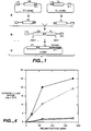
- This invention provides new methods of screening for antibodies that are internalized by particular target cells. Unlike prior art assay methods that simply detect binding to an external target on a cell (e.g. a receptor) the assays of this invention explicitly identify molecules that bind and are transported into the cell ( i.e . into a vacuole and/or the endoplasmic reticulum and/or into the cytosol itself).
- the antibodies identified by the methods of this invention are rapidly internalized into the cell. They are thus extremely useful for delivering effector moieties into the target cell. Moreover, once an internalizing antibody is identified it can be used to re-probe one or more cells or cell lines to identify previously unknown internalizing cellular targets (e.g ., receptors).
- selecting for internalization also selects for biologic function.
- Many receptors for example growth factor receptors
- use internalization as a way of modulating and regulating the effect of ligands.
- ligand binding can result in signal transduction and receptor internalization.
- the decrease in the number of receptors then causes down regulation of the effect of additional ligand.
- antibodies that bind growth factor receptors Hurwitz et al. (1995) Proc. Natl. Acad. Sci. USA. 92: 3353-3357 ).
- [g]rowth factors act by binding to and activating the intrinsic catalytic activity of their cell surface receptors, thereby initiating a signaling cascade leading to the cellular response.
- Growth factor/receptor complexes are not static residents of the cell surface membrane but undergo endocytotic trafficking processes of internalization and sorting to recycling or degradation. Consequently, growth factors are depleted from the extracellular medium and their receptors undergo down-regulation. These trafficking processes, by virtue of their influence on the kinetics of signaling growth factor/receptor complexes, are important modulators of cell behavioral responses" ( Reddy et al. (1996) Nature Biotech. 14: 1696-1699 )
- this invention provides methods for identifying internalizing antibodies.
- the methods involve contacting a "target" cell with one or more members of a phage display library displaying an antibody .
- the phage display library is a multivalent phage display library and it is believed that this invention provides the first description of a multivalent antibody phage display library.
- the cells are washed to remove externally bound phage (library members) and then internalized phage are released from the cells, e . g ., by cell lysis.
- the internalized phage are still viable (infectious).
- the internalized phage in the cell lysate can be recovered and expanded by using the lysate containing internalized phage to infect a bacterial host. Growth of infected bacteria leads to expansion of the phage which can be used for a subsequent round of selection. Each round of selection enriches for phage which are more efficiently internalized, more specific for the target cell or have improved binding characteristics.
- the phage display library is contacted with a subtractive cell line (i.e . a subtractive cell line is added to the target cells and culture media) to remove members of the phage display library that are not specific to the "target" cell(s).
- the subtractive cell line is preferably added under conditions in which members of the phage display library are not internalized (e.g. , at a temperature of about 4°C to about 20°C, more preferably at a temperature of about 4°C) so that non-specific binding members of the library are not internalized (sequestered) before they can be subtracted out by the subtractive cell line.
- the "target" cells are washed to remove the subtractive cell line and to remove non-specifically or weakly-bound phage.
- the target cells are then cultured under conditions where it is possible for internalization to occur (e.g. at a temperature of about 35°C to about 39°C, more preferably at a temperature of about 37°C).
- the duration of the internalization culture period will determine the internalization speed of the antibodies (phage display members) for which selection takes place. With shorter internalization periods more rapid internalizing antibodies are selected, while with longer internalization periods slower internalizing antibodies are selected.
- the internalization period is preferably less than about 120 minutes, more preferably less than about 60 minutes, and most preferably less than about 30 minutes or even less than about 20 minutes.
- the target cells are grown under conditions in which internalization can occur. For a number of cell lines, this involves culturing the cells adherently on culture plates.
- the target cells are washed to remove non-internalized (e.g. surface-bound phage).
- non-internalized e.g. surface-bound phage
- the cells can then be moved to clean media.
- they cells are trypsinized to free the cells from the extracellular matrix which may contain phage antibodies that bind the extracellular matrix. Freeing the cells into solution permits more thorough washing and moving of the cells to a new culture flask will leave behind any phage that may have stuck to the tissue culture dish.
- the cells can then be washed with a large volume of PBS and lysed to release the internalized phage, which can then be expanded, e . g . used to infect E . coli to produce phage for the next round of selection. It is noted that there is no need to actually visualize the internalized phage. Simple cell lysis and expansion of the formerly internalized phage is sufficient for recovering internalizing phage display members.
- an antibody that is internalized into a cell has been identified, it is possible to probe one or more cell types with the identified antibody to identify the target recognized and bound by the antibody. Since the antibody is an internalizing antibody it is likely that such targets are themselves internalizing targets (e.g . members or portions of internalizing receptors).
- the antibody can be labeled as described below.
- the cells can then be contacted with the antibody ( i . e . in vivo or in vitro ) and the cells or cellular regions to which the antibody binds can then be isolated.
- the antibodies can be used e.g . in an affinity matrix (e.g . affinity column) to isolate the targets (e . g. receptor or receptor subunits) to which they bind.
- affinity chromatography involves immobilizing (e.g. on a solid support) one or more species of the internalizing antibodies identified according to the methods of this invention. Cells, cellular lysate, or cellular homogenate are then contacted with the immobilized antibody which then binds to its cognate ligand. The remaining material is then washed away and the bound/isolated cognate ligand can then be released from the antibody for further use.
- the antibodies are used to immunoprecipitate the target from cell lysate.
- the precipitate is then run on an SDS-PAGE gel which is Western blotted onto nitrocellulose.
- the blot is probed with the precipitating antibody to identify the location of the target.
- the portion of the blot containing the target can then be sent for N-terminal protein sequencing.
- the N-terminal sequence can then be used to identify the target from standard databases, or DNA probes can be synthesized to probe genomic or cDNA libraries.
- This approach has been used to identify the antigen bound by a phage antibody. Selections of a phage antibody library were done on intact Chlamydia trachomatis (a bacterial like organism that causes Chlamydial diseases). Selected antibodies were then used as described above to identify the antigen bound.
- polypeptide and antibody fragments on the surface of viruses which infect bacteria makes it possible to isolate a single binding polypeptide or antibody fragment from a library of greater than 10 10 nonbinding clones.
- phage display To express polypeptide or antibody fragments on the surface of phage (phage display), a polypeptide or an antibody fragment gene is inserted into the gene encoding a phage surface protein (pIII) and the antibody fragment-pIII fusion protein is displayed on the phage surface ( McCafferry et al. (1990) Nature, 348: 552-554 ; Hoogenboom et al. (1991) Nucleic Acids Res. 19: 4133-4137 ).
- pIII phage surface protein
- phage bearing antigen binding polypeptides or antibody fragments can be separated from non-binding phage by antigen affinity chromatography ( McCafferry et al. (1990) Nature, 348: 552-554 ).
- affinity chromatography McCafferry et al. (1990) Nature, 348: 552-554 ).
- enrichment factors of 20 fold - 1,000,000 fold are obtained for a single round of affinity selection.
- more phage can be grown and subjected to another round of selection. In this way, an enrichment of 1000 fold in one round can become 1,000,000 fold in two rounds of selection ( McCafferry et al. (1990) Nature, 348: 552-554 ).
- amber codon between the antibody fragment gene and gene III.
- the amber codon makes it possible to easily switch between displayed and soluble (native) antibody fragment simply by changing the host bacterial strain ( Hoogenboom et a/. (1991) Nucleic Acids Res. 19: 4133-4137 ).
- Human antibodies can be produced without prior immunization by displaying very large and diverse V-gene repertoires on phage ( Marks et al. (1991) J. Mol. Biol. 222: 581-597 ).
- V H and V L repertoires present in human peripheral blood lymphocytes were isolated from unimmunized donors by PCR.
- the V-gene repertoires were spliced together at random using PCR to create a scFv gene repertoire which was cloned into a phage vector to create a library of 30 million phage antibodies ( Id .).
- binding antibody fragments have been isolated against more than 17 different antigens, including haptens, polysaccharides and proteins ( Marks et al. (1991) J. Mol. Biol. 222: 581-597 ; Marks et al. (1993). Bio/Technology. 10: 779-783 ; Griffiths et al. (1993) EMBO J. 12: 725-734 ; Clackson et al. (1991) Nature. 352: 624-628 ). Antibodies have been produced against self proteins, including human thyroglobulin, immunoglobulin, tumor necrosis factor and CEA ( Griffiths et al. (1993) EMBO J.
- antibody fragments against four different erythrocyte cell surface antigens were produced by selecting directly on erythrocytes ( Marks et al. (1993). Bio/technlogy. 10: 779-783 ).
- Antibodies were produced against blood group antigens with surface densities as low as 5,000 sites/cell. The antibody fragments were highly specific to the antigen used for selection, and were functional in agglutination and immunofluorescence assays.
- Antibodies against the lower density antigens were produced by first selecting the phage antibody library on a highly related cell type which lacked the antigen of interest.
- This negative selection removed binders against the higher density antigens and subsequent selection of the depleted phage antibody library on cells expressing the antigen of interest resulted in isolation of antibodies against that antigen.
- binders against the higher density antigens With a library of this size and diversity, at least one to several binders can be isolated against a protein antigen 70% of the time.
- the antibody fragments are highly specific for the antigen used for selection and have affinities in the 1 :M to 100 nM range ( Marks et al. (1991) J. Mol. Biol. 222: 581-597 ; Griffiths et al. (1993) EMBO J. 12: 725-734 ). Larger phage antibody libraries result in the isolation of more antibodies of higher binding affinity to a greater proportion of antigens.
- the probability of selecting internalizing antibodies from a phage-display antibody library is increased by increasing the valency of the displayed antibody.
- This approach takes advantage of normal cell-surface receptor biology. Often cell-surface receptors (e.g . growth factor receptors) activate upon binding their cognate ligand through a process of homo- or heterodimerization (or trimerization, or tetramerization, etc. ). The association of the receptor subunits in this process can be mediated directly ( e.g. when bound by a bivalent ligand) or indirectly by causing a conformational change in the receptor.
- cell-surface receptors e.g . growth factor receptors
- this invention utilizes a multivalent phage display antibody library. It is believed that no multivalent phage-display antibody libraries have been created prior to this invention. Unlike the multivalently displayed peptide phage libraries, phage antibody libraries typically display monomeric single chain Fv (scFv) or Fab antibody fragments fused to pIII as single copies on the phage surface using a phagemid system ( Marks et al. (1991) J. Mol. Biol. 222: 581-597 ; Sheets et al. (1998) Proc. Natl. Acad. Sci. USA 95: 6157-6162 .).
- scFv monomeric single chain Fv
- Fab antibody fragments fused to pIII as single copies on the phage surface using a phagemid system
- a multivalent phage display antibody library refers to a library in which each member (e.g . phage particle) displays, on average) two or more binding domains, wherein each binding domain includes a variable heavy and a variable light region. More generally, a multivalent phage display library displays, on average, two or more pIII fusions per phage particle. Multivalent phage display can be achieved by expressing diabodies (i.e., a protein formed by fusion or conjugation of two single chain antibodies ( e.g . scFv)) or by display of, on average, two or more antibodies on each phage particle. In contrast, a mono-valent library displays, on average, one single-chain antibody per viral particle.
- diabodies i.e., a protein formed by fusion or conjugation of two single chain antibodies (e.g . scFv)
- a mono-valent library displays, on average, one single-chain antibody per viral particle.
- Diabodies are scFv dimers where each chain consists of heavy (V H ) and light (V L ) chain variable domains connected using a linker (e.g . a peptide linker) that is too short to permit pairing between domains on the same chain. Consequently, pairing occurs between complementary domains of two different chains, creating a stable noncovalent dimer with two binding sites ( Holliger et al. (1993) Proc. Natl. Acad. Sci. 90: 6444-6448 ).
- a linker e.g a peptide linker
- the C6.5 diabody was constructed by shortening the peptide linker between the Ig V H and V L domains from 15 to 5 amino acids and binds ErbB2 on SKBR3 cells bivalently with a K d approximately 40 fold lower than C6.5 (4.0 x 10 -10 M) ( Adams et al. (1998) Brit. J. Cancer. 77: 1405-1412, 1998 ).
- Example 5 C6.5 diabody genes were subcloned for expression as pIII fusions in the phagemid pHEN-I ( Hoogenboom et al. (1991) Nucleic Acids Res. 19: 4133-4137 ). This.yielded phagemid predominantly expressing a single scFv or diabody-pIII fusion after rescue with helper phage ( Marks et al. (1992) J. Biol. Chem. 267: 16007-16010 ). Diabody phagemid display a bivalent antibody fragment resulting from intermolecular pairing of one scFv-pIII fusion molecule and one native scFv molecule. Using the teachings provided herein one of skill in the art can routinely produce other diabodies.
- Phage displaying bivalent diabodies or multiple copies of scFv were more efficiently endocytosed than phage displaying monomeric scFv and recovery of infectious phage was increased by preincubation of cells with chloroquine.
- antibody phage display libraries are created in which each viral particle, on average, expresses at least 2, preferably at least 3, more preferably at least 4, and most preferably at least 5 copies of a single chain antibody.
- each copy of pIII on the phage should express an antibody.
- proteolysis occurs and the number actually displayed is typically less.
- preferred multivalent antibody libraries are constructed in a phage vector and not a phagemid vector. This means that helper phage need not be added to make phage. Helper phage bring into the E. coli wild-type pIII that competes with the scFv-pIII fusion. Thus, in phagemid vector, this competition leads, on average, to only 1 (or less) antibody per phage.
- the single chain antibodies are subcloned from the phagemid vector into a phage vector.
- No helper phage is required and there is no competition between the wild-type pIII and the fusion scFv pIII fusion.
- the phage display two or more pIII fusions.
- Example 5 describes the subcloning of the C6.5 scFv gene into the phage vector fd-Sfi/Not. This results in phage with 3 to 5 copies each of scFv-pIII fusion protein.
- the target cells used in invention include any cell for which it is desired to identify an internalizing antibody or for which it is desired to identify an internalizing marker (e.g . receptor).
- the cells can include cells of multicellular eukaryotes, uni-cellular eukaryotes, including plants and fungi, and even prokaryotic cells.
- Preferred target cells are eukaryotic, more preferably vertebrate cells, and most preferably mammalian cells (e.g . cells from murines, bovines, primates including humans, largomorphs, canines, felines, and so forth).
- the cells can be normal healthy cells or cells characterized by a particular pathology ( e.g . tumor cells).
- Target cells can include any cell type where it would be useful to: 1) have an antibody specifically recognize the cell type or related cell types (for example for cell sorting, cell staining or other diagnostic procedures); 2) have a ligand which is specifically internalized into the cell type or related cell types (for example to deliver a toxic or therapeutic gene or protein).
- Additional target cells include, but are not limited to differentiated cells (i.e . differentiated to become a tissue, e.g . prostate, breast). Thus an antibody that recognized and killed prostate cells would be good for prostate cancer even if it killed normal prostate cells (the prostate is not an essential organ).
- Target cells may include tissue specific cells, and cells at a given developmental stage.
- Target cells may also include precursor cells, e.g . bone marrow stem cells, would be useful for isolating, perhaps stimulating for differentiation.
- Target cells can also include cell lines transfected with a gene for a known receptor (for example ErbB2) to which it would be useful to have internalizing antibodies.
- a gene for a known receptor for example ErbB2
- ErbB2 antibodies are not internalizing. Rather than immunizing with recombinant protein or selecting a phage library on recombinant protein, selection on ErbB2 transfected cells for internalization should yield precisely antibodies with the desired characteristics (internalization).
- a cDNA library could be transfected into a cell line (for example COS) from a desired target cell line or tissue and phage antibodies selected for internalization. After several rounds of selection, the phage could be used to stain and sort (for example by FACS) transfected cells. DNA can be recovered from the cells, yielding the sequences of internalizing receptors as well as phage antibodies that bind them.
- the phage display library is contacted with cells from a "subtractive" cell line. This step is intended to deplete or eliminate members of the phage display library that either bind the cells non-specifically or that bind to targets other than the target against which it is desired to obtain a binding polypeptide or antibody.
- the contacting with the cells from a "subtractive" cell line can occur before, during, or after the target cells are contacted with members of the phage display library.
- the contacting with cells of a subtractive cell line is simultaneous with contacting of the target cells.
- the target cell line grown adherent to a tissue culture plate
- the subtracting cell line in suspension in a single cell culture flask.
- subtractive cells display all the markers on the target cell except the marker (e.g . receptor) that is to act as a target for selection of the desired binding antibodies or binding polypeptides.
- Particularly preferred cells are thus closely related to the target cell(s), in terms of having common internalizing cell surface receptors (such as transferrin); for example fibroblasts. If one was selecting on a tumor cell line (for example a breast tumor cell line), than one could negatively select on a normal breast cell line. This may, however, deplete for antibodies that bind to overexpressed antigens, so again a parallel path would be to negatively select on fibroblasts. If one was using transfected cells, than non-transfected cell could be used as the subtractive cell line. Where the tumor is epithelial in origin, the referred subtractive cell will also be epithelial and even more preferably from the same tissue or organ.
- Particularly preferred subtractive cells include, but are not limited to, non-differentiated cell lines, non-transfected cells, mixtures of non-differentiated and non-transfected cells.
- preferred subtractive cell lines are preferably the non-tumor cells of the same tissue (for example, breast tumor cells versus normal breast epithelial cells).
- the subtractive cell line will be the non-transformed cell line used for library-construction ( e.g . COS, CHO, etc .).
- the "target" cell is a cell transformed with a gene or cDNA for a specific target receptor.
- the subtractive cell line is preferably the non-transformed cell line.
- the EGF-expressing cells are used as the target cell line, and the subtractive cell line is the untransformed CHO cells. Using this approach internalizing anti-EGF receptor antibodies were obtained.
- the subtractive cells are more effective when provided in excess over the target cells.
- the excess is preferably at least about a 2-fold to about a 1000-fold excess, more preferably about a 3-fold to about a 100-fold excess, and most preferably about a 5-fold to about a 50-fold excess. In one embodiment, a 5-fold excess is sufficient.
- washing steps are used in the methods of this invention.
- a "weak” washing step can be used to remove the subtractive cells and weakly or non-specifically binding members of the phage display library.
- a second strong washing step is preferably used after internalization of members of the phage display library. The "strong” washing step is intended to remove tightly- and weakly-bound surface phage.
- Buffers and methods for performing weak and strong wash steps are well known to those of skill in the art.
- weak washes can be done with standard buffers or culture media (e.g ., phosphate buffered saline (buffer) DMEM (culture media), etc .).
- buffers or culture media e.g ., phosphate buffered saline (buffer) DMEM (culture media), etc .
- Internalizing culture conditions are conditions in which the cell when bound by a member of a phage display library at an appropriate (e.g . Internalizing) site or receptor, transports the bound member into the cell. This can involve transport into a vesicle, into the endoplasmic reticulum, the golgi complex, or into the cytosol of the cell itself.
- Internalizing conditions are most easily achieved when the cells are cultured under conditions that mimic those of the cell in its native state.
- many cells e.g . epidermal cells, preferably grow as adherent layers attached to a basement membrane.
- Such cells more effectively internalize antibodies when they are cultured as adherent monolayers.
- Chloroquine and serum free medium both avoid non-specific internalization and enhance specific internalization (ligands in the serum that induce the internalization of receptor of interest and take with them non-specific phages being in the neighborhood).
- the cells should be cultured at a temperature and pH that permits internalization.
- Suitable temperature and pH range from about 35°C to about 39°C and from pH 6 to about pH 8, more preferably from about pH 6.5 to about pH 7.5, with preferred temperature and pH being about 37°C and pH 7.5 respectively.
- the cells are preincubated in serum culture medium for about two hours before adding the phages and the competitor (subtraction) cells.
- the internalized phage display library members can be identified directly or indirectly. Direct identification can be accomplished simply by visualizing the phage within a cell e.g . via immunofluorescent or confocal microscopy. Phage internalization can be identified by their ability to deliver a reporter gene that is expressed within the cell.
- the reporter gene can be one that produces a detectable signal (e.g . a fluorescent ( e.g . lux, green fluorescent protein, etc.) or colorimetric signal ( e.g . HRP, ⁇ -galactosidase) or can itself be a selectable marker ( e.g . an antibiotic resistance gene).
- a detectable signal e.g a fluorescent ( e.g . lux, green fluorescent protein, etc.
- colorimetric signal e.g . HRP, ⁇ -galactosidase
- the use of both ⁇ -galactosidase and GFP as reporter genes in such phage is described herein.
- the phage display member can bear a marker (e.g . a label) and cells containing the internalized phage can be detected simply by detection of the label (e.g . in a flow cytometer).
- a marker e.g . a label
- the direct methods preferably used for identification of the receptors or cells that are bound after selections are performed. It is noted that cell sorting approaches (FACs) will work with identification of either surface bound or internalized phage. However, an additional level of specificity can be achieved if the cells are first sorted for the presence of internalized phage prior to lysis. Direct methods are also used during the analysis phase to demonstrate that the phage selected are indeed internalized.
- FACs cell sorting approaches
- the internalized phage display library members can be identified indirectly.
- indirect detection methods the phage-display library member(s) do not need to be detected while they are present within the cell. It is sufficient that they simply have been internalized.
- Indirect identification is accomplished for example, by isolating and expanding the phage that were internalized into the cells as described below. Indirect identification is particularly well suited where the identified phage display library members are going to be used in subsequent rounds of selection or to isolate bacteria harboring monoclonal phage genomes for subsequent monoclonal phage characterization (that is for the analysis of selection results).
- phage display library members that have been internalized into target cells (e.g . mammalian tumor cells) remain viable and can be recovered and expanded into a "selected" library suitable for subsequent rounds of selection and/or isolation and characterization of particular members.
- target cells e.g . mammalian tumor cells
- the term "recovery” is intended to include recovery of the infectious phage and/or recovery of the phage antibody gene and/or recovery of a heterologous nucleic acid accompanying the antibody gene.
- the internalized phage can be isolated and expanded using standard methods. Typically these include lysing the cells (e.g ., with 100 mM triethylamine (high pH)), and using the lysate to infect a suitable bacterial host, e.g., E. coli TG1. The phage-containing bacteria are then cultured according to standard methods (see, e.g., Sambrook supra., Marks et al. (1991) J. Mol. Biol. 222: 581-597 ).
- an internalizing antibody As described below, once an internalizing antibody is identified additional copies of the antibody can be prepared using either chemical synthetic means or by the use of recombinant expression systems.
- other "related" internalizing antibodies can be identified by screening for antibodies that bind to the same epitope and/or by modification of the identified internalizing antibody to produce libraries of modified antibody and then rescreening antibodies in the library internalization.
- the internalizing antibodies can be chemically synthesized using well known methods of peptide synthesis.
- Solid phase synthesis in which the C-terminal amino acid of the sequence is attached to an insoluble support followed by sequential addition of the remaining amino acids in the sequence one preferred method for the chemical synthesis of single chain antibodies.
- Techniques for solid phase synthesis are described by Barany and Merrifield, Solid Phase Peptide Synthesis; pp. 3-284 in The Peptides: Analysis, Synthesis, Biology. Vol. 2: Special Methods in Peptide Synthesis, Part A ., Merrifield et al. (1963) J. Am. Chem. Soc., 85: 2149-2156 , and Stewart et al. (1984) Solid Phase Peptide Synthesis, 2nd ed. Pierce Chem. Co., Rockford, Ill ..
- the internalizing antibodies are prepared using standard techniques well known to those of skill in the art. Nucleic acid sequences encoding the internalizing antibodies are determined ( e.g . via Sanger sequencing). Using the sequence information, the nucleic acids may be chemically synthesized according to a number of standard methods known to those of skill in the art. Oligonucleotide synthesis, is preferably carried out on commercially available solid phase oligonucleotide synthesis machines ( Needham-VanDevanter et al. (1984) Nucleic Acids Res. 12: 6159-6168 ) or manually synthesized using the solid phase phosphoramidite triester method described by Beaucage et. al. ( Beaucage et. al. (1981) Tetrahedron Letts. 22(20): 1859-1862 ). Alternatively, nucleic acids encoding the antibody can be amplified and/or cloned according to standard methods.
- the idiotype represents the highly variable antigen-binding site of an antibody and is itself immunogenic. During the generation of an antibody-mediated immune response, an individual will develop antibodies to the antigen as well as anti-idiotype antibodies, whose immunogenic binding site (idiotype) mimics the antigen.
- Anti-idiotypic antibodies can be raised against the variable regions of internalizing antibodies identified in the screening systems of this invention using standard methods well known to those of skill in the art. Briefly, anti-idiotype antibodies can be made by injecting internalizing antibodies of this invention, or fragments thereof (e.g ., CDRs)) into an animal thereby eliciting antiserum against various antigenic determinants on the antibody, including determinants in the idiotypic region.
- CDRs fragments thereof
- Anti-analyte antibodies are well known in the art. Large molecular weight antigens (greater than approx. 5000 Daltons) can be injected directly into animals, whereas small molecular weight compounds (less than approx. 5000 Daltons) are preferably coupled to a high molecular weight immunogenic carrier, usually a protein, to render them immunogenic.
- the antibodies produced in response to immunization can be utilized as serum, ascites fluid, an immunoglobulin (Ig) fraction, an IgG fraction, or as affinity-purified monospecific material.
- Polyclonal anti-idiotype antibodies can be prepared by immunizing an animal with the antibodies of this invention prepared as described above. In general, it is desirable to immunize an animal which is species and allotype-matched with the animal from which the antibody (e.g . phage-display library) was derived. This minimizes the production of antibodies directed against non-idiotypic determinants.
- the antiserum so obtained is then usually absorbed extensively against normal serum from the same species from which the phage-display library was derived, thereby eliminating antibodies directed against non-idiotypic determinants. Absorption can be accomplished by passing antiserum over a gel formed by crosslinking normal (nonimmune) serum proteins with glutaraldehyde.
- Antibodies with anti-idiotypic specificity will pass directly through the gel, while those having specificity for non-idiotypic determinants will bind to the gel.
- Immobilizing nonimmune serum proteins on an insoluble polysaccharide support e.g ., sepharose
- an insoluble polysaccharide support e.g ., sepharose
- Monoclonal anti-idiotype antibodies can be produced using the method of Kohler et al. (1975) Nature 256: 495 .
- monoclonal anti-idiotype antibodies can be prepared using hybridoma technology which comprises fusing (1)spleen cells from a mouse immunized with the antigen or hapten-carrier conjugate of interest (i.e ., the antibodies or this invention or subsequences thereof) to (2) a mouse myeloma cell line which has been selected for resistance to a drug (e.g ., 8-azaguanine).
- a myeloma cell line which does not secrete an immunoglobulin.
- a preferred cell line is P3X63Ag8.653. This cell line is on deposit at the American Type Culture Collection as CRL-1580.
- Fusion can be carried out in the presence of polyethylene glycol according to established methods (see, e.g., Monoclonal Antibodies, R. Kennett, J. McKearn & K. Bechtol, eds. N.Y., Plenum Press, 1980 , and Current Topics in Microbiology & Immunology, Vol. 81, F. Melchers, M. Potter & N. L. Warner, eds., N.Y., Springer-Verlag, 1978 ).
- the resultant mixture of fused and unfused cells is plated out in hypoxanthine-aminopterin-thymidine (HAT) selective medium. Under these conditions, only hybrid cells will grow.
- HAT hypoxanthine-aminopterin-thymidine
- the culture medium is harvested and screened for the presence of monoclonal idiotypic, anti-analyte antibody by any one of a number of methods which include solid phase RIA and enzyme-linked immunosorbent assay.
- Cells from culture wells containing antibody of the desired specificity are then expanded and recloned.
- Cells from those cultures that remain -positive for the antibody of interest are then usually passed as ascites tumors in susceptible, histocompatible, pristane-primed mice.
- Ascites fluid is harvested by tapping the peritoneal cavity, retested for antibody, and purified as described above. If a nonsecreting myeloma line is used in the fusion, affinity purification of the monoclonal antibody is not usually necessary since the antibody is already homogeneous with respect to its antigen-binding characteristics. All that is necessary is to isolate it from contaminating proteins in ascites, i.e ., to produce an immunoglobulin fraction.
- the hybrid cell lines of interest can be grown in serum-free -tissue culture and the antibody harvested from the culture medium. In general, this is a less desirable method of obtaining large quantities of antibody because the yield is low. It is also possible to pass the cells intravenously in mice and to harvest the antibody from serum. This method is generally not preferred because of the small quantity of serum which can be obtained per bleed and because of the need for extensive purification from other serum components. However, some hybridomas will not grow as ascites tumors and therefore one of these alternative methods of obtaining antibody must be used.
- anti-idiotypic antibody instead of the anti-idiotypic antibody, other internalizing antibodies can be identified by cross-reactivity with the identified "prototypic” antibodies, against the epitope(s) used in the original selection. Competition between the "prototypic" internalizing antibodies and new candidates in an epitope-mapping format establishes that the antibodies are competing for the same epitope.
- mutant scFv gene repertories based on the sequence of a binding of an identified internalizing antibody, are created and expressed on the surface of phage.
- Higher affinity scFvs are selected on antigen as described above and in the Examples.
- a mutant scFv gene repertoire can be created containing the V H gene of the internalizing antibody and a human V L gene repertoire (light chain shuffling).
- the scFv gene repertoire can be cloned into the phage display vector pHEN-1 ( Hoogenboom et al. (1991) Nucleic Acids Res., 19: 4133-4137 ) and after transformation a library of transformants is obtained.
- the internalizing antibody V H CDR1 and/or CDR2, and/or CDR3 and light chain are cloned into a vector containing a human V H gene repertoire to create a phage antibody library transformants.
- chain shuffling to increase antibody affinity see Schier et al. (1996) J. Mol. Biol., 255: 28-43, 1996 .
- CDRs complementarity determining regions
- V H V H
- V L V L
- CDR1, CDR2, and CDR3 V L
- mutant antibody libraries can be created where partial or entire CDRs are randomized (V L CDR1 and CDR2 and V H CDR1, CDR2 and CDR3).
- each CDR is randomized in a separate library, using the known internalizing antibody as a template.
- the CDR sequences of the highest affinity mutants from each CDR library are combined to obtain an additive increase -in affinity.
- hGH human growth hormone
- V H CDR3 often occupies the center of the binding pocket, and thus mutations in this region are likely to result in an increase in affinity ( Clackson et al. (1995) Science, 267: 383-386 ).
- four V H CDR3 residues are randomized at a time using the nucleotides NNS ( see, e.g., Schier et al. (1996) Gene, 169: 147-155 ; Schier and Marks (1996) Human Antibodies and Hybridomas. 7: 97-105, 1996 ; and Schier et al. (1996) J. Mol. Biol. 263: 551-567, 1996 ).
- scFv' two internalizing scFvs are joined, either through a linker (e.g ., a carbon linker, a peptide, etc .) or through a disulfide bond between, for example, two cysteins.
- a linker e.g ., a carbon linker, a peptide, etc .
- disulfide bond between, for example, two cysteins.
- a cysteine residue is introduced by site directed mutagenesis between the myc tag and a hexahistidine tag at the carboxy-terminus of the antibodies described herein. Introduction of the correct sequence can be verified by DNA sequencing.
- the pelB leader directs expressed scFv to the periplasm and cloning sites (NcoI and NotI) exist to introduce F5 or C1 mutant scFv.
- the expressed scFv has the myc tag at the C-terminus, followed by 2 glycines, a cysteine, and then 6 histidines to facilitate purification by IMAC. After disulfide bond formation between the two cysteine residues, the two scFv are separated from each other by about 26 amino acids (two 11 amino acid myc tags and 4 glycines).
- scFv can be expressed from this construct, purified by IMAC, and analyzed by gel filtration.
- cysteine is reduced by incubation with 1 mM 3-mercaptoethanol, and half of the scFv blocked by the addition of DTNB. Blocked and unblocked scFvs are incubated together to form (scFv') 2 and the resulting material can be analyzed by gel filtration.
- the affinity of the F5 and C1 scFv' monomers and the F5 and C1 (scFv')2 dimers is determined by BIAcore.
- the (scFv') 2 dimer is created by joining the scFv' fragments through a linker, more preferably through a peptide linker.
- a linker more preferably through a peptide linker.
- This can be accomplished by a wide variety of means well known to those of skill in the art. For example, one preferred approach is described by Holliger et al. (1993) Proc. Natl. Acad. Sci. USA, 90: 6444-6448 ( see also WO 94/13804 ).
- selection for increased avidity involves measuring the affinity of the antibody for the target antigen (e.g ., c-erbB-2).
- target antigen e.g ., c-erbB-2
- the K d of F5, C1, or an F5- or C1-derived antibody the kinetics of binding to c-erbB-2 are determined in a BLAcore, a biosensor based on surface plasmon resonance.
- antigen is coupled to a derivatized sensor chip capable of detecting changes in mass. When antibody is passed over the sensor chip, antibody binds to the antigen resulting in an increase in mass that is quantifiable.
- Measurement of the rate of association as a function of antibody concentration can be used to calculate the association rate constant (k on ).
- buffer is passed over the chip and the rate of dissociation of antibody (k off ) determined.
- K on is typically measured in the range 1.0 x 10 2 to 5.0 x 10 6 and k off in the range 1.0 x 10 -1 to 1.0 x 10 -6 .
- the equilibrium constant K d is often calculated as k off /k on and thus is typically measured in the range 10 -5 to 10 12 . Affinities measured in this manner correlate well with affinities measured in solution by fluorescence quench titration.
- this invention provides libraries as claimed and described above
- kits as claimed
- the kits include members of a phage display library.
- the assay kits can additionally include any of the other components described herein for the practice of the assays of this invention.
- Such materials preferably include, but are not limited to, helper phage, one or more bacterial or mammalian cell lines, buffers, antibiotics, labels, and the like.
- kits may optionally include instructional materials containing directions (i.e ., protocols) disclosing the selection methods described herein. While the instructional materials typically comprise written or printed materials they are not limited to such. Any medium capable of storing such instructions and communicating them to an end user is contemplated . Such media include, but are not limited to electronic storage media (e.g., magnetic discs, tapes, cartridges, chips), optical media (e.g ., CD ROM), and the like. Such media may include addresses to internet sites that provide such instructional materials.
- electronic storage media e.g., magnetic discs, tapes, cartridges, chips
- optical media e.g ., CD ROM
- Example 1 Creation of a non-immune human Fab phage antibody library containing 10 9 -10 11 members
- V H and V L library on separate replicons, combine them into an scFv gene repertoire by splicing by overlap extension, and clone the scFv gene repertoire into the phage display vector pHEN1.
- Human peripheral blood lymphocyte and spleen RNA was primed with IgM heavy chain constant region and, kappa and lambda light chain constant region primers and first strand cDNA synthesized. 1 st strand cDNA was used as a template for PCR amplification of VH V ⁇ k and V ⁇ gene repertoires.
- the V H gene repertoires were cloned into the vector pUC119Sfi-Not as Ncol-NotI fragments, to create a library of 8.0 x 10⁸ members.
- the library was diverse by PCR fingerprinting. Single chain linker DNA was spliced onto the V L gene repertoires using PCR and the repertoire cloned as an XhoI-NotI fragment into the vector pHENIXscFv to create a library of 7.2 x 10 6 members.
- the V H and V L gene repertoires were amplified from their respective vectors and spliced together using PCR to create an scFv gene repertoire.
- the scFv gene repertoire was cloned as an NcoI-NotI fragment into the vector to create an scFv phage antibody library of 7.0 x 10 9 members.
- the library was diverse as determined by BstN1 fingerprinting.
- phage were prepared and selected on 14 different protein antigens. The results are shown in Table 1. scFv antibodies were obtained against all antigens used for selection, with between 3 and 15 unique scFv isolated per Table 1. Results of phage antibody library selections.
- Protein antigen used for selection Percentage (number) of ELISA positive clones Number of different antibodies isolated FGF Receptor ECD 69 (18/26) 15 BMP Receptor Type I ECD 50 (12/24) 12 Activin Receptor Type I ECD 66 (16/24) 7 Activin Receptor Type II ECD 66 (16/24) 4 Erb-B2 ECD 91 (31/34) 14 VEGF 50 (48/96) 6 BoNT/A 28 (26/92) 14 BoNT-A C-fragment 95 (87/92) 10 BoNT/B 10 (9/92) 5 BoNT/C 12 (11/92) 5 BoNT/E 9 (8/92) 3 Bungarotoxin 67 (64/96) 15 Cytochrome b5 55 (53/96) 5 Chlamydia trachomatis EB 66 (63/96) 7 antigen
- Chlamydia trachomatis elementary bodies yielded seven that specifically recognized chlamydia (Table 1).
- the scFv could be successfully used in a number of immunologic assays including ELISA, immunofluorescence, Western blotting, epitope mapping and immunoprecipitation.
- the number of binding antibodies for each antigen, and the affinities of the binding scFv are comparable to results obtained from the best phage antibody libraries (Table 3).
- Table 3 Comparison of protein binding antibodies selected from non-immune phage-display antibody libraries.
- N V-gene repertoires obtained from V-genes rearranged in vivo;
- SS semi-synthetic V-genes constructed from cloned V-gene segments and synthetic oligonucleotides encoding V H CDR3.
- ND not determined.
- Example 2 Uptake of scFV into cells by receptor mediated endocytosis and subsequent recovery.
- the 7.0 x 10 9 member scFv phage antibody library described above was selected on the malignant breast tumor cell lines MB231 and ZR-75-1, both with and without negative selections on the normal breast cell line HBL100. Similar results were obtained as described in section above. scFv were isolated that could not distinguish malignant from non-malignant cell lines.
- phage binding cell surface receptors could be taken up into cells by receptor mediated endocytosis and could then be recovered from cells by lysing the cells. This assumed: 1) that phage could be internalized by receptor mediated endocytosis and 2) that phage could be recovered in the infectious state from within cells prior to lysosomal degradation.
- the ability to select for internalized phage antibodies would have two major benefits: 1) the identification of antibodies that bind to receptors capable of internalization and 2) an added level of specificity in the selection process. Identification of antibodies which are internalized would be highly useful for many targeted therapeutic approaches where internalization is essential ( e.g . immunotoxins, targeted liposomes, targeted gene therapy vectors and others).
- C6.5 phage and C6.5 diabody phage see , copending application USSN 08/665,202 .
- C6.5 scFv is internalized, but at a slow rate, and that the C6:5 diabody is somewhat better internalized (probably because it causes receptor dimerization).
- C6.5 phage, C6.5 diabody phage or an irrelevant anti-Botulinum phage were incubated with SKBR3 cells (ErbB2 expressing breast tumor cell line) at either 37° C or 4° C and non-internalized phage removed by sequential washing with PBS and low pH glycine buffer.
- the cells were then permeabilized and biotinylated anti-M13-antibody added followed by streptavidin Texas Red. Cells were then examined by using a confocal microscope. Both C6.5 phage and C6.5 diabody phage were observed within the cytoplasm). Approximately 1% of cells had internalized C6.5 phage and 20% of the cells had internalized C6.5 diabody phage. There was no internalization of the anti-Botulinum phage.
- C6.5 phage or C6.5 diabody phage were incubated with SKBR3 cells at 37° C. Non bound phage were removed by washing with PBS and phage bound to the cell surface were eluted by washing twice with low pH glycine. The cells were then lysed and each fraction (the first and second glycine washes and the cytoplasmic fraction) used to infect E. coli TG1. Twenty times (C6.5) or 30 times (C6.5 diabody) more phage were bound to the cell surface than the anti-Botulinum phage (glycine 1 wash) (Table 4).
- Phage specificity 1st glycine wash 2nd glycine wash Lysed cell fraction anti-Botulinum 6.0 x 10 5 1.0 x 10 5 6.0 x 10⁵ Anti-ErbB2 (C6.5 scFv) 1.2 x 10 7 5.2 x 10 6 6.8 x 10 7 Anti-ErbB2 (C6.5 diabody) 1.8 x 10 7 2.8 x 10 6 1.7 x 10 7
- phage binding cell surface receptors can be taken up by cells and the infectious phage recovered from the cytoplasm.
- the amount of uptake is significantly greater than uptake of non-binding phage, and the 100 to 200 fold difference is well within the range that would allow enrichment from a library.
- What is unknown from the results is whether the phage antibodies are mediating receptor mediated internalization or whether they are merely taken up after binding by membrane turnover.
- Phage antibodies were rescued from the library and selected on SKBR3 cells.
- Phage antibodies were incubated with cells at 37°C, non-binding phage removed by washing cells with PBS and phage bound to cell surface antigens removed by sequential washes with low pH glycine. Cells were then lysed to release internalized phage and the lysate used to infect E. coli TG1 to prepare phage for the next round of selection. Three rounds of selection were performed. One hundred clones from each round of selection were analyzed for binding to SKBR3 cells and to ErbB2 extracellular domain by ELISA.
- the scFv gene from ELISA positive clones was PCR amplified and fingerprinted by digestion with BstN1. Two unique restriction patterns were identified. The scFv genes were sequenced and 2 unique ErbB2 binding scFv identified. Similar analysis of SKBR3 ELISA positive clones that did not bind ErbB2 identified an additional 11 unique scFv.
- phage were prepared from each unique clone and analyzed for binding to SKBR3 cells (high ErbB2 expression) as well as 2 other epithelial tumor cell lines (SK-OV-3, moderate ErbB2 expression and MCF7, low ErbB2 expression) and a normal breast cell line (HS578B). Each unique clone specifically stained tumor cell lines but not the normal breast cell line.
- SKBR3 and MCF7 cells were incubated with phage antibodies C6.5 (positive control), 3TF5 and 3GH7.
- C6.5 positive control
- 3TF5 3TF5 binding ErbB2
- 3GH7 3GH7 unknown.
- All 3 phage antibodies intensely stain SKBR3 cells (the selecting cell line and high ErbB2 expresser.
- C6.5 phage weakly stain MCF7 cells (low ErbB2 expressor).
- the anti-ErbB2 clone 3TF5 from the library stains MCF7 cells much more intensely then C6.5, as does 3GH7.
- SKBR3, SK-OV-3, MCF7 and HST578 cells were studied using native purified scFv 3TF5 and 3GH7.
- the scFv genes were subcloned into a vector which fuses a hexahistidine tag to the scFv C-terminus.
- scFv was then expressed, - harvested from the bacterial periplasm and purified by immobilized metal affinity chromatography.
- the two scFv intensely stain SKBR3 cells, and do not stain the normal breast cell line HST578.
- the intensity of staining is less than seen with phage. This is to be expected since the secondary antibody for phage staining recognizes the major coat protein (2500 copies/phage) resulting in tremendous signal amplification.
- the anti-ErbB2 phage antibody 3TF5 was studied further to determine if it was indeed internalized. This antibody was selected for initial study since its internalization could be compared to ErbB2 binding C6.5.
- 5.0x10 11 3TF5 or C6.5 phage were incubated with SKBR3 cells at 37°C or at 4°C. After washing with PBS, 3TF5 phage stained cells more intensely than C6.5 phage. After washing with low pH glycine, confocal microscopy revealed that 3TF5 phage were internalized by greater than 95% of cells, while C6.5 was internalized by only a few percent of cells. Incubation of either antibody at 4°C led to no internalization.
- the native purified 3TF5 scFv was similarly analyzed and was also efficiently internalized by SKBR3 cells. It should be noted that the native 3TF5 scFv existed only as a monomer with no appreciable dimerization or aggregation as determined by gel filtration.
- Phage that bind an internalizing cell surface receptor can be enriched more than 100 fold over non-binding phage. This level of enrichment is greater than that achieved by selecting on the cell surface.
- Phage display has the potential to produce antibodies with affinities that cannot be produced using conventional hybridoma technology. Ultra high affinity human antibody fragments could result in excellent tumor penetration, prolonged tumor retention, and rapid clearance from the circulation, leading to high specificity. We therefore undertook a series of experiments to develop methodologies to generate ultra high affinity human antibody fragments.
- Studies of the higher affinity scFv are pending. The results demonstrate our ability to increase antibody affinity to values not achievable from hybridoma technology and confirm the importance of affinity in tumor targeting
- C6.5 based molecules are being engineered for radioimmunotherapy.
- dimeric scFv 'diabodies' were created by shortening the linker between the V H and V I domains from 15 to 5 amino acids. Consequently, pairing occurs between complementary domains of two different chains, creating a stable noncovalently bound dimer with two binding sites.
- diabodies produced from the V-genes of C6.5 have a significantly higher apparent affinity and longer retention on the surface of SK-OV-3 cells compared to C6.5 scFv (T 1/2 > 5 hr vs. 5 min) ( Adams et al. (1998) Brit. J.
- C6.5 based molecules are being used to target doxorubicin containing stealth liposomes to ErbB2 expressing breast cancers ( Kirpotin et al. (1997) Biochemistry. 36: 66-75 ).
- the C6.5 gene was subcloned into an E. coli expression vector resulting in addition of a free cysteine residue at the C-terminus of the scFv.
- Purified C6.5cys scFv was conjugated to liposomes and in vitro uptake determined using SKBR3 cells. Total uptake was 3.4 mmol phospholipid/10 6 cells at 6 hour, with 70% of the uptake internalized.
- a large (7.0 x 10 9 member) phage antibody library has been created which can provide panels of human antibodies to purified antigens with affinities comparable to the affinities of antibodies produced by murine immunization.
- the phage antibodies binding cell surface receptors can be can be internalized by cells and recovered in an infectious state from within the cell. Methodologies were developed which permit enrichment of internalizing phage antibodies over non-internalizing antibodies more than 100 fold. These methodologies were then applied to select new scFv antibodies that bind to internalizing receptors on SKBR-3 cells. Several of these antibodies bind to ErbB2, but are internalized more efficiently than C6.5 based scFv. Many more antibodies bind to unknown internalizing receptors.
- Phage displaying bivalent diabodies or multiple copies of scFv were more efficiently endocytosed than phage displaying monomeric scFv and recovery of infectious phage was increased by preincubation of cells with chloroquine. Measurement of phage recovery from within the cytosol as a function of applied phage titer indicates that it is possible to select for endocytosable antibodies, even at the low concentrations that would exist for a single phage antibody member in a library of 10 9 .
- the SKBR3 breast tumor cell line was obtained from ATCC and grown in RPMI media supplemented with 10% FCS (Hyclone) in 5% CO 2 at 37°C.
- the C6.5 scFv phage vector was constructed by subcloning the C6.5 gene as a Sfi I/ Not I fragment from scFv C6.5 pHEN1 ( Schier et al. (1995) Immunotechnology 1: 63-71 ) into the phage vector fd / Sfi I lNot I (a gift of Andrew Griffiths, MRC Cambridge, UK).
- the C6.5 diabody phagemid vector was constructed by subcloning the C6.5 diabody gene ( Adams et al. (1998) Brit. J. Cancer. 77: 1405-1412, 1998 ) as a NcoI/NotI fragment into pHEN1 ( Hoogenboom et al. (1991) Nucleic Acids Res.
- SKBR3 cells were grown on coverslips in 6-well culture plates (Falcon) to 50% of confluency. Culture medium was renewed 2 hours prior to the addition of 5.10 11 cfu/ml of phage preparation (the phage preparation representing a maximum of 1/10 of the culture medium volume) or 20 ⁇ g/ml of purified scFv or diabody in phosphate buffered saline, pH 7.4 (PBS).
- PBS phosphate buffered saline
- Phage particles were detected with biotinylated anti-M13 immunoglobulins (5 Prime-3 Prime, Inc, diluted 300 times) (45 min. at RT) and Texas red-conjugated streptavidin (Amersham, diluted 300 times) (20 min. at RT). Soluble scFv and diabodies containing a C-terminal myc peptide tag were detected with the mouse mAb 9E10 (Santa Cruz Biotech, diluted 100 times) (45 min. at RT), anti-mouse biotinylated immunoglobulins (Amersham, diluted 100 times) and Texas red-conjugated streptavidin. Optical confocal sections were taken using a Bio-Rad MRC 1024 scanning laser confocal microscope. Alternatively, slides were analyzed with a Zeiss Axioskop UV fluorescent microscope.
- Subconfluent SKBR33 cells were grown in 6-well plates. Culture medium was renewed 2 hours prior to the experiment. Cells were incubated for varying times with different concentrations of phage preparation at 37°C. Following PBS and stripping buffer washes, performed exactly as described above for detection of internalized native antibody fragments and phage antibodies, the cells were washed again twice with PBS and lysed with 1 mL of 100 mM triethylamine (TEA). The stripping buffer washes and the TEA lysate were neutralized with 1/2 volume of Tris-HCl 1M, pH 7.4.
- TEA triethylamine
- cells were trypsinized after the three stripping buffer washes, collected in a 15 ml Falcon tube, washed twice with PBS and then lysed with TEA.
- SKBR3 cells were preincubated for two hours in the presence of complete medium containing 50 ⁇ M chloroquine prior to the addition of phage.
- Corresponding control samples in the absence of chloroquine were prepared at the same time.
- phage were titered on E. coli TG1 as described above.
- the human anti-ErbB2 scFv C6.5 was obtained by selecting a human scFv phage antibody library on recombinant ErbB2 extracellular domain (13).
- affinity on internalization we studied a scFv (C6ML3-9) which differs from C6.5 by 3 amino acids ( Schier et al. (1996) J. Mol. Biol. 263: 551-567 ).
- C6ML3-9 scFv is also a stable monomer in solution and binds the same epitope as C6.5 scFv but with a 16 fold lower K d (1.0 x 10 -9 M) ( Schier et al. (1996) J. Mol. Biol. 263: 551-567 ; Adams et al. (1998) Cancer Res. 58: 485-490 ). Since receptor homodimerization appears to typically be requisite for antibody internalization we also studied the dimeric C6.5 diabody ( Adams et al. (1998) Brit. J. Cancer. 77: 1405-1412, 1998 ).
- Diabodies are scFv dimers where each chain consists of heavy (V H ) and light (V L ) chain variable domains connected using a peptide linker which is too short to permit pairing between domains on the same chain. Consequently, pairing occurs between complementary domains of two different chains, creating a stable noncovalent dimer with two binding sites ( Holliger et al. (1993) Proc. Natl. Acad. Sci. 90: 6444-6448 ).
- the C6.5 diabody was constructed by shortening the peptide linker between the Ig V H and V L domains from 15 to 5 amino acids and binds ErbB2 on SKBR3 cells bivalently with a K d approximately 40 fold lower than C6.5 (4.0 x 10 -10 M) ( Adams et al. (1998) Brit. J. Cancer. 77: 1405-1412,1998 ).
- Native C6.5 scFv and C6.5 diabody was expressed and purified from E. coli and analyzed for endocytosis into ErbB2 expressing SKBR3 breast tumor cells by immunofluorescent confocal microscopy. As expected, monomeric C6.5 scFv is not significantly internalized whereas the dimeric C6.5 diabody can be detected in the cytoplasm of all cells visualized.
- the human breast cancer cell line SKBR3 was used as a target cell line for endocytosis. Its surface ErbB2 density is approximately 1.0 x 10 6 per cell ( Hynes et al. (1989) J. Cell. Biochem 39: 167-173 ).
- C6.5 scFv phagemids were incubated for 2 hours with SKBR3 cells grown on coverslips at 37°C to allow active internalization. Cells were extensively washed with PBS to remove non specific binding and washed an additional three times with high salt and low pH (stripping) buffer to remove phage specifically bound to cell surface receptors. Internalized phagemid were detected with a biotinylated M13 antiserum recognizing the major coat phage protein pVIII. An anti-botulinum toxin phagemid was used as a negative control. Staining was analyzed by using immunofluorescent microscopy.
- Infectious phage can be recovered from within the cell and their titre correlates with the level of uptake observed using immunofluorescence
- Table 6 Titration of membrane bound and intracellular phage. 3.0 x 10 11 cfu of monovalent C6.5 scFv phagemid, 16 fold higher affinity monovalent C6ML3-9 scFv phagemid, bivalent C6.5 diabody phagemid or multivalent C6.5 fd phage were incubated with sub confluent SKBR3 cells for 2 hours at 37°C. Cells were washed 6 times with PBS, 3 times with stripping buffer and then lysed to recover intracellular phage. The various fractions were neutralized and the phage titered. The total number of cfu of each fraction is reported.
- Non specific anti-botulinum phagemid were used to determine non specific recovery.
- Cell Surface Phage Titer (x 10 -5 ) Intracellular Phage Antibody 1 st Wash 2nd Wash 3rd Wash Phage Titer (x 10 -5 )
- Anti-botulinum phagemid 280 36 2.8 15 C6.5 scFv phagemid 600 96 7.6 52
- C6ML3-9 scFv phagemid 2500 140 32 270 C6.5 diabody phagemid 1800 120 13 450 C6.5 scFv phage 2300 620 56 2200
- Table 7 Specific enrichment of anti-ErbB2 phage compared to anti-botulinum phage.
- the titers of anti-ErbB2 phage are divided by the titers of the anti-botulinum phage (Table 6) to derive an enrichment ratio for specific vs nonspecific binding or internalization.
- **The titer of intracellular phage is divided by the titer of cell surface bound phage (Table 6) to derive the ratio of internalized phage vs surface bound phage.
- Some anti-botulinum phage might also be non-specifically endocytosed by cells, but this is likely to be a small amount given the immunofluorescence results.
- the titer of phage in the TEA fraction increased with increasing affinity and avidity of C6.5, with the highest titers observed for the dimeric C6.5 diabody phagemid and the multivalent C6.5 scFv phage (Table 6).
- the values represent a 30 fold (C6.5 diabody phagemid) and 146 fold (C6.5 scFv phage) increase in titer compared to the anti-botulinum phage (Table 6).
- the increase in the phage titer in the cell lysate compared to the last stripping wash is due to endocytosed phage.
- some of these phage could have come from the cell surface or intracellular matrix. While this could be true for a fraction of the phage from the cell lysate, the immunofluorescence results indicate that at least some of the phage are endocytosed.
- One indicator of the relative fraction of endocytosed phage for the different C6.5 molecules is to compare the amount of phage remaining on the cell surface prior to cell lysis (last stripping wash) with the amount recovered after cell lysis.
- phage antibodies can undergo receptor mediated endocytosis and remain infectious in a cell lysate.
- Selection of internalized phages from a phage library requires the optimization of the method to increase enrichment of specifically internalized phages over non-internalized phage.
- Two parameters can be improved: (1) reduction of the recovery of non-specific or non-internalized phage and (2) preservation of the infectivity of internalized phage.
- wild-type C6.5 scFv phagemid We chose this molecule because it was clearly endocytosed based on confocal microscopy, yet was the molecule undergoing the least degree of specific endocytosis.
- C6.5 scFv phagemid also represents the most commonly utilized format for display of non-immune phage antibody libraries (single copy pIII in a phagemid vector) and has an affinity (16 nM) more typical of Kd's of scFv from such libraries than the affinity matured C6ML3-9 scFv ( Sheets et al. (1998) Proc. Natl. Acad. Sci. USA 95: 6157-6162 ; Vaughan et al. (1996) Nature Biotech. 14: 309-314 ).
- SKBR3 cells were incubated with chloroquine and either C6.5 scFv phagemid or anti-botulinum phagemid. Cell lysates were titered at various time points to determined the number of intracellular phagemid. C6.5 scFv phagemid were present at the 20 minute time point and the amount of phagemid was comparable with or without the addition of chloroquine.
- SKBR3 cells were incubated with C6.5 scFv, C6ML3-9 scFv or C6.5 diabody phagemids or C6.5 scFv phage for 2 hours at 37°C. Cells were washed three times with stripping buffer, trypsinized and washed twice with PBS. Cells were lysed and intracellular phage titered on E. coli TG1. Phage recovery increased with increasing phage titer for all phage studied ( Figure 5 ).
- phagemid could not be recovered from within the cell at input titers less than 3.0 x 10 5 (C6.5 scFv) to 3.0 x 10 6 (C6ML3-9 scFv) This threshold decreased for bivalent and multivalent display (3.0 x 10 4 for C6.5 diabody phagemid and C6.5 scFv phage).
- phage displaying an anti-receptor antibody can be specifically endocytosed by receptor expressing cells and can be recovered from the cytosol in infectious form.
- the results demonstrate the feasibility of directly selecting internalizing antibodies from large non-immune phage libraries and identify the factors that will lead to successful selections.
- Phage displaying anti-ErbB2 antibody fragments are specifically endocytosed by ErbB2 expressing SKBR3 cells, can be visualized within the cytosol and can be recovered in an infectious form from within the cell.
- monovalent scFv antibody fragments were displayed monovalently in a phagemid system, recovery of internalized phage was only 3.5 to 18 fold above background.
- scFv monovalent scFv
- scFv could be especially useful for the construction of fusion molecules for the delivery of drugs, toxins or DNA into the cytoplasm. Since antibodies which mediate receptor internalization can cause receptor down regulation and growth inhibition ( Hurwitz et al. (1995) Proc. Natl. Acad Sci. USA 92: 3353-3357 ; Hudziak et al. (1989) Mol. Cell. Biol. 9: 1165-1172 ; Stancovski et al. (1991) Proc. Natl. Acad. Sci. USA 88: 8691-8698 ; Lewis et al. (1993) Cancer Immunol. Immunother. 37: 255-263 ), selection for endocytosable antibodies may also identify antibodies which directly inhibit or modulate cell growth.
- the F5 scFv gene was removed from pHEN1-FS by digestion of phagemid DNA with the restriction enzymes SfiI and NotI.
- a phage vector based on FdDOGI See prior Ref., but modified to insert an SfiI site into the gene will leader sequence, was digested with SfiI and NotI and the digested F5 gene ligated into digested phage Fd vector DNA. Recombinant transformant were identified.
- E. coli containing the F5 recombinant phage were grown in culture to produce F5-Fd phage (see Maniatis for phage preparation). F5 phages were then used to infect E.
- coli harboring a phagemid which contains a mammalian promoter (CMV) followed by either the gene for 3-galactosidase (pcDNA3.1/HisB/LacZ, In Vitrogen) or the gene for the enhanced green fluorescent protein (pN2EGFP, Clonetch plasmid) and a eucaryotic polyadenylation sequence.
- CMV mammalian promoter
- pcDNA3.1/HisB/LacZ In Vitrogen
- pN2EGFP enhanced green fluorescent protein
- Bacteria were grown overnight in the presence of tetracycline 15 ug/mL and either ampicillin 100 ug/mL (pCDNA3.1/HisB/LacZ containing bacteria) or Kanamycine 30 ug/mL (pN2EGFP containing bacteria).
- the phage prepared from the supernatant a mixture of F5-Fd coat contains either the reporter gene (about 50% of the phages) in a single strand format or the F5-Fd phage genome (about 50% of the phages).
- Incubation of ErbB2 positive cells 5.105 SKBR3 with 10 7 pfu the phage mix (Filtered twice through a 0.45 nm filter to sterility) allowed expression of the reporter gene in 1% of the cells. Cells incubated with an 10 time fold more negative control phage, i.e. reporter gene packaging in wild type Fd, showed no expression of the reporter genes.
- SKBR3 mixed population of ErbB2 high
- MCF7 ErbB2 low cells
- Example 7 Targeted gene delivery to mammalian cells by filamentous bacteriophage
- prokaryotic viruses can be re-engineered to infect eukaryotic cells resulting in expression of a portion of the bacteriophage genome.
- Phage capable of binding mammalian cells expressing the growth factor receptor ErbB2 and undergoing receptor mediated endocytosis were isolated by selection of a phage antibody library on breast tumor cells and recovery of infectious phage from within the cell.
- F5 phage were efficiently endocytosed into 100% of ErbB2 expressing SKBR3 cells.
- F5 phage were engineered to package the green fluorescent protein (GFP) reporter gene driven by the CMV promoter.
- GFP green fluorescent protein
- This approach can also be adapted to directly select, rather than screen, phage antibodies for targeted gene expression.
- the results demonstrate the potential of phage antibodies as an in vitro or in vivo targeted gene delivery vehicle.
- phage libraries can be selected not only for cell binding but also for internalization into mammalian cells. If the phage single stranded phage genome can be transcribed and translated, then it should prove possible to screen or select for phage which bind receptors in a manner which leads to endocytosis and delivery of the phage genome into the correct trafficking pathway leading to expression.
- filamentous phage displaying the anti-ErbB2 scFv F5 as a genetic fusion with the phage minor coat protein pIII can directly infect mammalian cells expressing ErbB2 leading to expression of a reporter gene contained in the phage genome.
- This offers a new way to discover targeting molecules for intracellular drug delivery or gene therapy by directly screening phage antibodies to identify those capable of undergoing endocytosis and delivering a gene intracellularly into the correct trafficking pathway for gene expression. This should significantly facilitate the identification of appropriate targets and targeting molecules for gene therapy or other applications where delivery into the cytosol is required.
- pcDNA3-GFP (6.1 Kbp) was obtained by subcloning the Hind III/ Not I fragment of pN 2 EGFP (4.7 Kbp) (Clontech) into the pcDNA3-HisB/LacZ (Invitrogen) Hind III/ Not I backbone.
- a fd-F5-phage vector was constructed by subcloning the Sfi I/ Not I scFv-F5 insert from pHEN-1 into the Sfi I/ Not I sites of fd-Sfi/Not (constructed from fd-tet-DOG ( Clackson et al.
- the pHEN-F5-GFP phagemid vector (6.8 Kbp) was obtained by subcloning the 1.6 Kbp pN 2 EGFP blunted Ase I/ Afl II fragment into the blunted EcoR I site of pHEN-F5. The orientation of the insert was analyzed by Not I restriction digest.
- SKBR3 and MCF7 were grown in RPMI complemented with 10% fetal bovine serum (FBS) (Hyclone). 50 % confluent SKBR3 cells grown in 6-well plates were transfected with 1 ⁇ g of DNA per well using Lipofectamine (GIBCO BRL) as recommended by the manufacturer.
- pN 2 EGFP dsDNA was prepared by alkaline lysis using the Maxiprep Qiagen Kit (Qiagen Inc.). ssDNA was extracted from 500 ⁇ l of phagemid preparation (see below) by 2 phenol extractions followed by ethanol precipitation.
- DNA was quantified by spectophotometry with 1.0 A 260 nm equal to 40 ⁇ g/ml for ssDNA or 50 ⁇ g/ml for dsDNA.
- GFP detection cells were detached using a trypsin-EDTA mix (GIBCO BRL) and analyzed on a FACScan (Becton Dickinson).
- pHEN-F5, pHEN-F5-GFP, pcDNA3-GFP or pN 2 EGFP phagemids were prepared from E . coli TG1 by superinfection with VCS-M13 helper phage (Stratagene) as previously described ( Marks et al. (1991) J. Mol. Biol. 222(3): 581-597 ). Fd-F5-phage were prepared from E . coli TG1 as previously described ( McCafferty et al. (1990) Nature 348(6301): 552-554 ).
- F5-GFP-phage and F5-LacZ-phage were prepared by superinfection of E coli TG1 containing pcDNA3-GFP with fd-F5-phage.
- Virus particles were purified from the culture supernatant by 2 polyethylene glycol precipitations ( Sambrook et al. (1990). Molecular cloning- a laboratory manual, Cold Spring Harbor Laboratory, New York ) resuspended in phosphate buffered saline, pH 7.4 (PBS), filtered through a 0.45 ⁇ m filter and stored at 4°C. Alternatively, the preparations were submitted to an additional CsCl ultracentrifugation step ( Smith and Scott (1993) Meth. Enzymol.
- the ratio of packaged helper phage DNA versus phagemid DNA was determined by titering (Sambrook et al., supra .) the phage for ampicillin and kanamycin resistance (for helper phage rescued pHEN-F5) or ampicillin and tetracycline resistance (for fd-F5 phage rescued pcDNA3-GFP).
- Cells were grown on coverslips to 50% confluency in 6 well-plates. Phage preparation (less than 10% of the culture medium) was added and the cells were incubated for 16 hours. The coverslips were washed 6 times with PBS, 3 times for 10 min with Glycine buffer (50 mM glycine, pH 2.8, NaCl 500 mM), neutralized with PBS and fixed with PBS-4% paraformaldehyde for 5 min at room temperature. Cells were permeabilized with cold acetone for 30 sec, saturated with PBS-1% BSA and incubated with anti-M13 antibody (d: 1/300 in the saturation solution) followed by streptavidin-Texas Red (Amersham) (d: 1/300 in the saturation solution). Coverslips were analyzed with an Axioskop fluorescent microscope (Zeiss).
- CsCl phage preparations were diluted at least 10 fold in cell culture medium, filtered through a 0.45 ⁇ m filter and added to 30% to 80% confluent cells. After incubation, the cells were trypsinized, washed with FACS buffer and analyzed for GFP expression by FACS.
- ErbB2 expression was quantitated by FACS using the anti-ErbB2 mouse mAb 4D5 which binds ErbB2 ECD (10 ⁇ g/ml) (1 hr), biotinylated sheep anti-mouse immunoglobulins (Amersham) and streptavidin-Phycoerythrin.
- the resulting virus particles display an average of 1 copy of scFv-pIII fusion protein and 3 to 4 copies of the wild type pIII minor coat protein from the helper phage ( Marks et al. (1992) J. Biol. Chem. 267(23): 16007-16010 ). As a result, the phagemid bind monovalently.
- phage antibodies were created by subcloning the F5 scFv DNA into the phage vector fd-Sfi/Not for fusion with the pIII protein.
- Virus particles referred to as fd-F5 phage, display 4 to 5 copies of scFv-pIII fusion protein ( Id .).
- F5 phage antibodies could be internalized by mammalian cells
- SKBR3 cells overexpressing ErbB2 were incubated for 16 hrs with fd-F5 phage (10 9 colony forming unit/ml, cfu/ml), F5 phagemid (10 11 cfu/ml), or with phagemids displaying an irrelevant anti-botulinum scFv-pIII fusion protein (10 12 cfu/ml) (Amersdorfer et al., 1997) as a negative control.
- the cell surface was stripped of phage antibodies using low pH glycine buffer, the cells permeabilized and fixed, and intracellular phage detected with anti-M13 antibody.
- the ratio of packaged phagemid DNA versus helper phage DNA was determined to be 100:1.
- fd-F5-GFP phage were generated by infecting E. coli TG1 carrying the pcDNA3-GFP phagemid (ampicillin resistant) with fd-F5 phage (tetracycline resistant), thus using fd-F5 phage as a helper phage.
- the fd-F5-GFP phage titer was approximately 5.0 x 10 8 ampicillin resistant cfu/ml of culture supernatant.
- Both phage and phagemid preparations were assessed for SKBR3 cell binding ( Figure 7 ). While both preparations bound SKBR3 cells, binding could be detected with as little as 10 8 cfu/ml of fd-F5-GFP phage cfu/ml (160 femtomolar) compared to 10 10 cfu/ml of F5-GFP phagemids (15 picomolar). Thus multivalent binding leads to an increase in the apparent binding constant of virus particles.
- GFP expression was identical regardless of the orientation of the f1 packaging signal (data not shown), indicating that transcription/translation was proceeding via synthesis of the complementary DNA strand.
- GFP expression was not detected in SKBR3 cells incubated with no phage or with helper phage packaging the reporter gene ( Figure 8A 4 and 8A5). Expression was also not seen in MCF7 cells incubated with no phage, helper phage or pHEN-F5-GFP, indicating the requirement of ErbB2 expression for targeted gene delivery ( Figure 8A 1, 8A2 and 4A3). Since gene transfer applications are likely to involve targeting of specific cells in an heterogeneous cell population, we performed the same experiment on a co-culture of SKBR3 and MCF7 cells ( Figure 8B ).
- SKBR3 cells were incubated for 60 hrs with increasing amounts of fd-F5-GFP phage or F5-GFP phagemids and the percent of GFP positive cells determined ( Figure 9A and 9B ).
- the minimal phage concentration required for detection of a significant number of GFP positive cells was approximately 4.0 x 10 7 cfu/ml for fd-F5-GFP phage (0.13%) and 1.0 x 10 10 cfu/ml for F5-GFP phagemid (0.12%).
- the amount of GFP expressed per cell also increased with phage concentration, with a small number of cells showing expression with phage titers as low as 2.0 x 10 7 cfu/ml (fd-F5-GFP phage) to 1.0 x 10 10 cfu/ml (F5-GFP phagemid).
- ssDNA single stranded
- dsDNA double stranded
- Transfection method Reporter plasmid Amount of reporter plasmid DNA % of GFP positive cells* F5-phagemid 15 ⁇ g 3.84 Mediated pHEN-F5-GFP 3.1 ⁇ g 1.44 0.78 ⁇ g 0.64 fd-F5-phage 5 ng 1.69 mediated pcDNA3-GFP 2.25 ng 0.87 1.25 ng 0.57 Helper phage 100 ⁇ g 0.12 mediated pN 2 GFP 20 ⁇ g 0.07 5 ⁇ g 0.06 Lipofectamine pN 2 GFP dsDNA 1 ⁇ g 1.27 ssDNA ssDNA 1 ⁇ g 0.98 *Cells were analysed 48 hours after transfection for GFP expression using FACS.
- Results are expressed in % of GFP positive cells. **For phage, the amount of reporter plasmid was calculated from the plasmid size and the number of ampicillin (pHEN-F5-GFP or pcDNA3-GFP) or kanamycin (pN 2 GFP) resistant colonies. Mock transfected cells contained an average of 0.05% GFP positive cells.
- filamentous phage displaying an anti-ErbB2 scFv antibody fragment as a genetic fusion with the minor coat protein pIII can be directly targeted to mammalian cells expressing the specificity of the scFv.
- Such phage undergo receptor mediated endocytosis and enter an intracellular trafficking pathway which ultimately leads to reporter gene expression.
- prokaryotic viruses can be re-engineered to infect eukaryotic cells resulting in expression of a portion of the bacteriophage genome.
- Gene expression was detected with as few as 2.0 x 10 7 cfu of phage and increased with increasing phage titer up to 4% of cells.
- Multivalent display decreased the threshold for detectable gene expression approximately 500 fold compared to monovalent display, most likely due to an increase in the functional affinity and an increased rate of receptor mediated endocytosis from receptor crosslinking.
- the lower titer of multivalent phage is due to interference of the fl origin of replication on the reporter phagemid with the fd phage antibody origin of replication ( Cleary and Ray (1980) Proc. Natl. Acad. Sci. USA 77(8): 4638-4642 ).
- Targeted infection of mammalian cells using phage which bind endocytosable receptors is likely to be a general phenomenon.
- fusing an anti-transferrin receptor scFv to gene III of pHEN-GFP results in GFP expression in 10% of MCF7 cells, 4% of SKBR3 cells, 1% of LNCaP cells and 1% of primary melanoma cells.
- targeted GFP gene delivery to FGF receptor expressing cells using biotinylated phage and a streptavidin-FGF fusion molecule was recently reported ( Larocca et al. (1998) Hum. Gene Ther. 9: 2393-2399 ).
- Phage antibody or peptide libraries are first selected for endocytosis by mammalian cells ( Barry et al. (1996) Nat. Med. 2: 299-305 ) or for binding to purified antigen, cells, tissues or organs. After subcloning the selected scFv genes into the pHEN-GFP vector, phage produced from individual colonies can be directly screened for gene expression. This is possible since expression can be detected with as little as 1.0 x 10 10 cfu of phagemids. This permits not only direct identification of endocytosed scFv but also the subset of receptor antibodies which undergo proper trafficking for gene expression.
- the scFv genes can be subcloned into fd-Sfi-Not which is then used to rescue the reporter phagemid.
- Use of scFv-fd phage also allows the targeting of a large number of different reporter genes in various expression vectors since many commercially available mammalian vectors contain fl origins of replication. As such, antibody targeted phage might prove useful transfection reagents, especially for cells difficult to transfect by standard techniques.
- mammalian cells are incubated with a phage antibody library containing the GFP gene, and then sorted based on GFP expression using FACS.
- Phage antibody DNA would be recovered from the mammalian cytoplasm by cell lysis and used to transfect E. coli and prepare more phage for another round of selection. If the quantities of recoverable phage DNA are inadequate, inclusion of the neomycin gene in the pHEN-GFP vector would permit selection of GFP expressing mammalian cells using G418 (Larocca et al. supra ).
- this system has promise as a targetable in vitro or in vivo gene therapy vehicle.
- the main limitations are infection efficiency, pharmacokinetics and immunogenicity.
- values achieved by targeted phage in this report (8.0 x 10 4 /ml of phage preparation) are not dissimilar to values reported for targeted retrovirus (10 3 -10 5 /ml of virus) ( Kasahara et al. (1994) Science 266: 1373-1376 ; Somia et al. (1995) Proc. Natl. Acad. Sci. USA 92(16): 7570-7574 ) but less than reported for adenovirus targeting strategies ( Douglas et al. (1996) Nat. Biotechnol.
- endosomal escape could be enhanced by co-administering replication defective adenovirus ( Curiel et al. (1991) Proc. Natl. Acad. Sci. USA 88(19): 8850-8854 ) or incorporating endosomal escape peptides ( Wagner et al. (1992) Proc. Natl. Acad. Sci. USA 89(17): 7934-7938 ) or proteins ( Fominaya and Wels (1996) J. Biol. Chem. 271(18): 10560-10568 ) into the phage major coat protein pVIII.
- replication defective adenovirus Curiel et al. (1991) Proc. Natl. Acad. Sci. USA 88(19): 8850-8854
- endosomal escape peptides Wagner et al. (1992) Proc. Natl. Acad. Sci. USA 89(17): 7934-7938
- proteins Fominaya and
- infection efficiency could be increased combinatorially by creating scFv targeted libraries of pVIII mutants and selecting for increased gene expression.
- biodistribution of phage is limited to the intravascular space. This would not affect in vitro phage gene therapy, but might limit in vivo uses to those targeting the vasculature. This still leaves numerous applications including those where neovascularization plays a role, such as cancer.
- immunogenieity it is likely that phage will be immunogenic, thus limiting the number of times that phage could be administered in vivo.
Landscapes
- Health & Medical Sciences (AREA)
- Chemical & Material Sciences (AREA)
- Life Sciences & Earth Sciences (AREA)
- Organic Chemistry (AREA)
- Genetics & Genomics (AREA)
- General Health & Medical Sciences (AREA)
- Engineering & Computer Science (AREA)
- Immunology (AREA)
- Biophysics (AREA)
- Biochemistry (AREA)
- Molecular Biology (AREA)
- Bioinformatics & Cheminformatics (AREA)
- Medicinal Chemistry (AREA)
- Zoology (AREA)
- General Engineering & Computer Science (AREA)
- Wood Science & Technology (AREA)
- Biotechnology (AREA)
- Biomedical Technology (AREA)
- Proteomics, Peptides & Aminoacids (AREA)
- Virology (AREA)
- Plant Pathology (AREA)
- Physics & Mathematics (AREA)
- Microbiology (AREA)
- Public Health (AREA)
- Pharmacology & Pharmacy (AREA)
- Veterinary Medicine (AREA)
- Animal Behavior & Ethology (AREA)
- Crystallography & Structural Chemistry (AREA)
- Epidemiology (AREA)
- Oncology (AREA)
- Gastroenterology & Hepatology (AREA)
- Chemical Kinetics & Catalysis (AREA)
- General Chemical & Material Sciences (AREA)
- Nuclear Medicine, Radiotherapy & Molecular Imaging (AREA)
- Micro-Organisms Or Cultivation Processes Thereof (AREA)
- Peptides Or Proteins (AREA)
- Measuring Or Testing Involving Enzymes Or Micro-Organisms (AREA)
- Investigating Or Analysing Biological Materials (AREA)
- Medicines That Contain Protein Lipid Enzymes And Other Medicines (AREA)
- Medicines Containing Antibodies Or Antigens For Use As Internal Diagnostic Agents (AREA)
Applications Claiming Priority (5)
| Application Number | Priority Date | Filing Date | Title |
|---|---|---|---|
| US8295398P | 1998-04-24 | 1998-04-24 | |
| US82953P | 1998-04-24 | ||
| US249529 | 1999-02-12 | ||
| US09/249,529 US6794128B2 (en) | 1998-04-24 | 1999-02-12 | Methods of selecting internalizing antibodies |
| PCT/US1999/008468 WO1999056129A1 (en) | 1998-04-24 | 1999-04-22 | Methods of selecting internalizing antibodies |
Publications (3)
| Publication Number | Publication Date |
|---|---|
| EP1073905A1 EP1073905A1 (en) | 2001-02-07 |
| EP1073905A4 EP1073905A4 (en) | 2004-12-08 |
| EP1073905B1 true EP1073905B1 (en) | 2011-09-21 |
Family
ID=26768026
Family Applications (1)
| Application Number | Title | Priority Date | Filing Date |
|---|---|---|---|
| EP99921396A Expired - Lifetime EP1073905B1 (en) | 1998-04-24 | 1999-04-22 | Methods of selecting internalizing antibodies |
Country Status (8)
| Country | Link |
|---|---|
| US (3) | US6794128B2 (enExample) |
| EP (1) | EP1073905B1 (enExample) |
| JP (2) | JP4493845B2 (enExample) |
| AT (1) | ATE525385T1 (enExample) |
| AU (1) | AU768784B2 (enExample) |
| CA (1) | CA2326499C (enExample) |
| DK (1) | DK1073905T3 (enExample) |
| WO (1) | WO1999056129A1 (enExample) |
Families Citing this family (46)
| Publication number | Priority date | Publication date | Assignee | Title |
|---|---|---|---|---|
| US7853411B2 (en) * | 1997-02-27 | 2010-12-14 | Cellomics, Inc. | System for cell-based screening |
| EP1100890A2 (en) | 1998-07-27 | 2001-05-23 | Genentech, Inc. | Improved transformation efficiency in phage display through modification of a coat protein |
| US7563874B2 (en) * | 1998-08-31 | 2009-07-21 | The Regents Of The University Of California | Therapeutic monoclonal antibodies that neutralize botulinum neurotoxins |
| ES2366100T3 (es) | 1999-02-22 | 2011-10-17 | Georgetown University | Inmunoliposomas dirigidos mediante un fragmento de anticuerpo para la administración sistémica de genes. |
| US9034329B2 (en) * | 1999-02-22 | 2015-05-19 | Georgetown University | Preparation of antibody or an antibody fragment-targeted immunoliposomes for systemic administration of therapeutic or diagnostic agents and uses thereof |
| US7780882B2 (en) * | 1999-02-22 | 2010-08-24 | Georgetown University | Simplified and improved method for preparing an antibody or an antibody fragment targeted immunoliposome for systemic administration of a therapeutic or diagnostic agent |
| EP1157041B1 (en) | 1999-03-01 | 2005-06-01 | Genentech, Inc. | Antibodies for cancer therapy and diagnosis |
| US7045283B2 (en) * | 2000-10-18 | 2006-05-16 | The Regents Of The University Of California | Methods of high-throughput screening for internalizing antibodies |
| WO2002048403A2 (en) * | 2000-12-11 | 2002-06-20 | Hk Pharmaceuticals, Inc. | Multiplexed protein expression and activity assay |
| DE60144063D1 (de) | 2000-12-18 | 2011-03-31 | Dyax Corp | Gerichtete bibliotheken die genetisch verpackt sind |
| US20030104402A1 (en) * | 2001-01-23 | 2003-06-05 | University Of Rochester | Methods of producing or identifying intrabodies in eukaryotic cells |
| CN1596306A (zh) * | 2001-03-07 | 2005-03-16 | 荧合金股份有限公司 | 用于将dna导入靶细胞的筛选方法 |
| US20030194798A1 (en) * | 2001-05-24 | 2003-10-16 | Surber Mark W. | Minicell compositions and methods |
| ITRM20010408A1 (it) | 2001-07-10 | 2003-01-10 | Univ Napoli Federico Ii | Mini-anticorpo umano citotossico per cellule tumorali che esprimono il recettore erbb2. |
| US20030073104A1 (en) * | 2001-10-02 | 2003-04-17 | Belcher Angela M. | Nanoscaling ordering of hybrid materials using genetically engineered mesoscale virus |
| WO2003062375A2 (en) * | 2001-12-19 | 2003-07-31 | Genentech, Inc. | Stabilizing polypeptides which have been exposed to urea |
| US7332585B2 (en) * | 2002-04-05 | 2008-02-19 | The Regents Of The California University | Bispecific single chain Fv antibody molecules and methods of use thereof |
| US7332580B2 (en) * | 2002-04-05 | 2008-02-19 | The Regents Of The University Of California | Bispecific single chain Fv antibody molecules and methods of use thereof |
| GB2392723B (en) * | 2002-09-04 | 2005-01-12 | Bioinvent Int Ab | Screening methods |
| US20050064508A1 (en) * | 2003-09-22 | 2005-03-24 | Semzyme | Peptide mediated synthesis of metallic and magnetic materials |
| US9701754B1 (en) * | 2002-10-23 | 2017-07-11 | City Of Hope | Covalent disulfide-linked diabodies and uses thereof |
| ATE533853T1 (de) * | 2004-02-24 | 2011-12-15 | Allergan Inc | Botulinumtoxin-screening-assays |
| WO2007094754A2 (en) * | 2005-01-27 | 2007-08-23 | The Regents Of The University Of Califordnia | Therapeutic monoclonal antibodies that neutralize botulinum neurotoxins |
| MX2007013200A (es) * | 2005-04-22 | 2008-03-24 | Johnson & Johnson | Composicion de inmunoliposoma para el direccionamiento hacia un receptor de la celula her2. |
| WO2007142781A1 (en) | 2006-05-15 | 2007-12-13 | Georgetown University | Preparation of antibody or an antibody fragment-targeted immunoliposomes for systemic administration of therapeutic or diagnostic agents and uses thereof |
| WO2008040348A2 (en) * | 2006-10-03 | 2008-04-10 | Genmab A/S | High througput methods for characterization of antibodies |
| US20100104572A1 (en) * | 2006-11-02 | 2010-04-29 | Sylvie Luria | Methods for screening for therapeutic molecules and use of the molecules therefrom |
| WO2008140493A2 (en) * | 2006-11-21 | 2008-11-20 | The Regents Of The University Of Californina | Anti-egfr family antibodies, bispecific anti-egfr family antibodies and methods of use thereof |
| CA2680741A1 (en) * | 2007-03-22 | 2009-01-15 | The Regents Of The University Of California | Therapeutic monoclonal antibodies that neutralize botulinum neurotoxins |
| US20090175784A1 (en) | 2007-05-11 | 2009-07-09 | Joshua Goldstein | Anti-Alpha V Immunoliposome Composition, Methods, and Uses |
| BRPI0814558A2 (pt) * | 2007-07-09 | 2015-01-06 | Univ Georgetown | Métodos para aagerar resposta imune usando liberação de ácido nucléico mediada por lipossoma catiônico |
| US9000131B2 (en) * | 2008-07-31 | 2015-04-07 | The Regents Of The University Of California | Antibodies that neutralize botulinum neurotoxins |
| AU2009331178A1 (en) * | 2008-12-26 | 2011-08-11 | Forerunner Pharma Research Co., Ltd. | Diagnosis and treatment of cancer using anti-LGR7 antibody |
| US8609095B2 (en) | 2010-03-04 | 2013-12-17 | Symphogen A/S | Anti-HER2 antibodies and compositions |
| TW201141519A (en) * | 2010-03-04 | 2011-12-01 | Symphogen As | Anti-HER2 antibodies and compositions |
| WO2012047427A2 (en) | 2010-08-31 | 2012-04-12 | The Regents Of The University Of California | Antibodies for botulinum neurotoxins |
| WO2012112730A2 (en) | 2011-02-15 | 2012-08-23 | Merrimack Pharmaceuticals, Inc. | Compositions and methods for delivering nucleic acid to a cell |
| GB201116364D0 (en) | 2011-09-22 | 2011-11-02 | Bioinvent Int Ab | Screening methods and uses thereof |
| US9097721B2 (en) | 2012-02-29 | 2015-08-04 | The Board Of Trustees Of The University Of Illinois | Compositions comprising engineered phosphothreonine affinity reagents, methods of making, and methods of use |
| MX374317B (es) | 2013-06-28 | 2025-03-06 | X Body Inc | Selecciones fenotípicas, descubrimiento del antígeno objetivo y uso de los mismos para la identificación de epítopes objetivo específicos de células objetivo. |
| EP3779440A1 (en) | 2013-09-23 | 2021-02-17 | X-Body, Inc. | Methods and compositions for generation of binding agents against cell surface antigens |
| CN104894652A (zh) * | 2015-06-25 | 2015-09-09 | 黄薇 | 一种cTnI人源化单链抗体库的构建及应用 |
| CN105273084B (zh) * | 2015-09-25 | 2018-10-09 | 中国人民解放军第四军医大学 | 靶向HER2的人源化改造的非内化单链抗体P2h2及应用 |
| CA3026236C (en) * | 2016-06-03 | 2021-01-26 | Samsung Life Public Welfare Foundation | Method for screening antibody using patient-derived tumor spheroids |
| JP6996821B2 (ja) | 2016-06-26 | 2022-02-04 | ジェノヴァ バイオファーマシューティカルズ リミテッド | 抗体ファージディスプレイライブラリー |
| EP3717682A4 (en) | 2017-11-30 | 2021-09-01 | University of Delhi, South Campus | LIBRARY OF ANTIBODY FRAGMENTS AND ITS USES |
Family Cites Families (11)
| Publication number | Priority date | Publication date | Assignee | Title |
|---|---|---|---|---|
| US4892827A (en) | 1986-09-24 | 1990-01-09 | The United States Of America As Represented By The Department Of Health And Human Services | Recombinant pseudomonas exotoxins: construction of an active immunotoxin with low side effects |
| US5242824A (en) * | 1988-12-22 | 1993-09-07 | Oncogen | Monoclonal antibody to human carcinomas |
| ATE408012T1 (de) | 1991-12-02 | 2008-09-15 | Medical Res Council | Herstellung von autoantikörpern auf phagenoberflächen ausgehend von antikörpersegmentbibliotheken |
| WO1994026787A1 (en) * | 1993-05-07 | 1994-11-24 | The Board Of Trustees Of The Leland Stanford Junior University | Method for generating cell type specific phage antibody libraries |
| US5733782A (en) | 1993-10-25 | 1998-03-31 | Creative Biomolecules, Inc. | Methods and compositions for high protein production from non-native DNA |
| US5736388A (en) * | 1994-12-30 | 1998-04-07 | Chada; Sunil | Bacteriophage-mediated gene transfer systems capable of transfecting eukaryotic cells |
| SE9503151D0 (sv) | 1995-09-13 | 1995-09-13 | Bioinvent Internatioal Ab | Combined ligand and receptor display |
| EP0927044A4 (en) * | 1996-04-16 | 1999-09-08 | Immusol Inc | DEFINED TARGET VIRAL VECTORS |
| WO1998005344A1 (en) * | 1996-08-05 | 1998-02-12 | Brigham And Women's Hospital, Inc. | Bacteriophage-mediated gene therapy |
| US6057098A (en) * | 1997-04-04 | 2000-05-02 | Biosite Diagnostics, Inc. | Polyvalent display libraries |
| US6054312A (en) * | 1997-08-29 | 2000-04-25 | Selective Genetics, Inc. | Receptor-mediated gene delivery using bacteriophage vectors |
-
1999
- 1999-02-12 US US09/249,529 patent/US6794128B2/en not_active Expired - Fee Related
- 1999-04-22 AU AU38622/99A patent/AU768784B2/en not_active Ceased
- 1999-04-22 WO PCT/US1999/008468 patent/WO1999056129A1/en not_active Ceased
- 1999-04-22 DK DK99921396.0T patent/DK1073905T3/da active
- 1999-04-22 CA CA2326499A patent/CA2326499C/en not_active Expired - Fee Related
- 1999-04-22 JP JP2000546239A patent/JP4493845B2/ja not_active Expired - Fee Related
- 1999-04-22 EP EP99921396A patent/EP1073905B1/en not_active Expired - Lifetime
- 1999-04-22 AT AT99921396T patent/ATE525385T1/de not_active IP Right Cessation
-
2004
- 2004-05-26 US US10/855,755 patent/US20050037339A1/en not_active Abandoned
-
2008
- 2008-05-23 US US12/154,688 patent/US20090105086A1/en not_active Abandoned
-
2009
- 2009-07-21 JP JP2009170482A patent/JP2009278993A/ja active Pending
Non-Patent Citations (2)
| Title |
|---|
| IVANENKOV V V ET AL: "Uptake and intracellular fate of phage display vectors in mammalian cells", BIOCHIMICA ET BIOPHYSICA ACTA. MOLECULAR CELL RESEARCH, ELSEVIER SCIENCE PUBLISHERS, AMSTERDAM, NL, vol. 1448, no. 3, 11 January 1999 (1999-01-11), pages 450 - 462, XP004277913, ISSN: 0167-4889, DOI: DOI:10.1016/S0167-4889(98)00162-1 * |
| IVANENKOV VASILY V ET AL: "Targeted delivery of multivalent phage display vectors into mammalian cells", BIOCHIMICA ET BIOPHYSICA ACTA, ELSEVIER, NL, vol. 1448, no. 3, 11 January 1999 (1999-01-11), pages 463 - 472, XP002157575, ISSN: 0006-3002, DOI: DOI:10.1016/S0167-4889(98)00163-3 * |
Also Published As
| Publication number | Publication date |
|---|---|
| EP1073905A4 (en) | 2004-12-08 |
| DK1073905T3 (da) | 2012-01-16 |
| AU768784B2 (en) | 2004-01-08 |
| JP2002513156A (ja) | 2002-05-08 |
| US20010008759A1 (en) | 2001-07-19 |
| JP2009278993A (ja) | 2009-12-03 |
| AU3862299A (en) | 1999-11-16 |
| ATE525385T1 (de) | 2011-10-15 |
| WO1999056129A1 (en) | 1999-11-04 |
| US20050037339A1 (en) | 2005-02-17 |
| EP1073905A1 (en) | 2001-02-07 |
| US20090105086A1 (en) | 2009-04-23 |
| CA2326499C (en) | 2012-07-10 |
| US6794128B2 (en) | 2004-09-21 |
| JP4493845B2 (ja) | 2010-06-30 |
| CA2326499A1 (en) | 1999-11-04 |
Similar Documents
| Publication | Publication Date | Title |
|---|---|---|
| EP1073905B1 (en) | Methods of selecting internalizing antibodies | |
| AU769304B2 (en) | Internalizing ErbB2 antibodies | |
| Poul et al. | Targeted gene delivery to mammalian cells by filamentous bacteriophage | |
| EP1272609B1 (en) | Single chain antibody against mutant p53 | |
| AU746040B2 (en) | Targeted gene delivery to cells by filamentous bacteriophage | |
| AU2004201332B2 (en) | Methods of selecting internalizing antibodies | |
| MXPA00010434A (en) | Methods of selecting internalizing antibodies | |
| AU2004201055B2 (en) | Internalizing ErbB2 antibodies | |
| AU2007234594A1 (en) | Internalizing ErbB2 antibodies | |
| MXPA00010440A (en) | INTERNALIZING ErbB2 ANTIBODIES | |
| Marks | Identification of Novel Breast Cancer Antigens Using Phage Antibody Libraries |
Legal Events
| Date | Code | Title | Description |
|---|---|---|---|
| PUAI | Public reference made under article 153(3) epc to a published international application that has entered the european phase |
Free format text: ORIGINAL CODE: 0009012 |
|
| 17P | Request for examination filed |
Effective date: 20001123 |
|
| AK | Designated contracting states |
Kind code of ref document: A1 Designated state(s): AT BE CH CY DE DK ES FI FR GB GR IE IT LI LU MC NL PT SE |
|
| RIC1 | Information provided on ipc code assigned before grant |
Ipc: 7G 01N 33/50 B Ipc: 7C 12N 15/62 B Ipc: 7C 12N 15/86 B Ipc: 7C 12N 15/63 B Ipc: 7C 12N 7/04 B Ipc: 7C 12N 7/00 B Ipc: 7C 07H 21/04 A |
|
| A4 | Supplementary search report drawn up and despatched |
Effective date: 20041022 |
|
| 17Q | First examination report despatched |
Effective date: 20050321 |
|
| 17Q | First examination report despatched |
Effective date: 20050321 |
|
| GRAP | Despatch of communication of intention to grant a patent |
Free format text: ORIGINAL CODE: EPIDOSNIGR1 |
|
| GRAS | Grant fee paid |
Free format text: ORIGINAL CODE: EPIDOSNIGR3 |
|
| GRAA | (expected) grant |
Free format text: ORIGINAL CODE: 0009210 |
|
| AK | Designated contracting states |
Kind code of ref document: B1 Designated state(s): AT BE CH CY DE DK ES FI FR GB GR IE IT LI LU MC NL PT SE |
|
| REG | Reference to a national code |
Ref country code: GB Ref legal event code: FG4D |
|
| REG | Reference to a national code |
Ref country code: CH Ref legal event code: EP |
|
| REG | Reference to a national code |
Ref country code: IE Ref legal event code: FG4D |
|
| REG | Reference to a national code |
Ref country code: CH Ref legal event code: NV Representative=s name: PATENTANWAELTE SCHAAD, BALASS, MENZL & PARTNER AG |
|
| REG | Reference to a national code |
Ref country code: DE Ref legal event code: R096 Ref document number: 69943722 Country of ref document: DE Effective date: 20111208 |
|
| REG | Reference to a national code |
Ref country code: DK Ref legal event code: T3 |
|
| REG | Reference to a national code |
Ref country code: NL Ref legal event code: VDEP Effective date: 20110921 |
|
| PG25 | Lapsed in a contracting state [announced via postgrant information from national office to epo] |
Ref country code: FI Free format text: LAPSE BECAUSE OF FAILURE TO SUBMIT A TRANSLATION OF THE DESCRIPTION OR TO PAY THE FEE WITHIN THE PRESCRIBED TIME-LIMIT Effective date: 20110921 Ref country code: SE Free format text: LAPSE BECAUSE OF FAILURE TO SUBMIT A TRANSLATION OF THE DESCRIPTION OR TO PAY THE FEE WITHIN THE PRESCRIBED TIME-LIMIT Effective date: 20110921 |
|
| PG25 | Lapsed in a contracting state [announced via postgrant information from national office to epo] |
Ref country code: AT Free format text: LAPSE BECAUSE OF FAILURE TO SUBMIT A TRANSLATION OF THE DESCRIPTION OR TO PAY THE FEE WITHIN THE PRESCRIBED TIME-LIMIT Effective date: 20110921 Ref country code: CY Free format text: LAPSE BECAUSE OF FAILURE TO SUBMIT A TRANSLATION OF THE DESCRIPTION OR TO PAY THE FEE WITHIN THE PRESCRIBED TIME-LIMIT Effective date: 20110921 Ref country code: GR Free format text: LAPSE BECAUSE OF FAILURE TO SUBMIT A TRANSLATION OF THE DESCRIPTION OR TO PAY THE FEE WITHIN THE PRESCRIBED TIME-LIMIT Effective date: 20111222 |
|
| REG | Reference to a national code |
Ref country code: AT Ref legal event code: MK05 Ref document number: 525385 Country of ref document: AT Kind code of ref document: T Effective date: 20110921 |
|
| PG25 | Lapsed in a contracting state [announced via postgrant information from national office to epo] |
Ref country code: BE Free format text: LAPSE BECAUSE OF FAILURE TO SUBMIT A TRANSLATION OF THE DESCRIPTION OR TO PAY THE FEE WITHIN THE PRESCRIBED TIME-LIMIT Effective date: 20110921 |
|
| PG25 | Lapsed in a contracting state [announced via postgrant information from national office to epo] |
Ref country code: PT Free format text: LAPSE BECAUSE OF FAILURE TO SUBMIT A TRANSLATION OF THE DESCRIPTION OR TO PAY THE FEE WITHIN THE PRESCRIBED TIME-LIMIT Effective date: 20120123 Ref country code: NL Free format text: LAPSE BECAUSE OF FAILURE TO SUBMIT A TRANSLATION OF THE DESCRIPTION OR TO PAY THE FEE WITHIN THE PRESCRIBED TIME-LIMIT Effective date: 20110921 Ref country code: IT Free format text: LAPSE BECAUSE OF FAILURE TO SUBMIT A TRANSLATION OF THE DESCRIPTION OR TO PAY THE FEE WITHIN THE PRESCRIBED TIME-LIMIT Effective date: 20110921 |
|
| PLBE | No opposition filed within time limit |
Free format text: ORIGINAL CODE: 0009261 |
|
| STAA | Information on the status of an ep patent application or granted ep patent |
Free format text: STATUS: NO OPPOSITION FILED WITHIN TIME LIMIT |
|
| 26N | No opposition filed |
Effective date: 20120622 |
|
| REG | Reference to a national code |
Ref country code: DE Ref legal event code: R097 Ref document number: 69943722 Country of ref document: DE Effective date: 20120622 |
|
| PG25 | Lapsed in a contracting state [announced via postgrant information from national office to epo] |
Ref country code: MC Free format text: LAPSE BECAUSE OF NON-PAYMENT OF DUE FEES Effective date: 20120430 |
|
| PG25 | Lapsed in a contracting state [announced via postgrant information from national office to epo] |
Ref country code: ES Free format text: LAPSE BECAUSE OF FAILURE TO SUBMIT A TRANSLATION OF THE DESCRIPTION OR TO PAY THE FEE WITHIN THE PRESCRIBED TIME-LIMIT Effective date: 20120101 |
|
| PG25 | Lapsed in a contracting state [announced via postgrant information from national office to epo] |
Ref country code: LU Free format text: LAPSE BECAUSE OF NON-PAYMENT OF DUE FEES Effective date: 20120422 |
|
| PGFP | Annual fee paid to national office [announced via postgrant information from national office to epo] |
Ref country code: GB Payment date: 20140428 Year of fee payment: 16 Ref country code: IE Payment date: 20140428 Year of fee payment: 16 |
|
| PGFP | Annual fee paid to national office [announced via postgrant information from national office to epo] |
Ref country code: FR Payment date: 20140417 Year of fee payment: 16 Ref country code: DE Payment date: 20140429 Year of fee payment: 16 Ref country code: CH Payment date: 20140428 Year of fee payment: 16 |
|
| PGFP | Annual fee paid to national office [announced via postgrant information from national office to epo] |
Ref country code: DK Payment date: 20140425 Year of fee payment: 16 |
|
| REG | Reference to a national code |
Ref country code: DE Ref legal event code: R119 Ref document number: 69943722 Country of ref document: DE |
|
| REG | Reference to a national code |
Ref country code: DK Ref legal event code: EBP Effective date: 20150430 |
|
| REG | Reference to a national code |
Ref country code: CH Ref legal event code: PL |
|
| GBPC | Gb: european patent ceased through non-payment of renewal fee |
Effective date: 20150422 |
|
| REG | Reference to a national code |
Ref country code: IE Ref legal event code: MM4A |
|
| PG25 | Lapsed in a contracting state [announced via postgrant information from national office to epo] |
Ref country code: DE Free format text: LAPSE BECAUSE OF NON-PAYMENT OF DUE FEES Effective date: 20151103 Ref country code: CH Free format text: LAPSE BECAUSE OF NON-PAYMENT OF DUE FEES Effective date: 20150430 Ref country code: LI Free format text: LAPSE BECAUSE OF NON-PAYMENT OF DUE FEES Effective date: 20150430 Ref country code: GB Free format text: LAPSE BECAUSE OF NON-PAYMENT OF DUE FEES Effective date: 20150422 |
|
| REG | Reference to a national code |
Ref country code: FR Ref legal event code: ST Effective date: 20151231 |
|
| PG25 | Lapsed in a contracting state [announced via postgrant information from national office to epo] |
Ref country code: FR Free format text: LAPSE BECAUSE OF NON-PAYMENT OF DUE FEES Effective date: 20150430 |
|
| PG25 | Lapsed in a contracting state [announced via postgrant information from national office to epo] |
Ref country code: IE Free format text: LAPSE BECAUSE OF NON-PAYMENT OF DUE FEES Effective date: 20150422 Ref country code: DK Free format text: LAPSE BECAUSE OF NON-PAYMENT OF DUE FEES Effective date: 20150430 |