EP1069182A1 - Allégement des symptomes associés à des états dus aux maladies inflammatoires en utilisant des anticorps contre CD18 - Google Patents
Allégement des symptomes associés à des états dus aux maladies inflammatoires en utilisant des anticorps contre CD18 Download PDFInfo
- Publication number
- EP1069182A1 EP1069182A1 EP00116049A EP00116049A EP1069182A1 EP 1069182 A1 EP1069182 A1 EP 1069182A1 EP 00116049 A EP00116049 A EP 00116049A EP 00116049 A EP00116049 A EP 00116049A EP 1069182 A1 EP1069182 A1 EP 1069182A1
- Authority
- EP
- European Patent Office
- Prior art keywords
- mab
- days
- antibody
- animals
- treatment
- Prior art date
- Legal status (The legal status is an assumption and is not a legal conclusion. Google has not performed a legal analysis and makes no representation as to the accuracy of the status listed.)
- Withdrawn
Links
Images


Classifications
-
- C—CHEMISTRY; METALLURGY
- C07—ORGANIC CHEMISTRY
- C07K—PEPTIDES
- C07K16/00—Immunoglobulins [IGs], e.g. monoclonal or polyclonal antibodies
- C07K16/18—Immunoglobulins [IGs], e.g. monoclonal or polyclonal antibodies against material from animals or humans
- C07K16/28—Immunoglobulins [IGs], e.g. monoclonal or polyclonal antibodies against material from animals or humans against receptors, cell surface antigens or cell surface determinants
- C07K16/2839—Immunoglobulins [IGs], e.g. monoclonal or polyclonal antibodies against material from animals or humans against receptors, cell surface antigens or cell surface determinants against the integrin superfamily
- C07K16/2845—Immunoglobulins [IGs], e.g. monoclonal or polyclonal antibodies against material from animals or humans against receptors, cell surface antigens or cell surface determinants against the integrin superfamily against integrin beta2-subunit-containing molecules, e.g. CD11, CD18
-
- A—HUMAN NECESSITIES
- A61—MEDICAL OR VETERINARY SCIENCE; HYGIENE
- A61P—SPECIFIC THERAPEUTIC ACTIVITY OF CHEMICAL COMPOUNDS OR MEDICINAL PREPARATIONS
- A61P29/00—Non-central analgesic, antipyretic or antiinflammatory agents, e.g. antirheumatic agents; Non-steroidal antiinflammatory drugs [NSAID]
-
- A—HUMAN NECESSITIES
- A61—MEDICAL OR VETERINARY SCIENCE; HYGIENE
- A61K—PREPARATIONS FOR MEDICAL, DENTAL OR TOILETRY PURPOSES
- A61K39/00—Medicinal preparations containing antigens or antibodies
- A61K2039/505—Medicinal preparations containing antigens or antibodies comprising antibodies
-
- A—HUMAN NECESSITIES
- A61—MEDICAL OR VETERINARY SCIENCE; HYGIENE
- A61K—PREPARATIONS FOR MEDICAL, DENTAL OR TOILETRY PURPOSES
- A61K38/00—Medicinal preparations containing peptides
-
- C—CHEMISTRY; METALLURGY
- C07—ORGANIC CHEMISTRY
- C07K—PEPTIDES
- C07K2317/00—Immunoglobulins specific features
- C07K2317/20—Immunoglobulins specific features characterized by taxonomic origin
- C07K2317/24—Immunoglobulins specific features characterized by taxonomic origin containing regions, domains or residues from different species, e.g. chimeric, humanized or veneered
-
- C—CHEMISTRY; METALLURGY
- C07—ORGANIC CHEMISTRY
- C07K—PEPTIDES
- C07K2319/00—Fusion polypeptide
Definitions
- the present invention relates generally to methods for the alleviation of symptoms associated with inflammatory disease states, and more particularly to the inhibition of inflammatory processes involved in the multiple sclerosis disease state through administration of a pharmaceutically effective amount of an antibody substance immunologically reactive with molecules expressed on the surface of leukocytes.
- Inflammation is a body process central to a number of diseases and is the body's primary defense against infection.
- the inflammatory process involves an orchestrated series of events initiated in response to tissue damage. In all cases, this cellular damage ultimately leads to the influx of white blood cells (leukocytes) to the site of injury. As leukocytes arrive at the site of injury, they become metabolically activated and begin to secrete specific proteins (mediators) that are generally of defensive significance, for example, in the eradication of bacteria.
- the inflammatory state may persist as a condition known as chronic inflammation.
- the mediators produced may amplify the inflammatory response and cause damage to otherwise normal tissue.
- tissue damage may result in chronic diseases such as arthritis, multiple sclerosis, asthma, emphysema, ulcerative colitis, and various autoimmune diseases.
- MS Multiple sclerosis
- plaque represent areas of axonal demyelination which are the hallmark of multiple sclerosis.
- the lesions contain inflammatory cells such as lymphocytes, macrophages and neutrophils in areas where myelin is being destroyed.
- the classic clinical features of multiple sclerosis include impaired vision and weakness or paralysis of one or more limbs.
- EAE allergic encephalomyelitis
- CNS central nervous system
- the migration of blood cells into extravascular sites of inflammation involves a complex series of events including: i) recognition of an intravascular chemotactic stimulus, ii) adherence to endothelium, iii) diapedesis across the endothelium, and iv) migration through subendothelial connective tissue.
- Endothelial cells found on lumenal surfaces of blood vessels are the first cells that leukocytes encounter during migration from the blood to the extravascular space. Molecules expressed by both the leukocytes and by the endothelial cells are important in regulating the adhesive interaction between these two cell types.
- leukocyte integrins variously designated “leukocyte integrins,” “leukointegrins,” and “CD11/CD18 integrins,” are involved in cell-cell and cell-protein interactions of all leukocytes.
- the CD11/CD18 antigen family consists of three heterodimers, each containing a unique ⁇ -chain (CD11a, CD11b, or CD11c), and a common ⁇ -chain (CD18).
- the CD11a/CD18 integrin is referred to as LFA-1; the CD11b/CD18 integrin is referred to as Mac-1; the CD11c/CD18 integrin is referred to as p 150,95.
- mAb 1B4 [IgG2a; Wright, et al., Proc. Nat'l. Acad. Sci. USA, 80: 5699-5703 (1983)]
- mAb 60.3 [IgG2a; Beatty, et al., J.
- mAb 60.3 inhibited PMN emigration in response to S. pneumoniae, hydrochloric acid, E. coli endotoxin, or PMA into both the abdominal wall and the peritoneal cavity.
- mAb 60.3 did not alter PMN emigration into the alveolar space, visceral pleura, or bronchial epithelium in response to S. pneumoniae or hydrochloric acid.
- mAb 60.3 markedly inhibited PMN emigration into the alveolar space in response to E.
- the present invention provides novel and effective methods for the treatment of inflammatory processes and the alleviation of symptoms associated with the multiple sclerosis disease state comprising administering a therapeutically effective amount of an anti-CD 18 and/or anti-LFA-1 antibody substance.
- the present invention addresses the use of anti-CD18 and/or anti-LFA-1 antibodies for the manufacture of a medicament for alleviation of symptoms associated with the multiple sclerosis disease state.
- Antibody substances useful in practice of the present invention include monoclonal and polyclonal antibodies, antibody fragments, single chain antibodies, chimeric and/or CDR-grafted (including humanized) antibodies and the like which are specifically immunoreactive with one or more epitopes presented by the common ⁇ -chain (CD18) of human leukocyte integrins.
- Antibodies may be of any class or subclass including IgG, IgA, IgD, IgE and/or IgM.
- the preferred antibodies for practice of the invention is mouse monoclonal antibody mAB 60.3 of the IgG2a isotype.
- the 23F2G antibody produced by A.T.C.C. HB 11081 has correspondingly been "humanized” and transformed mammalian cells expressing humanized forms of the mouse 23F2G antibody have been deposited under accession Nos. A.T.C.C. CRL 11397 and CRL 11398.
- EAE Experimental Allergic Encephalomyelitis
- Example 1 relates to the induction of EAE in monkeys.
- Example 2 describes treatment by mAb 60.3 antibody infusion and
- Example 3 relates to the evaluation of disease progression as monitored by a combination of clinical evaluation, blood analysis, and magnetic resonance imaging, including immunocytochemical staining of frozen tissue sections obtained post-mortem.
- Example 4 describes the preparation of hybridoma cell lines producing anti-human CD18 antibodies and
- Example 5 relates use of monoclonal antibodies from hybridoma 23F2G in treatment of EAE.
- Example 6 addresses humanization of antibody 23F2G and
- Example 7 describes additional treatments of EAE macaque monkeys with antibody 23F2G and humanized 23F2G.
- Prodromal signs including weight loss, anorexia, yawning, slow response to stimuli, irritability ⁇ Mild neurologic signs, including "headache” (acute distress), “apathy” (indifference), hypokinesia, drooling, clumsiness in using limbs, nystagmus + Moderate neurologic signs, including akinesia, blindness, ataxia, tremor, ptosis, seizures, paresis, incontinence + + Severe neurologic signs including somnolence, paraplegia, hemiplegia, or quadriplegia + + + Moribund state with semicoma, coma, decerebration or decortication D Death
- dexamethasone Over the seven day treatment period, the dose of dexamethasone, which started at 4 mg/kg, was halved every two days until only 1 mg had been in effect for three days.
- Six control EAE animals were treated only with dexamethasone, following the same protocol outlined above, and six additional controls were treated with continuous infusion of saline.
- Animals 89075, 89080, and 89069 were all clinically stable six weeks after onset.
- the mean survival time of mAb 60.3-treated animals was 30 days.
- Control animals treated with saline (excluding animal 87220) survived 1-5 days after onset, with a mean survival time of 3 days.
- Animals treated with dexamethasone survived 1-17 days after onset, with a mean survival time of 7 days, 3 of 6 dying of their disease 1-2 days after onset, and 3 of 6 surviving 9,13 and 17 days, respectively, until they were sacrificed due to deteriorating clinical signs ( ⁇ + +).
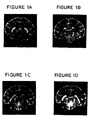
- MRI was used to map the anatomic distribution of the lesions in all six of the mAb-treated EAE animals, two dexamethasone-treated animals, and two saline-treated animals.
- Conventional spin-warp imaging was performed using a General Electric CSI-II NMR imager/spectrometer (2 Tesla magnet) as described in Rose et al., Biomed. and Pharmacother., 43 :347-353 (1989). Images were acquired once a week before the onset of clinical signs and twice a week after the onset of clinical signs. The animals were anesthetized with ketamine and positioned in the magnet so that the center of the brain was at magnet iso-center.
- Plate A represents a "normal" brain scan, 21 days after sensitization; Plate B reveals detection of a brainstem lesion two days after onset of clinical signs i.e., 28 days post sensitization: Plates C and D illustrate gradual progression of the brainstem lesion 35 and 38 days after sensitization, respectively.
- Plate A illustrates a normal brain scan 15 days post sensitization
- Plate B shows a brainstem lesion at the onset of clinical signs, i.e., 18 days after sensitization
- Plates C through F illustrate gradual resolution of the brainstem lesion through antibody treatment as monitored at 22, 25, 29 and 32 days post sensitization
- Plate G illustrates reappearance of a lesion in the same region which was accompanied by clinical relapse 39 days post sensitization and 14 days after cessation of treatment.
- Enlarging lesions are identified as prominent areas of increased intensity (white) on the scans.
- two weeks after the treatment was discontinued 89071 clinically relapsed and MR images demonstrated a larger, more intense lesion in the brainstem. The relapse was not treated and the animal was sacrificed.
- Animal 89080 developed a major hemorrhagic lesion in the lateral geniculate nucleus and striatum. Hemorrhagic lesions are usually fatal and usually do not respond to treatment (Shaw et al., supra ) but this lesion almost completely disappeared following administration of mAb 60.3. Animal 89069 had the mildest disease of any of the animals, as determined by MRI, with several small, but intense, sub-cortical white matter lesions that disappeared following treatment.
- Cryostat sections of cerebellum or cerebrum were stained with hematoxylin and eosin and by the immunoperoxidase technique in conjunction with mAbs to human leukocyte membrane antigens in order to analyze the composition of the cellular infiltrates.
- a tissue section obtained from the brain of animal 89070 and stained with mAb 60.3 is shown in Figure 3.
- Panel A at 390X magnification and stained with rabbit anti-mouse IgG, murine IgG coated cells are revealed in the lumen of blood vessels but not in brain tissue; and, in Panel B, at 390X magnification and stained with mAb 60.3, massive infiltration of PMBCs is seen in the exudate and extending out into adjacent white matter.
- Animal 89070 was the only mAb-treated animal to die before completion of treatment. Brain tissue taken from this animal was stained with a goat anti-mouse Ig antibody in order to determine whether the treatment mAb had entered the CNS before the animal died. MAb 60.3 reacts with both leukocytes and brain microglia. If this mAb had crossed the blood-brain-barrier during the treatment period, it was expected that a staining pattern similar to that shown in Figure 3, Panel B might be obtained. Alternatively, if the treatment mAb entered the CNS, bound only to blood leukocytes, the microglia would not have been stained. Instead, murine Ig-coated cells were only detectable in the vessel lumen, and not at all in the perivascular spaces. The absence of any positively stained leukocytes in the lesion stained with goat anti-mouse Ig, suggests that the animal died of an inflammatory condition existing before treatment was initiated and that the treatment antibody was given too late to be effective.
- the hematologic hallmarks of untreated EAE are a progressive leukocytosis and lymphopenia prior to the onset of clinical signs.
- the leukocytosis can represent as much as a four-fold increase in the number of circulating PMNC and no significant changes in the frequency or absolute numbers of monocytes, eosinophils, or basophils.
- the absolute numbers of lymphocytes and PMNC return, fairly soon after the initiation of treatment, to pre-sensitization levels.
- continuous monitoring of blood leukocytes provides a useful measure of disease progression which complements the clinical evaluation and magnetic resonance imaging.
- EAE animals were evaluated histologically postmortem for evidence of EAE. All had microscopic evidence of either mild to severe acute EAE or hyperacute EAE that generally correlated with the severity and duration of the disease. All but 2 of the 12 control animals showed either predominantly hyperacute lesions characterized by hemorrhages, subtotal necrosis, demyelination, and diffuse massive infiltrations of neutrophils into the CNS parenchyma; or severe acute perivascular lesions which were more compact, more confined to the perivascular spaces than the hyperacute lesions and composed predominantly of lymphocytes and macrophages, but some neutrophils, and with abundant demyelination as well as some axonal debris.
- the two exceptional control monkeys had a mixture of lesions ranging from mild to severe demyelinating lesions, composed predominantly of lymphocytes and macrophages with abundant myelin debris and little or no axonal reaction.
- All but one of the mAb-treated monkeys had a combination of: (a) older well developed demyelinated plaques with abundant myelin debris, sudanophilic lipid and little or no axonal reaction or lymphocytic infiltrate; and (b) more recent lesions corresponding to the terminal untreated relapses, which were compact, confined to perivascular spaces, with varying proportions of lymphocytes, monocytes, or neutrophils (consistent with diagnoses of mild to severe acute EAE) and some myelin debris.
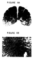
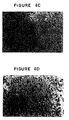
- These two types of lesions found in the same animal (#89069), are shown in Figure 4.
- Panel A at two-fold magnification, numerous inflammatory (area 1) and/or demyelinating (area 2) lesions can be ascertained in the white matter; in Panel B, at 325X magnification, area 2 of Panel A is revealed as being composed predominantly of macrophages scattered diffusely through the tissue and about blood vessels, with the small black granules constituting PAS positive glyco-lipoproteins; and in Panels C and D, at 130X and 325X magnification, respectively, area 1 of Panel A is revealed as being composed predominantly of PMNCs and no PAS positive insoluble glycoproteins.
- MAb 60.3 is known to inhibit plasma leakage in vivo [Arfors et al., Blood, 60 : 338-340, (1987)], probably the most important factor in improvement in MRI-detectable lesions, as changes in the tissue water characteristics can be visualized by changes in image intensity on the MRI scans.
- dexamethasone-treated animals did have longer mean survival time (7 days) than the saline-treated controls (3 days), but the difference was not statistically significant (p ⁇ 0.1). Thus, although dexamethasone slowed down the disease process, it had little apparent effect on the final outcome of the disease.
- mice Six to twelve week old Balb/c mice (Charles River Biotechnical Services, Inc., Wilmington, Massachusetts, IACUC #901103) were immunized with human T cell line Hut 78 to generate anti-CD 18 antibodies.
- two hybridoma-generating fusions (designated fusions 22 and 23) two Balb/c mice were bled retro-orbitally for the collection of pre-immune serum on day 0.
- each animal received a total of 5 x 10 6 Hut 78 cells in 0.2 ml sterile PBS intravenously.
- the mice were then immunized at two week intervals for six weeks and then at monthly intervals for three months.
- the final monthly boost was of glutaraldehyde fixed Hut 78 cells.
- the spleen from the mouse displaying the highest immune serum titer against the immunogen was removed sterilely.
- a single-cell suspension was formed by grinding the spleen between the frosted ends of two glass microscope slides submerged in serum free RPMI 1640, supplemented with 2 mM L-glutamine, 1 mM sodium pyruvate, 100 units/ml penicillin, and 100 ⁇ g/ml streptomycin (RPMI)(Gibco, Canada).
- the cell suspension was filtered through sterile 70-mesh Nitex cell strainer (Becton Dickinson, Parsippany, New Jersey), and washed twice by centrifuging at 200 g for 5 minutes and resuspending the pellet in 20 ml serum free RPMI.
- Thymocytes taken from 3 naive Balb/c mice were prepared in a similar manner.
- NS-1 myeloma cells kept in log phase in RPMI with 11% fetal bovine serum (FBS)(Hyclone Laboratories, Inc., Logan, Utah) for three days prior to fusion, were centrifuged at 200 g for 5 minutes, and the pellet was washed twice as described in the foregoing paragraph. After washing, each cell suspension brought to a final volume of 10 ml in serum free RPMI, and 10 ⁇ l was diluted 1:100. 20 ⁇ l of each dilution was removed, mixed with 20 ⁇ l 0.4% trypan blue stain in 0.85% saline (Gibco), loaded onto a hemacytometer (Baxter Healthcare Corp., Deerfield, Illinois) and counted.
- FBS fetal bovine serum
- the pellet was resuspended in 200 ml RPMI containing 15% FBS, 100 ⁇ M sodium hypoxanthine, 0.4 ⁇ M aminopterin, 16 ⁇ M thymidine (HAT) (Gibco), 25 units/ml IL-6 (Boehringer Mannheim) and 1.5 x 10 6 thymocytes/ml.
- the suspension was dispensed into ten 96-well flat bottom tissue culture plates (Corning, United Kingdom) at 200 ⁇ l/well. Cells in plates were fed on days 2, 4 and 6 days post fusion by aspirating approximately 100 ⁇ l/well plating medium described above except containing 10 units/ml IL-6 and lacking thymocytes.
- the reaction mixture was fixed by addition of 1% paraformaldehyde (pH 7.2 in PBS) and transferred to polystyrene tubes for FACS using a Becton Dickenson FACscan analyzer.
- 1% paraformaldehyde pH 7.2 in PBS
- the supernatant of hybridoma No. 22F12C inhibited binding of FITC-60.3 by about 90%
- supernatants from hybridoma Nos. 22J4A and 22B3B inhibited binding by about 40% and 10% respectively.
- Two hybridomas from fusion 23, 23F2G and 23I11B produced antibodies which inhibited FITC-60.3 binding by ⁇ 95%. These five hybridomas were cloned.
- Hybridoma cell line 23F2G was deposited with the American Type Culture Collection, 12301 Parklawn Drive, Rockville, M.D. 20852 U.S.A. on June 30, 1992 and accorded accession number A.T.C.C. HB 11081.
- the isotype of the antibody produced by hybridoma cell line 23F2G (referred to herein as mAb 23F2G) was determined to be IgG2a.
- Hybridomas 22F12C and 23I11B also produced antibodies of the IgG2a isotype, while hybridomas 22B3B and 22J4A produced IgG1 antibodies.
- Antibodies from hybridomas 22F12C, 23F2G and 23I11B were all found, in varying degrees, to: (1) block adhesion of human T cells to activated HUVEC monolayers; (2) block aggregation of 13-acetate (PMA) activated Con A blast cells phorbol 12-myristate; and (3) induce demargination of white blood cells in rabbits.
- the ability of the IgG1 antibodies produced by hybridomas 22B3B and 22J4A to block adhesion is as yet undetermined; mAb 22J4A was able to block aggregation, but mAb 22B3B was not; both antibodies induced demargination, but to a lesser degree than the others or mAb 60.3.
- mAb 22B3B and 22J4A are specific for an epitope present on the CD11a component of LFA-1 or an epitope associated with the CD11a/CD18 heterodimer rather than with an epitope of CD18.
- Hybridoma 23F2G was amplified by the ascites method and antibody was purified by affinity chromatography on Protein A under pyrogen-free conditions. From 25 ml of ascites, 120 mg of antibody preparation was isolated. This preparation contained only about 4 endotoxin units per mg.
- Animal 89186 was treated for a week with both dexamethasone (3 days at 4 mg/kg, 2 days at 2 mg/kg and 2 days at 1 mg/kg) and mAb 23F2G (7 days at 2 mg/kg, i.v.).
- the animal improved dramatically with complete resolution of paralysis three days after initiation of treatment. Lesions detected by MRI resolved four days after cessation of treatment and the animal improved dramatically with complete resolution of paralysis. Eight days following cessation of treatment, the animal relapsed and was sacrificed. Hematologic analysis revealed that mAb 23F2G caused steady demargination of leukocytes which peaked on the last day of treatment.
- the white blood cell count rose from 22.7 x 10 3 on the day of clinical onset to 82 x 10 3 /mm 3 on the last day of treatment.
- the antibody showed saturation of blood lymphocytes at the dose administered.
- Total RNA was isolated from the hybridoma cell line 23F2G (A.T.C.C. HB 11081) and first strand cDNA was synthesized using the total RNA as a template. The first strand cDNA was in turn used as a template for PCR reactions to obtain double-stranded DNA fragments encoding the variable regions of both the heavy and light chains of mAb 23F2G.
- the forward primer used to clone the heavy chain variable region was primer HFR1-4, the sequence of which is set out below in IUPAC nomenclature as SEQ ID NO: 1.
- the forward primer used to generate PCR fragments encoding the light chain variable region was primer LFR1-3, the sequence of which is set out below in IUPAC nomenclature as SEQ ID NO: 3.
- the resulting light and heavy chain PCR fragments were ligated into different vectors and eight to twelve independent clones were sequenced on both strands.
- the DNA and amino acid sequences corresponding to the heavy chain variable region of 23F2G are shown in SEQ ID NO: 5.
- the seven amino-terminal residues of the heavy chain sequence are indefinite because they correspond to the forward primer used in the PCR reaction.
- Amino acid residues 1 to 15, 31 to 45, 53 to 84 and 94 to 103 of SEQ ID NO: 5 comprise the framework regions of the mouse 23F2G heavy chain variable domain while amino acid residues 16 to 30, 46 to 52 and 35 to 93 of SEQ ID NO: 5 comprise the CDR regions.
- human frameworks chosen were as homologous as possible to those of 23F2G in order to increase the probability that the CDR regions would retain their correct conformations and consequently their affinity towards the antigen [see Kabat, et al., supra, and Kirkham et al. , EMBO J., 11: 603-609 (1992)] and (2) the human framework regions chosen contained a minimal number of unusual residues that could potentially provoke an immune response against the antibody in a human.
- the heavy chain of mAb 23F2G resides in the mouse subgroup II sequences, it is most homologous to sequences in human subgroup I. Therefore, a framework from the human subgroup I set of heavy chain variable sequences was chosen for use in a humanized version of mAb 23F2G. Rather than choose a particular framework from existing sequenced human antibodies, it was decided to use a consensus sequence for the human subgroup I variable region framework (Kabat, et al., supra ) so that the unusual residues often found in particular frameworks were avoided.
- the combined framework regions of the heavy chain of 23F2G excluding the amino-terminal seven amino acids, have 70% sequence identity to the human subgroup I consensus sequence. In comparison, the combined 23F2G heavy chain framework regions were less homologous to the human subgroup II and III consensus sequence (52% and 58%, respectively).
- Hybridomas 2 :124-134 (1991); Kettleborough et al., Protein Engineering, 4(7) :773-783 (1991); Tempest et al., BIO/TECHNOLOGY, 9 :266-271 (1991); Gorman et al., Proc. Nat'l. Acad. Sci. (USA), 88 :4181-4185 (1991); Queen et al., Proc. Nat'l. Acad. Sci. (USA), 86 :10029-10033 (1989); and Co et al., Proc. Nat'l. Acad. Sci.
- variable region framework sequences of the light chain of 23F2G are 71%, 69% and 72% identical to the human subgroup I, II, and III kappa consensus sequences, respectively. It was decided to use the human kappa subgroup I consensus framework sequence because it was highly homologous to that of 23F2G and because subgroup I frameworks have most commonly been used to humanize light chain variable regions. No mouse-specific residues were substituted in the light chain human framework regions.
- sequences designed for the humanized form of the 23F2G light and heavy variable regions consisted of the framework regions of the human variable region framework sequences as described above and the CDR regions of mouse mAb 23F2G.
- the DNA and deduced amino acid sequences of these humanized heavy and light chains variable regions are presented in SEQ ID NOs: 7 and 8, respectively.
- Amino acids residues 1 to 30, 36 to 49, 67 to 98 and 110 to 120 of SEQ ID NO: 7 comprise the framework regions of the humanized heavy chain variable domain, while amino acids residues 31 to 35, 50 to 66 and 99 to 109 of SEQ ID NO: 7 comprise the CDR regions.
- Amino acids residues 1 to 23, 39 to 53, 61 to 92 and 101 to 111 of SEQ ID NO: 8 comprise the framework regions of the humanized light chain variable domain, while amino acids residues 24 to 38, 54 to 60 and 93 to 100 of SEQ ID NO: 8 comprise the CDR regions.
- Two DNA fragments which together encoded the humanized version of the heavy chain variable region of 23F2G were assembled from complementary synthetic oligonucleotides 50-60 nucleotides in length. Sequences included in addition to sequences encoding the variable domain were (1) a HindIII restriction site at the 5' end to facilitate attachment to a promoter sequence, (2) an optimal translation initiation sequence just upstream of the initiator methionine, (3) DNA encoding a signal peptide, (4) a splice donor site just 3' to the variable region, and (5) an EcoRI site at the 3' end to facilitate attachment to the constant region DNA segment.
- the DNA sequence encoding the signal peptide was designed to be the same as that encoding the signal peptide associated with several human heavy chain subgroup I sequences (Kabat et al., supra ). Two DNA fragments together encoding the humanized light chain variable region DNA segment were also constructed from complementary synthetic oligonucleotides. The assembled heavy and light chain variable region DNA fragments were then cloned into pSK+ (Stratagene, La Jolla, CA) and their sequence was confirmed.
- pSK+ Stratagene, La Jolla, CA
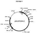
- a plasmid for expression of the humanized heavy chain of 23F2G was constructed by ligating together the two DNA fragments containing the humanized heavy chain variable region sequence, the constant regions of the human IgG 4 gene [cloned from a human T cell line (Hut 78)], and the expression vector pRc/CMV (Invitrogen).
- the expression vector pRc/CMV contains the immediate early promoter from the cytomegalovirus (CMV), a polylinker region downstream of the promoter, and a neomycin resistance cassette.
- the following four fragments were isolated and ligated: (1) the 5.5 kb HindIII/XbaI fragment from pRc/CMV, (2) a 6 kb EcoRI/XbaI fragment containing the IgG 4 sequences, (3) a ⁇ 200 bp HindIII/XhoI fragment containing the 5' half of the humanized heavy chain variable region sequence, and (4) a ⁇ 300 bp XhoI/EcoRI fragment containing the 3' half of the heavy chain variable region sequence.
- the ligation mix was used to transform E. coli, and a correct clone was confirmed by restriction digest and was designated pRc/HF2GH.2.
- a circular map of pRc/HF2GH.2 is shown in FIGURE 5.
- Large scale plasmid preparations of pRc/HF2GH.2 were performed using the alkaline lysis method as described in Sambrook et al., Molecular Cloning: A Laboratory Manual, Cold Spring Harbor Press (1989) and the plasmid was twice-banded in CsCl-ethidium bromide gradients.
- a plasmid for expression of the humanized light chain was constructed by ligating together two DNA fragments containing the humanized light chain variable region sequence, the human kappa gene, a dihydrofolate reductase (DHFR) gene expression cassette, and a part of the pRc/CMV vector containing the strong promoter from CMV.
- DHFR dihydrofolate reductase
- a plasmid containing the humanized light chain gene of 23F2G and DHFR sequences were first constructed.
- the first intermediate plasmid constructed, pRc/HF2GL. 1 was similar to that made for expression of the heavy chain gene in that the variable and constant region coding sequences were cloned between the HindIII and XbaI sites of pRc/CMV.
- a second intermediate plasmid, pSl1190-dhfr was made by inserting a 1.7kb SphI/BamHI fragment from pSV2-dhfr (ATCC 37146) into the corresponding site in the polylinker of the sequencing vector pSL1190 (Pharmacia, Piscataway, NJ).
- the SV40-DHFR sequences within pSL1190-dhfr were completely sequenced and were found to contain only minor differences from previously published sequence of these elements.
- the DHFR coding sequence within pSL1190-dhfr was found to encode the same polypeptide as the previously published sequence of mouse DHFR [Simonsen and Levinson, Proc. Natl. Acad. Sci. USA, 80: 2495-2499 (1983)].
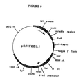
- pD/HF2GL.1 For expression of humanized 23F2G light chain, the following four fragments were ligated: (1) the 3.1 kb BamHI/HindIII fragment from pRc/CMV, (2) the 0.753 bp XbaI/Asp718 fragment from pRc/CMV, (3) the 1.2 kb fragment from pRc/HF2GL.1 (containing the complete humanized light chain gene), and (4) the 1.7 kb Asp718/BamHI fragment (containing SV40 promoter-DHFR sequences) from pSL1190-dhfr.
- FIGURE 6 A circular map of pD/HF2GL.1 is shown in FIGURE 6. Large scale preparations and purifications of these plasmids were performed as described above for the heavy chain expression plasmid.
- Lipofection was used to introduce both the heavy and light chain expression plasmids into the dhfr CHO cell line DXB11. Similar dhfr CHO cell lines are described in Urlaub, et al, Proc. Natl. Acad. Sci. USA, 77: 4216-4220 (1980). Selection for cells that were able to grow in the absence of nucleosides (selects for DHFR) and that were resistant to G418 (neomycin) ensured that transfectants contained both plasmids.
- a pool of three transfectants was subjected first to 25nM and then 100nM methotrexate (Sigma, St. Louis, MO) to select cells that had undergone amplification of the DHFR gene. After limited dilution cloning, one resulting cell line, designated 6E6 (A.T.C.C. CRL 11398) was found to produce humanized 23F2G at a rate of about 1 ng/cell/day and accumulate 5-10 mg antibody per liter of growth medium.
- Another cell line derived by transfection of the dhfr CHO cell line [Urlaub, et al., Cell, 33 : 405-412 (1983)] was isolated in a similar manner to cell line 6E6 and 6E6 and was named B13-24 (A.T.C.C. CRL 11397).
- Cell line B13-24 which is resistant to 200nM methotrexate, produces over 100 mg/l of antibody at a rate of about 25 pg/cell/day.
- the 6E6 and B13-24 cell lines can be cultured in Dulbecco's modified Earls Medium/Ham's F-12 (1:1 mix) with 5% heat inactivated fetal bovine serum at 37°C, 6% carbon dioxide and 100% humidity, and at a density of between 2x10 5 and 6x10 5 cells per milliliter.
- the media for the 6E6 cell line includes 100nM methotrexate while the media for B13-24 cell line includes 200nM methotrexate.
- Humanized 23F2G antibody was purified from the 6E6 and B13-24 cell lines as follows.
- Cell harvest fluid was first passed through a Protein A column, which binds IgGs.
- the column was washed with 35mM Tris buffer, 0.1% Tween 20, pH 7.85 and then with 50mM citrate, pH 5.0.
- the antibody was eluted with 50mM citrate, 0.02% Tween 20, pH 3.0, and kept under acidic conditions at room temperature for 15 minutes.
- the eluate was then chilled to 2-8°C and neutralized with cold 1M Tris buffer.
- the Protein A eluate was adjusted to 0.5M ammonium sulfate, 25mM Tris, pH 8.0 and loaded onto a Phenyl Sepharose column.
- the column was then washed with loading buffer (0.5M ammonium sulfate, 25mM Tris, pH 8.0), and eluted with 0.1M ammonium sulfate, 25mM Tris, pH 8.0.
- the eluate was then diluted 1/10 with purified water to prepare it for the next step.
- the diluted phenyl sepharose eluant was loaded onto a DEAE Sepharose Fast Flow column that has been equilibrated with 10mM Tris, pH 7.5. After loading, the column was washed with 10mM Tris, pH 7.5, followed by 10mM Tris, 50mM NaCl, pH 7.5.
- the antibody was eluted with 10mM Tris, 200mM NaCl, pH 7.5.
- the DEAE eluate is loaded onto a Sephadex G-25 Superfine column, and washed through with the final formulation buffer (50mM sodium acetate, 120mM NaCl, 0.02% Tween 20, pH 5.6).
- the eluted antibody was then diluted to the required concentration.
- RNA encoding the humanized 23F2G heavy chain was longer than expected.
- Subsequent cloning and sequencing of several cDNAs encoding the heavy chain revealed that a region in the DNA near that encoding the C-terminus had undergone a recombination event such that the sequence encoding the Gly-Lys sequence typically found at the C-termini of IgG 4 heavy chains was missing and was replaced by the sequence encoding Asp-Ser-Asn-Leu-Trp-Asn (SEQ ID NO: 9).
- the heavy chain mRNA derived from the cell line B13-24 is the expected size and therefore the C-terminal coding region of the heavy chain produced by cell line B13-24 is likely to be intact.
- Humanized 23F2G antibody competes effectively with flourescent murine 23F2G antibody in a cell binding assay. Fluorescent murine 23F2G antibody at 0.4 ⁇ g/ml was mixed with increasing amount of humanized 23F2G antibody. These antibody mixtures were incubated with the LFA-1 positive cell line HUT-78. After washing the cells were examined by flow cytometry for mean fluorescent intensity. In this assay humanized 23F2G antibody (at 1.01 ⁇ g/ml) bound to LFA-1 positive cells as efficiently as murine 23F2G antibody.
- M23F2G murine monoclonal antibody 23F2G
- Humanized antibody obtained from CHO 6E6 cells of Example 6 (“Hu23F2G”)
- additional Macaca Fascicularis monkeys were sensitized to myelin BP in the manner of Example 1 using 0.1 ml of a freshly prepared, somewhat more potent emulsion comprising 0.1-0.4 mg of monkey BP and 0.5 mg of heat-killed M. tuberculosis .
- M23F2G was purified from ascites fluid as in Example 5 and contained 5.2 endotoxin units per mg.
- Hu23F2G was purified from CHO cell 6E6 (A.T.C.C. CRL 11398) culture supernatant by affinity chromatography on Protein A under pyrogen-free conditions and contained 1.2 endotoxin units per mg.
- Treatment was started on the day of onset of disease.
- Treatment groups consisted of dexamethasone (4 mg/kg/day for 3 days) alone, dexamethasone plus M23F2G (2 mg/kg/day for 7 days), or dexamethasone plus Hu23F2G (2 mg/kg/day for 7 days).
- Treatment assignment of animals was made prior to the onset of clinical disease. Seven animals were treated with dexamethasone, five with M23F2G, and four with Hu23F2G.
- the endpoints used to evaluate the effects of treatment were: overall survival after onset of disease up to a maximum of 42 days; clinical score following treatment; and lesion burden (determined by MRI) following treatment. Clinical signs were monitored daily until time of death or sacrifice. Animals were scanned by MRI once a week. A modified clinical scoring system (vis-a-vis the previous scoring system in Table 1) was used to reflect the severity of disease and is set out in Table 6, below. Pre-treatment MRI scan data was obtained at onset ( ⁇ 3 days) of clinical disease; post-treatment scan data was obtained on 10 days after clinical onset ( ⁇ 3 days). MRI scans were analyzed by the software package NIH Image Version 1.44 (Bethesda, MD) to quantitate areas of abnormality.
- MRI lesions reflect brain edema and cellular infiltration caused by EAE and allow a non-invasive method of assessing the effect of treatment on brain inflammation.
- Clinical score and lesion area were determined at onset of disease ( ⁇ 3 days) and ten days post treatment ( ⁇ 3 days), except for two animals in the dexamethasone group (92152 and F91337) that died three days after onset, and the post scan was obtained on the day of death.
- the values for each of the 3 treatment groups are shown in Table 8.
- Clinical score values shown in Table 8 were those obtained on the same day as the scan. There was no significant difference between clinical scores at onset of disease between the three treatment groups; however, statistically significant differences were noted post-treatment in both clinical score and lesion area.
- M23F2G and Hu23F2G have the same beneficial effect on monkey EAE as antibody 60.3 in terms of survival. Analyses of clinical severity and MRI endpoints also supports a beneficial effect of M23F2G and Hu23F2G compared to dexamethasone alone in monkey EAE.
Landscapes
- Health & Medical Sciences (AREA)
- Chemical & Material Sciences (AREA)
- Organic Chemistry (AREA)
- Immunology (AREA)
- Life Sciences & Earth Sciences (AREA)
- General Health & Medical Sciences (AREA)
- Medicinal Chemistry (AREA)
- Biochemistry (AREA)
- Biophysics (AREA)
- Genetics & Genomics (AREA)
- Molecular Biology (AREA)
- Proteomics, Peptides & Aminoacids (AREA)
- Pain & Pain Management (AREA)
- Rheumatology (AREA)
- Chemical Kinetics & Catalysis (AREA)
- General Chemical & Material Sciences (AREA)
- Nuclear Medicine, Radiotherapy & Molecular Imaging (AREA)
- Pharmacology & Pharmacy (AREA)
- Animal Behavior & Ethology (AREA)
- Public Health (AREA)
- Veterinary Medicine (AREA)
- Medicines Containing Antibodies Or Antigens For Use As Internal Diagnostic Agents (AREA)
- Peptides Or Proteins (AREA)
- Preparation Of Compounds By Using Micro-Organisms (AREA)
Applications Claiming Priority (5)
| Application Number | Priority Date | Filing Date | Title |
|---|---|---|---|
| US91506892A | 1992-07-16 | 1992-07-16 | |
| US915068 | 1992-07-16 | ||
| US6069993A | 1993-05-10 | 1993-05-10 | |
| EP93918230A EP0604647A4 (en) | 1992-07-16 | 1993-07-16 | Alleviation of symptoms associated with inflammatory disease states. |
| US60699 | 2002-01-30 |
Related Parent Applications (1)
| Application Number | Title | Priority Date | Filing Date |
|---|---|---|---|
| EP93918230A Division EP0604647A4 (en) | 1992-07-16 | 1993-07-16 | Alleviation of symptoms associated with inflammatory disease states. |
Publications (1)
| Publication Number | Publication Date |
|---|---|
| EP1069182A1 true EP1069182A1 (fr) | 2001-01-17 |
Family
ID=26740253
Family Applications (2)
| Application Number | Title | Priority Date | Filing Date |
|---|---|---|---|
| EP00116049A Withdrawn EP1069182A1 (fr) | 1992-07-16 | 1993-07-16 | Allégement des symptomes associés à des états dus aux maladies inflammatoires en utilisant des anticorps contre CD18 |
| EP93918230A Ceased EP0604647A4 (en) | 1992-07-16 | 1993-07-16 | Alleviation of symptoms associated with inflammatory disease states. |
Family Applications After (1)
| Application Number | Title | Priority Date | Filing Date |
|---|---|---|---|
| EP93918230A Ceased EP0604647A4 (en) | 1992-07-16 | 1993-07-16 | Alleviation of symptoms associated with inflammatory disease states. |
Country Status (5)
| Country | Link |
|---|---|
| US (2) | US6737059B1 (fr) |
| EP (2) | EP1069182A1 (fr) |
| JP (1) | JPH06511156A (fr) |
| CA (1) | CA2119128C (fr) |
| WO (1) | WO1994002175A1 (fr) |
Cited By (1)
| Publication number | Priority date | Publication date | Assignee | Title |
|---|---|---|---|---|
| CN106986930A (zh) * | 2017-04-14 | 2017-07-28 | 广东省生物资源应用研究所 | 一种诱导食蟹猴实验性自身免疫性脑脊髓炎动物模型的蛋白和应用 |
Families Citing this family (9)
| Publication number | Priority date | Publication date | Assignee | Title |
|---|---|---|---|---|
| US5169930A (en) * | 1990-01-05 | 1992-12-08 | La Jolla Cancer Research Foundation | Fibronectin receptor |
| GB9115364D0 (en) | 1991-07-16 | 1991-08-28 | Wellcome Found | Antibody |
| WO1994004188A1 (fr) * | 1992-08-21 | 1994-03-03 | Genentech, Inc. | Procede pour traiter une affection ayant pour origine l'antigene 1 associe a la fonction lymphocytaire |
| US5914112A (en) * | 1996-01-23 | 1999-06-22 | Genentech, Inc. | Anti-CD18 antibodies in stroke |
| US20020081294A1 (en) | 1996-01-23 | 2002-06-27 | Genentech, Inc. | Co-administration of a thrombolytic and an anti-CD18 antibody in stroke |
| DK0877626T3 (da) * | 1996-01-23 | 2002-12-30 | Univ Vermont | Anti-CD18 antistoffer til anvendelse mod slagtilfælde |
| AU764382B2 (en) | 1999-03-19 | 2003-08-14 | Genentech Inc. | Treatment of LFA-1 associated disorders with increasing doses of LFA-1 antagonist |
| US6582698B1 (en) | 1999-03-19 | 2003-06-24 | Genentech, Inc. | Treatment method |
| EP1914242A1 (fr) * | 2006-10-19 | 2008-04-23 | Sanofi-Aventis | Nouveau anticorps Anti-CD38 pour le traitement du cancer |
Citations (3)
| Publication number | Priority date | Publication date | Assignee | Title |
|---|---|---|---|---|
| EP0346078A2 (fr) * | 1988-06-07 | 1989-12-13 | The Rockefeller University | Inhibition de l'activité leucocytaire dans les organes au cours d'une infection ou de tout autre traumatisme |
| WO1990010652A1 (fr) * | 1989-03-09 | 1990-09-20 | Dana Farber Cancer Institute | Procede de traitement d'infections virales a l'aide de lfa-1 |
| EP0438312A2 (fr) * | 1990-01-19 | 1991-07-24 | Merck & Co. Inc. | Anticorps recombinants humains anti-CD18 |
Family Cites Families (5)
| Publication number | Priority date | Publication date | Assignee | Title |
|---|---|---|---|---|
| US4797277A (en) | 1987-09-22 | 1989-01-10 | Pharmacia Ab | Method for reperfusion therapy |
| NZ236792A (en) * | 1990-01-19 | 1993-05-26 | Merck & Co Inc | Recombinant human anti-cd18 antibody, murine 1b4 heavy or light chain variable region, dna, vectors, mammalian host and pharmaceutical compositions |
| EP0546077A1 (fr) | 1990-08-27 | 1993-06-16 | Chiron Corporation | Medicaments peptidiques a base de cd18 utilises dans le traitement d'affections |
| WO1992004034A1 (fr) | 1990-08-31 | 1992-03-19 | Boehringer Ingelheim Pharmaceuticals, Inc. | Procede traitant les chocs endotoxiniques au moyen d'anticorps anti-adhesion |
| WO1992006697A1 (fr) | 1990-10-23 | 1992-04-30 | Repligen Corporation | Composition anti-inflammatoire |
-
1993
- 1993-07-16 EP EP00116049A patent/EP1069182A1/fr not_active Withdrawn
- 1993-07-16 JP JP6504608A patent/JPH06511156A/ja active Pending
- 1993-07-16 EP EP93918230A patent/EP0604647A4/en not_active Ceased
- 1993-07-16 CA CA002119128A patent/CA2119128C/fr not_active Expired - Lifetime
- 1993-07-16 WO PCT/US1993/006734 patent/WO1994002175A1/fr not_active Application Discontinuation
-
1994
- 1994-11-03 US US08/333,947 patent/US6737059B1/en not_active Expired - Fee Related
-
2004
- 2004-05-17 US US10/847,212 patent/US20050002932A1/en not_active Abandoned
Patent Citations (3)
| Publication number | Priority date | Publication date | Assignee | Title |
|---|---|---|---|---|
| EP0346078A2 (fr) * | 1988-06-07 | 1989-12-13 | The Rockefeller University | Inhibition de l'activité leucocytaire dans les organes au cours d'une infection ou de tout autre traumatisme |
| WO1990010652A1 (fr) * | 1989-03-09 | 1990-09-20 | Dana Farber Cancer Institute | Procede de traitement d'infections virales a l'aide de lfa-1 |
| EP0438312A2 (fr) * | 1990-01-19 | 1991-07-24 | Merck & Co. Inc. | Anticorps recombinants humains anti-CD18 |
Non-Patent Citations (1)
| Title |
|---|
| B. DAUGHERTY ET AL.: "Polymerase chain reaction facilitates the cloning, CDR-grafting and rapid expression of a murine monoclonal antibody directed against the CD18 component of leukocyte integrins.", NUCLEIC ACIDS RESEARCH, vol. 19, no. 9, 11 May 1991 (1991-05-11), OXFORD, GB, pages 2471 - 2476, XP000256796 * |
Cited By (1)
| Publication number | Priority date | Publication date | Assignee | Title |
|---|---|---|---|---|
| CN106986930A (zh) * | 2017-04-14 | 2017-07-28 | 广东省生物资源应用研究所 | 一种诱导食蟹猴实验性自身免疫性脑脊髓炎动物模型的蛋白和应用 |
Also Published As
| Publication number | Publication date |
|---|---|
| US6737059B1 (en) | 2004-05-18 |
| CA2119128A1 (fr) | 1994-02-03 |
| CA2119128C (fr) | 2000-05-30 |
| EP0604647A4 (en) | 1996-09-25 |
| WO1994002175A1 (fr) | 1994-02-03 |
| JPH06511156A (ja) | 1994-12-15 |
| EP0604647A1 (fr) | 1994-07-06 |
| US20050002932A1 (en) | 2005-01-06 |
Similar Documents
| Publication | Publication Date | Title |
|---|---|---|
| EP0842948B1 (fr) | ANTICORPS ANTI Fas LIGAND ET PROCEDE D'UTILISATION | |
| EP0528931B1 (fr) | Anticorps anti-molecule 1 d'adherence intercellulaire chimerique adaptes au modele humain, procede de preparation et d'utilisation | |
| EP0804237B1 (fr) | Anticorps humanises diriges contre la molecule d'adhesion leucocytaire vla-4 | |
| EP1140172B1 (fr) | Prevention et traitement de l'ischemie retinienne et de l'oedeme | |
| Nishikawa et al. | Antibodies to intercellular adhesion molecule 1/lymphocyte function-associated antigen 1 prevent crescent formation in rat autoimmune glomerulonephritis. | |
| EP2662091B1 (fr) | Anticorps anti-P-sélectine et procédés pour les utiliser dans le traitement des maladies inflammatoires | |
| US5817311A (en) | Methods of inhibiting T-cell medicated immune responses with LO-CD2a-specific antibodies | |
| US20120177666A1 (en) | Anti-tgf-beta receptor ii antibodies | |
| WO1993015764A1 (fr) | Traitement de l'inflammation intestinale | |
| US6114507A (en) | Anti-Fas ligand antibody and assay method using the anti-Fas ligand antibody | |
| CA2119128C (fr) | Soulagement des symptomes associes aux etats inflammatoires aigus | |
| US8207305B2 (en) | Sialoadhesin factor-2 antibodies | |
| WO1998040488A1 (fr) | Anticorps monoclonaux humanises anti-alphabeta 3 | |
| EP1960431B1 (fr) | Utilisation d'anticorps deficients en fonction effectrice pour le traitement de maladies auto-immunes | |
| US5854070A (en) | Murine and humanizer 23F2G antibodies and cell lines expressing said antibodies | |
| EP1272205B1 (fr) | Anticorps du facteur-2 de la sialoadhesine | |
| US7592006B1 (en) | Composition comprising the LO-CD2a antibody | |
| WO2023241659A1 (fr) | Méthodes de traitement d'un lymphome à l'aide d'anticorps anti-tigit | |
| IL145480A (en) | Use of CD11a antibody in the preparation of a drug for the treatment of psoriasis |
Legal Events
| Date | Code | Title | Description |
|---|---|---|---|
| PUAI | Public reference made under article 153(3) epc to a published international application that has entered the european phase |
Free format text: ORIGINAL CODE: 0009012 |
|
| 17P | Request for examination filed |
Effective date: 20000726 |
|
| AC | Divisional application: reference to earlier application |
Ref document number: 604647 Country of ref document: EP |
|
| AK | Designated contracting states |
Kind code of ref document: A1 Designated state(s): AT BE CH DE DK ES FR GB GR IE IT LI LU MC NL PT SE |
|
| AKX | Designation fees paid |
Free format text: AT BE CH DE DK ES FR GB GR IE IT LI LU MC NL PT SE |
|
| RAP1 | Party data changed (applicant data changed or rights of an application transferred) |
Owner name: THE BOARD OF REGENTS OF THE UNIVERSITY OF WASHINGT Owner name: ICOS CORPORATION |
|
| STAA | Information on the status of an ep patent application or granted ep patent |
Free format text: STATUS: THE APPLICATION HAS BEEN WITHDRAWN |
|
| 18W | Application withdrawn |
Effective date: 20080912 |
|
| REG | Reference to a national code |
Ref country code: HK Ref legal event code: WD Ref document number: 1036298 Country of ref document: HK |