EP1057028B1 - Treating cancer - Google Patents
Treating cancer Download PDFInfo
- Publication number
- EP1057028B1 EP1057028B1 EP99905082A EP99905082A EP1057028B1 EP 1057028 B1 EP1057028 B1 EP 1057028B1 EP 99905082 A EP99905082 A EP 99905082A EP 99905082 A EP99905082 A EP 99905082A EP 1057028 B1 EP1057028 B1 EP 1057028B1
- Authority
- EP
- European Patent Office
- Prior art keywords
- cyclin
- protein
- cells
- agent
- sample
- Prior art date
- Legal status (The legal status is an assumption and is not a legal conclusion. Google has not performed a legal analysis and makes no representation as to the accuracy of the status listed.)
- Expired - Lifetime
Links
- 206010028980 Neoplasm Diseases 0.000 title claims description 60
- 201000011510 cancer Diseases 0.000 title claims description 37
- 238000000034 method Methods 0.000 claims abstract description 47
- 238000012360 testing method Methods 0.000 claims abstract description 22
- 102000006311 Cyclin D1 Human genes 0.000 claims description 87
- 108010058546 Cyclin D1 Proteins 0.000 claims description 87
- 229940123237 Taxane Drugs 0.000 claims description 43
- 239000003795 chemical substances by application Substances 0.000 claims description 35
- 238000011282 treatment Methods 0.000 claims description 32
- 229940127089 cytotoxic agent Drugs 0.000 claims description 26
- 230000014509 gene expression Effects 0.000 claims description 22
- 239000000523 sample Substances 0.000 claims description 22
- 229930012538 Paclitaxel Natural products 0.000 claims description 21
- 229960001592 paclitaxel Drugs 0.000 claims description 21
- DKPFODGZWDEEBT-QFIAKTPHSA-N taxane Chemical class C([C@]1(C)CCC[C@@H](C)[C@H]1C1)C[C@H]2[C@H](C)CC[C@@H]1C2(C)C DKPFODGZWDEEBT-QFIAKTPHSA-N 0.000 claims description 21
- 239000002246 antineoplastic agent Substances 0.000 claims description 20
- 108020004999 messenger RNA Proteins 0.000 claims description 15
- 239000000975 dye Substances 0.000 claims description 13
- 238000000338 in vitro Methods 0.000 claims description 12
- 230000004568 DNA-binding Effects 0.000 claims description 10
- 238000001262 western blot Methods 0.000 claims description 8
- 238000001943 fluorescence-activated cell sorting Methods 0.000 claims description 7
- 239000007850 fluorescent dye Substances 0.000 claims description 5
- 238000002372 labelling Methods 0.000 claims description 5
- 208000036878 aneuploidy Diseases 0.000 claims description 3
- 239000006285 cell suspension Substances 0.000 claims description 3
- ZYVSOIYQKUDENJ-WKSBCEQHSA-N chromomycin A3 Chemical compound O([C@@H]1C[C@@H](O[C@H](C)[C@@H]1OC(C)=O)OC=1C=C2C=C3C[C@H]([C@@H](C(=O)C3=C(O)C2=C(O)C=1C)O[C@@H]1O[C@H](C)[C@@H](O)[C@H](O[C@@H]2O[C@H](C)[C@@H](O)[C@H](O[C@@H]3O[C@@H](C)[C@H](OC(C)=O)[C@@](C)(O)C3)C2)C1)[C@H](OC)C(=O)[C@@H](O)[C@@H](C)O)[C@@H]1C[C@@H](O)[C@@H](OC)[C@@H](C)O1 ZYVSOIYQKUDENJ-WKSBCEQHSA-N 0.000 claims description 3
- 238000005259 measurement Methods 0.000 claims description 3
- INAAIJLSXJJHOZ-UHFFFAOYSA-N pibenzimol Chemical group C1CN(C)CCN1C1=CC=C(N=C(N2)C=3C=C4NC(=NC4=CC=3)C=3C=CC(O)=CC=3)C2=C1 INAAIJLSXJJHOZ-UHFFFAOYSA-N 0.000 claims description 3
- 238000000684 flow cytometry Methods 0.000 claims description 2
- 230000003834 intracellular effect Effects 0.000 claims description 2
- 125000002456 taxol group Chemical group 0.000 claims description 2
- 102000003910 Cyclin D Human genes 0.000 claims 1
- 108090000259 Cyclin D Proteins 0.000 claims 1
- 150000001875 compounds Chemical class 0.000 claims 1
- 238000004624 confocal microscopy Methods 0.000 claims 1
- 108010025464 Cyclin-Dependent Kinase 4 Proteins 0.000 abstract description 4
- 102100032857 Cyclin-dependent kinase 1 Human genes 0.000 abstract description 4
- 101710106279 Cyclin-dependent kinase 1 Proteins 0.000 abstract description 4
- 238000003745 diagnosis Methods 0.000 abstract description 3
- 102100036252 Cyclin-dependent kinase 4 Human genes 0.000 abstract 2
- 208000006994 Precancerous Conditions Diseases 0.000 abstract 2
- 210000004027 cell Anatomy 0.000 description 107
- 239000003814 drug Substances 0.000 description 22
- 108090000623 proteins and genes Proteins 0.000 description 20
- RCINICONZNJXQF-MZXODVADSA-N taxol Chemical compound O([C@@H]1[C@@]2(C[C@@H](C(C)=C(C2(C)C)[C@H](C([C@]2(C)[C@@H](O)C[C@H]3OC[C@]3([C@H]21)OC(C)=O)=O)OC(=O)C)OC(=O)[C@H](O)[C@@H](NC(=O)C=1C=CC=CC=1)C=1C=CC=CC=1)O)C(=O)C1=CC=CC=C1 RCINICONZNJXQF-MZXODVADSA-N 0.000 description 20
- 229940079593 drug Drugs 0.000 description 18
- 230000035945 sensitivity Effects 0.000 description 18
- 102000004169 proteins and genes Human genes 0.000 description 11
- 102100033270 Cyclin-dependent kinase inhibitor 1 Human genes 0.000 description 10
- 239000000463 material Substances 0.000 description 10
- 231100000433 cytotoxic Toxicity 0.000 description 9
- 230000001472 cytotoxic effect Effects 0.000 description 9
- 108700020796 Oncogene Proteins 0.000 description 7
- 238000001574 biopsy Methods 0.000 description 7
- 239000002254 cytotoxic agent Substances 0.000 description 7
- 238000011160 research Methods 0.000 description 7
- 230000004044 response Effects 0.000 description 7
- IJGRMHOSHXDMSA-UHFFFAOYSA-N Atomic nitrogen Chemical compound N#N IJGRMHOSHXDMSA-UHFFFAOYSA-N 0.000 description 6
- 102000016736 Cyclin Human genes 0.000 description 6
- 108050006400 Cyclin Proteins 0.000 description 6
- 238000004458 analytical method Methods 0.000 description 6
- 238000009643 clonogenic assay Methods 0.000 description 6
- 231100000096 clonogenic assay Toxicity 0.000 description 6
- 230000035772 mutation Effects 0.000 description 6
- 230000002018 overexpression Effects 0.000 description 6
- 101000944380 Homo sapiens Cyclin-dependent kinase inhibitor 1 Proteins 0.000 description 5
- 108091006627 SLC12A9 Proteins 0.000 description 5
- 230000008901 benefit Effects 0.000 description 5
- 238000002512 chemotherapy Methods 0.000 description 5
- 230000003021 clonogenic effect Effects 0.000 description 5
- 231100000599 cytotoxic agent Toxicity 0.000 description 5
- 238000003365 immunocytochemistry Methods 0.000 description 5
- 239000006166 lysate Substances 0.000 description 5
- 239000002609 medium Substances 0.000 description 5
- 108020004707 nucleic acids Proteins 0.000 description 5
- 102000039446 nucleic acids Human genes 0.000 description 5
- 150000007523 nucleic acids Chemical class 0.000 description 5
- 230000004043 responsiveness Effects 0.000 description 5
- 230000004083 survival effect Effects 0.000 description 5
- 102100025064 Cellular tumor antigen p53 Human genes 0.000 description 4
- 206010033128 Ovarian cancer Diseases 0.000 description 4
- 238000003556 assay Methods 0.000 description 4
- 230000022131 cell cycle Effects 0.000 description 4
- 230000001413 cellular effect Effects 0.000 description 4
- 239000002875 cyclin dependent kinase inhibitor Substances 0.000 description 4
- 229940043378 cyclin-dependent kinase inhibitor Drugs 0.000 description 4
- 238000002405 diagnostic procedure Methods 0.000 description 4
- 238000001959 radiotherapy Methods 0.000 description 4
- 239000000725 suspension Substances 0.000 description 4
- 229940124597 therapeutic agent Drugs 0.000 description 4
- 238000002560 therapeutic procedure Methods 0.000 description 4
- 108091007914 CDKs Proteins 0.000 description 3
- 102000003903 Cyclin-dependent kinases Human genes 0.000 description 3
- 108090000266 Cyclin-dependent kinases Proteins 0.000 description 3
- 108020004414 DNA Proteins 0.000 description 3
- PEDCQBHIVMGVHV-UHFFFAOYSA-N Glycerine Chemical compound OCC(O)CO PEDCQBHIVMGVHV-UHFFFAOYSA-N 0.000 description 3
- ZMXDDKWLCZADIW-UHFFFAOYSA-N N,N-Dimethylformamide Chemical compound CN(C)C=O ZMXDDKWLCZADIW-UHFFFAOYSA-N 0.000 description 3
- 108700025695 Suppressor Genes Proteins 0.000 description 3
- 238000003491 array Methods 0.000 description 3
- 239000000872 buffer Substances 0.000 description 3
- 238000010790 dilution Methods 0.000 description 3
- 239000012895 dilution Substances 0.000 description 3
- 239000003112 inhibitor Substances 0.000 description 3
- 238000012417 linear regression Methods 0.000 description 3
- 239000007788 liquid Substances 0.000 description 3
- 230000007246 mechanism Effects 0.000 description 3
- 230000000869 mutational effect Effects 0.000 description 3
- 229910052757 nitrogen Inorganic materials 0.000 description 3
- 230000005855 radiation Effects 0.000 description 3
- 239000011550 stock solution Substances 0.000 description 3
- 210000001519 tissue Anatomy 0.000 description 3
- 108091032973 (ribonucleotides)n+m Proteins 0.000 description 2
- 102000040650 (ribonucleotides)n+m Human genes 0.000 description 2
- 229940126074 CDK kinase inhibitor Drugs 0.000 description 2
- 102000013701 Cyclin-Dependent Kinase 4 Human genes 0.000 description 2
- 102100034770 Cyclin-dependent kinase inhibitor 3 Human genes 0.000 description 2
- AOJJSUZBOXZQNB-TZSSRYMLSA-N Doxorubicin Chemical compound O([C@H]1C[C@@](O)(CC=2C(O)=C3C(=O)C=4C=CC=C(C=4C(=O)C3=C(O)C=21)OC)C(=O)CO)[C@H]1C[C@H](N)[C@H](O)[C@H](C)O1 AOJJSUZBOXZQNB-TZSSRYMLSA-N 0.000 description 2
- LFQSCWFLJHTTHZ-UHFFFAOYSA-N Ethanol Chemical compound CCO LFQSCWFLJHTTHZ-UHFFFAOYSA-N 0.000 description 2
- 101000945639 Homo sapiens Cyclin-dependent kinase inhibitor 3 Proteins 0.000 description 2
- 102000043276 Oncogene Human genes 0.000 description 2
- 206010061535 Ovarian neoplasm Diseases 0.000 description 2
- 101100401000 Saccharomyces cerevisiae MEL2 gene Proteins 0.000 description 2
- 230000003321 amplification Effects 0.000 description 2
- 238000013459 approach Methods 0.000 description 2
- DVQHYTBCTGYNNN-UHFFFAOYSA-N azane;cyclobutane-1,1-dicarboxylic acid;platinum Chemical compound N.N.[Pt].OC(=O)C1(C(O)=O)CCC1 DVQHYTBCTGYNNN-UHFFFAOYSA-N 0.000 description 2
- 230000012820 cell cycle checkpoint Effects 0.000 description 2
- 238000005119 centrifugation Methods 0.000 description 2
- 230000002759 chromosomal effect Effects 0.000 description 2
- 229960004316 cisplatin Drugs 0.000 description 2
- DQLATGHUWYMOKM-UHFFFAOYSA-L cisplatin Chemical compound N[Pt](N)(Cl)Cl DQLATGHUWYMOKM-UHFFFAOYSA-L 0.000 description 2
- 238000001514 detection method Methods 0.000 description 2
- 239000012154 double-distilled water Substances 0.000 description 2
- 238000009650 gentamicin protection assay Methods 0.000 description 2
- 210000005260 human cell Anatomy 0.000 description 2
- 238000000386 microscopy Methods 0.000 description 2
- 238000003199 nucleic acid amplification method Methods 0.000 description 2
- 230000003287 optical effect Effects 0.000 description 2
- YBYRMVIVWMBXKQ-UHFFFAOYSA-N phenylmethanesulfonyl fluoride Chemical compound FS(=O)(=O)CC1=CC=CC=C1 YBYRMVIVWMBXKQ-UHFFFAOYSA-N 0.000 description 2
- 238000012340 reverse transcriptase PCR Methods 0.000 description 2
- 210000002966 serum Anatomy 0.000 description 2
- 230000004797 therapeutic response Effects 0.000 description 2
- 206010044412 transitional cell carcinoma Diseases 0.000 description 2
- 238000011277 treatment modality Methods 0.000 description 2
- QRXMUCSWCMTJGU-UHFFFAOYSA-N 5-bromo-4-chloro-3-indolyl phosphate Chemical compound C1=C(Br)C(Cl)=C2C(OP(O)(=O)O)=CNC2=C1 QRXMUCSWCMTJGU-UHFFFAOYSA-N 0.000 description 1
- 102000002260 Alkaline Phosphatase Human genes 0.000 description 1
- 108020004774 Alkaline Phosphatase Proteins 0.000 description 1
- 208000005623 Carcinogenesis Diseases 0.000 description 1
- 102000002427 Cyclin B Human genes 0.000 description 1
- 108010068150 Cyclin B Proteins 0.000 description 1
- 108010060385 Cyclin B1 Proteins 0.000 description 1
- UHDGCWIWMRVCDJ-CCXZUQQUSA-N Cytarabine Chemical compound O=C1N=C(N)C=CN1[C@H]1[C@@H](O)[C@H](O)[C@@H](CO)O1 UHDGCWIWMRVCDJ-CCXZUQQUSA-N 0.000 description 1
- 230000006820 DNA synthesis Effects 0.000 description 1
- 239000006144 Dulbecco’s modified Eagle's medium Substances 0.000 description 1
- GHASVSINZRGABV-UHFFFAOYSA-N Fluorouracil Chemical compound FC1=CNC(=O)NC1=O GHASVSINZRGABV-UHFFFAOYSA-N 0.000 description 1
- 230000010190 G1 phase Effects 0.000 description 1
- 230000008051 G1/S transition checkpoint Effects 0.000 description 1
- 230000037059 G2/M phase arrest Effects 0.000 description 1
- 102100032340 G2/mitotic-specific cyclin-B1 Human genes 0.000 description 1
- 108010025076 Holoenzymes Proteins 0.000 description 1
- 241000701806 Human papillomavirus Species 0.000 description 1
- FBOZXECLQNJBKD-ZDUSSCGKSA-N L-methotrexate Chemical compound C=1N=C2N=C(N)N=C(N)C2=NC=1CN(C)C1=CC=C(C(=O)N[C@@H](CCC(O)=O)C(O)=O)C=C1 FBOZXECLQNJBKD-ZDUSSCGKSA-N 0.000 description 1
- GDBQQVLCIARPGH-UHFFFAOYSA-N Leupeptin Natural products CC(C)CC(NC(C)=O)C(=O)NC(CC(C)C)C(=O)NC(C=O)CCCN=C(N)N GDBQQVLCIARPGH-UHFFFAOYSA-N 0.000 description 1
- 206010058467 Lung neoplasm malignant Diseases 0.000 description 1
- 241000204031 Mycoplasma Species 0.000 description 1
- BKAYIFDRRZZKNF-VIFPVBQESA-N N-acetylcarnosine Chemical compound CC(=O)NCCC(=O)N[C@H](C(O)=O)CC1=CN=CN1 BKAYIFDRRZZKNF-VIFPVBQESA-N 0.000 description 1
- 102000048850 Neoplasm Genes Human genes 0.000 description 1
- 108700019961 Neoplasm Genes Proteins 0.000 description 1
- 239000000020 Nitrocellulose Substances 0.000 description 1
- 108091034117 Oligonucleotide Proteins 0.000 description 1
- 241000283973 Oryctolagus cuniculus Species 0.000 description 1
- 102000052575 Proto-Oncogene Human genes 0.000 description 1
- 108700020978 Proto-Oncogene Proteins 0.000 description 1
- 230000018199 S phase Effects 0.000 description 1
- 239000007983 Tris buffer Substances 0.000 description 1
- 229940122803 Vinca alkaloid Drugs 0.000 description 1
- JLCPHMBAVCMARE-UHFFFAOYSA-N [3-[[3-[[3-[[3-[[3-[[3-[[3-[[3-[[3-[[3-[[3-[[5-(2-amino-6-oxo-1H-purin-9-yl)-3-[[3-[[3-[[3-[[3-[[3-[[5-(2-amino-6-oxo-1H-purin-9-yl)-3-[[5-(2-amino-6-oxo-1H-purin-9-yl)-3-hydroxyoxolan-2-yl]methoxy-hydroxyphosphoryl]oxyoxolan-2-yl]methoxy-hydroxyphosphoryl]oxy-5-(5-methyl-2,4-dioxopyrimidin-1-yl)oxolan-2-yl]methoxy-hydroxyphosphoryl]oxy-5-(6-aminopurin-9-yl)oxolan-2-yl]methoxy-hydroxyphosphoryl]oxy-5-(6-aminopurin-9-yl)oxolan-2-yl]methoxy-hydroxyphosphoryl]oxy-5-(6-aminopurin-9-yl)oxolan-2-yl]methoxy-hydroxyphosphoryl]oxy-5-(6-aminopurin-9-yl)oxolan-2-yl]methoxy-hydroxyphosphoryl]oxyoxolan-2-yl]methoxy-hydroxyphosphoryl]oxy-5-(5-methyl-2,4-dioxopyrimidin-1-yl)oxolan-2-yl]methoxy-hydroxyphosphoryl]oxy-5-(4-amino-2-oxopyrimidin-1-yl)oxolan-2-yl]methoxy-hydroxyphosphoryl]oxy-5-(5-methyl-2,4-dioxopyrimidin-1-yl)oxolan-2-yl]methoxy-hydroxyphosphoryl]oxy-5-(5-methyl-2,4-dioxopyrimidin-1-yl)oxolan-2-yl]methoxy-hydroxyphosphoryl]oxy-5-(6-aminopurin-9-yl)oxolan-2-yl]methoxy-hydroxyphosphoryl]oxy-5-(6-aminopurin-9-yl)oxolan-2-yl]methoxy-hydroxyphosphoryl]oxy-5-(4-amino-2-oxopyrimidin-1-yl)oxolan-2-yl]methoxy-hydroxyphosphoryl]oxy-5-(4-amino-2-oxopyrimidin-1-yl)oxolan-2-yl]methoxy-hydroxyphosphoryl]oxy-5-(4-amino-2-oxopyrimidin-1-yl)oxolan-2-yl]methoxy-hydroxyphosphoryl]oxy-5-(6-aminopurin-9-yl)oxolan-2-yl]methoxy-hydroxyphosphoryl]oxy-5-(4-amino-2-oxopyrimidin-1-yl)oxolan-2-yl]methyl [5-(6-aminopurin-9-yl)-2-(hydroxymethyl)oxolan-3-yl] hydrogen phosphate Polymers Cc1cn(C2CC(OP(O)(=O)OCC3OC(CC3OP(O)(=O)OCC3OC(CC3O)n3cnc4c3nc(N)[nH]c4=O)n3cnc4c3nc(N)[nH]c4=O)C(COP(O)(=O)OC3CC(OC3COP(O)(=O)OC3CC(OC3COP(O)(=O)OC3CC(OC3COP(O)(=O)OC3CC(OC3COP(O)(=O)OC3CC(OC3COP(O)(=O)OC3CC(OC3COP(O)(=O)OC3CC(OC3COP(O)(=O)OC3CC(OC3COP(O)(=O)OC3CC(OC3COP(O)(=O)OC3CC(OC3COP(O)(=O)OC3CC(OC3COP(O)(=O)OC3CC(OC3COP(O)(=O)OC3CC(OC3COP(O)(=O)OC3CC(OC3COP(O)(=O)OC3CC(OC3COP(O)(=O)OC3CC(OC3COP(O)(=O)OC3CC(OC3CO)n3cnc4c(N)ncnc34)n3ccc(N)nc3=O)n3cnc4c(N)ncnc34)n3ccc(N)nc3=O)n3ccc(N)nc3=O)n3ccc(N)nc3=O)n3cnc4c(N)ncnc34)n3cnc4c(N)ncnc34)n3cc(C)c(=O)[nH]c3=O)n3cc(C)c(=O)[nH]c3=O)n3ccc(N)nc3=O)n3cc(C)c(=O)[nH]c3=O)n3cnc4c3nc(N)[nH]c4=O)n3cnc4c(N)ncnc34)n3cnc4c(N)ncnc34)n3cnc4c(N)ncnc34)n3cnc4c(N)ncnc34)O2)c(=O)[nH]c1=O JLCPHMBAVCMARE-UHFFFAOYSA-N 0.000 description 1
- 229940100198 alkylating agent Drugs 0.000 description 1
- 239000002168 alkylating agent Substances 0.000 description 1
- 230000000340 anti-metabolite Effects 0.000 description 1
- 229940100197 antimetabolite Drugs 0.000 description 1
- 239000002256 antimetabolite Substances 0.000 description 1
- 229940041181 antineoplastic drug Drugs 0.000 description 1
- 230000006907 apoptotic process Effects 0.000 description 1
- 108700042656 bcl-1 Genes Proteins 0.000 description 1
- 230000003115 biocidal effect Effects 0.000 description 1
- 230000015572 biosynthetic process Effects 0.000 description 1
- 210000000481 breast Anatomy 0.000 description 1
- 244000309466 calf Species 0.000 description 1
- 230000036952 cancer formation Effects 0.000 description 1
- 229960004562 carboplatin Drugs 0.000 description 1
- 231100000504 carcinogenesis Toxicity 0.000 description 1
- 230000005025 clonogenic survival Effects 0.000 description 1
- 201000010897 colon adenocarcinoma Diseases 0.000 description 1
- 238000009096 combination chemotherapy Methods 0.000 description 1
- 239000002299 complementary DNA Substances 0.000 description 1
- 238000010276 construction Methods 0.000 description 1
- 238000011109 contamination Methods 0.000 description 1
- 208000035250 cutaneous malignant susceptibility to 1 melanoma Diseases 0.000 description 1
- -1 cytosine arabinoside Chemical compound 0.000 description 1
- 238000011393 cytotoxic chemotherapy Methods 0.000 description 1
- 230000003247 decreasing effect Effects 0.000 description 1
- 230000001419 dependent effect Effects 0.000 description 1
- 238000011161 development Methods 0.000 description 1
- 201000010099 disease Diseases 0.000 description 1
- 208000037265 diseases, disorders, signs and symptoms Diseases 0.000 description 1
- 239000003534 dna topoisomerase inhibitor Substances 0.000 description 1
- 229960004679 doxorubicin Drugs 0.000 description 1
- 238000009509 drug development Methods 0.000 description 1
- 230000000694 effects Effects 0.000 description 1
- 230000005684 electric field Effects 0.000 description 1
- 238000001962 electrophoresis Methods 0.000 description 1
- VJJPUSNTGOMMGY-MRVIYFEKSA-N etoposide Chemical compound COC1=C(O)C(OC)=CC([C@@H]2C3=CC=4OCOC=4C=C3[C@@H](O[C@H]3[C@@H]([C@@H](O)[C@@H]4O[C@H](C)OC[C@H]4O3)O)[C@@H]3[C@@H]2C(OC3)=O)=C1 VJJPUSNTGOMMGY-MRVIYFEKSA-N 0.000 description 1
- 229960005420 etoposide Drugs 0.000 description 1
- 238000010195 expression analysis Methods 0.000 description 1
- 238000000799 fluorescence microscopy Methods 0.000 description 1
- 229960002949 fluorouracil Drugs 0.000 description 1
- 238000002825 functional assay Methods 0.000 description 1
- 238000010363 gene targeting Methods 0.000 description 1
- 238000009396 hybridization Methods 0.000 description 1
- 208000015181 infectious disease Diseases 0.000 description 1
- 239000000138 intercalating agent Substances 0.000 description 1
- 210000004347 intestinal mucosa Anatomy 0.000 description 1
- 238000011835 investigation Methods 0.000 description 1
- GDBQQVLCIARPGH-ULQDDVLXSA-N leupeptin Chemical compound CC(C)C[C@H](NC(C)=O)C(=O)N[C@@H](CC(C)C)C(=O)N[C@H](C=O)CCCN=C(N)N GDBQQVLCIARPGH-ULQDDVLXSA-N 0.000 description 1
- 108010052968 leupeptin Proteins 0.000 description 1
- 230000007774 longterm Effects 0.000 description 1
- 210000004072 lung Anatomy 0.000 description 1
- 201000005249 lung adenocarcinoma Diseases 0.000 description 1
- 201000005202 lung cancer Diseases 0.000 description 1
- 201000009546 lung large cell carcinoma Diseases 0.000 description 1
- 208000020816 lung neoplasm Diseases 0.000 description 1
- 230000003211 malignant effect Effects 0.000 description 1
- 239000003550 marker Substances 0.000 description 1
- 201000001441 melanoma Diseases 0.000 description 1
- 229960000485 methotrexate Drugs 0.000 description 1
- 230000011278 mitosis Effects 0.000 description 1
- 230000000394 mitotic effect Effects 0.000 description 1
- JPXMTWWFLBLUCD-UHFFFAOYSA-N nitro blue tetrazolium(2+) Chemical compound COC1=CC(C=2C=C(OC)C(=CC=2)[N+]=2N(N=C(N=2)C=2C=CC=CC=2)C=2C=CC(=CC=2)[N+]([O-])=O)=CC=C1[N+]1=NC(C=2C=CC=CC=2)=NN1C1=CC=C([N+]([O-])=O)C=C1 JPXMTWWFLBLUCD-UHFFFAOYSA-N 0.000 description 1
- 229920001220 nitrocellulos Polymers 0.000 description 1
- 239000003865 nucleic acid synthesis inhibitor Substances 0.000 description 1
- 238000002966 oligonucleotide array Methods 0.000 description 1
- 210000001672 ovary Anatomy 0.000 description 1
- 108700025694 p53 Genes Proteins 0.000 description 1
- 238000002360 preparation method Methods 0.000 description 1
- 230000002062 proliferating effect Effects 0.000 description 1
- 238000011084 recovery Methods 0.000 description 1
- 201000001281 rectum adenocarcinoma Diseases 0.000 description 1
- 230000022532 regulation of transcription, DNA-dependent Effects 0.000 description 1
- 238000010839 reverse transcription Methods 0.000 description 1
- 238000012163 sequencing technique Methods 0.000 description 1
- 206010041823 squamous cell carcinoma Diseases 0.000 description 1
- 238000000954 titration curve Methods 0.000 description 1
- 229940044693 topoisomerase inhibitor Drugs 0.000 description 1
- 231100000331 toxic Toxicity 0.000 description 1
- 230000002588 toxic effect Effects 0.000 description 1
- LENZDBCJOHFCAS-UHFFFAOYSA-N tris Chemical compound OCC(N)(CO)CO LENZDBCJOHFCAS-UHFFFAOYSA-N 0.000 description 1
- 210000004881 tumor cell Anatomy 0.000 description 1
- WFKWXMTUELFFGS-UHFFFAOYSA-N tungsten Chemical compound [W] WFKWXMTUELFFGS-UHFFFAOYSA-N 0.000 description 1
- 229910052721 tungsten Inorganic materials 0.000 description 1
- 239000010937 tungsten Substances 0.000 description 1
- 231100000925 very toxic Toxicity 0.000 description 1
- 210000003905 vulva Anatomy 0.000 description 1
Images

Classifications
-
- G—PHYSICS
- G01—MEASURING; TESTING
- G01N—INVESTIGATING OR ANALYSING MATERIALS BY DETERMINING THEIR CHEMICAL OR PHYSICAL PROPERTIES
- G01N33/00—Investigating or analysing materials by specific methods not covered by groups G01N1/00 - G01N31/00
- G01N33/48—Biological material, e.g. blood, urine; Haemocytometers
- G01N33/50—Chemical analysis of biological material, e.g. blood, urine; Testing involving biospecific ligand binding methods; Immunological testing
- G01N33/5005—Chemical analysis of biological material, e.g. blood, urine; Testing involving biospecific ligand binding methods; Immunological testing involving human or animal cells
- G01N33/5008—Chemical analysis of biological material, e.g. blood, urine; Testing involving biospecific ligand binding methods; Immunological testing involving human or animal cells for testing or evaluating the effect of chemical or biological compounds, e.g. drugs, cosmetics
- G01N33/5011—Chemical analysis of biological material, e.g. blood, urine; Testing involving biospecific ligand binding methods; Immunological testing involving human or animal cells for testing or evaluating the effect of chemical or biological compounds, e.g. drugs, cosmetics for testing antineoplastic activity
-
- A—HUMAN NECESSITIES
- A61—MEDICAL OR VETERINARY SCIENCE; HYGIENE
- A61P—SPECIFIC THERAPEUTIC ACTIVITY OF CHEMICAL COMPOUNDS OR MEDICINAL PREPARATIONS
- A61P35/00—Antineoplastic agents
-
- G—PHYSICS
- G01—MEASURING; TESTING
- G01N—INVESTIGATING OR ANALYSING MATERIALS BY DETERMINING THEIR CHEMICAL OR PHYSICAL PROPERTIES
- G01N2333/00—Assays involving biological materials from specific organisms or of a specific nature
- G01N2333/435—Assays involving biological materials from specific organisms or of a specific nature from animals; from humans
- G01N2333/46—Assays involving biological materials from specific organisms or of a specific nature from animals; from humans from vertebrates
- G01N2333/47—Assays involving proteins of known structure or function as defined in the subgroups
- G01N2333/4701—Details
- G01N2333/4748—Details p53
-
- G—PHYSICS
- G01—MEASURING; TESTING
- G01N—INVESTIGATING OR ANALYSING MATERIALS BY DETERMINING THEIR CHEMICAL OR PHYSICAL PROPERTIES
- G01N2333/00—Assays involving biological materials from specific organisms or of a specific nature
- G01N2333/90—Enzymes; Proenzymes
- G01N2333/91—Transferases (2.)
- G01N2333/912—Transferases (2.) transferring phosphorus containing groups, e.g. kinases (2.7)
- G01N2333/91205—Phosphotransferases in general
- G01N2333/9121—Phosphotransferases in general with an alcohol group as acceptor (2.7.1), e.g. general tyrosine, serine or threonine kinases
Definitions
- the present invention concerns a method for selecting the most appropriate therapy for patients suffering from cancer.
- the invention is particularly concerned with selecting a chemotherapeutic agent for treating cancer.
- the invention is particularly useful for choosing between a treatment agent comprising a platinating agent and a treatment agent comprising a taxane such as Paclitaxel (taxol).
- oncogenes and tumour suppressor genes may not only be implicated in carcinogenesis, but can also influence the sensitivity of malignant cells to therapeutic agents. Attempts have therefore been made to use these and other genes to try and predict the therapeutic response of human cancer to the presently available treatment modalities such as radiotherapy and/or cytotoxic chemotherapy. Research up to the present time, however, has generally attempted to only examine the expression of single tumour related genes as methods of predicting therapeutic response.
- mutations in the p53 tumour suppressor gene which can be found in around 50% of common cancers such as those of the breast, lung and ovary, are associated with resistance to treatment with cytotoxic drugs or radiation.
- cytotoxic drugs or radiation Despite a considerable body of work, however, there are at present no successful clinical tests by which the detection of mutations in the p53 gene alone can be used to predict with an acceptable degree of certainty whether or not a patient's cancer is likely to respond to chemotherapy with, for example, platinating agents or the newer cytotoxic agents such as taxanes.
- CDKIs proteins termed cyclin dependent kinase inhibitors (CDKIs) which include the protein p21 WAF1/CIP1 (p21).
- Cyclin D1 and CDK4 control the progress of cells through the cell cycle checkpoint between G1 and S-phase (the phase of DNA synthesis). Cyclin B1 and CDK1 control the cell cycle checkpoint just before mitosis.
- the expression of cyclin D1 protein in a series of 16 human cancer cell lines has been shown to be related to their intrinsic resistance to the cytotoxic drug CDDP (Warenius et al ., 1996). Cyclin D1 protein levels, however, showed no relationship with radiosensitivity, another treatment modality. The relationship between cyclin D1 and CDDP resistance is not, however, strong enough on its own to provide the basis of clinically useful predictive assays.
- Paclitaxel which is a member of the class of anti-cancer drugs known as taxanes, has been shown clinically to be of benefit when added to treatment with platinating agents in the clinical treatment of ovarian cancer. It has been reported that cells can become more sensitive to Paclitaxel when they lose normal p53 function as a result of infection with human papilloma virus constructs or SV40 virus constructs (Wahl et al , Nature Medicine, vol. 2, No. 1, 72-79, 1996). This is thought to result from increasing G2/M arrest and apoptosis. However, it is not the case that all p53 mutant cancer cells are sensitive to taxol. Accordingly, based on this correlation on its own these studies have not been able to engender a reliable predictive method for determining a likely effective treatment in specific cases.
- this invention provides methods of predicting whether human cancer cells are best treated with taxanes such as taxol or with other agents such as CDDP, by contemporaneously measuring the properties of the two cancer-related genes p 21 and Cyclin D1. Moreover, the co-relationship between these independently expressed cancer genes also provides previously undescribed targets to which a therapy that is more cancer specific can potentially be directed.
- this invention thus provides a method for selecting a chemotherapeutic agent for treating cancer, which method comprises:
- the over-expression of Cyclin D1, or the elevation of Cyclin D1 protein levels can be measured by any appropriate method, e.g. Western blotting.
- the point at which it is considered that the level is elevated or that the expression is over-expression is clear to the skilled person in this field, according to general teaching from the literature regarding usual levels of cyclin D1 in human cell lines (see Oncogene, 1993, vol. 8, 2127-2122; and Oncogene, 1995, vol. 10, 775-778). This point can be determined according to the judgement of the individual carrying out the present method, depending on the particular cancer cells and patient involved.
- p21 is a cyclin dependent kinase inhibitor which can be detected by Western blotting, immunocytochemistry or newer developing techniques, such as determining the relative abundance of p21 mRNA.
- the point at which it is considered that the p21 is effectively not expressed (or the expression is not elevated) or the p21 protein is effectively not detectable (or is effectively not elevated) is clear to the skilled person in this field, according to general teaching from the literature regarding usual levels of p21 in human cell lines (see Oncogene, 1995, vol. 11, 2021-2028; and Oncogene, 1996, vol.12(6), 1319-1324).
- kit for selecting a chemotherapeutic agent for treatment which kit comprises:
- the present invention is advantageous, since previously there were no indicators that simultaneously measuring the mutational status and/or levels of expression of the protein products of two or more oncogenes, proto-oncogenes or tumour suppressor genes in human cancer cells would be able to provide the basis of a reliable clinical test for whether clinical tumours were best treated with taxanes, or with alternative agents.
- chemotherapeutic agents other than taxanes such as CDDP
- CDDP chemotherapeutic agents other than taxanes
- the patient receives the right treatment earlier in the procedure, saving time which is vital for the patient's chances of recovery.
- the present invention specifically deals with measuring the levels of Cyclin D1 protein, in cells in which p21 WAF1/CIP1 protein levels have been assayed.
- High cyclin D1 levels or high cyclin D1 expression together with substantially undetectable p21 protein levels is indicative of a resistance to the cytotoxic effects of a chemotherapeutic agent other than a taxane (e.g. platinating agents such as CDDP) and is also strongly associated with a sensitivity to taxanes.
- a chemotherapeutic agent other than a taxane e.g. platinating agents such as CDDP
- the high correlation of taxane sensitivity to CDDP resistance in cells with substantially absent p21 protein, and high cyclin D1 levels or cyclin D1 over-expression also provides a potential target for drug development. Efforts are being made to develop drugs against cyclin D1. Such drugs are likely to be more effective when used together to treat cancers with the above p21 levels and cyclin D1 over-expression. Such drugs might also be used in combination with other agents such as taxol as potentiators of its effectiveness.
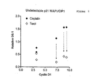
- Figure 1 shows that in cell lines with undetectable p21 and high cyclin D1 levels or cyclin D1 over-expression there is a strong relationship between resistance to CDDP and sensitivity to taxol, as measured by the D0. values (the dose of the drug which reduces clonogenic survival to 10% of the control, untreated cells).
- the cyclin D1/p21 mutation test may also indicate a correlation of taxane sensitivity to resistance to other cytotoxic drugs such as etoposide.
- Cyclin D1 protein is typically measured by Western blotting or immunocytochemistry.
- CDDP chemotherapeutic agent
- agents which can be replaced with taxanes include other platinating agents, such as carboplatin and paraplatin as well as other chemotherapeutic agents, for example alkylating agents, DNA intercalating agents such as Doxorubicin, topoisomerase inhibitors, anti-metabolites such as methotrexate, 5-fluorouracil, DNA synthesis inhibitors such as cytosine arabinoside, and mitotic inhibitors such as the vinca alkaloids.
- cyclin D1 is a relatively short lived protein under cyclical transcriptional control, it is likely that mRNA levels for cyclin D1 follow the same pattern as the cyclin D1 protein and show a similar strong relationship to CDDP resistance. This makes it possible to carry out a functional assay for resistance to CDDP by extracting mRNA from tumour samples and using this to determine the relative abundance of cyclin D1 mRNA and to detect mutations in the p53 mRNA.
- this is a cyclin dependent kinase inhibitor which can be detected by Western blotting, immunocytochemistry or newer developing techniques, such as determining the relative abundance of p21 mRNA.
- RNA levels can be effected in a number of ways.
- Reverse Transcriptase PCR (RTPCR) methods allow the quantity of single RNAs to be determined, but with a relatively low level of accuracy.
- Arrays of oligonucleotides are a relatively novel approach to nucleic acid analysis, allowing mutation analysis, sequencing by hybridisation and mRNA expression analysis. Methods of construction of such arrays have been developed, ( see for example: A.C. Pease et al. Proc. Natl. Acad. Sci. USA. 91, 5022 - 5026, 1994; U.
- An alternative embodiment of this invention can measure Cyclin D1 protein levels by immunocytochemistry using confocal laser fluorescence microscopy.
- a scanning system is used such as those described in PCT/US91/09217, PCT/NL/00081 and PCT/US95/01886.
- the microscopy system is also able to analyse multiple fluorescent dyes.
- Antibodies against p21 are labelled with one dye, an antibody against cyclin D1 (sc-6281, Santa Cruz Biotechnology, CA) is labelled with a second dye whilst a third DNA binding dye can be used to select for aneuploid cells.
- DNA binding dyes such as Hoechst 33258 dye, which binds AT-rich DNA or Chromomycin A 3 , which binds GC-rich DNA, are appropriate.
- a diagnostic test may comprise the steps of:
- a further embodiment of the diagnostic test can exploit Fluorescence Activated Cell Sorting (FACS).
- FACS Fluorescence Activated Cell Sorting
- a FACS instrument separates cells in a suspension in a manner dependent on the cells being labelled with a fluorescent marker.
- a typical FACS device operates as follows. Cells in a suspension travelling in single file are passed through a vibrating nozzle which causes the formation of droplets containing a single cell or none at all. The droplets pass through a laser beam. Fluorescence excited from each individual cell in its droplet by the laser is measured. After the detector the stream of cells in suspension pass through an electrostatic collar which gives the droplets a surface charge. The cells carrying droplets are given a positive or negative charge.
- the drop contains a cell that fluoresces with an intensity above a particular threshold, the drop gets a charge of one polarity. Unlabelled cells get a charge of the opposite polarity.
- the charged droplets are then deflected by an electric field and depending on their surface charge are directed into separate containers and are counted Droplets that contain more than one cell scatter light more than individual cells which is readily detected and so these are left uncharged and enter a third disposal container.
- Multi-channel fluorescent detection devices have been constructed that can separate cells on the basis of labelling with multiple different fluorescent labels. These have multiple lasers which can excite fluorescence at different frequencies and the detector will detect different emission frequencies. A three label system is appropriate for this test. The same labelled probes as those described above for use in a confocal scanning fluorescence microscope would be appropriate.
- a diagnostic test might comprise the steps of:
- Cyclin D1 inhibitors are likely to be non-selectively toxic, but if administered at low doses in conjunction with an agent such as a taxane, the combination may be more effective against tumours than either alone, particularly to cells overexpressing cyclin D1.
- Table 1 also shows relative values for cyclin D1 levels in five human in vitro cell lines not expressing p21 and six human in vitro cell lines expressing p21. Also shown are the absolute and relative D0.1 values (i.e. the dose of the drug that reduces the clonogenic cell survival to 10% of the untreated control cells) for CDDP and taxol. The D0.1 values were obtained by several independent clonogenic assays and the data fitted by linear regression analysis. The clonogenic assays were repeated until statistically satisfactory fits were achieved on linear regression.
- the D0.1 values were obtained by interpolation of the linear regression lines.
- the cytotoxic drug D0.1 values have been normalised and expressed as relative D0.1 values, to enable the relative sensitivity of each cell to each of the drugs to be compared.
- the mean D0.1 was calculated independently for CDDP and taxol; each absolute D0.1 value was then adjusted by dividing by the mean value to give a relative D0.1 value for each drug for each cell line.
- Table 1 The results set out in Table 1 are plotted in the graphs shown in Figures 1 and 2.
- Figure 1 shows relative D0.1 values for CDDP and taxol in five cell lines in which p21 protein was substantially undetectable. Three of the cell lines are relatively resistant to CDDP, but not to taxol. The remaining two cell lines which are less relatively resistant to CDDP are only slightly more sensitive to taxol than to CDDP. There would thus be an advantage in using taxol rather than cisplatin in the former three cell lines, but not in the latter two.
- Figure 2 shows that there is no relationship between Cyclin D1 protein levels and relative sensitivity to CDDP in cells in which p21 protein is present in detectable quantities. Moreover, some cell lines are relatively more resistant to taxol than CDDP. Thus, in cell lines in which p21 protein is detectable, Cyclin D1 is not a useful indicator either of resistance to CDDP or of whether the cell that would preferentially respond to taxol rather than CDDP.
- CDDP sensitivity 10 2 -10 5 cells were plated in 3 ml of Ham's F12 medium supplemented with 10% FCS in 6 well plates and incubated at 37°C in an atmosphere of 5% CO 2 for 8 hours. Dilutions of 0.02-2.0 ⁇ g/ml from a 1 mg/ml stock solution of CDDP (light protected) were then made and 1 ml of the appropriate dilution were added to each plate to give a final volume of 4 ml. The plates were then incubated at 37°C in an atmosphere of 5% CO 2 in darkness for 14 days in the presence of the CDDP. The medium was then removed, the cells were fixed in 70% ethanol and stained with 10% Giemsa and colonies of >100 cells counted.
- CDDP cell survival was determined at the 10% clonogenic cell survival level (D0.1) by interpolation of the fitted regression curve.
- cyclin D1 protein 150 ⁇ g of total cellular protein in 50 ⁇ l of lysate buffer were added per lane well to a 7.5% Laemmli separating gel and electrophoresis carried out at 16°C using 60V over 16 hours and a constant current of 500mA.
- Blots were transferred to nitrocellulose at 22°C over 16 hours using to a semi-dry blotting apparatus (Biorad, Richmond, CA), incubated with the a mouse IgG 1 monoclonal antibody to mammalian cyclins (G124-259.5, Pharmingen) and then incubated with rabbit anti-mouse conjugated antibodies (Dako, UK) at 1/1000 and developed in alkaline phosphatase buffer containing Nitroblue Tetrazolium and 5-Bromo-4-Chloro-3-Indoyl Phosphate, (Sigma, Poole, Dorset, UK) (50mg/ml in dimethylformamide) for 1 hr at room temperature in darkness. Colour development was arrested with double distilled water, and the blots were dried flat. Cyclins were clearly resolved as distinct bands, cyclin D1 having the lowest mobility.
- Quantitation of the protein product of the cyclin D1 gene was carried out by measurement of optical density on a Schimadzu scanning densitometer with tungsten light and expressed as O.D. units per 150 ⁇ g of total cellular protein. Titration curves obtained by loading different amounts of total cellular protein have previously shown that linear relationships for optical density (O.D.) could be obtained over the range found for cyclin D1 protein across the cell lines (Warenius et al 1994, Browning 1997). In order to compare different cyclin D1 protein levels between the cell lines, the mean O.D. value for all the lines was calculated and the relative O.D. for cyclin D1 protein in each individual cell line was normalised to the mean O.D. and multiplied by an arbitrary value of 5.0.
- O.D. optical density
- cyclin D1 over expression or elevated cyclin D1 protein levels is an indicator that cells which are resistant to platinating agents will remain responsive to taxanes, whereas cell lines with low cyclin D1 expression are likely to benefit from treatment with either taxanes or platinating agents (see Figure 1).
- the same correlation is not found in cells containing detectable quantities of p21 protein, in which some cell lines which are relatively resistant to CDDP are also resistant to taxanes, whilst others which are also relatively resistant to CDDP are sensitive to taxanes.
- the present invention allows selection of an appropriate treatment for certain cancers by identifying the p21 level, and cyclin D1 expression or cyclin D1 protein level elevation in tumour cells; if platinating agents are ineffective for such cells, then a taxane is likely to be an effective agent for treatment of the cancer instead.
Abstract
Description
- Extracting a biopsy of the tumour from a patient.
- Optionally micro-dissecting that material to separate normal tissue from tumour material.
- Preparing the biopsy material for microscopy which includes the steps of:
- Labelling the biopsy material with the above fluorescently labelled antibody probes against Cyclin D1. The biopsy material may also, optionally be labelled with antibody probes against p21 protein and with a DNA binding dye.
- Separating the labelled cells from unbound labelled probes.
- Placing the labelled biopsy material in a scanning confocal microscope to count cells
that:
- Over-express or show elevated levels of cyclin D1, i.e. are labelled with at least a threshold quantity of antibody against cyclin D1.
- Optionally, do not express p21, i.e. are labelled with below a threshold quantity of antibodies against p21. Alternatively, p21 levels might be determined by analysis of the mRNA as discussed above.
- Optionally, have chromosomal amplifications as detected by the intensity of fluorescence from DNA binding fluorescent dyes.
- Extracting a biopsy of the tumour from a patient.
- Optionally micro-dissecting that material to separate normal tissue from tumour material.
- Disrupting intracellular adhesion to form a single cell suspension.
- Labelling the suspended cells with the above fluorescently labelled probes against cyclin D1. The biopsy material may also, optionally be labelled with antibody probes against p21 and with a DNA binding dye.
- Separating the labelled cells from unbound labelled probes.
- Passing the labelled cell suspension through a FACS device to count cells that:
- Over-express or show elevated levels of cyclin Dl, i.e. are labelled with the anti-cyclin D1 antibody above a threshold for 'normal' expression.
- Optionally, do not express p21, i.e. are labelled with below a threshold quantity of antibody against p21.
- Optionally, have chromosomal amplifications as detected by the intensity of fluorescence from DNA binding fluorescent dyes.
| CDDP | Taxol | ||||
| Undetectable p21 | Relative Cyclin D1 protein levels | Absolute D0.1 | Relative D0.1 | Absolute D0.1 | Relative D0.1 |
| A431 | 3.21 (0.24) | 0.306 | 0.765 | 0.823 | 0.292 |
| HT29 | 9.39 (1.47) | 0.632 | 1.58 | 1.106 | 0.392 |
| MOR | 8.86 (0.87) | 0.629 | 1.573 | 1.209 | 0.429 |
| RT112 | 3.19 | 0.237 | 0.593 | 1.523 | 0.540 |
| MEL2 | 7.32 | 0.457 | 1.143 | 0.57 | 0.202 |
| Detectable p21 | |||||
| 2780 | 4.42 (0.06) | 0.255 | 0.637 | 2.113 | 0.749 |
| I407 | 0.28 (0.01) | 0.06 | 0.15 | 1.504 | 0.537 |
| HRT18 | 4.44 (0.01) | 0.864 | 2.16 | 10.415 | 3.693 |
| MGHU1 | 6.23 (0.28) | 0.4494 | 1.235 | 1.144 | 0.406 |
| CORL23 | 2.11 (0.05) | 0.338 | 0.845 | 0.497 | 0.176 |
| OAW42 | 6.65 | 0.108 | 0.27 | 10.097 | 3.581 |
| A431 | Squamous carcinoma vulva |
| HT29 | Adenocarcinoma colon |
| MOR | Adenocarcinoma lung |
| RT112 | Transitional cell carcinoma bladder |
| MEL2 | Malignant melanoma |
| 2780 | Ovarian carcinoma |
| I407 | Embrionic intestinal epithelium |
| HRT18 | Adenocarcinoma rectum |
| MGHU1 | Transitional cell carcinoma bladder |
| CORL23 | Large cell lung carcinoma |
| OAW42 | Ovarian carcinoma |
Claims (31)
- An in vitro method for selecting a chemotherapeutic agent for treating cancer, which method comprises:(a) testing a sample comprising cells that substantially do not express p21 and/or in which p21 protein is substantially undetectable, or an extract therefrom for the level of expression of Cyclin D1 or for the abundance of cyclin D1 protein; and(b) if cyclin D1 is overexpressed, and/or cyclin D1 protein is present at elevated levels, selecting for treatment a chemotherapeutic agent comprising a taxane;(c) if cyclin D1 is not overexpressed and/or cyclin D1 protein is substantially not present at elevated levels, selecting for treatment a chemotherapeutic agent comprising an agent other than a taxane.
- A method according to claim 1, wherein the chemotherapeutic agent is selected according to step (b), and the taxane is taxol.
- A method according to claim 1, wherein the chemotherapeutic agent is selected according to step (c), and the agent other than a taxane is a platinating agent.
- A method according to claim 2, wherein the chemotherapeutic agent selected for treatment further comprises a platinating agent.
- A method according to claim 3, wherein the chemotherapeutic agent selected for treatment further comprises a taxane.
- A method according to any preceding claim, wherein the sample has been extracted from a subject.
- A method according to any preceding claim, wherein the testing for the abundance of cyclin D protein comprises measuring the abundance of cyclin D1 mRNA.
- A method according to claim 7, wherein the measurement of the abundance of cyclin D1 mRNA comprises contacting the sample with a probe for cyclin D1 mRNA.
- A method according to any of claims 1-6, wherein the testing for the abundance of cyclin D1 protein is carried out using Western blotting.
- A method according to any of claims 1-6, wherein the testing for the abundance of cyclin D1 protein comprises contacting the sample with a labelled antibody against cyclin D1 protein.
- A method according to claim 10, wherein the antibody against cyclin D1 protein is 14841 C (from clone number G-124-259.5)
- A method according to claim 10 or claim 11, wherein at least one antibody is labelled with a fluorescent label.
- A method according to any preceding claim, further comprising contacting the sample with a DNA binding dye for labelling aneuploid cells.
- A method according to claim 13, wherein the DNA binding dye is Hoechst 33258, or Chromomycin A3 dye.
- A method according to any preceding claim, wherein the sample is a sample of cells.
- A method according to claim 15, wherein the testing is carried out by performing a cell count.
- A method according to claim 16, wherein the cell count is performed using multiparameter flow cytometry.
- A method according to claim 16, wherein the cell count is performed using scanning confocal microscopy.
- A method according to claim 16, wherein the cell count is performed using fluorescence activated cell sorting.
- A method according to any of claims 16-19, wherein the sample of cells is micro-dissected prior to performing the cell count, to separate normal tissue from tumour tissue.
- A method according to any of claims 16-20, wherein prior to performing the cell count, intracellular adhesion in the sample of cells is disrupted, to form a single cell suspension.
- A method according to any preceding claim, for selecting either an agent comprising a taxane, or an agent comprising a platinating agent, for treating cancer.
- A kit for selecting a chemotherapeutic agent for treatment, which kit comprises:(a) a means for identifying cells in which p21 is substantially not expressed and/or p21 protein is substantially undetectable; and(b) a means for testing for the level of expression of Cyclin D1 or for the abundance of cyclin D1 protein in cells or in a sample therefrom.
- A kit according to claim 23, for selecting either an agent comprising a taxane, or an agent comprising a compound other than a taxane such as a platinating agent, for treating cancer.
- A kit according to claim 23 or claim 24, wherein the means for testing for the abundance of cyclin D1 protein comprises a probe for cyclin D1 mRNA.
- A kit according to claim 23 or claim 24, wherein the means for testing for the abundance of cyclin D1 protein comprises a labelled antibody against cyclin D1 protein.
- A kit according to claim 26, wherein the antibody against cyclin D1 protein is 14841 C (from clone number G-124-259.5).
- A kit according to claim 26 or 27, wherein at least one antibody is labelled with a fluorescent label.
- A kit according to any of claims 23-28, further comprising a DNA binding dye, for labelling aneuploid cells.
- A kit according to claim 29, wherein the DNA binding dye is Hoechst 33258, or Chromomycin A3 dye.
- Use of a means for identifying cells in which p21 is substantially not expressed and/or p21 protein is substantially undetectable, and in addition a means for testing for the level of expression of Cyclin D1 or for the abundance of Cyclin D1 protein in cells or in a sample therefrom, for selecting either an agent comprising a taxane, or an agent comprising a platinating agent, in a method according to claim 1.
Applications Claiming Priority (11)
| Application Number | Priority Date | Filing Date | Title |
|---|---|---|---|
| GB9803446A GB2334577A (en) | 1998-02-18 | 1998-02-18 | Resistance of p53 mutant cancer cells to cytoxic effects of (chemo)therapeutic agents involving assay of cyclin D1 protein |
| GB9803447 | 1998-02-18 | ||
| GB9803446 | 1998-02-18 | ||
| GB9803447A GB2334578A (en) | 1998-02-18 | 1998-02-18 | Diagnosis of cancer involving assay of levels of cyclin-dependent kinase (CDK) isoenzymes |
| GBGB9812151.0A GB9812151D0 (en) | 1998-06-05 | 1998-06-05 | Treating cancer |
| GB9812151 | 1998-06-05 | ||
| GB9814545A GB2334579B (en) | 1998-02-18 | 1998-07-03 | Treating cancer |
| GB9814545 | 1998-07-03 | ||
| GB9903035A GB2335739A (en) | 1998-02-18 | 1999-02-10 | Screening anti-cancer agents |
| GB9903035 | 1999-02-10 | ||
| PCT/GB1999/000501 WO1999042835A1 (en) | 1998-02-18 | 1999-02-18 | Treating cancer |
Publications (2)
| Publication Number | Publication Date |
|---|---|
| EP1057028A1 EP1057028A1 (en) | 2000-12-06 |
| EP1057028B1 true EP1057028B1 (en) | 2003-04-23 |
Family
ID=27517448
Family Applications (7)
| Application Number | Title | Priority Date | Filing Date |
|---|---|---|---|
| EP99905084A Expired - Lifetime EP1057030B1 (en) | 1998-02-18 | 1999-02-18 | Treating cancer |
| EP99905081A Withdrawn EP1057027A2 (en) | 1998-02-18 | 1999-02-18 | Treating cancer |
| EP99905082A Expired - Lifetime EP1057028B1 (en) | 1998-02-18 | 1999-02-18 | Treating cancer |
| EP99905083A Expired - Lifetime EP1057029B1 (en) | 1998-02-18 | 1999-02-18 | Treating cancer |
| EP99906326A Expired - Lifetime EP1057033B1 (en) | 1998-02-18 | 1999-02-18 | Treating cancer |
| EP99905086A Expired - Lifetime EP1057031B1 (en) | 1998-02-18 | 1999-02-18 | Treating cancer |
| EP99905087A Expired - Lifetime EP1057032B1 (en) | 1998-02-18 | 1999-02-18 | Treating cancer |
Family Applications Before (2)
| Application Number | Title | Priority Date | Filing Date |
|---|---|---|---|
| EP99905084A Expired - Lifetime EP1057030B1 (en) | 1998-02-18 | 1999-02-18 | Treating cancer |
| EP99905081A Withdrawn EP1057027A2 (en) | 1998-02-18 | 1999-02-18 | Treating cancer |
Family Applications After (4)
| Application Number | Title | Priority Date | Filing Date |
|---|---|---|---|
| EP99905083A Expired - Lifetime EP1057029B1 (en) | 1998-02-18 | 1999-02-18 | Treating cancer |
| EP99906326A Expired - Lifetime EP1057033B1 (en) | 1998-02-18 | 1999-02-18 | Treating cancer |
| EP99905086A Expired - Lifetime EP1057031B1 (en) | 1998-02-18 | 1999-02-18 | Treating cancer |
| EP99905087A Expired - Lifetime EP1057032B1 (en) | 1998-02-18 | 1999-02-18 | Treating cancer |
Country Status (8)
| Country | Link |
|---|---|
| US (3) | US6521407B1 (en) |
| EP (7) | EP1057030B1 (en) |
| JP (7) | JP2002504687A (en) |
| AT (6) | ATE238556T1 (en) |
| AU (8) | AU741632B2 (en) |
| CA (7) | CA2321458A1 (en) |
| DE (6) | DE69907156T2 (en) |
| WO (8) | WO1999042834A2 (en) |
Families Citing this family (39)
| Publication number | Priority date | Publication date | Assignee | Title |
|---|---|---|---|---|
| WO2000009684A1 (en) * | 1998-08-14 | 2000-02-24 | Japan Science And Technology Corporation | NUCLEIC ACID CAPABLE OF BINDING SPECIFICALLY TO Ras TARGET PROTEIN |
| US8124630B2 (en) * | 1999-01-13 | 2012-02-28 | Bayer Healthcare Llc | ω-carboxyaryl substituted diphenyl ureas as raf kinase inhibitors |
| IL144144A0 (en) * | 1999-01-13 | 2002-05-23 | Bayer Ag | Omega-carboxy aryl substituted diphenyl ureas as p38 kinase inhibitors |
| AU2001245939A1 (en) * | 2000-03-24 | 2001-10-08 | Millennum Pharmaceuticals, Inc. | Compositions and methods for the identification, assessment, prevention, and therapy of human cancers |
| WO2001084156A2 (en) * | 2000-04-28 | 2001-11-08 | Millennium Pharmaceuticals, Inc. | Identification, assessment, prevention, and therapy of human cancers |
| US6905816B2 (en) | 2000-11-27 | 2005-06-14 | Intelligent Medical Devices, Inc. | Clinically intelligent diagnostic devices and methods |
| US7371763B2 (en) * | 2001-04-20 | 2008-05-13 | Bayer Pharmaceuticals Corporation | Inhibition of raf kinase using quinolyl, isoquinolyl or pyridyl ureas |
| US20080108672A1 (en) * | 2002-01-11 | 2008-05-08 | Bernd Riedl | Omega-Carboxyaryl Substituted Diphenyl Ureas As Raf Kinase Inhibitors |
| JP4636486B2 (en) * | 2002-02-11 | 2011-02-23 | バイエル、ファーマシューテイカルズ、コーポレイション | Arylurea with angiogenesis inhibitory activity |
| JP2003304884A (en) * | 2002-02-13 | 2003-10-28 | Japan Found Cancer Res | Method for estimating compatibility with anticancer agent |
| ES2320437T3 (en) * | 2002-03-25 | 2009-05-22 | Theryte Limited | CANCER TREATMENT. |
| GB2387385A (en) * | 2002-03-25 | 2003-10-15 | Theryte Ltd | Chemotherapeutic agents for treating cancer |
| JP2005529616A (en) * | 2002-06-18 | 2005-10-06 | アイアールエム エルエルシー | Diagnosis and treatment of chemotherapy-resistant tumors |
| ATE458830T1 (en) * | 2003-02-26 | 2010-03-15 | Sysmex Corp | METHOD FOR EXAMINING A CELL |
| US7557129B2 (en) * | 2003-02-28 | 2009-07-07 | Bayer Healthcare Llc | Cyanopyridine derivatives useful in the treatment of cancer and other disorders |
| ES2305808T3 (en) * | 2003-05-20 | 2008-11-01 | Bayer Healthcare Llc | DIARILURES WITH INHIBITING ACTIVITY OF QUINASAS. |
| CL2004001834A1 (en) | 2003-07-23 | 2005-06-03 | Bayer Pharmaceuticals Corp | COMPOUND 4- {4- [3- (4-CHLORO-3-TRIFLUOROMETILFENIL) -UREIDO] -3-FLUOROFENOXI} -PIRIDIN-2-METHYLAMIDE, RAF INHIBITOR, VEGFR, P38 AND PDGFR KINASES, ITS SALTS; PHARMACEUTICAL COMPOSIICON; PHARMACEUTICAL COMBINATION; AND ITS USE TO TREAT HYPERPROL DISORDERS |
| US20050136177A1 (en) * | 2003-08-25 | 2005-06-23 | Anthony Hesse | Method for coloring landscaping materials using foamable dry colorant |
| US8021831B2 (en) * | 2003-08-25 | 2011-09-20 | Board Of Regents, The University Of Texas System | Taxane chemosensitivity prediction test |
| BRPI0507958A (en) * | 2004-02-23 | 2007-07-17 | Novartis Ag | biomarkers |
| US20060019268A1 (en) * | 2004-03-26 | 2006-01-26 | Research Development Foundation | Molecular markers of cisplatin resistance in cancer and uses thereof |
| EP1767647B1 (en) | 2004-05-31 | 2010-11-24 | Sysmex Corporation | Method of judging malignancy of a mammalian cancer cell |
| KR101323574B1 (en) * | 2004-12-08 | 2013-10-30 | 아벤티스 파마슈티칼스 인크. | Method for measuring resistance or sensitivity to docetaxel |
| JP4944446B2 (en) * | 2005-01-31 | 2012-05-30 | シスメックス株式会社 | Effectiveness prediction method of anticancer drug treatment |
| US7957910B2 (en) * | 2005-01-31 | 2011-06-07 | Sysmex Corporation | Method for predicting effectiveness of chemotherapy |
| EP1870455A4 (en) * | 2005-03-31 | 2010-01-20 | Two Cells Co Ltd | Method for distinguishing mesenchymal stem cell using molecular marker and use thereof |
| WO2007002093A2 (en) * | 2005-06-21 | 2007-01-04 | Infinity Discovery, Inc. | Ansamycin formulations and methods of use thereof |
| JP5046574B2 (en) * | 2005-06-30 | 2012-10-10 | シスメックス株式会社 | Effectiveness prediction method of anticancer drug treatment |
| US7682785B2 (en) | 2005-06-30 | 2010-03-23 | Sysmex Corporation | Method for predicting effectiveness of chemotherapy using anticancer agent |
| JP4766969B2 (en) | 2005-09-14 | 2011-09-07 | シスメックス株式会社 | Organization property judgment device |
| CA2630920A1 (en) | 2005-11-22 | 2007-05-31 | University Of South Florida | Inhibition of cell proliferation |
| JP5111902B2 (en) * | 2007-03-14 | 2013-01-09 | シスメックス株式会社 | Cancer diagnosis support device |
| WO2008115561A2 (en) * | 2007-03-21 | 2008-09-25 | Bristol-Myers Squibb Company | Biomarkers and methods for determining sensitivity to microtubule-stabilizing agents |
| CN101677991A (en) * | 2007-04-12 | 2010-03-24 | 英菲尼蒂发现公司 | hydroquinone ansamycin formulations |
| US8921114B2 (en) | 2007-08-24 | 2014-12-30 | Sysmex Corporation | Diagnosis support system for cancer, diagnosis support information providing method for cancer, and computer program product |
| GB0804496D0 (en) | 2008-03-11 | 2008-04-16 | Theryte Ltd | Treating cancer |
| WO2010025448A2 (en) * | 2008-08-29 | 2010-03-04 | University Of South Florida | Inhibition of cell proliferation |
| CN102245021B (en) | 2008-10-15 | 2014-09-17 | 无限药品公司 | Ansamycin hydroquinone compositions |
| US20120108563A1 (en) * | 2009-05-19 | 2012-05-03 | Infinity Pharmaceuticals, Inc. | Methods Of Treating Liposarcoma |
Family Cites Families (11)
| Publication number | Priority date | Publication date | Assignee | Title |
|---|---|---|---|---|
| DK354487A (en) * | 1986-07-11 | 1988-01-12 | Noboru Yanaihara | ONCOGEN-RELATED PEPTIDES |
| CA1339069C (en) * | 1987-11-09 | 1997-07-29 | Henry Lee Niman | Polypeptide-induced monoclonal receptors to protein ligand |
| WO1991019006A1 (en) * | 1990-06-01 | 1991-12-12 | The United States Of America, As Represented By The Secretary, U.S. Department Of Commerce | MONOCLONAL ANTIBODIES FOR IDENTIFICATION AND PREPARATION OF raf-1 ONCOPROTEIN |
| ATE242806T1 (en) * | 1992-10-16 | 2003-06-15 | Cold Spring Harbor Lab | RELOCATION OF THE CYCLIN COMPLEX AND ITS RELATED APPLICATIONS |
| US5381224A (en) * | 1993-08-30 | 1995-01-10 | A. E. Dixon | Scanning laser imaging system |
| DE4444969C1 (en) * | 1994-12-16 | 1996-10-24 | Orga Med Dr Med Erwin Klopfer | Detection of antibodies against p53 in body fluids |
| AU6593196A (en) * | 1995-07-20 | 1997-02-18 | Paracelsian, Inc. | Determination of the presence of abnormal cellular proliferation through the detection of one or more cyclin dependent kinases |
| US5714329A (en) * | 1995-11-29 | 1998-02-03 | Sequana Theraputics, Inc. | Methods for the diagnosis of a genetic predisposition to cancer associated with variant CDK4 allele |
| WO1997038697A1 (en) * | 1996-04-15 | 1997-10-23 | The Trustees Of The University Of Pennsylvania | Sensitization of cells to radiation and chemotherapy |
| WO1997042222A1 (en) * | 1996-05-08 | 1997-11-13 | Cyclacel Limited | Methods and means for inhibition of cdk4 activity |
| WO1998034118A1 (en) * | 1997-01-30 | 1998-08-06 | Yale University | Diagnostic methods and compositions based on the distribution of rad51 |
-
1999
- 1999-02-18 AU AU25384/99A patent/AU741632B2/en not_active Ceased
- 1999-02-18 AU AU26300/99A patent/AU749180B2/en not_active Ceased
- 1999-02-18 JP JP2000532725A patent/JP2002504687A/en active Pending
- 1999-02-18 AU AU26301/99A patent/AU2630199A/en not_active Abandoned
- 1999-02-18 DE DE69907156T patent/DE69907156T2/en not_active Expired - Fee Related
- 1999-02-18 JP JP2000532719A patent/JP2002504353A/en not_active Withdrawn
- 1999-02-18 WO PCT/GB1999/000500 patent/WO1999042834A2/en not_active Application Discontinuation
- 1999-02-18 CA CA002321458A patent/CA2321458A1/en not_active Abandoned
- 1999-02-18 EP EP99905084A patent/EP1057030B1/en not_active Expired - Lifetime
- 1999-02-18 AU AU25380/99A patent/AU739001B2/en not_active Ceased
- 1999-02-18 DE DE69907152T patent/DE69907152D1/en not_active Expired - Lifetime
- 1999-02-18 AT AT99905086T patent/ATE238556T1/en not_active IP Right Cessation
- 1999-02-18 JP JP2000532712A patent/JP2002504683A/en not_active Ceased
- 1999-02-18 WO PCT/GB1999/000509 patent/WO1999042837A1/en active IP Right Grant
- 1999-02-18 CA CA002321481A patent/CA2321481A1/en not_active Abandoned
- 1999-02-18 AT AT99906326T patent/ATE238558T1/en not_active IP Right Cessation
- 1999-02-18 WO PCT/GB1999/000503 patent/WO1999042828A2/en active IP Right Grant
- 1999-02-18 JP JP2000532726A patent/JP2002504496A/en not_active Withdrawn
- 1999-02-18 US US09/622,277 patent/US6521407B1/en not_active Expired - Fee Related
- 1999-02-18 DE DE69907153T patent/DE69907153T2/en not_active Expired - Fee Related
- 1999-02-18 EP EP99905081A patent/EP1057027A2/en not_active Withdrawn
- 1999-02-18 EP EP99905082A patent/EP1057028B1/en not_active Expired - Lifetime
- 1999-02-18 AU AU25379/99A patent/AU743454B2/en not_active Ceased
- 1999-02-18 WO PCT/GB1999/000501 patent/WO1999042835A1/en active IP Right Grant
- 1999-02-18 EP EP99905083A patent/EP1057029B1/en not_active Expired - Lifetime
- 1999-02-18 DE DE69907155T patent/DE69907155T2/en not_active Expired - Fee Related
- 1999-02-18 JP JP2000532727A patent/JP2002504688A/en not_active Withdrawn
- 1999-02-18 AT AT99905082T patent/ATE238553T1/en not_active IP Right Cessation
- 1999-02-18 AT AT99905084T patent/ATE238555T1/en not_active IP Right Cessation
- 1999-02-18 EP EP99906326A patent/EP1057033B1/en not_active Expired - Lifetime
- 1999-02-18 WO PCT/GB1999/000502 patent/WO1999042090A2/en active IP Right Grant
- 1999-02-18 CA CA002321438A patent/CA2321438A1/en not_active Abandoned
- 1999-02-18 EP EP99905086A patent/EP1057031B1/en not_active Expired - Lifetime
- 1999-02-18 WO PCT/GB1999/000512 patent/WO1999042839A2/en active Application Filing
- 1999-02-18 WO PCT/GB1999/000506 patent/WO1999042821A2/en active IP Right Grant
- 1999-02-18 JP JP2000532728A patent/JP2002504354A/en not_active Withdrawn
- 1999-02-18 EP EP99905087A patent/EP1057032B1/en not_active Expired - Lifetime
- 1999-02-18 AT AT99905083T patent/ATE238554T1/en not_active IP Right Cessation
- 1999-02-18 WO PCT/GB1999/000505 patent/WO1999042836A1/en active IP Right Grant
- 1999-02-18 AU AU25381/99A patent/AU735896B2/en not_active Ceased
- 1999-02-18 CA CA002321480A patent/CA2321480A1/en not_active Abandoned
- 1999-02-18 CA CA002321467A patent/CA2321467A1/en not_active Abandoned
- 1999-02-18 US US09/622,577 patent/US6878526B1/en not_active Expired - Fee Related
- 1999-02-18 AU AU25382/99A patent/AU741712B2/en not_active Ceased
- 1999-02-18 DE DE69907151T patent/DE69907151D1/en not_active Expired - Lifetime
- 1999-02-18 AT AT99905087T patent/ATE238557T1/en not_active IP Right Cessation
- 1999-02-18 CA CA002321479A patent/CA2321479A1/en not_active Abandoned
- 1999-02-18 DE DE69907154T patent/DE69907154T2/en not_active Expired - Fee Related
- 1999-02-18 AU AU25385/99A patent/AU753588B2/en not_active Ceased
- 1999-02-18 CA CA002321482A patent/CA2321482A1/en not_active Abandoned
- 1999-02-18 JP JP2000532107A patent/JP2002503822A/en not_active Withdrawn
-
2002
- 2002-12-18 US US10/321,555 patent/US20030134315A1/en not_active Abandoned
Also Published As
Similar Documents
| Publication | Publication Date | Title |
|---|---|---|
| EP1057028B1 (en) | Treating cancer | |
| GB2334578A (en) | Diagnosis of cancer involving assay of levels of cyclin-dependent kinase (CDK) isoenzymes | |
| GB2335739A (en) | Screening anti-cancer agents | |
| GB2334577A (en) | Resistance of p53 mutant cancer cells to cytoxic effects of (chemo)therapeutic agents involving assay of cyclin D1 protein | |
| GB2334579A (en) | Sensitivity of cancer cells to anti-cancer agents involving measurement of properties of signal transduction factors | |
| Myllynen | Regulation of DNA Repair by EGF Receptor Signaling After X-Irradiation |
Legal Events
| Date | Code | Title | Description |
|---|---|---|---|
| PUAI | Public reference made under article 153(3) epc to a published international application that has entered the european phase |
Free format text: ORIGINAL CODE: 0009012 |
|
| 17P | Request for examination filed |
Effective date: 20000913 |
|
| AK | Designated contracting states |
Kind code of ref document: A1 Designated state(s): AT BE CH CY DE DK ES FI FR GB GR IE IT LI LU MC NL PT SE |
|
| 17Q | First examination report despatched |
Effective date: 20011029 |
|
| GRAH | Despatch of communication of intention to grant a patent |
Free format text: ORIGINAL CODE: EPIDOS IGRA |
|
| GRAH | Despatch of communication of intention to grant a patent |
Free format text: ORIGINAL CODE: EPIDOS IGRA |
|
| GRAA | (expected) grant |
Free format text: ORIGINAL CODE: 0009210 |
|
| AK | Designated contracting states |
Designated state(s): AT BE CH CY DE DK ES FI FR GB GR IE IT LI LU MC NL PT SE |
|
| PG25 | Lapsed in a contracting state [announced via postgrant information from national office to epo] |
Ref country code: NL Free format text: LAPSE BECAUSE OF FAILURE TO SUBMIT A TRANSLATION OF THE DESCRIPTION OR TO PAY THE FEE WITHIN THE PRESCRIBED TIME-LIMIT Effective date: 20030423 Ref country code: LI Free format text: LAPSE BECAUSE OF FAILURE TO SUBMIT A TRANSLATION OF THE DESCRIPTION OR TO PAY THE FEE WITHIN THE PRESCRIBED TIME-LIMIT Effective date: 20030423 Ref country code: IT Free format text: LAPSE BECAUSE OF FAILURE TO SUBMIT A TRANSLATION OF THE DESCRIPTION OR TO PAY THE FEE WITHIN THE PRESCRIBED TIME-LIMIT;WARNING: LAPSES OF ITALIAN PATENTS WITH EFFECTIVE DATE BEFORE 2007 MAY HAVE OCCURRED AT ANY TIME BEFORE 2007. THE CORRECT EFFECTIVE DATE MAY BE DIFFERENT FROM THE ONE RECORDED. Effective date: 20030423 Ref country code: FR Free format text: LAPSE BECAUSE OF FAILURE TO SUBMIT A TRANSLATION OF THE DESCRIPTION OR TO PAY THE FEE WITHIN THE PRESCRIBED TIME-LIMIT Effective date: 20030423 Ref country code: FI Free format text: LAPSE BECAUSE OF FAILURE TO SUBMIT A TRANSLATION OF THE DESCRIPTION OR TO PAY THE FEE WITHIN THE PRESCRIBED TIME-LIMIT Effective date: 20030423 Ref country code: CY Free format text: LAPSE BECAUSE OF FAILURE TO SUBMIT A TRANSLATION OF THE DESCRIPTION OR TO PAY THE FEE WITHIN THE PRESCRIBED TIME-LIMIT Effective date: 20030423 Ref country code: CH Free format text: LAPSE BECAUSE OF FAILURE TO SUBMIT A TRANSLATION OF THE DESCRIPTION OR TO PAY THE FEE WITHIN THE PRESCRIBED TIME-LIMIT Effective date: 20030423 Ref country code: BE Free format text: LAPSE BECAUSE OF FAILURE TO SUBMIT A TRANSLATION OF THE DESCRIPTION OR TO PAY THE FEE WITHIN THE PRESCRIBED TIME-LIMIT Effective date: 20030423 Ref country code: AT Free format text: LAPSE BECAUSE OF FAILURE TO SUBMIT A TRANSLATION OF THE DESCRIPTION OR TO PAY THE FEE WITHIN THE PRESCRIBED TIME-LIMIT Effective date: 20030423 |
|
| REG | Reference to a national code |
Ref country code: GB Ref legal event code: FG4D |
|
| REG | Reference to a national code |
Ref country code: CH Ref legal event code: EP |
|
| REF | Corresponds to: |
Ref document number: 69907151 Country of ref document: DE Date of ref document: 20030528 Kind code of ref document: P |
|
| REG | Reference to a national code |
Ref country code: IE Ref legal event code: FG4D |
|
| PG25 | Lapsed in a contracting state [announced via postgrant information from national office to epo] |
Ref country code: SE Free format text: LAPSE BECAUSE OF FAILURE TO SUBMIT A TRANSLATION OF THE DESCRIPTION OR TO PAY THE FEE WITHIN THE PRESCRIBED TIME-LIMIT Effective date: 20030723 Ref country code: PT Free format text: LAPSE BECAUSE OF FAILURE TO SUBMIT A TRANSLATION OF THE DESCRIPTION OR TO PAY THE FEE WITHIN THE PRESCRIBED TIME-LIMIT Effective date: 20030723 Ref country code: GR Free format text: LAPSE BECAUSE OF FAILURE TO SUBMIT A TRANSLATION OF THE DESCRIPTION OR TO PAY THE FEE WITHIN THE PRESCRIBED TIME-LIMIT Effective date: 20030723 Ref country code: DK Free format text: LAPSE BECAUSE OF FAILURE TO SUBMIT A TRANSLATION OF THE DESCRIPTION OR TO PAY THE FEE WITHIN THE PRESCRIBED TIME-LIMIT Effective date: 20030723 |
|
| PG25 | Lapsed in a contracting state [announced via postgrant information from national office to epo] |
Ref country code: DE Free format text: LAPSE BECAUSE OF FAILURE TO SUBMIT A TRANSLATION OF THE DESCRIPTION OR TO PAY THE FEE WITHIN THE PRESCRIBED TIME-LIMIT Effective date: 20030724 |
|
| NLV1 | Nl: lapsed or annulled due to failure to fulfill the requirements of art. 29p and 29m of the patents act | ||
| PG25 | Lapsed in a contracting state [announced via postgrant information from national office to epo] |
Ref country code: ES Free format text: LAPSE BECAUSE OF FAILURE TO SUBMIT A TRANSLATION OF THE DESCRIPTION OR TO PAY THE FEE WITHIN THE PRESCRIBED TIME-LIMIT Effective date: 20031030 |
|
| REG | Reference to a national code |
Ref country code: CH Ref legal event code: PL |
|
| PG25 | Lapsed in a contracting state [announced via postgrant information from national office to epo] |
Ref country code: LU Free format text: LAPSE BECAUSE OF NON-PAYMENT OF DUE FEES Effective date: 20040218 Ref country code: IE Free format text: LAPSE BECAUSE OF NON-PAYMENT OF DUE FEES Effective date: 20040218 Ref country code: GB Free format text: LAPSE BECAUSE OF NON-PAYMENT OF DUE FEES Effective date: 20040218 |
|
| PLBE | No opposition filed within time limit |
Free format text: ORIGINAL CODE: 0009261 |
|
| STAA | Information on the status of an ep patent application or granted ep patent |
Free format text: STATUS: NO OPPOSITION FILED WITHIN TIME LIMIT |
|
| PG25 | Lapsed in a contracting state [announced via postgrant information from national office to epo] |
Ref country code: MC Free format text: LAPSE BECAUSE OF NON-PAYMENT OF DUE FEES Effective date: 20040228 |
|
| 26N | No opposition filed |
Effective date: 20040126 |
|
| EN | Fr: translation not filed | ||
| GBPC | Gb: european patent ceased through non-payment of renewal fee |
Effective date: 20040218 |
|
| REG | Reference to a national code |
Ref country code: IE Ref legal event code: MM4A |
|
| REG | Reference to a national code |
Ref country code: HK Ref legal event code: WD Ref document number: 1033173 Country of ref document: HK |