EP1037658B1 - Ifnar/ifn complex - Google Patents
Ifnar/ifn complex Download PDFInfo
- Publication number
- EP1037658B1 EP1037658B1 EP98964071A EP98964071A EP1037658B1 EP 1037658 B1 EP1037658 B1 EP 1037658B1 EP 98964071 A EP98964071 A EP 98964071A EP 98964071 A EP98964071 A EP 98964071A EP 1037658 B1 EP1037658 B1 EP 1037658B1
- Authority
- EP
- European Patent Office
- Prior art keywords
- ifn
- ifnar
- ifnβ
- type
- ifnar2
- Prior art date
- Legal status (The legal status is an assumption and is not a legal conclusion. Google has not performed a legal analysis and makes no representation as to the accuracy of the status listed.)
- Expired - Lifetime
Links
- 108010050904 Interferons Proteins 0.000 claims description 173
- 102000014150 Interferons Human genes 0.000 claims description 172
- 229940079322 interferon Drugs 0.000 claims description 68
- 101000852870 Homo sapiens Interferon alpha/beta receptor 1 Proteins 0.000 claims description 66
- 230000000694 effects Effects 0.000 claims description 54
- 108090000765 processed proteins & peptides Proteins 0.000 claims description 42
- 230000000840 anti-viral effect Effects 0.000 claims description 40
- 230000004071 biological effect Effects 0.000 claims description 39
- 108020001507 fusion proteins Proteins 0.000 claims description 27
- 102000002227 Interferon Type I Human genes 0.000 claims description 25
- 108010014726 Interferon Type I Proteins 0.000 claims description 25
- 102000037865 fusion proteins Human genes 0.000 claims description 24
- 230000027455 binding Effects 0.000 claims description 23
- 150000003839 salts Chemical class 0.000 claims description 22
- 239000012634 fragment Substances 0.000 claims description 20
- 238000001727 in vivo Methods 0.000 claims description 20
- 108020004414 DNA Proteins 0.000 claims description 19
- 102000004196 processed proteins & peptides Human genes 0.000 claims description 17
- 108091028043 Nucleic acid sequence Proteins 0.000 claims description 16
- 229920001184 polypeptide Polymers 0.000 claims description 16
- 230000014509 gene expression Effects 0.000 claims description 14
- 238000000034 method Methods 0.000 claims description 14
- 108010047761 Interferon-alpha Proteins 0.000 claims description 13
- 102000006992 Interferon-alpha Human genes 0.000 claims description 13
- 239000013598 vector Substances 0.000 claims description 13
- 102000012740 beta Adrenergic Receptors Human genes 0.000 claims description 11
- 108010079452 beta Adrenergic Receptors Proteins 0.000 claims description 11
- 239000003814 drug Substances 0.000 claims description 11
- 230000001093 anti-cancer Effects 0.000 claims description 9
- 102000007438 Interferon alpha-beta Receptor Human genes 0.000 claims description 8
- 108010086140 Interferon alpha-beta Receptor Proteins 0.000 claims description 8
- 238000004519 manufacturing process Methods 0.000 claims description 8
- 230000000295 complement effect Effects 0.000 claims description 6
- 102000054261 human IFNAR1 Human genes 0.000 claims description 6
- 239000008194 pharmaceutical composition Substances 0.000 claims description 6
- 238000002560 therapeutic procedure Methods 0.000 claims description 6
- 230000008102 immune modulation Effects 0.000 claims description 5
- 230000003389 potentiating effect Effects 0.000 claims description 4
- 238000002360 preparation method Methods 0.000 claims description 4
- 238000012258 culturing Methods 0.000 claims description 2
- 102000053602 DNA Human genes 0.000 claims 2
- 239000003937 drug carrier Substances 0.000 claims 1
- 101000852865 Homo sapiens Interferon alpha/beta receptor 2 Proteins 0.000 description 148
- 102100036718 Interferon alpha/beta receptor 2 Human genes 0.000 description 140
- 229940047124 interferons Drugs 0.000 description 107
- 210000004027 cell Anatomy 0.000 description 62
- 102100036714 Interferon alpha/beta receptor 1 Human genes 0.000 description 60
- 108090000623 proteins and genes Proteins 0.000 description 45
- 125000005647 linker group Chemical group 0.000 description 35
- 241000699670 Mus sp. Species 0.000 description 30
- 235000001014 amino acid Nutrition 0.000 description 30
- 102000004169 proteins and genes Human genes 0.000 description 30
- 235000018102 proteins Nutrition 0.000 description 28
- 150000001413 amino acids Chemical class 0.000 description 27
- 101001054334 Homo sapiens Interferon beta Proteins 0.000 description 23
- 241000699666 Mus <mouse, genus> Species 0.000 description 23
- 239000000243 solution Substances 0.000 description 18
- 239000012911 assay medium Substances 0.000 description 16
- 210000002966 serum Anatomy 0.000 description 16
- 239000000203 mixture Substances 0.000 description 15
- 102000005962 receptors Human genes 0.000 description 15
- 108020003175 receptors Proteins 0.000 description 15
- 238000002965 ELISA Methods 0.000 description 14
- 238000006243 chemical reaction Methods 0.000 description 14
- 210000004978 chinese hamster ovary cell Anatomy 0.000 description 14
- -1 IL-1α Proteins 0.000 description 13
- 238000002474 experimental method Methods 0.000 description 13
- 238000002347 injection Methods 0.000 description 13
- 239000007924 injection Substances 0.000 description 13
- 241000711975 Vesicular stomatitis virus Species 0.000 description 11
- 230000006870 function Effects 0.000 description 11
- 230000003285 pharmacodynamic effect Effects 0.000 description 11
- 238000011533 pre-incubation Methods 0.000 description 11
- 102000035195 Peptidases Human genes 0.000 description 10
- 108091005804 Peptidases Proteins 0.000 description 10
- 239000004365 Protease Substances 0.000 description 10
- 238000007792 addition Methods 0.000 description 10
- 210000004369 blood Anatomy 0.000 description 10
- 239000008280 blood Substances 0.000 description 10
- 239000006228 supernatant Substances 0.000 description 10
- HKZAAJSTFUZYTO-LURJTMIESA-N (2s)-2-[[2-[[2-[[2-[(2-aminoacetyl)amino]acetyl]amino]acetyl]amino]acetyl]amino]-3-hydroxypropanoic acid Chemical compound NCC(=O)NCC(=O)NCC(=O)NCC(=O)N[C@@H](CO)C(O)=O HKZAAJSTFUZYTO-LURJTMIESA-N 0.000 description 9
- 125000003275 alpha amino acid group Chemical group 0.000 description 9
- 125000000539 amino acid group Chemical group 0.000 description 9
- 238000003556 assay Methods 0.000 description 9
- 238000009396 hybridization Methods 0.000 description 9
- 238000012163 sequencing technique Methods 0.000 description 9
- 102000004127 Cytokines Human genes 0.000 description 8
- 108090000695 Cytokines Proteins 0.000 description 8
- 108010005716 Interferon beta-1a Proteins 0.000 description 8
- ZDXPYRJPNDTMRX-VKHMYHEASA-N L-glutamine Chemical compound OC(=O)[C@@H](N)CCC(N)=O ZDXPYRJPNDTMRX-VKHMYHEASA-N 0.000 description 8
- 206010033799 Paralysis Diseases 0.000 description 8
- 102000052179 human IFNAR2 Human genes 0.000 description 8
- 238000011534 incubation Methods 0.000 description 8
- 239000000523 sample Substances 0.000 description 8
- 238000006467 substitution reaction Methods 0.000 description 8
- 108091032973 (ribonucleotides)n+m Proteins 0.000 description 7
- 108091003079 Bovine Serum Albumin Proteins 0.000 description 7
- KDXKERNSBIXSRK-UHFFFAOYSA-N Lysine Natural products NCCCCC(N)C(O)=O KDXKERNSBIXSRK-UHFFFAOYSA-N 0.000 description 7
- 238000004458 analytical method Methods 0.000 description 7
- 230000000259 anti-tumor effect Effects 0.000 description 7
- 238000002832 anti-viral assay Methods 0.000 description 7
- 238000004166 bioassay Methods 0.000 description 7
- 230000015572 biosynthetic process Effects 0.000 description 7
- 210000004899 c-terminal region Anatomy 0.000 description 7
- 239000012528 membrane Substances 0.000 description 7
- ZHNUHDYFZUAESO-UHFFFAOYSA-N Formamide Chemical compound NC=O ZHNUHDYFZUAESO-UHFFFAOYSA-N 0.000 description 6
- 108090001101 Hepsin Proteins 0.000 description 6
- 102000004989 Hepsin Human genes 0.000 description 6
- KDXKERNSBIXSRK-YFKPBYRVSA-N L-lysine Chemical compound NCCCC[C@H](N)C(O)=O KDXKERNSBIXSRK-YFKPBYRVSA-N 0.000 description 6
- OUYCCCASQSFEME-QMMMGPOBSA-N L-tyrosine Chemical compound OC(=O)[C@@H](N)CC1=CC=C(O)C=C1 OUYCCCASQSFEME-QMMMGPOBSA-N 0.000 description 6
- CSNNHWWHGAXBCP-UHFFFAOYSA-L Magnesium sulfate Chemical compound [Mg+2].[O-][S+2]([O-])([O-])[O-] CSNNHWWHGAXBCP-UHFFFAOYSA-L 0.000 description 6
- FAPWRFPIFSIZLT-UHFFFAOYSA-M Sodium chloride Chemical compound [Na+].[Cl-] FAPWRFPIFSIZLT-UHFFFAOYSA-M 0.000 description 6
- 125000003178 carboxy group Chemical group [H]OC(*)=O 0.000 description 6
- 201000010099 disease Diseases 0.000 description 6
- 208000037265 diseases, disorders, signs and symptoms Diseases 0.000 description 6
- 238000009472 formulation Methods 0.000 description 6
- 230000002519 immonomodulatory effect Effects 0.000 description 6
- 238000000338 in vitro Methods 0.000 description 6
- 238000001990 intravenous administration Methods 0.000 description 6
- 230000002035 prolonged effect Effects 0.000 description 6
- 241000894007 species Species 0.000 description 6
- 241000972773 Aulopiformes Species 0.000 description 5
- 108010074860 Factor Xa Proteins 0.000 description 5
- 101000959820 Homo sapiens Interferon alpha-1/13 Proteins 0.000 description 5
- 102100040019 Interferon alpha-1/13 Human genes 0.000 description 5
- COLNVLDHVKWLRT-QMMMGPOBSA-N L-phenylalanine Chemical compound OC(=O)[C@@H](N)CC1=CC=CC=C1 COLNVLDHVKWLRT-QMMMGPOBSA-N 0.000 description 5
- 102100030852 Run domain Beclin-1-interacting and cysteine-rich domain-containing protein Human genes 0.000 description 5
- DBMJMQXJHONAFJ-UHFFFAOYSA-M Sodium laurylsulphate Chemical compound [Na+].CCCCCCCCCCCCOS([O-])(=O)=O DBMJMQXJHONAFJ-UHFFFAOYSA-M 0.000 description 5
- BMQYVXCPAOLZOK-UHFFFAOYSA-N Trihydroxypropylpterisin Natural products OCC(O)C(O)C1=CN=C2NC(N)=NC(=O)C2=N1 BMQYVXCPAOLZOK-UHFFFAOYSA-N 0.000 description 5
- 230000009471 action Effects 0.000 description 5
- 238000003776 cleavage reaction Methods 0.000 description 5
- 238000010790 dilution Methods 0.000 description 5
- 239000012895 dilution Substances 0.000 description 5
- 239000013604 expression vector Substances 0.000 description 5
- 230000004927 fusion Effects 0.000 description 5
- 230000003834 intracellular effect Effects 0.000 description 5
- BMQYVXCPAOLZOK-XINAWCOVSA-N neopterin Chemical compound OC[C@@H](O)[C@@H](O)C1=CN=C2NC(N)=NC(=O)C2=N1 BMQYVXCPAOLZOK-XINAWCOVSA-N 0.000 description 5
- 229940038850 rebif Drugs 0.000 description 5
- 238000011160 research Methods 0.000 description 5
- 235000019515 salmon Nutrition 0.000 description 5
- 230000007017 scission Effects 0.000 description 5
- 230000001225 therapeutic effect Effects 0.000 description 5
- 238000001890 transfection Methods 0.000 description 5
- 238000001262 western blot Methods 0.000 description 5
- MTCFGRXMJLQNBG-REOHCLBHSA-N (2S)-2-Amino-3-hydroxypropansäure Chemical compound OC[C@H](N)C(O)=O MTCFGRXMJLQNBG-REOHCLBHSA-N 0.000 description 4
- DHMQDGOQFOQNFH-UHFFFAOYSA-N Glycine Chemical compound NCC(O)=O DHMQDGOQFOQNFH-UHFFFAOYSA-N 0.000 description 4
- 101000635799 Homo sapiens Run domain Beclin-1-interacting and cysteine-rich domain-containing protein Proteins 0.000 description 4
- 108010008281 Recombinant Fusion Proteins Proteins 0.000 description 4
- 102000007056 Recombinant Fusion Proteins Human genes 0.000 description 4
- 239000002253 acid Substances 0.000 description 4
- 230000002378 acidificating effect Effects 0.000 description 4
- 238000012217 deletion Methods 0.000 description 4
- 230000037430 deletion Effects 0.000 description 4
- 231100000673 dose–response relationship Toxicity 0.000 description 4
- 239000012091 fetal bovine serum Substances 0.000 description 4
- 239000000499 gel Substances 0.000 description 4
- 239000001963 growth medium Substances 0.000 description 4
- 239000000463 material Substances 0.000 description 4
- 235000019419 proteases Nutrition 0.000 description 4
- 239000011780 sodium chloride Substances 0.000 description 4
- UCSJYZPVAKXKNQ-HZYVHMACSA-N streptomycin Chemical compound CN[C@H]1[C@H](O)[C@@H](O)[C@H](CO)O[C@H]1O[C@@H]1[C@](C=O)(O)[C@H](C)O[C@H]1O[C@@H]1[C@@H](NC(N)=N)[C@H](O)[C@@H](NC(N)=N)[C@H](O)[C@H]1O UCSJYZPVAKXKNQ-HZYVHMACSA-N 0.000 description 4
- 238000007920 subcutaneous administration Methods 0.000 description 4
- 239000000126 substance Substances 0.000 description 4
- 230000004083 survival effect Effects 0.000 description 4
- 238000012360 testing method Methods 0.000 description 4
- 238000013518 transcription Methods 0.000 description 4
- 230000035897 transcription Effects 0.000 description 4
- 238000011282 treatment Methods 0.000 description 4
- QTBSBXVTEAMEQO-UHFFFAOYSA-N Acetic acid Chemical compound CC(O)=O QTBSBXVTEAMEQO-UHFFFAOYSA-N 0.000 description 3
- 241000894006 Bacteria Species 0.000 description 3
- 102000000844 Cell Surface Receptors Human genes 0.000 description 3
- 108010001857 Cell Surface Receptors Proteins 0.000 description 3
- 238000012286 ELISA Assay Methods 0.000 description 3
- LFQSCWFLJHTTHZ-UHFFFAOYSA-N Ethanol Chemical compound CCO LFQSCWFLJHTTHZ-UHFFFAOYSA-N 0.000 description 3
- WSFSSNUMVMOOMR-UHFFFAOYSA-N Formaldehyde Chemical compound O=C WSFSSNUMVMOOMR-UHFFFAOYSA-N 0.000 description 3
- 241000282412 Homo Species 0.000 description 3
- 102000003996 Interferon-beta Human genes 0.000 description 3
- 108090000467 Interferon-beta Proteins 0.000 description 3
- DCXYFEDJOCDNAF-REOHCLBHSA-N L-asparagine Chemical compound OC(=O)[C@@H](N)CC(N)=O DCXYFEDJOCDNAF-REOHCLBHSA-N 0.000 description 3
- 229930182816 L-glutamine Natural products 0.000 description 3
- AYFVYJQAPQTCCC-GBXIJSLDSA-N L-threonine Chemical compound C[C@@H](O)[C@H](N)C(O)=O AYFVYJQAPQTCCC-GBXIJSLDSA-N 0.000 description 3
- MUBZPKHOEPUJKR-UHFFFAOYSA-N Oxalic acid Chemical compound OC(=O)C(O)=O MUBZPKHOEPUJKR-UHFFFAOYSA-N 0.000 description 3
- 239000002202 Polyethylene glycol Substances 0.000 description 3
- 241000700605 Viruses Species 0.000 description 3
- 238000002835 absorbance Methods 0.000 description 3
- 230000004913 activation Effects 0.000 description 3
- 125000002252 acyl group Chemical group 0.000 description 3
- 239000005557 antagonist Substances 0.000 description 3
- 229940003504 avonex Drugs 0.000 description 3
- 229940098773 bovine serum albumin Drugs 0.000 description 3
- 238000012512 characterization method Methods 0.000 description 3
- 239000003153 chemical reaction reagent Substances 0.000 description 3
- 210000000349 chromosome Anatomy 0.000 description 3
- 238000010367 cloning Methods 0.000 description 3
- 230000003013 cytotoxicity Effects 0.000 description 3
- 231100000135 cytotoxicity Toxicity 0.000 description 3
- 230000003247 decreasing effect Effects 0.000 description 3
- 230000029087 digestion Effects 0.000 description 3
- 230000001976 improved effect Effects 0.000 description 3
- 230000001965 increasing effect Effects 0.000 description 3
- 238000003780 insertion Methods 0.000 description 3
- 230000037431 insertion Effects 0.000 description 3
- 238000010253 intravenous injection Methods 0.000 description 3
- 229910052943 magnesium sulfate Inorganic materials 0.000 description 3
- 239000003550 marker Substances 0.000 description 3
- 230000001404 mediated effect Effects 0.000 description 3
- 201000001441 melanoma Diseases 0.000 description 3
- 239000007758 minimum essential medium Substances 0.000 description 3
- 230000004048 modification Effects 0.000 description 3
- 238000012986 modification Methods 0.000 description 3
- 150000007523 nucleic acids Chemical group 0.000 description 3
- 239000002773 nucleotide Substances 0.000 description 3
- 125000003729 nucleotide group Chemical group 0.000 description 3
- 239000000546 pharmaceutical excipient Substances 0.000 description 3
- 239000013612 plasmid Substances 0.000 description 3
- 229920001223 polyethylene glycol Polymers 0.000 description 3
- 239000000047 product Substances 0.000 description 3
- 108091008146 restriction endonucleases Proteins 0.000 description 3
- 230000011664 signaling Effects 0.000 description 3
- 239000003381 stabilizer Substances 0.000 description 3
- 230000000087 stabilizing effect Effects 0.000 description 3
- 230000003612 virological effect Effects 0.000 description 3
- QGZKDVFQNNGYKY-UHFFFAOYSA-N Ammonia Chemical compound N QGZKDVFQNNGYKY-UHFFFAOYSA-N 0.000 description 2
- ZYPWIUFLYMQZBS-SRVKXCTJSA-N Asn-Lys-Lys Chemical compound C(CCN)C[C@@H](C(=O)N[C@@H](CCCCN)C(=O)O)NC(=O)[C@H](CC(=O)N)N ZYPWIUFLYMQZBS-SRVKXCTJSA-N 0.000 description 2
- 108020004705 Codon Proteins 0.000 description 2
- 241000588724 Escherichia coli Species 0.000 description 2
- WQZGKKKJIJFFOK-GASJEMHNSA-N Glucose Natural products OC[C@H]1OC(O)[C@H](O)[C@@H](O)[C@@H]1O WQZGKKKJIJFFOK-GASJEMHNSA-N 0.000 description 2
- 241000238631 Hexapoda Species 0.000 description 2
- 102000002265 Human Growth Hormone Human genes 0.000 description 2
- 108010000521 Human Growth Hormone Proteins 0.000 description 2
- 239000000854 Human Growth Hormone Substances 0.000 description 2
- VEXZGXHMUGYJMC-UHFFFAOYSA-N Hydrochloric acid Chemical compound Cl VEXZGXHMUGYJMC-UHFFFAOYSA-N 0.000 description 2
- 102100038069 Interferon regulatory factor 8 Human genes 0.000 description 2
- 102000008070 Interferon-gamma Human genes 0.000 description 2
- 108010074328 Interferon-gamma Proteins 0.000 description 2
- 125000000998 L-alanino group Chemical group [H]N([*])[C@](C([H])([H])[H])([H])C(=O)O[H] 0.000 description 2
- ODKSFYDXXFIFQN-BYPYZUCNSA-N L-arginine Chemical compound OC(=O)[C@@H](N)CCCN=C(N)N ODKSFYDXXFIFQN-BYPYZUCNSA-N 0.000 description 2
- AGPKZVBTJJNPAG-WHFBIAKZSA-N L-isoleucine Chemical compound CC[C@H](C)[C@H](N)C(O)=O AGPKZVBTJJNPAG-WHFBIAKZSA-N 0.000 description 2
- 125000000393 L-methionino group Chemical group [H]OC(=O)[C@@]([H])(N([H])[*])C([H])([H])C(SC([H])([H])[H])([H])[H] 0.000 description 2
- 102000003960 Ligases Human genes 0.000 description 2
- 108090000364 Ligases Proteins 0.000 description 2
- 239000004472 Lysine Substances 0.000 description 2
- OKIZCWYLBDKLSU-UHFFFAOYSA-M N,N,N-Trimethylmethanaminium chloride Chemical compound [Cl-].C[N+](C)(C)C OKIZCWYLBDKLSU-UHFFFAOYSA-M 0.000 description 2
- 101100498071 Neurospora crassa (strain ATCC 24698 / 74-OR23-1A / CBS 708.71 / DSM 1257 / FGSC 987) cys-17 gene Proteins 0.000 description 2
- 108010067372 Pancreatic elastase Proteins 0.000 description 2
- 102000016387 Pancreatic elastase Human genes 0.000 description 2
- 229930182555 Penicillin Natural products 0.000 description 2
- JGSARLDLIJGVTE-MBNYWOFBSA-N Penicillin G Chemical compound N([C@H]1[C@H]2SC([C@@H](N2C1=O)C(O)=O)(C)C)C(=O)CC1=CC=CC=C1 JGSARLDLIJGVTE-MBNYWOFBSA-N 0.000 description 2
- NQRYJNQNLNOLGT-UHFFFAOYSA-N Piperidine Chemical compound C1CCNCC1 NQRYJNQNLNOLGT-UHFFFAOYSA-N 0.000 description 2
- 239000006146 Roswell Park Memorial Institute medium Substances 0.000 description 2
- IOVBCLGAJJXOHK-SRVKXCTJSA-N Ser-His-His Chemical compound C([C@H](NC(=O)[C@H](CO)N)C(=O)N[C@@H](CC=1NC=NC=1)C(O)=O)C1=CN=CN1 IOVBCLGAJJXOHK-SRVKXCTJSA-N 0.000 description 2
- KQNDIKOYWZTZIX-FXQIFTODSA-N Ser-Ser-Arg Chemical compound OC[C@H](N)C(=O)N[C@@H](CO)C(=O)N[C@H](C(O)=O)CCCNC(N)=N KQNDIKOYWZTZIX-FXQIFTODSA-N 0.000 description 2
- QAOWNCQODCNURD-UHFFFAOYSA-N Sulfuric acid Chemical compound OS(O)(=O)=O QAOWNCQODCNURD-UHFFFAOYSA-N 0.000 description 2
- 108090000631 Trypsin Proteins 0.000 description 2
- 102000004142 Trypsin Human genes 0.000 description 2
- 108060008682 Tumor Necrosis Factor Proteins 0.000 description 2
- XSQUKJJJFZCRTK-UHFFFAOYSA-N Urea Chemical compound NC(N)=O XSQUKJJJFZCRTK-UHFFFAOYSA-N 0.000 description 2
- 208000036142 Viral infection Diseases 0.000 description 2
- 239000011543 agarose gel Substances 0.000 description 2
- 125000003277 amino group Chemical group 0.000 description 2
- 238000011319 anticancer therapy Methods 0.000 description 2
- 102000015736 beta 2-Microglobulin Human genes 0.000 description 2
- 108010081355 beta 2-Microglobulin Proteins 0.000 description 2
- 210000001124 body fluid Anatomy 0.000 description 2
- 239000010839 body fluid Substances 0.000 description 2
- 239000000872 buffer Substances 0.000 description 2
- 150000001768 cations Chemical class 0.000 description 2
- 239000006285 cell suspension Substances 0.000 description 2
- 230000008859 change Effects 0.000 description 2
- 238000011260 co-administration Methods 0.000 description 2
- 239000002299 complementary DNA Substances 0.000 description 2
- 230000000536 complexating effect Effects 0.000 description 2
- 238000010276 construction Methods 0.000 description 2
- 239000012228 culture supernatant Substances 0.000 description 2
- 125000000151 cysteine group Chemical class N[C@@H](CS)C(=O)* 0.000 description 2
- 230000002354 daily effect Effects 0.000 description 2
- 230000007423 decrease Effects 0.000 description 2
- 238000011161 development Methods 0.000 description 2
- 230000018109 developmental process Effects 0.000 description 2
- 238000005516 engineering process Methods 0.000 description 2
- 210000002950 fibroblast Anatomy 0.000 description 2
- 239000008103 glucose Substances 0.000 description 2
- 230000002163 immunogen Effects 0.000 description 2
- 230000006698 induction Effects 0.000 description 2
- 238000001802 infusion Methods 0.000 description 2
- 108010051621 interferon regulatory factor-8 Proteins 0.000 description 2
- 239000007927 intramuscular injection Substances 0.000 description 2
- 230000007794 irritation Effects 0.000 description 2
- 210000000265 leukocyte Anatomy 0.000 description 2
- 230000000670 limiting effect Effects 0.000 description 2
- 210000002540 macrophage Anatomy 0.000 description 2
- 239000002609 medium Substances 0.000 description 2
- 238000002844 melting Methods 0.000 description 2
- 230000008018 melting Effects 0.000 description 2
- 238000010369 molecular cloning Methods 0.000 description 2
- 210000000822 natural killer cell Anatomy 0.000 description 2
- 102000039446 nucleic acids Human genes 0.000 description 2
- 108020004707 nucleic acids Proteins 0.000 description 2
- 238000006384 oligomerization reaction Methods 0.000 description 2
- 229940049954 penicillin Drugs 0.000 description 2
- 238000012545 processing Methods 0.000 description 2
- 230000001681 protective effect Effects 0.000 description 2
- 239000000018 receptor agonist Substances 0.000 description 2
- 229940044601 receptor agonist Drugs 0.000 description 2
- 230000002829 reductive effect Effects 0.000 description 2
- 230000035945 sensitivity Effects 0.000 description 2
- 230000019491 signal transduction Effects 0.000 description 2
- 238000002415 sodium dodecyl sulfate polyacrylamide gel electrophoresis Methods 0.000 description 2
- 230000000638 stimulation Effects 0.000 description 2
- 229960005322 streptomycin Drugs 0.000 description 2
- 230000002195 synergetic effect Effects 0.000 description 2
- 238000004448 titration Methods 0.000 description 2
- 238000003146 transient transfection Methods 0.000 description 2
- 239000012588 trypsin Substances 0.000 description 2
- 210000004881 tumor cell Anatomy 0.000 description 2
- 235000002374 tyrosine Nutrition 0.000 description 2
- 210000002700 urine Anatomy 0.000 description 2
- MZOFCQQQCNRIBI-VMXHOPILSA-N (3s)-4-[[(2s)-1-[[(2s)-1-[[(1s)-1-carboxy-2-hydroxyethyl]amino]-4-methyl-1-oxopentan-2-yl]amino]-5-(diaminomethylideneamino)-1-oxopentan-2-yl]amino]-3-[[2-[[(2s)-2,6-diaminohexanoyl]amino]acetyl]amino]-4-oxobutanoic acid Chemical compound OC[C@@H](C(O)=O)NC(=O)[C@H](CC(C)C)NC(=O)[C@H](CCCN=C(N)N)NC(=O)[C@H](CC(O)=O)NC(=O)CNC(=O)[C@@H](N)CCCCN MZOFCQQQCNRIBI-VMXHOPILSA-N 0.000 description 1
- 102000040650 (ribonucleotides)n+m Human genes 0.000 description 1
- 102000007445 2',5'-Oligoadenylate Synthetase Human genes 0.000 description 1
- 108010086241 2',5'-Oligoadenylate Synthetase Proteins 0.000 description 1
- NHBKXEKEPDILRR-UHFFFAOYSA-N 2,3-bis(butanoylsulfanyl)propyl butanoate Chemical compound CCCC(=O)OCC(SC(=O)CCC)CSC(=O)CCC NHBKXEKEPDILRR-UHFFFAOYSA-N 0.000 description 1
- QKNYBSVHEMOAJP-UHFFFAOYSA-N 2-amino-2-(hydroxymethyl)propane-1,3-diol;hydron;chloride Chemical compound Cl.OCC(N)(CO)CO QKNYBSVHEMOAJP-UHFFFAOYSA-N 0.000 description 1
- 208000030507 AIDS Diseases 0.000 description 1
- 102000002260 Alkaline Phosphatase Human genes 0.000 description 1
- 108020004774 Alkaline Phosphatase Proteins 0.000 description 1
- QGZKDVFQNNGYKY-UHFFFAOYSA-O Ammonium Chemical compound [NH4+] QGZKDVFQNNGYKY-UHFFFAOYSA-O 0.000 description 1
- 206010059313 Anogenital warts Diseases 0.000 description 1
- NTAZNGWBXRVEDJ-FXQIFTODSA-N Arg-Asp-Asp Chemical compound [H]N[C@@H](CCCNC(N)=N)C(=O)N[C@@H](CC(O)=O)C(=O)N[C@@H](CC(O)=O)C(O)=O NTAZNGWBXRVEDJ-FXQIFTODSA-N 0.000 description 1
- 239000004475 Arginine Substances 0.000 description 1
- GMRGSBAMMMVDGG-GUBZILKMSA-N Asn-Arg-Arg Chemical compound C(C[C@@H](C(=O)N[C@@H](CCCN=C(N)N)C(=O)O)NC(=O)[C@H](CC(=O)N)N)CN=C(N)N GMRGSBAMMMVDGG-GUBZILKMSA-N 0.000 description 1
- BZMWJLLUAKSIMH-FXQIFTODSA-N Asn-Glu-Glu Chemical compound [H]N[C@@H](CC(N)=O)C(=O)N[C@@H](CCC(O)=O)C(=O)N[C@@H](CCC(O)=O)C(O)=O BZMWJLLUAKSIMH-FXQIFTODSA-N 0.000 description 1
- FBODFHMLALOPHP-GUBZILKMSA-N Asn-Lys-Glu Chemical compound [H]N[C@@H](CC(N)=O)C(=O)N[C@@H](CCCCN)C(=O)N[C@@H](CCC(O)=O)C(O)=O FBODFHMLALOPHP-GUBZILKMSA-N 0.000 description 1
- XAJRHVUUVUPFQL-ACZMJKKPSA-N Asp-Glu-Asp Chemical compound OC(=O)C[C@H](N)C(=O)N[C@@H](CCC(O)=O)C(=O)N[C@@H](CC(O)=O)C(O)=O XAJRHVUUVUPFQL-ACZMJKKPSA-N 0.000 description 1
- GHODABZPVZMWCE-FXQIFTODSA-N Asp-Glu-Glu Chemical compound OC(=O)C[C@H](N)C(=O)N[C@@H](CCC(O)=O)C(=O)N[C@@H](CCC(O)=O)C(O)=O GHODABZPVZMWCE-FXQIFTODSA-N 0.000 description 1
- 208000003950 B-cell lymphoma Diseases 0.000 description 1
- 208000032791 BCR-ABL1 positive chronic myelogenous leukemia Diseases 0.000 description 1
- 101100263837 Bovine ephemeral fever virus (strain BB7721) beta gene Proteins 0.000 description 1
- 238000011740 C57BL/6 mouse Methods 0.000 description 1
- OYPRJOBELJOOCE-UHFFFAOYSA-N Calcium Chemical compound [Ca] OYPRJOBELJOOCE-UHFFFAOYSA-N 0.000 description 1
- 241000283707 Capra Species 0.000 description 1
- 102000011727 Caspases Human genes 0.000 description 1
- 108010076667 Caspases Proteins 0.000 description 1
- 102000004225 Cathepsin B Human genes 0.000 description 1
- 108090000712 Cathepsin B Proteins 0.000 description 1
- 208000010833 Chronic myeloid leukaemia Diseases 0.000 description 1
- KRKNYBCHXYNGOX-UHFFFAOYSA-K Citrate Chemical compound [O-]C(=O)CC(O)(CC([O-])=O)C([O-])=O KRKNYBCHXYNGOX-UHFFFAOYSA-K 0.000 description 1
- 102100023804 Coagulation factor VII Human genes 0.000 description 1
- 108091026890 Coding region Proteins 0.000 description 1
- KPENUVBHAKRDQR-GUBZILKMSA-N Cys-His-Glu Chemical compound [H]N[C@@H](CS)C(=O)N[C@@H](CC1=CNC=N1)C(=O)N[C@@H](CCC(O)=O)C(O)=O KPENUVBHAKRDQR-GUBZILKMSA-N 0.000 description 1
- 102100025621 Cytochrome b-245 heavy chain Human genes 0.000 description 1
- 102000016928 DNA-directed DNA polymerase Human genes 0.000 description 1
- 108010014303 DNA-directed DNA polymerase Proteins 0.000 description 1
- KCXVZYZYPLLWCC-UHFFFAOYSA-N EDTA Chemical compound OC(=O)CN(CC(O)=O)CCN(CC(O)=O)CC(O)=O KCXVZYZYPLLWCC-UHFFFAOYSA-N 0.000 description 1
- 238000008157 ELISA kit Methods 0.000 description 1
- 101100316840 Enterobacteria phage P4 Beta gene Proteins 0.000 description 1
- 108010062466 Enzyme Precursors Proteins 0.000 description 1
- 102000010911 Enzyme Precursors Human genes 0.000 description 1
- 241000206602 Eukaryota Species 0.000 description 1
- 208000034454 F12-related hereditary angioedema with normal C1Inh Diseases 0.000 description 1
- 108010023321 Factor VII Proteins 0.000 description 1
- TWHDOEYLXXQYOZ-FXQIFTODSA-N Gln-Asn-Gln Chemical compound C(CC(=O)N)[C@@H](C(=O)N[C@@H](CC(=O)N)C(=O)N[C@@H](CCC(=O)N)C(=O)O)N TWHDOEYLXXQYOZ-FXQIFTODSA-N 0.000 description 1
- CITDWMLWXNUQKD-FXQIFTODSA-N Gln-Gln-Asn Chemical compound C(CC(=O)N)[C@@H](C(=O)N[C@@H](CCC(=O)N)C(=O)N[C@@H](CC(=O)N)C(=O)O)N CITDWMLWXNUQKD-FXQIFTODSA-N 0.000 description 1
- YGNPTRVNRUKVLA-DCAQKATOSA-N Gln-Met-Met Chemical compound CSCC[C@@H](C(=O)N[C@@H](CCSC)C(=O)O)NC(=O)[C@H](CCC(=O)N)N YGNPTRVNRUKVLA-DCAQKATOSA-N 0.000 description 1
- MCGNJCNXIMQCMN-DCAQKATOSA-N Glu-Met-Met Chemical compound CSCC[C@@H](C(O)=O)NC(=O)[C@H](CCSC)NC(=O)[C@@H](N)CCC(O)=O MCGNJCNXIMQCMN-DCAQKATOSA-N 0.000 description 1
- XEKAJTCACGEBOK-KKUMJFAQSA-N Glu-Met-Phe Chemical compound CSCC[C@@H](C(=O)N[C@@H](CC1=CC=CC=C1)C(=O)O)NC(=O)[C@H](CCC(=O)O)N XEKAJTCACGEBOK-KKUMJFAQSA-N 0.000 description 1
- AAHSHTLISQUZJL-QSFUFRPTSA-N Gly-Ile-Ile Chemical compound [H]NCC(=O)N[C@@H]([C@@H](C)CC)C(=O)N[C@@H]([C@@H](C)CC)C(O)=O AAHSHTLISQUZJL-QSFUFRPTSA-N 0.000 description 1
- ZOTGXWMKUFSKEU-QXEWZRGKSA-N Gly-Ile-Met Chemical compound [H]NCC(=O)N[C@@H]([C@@H](C)CC)C(=O)N[C@@H](CCSC)C(O)=O ZOTGXWMKUFSKEU-QXEWZRGKSA-N 0.000 description 1
- 239000004471 Glycine Substances 0.000 description 1
- 208000005176 Hepatitis C Diseases 0.000 description 1
- OMNVOTCFQQLEQU-CIUDSAMLSA-N His-Asn-Asp Chemical compound C1=C(NC=N1)C[C@@H](C(=O)N[C@@H](CC(=O)N)C(=O)N[C@@H](CC(=O)O)C(=O)O)N OMNVOTCFQQLEQU-CIUDSAMLSA-N 0.000 description 1
- SWSVTNGMKBDTBM-DCAQKATOSA-N His-Gln-Glu Chemical compound C1=C(NC=N1)C[C@@H](C(=O)N[C@@H](CCC(=O)N)C(=O)N[C@@H](CCC(=O)O)C(=O)O)N SWSVTNGMKBDTBM-DCAQKATOSA-N 0.000 description 1
- BXOLYFJYQQRQDJ-MXAVVETBSA-N His-Leu-Ile Chemical compound CC[C@H](C)[C@@H](C(=O)O)NC(=O)[C@H](CC(C)C)NC(=O)[C@H](CC1=CN=CN1)N BXOLYFJYQQRQDJ-MXAVVETBSA-N 0.000 description 1
- 102000008949 Histocompatibility Antigens Class I Human genes 0.000 description 1
- 108010088652 Histocompatibility Antigens Class I Proteins 0.000 description 1
- 101000959794 Homo sapiens Interferon alpha-2 Proteins 0.000 description 1
- 101000844245 Homo sapiens Non-receptor tyrosine-protein kinase TYK2 Proteins 0.000 description 1
- 101500028161 Homo sapiens Tumor necrosis factor-binding protein 1 Proteins 0.000 description 1
- 102000043138 IRF family Human genes 0.000 description 1
- 108091054729 IRF family Proteins 0.000 description 1
- CDGLBYSAZFIIJO-RCOVLWMOSA-N Ile-Gly-Gly Chemical compound CC[C@H](C)[C@H]([NH3+])C(=O)NCC(=O)NCC([O-])=O CDGLBYSAZFIIJO-RCOVLWMOSA-N 0.000 description 1
- KLJKJVXDHVUMMZ-KKPKCPPISA-N Ile-Phe-Trp Chemical compound CC[C@H](C)[C@@H](C(=O)N[C@@H](CC1=CC=CC=C1)C(=O)N[C@@H](CC2=CNC3=CC=CC=C32)C(=O)O)N KLJKJVXDHVUMMZ-KKPKCPPISA-N 0.000 description 1
- DGAQECJNVWCQMB-PUAWFVPOSA-M Ilexoside XXIX Chemical compound C[C@@H]1CC[C@@]2(CC[C@@]3(C(=CC[C@H]4[C@]3(CC[C@@H]5[C@@]4(CC[C@@H](C5(C)C)OS(=O)(=O)[O-])C)C)[C@@H]2[C@]1(C)O)C)C(=O)O[C@H]6[C@@H]([C@H]([C@@H]([C@H](O6)CO)O)O)O.[Na+] DGAQECJNVWCQMB-PUAWFVPOSA-M 0.000 description 1
- 108060003951 Immunoglobulin Proteins 0.000 description 1
- 102000001617 Interferon Receptors Human genes 0.000 description 1
- 108010054267 Interferon Receptors Proteins 0.000 description 1
- 102100036981 Interferon regulatory factor 1 Human genes 0.000 description 1
- 108090000890 Interferon regulatory factor 1 Proteins 0.000 description 1
- 108010002352 Interleukin-1 Proteins 0.000 description 1
- 102000000589 Interleukin-1 Human genes 0.000 description 1
- 108010002350 Interleukin-2 Proteins 0.000 description 1
- 108010024121 Janus Kinases Proteins 0.000 description 1
- 102000015617 Janus Kinases Human genes 0.000 description 1
- 208000007766 Kaposi sarcoma Diseases 0.000 description 1
- ODKSFYDXXFIFQN-BYPYZUCNSA-P L-argininium(2+) Chemical compound NC(=[NH2+])NCCC[C@H]([NH3+])C(O)=O ODKSFYDXXFIFQN-BYPYZUCNSA-P 0.000 description 1
- CKLJMWTZIZZHCS-REOHCLBHSA-N L-aspartic acid Chemical compound OC(=O)[C@@H](N)CC(O)=O CKLJMWTZIZZHCS-REOHCLBHSA-N 0.000 description 1
- WHUUTDBJXJRKMK-VKHMYHEASA-N L-glutamic acid Chemical compound OC(=O)[C@@H](N)CCC(O)=O WHUUTDBJXJRKMK-VKHMYHEASA-N 0.000 description 1
- 206010023849 Laryngeal papilloma Diseases 0.000 description 1
- WXDRGWBQZIMJDE-ULQDDVLXSA-N Leu-Phe-Met Chemical compound [H]N[C@@H](CC(C)C)C(=O)N[C@@H](CC1=CC=CC=C1)C(=O)N[C@@H](CCSC)C(O)=O WXDRGWBQZIMJDE-ULQDDVLXSA-N 0.000 description 1
- MJWVXZABPOKJJF-ACRUOGEOSA-N Leu-Phe-Phe Chemical compound [H]N[C@@H](CC(C)C)C(=O)N[C@@H](CC1=CC=CC=C1)C(=O)N[C@@H](CC1=CC=CC=C1)C(O)=O MJWVXZABPOKJJF-ACRUOGEOSA-N 0.000 description 1
- UCBPDSYUVAAHCD-UWVGGRQHSA-N Leu-Pro-Gly Chemical compound CC(C)C[C@H](N)C(=O)N1CCC[C@H]1C(=O)NCC(O)=O UCBPDSYUVAAHCD-UWVGGRQHSA-N 0.000 description 1
- UIIMIKFNIYPDJF-WDSOQIARSA-N Leu-Trp-Met Chemical compound C1=CC=C2C(C[C@@H](C(=O)N[C@@H](CCSC)C(O)=O)NC(=O)[C@@H](N)CC(C)C)=CNC2=C1 UIIMIKFNIYPDJF-WDSOQIARSA-N 0.000 description 1
- WPIKRJDRQVFRHP-TUSQITKMSA-N Leu-Trp-Trp Chemical compound [H]N[C@@H](CC(C)C)C(=O)N[C@@H](CC1=CNC2=C1C=CC=C2)C(=O)N[C@@H](CC1=CNC2=C1C=CC=C2)C(O)=O WPIKRJDRQVFRHP-TUSQITKMSA-N 0.000 description 1
- 206010025323 Lymphomas Diseases 0.000 description 1
- 108090000542 Lymphotoxin-alpha Proteins 0.000 description 1
- 102000004083 Lymphotoxin-alpha Human genes 0.000 description 1
- OVIVOCSURJYCTM-GUBZILKMSA-N Lys-Asp-Glu Chemical compound NCCCC[C@H](N)C(=O)N[C@@H](CC(O)=O)C(=O)N[C@H](C(O)=O)CCC(O)=O OVIVOCSURJYCTM-GUBZILKMSA-N 0.000 description 1
- 108010046938 Macrophage Colony-Stimulating Factor Proteins 0.000 description 1
- 102100028123 Macrophage colony-stimulating factor 1 Human genes 0.000 description 1
- 102000018697 Membrane Proteins Human genes 0.000 description 1
- 108010052285 Membrane Proteins Proteins 0.000 description 1
- VQILILSLEFDECU-GUBZILKMSA-N Met-Pro-Ala Chemical compound [H]N[C@@H](CCSC)C(=O)N1CCC[C@H]1C(=O)N[C@@H](C)C(O)=O VQILILSLEFDECU-GUBZILKMSA-N 0.000 description 1
- XIGAHPDZLAYQOS-SRVKXCTJSA-N Met-Pro-Pro Chemical compound CSCC[C@H](N)C(=O)N1CCC[C@H]1C(=O)N1[C@H](C(O)=O)CCC1 XIGAHPDZLAYQOS-SRVKXCTJSA-N 0.000 description 1
- UYDDNEYNGGSTDW-OYDLWJJNSA-N Met-Trp-Trp Chemical compound CSCC[C@@H](C(=O)N[C@@H](CC1=CNC2=CC=CC=C21)C(=O)N[C@@H](CC3=CNC4=CC=CC=C43)C(=O)O)N UYDDNEYNGGSTDW-OYDLWJJNSA-N 0.000 description 1
- 102000005741 Metalloproteases Human genes 0.000 description 1
- 108010006035 Metalloproteases Proteins 0.000 description 1
- 208000034578 Multiple myelomas Diseases 0.000 description 1
- 241001529936 Murinae Species 0.000 description 1
- 101000981253 Mus musculus GPI-linked NAD(P)(+)-arginine ADP-ribosyltransferase 1 Proteins 0.000 description 1
- 101001054328 Mus musculus Interferon beta Proteins 0.000 description 1
- 241000204031 Mycoplasma Species 0.000 description 1
- 208000033761 Myelogenous Chronic BCR-ABL Positive Leukemia Diseases 0.000 description 1
- 230000004988 N-glycosylation Effects 0.000 description 1
- 230000006051 NK cell activation Effects 0.000 description 1
- 208000015914 Non-Hodgkin lymphomas Diseases 0.000 description 1
- 102100032028 Non-receptor tyrosine-protein kinase TYK2 Human genes 0.000 description 1
- 238000000636 Northern blotting Methods 0.000 description 1
- 239000004677 Nylon Substances 0.000 description 1
- 230000004989 O-glycosylation Effects 0.000 description 1
- 108091034117 Oligonucleotide Proteins 0.000 description 1
- 108700026244 Open Reading Frames Proteins 0.000 description 1
- 241000283973 Oryctolagus cuniculus Species 0.000 description 1
- 239000002033 PVDF binder Substances 0.000 description 1
- 208000030852 Parasitic disease Diseases 0.000 description 1
- 208000037581 Persistent Infection Diseases 0.000 description 1
- FRMKIPSIZSFTTE-HJOGWXRNSA-N Phe-Tyr-Phe Chemical compound [H]N[C@@H](CC1=CC=CC=C1)C(=O)N[C@@H](CC1=CC=C(O)C=C1)C(=O)N[C@@H](CC1=CC=CC=C1)C(O)=O FRMKIPSIZSFTTE-HJOGWXRNSA-N 0.000 description 1
- IPVPGAADZXRZSH-RNXOBYDBSA-N Phe-Tyr-Trp Chemical compound [H]N[C@@H](CC1=CC=CC=C1)C(=O)N[C@@H](CC1=CC=C(O)C=C1)C(=O)N[C@@H](CC1=CNC2=C1C=CC=C2)C(O)=O IPVPGAADZXRZSH-RNXOBYDBSA-N 0.000 description 1
- 108091000080 Phosphotransferase Proteins 0.000 description 1
- 206010035226 Plasma cell myeloma Diseases 0.000 description 1
- AIOWVDNPESPXRB-YTWAJWBKSA-N Pro-Thr-Pro Chemical compound C[C@H]([C@@H](C(=O)N1CCC[C@@H]1C(=O)O)NC(=O)[C@@H]2CCCN2)O AIOWVDNPESPXRB-YTWAJWBKSA-N 0.000 description 1
- GZNYIXWOIUFLGO-ZJDVBMNYSA-N Pro-Thr-Thr Chemical compound [H]N1CCC[C@H]1C(=O)N[C@@H]([C@@H](C)O)C(=O)N[C@@H]([C@@H](C)O)C(O)=O GZNYIXWOIUFLGO-ZJDVBMNYSA-N 0.000 description 1
- 108010076504 Protein Sorting Signals Proteins 0.000 description 1
- 108010094028 Prothrombin Proteins 0.000 description 1
- 102100027378 Prothrombin Human genes 0.000 description 1
- 238000002123 RNA extraction Methods 0.000 description 1
- 239000012980 RPMI-1640 medium Substances 0.000 description 1
- 102000004265 STAT2 Transcription Factor Human genes 0.000 description 1
- 108010081691 STAT2 Transcription Factor Proteins 0.000 description 1
- 102000004495 STAT3 Transcription Factor Human genes 0.000 description 1
- 108010017324 STAT3 Transcription Factor Proteins 0.000 description 1
- 240000004808 Saccharomyces cerevisiae Species 0.000 description 1
- 229920005654 Sephadex Polymers 0.000 description 1
- 239000012507 Sephadex™ Substances 0.000 description 1
- 206010040047 Sepsis Diseases 0.000 description 1
- 238000012300 Sequence Analysis Methods 0.000 description 1
- XZKQVQKUZMAADP-IMJSIDKUSA-N Ser-Ser Chemical compound OC[C@H](N)C(=O)N[C@@H](CO)C(O)=O XZKQVQKUZMAADP-IMJSIDKUSA-N 0.000 description 1
- MTCFGRXMJLQNBG-UHFFFAOYSA-N Serine Natural products OCC(N)C(O)=O MTCFGRXMJLQNBG-UHFFFAOYSA-N 0.000 description 1
- 102000012479 Serine Proteases Human genes 0.000 description 1
- 108010022999 Serine Proteases Proteins 0.000 description 1
- 208000037065 Subacute sclerosing leukoencephalitis Diseases 0.000 description 1
- 206010042297 Subacute sclerosing panencephalitis Diseases 0.000 description 1
- TYVAWPFQYFPSBR-BFHQHQDPSA-N Thr-Ala-Gly Chemical compound [H]N[C@@H]([C@@H](C)O)C(=O)N[C@@H](C)C(=O)NCC(O)=O TYVAWPFQYFPSBR-BFHQHQDPSA-N 0.000 description 1
- NDZYTIMDOZMECO-SHGPDSBTSA-N Thr-Thr-Ala Chemical compound [H]N[C@@H]([C@@H](C)O)C(=O)N[C@@H]([C@@H](C)O)C(=O)N[C@@H](C)C(O)=O NDZYTIMDOZMECO-SHGPDSBTSA-N 0.000 description 1
- 108010022394 Threonine synthase Proteins 0.000 description 1
- 108090000190 Thrombin Proteins 0.000 description 1
- 108010000499 Thromboplastin Proteins 0.000 description 1
- 102100030859 Tissue factor Human genes 0.000 description 1
- 108091023040 Transcription factor Proteins 0.000 description 1
- 102000040945 Transcription factor Human genes 0.000 description 1
- GSEJCLTVZPLZKY-UHFFFAOYSA-N Triethanolamine Chemical compound OCCN(CCO)CCO GSEJCLTVZPLZKY-UHFFFAOYSA-N 0.000 description 1
- 229920004890 Triton X-100 Polymers 0.000 description 1
- 239000013504 Triton X-100 Substances 0.000 description 1
- 102000000852 Tumor Necrosis Factor-alpha Human genes 0.000 description 1
- 102100040247 Tumor necrosis factor Human genes 0.000 description 1
- 102400000089 Tumor necrosis factor-binding protein 1 Human genes 0.000 description 1
- CGDZGRLRXPNCOC-SRVKXCTJSA-N Tyr-Cys-Cys Chemical compound SC[C@@H](C(O)=O)NC(=O)[C@H](CS)NC(=O)[C@@H](N)CC1=CC=C(O)C=C1 CGDZGRLRXPNCOC-SRVKXCTJSA-N 0.000 description 1
- ZAGPDPNPWYPEIR-SRVKXCTJSA-N Tyr-Cys-Ser Chemical compound [H]N[C@@H](CC1=CC=C(O)C=C1)C(=O)N[C@@H](CS)C(=O)N[C@@H](CO)C(O)=O ZAGPDPNPWYPEIR-SRVKXCTJSA-N 0.000 description 1
- ANHVRCNNGJMJNG-BZSNNMDCSA-N Tyr-Tyr-Cys Chemical compound C1=CC(=CC=C1C[C@@H](C(=O)N[C@@H](CC2=CC=C(C=C2)O)C(=O)N[C@@H](CS)C(=O)O)N)O ANHVRCNNGJMJNG-BZSNNMDCSA-N 0.000 description 1
- CELJCNRXKZPTCX-XPUUQOCRSA-N Val-Gly-Ala Chemical compound CC(C)[C@H](N)C(=O)NCC(=O)N[C@@H](C)C(O)=O CELJCNRXKZPTCX-XPUUQOCRSA-N 0.000 description 1
- AEFJNECXZCODJM-UWVGGRQHSA-N Val-Val-Gly Chemical compound CC(C)[C@H]([NH3+])C(=O)N[C@@H](C(C)C)C(=O)NCC([O-])=O AEFJNECXZCODJM-UWVGGRQHSA-N 0.000 description 1
- SIIZPVYVXNXXQG-KGXOGWRBSA-N [(2r,3r,4r,5r)-5-(6-aminopurin-9-yl)-4-[[(3s,4r)-5-(6-aminopurin-9-yl)-3,4-dihydroxyoxolan-2-yl]methoxy-hydroxyphosphoryl]oxy-3-hydroxyoxolan-2-yl]methyl [(2r,4r,5r)-2-(6-aminopurin-9-yl)-4-hydroxy-5-(phosphonooxymethyl)oxolan-3-yl] hydrogen phosphate Polymers C1=NC2=C(N)N=CN=C2N1[C@@H]1O[C@H](COP(O)(=O)OC2[C@@H](O[C@H](COP(O)(O)=O)[C@H]2O)N2C3=NC=NC(N)=C3N=C2)[C@@H](O)[C@H]1OP(O)(=O)OCC([C@@H](O)[C@H]1O)OC1N1C(N=CN=C2N)=C2N=C1 SIIZPVYVXNXXQG-KGXOGWRBSA-N 0.000 description 1
- 150000007513 acids Chemical class 0.000 description 1
- 239000012190 activator Substances 0.000 description 1
- 239000004480 active ingredient Substances 0.000 description 1
- 230000006978 adaptation Effects 0.000 description 1
- 239000000556 agonist Substances 0.000 description 1
- 108010087924 alanylproline Proteins 0.000 description 1
- 125000001931 aliphatic group Chemical group 0.000 description 1
- 230000004075 alteration Effects 0.000 description 1
- 150000001408 amides Chemical class 0.000 description 1
- 150000001412 amines Chemical class 0.000 description 1
- 229910021529 ammonia Inorganic materials 0.000 description 1
- BFNBIHQBYMNNAN-UHFFFAOYSA-N ammonium sulfate Chemical compound N.N.OS(O)(=O)=O BFNBIHQBYMNNAN-UHFFFAOYSA-N 0.000 description 1
- 229910052921 ammonium sulfate Inorganic materials 0.000 description 1
- 230000003698 anagen phase Effects 0.000 description 1
- 238000000137 annealing Methods 0.000 description 1
- 230000003042 antagnostic effect Effects 0.000 description 1
- 230000001028 anti-proliverative effect Effects 0.000 description 1
- 239000000427 antigen Substances 0.000 description 1
- 230000000890 antigenic effect Effects 0.000 description 1
- 102000036639 antigens Human genes 0.000 description 1
- 108091007433 antigens Proteins 0.000 description 1
- ODKSFYDXXFIFQN-UHFFFAOYSA-N arginine Natural products OC(=O)C(N)CCCNC(N)=N ODKSFYDXXFIFQN-UHFFFAOYSA-N 0.000 description 1
- 125000003435 aroyl group Chemical group 0.000 description 1
- 230000003190 augmentative effect Effects 0.000 description 1
- 238000000376 autoradiography Methods 0.000 description 1
- 230000001580 bacterial effect Effects 0.000 description 1
- 230000009286 beneficial effect Effects 0.000 description 1
- 230000008901 benefit Effects 0.000 description 1
- 230000000975 bioactive effect Effects 0.000 description 1
- 230000008827 biological function Effects 0.000 description 1
- 230000033228 biological regulation Effects 0.000 description 1
- 230000037396 body weight Effects 0.000 description 1
- 210000002798 bone marrow cell Anatomy 0.000 description 1
- 238000010322 bone marrow transplantation Methods 0.000 description 1
- 239000011575 calcium Substances 0.000 description 1
- 229910052791 calcium Inorganic materials 0.000 description 1
- 230000009702 cancer cell proliferation Effects 0.000 description 1
- 239000004202 carbamide Substances 0.000 description 1
- 125000002837 carbocyclic group Chemical group 0.000 description 1
- 150000001720 carbohydrates Chemical class 0.000 description 1
- 239000000969 carrier Substances 0.000 description 1
- 230000024245 cell differentiation Effects 0.000 description 1
- 230000003915 cell function Effects 0.000 description 1
- 210000000170 cell membrane Anatomy 0.000 description 1
- 108091092328 cellular RNA Proteins 0.000 description 1
- 230000001413 cellular effect Effects 0.000 description 1
- 239000003795 chemical substances by application Substances 0.000 description 1
- 238000004587 chromatography analysis Methods 0.000 description 1
- 208000016532 chronic granulomatous disease Diseases 0.000 description 1
- 230000002281 colonystimulating effect Effects 0.000 description 1
- 230000000052 comparative effect Effects 0.000 description 1
- 230000009918 complex formation Effects 0.000 description 1
- 238000010668 complexation reaction Methods 0.000 description 1
- 150000001875 compounds Chemical class 0.000 description 1
- 238000012790 confirmation Methods 0.000 description 1
- 208000035250 cutaneous malignant susceptibility to 1 melanoma Diseases 0.000 description 1
- 235000018417 cysteine Nutrition 0.000 description 1
- 108010004073 cysteinylcysteine Proteins 0.000 description 1
- 102000003675 cytokine receptors Human genes 0.000 description 1
- 108010057085 cytokine receptors Proteins 0.000 description 1
- 230000001086 cytosolic effect Effects 0.000 description 1
- 230000001472 cytotoxic effect Effects 0.000 description 1
- SUYVUBYJARFZHO-RRKCRQDMSA-N dATP Chemical compound C1=NC=2C(N)=NC=NC=2N1[C@H]1C[C@H](O)[C@@H](COP(O)(=O)OP(O)(=O)OP(O)(O)=O)O1 SUYVUBYJARFZHO-RRKCRQDMSA-N 0.000 description 1
- SUYVUBYJARFZHO-UHFFFAOYSA-N dATP Natural products C1=NC=2C(N)=NC=NC=2N1C1CC(O)C(COP(O)(=O)OP(O)(=O)OP(O)(O)=O)O1 SUYVUBYJARFZHO-UHFFFAOYSA-N 0.000 description 1
- RGWHQCVHVJXOKC-SHYZEUOFSA-J dCTP(4-) Chemical compound O=C1N=C(N)C=CN1[C@@H]1O[C@H](COP([O-])(=O)OP([O-])(=O)OP([O-])([O-])=O)[C@@H](O)C1 RGWHQCVHVJXOKC-SHYZEUOFSA-J 0.000 description 1
- HAAZLUGHYHWQIW-KVQBGUIXSA-N dGTP Chemical compound C1=NC=2C(=O)NC(N)=NC=2N1[C@H]1C[C@H](O)[C@@H](COP(O)(=O)OP(O)(=O)OP(O)(O)=O)O1 HAAZLUGHYHWQIW-KVQBGUIXSA-N 0.000 description 1
- NHVNXKFIZYSCEB-XLPZGREQSA-N dTTP Chemical compound O=C1NC(=O)C(C)=CN1[C@@H]1O[C@H](COP(O)(=O)OP(O)(=O)OP(O)(O)=O)[C@@H](O)C1 NHVNXKFIZYSCEB-XLPZGREQSA-N 0.000 description 1
- 230000007123 defense Effects 0.000 description 1
- 230000005860 defense response to virus Effects 0.000 description 1
- 239000003398 denaturant Substances 0.000 description 1
- 230000001419 dependent effect Effects 0.000 description 1
- 206010012601 diabetes mellitus Diseases 0.000 description 1
- 102000004419 dihydrofolate reductase Human genes 0.000 description 1
- 239000003085 diluting agent Substances 0.000 description 1
- 238000006471 dimerization reaction Methods 0.000 description 1
- FSXRLASFHBWESK-UHFFFAOYSA-N dipeptide phenylalanyl-tyrosine Natural products C=1C=C(O)C=CC=1CC(C(O)=O)NC(=O)C(N)CC1=CC=CC=C1 FSXRLASFHBWESK-UHFFFAOYSA-N 0.000 description 1
- 238000009826 distribution Methods 0.000 description 1
- 239000002552 dosage form Substances 0.000 description 1
- 238000009585 enzyme analysis Methods 0.000 description 1
- 230000003203 everyday effect Effects 0.000 description 1
- 230000006624 extrinsic pathway Effects 0.000 description 1
- 229940012413 factor vii Drugs 0.000 description 1
- 238000004108 freeze drying Methods 0.000 description 1
- 230000005714 functional activity Effects 0.000 description 1
- 125000000524 functional group Chemical group 0.000 description 1
- 108091008053 gene clusters Proteins 0.000 description 1
- 230000007274 generation of a signal involved in cell-cell signaling Effects 0.000 description 1
- 230000002068 genetic effect Effects 0.000 description 1
- 230000030414 genetic transfer Effects 0.000 description 1
- 108010055341 glutamyl-glutamic acid Proteins 0.000 description 1
- 125000003827 glycol group Chemical group 0.000 description 1
- 230000013595 glycosylation Effects 0.000 description 1
- 238000006206 glycosylation reaction Methods 0.000 description 1
- 210000003714 granulocyte Anatomy 0.000 description 1
- 230000003394 haemopoietic effect Effects 0.000 description 1
- 201000009277 hairy cell leukemia Diseases 0.000 description 1
- 238000003306 harvesting Methods 0.000 description 1
- 210000003958 hematopoietic stem cell Anatomy 0.000 description 1
- 208000005252 hepatitis A Diseases 0.000 description 1
- 208000002672 hepatitis B Diseases 0.000 description 1
- 208000016861 hereditary angioedema type 3 Diseases 0.000 description 1
- 210000005260 human cell Anatomy 0.000 description 1
- 210000003917 human chromosome Anatomy 0.000 description 1
- 125000002887 hydroxy group Chemical group [H]O* 0.000 description 1
- 208000026278 immune system disease Diseases 0.000 description 1
- 102000018358 immunoglobulin Human genes 0.000 description 1
- 230000006872 improvement Effects 0.000 description 1
- RSAZYXZUJROYKR-UHFFFAOYSA-N indophenol Chemical compound C1=CC(O)=CC=C1N=C1C=CC(=O)C=C1 RSAZYXZUJROYKR-UHFFFAOYSA-N 0.000 description 1
- 239000000411 inducer Substances 0.000 description 1
- 230000001939 inductive effect Effects 0.000 description 1
- 239000012678 infectious agent Substances 0.000 description 1
- 230000005764 inhibitory process Effects 0.000 description 1
- 229910052500 inorganic mineral Inorganic materials 0.000 description 1
- 230000010354 integration Effects 0.000 description 1
- 108010010648 interferon alfacon-1 Proteins 0.000 description 1
- 229960003130 interferon gamma Drugs 0.000 description 1
- 108010045648 interferon omega 1 Proteins 0.000 description 1
- 229960001388 interferon-beta Drugs 0.000 description 1
- 229940076264 interleukin-3 Drugs 0.000 description 1
- 238000007918 intramuscular administration Methods 0.000 description 1
- 230000006623 intrinsic pathway Effects 0.000 description 1
- 230000000366 juvenile effect Effects 0.000 description 1
- 210000003734 kidney Anatomy 0.000 description 1
- 208000009000 laryngeal papillomatosis Diseases 0.000 description 1
- 231100000518 lethal Toxicity 0.000 description 1
- 230000001665 lethal effect Effects 0.000 description 1
- 210000005228 liver tissue Anatomy 0.000 description 1
- 238000011866 long-term treatment Methods 0.000 description 1
- 230000007774 longterm Effects 0.000 description 1
- 238000000504 luminescence detection Methods 0.000 description 1
- 210000004072 lung Anatomy 0.000 description 1
- 206010025135 lupus erythematosus Diseases 0.000 description 1
- 210000004698 lymphocyte Anatomy 0.000 description 1
- 125000003588 lysine group Chemical group [H]N([H])C([H])([H])C([H])([H])C([H])([H])C([H])([H])C([H])(N([H])[H])C(*)=O 0.000 description 1
- 230000002132 lysosomal effect Effects 0.000 description 1
- 210000004962 mammalian cell Anatomy 0.000 description 1
- 239000011159 matrix material Substances 0.000 description 1
- 230000007246 mechanism Effects 0.000 description 1
- 229940127554 medical product Drugs 0.000 description 1
- 108020004999 messenger RNA Proteins 0.000 description 1
- 239000011707 mineral Substances 0.000 description 1
- 239000003226 mitogen Substances 0.000 description 1
- 238000002156 mixing Methods 0.000 description 1
- 201000006417 multiple sclerosis Diseases 0.000 description 1
- 238000002703 mutagenesis Methods 0.000 description 1
- 231100000350 mutagenesis Toxicity 0.000 description 1
- 206010028417 myasthenia gravis Diseases 0.000 description 1
- OHDXDNUPVVYWOV-UHFFFAOYSA-N n-methyl-1-(2-naphthalen-1-ylsulfanylphenyl)methanamine Chemical compound CNCC1=CC=CC=C1SC1=CC=CC2=CC=CC=C12 OHDXDNUPVVYWOV-UHFFFAOYSA-N 0.000 description 1
- 230000002956 necrotizing effect Effects 0.000 description 1
- 239000013642 negative control Substances 0.000 description 1
- 210000000440 neutrophil Anatomy 0.000 description 1
- 230000030648 nucleus localization Effects 0.000 description 1
- 229920001778 nylon Polymers 0.000 description 1
- 230000000771 oncological effect Effects 0.000 description 1
- 150000007524 organic acids Chemical class 0.000 description 1
- 235000005985 organic acids Nutrition 0.000 description 1
- 150000007530 organic bases Chemical class 0.000 description 1
- 235000006408 oxalic acid Nutrition 0.000 description 1
- 210000000496 pancreas Anatomy 0.000 description 1
- 210000003819 peripheral blood mononuclear cell Anatomy 0.000 description 1
- 210000001539 phagocyte Anatomy 0.000 description 1
- 125000002467 phosphate group Chemical group [H]OP(=O)(O[H])O[*] 0.000 description 1
- 230000026731 phosphorylation Effects 0.000 description 1
- 238000006366 phosphorylation reaction Methods 0.000 description 1
- 102000020233 phosphotransferase Human genes 0.000 description 1
- 230000004962 physiological condition Effects 0.000 description 1
- 230000006461 physiological response Effects 0.000 description 1
- 230000001817 pituitary effect Effects 0.000 description 1
- 230000004983 pleiotropic effect Effects 0.000 description 1
- 229920002401 polyacrylamide Polymers 0.000 description 1
- 230000008488 polyadenylation Effects 0.000 description 1
- 108091033319 polynucleotide Proteins 0.000 description 1
- 102000040430 polynucleotide Human genes 0.000 description 1
- 239000002157 polynucleotide Substances 0.000 description 1
- 229920002981 polyvinylidene fluoride Polymers 0.000 description 1
- 239000013641 positive control Substances 0.000 description 1
- 230000029279 positive regulation of transcription, DNA-dependent Effects 0.000 description 1
- 150000003141 primary amines Chemical class 0.000 description 1
- MFDFERRIHVXMIY-UHFFFAOYSA-N procaine Chemical compound CCN(CC)CCOC(=O)C1=CC=C(N)C=C1 MFDFERRIHVXMIY-UHFFFAOYSA-N 0.000 description 1
- 229960004919 procaine Drugs 0.000 description 1
- 238000000159 protein binding assay Methods 0.000 description 1
- 230000009822 protein phosphorylation Effects 0.000 description 1
- 230000006337 proteolytic cleavage Effects 0.000 description 1
- 229940039716 prothrombin Drugs 0.000 description 1
- 238000000746 purification Methods 0.000 description 1
- 239000011535 reaction buffer Substances 0.000 description 1
- 239000002464 receptor antagonist Substances 0.000 description 1
- 229940044551 receptor antagonist Drugs 0.000 description 1
- 108010003189 recombinant human tumor necrosis factor-binding protein-1 Proteins 0.000 description 1
- 230000006798 recombination Effects 0.000 description 1
- 238000005215 recombination Methods 0.000 description 1
- 230000001105 regulatory effect Effects 0.000 description 1
- 230000004044 response Effects 0.000 description 1
- 230000002441 reversible effect Effects 0.000 description 1
- 206010039073 rheumatoid arthritis Diseases 0.000 description 1
- 239000002342 ribonucleoside Substances 0.000 description 1
- 238000012216 screening Methods 0.000 description 1
- 150000003335 secondary amines Chemical class 0.000 description 1
- 238000013207 serial dilution Methods 0.000 description 1
- 125000003607 serino group Chemical group [H]N([H])[C@]([H])(C(=O)[*])C(O[H])([H])[H] 0.000 description 1
- 239000012679 serum free medium Substances 0.000 description 1
- 102000035025 signaling receptors Human genes 0.000 description 1
- 108091005475 signaling receptors Proteins 0.000 description 1
- 238000002741 site-directed mutagenesis Methods 0.000 description 1
- 238000012868 site-directed mutagenesis technique Methods 0.000 description 1
- 239000011734 sodium Substances 0.000 description 1
- 229910052708 sodium Inorganic materials 0.000 description 1
- 239000001509 sodium citrate Substances 0.000 description 1
- NLJMYIDDQXHKNR-UHFFFAOYSA-K sodium citrate Chemical compound O.O.[Na+].[Na+].[Na+].[O-]C(=O)CC(O)(CC([O-])=O)C([O-])=O NLJMYIDDQXHKNR-UHFFFAOYSA-K 0.000 description 1
- 239000001488 sodium phosphate Substances 0.000 description 1
- 229910000162 sodium phosphate Inorganic materials 0.000 description 1
- 230000003393 splenic effect Effects 0.000 description 1
- 230000006641 stabilisation Effects 0.000 description 1
- 238000013112 stability test Methods 0.000 description 1
- 238000011105 stabilization Methods 0.000 description 1
- 238000012289 standard assay Methods 0.000 description 1
- 238000010561 standard procedure Methods 0.000 description 1
- 238000010254 subcutaneous injection Methods 0.000 description 1
- 239000007929 subcutaneous injection Substances 0.000 description 1
- 238000003786 synthesis reaction Methods 0.000 description 1
- PFBLRDXPNUJYJM-UHFFFAOYSA-N tert-butyl 2-methylpropaneperoxoate Chemical compound CC(C)C(=O)OOC(C)(C)C PFBLRDXPNUJYJM-UHFFFAOYSA-N 0.000 description 1
- 210000001550 testis Anatomy 0.000 description 1
- 229940124597 therapeutic agent Drugs 0.000 description 1
- 125000000341 threoninyl group Chemical group [H]OC([H])(C([H])([H])[H])C([H])(N([H])[H])C(*)=O 0.000 description 1
- 229960004072 thrombin Drugs 0.000 description 1
- 210000001685 thyroid gland Anatomy 0.000 description 1
- 210000001519 tissue Anatomy 0.000 description 1
- 230000000699 topical effect Effects 0.000 description 1
- 230000002588 toxic effect Effects 0.000 description 1
- 231100000563 toxic property Toxicity 0.000 description 1
- 230000001052 transient effect Effects 0.000 description 1
- 238000013519 translation Methods 0.000 description 1
- RYFMWSXOAZQYPI-UHFFFAOYSA-K trisodium phosphate Chemical compound [Na+].[Na+].[Na+].[O-]P([O-])([O-])=O RYFMWSXOAZQYPI-UHFFFAOYSA-K 0.000 description 1
- OUYCCCASQSFEME-UHFFFAOYSA-N tyrosine Natural products OC(=O)C(N)CC1=CC=C(O)C=C1 OUYCCCASQSFEME-UHFFFAOYSA-N 0.000 description 1
- 150000003668 tyrosines Chemical class 0.000 description 1
- 210000003556 vascular endothelial cell Anatomy 0.000 description 1
- 230000035899 viability Effects 0.000 description 1
- 230000009385 viral infection Effects 0.000 description 1
- 238000005406 washing Methods 0.000 description 1
- 150000003751 zinc Chemical class 0.000 description 1
Images


Classifications
-
- A—HUMAN NECESSITIES
- A61—MEDICAL OR VETERINARY SCIENCE; HYGIENE
- A61K—PREPARATIONS FOR MEDICAL, DENTAL OR TOILETRY PURPOSES
- A61K38/00—Medicinal preparations containing peptides
- A61K38/16—Peptides having more than 20 amino acids; Gastrins; Somatostatins; Melanotropins; Derivatives thereof
- A61K38/17—Peptides having more than 20 amino acids; Gastrins; Somatostatins; Melanotropins; Derivatives thereof from animals; from humans
- A61K38/19—Cytokines; Lymphokines; Interferons
- A61K38/21—Interferons [IFN]
-
- A—HUMAN NECESSITIES
- A61—MEDICAL OR VETERINARY SCIENCE; HYGIENE
- A61P—SPECIFIC THERAPEUTIC ACTIVITY OF CHEMICAL COMPOUNDS OR MEDICINAL PREPARATIONS
- A61P31/00—Antiinfectives, i.e. antibiotics, antiseptics, chemotherapeutics
- A61P31/12—Antivirals
-
- A—HUMAN NECESSITIES
- A61—MEDICAL OR VETERINARY SCIENCE; HYGIENE
- A61P—SPECIFIC THERAPEUTIC ACTIVITY OF CHEMICAL COMPOUNDS OR MEDICINAL PREPARATIONS
- A61P35/00—Antineoplastic agents
-
- A—HUMAN NECESSITIES
- A61—MEDICAL OR VETERINARY SCIENCE; HYGIENE
- A61P—SPECIFIC THERAPEUTIC ACTIVITY OF CHEMICAL COMPOUNDS OR MEDICINAL PREPARATIONS
- A61P37/00—Drugs for immunological or allergic disorders
-
- A—HUMAN NECESSITIES
- A61—MEDICAL OR VETERINARY SCIENCE; HYGIENE
- A61P—SPECIFIC THERAPEUTIC ACTIVITY OF CHEMICAL COMPOUNDS OR MEDICINAL PREPARATIONS
- A61P37/00—Drugs for immunological or allergic disorders
- A61P37/02—Immunomodulators
-
- A—HUMAN NECESSITIES
- A61—MEDICAL OR VETERINARY SCIENCE; HYGIENE
- A61P—SPECIFIC THERAPEUTIC ACTIVITY OF CHEMICAL COMPOUNDS OR MEDICINAL PREPARATIONS
- A61P43/00—Drugs for specific purposes, not provided for in groups A61P1/00-A61P41/00
-
- C—CHEMISTRY; METALLURGY
- C07—ORGANIC CHEMISTRY
- C07K—PEPTIDES
- C07K14/00—Peptides having more than 20 amino acids; Gastrins; Somatostatins; Melanotropins; Derivatives thereof
- C07K14/435—Peptides having more than 20 amino acids; Gastrins; Somatostatins; Melanotropins; Derivatives thereof from animals; from humans
- C07K14/52—Cytokines; Lymphokines; Interferons
- C07K14/555—Interferons [IFN]
- C07K14/56—IFN-alpha
-
- C—CHEMISTRY; METALLURGY
- C07—ORGANIC CHEMISTRY
- C07K—PEPTIDES
- C07K14/00—Peptides having more than 20 amino acids; Gastrins; Somatostatins; Melanotropins; Derivatives thereof
- C07K14/435—Peptides having more than 20 amino acids; Gastrins; Somatostatins; Melanotropins; Derivatives thereof from animals; from humans
- C07K14/52—Cytokines; Lymphokines; Interferons
- C07K14/555—Interferons [IFN]
- C07K14/565—IFN-beta
-
- C—CHEMISTRY; METALLURGY
- C07—ORGANIC CHEMISTRY
- C07K—PEPTIDES
- C07K14/00—Peptides having more than 20 amino acids; Gastrins; Somatostatins; Melanotropins; Derivatives thereof
- C07K14/435—Peptides having more than 20 amino acids; Gastrins; Somatostatins; Melanotropins; Derivatives thereof from animals; from humans
- C07K14/705—Receptors; Cell surface antigens; Cell surface determinants
- C07K14/715—Receptors; Cell surface antigens; Cell surface determinants for cytokines; for lymphokines; for interferons
-
- C—CHEMISTRY; METALLURGY
- C07—ORGANIC CHEMISTRY
- C07K—PEPTIDES
- C07K2319/00—Fusion polypeptide
Definitions
- the present invention relates to a Type I interferon complex, composed of the polypeptide sequence of the human interferon ⁇ / ⁇ receptor (IFNAR) extracellular domain and a Type I interferon (IFN ⁇ , IFN ⁇ , and IFN ⁇ ).
- IFNAR human interferon ⁇ / ⁇ receptor
- IFN ⁇ , IFN ⁇ , and IFN ⁇ Type I interferon
- Such a complex improves the stability, enhances the potency, and prolongs the pharmacokinetics in vivo of free IFN for anti-viral, anti-cancer and immune modulating activity.
- the complex is a fusion protein, or a covalent complex, or a non-covalent complex containing the polypeptide sequence of the entire extracellular domain of IFNAR2, or any interferon-binding subfraction thereof, complexed to a Type I interferon (IFN ⁇ , IFN ⁇ , IFN ⁇ ), or any biologically active subfraction thereof.
- IFN ⁇ , IFN ⁇ , IFN ⁇ Type I interferon
- Interferons are classified either as the leukocyte and fibroblast derived Type I interferons, or as the mitogen induced or "immune" Type II interferons (Pestka et al, 1987).
- Type I interferons include interferon alpha (IFN ⁇ ), interferon beta (IFN ⁇ ) and interferon omega (IFN ⁇ ), while Type II interferon includes interferon gamma (IFN ⁇ ).
- IFN ⁇ , IFN ⁇ and IFN ⁇ genes are clustered on the short arm of chromosome 9 (Lengyl, 1982). There are at least 25 non-allelic IFN ⁇ genes, 6 non-allelic IFN ⁇ genes and a single IFN ⁇ gene.
- IFN ⁇ genes share at least 80% sequence identity with each other.
- the IFN ⁇ gene shares approximately 50% sequence identity with IFN ⁇ ; and the IFN ⁇ gene shares 70% homology with IFN ⁇ (Weissman et al, 1986; Dron et al, 1992).
- IFN ⁇ has a molecular weight range of 17-23 kDa (165-166 amino acids), IFN ⁇ , ⁇ 23 kDa (166 amino acids) and IFN ⁇ , ⁇ 24 kDa (172 amino acids).
- Type I interferons are pleiotropic cytokines having activity in host defense against viral and parasitic infections, as anti-cancer cytokines and as immune modulators (Baron et al, 1994; Baron et al, 1991).
- Type I interferon physiological responses include anti-proliferative activity on normal and transformed cells; stimulation of cytotoxic activity in lymphocytes, natural killer cells and phagocytic cells; modulation of cellular differentiation; stimulation of expression of class I MHC antigens; inhibition of class II MHC; and modulation of a variety of cell surface receptors.
- IFN ⁇ and IFN ⁇ are secreted constitutively by most human cells at low levels with expression being up-regulated by addition of a variety of inducers, including infectious agents (viruses, bacteria, mycoplasma and protozoa), dsRNA, and cytokines (M-CSF, IL-1 ⁇ , IL-2, TNF ⁇ ).
- infectious agents viruses, bacteria, mycoplasma and protozoa
- dsRNA dsRNA
- cytokines M-CSF, IL-1 ⁇ , IL-2, TNF ⁇
- the actions of Type I interferon in vivo can be monitored using the surrogate markers, neopterin, 2',5' oligoadenylate synthetase, and ⁇ 2 microglobulin (Alam et al, 1997; Fierlbeck et al, 1996; Salmon et al, 1996).
- Type I interferons act through a cell surface receptor complex to induce specific biologic effects, such as anti-viral, anti-tumor, and immune modulatory activity.
- the Type I IFN receptor (IFNAR) is a hetero-multimeric receptor complex composed of at least two different polypeptide chains (Colamonici et al, 1992; Colamonici et al, 1993; Platanias et al, 1993). The genes for these chains are found on chromosome 21, and their proteins are expressed on the surface of most cells (Tan et al, 1973). The receptor chains were originally designated alpha and beta because of their ability to be recognized by the monoclonal antibodies IFN ⁇ R3 and IFNaR ⁇ 1, respectively.
- IFNAR1 alpha chain, Uze subunit
- IFNAR2 beta chain, B L , IFN ⁇ / ⁇ R
- the IFNAR1 and IFNAR2 ⁇ S and ⁇ L subunits have been cloned (Novick et al, 1994; Domanski et al, 1995).
- the IFNAR2 ⁇ S and ⁇ L subunits have identical extracellular and transmembrane domains; however, in the cytoplasmic domain they only share identity in the first 15 amino acids.
- the IFNAR2 subunit alone is able to bind IFN ⁇ / ⁇ , while the IFNAR1 subunit is unable to bind IFN ⁇ / ⁇ .
- IFNAR1/2 complex ⁇ / ⁇ S or ⁇ / ⁇ L subunits bind IFN ⁇ with high affinity, only the ⁇ / ⁇ L pair appears to be a functional signaling receptor.
- a soluble IFNAR has been identified in both human urine and serum (Novick et al, 1994; Novick et al, 1995; Novick et al, 1992; Lutfalla et al, 1995).
- the soluble IFNAR isolated from serum has an apparent molecular weight of 55 kDa on SDS-PAGE, while the soluble IFNAR from urine has an apparent molecular weight of 40-45 kDa (p40).
- Transcripts for the soluble p40 IFNAR2 are present at the mRNA level and encompass almost the entire extracellular domain of the IFNAR2 subunit with two new amino acids at the carboxy terminal end.
- the soluble IFNAR2 receptor There are five potential glycosylation sites on the soluble IFNAR2 receptor.
- the soluble p40 IFNAR2 has been shown to bind IFN ⁇ 2 and IFN ⁇ and to inhibit in vitro the anti-viral activity of a mixture of IFN ⁇ species ("leukocyte IFN") and individual Type I IFNs (Novick et al, 1995).
- a recombinant IFNAR2 subunit Ig fusion protein was shown to inhibit the binding of a variety of Type I IFN species (IFN ⁇ A, IFN ⁇ B, IFN ⁇ D, IFN ⁇ , IFN ⁇ Con1 and IFN ⁇ ) to Daudi cells and ⁇ / ⁇ S subunit double transfected COS cells.
- IFN signaling pathways have recently been identified (Platanias et al, 1996; Yan et al, 1996; Qureshi et al, 1996; Duncan et al, 1996; Sharf et al, 1995; Yang et al, 1996).
- Initial events leading to signaling are thought to occur by the binding of IFN ⁇ / ⁇ / ⁇ to the IFNAR2 subunit, followed by the IFNAR1 subunit associating to form an IFNAR1/2 complex (Platanias et al, 1994).
- IFN ⁇ / ⁇ / ⁇ The binding of IFN ⁇ / ⁇ / ⁇ to the IFNAR1/2 complex results in the activation of two Janus kinases (Jak1 and Tyk2) which are believed to phosphorylate specific tyrosines on the IFNAR1 and IFNAR2 subunits. Once these subunits are phosphorylated, STAT molecules (STAT 1, 2 and 3) are phosphorylated, which results in dimerization of STAT transcription complexes followed by nuclear localization of the transcription complex and the activation of specific IFN inducible genes.
- Type I IFNs The pharmacokinetics and pharmacodynamics of Type I IFNs have been assessed in humans (Alan et al, 1997; Fierlbeck et al, 1996; Salmon et al, 1996). The clearance of IFN ⁇ is fairly rapid with the bioavailability of IFN ⁇ lower than expected for most cytokines. Although the pharmacodynamics of IFN ⁇ have been assessed in humans, no clear correlation has been established between the bioavailability of IFN ⁇ and clinical efficacy.
- PK pharmacokinetics
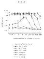
- PD pharmacodynamics
- neopterin exhibited a very similar profile between REBIF® and AVONEX® with maximal neopterin levels achieved at ⁇ 44-50 hours post-injection, remaining elevated until 72 hours post-injection and then dropping to baseline gradually by 144 hours
- Type I interferon complex composed of soluble IFNAR complexed with Type I interferons (IFN) exhibits improved stability, enhanced potency, and elongated pharmacokinetics in vivo compared with free IFN for anti-viral, anti-cancer and immune modulating activity.
- IFN Type I interferons
- the present invention thus provides a Type I interferon (IFN) complex, composed of the polypeptide sequence of a human interferon ⁇ / ⁇ receptor (IFNAR) subunit extracellular domain and Type I interferons, which exhibits improved stability, enhanced potency, and/or prolonged pharmacokinetics in vivo compared to free IFN for anti-viral, anti-cancer and immune modulating activity.
- IFN interferon
- the complex is of the IFNAR2 subunit extracellular domain with any Type I interferon or the IFNAR1 subunit with IFN ⁇ .
- the complex is a fusion protein, or a covalent complex, or a non-covalent complex containing the polypeptide sequence of the entire extracellular domain of IFNAR, preferably IFNAR2, or any interferon-binding subfraction thereof, complexed to IFN ⁇ or IFN ⁇ or IFN ⁇ , or any biologically active subfraction thereof.
- IFNAR is intended to comprehend any of the known extracellular IFNAR receptors as defined above, as well as any active fragments thereof.
- IFNAR can be optionally fused to another protein, for example, an immunoglobulin such as IgG.
- IFN, IFN ⁇ , IFN ⁇ , and IFN ⁇ are intended as one of the more than 20 Type I interferons identified to date, or any other Type I interferon identified in the future.
- the complex is composed of IFN ⁇ or IFN ⁇ , covalently linked to IFNAR2 via chemical linkage.
- a further embodiment comprises a complex composed of IFN ⁇ or IFN ⁇ , non-covalently complexed to IFNAR2.
- This further embodiment also includes a composition containing a Type I IFN and IFNAR2 in any ratio.
- a formulation of Type I IFN with an excess of IFNAR2 as defined above is also included in the definition of "complex" of the present application.
- the two components may also be administered separately so as to form the complex in vivo.
- the complex is a mixture of IFNAR2 and IFN, obtained by simultaneous or subsequent co-administration of IFN ⁇ or IFN ⁇ and soluble IFNAR2.
- the IFNAR can be administered without any concomitant administration of IFN, so that the complex may be formed in vivo with endogenous circulating IFN, thereby potentiating the effects of the endogenous IFN.
- the complex is composed of IFN ⁇ or IFN ⁇ or IFN ⁇ fused to IFNAR2 as a recombinant fusion protein, where the IFN and the IFNAR2 moieties are optionally fused via a flexible peptide linker molecule.
- This peptide linker may or may not be cleavable in vivo.
- the invention further relates to DNA encoding such fusion proteins, vectors containing such DNA, host cells transformed with such vectors in such a manner as to express the fusion proteins and methods of production of such fusion proteins by culturing such host cells and isolating the fusion proteins expressed thereby.
- a further aspect of the present invention are the methods of use of the complexes of the present invention for prolonging the in vivo effect of IFN, which is useful in the treatment of any disease or condition which is treatable by IFN.
- IFNAR as a stabilizer in formulations of IFN.
- Free IFN ⁇ has a tendency to oligomerize. This is prevented once it is complexed to IFNAR, particularly IFNAR2.
- Present day formulations of recombinant IFN ⁇ must have an acidic pH, which may cause some localized irritation when administered.
- Non-acidic compositions can be formulated if IFNAR is used as a stabilizer.
- IFNAR/IFN complex of the present invention and the enabling technology required to produce this complex is described in detail hereinbelow. For most of the results, IFN ⁇ has been chosen as a non-limiting example.
- the C-terminal end of IFNAR2, or any interferon-binding subsequence thereof has been fused to the N-terminal end of IFN ⁇ , or biologically active fragments thereof, as this requires the shortest distance to be bridged between the two molecules.
- the reverse constructs can also be prepared, where the C-terminal end of IFN ⁇ , or fragments thereof, are fused to the N-terminal end of IFNAR2, or subsequences thereof.
- the estimated distance between the constrained C-terminal extracellular domain of IFNAR2 and the N-terminal of IFN ⁇ in the active complex model is ⁇ 80 Angstroms.
- a flexible peptide linker for example, Gly-Gly-Gly-Gly-Ser (GGGGS) (SEQ ID NO:1) repeats, may be employed.
- the linker may be flexible and a target for proteolytic cleavage by serum, membrane bound, and/or cellular proteases.
- the engineered IFNAR2/IFN ⁇ complex fusion may have a Factor Xa cleavage site.
- Factor Xa cleaves prothrombin at two locations Arg273 and Arg322, and it has a tetrapeptide recognition signal Ile-Glu-Glu-Arg (SEQ ID NO:2) (Nagai et al, 1984).
- Factor Xa itself is generated from both intrinsic and extrinsic pathways by a variety of activators, including tissue factor, which is released by vascular endothelial cells, macrophages and neutrophils.
- Factor Xa can act on a sIFNAR2/IFN fusion protein containing a Factor Xa recognition sequence in the linker domain and release the IFNAR2/IFN complex such that the complex can function as a non-covalent complex.
- the IFNAR2/IFN ⁇ complex fusion may have a cell membrane protease (e.g., hepsin) cleavage site.
- Hepsin is a 51 kDa membrane bound serine protease zymogen expressed in high levels in liver tissue but is also found in kidney, pancreas, lung, thyroid, pituitary and testis.
- One sequence known to be cleaved by hepsin is the Arg152-Ile153 peptide bond in Factor VII. Hepsin has been implicated in the formation of thrombin on tumor cells (Kazam et al, 1995).
- Hepsin can act on a sIFNAR2/IFN fusion protein containing a hepsin recognition sequence in the linker domain and release the IFNAR2/IFN complex such that the complex can function in a non-covalent fashion.
- the IFNAR2/IFN ⁇ fusion complex may have an intracellular protease cleavage site.
- proteases are released by necrosing and apoptosing cells. Included in these are caspases (Interleukin 1 beta-converting enzyme-like proteases), metallo proteinases, lysosomal proteases (e.g., cathepsin B) and elastase.
- Elastase is released by granulocytes during disease states (e.g., sepsis) and has a broad specificity regarding amino acid cleavage sequence, akin to that of trypsin (Ertel et al, 1994; Szilagyi et al, 1995).
- Intracellular proteases can act on a SIFNAR2/IFN fusion protein containing an intracellular protease recognition sequence in the linker domain and release the IFNAR2/IFN complex such that the complex can function as a non-covalent complex.
- Covalent Complex One example of generating chemical crosslinked molecules is to site-specifically modify the IFNAR2 by reacting a biodegradable linker, such as polyethylene glycol (PEG), with cysteines present or engineered into IFNAR2 using either an amino acid substitution, such as Ser 210 to Cys or Asn 89 to Cys, (site-directed mutagenesis).
- a biodegradable linker such as polyethylene glycol (PEG)
- cysteines present or engineered into IFNAR2 using either an amino acid substitution, such as Ser 210 to Cys or Asn 89 to Cys, (site-directed mutagenesis).
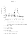
- Non-Covalent Complex Human IFNAR2 is complexed with IFN ⁇ under conditions which maximize the generation of the active complex. In vitro, an optimum ratio of 2.5 ng of IFNAR2 to 1 international unit (IU) of IFN ⁇ is required to yield a maximally active complex, as reported in the examples. The optimum ratio of IFNAR2 to IFN ⁇ which maximizes the generation of the active complex for in vivo activity is currently being determined, although it appears that the optimum ratio will be dependent on the concentration of IFN ⁇ .
- the optimum ratio of IFNAR2:IFN ⁇ in the enhancement of anti-tumor activity at a concentration of 2 x 10 4 IU/mouse/day IFN ⁇ was 2.5 ng IFNAR2 per pg IFN ⁇ , while at a concentration of 5 x 10 4 IU/mouse/day IFN ⁇ , 0.3 ng IFNAR2 per pg IFN ⁇ was optimum.
- the same ratio as used to maximize the generation of the active complex in vitro resulted in elongated pharmacokinetics of the IFN in vivo.
- IFNAR2 and IFN ⁇ used to generate the complex are recombinant molecules.
- the interferon/receptor complex exhibited enhanced activity when compared to the activity of IFN ⁇ alone.
- a constant concentration of IFN ⁇ was mixed with varying concentrations of recombinant sIFNAR2, and this mixture (IFNAR2/IFN complex) was added to WISH cells (human amniotic cells). These WISH cells were then challenged with vesicular stomatitis virus (VSV), and the anti-viral activity of IFN monitored as the degree of cell survival following 48-hour incubation.
- VSV vesicular stomatitis virus
- the interferon receptor complex When administered in vivo, the interferon receptor complex enhances the bioavailability, pharmacokinetics, and/or pharmacodynamics of the IFN, thus augmenting the anti-viral, anti-cancer and immune modulating properties of the IFN.
- the enhanced bioavailability of IFN mediated by the complex can be gained by either pre-formation of an IFN/IFNAR non-covalent complex, co-administration of free IFN with IFNAR, via administration of the IFN or IFNAR component sequentially, via administration of an IFN/IFNAR covalent complex or via administration of an IFNAR/IFN fusion protein.
- such enhanced bioavailability may also be accomplished by administering the IFNAR component alone, without adding any IFN.
- the IFNAR will form the "complex" in vivo with endogenous IFN and, thus, enhance the bioavailability, pharmacokinetics and/or pharmacodynamics of the endogenous IFN. This is particularly useful for the treatment of patients having a disease or condition which naturally causes the induction of native IFN, so that the IFN will already be circulating for its intended natural effect of fighting such disease or condition.
- the added IFNAR will potentiate the effects of the native IFN.
- the preferred molecules for use in the complexes of the present invention have the sequence of a native IFN and IFNAR.
- the native sequence is that of a naturally occurring human IFN or IFNAR. Such sequences are known and can be readily found in the literature. Naturally occurring allelic variations are also considered to be native sequences.
- the present invention also concerns analogs of the above IFNAR2/IFN complex of the invention, which analogs retain essentially the same biological activity of the complex having essentially the sequences of native IFNAR2 and IFN.
- Such analogs may be ones in which up to about 30 amino acid residues may be deleted, added or substituted by others in the IFNAR2 and/or IFN moieties of the complex, such that modifications of this kind do not substantially change the biological activity of the chimeric protein analog with respect to the complex itself.
- the various analogs may differ most from each other and from the basic complex molecule (that with essentially only naturally occurring IFNAR2 and IFN sequences) at the site of the linker peptide which joins the two moieties in the complex.
- such a linker is preferably up to about 30 amino acids in length, and serves to separate the IFNAR2 and IFN moieties from each other in the complex.
- these analogs are those in which one or more and up to about 30 of the amino acid residues of the basic complex of the invention are replaced by different amino acid residues, or are deleted, or one or more amino acid residues, up to about 30, are added to the original sequence of complex of the invention (that with essentially only the native IFNAR2/IFN sequences) without changing considerably the activity of the resulting products as compared with basic complex of the invention.
- These analogs are prepared by known synthesis and/or by site-directed mutagenesis techniques or any other known technique suitable therefor.
- Any such analog preferably has a sequence of amino acids sufficiently duplicative of that of the basic IFNAR2/IFN complex such as to have substantially similar activity thereto.
- any given analog has substantially the same activity and/or stability as the basic complex of the invention by means of routine experimentation, comprising subjecting each such analog to the biological activity and stability tests set forth in Examples 2-7 below.
- Analogs of the complex which can be used in accordance with the present invention, or nucleic acid sequence coding therefor, include a finite set of substantially corresponding sequences as substitution peptides or polynucleotides which can be routinely obtained by one of ordinary skill in the art, without undue experimentation, based on the teachings and guidance presented herein.
- substitution peptides or polynucleotides which can be routinely obtained by one of ordinary skill in the art, without undue experimentation, based on the teachings and guidance presented herein.
- amino acid substitutions of those in the complex having essentially the naturally occurring IFNAR2 and IFN sequences may include synonymous amino acids within a group which have sufficient similar physicochemical properties that substitution between members of the group will preserve the biological function of the molecule (Grantham, 1974).
- insertions and deletions of amino acids may also be made in the above-defined sequences without altering their function, particularly if the insertions or deletions only involve a few amino acids, e.g., under thirty, and preferably under ten, and do not remove or displace amino acids which are critical to a functional conformation, e.g., cysteine residues (Anfinsen, 1973). Analogs produced by such deletions and or insertions come within the purview of the present invention.
- the synonymous amino acid groups are those defined in Table I. More preferably, the synonymous amino acid groups are those defined in Table II; and most preferably the synonymous amino acid groups are those defined in Table III.
- Preferred Groups of Synonymous Amino Acids Amino Acid Synonymous Group Ser Ser, Thr, Gly, Asn Arg Arg, Gln, Lys, Glu, His Leu Ile, Phe, Tyr, Met, Val, Leu Pro Gly, Ala, Thr, Pro Thr Pro, Ser, Ala, Gly, His, Gln, Thr Ala Gly, Thr, Pro, Ala Val Met, Tyr, Phe, Ile, Leu, Val Gly Ala, Thr, Pro, Ser, Gly Ile Met, Tyr, Phe, Val, Leu, Ile Phe Trp, Met, Tyr, Ile, Val, Leu, Phe Tyr Trp, Met, Phe, Ile, Val, Leu, Tyr Trp, Met, Phe, Ile, Val, Leu, Tyr Cys
- Examples of production of amino acid substitutions in proteins which can be used for obtaining analogs of the complex IFNAR2/IFN for use in the present invention include any known method steps, such as presented in U.S. patents RE 33,653; 4,959,314; 4,588,585 and 4,737,462, to Mark et al; 5,116,943 to Koths et al; 4,965,195 to Namen et al; and 5,017,691 to Lee, et al, and lysine substituted proteins presented in US patent 4,904,584 (Shaw et al).
- any analog of the complex for use in the present invention has an amino acid sequence essentially corresponding to that of the above-noted basic complex of the invention.
- the term "essentially corresponding to” is intended to comprehend analogs with minor changes to the sequence of the basic complex which do not affect the basic characteristics thereof, particularly insofar as its ability to inhibit cancer cell proliferation or promote bone marrow transplantations, for example, is concerned.
- the type of changes which are generally considered to fall within the "essentially corresponding to” language are those which would result from conventional mutagenesis techniques of the DNA encoding the complex of the invention, resulting in a few minor modifications, and screening for the desired activity in the manner discussed above.
- the IFNAR2 portion of the complex will have a core sequence which is the same as that of the native sequence or biologically active fragment thereof, or a variant thereof which has an amino acid sequence having at least 70% identity to the native amino acid sequence and retains the biological activity thereof. More preferably, such a sequence has at least 85% identity, at least 90% identity, or most preferably at least 95% identity to the native sequence.
- the core sequence which may be used is the native sequence, or a biologically active fragment thereof, or a variant thereof which has an amino acid sequence having at least 70% identity thereto, more preferably, at least 85% or at least 90% identity, and most preferably at least 95% identity.
- Such analogs must retain the biological activity of the native IFN sequence or fragment thereof, or have antagonist activity as discussed hereinbelow.
- sequence identity means that the sequences are compared as follows.
- the sequences are aligned using Version 9 of the Genetic Computing Group's GAP (global alignment program), using the default (BLOSUM62) matrix (values -4 to +11) with a gap open penalty of -12 (for the first null of a gap) and a gap extension penalty of -4 (per each additional consecutive null in the gap).
- BLOSUM62 global alignment program
- percentage identity is calculated by expressing the number of matches as a percentage of the number of amino acids in the claimed sequence.
- Analogs in accordance with the present invention may also be determined in accordance with the following procedure.
- the DNA of the native sequence is known to the prior art and is either found in the literature cited in the background section of the present specification or can be readily located by those of ordinary skill in the art.
- Polypeptides encoded by any nucleic acid, such as DNA or RNA, which hybridize to the complement of the native DNA or RNA under highly stringent or moderately stringent conditions, as long as that polypeptide maintains the biological activity of the native sequence or, in the case of IFN, either maintains the biological activity or possesses antagonistic activity, are also considered to be within the scope of the present invention.
- Stringency conditions are a function of the temperature used in the hybridization experiment, the molarity of the monovalent cations and the percentage of formamide in the hybridization solution.
- Tm melting temperature
- Tm 81.5°C + 16.6 ( Log M) + 0.41 (%GC) - 0.61 (% form) - 500/L
- M is the molarity of monovalent cations
- %GC is the percentage of G and C nucleotides in the DNA
- % form is the percentage of formamide in the hybridization solution
- L is the length of the hybrid in base pairs.
- the amount of mismatch permitted is increased by about 1%.
- the Tm used for any given hybridization experiment at the specified salt and formamide concentrations is 10°C below the Tm calculated for a 100% hybrid according to equation of Meinkoth, hybridization will occur even if there is up to about 10% mismatch.
- highly stringent conditions are those which are tolerant of up to about 15% sequence divergence, while moderately stringent conditions are those which are tolerant of up to about 20% sequence divergence.
- examples of highly stringent (12-15°C below the calculated Tm of the hybrid) and moderately (15-20°C below the calculated Tm of the hybrid) conditions use a wash solution of 2 X SSC (standard saline citrate) and 0.5% SDS at the appropriate temperature below the calculated Tm of the hybrid.
- the ultimate stringency of the conditions is primarily due to the washing conditions, particularly if the hybridization conditions used are those which allow less stable hybrids to form along with stable hybrids. The wash conditions at higher stringency then remove the less stable hybrids.
- a common hybridization condition that can be used with the highly stringent to moderately stringent wash conditions described above is hybridization in a solution of 6 X SSC (or 6 X SSPE), 5 X Denhardt's reagent, 0.5% SDS, 100 ⁇ g/ml denatured, fragmented salmon sperm DNA at a temperature approximately 20° to 25°C below the Tm. If mixed probes are used, it is preferable to use tetramethyl ammonium chloride (TMAC) instead of SSC (Ausubel, 1987, 1998).
- TMAC tetramethyl ammonium chloride
- “Functional derivatives” as used herein covers derivatives which may be prepared from the functional groups which occur as side chains on the residues or the N- or C-terminal groups, by means known in the art, and are included in the invention as long as they remain pharmaceutically acceptable, i.e., they do not destroy the biological activity of the corresponding protein of the complex as described herein and do not confer toxic properties on compositions containing it or the complex made therefor.
- Derivatives may have chemical moieties, such as carbohydrate or phosphate residues, provided such a fraction has the same biological activity and remains pharmaceutically acceptable.
- derivatives may include aliphatic esters of the carboxyl of the carboxyl groups, amides of the carboxyl groups by reaction with ammonia or with primary or secondary amines, N-acyl derivatives or free amino groups of the amino acid residues formed with acyl moieties (e.g., alkanoyl or carbocyclic aroyl groups) or O-acyl derivatives of free hydroxyl group (e.g., that of seryl or threonyl residues) formed with acyl moieties.
- Such derivatives may also include for example, polyethylene glycol side-chains which may mask antigenic sites and extend the residence of the complex or the portions thereof in body fluids.
- derivatives is intended to include only those derivatives that do not change one amino acid to another of the twenty commonly-occurring natural amino acids.
- salts herein refers to both salts of carboxyl groups and to acid addition salts of amino groups of the complex of the invention or analogs thereof.
- Salts of a carboxyl group may be formed by means known in the art and include inorganic salts, for example, sodium, calcium, ammonium, ferric or zinc salts, and the like, and salts with organic bases as those formed, for example, with amines, such as triethanolamine, arginine or lysine, piperidine, procaine and the like.
- Acid addition salts include, for example, salts with mineral acids, such as, for example, hydrochloric acid or sulfuric acid, and salts with organic acids, such as, for example, acetic acid or oxalic acid.
- any such salts must have substantially similar biological activity to the complex of the invention or its analogs.
- biological activity as used herein is interpreted s follows. Insofar as the IFNAR2 portion of the complex is concerned, the important biological activity is its ability to bind to Type I interferon. Thus, analogs or variants, salts and functional derivatives must be those chosen so as to maintain this interferon-binding ability. This can be tested by routine binding assay experiments.
- fragments of the IFNAR2, or analogs thereof can also be used as long as they retain their interferon-binding activity. Fragments may readily be prepared by removing amino acids from either end of the interferon-binding polypeptide and testing the resultant for interferon-binding properties. Proteases for removing one amino acid at a time from either the N-terminal or the C-terminal of a polypeptide are known, and so determining fragments which retain interferon-binding ability involves only routine experimentation.
- the polypeptide which has such interferon-binding activity can also contain additional amino acid residues flanking the interferon-binding polypeptide.
- additional flanking residues affect the basic and novel characteristics of the core peptide, i.e., its interferon-binding characteristics, by routine experimentation.
- the term "consisting essentially of”, when referring to a specified sequence, means that additional flanking residues can be present which do not affect the basic and novel characteristic of the specified sequence. This term does not comprehend substitutions, deletions or additions within the specified sequence.
- IFNAR2 or sIFNAR2 have been used throughout this description and in the examples, it should be understood that this is merely the preferred example and that the IFNAR1 subunit, and particularly its extracellular domain, may be substituted for IFNAR2 wherever IFNAR2 is referred to in the present disclosure.
- IFNAR1 can only be used in conjunction with interferons to which it binds. IFNAR1 is known to bind to IFN ⁇ . Any complex using IFNAR1 must be with a species of interferon, and preferably a species of IFN ⁇ , to which the IFNAR1 binds.
- the biological activity which must be maintained in any analog or variant, salt, functional derivative or fragment is the activity of the interferon relied upon for the intended utility. In most instances, this will be the ability to bind to a native cell surface receptor and thereby mediate signal production by the receptor. Thus, any such analog, derivative or fragment should maintain such receptor agonist activity to be useful in the present invention for such a utility.
- Such an antagonist can also be used for prolonged beneficial effect by means of the complex of the present invention.
- analogs which are still bound by the receptor and by the IFNAR portion of the complex but which do not mediate a signal and block signal generation by the native interferon on that receptor may also be considered to be biologically active for the purpose of this invention and to be encompassed by the term interferon when used with respect to the complexes of the present invention.
- Straightforward assays can determine whether any such analog maintains such receptor agonist activity or has receptor antagonist activity and would, thus, be useful for one of the utilities of the present invention.
- the present invention also concerns DNA sequences encoding the above complex of the invention and its analogs, as well as DNA vectors carrying such DNA sequences for expression in suitable prokaryotic or eukaryotic host cells.
- DNA sequences encoding the above complex of the invention and its analogs, as well as DNA vectors carrying such DNA sequences for expression in suitable prokaryotic or eukaryotic host cells.
- the ability to generate large quantities of heterologous proteins using a recombinant protein expression system has led to the development of various therapeutic agents, e.g., t-PA and EPO (Edington, 1995).
- the various expression hosts from which recombinant proteins can be generated range from prokaryotic in origin (e.g., bacteria) (Olins, 1993), through lower eukaryotes (e.g., yeast) (Ratner, 1989) to higher eukaryotic species (e.g., insect and mammalian cells (Reuveny, 1993; Reff, 1993). All of these systems rely upon the same principle - introducing the DNA sequence of the protein of interest into the chosen cell type (in a transient or stable fashion, as an integrated or episomal element) and using the host transcription, translation and transportation machinery to over-express the introduced DNA sequence as a heterologous protein (Keown, 1990).
- prokaryotic in origin e.g., bacteria
- lower eukaryotes e.g., yeast
- higher eukaryotic species e.g., insect and mammalian cells
- chosen sequences of DNA can be physically linked to generate transcripts which develop into novel fusion proteins where once independent proteins are now expressed as one polypeptide unit (Ibanez, 1991).
- the activity of such fusion proteins can be different, e.g., more potent, than either of the individual proteins (Curtis, 1991).
- Human IFN ⁇ is derived from a production process which uses the mammalian Chinese hamster ovary cell (CHO).
- Type 1 interferons can be expressed in a variety of host cells including bacteria (Utsumi, 1987), insect (Smith, 1983) and human (Christofinis, 1981).
- Human sIFNAR2 was also expressed using the CHO host cell.
- soluble receptors such as sIFNAR2
- the DNA for each gene was introduced into the CHO genome using a transfection procedure which resulted in recombination and integration of the expression vector. Cells which expressed the protein of interest were then isolated, cultured and the protein recovered and purified using standard industrial practices well known in the art.
- the invention also concerns a pharmaceutical composition
- a pharmaceutical composition comprising as active ingredient an IFNAR2/IFN complex or analogs thereof or mixtures thereof or salts thereof and a pharmaceutical acceptable carrier, diluent or excipient.
- An embodiment of the pharmaceutical composition of the invention includes a pharmaceutical composition for enhanced IFN type action, in the treatment of viral diseases, in anti-cancer therapy, in immune modulation therapy and other applications of interferons and cytokines related thereto.
- compositions of the invention are prepared for administration by mixing the complex or its analogs with physiologically acceptable carriers and/or stabilizers and/or excipients, and prepared in dosage form, e.g., by lyophilization in dosage vials.
- the method of administration can be via any of the accepted modes of administration for similar agents and will depend on the condition to be treated, e.g., intravenously, intramuscularly, subcutaneously, by local injection or topical application, or continuously by infusion, etc.
- the amount of active compound to be administered will depend on the route of administration, the disease to be treated and the condition of the patient. Local injection, for instance, will require a lower amount of the protein on a body weight basis than will intravenous infusion.
- Free IFN ⁇ has a tendency to oligomerize. To suppress this tendency, present day formulations of IFN ⁇ have an acidic pH, which may cause some localized irritation when administered. As IFNAR can serve as a stabilizing factor for IFN ⁇ and thereby prevent oligomerization, its use in IFN ⁇ formulations can serve to stabilize the IFN ⁇ and thereby obviate the necessity of acidic formulations. Accordingly, a non-acidic pharmaceutical composition containing IFN ⁇ and IFNAR, along with other conventional pharmaceutically acceptable excipients, is also a part of the present invention.
- the present invention also concerns uses of the complex of the invention or its analogs or mixtures thereof for anti-viral, anti-cancer and immune modulation therapy.
- the interferon receptor-interferon complexes of this invention are useful for anti-viral therapy in such therapeutic indications as chronic granulomatous disease, condyloma acuminatum, juvenile laryngeal papillomatosis, hepatitis A and chronic infection with hepatitis B and C viruses.
- interferon receptor-interferon complexes of this invention are useful for anti-cancer therapy in such therapeutic indications as hairy cell leukemia, Kaposi's sarcoma, multiple myeloma, chronic myelogenous leukemia, non-Hodgkins's lymphoma and melanoma.
- interferon receptor-interferon complexes of this invention are also useful for immune modulation therapy, such as multiple sclerosis, rheumatoid arthritis, myasthenia gravis, diabetes, AIDS, lupus, etc.
- the present invention also concerns the complex or analogs thereof or mixtures thereof for use in the preparation of medicaments for treating the above-mentioned ailments or for use in the above noted indications.
- Anti-viral activity of Type I interferons (used at predetermined EC 50 concentrations) in the presence of approximately 30 ng/ml sIFNAR2 reveals optimal agonist activity with IFN ⁇ , but not alone. Anti-viral activity was measured by MTT conversion 48h after VSV challenge.
- the kinetics of the IFNAR2/IFN ⁇ complex formation was examined by assessing the anti-viral activity of a sub-effective concentration of IFN ⁇ (0.95 IU/ml) at various times of preincubation with different concentrations of IFNAR2. As seen in Figure 3, 4 hours of preincubation were necessary to give enhanced anti-viral activity of IFN ⁇ at this sub-effective dose. Thus, at levels of IFN ⁇ which are inactive by themselves, addition of IFNAR2 to generate a complex resulted in significant IFN anti-viral activity. This experiment provides additional support for the IFNAR2/IFN ⁇ complex enhanced IFN activity.
- IFN ⁇ bioactivity rapidly declines following in vitro reconstitution at 37°C (physiologic saline buffer pH 7.4). This is, at least in part, due to the formation of IFN ⁇ oligomeric structures which have reduced activity.
- IFNAR2 enhances the stability of IFN ⁇ at physiologic pH, an experiment was conducted in which varying concentrations of IFN ⁇ were incubated alone (in RPMI 1640 media, Gibco) or in the same media in the presence of IFNAR2 at a constant ratio of IFNAR2 to IFN ⁇ (2.5 ng/IU).
- IFN ⁇ 500 IU/ml was preincubated with an equal volume of either sIFNAR2 (1.25 ⁇ g/ml) or RPMI only, for 3 hours at 37°C. Titration of both IFN ⁇ solutions was performed in WISH assay medium in a 96-well plate prior to the addition of WISH cells. VSV was added after 24 hours and was assessed after an additional 48-hour incubation as determined by MTT conversion.
- IFN ⁇ alone has an ED 50 of ⁇ 104 IU/ml
- the high ED 50 of IFN ⁇ alone may be due to the oligomerization of IFN ⁇ in solution.
- the above results again show the enhanced stability of IFN ⁇ when complexed with IFNAR2.
- IFN ⁇ insulin receptor 2
- BSA bovine serum albumin
- IFN ⁇ 500 IU/ml was preincubated with an equal volume of either the indicated proteins (1.25 ⁇ g/ml) or RPMI only, for 4 hours at 37°C. Titration of these IFN ⁇ solutions in WISH assay medium was performed in a 96-well plate prior to the addition of WISH cells. Freshly-prepared IFN ⁇ was also included to determine the effect of the preincubation on IFN ⁇ activity. VSV was added after 24 hours, and CPE was assessed after an additional 48 hour incubation as determined by MTT conversion.
- IFN ⁇ Preincubation of IFN ⁇ with non-specific proteins (i.e., BSA or human IgG) at 2.5 ng/IU IFN ⁇ did not protect the activity of IFN ⁇ after reconstitution.
- the activity of the IFNAR2/IFN ⁇ complex in this assay was similar to freshly added IFN ⁇ , which supports the finding that sIFNAR2 stabilizes IFN ⁇ activity.
- IFNAR2/IFN ⁇ complex showed a greatly prolonged pharmacokinetic profile of IFN ⁇ in the mouse based on ELISA and bioassay analysis ( Figures 6 and 7).
- B6D2F1 strain mice received a single intravenous bolus injection of either human IFN ⁇ (2.5 x 10 6 IU/kg) or the same concentration of IFN ⁇ preincubated for 1 hour at 4°C with human IFNAR2 (2.5 ng/IU of IFN).
- Sera were collected 0.05 to 48 hours post-administration from the retro-orbital plexus, and IFN ⁇ concentration and IFN ⁇ anti-viral activity was assessed by ELISA ( Figure 6) or WISH bioassay ( Figure 7), respectively. Serum concentrations falling below the level of assay sensitivity (7.55 IU/ml) in the ELISA assay were not plotted.
- IFNAR2/IFN ⁇ 2a complex showed a greatly prolonged pharmacokinetic profile of IFN ⁇ 2a in the mouse based on ELISA and bioassay tests ( Figures 8 and 9).
- B6D2F1 strain mice received a single intravenous bolus injection of either human IFN ⁇ (1.25 x 10 5 IU/kg) or the same concentration of IFN ⁇ preincubated for 1 hour at 4°C with human IFNAR2 (14.9 ng/IU of IFN).
- Sera were collected at indicated times from the retro-orbital plexus, and IFN ⁇ 2a concentration and IFN ⁇ anti-viral activity was assessed by ELISA ( Figure 8) or WISH bioassay ( Figure 9), respectively.
- IFN ⁇ was assessed by ELISA specific for human IFN ⁇ . Serum concentrations falling below the level of assay sensitivity (30 IU/ml) in the ELISA were not plotted.
- IFNAR2extracellular (linker) mature IFN ⁇ ...ESEFS( GGGGS ) n MSY ..., where n 0 (SEQ ID NO:5), 1 (SEQ ID NO:7), 2 (SEQ ID NO:6), 3 (SEQ ID NO:5), 4 (SEQ ID NO:4), and 5 (SEQ ID NO:3)
- the expression vector pSVEIF which contained the gene expressing human recombinant IFN, was used as the template for PCR.
- Synthetic primers were designed so that only the mature protein coding region of human IFN (MSY%) could be amplified from the template.
- the 5' primer consisted of sequences for a digested-SmaI site, the last seven amino acids of IFNAR2 (GQESEFS) (residues 344-349 of SEQ ID NO:14), and the (GGGGS) 1 (SEQ ID NO:1) linker. BamHI and XhoI sites were also introduced in the 5' PCR primer to facilitate cloning of the other cassettes.
- the 3' primer contained an AvrII site immediately following the TGA stop codon of hIFN ⁇ .
- the PCR contained approximately 1 g of template DNA, 1 g of each PCR primer, 0.2 mM each of dATP, dCTP, dGTP, and dTTP, 1X ThermoPol Reaction Buffer (10 mM KCl, 20 mM Tris-HCl, (pH 8.8 at 25°C), 10 mM (NH 4 ) 2 SO 4 , 2 mM MgSO 4 , 0.1% Triton X-100: New England Biolabs; Beverly, MA), and 3 mM MgSO 4 (5 mM MgSO 4 final concentration) in a reaction volume of 100 ⁇ l.
- PCR consisted of 20 cycles of the following: (a) 99.9°C for 30 seconds, (b) 65°C-55°C for 30 seconds, decreasing 0.5°C with each cycle, and (c) 75°C for 40 seconds. An additional 15 cycles were done with the above profile but with the annealing temperature held at 55°C.
- the PCR reactions were purified using the WizardTM PCR Preps DNA Purification System (Promega; Madison, WI). After digestion of the PCR products with AvrII, the reactions were purified on low melting point agarose gels.
- the gel-purified fragment containing the hIFN ⁇ mature protein sequence with 1 GS linker was ligated into the (SmaI + AvrII)-digested expression vector pCMV-p40, which contained the gene coding for the soluble form of human recombinant IFNAR2.
- the ligation reactions were used to transform competent E. coli XL-1 Blue cells using standard methods (Sambrook et al, 1989). Correct assembly of the construction, called pCMV-IFNAR2/IFNA ⁇ GS, was confirmed by restriction endonuclease digestion and sequencing of the PCR-generated region of the "Interfusion".
- the 0 GS cassettes contained SmaI and XhoI overhangs.
- the vector was digested with SmaI and XhoI for ligation of the cassette.
- Figure 12 is a representative plasmid map of the pCMV-IFNAR2/IFNA ⁇ "Interfusion" expression vectors. Transcription of the IFNAR2/IFN ⁇ fusion protein is directed by the human immediate early CMV promoter. The human growth hormone (hGH) polyadenylation signal sequence provided by the vector was used for 3' processing of the IFNAR2/IFN ⁇ transcripts. List of Vectors No. Name Exp.
- Vector GGGGS (SEQ ID NO:1) Linker 1 pCMV-IFNAR2-IFN ⁇ , 0GS pCMV.PA4 N/A 2 pCMV-IFNAR2-IFN ⁇ , 1GS pCMV.PA4 One 3 pCMV-IFNAR2-IFN ⁇ , 2GS pCMV.PA4 Two 4 pCMV-IFNAR2-IFN ⁇ , 3GS pCMV.PA4 Three 5 pCMV-IFNAR2-IFN ⁇ , 4GS pCMV.PA5 Four 6 pCMV-IFNAR2-IFN ⁇ , 5GS pCMV.PA4 Five Sequence data was obtained for the PCR-generated region of IFNAR2/IFN ⁇ 1GS fusion; this data showed that the sequence was as predicted. Sequence data was obtained for the peptide linker region of the other constructions; the sequences were also as predicted.
- a Northern blot analysis was performed on cells transfected with each construct. A band of approximately 1.4 kb in size was present in all lanes containing the IFNAR2/IFN ⁇ n GS samples for both the IFNAR2 probe and for the IFN ⁇ probe. With the IFN ⁇ probe, an additional band of approximately 0.9 kb is observed. A band of this size would correspond to an alternatively spliced transcript that would contain the last six amino acids of IFNAR2, the (n) GS peptide linker, and the hIFN mature protein coding sequence. This was confirmed by sequencing cDNA prepared from the total RNA isolated from the transiently transfected CHO cells of Example 9.
- CHO-DUKX cells are a clonal mutant of Chinese hamster ovary cells lacking dihydrofolate reductase activity (Urlaub et al (1980); Graff et al (1982)). The cells were maintained in Alpha Minimum Essential Medium ( ⁇ MEM) plus ribonucleosides and dexoyribonucleosides, supplemented with 10% fetal bovine serum (FBS) and 1% L-glutamine (complete medium). A transient transfection was done using the Lipofectamine PLUSTM Reagent (GibcoBRL Life Technologies; Gaithersburg, MD) and the protocol provided by the manufacturer.
- ⁇ MEM Alpha Minimum Essential Medium
- FBS fetal bovine serum
- L-glutamine complete medium
- RNA Extraction and Northern Analysis Total cellular RNA (Chomczynski et al, 1987) was extracted from the transiently transfected CHO-DUKX cells. Ten g of total RNA per lane was size-fractionated in agarose gels which contained formaldehyde as a denaturant. Samples were loaded in duplicate sets. The RNA was transferred to GeneScreen Plus nylon membranes (DuPont/NEN Medical Products; Boston, MA) by capillary blot in 10 X SSC (1.5M sodium chloride, 0.15M sodium citrate). The immobilized RNA was hybridized to 32 P-labeled hIFNAR2 and hIFN ⁇ PCR fragments in a solution modified from that of Church and Gilbert (1984).
- the buffer contained 0.25 M sodium phosphate, pH 7.2, 0.25M sodium chloride, 7% sodium dodecyl sulfate (SDS), 1 mM ethlylenediaminetetraacetic acid and 100 ⁇ g/ml E. coli tRNA.
- the 32 P-labeled probes were generated using a commercially available kit ("High Prime” Boehringer Mannheim; Indianapolis, IN) and the procedures described therein. Non-incorporated radioactivity was removed by chromatography on Sephadex G-50 columns. After hybridization, the blots were washed; the most stringent condition used was 0.2 X SSC, 0.1% SDS at 65°C. The blots were subjected to autoradiography.
- CHO cell supernatants containing expressed recombinant proteins (as detected by ELISA and Western Blot), or CHO culture medium alone, were added in duplicate to the top row of 96-well flat-bottom tissue culture plates in a volume of 75 ⁇ l/well.
- 50 ⁇ l of WISH cell assay medium was added to the remaining wells of the plate.
- Three-fold serial dilutions of each sample were performed by removing 25 ⁇ l from the wells containing the supernatants (top row) and adding this to the next row containing the 50 ⁇ l of the WISH assay medium.
- the supernatant in the top row was replaced with WISH assay medium containing 1000 IU/ml human IFN ⁇ , which was likewise serially diluted three-fold down the length of the plate.
- WISH assay medium containing 1000 IU/ml human IFN ⁇ , which was likewise serially diluted three-fold down the length of the plate.
- Each well then received 50 ⁇ l of a WISH cell suspension (0.6 x 10 6 cells/ml in WISH assay medium) so that the final concentration in the top row containing REBIF® is 500 IU/ml, and the starting dilution for the CHO cell supernatants is 1:2.
- each well (except those designated as the uninfected control wells) received 50 ⁇ l of WISH assay medium containing vesicular stomatitis virus (1:10 of the stock). Viability of the WISH cells was determined, following an additional 48 hours of culture, by MTT conversion.
- Anti-viral activity mediated by the Interfusion molecules was determined by normalizing the dilutional factor of the supernatants necessary to achieve the EC 50 to the EC 50 determined for the purified human IFN ⁇ standard. As the linker length increases, so too does the anti-viral activity of the Interfusion constructs.
- This example is a pharmacokinetic study of human IFN ⁇ /sIFNAR2 complex in the mouse upon intravenous administration. Comparisons are performed between preformed and separately injected complex components. Thirty-six D2F1 strain mice (6 - 8 wks) (approximately 20 g each) are separated into four groups as follows:
- Blood samples (approximately 200 ⁇ l/sample) are collected at the specified times by disruption of the retro-orbital venous plexus with a capillary tube.
- Three mice of each group have blood samples taken at 0.05, 2 and 12 hours post administration.
- Three mice of each group have blood samples taken at 0.54 and 24 hours post administration and three of each group have blood samples taken at 1, 8 and 48 hours post administration.
- Blood samples were allowed to clot at one hour at room temperature rimmed and microcentrifuged.
- Sera removed therefrom were stored at -70 C until all samples were collected.
- Sera are assayed for the presence of human IFN ⁇ by means of IFN ⁇ specific ELISA using the Toray human IFN ⁇ ELISA kit (TFB, Inc.) and were assayed for bioactivity using the WISH antiviral assay.
- mice C57BL/6 mice were treated with a complex of "Universal" IFN (human IFN ⁇ A/D) and sIFNAR2.
- the protective effect in terms of cytotoxicity was measured as compared with administration of various doses of Universal IFN alone or a control.
- the first group received 5 x 10 3 IU Universal IFN, complexed with 5/ng IU sIFNAR2.
- the second group received 5 x 10 3 IU Universal IFN.
- the third group received 5 x 10 4 IU Universal IFN and the fourth group received PBS/2% NMS.
- NK activity was measured as cytotoxicity of splenic cells against the NK target cells YAC-1. The results are shown in Figure 16.
- the NK activity was significantly greater in mice treated with the Universal IFN/sIFNAR2 complex as compared with mice treated with Universal IFN only.
- mice developed paralysis between days 14 - 20 post tumor cell injection in association with histological evidence of diffused lymphoma. Notably, survival of these mice can be prolonged in dose dependent fashion by daily subcutaneous administration of human IFN ⁇ .
- This model is being used to evaluate IFNAR2 as a potentiator of the biological activity associated with type 1 IFNs in vivo.
- mice In order to establish the relationship between the mean time to paralysis and the dose of IFN ⁇ in the Daudi scid model, five groups of 5 BALB/cByJSmn-scid/scid strain mice, 4-5 wks of age, female, received a subcutaneous administration of 200 ⁇ l per mouse per day of human IFN ⁇ every day from day 0 to day 30.
- the groups of mice received the following amounts of IFN.
- mice were treated by the same protocol discussed in Example 13, except that the test materials administered to each of the groups was as follows: IFN ⁇ only/mouse IFN ⁇ plus sIFNAR2/mouse Group 1 2 x 10 2 IU Group 4 2 x 10 2 IU plus 0.5 ⁇ g Group 2 2 x 10 3 IU Group 5 2 x 10 3 IU plus 5.0 ⁇ g Group 3 2 x 10 4 IU Group 6 2 x 10 4 IU plus 50.0 ⁇ g Group 7 received Daudi cells, treament is dilluent only/mouse IFN ⁇ plus sIFNAR2/mouse Group 1 2 x 10 2 IU Group 4 2 x 10 2 IU plus 0.5 ⁇ g Group 2 2 x 10 3 IU Group 5 2 x 10 3 IU plus 5.0 ⁇ g Group 3 2 x 10 4 IU Group 6 2 x 10 4 IU plus 50.0 ⁇ g Group 7 received Daudi cells, treament is dilluent only
- This example is a pharmacokinetic study to determine the serum half life of the Interfusion 5GS molecule in mouse serum following a single bolus intravenous injection.
- Twenty-one female B6D2F1 strain mice (6 - 8 wks) (approximately 20g each) were separated into three groups as follows:
- C max and T max is 10,000 IU/ml for Groups 1 and 2.
- Three mice of each of Groups 1 and 2 had blood sampled at 0.05, 2 and 12 hours post administration.
- Three mice of each of Groups 1 and 2 had blood sampled at 0.5, 4 and 24 hours post administration, and three mice of each of Groups 1 and 2 had blood sampled at 1, 8 and 48 hours post administration.
- Sera were assayed for the presence of bioactive human IFN ⁇ using the WISH assay.
Description
| Preferred Groups of Synonymous Amino Acids | |
| Amino Acid | Synonymous Group |
| Ser | Ser, Thr, Gly, Asn |
| Arg | Arg, Gln, Lys, Glu, His |
| Leu | Ile, Phe, Tyr, Met, Val, Leu |
| Pro | Gly, Ala, Thr, Pro |
| Thr | Pro, Ser, Ala, Gly, His, Gln, Thr |
| Ala | Gly, Thr, Pro, Ala |
| Val | Met, Tyr, Phe, Ile, Leu, Val |
| Gly | Ala, Thr, Pro, Ser, Gly |
| Ile | Met, Tyr, Phe, Val, Leu, Ile |
| Phe | Trp, Met, Tyr, Ile, Val, Leu, Phe |
| Tyr | Trp, Met, Phe, Ile, Val, Leu, Tyr |
| Cys | Ser, Thr, Cys |
| His | Glu, Lys, Gln, Thr, Arg, His |
| Gln | Glu, Lys, Asn, His, Thr, Arg, Gln |
| Asn | Gln, Asp, Ser, Asn |
| Lys | Glu, Gln, His, Arg, Lys |
| Asp | Glu, Asn, Asp |
| Glu | Asp, Lys, Asn, Gln, His, Arg, Glu |
| Met | Phe, Ile, Val, Leu, Met |
| Trp | Trp |
| More Preferred Groups of Synonymous Amino Acids | |
| Amino Acid | Synonymous Group |
| Ser | Ser |
| Arg | His, Lys, Arg |
| Leu | Leu, Ile, Phe, Met |
| Pro | Ala, Pro |
| Thr | Thr |
| Ala | Pro, Ala |
| Val | Val, Met, Ile |
| Gly | Gly |
| Ile | Ile, Met, Phe, Val, Leu |
| Phe | Met, Tyr, Ile, Leu, Phe |
| Tyr | Phe, Tyr |
| Cys | Cys, Ser |
| His | His, Gin, Arg |
| Gin | Glu, Gin, His |
| Asn | Asp, Asn |
| Lys | Lys, Arg |
| Asp | Asp, Asn |
| Glu | Glu, Gln |
| Met | Met, Phe, Ile, Val, Leu |
| Trp | Trp |
| Most Preferred Groups of Synonymous Amino Acids | |
| Amino Acid | Synonymous Group |
| Ser | Ser |
| Arg | Arg |
| Leu | Leu, Ile, Met |
| Pro | Pro |
| Thr | Thr |
| Ala | Ala |
| Val | Val |
| Gly | Gly |
| Ile | Ile, Met, Leu |
| Phe | Phe |
| Tyr | Tyr |
| Cys | Cys, Ser |
| His | His |
| Gln | Gln |
| Asn | Asn |
| Lys | Lys |
| Asp | Glu |
| Glu | Glu |
| Met | Met, Ile, Leu |
| Trp | Met |
- WISH cells (ATCC CCL 25)
- Vesicular Stomatitis Virus stocks (ATCC V-520-001-522), stored at -70°C
- IFNβ, human recombinant, InterPharm Laboratories LTD 90 x 106 IU/ml, specific activity: 264.5 x 10⁶ IU/mg.
- Soluble human IFNAR2, 373 µg/ml stock concentration in PBS
- WISH Growth medium (MEM high glucose with Earls salts + 10% FBS + 1.0% L-glutamine + Penicillin/Streptomycin (100 U/ml, 100 µg/ml))
- WISH Assay medium (MEM high glucose with Earls salts + 5% FBS + 1.0% L-glutamine + Penicillin/Streptomycin (100 U/ml, 100 µg/ml), MTT at 5 mg/ml in PBS, stored at - 70°C.
- Dilute recombinant human IFNβ to 19 IU/ml (4X the predetermined EC50 dose) in WISH assay medium.
- Starting at 90 µg/ml, make eleven (11) three-fold dilutions of human recombinant sIFNAR2 in Eppindorf tubes in WISH assay medium. Twelfth tube contains WISH assay medium only.
- Add 25 µl of IFNβ to each well in a flat-bottomed 96-well
plate (add 25 µl of WISH assay medium alone to one
3
X 12 well section to control for IFNAR2 effects in the absence of IFNβ). - Add 25 µl of each dilution of the sIFNAR2 (or assay medium only in row twelve) in triplicate down the twelve rows of the 96-well plate.
- Preincubate IFNβ with SIFNAR2 for 1-4 hours in 37°C incubator prior to the addition of WISH cells.
- Harvest log growth phase WISH cells with trypsin/EDTA solution, wash in WISH assay medium, and bring to a final concentration of 0.8 x 10⁶ cells/ml.
- Add 50 µl of WISH cell suspension (4 x 10⁴ cells per well) to each well. Final concentration of both IFNβ and IFNAR2 is that which is exposed to the cells, so that the final concentration of IFNβ is 4.75 IU/ml (1X), and the final concentration of sIFNAR2 in row one is 22.5 µg/ml,
- After incubation for 24 hours in a 5% CO2 humidified incubator, 50 µl of a 1:10 dilution (in WISH assay medium) of VSV stock (a dose predetermined to lyse 100% of WISH cells within 48 hours) is added to all wells except for the no virus control wells (these receive an equal volume of assay medium only).
- After an additional 48 hours, 25 µl of MTT solution is added to all wells, after which plates are incubated for an additional 2 hours in the incubator.
- Contents of wells are removed by plate inversion, and 200 µl of 100% ethanol is added to wells.
- After 1 hour, plates are read at 595 nm using the Soft max Pro software package and Spectramax spectrophotometer system (Molecular Devices).
...ESEFS(GGGGS)nMSY ..., where n = 0 (SEQ ID NO:5), 1 (SEQ ID NO:7), 2 (SEQ ID NO:6), 3 (SEQ ID NO:5), 4 (SEQ ID NO:4), and 5 (SEQ ID NO:3)
| List of Vectors | ||||
| No. | Name | Exp. Vector | GGGGS (SEQ ID NO:1) | |
| 1 | pCMV-IFNAR2-IFNβ, 0GS | pCMV.PA4 | N/ | |
| 2 | pCMV-IFNAR2-IFNβ, 1GS | pCMV.PA4 | One | |
| 3 | pCMV-IFNAR2-IFNβ, 2GS | pCMV.PA4 | Two | |
| 4 | pCMV-IFNAR2-IFNβ, 3GS | pCMV.PA4 | Three | |
| 5 | pCMV-IFNAR2-IFNβ, 4GS | | Four | |
| 6 | pCMV-IFNAR2-IFNβ, 5GS | pCMV.PA4 | Five |
| Interfusion Constructs Transiently Transfected into CHO Cells | ||||||
| Sample Identification | [hIFNβ] , (U/ml) | [hIFNAR2], (ng/ml) | [hIFNβ], (µg/ml) | [hIFNβ], (pmols) | [hIFNAR2], (pmols) | IFH/IFNAR |
| 0GS | 29,175 | 1094 | 0.146 | 6.571 | 31.802 | 0.207 |
| 1GS | 24,906 | 994 | 0.125 | 5.609 | 28.895 | 0.194 |
| 2GS | 13,597 | 600 | 0.068 | 3.063 | 17.442 | 0.175 |
| 3GS | 14,998 | 718 | 0.075 | 3.378 | 20.857 | 0.162 |
| 4GS | 9,998 | 535 | 0.050 | 2.251 | 15.538 | 0.145 |
| 5GS | 9,597 | 540 | 0.048 | 2.161 | 15.698 | 0.138 |
| IFNAR2 | Not Found | 1176 | 34.200 | |||
| Culture | Not Found | 0 |
| Days to Paralysis | Mean (± SD) | |
| | 26, 31, 35, 37, 40 | 33.8 (5.4) |
| | 20, 23, 23, 23, 26 | 23.0 (2.1) |
| | 20, 20, 22, 22, 23 | 21.4 (1.3) |
| | 17, 18, 18, 18, 18 | 17.8 (0.5) |
| | 14, 14, 15, 15, 15 | 14.6 (0.6) |
| IFNβ only/mouse | IFNβ plus sIFNAR2/ | |||
| Group | ||||
| 1 | 2 x 10² | Group | 4 | 2 x 10² IU plus 0.5 |
| Group | ||||
| 2 | 2 x 10³ | Group | 5 | 2 x 10³ IU plus 5.0 |
| Group | ||||
| 3 | 2 x 10⁴ | Group | 6 | 2 x 10⁴ IU plus 50.0 |
| Group | ||||
| 7 | received Daudi cells, treament is dilluent only |
| Days to Paralysis | Mean (± SD) | |
| | 16, 16, 17, 18, 19 | 17.2 (1.3) |
| | 17, 18, 18, 19, 19 | 18.2 (0.8) |
| | 17, 17, 17, 18, 18 | 17.6 (0.9) |
| | 17, 17, 18, 18, 19 | 17.8 (0.8) |
| | 17, 18, 19, 20, 20 | 18.8 (1.3) |
| | 21, 22, 22, 23, 26 | 22.8 (1.9) |
| | 16, 16, 17, 17, 17 | 16.6 (0.6) |
CUNNINGHAM, Mark
SHERRIS, David
EL TAYAR, Nabil
McKENNA, Sean
- <120> IFNAR2/IFN COMPLEX
- <130> TEPPER1A.SEQ
- <140> not yet received
- <141> 1998-12-18
- <150> 60/068,295
- <151> 1997-12-19
- <160> 14
- <170> PatentIn Ver. 2.0
- <210> 1
- <211> 5
- <212> PRT
- <213> Unknown
- <220>
- <223> Description of Unknown Organism: Peptide
- <400> 1

- <210> 2
- <211> 4
- <212> PRT
- <213> Unknown
- <220>
- <223> Description of Unknown Organism: Peptide
- <400> 2

- <210> 3
- <211> 33
- <212> PRT
- <213> Unknown
- <220>
- <223> Description of Unknown Organism: Peptide
- <400> 3

- <210> 4
- <211> 28
- <212> PRT
- <213> Unknown
- <220>
- <223> Description of Unknown Organism: Peptide
- <400> 4

- <210> 5
- <211> 23
- <212> PRT
- <213> Unknown
- <220>
- <223> Description of Unknown Organism: Peptide
- <400> 5

- <210> 6
- <211> 18
- <212> PRT
- <213> Unknown
- <220>
- <223> Description of Unknown Organism: Peptide
- <400> 6

- <210> 7
- <211> 13
- <212> PRT
- <213> Unknown
- <220>
- <223> Description of Unknown Organism: Peptide
- <400> 7

- <210> 8
- <211> 36
- <212> PRT
- <213> Unknown
- <220>
- <223> Description of Unknown Organism: Peptide
- <400> 8

- <210> 9
- <211> 23
- <212> PRT
- <213> Unknown
- <220>
- <223> Description of Unknown Organism: Peptide
- <400> 9

- <210> 10
- <211> 20
- <212> PRT
- <213> Unknown
- <220>
- <223> Description of Unknown Organism: Peptide
- <400> 10

- <210> 11
- <211> 17
- <212> PRT
- <213> Unknown
- <220>
- <223> Description of Unknown Organism: Peptide
- <400> 11

- <210> 12
- <211> 21
- <212> PRT
- <213> Unknown
- <220>
- <223> Description of Unknown Organism: Peptide
- <400> 12

- <210> 13
- <211> 34
- <212> PRT
- <213> Unknown
- <220>
- <223> Description of Unknown Organism: Peptide
- <400> 13

- <210> 14
- <211> 400
- <212> PRT
- <213> Unknown
- <220>
- <223> Description of Unknown Organism: Peptide
- <400> 14


Claims (25)
- A complex comprising a Type I interferon (IFN) and a subunit of the human interferon α/β receptor (IFNAR) which is capable of binding to the Type I IFN of the complex, wherein said Type I IFN is:a) a native Type I IFN;b) a fragment of a) which has Type I IFN biological activity;c) a variant of a) or b) which has at least 70% sequence identity with a) or b) and which has Type I IFN biological activity;d) a variant of a) or b) which is encoded by a DNA sequence which hybridises to the complement of the native DNA sequence encoding a) or b) under moderately stringent conditions and which has Type-I IFN biological activity; ore) a salt or functional derivative of, a), b), c), or d) which has Type I IFN biological activity; and
wherein said IFNAR is:f) a native human IFNAR polypeptide chain;g) a fragment of f) which has IFNAR biological activity;h) a variant of f) or g) which has at least 70% sequence identity with f) or g) and which has IFNAR biological activity;i) a variant of f) or g) which is encoded by a DNA sequence which hybridises to the complement of the native DNA sequence encoding f) or g) under moderately stringent conditions and which has IFNAR biological activity; orj) a salt or functional derivative of f), g), h), or i) which has IFNAR biological activity. - A complex according to claim 1, wherein said Type I IFN is bound to said IFNAR by a covalent bond.
- A complex according to claim 1, wherein said Type I IFN is bound to said. IFNAR by a peptide bond.
- A complex according to claim 3, wherein said Type I IFN is linked to said IFNAR by means of a peptide linker.
- A complex according to claim 4, wherein said peptide linker is (GGGGS)n, wherein n = 1 - 5.
- A complex according to any one of claims 1 to 5, wherein said Type I IFN is an IFNα, an IFNβ, or an IFNω.
- A complex according to claim 5, wherein said Type I IFN is IFNβ.
- A complex according to any one of claim 1 to 7, wherein said IFNAR is the beta subunit of the human interferon α/β receptor (IFNAR2).
- A DNA molecule encoding a fusion protein according to claim 3, 4 or 5.
- A vector comprising a DNA molecule according to claim 9.
- A host cell transformed with a vector in accordance with claim 10, in a manner which permits expression of said fusion protein.
- A pharmaceutical composition comprising a pharmaceutically acceptable carrier and a complex according to any one of claims 1 to 8.
- A complex according to any one of claims 1 to 8, for use as a medicament.
- A complex according to claim 13, for use in anti-viral, anti-cancer or immune modulation therapy.
- A complex according to claim 13 or 14 for use in prolonging the in vivo effects of Type I IFN in a patient.
- A medicament comprising a Type I interferon (IFN) and a subunit of the human interferon α/β receptor (IFNAR) for separate, simultaneous or sequential administration to a patient for anti-viral, anti-cancer or immune modulation therapy, wherein Type I IFN is:a) a native Type I IFN;b) a fragment of a) which has Type I IFN biological activity;
- A medicament according to claim 16 wherein said Type I interferon (IFN) and said INFAR are administered separately and a complex according to claims 1 to 8 is formed in vivo.
- A medicament according to claim 16 or 17 wherein IFN is IFNα, IFNβ, or IFNω.
- A medicament according to claim 18 wherein the IFN is IFNβ.
- A medicament according to claims 16 to 19 wherein said INFAR as the beta subunit of the huluan interferon α/β receptor (IFNAR2).
- Use of the human interferon α/β receptor (IFNAR) in the manufacture of a medicament for potentiating the biological effects of Type I interferon (IFN), wherein said IFNAR is:a) a native human IFNAR polypeptide chain;b) a fragment of a) which has IFNAR biological activity;c) a variant of a) or b) which as at least 70% sequence identity with a) or b) and which has IFNAR biological activity;d) a variant of a) or b) which is encoded by a DNA sequence which hybridises to the complement of the native DNA sequence encoding a) or b) under moderately stringent conditions and which has IFNAR biological activity; ore) a salt or functional derivative of a), b), c) or d) which has IFNAR biological activity.
- Use of Type I interferon (IFN) and a subunit of the human interferon α/β receptor (IFNAR) which is capable of binding to said Type I IFN, in the manufacture of a combined preparation for simultaneous, sequential, or separate administration for prolonging the in vivo effect of the Type I IFN, wherein said Type I IFN is:a) a native Type I IFN;b) a fragment of a) which has Type I IFN biological activity;c) a variant of a) or b) which has at least 70% sequence identity with a) or b) and which has Type I IFN biological activity;d) a variant of a) or b) which is encoded by a DNA sequence which hybridises to the complement of the native DNA sequence encoding a) or b) under moderately stringent conditions and which has Type I IFN biological activit; ore) a salt or functional derivative of a), b), c), or d) which has Type I IFN biological activity; and
wherein said IFNAR is:f) a native human IFNAR polypeptide chain;g) a fragment of f) which has IFNAR biological activity;h) a variant of f) or g) which has at least 70% sequence identity with f) or g) and which has IFNAR biological activity;i) a variant of f) or g) which is encoded by a DNA sequence which hybridises to the complement of the native DNA sequence encoding f) or g) under moderately stringent conditions and which has IFNAR biological activity; orj) a salt or functional derivative of f), g), h), or i) which has IFNAR biological activity. - Use of a complex according to any one of claims 1 to 8 in the manufacture of a medicament for use in prolonging the in vivo effect of Type I IFN.
- A method of making a fusion protein comprising culturing a host cell in accordance with claim 11 and recovering the fusion protein expressed thereby:
- A method for improving the shelf life of Type I interferon, comprising storing said interferon in the form of a complex according to any one of claims 1 to 8.
Priority Applications (1)
| Application Number | Priority Date | Filing Date | Title |
|---|---|---|---|
| SI9830126T SI1037658T1 (en) | 1997-12-19 | 1998-12-18 | Ifnar/ifn complex |
Applications Claiming Priority (3)
| Application Number | Priority Date | Filing Date | Title |
|---|---|---|---|
| US6829597P | 1997-12-19 | 1997-12-19 | |
| US68295P | 1997-12-19 | ||
| PCT/US1998/026926 WO1999032141A1 (en) | 1997-12-19 | 1998-12-18 | Ifnar2/ifn complex |
Publications (2)
| Publication Number | Publication Date |
|---|---|
| EP1037658A1 EP1037658A1 (en) | 2000-09-27 |
| EP1037658B1 true EP1037658B1 (en) | 2002-06-05 |
Family
ID=22081665
Family Applications (1)
| Application Number | Title | Priority Date | Filing Date |
|---|---|---|---|
| EP98964071A Expired - Lifetime EP1037658B1 (en) | 1997-12-19 | 1998-12-18 | Ifnar/ifn complex |
Country Status (28)
| Country | Link |
|---|---|
| US (1) | US6372207B1 (en) |
| EP (1) | EP1037658B1 (en) |
| JP (1) | JP4601163B2 (en) |
| KR (1) | KR100593981B1 (en) |
| CN (1) | CN1241638C (en) |
| AR (1) | AR020315A1 (en) |
| AT (1) | ATE218362T1 (en) |
| AU (1) | AU755078B2 (en) |
| BG (1) | BG64920B1 (en) |
| BR (1) | BR9813753A (en) |
| CA (1) | CA2311648C (en) |
| DE (1) | DE69805844T2 (en) |
| DK (1) | DK1037658T3 (en) |
| EA (1) | EA003635B1 (en) |
| EE (1) | EE200000355A (en) |
| ES (1) | ES2174530T3 (en) |
| HK (1) | HK1032914A1 (en) |
| HU (1) | HUP0100456A3 (en) |
| IL (2) | IL136855A0 (en) |
| NO (1) | NO20002691L (en) |
| NZ (1) | NZ504771A (en) |
| PL (1) | PL197568B1 (en) |
| PT (1) | PT1037658E (en) |
| SK (1) | SK9222000A3 (en) |
| TR (1) | TR200001961T2 (en) |
| UA (1) | UA74132C2 (en) |
| WO (1) | WO1999032141A1 (en) |
| ZA (1) | ZA9811634B (en) |
Cited By (3)
| Publication number | Priority date | Publication date | Assignee | Title |
|---|---|---|---|---|
| US7749735B2 (en) | 2001-12-31 | 2010-07-06 | Yeda Research And Development Co., Ltd. | IFNAR2 mutants, their production and use |
| CN101880326B (en) * | 2003-08-25 | 2018-02-16 | 东丽株式会社 | Interferon beta compound |
| US10947295B2 (en) | 2017-08-22 | 2021-03-16 | Sanabio, Llc | Heterodimers of soluble interferon receptors and uses thereof |
Families Citing this family (14)
| Publication number | Priority date | Publication date | Assignee | Title |
|---|---|---|---|---|
| IL122818A0 (en) * | 1997-07-10 | 1998-08-16 | Yeda Res & Dev | Chimeric interleukin-6 soluble receptor/ligand protein analogs thereof and uses thereof |
| AUPP670698A0 (en) * | 1998-10-23 | 1998-11-19 | Monash University | A method of regulation |
| US7138379B2 (en) | 2000-07-26 | 2006-11-21 | Schering Aktiengesellschaft | Use of the interferon receptor 2c polypeptide chain to enhance the anti-growth effects of type I interferons |
| US20040175359A1 (en) * | 2002-11-12 | 2004-09-09 | Desjarlais John Rudolph | Novel proteins with antiviral, antineoplastic, and/or immunomodulatory activity |
| CN100400664C (en) * | 2005-09-06 | 2008-07-09 | 中国人民解放军第四军医大学 | Preparation method of fusion protein of tumour blood-targeted peptide and human interferon alpha-2b |
| WO2008130382A2 (en) | 2006-10-31 | 2008-10-30 | East Carolina University | Fusion proteins comprising an anti -inflammatory cytokine and an antigen for treatment of immune disorders |
| GB0715383D0 (en) * | 2007-08-08 | 2007-09-19 | Asterion Ltd | Interferon |
| GB0815216D0 (en) * | 2008-08-21 | 2008-09-24 | Asterion Ltd | Interleukin |
| US20120082644A1 (en) | 2009-03-31 | 2012-04-05 | Mannie Mark D | Cytokines and neuroantigens for treatment of immune disorders |
| KR101293363B1 (en) * | 2011-02-28 | 2013-08-05 | 성균관대학교산학협력단 | A Composition for Preventing Bird Flu Comprising Interferon Receptor gene |
| CA2906526C (en) * | 2013-03-15 | 2022-12-06 | Ellen Chi | Interferon alpha and omega antibody antagonists |
| LT6164B (en) | 2013-10-15 | 2015-06-25 | Uab Biotechnologinės Farmacijos Centras "Biotechpharma" | Fused proteins of interferon alpha 5 with another cytokine and process for production thereof |
| JP2020528878A (en) * | 2017-06-20 | 2020-10-01 | ザ ボード オブ リージェンツ オブ ザ ユニバーシティー オブ テキサス システム | Interferon prodrug for cancer treatment |
| CN112646783B (en) * | 2020-12-30 | 2023-12-01 | 广州医科大学附属市八医院 | Expressed gene, construction method of fertilized egg modified by gene and construction method of mouse model |
Family Cites Families (3)
| Publication number | Priority date | Publication date | Assignee | Title |
|---|---|---|---|---|
| IL106591A (en) | 1992-09-03 | 2008-03-20 | Yeda Res & Dev | Interferon alpha/beta binding protein, its preparation and pharmaceutical compositions containing it |
| US5821078A (en) * | 1992-09-03 | 1998-10-13 | Yeda Research And Development Co. Ltd. | Nucleic acid encoding interferon-α/β binding protein |
| IL107378A (en) * | 1993-10-24 | 2005-05-17 | Yeda Res & Dev | SOLUBLE INTERFERON alpha-RECEPTOR, ITS PREPARATION AND USE |
-
1998
- 1998-12-18 TR TR2000/01961T patent/TR200001961T2/en unknown
- 1998-12-18 PT PT98964071T patent/PT1037658E/en unknown
- 1998-12-18 NZ NZ504771A patent/NZ504771A/en unknown
- 1998-12-18 EP EP98964071A patent/EP1037658B1/en not_active Expired - Lifetime
- 1998-12-18 ES ES98964071T patent/ES2174530T3/en not_active Expired - Lifetime
- 1998-12-18 CA CA2311648A patent/CA2311648C/en not_active Expired - Fee Related
- 1998-12-18 JP JP2000525131A patent/JP4601163B2/en not_active Expired - Fee Related
- 1998-12-18 EE EEP200000355A patent/EE200000355A/en unknown
- 1998-12-18 AT AT98964071T patent/ATE218362T1/en not_active IP Right Cessation
- 1998-12-18 ZA ZA9811634A patent/ZA9811634B/en unknown
- 1998-12-18 HU HU0100456A patent/HUP0100456A3/en unknown
- 1998-12-18 US US09/215,212 patent/US6372207B1/en not_active Expired - Lifetime
- 1998-12-18 KR KR1020007006160A patent/KR100593981B1/en not_active IP Right Cessation
- 1998-12-18 BR BR9813753-0A patent/BR9813753A/en not_active Application Discontinuation
- 1998-12-18 AU AU19269/99A patent/AU755078B2/en not_active Ceased
- 1998-12-18 DE DE69805844T patent/DE69805844T2/en not_active Expired - Lifetime
- 1998-12-18 AR ARP980106515A patent/AR020315A1/en active IP Right Grant
- 1998-12-18 IL IL13685598A patent/IL136855A0/en active IP Right Grant
- 1998-12-18 PL PL341423A patent/PL197568B1/en not_active IP Right Cessation
- 1998-12-18 DK DK98964071T patent/DK1037658T3/en active
- 1998-12-18 UA UA2000074350A patent/UA74132C2/en unknown
- 1998-12-18 CN CNB988123924A patent/CN1241638C/en not_active Expired - Fee Related
- 1998-12-18 WO PCT/US1998/026926 patent/WO1999032141A1/en active IP Right Grant
- 1998-12-18 SK SK922-2000A patent/SK9222000A3/en unknown
- 1998-12-18 EA EA200000685A patent/EA003635B1/en not_active IP Right Cessation
-
2000
- 2000-05-25 NO NO20002691A patent/NO20002691L/en not_active Application Discontinuation
- 2000-06-15 BG BG104539A patent/BG64920B1/en unknown
- 2000-06-18 IL IL136855A patent/IL136855A/en not_active IP Right Cessation
-
2001
- 2001-05-23 HK HK01103574A patent/HK1032914A1/en not_active IP Right Cessation
Cited By (3)
| Publication number | Priority date | Publication date | Assignee | Title |
|---|---|---|---|---|
| US7749735B2 (en) | 2001-12-31 | 2010-07-06 | Yeda Research And Development Co., Ltd. | IFNAR2 mutants, their production and use |
| CN101880326B (en) * | 2003-08-25 | 2018-02-16 | 东丽株式会社 | Interferon beta compound |
| US10947295B2 (en) | 2017-08-22 | 2021-03-16 | Sanabio, Llc | Heterodimers of soluble interferon receptors and uses thereof |
Also Published As
Similar Documents
| Publication | Publication Date | Title |
|---|---|---|
| EP1037658B1 (en) | Ifnar/ifn complex | |
| Grace et al. | Structural and biologic characterization of pegylated recombinant IFN-α 2b | |
| Ceaglio et al. | Novel long-lasting interferon alpha derivatives designed by glycoengineering | |
| WO1999002711A2 (en) | Fusion proteins with an immunoglobulin hinge region linker | |
| CN1970572A (en) | Interferon alpha mutant and its polyethylene glycol derivative | |
| Arduini et al. | Expression, purification, and characterization of rat interferon-β, and preparation of an N-terminally PEGylated form with improved pharmacokinetic parameters | |
| CN101967196A (en) | Interferon fusion protein, preparation thereof and application thereof | |
| US6242570B1 (en) | Production and use of recombinant protein multimers with increased biological activity | |
| US20100239530A1 (en) | Ifnar2 mutants, their production and use | |
| EP2742064B1 (en) | Derivatives of recombinant proteins, homo-multimers of granulocyte colony-stimulating factor and method of preparation thereof | |
| KR20030094336A (en) | Interferon gamma polypeptide variants | |
| US5684129A (en) | Interferon receptor binding peptides | |
| MXPA00005886A (en) | Ifnar2/ifn complex | |
| US7235232B2 (en) | Interferon alpha hybrids | |
| CZ20002287A3 (en) | IFNAR2/IFN complex | |
| AU2002366976B2 (en) | IFNAR2 mutants, their production and use | |
| AU2007202312A1 (en) | IFNAR2 mutants, their production and use | |
| CA2495480A1 (en) | Interferon and immunoglobulin fc fragment hybrid |
Legal Events
| Date | Code | Title | Description |
|---|---|---|---|
| PUAI | Public reference made under article 153(3) epc to a published international application that has entered the european phase |
Free format text: ORIGINAL CODE: 0009012 |
|
| 17P | Request for examination filed |
Effective date: 20000614 |
|
| AK | Designated contracting states |
Kind code of ref document: A1 Designated state(s): AT BE CH CY DE DK ES FI FR GB GR IE IT LI LU MC NL PT SE |
|
| AX | Request for extension of the european patent |
Free format text: AL PAYMENT 20000614;LT PAYMENT 20000614;LV PAYMENT 20000614;MK PAYMENT 20000614;RO PAYMENT 20000614;SI PAYMENT 20000614 |
|
| 17Q | First examination report despatched |
Effective date: 20001116 |
|
| GRAG | Despatch of communication of intention to grant |
Free format text: ORIGINAL CODE: EPIDOS AGRA |
|
| GRAG | Despatch of communication of intention to grant |
Free format text: ORIGINAL CODE: EPIDOS AGRA |
|
| GRAG | Despatch of communication of intention to grant |
Free format text: ORIGINAL CODE: EPIDOS AGRA |
|
| GRAH | Despatch of communication of intention to grant a patent |
Free format text: ORIGINAL CODE: EPIDOS IGRA |
|
| RTI1 | Title (correction) |
Free format text: IFNAR/IFN COMPLEX |
|
| GRAH | Despatch of communication of intention to grant a patent |
Free format text: ORIGINAL CODE: EPIDOS IGRA |
|
| GRAA | (expected) grant |
Free format text: ORIGINAL CODE: 0009210 |
|
| AK | Designated contracting states |
Kind code of ref document: B1 Designated state(s): AT BE CH CY DE DK ES FI FR GB GR IE IT LI LU MC NL PT SE |
|
| AX | Request for extension of the european patent |
Free format text: AL PAYMENT 20000614;LT PAYMENT 20000614;LV PAYMENT 20000614;MK PAYMENT 20000614;RO PAYMENT 20000614;SI PAYMENT 20000614 |
|
| REF | Corresponds to: |
Ref document number: 218362 Country of ref document: AT Date of ref document: 20020615 Kind code of ref document: T |
|
| REG | Reference to a national code |
Ref country code: GB Ref legal event code: FG4D |
|
| REG | Reference to a national code |
Ref country code: CH Ref legal event code: EP |
|
| REG | Reference to a national code |
Ref country code: CH Ref legal event code: NV Representative=s name: E. BLUM & CO. PATENTANWAELTE |
|
| REG | Reference to a national code |
Ref country code: IE Ref legal event code: FG4D |
|
| REF | Corresponds to: |
Ref document number: 69805844 Country of ref document: DE Date of ref document: 20020711 |
|
| REG | Reference to a national code |
Ref country code: DK Ref legal event code: T3 |
|
| REG | Reference to a national code |
Ref country code: GR Ref legal event code: EP Ref document number: 20020402454 Country of ref document: GR |
|
| REG | Reference to a national code |
Ref country code: PT Ref legal event code: SC4A Free format text: AVAILABILITY OF NATIONAL TRANSLATION Effective date: 20020821 |
|
| REG | Reference to a national code |
Ref country code: ES Ref legal event code: FG2A Ref document number: 2174530 Country of ref document: ES Kind code of ref document: T3 |
|
| ET | Fr: translation filed | ||
| PLBE | No opposition filed within time limit |
Free format text: ORIGINAL CODE: 0009261 |
|
| STAA | Information on the status of an ep patent application or granted ep patent |
Free format text: STATUS: NO OPPOSITION FILED WITHIN TIME LIMIT |
|
| 26N | No opposition filed |
Effective date: 20030306 |
|
| REG | Reference to a national code |
Ref country code: SI Ref legal event code: IF |
|
| REG | Reference to a national code |
Ref country code: CH Ref legal event code: PUE Owner name: LABORATOIRES SERONO SA Free format text: APPLIED RESEARCH SYSTEMS ARS HOLDING N.V.#PIETERMAAI 15#CURACAO (AN) -TRANSFER TO- LABORATOIRES SERONO SA#CENTRE INDUSTRIEL#1267 COINSINS, VAUD (CH) |
|
| REG | Reference to a national code |
Ref country code: CH Ref legal event code: PFA Owner name: APPLIED RESEARCH SYSTEMS ARS HOLDING N.V. Free format text: APPLIED RESEARCH SYSTEMS ARS HOLDING N.V.#PIETERMAAI 15#CURACAO (AN) -TRANSFER TO- APPLIED RESEARCH SYSTEMS ARS HOLDING N.V.#PIETERMAAI 15#CURACAO (AN) |
|
| REG | Reference to a national code |
Ref country code: PT Ref legal event code: PC4A Owner name: LABORATOIRES SERONO SA, CH Effective date: 20071030 |
|
| NLS | Nl: assignments of ep-patents |
Owner name: LABORATOIRES SERONO SA Effective date: 20070920 |
|
| REG | Reference to a national code |
Ref country code: GB Ref legal event code: 732E |
|
| REG | Reference to a national code |
Ref country code: SI Ref legal event code: SP73 Owner name: LABORATOIRES SERONO SA; CH Effective date: 20071004 |
|
| PGFP | Annual fee paid to national office [announced via postgrant information from national office to epo] |
Ref country code: LU Payment date: 20071211 Year of fee payment: 10 |
|
| REG | Reference to a national code |
Ref country code: FR Ref legal event code: TP |
|
| PGFP | Annual fee paid to national office [announced via postgrant information from national office to epo] |
Ref country code: NL Payment date: 20081203 Year of fee payment: 11 Ref country code: MC Payment date: 20081127 Year of fee payment: 11 Ref country code: IE Payment date: 20081216 Year of fee payment: 11 Ref country code: DK Payment date: 20081212 Year of fee payment: 11 |
|
| REG | Reference to a national code |
Ref country code: ES Ref legal event code: PC2A |
|
| PGFP | Annual fee paid to national office [announced via postgrant information from national office to epo] |
Ref country code: PT Payment date: 20081210 Year of fee payment: 11 Ref country code: FI Payment date: 20081212 Year of fee payment: 11 Ref country code: AT Payment date: 20081211 Year of fee payment: 11 |
|
| PGFP | Annual fee paid to national office [announced via postgrant information from national office to epo] |
Ref country code: SE Payment date: 20081205 Year of fee payment: 11 |
|
| PGFP | Annual fee paid to national office [announced via postgrant information from national office to epo] |
Ref country code: GR Payment date: 20081113 Year of fee payment: 11 |
|
| REG | Reference to a national code |
Ref country code: CH Ref legal event code: PFA Owner name: MERCK SERONO SA Free format text: LABORATOIRES SERONO SA#CENTRE INDUSTRIEL#1267 COINSINS, VAUD (CH) -TRANSFER TO- MERCK SERONO SA#CENTRE INDUSTRIEL#1267 COINSINS, VAUD (CH) Ref country code: CH Ref legal event code: NV Representative=s name: MERCK SERONO S.A. - GENEVA |
|
| PGFP | Annual fee paid to national office [announced via postgrant information from national office to epo] |
Ref country code: CY Payment date: 20081119 Year of fee payment: 11 Ref country code: BE Payment date: 20090112 Year of fee payment: 11 |
|
| REG | Reference to a national code |
Ref country code: PT Ref legal event code: MM4A Free format text: LAPSE DUE TO NON-PAYMENT OF FEES Effective date: 20100618 |
|
| BERE | Be: lapsed |
Owner name: S.A. LABORATOIRES SERENO Effective date: 20091231 |
|
| PG25 | Lapsed in a contracting state [announced via postgrant information from national office to epo] |
Ref country code: LU Free format text: LAPSE BECAUSE OF NON-PAYMENT OF DUE FEES Effective date: 20081218 |
|
| REG | Reference to a national code |
Ref country code: NL Ref legal event code: V1 Effective date: 20100701 |
|
| EUG | Se: european patent has lapsed | ||
| PG25 | Lapsed in a contracting state [announced via postgrant information from national office to epo] |
Ref country code: PT Free format text: LAPSE BECAUSE OF NON-PAYMENT OF DUE FEES Effective date: 20100618 Ref country code: MC Free format text: LAPSE BECAUSE OF NON-PAYMENT OF DUE FEES Effective date: 20100701 |
|
| REG | Reference to a national code |
Ref country code: DK Ref legal event code: EBP |
|
| PG25 | Lapsed in a contracting state [announced via postgrant information from national office to epo] |
Ref country code: FI Free format text: LAPSE BECAUSE OF NON-PAYMENT OF DUE FEES Effective date: 20091218 Ref country code: AT Free format text: LAPSE BECAUSE OF NON-PAYMENT OF DUE FEES Effective date: 20091218 |
|
| REG | Reference to a national code |
Ref country code: IE Ref legal event code: MM4A |
|
| REG | Reference to a national code |
Ref country code: SI Ref legal event code: KO00 Effective date: 20100818 |
|
| PG25 | Lapsed in a contracting state [announced via postgrant information from national office to epo] |
Ref country code: NL Free format text: LAPSE BECAUSE OF NON-PAYMENT OF DUE FEES Effective date: 20100701 Ref country code: IE Free format text: LAPSE BECAUSE OF NON-PAYMENT OF DUE FEES Effective date: 20091218 Ref country code: CY Free format text: LAPSE BECAUSE OF NON-PAYMENT OF DUE FEES Effective date: 20091218 Ref country code: BE Free format text: LAPSE BECAUSE OF NON-PAYMENT OF DUE FEES Effective date: 20091231 |
|
| REG | Reference to a national code |
Ref country code: FR Ref legal event code: CD |
|
| PG25 | Lapsed in a contracting state [announced via postgrant information from national office to epo] |
Ref country code: DK Free format text: LAPSE BECAUSE OF NON-PAYMENT OF DUE FEES Effective date: 20100104 |
|
| PG25 | Lapsed in a contracting state [announced via postgrant information from national office to epo] |
Ref country code: SE Free format text: LAPSE BECAUSE OF NON-PAYMENT OF DUE FEES Effective date: 20091219 |
|
| REG | Reference to a national code |
Ref country code: DE Ref legal event code: R081 Ref document number: 69805844 Country of ref document: DE Owner name: MERCK SERONO SA, COINSINS, CH Free format text: FORMER OWNER: LABORATOIRES SERONO S.A., COINSINS, VAUD, CH Effective date: 20110427 Ref country code: DE Ref legal event code: R081 Ref document number: 69805844 Country of ref document: DE Owner name: MERCK SERONO SA, CH Free format text: FORMER OWNER: LABORATOIRES SERONO S.A., COINSINS, CH Effective date: 20110427 |
|
| REG | Reference to a national code |
Ref country code: ES Ref legal event code: PC2A Owner name: MERCK SERONO SA Effective date: 20110901 |
|
| PGFP | Annual fee paid to national office [announced via postgrant information from national office to epo] |
Ref country code: DE Payment date: 20141209 Year of fee payment: 17 Ref country code: CH Payment date: 20141212 Year of fee payment: 17 Ref country code: GB Payment date: 20141217 Year of fee payment: 17 Ref country code: ES Payment date: 20141111 Year of fee payment: 17 |
|
| PGFP | Annual fee paid to national office [announced via postgrant information from national office to epo] |
Ref country code: FR Payment date: 20141208 Year of fee payment: 17 |
|
| PGFP | Annual fee paid to national office [announced via postgrant information from national office to epo] |
Ref country code: IT Payment date: 20141111 Year of fee payment: 17 |
|
| REG | Reference to a national code |
Ref country code: DE Ref legal event code: R119 Ref document number: 69805844 Country of ref document: DE |
|
| REG | Reference to a national code |
Ref country code: CH Ref legal event code: PL |
|
| GBPC | Gb: european patent ceased through non-payment of renewal fee |
Effective date: 20151218 |
|
| REG | Reference to a national code |
Ref country code: FR Ref legal event code: ST Effective date: 20160831 |
|
| PG25 | Lapsed in a contracting state [announced via postgrant information from national office to epo] |
Ref country code: LI Free format text: LAPSE BECAUSE OF NON-PAYMENT OF DUE FEES Effective date: 20151231 Ref country code: CH Free format text: LAPSE BECAUSE OF NON-PAYMENT OF DUE FEES Effective date: 20151231 Ref country code: DE Free format text: LAPSE BECAUSE OF NON-PAYMENT OF DUE FEES Effective date: 20160701 Ref country code: GB Free format text: LAPSE BECAUSE OF NON-PAYMENT OF DUE FEES Effective date: 20151218 |
|
| PG25 | Lapsed in a contracting state [announced via postgrant information from national office to epo] |
Ref country code: FR Free format text: LAPSE BECAUSE OF NON-PAYMENT OF DUE FEES Effective date: 20151231 |
|
| PG25 | Lapsed in a contracting state [announced via postgrant information from national office to epo] |
Ref country code: IT Free format text: LAPSE BECAUSE OF NON-PAYMENT OF DUE FEES Effective date: 20151218 |
|
| REG | Reference to a national code |
Ref country code: ES Ref legal event code: FD2A Effective date: 20170126 |
|
| PG25 | Lapsed in a contracting state [announced via postgrant information from national office to epo] |
Ref country code: ES Free format text: LAPSE BECAUSE OF NON-PAYMENT OF DUE FEES Effective date: 20151219 |