EP1018552B1 - Production of pluripotent granulocyte colony-stimulating factor - Google Patents
Production of pluripotent granulocyte colony-stimulating factor Download PDFInfo
- Publication number
- EP1018552B1 EP1018552B1 EP00101091A EP00101091A EP1018552B1 EP 1018552 B1 EP1018552 B1 EP 1018552B1 EP 00101091 A EP00101091 A EP 00101091A EP 00101091 A EP00101091 A EP 00101091A EP 1018552 B1 EP1018552 B1 EP 1018552B1
- Authority
- EP
- European Patent Office
- Prior art keywords
- csf
- hpg
- cells
- dna
- sequence
- Prior art date
- Legal status (The legal status is an assumption and is not a legal conclusion. Google has not performed a legal analysis and makes no representation as to the accuracy of the status listed.)
- Expired - Lifetime
Links
Images

Classifications
-
- C—CHEMISTRY; METALLURGY
- C07—ORGANIC CHEMISTRY
- C07K—PEPTIDES
- C07K14/00—Peptides having more than 20 amino acids; Gastrins; Somatostatins; Melanotropins; Derivatives thereof
- C07K14/435—Peptides having more than 20 amino acids; Gastrins; Somatostatins; Melanotropins; Derivatives thereof from animals; from humans
- C07K14/52—Cytokines; Lymphokines; Interferons
- C07K14/53—Colony-stimulating factor [CSF]
- C07K14/535—Granulocyte CSF; Granulocyte-macrophage CSF
-
- C—CHEMISTRY; METALLURGY
- C07—ORGANIC CHEMISTRY
- C07K—PEPTIDES
- C07K16/00—Immunoglobulins [IG], e.g. monoclonal or polyclonal antibodies
- C07K16/18—Immunoglobulins [IG], e.g. monoclonal or polyclonal antibodies against material from animals or humans
- C07K16/24—Immunoglobulins [IG], e.g. monoclonal or polyclonal antibodies against material from animals or humans against cytokines, lymphokines or interferons
- C07K16/243—Colony Stimulating Factors
-
- A—HUMAN NECESSITIES
- A61—MEDICAL OR VETERINARY SCIENCE; HYGIENE
- A61K—PREPARATIONS FOR MEDICAL, DENTAL OR TOILETRY PURPOSES
- A61K38/00—Medicinal preparations containing peptides
Definitions
- the present invention pertains in general to hematopoietic growth factors and to polynucleotides encoding such factors.
- the present application pertains in particular to human pluripotent granulocyte colony-stimulating factor (hpG-CSF).
- the human blood-forming (hematopoietic) system replaces a variety of white blood cells (including neutrophils, macrophages, and basophils/mast cells), red blood cells (erythrocytes) and clot-forming cells (mega-karyocytes/platelets).
- white blood cells including neutrophils, macrophages, and basophils/mast cells
- red blood cells erythrocytes
- clot-forming cells mega-karyocytes/platelets.
- the hematopoietic system of the average human male has been estimated to produce on the order of 4.5 X 10 11 granulocytes and erythrocytes every year, which is equivalent to an annual replacement of total body weight. Dexter et al., BioEssays , 2 , 154-158 (1985).
- hematopoietic growth factors account for the differentiation of a small number of progenitor "stem cells" into the variety of blood cell lines, for the tremendous proliferation of those lines, and for the ultimate differentiation of mature blood cells from those lines. Because the hematopoietic growth factors are present in extremely small amounts, the detection and identification of these factors has relied upon an array of assays which as yet only distinguish among the different factors on the basis of stimulative effects on cultured cells under artificial conditions. As a result, a large number of names have been coined to denote a much smaller number of factors.
- IL-3, BPA, multi-CSF, HCGF, MCGF and PSF are all acronyms which are now believed to apply to a single murine hematopoietic growth factor.
- hpCSF human pluripotent colony-stimulating factor
- pluripoietin hematopoietic growth factor
- the hpCSF purified from this cell line has been reported to stimulate proliferation and differentiation of pluripotent progenitor cells leading to the production of all major blood cell types in assays using human bone marrow progenitor cells. Welte et al., Proc. Natl. Acad. Sci. (USA) , 82 , 1526-1530 (1985).
- hpCSF Purification of hpCSF employed: (NH 4 ) 2 SO 4 precipitation; anion exchange chromatography (DEAE cellulose, DE52); gel filtration (AcA54 column); and C18 reverse phase high performance liquid chromatography.
- SDS sodium dodecyl sulphate
- PAGE gel electrophoresis
- human CSF- ⁇ Another factor, designated human CSF- ⁇ , has also been isolated from human bladder carcinoma cell line 5637 and has been described as a competitor of murine 125 I-labelled granulocyte colony-stimulating factor (G-CSF) for binding to WEHI-3B D + cells in a dose-response relationship identical to that of unlabelled murine G-CSF [Nicola, et al., Nature , 314 , 625-628 (1985)]. This dose-response relationship had previously been reported to be unique to unlabelled murine G-CSF and not possessed by such factors as M-CSF, GM-CSF, or multi-CSF [Nicola, et al., Proc. Natl. Acad. Sci.
- G-CSF murine 125 I-labelled granulocyte colony-stimulating factor
- CSF-s and G-CSF are also unique among CSF's in that they share a high degree of ability to induce differentiation of WEHI-3B D + cells.
- G-CSF stimulates mixed granulocyte/macrophage colony-forming cells [Nicola, et al., (1984) supra ], which is consistent with preliminary results indicating the appearance of granulocytic, monocytic, mixed granulocytic/monocytic and eosinophilic colonies (CFU-GEMM) after 14 days incubation of human bone marrow cultures with hpCSF.
- CSF- ⁇ has also been described as stimulating formation of neutrophilic granulocytic colonies in assays which employed mouse bone marrow cells, a property which has been a criterion for identification of a factor as a G-CSF.
- human CSF- ⁇ has been identified with G-CSF (granulocytic colony stimulating factor).
- human CSF- ⁇ of Nicola, et al., supra and the hpCSF of Welte, et al., supra , are the same factor which could properly be referred to as a human pluripotent granulocyte colony-stimulating factor (hpG-CSF). Characterization and recombinant production of hpG-CSF would be particularly desirable in view of the reported ability of murine G-CSF to completely suppress an in vitro WEHI-3B D + leukemic cell population at "quite normal concentrations", and the reported ability of crude, injected preparations of murine G-CSF to suppress established transplanted myeloid leukemias in mice. Metcalf, Science , 229 , 16-22 (1985). See also, Sachs, Scientific American , 284(1) , 40-47 (1986).
- hpG-CSF may prove to be therapeutically significant and hence need to be available in commercial scale quantities, isolation from cell cultures is unlikely to provide an adequate source of material. It is noteworthy, for example, that restrictions appear to exist against commercial use of Human Tumor Bank cells such as the human bladder carcinoma cell line 5637 (A.T.C.C. HTB9) which have been reported as sources of natural hpCSF isolates in Welte, et al. (1985, supra ).
- hpG-CSF human pluripotent granulocyte colony-stimulating factor
- compositions comprising effective amounts of polypeptide products produced by the process of the invention together with suitable diluents, adjuvants and/or carriers useful in hpG-CSF therapy.
- Polypeptide products produced by the process of the present invention may be useful, alone or in combination with other hematopoietic factors or drugs in the treatment of hematopoietic disorders, such as aplastic anemia. They may also be useful in the treatment of hematopoietic deficits arising from chemotherapy or from radiation therapy.
- the success of bone marrow transplantation may be enhanced by application of hpG-CSF. Wound healing burn treatment and the treatment of bacterial inflammation may also benefit from the application of hpG-CSF.
- hpG-CSF may also be useful in the treatment of leukemia based upon a reported ability to differentiate leukemic cells. Welte, et al., Proc. Natl. Acad. Sci. (USA) , 82 , 1526-1530 (1985) and Sachs, supra .
- the Figure is a partial restriction endonuclease map of the hpG-CSF gene accompanied by arrows depicting the sequencing strategy used to obtain the genomic sequence.
- DNA sequences encoding part or all of the polypeptide sequence of hpG-CSF have been isolated and characterized.
- Example 1 is directed to amino acid sequencing of hpG-CSF.
- Example 2 is directed to the preparation of a cDNA library for colony hybridization screening.
- Example 3 relates to construction of hybridization probes.
- Example 4 relates to hybridization screening, identification of positive clones, DNA sequencing of a positive cDNA clone and the generation of polypeptide primary structural conformation (amino acid sequence) information.
- Example 5 is directed to the identification and sequencing of a genomic clone encoding hpG-CSF.
- Example 6 is directed to the construction of a manufactured gene encoding hpG-CSF wherein E.coli preference codons are employed.
- Example 7 is directed to procedures for construction of an E. coli transformation vector incorporating hpG-CSF-encoding DNA, the use of the vector in procaryotic expression of hpG-CSF, and to analysis of properties of recombinant products of the invention.
- Example 8 is directed to procedures for generating analogs of hpG-CSF wherein cysteine residues are replaced by another suitable Amino acid residue by means of mutagenesis performed on DNA encoding hpG-CSF.
- Example 9 is directed to procedures for the construction of a vector incorporating hpG-CSF analog-encoding DNA derived from a positive cDNA clone, the use of the vector for transfection of COS-1 cells, and the cultured growth of the transfected cells.
- Example 10 relates to physical and biological properties or recombinant polypeptide products of the invention.
- N-terminal amino acid sequence of this sample of hpG-CSF was determined in a Run #1 by micro-sequence analysis using an AB407A gas phase sequencer (Applied Biosystems, Foster City, California) to provide the sequence information set out in Table I below.
- Tables I-IV single letter codes are employed, "X" designates a residue which was not unambiguously determined and residues in parentheses were only alternatively or tentatively assigned.
- Run #2 a second sample (5-6 ⁇ g, -95% pure) was obtained from Sloan Kettering as for Run #1 and a sequencing procedure was performed as for Run #1.
- This sample was from the same lot of material employed to generate Fig. 4 of Welte, et al., Proc. Natl. Acad. Sci. (USA) , 82 , 1526-1530 (1985). The results are given in Table II.
- Run #2 did not provide a sufficiently long, unambiguous sequence from which a reasonable number of probes could be constructed to search for hpG-CSF DNA. It was calculated that at least 1536 probes would have been required to attempt isolation of cDNA based on the sequence of Table II. Again, contamination of the sample was believed to be the problem.
- a third sample (3-5 ⁇ g, -40% pure) was obtained from Sloan Kettering as above. This preparation was electroblotted after separation by SDS-PAGE in an attempt at further purification. Sequence analysis of this sample yielded no data.
- cells of a bladder carcinoma cell line 5637 (subclone 1A6) as produced at Sloan-Kettering were obtained from Dr. E. Platzer.
- Cells were initially cultured Iscove's medium (GIBCO, Grand Island, New York) in flasks to confluence. When confluent, the cultures were trypsinized and seeded into roller bottles (1-1/2 flasks/bottle) each containing 25 ml of preconditioned Iscove's medium under 5% CO 2 . The cells were grown overnight at 37°C. at 0.3 rpm.
- Cytodex-1 beads (Pharmacia, Uppsala, Sweden) were washed and sterilized using the following procedures. Eight grams of beads were introduced into a bottle and 400 ml of PBS was added. Beads were suspended by swirling gently for 3 hours. After allowing the beads to settle, the PBS was drawn off, the beads were rinsed in PBS and fresh PBS was added. The beads were autoclaved for 15 minutes. Prior to use, the beads were washed in Iscove's medium plus 10% fetal calf serum (FCS) before adding fresh medium plus 10% FCS to obtain treated beads.
- FCS fetal calf serum
- the roller bottle cultures were washed with 50 ml PBS and rolled for 10 min. before removing the PBS.
- the cells were cultured for 48 hours in medium A [Iscove's medium containing 0.2% FCS, 10 -8 M hydrocortisone, 2mM glutamine, 100 units/ml penicillin, and 100 ⁇ g/ml streptomycin].
- medium A Iscove's medium containing 0.2% FCS, 10 -8 M hydrocortisone, 2mM glutamine, 100 units/ml penicillin, and 100 ⁇ g/ml streptomycin.
- the culture supernatant was harvested by centrifugation at 3000 rpm for 15 min., and stored at -70°C.
- the cultures were refed with medium A containing 10% FCS and were cultured for 48 hours. After discarding the medium, the cells were washed with PBS as above and cultured for 48 hours in medium A. The supernatant was again harvested and treated as previously described.
- Approximately 30 liters of medium conditioned by 1A6 cells were concentrated to about 2 liters on a Millipore Pellicon unit equipped with 2 cassettes having 10,000 M.W. cutoffs at a filtrate rate of about 200 ml/min. and at a retentate rate of about 1000 ml/min.
- the concentrate was diafiltered with about 10 liters of 50mM Tris (pH 7.8) using the same apparatus and same flow rates.
- the diafiltered concentrate was loaded at 40 ml/min. onto a 1 liter DE cellulose column equilibrated in 50mM Tris (pH 7.8).
- the column was washed at the same rate with 1 liter of 50mM Tris (pH 7.8) and then with 2 liters of 50mM Tris (pH 7.8) with 50mM NaCl.
- the column was then sequentially eluted with six 1 liter solutions of 50mM Tris (pH 7.5) containing the following concentrations of NaCl: 75mM; 100mM; 125mM; 150mM; 200mM; and 300mM.
- Fractions (50 ml) were collected, and active fractions were pooled and concentrated to 65 ml on an Amicon ultrafiltration stirred cell unit equipped with a YM5 membrane.
- This concentrate was loaded onto a 2 liter AcA54 gel filtration column equilibrated in PBS. The column was run at 80 ml/hr. and 10 ml fractions were collected. Active fractions were pooled and loaded directly onto a C4 high performance liquid chromatography (HPLC) column.
- HPLC high performance liquid chromatography
- Samples ranging in volume from 125 ml to 850 ml and containing 1-8 mg of protein, about 10% of which was hpG-CSF, were loaded onto the column at a flow rate ranging from 1 ml to 4 ml per minute. After loading and an initial washing with 0.1M ammonium acetate (pH 6.0-7.0) in 80% 2-propanol at a flow rate of 1/ml/min. One milliliter fractions were collected and monitored for proteins at 220nm, 260nm and 280nm.
- Run #4 beyond 31 cycles (corresponding to residue 31 in Table III) no further significant sequence information was obtained.
- 14 ⁇ g of hpG-CSF purified from conditioned medium were reduced with 10 ⁇ l of ⁇ -mercaptoethanol for one hour at 45°C, then thoroughly dried under a vacuum.
- the protein residue was then redissolved in 5% formic acid before being applied to a polybrenized glass fiber disc. Sequence analysis was carried out as for Run #4 above. The results of Run #5 are given in Table IV.
- the amino acid sequence give in Table IV was sufficiently long (44 residues) and unambiguous to construct probes for obtaining hpG-CSF cDNA as described infra .
- the sterile aqueous RNA solution contained total RNA from the IA6 cells.
- the solution was passed through a column containing oligodeoxy-thymidylate [oligo(dT)] (Collaborative Research, Inc., Waltham, Massachusetts.
- Poly-Adenylated (poly-A + ) tails characteristic of messenger RNA adhere to the column while ribosomal RNA is eluted.
- poly-A + mRNA poly-adenylated messenger RNA
- the isolated poly-A + messenger RNA was pre-treated with methylmercury hydroxide (Alpha Ventron, Danvers, Massachusetts) at a final concentration of 4 mM for 5 minutes at room temperature prior to use in a cDNA reaction.
- methylmercury hydroxide Alpha Ventron, Danvers, Massachusetts
- the methylmercury hydroxide treatment denatured interactions of messenger RNA, both with itself and with contaminating molecules that inhibit translation. Payvar, et al., J.Biol.Chem. , 258 , 7636-7642 (1979).
- a cDNA bank was prepared using mRNA obtained from IA6 cells. The cDNAs were then transformed by incubation into a host microorganism E.coli K-12 strain HB101 for amplification.
- Hybridization probes designed on the basis of the hpG-CSF amino terminal sequence of Table IV consisted of a set of 24 oligonucleotides each being 23 bases in length and containing three inosine residues.
- the probe oligonucleotides were manufactured according to the procedure of Caruthers, et al., Genetic Engineering , 4 , 1-18 (1982) and labeled with ⁇ - 32 P ATP by kinasing with polynucleotide kinase.
- the probe oligonucleotides, corresponding to the messenger RNA for residues 23-30 of the sequence of Table IV, are illustrated in Table V.
- inosine may have a destabilizing effect if base paired with a G or T.
- inosines may appear to have a neutral effect because they average out as a group to near neutrality (e.g., three having paired favorably with C and two not favorable to pairing with T).
- N-myc test probes were of the same length, contained I's in the same relative positions and had potentially the same average Tm (62-66°C., not accounting for the 3 or 4 inosine residues included) as the hpG-CSF probes.
- N-myc test probes Two sets were constructed according to the procedure of Caruthers, et al., supra .
- Set I as illustrated in Table VI included: 1, a 23 mer with perfect match; 2, in which three third position C's were replaced with I's generating the worst possible case for adding I's; and 3, in which four third position C's were replaced with I's.
- the second set of test probes was designed to represent a more random distribution of inosine base pairs, that might give an overall neutral base pairing effect.
- Set II as illustrated in Table VI, included: 4, containing two I's that will base pair with C's and one with a G; and 5, identical to 4 with the addition of one more I:G base pair.
- the filter representing N-myc probe 3 gave a very weak signal relative to the other four probed filters and was not washed any further.
- the Geiger counter gave the following percent signal with probe one being normalized to 100%: Probe 2, 20%; Probe 3 (45°C.], 2%; Probe 4, 92%; and Probe 5, 75%.
- the percentages were: Probe 2, 16%; Probe 4, 100%; and Probe 5, 80%.
- a final wash at 60°C. yielded the following percentages: Probe 2, 1.6%; Probe 4, 90%; and Probe 5, 70%.
- bacteria containing recombinants with cDNA inserts as prepared in Example 2 were spread on 24 nitrocellulose filters (Millipore, Bedford, Massachusetts) laid on agar plates. The plates were then incubated to establish approximately 150,000 colonies which were replica plated to 24 other nitrocellulose filters. The replicas were incubated until distinct colonies appeared.
- the bacteria on the filters were lysed on sheets of Whatman 3 MM paper barely saturated with sodium hydroxide (0.5M) for 10 minutes, then blotted with Tris (1M) for 2 minutes, followed by blotting with Tris (0.5M) containing NaCl (1.5M) for 10 minutes.
- the filters were prehybridized for 2 hours at 65°C. in 750 ml of 10X Denhardt's, 0.2% SDS and 6X SSC. The filters were rinsed in 6X SSC, then placed four in a bag and hybridized for 14 hours in 6X SSC and 10X Denhardt's. There was approximately 15 ml of solution per bag containing 50 x 10 6 cpm of 32 P-labeled probe (oligonucleotides).
- the filters were washed three times in 6X SSC (1 liter/wash) at room temperature for 10 minutes each. The filters were then washed two times at 45°C. for 15 minutes each, once at 50° for 15 minutes and once at 55°C. for 15 minutes using 1 liter volumes of 6X SSC. The filters were autoradiographed for 2 hours at -70°C. using an intensifying screen and Kodak XAR-2 film. On this autoradiograph, there were 40-50 positive signals detected including 5 very intense signals.
- the areas containing the strongest five signals and an additional five positives were scraped from the master plates and replated for a secondary screening using the same probe mixture under the same conditions.
- the wash procedure differed in that the high temperature washes consisted of two at 55°C. for 15 minutes each and then one at 60°C. for 15 minutes.
- the final wash temperature in the second screening was raised because the aggregate melting temperature for the 24 23-mers was 60-68°C., similar to that of the N-myc probes.
- the filters were left damp and an autoradiograph was made. Comparison of this autoradiograph with a second autoradiograph taken for a similar period of time after a final wash at 60°C.
- the above sequence is not readily susceptible for securing direct expression of hpG-CSF in a microbial host.
- the hpG-CSF coding region should be provided with an initial ATG codon and the sequence should be inserted in a transformation vector at a site under control of a suitable promoter/regulator DNA sequence.
- cDNA encoding hpG-CSF as isolated in the previous example was used to screen a genomic clone.
- a phage lambda human fetal liver genomic library [prepared according to the procedure of Lawn, et al. Cell , 15 , 1157-1174 (1978) and obtained from T. Maniatis] was screened using a nick translated probe consisting of two hpG-CSF cDNA fragments isolated by digestion with HgiA I and Stu I ( HgiA I to Stu I, 649 b.p.; Stu I to Stu I, 639 b.p.).
- clone 2 was used for further characterization.
- DNA from clone 2 was digested with Rl to release an 8500 bp hpG-CSF containing fragment which was subsequently subcloned into pBR322 and further mapped by restriction endonuclease digests, Southern Blotting, M13 subcloning and sequencing. The sequence obtained is as set out in Table VIII.
- a restriction endonuclease map (approximately 3.4 Kb) of genomic DNA containing the hpG-CSF gene is detailed in Figure 1.
- the restriction endonucleases shown in Figure 1 are: Nco I, N; Pst I, P; Bam HI, B; Apa I, A; Xho I, X; and Kpn , K.
- the arrows below the map depict the sequencing strategy used to obtain the genomic sequence.
- the boxed regions are those found in the cDNA clone with the dashed open ended box representing sequence not present in the cDNA clone, but identified by probing mRNA blots. The identification of coding sequences proposed for exon one was carried out by Northern blot analysis.
- a 24 mer oligonucleotide probe 5' CAGCAGCTGCAGGGCCATCAGCTT 3' , spanning the predicted splice junctures for exons 1 and 2 was hybridized to hpG-CSF mRNA in a Northern blot format. The resulting blot shows an mRNA the same size (-1650 bp) as that seen with an exon 2 oligonucleotide probe.
- This data combined with the ability to direct expression of hpG-CSF from the pSVGM-Ppol vector (Example 9) using the Met initiation codon depicted in Table VIII, defines the coding sequences contained in exon 1. Exons 2-5 are defined by the coding sequences obtained in the cDNA clone (Ppo2) of the hpG-CSF gene (Table VII).
- This example relates to preparation of a manufactured gene encoding hpG-CSF and including E.coli preference codons.
- the protocol employed was generally as set out in the disclosure of co-owned Alton, et al., PCT Publication No. WO83/04053, which is incorporated by reference herein.
- the genes were designed for initial assembly of component oligonucleotides into multiple duplexes which, in turn, were assembled into three discrete sections. These sections were designed for ready amplification and, upon removal from the amplification system, could be assembled sequentially or through a multiple fragment ligation in a suitable expression vector.
- Sections I, II and II The construction of Sections I, II and II is illustrated in Table IX though XIV.
- oligonucleotides 1-14 were assembled into 7 duplexes (1 and 8); 2 and 9; 3 and 10; 4 and 11; 5 and 12; 6 and 13; and 7 and 14).
- the 7 duplexes were then ligated to form Section I as shown in Table X.
- Section I includes an upstream Xba I sticky end and a downstream Bam HI sticky end useful for ligation to amplification and expression vectors and for ligation to Section II.
- Section II As illustrated in Tables XI and XII, in the construction of Section II, oligonucleotides 15-30 were assembled into 8 duplexes (15 and 23; 16 and 24; 17 and 25; 18 and 26; 19 and 27; 20 and 28; 21 and 29; and 22 and 30). These 8 duplexes were then ligated to form Section II, as shown in Table XII. As further shown in Table XII, Section II has an upstream Bam HI sticky end and a downstream Eco RI sticky end useful for ligation to an amplification vector and for ligation to Section I. Near its downstream end, Section II also includes a downstream Sst I site useful in the eventual ligation Sections II and III.
- Section III was constructed as shown in Tables XIII and XIV.
- oligonucleotides 31-42 were assembled into 6 duplexes (31 and 37; 32 and 38; 33 and 39; 34 and 40; 35 and 41; and 36 and 42).
- the 6 duplexes were then ligated to form Section III as depicted in Table XIV.
- Section III includes an upstream Bam HI sticky end and a downstream Eco RI sticky end useful for ligating into an amplification vector, and at least in the case of the Eco RI end, into an expression vector.
- Section II has an upstream Sst I site useful in the eventual ligation of Sections II and III.
- the Xba I to Bam HI fragment formed by Section I is ligated into an M13mp11 phage vector opened with Xba I and Bam HI.
- the vector is then reopened by digestion with Bam HI and Eco RI, followed by ligation with the Bam HI to Eco RI fragment formed by Section II.
- Sections I and II have been joined in proper orientation.
- another M13mpll vector is opened by Bam HI to Eco RI digestion and then ligated with the Bam HI to Eco RI fragment formed by Section III.
- the vector containing Sections I and II is digested with Xba I and Sst I.
- the vector containing Section III is digested with SstI and Eco RI.
- Both of the smaller of the two fragments resulting from each digestion are ligated into a a plasmid pCFM1156 which is previously opened with Xba I and Eco RI.
- the product of this reaction is an expression plasmid containing a continuous DNA sequence, as shown in Table XV, encoding the entire hpG-CSF polypeptide with an amino terminal methionine codon (ATG) for E.coli translation initiation.
- the expression plasmid pCFM1156 may readily be constructed from a plasmid pCFM836, the construction of which is described in published European Patent Application No. 136,490.
- pCFM836 is first cut with Nde I and then blunt-ended with Pol I such that both existing Nde I sites are destroyed.
- the vector is digested with Cla I and Sac II to remove an existing polylinker before ligation to a substitute polylinker as illustrated in Table XVI.
- This substitute polylinker may be constructed according to the procedure of Alton, et al., supra .
- Control of expression in the expression pCFM1156 plasmid is by means of a lambda P L promoter, which itself may be under the control of a C I857 repressor gene (such as is provided in E.coli strain K12 ⁇ Htrp).
- This example relates to E. coli expression of an hpG-CSF polypeptide by means of a DNA sequence encoding [Met -1 ] hpCSF.
- the sequence employed was partially synthetic and partially cDNA-derived.
- the synthetic sequence employed E. coli preference codons.
- Plasmid Ppo2 containing the hpG-CSF gene shown in Table VII, was digested with HgiAI and StuI providing an approximately 645 base pair fragment including the gene for mature hpCSF (as shown in Table VII) with seven of the leader sequence residue codons at the 5' end and about 100 base pairs of the 3' non-coding region.
- HgiAI digestion leaves a 5', 4-base sticky end identical to that of PstI
- StuI leaves a blunt end. This allows for ready insertion of the fragment into M13 mp8 (Rf) cut with PstI and with the blunt-end-forming restriction enzyme, HincII .
- the hpG-CSF DNA was excised by digestion with ApaI and BamHI which cut, respectively, at the ApaI site spanning the codons for residues +3 to +5 of hpCSF and at a BamHI site "downstream" of the HincII site in the M13 mp8 restriction polylinker.
- ApaI and BamHI which cut, respectively, at the ApaI site spanning the codons for residues +3 to +5 of hpCSF and at a BamHI site "downstream" of the HincII site in the M13 mp8 restriction polylinker.
- a synthetic fragment was prepared as set out in Table XVII below.
- the linker includes an ApaI sticky end, codons specifying the initial three residues of the amino terminal of hpG-CSF ("restoring" the Thr 1 , Pro 2 , Leu 3- specifying codons deleted upon ApaI digestion of the M13 DNA described above and employing codons preferentially expressed in E. coli ), a translation initiating ATG, a sequence of 24 base pairs providing a ribosome binding site, and an XbaI sticky end.
- the expression vector employed for E. coli expression was that described as pCFM536 in European Patent Application No. 136,490, by Morris, published April 10, 1985. (See also, A.T.C.C. 39934, E. coli JM103 harboring pCFM536). Briefly, plasmid pCFM536 was digested with Xba I and BamHI . The hpG-CSF fragment ( ApaI / BamHI ) and linker ( XbaI/ApaI ) described above were then ligated thereinto to form a plasmid designated p536Ppo2 .
- the level of expression of hpG-CSF by the transformed cells was estimated on a SDS-poly acrylamide gel stained with coomassie blue dye to be 3-5% of total cellular protein.
- Cells were harvested by centrifugation at 3500 g for 10 minutes in a JS-4.2 rotor. Cells at 25% (w/v) in water were broken by passing 3 times through a French Pressure Cell at 10,000 p.s.i. The broken cell suspension was centrifuged at 10,000 g for 15 minutes in a JA-20 rotor. The pellet was resuspended in water and solubilized at about 5 mg/ml total protein in 1% lauric acid, 50 mM Tris, pH 8.7. The solubilized pellet material was centrifuged at 15,000 g for 10 minutes and to the supernatant CuSO 4 was added to 20 mM. After 1 hour, this sample was loaded onto a C4 HPLC column for purification according to the procedures of example 1 (B) with adjustments made for volume and concentration.
- a second purification procedure was developed to yield larger quantities of hpG-CSF formulated in a nonorganic-containing buffer. This material is suitable for in vivo studies.
- One hundred and fifty grams of cell paste was resuspended in about 600 ml of 1 mM DTT and passed 4 times through a Manton Gualin Homogenizer at about 7000 PSI.
- the broken cell suspension was centrifuged at 10,000 g for 30 minutes and the pellet was resuspended in 400 ml of 1% deoxycholate (DOC), 5 mM EDTA, 5 mM DTT, and 50 mM Tris, pH 9. This suspension was mixed at room temperature for 30 minutes and centrifuged at 10,000 g for 30 minutes.
- DOC deoxycholate
- the pellet was resuspended in about 400 ml of water and centrifuged at 10,000 g for 30 minutes.
- the pellet was solubilized in 100 ml of 2% Sarkosyl and 50 mM at pH 8.
- CuSO 4 was added to 20 ⁇ M and the mixture was stirred 16 hours at room temperature, and then centrifuged at 20,000 g for 30 minutes.
- To the supernatant was added 300 ml acetone. This mixture was put on ice for 20 minutes and then centrifuged at 5000 g for 30 minutes.
- the pellet was dissolved in 250 ml of 6 M guanidine and 40 mM sodium acetate at pH 4, and put over a 1,200 ml G-25 column equilibrated and run in 20 mM sodium acetate at pH 5.4.
- the hpG-CSF peak (about 400 ml) was pooled and put on a 15 ml CM-cellulose column equilibrated in 20 mM sodium acetate at pH 5.4. After loading, the column was washed with 60 ml of 20 mM sodium acetate at pH 5.4 and with 25 mM sodium chloride, and then the column was eluted with 200 ml of 20 mM sodium acetate at pH 5.4 and with 37 mM sodium chloride.
- hpG-CSF was 1.5 mg/ml, is greater than 95% pure as determined by analysis on a gel, and contained less than 0.5 ng of pyrogen per 0.5 mg of hpG-CSF.
- the pyrogen level was determined using a Limulus Amebocyte Lysate (LAL) test kit (M. A. Bioproducts, Walkersville, Maryland).
- This example relates to the use of recombinant methods to generate analogs of hpG-CSF wherein cysteine residues present at positions 17, 36, 42, 64 and 74 were individually replaced by a suitable amino acid residue.
- the plasmid pCFM746 may be constructed by cleaving a plasmid pCFM736 (the construction of which from deposited and publically available materials is described in Morris, published PCT Application No. WO85/00829, published February 28, 1985) with Cla I and Bam HI to remove an existing polylinker and by substituting the following polylinker.
- the pellet was dissolved in 10 ml of 2% Sarkosyl, 50 mM DTT, 50 mM Tris, pH 8. After mixing for 1 hour, the mixture was clarified by centrifugation at 20,000 g for 30 minutes, and then applied to a 300 ml G-75 column equilibrated and run in 1% Sarkosyl, 50 mM Tris, pH 8. Fractions containing the analog were pooled and allowed to air oxidize by standing with exposure to air for at least one day. Final concentrations ranged from 0.5 - 5 mg/ml.
- a mammalian cell expression system was devised to ascertain whether an active polypeptide product of hpG-CSF DNA could be expressed in and secreted by mammalian cells (COS-1, A.T.C.C. CRL-1650).
- This system was designed to provide for secretion of a polypeptide analog of hpGCSF via expression and secretory processing of a partially synthetic, partially cDNA-derived construction encoding [Ala 1 ] hpG-CSF preceded by a leader polypeptide having the sequence of residues attributed to human GM-CSF in Wong, et al., Science , 228 , 810-815 (1985) and Lee, et al., Proc. Natl. Acad. Sci. (USA) , 82 , 4360-4364 (1985),
- the expression vector employed for preliminary studies of expression of polypeptide products of the invention was a "shuttle" vector incorporating both pBR322 and SV40 DNA which had been designed to allow for autonomous replication in both E. coli and mammalian cells, with mammalian cell expression of inserted exogenous DNA under control of a viral promoter/regulator DNA sequence.
- This vector designated pSVDM-19, harbored in E. coli 101, was deposited August 23, 1985, with the American Type Culture Collection, 12301 Parklawn Drive, Rockville, Maryland, and received the accession No. A.T.C.C. 53241.
- the sequence includes HindIII and ApaI sticky ends and codons for the 17 amino acid residues attributed to the "leader" of human GM-CSF.
- codons specifying an alanine residue, a proline residue and a leucine residue.
- the proline and leucine residues duplicate the amino acids present at positions +2 and +3 of hpG-CSF, while the alanine residue is duplicative of the initial amino terminal (+1) residue of GM-CSF rather than hpG-CSF.
- Replacement of threonine by alanine was designed to be facilitative of proper host cell "processing off" of the GM-CSF leader by cellular mechanisms ordinarily involved in GM-CSF secretory processing.
- Plasmid pSVDM-19 was digested with KpnI and the site was blunt ended with Klenow enzyme. Thereafter the DNA was cut with HindIII . The resulting large fragment was combined and ligated with the HindIII / PvuII fragment shown in Table VII (isolated from plasmid Ppo2 as the second largest fragment resulting from HindIII digestion and partial digestion with PvuII ) to form plasmid pSV-Ppol. The manufactured GM-CSF leader sequence fragment of Table VIII was then ligated into pSV-Ppol (following its cleavage with HindIII and ApaI ) to yield plasmid pSVGM-Ppol.
- Plasmid pSVGM-Ppol DNA was transformed into duplicate 60 mm plates of COS-1 cells essentially as described in Wigler, et al., Cell , 14 , 725-731 (1978).
- plasmid pSVDM-19 was also transformed into COS-1 cells.
- Tissue culture supernatants were harvested 5 days post-transfection and assayed for hpG-CSF activity. Yields of [Ala 1 ]hpG-CSF from the culture supernatant were on the order of 1 to 2.5 ⁇ g/ml.
- the newly synthesized hpG-CSF gene containing a Thr codon in position one was isolated by cleavage with Hind III and Eco RI. The fragment was then cloned into pSVDM-19 prepared by cleavage with the same two restriction endonucleases. The resulting vector pSVGM-Ppo(Thr) was transformed into COS cells and the yields of hpG-CSF measured in the culture supernates ranged from 1 to 5 ⁇ g/ml.
- genomic sequence whose isolation is described in Example 5 was employed to form an expression vector for mammalian cell expression of hpG-CSF. More specifically, pSVDM-19 was digested with Kpn I and Hind III and the large fragment used in a four-way ligation with a synthetic linker with Hind III and Nco I sticky ends, as shown in Table XXI. An Nco I- Bam HI fragment containing exon 1 isolated from pBR322 (8500 hpG-CSF), a genomic subclone, and a Bam HI- Kpn I fragment containing exons 2-5 isolated from the plasmid pBR322 (8500 hpG-CSF genomic subclone). The resulting mammalian expression vector, pSV/ghG-CSF produced 1 to 2.5 ⁇ g/ml of hpG-CSF from transformed COS cells.
- This example relates to physical and biological properties or recombinant polypeptide.
- Recombinant hpG-CSF products of E. coli expression as in Example 7 had an apparent molecular weight of 18.8 kD when determined in reducing SDS-PAGE (as would be predicted from the deduced amino acid analysis of Table VII, whereas natural isolates purified as described in Example 1 had an apparent molecular weight of 19.6 kD.
- the presence of N-glycans associated with the natural isolates could effectively be ruled out on the basis of the lack of asparagine residues in the primary sequence of hpG-CSF in Table VII and therefore a procedure was devised to determine if O-glycans were responsible for molecular weight differences between natural isolates and the non-glycosylated recombinant products.
- the apparent molecular weight of the isolate shifted from 19.6 kD to 19.2 kD, suggestive of removal of a sailic acid residue.
- the molecular weight shifted to 18.8 kD -- identical to the apparent molecular weight of the E. coli derived material.
- the sensitivity of the carbohydrate structure to neuraminidase and O-glycanase suggests the following structure for the carbohydrate component: N -acetylneuraminic acid- ⁇ (2-6)(galactose ⁇ (1-3) N-acetylgalactoseamine-R, wherein R is serine or threonine.
- Proliferation induction of human bone marrow cells was assayed on the basis of increased incorporation of 3 H-thymidine.
- Human bone marrow from healthy donors was subjected to a density cut with Ficoll-Hypaque (1.077g/ml, Pharmacia) and low density cells were suspended in Iscove's medium (GIBCO) containing 10% fetal bovine serum and glutamine pen-strep.
- Iscove's medium containing 10% fetal bovine serum and glutamine pen-strep.
- 2x10 4 human bone marrow cells were incubated with either control medium or the recombinant E. coli material of Example 7 in 96 flat bottom well plates at 37°C. in 5% CO 2 in air for 2 days. The samples were assayed in duplicate and the concentration varied over a 10,000 fold range.
- a second human bone marrow cell proliferation study was carried out using culture medium of transfected COS-1 cells as prepared in Example 9 and yielded similar results, indicating that encoded polypeptide products were indeed secreted into culture medium as active materials.
- Capacity of recombinant, E. coli -derived materials to induce differentiation of the murine myelomonocytic leukemic cell line WEHI-3B D + was assayed in semi-solid agar medium as described in Metcalf, Int. J. Cancer , 25 , 225 (1980).
- the recombinant hpG-CSF product and media controls were incubated with -60 WEHI-3B D + cells/well at 37°C. in 5% CO 2 in air for 7 days.
- the samples were incubated in 24 flat bottom well plates and the concentration varied over a 2000-fold range. Colonies were classified as undifferentiated, partially differentiated or wholly differentiated and colony cell counts were counted microscopically.
- the E. coli recombinant material was found to induce differentiation.
- hpG-CSF pluripotent human G-CSF
- rhpG-CSF recombinant pluripotent human G-CSF
- Medium control consisted of Iscove's modified Dulbecco medium plus 10% FCS, 0.2 mM hemin and 1 unit of recombinant erythropoietin.
- CFU-GM assay target cells were plated at 1 x 10 5 in 1 ml of 0.3% agar culture medium that included supplemented McCoy's 5A medium and 10% heat inactivated fetal calf serum. Cultures were scored for colonies (greater than 40 cells per aggregate) and morphology assessed on day 7 of culture. The number of colonies is shown as the mean ⁇ SEM as determined from quadruplicate plates.
- Colonies formed in the CFU-GM assay were all found to be chloracetate esterase positive and non-specific esterase (alpha-naphthyl acetate esterase) negative, consistent with the colonies being granulocyte in type.
- Both natural hpG-CSF and rhpG-CSF were found to have a specific activity of a approximately 1 x 10 8 U/mg pure protein, when assayed by serial dilution in a CFU-GM assay.
- the BFU-E and CFU-GEMM data in Table XXII are representative of three separate experiments and similar to the data reported previously for natural hpG-CSF.
- rhpG-CSF is extremely pure and free of other potential mammalian growth factors by virtue of its production in E.coli .
- rhpG-CSF is capable of supporting mixed colony formation (CFU-GEMM) and BFU-E when added in the presence of recombinant erythropoietin.
- the murine and freshly obtained human peripheral blood myeloid leukemic cells were washed three times with PBS/1% BSA.
- WEHI-3B(D + ) cells (5 x 10 6 ) or fresh leukemic cells (3 x 10 6 ) were incubated in duplicate in PBS/1% BSA (100 ⁇ l) in the absence or presence of various concentrations (volume: 10 ⁇ l) of unlabeled hpG-CSF, rhpG-CSF or GM-CSF and in the presence of 125 I-hpG-CSF (approx. 100,000 cpm or 1 ng) at 0°C. for 90 min. (total volume: 120 ⁇ l).
- 125 I-hpG-CSF demonstrated binding to the WEHI-3B(D + ) leukemic cells.
- the binding was inhibited in a dose dependent manner by unlabeled natural hpG-CSF or rhpG-CSF, but not by GM-CSF.
- binding of natural hpG-CSF to human myelomonocytic leukemic cells was observed. The binding to these cells is paralleled in response to natural hpG-CSF in liquid cultures by differentiation into mature macrophages as judged by morphology.
- M2 myeloblastic leukemia
- the antigen employed was pluripotent G-CSF purified from the human bladder carcinoma cell line 5637 (1A6) as prepared in Example 1 (B). This material was judged to be 85% pure based on silver nitrate staining of polyacrylamide gels.
- Six week-old Balb/C mice were immunized with multiple-site subcutaneous injections of antigen. The antigen was resuspended in PBS and emulsified with equal volumes of Freund's complete adjuvant. The dose was 5 to 7 ⁇ g of antigen per mouse per injection. A booster immunization was administered 18 days later with the same amount of antigen emulsified with an equal volume of Freund's incomplete adjuvant. 4 days later mouse serum was taken to test for the antibody specific to human pluripotent G-CSF.
- Dynatech Immulon II Removawell strips in holders were coated with hpG-CSF 5 ⁇ g/ml in 50mM carbonate-bicarbonate buffer, pH 9.2. Wells were coated with 0.25 ⁇ g in a volume of 50 ⁇ l. Antigen coated plates were incubated 2 hours at room temperature and overnight at 4°C. The solution was decanted and the plates were incubated 30 minutes with PBS containing 5% BSA to block the reactive surface. This solution was decanted and the diluted preimmune or test sera were added to the wells and incubated for 2 hours at room temperature. Sera were diluted with PBS, pH 7.0 containing 1% BSA.
- the serum solution was decanted and plates were washed three times with Wash Solution (KPL, Gaithersburg, Maryland). Approximately 200,000 cpm of iodinated rabbit anti-mouse IgG (NEN, Boston, Massachusetts) in 50 ⁇ l PBS, pH 7.0 containing 1% BSA was added to each well. After incubating 1-1/2 hours at room temperature, the solution was decanted and plates were washed 5 times with Wash Solution. Wells were removed from holder and counted in a Beckman 5500 gamma counter. High-titered mouse sera showed greater than 12-fold higher reactivity than the corresponding preimmune sera at a dilution of 1:100.
- E. coli- derived hpG-CSF The immunological properties of E. coli- derived hpG-CSF were determined by reactivity to high-titered mouse serum specific to mammalian-cell derived hpG-CSF. 0.25 ⁇ g of 90% pure E. coli -derived protein was coated to Immulon II Removawells in a volume of 50 ⁇ l and mouse serum was assayed as described above.
- High-titered mouse sera showed a 24-fold higher reactivity to the E. coli -derived material than did the corresponding preimmune sera at a dilution of 1:100.
- [ser 17 ]hpG-CSF, [Ser 36 ]hpG-CSF, [Ser 42 ]hpG-CSF, [Ser 64 ]hpG-CSF, and [Ser 74 ]hpG-CSF products prepared according to Example 9 were assay for hpG-CSF activity in the 3 H-thymidine uptake, CFU-GM, and WEHI3B D + assays. In each assay, the [Ser 17 ] analog had activity comparable to that of recombinant molecules having the native structure.
- Alzet® osmotic pumps (Alzet Corp., Palo Alto, CA; Model 2001) were connected to indwelling right jugular vein catheters and implanted subcutaneously in seven male Syrian golden hamster.
- Four of the pumps contained a buffer [20 mM sodium acetate (pH 5.4) and 37 mM sodium chloride] and 1.5 mg/ml E.coli -derived hpG-CSF while 3 contained buffer alone.
- the claimed pumping rate for the osmotic pumps was 1 microliter/hr. for up to seven days.
- the mean granulocyte count of the four treated hamsters was six-fold higher than that of the three (buffer) controls and the increased granulocyte count was reflected in a four-fold increase in total lymphocytes. Erythrocyte count was unchanged by treatment.
- hpG-CSF In addition to naturally-occurring allelic forms of hpG-CSF, the present invention also embraces other hpG-CSF products such as polypeptide analogs of hpG-CSF and fragments of hpG-CSF.
- hpG-CSF products such as polypeptide analogs of hpG-CSF and fragments of hpG-CSF.
- Alton, et al. WO/83/04053
- modifications of cDNA and genomic genes may be readily accomplished by well-known site-directed mutagenesis techniques and employed to generate analogs and derivatives of.
- projected products of the invention include those which are foreshortened by e.g., deletions; or those which are more stable to hydrolysis (and, therefore, may have more pronounced or longer lasting effects than naturally-occurring); or which have been altered to delete one or more a potential sites for o-glycosylation (which may result in higher activities for yeast-produced products); or which have one or more cysteine residues deleted or replaced by, e.g., alanine or serine residues and are potentially more easily isolated in active form from micrbial systems; or which have one or more tyrosine residues replaced by phenylalanine and may bind more or less readily to hpG-CSF receptors on target cells.
- polypeptide fragments duplicating only a part of the continuous amino acid sequence or secondary conformations within hpG-CSF which fragments may possess one activity of (e.g., receptor binding) and not others (e.g., colony growth stimulating activity). It is noteworthy that activity is not necessary for any one or more of the products of the invention to have therapeutic utility [see, Weiland, et al., Blut , 44 , 173-175 (1982)] or utility in other contexts, such as in assays of hpG-CSF antagonism.
- Competitive antagonists may be quite useful in, for example, cases of overproduction of hpG-CSF.
- the DNA sequence described herein which encodes hpG-CSF polypeptides is valuable for the information which it provides concerning the amino acid sequence of the mammalian protein which has heretofore been unavailable despite analytical processing of isolates of naturally-occurring products.
- the DNA sequences are also conspicuously valuable as products useful in effecting the large scale microbial synthesis of hpG-CSF by a variety of recombinant techniques.
- DNA sequences provided by the invention are useful in generating new and useful viral and circular plasmid DNA vectors, new and useful transformed and transfected microbial procaryotic and eucaryotic host cells (including bacterial and yeast cells and mammalian cells grown in culture), and new and useful methods for cultured growth of such microbial host cells capable of expression of hpG-CSF and its related products.
- DNA sequences of the invention are also conspicuously suitable materials for use as labelled probes in isolating hpG-CSF and related protein encoding human genomic DNA as well as cDNA and genomic DNA sequences of other mammalian species.
- DNA sequences may also be useful in various alternative methods of protein synthesis (e.g., in insect cells) or in genetic therapy in humans and other mammals.
- DNA sequences of the invention are expected to be useful in developing transgenic mammalian species which may serve as eucaryotic "hosts" for production of hpG-CSF and hpG-CSF products in quantity. See, generally, Palmiter, et al., Science , 222(4625) , 809-814 (1983).
- hpG-CSF fragments and polypeptide analogs are reports of the immunological activity of synthetic peptides which substantially duplicate the amino acid sequence extant in naturally-occurring proteins, glycoproteins and nucleo-proteins. More specifically, relatively low molecular weight polypeptides have been shown to participate in immune reactions which are similar in duration and extent to the immune reactions of physiologically significant proteins such as viral antigens, polypeptide hormones, and the like. Included among the immune reactions of such polypeptides is the provocation of the formation of specific antibodies in immunologically active animals.
Landscapes
- Health & Medical Sciences (AREA)
- Chemical & Material Sciences (AREA)
- Organic Chemistry (AREA)
- Life Sciences & Earth Sciences (AREA)
- Medicinal Chemistry (AREA)
- Immunology (AREA)
- General Health & Medical Sciences (AREA)
- Genetics & Genomics (AREA)
- Biochemistry (AREA)
- Molecular Biology (AREA)
- Proteomics, Peptides & Aminoacids (AREA)
- Biophysics (AREA)
- Toxicology (AREA)
- Zoology (AREA)
- Gastroenterology & Hepatology (AREA)
- Preparation Of Compounds By Using Micro-Organisms (AREA)
- Peptides Or Proteins (AREA)
- Medicines That Contain Protein Lipid Enzymes And Other Medicines (AREA)
Description
- The present invention pertains in general to hematopoietic growth factors and to polynucleotides encoding such factors. The present application pertains in particular to human pluripotent granulocyte colony-stimulating factor (hpG-CSF).
- The human blood-forming (hematopoietic) system replaces a variety of white blood cells (including neutrophils, macrophages, and basophils/mast cells), red blood cells (erythrocytes) and clot-forming cells (mega-karyocytes/platelets). The hematopoietic system of the average human male has been estimated to produce on the order of 4.5 X 10¹¹ granulocytes and erythrocytes every year, which is equivalent to an annual replacement of total body weight. Dexter et al., BioEssays, 2, 154-158 (1985).
- It is believed that small amounts of certain hematopoietic growth factors account for the differentiation of a small number of progenitor "stem cells" into the variety of blood cell lines, for the tremendous proliferation of those lines, and for the ultimate differentiation of mature blood cells from those lines. Because the hematopoietic growth factors are present in extremely small amounts, the detection and identification of these factors has relied upon an array of assays which as yet only distinguish among the different factors on the basis of stimulative effects on cultured cells under artificial conditions. As a result, a large number of names have been coined to denote a much smaller number of factors. As an example of the resultant confusion the terms, IL-3, BPA, multi-CSF, HCGF, MCGF and PSF are all acronyms which are now believed to apply to a single murine hematopoietic growth factor. Metcalf, Science, 229, 16-22 (1985). See also, Burgess, et al. J.Biol.Chem., 252, 1988 (1977), Das, et al. Blood, 58, 600 (1980), Ihle, et al., J.Immunol., 129, 2431 (1982), Nicola, et al., J.Biol.Chem., 258, 9017 (1983), Metcalf, et al., Int.J.Cancer, 30, 773 (1982), and Burgess, et al. Int.J.Cancer, 26, 647 (1980), relating to various murine growth regulatory glycoproteins.
- The application of recombinant genetic techniques has brought some order out of this chaos, For example, the amino acid and DNA sequences for human erythropoietin, which stimulates the production of erythrocytes, have been obtained. (See, Lin, PCT Published Application No. 85/02610, published June 20, 1985.) Recombinant methods have also been applied to the isolation of cDNA for a human granulocyte-macrophage colony-stimulating factor. See, Lee, et al., Proc. Natl. Acad. Sci. (USA), 82, 4360-4364 (1985) and Wong, et al., Science, 228, 810-814 (1985). See also Yokota, et al. Proc. Natl, Acad. Sci. (USA), 81, 1070 (1984), Fung, et al., Nature, 307, 233 (1984), and Gough, et al., Nature, 309, 763 (1984) relating to cloning of murine genes, as well as Kawasaki, et al., Science, 230, 291 (1985) relating to human M-CSF.
- A human hematopoietic growth factor, called human pluripotent colony-stimulating factor (hpCSF) or pluripoietin, has been shown to be present in the culture medium of a human bladder carcinoma cell line denominated 5637 and deposited under restrictive conditions with the American Type Culture Collection, Rockville, Maryland as A.T.C.C. Deposit No. HTB-9. The hpCSF purified from this cell line has been reported to stimulate proliferation and differentiation of pluripotent progenitor cells leading to the production of all major blood cell types in assays using human bone marrow progenitor cells. Welte et al., Proc. Natl. Acad. Sci. (USA), 82, 1526-1530 (1985). Purification of hpCSF employed: (NH4)2SO4 precipitation; anion exchange chromatography (DEAE cellulose, DE52); gel filtration (AcA54 column); and C18 reverse phase high performance liquid chromatography. A protein identified as hpCSF, which is eluted in the second of two peaks of activity in C18 reverse phase HPLC fractions, was reported to have a molecular weight (MW) of 18,000 as determined by sodium dodecyl sulphate (SDS)-polyacrylamide gel electrophoresis (PAGE) employing silver staining. HpCSF was earlier reported to have an isoelectric point of 5.5 [Welte, et al., J. Cell. Biochem., Supp 9A, 116 (1985)] and a high differentiation activity for the mouse myelomonocytic leukemic cell line WEHI-3B D+ [Welte, et al., UCLA Symposia on Molecular and Cellular Biology, Gale, et al., eds., New Series, 28 (1985)]. Preliminary studies indicate that the factor identified as hpCSF has predominately granulocyte colony-stimulating activity during the first seven days in a human CFU-GM assay.
- Another factor, designated human CSF-β, has also been isolated from human bladder carcinoma cell line 5637 and has been described as a competitor of murine 125I-labelled granulocyte colony-stimulating factor (G-CSF) for binding to WEHI-3B D+ cells in a dose-response relationship identical to that of unlabelled murine G-CSF [Nicola, et al., Nature, 314, 625-628 (1985)]. This dose-response relationship had previously been reported to be unique to unlabelled murine G-CSF and not possessed by such factors as M-CSF, GM-CSF, or multi-CSF [Nicola, et al., Proc. Natl. Acad. Sci. (USA), 81, 3765-3769 (1984)]. CSF-s and G-CSF are also unique among CSF's in that they share a high degree of ability to induce differentiation of WEHI-3B D+ cells. Nicola, et al., Immunology Today, 5, 76-80 (1984). At high concentrations, G-CSF stimulates mixed granulocyte/macrophage colony-forming cells [Nicola, et al., (1984) supra], which is consistent with preliminary results indicating the appearance of granulocytic, monocytic, mixed granulocytic/monocytic and eosinophilic colonies (CFU-GEMM) after 14 days incubation of human bone marrow cultures with hpCSF. CSF-β has also been described as stimulating formation of neutrophilic granulocytic colonies in assays which employed mouse bone marrow cells, a property which has been a criterion for identification of a factor as a G-CSF. On the basis of these similarities, human CSF-β has been identified with G-CSF (granulocytic colony stimulating factor). Nicola et al., Nature, 314, 625-628 (1985).
- Based upon their common properties, it appears that human CSF-β of Nicola, et al., supra, and the hpCSF of Welte, et al., supra, are the same factor which could properly be referred to as a human pluripotent granulocyte colony-stimulating factor (hpG-CSF). Characterization and recombinant production of hpG-CSF would be particularly desirable in view of the reported ability of murine G-CSF to completely suppress an in vitro WEHI-3B D+ leukemic cell population at "quite normal concentrations", and the reported ability of crude, injected preparations of murine G-CSF to suppress established transplanted myeloid leukemias in mice. Metcalf, Science, 229, 16-22 (1985). See also, Sachs, Scientific American, 284(1), 40-47 (1986).
- To the extent that hpG-CSF may prove to be therapeutically significant and hence need to be available in commercial scale quantities, isolation from cell cultures is unlikely to provide an adequate source of material. It is noteworthy, for example, that restrictions appear to exist against commercial use of Human Tumor Bank cells such as the human bladder carcinoma cell line 5637 (A.T.C.C. HTB9) which have been reported as sources of natural hpCSF isolates in Welte, et al. (1985, supra).
- According to the present invention there is provided a process for the production of a human pluripotent granulocyte colony-stimulating factor (hpG-CSF) product having the in vivo granulocytopoletic biological property of naturally occurring hpG-CSF comprising the steps of:
- (a) culturing under suitable nutrient conditions, mammalian cells comprising promoter DNA, other than hpG-CSF promoter DNA, operatively linked to DNA encoding a hpG-CSF polypeptide having the mature amine acid sequence of Table 7; and
- (b) isolating said hpG-CSF expressed by said cells. Preferably, the promoter DNA is viral promoter DNA. It is also preferred that the cells are primate cells, especially COS cells.
- Also comprehended are pharmaceutical compositions comprising effective amounts of polypeptide products produced by the process of the invention together with suitable diluents, adjuvants and/or carriers useful in hpG-CSF therapy.
- Polypeptide products produced by the process of the present invention may be useful, alone or in combination with other hematopoietic factors or drugs in the treatment of hematopoietic disorders, such as aplastic anemia. They may also be useful in the treatment of hematopoietic deficits arising from chemotherapy or from radiation therapy. The success of bone marrow transplantation, for example, may be enhanced by application of hpG-CSF. Wound healing burn treatment and the treatment of bacterial inflammation may also benefit from the application of hpG-CSF. In addition, hpG-CSF may also be useful in the treatment of leukemia based upon a reported ability to differentiate leukemic cells. Welte, et al., Proc. Natl. Acad. Sci. (USA), 82, 1526-1530 (1985) and Sachs, supra.
- Numerous aspects and advantages of the invention will be apparent to those skilled in the art upon consideration of the following detailed description which provides illustrations of the practice of the invention in its presently preferred embodiments.
- The Figure is a partial restriction endonuclease map of the hpG-CSF gene accompanied by arrows depicting the sequencing strategy used to obtain the genomic sequence.
- DNA sequences encoding part or all of the polypeptide sequence of hpG-CSF have been isolated and characterized.
- The following examples are presented by way of illustration and are specifically directed to procedures carried out prior to identification of hpG-CSF cDNA and genomic clones, to procedures resulting in such identification, and to the sequencing, development of expression systems based on cDNA, genomic and manufactured genes and verification of expression hpG-CSF and analog products in such systems.
- More particularly, Example 1 is directed to amino acid sequencing of hpG-CSF. Example 2 is directed to the preparation of a cDNA library for colony hybridization screening. Example 3 relates to construction of hybridization probes. Example 4 relates to hybridization screening, identification of positive clones, DNA sequencing of a positive cDNA clone and the generation of polypeptide primary structural conformation (amino acid sequence) information. Example 5 is directed to the identification and sequencing of a genomic clone encoding hpG-CSF. Example 6 is directed to the construction of a manufactured gene encoding hpG-CSF wherein E.coli preference codons are employed.
- Example 7 is directed to procedures for construction of an E. coli transformation vector incorporating hpG-CSF-encoding DNA, the use of the vector in procaryotic expression of hpG-CSF, and to analysis of properties of recombinant products of the invention. Example 8 is directed to procedures for generating analogs of hpG-CSF wherein cysteine residues are replaced by another suitable Amino acid residue by means of mutagenesis performed on DNA encoding hpG-CSF. Example 9 is directed to procedures for the construction of a vector incorporating hpG-CSF analog-encoding DNA derived from a positive cDNA clone, the use of the vector for transfection of COS-1 cells, and the cultured growth of the transfected cells. Example 10 relates to physical and biological properties or recombinant polypeptide products of the invention.
- A sample (3-4µg, 85-90% pure of SDS, silver stain-PAGE) of hpG-CSF was obtained from Sloan Kettering Institute, New York, New York, as isolated and purified according to Welte, et al., Proc. Natl. Acad. Sci. (USA), 82, 1526-1530 (1985).
- The N-terminal amino acid sequence of this sample of hpG-CSF was determined in a Run #1 by micro-sequence analysis using an AB407A gas phase sequencer (Applied Biosystems, Foster City, California) to provide the sequence information set out in Table I below. In Tables I-IV single letter codes are employed, "X" designates a residue which was not unambiguously determined and residues in parentheses were only alternatively or tentatively assigned.

- A high background was present in every cycle of the run for which results are reported in Table I, indicating that the sample had many contaminating components, probably in the form of chemical residues from purification. The sequence was retained only for reference use.
- In
Run # 2, a second sample (5-6 µg, -95% pure) was obtained from Sloan Kettering as for Run #1 and a sequencing procedure was performed as for Run #1. This sample was from the same lot of material employed to generate Fig. 4 of Welte, et al., Proc. Natl. Acad. Sci. (USA), 82, 1526-1530 (1985). The results are given in Table II.
- Although more residues were identified,
Run # 2 did not provide a sufficiently long, unambiguous sequence from which a reasonable number of probes could be constructed to search for hpG-CSF DNA. It was calculated that at least 1536 probes would have been required to attempt isolation of cDNA based on the sequence of Table II. Again, contamination of the sample was believed to be the problem. - Accordingly, a third sample (3-5 µg, -40% pure) was obtained from Sloan Kettering as above. This preparation was electroblotted after separation by SDS-PAGE in an attempt at further purification. Sequence analysis of this sample yielded no data.
- In order to obtain a sufficient amount of pure material to perform suitably definitive amino acid sequence analysis, cells of a bladder carcinoma cell line 5637 (subclone 1A6) as produced at Sloan-Kettering were obtained from Dr. E. Platzer. Cells were initially cultured Iscove's medium (GIBCO, Grand Island, New York) in flasks to confluence. When confluent, the cultures were trypsinized and seeded into roller bottles (1-1/2 flasks/bottle) each containing 25 ml of preconditioned Iscove's medium under 5% CO2. The cells were grown overnight at 37°C. at 0.3 rpm.
- Cytodex-1 beads (Pharmacia, Uppsala, Sweden) were washed and sterilized using the following procedures. Eight grams of beads were introduced into a bottle and 400 ml of PBS was added. Beads were suspended by swirling gently for 3 hours. After allowing the beads to settle, the PBS was drawn off, the beads were rinsed in PBS and fresh PBS was added. The beads were autoclaved for 15 minutes. Prior to use, the beads were washed in Iscove's medium plus 10% fetal calf serum (FCS) before adding fresh medium plus 10% FCS to obtain treated beads.
- After removing all but 30 ml of the medium from each roller bottle, 30 ml of fresh medium plus 10% FCS and 40 ml of treated beads were added to the bottles. The bottles were gassed with 5% CO2 and all bubbles were removed by suction. The bottles were placed in roller racks at 3 rpm for 1/2 hour before reducing the speed to 0.3 rpm. After 3 hours, an additional flask was trypsinized and added to each roller bottle containing beads.
- At 40% to 50% of confluence the roller bottle cultures were washed with 50 ml PBS and rolled for 10 min. before removing the PBS. The cells were cultured for 48 hours in medium A [Iscove's medium containing 0.2% FCS, 10-8M hydrocortisone, 2mM glutamine, 100 units/ml penicillin, and 100 µg/ml streptomycin]. Next, the culture supernatant was harvested by centrifugation at 3000 rpm for 15 min., and stored at -70°C. The cultures were refed with medium A containing 10% FCS and were cultured for 48 hours. After discarding the medium, the cells were washed with PBS as above and cultured for 48 hours in medium A. The supernatant was again harvested and treated as previously described.
- Approximately 30 liters of medium conditioned by 1A6 cells were concentrated to about 2 liters on a Millipore Pellicon unit equipped with 2 cassettes having 10,000 M.W. cutoffs at a filtrate rate of about 200 ml/min. and at a retentate rate of about 1000 ml/min. The concentrate was diafiltered with about 10 liters of 50mM Tris (pH 7.8) using the same apparatus and same flow rates. The diafiltered concentrate was loaded at 40 ml/min. onto a 1 liter DE cellulose column equilibrated in 50mM Tris (pH 7.8). After loading, the column was washed at the same rate with 1 liter of 50mM Tris (pH 7.8) and then with 2 liters of 50mM Tris (pH 7.8) with 50mM NaCl. The column was then sequentially eluted with six 1 liter solutions of 50mM Tris (pH 7.5) containing the following concentrations of NaCl: 75mM; 100mM; 125mM; 150mM; 200mM; and 300mM. Fractions (50 ml) were collected, and active fractions were pooled and concentrated to 65 ml on an Amicon ultrafiltration stirred cell unit equipped with a YM5 membrane. This concentrate was loaded onto a 2 liter AcA54 gel filtration column equilibrated in PBS. The column was run at 80 ml/hr. and 10 ml fractions were collected. Active fractions were pooled and loaded directly onto a C4 high performance liquid chromatography (HPLC) column.
- Samples, ranging in volume from 125 ml to 850 ml and containing 1-8 mg of protein, about 10% of which was hpG-CSF, were loaded onto the column at a flow rate ranging from 1 ml to 4 ml per minute. After loading and an initial washing with 0.1M ammonium acetate (pH 6.0-7.0) in 80% 2-propanol at a flow rate of 1/ml/min. One milliliter fractions were collected and monitored for proteins at 220nm, 260nm and 280nm.
- As a result of purification, fractions containing hpG-CSF were clearly separated (as fractions 72 and 73 of 80) from other protein-containing fractions. HpG-CSF was isolated (150-300 µg) at a purity of about 85±5% and at a yield of about 50%. From this purified material 9 ug was used in Run #4, an amino acid sequence analysis wherein the protein sample was applied to a TFA-activated glass fiber disc without polybrene. Sequence analysis was carried out with an AB 470A sequencer according to the methods of Hewick, et al., J. Biol. Chem., 256, 7990-7997 (1981) and Lai, Anal. Chim. Acta, 163, 243-248 (1984). The results of Run #4 appear in Table III.

- In Run #4, beyond 31 cycles (corresponding to residue 31 in Table III) no further significant sequence information was obtained. In order to obtain a longer unambiguous sequence, in a Run #5, 14 µg of hpG-CSF purified from conditioned medium were reduced with 10 µl of β-mercaptoethanol for one hour at 45°C, then thoroughly dried under a vacuum. The protein residue was then redissolved in 5% formic acid before being applied to a polybrenized glass fiber disc. Sequence analysis was carried out as for Run #4 above. The results of Run #5 are given in Table IV.

- The amino acid sequence give in Table IV was sufficiently long (44 residues) and unambiguous to construct probes for obtaining hpG-CSF cDNA as described infra.
- Among standard procedures for isolating cDNA sequences of interest is the preparation of plasmid-borne cDNA "libraries" derived from reverse transcription of mRNA abundant in donor cells selected on the basis of their expression of a target gene. Where substantial portions of the amino acid sequence of a polypeptide are known, labelled, single-stranded DNA probe sequences duplicating a sequence putatively present in the "target" cDNA may be employed in DNA/DNA hybridization procedures carried out on cloned copies of the cDNA which have been denatured to single stranded form. Weissman, et al., U.S. Patent No. 4,394,443; Wallace, et al., Nucleic Acids Res., 6, 3543-3557 (1979), and Reyes, et al., Proc. Natl. Acad. Sci. (USA), 79, 3270-3274 (1982), and Jaye, et al., Nucleic Acids Res., 11, 2325-2335 (1983). See also, U.S. Patent No. 4,358,535 to Falkow, et al., relating to DNA/DNA hybridization procedures in effecting diagnosis; and Davis, et al., "A Manual for Genetic Engineering, Advanced Bacterial Genetics", Cold Spring Harbor Laboratory, Cold Spring Harbor, N.Y. (1980) at pp. 55-58 and 174-176, relating to colony and plaque hybridization techniques.
- Total RNA was extracted from approximately 1 gram of cells from a bladder carcinoma cell line 5637 (1A6) using a guanidinium thiocynate procedure for quantitative isolation of intact RNA. [Chirgwin, et al., Biochemistry, 18, 5294-5299 (1979)].
- The sterile aqueous RNA solution contained total RNA from the IA6 cells. To obtain only the messenger RNA from the total RNA solution, the solution was passed through a column containing oligodeoxy-thymidylate [oligo(dT)] (Collaborative Research, Inc., Waltham, Massachusetts. Poly-Adenylated (poly-A+) tails characteristic of messenger RNA adhere to the column while ribosomal RNA is eluted. As a result of this procedure, approximately 90 µg of poly-adenylated messenger RNA (poly-A+ mRNA) were isolated. The isolated poly-A+ messenger RNA was pre-treated with methylmercury hydroxide (Alpha Ventron, Danvers, Massachusetts) at a final concentration of 4 mM for 5 minutes at room temperature prior to use in a cDNA reaction. The methylmercury hydroxide treatment denatured interactions of messenger RNA, both with itself and with contaminating molecules that inhibit translation. Payvar, et al., J.Biol.Chem., 258, 7636-7642 (1979).
- According to the Okayama procedure [Okayama, et al., Molecular & Cellular Biology, 2, 161-170 (1982)], a cDNA bank was prepared using mRNA obtained from IA6 cells. The cDNAs were then transformed by incubation into a host microorganism E.coli K-12 strain HB101 for amplification.
- Hybridization probes designed on the basis of the hpG-CSF amino terminal sequence of Table IV consisted of a set of 24 oligonucleotides each being 23 bases in length and containing three inosine residues. The probe oligonucleotides were manufactured according to the procedure of Caruthers, et al., Genetic Engineering, 4, 1-18 (1982) and labeled with γ-32P ATP by kinasing with polynucleotide kinase. The probe oligonucleotides, corresponding to the messenger RNA for residues 23-30 of the sequence of Table IV, are illustrated in Table V.

- The assignment of neutrality to I's was based on the published work of Takahashi, et al., Proc. Natl. Acad. Sci. (USA), 82, 1931-1935 (1985) and Ohtsuka, et al., J. Biol. Chem., 260, 2605-2608 (1985). However, inosine may have a destabilizing effect if base paired with a G or T. in Takahashi, et al., inosines may appear to have a neutral effect because they average out as a group to near neutrality (e.g., three having paired favorably with C and two not favorable to pairing with T).
- To test the effect of having I's base pair with G's, control experiments were designed using an N-myc gene sequence and clone. The sequences picked from the N-myc gene had the same overall G and C content at the first two positions of each codon as was prescribed by the hpG-CSF probes. Thus, the N-myc test probes were of the same length, contained I's in the same relative positions and had potentially the same average Tm (62-66°C., not accounting for the 3 or 4 inosine residues included) as the hpG-CSF probes.
- Two sets of N-myc test probes were constructed according to the procedure of Caruthers, et al., supra. Set I, as illustrated in Table VI included: 1, a 23 mer with perfect match; 2, in which three third position C's were replaced with I's generating the worst possible case for adding I's; and 3, in which four third position C's were replaced with I's. The second set of test probes was designed to represent a more random distribution of inosine base pairs, that might give an overall neutral base pairing effect. Set II, as illustrated in Table VI, included: 4, containing two I's that will base pair with C's and one with a G; and 5, identical to 4 with the addition of one more I:G base pair.
TABLE VI N-myc Test Probes 1. 5'CAC AAC TAT GCC GCC CCC TCC CC 3'2. 5'CAC AAC TAT GCI GCC CCI TCI CC 3'3. 5'CAI AAC TAT GCI GCC CCI TCI CC3' 4. 5'AAC GAG CTG TGI GGC AGI CCI GC3' 5. 5'AAI GAG CTG TGI GGC AGI CCI GC3' - Five replica filters containing N-myc DNA sequences and chicken growth hormone DNA sequences (as a negative control) were baked in a vacuum oven for 2 hours at 80°C. prior to hybridization. All filters were hybridized as described in Example 4 for the hpG-CSF probes except the period of hybridization was only 6 hours. Filters were washed three times at room temperature then once at 45°C., 10 minutes each. The filters were monitored with a Geiger counter.
- The filter representing N-
myc probe 3 gave a very weak signal relative to the other four probed filters and was not washed any further. After a 10 minute 50°C. wash, the Geiger counter gave the following percent signal with probe one being normalized to 100%:Probe 2, 20%; Probe 3 (45°C.], 2%; Probe 4, 92%; and Probe 5, 75%. After a 55°C. wash, the percentages were:Probe 2, 16%; Probe 4, 100%; and Probe 5, 80%. A final wash at 60°C. yielded the following percentages:Probe 2, 1.6%; Probe 4, 90%; and Probe 5, 70%. - Thus, in the presence of three I's, as in
probes 2 and 4, up to a 60-fold difference in signal is observed as the theoretical Tm (I's not included in the calculation) is approached [based upon a worst case I base pairing (Probe 2) and a relatively neutral I base pairing case (Probe 4)]. - The standardization information gained by the N-myc test hybridizations was utilized in washing and monitoring of the hpG-CSF hybridization as indicated below, to gauge the degree of confidence with which the results of less than stringent washing might be accepted.
- According to the procedure of Hanahan, et al., J. Mol. Biol., 166, 557-580 (1983), bacteria containing recombinants with cDNA inserts as prepared in Example 2 were spread on 24 nitrocellulose filters (Millipore, Bedford, Massachusetts) laid on agar plates. The plates were then incubated to establish approximately 150,000 colonies which were replica plated to 24 other nitrocellulose filters. The replicas were incubated until distinct colonies appeared. The bacteria on the filters were lysed on sheets of
Whatman 3 MM paper barely saturated with sodium hydroxide (0.5M) for 10 minutes, then blotted with Tris (1M) for 2 minutes, followed by blotting with Tris (0.5M) containing NaCl (1.5M) for 10 minutes. When the filters were nearly dry, they were baked for 2 hours at 80°C. in a vacuum oven prior to nucleic acid hybridization. [Wahl, et al., Proc. Natl. Acad. Sci. (USA), 76, 3683-3687 (1979)]; and Maniatis, et al., Cell, 81, 163-182 (1976). - The filters were prehybridized for 2 hours at 65°C. in 750 ml of 10X Denhardt's, 0.2% SDS and 6X SSC. The filters were rinsed in 6X SSC, then placed four in a bag and hybridized for 14 hours in 6X SSC and 10X Denhardt's. There was approximately 15 ml of solution per bag containing 50 x 106 cpm of 32P-labeled probe (oligonucleotides).
- After hybridization, the filters were washed three times in 6X SSC (1 liter/wash) at room temperature for 10 minutes each. The filters were then washed two times at 45°C. for 15 minutes each, once at 50° for 15 minutes and once at 55°C. for 15 minutes using 1 liter volumes of 6X SSC. The filters were autoradiographed for 2 hours at -70°C. using an intensifying screen and Kodak XAR-2 film. On this autoradiograph, there were 40-50 positive signals detected including 5 very intense signals.
- The areas containing the strongest five signals and an additional five positives were scraped from the master plates and replated for a secondary screening using the same probe mixture under the same conditions. The wash procedure differed in that the high temperature washes consisted of two at 55°C. for 15 minutes each and then one at 60°C. for 15 minutes. Based on the N-myc probe study of Example 3, the final wash temperature in the second screening was raised because the aggregate melting temperature for the 24 23-mers was 60-68°C., similar to that of the N-myc probes. Just after the second 55°C. wash, the filters were left damp and an autoradiograph was made. Comparison of this autoradiograph with a second autoradiograph taken for a similar period of time after a final wash at 60°C. showed that only two of the 10 clones being tested did not suffer a substantial loss in signal in rising from 55-60°C. These two clones were later shown to be of nearly identical lengths and restriction endoclease patterns. One clone designated Ppo2, was selected for sequencing.
- Sequencing of the recombinant hpG-CSF cDNA clone, Ppo2, obtained by the above procedure was accomplished by the dideoxy method of Sanger, et al., Proc. Natl. Acad. Sci. (USA) 74, 5463-5467 (1977). The single-stranded DNA phage M-13 was used as a cloning vector for supplying single-stranded DNA templates from the double-stranded cDNA clones. The Sanger, et al., method revealed the sequence as set forth in Table VII accompanied by its amino acid translation and a complementary strand in the polypeptide coding region.


- The following characteristics of the sequence of Table VII are of note. At the 5' end of the sequence there are shown bases corresponding to those of the poly G cDNA linker. There then occur about five bases (designated as "N") whose sequence could not readily be determined unambiguously by the Sanger, et al. method due to the preceding multiple G's. The sequence thereafter reveals a series of 12 codons encoding a portion of a putative leader sequence for the polypeptide. Based on correspondence to the amino terminal sequence of natural isolates of hpCSF described in Example 1, the initial threonine residue of the putative "mature" form of hpG-CSF is indicated by +1. Mature hpG-CSF is thereafter revealed to include 174 residues as indicated. Following the "stop" codon (the OP codon, TGA) are approximately 856 bases of an untranslated 3' sequence and multiple A's of the poly A "tail". Unique HgiAi, and ApaI restriction endonuclease recognition sites, as well as two StuI sites (discussed infra with respect to construction of procaryotic and eucaryotic expression systems) are also designated in Table VII. Owing to the lack of asparagine residues in the polypeptide, there are no apparent sites for N-glycosylation. The underscored 6 bases near the end of the 3' untranslated sequence represent a potential polyadenylation site.
- It is noteworthy that each of two additional cDNA clones identified by the hybridization procedures described above from among a total of 450,000 clones failed to include DNA encoding the entire leader sequence from the transcription initiation site onward. Indeed, all three hpG-CSF clones terminated in the 5' region at exactly the same site, indicating that secondary structure of the mRNA transcribed severely hinders cDNA formation beyond this site. As a practical matter, therefore, cDNA expression screening such as described in Okayama, et al., Mol. and Cell. Biol., 3, 280-289 (1983) and as actually employed to isolate GM-CSF in Wong, et al., Science, 228, 810-814 (1985) could not have readily applied to isolation of hpCSF DNA because such isolation systems ordinarily rely upon the presence of a full length cDNA transcript in the clones assayed.
- The above sequence is not readily susceptible for securing direct expression of hpG-CSF in a microbial host. To secure such expression, the hpG-CSF coding region should be provided with an initial ATG codon and the sequence should be inserted in a transformation vector at a site under control of a suitable promoter/regulator DNA sequence.
- In this example, cDNA encoding hpG-CSF as isolated in the previous example was used to screen a genomic clone. A phage lambda human fetal liver genomic library [prepared according to the procedure of Lawn, et al. Cell, 15, 1157-1174 (1978) and obtained from T. Maniatis] was screened using a nick translated probe consisting of two hpG-CSF cDNA fragments isolated by digestion with HgiAI and StuI (HgiAI to StuI, 649 b.p.; StuI to StuI, 639 b.p.). A total of approximately 500,000 phage were plated on 12 (15 cm) petri dishes and plaque lifted and hybridized to probe using the Benton/Davison procedure [Benton, et al., Science, 196, 180 (1977)]. A total of 12 positive clones were observed. Three clones (1-3) yielding the strongest signals upon autoradiography in a secondary screening were grown in 1 liter cultures and mapped by restriction enzyme digestion and Southern blotting using a radiolabeled 24-mer oligonucleotide (kinased with γ-32P ATP) 5'CTGCACTGTCCAGAGTGCACTGTG3'. The mapping results showed that
isolates 1 and 3 were identical and 2 contained 2000 additional bases 5' to the hpG-CSF gene. Therefore,clone 2 was used for further characterization. DNA fromclone 2 was digested with Rl to release an 8500 bp hpG-CSF containing fragment which was subsequently subcloned into pBR322 and further mapped by restriction endonuclease digests, Southern Blotting, M13 subcloning and sequencing. The sequence obtained is as set out in Table VIII.
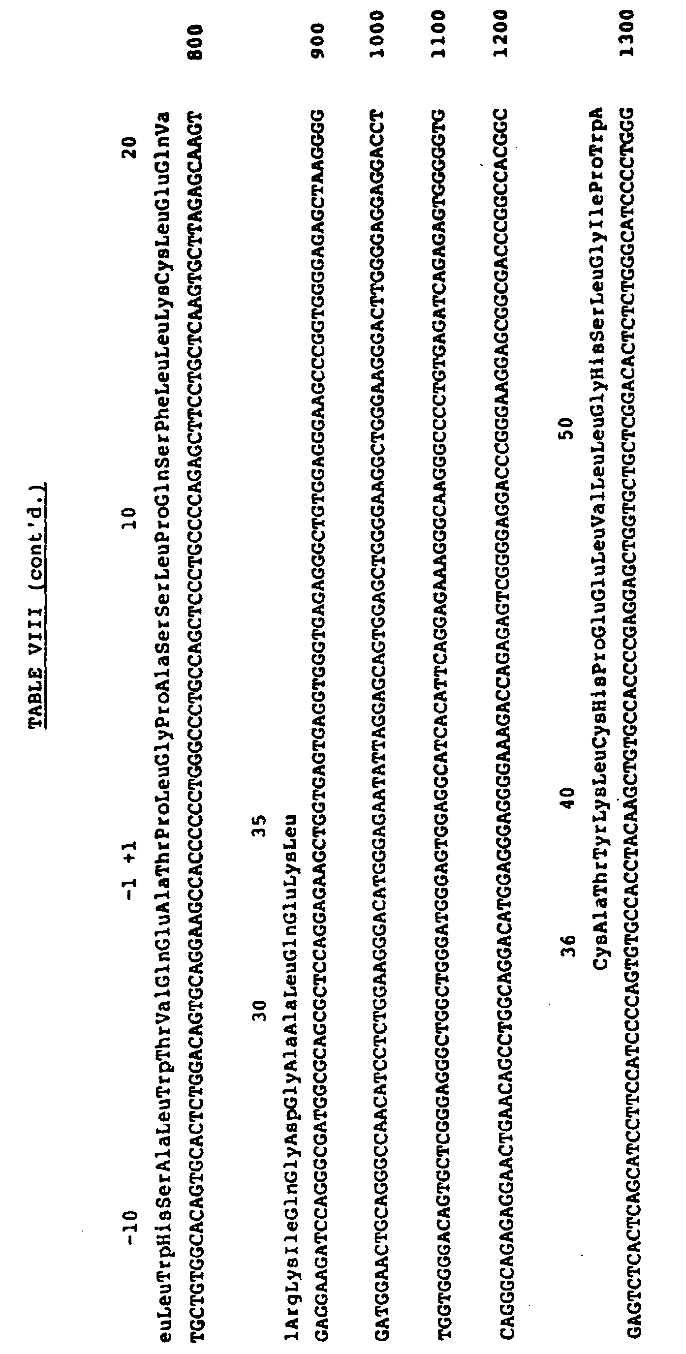

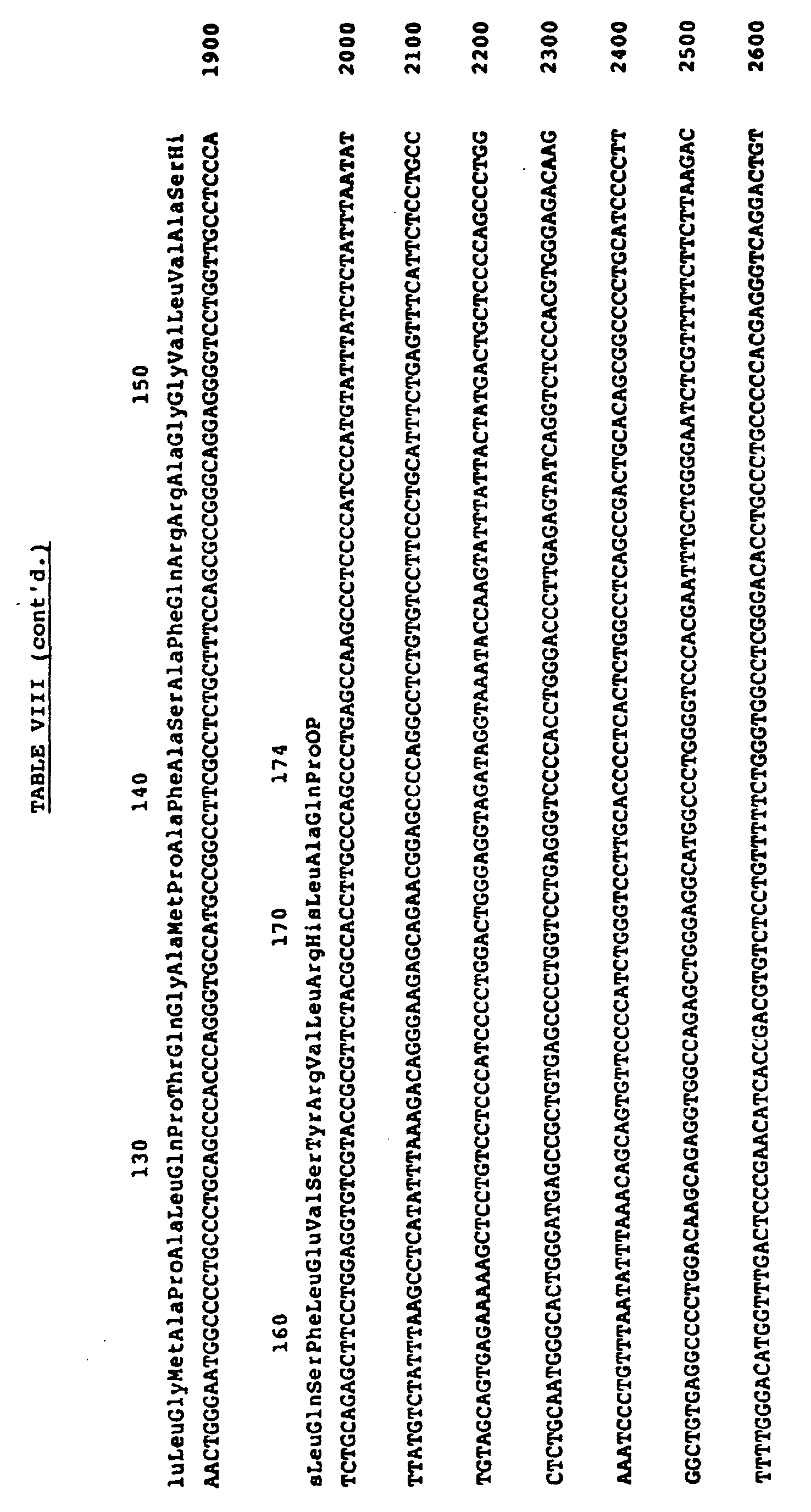

- A restriction endonuclease map (approximately 3.4 Kb) of genomic DNA containing the hpG-CSF gene is detailed in Figure 1. The restriction endonucleases shown in Figure 1 are: NcoI, N; PstI, P; BamHI, B; ApaI, A; XhoI, X; and Kpn, K. The arrows below the map depict the sequencing strategy used to obtain the genomic sequence. The boxed regions are those found in the cDNA clone with the dashed open ended box representing sequence not present in the cDNA clone, but identified by probing mRNA blots. The identification of coding sequences proposed for exon one was carried out by Northern blot analysis. A 24 mer oligonucleotide probe, 5'CAGCAGCTGCAGGGCCATCAGCTT3', spanning the predicted splice junctures for
exons 1 and 2 was hybridized to hpG-CSF mRNA in a Northern blot format. The resulting blot shows an mRNA the same size (-1650 bp) as that seen with anexon 2 oligonucleotide probe. This data combined with the ability to direct expression of hpG-CSF from the pSVGM-Ppol vector (Example 9) using the Met initiation codon depicted in Table VIII, defines the coding sequences contained in exon 1. Exons 2-5 are defined by the coding sequences obtained in the cDNA clone (Ppo2) of the hpG-CSF gene (Table VII). - This example relates to preparation of a manufactured gene encoding hpG-CSF and including E.coli preference codons.
- Briefly stated, the protocol employed was generally as set out in the disclosure of co-owned Alton, et al., PCT Publication No. WO83/04053, which is incorporated by reference herein. The genes were designed for initial assembly of component oligonucleotides into multiple duplexes which, in turn, were assembled into three discrete sections. These sections were designed for ready amplification and, upon removal from the amplification system, could be assembled sequentially or through a multiple fragment ligation in a suitable expression vector.
- The construction of Sections I, II and II is illustrated in Table IX though XIV. In the construction of Section I, as illustrated in Tables IX and X, oligonucleotides 1-14 were assembled into 7 duplexes (1 and 8); 2 and 9; 3 and 10; 4 and 11; 5 and 12; 6 and 13; and 7 and 14). The 7 duplexes were then ligated to form Section I as shown in Table X. It may also be noted in Table X that Section I includes an upstream XbaI sticky end and a downstream BamHI sticky end useful for ligation to amplification and expression vectors and for ligation to Section II.
TABLE IX EChpG-CSFDNA SECTION I CTAGAAAAAACCAAGGAGGTAATAAA 1 TAATGACTCCATTAGGTCCTGCTTCTTCT 2 CTGCCGCAAAGCTTTCTGCTGAAATGTCTGG 3 AACAGGTTCGTAAAATCCAGGGTGACGGT 4 GCTGCACTGCAAGAAAAACTGTGCGCTA 5 CTTACAAACTGTGCCATCCGGAAGAGC 6 TGGTACTGCTGGGTCATTCTCTTGG 7 CATTATTTATTACCTCCTTGGTTTTTT 8 GCAGAGAAGAAGCAGGACCTAATGGAGT 9 TGTTCCAGACATTTCAGCAGAAAGCTTTGCG 10 CAGCACCGTCACCCTGGATTTTACGAACC 11 TAAGTAGCGCACAGTTTTTCTTGCAGTG 12 ACCAGCTCTTCCGGATGGCACAGTTTG 13 GATCCCAAGAGAATGACCCAGCAGT 14 
- As illustrated in Tables XI and XII, in the construction of Section II, oligonucleotides 15-30 were assembled into 8 duplexes (15 and 23; 16 and 24; 17 and 25; 18 and 26; 19 and 27; 20 and 28; 21 and 29; and 22 and 30). These 8 duplexes were then ligated to form Section II, as shown in Table XII. As further shown in Table XII, Section II has an upstream BamHI sticky end and a downstream EcoRI sticky end useful for ligation to an amplification vector and for ligation to Section I. Near its downstream end, Section II also includes a downstream SstI site useful in the eventual ligation Sections II and III.
TABLE XI EChpG-CSFDNA SECTION II GATCCCGTGGGCTCCGCTGTCTTCT 15 TGTCCATCTCAAGCTCTTCAGCTGGC 16 TGGTTGTCTGTCTCAACTGCATTCTGGT 17 CTGTTCCTGTATCAGGGTCTTCTG 18 CAAGCTCTGGAAGGTATCTCTCCGGA 19 ACTGGGTCCGACTCTGCACACTCTGCA 20 GCTAGATGTAGCTGACTTTGCTACTACT 21 ATTTGGCAACAGATGGAAGAGCTCAAAG 22 GACAAGAAGACAGCGGAGCCCACGG 23 ACCAGCCAGCTGAAGAGCTTGAGATG 24 ACAGACCAGAATGCAGTTGAGACAGACA 25 CTTGCACAAGACCCTGATACAGGA 26 CAGTTCCGGAGAGATACCTTCCAGAG 27 TAGCTGCAGAGTGTCCAGAGTCGGACC 28 AAATAGTAGTAGCAAAGTCAGCTACATC 29 AATTCTTTGAGCTCTTCCATCTGTTGCC 30 
- Finally, Section III was constructed as shown in Tables XIII and XIV. For this construction, oligonucleotides 31-42 were assembled into 6 duplexes (31 and 37; 32 and 38; 33 and 39; 34 and 40; 35 and 41; and 36 and 42). The 6 duplexes were then ligated to form Section III as depicted in Table XIV. As also shown in Table XIV, Section III includes an upstream BamHI sticky end and a downstream EcoRI sticky end useful for ligating into an amplification vector, and at least in the case of the EcoRI end, into an expression vector. In addition, Section II has an upstream SstI site useful in the eventual ligation of Sections II and III.
TABLE XIII EChpG-CSFDNA SECTION III GATCCAAAGAGCTCGGTATGGCACCAG 31 CTCTGCAACCGACTCAAGGTGCTATGCCG 32 GCATTCGCTTCTGCATTCCAGCGTCGTGC 33 AGGAGGTGTACTGGTTGCTTCTCATCTG 34 CAATCTTTCCTGGAAGTATCTTACCGTGT 35 TCTGCGTCATCTGGCTCAGCCGTAATAG 36 AGAGCTGGTGCCATACCGAGCTCTTTG 37 ATGCCGGCATAGCACCTTGAGTCGGTTGC 38 TCCTGCACGACGCTGGAATGCAGAAGCGA 39 ATTGCAGATGAGAAGCAACCAGTACACC 40 CAGAACACGGTAAGATACTTCCAGGAAAG 41 AATTCTATTACGGCTGAGCCAGATGACG 42 
- The XbaI to BamHI fragment formed by Section I is ligated into an M13mp11 phage vector opened with XbaI and BamHI. The vector is then reopened by digestion with BamHI and EcoRI, followed by ligation with the BamHI to EcoRI fragment formed by Section II. At this stage, Sections I and II have been joined in proper orientation. Next, another M13mpll vector is opened by BamHI to EcoRI digestion and then ligated with the BamHI to EcoRI fragment formed by Section III.
- The vector containing Sections I and II is digested with XbaI and SstI. Likewise, the vector containing Section III is digested with SstI and EcoRI. Both of the smaller of the two fragments resulting from each digestion are ligated into a a plasmid pCFM1156 which is previously opened with XbaI and EcoRI. The product of this reaction is an expression plasmid containing a continuous DNA sequence, as shown in Table XV, encoding the entire hpG-CSF polypeptide with an amino terminal methionine codon (ATG) for E.coli translation initiation.

- Although any suitable vector may be employed to express this DNA, the expression plasmid pCFM1156 may readily be constructed from a plasmid pCFM836, the construction of which is described in published European Patent Application No. 136,490. pCFM836 is first cut with NdeI and then blunt-ended with PolI such that both existing NdeI sites are destroyed. Next, the vector is digested with ClaI and SacII to remove an existing polylinker before ligation to a substitute polylinker as illustrated in Table XVI. This substitute polylinker may be constructed according to the procedure of Alton, et al., supra. Control of expression in the expression pCFM1156 plasmid is by means of a lambda PL promoter, which itself may be under the control of a CI857 repressor gene (such as is provided in E.coli strain K12ΔHtrp).

- This example relates to E. coli expression of an hpG-CSF polypeptide by means of a DNA sequence encoding [Met-1] hpCSF. The sequence employed was partially synthetic and partially cDNA-derived. The synthetic sequence employed E. coli preference codons.
- Plasmid Ppo2, containing the hpG-CSF gene shown in Table VII, was digested with HgiAI and StuI providing an approximately 645 base pair fragment including the gene for mature hpCSF (as shown in Table VII) with seven of the leader sequence residue codons at the 5' end and about 100 base pairs of the 3' non-coding region. HgiAI digestion leaves a 5', 4-base sticky end identical to that of PstI, and StuI leaves a blunt end. This allows for ready insertion of the fragment into M13 mp8 (Rf) cut with PstI and with the blunt-end-forming restriction enzyme, HincII. Upon amplification in M13, the hpG-CSF DNA was excised by digestion with ApaI and BamHI which cut, respectively, at the ApaI site spanning the codons for residues +3 to +5 of hpCSF and at a BamHI site "downstream" of the HincII site in the M13 mp8 restriction polylinker. In order to allow for E. coli expression of the hpG-CSF polypeptide, a synthetic fragment was prepared as set out in Table XVII below.

- As may be determined from analysis of Table XVII, the linker includes an ApaI sticky end, codons specifying the initial three residues of the amino terminal of hpG-CSF ("restoring" the Thr1, Pro2, Leu3-specifying codons deleted upon ApaI digestion of the M13 DNA described above and employing codons preferentially expressed in E. coli), a translation initiating ATG, a sequence of 24 base pairs providing a ribosome binding site, and an XbaI sticky end.
- The expression vector employed for E. coli expression was that described as pCFM536 in European Patent Application No. 136,490, by Morris, published April 10, 1985. (See also, A.T.C.C. 39934, E. coli JM103 harboring pCFM536). Briefly, plasmid pCFM536 was digested with XbaI and BamHI. The hpG-CSF fragment (ApaI/BamHI) and linker (XbaI/ApaI) described above were then ligated thereinto to form a plasmid designated p536Ppo2.
- Plasmid p536Ppo2 was transformed into a phage resistant variant of the E. coli AM7 strain which has previously been transformed with plasmid pMW1 (A.T.C.C. No. 39933) harboring a CI857 gene. Transformation was verified on the basis of the antibiotic (amp) resistance marker gene carried on the pCFM536 progenitor plasmid. Cultures of cells in LB broth (ampicillin 50 µg 1ml) were maintained at 28°C. and upon growth of cells in culture to A600 = 0.5, hpCSF expression was induced by raising the culture temperature to 42°C. for 3 hours. The final O.D. of the culture was A600 = 1.2.
- The level of expression of hpG-CSF by the transformed cells was estimated on a SDS-poly acrylamide gel stained with coomassie blue dye to be 3-5% of total cellular protein.
- Cells were harvested by centrifugation at 3500 g for 10 minutes in a JS-4.2 rotor. Cells at 25% (w/v) in water were broken by passing 3 times through a French Pressure Cell at 10,000 p.s.i. The broken cell suspension was centrifuged at 10,000 g for 15 minutes in a JA-20 rotor. The pellet was resuspended in water and solubilized at about 5 mg/ml total protein in 1% lauric acid, 50 mM Tris, pH 8.7. The solubilized pellet material was centrifuged at 15,000 g for 10 minutes and to the supernatant CuSO4 was added to 20 mM. After 1 hour, this sample was loaded onto a C4 HPLC column for purification according to the procedures of example 1 (B) with adjustments made for volume and concentration.
- A second purification procedure was developed to yield larger quantities of hpG-CSF formulated in a nonorganic-containing buffer. This material is suitable for in vivo studies. One hundred and fifty grams of cell paste was resuspended in about 600 ml of 1 mM DTT and passed 4 times through a Manton Gualin Homogenizer at about 7000 PSI. The broken cell suspension was centrifuged at 10,000 g for 30 minutes and the pellet was resuspended in 400 ml of 1% deoxycholate (DOC), 5 mM EDTA, 5 mM DTT, and 50 mM Tris, pH 9. This suspension was mixed at room temperature for 30 minutes and centrifuged at 10,000 g for 30 minutes. The pellet was resuspended in about 400 ml of water and centrifuged at 10,000 g for 30 minutes. The pellet was solubilized in 100 ml of 2% Sarkosyl and 50 mM at pH 8. CuSO4 was added to 20 µM and the mixture was stirred 16 hours at room temperature, and then centrifuged at 20,000 g for 30 minutes. To the supernatant was added 300 ml acetone. This mixture was put on ice for 20 minutes and then centrifuged at 5000 g for 30 minutes. The pellet was dissolved in 250 ml of 6 M guanidine and 40 mM sodium acetate at pH 4, and put over a 1,200 ml G-25 column equilibrated and run in 20 mM sodium acetate at pH 5.4. The hpG-CSF peak (about 400 ml) was pooled and put on a 15 ml CM-cellulose column equilibrated in 20 mM sodium acetate at pH 5.4. After loading, the column was washed with 60 ml of 20 mM sodium acetate at pH 5.4 and with 25 mM sodium chloride, and then the column was eluted with 200 ml of 20 mM sodium acetate at pH 5.4 and with 37 mM sodium chloride. 150 ml of this eluent was concentrated to 10 ml and applied to a 300 ml G-75 column equilibrated and run in 20 mM sodium acetate and 100 mM sodium chloride at pH 5.4. The peak fractions comprising 35 ml were pooled and filter sterilized. The final concentration of hpG-CSF was 1.5 mg/ml, is greater than 95% pure as determined by analysis on a gel, and contained less than 0.5 ng of pyrogen per 0.5 mg of hpG-CSF. The pyrogen level was determined using a Limulus Amebocyte Lysate (LAL) test kit (M. A. Bioproducts, Walkersville, Maryland).
- This example relates to the use of recombinant methods to generate analogs of hpG-CSF wherein cysteine residues present at positions 17, 36, 42, 64 and 74 were individually replaced by a suitable amino acid residue.
- Site directed mutagenesis procedures according to Souza, et al., published PCT Application No. WO85/00817, published February 28, 1985, were carried out on [Met-1] encoding DNA of plasmid p536Ppo2, described infra, using synthetic oligonucleotides ranging in size from 20 to 23 bases as set out in Table XVIII below. Oligonucleotide No. 1 allowed for formation of a gene encoding [Ser17]hpG-CSF; oligonucleotide No. 2 allowed for formation of [Ser36]hpG-CSF, and so on.
TABLE XVIII Oligonucleotide Sequence 1. 5'-CTG CTC AAG TCC TTA GAG CAA GT-3' 2. 5'-GAG AAG CTG TCT GCC ACC TACA-3' 3. 5'-TAC AAG CTG TCC CAC CCC GAG-3' 4. 5'-TGA GCA GCT CCC CCA GCC AG-3' 5. 5'-CTG GCA GGC TCC TTG AGC CAA-3' - The Cys to Ser site directed mutagenesis restrictions were carried out using M13 mp10 containing an XbaI-BamHI hpG-CSF fragment isolated from p536Ppo2 as a template. DNA from each M13mp10 clone containing a Cys-Ser substitution was treated with XbaI and BamHI. The resulting fragment was cloned into expression vector pCFM746 and expression products were isolated as in Example 7.
- The plasmid pCFM746 may be constructed by cleaving a plasmid pCFM736 (the construction of which from deposited and publically available materials is described in Morris, published PCT Application No. WO85/00829, published February 28, 1985) with ClaI and BamHI to remove an existing polylinker and by substituting the following polylinker.

- In a purification procedure for Cys to Ser analogs according to the present invention, about 10-15 g of cell paste was resuspended in 40 ml of 1 mM DTT and passed 3 times through a French Pressure Cell at 10,000 psi. The broken cell suspension was centrifuged at 1,000 g for 30 minutes. The pellet was resuspended in 1% DOC, 5 mM EDTA, 5 mM DTT, 50 mM Tris, pH 9 and allowed to mix 30 minutes at room temperature. The mixture was centrifuged at 10,000 g for 30 minutes, resuspended in 40 ml H2O, and recentrifuged as 10,000 g for 30 minutes. The pellet was dissolved in 10 ml of 2% Sarkosyl, 50 mM DTT, 50 mM Tris, pH 8. After mixing for 1 hour, the mixture was clarified by centrifugation at 20,000 g for 30 minutes, and then applied to a 300 ml G-75 column equilibrated and run in 1% Sarkosyl, 50 mM Tris, pH 8. Fractions containing the analog were pooled and allowed to air oxidize by standing with exposure to air for at least one day. Final concentrations ranged from 0.5 - 5 mg/ml.
- In this example, a mammalian cell expression system was devised to ascertain whether an active polypeptide product of hpG-CSF DNA could be expressed in and secreted by mammalian cells (COS-1, A.T.C.C. CRL-1650). This system was designed to provide for secretion of a polypeptide analog of hpGCSF via expression and secretory processing of a partially synthetic, partially cDNA-derived construction encoding [Ala1] hpG-CSF preceded by a leader polypeptide having the sequence of residues attributed to human GM-CSF in Wong, et al., Science, 228, 810-815 (1985) and Lee, et al., Proc. Natl. Acad. Sci. (USA), 82, 4360-4364 (1985),
- The expression vector employed for preliminary studies of expression of polypeptide products of the invention was a "shuttle" vector incorporating both pBR322 and SV40 DNA which had been designed to allow for autonomous replication in both E. coli and mammalian cells, with mammalian cell expression of inserted exogenous DNA under control of a viral promoter/regulator DNA sequence. This vector, designated pSVDM-19, harbored in E. coli 101, was deposited August 23, 1985, with the American Type Culture Collection, 12301 Parklawn Drive, Rockville, Maryland, and received the accession No. A.T.C.C. 53241.
- The specific manipulations involved in the expression vector construction were as follows. A leader-encoding DNA sequence was synthesized as set out in Table XX below.
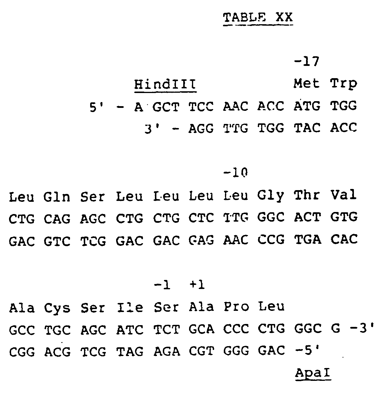
- As indicated in Table XX, the sequence includes HindIII and ApaI sticky ends and codons for the 17 amino acid residues attributed to the "leader" of human GM-CSF. There follow codons specifying an alanine residue, a proline residue and a leucine residue. The proline and leucine residues duplicate the amino acids present at positions +2 and +3 of hpG-CSF, while the alanine residue is duplicative of the initial amino terminal (+1) residue of GM-CSF rather than hpG-CSF. Replacement of threonine by alanine was designed to be facilitative of proper host cell "processing off" of the GM-CSF leader by cellular mechanisms ordinarily involved in GM-CSF secretory processing.
- Plasmid pSVDM-19 was digested with KpnI and the site was blunt ended with Klenow enzyme. Thereafter the DNA was cut with HindIII. The resulting large fragment was combined and ligated with the HindIII/PvuII fragment shown in Table VII (isolated from plasmid Ppo2 as the second largest fragment resulting from HindIII digestion and partial digestion with PvuII) to form plasmid pSV-Ppol. The manufactured GM-CSF leader sequence fragment of Table VIII was then ligated into pSV-Ppol (following its cleavage with HindIII and ApaI) to yield plasmid pSVGM-Ppol.
- Calcium phosphate precipitates (1-5µg) of plasmid pSVGM-Ppol DNA was transformed into duplicate 60 mm plates of COS-1 cells essentially as described in Wigler, et al., Cell, 14, 725-731 (1978). As a control, plasmid pSVDM-19 was also transformed into COS-1 cells. Tissue culture supernatants were harvested 5 days post-transfection and assayed for hpG-CSF activity. Yields of [Ala1]hpG-CSF from the culture supernatant were on the order of 1 to 2.5 µg/ml.
- Following successful expression of the [Ala1]hpG-CSF product encoded plasmid pSVGM-Ppol in COS-1 cells, another vector was constructed which included the human GM-CSF leader sequence but had a codon for a threonine residue (naturally occurring at position 1 of hpG-CSF) replacing the codon for alanine at that position. Briefly, an oligonucleotide was synthesized (5'CAGCATCTCTACACCTCTGGG) for site-directed mutagenesis (SDM). The HindIII to BamHI hpG-CSF fragment in pSVGM-Ppol was ligated into M18mp10 for the SDM. The newly synthesized hpG-CSF gene containing a Thr codon in position one was isolated by cleavage with HindIII and EcoRI. The fragment was then cloned into pSVDM-19 prepared by cleavage with the same two restriction endonucleases. The resulting vector pSVGM-Ppo(Thr) was transformed into COS cells and the yields of hpG-CSF measured in the culture supernates ranged from 1 to 5 µg/ml.
- Finally, the genomic sequence whose isolation is described in Example 5 was employed to form an expression vector for mammalian cell expression of hpG-CSF. More specifically, pSVDM-19 was digested with KpnI and HindIII and the large fragment used in a four-way ligation with a synthetic linker with HindIII and NcoI sticky ends, as shown in Table XXI. An NcoI-BamHI fragment containing exon 1 isolated from pBR322 (8500 hpG-CSF), a genomic subclone, and a BamHI-KpnI fragment containing exons 2-5 isolated from the plasmid pBR322 (8500 hpG-CSF genomic subclone). The resulting mammalian expression vector, pSV/ghG-CSF produced 1 to 2.5 µg/ml of hpG-CSF from transformed COS cells.

- This example relates to physical and biological properties or recombinant polypeptide.
- Recombinant hpG-CSF products of E. coli expression as in Example 7 had an apparent molecular weight of 18.8 kD when determined in reducing SDS-PAGE (as would be predicted from the deduced amino acid analysis of Table VII, whereas natural isolates purified as described in Example 1 had an apparent molecular weight of 19.6 kD. The presence of N-glycans associated with the natural isolates could effectively be ruled out on the basis of the lack of asparagine residues in the primary sequence of hpG-CSF in Table VII and therefore a procedure was devised to determine if O-glycans were responsible for molecular weight differences between natural isolates and the non-glycosylated recombinant products. Approximately 5 µg of the natural isolate material was treated with neuraminidase (Calbiochem, LaJolla, California), a 0.5 µg sample was removed, and the remaining material was incubated with 4 mU O-Glycanase (endo-x-n-acetylgalactoseaminidase, Genzyme, Boston, Massachusetts) at 37°C. Aliquots were removed after 1/2, 2 and 4 hours of incubation. These samples were subjected to SDS-PAGE side by side with the E. coli derived recombinant material. After neuraminidase treatment, the apparent molecular weight of the isolate shifted from 19.6 kD to 19.2 kD, suggestive of removal of a sailic acid residue. After 2 hours of treatment with O-glycanase, the molecular weight shifted to 18.8 kD -- identical to the apparent molecular weight of the E. coli derived material. The sensitivity of the carbohydrate structure to neuraminidase and O-glycanase suggests the following structure for the carbohydrate component: N-acetylneuraminic acid-α(2-6)(galactose β (1-3) N-acetylgalactoseamine-R, wherein R is serine or threonine.
- Proliferation induction of human bone marrow cells was assayed on the basis of increased incorporation of 3H-thymidine. Human bone marrow from healthy donors was subjected to a density cut with Ficoll-Hypaque (1.077g/ml, Pharmacia) and low density cells were suspended in Iscove's medium (GIBCO) containing 10% fetal bovine serum and glutamine pen-strep. Subsequently, 2x10⁴ human bone marrow cells were incubated with either control medium or the recombinant E. coli material of Example 7 in 96 flat bottom well plates at 37°C. in 5% CO2 in air for 2 days. The samples were assayed in duplicate and the concentration varied over a 10,000 fold range. Cultures were then pulsed for 4 hours with 0.5 µ Ci/well of 3H-Thymidine (New England Nuclear, Boston, Massachusetts). 3H-Thymidine uptake was measured as described in Ventua, et al., Blood, 61, 781 (1983). In this assay human hpG-CSF isolates can induce 3H-Thymidine incorporation into human bone marrow cells at levels approximately 4-10 times higher than control supernatants. The E. coli-derived hpG-CSF material of Example 6 had similar properties.
- A second human bone marrow cell proliferation study was carried out using culture medium of transfected COS-1 cells as prepared in Example 9 and yielded similar results, indicating that encoded polypeptide products were indeed secreted into culture medium as active materials.
- Capacity of recombinant, E. coli-derived materials to induce differentiation of the murine myelomonocytic leukemic cell line WEHI-3B D+ was assayed in semi-solid agar medium as described in Metcalf, Int. J. Cancer, 25, 225 (1980). The recombinant hpG-CSF product and media controls were incubated with -60 WEHI-3B D+ cells/well at 37°C. in 5% CO2 in air for 7 days. The samples were incubated in 24 flat bottom well plates and the concentration varied over a 2000-fold range. Colonies were classified as undifferentiated, partially differentiated or wholly differentiated and colony cell counts were counted microscopically. The E. coli recombinant material was found to induce differentiation.
- Natural isolates of pluripotent human G-CSF (hpG-CSF) and the recombinant pluripotent human G-CSF (rhpG-CSF) were found to cause human bone marrow cells to proliferate and differentiate. These activities were measured in CFU-GM [Broxmeyer, et al., Exp.Hematol., 5, 87, (1971)] PFU-E and CFU-GEMM assays [Lu, et al., Blood, 61, 250 (1983)] using low density, non-adherent bone marrow cells from healthy human volunteers. A comparison of CFU-GM, BFU-E and CFU-GEmm biological activities using either 500 units of hpG-CSF or rhpG-CSF are shown in Table XXII below.
- All the colony assays were performed with low density non-adherent bone marrow cells. Human bone marrow cells were subject to a density cut with Ficoll-Hypaque (density, 1.077 g/cm3; Pharmacia). The low density cells were then resuspended in Iscove's modified Dulbecco's medium containing fetal calf serum and placed for adherence on Falcon tissue culture dishes (No. 3003, Becton Dickenson, Cockeysville, MD.) for 1-1/2 hours at 37°C.
TABLE XXII CFU-GM BFU-E CFU-GEMM Medium natural 0±0 26±1 0±0 hpG-CSF 83±5.4 83±6.7 4±0 rhpG-CSF 87±5 81±0.1 6±2 - Medium control consisted of Iscove's modified Dulbecco medium plus 10% FCS, 0.2 mM hemin and 1 unit of recombinant erythropoietin.
- For the CFU-GM assay target cells were plated at 1 x 10⁵ in 1 ml of 0.3% agar culture medium that included supplemented McCoy's 5A medium and 10% heat inactivated fetal calf serum. Cultures were scored for colonies (greater than 40 cells per aggregate) and morphology assessed on day 7 of culture. The number of colonies is shown as the mean ±SEM as determined from quadruplicate plates.
- For the BFU-E and CFU-GEMM assays, cells (1 x 10⁵) were added to a 1 ml mixture of Iscove's modified Dulbecco medium (Gibco), 0.8% methylcellulose, 30% fetal calf serum 0.05 nM 2-mercaptoethanol, 0.2 mM hemin and 1 unit of recombinant erythropoietin. Dishes were incubated in a humidified atmosphere of 5% CO2 and 5% O2. Low oxygen tension was obtained using an oxyreducer from Reming Bioinstruments (Syracuse, N.Y.). Colonies were scored after 14 days of incubation. The number of colonies is shown as the mean ±SEM, as determined from duplicate plates.
- Colonies formed in the CFU-GM assay were all found to be chloracetate esterase positive and non-specific esterase (alpha-naphthyl acetate esterase) negative, consistent with the colonies being granulocyte in type. Both natural hpG-CSF and rhpG-CSF were found to have a specific activity of a approximately 1 x 10⁸ U/mg pure protein, when assayed by serial dilution in a CFU-GM assay. The BFU-E and CFU-GEMM data in Table XXII are representative of three separate experiments and similar to the data reported previously for natural hpG-CSF. It is important to note that the rhpG-CSF is extremely pure and free of other potential mammalian growth factors by virtue of its production in E.coli. Thus rhpG-CSF is capable of supporting mixed colony formation (CFU-GEMM) and BFU-E when added in the presence of recombinant erythropoietin.
- It was previously reported that WEHI-3B(D+) cells and human leukemic cells from newly diagnosed leukemias will bind 125I-labeled murine G-CSF and that this binding can be complete for by addition of unlabeled G-CSF or human CSF-β. The ability of natural hpG-CSF and rhpG-CSF to compete for binding of 125I-hpG-CSF to human and murine leukemic cells was tested. Highly purified natural hpG-CSF (>95% pure; 1µg) was iodinated [Tejedor, et al., Anal.Biochem., 127, 143 (1982)] was separated from reactants by gel filtration and ion exchange chromatography. The specific activity of the natural 125I-hpG-CSF was approximately µCi/µg protein. Murine WEHI-3B(D+) and two human peripheral blood myeloid leukemic cell preparations (ANLL, one classified as M4, the other as MSB) were tested for their ability to bind 125I-hpG-CSF.
- The murine and freshly obtained human peripheral blood myeloid leukemic cells were washed three times with PBS/1% BSA. WEHI-3B(D+) cells (5 x 10⁶) or fresh leukemic cells (3 x 10⁶) were incubated in duplicate in PBS/1% BSA (100 µl) in the absence or presence of various concentrations (volume: 10 µl) of unlabeled hpG-CSF, rhpG-CSF or GM-CSF and in the presence of 125I-hpG-CSF (approx. 100,000 cpm or 1 ng) at 0°C. for 90 min. (total volume: 120 µl). Cells were then resuspended and layered over 200 µl ice cold FCS in a 350 µl plastic centrifuge tube and centrifuged (1000 g; 1 min.). The pellet was collected by cutting off the end of the tube and pellet and supernatant counted separately in a gamma counter (Packard).
- Specific binding (cpm) was determined as total binding in the absence of a competitor (mean of duplicates) minus binding (cpm) in the presence of 100-fold excess of unlabeled hpG-CSF (non-specific binding). The non-specific binding was maximally 2503 cpm for WEHI-3H(D+) cells, 1072 cpm for ANLL (M4) cells and 1125 cpm for ANLL (M5B) cells. Experiments one and two were run on separate days using the same preparation of 125I-hpG-CSF and display internal consistency in the percent inhibition noted for 2000 units of hpG-CSF. Data obtained are reported in Table XXIII below.
TABLE XXIII WEHI-3B(D+) ANLL (M4) ANLL (M5B) Competitor (U/ml) cpm % Inhib. cpm % Inhib. cpm % Inhib. Exp. 1 none 0 6,608 - 1,218 - 122 - natural hpG-CSF: 10,000 685 90 2,000 1,692 74 34 97 -376 0 200 2,031 69 rhpG-CSF: 10,000 0 100 2,000 1,185 82 202 83 0 0 200 2,330 65 Exp. 2 none 0 2,910 0 natural hpG-CSF: 2,000 628 78 GM-CSF: 2,000 3,311 0 - As shown in Table XXIII, 125I-hpG-CSF demonstrated binding to the WEHI-3B(D+) leukemic cells. The binding was inhibited in a dose dependent manner by unlabeled natural hpG-CSF or rhpG-CSF, but not by GM-CSF. In addition, binding of natural hpG-CSF to human myelomonocytic leukemic cells (ANLL, M4) was observed. The binding to these cells is paralleled in response to natural hpG-CSF in liquid cultures by differentiation into mature macrophages as judged by morphology. The absence of binding of natural 125I-hpG-CSF to monocytic leukemic cells from another patient (ANLL, M5B) suggests that certain leukemias may differentially express or lack receptors for hpG-CSF. The ability of rhpG-CSF to compete for the binding of natural 125I-hpG-CSF, similar to natural hpG-CSF, suggests that the receptors recognize both forms equally well.
- These studies demonstrating the binding of natural 125I-labeled hpG-CSF to leukemic cells are paralleled in culture by the ability of natural hpG-CSF to induce granulocytic and monocytic differentiation of light density bond marrow cells obtained from one patient with an acute promyelocytic leukemia (M3) and a second patient with an acute myeloblastic leukemia (M2). Cells from each patient were cultured for four days in medium alone or in the presence of 1 x 10⁵ units of rhpG-CSF. Cells from the M3 control cultures incubated in medium alone were still promyelocyte in type; while cells cultured in the presence of rhpG-CSF showed mature cells of the myeloid type including a metamyelocyte, giant band form and segmented meutrophilis and monocyte. The actual differentials for this patient, on 100 cells evaluated for the control, 100% promyelocytes, and for the rhpG-CSF treated cells, 22% blasts plus promyelocytes, 7% myelocytes, 35% metamyelocytes, 20% band forms plus segmented neutrophils, 14% monocytes and 2% macrophages. Of note is the fact that one of the polymorphonuclear granulocytes still contained a prominent auer rod, suggesting that at least this cell represented a differentiated cell belonging to the leukemic clone. Cells from the second patient with a myeloblastic leukemia (M2) were also cultured for four days in the presence of absence of rhpG-CSF. Visual analysis of M2 cells cultured in medium alone revealed large "blast-like" cells, some of which had nucleoli. Some of the M2 cells, when treated with rhpG-CSF, differentiated to mature segmented neutrophils displaying residual auer rods in the center neutrophil suggesting differentiation occurring in a cell belonging to the leukemic clone. The actual differentiation of 100 cells evaluated morphologically revealed that control cells consisted of 100% blasts. The rhpG-CSF treated cells consisted of 43% blasts, 1% myelocytes, 15% metamyelocytes, 28% band forms plus segmented neutrophils, 2% promonocytes and 11% monocytes. The leukemic cells were also examined for differentiation at four other concentrations of rhpG-CSF (5 x 10³, 1 x 10⁴, 2.5 x 10⁴ and 5 x 10⁴ U/ml, data not shown). Even at the lowest concentration of rhpG-CSF tested (5 x 10³ U/ml), there was significant differentiation (cells differentiated beyond myelocytes) of the M3 (50%) and M2 (37%) leukemic cells.
- To prepare polyclonal antibodies for immunoassay use the antigen employed was pluripotent G-CSF purified from the human bladder carcinoma cell line 5637 (1A6) as prepared in Example 1 (B). This material was judged to be 85% pure based on silver nitrate staining of polyacrylamide gels. Six week-old Balb/C mice were immunized with multiple-site subcutaneous injections of antigen. The antigen was resuspended in PBS and emulsified with equal volumes of Freund's complete adjuvant. The dose was 5 to 7 µg of antigen per mouse per injection. A booster immunization was administered 18 days later with the same amount of antigen emulsified with an equal volume of Freund's incomplete adjuvant. 4 days later mouse serum was taken to test for the antibody specific to human pluripotent G-CSF.
- Dynatech Immulon II Removawell strips in holders (Dynateck Lab., Inc., Alexandria, Virginia) were coated with hpG-CSF 5 µg/ml in 50mM carbonate-bicarbonate buffer, pH 9.2. Wells were coated with 0.25 µg in a volume of 50 µl. Antigen coated plates were incubated 2 hours at room temperature and overnight at 4°C. The solution was decanted and the plates were incubated 30 minutes with PBS containing 5% BSA to block the reactive surface. This solution was decanted and the diluted preimmune or test sera were added to the wells and incubated for 2 hours at room temperature. Sera were diluted with PBS, pH 7.0 containing 1% BSA. The serum solution was decanted and plates were washed three times with Wash Solution (KPL, Gaithersburg, Maryland). Approximately 200,000 cpm of iodinated rabbit anti-mouse IgG (NEN, Boston, Massachusetts) in 50 µl PBS, pH 7.0 containing 1% BSA was added to each well. After incubating 1-1/2 hours at room temperature, the solution was decanted and plates were washed 5 times with Wash Solution. Wells were removed from holder and counted in a Beckman 5500 gamma counter. High-titered mouse sera showed greater than 12-fold higher reactivity than the corresponding preimmune sera at a dilution of 1:100.
- The immunological properties of E. coli-derived hpG-CSF were determined by reactivity to high-titered mouse serum specific to mammalian-cell derived hpG-CSF. 0.25 µg of 90% pure E. coli-derived protein was coated to Immulon II Removawells in a volume of 50 µl and mouse serum was assayed as described above.
- High-titered mouse sera showed a 24-fold higher reactivity to the E. coli-derived material than did the corresponding preimmune sera at a dilution of 1:100.
- [ser17]hpG-CSF, [Ser36]hpG-CSF, [Ser42]hpG-CSF, [Ser64]hpG-CSF, and [Ser74]hpG-CSF products prepared according to Example 9 were assay for hpG-CSF activity in the 3H-thymidine uptake, CFU-GM, and WEHI3B D+ assays. In each assay, the [Ser17] analog had activity comparable to that of recombinant molecules having the native structure. The remaining analogs had on the order of 100-fold lesser activity in the 3H-thymidine uptake assay, 250-fold lesser activity in the CFU-GM assay, and 500-fold lesser activity in the WEHI-3B D+ assay. This data is supportive of the proposition that cysteines at positions 36, 42, 64 and 74 may be needed for full biological activity.
- Alzet® osmotic pumps (Alzet Corp., Palo Alto, CA; Model 2001) were connected to indwelling right jugular vein catheters and implanted subcutaneously in seven male Syrian golden hamster. Four of the pumps contained a buffer [20 mM sodium acetate (pH 5.4) and 37 mM sodium chloride] and 1.5 mg/ml E.coli-derived hpG-CSF while 3 contained buffer alone. The claimed pumping rate for the osmotic pumps was 1 microliter/hr. for up to seven days. At the third day after implantation of the pumps, the mean granulocyte count of the four treated hamsters was six-fold higher than that of the three (buffer) controls and the increased granulocyte count was reflected in a four-fold increase in total lymphocytes. Erythrocyte count was unchanged by treatment. These results indicate that the recombinant material produces a specific enhancement of production and/or release of granulocytes in a mammal.
- In addition to naturally-occurring allelic forms of hpG-CSF, the present invention also embraces other hpG-CSF products such as polypeptide analogs of hpG-CSF and fragments of hpG-CSF. Following the procedures of the above-noted published application by Alton, et al. (WO/83/04053) one may readily design and manufacture genes coding for microbial expression of polypeptides having primary conformations which differ from that herein specified for in terms of the identity or location of one or more residues (e.g., substitutions, terminal and intermediate additions and deletions). Alternately, modifications of cDNA and genomic genes may be readily accomplished by well-known site-directed mutagenesis techniques and employed to generate analogs and derivatives of. Such products would share at least one of the biological properties of hpG-CSF but may differ in others. As examples, projected products of the invention include those which are foreshortened by e.g., deletions; or those which are more stable to hydrolysis (and, therefore, may have more pronounced or longer lasting effects than naturally-occurring); or which have been altered to delete one or more a potential sites for o-glycosylation (which may result in higher activities for yeast-produced products); or which have one or more cysteine residues deleted or replaced by, e.g., alanine or serine residues and are potentially more easily isolated in active form from micrbial systems; or which have one or more tyrosine residues replaced by phenylalanine and may bind more or less readily to hpG-CSF receptors on target cells. Also comprehended are polypeptide fragments duplicating only a part of the continuous amino acid sequence or secondary conformations within hpG-CSF, which fragments may possess one activity of (e.g., receptor binding) and not others (e.g., colony growth stimulating activity). It is noteworthy that activity is not necessary for any one or more of the products of the invention to have therapeutic utility [see, Weiland, et al., Blut, 44, 173-175 (1982)] or utility in other contexts, such as in assays of hpG-CSF antagonism. Competitive antagonists may be quite useful in, for example, cases of overproduction of hpG-CSF.
- The DNA sequence described herein which encodes hpG-CSF polypeptides is valuable for the information which it provides concerning the amino acid sequence of the mammalian protein which has heretofore been unavailable despite analytical processing of isolates of naturally-occurring products. The DNA sequences are also conspicuously valuable as products useful in effecting the large scale microbial synthesis of hpG-CSF by a variety of recombinant techniques. Put another way, DNA sequences provided by the invention are useful in generating new and useful viral and circular plasmid DNA vectors, new and useful transformed and transfected microbial procaryotic and eucaryotic host cells (including bacterial and yeast cells and mammalian cells grown in culture), and new and useful methods for cultured growth of such microbial host cells capable of expression of hpG-CSF and its related products. DNA sequences of the invention are also conspicuously suitable materials for use as labelled probes in isolating hpG-CSF and related protein encoding human genomic DNA as well as cDNA and genomic DNA sequences of other mammalian species. DNA sequences may also be useful in various alternative methods of protein synthesis (e.g., in insect cells) or in genetic therapy in humans and other mammals. DNA sequences of the invention are expected to be useful in developing transgenic mammalian species which may serve as eucaryotic "hosts" for production of hpG-CSF and hpG-CSF products in quantity. See, generally, Palmiter, et al., Science, 222(4625), 809-814 (1983).
- Of applicability to hpG-CSF fragments and polypeptide analogs are reports of the immunological activity of synthetic peptides which substantially duplicate the amino acid sequence extant in naturally-occurring proteins, glycoproteins and nucleo-proteins. More specifically, relatively low molecular weight polypeptides have been shown to participate in immune reactions which are similar in duration and extent to the immune reactions of physiologically significant proteins such as viral antigens, polypeptide hormones, and the like. Included among the immune reactions of such polypeptides is the provocation of the formation of specific antibodies in immunologically active animals. See, e.g., Lerner, et al., Cell, 23, 309-310 (1981); Ross, et al., Nature, 294, 654-656 (1981) ; Walter, et al., Proc. Natl. Acad. Sci. (USA), 77, 5197-5200 (1980); Lerner, et al., Proc. Natl. Acad. Sci. (USA), 78, 3403-3407 (1981); Walter, et al., Proc. Natl. Acad. Sci. (USA), 78, 4882-4886 (1981) ; Wong, et al., Proc. Natl. Acad. Sci. (USA), 78, 7412-7416 (1981); Green, et al., Cell, 28, 477-487 (1982); Nigg, et al., Proc. Natl. Acad. Sci. (USA), 79, 5322-5326 (1982); Baron, et al., Cell, 28, 395-404 (1982); Dreesman, et al., Nature, 295, 185-160 (1982); and Lerner, Scientific American, 248, No. 2, 66-74 (1983). See, also, Kaiser, et al., Science, 223, 249-255 (1984) relating to biological and immunological activities of synthetic peptides which approximately share secondary structures of peptide hormones but may not share their primary structural conformation.
Claims (4)
- A process for the production of a human pluripotent granulocyte colony-stimulating factor (hpG-CSF) product having the in vivo granulocytopoietic biological property of naturally occurring hpG-CSF comprising the steps of:(a) culturing under suitable nutrient conditions, mammalian cells comprising promoter DNA, other than hpG-CSF promoter DNA, operatively linked to DNA encoding a hpG-CSF polypeptide having the mature amino acid sequence of Table 7; and(b) isolating said hpG-CSF expressed by said cells.
- The process of claim 1 wherein the promoter DNA is viral promoter DNA.
- The process according to claim 1 wherein said cells are primate cells,
- The process according to claim 3 wherein said primate cells are COS cells.
Applications Claiming Priority (6)
| Application Number | Priority Date | Filing Date | Title |
|---|---|---|---|
| US76895985A | 1985-08-23 | 1985-08-23 | |
| US768959 | 1986-03-03 | ||
| US835548 | 1986-03-03 | ||
| US06/835,548 US4810643A (en) | 1985-08-23 | 1986-03-03 | Production of pluripotent granulocyte colony-stimulating factor |
| EP90111660A EP0396158A1 (en) | 1985-08-23 | 1986-08-22 | Production of pluripotent granulocyte colony-stimulating factor |
| EP86905530A EP0237545B2 (en) | 1985-08-23 | 1986-08-22 | Production of pluripotent granulocyte colony-stimulating factor |
Related Parent Applications (3)
| Application Number | Title | Priority Date | Filing Date |
|---|---|---|---|
| EP90111660A Division EP0396158A1 (en) | 1985-08-23 | 1986-08-22 | Production of pluripotent granulocyte colony-stimulating factor |
| EP86905530.1 Division | 1987-03-03 | ||
| EP90111660.8 Division | 1990-06-20 |
Publications (2)
| Publication Number | Publication Date |
|---|---|
| EP1018552A1 EP1018552A1 (en) | 2000-07-12 |
| EP1018552B1 true EP1018552B1 (en) | 2006-07-05 |
Family
ID=27233294
Family Applications (3)
| Application Number | Title | Priority Date | Filing Date |
|---|---|---|---|
| EP90111660A Ceased EP0396158A1 (en) | 1985-08-23 | 1986-08-22 | Production of pluripotent granulocyte colony-stimulating factor |
| EP00101091A Expired - Lifetime EP1018552B1 (en) | 1985-08-23 | 1986-08-22 | Production of pluripotent granulocyte colony-stimulating factor |
| EP01118552A Withdrawn EP1186663A1 (en) | 1985-08-23 | 1986-08-22 | Production of pluripotent granulocyte colony-stimulating factor |
Family Applications Before (1)
| Application Number | Title | Priority Date | Filing Date |
|---|---|---|---|
| EP90111660A Ceased EP0396158A1 (en) | 1985-08-23 | 1986-08-22 | Production of pluripotent granulocyte colony-stimulating factor |
Family Applications After (1)
| Application Number | Title | Priority Date | Filing Date |
|---|---|---|---|
| EP01118552A Withdrawn EP1186663A1 (en) | 1985-08-23 | 1986-08-22 | Production of pluripotent granulocyte colony-stimulating factor |
Country Status (1)
| Country | Link |
|---|---|
| EP (3) | EP0396158A1 (en) |
Families Citing this family (11)
| Publication number | Priority date | Publication date | Assignee | Title |
|---|---|---|---|---|
| CA2139385C (en) * | 1994-02-04 | 2001-12-25 | Gottfried Alber | Products containing g-csf and tnf binding protein |
| WO1997012977A1 (en) * | 1995-10-05 | 1997-04-10 | G.D. Searle & Co. | Novel g-csf receptor agonists |
| US6100070A (en) * | 1995-10-05 | 2000-08-08 | G. D. Searle & Co. | G-CSF receptor agonists |
| US8435939B2 (en) | 2000-09-05 | 2013-05-07 | Biokine Therapeutics Ltd. | Polypeptide anti-HIV agent containing the same |
| AU2003261723A1 (en) | 2002-08-27 | 2004-03-19 | Takeda Chemical Industries, Ltd. | Cxcr4 antagonist and use thereof |
| WO2008075371A2 (en) | 2006-12-21 | 2008-06-26 | Biokine Therapeutics Ltd. | T-140 peptide analogs having cxcr4 super-agonist activity for immunomodulation |
| US9155780B2 (en) | 2009-02-11 | 2015-10-13 | Yeda Research And Development Co. Ltd. | Short beta-defensin-derived peptides |
| BRPI1009663A2 (en) | 2009-06-14 | 2016-10-11 | Biokine Therapeutics Ltd | "method for elevating platelet levels in an individual with this requirement, method of inhibiting bleeding in an individual in need thereof, and pharmaceutical composition" |
| EP2841084B1 (en) | 2012-04-24 | 2018-05-30 | Biokine Therapeutics Ltd. | Cxcr4 antagonist peptide for use in the treatment of large cell lung cancer |
| US10786547B2 (en) | 2015-07-16 | 2020-09-29 | Biokine Therapeutics Ltd. | Compositions, articles of manufacture and methods for treating cancer |
| KR102033920B1 (en) | 2016-02-23 | 2019-10-18 | 바이오라인알엑스 리미티드 | How to treat acute myeloid leukemia |
Family Cites Families (3)
| Publication number | Priority date | Publication date | Assignee | Title |
|---|---|---|---|---|
| US4294156A (en) * | 1978-10-06 | 1981-10-13 | Mechanical Plastics Corporation | High strength anchor assembly for fastener |
| ATE65798T1 (en) * | 1985-02-08 | 1991-08-15 | Chugai Pharmaceutical Co Ltd | HUMAN GRANULOXCYTE COLONY STIMULATION FACTOR. |
| DE3681551D1 (en) * | 1985-09-30 | 1991-10-24 | Chugai Pharmaceutical Co Ltd | HUMAN GRANULOCYTE COLONY STIMULATING FACTOR. |
-
1986
- 1986-08-22 EP EP90111660A patent/EP0396158A1/en not_active Ceased
- 1986-08-22 EP EP00101091A patent/EP1018552B1/en not_active Expired - Lifetime
- 1986-08-22 EP EP01118552A patent/EP1186663A1/en not_active Withdrawn
Also Published As
| Publication number | Publication date |
|---|---|
| EP1018552A1 (en) | 2000-07-12 |
| EP0396158A1 (en) | 1990-11-07 |
| EP1186663A1 (en) | 2002-03-13 |
Similar Documents
| Publication | Publication Date | Title |
|---|---|---|
| US5582823A (en) | Methods of treating bacterial inflammation and granulocytopoiesis by administering human pluripotent granulocyte colony-stimulating factor | |
| US4999291A (en) | Production of human pluripotent granulocyte colony-stimulating factor | |
| US6004548A (en) | Analogs of pluripotent granulocyte colony-stimulating factor | |
| EP1018552B1 (en) | Production of pluripotent granulocyte colony-stimulating factor | |
| AU727724B2 (en) | Production of pluripotent granulocyte colony-stimulating factor | |
| HK1029600B (en) | Production of pluripotent granulocyte colony-stimulating factor | |
| AU769969B2 (en) | Production of pluripotent granulocyte colony-stimulating factor | |
| HK71892A (en) | Production of pluripotent granulocyte colony-stimulating factor | |
| HK1010396A (en) | Production of pluripotent granulocyte colony-stimulating factor | |
| AU2004202013A1 (en) | Production of pluripotent granulocyte colony-stimulating factor | |
| DK175551B1 (en) | Poly:peptide with granulocyte colony stimulating factor activity - obtd. by recombinant DNA procedures for treating haematopoietic disorders | |
| BG63065B2 (en) | Analogues of pluripotent granulocytic colony-stimulating factor | |
| BG63285B2 (en) | Production of human pluripotent granulocytic colony-stimulation factor |
Legal Events
| Date | Code | Title | Description |
|---|---|---|---|
| PUAI | Public reference made under article 153(3) epc to a published international application that has entered the european phase |
Free format text: ORIGINAL CODE: 0009012 |
|
| 17P | Request for examination filed |
Effective date: 20000120 |
|
| AC | Divisional application: reference to earlier application |
Ref document number: 396158 Country of ref document: EP Ref document number: 237545 Country of ref document: EP |
|
| AK | Designated contracting states |
Kind code of ref document: A1 Designated state(s): AT BE CH DE FR GB IT LI LU NL SE |
|
| AKX | Designation fees paid |
Free format text: AT BE CH DE FR GB IT LI LU NL SE |
|
| 17Q | First examination report despatched |
Effective date: 20031218 |
|
| GRAP | Despatch of communication of intention to grant a patent |
Free format text: ORIGINAL CODE: EPIDOSNIGR1 |
|
| GRAS | Grant fee paid |
Free format text: ORIGINAL CODE: EPIDOSNIGR3 |
|
| GRAA | (expected) grant |
Free format text: ORIGINAL CODE: 0009210 |
|
| AC | Divisional application: reference to earlier application |
Ref document number: 0237545 Country of ref document: EP Kind code of ref document: P Ref document number: 0396158 Country of ref document: EP Kind code of ref document: P |
|
| AK | Designated contracting states |
Kind code of ref document: B1 Designated state(s): AT BE CH DE FR GB IT LI LU NL SE |
|
| PG25 | Lapsed in a contracting state [announced via postgrant information from national office to epo] |
Ref country code: IT Free format text: LAPSE BECAUSE OF FAILURE TO SUBMIT A TRANSLATION OF THE DESCRIPTION OR TO PAY THE FEE WITHIN THE PRESCRIBED TIME-LIMIT;WARNING: LAPSES OF ITALIAN PATENTS WITH EFFECTIVE DATE BEFORE 2007 MAY HAVE OCCURRED AT ANY TIME BEFORE 2007. THE CORRECT EFFECTIVE DATE MAY BE DIFFERENT FROM THE ONE RECORDED. Effective date: 20060705 |
|
| REG | Reference to a national code |
Ref country code: GB Ref legal event code: FG4D |
|
| REG | Reference to a national code |
Ref country code: CH Ref legal event code: EP |
|
| REF | Corresponds to: |
Ref document number: 3650788 Country of ref document: DE Date of ref document: 20060817 Kind code of ref document: P |
|
| PG25 | Lapsed in a contracting state [announced via postgrant information from national office to epo] |
Ref country code: GB Free format text: LAPSE BECAUSE OF EXPIRATION OF PROTECTION Effective date: 20060821 |
|
| PG25 | Lapsed in a contracting state [announced via postgrant information from national office to epo] |
Ref country code: NL Free format text: LAPSE BECAUSE OF EXPIRATION OF PROTECTION Effective date: 20060822 |
|
| PG25 | Lapsed in a contracting state [announced via postgrant information from national office to epo] |
Ref country code: SE Free format text: LAPSE BECAUSE OF NON-PAYMENT OF DUE FEES Effective date: 20060823 |
|
| REG | Reference to a national code |
Ref country code: HK Ref legal event code: GR Ref document number: 1029600 Country of ref document: HK |
|
| REG | Reference to a national code |
Ref country code: CH Ref legal event code: PL Ref country code: CH Ref legal event code: NV Representative=s name: A. BRAUN, BRAUN, HERITIER, ESCHMANN AG PATENTANWAE |
|
| REG | Reference to a national code |
Ref country code: GB Ref legal event code: PE20 |
|
| REG | Reference to a national code |
Ref country code: SE Ref legal event code: TRGR |
|
| NLV7 | Nl: ceased due to reaching the maximum lifetime of a patent |
Effective date: 20060822 |
|
| ET | Fr: translation filed | ||
| PLBE | No opposition filed within time limit |
Free format text: ORIGINAL CODE: 0009261 |
|
| STAA | Information on the status of an ep patent application or granted ep patent |
Free format text: STATUS: NO OPPOSITION FILED WITHIN TIME LIMIT |
|
| EUG | Se: european patent has lapsed | ||
| 26N | No opposition filed |
Effective date: 20070410 |
|
| BE20 | Be: patent expired |
Owner name: *KIRIN-AMGEN INC. Effective date: 20060822 |