EP1017845B1 - Diagnostic test for alzheimer's disease - Google Patents
Diagnostic test for alzheimer's disease Download PDFInfo
- Publication number
- EP1017845B1 EP1017845B1 EP98946146A EP98946146A EP1017845B1 EP 1017845 B1 EP1017845 B1 EP 1017845B1 EP 98946146 A EP98946146 A EP 98946146A EP 98946146 A EP98946146 A EP 98946146A EP 1017845 B1 EP1017845 B1 EP 1017845B1
- Authority
- EP
- European Patent Office
- Prior art keywords
- ache
- csf
- wga
- con
- alzheimer
- Prior art date
- Legal status (The legal status is an assumption and is not a legal conclusion. Google has not performed a legal analysis and makes no representation as to the accuracy of the status listed.)
- Expired - Lifetime
Links
- 208000024827 Alzheimer disease Diseases 0.000 title claims abstract description 127
- 238000002405 diagnostic procedure Methods 0.000 title description 7
- 108010022752 Acetylcholinesterase Proteins 0.000 claims abstract description 204
- 229940022698 acetylcholinesterase Drugs 0.000 claims abstract description 204
- 108010046516 Wheat Germ Agglutinins Proteins 0.000 claims abstract description 45
- 108010062580 Concanavalin A Proteins 0.000 claims abstract description 42
- 238000000034 method Methods 0.000 claims abstract description 19
- 238000003745 diagnosis Methods 0.000 claims abstract description 6
- 102000012440 Acetylcholinesterase Human genes 0.000 claims abstract 25
- 210000001175 cerebrospinal fluid Anatomy 0.000 claims description 68
- 230000000694 effects Effects 0.000 claims description 42
- 239000002523 lectin Substances 0.000 claims description 27
- 108090001090 Lectins Proteins 0.000 claims description 25
- 102000004856 Lectins Human genes 0.000 claims description 25
- 238000004458 analytical method Methods 0.000 claims description 15
- 238000001514 detection method Methods 0.000 claims description 3
- 238000000338 in vitro Methods 0.000 claims description 3
- 230000013595 glycosylation Effects 0.000 abstract description 35
- 238000006206 glycosylation reaction Methods 0.000 abstract description 35
- HBOMLICNUCNMMY-KJFJCRTCSA-N 1-[(4s,5s)-4-azido-5-(hydroxymethyl)oxolan-2-yl]-5-methylpyrimidine-2,4-dione Chemical compound O=C1NC(=O)C(C)=CN1C1O[C@H](CO)[C@@H](N=[N+]=[N-])C1 HBOMLICNUCNMMY-KJFJCRTCSA-N 0.000 abstract description 3
- 210000001124 body fluid Anatomy 0.000 abstract description 3
- 239000010839 body fluid Substances 0.000 abstract description 3
- 102100033639 Acetylcholinesterase Human genes 0.000 description 179
- 102000001708 Protein Isoforms Human genes 0.000 description 62
- 108010029485 Protein Isoforms Proteins 0.000 description 62
- 210000005153 frontal cortex Anatomy 0.000 description 34
- 210000001638 cerebellum Anatomy 0.000 description 23
- 208000012902 Nervous system disease Diseases 0.000 description 22
- 208000025966 Neurological disease Diseases 0.000 description 22
- 206010012289 Dementia Diseases 0.000 description 19
- 210000004556 brain Anatomy 0.000 description 17
- 238000004062 sedimentation Methods 0.000 description 14
- 229920000936 Agarose Polymers 0.000 description 12
- 229930006000 Sucrose Natural products 0.000 description 9
- CZMRCDWAGMRECN-UGDNZRGBSA-N Sucrose Chemical compound O[C@H]1[C@H](O)[C@@H](CO)O[C@@]1(CO)O[C@@H]1[C@H](O)[C@@H](O)[C@H](O)[C@@H](CO)O1 CZMRCDWAGMRECN-UGDNZRGBSA-N 0.000 description 9
- 239000005720 sucrose Substances 0.000 description 9
- 229920002684 Sepharose Polymers 0.000 description 8
- 229920004890 Triton X-100 Polymers 0.000 description 8
- 239000013504 Triton X-100 Substances 0.000 description 8
- 238000000692 Student's t-test Methods 0.000 description 7
- 238000003556 assay Methods 0.000 description 7
- 239000011780 sodium chloride Substances 0.000 description 7
- QYOVMAREBTZLBT-KTKRTIGZSA-N CCCCCCCC\C=C/CCCCCCCCOCCOCCOCCOCCOCCOCCOCCOCCOCCOCCO Chemical compound CCCCCCCC\C=C/CCCCCCCCOCCOCCOCCOCCOCCOCCOCCOCCOCCOCCO QYOVMAREBTZLBT-KTKRTIGZSA-N 0.000 description 6
- 230000002547 anomalous effect Effects 0.000 description 6
- 239000000872 buffer Substances 0.000 description 6
- 230000003247 decreasing effect Effects 0.000 description 6
- 238000002474 experimental method Methods 0.000 description 6
- 238000004191 hydrophobic interaction chromatography Methods 0.000 description 6
- 102000004169 proteins and genes Human genes 0.000 description 6
- 108090000623 proteins and genes Proteins 0.000 description 6
- QKNYBSVHEMOAJP-UHFFFAOYSA-N 2-amino-2-(hydroxymethyl)propane-1,3-diol;hydron;chloride Chemical compound Cl.OCC(N)(CO)CO QKNYBSVHEMOAJP-UHFFFAOYSA-N 0.000 description 5
- 102000002260 Alkaline Phosphatase Human genes 0.000 description 5
- 108020004774 Alkaline Phosphatase Proteins 0.000 description 5
- 208000037259 Amyloid Plaque Diseases 0.000 description 5
- 102000016938 Catalase Human genes 0.000 description 5
- 108010053835 Catalase Proteins 0.000 description 5
- 102000004190 Enzymes Human genes 0.000 description 5
- 108090000790 Enzymes Proteins 0.000 description 5
- FAPWRFPIFSIZLT-UHFFFAOYSA-M Sodium chloride Chemical compound [Na+].[Cl-] FAPWRFPIFSIZLT-UHFFFAOYSA-M 0.000 description 5
- 210000004369 blood Anatomy 0.000 description 5
- 239000008280 blood Substances 0.000 description 5
- 230000002490 cerebral effect Effects 0.000 description 5
- 230000008859 change Effects 0.000 description 5
- 229940088598 enzyme Drugs 0.000 description 5
- 239000000178 monomer Substances 0.000 description 5
- 230000002861 ventricular Effects 0.000 description 5
- 101000738771 Homo sapiens Receptor-type tyrosine-protein phosphatase C Proteins 0.000 description 4
- 101000899806 Homo sapiens Retinal guanylyl cyclase 1 Proteins 0.000 description 4
- TWRXJAOTZQYOKJ-UHFFFAOYSA-L Magnesium chloride Chemical compound [Mg+2].[Cl-].[Cl-] TWRXJAOTZQYOKJ-UHFFFAOYSA-L 0.000 description 4
- 230000002159 abnormal effect Effects 0.000 description 4
- 239000000539 dimer Substances 0.000 description 4
- 201000010099 disease Diseases 0.000 description 4
- 208000037265 diseases, disorders, signs and symptoms Diseases 0.000 description 4
- 210000001652 frontal lobe Anatomy 0.000 description 4
- 238000011534 incubation Methods 0.000 description 4
- 230000001575 pathological effect Effects 0.000 description 4
- 210000002966 serum Anatomy 0.000 description 4
- 239000006228 supernatant Substances 0.000 description 4
- 210000001519 tissue Anatomy 0.000 description 4
- GPRLSGONYQIRFK-MNYXATJNSA-N triton Chemical compound [3H+] GPRLSGONYQIRFK-MNYXATJNSA-N 0.000 description 4
- 102100032404 Cholinesterase Human genes 0.000 description 3
- IOIMDJXKIMCMIG-UHFFFAOYSA-N Diphosphoramide, N,N',N'',N'''-tetrakis(1-methylethyl)- Chemical compound CC(C)NP(=O)(NC(C)C)OP(=O)(NC(C)C)NC(C)C IOIMDJXKIMCMIG-UHFFFAOYSA-N 0.000 description 3
- 208000023105 Huntington disease Diseases 0.000 description 3
- 208000018737 Parkinson disease Diseases 0.000 description 3
- 241000209140 Triticum Species 0.000 description 3
- 235000021307 Triticum Nutrition 0.000 description 3
- 239000012134 supernatant fraction Substances 0.000 description 3
- 102000014303 Amyloid beta-Protein Precursor Human genes 0.000 description 2
- 108010079054 Amyloid beta-Protein Precursor Proteins 0.000 description 2
- 241000283690 Bos taurus Species 0.000 description 2
- 108010053652 Butyrylcholinesterase Proteins 0.000 description 2
- 108090000322 Cholinesterases Proteins 0.000 description 2
- 208000011990 Corticobasal Degeneration Diseases 0.000 description 2
- 241000588724 Escherichia coli Species 0.000 description 2
- 208000009829 Lewy Body Disease Diseases 0.000 description 2
- 201000002832 Lewy body dementia Diseases 0.000 description 2
- -1 RCA120 Proteins 0.000 description 2
- 239000007983 Tris buffer Substances 0.000 description 2
- 201000004810 Vascular dementia Diseases 0.000 description 2
- 230000004075 alteration Effects 0.000 description 2
- 210000004727 amygdala Anatomy 0.000 description 2
- 238000012742 biochemical analysis Methods 0.000 description 2
- 238000005119 centrifugation Methods 0.000 description 2
- 239000000470 constituent Substances 0.000 description 2
- 239000000284 extract Substances 0.000 description 2
- 238000000605 extraction Methods 0.000 description 2
- 239000000499 gel Substances 0.000 description 2
- 210000004185 liver Anatomy 0.000 description 2
- 229910001629 magnesium chloride Inorganic materials 0.000 description 2
- 239000003550 marker Substances 0.000 description 2
- 239000000463 material Substances 0.000 description 2
- 238000005259 measurement Methods 0.000 description 2
- 239000000203 mixture Substances 0.000 description 2
- 210000002682 neurofibrillary tangle Anatomy 0.000 description 2
- 238000011160 research Methods 0.000 description 2
- 201000000980 schizophrenia Diseases 0.000 description 2
- 238000012216 screening Methods 0.000 description 2
- 241000894007 species Species 0.000 description 2
- 238000010561 standard procedure Methods 0.000 description 2
- 239000000725 suspension Substances 0.000 description 2
- LENZDBCJOHFCAS-UHFFFAOYSA-N tris Chemical compound OCC(N)(CO)CO LENZDBCJOHFCAS-UHFFFAOYSA-N 0.000 description 2
- GFFIJCYHQYHUHB-UHFFFAOYSA-N 2-acetylsulfanylethyl(trimethyl)azanium Chemical compound CC(=O)SCC[N+](C)(C)C GFFIJCYHQYHUHB-UHFFFAOYSA-N 0.000 description 1
- NTBLZMAMTZXLBP-UHFFFAOYSA-M 2-acetylsulfanylethyl(trimethyl)azanium;iodide Chemical compound [I-].CC(=O)SCC[N+](C)(C)C NTBLZMAMTZXLBP-UHFFFAOYSA-M 0.000 description 1
- KIUMMUBSPKGMOY-UHFFFAOYSA-N 3,3'-Dithiobis(6-nitrobenzoic acid) Chemical compound C1=C([N+]([O-])=O)C(C(=O)O)=CC(SSC=2C=C(C(=CC=2)[N+]([O-])=O)C(O)=O)=C1 KIUMMUBSPKGMOY-UHFFFAOYSA-N 0.000 description 1
- 208000000044 Amnesia Diseases 0.000 description 1
- 102000013455 Amyloid beta-Peptides Human genes 0.000 description 1
- 108010090849 Amyloid beta-Peptides Proteins 0.000 description 1
- 102000009091 Amyloidogenic Proteins Human genes 0.000 description 1
- 108010048112 Amyloidogenic Proteins Proteins 0.000 description 1
- 206010059245 Angiopathy Diseases 0.000 description 1
- 244000105624 Arachis hypogaea Species 0.000 description 1
- 235000010777 Arachis hypogaea Nutrition 0.000 description 1
- 108091003079 Bovine Serum Albumin Proteins 0.000 description 1
- 244000045232 Canavalia ensiformis Species 0.000 description 1
- 235000010520 Canavalia ensiformis Nutrition 0.000 description 1
- 101710132601 Capsid protein Proteins 0.000 description 1
- 108010058699 Choline O-acetyltransferase Proteins 0.000 description 1
- 102100023460 Choline O-acetyltransferase Human genes 0.000 description 1
- 102000003914 Cholinesterases Human genes 0.000 description 1
- SHZGCJCMOBCMKK-UHFFFAOYSA-N D-mannomethylose Chemical group CC1OC(O)C(O)C(O)C1O SHZGCJCMOBCMKK-UHFFFAOYSA-N 0.000 description 1
- 241001634308 Dolichus Species 0.000 description 1
- 201000010374 Down Syndrome Diseases 0.000 description 1
- 102100021238 Dynamin-2 Human genes 0.000 description 1
- 238000002965 ELISA Methods 0.000 description 1
- PNNNRSAQSRJVSB-SLPGGIOYSA-N Fucose Chemical group C[C@H](O)[C@@H](O)[C@H](O)[C@H](O)C=O PNNNRSAQSRJVSB-SLPGGIOYSA-N 0.000 description 1
- 244000068988 Glycine max Species 0.000 description 1
- 235000010469 Glycine max Nutrition 0.000 description 1
- 101000817607 Homo sapiens Dynamin-2 Proteins 0.000 description 1
- 101000746373 Homo sapiens Granulocyte-macrophage colony-stimulating factor Proteins 0.000 description 1
- SHZGCJCMOBCMKK-DHVFOXMCSA-N L-fucopyranose Chemical group C[C@@H]1OC(O)[C@@H](O)[C@H](O)[C@@H]1O SHZGCJCMOBCMKK-DHVFOXMCSA-N 0.000 description 1
- 244000043158 Lens esculenta Species 0.000 description 1
- 235000010666 Lens esculenta Nutrition 0.000 description 1
- 208000005314 Multi-Infarct Dementia Diseases 0.000 description 1
- OVRNDRQMDRJTHS-CBQIKETKSA-N N-Acetyl-D-Galactosamine Chemical group CC(=O)N[C@H]1[C@@H](O)O[C@H](CO)[C@H](O)[C@@H]1O OVRNDRQMDRJTHS-CBQIKETKSA-N 0.000 description 1
- MBLBDJOUHNCFQT-UHFFFAOYSA-N N-acetyl-D-galactosamine Natural products CC(=O)NC(C=O)C(O)C(O)C(O)CO MBLBDJOUHNCFQT-UHFFFAOYSA-N 0.000 description 1
- 240000000528 Ricinus communis Species 0.000 description 1
- 235000004443 Ricinus communis Nutrition 0.000 description 1
- 108010017507 Ricinus communis agglutinin-1 Proteins 0.000 description 1
- 239000004809 Teflon Substances 0.000 description 1
- 229920006362 Teflon® Polymers 0.000 description 1
- 240000003864 Ulex europaeus Species 0.000 description 1
- 235000010730 Ulex europaeus Nutrition 0.000 description 1
- ZAEXMNKDGJNLTA-UHFFFAOYSA-N [4-[5-[4-[dimethyl(prop-2-enyl)azaniumyl]phenyl]-3-oxopentyl]phenyl]-dimethyl-prop-2-enylazanium Chemical compound C1=CC([N+](C)(CC=C)C)=CC=C1CCC(=O)CCC1=CC=C([N+](C)(C)CC=C)C=C1 ZAEXMNKDGJNLTA-UHFFFAOYSA-N 0.000 description 1
- 239000000910 agglutinin Substances 0.000 description 1
- 229940019748 antifibrinolytic proteinase inhibitors Drugs 0.000 description 1
- 230000003542 behavioural effect Effects 0.000 description 1
- AFYNADDZULBEJA-UHFFFAOYSA-N bicinchoninic acid Chemical compound C1=CC=CC2=NC(C=3C=C(C4=CC=CC=C4N=3)C(=O)O)=CC(C(O)=O)=C21 AFYNADDZULBEJA-UHFFFAOYSA-N 0.000 description 1
- 239000003150 biochemical marker Substances 0.000 description 1
- 229940098773 bovine serum albumin Drugs 0.000 description 1
- 229940021260 by ache Drugs 0.000 description 1
- 230000015556 catabolic process Effects 0.000 description 1
- 230000001413 cellular effect Effects 0.000 description 1
- 230000001713 cholinergic effect Effects 0.000 description 1
- 229940048961 cholinesterase Drugs 0.000 description 1
- 238000003759 clinical diagnosis Methods 0.000 description 1
- 230000003920 cognitive function Effects 0.000 description 1
- 230000001054 cortical effect Effects 0.000 description 1
- 238000006731 degradation reaction Methods 0.000 description 1
- 238000000432 density-gradient centrifugation Methods 0.000 description 1
- 230000008021 deposition Effects 0.000 description 1
- 238000010790 dilution Methods 0.000 description 1
- 239000012895 dilution Substances 0.000 description 1
- 210000003743 erythrocyte Anatomy 0.000 description 1
- 238000011156 evaluation Methods 0.000 description 1
- 230000001605 fetal effect Effects 0.000 description 1
- 239000012530 fluid Substances 0.000 description 1
- 229930182830 galactose Natural products 0.000 description 1
- 239000011521 glass Substances 0.000 description 1
- 238000004128 high performance liquid chromatography Methods 0.000 description 1
- 210000001320 hippocampus Anatomy 0.000 description 1
- 230000001900 immune effect Effects 0.000 description 1
- 230000002055 immunohistochemical effect Effects 0.000 description 1
- 239000003112 inhibitor Substances 0.000 description 1
- 238000001155 isoelectric focusing Methods 0.000 description 1
- 230000003902 lesion Effects 0.000 description 1
- 238000011068 loading method Methods 0.000 description 1
- 231100000863 loss of memory Toxicity 0.000 description 1
- 238000009593 lumbar puncture Methods 0.000 description 1
- 238000004949 mass spectrometry Methods 0.000 description 1
- 239000012528 membrane Substances 0.000 description 1
- 238000002156 mixing Methods 0.000 description 1
- 230000001423 neocortical effect Effects 0.000 description 1
- 230000001722 neurochemical effect Effects 0.000 description 1
- 230000007138 neurofibrillary change Effects 0.000 description 1
- 230000000926 neurological effect Effects 0.000 description 1
- 230000002981 neuropathic effect Effects 0.000 description 1
- 210000004940 nucleus Anatomy 0.000 description 1
- 230000001936 parietal effect Effects 0.000 description 1
- 230000007170 pathology Effects 0.000 description 1
- 239000008188 pellet Substances 0.000 description 1
- 239000000137 peptide hydrolase inhibitor Substances 0.000 description 1
- 235000020030 perry Nutrition 0.000 description 1
- 238000002360 preparation method Methods 0.000 description 1
- 230000000750 progressive effect Effects 0.000 description 1
- 230000006337 proteolytic cleavage Effects 0.000 description 1
- 238000011084 recovery Methods 0.000 description 1
- 150000003839 salts Chemical class 0.000 description 1
- 230000035945 sensitivity Effects 0.000 description 1
- 238000000856 sucrose gradient centrifugation Methods 0.000 description 1
- 235000000346 sugar Nutrition 0.000 description 1
- 150000008163 sugars Chemical class 0.000 description 1
- 238000012360 testing method Methods 0.000 description 1
- 238000010257 thawing Methods 0.000 description 1
- 238000000108 ultra-filtration Methods 0.000 description 1
- 238000005199 ultracentrifugation Methods 0.000 description 1
Images

Classifications
-
- C—CHEMISTRY; METALLURGY
- C12—BIOCHEMISTRY; BEER; SPIRITS; WINE; VINEGAR; MICROBIOLOGY; ENZYMOLOGY; MUTATION OR GENETIC ENGINEERING
- C12N—MICROORGANISMS OR ENZYMES; COMPOSITIONS THEREOF; PROPAGATING, PRESERVING, OR MAINTAINING MICROORGANISMS; MUTATION OR GENETIC ENGINEERING; CULTURE MEDIA
- C12N9/00—Enzymes; Proenzymes; Compositions thereof; Processes for preparing, activating, inhibiting, separating or purifying enzymes
- C12N9/14—Hydrolases (3)
- C12N9/16—Hydrolases (3) acting on ester bonds (3.1)
- C12N9/18—Carboxylic ester hydrolases (3.1.1)
-
- G—PHYSICS
- G01—MEASURING; TESTING
- G01N—INVESTIGATING OR ANALYSING MATERIALS BY DETERMINING THEIR CHEMICAL OR PHYSICAL PROPERTIES
- G01N33/00—Investigating or analysing materials by specific methods not covered by groups G01N1/00 - G01N31/00
- G01N33/48—Biological material, e.g. blood, urine; Haemocytometers
- G01N33/50—Chemical analysis of biological material, e.g. blood, urine; Testing involving biospecific ligand binding methods; Immunological testing
- G01N33/68—Chemical analysis of biological material, e.g. blood, urine; Testing involving biospecific ligand binding methods; Immunological testing involving proteins, peptides or amino acids
- G01N33/6893—Chemical analysis of biological material, e.g. blood, urine; Testing involving biospecific ligand binding methods; Immunological testing involving proteins, peptides or amino acids related to diseases not provided for elsewhere
- G01N33/6896—Neurological disorders, e.g. Alzheimer's disease
-
- G—PHYSICS
- G01—MEASURING; TESTING
- G01N—INVESTIGATING OR ANALYSING MATERIALS BY DETERMINING THEIR CHEMICAL OR PHYSICAL PROPERTIES
- G01N2333/00—Assays involving biological materials from specific organisms or of a specific nature
- G01N2333/90—Enzymes; Proenzymes
- G01N2333/914—Hydrolases (3)
- G01N2333/916—Hydrolases (3) acting on ester bonds (3.1), e.g. phosphatases (3.1.3), phospholipases C or phospholipases D (3.1.4)
-
- G—PHYSICS
- G01—MEASURING; TESTING
- G01N—INVESTIGATING OR ANALYSING MATERIALS BY DETERMINING THEIR CHEMICAL OR PHYSICAL PROPERTIES
- G01N2800/00—Detection or diagnosis of diseases
- G01N2800/28—Neurological disorders
- G01N2800/2814—Dementia; Cognitive disorders
- G01N2800/2821—Alzheimer
-
- Y—GENERAL TAGGING OF NEW TECHNOLOGICAL DEVELOPMENTS; GENERAL TAGGING OF CROSS-SECTIONAL TECHNOLOGIES SPANNING OVER SEVERAL SECTIONS OF THE IPC; TECHNICAL SUBJECTS COVERED BY FORMER USPC CROSS-REFERENCE ART COLLECTIONS [XRACs] AND DIGESTS
- Y10—TECHNICAL SUBJECTS COVERED BY FORMER USPC
- Y10S—TECHNICAL SUBJECTS COVERED BY FORMER USPC CROSS-REFERENCE ART COLLECTIONS [XRACs] AND DIGESTS
- Y10S435/00—Chemistry: molecular biology and microbiology
- Y10S435/8215—Microorganisms
- Y10S435/822—Microorganisms using bacteria or actinomycetales
- Y10S435/827—Actinoplanes
Definitions
- the present invention is concerned with a diagnostic test for Alzheimer's disease.
- AD Alzheimer's disease
- a ⁇ amyloid protein
- APP amyloid protein precursor
- AD Alzheimer's disease
- AChE acetylcholinesterase
- choline acetyltransferase activity in regions of the brain such as the cortex, hippocampus, amygdala and nucleus basalis
- the loss of cholinergic structure and markers correlates with the number of plaque and tangle lesions present, as well as with the clinical severity of the disease (Perry et al ., 1978; Wilcock et al. , 1982; Neary et al ., 1986; Perry, 1986).
- AD Alzheimer's disease
- the assay of levels of AChE activity in the blood and the cerebrospinal fluid (CSF) has been proposed as an ante mortem diagnostic test for AD. However, no consensus has been reached as to whether the levels of AChE are consistently affected in these tissues.
- the level of serum or plasma AChE has been reported to be increased (Perry et al ., 1982; Atack et al., 1985), decreased (Nakano et al., 1986; Yamamoto et al., 1990) or unchanged (St. Clair et al., 1986; Sirvio et al., 1989) in AD patients.
- the level of erythrocyte AChE has been reported as either unaffected (Atack et al., 1985; Perry et al., 1982) or decreased (Chipperfield et al., 1981).
- the level of AChE activity in the CSF of AD patients has been reported to be decreased (most recently by Appleyard and McDonald, 1992; Shen et al., 1993) or unchanged (most recently by Appleyard et al., 1987; Ruberg et al., 1987).
- AChE has been shown to exist as up to six different molecular isoforms, three of which are the monomeric (G1), dimeric (G2) and tetrameric (G4) isoforms (Massoulié et al., 1993).
- the relative proportion of the different isoforms of AChE are markedly affected in AD, with a decrease in the G4 isoform in the parietal cortex (Atack et al., 1983), and an increase in the G1 isoform (Arendt et al., 1992). Similar changes have been identified in other AD brain regions including Brodman areas 9, 10, 11, 21 and 40, as well as the amygdala (Fishman et al., 1986).
- Asymmetric collagen-tailed isoforms (A12) are increased by up to 400% in Brodman area 21, although they represent only a trace amount of the total AChE in the human brain (Younkin et al., 1986).
- An anomalous isoform of AChE distinguished by its isoelectric point, has been detected in the CSF of AD patients (Navaratnam et al., 1991; Smith et al., 1991), and a method for screening for AD based on these findings is described in US patent number 5,200,324.
- the method comprises determining, by means of isoelectric focusing, if a patient has an anomalous form of AChE in his CSF.
- the isoform detected by Navaratnam et al and Smith et al has also been detected in the CSF of patients with other neurological diseases (Shen and Zhang, 1993).
- Mimori et al (Behavioural Brain Research 83, 25-30 (1997)) describes the effects of certain inhibitors on acetylcholinesterase in Alzheimer's disease. Tornel et al (Neuroscience Letters 145, 59-62 (1992)) describes lectin binding properties of AChE. Michaelson et al (Brain Research 611, 75-80 (1993)) describes the forms of AChE present in fetal brain serum.
- a method for the diagnosis of Alzheimer's disease in a patient comprising detecting in vitro the presence of acetylcholinesterase in a sample of cerebrospinal fluid from a patient, wherein said AChE has a relatively lesser affinity for Concanavalin A and a relatively greater affinity for wheat germ agglutinin than non-AD AChE.
- the relative proportion of AChE with a first glycosylation pattern and AChE with a second glycosylation pattern is measured.
- Measurement of the relative proportions of AChE with first and second glycosylation patterns may be carried out in any convenient manner, for example, by using biochemical analysis techniques such as HPLC and mass spectrometry, or immunological techniques such as ELISA or, assays.
- biochemical analysis techniques such as HPLC and mass spectrometry
- immunological techniques such as ELISA or, assays.
- a particularly preferred means of measuring the relative proportions of the isoforms of AChE involves a lectin-binding analysis.
- the binding to Con A is determined, then the binding to WGA is determined, and a ratio calculated.
- the ratio is characteristic of the glycosylation pattern. It is particularly convenient to measure the activity of unbound AChE in each experiment, hence the ratio of AChE unbound to Con A to the ratio of AChE unbound to WGA is determined. This ratio is referred to hereinafter as a C/W ratio.
- the C/W ratio has generally been found to be above 0.95 whereas for non-sufferers of AD the C/W ratio is typically below 0.95.
- the total AChE activity is measured and the C/W ratio plotted against AChE activity.
- a monoclonal antibody specific for AChE with an altered glycosylation pattern is used to detect its presence.
- the monoclonal antibody is MA3-042 (clone HR2), available from Chemicon International Inc of Temecula, California.
- Other suitable monoclonal antibodies may be used, for example, MA304 (clone AE1) also available from Chemicon International Inc.
- abnormal isoform is the amphiphilic, monomeric isoform of AChE and/or the amphiphilic, dimeric isoform of AChE.
- the body fluid analysed in the method of the invention is cerebrospinal fluid (CSF).
- CSF cerebrospinal fluid
- An abnormal isoform of the acetylcholinesterase (AChE) with an altered pattern of glycosylation, being the amphiphilic, monomeric isoform of AChE has a relatively lesser affinity for Concanavalin (Con A) and a relatively greater affinity for wheat germ agglutinin (WGA) than AChE with an unaltered glycosylation pattern.
- An abnormal isoform of acetylcholinesterase (AChE) with an altered glycosylation pattern, being the amphiphilic, dimeric isoform of AChE has a relatively lesser affinity for Concanavalin A (Con A) and a relatively greater affinity for wheat germ agglutinin (WGA) than AChE with an unaltered glycosylation pattern.
- Con A Concanavalin A
- WGA wheat germ agglutinin
- Immobilised lectins Con A- and LCA-Sepharose, WGA-, RCA 120 -, DBA-, UEA I -, SBA and PNA-agarose), phenyl-agarose, bovine liver catalase, E.
- Lumbar or ventricular CSF was obtained post mortem; 18 controls with no clinical or pathological dementia and no clinical or pathological dementia and no evidence of brain pathology, 27 cases of AD, 7 cases of dementia non-AD type (DNAT, 5 frontal lobe dementia, 1 Lewy body dementia/Parkinson's disease and 1 multi-infarct dementia/congophilic amyloid angiopathy), and 6 cases of other neurological disorders (ND, 4 Huntington's disease, 1 schizophrenia and 1 corticobasal degeneration).
- the average age in the control group was 68 ⁇ 4years, there were 10 females and 8males and the PMI was 40 ⁇ 6.
- the age was 81 ⁇ 2 years, there were 13 female and 14 males and the PMI was 35 ⁇ 6.
- the enzyme-lectin mixture was incubated overnight at 4°C, and then centrifuged (1,000 xg, 15 min). AChE activity was assayed in the supernatant fractions. Data were analysed using a Student's t -test.
- Ventricular and lumbar CSF, frontal cortical and cerebellar samples were obtained post mortem and stored at -80°C.
- DP non-neuritic Ab-immunoreactive diffuse plaques
- ND neurological diseases
- Samples of CSF were thawed slowly at 4°C and then centrifuged at 1,000 ⁇ g for 15 min prior to use.
- Small pieces (0.5 g) of frontal cortex and cerebellum were thawed slowly at 4°C, weighed and homogenised (10% w/v) in ice-cold Tris-saline buffer (TSB; 50 mM Tris-HCl, 1 M NaCl, and 50 mM MgCl 2 , pH 7.4) containing a cocktail of proteinase inhibitors (Silman et al., 1978).
- TTB Tris-saline buffer
- Tissues were homogenised with a glass/Teflon homogeniser and then sonicated with 10-15 bursts at 50% intermittency at setting 4 using a Branson sonifier.
- the suspension was centrifuged at 100,000 ⁇ g at 4oC in a Beckman L8-80M ultracentrifuge using a 70.1 Ti rotor for 1 hr to recover a salt-soluble ChE fraction (SS).
- the pellet was re-extracted with an equal volume of TSB containing 1% (w/v) Triton X-100, and the suspension centrifuged at 100,000 ⁇ g at 4°C for 1 hr to obtain a Triton X-100-soluble ChE fraction (TS).
- This double-extraction method recovered 80-90% of the total ChE activity (Sáez-Valero et al ., 1993; Moral-Naranjo et al., 1996).
- AChE activity was determined by a modified microassay method of Ellman (Sáez-Valero et al ., 1993). One unit of AChE activity was defined as the number of nmoles of acetylthiocholine hydrolysed per min at 22°C. Protein concentrations were determined using the bicinchoninic acid method with bovine serum albumin as standard (Smith et al ., 1985).
- Amphiphilic AChE forms were separated from hydrophilic forms by hydrophobic interaction chromatography on phenyl-agarose as previously described (Sáez-Valero et al., 1993).
- CSF (10 ml-pooled from four samples obtained from four different subjects) was applied to a column (10 ⁇ 1 cm) of phenyl-agarose.
- a hydrophilic fraction (HF) containing hydrophilic isoforms of AChE was eluted with 30 ml of TSB, and then an amphiphilic fraction (AF) containing bound amphiphilic isoforms was eluted with 50 mM Tris-HCl (TB, pH 7.4) containing 2% (w/v) Triton X-100. Peak fractions with high AChE activity were pooled and concentrated using Ultrafree-4 Centrifugal Filter Device Biomax 10 kDa concentrators (Millipore Corporation, Bedford, MA, USA).
- a ratio of AChE species G 4 /(G 2 +G 1 ), that reflected the proportion of G 4 molecules (G 4 na +G 4 a ) versus both light globular AChE isoforms, G 2 a and G 1 a was defined.
- Estimation of the relative proportions of each molecular form of AChE was performed by adding the activities under each peak (G 4 or G 2 +G 1 ) and calculating the relative percentages (recovery >95%).
- Samples (0.3 ml) were added to 0.1 ml (hydrated volume) of Sepharose 4B (control), Con A, WGA, RCA 120 , LCA, DBA, UEA I , SBA or PNA immobilised in agarose or Sepharose.
- the enzyme-lectin mixture was incubated overnight at 4°C with gentle mixing. Bound and free AChE were separated by centrifugation at 1000 ⁇ g for 15 min at 4°C in a Beckman J2-21M/E centrifuge using a JA-20 rotor, and the unbound AChE was assayed in the supernatant fraction.
- Percentage of unbound AChE in the lectin incubation was calculated as (AChE unbound to lectin / AChE unbound to Sepharose) ⁇ 100.
- the C/W ratio was calculated according to the formula, AChE activity unbound in the Con A incubation divided by the AChE activity unbound in the WGA incubation. It was observed that this ratio detects a specific alteration in AChE glycosylation that occurs in AD CSF.
- AChE glycosylation of AChE
- CSF samples from 18 controls and 30 cases of AD were incubated with different immobilised lectins, which recognise different sugars.
- AChE bound strongly to Con A, WGA and LCA but weakly to RCA 120 , PNA, DBA, UEA I and SBA (Table 1), suggesting that most of the enzyme was devoid of terminal galactose, terminal N-acetyl-galactosamine or fucose.
- AChE glycosylation reflect a change in the expression or glycosylation of brain AChE isoforms
- the levels of AChE activity in samples of frontal cortex and cerebellum were examined. Samples were homogenised with salt and Triton X-100 to extract soluble and membrane-bound AChE isoforms, and then the AChE activity determined in both fractions (Table 2).
- the frontal cortex samples from AD patients had significantly less AChE activity in the Triton X-100-soluble (TS) fraction ( ⁇ 40%), with no difference in levels in the salt-soluble (SS) fraction compared with controls (Table 3).
- the C/W ratio was calculated as defined in Table 2. Aliquots of the supernatants (SS+TS) were also analysed by sucrose density gradient sedimentation to identify AChE isoforms. Values are means ⁇ SEM. a Significantly different (P ⁇ 0.005) from the control group as assessed by Student's t test; b significantly different (P ⁇ 0.05) from the control group as assessed by Student's t test.
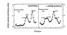
- Triton X-100 (1 % w/v) solubilized AChE were incubated overnight at 4°C without (see left panel of Fig 6) or with (see right panel of Fig 6) MA3-042 (dilution 1:50 by vol.).
- AChE isoforms were separated by centrifugation on 5-20% sucrose gradients made in 50 mM Tris saline buffer pH 7.4 containing 0.5% Triton X-100. The tube was centrifuged at 150,000 xg at 4°C, fractions were collected from the bottom and assayed for AChE activity. Sedimentation markers were catalase (11.4S) and alkaline phosphatase (6.1S).
- Blood is collected and 1 ml of plasma or serum prepared using standard techniques.
- the fluid is passed across a 5 ml RCA-Agarose (RCA stands for ricinus communis agglutinin) to remove butyrylcholinesterase and the amount of acetylcholinesterase activity eluting from the column is monitored using the Ellman assay and the peak 2 ml of activity collected.
- This material would then be incubated for 10 min at ambient temperature with 50 micromolar iso-OMPA to inhibit the remaining butyrylcholinesterase, then passed across a 1 ml column of MAb MA3-042 coupled to Sepharose to remove non-specific AChE isoforms.
- the amount of activity eluting from the column is assayed using the Ellman assay.
- the amount of activity present in this fraction is greater in AD cases than in non-AD cases. There is normally less that about 40 mUnits of AChE / ml of original plasma or serum.
- the present invention provides a diagnostic test for Alzheimer's disease.
Abstract
Description
- The present invention is concerned with a diagnostic test for Alzheimer's disease.
- Alzheimer's disease (AD) is a common progressive dementia involving loss of memory and higher cognitive function. The disease is characterized by the presence of amyloid deposits in the brains of sufferers. These deposits are found both extracellularly (amyloid plaques) and intracellularly (neurofibrillary tangles). The principal constituent of amyloid plaques is the amyloid protein (Aβ) which is produced by proteolytic cleavage for the amyloid protein precursor (APP) (Evin et al., 1994). The principal constituent of neurofibrillary tangles is the cytoskeletal protein tau (Kosik, 1992).
- One of the characteristic neurochemical changes observed in AD is the loss of acetylcholinesterase (AChE) and choline acetyltransferase activity in regions of the brain such as the cortex, hippocampus, amygdala and nucleus basalis (Whitehouse et al., 1981, 1982; Struble et al., 1982; Mesulam and Geula, 1988). The loss of cholinergic structure and markers correlates with the number of plaque and tangle lesions present, as well as with the clinical severity of the disease (Perry et al., 1978; Wilcock et al., 1982; Neary et al., 1986; Perry, 1986).
- Accurate diagnosis of AD during life is essential. However, clinical evaluation is at best only about 80% accurate. Therefore, there is a need to identify specific biochemical markers of AD. So far, analysis of blood or cerebrospinal fluid (CSF) has not yielded a biochemical marker of sufficient diagnostic value (Blass et al., 1998), although detectable differences are reported in the levels of certain proteins (Motter et al., 1995).
- The assay of levels of AChE activity in the blood and the cerebrospinal fluid (CSF) has been proposed as an ante mortem diagnostic test for AD. However, no consensus has been reached as to whether the levels of AChE are consistently affected in these tissues. The level of serum or plasma AChE has been reported to be increased (Perry et al., 1982; Atack et al., 1985), decreased (Nakano et al., 1986; Yamamoto et al., 1990) or unchanged (St. Clair et al., 1986; Sirvio et al., 1989) in AD patients. The level of erythrocyte AChE has been reported as either unaffected (Atack et al., 1985; Perry et al., 1982) or decreased (Chipperfield et al., 1981). The level of AChE activity in the CSF of AD patients has been reported to be decreased (most recently by Appleyard and McDonald, 1992; Shen et al., 1993) or unchanged (most recently by Appleyard et al., 1987; Ruberg et al., 1987).
- AChE has been shown to exist as up to six different molecular isoforms, three of which are the monomeric (G1), dimeric (G2) and tetrameric (G4) isoforms (Massoulié et al., 1993). The relative proportion of the different isoforms of AChE are markedly affected in AD, with a decrease in the G4 isoform in the parietal cortex (Atack et al., 1983), and an increase in the G1 isoform (Arendt et al., 1992). Similar changes have been identified in other AD brain regions including Brodman
areas - However, to date changes in AChE expression and isoform distribution have not been found to be of sufficient sensitivity or specificity to be useful diagnostic markers of AD.
- An anomalous isoform of AChE, distinguished by its isoelectric point, has been detected in the CSF of AD patients (Navaratnam et al., 1991; Smith et al., 1991), and a method for screening for AD based on these findings is described in US patent number 5,200,324. The method comprises determining, by means of isoelectric focusing, if a patient has an anomalous form of AChE in his CSF. However, the isoform detected by Navaratnam et al and Smith et al has also been detected in the CSF of patients with other neurological diseases (Shen and Zhang, 1993). Indeed, this is suggested in US patent number 5,200,324 at
column 7 lines 19-22, where it is stated that the anomalous AChE "was present in the CSF of four out of eight patients with a clinical diagnosis of possible dementia, but who did not satisfy strict histopathological criteria for Alzheimer's disease". - Moreover, the passage at
column 7 lines 60-61 of the US patent indicates that the detection of AChE - AD in lumbar CSF depends upon the amount of CSF analysed, andcolumn 8 lines 38-40 state that the anomalous band was often rather faint and the gels run were not always ideal. Accordingly, a loading of 5 mU per track was adopted as a standard procedure for screening CSF for the presence of the anomalous form of AChE, and each gel was read independently by four individuals who recorded their interpretation. Thus, there are technical problems associated with the assay described which can only be overcome by adopting an arbitrary set of conditions to avoid false readings, which then makes interpretation of the results difficult. - The suggestion that the anomalous form of AChE detected by Navaratnam et al and Smith et al is not unique to AD patients, together with the technical problems associated with the assay described in US patent number 5,200,324 suggests that the abnormal electroform of AChE discovered by Navaratnam et al and Smith et al will not form the basis of a diagnostic test for AD suitable for clinical use.
- Mimori et al (Behavioural Brain Research 83, 25-30 (1997)) describes the effects of certain inhibitors on acetylcholinesterase in Alzheimer's disease. Tornel et al (Neuroscience Letters 145, 59-62 (1992)) describes lectin binding properties of AChE. Michaelson et al (Brain Research 611, 75-80 (1993)) describes the forms of AChE present in fetal brain serum.
- There remains a need for a diagnostic test for AD based on a biochemical analysis of body fluids such as blood or CSF and the present invention provides such a test on the basis that AChE of AD patients shows a different glycosylation pattern to the AChE of non-AD groups.
- According to a first aspect of the present invention there is provided a method for the diagnosis of Alzheimer's disease in a patient, comprising detecting in vitro the presence of acetylcholinesterase in a sample of cerebrospinal fluid from a patient, wherein said AChE has a relatively lesser affinity for Concanavalin A and a relatively greater affinity for wheat germ agglutinin than non-AD AChE.
- In one embodiment of the invention the relative proportion of AChE with a first glycosylation pattern and AChE with a second glycosylation pattern is measured.
- Measurement of the relative proportions of AChE with first and second glycosylation patterns may be carried out in any convenient manner, for example, by using biochemical analysis techniques such as HPLC and mass spectrometry, or immunological techniques such as ELISA or, assays. However, a particularly preferred means of measuring the relative proportions of the isoforms of AChE involves a lectin-binding analysis.
- It has been established that approximately 75-95% of the AChE in the CSF of AD patients binds to Concanavalin (Con A) or wheat germ agglutinin (WGA) but with different specificity to each. Accordingly, in a particularly preferred embodiment of the invention, in order to identify the glycosylation pattern of AChE in the sample, the binding to Con A is determined, then the binding to WGA is determined, and a ratio calculated. The ratio is characteristic of the glycosylation pattern. It is particularly convenient to measure the activity of unbound AChE in each experiment, hence the ratio of AChE unbound to Con A to the ratio of AChE unbound to WGA is determined. This ratio is referred to hereinafter as a C/W ratio. For patients with AD, the C/W ratio has generally been found to be above 0.95 whereas for non-sufferers of AD the C/W ratio is typically below 0.95.
- Advantageously, the total AChE activity is measured and the C/W ratio plotted against AChE activity.
- In an alternative embodiment of the invention there is provided a monoclonal antibody specific for AChE with an altered glycosylation pattern is used to detect its presence. Typically the monoclonal antibody is MA3-042 (clone HR2), available from Chemicon International Inc of Temecula, California. Other suitable monoclonal antibodies may be used, for example, MA304 (clone AE1) also available from Chemicon International Inc.
- While not wishing to be bound by theory, it is believed that the abnormal isoform is the amphiphilic, monomeric isoform of AChE and/or the amphiphilic, dimeric isoform of AChE.
- The body fluid analysed in the method of the invention is cerebrospinal fluid (CSF).
- An abnormal isoform of the acetylcholinesterase (AChE) with an altered pattern of glycosylation, being the amphiphilic, monomeric isoform of AChE has a relatively lesser affinity for Concanavalin (Con A) and a relatively greater affinity for wheat germ agglutinin (WGA) than AChE with an unaltered glycosylation pattern.
- An abnormal isoform of acetylcholinesterase (AChE) with an altered glycosylation pattern, being the amphiphilic, dimeric isoform of AChE has a relatively lesser affinity for Concanavalin A (Con A) and a relatively greater affinity for wheat germ agglutinin (WGA) than AChE with an unaltered glycosylation pattern.
-
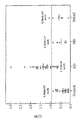
- Figure 1 is a plot of the C/W ratio for a number of patients in a control group, patients with Alzheimer's disease (AD), patients with other neurological disorders distinct from Alzheimer's disease (AD) and patients with non-Alzheimer's disease type dementia (DNAT). Circles represent ventricular CSF; triangles represent lumbar CSF; open symbols =≥ 60 years old; black symbols =≤ 60 years old. Mean values are expressed ± S.E.M. * = significantly different from AD (P<0.001). The experiment is described in Example 1.
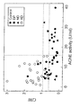
- Figure 2 is a plot of C/W ratio vs. AChE activity in post mortem human CSF. Dashed lines show values of C/W and AChE activity which maximally discriminate between AD and non-AD groups. Approximately 80% of all AD samples were above a cutoff value of C/W = 0.95, whereas all AD samples were above C/W = 0.60. Similarly all AD samples had less than 15.8 U/ml of AChE activity. The experiment is described in Example 2.
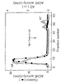
- Figure 3 shows AChE activity vs fraction number for hydrophobic interaction chromatography of CSF AChE on phenyl-agarose. Samples of CSF from AD patients (open circles) or controls (closed circles) were applied to 10 ml columns of phenyl-agarose. Hydrophilic AChE isoforms (HF) were eluted with 50 mM Tris-saline buffer and then bound amphiphilic isoforms (AF) were eluted with 50 mM Tris-HCl (TB) (pH 7.4) containing 2% (w/v) Triton X-100. Fractions of 1.4 ml were collected and assayed for AChE activity.
- Figure 4 is an analysis of AChE isoforms and glycosylation in AD and control CSF. A hydrophilic fraction (HF) and an amphiphilic fraction (AF) were obtained from a total CSF fraction by hydrophobic interaction chromatography (Fig. 2). The C/W ratio in the total CSF, HF and AF fractions was determined, and then fractions were applied to 5-20% sucrose density gradients containing 0.5% (w/v) Brij 97 and centrifuged at 150,000xg for 18 hr. Fractions from the sucrose gradient were collected and assayed for AChE activity. Enzymes of known sedimentation coefficient, catalase (C, 11.4S) and alkaline phosphatase (P, 6.1S) were used to determine the approximate sedimentation coefficients of AChE isoforms.
- Figure 5 is an analysis of AChE isoforms and glycosylation in frontal cortex and cerebellum from controls, non-demented individuals with diffuse plaques (DP) and AD patients. Samples of brain were homogenised and extracted to obtain SS and TS fractions. Equal volumes of SS and TS fractions were mixed and applied to 5-20% sucrose density gradients containing 0.5% (w/v) Brij 97 and centrifuged at 150,000×g for 18 hr. Fractions were collected and assayed for AChE activity. Individual AChE isoforms were identified by its coefficient of sedimentation using enzyme markers: catalase (C, 11.4S) and alkaline phosphatase (P, 6.1S). The enzyme peaks of G4 and G2+G1 AChE were selected, concentrated and dialysed to remove sucrose. The major G4 and G2+G1 peaks were then analysed by lectin binding using Con A and WGA and the C/W ratio determined from each peak.
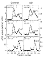
- Figure 6 shows the effect of monoclonal antibody MA3-042 on the sedimentation velocity of AChE isoforms from human frontal cortex, as described in Example 3.
- Abbreviations used:
- AChE, acetylcholinesterase; ChE, cholinesterase; Aβ, amyloid β protein; AD, Alzheimer's disease; DP, diffuse plaques; ND, other neurological diseases; PMI, post mortem interval; PBS, phosphate-saline buffer; TB, Tris buffer; TSB, Tris-saline buffer ; SS, salt-soluble supernatant; TS, Triton X-100-soluble supernatant; AF, amphiphilic fraction; HF, hydrophilic fraction; Ga, globular amphiphilic isoform; Gna, globular non-amphiphilic isoform; and agglutinins from Canavalia ensiformis (Concanavalin A), Con A; Triticum vulgaris (wheat germ), WGA; Ricinus communis, RCA120; Lens culinaris, LCA; Dolichus biflorus, DBA; Ulex europaeus, UEAI; Glycine max, SBA; and Arachis hypogaea, PNA.
- Immobilised lectins (Con A- and LCA-Sepharose, WGA-, RCA120-, DBA-, UEAI-, SBA and PNA-agarose), phenyl-agarose, bovine liver catalase, E. coli alkaline phosphatase, polyoxyethylene-10-oleyl ether (Brij 97), Triton X-100 , tetraisopropyl pyrophosphoramide (iso-OMPA), 1,5-bis(4-allydimethyl-ammoniumphenyl)-pentan-3-1 dibromide (BW284c51), acetylthiocholine iodide and 5,5'-dithio-bis-2-nitrobenzoic acid (DTNB) were all obtained from Sigma-Aldrich Pty. Ltd. (Seven Hills, NSW, Australia). Sepharose CL-4B was purchased from Pharmacia Biotech AB (Uppsala, Sweden).
- Lumbar or ventricular CSF was obtained post mortem; 18 controls with no clinical or pathological dementia and no clinical or pathological dementia and no evidence of brain pathology, 27 cases of AD, 7 cases of dementia non-AD type (DNAT, 5 frontal lobe dementia, 1 Lewy body dementia/Parkinson's disease and 1 multi-infarct dementia/congophilic amyloid angiopathy), and 6 cases of other neurological disorders (ND, 4 Huntington's disease, 1 schizophrenia and 1 corticobasal degeneration). The average age in the control group was 68±4years, there were 10 females and 8males and the PMI was 40±6. In the AD group the age was 81±2 years, there were 13 female and 14 males and the PMI was 35±6. In the ND group the age was 65±6, there were 3 females and 3 males and the PMI was 45±12. In the DNAT group the age was 76 ±3, there were 4 female and 3 males and the PMI was 34±11. Samples of CSF were stored at -70°C and centrifuged at 1,000 xg for 15 min prior to analysis. AChE activity was assayed at 22°C by a modified microassay of the Ellman method (Ellman et al 1961). Aliquots (0.3 ml) were mixed with 0.1 ml of Sepharose 4B in PBS (control), concanavalin A (Con A) or wheat germ agglutinin (WGA, Triticum vulgaris) immobilised on Sepharose. The enzyme-lectin mixture was incubated overnight at 4°C, and then centrifuged (1,000 xg, 15 min). AChE activity was assayed in the supernatant fractions. Data were analysed using a Student's t-test.
- The total AChE values in ventricular CSF samples of subjects ≥60 yrs old were significantly lower in the AD group (6.98±0.82 nmol/min/ml) than in controls (17.24±4.28 nmol/min/ml; P <0.001). However, as reported previously, (Appleyard et al., 1983), the large overlap (40%) between the data prevents the use of total AChE as a significant diagnostic marker.
- However, lectin-binding analysis revealed a significant difference between the AD group and controls. Approximately 75-95% of the AChE in the CSFs bound to Con A or WGA. A ratio (C/W ratio) was defined as AChE unbound to Con A divided by AChE unbound to WGA. The mean C/W ratio for the AD group was significantly different from controls (Figure 1). Of the 27 CSFs from confirmed AD, 21 samples had a C/W ratio >0.95. All 18 control samples had C/W <0.95, without significant differences between younger (n=5, C/W=0.37±0.10) and older subjects (n=6, 0.38±0.08) samples. No correlation in C/W ratio was noted with post mortem interval (PMI). The data are represented graphically in Figure 1.
- The data indicate that lectin-binding analysis of CSF AChE could provide a diagnostic test for AD which is 80% sensitive and 97% specific. Thus it was proposed that differences observed in the glycosylation pattern of AChE in CSF may be useful as an ante mortem diagnostic marker for AD, particularly when used in combination with measurement of other biochemical markers.
- Ventricular and lumbar CSF, frontal cortical and cerebellar samples were obtained post mortem and stored at -80°C. Three non-AD groups of samples were defined, 1) controls with no clinical or pathological features of dementia (n = 18), 2) individuals who showed no clinical signs of dementia but who were found to have a moderate number of non-neuritic Ab-immunoreactive diffuse plaques (DP), but no evidence of neocortical neurofibrillary changes (n = 6), and 3) individuals with various neurological diseases (ND) containing 7 cases of non-AD type dementia (5 frontal lobe dementia, 1 Lewy body dementia and 1 vascular dementia) and 7 cases of other neurological disorders (4 Huntington's disease, 1 Parkinson's disease, 1 schizophrenia and 1 corticobasal degeneration). Cases of AD were selected on the basis of their clinical history of dementia and neuropathological CERAD diagnosis (Mirra et al., 1994). All the CSF samples included in the AD and ND groups were ventricular and only 5 control and 1 DP CSF samples (from a total of 18 and 6 subjects, respectively) were taken by lumbar puncture. Immunohistochemical examination of the cerebellar samples showed that, unlike the frontal cortex, none of the AD tissue possessed compact neuritic amyloid plaque deposition (data not shown), consistent with previous studies (Mann et al., 1996).
- It has been shown (Grassi et al., 1982; Fishman et al., 1986; Sáez-Valero et al., 1993) that for a post mortem interval (PMI) greater than 72 hr, storage at -20°C or repeated cycles of freeze-thawing caused degradation of AChE, which confounded glycosylation analysis. Therefore, only samples with a PMI of less than 72 hr (PMI = 36 ± 4 hr) were used. There was no significant difference in PMI between each group of samples.
- Samples of CSF were thawed slowly at 4°C and then centrifuged at 1,000×g for 15 min prior to use. Small pieces (0.5 g) of frontal cortex and cerebellum were thawed slowly at 4°C, weighed and homogenised (10% w/v) in ice-cold Tris-saline buffer (TSB; 50 mM Tris-HCl, 1 M NaCl, and 50 mM MgCl2, pH 7.4) containing a cocktail of proteinase inhibitors (Silman et al., 1978). Tissues were homogenised with a glass/Teflon homogeniser and then sonicated with 10-15 bursts at 50% intermittency at setting 4 using a Branson sonifier. The suspension was centrifuged at 100,000×g at 4ºC in a Beckman L8-80M ultracentrifuge using a 70.1 Ti rotor for 1 hr to recover a salt-soluble ChE fraction (SS). The pellet was re-extracted with an equal volume of TSB containing 1% (w/v) Triton X-100, and the suspension centrifuged at 100,000×g at 4°C for 1 hr to obtain a Triton X-100-soluble ChE fraction (TS). This double-extraction method recovered 80-90% of the total ChE activity (Sáez-Valero et al., 1993; Moral-Naranjo et al., 1996).
- AChE activity was determined by a modified microassay method of Ellman (Sáez-Valero et al., 1993). One unit of AChE activity was defined as the number of nmoles of acetylthiocholine hydrolysed per min at 22°C. Protein concentrations were determined using the bicinchoninic acid method with bovine serum albumin as standard (Smith et al., 1985).
- Amphiphilic AChE forms were separated from hydrophilic forms by hydrophobic interaction chromatography on phenyl-agarose as previously described (Sáez-Valero et al., 1993). CSF (10 ml-pooled from four samples obtained from four different subjects) was applied to a column (10×1 cm) of phenyl-agarose. A hydrophilic fraction (HF) containing hydrophilic isoforms of AChE was eluted with 30 ml of TSB, and then an amphiphilic fraction (AF) containing bound amphiphilic isoforms was eluted with 50 mM Tris-HCl (TB, pH 7.4) containing 2% (w/v) Triton X-100. Peak fractions with high AChE activity were pooled and concentrated using Ultrafree-4 Centrifugal
Filter Device Biomax 10 kDa concentrators (Millipore Corporation, Bedford, MA, USA). - Molecular isoforms of AChE were analysed by ultracentrifugation at 150,000×g in a continuous sucrose gradient (5-20% w/v) for 18 hr at 4°C in a Beckman SW40 rotor. The gradients contained 10 ml of 50 mM Tris-HCl (pH 7.4) containing 0.5 M NaCl, 50 mM MgCl2 and 0.5% (w/v) Brij 97. Approximately 40 fractions were collected from the bottom of each tube. Enzymes of known sedimentation coefficient, bovine liver catalase (11.4S, S20,w, Svedberg Units) and E. coli alkaline phosphatase (6.1S) were used in the gradients to determine the approximate sedimentation coefficients of AChE isoforms. A ratio of AChE species G4/(G2+G1), that reflected the proportion of G4 molecules (G4 na+G4 a) versus both light globular AChE isoforms, G2 a and G1 a was defined. Estimation of the relative proportions of each molecular form of AChE was performed by adding the activities under each peak (G4 or G2+G1) and calculating the relative percentages (recovery >95%).
- Samples (0.3 ml) were added to 0.1 ml (hydrated volume) of Sepharose 4B (control), Con A, WGA, RCA120, LCA, DBA, UEAI, SBA or PNA immobilised in agarose or Sepharose. The enzyme-lectin mixture was incubated overnight at 4°C with gentle mixing. Bound and free AChE were separated by centrifugation at 1000×g for 15 min at 4°C in a Beckman J2-21M/E centrifuge using a JA-20 rotor, and the unbound AChE was assayed in the supernatant fraction. Percentage of unbound AChE in the lectin incubation was calculated as (AChE unbound to lectin / AChE unbound to Sepharose) × 100. The C/W ratio was calculated according to the formula, AChE activity unbound in the Con A incubation divided by the AChE activity unbound in the WGA incubation. It was observed that this ratio detects a specific alteration in AChE glycosylation that occurs in AD CSF.
- To examine the glycosylation of AChE, CSF samples from 18 controls and 30 cases of AD were incubated with different immobilised lectins, which recognise different sugars. AChE bound strongly to Con A, WGA and LCA but weakly to RCA120, PNA, DBA, UEAI and SBA (Table 1), suggesting that most of the enzyme was devoid of terminal galactose, terminal N-acetyl-galactosamine or fucose.
- There was a small but significant difference in the binding of AChE to Con A and WGA between the AD group and controls (Table 1). As the percentage of AChE unbound in the AD CSF was increased for Con A and decreased for WGA, a ratio (C/W = [% AChE that does not bind to Con A] / [% AChE that does not bind to WGA]) was defined, which provided greater discrimination between the two groups (Table 1). Using this method, it was found that the mean C/W ratio for the AD group was significantly greater than for the other control groups, including cases with diffuse plaques (non-demented, DP), and patients with other neurological and neuropsychiatric diseases (ND) (Fig. 2), consistent with the results shown in Example 1. Of the 30 CSF samples from confirmed AD cases, 24 samples were above a cut-off value of C/W = 0.95 (Fig. 2). Only one sample from 18 controls, one out of 6 samples from cases with diffuse plaques, and one out of 14 samples from the other neurological diseases group, a frontal lobe dementia case, were above this value. The 6 AD samples with C/W ratios lower than 0.95 had C/W ratios > 0.60, a value higher than the C/W mean of the non-AD groups (control = 0.53 ± 0.1; DP = 0.46 ± 0.2; ND = 0.53 ± 0.1).
- No correlation could be found between the C/W ratio and the PMI that could suggest that different C/W ratio in the AD group was due to differences in PMI. Furthermore, there was no significant difference in the PMI between the AD (33 ± 6 hr) and non-AD samples (40 ± 6 hr).
- CSF samples were additionally analysed for total AChE activity (Fig. 2). As previously reported (Appleyard et al., 1983; Atack et al., 1988), the CSF from patients with AD had significantly lower AChE activity (6.5 ± 0.8 U/ml) than controls (15.8 ± 2.9 U/ml) or patients with other diseases (12.4 ± 2.4 U/ml). However, the C/W ratio was a more reliable index of clinical status than the total level of AChE activity in the CSF (Fig. 2).
- To determine whether the alteration in glycosylation was due to changes in a specific isoform of AChE, CSF samples were analysed by hydrophobic interaction chromatography to separate amphiphilic (Ga) and hydrophilic species (Gna) (Fig. 3), and by sucrose density gradient centrifugation in 0.5% (w/v) Brij 97 to separate individual molecular weight isoforms (G4, G2 and G1) (Fig. 3). A decrease in the proportion of G4 AChE in AD CSF compared to controls (Fig. 4, top panels) was observed. The ratio of (G4/(G2+G1) was significantly (P < 0.01) higher in controls (1.80 ± 0.12; n = 4) than in AD cases (1.16 ± 0.12; n = 4). To separate hydrophilic isoforms from amphiphilic isoforms, CSF was fractionated by hydrophobic interaction chromatography on phenyl-agarose (Fig. 3). A smaller percentage of AChE in the normal CSF bound to phenyl-agarose (12 ± 3 %, n = 4) than in the AD CSF (38 ± 4%, n = 4; P < 0.001). Sedimentation analysis of the unbound hydrophilic fraction (HF) showed a main peak of 10.8S, consistent with a hydrophilic tetrameric (G4 na) isoform (Atack et al., 1987), as well as a small amount of lighter AChE isoforms, 5.1S dimers and 4.3S monomers (Fig. 4). The bound amphiphilic fraction from the phenyl-agarose column contained a minor peak of 9.0-9.5S (probably an amphiphilic tetramer, G4 a) and a major peak of amphiphilic globular dimer (G2 a, 4.2S) and monomer (G1 a, 3.1S). The level of the amphiphilic light isoforms was greater in the AD CSF than in controls (Fig. 4).
- Incubation of the HF and AF with immobilised Con A and WGA showed that there was an increase in the C/W ratio in AD CSF, and that the high C/W ratio was associated with an amphiphilic fraction containing dimers and monomers (Fig. 4). The data indicate that the contribution of G2 and G1 AChE in AD CSF was mainly responsible for the increased C/W ratio of total AChE in the AD CSF.
- To determine whether the changes in AChE glycosylation reflect a change in the expression or glycosylation of brain AChE isoforms, the levels of AChE activity in samples of frontal cortex and cerebellum were examined. Samples were homogenised with salt and Triton X-100 to extract soluble and membrane-bound AChE isoforms, and then the AChE activity determined in both fractions (Table 2). The frontal cortex samples from AD patients had significantly less AChE activity in the Triton X-100-soluble (TS) fraction (~40%), with no difference in levels in the salt-soluble (SS) fraction compared with controls (Table 3). The results are consistent with previous studies that indicate that the major G4 isoform is decreased only in the TS fraction (Younkin et al., 1986; Siek et al., 1990). A small but significant decrease (~15%) in the protein content of the TS fraction of both AD and ND groups was also observed. The level of AChE in the frontal cortex samples of the ND group was significantly different from controls in both the SS and TS fraction (Table 2). However, as the ND group was heterogeneous (2 frontal lobe dementia, 1 Huntington's disease and 1 Parkinson's disease), the significance of changes in AChE levels is unclear. Levels of AChE in cerebellum were also significantly decreased in the TS fraction from the AD group (Table 2).
- To determine whether different glycosylation pattern of AChE in AD CSF is also present in the AD brain, the glycosylation of brain AChE was examined by lectin binding. Homogenates from frontal cortex and cerebellum were incubated with immobilised Con A or WGA and the amount of activity unbound was calculated. In the AD frontal cortex, the % AChE activity that did not bind to Con A or WGA was significantly different from controls (Table 3). Similar to the CSF AChE, the C/W ratio of frontal cortex AChE was greater in AD than in non-AD samples (Table 3). This increase was due to a large increase in the amount of AChE that did not bind to Con A, and was in spite of an increase in the amount of AChE that did not bind to WGA (Table 3). There was no increase in the C/W ratio in the DP and ND group (Table 3). No difference in lectin binding was observed between AD and non-AD groups in the cerebellar fractions (Table 3).
- To determine the cause of the altered glycosylation in AD brain, the pattern of AChE isoforms in the frontal cortex and cerebellum was examined. Equal volumes of SS and ST supernatants (total AChE activity) were pooled and then analysed by sucrose density gradient sedimentation with 0.5% (w/v) Brij 97 to separate the major AChE isoforms (Fig. 5). Based on their sedimentation coefficients (Atack et al., 1986; Massoulié et al., 1982) it was possible to identify hydrophilic (G4 na, 10.7 ± 0.1S) and amphiphilic tetramers (G4 a, 8.6 ± 0.1S), amphiphilic dimers (G2 a, 4.7 ± 0.1S) and monomers (G1 a, 3.0 ± 0.1S) of AChE (Fig. 6). There were no differences in the sedimentation coefficient (S) of individual isoforms from each group. Due to the overlap in the sedimentation coefficients between AChE G4 na and G4 a, it was not possible to separate these isoforms completely (Fig. 5). However, the contribution of G4 a was greater than G4 na. Asymmetric (A12) AChE isoforms were identified in trace amounts (2-5%) in some of the fractions.
- A significant decrease in G4 (40% of the mean control value, P < 0.001) and in G2+G1 AChE (60% of the mean control value, P = 0.002) was detected in the fractions from AD frontal cortex. This change in the relative proportion of AChE isoforms was reflected in the G4/(G2+G1) ratio, which was significantly lower in the AD samples (Table 3). Interestingly, a similar and statistically significant decrease was found in the G4/(G2+G1) ratio for the DP subjects. This change in ratio was due to a 25% increase in the level of G2+G1 and a small decrease (10%) in G4 AChE, although neither change on its own was statistically significant. No variation in AChE G4/(G2+G1) was found in the AD cerebellum (Table 3), despite a statistically significant decrease (40%) in AChE in the TS fraction (Table 2) and in the total level of G4 AChE (G4 in controls = 380 ± 40 U/ml, G4 in ADs = 195 ± 70 U/ml; P = 0.008).
- Since it was found that the ratio of AChE was altered in the frontal cortex of AD patients, steps were taken to ascertain whether the increase in the C/W ratio of brain AChE was due to a change in glycosylation or in the expression of a specific isoform of AChE. Individual AChE isoforms were separated by sucrose gradient centrifugation and then fractions from the G4 or G2+G1 peaks were pooled, dialysed against TSB-Triton X-100 buffer and concentrated by ultrafiltration. AChE isoforms were then assayed by lectin binding and a C/W ratio calculated for each isoform (Fig. 5).
- No differences were observed in the C/W ratio of G4 AChE between the AD and non-AD groups (Fig. 5). However, in all frontal cortex samples the G2+G1 fraction possessed C/W ratios >1.00, demonstrating that G2 or G1 AChE is glycosylated differently from the G4 isoform. Moreover, the C/W ratio for G2+G1 AChE was higher in the AD group than controls or DP. Similarly, the C/W ratio of the amphiphilic fraction from CSF (containing predominantly G2+G1 AChE) was higher in the AD group than in controls (Fig. 3). There was no correlation between the G4/(G2+G1) ratio and the C/W ratio in the DP group in frontal cortex. In the cerebellum, no differences were observed in the C/W ratios of G4 AChE or G2+G1 AChE between AD and non-AD groups (Fig. 4). The G2+G1 fractions, from both AD and non-AD cerebellar groups, had a C/W < 0.50, in contrast to the same fraction from frontal cortex (C/W > 1.00) indicating differences in the pattern of glycosylation of G2+G1 AChE between both brain areas.
- This Example shows that AChE is glycosylated differently in the frontal cortex and CSF of AD patients compared with AChE from non-AD groups including patients with non AD-type dementias. This difference in glycosylation is due to an increase in the proportion of differentially glycosylated amphiphilic dimeric and monomeric AChE in the AD samples. The results suggest that the abnormally glycosylated AChE in AD CSF may be derived from the brain as a similar difference in glycosylation was also found in the frontal cortex of AD patients.
Table 1. Lectin-binding of AChE in CSF. Lectin AChE unbound (%) Control AD Con A 5.5 ± 0.8 10.1 ± 1.1b WGA 11.3 ± 1.7 7.0 ± 0.6b Con A / WGA (C/W) 0.53 ± 0.1 1.37 ± 0.1 a LCA 17.2 ± 4.2 15.0 ± 1.3 RCA120 74.1 ± 3.4 70.8 ± 2.7 SBA 83.0 ± 2.1 82.2 ± 1.9 UEAI 91.6 ± 2.2 87.6 ± 1.9 PNA 92.4 ± 1.7 92.3 ± 1.4 DBA 98.9 ± 0.8 95.8 ± 1.7 Table 2. AChE activity and protein levels in human frontal cortex and cerebellum Group / Source AChE activity (U/ml) Protein (mg/ml) SS TS SS TS Control Frontal Cortex (n= 11; 63±5 y; 7F/4M) 3.7 ± 0.4 15.1 ± 1.5 2.1 ± 0.1 2.4 ± 0.1 Cerebellum (n= 7; 66±5 y; 4F/3M) 64 ± 6 264 ± 25 2.5 ± 0.1 1.9 ± 0.1 DP Frontal Cortex (n= 6; 81±2 y; 4F/2M) 5.5 ± 0.9 12.7 ± 1.7 2.1 ± 0.1 2.2 ± 0.1 Cerebellum (n= 5; 81±3 y; 3F/2M) 49 ± 8 182 ± 46 2.6 ±0.1 1.9 ± 0.1 ND Frontal Cortex (n= 4; 67±9 y; 2F/2M) 5.4 ± 0.6a 9.3 ± 1.7b 2.1 ± 0.2 2.0 ± 0.1b Cerebellum (n= 2; 78±14 y; 1F/1M) 45 ± 8 160 ± 50 2.7 ± 0.2 2.3 ± 0.2 AD Frontal Cortex (n= 14; 73±3 y; 8F/6M) 3.7 ± 0.3 9.0 ± 0. 9a 2.1 ± 0.1 2.1 ± 0.1a Cerebellum (n= 7; 73±6 y; 5F/2M) 48 ± 12 160 ± 28b 2.6 ± 0.1 2.0 ±0.1 Table 3. Lectin binding and AChE isoforms in frontal cortex and cerebellum Group / Source Lectin binding AChE ratio AChE unbound to Con A (%) AChE unbound to WGA (%) C/W G4/(G2+G1) Control Frontal Cortex (n= 11; 63±5 y; 7F/4M) 6.9 ± 0.8 12.3 ± 1.2 0.56 ± 0.03 1.90 ± 0.14 Cerebellum (n= 7; 66±5 y; 4F/3M) 1.8 ± 0.1 10.7 ± 0.9 0.18 ± 0.02 3.02 ± 0.2 DP Frontal Cortex (n= 6; 81±2 y; 4F/2M) 7.4 ± 0.8 15.0 ± 1.0 0.50 ± 0.06 1.32 ± 0.12b Cerebellum (n= 5; 81±3 y; 3F/2M) 2.9 ± 0.7 12.2 ± 1.3 0.23 ± 0.05 2.18 ± 0.33 ND Frontal Cortex (n= 4; 67±9 y; 2F/2M) 7.0 ± 0.6 13.2 ± 1.2 0.47 ± 0.05 2.61 ± 0.73 Cerebellum (n= 2; 78±14 y; 1F/1m) 1.8 ± 0.2 10.1 ± 0.3 0.21 ± 0.10 2.50 ± 0.70 AD Frontal Cortex (n= 14; 73±3 y; 8F/6M) 13.1 ± 1.3a 19.7 ± 1.4a 0.66± 0.03b 1.34 ± 0.18b Cerebellum (n= 7; 73±6 y; 5F/2M) 2.4 ± 0.3 13.5 ± 2.3 0.19 ± 0.02 2.33 ± 0.49 - Samples of Triton X-100 (1 % w/v) solubilized AChE were incubated overnight at 4°C without (see left panel of Fig 6) or with (see right panel of Fig 6) MA3-042 (dilution 1:50 by vol.). AChE isoforms were separated by centrifugation on 5-20% sucrose gradients made in 50 mM Tris saline buffer pH 7.4 containing 0.5% Triton X-100. The tube was centrifuged at 150,000 xg at 4°C, fractions were collected from the bottom and assayed for AChE activity. Sedimentation markers were catalase (11.4S) and alkaline phosphatase (6.1S). As seen in Fig 6, all of the peaks shift in the presence of MA3-042, indicating binding of the monoclonal antibody to the particular isoform represented by each peak, except that a peak remains around 4S. The difference between 4.0S and 4.2S is statistically insignificant, suggesting that the 4.2S peak represents an isoform with a modified glycosylation pattern not recognised by MA3-042. As will be appreciated by those skilled in the art, this peak represents an AChE monomer, which has a molecular weight of about 70000 kDa.
- Blood is collected and 1 ml of plasma or serum prepared using standard techniques. The fluid is passed across a 5 ml RCA-Agarose (RCA stands for ricinus communis agglutinin) to remove butyrylcholinesterase and the amount of acetylcholinesterase activity eluting from the column is monitored using the Ellman assay and the
peak 2 ml of activity collected. This material would then be incubated for 10 min at ambient temperature with 50 micromolar iso-OMPA to inhibit the remaining butyrylcholinesterase, then passed across a 1 ml column of MAb MA3-042 coupled to Sepharose to remove non-specific AChE isoforms. The amount of activity eluting from the column is assayed using the Ellman assay. The amount of activity present in this fraction is greater in AD cases than in non-AD cases.
There is normally less that about 40 mUnits of AChE / ml of original plasma or serum. - The present invention provides a diagnostic test for Alzheimer's disease.
-
- Appleyard M. E. and McDonald B. (1992) Acetylcholinesterase and butyrylcholinesterase activities in cerebrospinal fluid from different levels of the neuraxis of patients with dementia of the Alzheimer type. J. Neurol. Neurosurg. Psychiat. 55, 1074-1078.
- Appleyard M. E., Smith A. D., Berman P., Wilcock G. K., Esiri M. M., Bowen D. M.. and Neary D. (1987) Cholinesterase activities in cerebrospinal fluid of patients with senile dementia of the Alzheimer Type. Brain 110, 1309-1322.
- Appleyard M. E., Smith A. D., Wilcock G. K. and Esiri M. M. Decreased CSF acetylcholinesterase activity in Alzheimer's disease. Lancet 1983; 20:452
- Arendt T., Bigl V., Walther F. and Sonntag M. (1984) Decreased ratio of CSF acetylcholinesterase to butyrylcholinesterase activity in Alzheimer's disease. Lancet i, 173.
- Arendt T., Bruckner M. K., Lange M. and Bigl V. (1992) Changes in acetylcholinesterase and butyrylcholinesterase in Alzheimer's disease resemble embryonic development - A study of molecular forms. Neurochem. Intl. 21, 381-396.
- Atack J. R., Perry E. K, Bonham, J. R., Candy, J. M., and Perry R. H. (1986) Molecular forms of acetylcholinesterase in the aged human central nervous system. J. Neurochem. 47, 267-267.
- Atack J. R., Perry E. K., Bonham J. R., and Perry R. H. (1987) Molecular forms of acetylcholinesterase and butyrylcholinesterase in human plasma and cerebrospinal fluid. J. Neurochem. 48, 1845-1850.
- Atack J. R., May C., Kaye J. A., Kay A. D., and Rapoport S. I. (1988) Cerebrospinal fluid cholinesterases in aging and in dementia of the Alzheimer type. Ann. Neurol. 23, 161-167.
- Atack J. R., Perry E. K., Bonham J. R., Perry R. H., Tomlinson B. E., Blessed G. and Fairbairn A. (1983) Molecular forms of acetylcholinesterase in senile demential of Alzheimer's type: selective loss of the intermediate (10S) form. Neurosci. Lett. 40, 199-204.
- Atack J. R., Perry E. K., Perry R. H., Wilson I. D., Bober M. J., Blessed G. and Tomlinson B. E. (1985) Blood acetyl- and butyrylcholinesterase in senile dementia of Alzheimer type. J. neurol. Sci. 70, 1-12.
- Blass J. P., Blennow K., Delacourte A., Frisoni G. B., Jefferies W. A., McRae A., Wisniewski H. M., Parshad R., Scinto L. F. M., Scheltens P., Riekkinen P. J., Swanwick G. R. J., Wahlund L.-O., Trojanowski J. Q., Winbland B., Ihara Y., et al. (1998) Consensus report of the Working Group on: "molecular and biochemical markers of Alzheimer's disease". Neurobiol. Aging 19, 109-116.
- Davies C. A., Mann D. M. A., Sumpter P. Q., and Yates P. O. (1987) A quantitative morphometric analysis of the neuronal and synaptic content of the frontal and temporal cortex in patients with Alzheimer's disease. J. Neurol. Sci. 78, 151-164.
- Ellman G. E., Courtney K. D., Andres Jr. V. and Featherstone R. M. (1961) A new and rapid colorimetric determination of acetylcholinesterase activity. Biochem. Pharmacol. 7, 88-95
- Fishman E. B., Siek G. C., MacCallum R. D., Bird E. D., Volicer L., and Marquis J. K. (1986) Distribution of the molecular forms of acetylcholinesterase in human brain, alterations in dementia of the Alzheimer type. Ann. Neurol. 19, 246-252.
- Friede R. L. (1965) Enzyme histochemical studies of senile plaques. J. Neuropathol. Exp. Neurol. 24, 477-491.
- Geula C., and Mesulam M.-M. (1989) Special properties of cholinesterases in the cerebral cortex of Alzheimer's disease. Brain Res. 498, 185-189.
- Grassi J., Vigny M., and Massoulié J. (1982) Molecular forms of acetylcholinesterase in bovine caudate nucleus and superior cervical ganglion: solubility properties and hydrophobic character. J. Neurochem. 38, 457-469.
- Guillozet A. L., Smiley J., Mash D. C. and Mesulam M.-M. (1997) Butyrylcholinesterase in the life cicle of amyloid plaques. Ann. Neurol. 42, 909-918.
- Hogan B., Costantini F., and Lacy E. (1986) Manipulating the Mouse Embryo, A laboratory manual, Cold Spring Habor, New York.
- Inestrosa N. C., Alvarez A., Perez C. A., Moreno R. D., Vicente M., Linker C., Casanueva O. I., Soto C., and Garrido J. (1996a) Acetylcholinesterase accelerates assembly of amyloid-b-peptides into Alzheimer's fibrils - possible role of the peripheral site of the enzyme. Neuron 16, 881-891.
- Inestrosa N. C., Alvarez A., and Calderon F. (1996b) Acetylcholinesterase is a senile plaque component that promotes assembly of amyloid beta-peptide into Alzheimer's filaments. Mol.
Psychiatry 1, 359-361. - Kang J., Lemaire H.-G., Unterbeck A., Salbaum J. M., Masters C. L., Grzeschik K.-H., Multhaup G., Beyreuther K., and Müller-Hill B. (1987) The precursor of Alzheimer's disease amyloid A4 protein resembles a cell-surface receptor. Nature 325, 733-736.
- Liao J., Heider H., Sun M.-C., and Brodbeck U. (1992) Different glycosylation in acetylcholinesterases from mammalian brain and erythrocytes. J. Neurochem. 58, 1230-1238.
- Luo Z., Fuentes M.-E., and Taylor P. (1994) Regulation of acetylcholinesterase mRNA stability by calcium during differentiation from myoblasts to myotubes. J. Biol. Chem. 269, 27216-27223.
- Mann D. M. A., Iwatsubo T., and Snowden J. S. (1996) Atypical amyloid (Ab) deposition in the cerebellum in Alzheimer's disease: an immunohistochemical study using end-specific Ab monoclonal antibodies. Acta Neuropathol. 91, 647-653.
- Massoulié J., and Bon S. (1982) The molecular forms of cholinesterase and acetylcholinesterase in vertebrates. Ann. Rev. Neurosci. 5, 57-106.
- Massoulié J., Pezzementi L., Bon S., Krejci E., and Vallette F.-M. (1993) Molecular and cellular biology of cholinesterases. Prog. Neurobiol. 41, 31-91.
- Masters C. L., Simms G., Weinman N. A., Multhaup G., McDonald B. L., and Beyreuther K. (1985) Amyloid plaque core protein in Alzheimer's disease and Down syndrome. Proc. Natl. Acad. Sci. USA 82, 4245-4249.
- Méflah K., Bernard S., and Massoulié J.(1984) Interactions with lectins indicate differences in the carbohydrate composition of the membrane-bound enzymes acetylcholinesterase and 5'-nucleotidase in different cell types. Biochimie 66, 59-69.
- Mesulam M.-M., Geula C., and Morán M. A. (1987) Anatomy of cholinesterase inhibition in Alzheimer's disease, effect of physostigmine and tetrahydroaminoacridine on plaques and tangles. Ann. Neurol. 22, 683-691.
- Michaelson S., and Small D. H. (1993) A protease is recovered with a dimeric form of acetylcholinesterase in fetal bovine serum. Brain Res. 611, 75-80.
- Mirra S. S., Gearing D. W., McKeel D. W., Crain B. J., Hughes J. P., Vanbelle G., Heyman A., Ball M. J., Clark A. W., Hansen L. A., Hedreen J. C., Joachim C. L., Kim R. C., Kirkpatrick J. B., Markesbery W. R., Davis D., Martinez A. J., Miller C. A., Moossy J., Morris J., Nochlin D., Perl D. P., Purohit D., Petito C. K., Rao G. R., et al. (1994) Interlaboratory comparison neuropathology assessments in Alzheimer's disease: A study of the Consortium to Establish a Registry of Alzheimer's Disease (CERAD). J. Neuropath. Exp. Neurol. 53, 303-315.
- Moral-Naranjo M. T., Cabezas-Herrera J., and Vidal C. J. (1996) Molecular forms of acetyl- and butyrylcholinesterase in normal and dystrophic mouse brain. J Neurosci. Res. 43, 224-234.
- Morin M. A., Mufson E. J., and Gómez-Ramos P. (1993) Colocalization of cholinesterases with b amyloid protein in aged and Alzheimer's brains. Acta Neuropathol. 85, 362-369.
- Motter R., Vigopelfrey C., Kholodenko D., Barbour R., Johnsonwood K., Galasko D., Chang L., Miller B., Clark C., Green R., Olson D., Southwick P., Wolfert R., Munroe B., Lieberburg I., Seubert P., and Schenk D. (1995) Reduction of b-amyloid peptide42 in the cerebrospinal fluid of patients with Alzheimer's disease. Ann. Neurol. 38, 643-648.
- Navaratnam D. S., Priddle J. D., McDonald B., Esiri M. M., Robinson J. R., and Smith A. D. (1991) Anomalous molecular form of acetylcholinesterase in cerebrospinal fluid in histologically diagnosed Alzheimer's disease. Lancet 337, 447-450.
- Pfeffer R. I., Afifi A. A., and Chance J. M. (1987) Prevalence of Alzheimer's disease in a retirement community. Am. J. Epidemiol. 125, 420-436.
- Probst A., Langui D., and Ulrich J. (1991) Alzheimer's disease: a description of the structural lesions. Brain Pathol. 1, 229-239.
- Sáez-Valero J., Tornel P. L., Munoz-Delgado E., and Vidal C. J. (1993) Amphiphilic and hydrophilic forms of acetyl- and butyrylcholinesterase in human brain. J. Neurosci. Res. 35, 678-689.
- Saez-Valero J., Sberna G., McLean C., Masters C. L., and Small D. H. (1997) Glycosylation of acetylcholinesterase as diagnostic marker for Alzheimer's disease. Lancet 350, 929.
- Saxena A., Raveh L., Ashani Y., and Doctor B. P. (1997) Structure of glycan moieties responsible for the extended circulatory life time of fetal bovine serum acetylcholinesterase and equine serum butyrylcholinesterase. Biochemistry 36, 7481-7489.
- Saxena A., Ashani Y., Raveh L., Stevenson D., Patel T., and Doctor B. P. (1998) Role of oligosaccharides in the pharmacokinetics of tissue-derived and genetically engineered cholinesterases. Mol. Pharmacol. 53, 112-122.
- Sberna G., Sáez-Valero J., Beyreuther K., Masters C. L., and Small D.H. (1997) The amyloid b-protein of Alzheimer's disease increases acetylcholinesterase expression by increasing intracellular calcium in embryonal carcinoma P19 cells. J. Neurochem. 69, 1177-1184.
- Sberna G., Sáez-Valero J., Li Q.X., Czech C., Beyreuther K., Masters C. L., McLean, C. A., and Small D.H. (1998) Acetylcholinesterase is increased in the brains of transgenic mice expressing the C-terminal fragment (CT100) of the b-amyloid protein precursor of Alzheimer's disease. J. Neurochem. 71, 723-731.
- Schegg K. M., Harrington L. S., Nielsen S., Zwieg R. M., and Peacock J. H. (1992) Soluble and membrane-bound forms of brain acetylcholinesterase in Alzheimer's disease. Neurobiol. Aging 13, 697-704.
- Schoenberg B. S., Kokmen E., and Okazaki H. (1987) Alzheimer's disease and other dementing illnesses in a defined United States population: incidence rates and clinical features. Ann. Neurol. 22, 724-729.
- Shen Z.-X., and Zhang Z. (1993) Anomalous acetylcholinesterase in CSF without clinical diagnosis of Alzheimer's disease. Lancet 342, 62.
- Shen Z.-X. (1997) An CSF anomalous molecular form of acetylcholinesterase in demented and non-demented subject.
Neuroreport 8, 3229-3232. - Siek G. C., Katz L. S., Fishman E. B., Korosi T. S., and Marquis J. K. (1990) Molecular forms of acetylcholinesterase in subcortical areas of normal and Alzheimer disease brain. Biol Psychiatry 27, 573-580.
- Silman I., Lyles J. M., and Barnard E. A. (1978) Intrinsic forms of acetylcholinesterase in skeletal muscle. FEBS Letters 94, 166-170.
- Small D. H., Michaelson S., and Sberna G. (1996) Non-classical actions of cholinesterases: role in cellular differentiation, tumorigenesis and Alzheimer's disease. Neurochem. Intl. 28, 453-483.
- Smith P. K., Krohn R. I., Hermanson G. T., Mallia A. K., Gartner F. H., Provenzano M. D., Fujimoto E. K., Goeke N. M., Olson B. J., and Klenk D.C. (1985) Measurement of protein using bicinchoninic acid. Anal. Biochem. 150, 76-85.
- Smith A. D., Jobst K. A., Navaratnam D. S., Shen Z.-X., Priddle J. D., McDonald B., King E., and Esiri M. M.(1991) Anomalous acetylcholinesterase in lumbar CSF in Alzheimer's disease. Lancet 338, 1538.
- Soreq H., Ben-Aziz R., Prody C. A., Seidman S., Gnatt A., Neville L., Lieman-Hurwitz J., Lev-Lehman E., Ginzberg D., Lapidot-Lifson Y., and Zakut H. (1990) Molecular cloning and construction of the coding region for human acetylcholinesterase reveals a G+C-rich attenuating structure. Proc. Natl. Acad. Sci. USA 87, 9688-9692.
- Treskatis S., Christoph E., and Layer P. G. (1992) Butyrylcholinesterase from chicken brain is smaller than that from serum: its purification, glycosylation and membrane association. J. Neurochem. 58, 2236-2247.
- Ulrich J., Meier-Ruge W., Probst A., Meier E., and Ipsen S. (1990) Senile plaques, staining for acetylcholinesterase and A4 protein, a comparative study in the hippocampus and entorhinal cortex. Acta Neuropathol 80, 624-628.
- Vidal C. J. (1996) Glycosylation of cholinesterases and its alteration in some pathological states. Recent Res. Devel. Neurochem. 1, 37-54.
- Wright C. I., Geula C., and Mesulam M.-M. (1993) Neuroglial cholinesterases in the normal brain and in Alzheimer's disease, relationship to plaques, tangles and patterns of selective vulnerability. Ann. Neurol. 34, 373-384.
- Younkin S. G., Goodridge B., Katz J., Lockett G., Nafziger D., Usiak M. F., and Younkin L.H. (1986) Molecular forms of acetylcholinesterase in Alzheimer's disease. Fed. Proc. 45, 2982-2988.
Claims (8)
- An in vitro method for the diagnosis of Alzheimer's disease (AD) in a patient, comprising detecting in vitro the presence of acetylcholinesterase (AChE) in a sample of cerebrospinal fluid (CSF) from a patient, wherein said AChE has a relatively lesser affinity for Concanavalin A (Con A) and a relatively greater affinity for wheat germ agglutinin (WGA) than non-AD AChE.
- The method of claim 1 wherein(1) the binding to Concanavalin A (Con A) and wheat germ agglutinin (WGA)is measured by lectin binding analysis;(2) the ratio of AChE unbound to Con A to AChE unbound to WGA is calculated; and wherein(3) said ratio is above 0.95 in AD patients.
- The method of any one of claim 1 or claim 2 wherein the total AChE activity is also determined.
- The method of any one of claims 1 to 3 wherein the ratio of AChE unbound to Con A to AChE unbound to WGA is plotted against total AChE activity.
- The method of claim 1 or claim 2 wherein a monoclonal antibody is used to detect the presence of said AChE.
- The method of claim 5 wherein the monoclonal antibody is MA3-042 and said AChE is detected by its failure to bind.
- The method according to claim 1 wherein said detection comprises subjecting a CSF sample to lectin binding analysis to determine the amount of AChE unbound to Concanavalin A, wherein if the level of AChE unbound to Con A is from 9.0% to 11.1%, this is indicative of the presence of AChE having a relatively lesser affinity for Concanavalin A (Con A) and a relatively greater affinity for wheat germ agglutinin (WGA) than non-AD AChE.
- The method according to claim 1 wherein said detection comprises subjecting a CSF sample to lectin binding analysis to determine the amount of AChE unbound to wheat germ agglutinin (WGA), wherein if the level of AChE unbound to WGA is from 6.4% to 7.6% then this is indicative of the presence of AChE having a relatively lesser affinity for Concanavalin A (Con A) and a relatively greater affinity for wheat germ agglutinin (WGA) than non-AD AChE.
Applications Claiming Priority (3)
| Application Number | Priority Date | Filing Date | Title |
|---|---|---|---|
| AUPO943297 | 1997-09-24 | ||
| AUPO9432A AUPO943297A0 (en) | 1997-09-24 | 1997-09-24 | Diagnostic test for alzheimer's disease |
| PCT/AU1998/000809 WO1999015695A1 (en) | 1997-09-24 | 1998-09-24 | Diagnostic test for alzheimer's disease |
Publications (3)
| Publication Number | Publication Date |
|---|---|
| EP1017845A1 EP1017845A1 (en) | 2000-07-12 |
| EP1017845A4 EP1017845A4 (en) | 2003-07-02 |
| EP1017845B1 true EP1017845B1 (en) | 2006-04-19 |
Family
ID=3803714
Family Applications (1)
| Application Number | Title | Priority Date | Filing Date |
|---|---|---|---|
| EP98946146A Expired - Lifetime EP1017845B1 (en) | 1997-09-24 | 1998-09-24 | Diagnostic test for alzheimer's disease |
Country Status (11)
| Country | Link |
|---|---|
| US (1) | US6461831B1 (en) |
| EP (1) | EP1017845B1 (en) |
| JP (1) | JP2001517456A (en) |
| AT (1) | ATE323781T1 (en) |
| AU (1) | AUPO943297A0 (en) |
| BR (1) | BR9813015A (en) |
| CA (1) | CA2304949A1 (en) |
| DE (1) | DE69834263D1 (en) |
| HU (1) | HUP0003684A3 (en) |
| IL (1) | IL135211A0 (en) |
| WO (1) | WO1999015695A1 (en) |
Families Citing this family (15)
| Publication number | Priority date | Publication date | Assignee | Title |
|---|---|---|---|---|
| IL130225A0 (en) | 1999-05-31 | 2000-06-01 | Yissum Res Dev Co | Novel uses of antibodies against ache and peptides thereof |
| US20020150878A1 (en) * | 2001-01-23 | 2002-10-17 | Small David Henry | Method for the diagnosis of Alzheimer's Disease and other prion related disorders |
| CA2510250A1 (en) * | 2002-12-20 | 2004-08-12 | Momenta Pharmaceuticals, Inc. | Glycan markers for diagnosing and monitoring disease |
| GB2404734A (en) * | 2003-08-05 | 2005-02-09 | Secr Defence | A method of screening a sample for abnormally glycosylated and/or expressed proteins |
| JP2007513337A (en) | 2003-11-19 | 2007-05-24 | サトリス, インコーポレイテッド | Diagnostic, stratified and monitoring methods for Alzheimer's disease |
| US20060094064A1 (en) * | 2003-11-19 | 2006-05-04 | Sandip Ray | Methods and compositions for diagnosis, stratification, and monitoring of alzheimer's disease and other neurological disorders in body fluids |
| US20050272643A1 (en) * | 2004-06-08 | 2005-12-08 | Windber Research Institute | Removal of serum albumin from human serum or plasma |
| US7851172B2 (en) | 2006-07-25 | 2010-12-14 | University Of Kentucky Research Foundation | Biomarkers of mild cognitive impairment and alzheimer's disease |
| US20100124756A1 (en) * | 2008-10-10 | 2010-05-20 | Sandip Ray | Collection of biomarkers for diagnosis and monitoring of alzheimer's disease in body fluids |
| US20110237537A1 (en) * | 2009-05-29 | 2011-09-29 | Lombard Jay L | Methods for assessment and treatment of mood disorders via single nucleotide polymorphisms analysis |
| US8355927B2 (en) | 2010-11-05 | 2013-01-15 | Genomind, Llc | Neuropsychiatric test reports |
| US9493834B2 (en) | 2009-07-29 | 2016-11-15 | Pharnext | Method for detecting a panel of biomarkers |
| US20110202284A1 (en) * | 2010-02-10 | 2011-08-18 | Mcreynolds Cristopher | Novel groups of biomarkers for diagnosing alzheimer's disease |
| ES2395063B1 (en) * | 2011-03-04 | 2014-02-07 | Universidad Miguel Hernández De Elche | Method to determine Alzheimer's disease by detecting glycoproteins carrying the HNK-1 glycoepitope |
| CA2877975C (en) | 2012-04-13 | 2021-09-21 | Oasis Diagnostics Corporation | Specific salivary biomarkers for risk detection, early diagnosis, prognosis and monitoring of alzheimer's and parkinson's diseases |
Family Cites Families (3)
| Publication number | Priority date | Publication date | Assignee | Title |
|---|---|---|---|---|
| US5595883A (en) * | 1990-06-01 | 1997-01-21 | E. R. Squibb & Sons, Inc. | Method of diagnosing alzheimer's disease by measuring acetylcholinesterase activity in ocular fluid |
| US5200324A (en) * | 1990-09-04 | 1993-04-06 | E. R. Squibb & Sons, Inc. | Method of diagnosing senile dementia of the Alzheimer type |
| CA2048700A1 (en) | 1990-09-04 | 1992-03-05 | Dasakumar S. Navaratnam | Method for diagnosing senile dementia of the alzheimer type |
-
1997
- 1997-09-24 AU AUPO9432A patent/AUPO943297A0/en not_active Abandoned
-
1998
- 1998-09-24 CA CA002304949A patent/CA2304949A1/en not_active Abandoned
- 1998-09-24 BR BR9813015-3A patent/BR9813015A/en not_active IP Right Cessation
- 1998-09-24 DE DE69834263T patent/DE69834263D1/en not_active Expired - Lifetime
- 1998-09-24 HU HU0003684A patent/HUP0003684A3/en unknown
- 1998-09-24 WO PCT/AU1998/000809 patent/WO1999015695A1/en active IP Right Grant
- 1998-09-24 AT AT98946146T patent/ATE323781T1/en not_active IP Right Cessation
- 1998-09-24 IL IL13521198A patent/IL135211A0/en unknown
- 1998-09-24 EP EP98946146A patent/EP1017845B1/en not_active Expired - Lifetime
- 1998-09-24 US US09/509,203 patent/US6461831B1/en not_active Expired - Fee Related
- 1998-09-24 JP JP2000512983A patent/JP2001517456A/en active Pending
Also Published As
| Publication number | Publication date |
|---|---|
| EP1017845A4 (en) | 2003-07-02 |
| ATE323781T1 (en) | 2006-05-15 |
| JP2001517456A (en) | 2001-10-09 |
| CA2304949A1 (en) | 1999-04-01 |
| WO1999015695A1 (en) | 1999-04-01 |
| HUP0003684A2 (en) | 2001-02-28 |
| EP1017845A1 (en) | 2000-07-12 |
| US6461831B1 (en) | 2002-10-08 |
| IL135211A0 (en) | 2001-05-20 |
| HUP0003684A3 (en) | 2003-04-28 |
| DE69834263D1 (en) | 2006-05-24 |
| BR9813015A (en) | 2000-08-15 |
| AUPO943297A0 (en) | 1997-10-16 |
Similar Documents
| Publication | Publication Date | Title |
|---|---|---|
| Sáez‐Valero et al. | Molecular isoform distribution and glycosylation of acetylcholinesterase are altered in brain and cerebrospinal fluid of patients with Alzheimer’s disease | |
| EP1017845B1 (en) | Diagnostic test for alzheimer's disease | |
| US20020022242A1 (en) | Diagnostic test for alzheimer's disease | |
| Selcen et al. | Dok‐7 myasthenia: phenotypic and molecular genetic studies in 16 patients | |
| Inestrosa et al. | Acetylcholinesterase accelerates assembly of amyloid-β-peptides into Alzheimer's fibrils: possible role of the peripheral site of the enzyme | |
| Paragh et al. | Serum paraoxonase activity changes in patients with Alzheimer's disease and vascular dementia | |
| JP5668201B2 (en) | Screening method | |
| Nakano et al. | Acetylcholinesterase activity in cerebrospinal fluid of patients with Alzheimer's disease and senile dementia | |
| Den Velde et al. | Some cerebral proteins and enzyme systems in Alzheimer's presenile and senile dementia | |
| EP0527823A1 (en) | Purification, detection and methods of use of protease nexin-2 | |
| Zubenko et al. | Cerebrospinal fluid levels of angiotensin-converting enzyme, acetylcholinesterase, and dopamine metabolites in dementia associated with Alzheimer's disease and Parkinson's disease: a correlative study | |
| Sirviö et al. | Cholinesterases in the cerebrospinal fluid, plasma, and erythrocytes of patients with Alzheimer's disease | |
| Liu et al. | A novel reciprocal and biphasic relationship between membrane cholesterol and β‐secretase activity in SH‐SY5Y cells and in human platelets | |
| US5595883A (en) | Method of diagnosing alzheimer's disease by measuring acetylcholinesterase activity in ocular fluid | |
| US20060177432A1 (en) | Methods and reagents for protease inhibition | |
| Silveyra et al. | Altered glycosylation of acetylcholinesterase in Creutzfeldt–Jakob disease | |
| JPH07509447A (en) | Compounds and methods for inhibition of β-protein function | |
| Boopathy et al. | Aryl acylamidase activity on acetylcholinesterase is high during early chicken brain development | |
| AU744215B2 (en) | Diagnostic test for Alzheimer's disease | |
| García‐Ayllón et al. | Cerebrospinal fluid acetylcholinesterase changes after treatment with donepezil in patients with Alzheimer’s disease | |
| MXPA00002858A (en) | Diagnostic test for alzheimer's disease | |
| CZ20001053A3 (en) | Diagnostic test for Alzheimer's disease | |
| US20070224657A1 (en) | Distribution of PON1 as a marker of lipid related disorders | |
| Boopathy et al. | Human serum cholinesterase from liver pathological samples exhibit highly elevated aryl acylamidase activity | |
| PL190612B1 (en) | Alzheimer's disease diagnosing test |
Legal Events
| Date | Code | Title | Description |
|---|---|---|---|
| PUAI | Public reference made under article 153(3) epc to a published international application that has entered the european phase |
Free format text: ORIGINAL CODE: 0009012 |
|
| 17P | Request for examination filed |
Effective date: 20000417 |
|
| AK | Designated contracting states |
Kind code of ref document: A1 Designated state(s): AT BE CH CY DE DK ES FI FR GB GR IE IT LI LU MC NL PT SE |
|
| A4 | Supplementary search report drawn up and despatched |
Effective date: 20030521 |
|
| RIC1 | Information provided on ipc code assigned before grant |
Ipc: 7G 01N 33/68 B Ipc: 7C 12N 9/16 B Ipc: 7G 01N 33/573 B Ipc: 7C 12Q 1/46 A |
|
| 17Q | First examination report despatched |
Effective date: 20031230 |
|
| RAP1 | Party data changed (applicant data changed or rights of an application transferred) |
Owner name: MONASH UNIVERSITY |
|
| GRAP | Despatch of communication of intention to grant a patent |
Free format text: ORIGINAL CODE: EPIDOSNIGR1 |
|
| GRAS | Grant fee paid |
Free format text: ORIGINAL CODE: EPIDOSNIGR3 |
|
| GRAA | (expected) grant |
Free format text: ORIGINAL CODE: 0009210 |
|
| RIN1 | Information on inventor provided before grant (corrected) |
Inventor name: SBERNA, GIAN Inventor name: SAEZ-VALERO, JAVIER Inventor name: SMALL, DAVID, HENRY |
|
| AK | Designated contracting states |
Kind code of ref document: B1 Designated state(s): AT BE CH CY DE DK ES FI FR GB GR IE IT LI LU MC NL PT SE |
|
| PG25 | Lapsed in a contracting state [announced via postgrant information from national office to epo] |
Ref country code: NL Free format text: LAPSE BECAUSE OF FAILURE TO SUBMIT A TRANSLATION OF THE DESCRIPTION OR TO PAY THE FEE WITHIN THE PRESCRIBED TIME-LIMIT Effective date: 20060419 Ref country code: LI Free format text: LAPSE BECAUSE OF FAILURE TO SUBMIT A TRANSLATION OF THE DESCRIPTION OR TO PAY THE FEE WITHIN THE PRESCRIBED TIME-LIMIT Effective date: 20060419 Ref country code: IT Free format text: LAPSE BECAUSE OF FAILURE TO SUBMIT A TRANSLATION OF THE DESCRIPTION OR TO PAY THE FEE WITHIN THE PRESCRIBED TIME-LIMIT;WARNING: LAPSES OF ITALIAN PATENTS WITH EFFECTIVE DATE BEFORE 2007 MAY HAVE OCCURRED AT ANY TIME BEFORE 2007. THE CORRECT EFFECTIVE DATE MAY BE DIFFERENT FROM THE ONE RECORDED. Effective date: 20060419 Ref country code: FI Free format text: LAPSE BECAUSE OF FAILURE TO SUBMIT A TRANSLATION OF THE DESCRIPTION OR TO PAY THE FEE WITHIN THE PRESCRIBED TIME-LIMIT Effective date: 20060419 Ref country code: CH Free format text: LAPSE BECAUSE OF FAILURE TO SUBMIT A TRANSLATION OF THE DESCRIPTION OR TO PAY THE FEE WITHIN THE PRESCRIBED TIME-LIMIT Effective date: 20060419 Ref country code: BE Free format text: LAPSE BECAUSE OF FAILURE TO SUBMIT A TRANSLATION OF THE DESCRIPTION OR TO PAY THE FEE WITHIN THE PRESCRIBED TIME-LIMIT Effective date: 20060419 Ref country code: AT Free format text: LAPSE BECAUSE OF FAILURE TO SUBMIT A TRANSLATION OF THE DESCRIPTION OR TO PAY THE FEE WITHIN THE PRESCRIBED TIME-LIMIT Effective date: 20060419 |
|
| REG | Reference to a national code |
Ref country code: GB Ref legal event code: FG4D |
|
| REF | Corresponds to: |
Ref document number: 69834263 Country of ref document: DE Date of ref document: 20060524 Kind code of ref document: P |
|
| REG | Reference to a national code |
Ref country code: IE Ref legal event code: FG4D |
|
| PG25 | Lapsed in a contracting state [announced via postgrant information from national office to epo] |
Ref country code: SE Free format text: LAPSE BECAUSE OF FAILURE TO SUBMIT A TRANSLATION OF THE DESCRIPTION OR TO PAY THE FEE WITHIN THE PRESCRIBED TIME-LIMIT Effective date: 20060719 Ref country code: DK Free format text: LAPSE BECAUSE OF FAILURE TO SUBMIT A TRANSLATION OF THE DESCRIPTION OR TO PAY THE FEE WITHIN THE PRESCRIBED TIME-LIMIT Effective date: 20060719 |
|
| PG25 | Lapsed in a contracting state [announced via postgrant information from national office to epo] |
Ref country code: DE Free format text: LAPSE BECAUSE OF FAILURE TO SUBMIT A TRANSLATION OF THE DESCRIPTION OR TO PAY THE FEE WITHIN THE PRESCRIBED TIME-LIMIT Effective date: 20060720 |
|
| PG25 | Lapsed in a contracting state [announced via postgrant information from national office to epo] |
Ref country code: ES Free format text: LAPSE BECAUSE OF FAILURE TO SUBMIT A TRANSLATION OF THE DESCRIPTION OR TO PAY THE FEE WITHIN THE PRESCRIBED TIME-LIMIT Effective date: 20060730 |
|
| PG25 | Lapsed in a contracting state [announced via postgrant information from national office to epo] |
Ref country code: PT Free format text: LAPSE BECAUSE OF FAILURE TO SUBMIT A TRANSLATION OF THE DESCRIPTION OR TO PAY THE FEE WITHIN THE PRESCRIBED TIME-LIMIT Effective date: 20060919 |
|
| PG25 | Lapsed in a contracting state [announced via postgrant information from national office to epo] |
Ref country code: IE Free format text: LAPSE BECAUSE OF NON-PAYMENT OF DUE FEES Effective date: 20060925 |
|
| PG25 | Lapsed in a contracting state [announced via postgrant information from national office to epo] |
Ref country code: MC Free format text: LAPSE BECAUSE OF NON-PAYMENT OF DUE FEES Effective date: 20060930 |
|
| NLV1 | Nl: lapsed or annulled due to failure to fulfill the requirements of art. 29p and 29m of the patents act | ||
| REG | Reference to a national code |
Ref country code: CH Ref legal event code: PL |
|
| PLBE | No opposition filed within time limit |
Free format text: ORIGINAL CODE: 0009261 |
|
| STAA | Information on the status of an ep patent application or granted ep patent |
Free format text: STATUS: NO OPPOSITION FILED WITHIN TIME LIMIT |
|
| 26N | No opposition filed |
Effective date: 20070122 |
|
| EN | Fr: translation not filed | ||
| REG | Reference to a national code |
Ref country code: HK Ref legal event code: WD Ref document number: 1029809 Country of ref document: HK |
|
| GBPC | Gb: european patent ceased through non-payment of renewal fee |
Effective date: 20060924 |
|
| PG25 | Lapsed in a contracting state [announced via postgrant information from national office to epo] |
Ref country code: GB Free format text: LAPSE BECAUSE OF NON-PAYMENT OF DUE FEES Effective date: 20060924 |
|
| PG25 | Lapsed in a contracting state [announced via postgrant information from national office to epo] |
Ref country code: GR Free format text: LAPSE BECAUSE OF FAILURE TO SUBMIT A TRANSLATION OF THE DESCRIPTION OR TO PAY THE FEE WITHIN THE PRESCRIBED TIME-LIMIT Effective date: 20060720 Ref country code: FR Free format text: LAPSE BECAUSE OF FAILURE TO SUBMIT A TRANSLATION OF THE DESCRIPTION OR TO PAY THE FEE WITHIN THE PRESCRIBED TIME-LIMIT Effective date: 20070309 |
|
| PG25 | Lapsed in a contracting state [announced via postgrant information from national office to epo] |
Ref country code: LU Free format text: LAPSE BECAUSE OF NON-PAYMENT OF DUE FEES Effective date: 20060924 |
|
| PG25 | Lapsed in a contracting state [announced via postgrant information from national office to epo] |
Ref country code: FR Free format text: LAPSE BECAUSE OF FAILURE TO SUBMIT A TRANSLATION OF THE DESCRIPTION OR TO PAY THE FEE WITHIN THE PRESCRIBED TIME-LIMIT Effective date: 20060419 Ref country code: CY Free format text: LAPSE BECAUSE OF FAILURE TO SUBMIT A TRANSLATION OF THE DESCRIPTION OR TO PAY THE FEE WITHIN THE PRESCRIBED TIME-LIMIT Effective date: 20060419 |