EP1002066B1 - Composition and method for regulating the adhesion of cells and biomolecules to hydrophobic surfaces - Google Patents
Composition and method for regulating the adhesion of cells and biomolecules to hydrophobic surfaces Download PDFInfo
- Publication number
- EP1002066B1 EP1002066B1 EP98903402A EP98903402A EP1002066B1 EP 1002066 B1 EP1002066 B1 EP 1002066B1 EP 98903402 A EP98903402 A EP 98903402A EP 98903402 A EP98903402 A EP 98903402A EP 1002066 B1 EP1002066 B1 EP 1002066B1
- Authority
- EP
- European Patent Office
- Prior art keywords
- cells
- cell
- egap
- activated polymer
- conjugated
- Prior art date
- Legal status (The legal status is an assumption and is not a legal conclusion. Google has not performed a legal analysis and makes no representation as to the accuracy of the status listed.)
- Expired - Lifetime
Links
Images


Classifications
-
- C—CHEMISTRY; METALLURGY
- C12—BIOCHEMISTRY; BEER; SPIRITS; WINE; VINEGAR; MICROBIOLOGY; ENZYMOLOGY; MUTATION OR GENETIC ENGINEERING
- C12N—MICROORGANISMS OR ENZYMES; COMPOSITIONS THEREOF; PROPAGATING, PRESERVING, OR MAINTAINING MICROORGANISMS; MUTATION OR GENETIC ENGINEERING; CULTURE MEDIA
- C12N7/00—Viruses; Bacteriophages; Compositions thereof; Preparation or purification thereof
-
- A—HUMAN NECESSITIES
- A61—MEDICAL OR VETERINARY SCIENCE; HYGIENE
- A61L—METHODS OR APPARATUS FOR STERILISING MATERIALS OR OBJECTS IN GENERAL; DISINFECTION, STERILISATION OR DEODORISATION OF AIR; CHEMICAL ASPECTS OF BANDAGES, DRESSINGS, ABSORBENT PADS OR SURGICAL ARTICLES; MATERIALS FOR BANDAGES, DRESSINGS, ABSORBENT PADS OR SURGICAL ARTICLES
- A61L24/00—Surgical adhesives or cements; Adhesives for colostomy devices
-
- C—CHEMISTRY; METALLURGY
- C07—ORGANIC CHEMISTRY
- C07C—ACYCLIC OR CARBOCYCLIC COMPOUNDS
- C07C281/00—Derivatives of carbonic acid containing functional groups covered by groups C07C269/00 - C07C279/00 in which at least one nitrogen atom of these functional groups is further bound to another nitrogen atom not being part of a nitro or nitroso group
- C07C281/02—Compounds containing any of the groups, e.g. carbazates
-
- C—CHEMISTRY; METALLURGY
- C07—ORGANIC CHEMISTRY
- C07K—PEPTIDES
- C07K1/00—General methods for the preparation of peptides, i.e. processes for the organic chemical preparation of peptides or proteins of any length
- C07K1/107—General methods for the preparation of peptides, i.e. processes for the organic chemical preparation of peptides or proteins of any length by chemical modification of precursor peptides
- C07K1/1072—General methods for the preparation of peptides, i.e. processes for the organic chemical preparation of peptides or proteins of any length by chemical modification of precursor peptides by covalent attachment of residues or functional groups
- C07K1/1077—General methods for the preparation of peptides, i.e. processes for the organic chemical preparation of peptides or proteins of any length by chemical modification of precursor peptides by covalent attachment of residues or functional groups by covalent attachment of residues other than amino acids or peptide residues, e.g. sugars, polyols, fatty acids
-
- C—CHEMISTRY; METALLURGY
- C08—ORGANIC MACROMOLECULAR COMPOUNDS; THEIR PREPARATION OR CHEMICAL WORKING-UP; COMPOSITIONS BASED THEREON
- C08G—MACROMOLECULAR COMPOUNDS OBTAINED OTHERWISE THAN BY REACTIONS ONLY INVOLVING UNSATURATED CARBON-TO-CARBON BONDS
- C08G65/00—Macromolecular compounds obtained by reactions forming an ether link in the main chain of the macromolecule
- C08G65/02—Macromolecular compounds obtained by reactions forming an ether link in the main chain of the macromolecule from cyclic ethers by opening of the heterocyclic ring
- C08G65/32—Polymers modified by chemical after-treatment
- C08G65/329—Polymers modified by chemical after-treatment with organic compounds
-
- C—CHEMISTRY; METALLURGY
- C12—BIOCHEMISTRY; BEER; SPIRITS; WINE; VINEGAR; MICROBIOLOGY; ENZYMOLOGY; MUTATION OR GENETIC ENGINEERING
- C12N—MICROORGANISMS OR ENZYMES; COMPOSITIONS THEREOF; PROPAGATING, PRESERVING, OR MAINTAINING MICROORGANISMS; MUTATION OR GENETIC ENGINEERING; CULTURE MEDIA
- C12N5/00—Undifferentiated human, animal or plant cells, e.g. cell lines; Tissues; Cultivation or maintenance thereof; Culture media therefor
- C12N5/0068—General culture methods using substrates
-
- G—PHYSICS
- G01—MEASURING; TESTING
- G01N—INVESTIGATING OR ANALYSING MATERIALS BY DETERMINING THEIR CHEMICAL OR PHYSICAL PROPERTIES
- G01N33/00—Investigating or analysing materials by specific methods not covered by groups G01N1/00 - G01N31/00
- G01N33/48—Biological material, e.g. blood, urine; Haemocytometers
- G01N33/50—Chemical analysis of biological material, e.g. blood, urine; Testing involving biospecific ligand binding methods; Immunological testing
- G01N33/53—Immunoassay; Biospecific binding assay; Materials therefor
- G01N33/543—Immunoassay; Biospecific binding assay; Materials therefor with an insoluble carrier for immobilising immunochemicals
- G01N33/54353—Immunoassay; Biospecific binding assay; Materials therefor with an insoluble carrier for immobilising immunochemicals with ligand attached to the carrier via a chemical coupling agent
-
- C—CHEMISTRY; METALLURGY
- C12—BIOCHEMISTRY; BEER; SPIRITS; WINE; VINEGAR; MICROBIOLOGY; ENZYMOLOGY; MUTATION OR GENETIC ENGINEERING
- C12N—MICROORGANISMS OR ENZYMES; COMPOSITIONS THEREOF; PROPAGATING, PRESERVING, OR MAINTAINING MICROORGANISMS; MUTATION OR GENETIC ENGINEERING; CULTURE MEDIA
- C12N2533/00—Supports or coatings for cell culture, characterised by material
- C12N2533/30—Synthetic polymers
-
- C—CHEMISTRY; METALLURGY
- C12—BIOCHEMISTRY; BEER; SPIRITS; WINE; VINEGAR; MICROBIOLOGY; ENZYMOLOGY; MUTATION OR GENETIC ENGINEERING
- C12N—MICROORGANISMS OR ENZYMES; COMPOSITIONS THEREOF; PROPAGATING, PRESERVING, OR MAINTAINING MICROORGANISMS; MUTATION OR GENETIC ENGINEERING; CULTURE MEDIA
- C12N2533/00—Supports or coatings for cell culture, characterised by material
- C12N2533/50—Proteins
- C12N2533/52—Fibronectin; Laminin
-
- C—CHEMISTRY; METALLURGY
- C12—BIOCHEMISTRY; BEER; SPIRITS; WINE; VINEGAR; MICROBIOLOGY; ENZYMOLOGY; MUTATION OR GENETIC ENGINEERING
- C12N—MICROORGANISMS OR ENZYMES; COMPOSITIONS THEREOF; PROPAGATING, PRESERVING, OR MAINTAINING MICROORGANISMS; MUTATION OR GENETIC ENGINEERING; CULTURE MEDIA
- C12N2740/00—Reverse transcribing RNA viruses
- C12N2740/00011—Details
- C12N2740/10011—Retroviridae
- C12N2740/16011—Human Immunodeficiency Virus, HIV
- C12N2740/16051—Methods of production or purification of viral material
-
- Y—GENERAL TAGGING OF NEW TECHNOLOGICAL DEVELOPMENTS; GENERAL TAGGING OF CROSS-SECTIONAL TECHNOLOGIES SPANNING OVER SEVERAL SECTIONS OF THE IPC; TECHNICAL SUBJECTS COVERED BY FORMER USPC CROSS-REFERENCE ART COLLECTIONS [XRACs] AND DIGESTS
- Y10—TECHNICAL SUBJECTS COVERED BY FORMER USPC
- Y10S—TECHNICAL SUBJECTS COVERED BY FORMER USPC CROSS-REFERENCE ART COLLECTIONS [XRACs] AND DIGESTS
- Y10S436/00—Chemistry: analytical and immunological testing
- Y10S436/811—Test for named disease, body condition or organ function
-
- Y—GENERAL TAGGING OF NEW TECHNOLOGICAL DEVELOPMENTS; GENERAL TAGGING OF CROSS-SECTIONAL TECHNOLOGIES SPANNING OVER SEVERAL SECTIONS OF THE IPC; TECHNICAL SUBJECTS COVERED BY FORMER USPC CROSS-REFERENCE ART COLLECTIONS [XRACs] AND DIGESTS
- Y10—TECHNICAL SUBJECTS COVERED BY FORMER USPC
- Y10S—TECHNICAL SUBJECTS COVERED BY FORMER USPC CROSS-REFERENCE ART COLLECTIONS [XRACs] AND DIGESTS
- Y10S436/00—Chemistry: analytical and immunological testing
- Y10S436/823—Immunogenic carrier or carrier per se
-
- Y—GENERAL TAGGING OF NEW TECHNOLOGICAL DEVELOPMENTS; GENERAL TAGGING OF CROSS-SECTIONAL TECHNOLOGIES SPANNING OVER SEVERAL SECTIONS OF THE IPC; TECHNICAL SUBJECTS COVERED BY FORMER USPC CROSS-REFERENCE ART COLLECTIONS [XRACs] AND DIGESTS
- Y10—TECHNICAL SUBJECTS COVERED BY FORMER USPC
- Y10S—TECHNICAL SUBJECTS COVERED BY FORMER USPC CROSS-REFERENCE ART COLLECTIONS [XRACs] AND DIGESTS
- Y10S530/00—Chemistry: natural resins or derivatives; peptides or proteins; lignins or reaction products thereof
- Y10S530/81—Carrier - bound or immobilized peptides or proteins and the preparation thereof, e.g. biological cell or cell fragment as carrier
- Y10S530/812—Peptides or proteins is immobilized on, or in, an organic carrier
- Y10S530/815—Carrier is a synthetic polymer
- Y10S530/816—Attached to the carrier via a bridging agent
Definitions
- the present invention is related to a composition and method for regulating the adhesion of cells, organisms, and molecules to hydrophobic surfaces. More specifically, the present invention is directed to a biomolecule, such as a protein, peptides amino acids, nucleic acids, lipids, and carbohydrates conjugated to an end-group activated polymers (EGAPs) and uses thereof.
- EGAPs end-group activated polymers
- transmembrane proteins When the portion of the transmembrane protein which is outside of the cell encounters specific molecules in the surrounding environment, it undergoes structural and conformational changes which triggers biological reactions inside the cell.
- cells form complex multilayer structures which ultimately form tissues and organs. Tissue and organ formation, however, requires specific contacts with the environment. These cells are referred to as “anchorage-dependent” because they will not grow properly, if at all, unless they are anchored to others cells, an extracellular matrix (ECM), or other surface.
- ECM extracellular matrix
- An ECM is a complex and variable array of molecules secreted by cells, such as collagens, glycosaminoglycans, proteoglycans, and glycoproteins. Together these cellular products form the basal lamina, bone, and cartilage which give tissues and organs their shape and strength. In fact, contact between anchorage-dependent cells and the ECM in many instances plays a dramatic role in determining the cells' shape, position, metabolism, differentiation and growth.
- B-cell contact is also important in other biological functions, such as the activation of an immune response.
- the immune system is a complex network of cells that have the ability to recognize and rid the body of foreign substances, such as viruses, bacteria and parasites.
- One mechanism used by the immune system to rid itself of foreign substances is a humoral response.
- a humoral response involves activation of specific cells called B cell lymphocytes.
- B-cells are activated when transmembrane proteins on their surface bind to foreign substances called antigens. Specifically, binding of B-cells to antigens stimulates B cells to proliferate and differentiate into immunoglobulin or antibody producing plasma cells.
- the antibodies produced by plasma cells travel throughout the body binding to the pathogen or foreign substance. Binding of antibodies to foreign substances activates several other immunological pathways, including the "complement" pathway.
- the complement pathway is designed to destroy the foreign substance and to initiate an inflammatory response in the organism.
- Tissue or cell cultures comprise cells from a plant or animal which are grown outside the organism from which they originate. These cells are often grown, for example, in petri dishes under specific environmental conditions. Cell cultures are of great importance because they represent biological "factories" capable of producing large quantities of biological products such as growth factors, antibodies, and viruses. These products can then be isolated from the cell cultures and used, for example, to treat human disease. In addition, cell cultures are a potential source of tissue which could be used for transplantation into humans. For example, cell cultured skin cells could potentially be used in skin grafts to replace diseased or damaged skin. Finally, cell cultures usually comprise cells from only one or a few tissues or organs.
- cell cultures provide scientists with a system for studying the properties of individual cell types without the complications and risk of working with the entire organism. For example, the effects of pharmaceutical drugs on certain cell types could be tested on cell cultures prior to clinical trials in order to assess the drug's health risks.
- cells grown in culture are either anchored to an ECM or another cell.
- Only cells of the circulatory system e.g., lymphocytes and red blood cells
- Many anchorage-dependent cells can grow on glass or plastic surfaces, such as polystyrene.
- These cells however, often lose their natural architecture and do not function normally (e.g., the ability to differentiate and respond to hormones). Accordingly, these cells do not precisely mimic a cell's biological functions in vivo and thus have limited potential.
- ECM protein such as collagen, fibronectin, laminin and the like. These proteins bind to surfaces such as polystyrene through a process known as adsorption. Although ECM coated cell culture surfaces have led to improved culture conditions, they are far from ideal.
- biomolecules such as proteins
- the biological activity of proteins is conferred by their unique structure and their ability to undergo conformational changes upon binding to a substrate or other physiological event.
- the structure of proteins was measured using a technique called microcalorimetry.
- Microcalorimetric studies demonstrated that proteins which are bound to hydrophobic surfaces loose essentially all their cooperatively folded structure compared to the same protein in solution. Because a protein's structure and its ability to undergo conformational changes strongly correlates with biological activity, these data suggest that most proteins that are adsorbed by a hydrophobic surface loose there in vivo biological activity.
- cells in culture release molecules such as serum proteins and growth factors into the culture media
- molecules such as serum proteins and growth factors into the culture media
- secretion and concentration of these molecules in the culture media are critical to the biological function of neighboring cells.
- the careful balance and concentration of secreted molecules are disrupted because secreted molecules are adsorbed by the cell culture surface.
- the communication and biological function of cells grown under current cell culture techniques does not mimic in vivo environment.
- a second problem area created by cell contact is biocompatibility, It is generally acknowledged that artificial biomaterials, including fabricated biomedical polymers, are much less immunologically active than transplants or tissue-derived biomaterials. Nevertheless, the use of non- physiological biomaterials in many lifesaving medical devices, either extracorporeal or implanted, often leads to adverse side-effects for the patient.
- the adverse side-effects observed are usually a consequence of contact between cells, proteins, and other biological fluids in the blood with the artificial biomaterial.
- contact with the artificial biomaterial activates two major biological processes: coagulation and complement.
- the complement pathway is designed to destroy the foreign substance and to initiate an inflammatory response in the organism.
- Activation of the coagulation cascade can be controlled to a limited extent with the use of anticoagulants, e.g., heparin. Heparin, however, is not well suited for extended use such as in the case of a permanent implant, Further, currently there is no clinically available agent that can prevent or suppress artificial surface-initiated activation of a complement. Thus, activation of the coagulation and complement systems upon blood contact is a major problem with respect to biomaterial transplantation.
- tissue culture cells could adhere and grow on the biomolecule coated surface.
- biomolecule coated surface did not adsorb proteins and other molecules secreted by the cells in culture.
- the present invention is directed at a composition and method for regulating the adhesion of cells and biomolecules to hydrophobic surfaces and hydrophobic coated surfaces.
- the composition is an end-group activated polymer (EGAP) generally comprises a block copolymer surfactant backbone and an activation or reactive group.
- the polymeric block copolymer surfactant of the present invention may be any surfactant having a hydrophobic region capable of adsorbing onto a hydrophobic surface and a hydrophilic region which extends away from the surface when the hydrophobic region is adsorbed to the hydrophobic surface.
- the EGAP is synthesized by reacting the block copolymer surfactant with 4-nitrophenylchloroformate followed by 2-(2-pyridyldithio)ethylamine.
- biomolecules can be conjugated to EGAP, include natural or recombinant growth factor, mitogens, growth peptides, differentiating factors, sugars, carbohydrates, polysaccharides, lipids, sterols, fatty acids and nucleic acid.
- the biomolecule contains a natural or artificial thiol group. These biomolecules are conjugated to EGAP via a disulfide linkage.
- the biomolecule conjugated EGAP surface can be put to a wide variety of uses.
- the composition can be used to attach organisms and molecules for growth or biological analysis.
- this is done by contacting a hydrophobic surface with an EGAP for a time sufficient for the EGAP to be adsorbed by the hydrophobic surface.
- a biomolecule is then conjugated to the EGAP adsorbed to the hydrophobic surface to form a biomolecule conjugated EGAP surface.
- organisms or molecules are placed in contact with the biomolecule conjugated EGAP coated surface such that the organism or molecule adheres to the biomolecule conjugated EGAP coated surface.
- the organism or molecule is a eukaryotic or prokaryotic cell, a virus, an antibody or a pharmaceutical drug.
- the biomolecule conjugated EGAP surface can also be used to selecting at least one desired organism or molecule from a mixture of at least two organisms or molecules. This is done by first adsorbing EGAP onto a hydrophobic surface. A biomolecule unique for a desired organism or molecule being selected is then conjugated to the EGAP adsorbed to the hydrophobic surface. A mixture of organisms or molecules containing the desired organism or molecule is then contacted with the biomolecule conjugated EGAP coated surface and the desired organism or molecule is allowed to adhere to the unique biomolecule. Finally, non-adhered organisms or molecules are removed.
- the present invention is directed to a novel compound and method for regulating the adhesion of culture cells to a hydrophobic surface. More specifically, the invention is directed to biomolecules that have been conjugated to end-group activated polymer (EGAPs). Biomolecule conjugated EGAPs can be used to coat hydrophobic surfaces making them suitable for a wide range of biochemical and medical uses.
- EGAPs end-group activated polymer
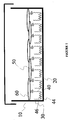
- the present invention is a system 10 for attaching and growing cells in vitro.
- System 10 generally comprises a hydrophobic tissue culture surface 20, biomolecule conjugated EGAP 30, and cells 60.
- System 10 is constructed by first preparing a modified end-group polymer (EGAP) 40.
- EGAP 40 comprises a hydrophobic block 44 and two hydrophilic blocks 46.
- EGAP 40 is modified by, for example, reacting at least one hydrophilic block 46 with 4-nitrophenyl chloroformate followed by 2-(2-pyridyldithio) ethyl amine.
- EGAP 40 is then applied onto a hydrophobic tissue culture surface 20.
- hydrophobic block 44 of EGAP 40 is adsorbed by hydrophobic tissue culture surface 20.
- Hydrophilic blocks 46 does not adsorb, however. Instead, hydrophilic blocks 46 extend from the surface in a "seaweed" fashion.
- biomolecule 50 is thiolated by methods well known in the art.
- biomolecule 50 is thiolated with reduced glutathione.
- EGAP 40 and biomolecule 50 are then reacted to form biomolecule conjugated EGAP 30.
- cells 60 are seeded on the biomolecule conjugated EGAP coated surface 20. Cells 60 attach to biomolecule 50 , extend processes, and proliferate in an environment that resembles an in vivo setting.
- hydrophobic surfaces hydrophobic surfaces
- EGAP EGAP
- binding EGAPs to hydrophobic surfaces EGAP
- suitable biomolecule conjugates biomolecule conjugated EGAPs
- biomolecule conjugated EGAP coated surfaces uses for biomolecule conjugated EGAP coated surfaces.
- hydrophobic polymer surfaces of the present invention comprise any suitable polymer or surface coating material which imparts a hydrophobic character to the surface of the substrate.
- hydrophobic is meant that the surface has a water contact angle greater than about 60° preferably greater than about 70°.
- Suitable polymers or biomaterials with surfaces having a water contact angle greater than70° include, but are not limited to polystyrene (PS), polymethylmethacrylate (PMMA), polyolefins (e.g.
- PE polyethylene
- PP polypropylene
- PVC polyvinylchloride
- silicones polyacrylonitrile (PAN), copolymers of polyacrylonitrile/polyvinal chloride, polysulfone, poly (ether sulfone) (PES), certain polyurethanes, pyrolized Materials, and block copolymers containing these constituents.
- hydrophobic polymer surfaces water contact angles between 60 ° and 70 °
- PVAC polymer surfaces
- Adsorption upon these polymers would be expected to be reduced compared to more hydrophobic polymers such as PS and PMMA.
- detachment of the block copolymer surfactant from the polymer surface over time would be expected.
- These and non-hydrophobic surfaces may be treated to render them hydrophobic before block copolymer surfactant adsorption.
- silica can be treated with dimethyl-dichloro silane to provide a hydrophobic surface.
- the polymer may be porous or nonporous, or be in the form a flat surface (e.g. a microtiter plate), or any suitable shape, such as micro beads, and the like used in chromatography applications.
- the polymeric surfactant may also be adsorbed upon colloidal or latex particles of a suitable hydrophobic polymer.
- end-group activated polymers refers to modified block copolymers surfactants.
- the EGAPs of the present invention are of the type defined in U.S. Patent No. 5,516,703 entitled "Coating of Hydrophobic Surfaces to Render Them Protein Resistant While Permitting Covalent Attachment of Specific Ligands" which is hereby incorporated by reference.
- EGAP is block copolymer surfactant where at least one of the hydrophilic chains has been modified to make it chemically reactive to biomolecules.
- an EGAP generally comprises a block copolymer surfactant backbone and an activation group or reactive group.
- the polymeric block copolymer surfactant of the present invention may be any surfactant having a hydrophobic region capable of adsorbing onto a hydrophobic surface and a hydrophilic region which extends away from the surface when the hydrophobic region is adsorbed to the hydrophobic surface.
- the block copolymer surfactant backbone of EGAP may be in the form of any arrangement of the poly(ethylene oxide) (PEO) and poly(propylene oxide) (PPO) blocks with the general formula: (HO-PEO) (PPO) b (1) where (a) and (b) are integers.
- PEO poly(ethylene oxide)
- PPO poly(propylene oxide)
- the polymeric block copolymer has a PEO (-C 2 H 4 -O-) content between 10 wt% and 80 wt%, preferably 50 wt% and 80 wt%, and more preferably between 70 wt% and 80 wt%.
- the PEO chains or blocks are of the general formula: -(-C 2 H 4 -O-) u - (2) where (u) is the same or different for different PEO blocks in the molecule. Typically, (u) is greater than 50, preferably between 50 and 150, more preferably between 80 and 130.
- the PPO blocks are of the general formula; -(C 3 H 6 -O-) v - (3) where (v) may be the same or different for different PPO blocks in the molecule. Typically, (v) is greater than 25, preferably between 25 and 75, and more preferably is between 30 and 60.
- the block copolymers may be branched structures and include other structures (e.g. bridging structures, or branching structures) and substituents that do not materially affect the ability of the block copolymer to adsorb upon and cover a hydrophobic surface.
- the block copolymer surfactant used to make EGAP is a polymeric tri-block copolymer with pendant -OH groups, as in Formula (4) below.
- These tri-block copolymers have a hydrophobic center block of polypropylene oxide and hydrophilic end blocks of polyethylene oxide with terminal -OH groups, and can be represented by the formula: HO-(-C 2 H 4 -O-) x -(-C 3 H 6 -O-) y -(-C 2 H 4 -O-) z -H (4) where (y) is between 25 and 75, preferably between 30 and 60, and (x) and (z) are preferably the same but may be different, and are between 50 and 150, preferably between 80 and 130.
- Block copolymer surfactants of the type described are commercially available from, for example, BASF.
- the end-group of the polymer is activated by methods well known in the art. Briefly, the -OH end groups of the PEO chains of the Polymeric surfactants are modified to introduce a small reactive organic group which is stable in water. Using the block copolymer surfactants represented by equation (4) as an example, if both -OH groups on the pendant PEO chains are substituted, the modified surfactant has the formula; R-O-(-C 2 H 4 -O-) x -(-C 3 H 6 -O-) y -(-C 2 H 4 -O-) z -R (5) where R is a reactive group.
- the general formula for the modified polymeric surfactants of the invention is: (HO-PEO) c (R-O-PEO) d (PPO) b (6) where (c+d) is equal to (a) in formula (1), and (c) is 0 or a positive integer, and (b) is defined above for formula (1).
- the R group may be any reactive group that is stable in water and will impart the desired selective reactivity for the substrate surface when the modified surfactant is adsorbed upon the surface.
- the specific reactivity may be to any non-water entity or entities.
- the R groups are chosen such that they do not significantly impair adsorption of the modified polymeric surfactant on the hydrophobic surface.
- the reactive R group contains a hydrazino group (by further reacting a p-nitrophenyl group), a thiopyridyl group, a tyrosyl residue, or a maleimide.
- R may also be a member of the group consisting of hydrazino, thiopyridyl, tyrosyl, maleimide, 2-pyridyl disulphide, 5-nitro-2-pyridyl disulphide, 4-pyridyl disulphide, 5-carboxy-2-pyridyl disulphide, and the nitrogen oxides of 2-pyridyl disulfide, 5-nitro-2-pyridyl disulfide, 4-pyridyl disulfide, and 5-carboxy-2-pyidyl disulphide as well as other groups well known in the art,
- the R group is for tile immobilization of biomolecules and contains the structure: -S-S-R" (9) where R" is selected from the group consisting of (1) 2-benzothiazolyl, (2) 5-nitro-2-pyridyl, (3) 2- pyridyl, (4) 4-pyridyl, (5) 5-caxboxy-2-pyridyl, and (6) the N-oxides of any of (2) to (5).
- R is selected from the group consisting of (1) 2-benzothiazolyl, (2) 5-nitro-2-pyridyl, (3) 2- pyridyl, (4) 4-pyridyl, (5) 5-caxboxy-2-pyridyl, and (6) the N-oxides of any of (2) to (5).
- the EGAP is adsorbed onto an appropriate hydrophobic surface. This simply requires mixing the appropriate amount of EGAP with the hydrophobic surface. Usually approximately two hours is sufficient to completely coat the hydrophobic surface with EGAP. Depending on the shape and size of the hydrophobic surface to be coated, it may be advantageous to shake the mixture to ensure that the entire surface area becomes coated.
- the concentration of EGAP can be regulated by diluting the EGAP with polymer that has not been activated (i.e., block copolymer surfactants).
- block copolymer surfactants i.e., block copolymer surfactants
- the surface does not adsorb cell, proteins and other biomolecules.
- various hydrophobic material surfaces coated with a unmodified block copolymer surfactants such as PluronicTM F10S substantially decrease the adhesiveness of the surface to NIH3T3 cells.
- proteins that can be conjugated to EGAP according to the composition and the method of the present invention.
- the proteins can be native, recombinant, or synthesized.
- the term protein as it is used herein is not limited to naturally occurring molecules, but includes molecules such as synthetic proteins which have no biological origin.
- the proteins are ECM proteins, adhesion proteins or growth factors.
- ECM proteins ECM proteins
- adhesion proteins growth factors.
- EGAP could be conjugated to an ECM protein.
- EGAP could be conjugated to one or more of the various collagen molecules currently known or hereafter isolated.
- Collagen is the name given to a superfamily of ECM proteins whose primary role is forming and preserving the structural integrity of the ECM and cells.
- Collagen's characteristic triple-helix domain forms fibrils, filaments, or network, either alone or in combination with other ECM components.
- Collagen IV is a major component of basement membranes and forms a network to which other basement components, such as laminin, nidogen, heparin and heparan sulfate proteoglycans, interact. Many cells types adhere to Type IV collagen. See, e.g., Glansville, R W. "Structure and Function of Collagen Types," Academic Press Inc., pp. 43-79 . Moreover, regions within Type IV collagen are known to promote or inhibit tell adhesion. Vandenberg et al., J. Cell Biol, 113: 1475-1483 (1991 ); Tsilibary et al., J. Cell Biol. 111: 1593-1591 . Surfaces coated with Type IV collagen (or specific regions or Type IV collagen) conjugated EGAPs, therefore, could be used to both promote and inhibit cell attachment and growth in vitro.
- Type IV collagen can be obtained from basement membranes treated with pepsin or with bacterial collagenase. Glansville, R W. "Structure and Function of Collagen Types," Academic Press Inc., pp. 43-79 ; Hudson, et al., "extracellular Matrix Macromolecules - A Practical Approach” (M.A. Haralson and J.R Hassell, eds.) Oxford; New York: IRL Press, (1996 ). Moreover, the complete primary structures of mouse and human a(IV) and a2(IV) chains have been deduced from cDNA sequences and mouse and human a(IV) and a2(IV) genomic clones have been extensively characterized Vuorio et al., Annu. Rev.
- the biomolecule could be a fibrillar collagen which includes five different molecular types (I, II, III, V, and XI). Fibrillar collagens polymerize to form fibrils that serve as stabilizing scaffolds in extracellular matrices. Cell attachment, differentiation, and migration are influenced by fibrillar collagens. It has been shown that fibrillar collagens-interact with cells through receptors on the cell surface. See e.g., Hemler, ME., Annu. Rev. Immunol. 8: 365-400 (1990 ).
- Fibrillar collagen is commercially available from, for example, Sigma, St. Louis, Mo.
- methods of purifying, as well as cDNAs coding fibrillar collagen are well known in the art. (See e.g., Vuorio, et al. Annu. Rev. Blochem. 59: 837-872 (1990 ).)
- EGAP could also be conjugated to one or more fibronectin molecules or peptides thereof.
- the subunits of fibronectins vary in size between approximately 23 5 and 270 kDa plus carbohydrates.
- Extended polypeptide segments in certain parts of the molecule are highly susceptible to proteolysis, which generates a series of protease resistant domains, each comprising several of the repeating modules. These domains contain a variety of binding sites for other molecules, including collagens, fibrin, heparin/heparan sulphate, and cell surface receptor integrins.
- Fibronectins are widely expressed in embryos and mature cells, especially in regions of active morphogenesis, cell migration, and inflammation. Fibronectins promote the adhesion and spreading of many cell types by binding to several different integrin receptors. See, e.g., Hynes, R.O., Cell 48:549-554 (1987 ). Tumor cells show reduced levels of fibronectin and levels in plasma fall in various forms of trauma. In contrast, fibronectin levels are elevated during wound healing and fibrosis.
- Fibronectin conjugated EGAPs could be used in tissue culture to assist in cell adhesion, morphogenesis, and cell migration.
- biomaterials such as surgical wraps, could be coated with fibronectin conjugated particles to aid and accelerate wound healing.
- fibronectin has two cell binding sites which are recognized by two different integrin receptors.
- the first cell binding site comprises three residues: arginine-glycine-aspartic acid, or RGD.
- the second cell binding site comprises the peptide: glutamic acid-isoleucine-leucine-aspartic asid-valine, or EILDV.
- RGD arginine-glycine-aspartic acid
- RGD arginine-glycine-aspartic acid
- the second cell binding site comprises the peptide: glutamic acid-isoleucine-leucine-aspartic asid-valine, or EILDV.
- RGD, RGDS, RGES, RFDS, GRDGS, and GRGS are commercially available from, for example, Sigma, St. Louis, MO.
- peptides such as GRGDTP inhibit cell attachment of fibronectin, vitronectin, and Type I collagen.
- Amino acid sequence QPPRARI is the binding site for the carboxy-terminal heparin binding domain.
- Peptides that inhibit platelet aggregation and inhibit fibronectin binding to bacteria are also well known and commercially available. EGAP, therefore, can be simply conjugated with any number of peptides or domains to obtain the desired results according to the present invention.
- EGAP could be conjugated to agrin.
- Agrin is ECM glycoprotein which can take the form of either a 150 kDa or a 95 kDa protein.
- Agrin is localized at the neuromuscular junction and induces clustering of acetylcholine receptors on skeletal myotubes in cell culture. Clustering of this receptor is one of the most dramatic events in neuromuscular synapse formation and regeneration in vivo. Purified agrin has been shown to induce clustering of synaptic molecules in vitro, such as ECM-associated acetylcholinesterase and membrane-associated acetylcholine receptors, and it is very likely to function similarly in vivo.
- agrin As an agent that induces differentiation in skeletal myotubes, agrin is synthesized in motor neurons and transported to their terminals in skeletal muscles. Agrin has been shown to induce phosphorylation on tyrosine on the acetylcholine receptor ⁇ - subunit. Treatments that inhibit receptor aggregation prevent tyrosine phosphorylation. Results suggest that the agrin receptor regulates a tyrosine protein kinase or phosphatase that in turn regulates receptor clustering. These and other data demonstrate that the extracellular matrix protein agrin contains all the essential information needed to form a neuromuscular synapse.
- Agrin can be obtained from the basal-lamina enriched fraction of T. Californica electric organ as described in Nitkin et al., J. Cell Biol., 105: 2471-2478 (1987 ).
- portions of agrin glycoprotein can be obtained from available cDNAs using recombinant techniques well known in the art. Tsim et al., Neuron, 8: 677-689 (1992 ).
- Monoclonal antibodies against T. Californica agrin are also available. Reist et al. J. Cell. Biol., 105: 2457-2469 (1987 ).
- ECM components and proteins such as aggrecan, biglycan, bone sialoprotein, cartilage matrix protein, Cat-301 proteoglycan, CD44, cholinesterases, FACIT collagens (Type IX, XII, XIV), other collagens (Type VI, VII, XIII), short chain collagens (Type VIII, X), decorin, elastin, fibrinogen, fibroglycan, fibromodulin, fibulin, glypican, HB-GAM, hyaluronan and hyaluronan binding proteins, J1 glycoproteins, laminin, laminin binding proteins, link protein, mucins, nidogen/entactin, osteopontin, perlecan, plasminogen, plasminogen activator inhibitor 1, plasminogen activator inhibitor 2, proteins containing Ca 2+ -dependent carbohydrate recognition domains , restrictin, serglycin, SPARC/osteonect
- Cell adhesion molecules are molecules that, in addition to mediating cell-ECM contact, mediate cell-cell contact Cell adhesion is required at all stages in development and critical to the overall-organization of tissues and organs. To date, there have been hundreds of cell adhesion molecules isolated and characterized. These molecules can be roughly categorized into five protein superfamilies: (1) the immunoglobulin (Ig) superfamily; (2) the cadherin superfamily, (3) the integrin superfamily; (4) the selectin superfamily; and (5) the H-CAM superfamily.
- Ig immunoglobulin
- the Ig superfamily for example, which includes other proteins such as CAM and N-CAM are primarily involved in cellular recognition. As such, immunoglobulins or antibodies play a critical role in proper immune function. See Williams et al., Ann. Rev. Immunol. 6: 381-405 (1988 ). As will be discussed below, the Ig superfamily of molecules forms the bases for immunoassays in the art.
- the same strategy could be used to conjugated numerous adhesion proteins to EGAP for the purposes of biological analysis or cell growth.
- Examples include AMOG. cadherins, CD2, CD4, CD8, C-CAM (CELL-CAM 105), cell surface galactosyltransferase, connexins, desmocollins, desmoglein, fasciclin I, fasciclin II, fasciclin III, F11.
- GP Ib-IX complex integrins, intercellular adhesion molecules, L1, leukocyte common antigen protein tyrosine phosphate (LCA, CD45), LFA-1, LFA-3, mannose binding proteins, (MBP), MUC18, myelin associated glycoprotein (MAG), neural cell adhesion molecule (NCAM), neurofascin, neruoglian, neurotactin, PECAM-1, PH-20, selectins, TAG-1. VCAM-1 and the like.
- L1 leukocyte common antigen protein tyrosine phosphate
- MBP mannose binding proteins
- MUC18 myelin associated glycoprotein
- MAG myelin associated glycoprotein
- NCAM neural cell adhesion molecule
- neurofascin neurofascin
- neruoglian neurotactin
- PECAM-1 PECAM-1
- PH-20 selectins
- selectins TAG-1.
- VCAM-1 and the like selectins, TAG
- Biomolecules such as growth factors, mitogens, and differentiation factors can stimulate cell division. Many of these biomolecules are normally present in growth serum used in most cell culture media. Others biomolecules, like some bacterial lipopolysaccharides and certain cell agglutinating proteins (lectins), are not present in normal growth media Nevertheless, they are an integral part of work aimed at discerning what signals regulate cell growth and thus have huge implications in cancer research.
- PDGF platelet-derived growth factor
- PDGF is the major polypeptide mitogen in cell culture serum.
- studies in fibroblast demonstrate that PDGF is required to make cells competent to other growth factors. That is, the cell will not respond to other growth factors and mitogens unless they are first exposed to PDGF.
- PDGF is stored in the ⁇ granules of the blood platelets. During blood clotting and platelet adhesion, PDGF is released from these a granules. The release of PDGF at the site of injury causes some cell types to migrate to the site of injury and causes other cell types to divide. Accordingly, PDGF conjugated EGAPs could be used to promote cell growth in culture. In addition, PDGF could be used to promote wound healing in vivo by applying it to wounds in conjunction with an appropriate biomaterial, The PDGF would be tethered to EGAP which in turn would be adsorbed to the biomaterial. Thus, unlike other drug delivery methods, the amount of PDGF stimulation and the site of stimulation could be tightly regulated.
- EGF EGF
- TGF- ⁇ TGF- ⁇
- NGF NGF
- IGF-I IGF-II
- GH GHRF
- a stem cell can become a plasma cell, a memory B lymphocyte, an activated T cell, a macrophage, blood platelets, or a erythrocyte.
- stem cells could be grown on one or more of these factors conjugated to EGAP. As such, the fate of the cell can be carefully controlled. For example, the ability to regulate T cell production in vitro from precursor cells could be used to supplement the loss of T cells that leads to acquired immunodeficiency syndrome (AIDS).
- AIDS acquired immunodeficiency syndrome
- the differentiating factor erythropoietin is currently being used to increase red blood cell production in patients that have lost large volumes of blood.
- multi-CSF II-3
- GM-CSF GM-CSF
- G-CSF G-CSF
- M-CSF can also be conjugated to EGAP.
- Biomolecules can be conjugated to EGAP using numerous methods known in the art, By reacting hydroxylated block copolymer surfactants with 4-nitrophenyl chloroformate, one can efficiently conjugate biomolecules having a variety of reactive groups.
- EGAPs react relatively easily in an organic solvent with amino groups, 2-pyridyl disulfide, peptide, hydrazino and other amino containing molecules.
- tyrosyl groups for radioisotope labeling purpose can be subsequently coupled to the EGAP by a reaction with the Bolton-Hunter reagent.
- Biomolecules are conjugated via amine groups.
- 4-nitrophenyl chloroformate activated EGAP was conjugated to a biomolecule via an amine group on a peptide.
- the peptide glycyltryptophan (Giy-Trp) was mixed with ap appropriate amount of 4-nitrophenyl chloroformate activated EGAP.
- the two compounds were allowed to react at 25°C overnight
- the reaction mixture was then purified by passing it through a Sephadex column.
- Gly-Trp conjugated EGAP was confirmed by dry weight and photometric analysis.
- Biomolecules can also be conjugated to EGAP via a disulfide bond.
- EGAP molecules as discussed above, can be activated by introducing a reactive group containing a disulfide derivative such as a 2-(2-pyndyldithio)ethylamine. This method of conjugation is preferred because the rate of hydrolysis of the 2-pyridyl disulfides groups at about pH 8.5 is almost negligible in comparison to the rate of the thiol-disulfide exchange reaction. As such, only a small concentration of biomolecule is required.
- this conjugation method provides an easy way to detect the degree of biomolecule conjugation.
- the reaction between the thiol group on the biomolecule and 2-(2-pyridyldithio)ethylamine releases thiopyridone.
- Thiopyridone concentration can be readily and accurately quantified by spectroscopic detection at 343 nm with an extinction coefficient of 8060/cm 1 M -1 .
- the concentration of thiopyridone is directly proportional to the degree of biomolecule conjugation.
- thiol-disulfide exchange is a reversible reaction
- bound biomolecules can be released from the solid phase by addition of a thiol-containing reagents, such as dithiothreitol (DTT).
- DTT dithiothreitol
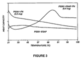
- the biomolecules fibronectin and human serum albumin was conjugated to EGAP using the methods described above. As illustrated in Figures 3 and 4 , microcalorimetry studies indicate that these biomolecules retain their native secondary structure when tethered to EGAPs.
- biomolecule conjugated EGAP the number of uses for biomolecule conjugated EGAPs is also large. Below are exemplary uses for biomolecule conjugated EGAPs.
- composition and the method of the present invention can be used to attach and grow cells in culture.
- EGAP can be conjugated to any number of ECM and cell adhesion proteins as well as growth and differentiation factors
- NIH 3T3 cells were grown on hydrophobic culture surface coated with GRGDSY conjugated EGAP.
- GRGDSY is a peptide corresponding to a cell binding site of fibronectin.
- the GRGDSY was conjugated to an F108 derivative EGAP via a disulfide bond prepared according to the 2-pyridyl disulfide conjugation method described above.
- NIH 3T3 cells were seeded at a concentration of 6 x 10 3 cells/cm 2 in DMEM supplemented with 10% bovine serum.
- composition and the method of the present invention can be used to sort cells and other biological material. It will be appreciated by one skilled in the art that it is often desirable to select one cell type from a mixture of cells. For example, identifying lymphocytes as either T cells or B cells is useful in diagnosing various diseases, including lymphoproliferative malignancies, immunodeficiency diseases, unexpected infections diseases, monitoring of transplants, and acquired immunologic disorders such as AIDS.
- Current methods involve a combination of density gradient centrifugation and either fluorencence microscopy or cell flow cytometry (or fluorescence-activated cell sorter). These methods are tedious and expensive. Moreover, the stress of the procedure often damages the cells making it difficult, if not impossible, to grow the cells once they have been selected.
- the method of the present invention could be used to quickly sort cells.
- EGAP could be conjugated to a number of biomolecules that are specific for the desired cells type.
- the biomolecule could be a monoclonal antibody against a specific cell surface antigen such as a transmembrane receptor or a particular carbohydrate moiety.
- many of these biomolecules, including CD2, CD3, CD4, CD8 on T cells and CD 19, CD20, CD2, and surface immunoglobulins on B cells are all commercially available from, for example, Sigma Chemical Company, St. Louis, MO.
- the cell specific biomolecule conjugated EGAP is coated on polystyrene beads.
- the polystyrene beads are then combined with a mixture of cells under appropriate incubation conditions and growth media that does not contain molecules that will bind to the cell specific biomolecule. After the cells have had an opportunity to bind to the cell specific biomolecule, the polystyrene beads are separated from the remaining unattached cells.
- the separation means is any means well known in the art including magnetic, streptavidin separation, or mechanical separation such as gentle centrifugation.
- a subpopulation of the EGAPs coated to the polystyrene beads comprises biotin conjugated EGAP.
- the biotin is available for binding and separation with streptavidin, such as streptavidin MagneSpheres® paramagnetic particles sold by Promega, Madison, WI. After several gentle washes to remove non-specifically bond cells, the collected polystyrene beads are assayed directly using common bioassays well known in the art or cultured. These methods of cell sorting are only exemplary of the many cell sorting methods that can be used with the composition and method of the present invention.
- PS latex particles with a diameter of 261 nm were purchased as a 10% (w/v) suspension were purchased from Seradyn.
- the block copolymer surfactants surfactant used was F108 having a molecular weight of 14600 were donated by BASF Co.
- N -succinimidyl-3-(2-pyridyldithiol) propionate (SPDP) was obtained from Pierce.
- Dithiotheeitol (DTT) was from Bio-Rad.
- Fibronectin solution (FN, 1.5 mg/ml) was isolated from human plasma, and disposable prepacked PD-10 columns were purchased from Pharmacia, WL
- the PluronicTM F108-2-pyridyl disulfide derivative (EGAP) was synthesized as described above.
- Adsorption reaction was carried out in a mixture consisting of 10- ⁇ L of the suspension of PS latex particles and 200 ⁇ L of 0.5% (w/w) EGAP dissolved its deionized water This mixture was incubated for 2 hours with shaking at room PS microspheres were washed and recovered by table centrifugation Eppendorf 5415C).
- Thiolarion of biomolecules the methodology described previously (4). Briefly, the reaction mixture of 2 mL of fibronectin solution and 20 pL of 5 mM SPDP solution was kept for I hour at room temperature with shaking, after which it was passed through a PD-10 column. TheSPDP-modified fibronectin (FN-SPDP) was collected; its emergence from the PD-10 column was monitored by UV adsorbance at 280 nm. Thiolated fibronectin was then obtained by adding 4 ⁇ L of a 50 mM DTT solution to the SPDPmodified fibronectin and keeping the mixture for 30 minutes at room temperature. The sulfhydryl concentration was calculated by quantifying the concentration of released 2-thiopyridone as described earlier.
- Low molecular weight reaction products were romeved by passing the thiolated fibronectin (FN-SH) reaction mix through a PD- 10 column.
- the coated PS latex particles were added to FN-SH or FN solution and the linking reaction was allowed to take place for I hour at room temperature under shaking.
- the latex particles were washed and characterized by means of differential scanning microcalorimetry (DSC). Quantification of the amount of FN or FN-SH bound onto the latex particles was performed by amino acid analysis as well as by Micro BCA assay, as described before. DSC (Hart Scientific, Model 4207) studies were carried out as reported previously.
- FN shows melting transitions at different temperatures and with different enthalpic contents. It is therefore likely that surface adsorption might strongly affect the FN structure.
- Figure 3 is a comparison of FN adsorbed and bound through the PEO tether offered by the modified PluronicTM F108, respectively. No cooperative transitions are in evidence for the adsorbed protein, while FN tethered to the surface shows the normal, complex transition pattern. Apparent transition enthalpies for free and immobilized FN are listed in Table 1. TABLE 1 Fibronectin T m (°C) ⁇ H(Kcal/mol) In PBS 55-0 570 Conjugated to BGAP 55-80 440
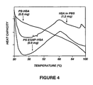
- HSA conjugated EGAP was thiolated, purified, and conjugated to EGAP essentially as described in Example 2.
- the three traces in the Figure 4 represent the thermograms for protein in solution, protein adsorbed onto bare PS particles, and proteins attached to the surface through the PluronicTM F108 intermediate.
- PluronicTM F108 PluronicTM F108 intermediate.
- all cooperative transitions are absent from the protein-particle adsorption complex.
- the PEO tethered sample shows the characteristic complex melting curve of native HSA, reflecting the differential collapse of the three lobes of this protein.
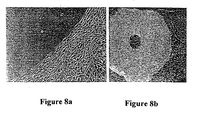
- Osteoblast cells were seeded onto a polystyrene substrate which had been treated with C F108 or only in a localized circular area in the center of the substrate.
- FIGs 8a and 8b cells which were seeded in serum containing media did not attach to the PEO modified area but were found to attach, spread, and proliferate well on unmodified areas.
- Figure 8a is a close-up displaying cells aligned at an interface between PEO treated and unmodified areas.
- Figure 8b cells were fixed and removed from culture well after adequate time to lay down a substantial ECM. A dark spot in the center corresponds to the PEO treated area where there were no cells.
- Surgical discard from reduction mammoplasty was digested by standard method into single and small cell aggregates of epithelial cells.
- Cells were placed on a PluronicTM coated tissue culture plastic in CDM# media. Following a two week incubation at 37°C, 95% of cells were alive as indicated by the vital dyes.
- NIH 3T3 cells were attached to and grown on GRGDS conjugated EGAP as follows. EGAP formation and GRGDS conjugation was carried out essentially as described above. Briefly, the hydroxyl ends of Block copolymer surfactant was activated to form an EGAP using 4-nitrophenyl chloroformate followed by 2-pyridyl disulfide.
- a portion of the EGAP was derivatized with the Bolton- Hunter Reagent. This allows the polymer to be labeled with radioactive iodine and thus provides a means to accurately determine the surface concentration of the EGAP.
- GRGDS peptide was synthesized with a tyrosine residue at its carboxyl terminus using methods well known in the art.
- the tyrosine residue allows incorporation of radioactive iodine and thereby enables accurate determination of surface peptide concentration.
- Fibronectin peptide Gly-Arg-Gly-Asp-Ser-Tyr or GRGDSY was conjugated to activated F108 (EGAP) and used to coat polystyrene (PS) culture dishes as described above. Cells were seeded at 6 x 10³ cells/cm 2 in DMEM supplemented with 10% bovine serum. Substrates were washed after 24 hrs.
- NIT 3T3 cells to GRGDSY conjugated F108 was compared with untreated PS surface (PS), PS surface coated with F108 alone (F108), F108 adsorbed PS treated with GRGDSY (not conjugated) (F108/RGD), and pyridyl disulfate activated F108 (EGAP) adsorbed PS without GRGDSY (PDSF108).
- Figures 5 and 6 respectively, illustrate that NIFIH 3T3 cells do adhere and spread processes on GRGDSY conjugated EGAP surfaces, but do not adhere to F108 treated surface.
Abstract
Description
- The present invention is related to a composition and method for regulating the adhesion of cells, organisms, and molecules to hydrophobic surfaces. More specifically, the present invention is directed to a biomolecule, such as a protein, peptides amino acids, nucleic acids, lipids, and carbohydrates conjugated to an end-group activated polymers (EGAPs) and uses thereof.
- Normal development and function in living organisms require interactions between cells and the molecules in the surrounding environment. One way cells communicate is via molecules that span the membrane of the cell called transmembrane proteins. When the portion of the transmembrane protein which is outside of the cell encounters specific molecules in the surrounding environment, it undergoes structural and conformational changes which triggers biological reactions inside the cell.
- For example, in vivo, cells form complex multilayer structures which ultimately form tissues and organs. Tissue and organ formation, however, requires specific contacts with the environment. These cells are referred to as "anchorage-dependent" because they will not grow properly, if at all, unless they are anchored to others cells, an extracellular matrix (ECM), or other surface.
- An ECM is a complex and variable array of molecules secreted by cells, such as collagens, glycosaminoglycans, proteoglycans, and glycoproteins. Together these cellular products form the basal lamina, bone, and cartilage which give tissues and organs their shape and strength. In fact, contact between anchorage-dependent cells and the ECM in many instances plays a dramatic role in determining the cells' shape, position, metabolism, differentiation and growth.
- Cell contact is also important in other biological functions, such as the activation of an immune response. The immune system is a complex network of cells that have the ability to recognize and rid the body of foreign substances, such as viruses, bacteria and parasites. One mechanism used by the immune system to rid itself of foreign substances is a humoral response. A humoral response involves activation of specific cells called B cell lymphocytes. B-cells are activated when transmembrane proteins on their surface bind to foreign substances called antigens. Specifically, binding of B-cells to antigens stimulates B cells to proliferate and differentiate into immunoglobulin or antibody producing plasma cells.
- The antibodies produced by plasma cells travel throughout the body binding to the pathogen or foreign substance. Binding of antibodies to foreign substances activates several other immunological pathways, including the "complement" pathway. The complement pathway is designed to destroy the foreign substance and to initiate an inflammatory response in the organism.
- While cell contact with other cells and the environment is critical to the overall health and biological function of an organism, it creates unique problems in the art of biotechnology. Specifically, two areas where cell contact requirements create problems are: (1) cell culture; and (2) biomaterial transplantation.
- Tissue or cell cultures comprise cells from a plant or animal which are grown outside the organism from which they originate. These cells are often grown, for example, in petri dishes under specific environmental conditions. Cell cultures are of great importance because they represent biological "factories" capable of producing large quantities of biological products such as growth factors, antibodies, and viruses. These products can then be isolated from the cell cultures and used, for example, to treat human disease. In addition, cell cultures are a potential source of tissue which could be used for transplantation into humans. For example, cell cultured skin cells could potentially be used in skin grafts to replace diseased or damaged skin. Finally, cell cultures usually comprise cells from only one or a few tissues or organs. Consequently, cell cultures provide scientists with a system for studying the properties of individual cell types without the complications and risk of working with the entire organism. For example, the effects of pharmaceutical drugs on certain cell types could be tested on cell cultures prior to clinical trials in order to assess the drug's health risks.
- Like most cells in vivo, cells grown in culture are either anchored to an ECM or another cell. Only cells of the circulatory system (e.g., lymphocytes and red blood cells) grow unattached and suspended in solution in vitro. Many anchorage-dependent cells can grow on glass or plastic surfaces, such as polystyrene. These cells, however, often lose their natural architecture and do not function normally (e.g., the ability to differentiate and respond to hormones). Accordingly, these cells do not precisely mimic a cell's biological functions in vivo and thus have limited potential.
- For this reason, glass and plastic cell culture dishes are often coated with an ECM protein such as collagen, fibronectin, laminin and the like. These proteins bind to surfaces such as polystyrene through a process known as adsorption. Although ECM coated cell culture surfaces have led to improved culture conditions, they are far from ideal.
- First, biomolecules, such as proteins, often become inactivated upon adsorption to hydrophobic surfaces. The biological activity of proteins is conferred by their unique structure and their ability to undergo conformational changes upon binding to a substrate or other physiological event. In one study, the structure of proteins was measured using a technique called microcalorimetry. Microcalorimetric studies demonstrated that proteins which are bound to hydrophobic surfaces loose essentially all their cooperatively folded structure compared to the same protein in solution. Because a protein's structure and its ability to undergo conformational changes strongly correlates with biological activity, these data suggest that most proteins that are adsorbed by a hydrophobic surface loose there in vivo biological activity.
- Second, the conformation and orientation of immobilized proteins have important effects on the nature of their interaction with cells. D.J. Juliano, S.S. Saaedra and GA. Truskey, Journal of Biomedical Materials Research 2-7 1103-1113 (1993). Both are influenced by the chemistry and physical properties of the underlying substrate as well as by the method of immobilization. K. Lewandowska, E. Pergament, N. Sukenik and L.A. Culp, The Journal of Biomedical Materials Research 21 1343-1363 (1992).
- Third, like in vivo, cells in culture release molecules such as serum proteins and growth factors into the culture media As discussed above, the secretion and concentration of these molecules in the culture media are critical to the biological function of neighboring cells. Under current cell culture conditions, the careful balance and concentration of secreted molecules are disrupted because secreted molecules are adsorbed by the cell culture surface. Thus, the communication and biological function of cells grown under current cell culture techniques does not mimic in vivo environment.
- Finally, the surface concentration of ECM components is a critical factor in the regulation of cell behavior. The ability to control and vary surface biomolecule concentration is therefore of upmost importance and depends on the method of immobilization and in some cases the physical nature of the base material, Simple ECM adsorption to cell culture substrates does not meet these requirements.
- " Covalent Surface Immobilisation of Arg-Gly-Asp and Tyr-Ile-Gly-Arg Containing Peptides to Obtain Well-Defined Cell-Adhesive Structures ", Massia et al., Analytical Biochemistry 187, 292-301 (1990), " Chemical Modification of Surface Active Poly(ethylene oxide)-Poly(propylene oxide) Triblock Polymers" Li et al., Bioconjugate Chem 1996, 7, 592-599 and
WO95/06251 - In short, to date there is no single method for conjugating proteins to potential cell culture substrates which addresses all these major concerns. Thus, current research is hindered by the fact that cell cultures do not accurately mimic an in vivo environment.
- A second problem area created by cell contact is biocompatibility, It is generally acknowledged that artificial biomaterials, including fabricated biomedical polymers, are much less immunologically active than transplants or tissue-derived biomaterials. Nevertheless, the use of non- physiological biomaterials in many lifesaving medical devices, either extracorporeal or implanted, often leads to adverse side-effects for the patient.
- The adverse side-effects observed are usually a consequence of contact between cells, proteins, and other biological fluids in the blood with the artificial biomaterial. Typically, contact with the artificial biomaterial activates two major biological processes: coagulation and complement. As discussed above, the complement pathway is designed to destroy the foreign substance and to initiate an inflammatory response in the organism.
- Activation of the coagulation cascade can be controlled to a limited extent with the use of anticoagulants, e.g., heparin. Heparin, however, is not well suited for extended use such as in the case of a permanent implant, Further, currently there is no clinically available agent that can prevent or suppress artificial surface-initiated activation of a complement. Thus, activation of the coagulation and complement systems upon blood contact is a major problem with respect to biomaterial transplantation.
- From the foregoing, it will be appreciated that it would be an advancement in the art to provide a method of coating tissue culture surfaces with ECM proteins or other biomolecules that does not destroy the biological activity of the biomolecule.
- It would be a further advancement in the art if the tissue culture cells could adhere and grow on the biomolecule coated surface.
- It would be yet another advancement in the art if the biomolecule coated surface did not adsorb proteins and other molecules secreted by the cells in culture.
- Such compositions and methods are disclosed and claimed herein.
- The present invention is directed at a composition and method for regulating the adhesion of cells and biomolecules to hydrophobic surfaces and hydrophobic coated surfaces. Generally, the composition is an end-group activated polymer (EGAP) generally comprises a block copolymer surfactant backbone and an activation or reactive group. The polymeric block copolymer surfactant of the present invention may be any surfactant having a hydrophobic region capable of adsorbing onto a hydrophobic surface and a hydrophilic region which extends away from the surface when the hydrophobic region is adsorbed to the hydrophobic surface. In one embodiment, the EGAP is synthesized by reacting the block copolymer surfactant with 4-nitrophenylchloroformate followed by 2-(2-pyridyldithio)ethylamine.
- A large range of biomolecules can be conjugated to EGAP, include natural or recombinant growth factor, mitogens, growth peptides, differentiating factors, sugars, carbohydrates, polysaccharides, lipids, sterols, fatty acids and nucleic acid. In one embodiment, the biomolecule contains a natural or artificial thiol group. These biomolecules are conjugated to EGAP via a disulfide linkage.
- The biomolecule conjugated EGAP surface can be put to a wide variety of uses. For example, the composition can be used to attach organisms and molecules for growth or biological analysis.
- Briefly, this is done by contacting a hydrophobic surface with an EGAP for a time sufficient for the EGAP to be adsorbed by the hydrophobic surface. A biomolecule is then conjugated to the EGAP adsorbed to the hydrophobic surface to form a biomolecule conjugated EGAP surface. After washing of unconjugated biomolecule, organisms or molecules are placed in contact with the biomolecule conjugated EGAP coated surface such that the organism or molecule adheres to the biomolecule conjugated EGAP coated surface. In one embodiment, the organism or molecule is a eukaryotic or prokaryotic cell, a virus, an antibody or a pharmaceutical drug.
- The biomolecule conjugated EGAP surface can also be used to selecting at least one desired organism or molecule from a mixture of at least two organisms or molecules. This is done by first adsorbing EGAP onto a hydrophobic surface. A biomolecule unique for a desired organism or molecule being selected is then conjugated to the EGAP adsorbed to the hydrophobic surface. A mixture of organisms or molecules containing the desired organism or molecule is then contacted with the biomolecule conjugated EGAP coated surface and the desired organism or molecule is allowed to adhere to the unique biomolecule. Finally, non-adhered organisms or molecules are removed.
- These and other objects and advantages of the present invention will become apparent upon reference to the accompanying drawings and graphs and upon reading the following detailed description and appended claims.
- A more particular descriptions of the invention briefly described above will be rendered by reference to the appended drawings and graphs. These drawings and graphs only provide information concerning typical embodiments of the invention and are not therefore to be considered limiting of its scope.
-
Figure 1 is a schematic representation of cells attached to a tissue culture surface coated with the composition of the present invention. -
Figure 2 is a graph illustrating the adhesiveness of NIH 3T3 cells to Pluronic™ F108 coated hydrophobic surfaces. -
Figure 3 is a graph illustrating the thermal stability of fibronectin (FN) adsorbed by a hydrophobic surface (PS261-FN), and conjugated to EGAP coated hydrophobic surface (PS261- EGAP-FN). Unconjugated EGAP adsorbed by a hydrophobic surface (PS261-EGAP) was used as a control. -
Figure 4 is a graph illustrating the thermal stability of human serum albumin (HSA) free in phosphate buffered saline solution (HSA in PBS), adsorbed by a hydrophobic surface (PS-HSA), and conjugated to EGAP coated hydrophobic surface (PS-EGAP-HSA). -
Figure 5 is a picture of fibroblast cells attached and growing on a fibronectin peptide GRGDSY conjugated EGAP coated surface. -
Figure 6 is a picture illustrating that fibroblast cells were unable to attach to unconjugated EGAP coated surface. -
Figure 7 is a graph illustrating the adhesion of cells to surfaces coated with F-108 (F10S), F- 108 containing unconjugated GRGDSY (F108/RGD), 2-pyridyl disulfide conjugated F-108 (PDSFIO8), GRGDSY conjugated EGAP (PDSF 108/RGD), and untreated polystyrene (PS). -
Figures 8a and 8b illustrate that cells did not attach when seeded on poly(ethylene oxide) (PEO) modified surfaces but were found to attach, spread, and proliferate well on unmodified areas.Figure 8a is a close-up displaying cells aligned at an interface between PEO treated and unmodified areas. InFigure 8b , cells were fixed and removed from culture well after adequate time to lay down a substantial ECM. A dark spot in the center corresponds to the PEO treated area where there were no cells. -
Figure 9 illustrates that a hydrophobic surface coated with fibronectin peptide RGDS conjugated to EGAP (RGDS-PS) were found to support cell adhesion, (2 -pyri dyldithio)ethyiamine modified EGAP(PDSF108-PS)displayed an intermediate level of adhesiveness, and F108 coated polystyrene was relatively non-adhesive to fibroblast cells. - The present invention is directed to a novel compound and method for regulating the adhesion of culture cells to a hydrophobic surface. More specifically, the invention is directed to biomolecules that have been conjugated to end-group activated polymer (EGAPs). Biomolecule conjugated EGAPs can be used to coat hydrophobic surfaces making them suitable for a wide range of biochemical and medical uses.
- Reference is now made to
Figure 1 . With reference toFigure 1 , in one preferred embodiment, the present invention is asystem 10 for attaching and growing cells in vitro.System 10 generally comprises a hydrophobictissue culture surface 20, biomolecule conjugatedEGAP 30, andcells 60. -
System 10 is constructed by first preparing a modified end-group polymer (EGAP) 40. In one embodiment,EGAP 40 comprises ahydrophobic block 44 and twohydrophilic blocks 46.EGAP 40 is modified by, for example, reacting at least onehydrophilic block 46 with 4-nitrophenyl chloroformate followed by 2-(2-pyridyldithio) ethyl amine. -
EGAP 40 is then applied onto a hydrophobictissue culture surface 20. Upon application,hydrophobic block 44 ofEGAP 40 is adsorbed by hydrophobictissue culture surface 20. Hydrophilic blocks 46 does not adsorb, however. Instead,hydrophilic blocks 46 extend from the surface in a "seaweed" fashion. OnceEGAP 40 has adsorbed onto the hydrophobictissue culture surface 20,excess EGAP 30 is removed andtissue culture surface 20 is washed. - Simultaneously,
biomolecule 50 is thiolated by methods well known in the art. In one *embodiment,biomolecule 50 is thiolated with reduced glutathione.EGAP 40 andbiomolecule 50 are then reacted to form biomolecule conjugatedEGAP 30. After excess biomolecule conjugatedEGAP 30 has been removed andtissue culture surface 20 has been washed,cells 60 are seeded on the biomolecule conjugated EGAP coatedsurface 20.Cells 60 attach to biomolecule 50, extend processes, and proliferate in an environment that resembles an in vivo setting. - In order to better understand the details of the present invention, the following discussion is divided in six sections: (1) hydrophobic surfaces; (2) EGAP; (3) binding EGAPs to hydrophobic surfaces; (4) suitable biomolecule conjugates; (5) biomolecule conjugated EGAPs; and (6) uses for biomolecule conjugated EGAP coated surfaces.
- The hydrophobic polymer surfaces of the present invention comprise any suitable polymer or surface coating material which imparts a hydrophobic character to the surface of the substrate. By "hydrophobic" is meant that the surface has a water contact angle greater than about 60° preferably greater than about 70°. Suitable polymers or biomaterials with surfaces having a water contact angle greater than70° include, but are not limited to polystyrene (PS), polymethylmethacrylate (PMMA), polyolefins (e.g. polyethylene (PE), polypropylene (PP)), polyvinylchloride (PVC), silicones, polyacrylonitrile (PAN), copolymers of polyacrylonitrile/polyvinal chloride, polysulfone, poly (ether sulfone) (PES), certain polyurethanes, pyrolized Materials, and block copolymers containing these constituents.
- Lesser hydrophobic polymer surfaces (water contact angles between 60° and 70°), such as PVAC are also contemplated by the invention but are less are preferred. Adsorption upon these polymers would be expected to be reduced compared to more hydrophobic polymers such as PS and PMMA. Moreover, detachment of the block copolymer surfactant from the polymer surface over time would be expected. These and non-hydrophobic surfaces, however, may be treated to render them hydrophobic before block copolymer surfactant adsorption. For example, silica can be treated with dimethyl-dichloro silane to provide a hydrophobic surface.
- The polymer may be porous or nonporous, or be in the form a flat surface (e.g. a microtiter plate), or any suitable shape, such as micro beads, and the like used in chromatography applications. The polymeric surfactant may also be adsorbed upon colloidal or latex particles of a suitable hydrophobic polymer.
- As used herein, the terms end-group activated polymers (EGAP) refers to modified block copolymers surfactants. In one embodiment, the EGAPs of the present invention are of the type defined in
U.S. Patent No. 5,516,703 entitled "Coating of Hydrophobic Surfaces to Render Them Protein Resistant While Permitting Covalent Attachment of Specific Ligands" which is hereby incorporated by reference. Briefly, EGAP is block copolymer surfactant where at least one of the hydrophilic chains has been modified to make it chemically reactive to biomolecules. Accordingly, an EGAP generally comprises a block copolymer surfactant backbone and an activation group or reactive group. - The polymeric block copolymer surfactant of the present invention may be any surfactant having a hydrophobic region capable of adsorbing onto a hydrophobic surface and a hydrophilic region which extends away from the surface when the hydrophobic region is adsorbed to the hydrophobic surface. In one embodiment, the block copolymer surfactant backbone of EGAP may be in the form of any arrangement of the poly(ethylene oxide) (PEO) and poly(propylene oxide) (PPO) blocks with the general formula:
(HO-PEO) (PPO)b (1)
where (a) and (b) are integers. Preferably (a) is between 1 and 6, and (b) is between 1 and 3. more preferably (a) is 1 to 2, and (b) is 1. The polymeric block copolymer has a PEO (-C2H4-O-) content between 10 wt% and 80 wt%, preferably 50 wt% and 80 wt%, and more preferably between 70 wt% and 80 wt%. - The PEO chains or blocks are of the general formula:
-(-C2H4-O-)u- (2)
where (u) is the same or different for different PEO blocks in the molecule. Typically, (u) is greater than 50, preferably between 50 and 150, more preferably between 80 and 130. The PPO blocks are of the general formula;
-(C3H6-O-)v- (3)
where (v) may be the same or different for different PPO blocks in the molecule. Typically, (v) is greater than 25, preferably between 25 and 75, and more preferably is between 30 and 60. - The block copolymers may be branched structures and include other structures (e.g. bridging structures, or branching structures) and substituents that do not materially affect the ability of the block copolymer to adsorb upon and cover a hydrophobic surface.
- In one embodiment, the block copolymer surfactant used to make EGAP is a polymeric tri-block copolymer with pendant -OH groups, as in Formula (4) below. These tri-block copolymers have a hydrophobic center block of polypropylene oxide and hydrophilic end blocks of polyethylene oxide with terminal -OH groups, and can be represented by the formula:
HO-(-C2H4-O-)x-(-C3H6-O-)y-(-C2H4-O-)z-H (4)
where (y) is between 25 and 75, preferably between 30 and 60, and (x) and (z) are preferably the same but may be different, and are between 50 and 150, preferably between 80 and 130. Block copolymer surfactants of the type described are commercially available from, for example, BASF. - The end-group of the polymer is activated by methods well known in the art. Briefly, the -OH end groups of the PEO chains of the Polymeric surfactants are modified to introduce a small reactive organic group which is stable in water. Using the block copolymer surfactants represented by equation (4) as an example, if both -OH groups on the pendant PEO chains are substituted, the modified surfactant has the formula;
R-O-(-C2H4-O-)x-(-C3H6-O-)y-(-C2H4-O-)z-R (5)
where R is a reactive group. Accordingly, the general formula for the modified polymeric surfactants of the invention is:
(HO-PEO)c(R-O-PEO)d(PPO)b (6)
where (c+d) is equal to (a) in formula (1), and (c) is 0 or a positive integer, and (b) is defined above for formula (1). The R group may be any reactive group that is stable in water and will impart the desired selective reactivity for the substrate surface when the modified surfactant is adsorbed upon the surface. The specific reactivity may be to any non-water entity or entities. - The R groups are chosen such that they do not significantly impair adsorption of the modified polymeric surfactant on the hydrophobic surface. For examples, in a preferred embodiment of the invention, the reactive R group contains a hydrazino group (by further reacting a p-nitrophenyl group), a thiopyridyl group, a tyrosyl residue, or a maleimide. R may also be a member of the group consisting of hydrazino, thiopyridyl, tyrosyl, maleimide, 2-pyridyl disulphide, 5-nitro-2-pyridyl disulphide, 4-pyridyl disulphide, 5-carboxy-2-pyridyl disulphide, and the nitrogen oxides of 2-pyridyl disulfide, 5-nitro-2-pyridyl disulfide, 4-pyridyl disulfide, and 5-carboxy-2-pyidyl disulphide as well as other groups well known in the art,
- In another embodiment the R group is for tile immobilization of biomolecules and contains the structure:
-S-S-R" (9)
where R" is selected from the group consisting of (1) 2-benzothiazolyl, (2) 5-nitro-2-pyridyl, (3) 2- pyridyl, (4) 4-pyridyl, (5) 5-caxboxy-2-pyridyl, and (6) the N-oxides of any of (2) to (5). SeeUnited States Patents 4,149,003 to Carlson et al. and4,711,951 to Axen et al. which are hereby incorporated by reference. - Once the EGAP is formed, the EGAP is adsorbed onto an appropriate hydrophobic surface. This simply requires mixing the appropriate amount of EGAP with the hydrophobic surface. Usually approximately two hours is sufficient to completely coat the hydrophobic surface with EGAP. Depending on the shape and size of the hydrophobic surface to be coated, it may be advantageous to shake the mixture to ensure that the entire surface area becomes coated.
- It will be appreciated that the concentration of EGAP can be regulated by diluting the EGAP with polymer that has not been activated (i.e., block copolymer surfactants). In this way, the number of EGAP reactive sites, and hence, conjugated biomolecules can be regulated. In addition, by using a block copolymer surfactant to dilute the EGAP, the surface does not adsorb cell, proteins and other biomolecules. For example, as illustrated in
Figure 2 , various hydrophobic material surfaces coated with a unmodified block copolymer surfactants such as Pluronic™ F10S substantially decrease the adhesiveness of the surface to NIH3T3 cells. - It will be appreciated by one skilled in the art that there are a large number of proteins that can be conjugated to EGAP according to the composition and the method of the present invention. The proteins can be native, recombinant, or synthesized. In fact, the term protein as it is used herein is not limited to naturally occurring molecules, but includes molecules such as synthetic proteins which have no biological origin.
- In a preferred embodiment, the proteins are ECM proteins, adhesion proteins or growth factors. Below is an exemplary review of some of the proteins that can be conjugated to EGAP and used according to the present invention.
- EGAP could be conjugated to an ECM protein. For examples, EGAP could be conjugated to one or more of the various collagen molecules currently known or hereafter isolated. Collagen is the name given to a superfamily of ECM proteins whose primary role is forming and preserving the structural integrity of the ECM and cells. Collagen's characteristic triple-helix domain forms fibrils, filaments, or network, either alone or in combination with other ECM components.
- Collagen IV is a major component of basement membranes and forms a network to which other basement components, such as laminin, nidogen, heparin and heparan sulfate proteoglycans, interact. Many cells types adhere to Type IV collagen. See, e.g., Glansville, R W. "Structure and Function of Collagen Types," Academic Press Inc., pp. 43-79. Moreover, regions within Type IV collagen are known to promote or inhibit tell adhesion. Vandenberg et al., J. Cell Biol, 113: 1475-1483 (1991); Tsilibary et al., J. Cell Biol. 111: 1593-1591. Surfaces coated with Type IV collagen (or specific regions or Type IV collagen) conjugated EGAPs, therefore, could be used to both promote and inhibit cell attachment and growth in vitro.
- Type IV collagen can be obtained from basement membranes treated with pepsin or with bacterial collagenase. Glansville, R W. "Structure and Function of Collagen Types," Academic Press Inc., pp. 43-79; Hudson, et al., "extracellular Matrix Macromolecules - A Practical Approach" (M.A. Haralson and J.R Hassell, eds.) Oxford; New York: IRL Press, (1996). Moreover, the complete primary structures of mouse and human a(IV) and a2(IV) chains have been deduced from cDNA sequences and mouse and human a(IV) and a2(IV) genomic clones have been extensively characterized Vuorio et al., Annu. Rev. Biochem. 59: 837-872 (1990); Sandell, L.J. and Boyd. C.D., "Extracellular Matrix Genes" (L.J. Sandell and C.D. Boyd, eds.) Academic Press Inc. pp. 1-56 (1990); Blumberg, B. and Kurkinen, M. "Extracellular Matrix Genes (L.J. Sandell and C.D. Boyd, eds.) Academic Press Inc., pp. 115-135 (1990). The corresponding polypeptides coded by these genes, therefore, could be obtained using recombinant techniques wall known in the art.
- In another example, the biomolecule could be a fibrillar collagen which includes five different molecular types (I, II, III, V, and XI). Fibrillar collagens polymerize to form fibrils that serve as stabilizing scaffolds in extracellular matrices. Cell attachment, differentiation, and migration are influenced by fibrillar collagens. It has been shown that fibrillar collagens-interact with cells through receptors on the cell surface. See e.g., Hemler, ME., Annu. Rev. Immunol. 8: 365-400 (1990).
- Surfaces coated with fibrillar collagen conjugated EGAPs, therefore, would promote cell attachment, differentiation and migration and better mimic in vivo biological conditions. Fibrillar collagen is commercially available from, for example, Sigma, St. Louis, Mo. Moreover, methods of purifying, as well as cDNAs coding fibrillar collagen, are well known in the art. (See e.g., Vuorio, et al. Annu. Rev. Blochem. 59: 837-872 (1990).)
- EGAP could also be conjugated to one or more fibronectin molecules or peptides thereof. The subunits of fibronectins vary in size between approximately 23 5 and 270 kDa plus carbohydrates. Extended polypeptide segments in certain parts of the molecule are highly susceptible to proteolysis, which generates a series of protease resistant domains, each comprising several of the repeating modules. These domains contain a variety of binding sites for other molecules, including collagens, fibrin, heparin/heparan sulphate, and cell surface receptor integrins.
- Fibronectins are widely expressed in embryos and mature cells, especially in regions of active morphogenesis, cell migration, and inflammation. Fibronectins promote the adhesion and spreading of many cell types by binding to several different integrin receptors. See, e.g., Hynes, R.O., Cell 48:549-554 (1987). Tumor cells show reduced levels of fibronectin and levels in plasma fall in various forms of trauma. In contrast, fibronectin levels are elevated during wound healing and fibrosis.
- Fibronectin conjugated EGAPs, therefore, could be used in tissue culture to assist in cell adhesion, morphogenesis, and cell migration. Moreover, biomaterials, such as surgical wraps, could be coated with fibronectin conjugated particles to aid and accelerate wound healing.
- The full length polypeptide of fibronectin, like many other proteins, is not required for many of the activities and properties described above. For example, it is known that fibronectin has two cell binding sites which are recognized by two different integrin receptors. The first cell binding site comprises three residues: arginine-glycine-aspartic acid, or RGD. The second cell binding site comprises the peptide: glutamic acid-isoleucine-leucine-aspartic asid-valine, or EILDV. Many of these peptide, including RGD, RGDS, RGES, RFDS, GRDGS, and GRGS are commercially available from, for example, Sigma, St. Louis, MO.
- Other peptides such as GRGDTP inhibit cell attachment of fibronectin, vitronectin, and Type I collagen. Amino acid sequence QPPRARI is the binding site for the carboxy-terminal heparin binding domain. Peptides that inhibit platelet aggregation and inhibit fibronectin binding to bacteria are also well known and commercially available. EGAP, therefore, can be simply conjugated with any number of peptides or domains to obtain the desired results according to the present invention.
- In another example, EGAP could be conjugated to agrin. Agrin is ECM glycoprotein which can take the form of either a 150 kDa or a 95 kDa protein. Agrin is localized at the neuromuscular junction and induces clustering of acetylcholine receptors on skeletal myotubes in cell culture. Clustering of this receptor is one of the most dramatic events in neuromuscular synapse formation and regeneration in vivo. Purified agrin has been shown to induce clustering of synaptic molecules in vitro, such as ECM-associated acetylcholinesterase and membrane-associated acetylcholine receptors, and it is very likely to function similarly in vivo.
- As an agent that induces differentiation in skeletal myotubes, agrin is synthesized in motor neurons and transported to their terminals in skeletal muscles. Agrin has been shown to induce phosphorylation on tyrosine on the acetylcholine receptor β- subunit. Treatments that inhibit receptor aggregation prevent tyrosine phosphorylation. Results suggest that the agrin receptor regulates a tyrosine protein kinase or phosphatase that in turn regulates receptor clustering. These and other data demonstrate that the extracellular matrix protein agrin contains all the essential information needed to form a neuromuscular synapse. Therefore, the ability to conjugate agrin to EGAP to form an agrin conjugated EGA would be a significant advancement in co-culture cell technology. This, together with the fact that conjugated block copolymer surfactant surfaces do not adsorb secreted cellular products that could be important for differentiation and growth, would approximate an in vitro system to study the formation and function of the neuromuscular junction. Understanding the formation of the neuromuscular junction is the first step in understanding the formation of more complex synapses in the central nervous system.
- Agrin can be obtained from the basal-lamina enriched fraction of T. Californica electric organ as described in Nitkin et al., J. Cell Biol., 105: 2471-2478 (1987). Alternatively, portions of agrin glycoprotein can be obtained from available cDNAs using recombinant techniques well known in the art. Tsim et al., Neuron, 8: 677-689 (1992). Monoclonal antibodies against T. Californica agrin are also available. Reist et al. J. Cell. Biol., 105: 2457-2469 (1987).
- In addition, one skilled in the art will appreciate that other ECM components and proteins, such as aggrecan, biglycan, bone sialoprotein, cartilage matrix protein, Cat-301 proteoglycan, CD44, cholinesterases, FACIT collagens (Type IX, XII, XIV), other collagens (Type VI, VII, XIII), short chain collagens (Type VIII, X), decorin, elastin, fibrinogen, fibroglycan, fibromodulin, fibulin, glypican, HB-GAM, hyaluronan and hyaluronan binding proteins, J1 glycoproteins, laminin, laminin binding proteins, link protein, mucins, nidogen/entactin, osteopontin, perlecan, plasminogen, plasminogen activator inhibitor 1, plasminogen activator inhibitor 2, proteins containing Ca2+-dependent carbohydrate recognition domains , restrictin, serglycin, SPARC/osteonectin, syndecan, tenascin, thrombospodin, tissue-type plasmogen activator, urokinase type plasminogen activator, versican, vitronectin, and von Willebrand Factor could be conjugated to EGAP and used in numerous biological processes and assays.
- Cell adhesion molecules are molecules that, in addition to mediating cell-ECM contact, mediate cell-cell contact Cell adhesion is required at all stages in development and critical to the overall-organization of tissues and organs. To date, there have been hundreds of cell adhesion molecules isolated and characterized. These molecules can be roughly categorized into five protein superfamilies: (1) the immunoglobulin (Ig) superfamily; (2) the cadherin superfamily, (3) the integrin superfamily; (4) the selectin superfamily; and (5) the H-CAM superfamily.
- The Ig superfamily, for example, which includes other proteins such as CAM and N-CAM are primarily involved in cellular recognition. As such, immunoglobulins or antibodies play a critical role in proper immune function. See Williams et al., Ann. Rev. Immunol. 6: 381-405 (1988). As will be discussed below, the Ig superfamily of molecules forms the bases for immunoassays in the art.
- The feasibility and value of conjugating adhesion protein of the immunoglobulin superfamily to EGAP has been demonstrated.
U.S. Patent No. 5,516,703 . In one embodiment, a significant increase in antigen binding was observed when anti-IgE was immobilized to a polystyrene surface via an EGAP tether rather than immobilization through simple adsorption as is the common practice in the art today. In fact, antigen binding to anti-IgE conjugated EGAP was on average 4 times greater that anti IgE adsorbed to the PS surface. - The same strategy could be used to conjugated numerous adhesion proteins to EGAP for the purposes of biological analysis or cell growth. Examples include AMOG. cadherins, CD2, CD4, CD8, C-CAM (CELL-CAM 105), cell surface galactosyltransferase, connexins, desmocollins, desmoglein, fasciclin I, fasciclin II, fasciclin III, F11. GP Ib-IX complex, integrins, intercellular adhesion molecules, L1, leukocyte common antigen protein tyrosine phosphate (LCA, CD45), LFA-1, LFA-3, mannose binding proteins, (MBP), MUC18, myelin associated glycoprotein (MAG), neural cell adhesion molecule (NCAM), neurofascin, neruoglian, neurotactin, PECAM-1, PH-20, selectins, TAG-1. VCAM-1 and the like. In fact, hundreds of immunoglobulin proteins directed at different cellular components are commercially available from, for example, Sigma Chemical Co. Louis, MO.
- Cell contact with a number of different biomolecules, such as growth factors, mitogens, and differentiation factors can stimulate cell division. Many of these biomolecules are normally present in growth serum used in most cell culture media. Others biomolecules, like some bacterial lipopolysaccharides and certain cell agglutinating proteins (lectins), are not present in normal growth media Nevertheless, they are an integral part of work aimed at discerning what signals regulate cell growth and thus have huge implications in cancer research.
- For example, platelet-derived growth factor (PDGF), would be an ideal candidate to conjugate to EGAP. PDGF is the major polypeptide mitogen in cell culture serum. In fact, studies in fibroblast demonstrate that PDGF is required to make cells competent to other growth factors. That is, the cell will not respond to other growth factors and mitogens unless they are first exposed to PDGF.
- In vivo, PDGF is stored in the α granules of the blood platelets. During blood clotting and platelet adhesion, PDGF is released from these a granules. The release of PDGF at the site of injury causes some cell types to migrate to the site of injury and causes other cell types to divide. Accordingly, PDGF conjugated EGAPs could be used to promote cell growth in culture. In addition, PDGF could be used to promote wound healing in vivo by applying it to wounds in conjunction with an appropriate biomaterial, The PDGF would be tethered to EGAP which in turn would be adsorbed to the biomaterial. Thus, unlike other drug delivery methods, the amount of PDGF stimulation and the site of stimulation could be tightly regulated.
- In addition, one skilled in the art will appreciate that other growth factors and mitogens, such as EGF, TGF-α, TGF-β, NGF, IGF-I, IGF-II, GH, and GHRF can also be conjugated to EGAP according to the method of the present invention and be used for cell culture and a variety of biological assays.
- Another group of biomolecules that could be conjugated to EGAP are differentiating factors. These factors determine the fate of precursor cells, such as stem cells. For example, depending on the differentiating factor or factors which a stem cell is exposed, a stem cell can become a plasma cell, a memory B lymphocyte, an activated T cell, a macrophage, blood platelets, or a erythrocyte.
- These factors, therefore, have tremendous implications in vitro and in vivo. In vitro, stem cells could be grown on one or more of these factors conjugated to EGAP. As such, the fate of the cell can be carefully controlled. For example, the ability to regulate T cell production in vitro from precursor cells could be used to supplement the loss of T cells that leads to acquired immunodeficiency syndrome (AIDS). In vivo, the differentiating factor erythropoietin is currently being used to increase red blood cell production in patients that have lost large volumes of blood.
- In addition, one skilled in the art will appreciate that other differentiating factors and proteins, such as multi-CSF (II-3), GM-CSF, G-CSF, and M-CSF can also be conjugated to EGAP.
- Biomolecules can be conjugated to EGAP using numerous methods known in the art, By reacting hydroxylated block copolymer surfactants with 4-nitrophenyl chloroformate, one can efficiently conjugate biomolecules having a variety of reactive groups. For example, EGAPs react relatively easily in an organic solvent with amino groups, 2-pyridyl disulfide, peptide, hydrazino and other amino containing molecules. Using hydrazino groups as the bridge, tyrosyl groups for radioisotope labeling purpose can be subsequently coupled to the EGAP by a reaction with the Bolton-Hunter reagent.
- Biomolecules are conjugated via amine groups. In one embodiment, 4-nitrophenyl chloroformate activated EGAP was conjugated to a biomolecule via an amine group on a peptide. The peptide glycyltryptophan (Giy-Trp) was mixed with ap appropriate amount of 4-nitrophenyl chloroformate activated EGAP. The two compounds were allowed to react at 25°C overnight The reaction mixture was then purified by passing it through a Sephadex column. Gly-Trp conjugated EGAP was confirmed by dry weight and photometric analysis.
- Biomolecules can also be conjugated to EGAP via a disulfide bond. EGAP molecules, as discussed above, can be activated by introducing a reactive group containing a disulfide derivative such as a 2-(2-pyndyldithio)ethylamine. This method of conjugation is preferred because the rate of hydrolysis of the 2-pyridyl disulfides groups at about pH 8.5 is almost negligible in comparison to the rate of the thiol-disulfide exchange reaction. As such, only a small concentration of biomolecule is required.
- Moreover, this conjugation method provides an easy way to detect the degree of biomolecule conjugation. The reaction between the thiol group on the biomolecule and 2-(2-pyridyldithio)ethylamine releases thiopyridone. Thiopyridone concentration can be readily and accurately quantified by spectroscopic detection at 343 nm with an extinction coefficient of 8060/cm 1M-1. Thus, the concentration of thiopyridone is directly proportional to the degree of biomolecule conjugation.
- Finally, since the thiol-disulfide exchange is a reversible reaction, bound biomolecules can be released from the solid phase by addition of a thiol-containing reagents, such as dithiothreitol (DTT).
- In one embodiment, the biomolecules fibronectin and human serum albumin was conjugated to EGAP using the methods described above. As illustrated in
Figures 3 and4 , microcalorimetry studies indicate that these biomolecules retain their native secondary structure when tethered to EGAPs. - It will be appreciated by one skilled in the art that given the large number of biomolecules that can be conjugated to EGAP, the number of uses for biomolecule conjugated EGAPs is also large. Below are exemplary uses for biomolecule conjugated EGAPs.
- The composition and the method of the present invention can be used to attach and grow cells in culture. As discussed above, EGAP can be conjugated to any number of ECM and cell adhesion proteins as well as growth and differentiation factors
- In one embodiment, NIH 3T3 cells were grown on hydrophobic culture surface coated with GRGDSY conjugated EGAP. GRGDSY is a peptide corresponding to a cell binding site of fibronectin. The GRGDSY was conjugated to an F108 derivative EGAP via a disulfide bond prepared according to the 2-pyridyl disulfide conjugation method described above. Once the GRGDSY was conjugated to EGAP and the hydrophobic surface was washed, NIH 3T3 cells were seeded at a concentration of 6 x 10³ cells/cm2 in DMEM supplemented with 10% bovine serum. Qualitatively, as illustrate in
Figure 5 , fibroblast cells were in good health and were able to attach extend processes in GRGDSY conjugated EGAP. On the contrary, as illustrated inFigure 6 , no attachment was observed on cells seeded on unconjugated F-108. - Quantitatively, as illustrate in
Figure 7 , nearly the same number of cells attached to GRGDSY conjugated EGAP (PDSF108/RGD) as attached to untreated polystyrene (PS). Moreover, cell attachment and growth is directly related to the GRGDSY conjugated EGAP as very little attachment, if any, was found when the surface was coated with F-108 (F108), F-108 containing unconjugated GRGDSY (F108/RGD), and 2-pyridyl disulfide conjugated F-108 (PDSF108). - It will be appreciated by one skilled in the art that the same principles and methodologies could be used to grow other cells, including other eukaryotic cells such as insect cells, yeast and plant cells, and prokaryotic cells such as bacteria.
- The composition and the method of the present invention can be used to sort cells and other biological material. It will be appreciated by one skilled in the art that it is often desirable to select one cell type from a mixture of cells. For example, identifying lymphocytes as either T cells or B cells is useful in diagnosing various diseases, including lymphoproliferative malignancies, immunodeficiency diseases, unexpected infections diseases, monitoring of transplants, and acquired immunologic disorders such as AIDS. Current methods involve a combination of density gradient centrifugation and either fluorencence microscopy or cell flow cytometry (or fluorescence-activated cell sorter). These methods are tedious and expensive. Moreover, the stress of the procedure often damages the cells making it difficult, if not impossible, to grow the cells once they have been selected.
- The method of the present invention could be used to quickly sort cells. EGAP could be conjugated to a number of biomolecules that are specific for the desired cells type. For example, the biomolecule could be a monoclonal antibody against a specific cell surface antigen such as a transmembrane receptor or a particular carbohydrate moiety. Many of these biomolecules, including CD2, CD3, CD4, CD8 on T cells and CD 19, CD20, CD2, and surface immunoglobulins on B cells are all commercially available from, for example, Sigma Chemical Company, St. Louis, MO.
- In one embodiment, the cell specific biomolecule conjugated EGAP is coated on polystyrene beads. The polystyrene beads are then combined with a mixture of cells under appropriate incubation conditions and growth media that does not contain molecules that will bind to the cell specific biomolecule. After the cells have had an opportunity to bind to the cell specific biomolecule, the polystyrene beads are separated from the remaining unattached cells. The separation means is any means well known in the art including magnetic, streptavidin separation, or mechanical separation such as gentle centrifugation. In one embodiment, a subpopulation of the EGAPs coated to the polystyrene beads comprises biotin conjugated EGAP. Therefore, the biotin is available for binding and separation with streptavidin, such as streptavidin MagneSpheres® paramagnetic particles sold by Promega, Madison, WI. After several gentle washes to remove non-specifically bond cells, the collected polystyrene beads are assayed directly using common bioassays well known in the art or cultured. These methods of cell sorting are only exemplary of the many cell sorting methods that can be used with the composition and method of the present invention.
- The following examples are given to illustrate various embodiments which have been made with the present invention. It is to be understood that the following examples are not comprehensive or exhaustive of the many types of embodiments which can be prepared in accordance with the present invention.
-
- (A) 1,3 Diaminopropane. 1,3 Diaminopropane (3.3g) was mixed with 5 mL of deionized water, After the pH was adjusted to 8.2 with concentrated HCL, the solution was mixed with a solution of 0.5g of 4-nitrophenyl chloroformate activated Pluronic™ F108 in 5.0 mL of deionized water. The reaction mixture, which immediately turned yellow, was kept at 25 C for 15h. This solution was transferred to a dialysis tubing (with a molecular weight cutoff of 3500) and was dialyzed against 4 L of deionized water. During the 48h dialysis process, water, was changed five times until the low molecular weight material was assumed to be completely removed The product was then recovered by lyophilization. The degree of substitution was determined by elemental nitrogen analysis. In this-calculation the nitrogen content determined per a given mass of product was taken to exclusively derive from the attached diamine.
- (B) 2-Aminoethanesulfonic Acid (Taurine). Taurine (3-4g) was dissolved in 7mL of deionized water, and the pH solution was adjusted to 9.4 with 2M Hcl, The solution was mixed with a 5mL water solution of 0.5g of 4-nitrophenyl chloroformate activated Pluronic™ F108. The resulting reaction mixture was kept at 25 C overnight, and the product was obtained after dialysis and lyophilization as described previously. The degree of substitution was determined through sulfur and nitrogen analysis of a known amount of product; its molar taurine content was then readily calculated.
- (C) Glycyltryptophan (Gly-Trp). Two 11 mg portions of Gly-Trp were each dissolved in a vial with 2mL of methanol. One of the vials contained 0.05 mL of 1.2 M TEA. To both vials was added 11 mag of 4-nitrohenyl chloroformate activated Pluronic™ F108, and the final solutions were kept at 25 C overnight. The reaction mixtures were then passed through PD-10 Sephadex G-25 columns, and the void fractions were pooled The amount of bound Gly-Trp was determined by dry weight determination and photometric analysis using a molar extinction coefficient of 6170 cm-1 M-1 for the tryptophan residue.
- PS latex particles with a diameter of 261 nm were purchased as a 10% (w/v) suspension were purchased from Seradyn. The block copolymer surfactants surfactant used was F108 having a molecular weight of 14600 were donated by BASF Co. N-succinimidyl-3-(2-pyridyldithiol) propionate (SPDP) was obtained from Pierce. Dithiotheeitol (DTT) was from Bio-Rad. Fibronectin solution (FN, 1.5 mg/ml) was isolated from human plasma, and disposable prepacked PD-10 columns were purchased from Pharmacia, WL
- The Pluronic™ F108-2-pyridyl disulfide derivative (EGAP) was synthesized as described above.
- Adsorption reaction was carried out in a mixture consisting of 10-µL of the suspension of PS latex particles and 200 µL of 0.5% (w/w) EGAP dissolved its deionized water This mixture was incubated for 2 hours with shaking at room PS microspheres were washed and recovered by table centrifugation Eppendorf 5415C).
- Thiolarion of biomolecules the methodology described previously (4). Briefly, the reaction mixture of 2 mL of fibronectin solution and 20 pL of 5 mM SPDP solution was kept for I hour at room temperature with shaking, after which it was passed through a PD-10 column. TheSPDP-modified fibronectin (FN-SPDP) was collected; its emergence from the PD-10 column was monitored by UV adsorbance at 280 nm. Thiolated fibronectin was then obtained by adding 4µL of a 50 mM DTT solution to the SPDPmodified fibronectin and keeping the mixture for 30 minutes at room temperature. The sulfhydryl concentration was calculated by quantifying the concentration of released 2-thiopyridone as described earlier.
- Low molecular weight reaction products were romeved by passing the thiolated fibronectin (FN-SH) reaction mix through a PD- 10 column. The coated PS latex particles were added to FN-SH or FN solution and the linking reaction was allowed to take place for I hour at room temperature under shaking. The latex particles were washed and characterized by means of differential scanning microcalorimetry (DSC). Quantification of the amount of FN or FN-SH bound onto the latex particles was performed by amino acid analysis as well as by Micro BCA assay, as described before. DSC (Hart Scientific, Model 4207) studies were carried out as reported previously.
- FN shows melting transitions at different temperatures and with different enthalpic contents. It is therefore likely that surface adsorption might strongly affect the FN structure.
Figure 3 is a comparison of FN adsorbed and bound through the PEO tether offered by the modified Pluronic™ F108, respectively. No cooperative transitions are in evidence for the adsorbed protein, while FN tethered to the surface shows the normal, complex transition pattern. Apparent transition enthalpies for free and immobilized FN are listed in Table 1.TABLE 1 Fibronectin Tm(°C) ΔH(Kcal/mol) In PBS 55-0 570 Conjugated to BGAP 55-80 440 - Human Serum Albumin (HSA) conjugated EGAP was thiolated, purified, and conjugated to EGAP essentially as described in Example 2. The attachment of HSA to a PEO tether, already in place at the surface, was an obvious route to retention of structure, as seen in
Figure 4 . The three traces in theFigure 4 represent the thermograms for protein in solution, protein adsorbed onto bare PS particles, and proteins attached to the surface through the Pluronic™ F108 intermediate. As with fibronectin, all cooperative transitions are absent from the protein-particle adsorption complex. however, the PEO tethered sample shows the characteristic complex melting curve of native HSA, reflecting the differential collapse of the three lobes of this protein. The transition enthalpies associated with thermal unfolding of HSA in the three different states are listed in Table 2. Due to the irreversible nature of these thermal transitions it should be noted that the listed values represent apparent enthalpies.TABLE 2 Human Serum Albumin Tm(°C) ΔH(Kcal/mol) In PBS 60-75 580 Conjugated to EGAP 60-75 460 - Osteoblast cells were seeded onto a polystyrene substrate which had been treated with C F108 or only in a localized circular area in the center of the substrate. As illustrated in
Figures 8a and 8b , cells which were seeded in serum containing media did not attach to the PEO modified area but were found to attach, spread, and proliferate well on unmodified areas.Figure 8a is a close-up displaying cells aligned at an interface between PEO treated and unmodified areas. InFigure 8b , cells were fixed and removed from culture well after adequate time to lay down a substantial ECM. A dark spot in the center corresponds to the PEO treated area where there were no cells. - Surgical discard from reduction mammoplasty was digested by standard method into single and small cell aggregates of epithelial cells. Cells were placed on a Pluronic™ coated tissue culture plastic in CDM# media. Following a two week incubation at 37°C, 95% of cells were alive as indicated by the vital dyes.
- NIH 3T3 cells were attached to and grown on GRGDS conjugated EGAP as follows. EGAP formation and GRGDS conjugation was carried out essentially as described above. Briefly, the hydroxyl ends of Block copolymer surfactant was activated to form an EGAP using 4-nitrophenyl chloroformate followed by 2-pyridyl disulfide.
- A portion of the EGAP was derivatized with the Bolton- Hunter Reagent. This allows the polymer to be labeled with radioactive iodine and thus provides a means to accurately determine the surface concentration of the EGAP.
- GRGDS peptide was synthesized with a tyrosine residue at its carboxyl terminus using methods well known in the art. The tyrosine residue allows incorporation of radioactive iodine and thereby enables accurate determination of surface peptide concentration.
- ESCA analyses of PS modified with F108 demonstrate that a high degree of PEO coverage is obtained. As illustrated in Table 3 below, this result has been confirmed by contact angle measurements which show that a substantial increase in the degree of substrate hydrophilicity occurs upon coating PS with F108 .
TABLE 3 Substrate Average Contact Angle Untreated Polystyrene 82 F-108 Coated Polystyrene 68 - The plateau concentration of F-108 adsorbed onto PS was determined by isotope 125I labeling and was found to be 3.3 mg/m2. This corresponds to one triblock every 7.4 nm2. ESCA measurements have also been used to confirm the presence of active sites for peptide coupling on PS coated with derivatized triblocks and the presence of peptides on PS conjugated with peptide via activated triblocks (Table 4).
TABLE 4 Substrate Treatment Element Atom% F108-PS Rxn w/AgNO3 Ag 0.2 PDS-F108-PS Rxn w/AgNO3 Ag 0.8 PDS-F108-PS N 0.3 PDC-F108-PS Rxn w/GRGDS N 0.8 - As a further means for characterizing PEO and GRGDS modified PS substrates, cell cultures were grown on conjugated EGAP coated culture surfaces. NIH 3T3 fibroblasts were seeded onto GRGDS conjugated EGAP coated polystyrene(RGD-PS), EGAP coated polystyrene (PDS-FI08-PS), and F108 coated polystyrene (F108-PS) substrates at approximately 1x10⁴/cm2. Thirty minutes after seedling, the substrates were gently washed. The attached cells were incubated for 24 hours, after which, the substrates were again gently washed and fixed for counting. As illustrated in
Figure 9 , RGD-PS were found to support cell adhesion, PDSF108-PS displayed an intermediate level of adhesiveness, and F108 coated polystyrene was relatively non-adhesive to fibroblast cells. - Fibronectin peptide Gly-Arg-Gly-Asp-Ser-Tyr or GRGDSY was conjugated to activated F108 (EGAP) and used to coat polystyrene (PS) culture dishes as described above. Cells were seeded at 6 x 10³ cells/cm2 in DMEM supplemented with 10% bovine serum. Substrates were washed after 24 hrs. The attachment of NIT 3T3 cells to GRGDSY conjugated F108 (PDSF108/RGD)was compared with untreated PS surface (PS), PS surface coated with F108 alone (F108), F108 adsorbed PS treated with GRGDSY (not conjugated) (F108/RGD), and pyridyl disulfate activated F108 (EGAP) adsorbed PS without GRGDSY (PDSF108).
- The results of NIH 3T3 attachment to these various surfaces is summarized below.
TABLE 5 Substrate Cell/cm2 Standard Dev. of Mean F108 0 0 F108/RGD 13 9.6 PDSF108 462 38.5 PDSF108/RGD 5331 465.4 PS 5821 325.0 -
Figures 5 and6 , respectively, illustrate that NIFIH 3T3 cells do adhere and spread processes on GRGDSY conjugated EGAP surfaces, but do not adhere to F108 treated surface.
Claims (14)
- A method for the attachment of cells to a surface for growth comprising the steps of:a. contacting a hydrophobic surface with an end-group activated polymer for a time sufficient for said end-group activated polymer to be adsorbed by the hydrophobic surface;b. conjugating a natural or recombinant adhesive protein to the activated end-group of the end-group activated polymer adsorbed to the hydrophobic surface to form an adhesive protein conjugated end-group activated polymer surface;c. contacting at least one cell to the adhesive protein conjugated to the end-group activated polymer coated surface such that said cell adheres to the adhesive protein conjugated end-group activated polymer coated surface; andd. growing at least one of the adhered cells in-vitro.
- The method according to claim 1, wherein the end-group activated polymer is synthesized by reacting a block copolymer surfactant with 4-nitrophenylchloroformate followed by 2-(2-pyridyldithio)ethylamine.
- The method according to claim 1, wherein the adhesive protein contains a thiol and the end-group activated polymer is formed from a block copolymer surfactant having the formula:
(HO-PEO)c(OH-PEO)d(PPO)b
wherein b is an integer from 1 to 3, (c+d) is an integer between 1 and 6, c is an integer between 0 and 5, and d is at least 1, wherein PEO is of the formula:
-(C2H4-O-)u-
where u is greater than 50, where PPO is of the formula:
-(C3H6-O-)v-
where v is greater than 25. - The method according to claim 1, wherein in step c), the cell is a eukaryotic or prokaryotic cell.
- The method according to claim 1, wherein the end-group activated polymer is prepared by modifying a block copolymer surfactant with a reactive group to obtain an end-group activated polymer, wherein the end-group activated polymer is formed from a block copolymer surfactant having a formula:
(HO-PEO)c(OH-PEO)d(PPO)b
where b is an integer from 1 to 3, (c+d) is an integer between I and 6, c is an integer between 0 and 5, and d is at least 1, where PEO is of the formula:
-(C2H4-O-)u-
where u is greater than 50, where PPO is of the formula:
-(C3H6-O-)v-
where v is greater than 25. - The method according to claim 6, wherein the end-group activated polymer is prepared by modifying a block copolymer surfactant with a reactive group to obtain an end-group activated polymer, wherein the block copolymer surfactant is modified with a reactive group selected from the group consisting of hydrazino, thiopyridyl, tyrosyl, maleimide, 2-pyridyl disulphide, 5-nitro-2-pyridyl disulphide, 4-pyridyl disulphide, 5-carboxy-2-pyridyl disulphide, and the nitrogen oxides of 2-pyridyl disulfide, 5-nitro-2-pyridyl disulfide, 4-pyridyl disulfide, and 5-carboxy-2-pyridyl disulphide.
- The method according to claim 1, wherein the adhesive protein is unique for a desired cell, wherein the adhesive protein conjugated end-group activated polymer coated surface is contacted with a mixture of cells containing the desired cell, and wherein the desired cell adheres to the adhesive protein conjugated end-group activated polymer coated hydrophobic surface, further comprising the step of removing the non-adhered cells.
- The method according to claim 7 wherein the end-group activated polymer is conjugated to the adhesive protein before it is adsorbed onto the hydrophobic surface.
- The method according to claim 7 wherein the desired cell is separated from undesired cells by fluid dynamics.
- The method according to claim 7 wherein the desired cell is separated from undesired cells by magnetic means.
- The method according to claim 7 wherein the separation means comprises streptavidin.
- The method according to claim 7 wherein the cell is a eukaryotic or prokaryotic cell.
- The method according to claim 1, wherein the hydrophobic surface is a biomaterial, wherein the adhesive protein is immunologically-inert.
- The method according to claim 13 wherein the biomaterial is a hydrophilic material coated with a hydrophobic material.
Applications Claiming Priority (3)
| Application Number | Priority Date | Filing Date | Title |
|---|---|---|---|
| US08/784,203 US6284503B1 (en) | 1993-08-20 | 1997-01-15 | Composition and method for regulating the adhesion of cells and biomolecules to hydrophobic surfaces |
| US784203 | 1997-01-15 | ||
| PCT/US1998/000337 WO1998031734A1 (en) | 1997-01-15 | 1998-01-15 | Composition and method for regulating the adhesion of cells and biomolecules to hydrophobic surfaces |
Publications (3)
| Publication Number | Publication Date |
|---|---|
| EP1002066A1 EP1002066A1 (en) | 2000-05-24 |
| EP1002066A4 EP1002066A4 (en) | 2004-12-01 |
| EP1002066B1 true EP1002066B1 (en) | 2011-03-09 |
Family
ID=25131669
Family Applications (1)
| Application Number | Title | Priority Date | Filing Date |
|---|---|---|---|
| EP98903402A Expired - Lifetime EP1002066B1 (en) | 1997-01-15 | 1998-01-15 | Composition and method for regulating the adhesion of cells and biomolecules to hydrophobic surfaces |
Country Status (10)
| Country | Link |
|---|---|
| US (3) | US6284503B1 (en) |
| EP (1) | EP1002066B1 (en) |
| JP (1) | JP2001512565A (en) |
| KR (1) | KR20000070229A (en) |
| AT (1) | ATE501249T1 (en) |
| AU (1) | AU740877B2 (en) |
| CA (1) | CA2277995C (en) |
| DE (1) | DE69842166D1 (en) |
| IL (2) | IL130961A0 (en) |
| WO (1) | WO1998031734A1 (en) |
Families Citing this family (51)
| Publication number | Priority date | Publication date | Assignee | Title |
|---|---|---|---|---|
| US6284503B1 (en) | 1993-08-20 | 2001-09-04 | University Of Utah Research Foundation | Composition and method for regulating the adhesion of cells and biomolecules to hydrophobic surfaces |
| DE69841937D1 (en) * | 1997-04-21 | 2010-11-25 | California Inst Of Techn | MULTIFUNCTIONAL POLYMER COATING |
| DE19748489A1 (en) * | 1997-11-03 | 1999-05-06 | Roche Diagnostics Gmbh | Polyethylene glycol-derivatized biomolecules and their use in heterogeneous detection methods |
| AR021240A1 (en) * | 1998-11-13 | 2002-07-03 | Commw Scient Ind Res Org | COUPLED PEPTIDES |
| US6884628B2 (en) * | 1999-04-28 | 2005-04-26 | Eidgenossische Technische Hochschule Zurich | Multifunctional polymeric surface coatings in analytic and sensor devices |
| JP2001083162A (en) * | 1999-09-17 | 2001-03-30 | Fuji Photo Film Co Ltd | Filmy biopolymer absorbent and absorbing kit |
| US7220276B1 (en) * | 2000-03-06 | 2007-05-22 | Surmodics, Inc. | Endovascular graft coatings |
| US6706479B2 (en) * | 2000-10-05 | 2004-03-16 | Virginia Tech Intellectual Properties, Inc. | Bio-chip, photoluminescent methods for identifying biological material, and apparatuses for use with such methods and bio-chips |
| US7700062B2 (en) * | 2000-10-13 | 2010-04-20 | University Of Cincinnati | Methods for synthesizing precipitated silica and use thereof |
| DE10100961B4 (en) | 2001-01-11 | 2005-08-04 | Polyzenix Gmbh | Body-compatible material and substrate coated with this material for the cultivation of cells and artificial organic implants constructed or grown from cells |
| US9080146B2 (en) | 2001-01-11 | 2015-07-14 | Celonova Biosciences, Inc. | Substrates containing polyphosphazene as matrices and substrates containing polyphosphazene with a micro-structured surface |
| US7645721B2 (en) * | 2001-02-02 | 2010-01-12 | Richard Nagler | End group activated polymers with oligonucleotide ligands |
| US20050244843A1 (en) * | 2001-11-16 | 2005-11-03 | Wen-Tien Chen | Blood test prototypes and methods for the detection of circulating tumor and endothelial cells |
| US20030148017A1 (en) * | 2001-12-07 | 2003-08-07 | Olli Tuominen | Copolymer coating for a hydrophobic membrane |
| JP2005531616A (en) * | 2002-06-29 | 2005-10-20 | アクヴァノヴァ・ジャーマン・ソリュービリセイト・テクノロジーズ・(アーゲーテー)・ゲゼルシャフト・ミット・ベシュレンクテル・ハフツング | Isoflavone concentrate and process for producing the same |
| US8227411B2 (en) * | 2002-08-20 | 2012-07-24 | BioSurface Engineering Technologies, Incle | FGF growth factor analogs |
| US7598224B2 (en) * | 2002-08-20 | 2009-10-06 | Biosurface Engineering Technologies, Inc. | Dual chain synthetic heparin-binding growth factor analogs |
| US7166574B2 (en) | 2002-08-20 | 2007-01-23 | Biosurface Engineering Technologies, Inc. | Synthetic heparin-binding growth factor analogs |
| US20050059140A1 (en) * | 2003-09-12 | 2005-03-17 | Andrea Liebmann-Vinson | Methods of surface modification to enhance cell adhesion |
| DE10358565B4 (en) * | 2003-12-15 | 2007-06-28 | P.A.L.M. Microlaser Technologies Ag | A receiving element for receiving an object released from a biological mass by means of laser radiation and method for obtaining and processing a biological object |
| US7414028B1 (en) | 2004-02-04 | 2008-08-19 | Biosurface Engineering Technologies, Inc. | Growth factor analogs |
| US20060024347A1 (en) * | 2004-02-10 | 2006-02-02 | Biosurface Engineering Technologies, Inc. | Bioactive peptide coatings |
| US7528105B1 (en) | 2004-02-10 | 2009-05-05 | Biosurface Engineering Technologies | Heterodimeric chain synthetic heparin-binding growth factor analogs |
| US7671012B2 (en) | 2004-02-10 | 2010-03-02 | Biosurface Engineering Technologies, Inc. | Formulations and methods for delivery of growth factor analogs |
| US20080227696A1 (en) * | 2005-02-22 | 2008-09-18 | Biosurface Engineering Technologies, Inc. | Single branch heparin-binding growth factor analogs |
| CA2555583A1 (en) | 2004-02-20 | 2005-09-09 | Biosurface Engineering Technologies, Inc. | Positive modulator of bone morphogenic protein-2 |
| WO2006086617A2 (en) * | 2005-02-10 | 2006-08-17 | Emory University | Polyethylene oxide polymers including anti-inflammatory glycodendrons |
| EP1712284B1 (en) * | 2005-04-15 | 2012-10-10 | Samsung Electronics Co., Ltd. | Cell separation method using hydrophobic solid supports |
| US7749701B2 (en) * | 2005-08-11 | 2010-07-06 | Agilent Technologies, Inc. | Controlling use of oligonucleotide sequences released from arrays |
| DE102005040211B4 (en) | 2005-08-16 | 2010-02-11 | Maquet Cardiopulmonary Ag | Use of nonionic esters in a coating for blood contacting surfaces and medical device |
| US20070225795A1 (en) * | 2006-03-24 | 2007-09-27 | Juan Granada | Composite vascular prosthesis |
| US7820172B1 (en) | 2006-06-01 | 2010-10-26 | Biosurface Engineering Technologies, Inc. | Laminin-derived multi-domain peptides |
| CA2692240C (en) * | 2006-06-22 | 2018-03-13 | Biosurface Engineering Technologies, Inc. | Composition and method for delivery of bmp-2 amplifier/co-activator for enhancement of osteogenesis |
| KR101457716B1 (en) * | 2006-11-01 | 2014-11-03 | 베크만 컬터, 인코포레이티드 | Binding surfaces for affinity assays |
| US20080187188A1 (en) * | 2007-02-07 | 2008-08-07 | Oleg Beletski | Systems, apparatuses and methods for facilitating efficient recognition of delivered content |
| PT2150282T (en) * | 2007-04-16 | 2018-07-09 | Regentis Biomaterials Ltd | Compositions and methods for scaffold formation |
| US8529897B2 (en) * | 2007-08-16 | 2013-09-10 | Carnegie Mellon University | Inflammation-regulating compositions and methods |
| CN101939418A (en) | 2008-01-30 | 2011-01-05 | 杰龙公司 | Synthetic surfaces for culturing stem cell derived cardiomyocytes |
| US8329469B2 (en) | 2008-01-30 | 2012-12-11 | Geron Corporation | Swellable (meth)acrylate surfaces for culturing cells in chemically defined media |
| KR20110007600A (en) | 2008-01-30 | 2011-01-24 | 제론 코포레이션 | Cell culture article and screening |
| US8513009B2 (en) | 2008-01-30 | 2013-08-20 | Geron Corporation | Synthetic surfaces for culturing stem cell derived oligodendrocyte progenitor cells |
| DE102008027133A1 (en) | 2008-05-30 | 2009-12-03 | Ls Medcap Gmbh | Fully synthetic albumin analog |
| WO2010036257A1 (en) * | 2008-09-25 | 2010-04-01 | Celonova Biosciences, Inc. | Substrates containing polyphosphazene as matrices and substrates containing polyphosphazene with a micro-structured surface |
| EP2442839B1 (en) | 2009-04-09 | 2017-10-04 | The University of Queensland | Block copolymer blends |
| DE102009040880B4 (en) * | 2009-09-09 | 2012-10-18 | Andreas Hettich Gmbh & Co. Kg | Apparatus for performing hemostasis tests |
| WO2011063140A2 (en) * | 2009-11-18 | 2011-05-26 | Affinergy, Inc. | Implantable bone graft materials |
| AU2012315743B2 (en) * | 2011-09-30 | 2017-09-28 | Wake Forest University Health Sciences | Bioscaffolds for formation of motor endplates and other specialized tissue structures |
| CA3028736A1 (en) * | 2016-06-27 | 2018-01-04 | University Of Louisville Research Foundation, Inc. | Spheroids including biologically-relevant materials and related methods |
| US10269569B2 (en) * | 2016-11-29 | 2019-04-23 | Taiwan Semiconductor Manufacturing Company, Ltd. | Semiconductor device and methods of manufacture |
| JP7192314B2 (en) | 2017-08-31 | 2022-12-20 | 東洋インキScホールディングス株式会社 | Adhesion inhibitor for proteins, cells or microorganisms, and uses thereof |
| US10941274B1 (en) | 2020-09-01 | 2021-03-09 | King Abdulaziz University | Nanoparticle-infused ABS filament for 3D-printed materials and uses for neutron detection and discrimination |
Family Cites Families (26)
| Publication number | Priority date | Publication date | Assignee | Title |
|---|---|---|---|---|
| US4264766A (en) | 1877-09-19 | 1981-04-28 | Hoffmann-La Roche Inc. | Immunological diagnostic reagents |
| US4179337A (en) | 1973-07-20 | 1979-12-18 | Davis Frank F | Non-immunogenic polypeptides |
| SE420733B (en) | 1974-05-30 | 1981-10-26 | Exploaterings Ab Tbf | GEL PRODUCT, INCLUDING THIOSULPHATE GROUPS AND SETS FOR PREPARING THEREOF |
| US3966580A (en) | 1974-09-16 | 1976-06-29 | The University Of Utah | Novel protein-immobilizing hydrophobic polymeric membrane, process for producing same and apparatus employing same |
| SE430062B (en) | 1977-03-04 | 1983-10-17 | Pharmacia Fine Chemicals Ab | COUPLING OR TIOLATION REAGENTS |
| DE2850058A1 (en) | 1978-11-18 | 1980-05-29 | Bayer Ag | POLYAETHER DERIVATIVES, METHOD FOR THE PRODUCTION THEREOF AND THEIR USE AS MEDICINAL PRODUCTS |
| US4259474A (en) | 1979-09-25 | 1981-03-31 | Gaf Corporation | Sulfur-containing polyoxyalkylenes |
| SE8102194L (en) | 1981-04-06 | 1982-10-07 | Pharmacia Ab | THERAPEUTIC ACTIVE ORGANIC ASSOCIATION AND PHARMACEUTICAL PREPARATION INCLUDING THIS |
| DE3380726D1 (en) | 1982-06-24 | 1989-11-23 | Japan Chem Res | Long-acting composition |
| DE3532593A1 (en) | 1985-09-12 | 1987-03-12 | Koemmerling Kunststoff | EXTRUDED PLASTIC PROFILE ROD FOR FRAME OF WINDOWS AND DOORS |
| DE3532626A1 (en) | 1985-09-12 | 1987-03-19 | Boehringer Mannheim Gmbh | METHOD FOR DETERMINING A PARTNER OF AN IMMUNE REACTION |
| EP0263184B1 (en) | 1986-03-28 | 1992-10-28 | Toray Industries, Inc. | Immobilized physiologically active material |
| US5674911A (en) | 1987-02-20 | 1997-10-07 | Cytrx Corporation | Antiinfective polyoxypropylene/polyoxyethylene copolymers and methods of use |
| US5567859A (en) | 1991-03-19 | 1996-10-22 | Cytrx Corporation | Polyoxypropylene/polyoxyethylene copolymers with improved biological activity |
| US5006333A (en) | 1987-08-03 | 1991-04-09 | Ddi Pharmaceuticals, Inc. | Conjugates of superoxide dismutase coupled to high molecular weight polyalkylene glycols |
| IE64284B1 (en) | 1987-08-03 | 1995-07-26 | Ddi Pharmaceuticals | Conjugates of superoxide dismutase |
| US4929510A (en) | 1988-04-29 | 1990-05-29 | State University Of New York | Biocompatible polymer articles |
| US5248820A (en) | 1989-02-21 | 1993-09-28 | Pfizer Inc. | Crystalline N-(S-3-methylheptanoyl)-D-gamma-glutamyl-glycyl-D-alanine, and processes and intermediates therefor |
| CA2028804C (en) | 1989-11-21 | 2000-06-20 | Howard J. Buttery | Biomosaic polymers and method for preparing same |
| US5219926A (en) | 1989-12-29 | 1993-06-15 | Berol Nobel Ab | Method of covalently bonding biopolymer to a solid hydrophilic organic polymer |
| JPH06506217A (en) | 1991-03-18 | 1994-07-14 | エンゾン,インコーポレーテッド | Hydrazine-containing conjugates of polypeptides or glycopolypeptides and polymers |
| US5516703A (en) * | 1993-08-20 | 1996-05-14 | The University Of Utah | Coating of hydrophobic surfaces to render them protein resistant while permitting covalent attachment of specific ligands |
| US6284503B1 (en) * | 1993-08-20 | 2001-09-04 | University Of Utah Research Foundation | Composition and method for regulating the adhesion of cells and biomolecules to hydrophobic surfaces |
| BE1008955A3 (en) | 1994-11-14 | 1996-10-01 | Univ Catholique Louvain | Process for obtaining and products obtained biomaterials. |
| US5783178A (en) | 1994-11-18 | 1998-07-21 | Supratek Pharma. Inc. | Polymer linked biological agents |
| US5565188A (en) | 1995-02-24 | 1996-10-15 | Nanosystems L.L.C. | Polyalkylene block copolymers as surface modifiers for nanoparticles |
-
1997
- 1997-01-15 US US08/784,203 patent/US6284503B1/en not_active Expired - Lifetime
-
1998
- 1998-01-15 AT AT98903402T patent/ATE501249T1/en not_active IP Right Cessation
- 1998-01-15 KR KR1019997006456A patent/KR20000070229A/en not_active Application Discontinuation
- 1998-01-15 WO PCT/US1998/000337 patent/WO1998031734A1/en not_active Application Discontinuation
- 1998-01-15 AU AU60182/98A patent/AU740877B2/en not_active Expired
- 1998-01-15 DE DE69842166T patent/DE69842166D1/en not_active Expired - Lifetime
- 1998-01-15 CA CA002277995A patent/CA2277995C/en not_active Expired - Fee Related
- 1998-01-15 JP JP53443898A patent/JP2001512565A/en active Pending
- 1998-01-15 IL IL13096198A patent/IL130961A0/en active IP Right Grant
- 1998-01-15 EP EP98903402A patent/EP1002066B1/en not_active Expired - Lifetime
-
1999
- 1999-07-14 IL IL130961A patent/IL130961A/en not_active IP Right Cessation
-
2001
- 2001-09-04 US US09/946,079 patent/US6670199B2/en not_active Expired - Fee Related
-
2003
- 2003-12-30 US US10/748,061 patent/US7208299B2/en not_active Expired - Fee Related
Non-Patent Citations (14)
| Title |
|---|
| Bridgett et al. (1992) Biomat. 13, 411-416. * |
| Gigout et al. (2008) Biotechnol. Bioengin., (in press). * |
| Hansen et al. (1994) Mol. Biol. Cell 5, 967-975. * |
| Lee et al. (1989) J. Biomed. Mat. Res. 23, 351-368. * |
| Lee et al. (1998) J. Biomed. Mat. Res. 40, 314-323. * |
| Li et al. (1996) In: Hydrophilic Polymers, JE Glass ed., 61-78. * |
| Liu et al. (2002) J. Biomed. Mat. Res. 60, 126-134. * |
| Mooney et al. (1992) J. Cell Physiol. 151, 497-505. * |
| Mooney et al. (1994) Mol. Biol. Cell 5, 1281-1288. * |
| Mooney et al. (1995) J. Cell Sci. 108, 2311-2320. * |
| Portoles et al. (1994) J. Biomed. Mat. Res. 28, 303-309. * |
| Shi (1999) Dissertation thesis, Univ. Utah. * |
| Wang & Ho (2004) Faseb. J. 18, 525-527. * |
| Wesenberg et al. (2005) Biofilms 2, 63-71. * |
Also Published As
| Publication number | Publication date |
|---|---|
| IL130961A0 (en) | 2001-01-28 |
| ATE501249T1 (en) | 2011-03-15 |
| AU740877B2 (en) | 2001-11-15 |
| AU6018298A (en) | 1998-08-07 |
| IL130961A (en) | 2009-02-11 |
| WO1998031734A1 (en) | 1998-07-23 |
| US6284503B1 (en) | 2001-09-04 |
| DE69842166D1 (en) | 2011-04-21 |
| US20040170964A1 (en) | 2004-09-02 |
| CA2277995A1 (en) | 1998-07-23 |
| CA2277995C (en) | 2003-03-18 |
| KR20000070229A (en) | 2000-11-25 |
| US6670199B2 (en) | 2003-12-30 |
| EP1002066A1 (en) | 2000-05-24 |
| US20020019037A1 (en) | 2002-02-14 |
| JP2001512565A (en) | 2001-08-21 |
| US7208299B2 (en) | 2007-04-24 |
| EP1002066A4 (en) | 2004-12-01 |
Similar Documents
| Publication | Publication Date | Title |
|---|---|---|
| EP1002066B1 (en) | Composition and method for regulating the adhesion of cells and biomolecules to hydrophobic surfaces | |
| JP2855223B2 (en) | Biocompatible coating | |
| Neff et al. | A novel method for surface modification to promote cell attachment to hydrophobic substrates | |
| US5217492A (en) | Biomolecule attachment to hydrophobic surfaces | |
| US5258041A (en) | Method of biomolecule attachment to hydrophobic surfaces | |
| CA2500421A1 (en) | Cell adhesion resisting surfaces | |
| Chen et al. | The impact of antifouling layers in fabricating bioactive surfaces | |
| US5874569A (en) | Method of preparing tresyl-activated dextran, article having tresyl-activated dextran fixed covalently to its surface, and immobilization of chemical compounds thereto | |
| DE69920972T2 (en) | SELECTION OF WHOLE CELLS USING AZLACTON-FUNCTIONALIZED CARRIERS | |
| US20050282884A1 (en) | N-aryl-carbamic acid ester-derived and valeric acid ester-derived cross-linkers and conjugates, and methods for their synthesis and use | |
| EP1363961B1 (en) | End group activated polymers with oligonucleotide ligands | |
| Bergström et al. | Microemulsions as reaction media for immobilization of proteins to hydrophilized surfaces | |
| Hoffman | Applications of synthetic polymeric biomaterials in medicine and biotechnology | |
| AU636001B2 (en) | Method of covalently bonding proteins to hydrophilic surfaces | |
| Zhang | Silicon surface bioengineering for tailored protein adsorption and controlled cellular behavior | |
| Kayirhan-Denizli et al. | Fibronectin purification from human plasma in a packed-bed column system with gelatin immobilized PHEMA microspheres | |
| CN114858543A (en) | Compositions and methods for rapid separation of whole blood | |
| Sarboloukii et al. | PREPARATION AND APPLICATIONS OF FLUORESCENT POLYGLUTARALDEHYDE IMMUNOMICROSPHERES | |
| Sarbolouki et al. | PREPARATION AND APPLICATIONS () E FLUORESCENT POLYGLUTARALDEHYDE |
Legal Events
| Date | Code | Title | Description |
|---|---|---|---|
| PUAI | Public reference made under article 153(3) epc to a published international application that has entered the european phase |
Free format text: ORIGINAL CODE: 0009012 |
|
| 17P | Request for examination filed |
Effective date: 19990813 |
|
| AK | Designated contracting states |
Kind code of ref document: A1 Designated state(s): AT BE CH DE DK ES FI FR GB GR IE IT LI LU MC NL PT SE |
|
| RIN1 | Information on inventor provided before grant (corrected) |
Inventor name: NEFF, JENNIFER Inventor name: CALDWELL, KARIN, D. Inventor name: TRESCO, PATRICK, A. |
|
| RIN1 | Information on inventor provided before grant (corrected) |
Inventor name: NEFF, JENNIFER Inventor name: CALDWELL, KARIN, D. Inventor name: TRESCO, PATRICK, A. |
|
| RIC1 | Information provided on ipc code assigned before grant |
Ipc: 7G 01N 33/543 B Ipc: 7C 12N 11/14 A |
|
| A4 | Supplementary search report drawn up and despatched |
Effective date: 20041014 |
|
| RA4 | Supplementary search report drawn up and despatched (corrected) |
Effective date: 20050408 |
|
| 17Q | First examination report despatched |
Effective date: 20050901 |
|
| 17Q | First examination report despatched |
Effective date: 20050901 |
|
| GRAP | Despatch of communication of intention to grant a patent |
Free format text: ORIGINAL CODE: EPIDOSNIGR1 |
|
| DAX | Request for extension of the european patent (deleted) | ||
| GRAS | Grant fee paid |
Free format text: ORIGINAL CODE: EPIDOSNIGR3 |
|
| GRAA | (expected) grant |
Free format text: ORIGINAL CODE: 0009210 |
|
| AK | Designated contracting states |
Kind code of ref document: B1 Designated state(s): AT BE CH DE DK ES FI FR GB GR IE IT LI LU MC NL PT SE |
|
| REG | Reference to a national code |
Ref country code: GB Ref legal event code: FG4D |
|
| REG | Reference to a national code |
Ref country code: CH Ref legal event code: EP |
|
| REG | Reference to a national code |
Ref country code: IE Ref legal event code: FG4D |
|
| REF | Corresponds to: |
Ref document number: 69842166 Country of ref document: DE Date of ref document: 20110421 Kind code of ref document: P |
|
| REG | Reference to a national code |
Ref country code: DE Ref legal event code: R096 Ref document number: 69842166 Country of ref document: DE Effective date: 20110421 |
|
| REG | Reference to a national code |
Ref country code: NL Ref legal event code: VDEP Effective date: 20110309 |
|
| PG25 | Lapsed in a contracting state [announced via postgrant information from national office to epo] |
Ref country code: GR Free format text: LAPSE BECAUSE OF FAILURE TO SUBMIT A TRANSLATION OF THE DESCRIPTION OR TO PAY THE FEE WITHIN THE PRESCRIBED TIME-LIMIT Effective date: 20110610 Ref country code: ES Free format text: LAPSE BECAUSE OF FAILURE TO SUBMIT A TRANSLATION OF THE DESCRIPTION OR TO PAY THE FEE WITHIN THE PRESCRIBED TIME-LIMIT Effective date: 20110620 Ref country code: SE Free format text: LAPSE BECAUSE OF FAILURE TO SUBMIT A TRANSLATION OF THE DESCRIPTION OR TO PAY THE FEE WITHIN THE PRESCRIBED TIME-LIMIT Effective date: 20110309 |
|
| PG25 | Lapsed in a contracting state [announced via postgrant information from national office to epo] |
Ref country code: FI Free format text: LAPSE BECAUSE OF FAILURE TO SUBMIT A TRANSLATION OF THE DESCRIPTION OR TO PAY THE FEE WITHIN THE PRESCRIBED TIME-LIMIT Effective date: 20110309 Ref country code: AT Free format text: LAPSE BECAUSE OF FAILURE TO SUBMIT A TRANSLATION OF THE DESCRIPTION OR TO PAY THE FEE WITHIN THE PRESCRIBED TIME-LIMIT Effective date: 20110309 Ref country code: NL Free format text: LAPSE BECAUSE OF FAILURE TO SUBMIT A TRANSLATION OF THE DESCRIPTION OR TO PAY THE FEE WITHIN THE PRESCRIBED TIME-LIMIT Effective date: 20110309 |
|
| PG25 | Lapsed in a contracting state [announced via postgrant information from national office to epo] |
Ref country code: BE Free format text: LAPSE BECAUSE OF FAILURE TO SUBMIT A TRANSLATION OF THE DESCRIPTION OR TO PAY THE FEE WITHIN THE PRESCRIBED TIME-LIMIT Effective date: 20110309 |
|
| PG25 | Lapsed in a contracting state [announced via postgrant information from national office to epo] |
Ref country code: PT Free format text: LAPSE BECAUSE OF FAILURE TO SUBMIT A TRANSLATION OF THE DESCRIPTION OR TO PAY THE FEE WITHIN THE PRESCRIBED TIME-LIMIT Effective date: 20110711 |
|
| PLBE | No opposition filed within time limit |
Free format text: ORIGINAL CODE: 0009261 |
|
| STAA | Information on the status of an ep patent application or granted ep patent |
Free format text: STATUS: NO OPPOSITION FILED WITHIN TIME LIMIT |
|
| 26N | No opposition filed |
Effective date: 20111212 |
|
| PG25 | Lapsed in a contracting state [announced via postgrant information from national office to epo] |
Ref country code: DK Free format text: LAPSE BECAUSE OF FAILURE TO SUBMIT A TRANSLATION OF THE DESCRIPTION OR TO PAY THE FEE WITHIN THE PRESCRIBED TIME-LIMIT Effective date: 20110309 |
|
| REG | Reference to a national code |
Ref country code: DE Ref legal event code: R097 Ref document number: 69842166 Country of ref document: DE Effective date: 20111212 |
|
| PG25 | Lapsed in a contracting state [announced via postgrant information from national office to epo] |
Ref country code: MC Free format text: LAPSE BECAUSE OF NON-PAYMENT OF DUE FEES Effective date: 20120131 |
|
| REG | Reference to a national code |
Ref country code: CH Ref legal event code: PL |
|
| GBPC | Gb: european patent ceased through non-payment of renewal fee |
Effective date: 20120115 |
|
| REG | Reference to a national code |
Ref country code: IE Ref legal event code: MM4A |
|
| PG25 | Lapsed in a contracting state [announced via postgrant information from national office to epo] |
Ref country code: GB Free format text: LAPSE BECAUSE OF NON-PAYMENT OF DUE FEES Effective date: 20120115 Ref country code: LI Free format text: LAPSE BECAUSE OF NON-PAYMENT OF DUE FEES Effective date: 20120131 Ref country code: CH Free format text: LAPSE BECAUSE OF NON-PAYMENT OF DUE FEES Effective date: 20120131 |
|
| PG25 | Lapsed in a contracting state [announced via postgrant information from national office to epo] |
Ref country code: IE Free format text: LAPSE BECAUSE OF NON-PAYMENT OF DUE FEES Effective date: 20120115 |
|
| PGFP | Annual fee paid to national office [announced via postgrant information from national office to epo] |
Ref country code: DE Payment date: 20140131 Year of fee payment: 17 |
|
| PG25 | Lapsed in a contracting state [announced via postgrant information from national office to epo] |
Ref country code: LU Free format text: LAPSE BECAUSE OF NON-PAYMENT OF DUE FEES Effective date: 20120115 |
|
| PGFP | Annual fee paid to national office [announced via postgrant information from national office to epo] |
Ref country code: IT Payment date: 20140116 Year of fee payment: 17 Ref country code: FR Payment date: 20131223 Year of fee payment: 17 |
|
| REG | Reference to a national code |
Ref country code: DE Ref legal event code: R119 Ref document number: 69842166 Country of ref document: DE |
|
| PG25 | Lapsed in a contracting state [announced via postgrant information from national office to epo] |
Ref country code: DE Free format text: LAPSE BECAUSE OF NON-PAYMENT OF DUE FEES Effective date: 20150801 |
|
| REG | Reference to a national code |
Ref country code: FR Ref legal event code: ST Effective date: 20150930 |
|
| PG25 | Lapsed in a contracting state [announced via postgrant information from national office to epo] |
Ref country code: FR Free format text: LAPSE BECAUSE OF NON-PAYMENT OF DUE FEES Effective date: 20150202 |
|
| PG25 | Lapsed in a contracting state [announced via postgrant information from national office to epo] |
Ref country code: IT Free format text: LAPSE BECAUSE OF NON-PAYMENT OF DUE FEES Effective date: 20150115 |