EP0968716B1 - Drugs for ameliorating ocular circulatory disorders - Google Patents
Drugs for ameliorating ocular circulatory disorders Download PDFInfo
- Publication number
- EP0968716B1 EP0968716B1 EP97909623A EP97909623A EP0968716B1 EP 0968716 B1 EP0968716 B1 EP 0968716B1 EP 97909623 A EP97909623 A EP 97909623A EP 97909623 A EP97909623 A EP 97909623A EP 0968716 B1 EP0968716 B1 EP 0968716B1
- Authority
- EP
- European Patent Office
- Prior art keywords
- eye
- instillation
- disease
- ophthalmic solution
- dihydropyridine
- Prior art date
- Legal status (The legal status is an assumption and is not a legal conclusion. Google has not performed a legal analysis and makes no representation as to the accuracy of the status listed.)
- Expired - Lifetime
Links
Images


Classifications
-
- A—HUMAN NECESSITIES
- A61—MEDICAL OR VETERINARY SCIENCE; HYGIENE
- A61K—PREPARATIONS FOR MEDICAL, DENTAL OR TOILETRY PURPOSES
- A61K31/00—Medicinal preparations containing organic active ingredients
- A61K31/33—Heterocyclic compounds
- A61K31/395—Heterocyclic compounds having nitrogen as a ring hetero atom, e.g. guanethidine or rifamycins
- A61K31/435—Heterocyclic compounds having nitrogen as a ring hetero atom, e.g. guanethidine or rifamycins having six-membered rings with one nitrogen as the only ring hetero atom
- A61K31/44—Non condensed pyridines; Hydrogenated derivatives thereof
- A61K31/4427—Non condensed pyridines; Hydrogenated derivatives thereof containing further heterocyclic ring systems
- A61K31/4439—Non condensed pyridines; Hydrogenated derivatives thereof containing further heterocyclic ring systems containing a five-membered ring with nitrogen as a ring hetero atom, e.g. omeprazole
-
- A—HUMAN NECESSITIES
- A61—MEDICAL OR VETERINARY SCIENCE; HYGIENE
- A61K—PREPARATIONS FOR MEDICAL, DENTAL OR TOILETRY PURPOSES
- A61K31/00—Medicinal preparations containing organic active ingredients
- A61K31/33—Heterocyclic compounds
- A61K31/395—Heterocyclic compounds having nitrogen as a ring hetero atom, e.g. guanethidine or rifamycins
- A61K31/495—Heterocyclic compounds having nitrogen as a ring hetero atom, e.g. guanethidine or rifamycins having six-membered rings with two or more nitrogen atoms as the only ring heteroatoms, e.g. piperazine or tetrazines
- A61K31/496—Non-condensed piperazines containing further heterocyclic rings, e.g. rifampin, thiothixene
-
- A—HUMAN NECESSITIES
- A61—MEDICAL OR VETERINARY SCIENCE; HYGIENE
- A61K—PREPARATIONS FOR MEDICAL, DENTAL OR TOILETRY PURPOSES
- A61K31/00—Medicinal preparations containing organic active ingredients
- A61K31/33—Heterocyclic compounds
- A61K31/395—Heterocyclic compounds having nitrogen as a ring hetero atom, e.g. guanethidine or rifamycins
- A61K31/55—Heterocyclic compounds having nitrogen as a ring hetero atom, e.g. guanethidine or rifamycins having seven-membered rings, e.g. azelastine, pentylenetetrazole
- A61K31/551—Heterocyclic compounds having nitrogen as a ring hetero atom, e.g. guanethidine or rifamycins having seven-membered rings, e.g. azelastine, pentylenetetrazole having two nitrogen atoms, e.g. dilazep
-
- A—HUMAN NECESSITIES
- A61—MEDICAL OR VETERINARY SCIENCE; HYGIENE
- A61P—SPECIFIC THERAPEUTIC ACTIVITY OF CHEMICAL COMPOUNDS OR MEDICINAL PREPARATIONS
- A61P27/00—Drugs for disorders of the senses
-
- A—HUMAN NECESSITIES
- A61—MEDICAL OR VETERINARY SCIENCE; HYGIENE
- A61P—SPECIFIC THERAPEUTIC ACTIVITY OF CHEMICAL COMPOUNDS OR MEDICINAL PREPARATIONS
- A61P27/00—Drugs for disorders of the senses
- A61P27/02—Ophthalmic agents
-
- A—HUMAN NECESSITIES
- A61—MEDICAL OR VETERINARY SCIENCE; HYGIENE
- A61P—SPECIFIC THERAPEUTIC ACTIVITY OF CHEMICAL COMPOUNDS OR MEDICINAL PREPARATIONS
- A61P27/00—Drugs for disorders of the senses
- A61P27/02—Ophthalmic agents
- A61P27/06—Antiglaucoma agents or miotics
Definitions
- the present invention relates to an agent for ameliorating ocular circulatory disorder. More particularly, the present invention relates to an agent for ameliorating ocular circulatory disorder, comprising a specific 1,4-dihydropyridine derivative, i.e. 3-(4-allyl-1-piperazinyl)-2,2-dimethylpropyl methyl 2,6-dimethyl-4-(m-nitrophenyl)-1,4-dihydropyridine-3,5-dicarboxylate or an acid addition salt thereof as an active ingredient in the form of an eye drop or eye ointment.
- a specific 1,4-dihydropyridine derivative i.e. 3-(4-allyl-1-piperazinyl)-2,2-dimethylpropyl methyl 2,6-dimethyl-4-(m-nitrophenyl)-1,4-dihydropyridine-3,5-dicarboxylate or an acid addition salt thereof as an active ingredient in the form of an eye drop or eye ointment.
- An intraocular blood circulation (hereinafter to be referred to as ocular circulation) has two major pathways, one being a circulation via ciliary artery and the other being a circulation via central retinal artery.
- the ciliary artery is connected to the arteries of choroid, optic disc, iris, ciliary body and the like, and the blood is discharged from the eye through the vortex vein.
- the central retinal artery passes through optic nerve and is connected to the central retinal vein, wherein a part thereof is branched into arteriola at the optic disc and then into capillary.
- the circulatory disorder of ciliary artery includes normal tension glaucoma, retinitis pigmentosa, macular degeneration, ischemic optic neuropathy, iridocyclitis and the like.
- the circulatory disorder of the central retinal artery includes retinal artery occlusion, retinal vein occlusion, diabetic retinopathy, ischemic optic neuropathy, retinochoroidal disease following choroidal lesion, retinochomidal disease associated with systemic disease and the like.
- the diseases caused by the above-mentioned ocular circulation disorders make an onset when the smooth circulation of retina, optic disc, choroid, iris, ciliary body and the like is prevented.
- normal tension glaucoma also referred to as low tension glaucoma
- normal tension glaucoma due to blood circulatory disorder in optic disc
- therapeutic method of the disease has been desired.
- normal tension glaucoma has been elucidated to be of a disease type of glaucoma having the highest incidence, though the intraocular pressure is in the normal range, so that it is considered to be distinct from the generally known glaucoma, namely the glaucoma caused by a high intraocular pressure.
- the normal tension glaucoma is generally considered to be a disease associated with (1) an intraocular pressure including a biological rhythm of not more than 21 mmHg; (2) a normal open angle, (3) glaucomatous optic disc cupping and the corresponding visual field disorder (4) no intracranial lesion or paranasal sinuses disease which can cause optic atrophy and (5) no mass hemorrhage or shock [ Low Tension Glaucoma and endotheline (ET-1), Folia Ophthalmolgica Japonica, voL 43, pp. 554-559 (1992 ) and Low Tension Glaucoma - History and Concept, Journal of the Eye, vol. 8, pp. 493-500 (1991 )).
- ET-1 endotheline-1
- a visual evoked potential detects visual optic response of optic pathway from retinal ganglion cell to light, and can be one indication of visual field disorder.
- VEP shows attenuation of amplitude, disappearance of wave factor and prolonged peak latency due to disorders in retinal center and visual pathway.
- ET-1 results in attenuation of amplitude and prolonged peak latency [ Changes in Visual Function by Injection into Endotheline Vitreous Body, Journal of Japanese Ophthalmological Society, vol. 97, pp. 467-473 (1993 )]. From these, a possible involvement of various biological vasocontracting substances is suggested with respect to circulatory disorders in normal tension glaucoma, of which ET-1 is particularly plausible, wherein the possibility is suggested that ET-1 may cause an ocular circulation disorder, decrease optic disc blood flow and deteriorate visual function. Therefore, amelioration of an ocular circulation disorder induced by ET-1 is considered to be one of the effective therapeutic methods of normal tension glaucoma.
- the ocular circulation disorder is most frequently seen among the retinochoroidal diseases.
- the retinochoroidal disease caused by an ocular circulation disorder is exemplified by retinal artery occlusion, retinal vein occlusion, diabetic retinopathy, retinitis pigmentosa, macular degeneration, retinochoroidal disease following choroidal lesion, retinochoroidal disease associated with systemic disease, and the like. While the etiology of retinal artery occlusion and retinal vein occlusion is unknown, the lumen of retinal artery or vein is occluded to cause circulation disorder in retina and optic disc.
- An ocular circulation disorder is said to be observed in macular degeneration as well.
- the above-mentioned diseases that accompany ocular circulation disorder have been treated by an oral administration of tocopherol nicotinate (Juvela N : vitamin E preparation manufactured by EISAI CO., LTD.)
- the optic nerve disease associated with an optic nerve disorder is exemplified by ischemic optic neuropathy and the like.
- the ischemic optic neuropathy gives an onset by circulatory disorder of optic nerve nutrient blood vessel.
- the disease accompanied by iris ciliary body circulatory disorder is exemplified by iridocyclitis and the like.
- calcium antagonists representatively including nicardipine are known, which inhibit influx of Ca ion necessary for contraction of cardiac muscle and vascular smooth muscle, thereby relaxing the cardiac muscle and vascular smooth muscle and thus leading to vasodilation that increases the blood flow.
- Japanese Patent Unexamined Publication No. 63-225355 discloses that certain 1,4-dihydropyridine derivative shows calcium antagonism, such as coronary arterial vasodilating action, cerebral vasodilating action, peripheral vasodilating action, intraocular smooth muscle relaxing action, renal vasodilating action and the like, thereby suggesting the usefulness thereof as a peripheral circulation ameliorating agent and for the prophylaxis and treatment of glaucoma.
- Japanese Patent Unexamined Publication No. 63-225355 fails to disclose or suggest that the peripheral vasodilating action is expressed on retinochoroid and that the intraocular smooth muscle relaxing action is expressed on retinochoroid. It also fails to refer to the inhibition of contraction of blood vessel and decrease in blood flow, or suppression of attenuation of the amplitude of VEP by ET-1.
- this reference does not disclose that this derivative is useful for the prophylaxis and treatment of the diseases caused by ocular circulation disorder such as normal tension glaucoma, and retinitis pigmentosa, macular degeneration, ischemic optic neuropathy, iridocyclitis, retinal artery occlusion, retinal vein occlusion, diabetic retinopathy, retinochoroidal disease following choroidal lesion, retinochoroidal disease accompanied by systemic disease.
- ocular circulation disorder such as normal tension glaucoma, and retinitis pigmentosa, macular degeneration, ischemic optic neuropathy, iridocyclitis, retinal artery occlusion, retinal vein occlusion, diabetic retinopathy, retinochoroidal disease following choroidal lesion, retinochoroidal disease accompanied by systemic disease.
- EP-A-0289746 and WO-A-93/23082 disclose that 3-(4-allyl-1-piperazinyl)-2,2-dimethylpropyl methyl 2,6-dimethyl-4-(m-nitrophenyl)-1,4-dihydropyridine-3,5-dicarboxylate (hereinafter referred to as Compound A); is useful for for the treatment of glaucoma.
- MAETANI SATORU ET AL "Effect of systemic calcium antagonist on a model of ocular circulation disturbance induced by endothelin-1."
- NIPPON GANKA GAKKAI ZASSHI, vol. 99, no. 1, 1995, pages 40 to 46, XP008015240 ISSN: 0029-0203 discloses that the calcium antagonist nicardipine hydrochloride inhibits the disturbance of ocular circulation induced by ET-1, by systemic administration.
- KANDA M ET AL "Effects of the novel water-soluble calcium antagonist (+/-)-3-(4-allyl-1-piperazinyl)-2,2-dimethylpropyl methyl 1,4-dihydro-2,6-dimethyl-4-(3-nitrophenyl)-3,5-pyridinedicarboxylate dihydrochloride on the endothelium-independent and endothelium-dependent contraction in isolated canine cerebral arteries"
- ARZNEIMITTEL-FORSCHUNG (1996 JUL) 46 (7) 663-6 .
- XP002235686 discloses that Compound A inhibited the ET-1-induced contraction in isolated canine cerebral arteries by systemic administration.
- EP-A-0577913 discloses the use of timolol or its maleate or hemisuccinate form, for the topical treatment of glaucoma and ocular hypertension.
- the present invention aims at solving the above-mentioned problems and has been made with the purpose of providing an agent for ameliorating an ocular circulatory disorder, which has a superior blood flow increasing action in retinochoroid and which has an inhibitory action on the vasocontraction, decrease in blood flow and attenuation of the amplitude ofVEP by ET-1 which is one of the biological vasocontracting substances.
- the present inventors have conducted intensive studies in an attempt to achieve the above-mentioned objects and found that, from among the calcium antagonists, the above-mentioned 1,4-dihydropyridine derivative alone shows a superior blood flow increasing action in retinochoroid and an inhibitory action on vasocontraction, decrease in blood flow and attenuation of the amplitude of VEP by ET-1, and that, particularly in the form of an eye drop, the compound of the present invention does not increase intraocular pressure, though other 1,4-dihydropyridine derivatives reportedly do [nicardipine chloride ( Effects of Ca2+ Channel Blocker on Intraocular Pressure and Kinetics of Aqueous Humor of House Rabbits, Journal of Japanese Ophthalmological Society, 97, 665-671 (1993 )].
- the present invention provides the following:
- the agent for ameliorating ocular circulatory disorder of the present invention contains the above-mentioned 1,4-dihydropyndine derivative or an acid addition salt thereof as an active ingredient.
- the 1,4-dihydropyridine derivative (I) of the present invention is the following compound.
- the 1,4-dihydropyridine derivative of the present invention has a blood flow increasing action in retinochoroid, and inhibits vasocontraction and decrease in blood flow caused by ET-1, which is one of the biological vasocontracting substances, in mammals (e.g., human, cow, horse, mouse, rat, dog, cat, rabbit and the like), and also inhibits increase in intraocular pressure when in the form of an eye drop.
- ET-1 is one of the biological vasocontracting substances, in mammals (e.g., human, cow, horse, mouse, rat, dog, cat, rabbit and the like), and also inhibits increase in intraocular pressure when in the form of an eye drop.
- the 1,4-dihydropyridine derivative of the present invention is a calcium antagonist, but nicardipine, which is also a calcium antagonist, does not show such effect.
- the effect is characteristic of the compound of the present invention.
- 1,4-dihydropyridine derivative and an acid addition salt thereof are useful as an agent for ameliorating ocular circulatory disorder in mammals such as human, cow, horse, dog, mouse, rat and the like, and they are expected to be applicable for the prophylaxis and treatment of the diseases selected from normal tension glaucoma caused by ocular circulation disorder, retinitis pigmentosa, macular degeneration, ischemic optic neuropathy, iridocyclitis, retinal artery occlusion, retinal vein occlusion, diabetic retinopathy, retinochoroidal disease following choroidal lesion, and retinochoroidal disease accompanied by systemic disease.
- diseases selected from normal tension glaucoma caused by ocular circulation disorder, retinitis pigmentosa, macular degeneration, ischemic optic neuropathy, iridocyclitis, retinal artery occlusion, retinal vein occlusion, diabetic retinopathy, retinochoroidal disease following choroidal lesion,
- the agent for ameliorating ocular circulatory disorder of the present invention may contain a compound capable of decreasing intraocular pressure, in addition to the 1,4-dihydropyridine derivative.
- the compound capable of decreasing intraocular pressure to be used in the present invention is free of any particular limitation and may be a known compound.
- Examples thereof include e.g., pilocarpine, carbachol, acetylcholine esterase inhibitor, sympathetic agent (e.g., epinephrine, dipivalyl epinephrine, paraaminoclonidine, ⁇ -methyldipivalyl epinephrine, apraclonidine, clondine), ⁇ -blocker (e.g., betaxolol, levobunolol, timolol), carbonate dehydratase inhibitor (e.g., acetazolamide, methazolamide, ethoxzolamide, MK507).
- timolol betaxolol, levobunolol, carteolol, pilocarpine, carbachol, MK927, MK507, AL04414, AL04623, AL04862, epinephrine, dipivalyl epinephrine, ⁇ -methyldipivalylepinephrine, apraclonidine and clonidine.
- a compound capable of decreasing intraocular pressure makes possible the prophylaxis and treatment of glaucoma caused by circulatory disorder (e.g., normal tension glaucoma).
- circulatory disorder e.g., normal tension glaucoma
- the 1,4-dihydropyridme derivative and an acid addition salt thereof are used as pharmaceutical products, they can be administered in the form of an eye ointment or an eye drop.
- the particularly preferable dosage form is an eye drop.
- the 1,4-diydropyridine derivative and an acid addition salt thereof have high solubility in water as compared to conventional calcium antagonists such as nicardipine and the like. They can be easily prepared into an eye drop and the like, which is clinically extremely easy to handle.
- the pH thereof is generally set to 3 to 7, preferably 4 to 6.
- the preparation in the above-mentioned dosage form can be produced by admixing additives typically necessary for usual preparation, and processing according to a conventional method.
- the additives to be used for an eye drop include the following.
- Buffering agent may be phosphate buffer, borate bluffer, citrate buffer, tartrate buffer, acetate buffer, amino acid and the like. Preferred are buffers having buffer capability in the pH range of from 2 to 9.
- isotonizing agent examples include saccharides such as sorbitol, glucose, mannitol and the like, polyhydric alcohol such as glycerol, polyethylene glycol, propylene glycol and the like, and salt such as sodium chloride and the like.
- preservative examples include benzalkonium chloride, benzetonium chloride, p-hydroxybenzoic acid esters such as methyl p-hydroxybenzoale, ethyl p-hydroxybenzoate and the like, benzyl alcohol, phenetyl alcohol, sorbic acid and a salt thereof, timerosal, chlorobutanol and the like.
- thickener examples include hydroxyethyl cellulose, hydroxypropyl cellulose, methyl cellulose, hydroxypropylmethyl cellulose, carboxymethyl cellulose and a salt thereof.
- solubilizer examples include water soluble polymer such as cyclodextrin, polyvinylpyrrolidone and the like, surfactant such as polysorbate 80 and the like.
- chelating agent examples include sodium edetate, sodium citrate, condensed sodium phosphate and the like.
- suspending agent examples include surfactant such as polysorbate 80 and the like, and water soluble polymer such as sodium methyl cellulose, hydroxypropyl methyl cellulose, methyl cellulose and the like.
- a preparation containing 1,4-dihydropyridine derivative (I) and a compound that decreases intraocular pressure each in a concentration of 0.0001-10 w/v%, preferably 0.001-5 w/v%, can be administered several times a day, preferably 1 to 6 times, several drops a time, preferably 1 to 3 drops.
- a preparation containing 1,4-dihydropyridine derivative (I) and a compound that decreases intraocular pressure each in a concentration of 0.0001-10 w/v%, preferably 0.001- 5 w/v%, can be applied several times a day, preferably 1 to 6 times.
- Compound of the present invention 0.01 g Sodium acetate 0.1 g ⁇ -cyclodextrin 0.190 g Sodium chloride 0.9 g Benzalkonium chloride 0.005 g
- Acetic acid suitable amount Sterile purified water suitable amount Total amount 100 ml (pH 5)
- Compound of the present invention 0.1 g Timolol 0.5 g Sodium acetate 0.1 g Sodium edetate 0.005 g Sodium chloride 0.9 g Benzalkonium chloride 0.005 g Acetic acid suitable amount Sterile purified water suitable amount Total amount 100 ml (pH 5)
- An eye ointment containing the compound of the present invention was prepared.
- Compound of the present invention 0.1 g Liquid paraffin 10 g Sterile purified water suitable amount Total amount 100 g
- mice Male Dutch color house rabbits weighing about 2 kg were purchased from Fukusaki Rabbitery Co-operation and used upon confirmation of the absence of abnormality in the eye. The rabbits were bred at temperature 23 ⁇ 3°C and humidity 55 ⁇ 10% on solid feed (Labo R Stock, manufactured by NIHON NOSAN KOGYO K.K., 100 g a day), while allowing free access to tap water.
- the ophthalmic solution prepared in Example 1 containing the compound A of the present invention in a concentration of 0.1% (hereinafter to be sometimes referred to as 0.1% ophthalmic solution) was used.
- 0.1% nicardipine hydrochloride solution (trademark : Perdipine injection 2 mg, manufactured by YAMANOUCHI PHARMACEUTICAL CO., LTD., hereinafter to be sometimes referred to as nicardipine hydrochloride-containing preparation).
- the rabbits were placed in a retaining cage and optic disc was examined for abnormality upon mydriasis with mydrin P (trademark, manufactured by SANTEN PHARMACEUTICAL CO., LTD.)
- mydrin P trademark, manufactured by SANTEN PHARMACEUTICAL CO., LTD.
- Urethane dissolved in distilled water in 20%, 1 g/kg was subcutaneously administered in the abdomen for systemic anesthesia.
- a plate type indifferent electrode manufactured by Biomedical Science, BE-R10 was subcutaneously attached to the head under stable anesthesia.
- the upper and lower eyelids of both eyes were pulled open upward and downward with a suture and conjunctiva at 6 o'clock was incised.
- the suture was passed through subrectus muscle and pulled downward to fix the eyeball Sclera at about 3 mm from corneal limbus at 6 o'clock was opened with a 27 G needle and a needle different electrode (manufactured by Biomedical Science, BE-NSP 450-30) was punctured from there through vitreous body into optic disc.
- the opening of the sclera and different electrode were fixed with Aron Alpha (trademark, manufactured by KONISHI CO., LTD.)
- a 0.1% ophthalmic solution or a nicardipine hydrochloride-containing preparation (20 ⁇ l) was instilled into one eye and physiological saline (20 ⁇ l) was instilled into the other eye.
- the relative optic disc blood flow (blood flow when initial value was 100%) in the normal eye after instillation of the 0.1% ophthalmic solution and nicardipine hydrochloride-containing preparation is shown in Fig. 1 and Fig. 2 , respectively.
- the initial value of the blood flow before instillation was 27.4 - 58.5 ml/min/100 g.
- the eye after instillation of the 0.1% ophthalmic solution showed a significantly increased blood flow after 15 min as compared to the eye after instillation of physiological saline, and at 45 min after instillation, the maximum increase of 31.7% was obtained as compared to the initial value. The action lasted for 180 min after the instillation.
- the eye after instillation of the nicardipine hydrochloride-containing preparation showed a 5-10% increase in blood flow as compared to the initial value, for 30 min after instillation, but the eye instilled with physiological saline also showed an increase. Thus, significant difference was not observed. At any point in time thereafter, increase in blood flow could not be found.
- the 0.1% ophthalmic solution increased the blood flow in the optic disc because the compound of the present invention acted on vascular smooth muscle in the optic disc via cornea to cause vasodilation, and because the compound of the present invention caused dilation of long posterior ciliary artery via sclera to ultimately increase the choroidal circulation.
- 10 -6 M ET-1 (10 ⁇ l, derived from human, manufactured by SIGMA) was injected into the central part of the vitreous body of both eyes while observing the eye ground with a vitrectomy lens.
- the retinal blood vessel diameter was macroscopically observed before injection of ET-1 and after injection at 30-minute intervals for 150 minutes using an inverted image mirror after measurement of blood flow according to the following evaluation criteria.
- Normal retinal blood vessel diameter 0 Decreased to 3/4 of retinal blood vessel diameter 1 Decreased to 1/2 of retinal blood vessel diameter 2 Decreased to 1/4 of retinal blood vessel diameter 3 Presence of blood vessel barely confirmed or Complete occlusion 4
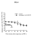
- Fig. 3 and Fig. 4 The effect on the decrease in relative optic disc blood flow due to ET-1 injection, after installation of 0.1% ophthalmic solution and nicardipine hydrochloride-containing preparation is shown in Fig. 3 and Fig. 4 , respectively.
- the initial value of blood flow before instillation was 32.3-67.1 ml/min/100 g.
- the eye after instillation of the 0.1% ophthalmic solution showed a significant 9% increase in blood flow after 30 min from the instillation as compared to the initial value.
- the action lasted for 60 minutes after the instillation.
- ET-1 was injected at this point.
- the eye after instillation of physiological saline showed a 12% decrease in blood flow 30 min after instillation as compared to immediately before the ET-1 injection, and the 32% maximum decrease after 150 min.
- the eye instilled with the 0.1% ophthalmic solution showed the same initial value of blood flow at any point in time after injection of ET-1.
- the 0.1% ophthalmic solution therefore completely inhibited the decrease in blood flow.
- the eye instilled with a nicardipine hydrochloride-containing preparation showed a 10% decrease at 60 min after the injection of ET-1 as compared to that before the injection and showed the maximum 16% decrease in the blood flow at 150 minutes after the injection.
- the same decrease in blood flow as achieved by the instillation of physiological saline was seen at any point in time.
- the retinal blood vessel of the eye instilled with physiological saline contracted to 1/2 of the normal size, and 120 minutes later, the blood vessel almost completely occluded and the action lasted for 150 minutes after the instillation.
- the retinal vasocontraction was inhibited by 65% at 30 minutes after the ET-1 injection and the significant inhibition lasted for 150 minutes after the instillation.
- mice Male Dutch color house rabbits weighing about 2 kg were purchased from Fukusaki Rabbitery Co-operation and diurnal variation of intraocular pressure was measured in advance using a pneumatonograph (PTG), based on which 26 rabbits with stable intraocular pressure were used.
- the rabbits were bred at temperature 23 ⁇ 3°C and humidity 55 ⁇ 10% on solid feed (Labo R Stock, manufactured by NIHON NOSAN KOGYO K.K., 100 g a day), while allowing free access to tap water.
- the 0.1% ophthalmic solution, and the ophthalmic solutions having a concentration of 0.0001%, 0.001% and 0.01%, which contained the compound A of the present invention (hereinafter to be respectively referred to as 0.0001% ophthalmic solution, 0.001% ophthalmic solution and 0.01% ophthalmic solution, these ophthalmic solutions optionally being generally referred to as inventive compound A-containing preparation), prepared in the same manner as in Example 1, except that the content of the compound A of the present invention was changed to 0.0001%, 0.001% and 0.01% concentration, were used.
- timolol maleate ophthalmic solution which is a ⁇ -blocker, (trademark : Timoptol 0.5%, manufactured by Banyu Pharmaceutical Co., Ltd., hereinafter to be referred to as timolol ophthalmic solution) was used.
- test preparation 50 ⁇ l was instilled into one eye and physiological saline (50 ⁇ l) was instilled into the other eye of the above-mentioned male Dutch color house rabbits.
- physiological saline 50 ⁇ l was instilled into the other eye of the above-mentioned male Dutch color house rabbits.
- intraocular pressure of both eyes was measured using PTG at 30 minutes, 1 hour, 2 hours and 4 hours after the instillation.
- the time course changes in the intraocular pressure after instillation of the 0.1% ophthalmic solution and timolol ophthalmic solution are shown in Fig. 7 .
- the inventive compound A-containing preparation of 0.1% ophthalmic solution, 0.01% ophthalmic solution, 0.001% ophthalmic solution and 0.0001% ophthalmic solution showed an intraocular pressure only within the range of physiological variation and increase was not observed.
- instillation of a timolol ophthalmic solution led to a significant decrease in intraocular pressure, wherein the maximum decrease in intraocular pressure of 1.1 mmHg was seen at 30 minutes and one hour after the instillation.
- Experimental Example 4 concurrent use with compound capable of decreasing intraocular pressure
- a 1.0% timolol ophthalmic solution (20 ⁇ l) was instilled into both eyes of 8 house rabbits with stable intraocular pressure.
- a 0.1% ophthalmic solution was instilled by 20 ⁇ l to one eye and physiological saline was instilled by 20 ⁇ l to the other eye.
- the intraocular pressure was measured using PTG at 30 minutes before Timolol instillation, immediately before instillation, 15 minutes after instillation (immediately before instillation of 0.1% ophthalmic solution or physiological saline), and 30 minutes, 1 hour, 2 hours and 4 hours after instillation.
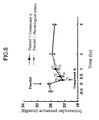
- timolol The effects of timolol on the decrease in intraocular pressure by the instillation of the 0.1% ophthalmic solution after instillation of 1.0% timolol ophthalmic solution are shown in Fig. 8 .
- the 0.1% ophthalmic solution was instilled into one eye and physiological saline was instilled into the other eye.
- the eye instilled with 0.1% solution showed a decrease in intraocular pressure of 3.3 mmHg, and the eye instilled with physiological saline showed a decrease in intraocular pressure of 3.4 mmHg as compared to immediately before timolol instillation.
- the both eyes regained intraocular pressure and returned to the initial value in 2 hours.
- a stainless vis electrode manufactured by UNIQUE MEDICAL CO., LTD.
- VEP was attached to the head of the house rabbits under general anesthesia and after about 2 weeks of awakening period, the rabbits were subjected to the test.
- VEP was measured by shooting 1.2 J xenon arc light 32 times from 30 cm before the eye under mydriasis with mydrin P (trademark) instillation and averaged. The VEP was measured at 30 minutes, 15 minutes and immediately before ET-1 injection, and after injection at 15-minute intervals for 120 minutes.
- a 0.1% ophthalmic solution was instilled by 20 ⁇ l to one eye and physiological saline was instilled by 20 ⁇ l to the other eye at 30 minutes before ET-1 injection.
- 10 -6 M ET-1 (10 ⁇ l, derived from human, manufactured by SIGMA) was injected into the central part of the vitreous body of both eyes.
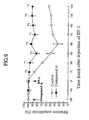
- Fig. 9 The effects of the instillation of the 0.1% ophthalmic solution on the attenuation of amplitude of VEP due to ET-1 is shown in Fig. 9 .
- the initial value of amplitude ofVEP before instillation was 30.0-76.7.
- the amplitude ofVEP of the eye instilled with physiological saline decreased by 15% as compared to that before injection and decreased by 67% at maximum 60 minutes later.
- the eye instilled with the 0.1% ophthalmic solution showed significant inhibition at any point in time of the attenuation of amplitude ofVEP of the eye instilled with physiological saline.
- the agent for ameliorating ocular circulatory disorder of the present invention increases optic disc blood flow of normal eye particularly by instillation, and inhibits vasocontraction of retinal blood vessel, decrease in optic disc blood flow and attenuation of the amplitude of VEP caused by ET-1, without increasing the intraocular pressure.
- the inventive compound is suggested to be effective as a therapeutic agent against normal tension glaucoma caused by ocular circulation disorder and retinitis pigmentosa, macular degeneration, ischemic optic neuropathy, iridocyclitis, retinal artery occlusion, retinal vein occlusion, diabetic retinopathy, ischemic optic neuropathy, retinochoroidal disease following choroidal lesion, and retinochoroidal disease associated with systemic disease.
Description
- The present invention relates to an agent for ameliorating ocular circulatory disorder. More particularly, the present invention relates to an agent for ameliorating ocular circulatory disorder, comprising a specific 1,4-dihydropyridine derivative, i.e. 3-(4-allyl-1-piperazinyl)-2,2-
dimethylpropyl methyl 2,6-dimethyl-4-(m-nitrophenyl)-1,4-dihydropyridine-3,5-dicarboxylate or an acid addition salt thereof as an active ingredient in the form of an eye drop or eye ointment. - An intraocular blood circulation (hereinafter to be referred to as ocular circulation) has two major pathways, one being a circulation via ciliary artery and the other being a circulation via central retinal artery. The ciliary artery is connected to the arteries of choroid, optic disc, iris, ciliary body and the like, and the blood is discharged from the eye through the vortex vein. The central retinal artery passes through optic nerve and is connected to the central retinal vein, wherein a part thereof is branched into arteriola at the optic disc and then into capillary. Of these, the circulatory disorder of ciliary artery includes normal tension glaucoma, retinitis pigmentosa, macular degeneration, ischemic optic neuropathy, iridocyclitis and the like. The circulatory disorder of the central retinal artery includes retinal artery occlusion, retinal vein occlusion, diabetic retinopathy, ischemic optic neuropathy, retinochoroidal disease following choroidal lesion, retinochomidal disease associated with systemic disease and the like. As is evident from the description of the above-mentioned ocular circulation pathway, the diseases caused by the above-mentioned ocular circulation disorders make an onset when the smooth circulation of retina, optic disc, choroid, iris, ciliary body and the like is prevented.
- In recent years, there has been a high incidence of glaucoma, in particular, normal tension glaucoma (also referred to as low tension glaucoma) due to blood circulatory disorder in optic disc, and therapeutic method of the disease has been desired. By the epidemiological studies in recent years, normal tension glaucoma has been elucidated to be of a disease type of glaucoma having the highest incidence, though the intraocular pressure is in the normal range, so that it is considered to be distinct from the generally known glaucoma, namely the glaucoma caused by a high intraocular pressure. The normal tension glaucoma is generally considered to be a disease associated with (1) an intraocular pressure including a biological rhythm of not more than 21 mmHg; (2) a normal open angle, (3) glaucomatous optic disc cupping and the corresponding visual field disorder (4) no intracranial lesion or paranasal sinuses disease which can cause optic atrophy and (5) no mass hemorrhage or shock [Low Tension Glaucoma and endotheline (ET-1), Folia Ophthalmolgica Japonica, voL 43, pp. 554-559 (1992) and Low Tension Glaucoma - History and Concept, Journal of the Eye, vol. 8, pp. 493-500 (1991)).
- A recent report has documented that an oral administration of a circulation ameliorating agent to a patient with normal tension glaucoma resulted in an increased optic disc blood flow and an ameliorated normal tension glaucoma [Influence of Ca2+ antagonist on changes in visual field in low tension glaucoma, Journal of Japanese Ophthalmological Society, vol. 92, pp. 792-797 (1988)]. There is a possibility that various biological vasocontracting substances that decrease blood flow may be involved in patients with normal tension glaucoma, and a significantly higher endotheline-1 (hereinafter sometimes referred to as ET-1) concentration in blood as compared to the level in healthy subjects has been reported [(Low Tension Glaucoma and Endotheline (ET-1), Folia Ophthalmolgica Japonica, voL 43, pp. 554-559 (1992)]. ET-1 is considered to act on a receptor present in vascular smooth muscle cells and directly opens the voltage-dependent Ca2+ channel [Intracellular Signal Transduction Pathway Relating to the Action and Regulation of Release of Endotheline, Experimental Medicine, )].
- Moreover, a decrease in optic disc blood flow by an injection of ET-1 into the vitreous body has been disclosed [Effect of Endotheline-1 on Ocular Circulation, Journal of Japanese Ophthalmological Society, voL 97, pp. 678-682 (1993)]. A visual evoked potential (hereinafter also referred to as VEP) detects visual optic response of optic pathway from retinal ganglion cell to light, and can be one indication of visual field disorder. VEP shows attenuation of amplitude, disappearance of wave factor and prolonged peak latency due to disorders in retinal center and visual pathway. It has been disclosed that an injection of ET-1 results in attenuation of amplitude and prolonged peak latency [Changes in Visual Function by Injection into Endotheline Vitreous Body, Journal of Japanese Ophthalmological Society, vol. 97, pp. 467-473 (1993)]. From these, a possible involvement of various biological vasocontracting substances is suggested with respect to circulatory disorders in normal tension glaucoma, of which ET-1 is particularly plausible, wherein the possibility is suggested that ET-1 may cause an ocular circulation disorder, decrease optic disc blood flow and deteriorate visual function. Therefore, amelioration of an ocular circulation disorder induced by ET-1 is considered to be one of the effective therapeutic methods of normal tension glaucoma.
- The ocular circulation disorder is most frequently seen among the retinochoroidal diseases. The retinochoroidal disease caused by an ocular circulation disorder is exemplified by retinal artery occlusion, retinal vein occlusion, diabetic retinopathy, retinitis pigmentosa, macular degeneration, retinochoroidal disease following choroidal lesion, retinochoroidal disease associated with systemic disease, and the like. While the etiology of retinal artery occlusion and retinal vein occlusion is unknown, the lumen of retinal artery or vein is occluded to cause circulation disorder in retina and optic disc. In addition, the high ET-1 concentration in blood of a patient has been also reported [Deviation of Vasospasm Factor in Retinal Artery Occlusion, Japanese Journal of Clinical Ophthalmology, vol. 46, pp. 431-434 (1992)]. It is a well-known fact that thrombosis occurs in retinal blood vessel in diabetic retinopathy, which in tum causes retinal circulatory disorder. The retinitis pigmentosa is a binocular retinal disease, which starts with night blindness in school age, gradually progresses into abnormal visual field and visual loss, and may ultimately end in blindness. This disease is hereditary and the degeneration of retinal photoreceptor cell proceeds with increasingly narrower retinochoroidal blood vessel and circulatory disorders. An ocular circulation disorder is said to be observed in macular degeneration as well. The above-mentioned diseases that accompany ocular circulation disorder have been treated by an oral administration of tocopherol nicotinate (Juvela N : vitamin E preparation manufactured by EISAI CO., LTD.)
- The optic nerve disease associated with an optic nerve disorder is exemplified by ischemic optic neuropathy and the like. The ischemic optic neuropathy gives an onset by circulatory disorder of optic nerve nutrient blood vessel. The disease accompanied by iris ciliary body circulatory disorder is exemplified by iridocyclitis and the like.
- As the drug having peripheral vasodilating action, calcium antagonists representatively including nicardipine are known, which inhibit influx of Ca ion necessary for contraction of cardiac muscle and vascular smooth muscle, thereby relaxing the cardiac muscle and vascular smooth muscle and thus leading to vasodilation that increases the blood flow.
- Japanese Patent Unexamined Publication No.
63-225355 - However, Japanese Patent Unexamined Publication No.
63-225355 - It has been also reported that the above-mentioned 1,4-dihydropyridine derivative increases blood flow in the brain, brown adipose tissue, small intestine, large intestine and skin of rat, but decreases blood flow in the liver, spleen, kidney, adrenal gland and skeletal muscle; that the decrease in blood flow by ET-1 can be suppressed in the kidney, adrenal gland, brown adipose tissue, small intestine, large intestine and skeletal muscle, but otherwise in the brain, lung and skin; and that the vasocontraction by ET-1 can be suppressed in the kidney, adrenal gland, brown adipose tissue, small intestine, large intestine and skeletal muscle but otherwise in the brain, lung and skin [Hypertens Res 17, 29-34 (1994)]. Therefore, while the above-mentioned blood flow increasing action of 1,4-dihydropyridine derivative and inhibition of the decrease in blood flow and vasocontraction by ET-1 are organ specific, there is no knowing if such action can be also found in retinochoroid.
-
EP-A-0289746 andWO-A-93/23082 dimethylpropyl methyl 2,6-dimethyl-4-(m-nitrophenyl)-1,4-dihydropyridine-3,5-dicarboxylate (hereinafter referred to as Compound A); is useful for for the treatment of glaucoma. - MAETANI SATORU ET AL: "Effect of systemic calcium antagonist on a model of ocular circulation disturbance induced by endothelin-1." NIPPON GANKA GAKKAI ZASSHI, vol. 99, no. 1, 1995, discloses that the calcium antagonist nicardipine hydrochloride inhibits the disturbance of ocular circulation induced by ET-1, by systemic administration.
- KANDA M ET AL: "Effects of the novel water-soluble calcium antagonist (+/-)-3-(4-allyl-1-piperazinyl)-2,2-., XP002235686 discloses that Compound A inhibited the ET-1-induced contraction in isolated canine cerebral arteries by systemic administration.
-
EP-A-0577913 discloses the use of timolol or its maleate or hemisuccinate form, for the topical treatment of glaucoma and ocular hypertension. - The present invention aims at solving the above-mentioned problems and has been made with the purpose of providing an agent for ameliorating an ocular circulatory disorder, which has a superior blood flow increasing action in retinochoroid and which has an inhibitory action on the vasocontraction, decrease in blood flow and attenuation of the amplitude ofVEP by ET-1 which is one of the biological vasocontracting substances.
- The present inventors have conducted intensive studies in an attempt to achieve the above-mentioned objects and found that, from among the calcium antagonists, the above-mentioned 1,4-dihydropyridine derivative alone shows a superior blood flow increasing action in retinochoroid and an inhibitory action on vasocontraction, decrease in blood flow and attenuation of the amplitude of VEP by ET-1, and that, particularly in the form of an eye drop, the compound of the present invention does not increase intraocular pressure, though other 1,4-dihydropyridine derivatives reportedly do [nicardipine chloride (Effects of Ca2+ Channel Blocker on Intraocular Pressure and Kinetics of Aqueous Humor of House Rabbits, Journal of Japanese Ophthalmological Society, 97, 665-671 (1993)].
- Accordingly, the present invention provides the following:
- (1) 3-(4-Allyl-1-piperazinyl)-2,2-
dimethylpropyl methyl 2,6-dimethyl-4-(m-nitrophenyl)-1,4-dihydropyridine-3,5-dicarboxylate or an acid addition salt thereof in the form of an eye drop or an eye ointment for the prophylaxis and treatment of a disease caused by a circulatory disorder in the ciliary artery system selected from the group consisting of normal tension glaucoma, retinitis pigmentosa, macular degeneration, ischemic optic neuropathy and iridocyclitis. - (2) 3-(4-Allyl-1-piperazinyl)-2,2-
dimethylpropyl methyl 2,6-dimethyl-4-(m-nitrophenyl)-1,4-dihydropyridine-3,5-dicarboxylate or an acid addition salt thereof in the form of an eye drop or an eye ointment for the prophylaxis and treatment of a disease caused by a circulatory disorder in the central retinal artery system selected from the group consisting of retinal artery occlusion, retinal vein occlusion, diabetic retinopathy, ischemic optic neuropathy, retinochoroidal disease following choroidal lesion and retinochoroical disease accompanied by systemic disease. - (3) A combination of 3-(4-allyl-1-piperazinyl)-2,2-
dimethylpropyl methyl 2,6-dimethyl-4-(m-nitrophenyl)-1,4-dihydropyridine-3,5-dicarboxylate or an acid addition salt thereof and timolol in the form of an eye drop or an eye ointment for the prophylaxis and treatment of normal tension glaucoma. - (4) An agent comprising 3-(4-allyl-1-piperazinyl)-2,2-
dimethylpropyl methyl 2,6-dimethyl-4-(m-nitrophenyl)-1,4-dihydropyridine-3,5-dicarboxylate or an acid addition salt thereof in the form of an eye drop or an eye ointment for the prophylaxis and treatment of a disease caused by a circulatory disorder in the ciliary artery system selected from the group consisting of normal tension glaucoma, retinitis pigmentosa, macular degeneration, ischemic optic neuropathy and iridocyclitis. - (5) An agent comprising 3-(4-allyl-1-piperazinyl)-2,2-
dimethylpropyl methyl 2,6-dimethyl-4-(m-nitrophenyl)-1,4-dihydropyridine-3,5-dicarboxylate or an acid addition salt thereof in the form of an eye drop or an eye ointment for the prophylaxis and treatment of a disease caused by a circulatory disorder in the central retinal artery system selected from the group consisting of retinal artery occlusion, retinal vein occlusion, diabetic retinopathy, ischemic optic neuropathy, retinochoroidal disease following choroidal lesion and retinochoroical disease accompanied by systemic disease. - (6) An agent comprising a combination of 3-(4-allyl-1-piperazinyl)-2,2-
dimethylpropyl methyl 2,6-dimethyl-4-(m-nitrophenyl)-1,4-dihydropyridine-3,5-dicarboxylate or an acid addition salt thereof and timolol in the form of an eye drop or an eye ointment for the prophylaxis and treatment of normal tension glaucoma. -
-
Fig. 1 is a graph showing the time course changes in relative optic disc blood flow (blood flow at each point in time when the initial value is 100%) in normal eye after instillation of a 0.1% ophthalmic solution (containing 0.1% of the compound A of the present invention) in Experimental Example 1, wherein the axis of abscissa shows time (min) and the axis of ordinate shows relative optic disc blood flow (%). Each value shows mean±standard error (n=6). Significant difference from the control is found in *1 ; P<0.01,*2 ; P<0.001 (paired t-test). The black circle shows instillation of a 0.1% ophthalmic solution and white circle shows instillation of physiological saline. -
Fig. 2 is a graph showing the time course changes in relative optic disc blood flow in normal eye after instillation of a preparation containing nicardipine hydrochloride in Experimental Example 1, wherein the axis of abscissa shows time (min) and the axis of ordinate shows relative optic disc blood flow (%). Each value shows mean± standard error (n=6). The black circle shows instillation of a preparation containing nicardipine hydrochloride and the white circle shows instillation of physiological saline. -
Fig. 3 is a graph showing the time course changes in relative optic disc blood flow upon injection of ET-1 after instillation of a 0.1% ophthalmic solution (containing 0.1% of the compound A of the present invention) in Experimental Example 2, wherein the axis of abscissa shows time (min) when the injection of ET-1 is 0 and the axis of ordinate shows relative optic disc blood flow (%). Each value shows mean±standard error (n=7). A significant difference from control is found in #1 ; P <0.05. A significant difference from control is found in *1 ; P <0.05, *2 ; P<0.0 1, *3 ; P<0.001 (paired t-test) in the amount of difference at each point in time from that immediately before the addition of ET-1. The black circle shows instillation of a 0.1% ophthalmic solution and white circle shows instillation of physiological saline. -
Fig. 4 is a graph showing the time course changes in relative optic disc blood flow upon injection of ET-1 after instillation of a preparation containing nicardipine hydrochloride in Experimental Example 2, wherein the axis of abscissa shows time (min) when the injection of ET-1 is 0 and the axis of ordinate shows relative optic disc blood flow (%). Each value shows mean±standard error (n=6). The black circle shows instillation of a preparation containing nicardipine hydrochloride and white circle shows instillation of physiological saline. -
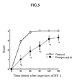
Fig. 5 is a graph showing the time course changes in vasocontraction upon injection of ET-1 after instillation of a 0.1% ophthalmic solution (containing 0.1% of the compound A of the present invention) in Experimental Example 2, wherein the axis of abscissa shows time (min) when the injection of ET-1 is 0 and the axis of ordinate shows score. Each value shows mean±standard error (n=7). A significant difference from control is found in *1 ; P<0.05, *2 ; P<0.01 (paired t-test). The black circle shows instillation of a 0.1% ophthalmic solution and white circle shows instillation of physiological saline. -
Fig. 6 is a graph showing the time course changes in vasocontraction upon injection of ET-1 after instillation of a preparation containing nicardipine hydrochloride wherein the axis of abscissa shows time (min) when the injection of ET-1 is 0 and the axis of ordinate shows score. Each value shows mean± standard error (n=6). The black circle shows instillation of nicardipine hydrochloride and white circle shows instillation of physiological saline. -
Fig. 7 is a graph showing the time course changes in intraocular pressure after instillation of a preparation containing compound A of the present invention (0.1% ophthalmic solution, 0.01% ophthalmic solution, 0.001% ophthalmic solution and 0.0001% ophthalmic solution) or a timolol ophthalmic solution. The axis of abscissa shows time (min) and the axis of ordinate shows intraocular pressure (mmHg). The black circle shows instillation of the timolol ophthalmic solution, white triangle shows that of 0.1% ophthalmic solution, black triangle shows instillation of 0.01% ophthalmic solution, white square shows instillation of a 0.001% ophthalmic solution, black square shows instillation of a 0.0001% ophthalmic solution and white circle shows instillation of physiological saline. -
Fig. 8 is a graph showing the time course changes in intraocular pressure after instillation of a 0.1% ophthalmic solution (containing compound A of the present invention) or physiological saline after instillation of a 1.0% timolol ophthalmic solution in Experimental Example 4. The axis of abscissa shows time (hr) when the instillation of the 1.0% timolol ophthalmic solution is 0 and the axis of ordinate shows intraocular pressure (mmHg). Each value shows mean± standard error (n=8). A significant difference from the initial value is found in *1 ; P<0.05,*2 ; P<0.01,*3 ; P<0.00 (paired t-test). The black circle shows instillation of a 0.1% ophthalmic solution and white circle shows instillation of physiological saline. -
Fig. 9 is a graph showing the time course changes in relative amplitude of VEP upon injection of ET-1 after instillation of a 0.1% ophthalmic solution (containing 0.1% of the compound A of the present invention) in Experimental Example 5, wherein the axis of abscissa shows time (min) when the injection of ET-1 is 0 and the axis of ordinate shows relative amplitude (%). Each value shows mean± standard error (n=3). A significant difference from control is found at each point in time in *1 ; P<0.05,*2 ; P<0.01,*3 ; P<0.00 (paired t-test). The black circle shows instillation of a 0.1% ophthalmic solution and white circle shows instillation of physiological saline. - The present invention is described in detail in the following. The agent for ameliorating ocular circulatory disorder of the present invention contains the above-mentioned 1,4-dihydropyndine derivative or an acid addition salt thereof as an active ingredient.
- The 1,4-dihydropyridine derivative (I) of the present invention is the following compound.
- 3-(4-allyl-1-piperazinyl)-2,2-
dimethylpropyl methyl 2,6-dimethyl-4-(m-nitrophenyl)-1,4-dihydropyridine-3,5-dicarboxylate dihydrochloride (hereinafter to be referred to as compound A of the present invention) - The 1,4-dihydropyridine derivative of the present invention has a blood flow increasing action in retinochoroid, and inhibits vasocontraction and decrease in blood flow caused by ET-1, which is one of the biological vasocontracting substances, in mammals (e.g., human, cow, horse, mouse, rat, dog, cat, rabbit and the like), and also inhibits increase in intraocular pressure when in the form of an eye drop.
- The 1,4-dihydropyridine derivative of the present invention is a calcium antagonist, but nicardipine, which is also a calcium antagonist, does not show such effect. The effect is characteristic of the compound of the present invention.
- Accordingly, 1,4-dihydropyridine derivative and an acid addition salt thereof are useful as an agent for ameliorating ocular circulatory disorder in mammals such as human, cow, horse, dog, mouse, rat and the like, and they are expected to be applicable for the prophylaxis and treatment of the diseases selected from normal tension glaucoma caused by ocular circulation disorder, retinitis pigmentosa, macular degeneration, ischemic optic neuropathy, iridocyclitis, retinal artery occlusion, retinal vein occlusion, diabetic retinopathy, retinochoroidal disease following choroidal lesion, and retinochoroidal disease accompanied by systemic disease.
- The agent for ameliorating ocular circulatory disorder of the present invention may contain a compound capable of decreasing intraocular pressure, in addition to the 1,4-dihydropyridine derivative. The compound capable of decreasing intraocular pressure to be used in the present invention is free of any particular limitation and may be a known compound. Examples thereof include e.g., pilocarpine, carbachol, acetylcholine esterase inhibitor, sympathetic agent (e.g., epinephrine, dipivalyl epinephrine, paraaminoclonidine, α-methyldipivalyl epinephrine, apraclonidine, clondine), β-blocker (e.g., betaxolol, levobunolol, timolol), carbonate dehydratase inhibitor (e.g., acetazolamide, methazolamide, ethoxzolamide, MK507). Of these, preferred are timolol, betaxolol, levobunolol, carteolol, pilocarpine, carbachol, MK927, MK507, AL04414, AL04623, AL04862, epinephrine, dipivalyl epinephrine, α-methyldipivalylepinephrine, apraclonidine and clonidine.
- The concurrent use of a compound capable of decreasing intraocular pressure makes possible the prophylaxis and treatment of glaucoma caused by circulatory disorder (e.g., normal tension glaucoma).
- When the 1,4-dihydropyridme derivative and an acid addition salt thereof are used as pharmaceutical products, they can be administered in the form of an eye ointment or an eye drop. In consideration of influence on other circulatory system, the particularly preferable dosage form is an eye drop.
- The 1,4-diydropyridine derivative and an acid addition salt thereof have high solubility in water as compared to conventional calcium antagonists such as nicardipine and the like. They can be easily prepared into an eye drop and the like, which is clinically extremely easy to handle.
- When an eye drop is prepared, the pH thereof is generally set to 3 to 7, preferably 4 to 6.
- The preparation in the above-mentioned dosage form can be produced by admixing additives typically necessary for usual preparation, and processing according to a conventional method. For example, the additives to be used for an eye drop include the following.
- Buffering agent may be phosphate buffer, borate bluffer, citrate buffer, tartrate buffer, acetate buffer, amino acid and the like. Preferred are buffers having buffer capability in the pH range of from 2 to 9.
- Examples of isotonizing agent include saccharides such as sorbitol, glucose, mannitol and the like, polyhydric alcohol such as glycerol, polyethylene glycol, propylene glycol and the like, and salt such as sodium chloride and the like.
- Examples of preservative include benzalkonium chloride, benzetonium chloride, p-hydroxybenzoic acid esters such as methyl p-hydroxybenzoale, ethyl p-hydroxybenzoate and the like, benzyl alcohol, phenetyl alcohol, sorbic acid and a salt thereof, timerosal, chlorobutanol and the like.
- Examples of thickener include hydroxyethyl cellulose, hydroxypropyl cellulose, methyl cellulose, hydroxypropylmethyl cellulose, carboxymethyl cellulose and a salt thereof.
- Examples of solubilizer (stabilizer) include water soluble polymer such as cyclodextrin, polyvinylpyrrolidone and the like, surfactant such as
polysorbate 80 and the like. - Examples of chelating agent include sodium edetate, sodium citrate, condensed sodium phosphate and the like.
- Examples of suspending agent include surfactant such as
polysorbate 80 and the like, and water soluble polymer such as sodium methyl cellulose, hydroxypropyl methyl cellulose, methyl cellulose and the like. - While the dose and administration frequency vary depending on symptom, age, body weight and administration route, when, for example, administered as an eye drop to an adult, a preparation containing 1,4-dihydropyridine derivative (I) and a compound that decreases intraocular pressure each in a concentration of 0.0001-10 w/v%, preferably 0.001-5 w/v%, can be administered several times a day, preferably 1 to 6 times, several drops a time, preferably 1 to 3 drops. When in use as an eye ointment, a preparation containing 1,4-dihydropyridine derivative (I) and a compound that decreases intraocular pressure each in a concentration of 0.0001-10 w/v%, preferably 0.001- 5 w/v%, can be applied several times a day, preferably 1 to 6 times.
- The present invention is explained in more detail in the following by way of Examples and Experimental Examples.
- An ophthalmic solution having the following composition, that contained the compound of the present invention, was prepared.
Compound of the present invention 0.1 g Sodium acetate 0.1 g Sodium chloride 0.9 g Benzalkonium chloride 0.005 g Acetic acid suitable amount Sterile purified water suitable amount Total amount 100 ml (pH 5) - An ophthalmic solution having the following composition, that contained the compound of the present invention, was prepared.
Compound of the present invention 0.01 g Sodium acetate 0.1 g β-cyclodextrin 0.190 g Sodium chloride 0.9 g Benzalkonium chloride 0.005 g Acetic acid suitable amount Sterile purified water suitable amount Total amount 100 ml (pH 5) - An ophthalmic solution having the following composition, that contained the compound of the present invention, was prepared.
Compound of the present invention 0.001 g Sodium acetate 0.1 g Sodium edetate 0.01 g Sodium chloride 0.9 g Benzalkonium chloride 0.005 g Acetic acid suitable amount Sterile purified water suitable amount Total amount 100 ml (pH 5) - An ophthalmic solution having the following composition, that contained the compound of the present invention, was prepared.
Compound of the present invention 0.1 g Sodium acetate 0.1 g Sodium edetate 0.005 g Sodium chloride 0.9 g Benzalkonium chloride 0.005 g Acetic acid suitable amount Sterile purified water suitable amount Total amount 100 ml (pH 5) - An ophthalmic solution having the following composition, that contained the compound of the present invention, was prepared.
Compound of the present invention 0.1 g Timolol 0.5 g Sodium acetate 0.1 g Sodium edetate 0.005 g Sodium chloride 0.9 g Benzalkonium chloride 0.005 g Acetic acid suitable amount Sterile purified water suitable amount Total amount 100 ml (pH 5) - An eye ointment containing the compound of the present invention was prepared.
Compound of the present invention 0.1 g Liquid paraffin 10 g Sterile purified water suitable amount Total amount 100 g - Male Dutch color house rabbits weighing about 2 kg were purchased from Fukusaki Rabbitery Co-operation and used upon confirmation of the absence of abnormality in the eye. The rabbits were bred at temperature 23±3°C and humidity 55± 10% on solid feed (Labo R Stock, manufactured by NIHON NOSAN KOGYO K.K., 100 g a day), while allowing free access to tap water.
- The ophthalmic solution prepared in Example 1 containing the compound A of the present invention in a concentration of 0.1% (hereinafter to be sometimes referred to as 0.1% ophthalmic solution) was used. As a control drug, 0.1% nicardipine hydrochloride solution (trademark :
Perdipine injection 2 mg, manufactured by YAMANOUCHI PHARMACEUTICAL CO., LTD., hereinafter to be sometimes referred to as nicardipine hydrochloride-containing preparation). - The rabbits were placed in a retaining cage and optic disc was examined for abnormality upon mydriasis with mydrin P (trademark, manufactured by SANTEN PHARMACEUTICAL CO., LTD.) Urethane (dissolved in distilled water in 20%, 1 g/kg) was subcutaneously administered in the abdomen for systemic anesthesia. One or two hours later, a plate type indifferent electrode (manufactured by Biomedical Science, BE-R10) was subcutaneously attached to the head under stable anesthesia. The upper and lower eyelids of both eyes were pulled open upward and downward with a suture and conjunctiva at 6 o'clock was incised. The suture was passed through subrectus muscle and pulled downward to fix the eyeball Sclera at about 3 mm from corneal limbus at 6 o'clock was opened with a 27 G needle and a needle different electrode (manufactured by Biomedical Science, BE-NSP 450-30) was punctured from there through vitreous body into optic disc. The opening of the sclera and different electrode were fixed with Aron Alpha (trademark, manufactured by KONISHI CO., LTD.)
- After puncture of different electrode, the rabbits were stood for about one hour. The rabbits were set to inhale 10% hydrogen for about 5 min, and the height from the base line to the curve of the clearance curve depicted on a recorder was measured at 12-seoond intervals starting from the peak. The relationship between the value obtained and the time was plotted on a semilogarithm graph. A straight line was drawn to gather a greatest number possible of measurement dots and half-life (T1/2) was calculated from the straight line. The blood flow was determined from the following theoretical formula of Kety (Journal of Clinical Investigate, voL 27, pp. 476-483 (1948)). The test was started after measurement of blood flow at 15-minute intervals and after the measurement of stable values became available. The initial value was an average of two measurements before starting the test.

- A 0.1% ophthalmic solution or a nicardipine hydrochloride-containing preparation (20 µl) was instilled into one eye and physiological saline (20 µl) was instilled into the other eye.
- The relative optic disc blood flow (blood flow when initial value was 100%) in the normal eye after instillation of the 0.1% ophthalmic solution and nicardipine hydrochloride-containing preparation is shown in
Fig. 1 andFig. 2 , respectively. The initial value of the blood flow before instillation was 27.4 - 58.5 ml/min/100 g. - The eye after instillation of the 0.1% ophthalmic solution showed a significantly increased blood flow after 15 min as compared to the eye after instillation of physiological saline, and at 45 min after instillation, the maximum increase of 31.7% was obtained as compared to the initial value. The action lasted for 180 min after the instillation. On the other hand, the eye after instillation of the nicardipine hydrochloride-containing preparation showed a 5-10% increase in blood flow as compared to the initial value, for 30 min after instillation, but the eye instilled with physiological saline also showed an increase. Thus, significant difference was not observed. At any point in time thereafter, increase in blood flow could not be found.
- It is postulated that the 0.1% ophthalmic solution increased the blood flow in the optic disc because the compound of the present invention acted on vascular smooth muscle in the optic disc via cornea to cause vasodilation, and because the compound of the present invention caused dilation of long posterior ciliary artery via sclera to ultimately increase the choroidal circulation.
- Measured in the same manner as in Experimental Example 1.
- Instilled in the same manner as in Experimental Example 1.
- At 60 minutes after the instillation of the drug, 10-6M ET-1 (10 µl, derived from human, manufactured by SIGMA) was injected into the central part of the vitreous body of both eyes while observing the eye ground with a vitrectomy lens.
- The retinal blood vessel diameter was macroscopically observed before injection of ET-1 and after injection at 30-minute intervals for 150 minutes using an inverted image mirror after measurement of blood flow according to the following evaluation criteria.
Normal retinal blood vessel diameter 0 Decreased to 3/4 of retinal blood vessel diameter 1 Decreased to 1/2 of retinal blood vessel diameter 2 Decreased to 1/4 of retinal blood vessel diameter 3 Presence of blood vessel barely confirmed or Complete occlusion 4 - The effect on the decrease in relative optic disc blood flow due to ET-1 injection, after installation of 0.1% ophthalmic solution and nicardipine hydrochloride-containing preparation is shown in
Fig. 3 andFig. 4 , respectively. The initial value of blood flow before instillation was 32.3-67.1 ml/min/100 g. - The eye after instillation of the 0.1% ophthalmic solution showed a significant 9% increase in blood flow after 30 min from the instillation as compared to the initial value. The action lasted for 60 minutes after the instillation. ET-1 was injected at this point. As a result, the eye after instillation of physiological saline showed a 12% decrease in
blood flow 30 min after instillation as compared to immediately before the ET-1 injection, and the 32% maximum decrease after 150 min. - In contrast, the eye instilled with the 0.1% ophthalmic solution showed the same initial value of blood flow at any point in time after injection of ET-1. The 0.1% ophthalmic solution therefore completely inhibited the decrease in blood flow. The eye instilled with a nicardipine hydrochloride-containing preparation showed a 10% decrease at 60 min after the injection of ET-1 as compared to that before the injection and showed the maximum 16% decrease in the blood flow at 150 minutes after the injection. As a result, the same decrease in blood flow as achieved by the instillation of physiological saline was seen at any point in time.
- The effect on the retinal vasocontraction due to ET-1 injection, after instillation of 0.1% ophthalmic solution and nicardipine hydrochloride-containing preparation, is shown in
Fig. 5 andFig. 6 , respectively. - At 30 minutes after the ET-1 injection, the retinal blood vessel of the eye instilled with physiological saline contracted to 1/2 of the normal size, and 120 minutes later, the blood vessel almost completely occluded and the action lasted for 150 minutes after the instillation. In the eye instilled with 0.1% ophthalmic solution, the retinal vasocontraction was inhibited by 65% at 30 minutes after the ET-1 injection and the significant inhibition lasted for 150 minutes after the instillation.
- In contrast, the eye instilled with nicardipine hydrochloride-containing preparation showed contraction of retinal blood vessel to 1/2 or less of the normal size, after 30 minutes from the ET-1 injection and
complete occlusion 90 minutes later. Thus, there was found no significant difference from the eye instilled with physiological saline. - Male Dutch color house rabbits weighing about 2 kg were purchased from Fukusaki Rabbitery Co-operation and diurnal variation of intraocular pressure was measured in advance using a pneumatonograph (PTG), based on which 26 rabbits with stable intraocular pressure were used. The rabbits were bred at temperature 23±3°C and humidity 55±10% on solid feed (Labo R Stock, manufactured by NIHON NOSAN KOGYO K.K., 100 g a day), while allowing free access to tap water.
- The 0.1% ophthalmic solution, and the ophthalmic solutions having a concentration of 0.0001%, 0.001% and 0.01%, which contained the compound A of the present invention (hereinafter to be respectively referred to as 0.0001% ophthalmic solution, 0.001% ophthalmic solution and 0.01% ophthalmic solution, these ophthalmic solutions optionally being generally referred to as inventive compound A-containing preparation), prepared in the same manner as in Example 1, except that the content of the compound A of the present invention was changed to 0.0001%, 0.001% and 0.01% concentration, were used. As a control drug, a 0.5% timolol maleate ophthalmic solution, which is a β-blocker, (trademark : Timoptol 0.5%, manufactured by Banyu Pharmaceutical Co., Ltd., hereinafter to be referred to as timolol ophthalmic solution) was used.
- A test preparation (50 µl) was instilled into one eye and physiological saline (50 µl) was instilled into the other eye of the above-mentioned male Dutch color house rabbits. Using the value immediately before instillation as the initial value, the intraocular pressure of both eyes was measured using PTG at 30 minutes, 1 hour, 2 hours and 4 hours after the instillation.
- The time course changes in the intraocular pressure after instillation of the 0.1% ophthalmic solution and timolol ophthalmic solution are shown in
Fig. 7 . The inventive compound A-containing preparation of 0.1% ophthalmic solution, 0.01% ophthalmic solution, 0.001% ophthalmic solution and 0.0001% ophthalmic solution showed an intraocular pressure only within the range of physiological variation and increase was not observed. In contrast, instillation of a timolol ophthalmic solution led to a significant decrease in intraocular pressure, wherein the maximum decrease in intraocular pressure of 1.1 mmHg was seen at 30 minutes and one hour after the instillation. Experimental Example 4 : concurrent use with compound capable of decreasing intraocular pressure - Male Dutch color house rabbits weighing about 2 kg were purchased from Fukusaki Rabbitery Co-operation and habituated in a retaining cage. The diurnal variation of intraocular pressure was measured in advance using a pneumatonograph (PTG), based on which 16 rabbits with stable intraocular pressure were used. The rabbits were bred at temperature 23±3°C and humidity 55± 10% on solid feed (Labo R Stock, manufactured by NIHON NOSAN KOGYO K.K., 100 g a day), while allowing free access to tap water.
- A 1.0% timolol ophthalmic solution (20 µl) was instilled into both eyes of 8 house rabbits with stable intraocular pressure. At 15 minutes after the instillation, a 0.1% ophthalmic solution was instilled by 20 µl to one eye and physiological saline was instilled by 20 µl to the other eye. The intraocular pressure was measured using PTG at 30 minutes before Timolol instillation, immediately before instillation, 15 minutes after instillation (immediately before instillation of 0.1% ophthalmic solution or physiological saline), and 30 minutes, 1 hour, 2 hours and 4 hours after instillation.
- The effects of timolol on the decrease in intraocular pressure by the instillation of the 0.1% ophthalmic solution after instillation of 1.0% timolol ophthalmic solution are shown in
Fig. 8 . At 15 minutes after timolol instillation, a decrease in intraocular pressure of 4.0 mmHg and 3.2 mmHg as compared to immediately before timolol instillation was seen in the both eyes. Immediately thereafter, the 0.1% ophthalmic solution was instilled into one eye and physiological saline was instilled into the other eye. At 15 minutes later (30 minutes after timolol instillation), the eye instilled with 0.1% solution showed a decrease in intraocular pressure of 3.3 mmHg, and the eye instilled with physiological saline showed a decrease in intraocular pressure of 3.4 mmHg as compared to immediately before timolol instillation. Thus, no effects of instillation of the 0.1% ophthalmic solution was found. Thereafter, the both eyes regained intraocular pressure and returned to the initial value in 2 hours. - A stainless vis electrode (manufactured by UNIQUE MEDICAL CO., LTD.) for VEP measurement was attached to the head of the house rabbits under general anesthesia and after about 2 weeks of awakening period, the rabbits were subjected to the test. VEP was measured by shooting 1.2 J xenon arc light 32 times from 30 cm before the eye under mydriasis with mydrin P (trademark) instillation and averaged. The VEP was measured at 30 minutes, 15 minutes and immediately before ET-1 injection, and after injection at 15-minute intervals for 120 minutes.
- A 0.1% ophthalmic solution was instilled by 20 µl to one eye and physiological saline was instilled by 20 µl to the other eye at 30 minutes before ET-1 injection.
- At 30 minutes after the drug instillation, 10-6M ET-1 (10 µl, derived from human, manufactured by SIGMA) was injected into the central part of the vitreous body of both eyes.
- The effects of the instillation of the 0.1% ophthalmic solution on the attenuation of amplitude of VEP due to ET-1 is shown in
Fig. 9 . The initial value of amplitude ofVEP before instillation was 30.0-76.7. After ET-1 injection, the amplitude ofVEP of the eye instilled with physiological saline decreased by 15% as compared to that before injection and decreased by 67% at maximum 60 minutes later. In contrast, the eye instilled with the 0.1% ophthalmic solution showed significant inhibition at any point in time of the attenuation of amplitude ofVEP of the eye instilled with physiological saline. - As is evident from the foregoing explanation, the agent for ameliorating ocular circulatory disorder of the present invention increases optic disc blood flow of normal eye particularly by instillation, and inhibits vasocontraction of retinal blood vessel, decrease in optic disc blood flow and attenuation of the amplitude of VEP caused by ET-1, without increasing the intraocular pressure. Therefore, the inventive compound is suggested to be effective as a therapeutic agent against normal tension glaucoma caused by ocular circulation disorder and retinitis pigmentosa, macular degeneration, ischemic optic neuropathy, iridocyclitis, retinal artery occlusion, retinal vein occlusion, diabetic retinopathy, ischemic optic neuropathy, retinochoroidal disease following choroidal lesion, and retinochoroidal disease associated with systemic disease.
Claims (6)
- 3-(4-Allyl-1-piperazinyl)-2,2-dimethylpropyl methyl 2,6-dimethyl-4-(m-nitrophenyl)-1,4-dihydropyridine-3,5-dicarboxylate or an acid addition salt thereof in the form of an eye drop or an eye ointment for the prophylaxis and treatment of a disease caused by a circulatory disorder in the ciliary artery system selected from the group consisting of normal tension glaucoma, retinitis pigmentosa, macular degeneration, ischemic optic neuropathy and iridocyclitis.
- 3-(4-Allyl-1-piperazinyl)-2,2-dimethylpropyl methyl 2,6-dimethyl-4-(m-nitrophenyl)-1,4-dihydropyridine-3,5-dicarboxylate or an acid addition salt thereof in the form of an eye drop or an eye ointment for the prophylaxis and treatment of a disease caused by a circulatory disorder in the central retinal artery system selected from the group consisting of retinal artery occlusion, retinal vein occlusion, diabetic retinopathy, ischemic optic neuropathy, retinochoroidal disease following choroidal lesion and retinochoroical disease accompanied by systemic disease.
- A combination of 3-(4-allyl-1-piperazinyl)-2,2-dimethylpropyl methyl 2,6-dimethyl-4-(m-nitrophenyl)-1,4-dihydropyridine-3,5-dicarboxylate or an acid addition salt thereof and timolol in the form of an eye drop or an eye ointment for the prophylaxis and treatment of normal tension glaucoma.
- An agent comprising 3-(4-allyl-1-piperazinyl)-2,2-dimethylpropyl methyl 2,6-dimethyl-4-(m-nitrophenyl)-1,4-dihydropyridine-3,5-dicarboxylate or an acid addition salt thereof in the form of an eye drop or an eye ointment for the prophylaxis and treatment of a disease caused by a circulatory disorder in the ciliary artery system selected from the group consisting of normal tension glaucoma, retinitis pigmentosa, macular degeneration, ischemic optic neuropathy and iridocyclitis.
- An agent comprising 3-(4-allyl-1-piperazinyl)-2,2-dimethylpropyl methyl 2,6-dimethyl-4-(m-nitrophenyl)-1,4-dihydropyridine-3,5-dicarboxylate or an acid addition salt thereof in the form of an eye drop or an eye ointment for the prophylaxis and treatment of a disease caused by a circulatory disorder in the central retinal artery system selected from the group consisting of retinal artery occlusion, retinal vein occlusion, diabetic retinopathy, ischemic optic neuropathy, retinochoroidal disease following choroidal lesion and retinochoroical disease accompanied by systemic disease.
- An agent comprising a combination of 3-(4-allyl-1-piperazinyl)-2,2-dimethylpropyl methyl 2,6-dimethyl-4-(m-nitrophenyl)-1,4-dihydropyridine-3,5-dicarboxylate or an acid addition salt thereof and timolol in the form of an eye drop or an eye ointment for the prophylaxis and treatment of normal tension glaucoma.
Applications Claiming Priority (5)
| Application Number | Priority Date | Filing Date | Title |
|---|---|---|---|
| JP28568496 | 1996-10-28 | ||
| JP28568496 | 1996-10-28 | ||
| JP24555997 | 1997-09-10 | ||
| JP24555997 | 1997-09-10 | ||
| PCT/JP1997/003866 WO1998018471A1 (en) | 1996-10-28 | 1997-10-23 | Drugs for ameliorating ocular circulatory disorders |
Publications (3)
| Publication Number | Publication Date |
|---|---|
| EP0968716A1 EP0968716A1 (en) | 2000-01-05 |
| EP0968716A4 EP0968716A4 (en) | 2003-05-21 |
| EP0968716B1 true EP0968716B1 (en) | 2008-12-31 |
Family
ID=26537282
Family Applications (1)
| Application Number | Title | Priority Date | Filing Date |
|---|---|---|---|
| EP97909623A Expired - Lifetime EP0968716B1 (en) | 1996-10-28 | 1997-10-23 | Drugs for ameliorating ocular circulatory disorders |
Country Status (7)
| Country | Link |
|---|---|
| US (1) | US6451799B1 (en) |
| EP (1) | EP0968716B1 (en) |
| JP (1) | JP4828670B2 (en) |
| CA (1) | CA2270119A1 (en) |
| DE (1) | DE69739190D1 (en) |
| ES (1) | ES2317650T3 (en) |
| WO (1) | WO1998018471A1 (en) |
Families Citing this family (7)
| Publication number | Priority date | Publication date | Assignee | Title |
|---|---|---|---|---|
| CA2360689A1 (en) | 1999-01-29 | 2000-08-03 | Senju Pharmaceutical Co., Ltd. | Anti-inflammatory agents and inhibitors against increase in ocular tension caused by irradiation with lasers, containing 1,4-dihydropyridine derivatives |
| DE60014629T2 (en) * | 1999-04-14 | 2006-02-23 | Senju Pharmaceutical Co., Ltd. | 1,4-dihydropyridine derivatives as inhibitors of visual hypofunction induced by optic nerve cell injury induced by factors other than optical circulatory disorders. |
| US8088773B2 (en) * | 2005-05-12 | 2012-01-03 | The Texas A&M University System | Therapeutic compositions and methods |
| KR20140026410A (en) * | 2011-03-15 | 2014-03-05 | 유니버시티 오브 유타 리서치 파운데이션 | Methods of diagnosing and treating vascular associated maculopathy and symptoms thereof |
| SG11201408230VA (en) | 2012-06-11 | 2015-01-29 | Macuclear Inc | Therapeutic formulation and methods of treatment |
| ITUB20169937A1 (en) | 2016-01-12 | 2017-07-12 | Medical And Biotechnological Services In Sigla M B S Srl | PHARMACEUTICAL FORMULATIONS AND THEIR USE FOR THE TREATMENT OF PIGMENTAL RETINITIS |
| EP4233867A1 (en) | 2022-02-24 | 2023-08-30 | Nicox SA | Pde5 inhibitor for use in the treatment of anterior ischemic optic neuropathy |
Citations (1)
| Publication number | Priority date | Publication date | Assignee | Title |
|---|---|---|---|---|
| EP0577913A1 (en) * | 1992-07-08 | 1994-01-12 | Leiras Oy | Pharmaceutical preparation |
Family Cites Families (6)
| Publication number | Priority date | Publication date | Assignee | Title |
|---|---|---|---|---|
| JPH0688973B2 (en) * | 1987-03-12 | 1994-11-09 | 京都薬品工業株式会社 | 1,4-dihydropyridine derivative |
| JPH03145464A (en) * | 1989-10-30 | 1991-06-20 | Kyoto Yakuhin Kogyo Kk | Optically active 1,4-dihydropyridine derivative |
| JP3145464B2 (en) | 1991-09-30 | 2001-03-12 | 株式会社リコー | Decal equipment |
| EP0639986A1 (en) | 1992-05-13 | 1995-03-01 | Alcon Laboratories, Inc. | Topical ophthalmic compositions comprising a combination of calcium antagonists with known antiglaucoma agents |
| US5922773A (en) * | 1992-12-04 | 1999-07-13 | The Children's Medical Center Corp. | Glaucoma treatment |
| US5435998A (en) * | 1994-08-03 | 1995-07-25 | Abelson; Mark B. | Treatment of low-tension glaucoma by topical administration of calcium channel blocking agents |
-
1997
- 1997-10-23 JP JP52027698A patent/JP4828670B2/en not_active Expired - Fee Related
- 1997-10-23 EP EP97909623A patent/EP0968716B1/en not_active Expired - Lifetime
- 1997-10-23 CA CA002270119A patent/CA2270119A1/en not_active Abandoned
- 1997-10-23 WO PCT/JP1997/003866 patent/WO1998018471A1/en active Application Filing
- 1997-10-23 DE DE69739190T patent/DE69739190D1/en not_active Expired - Lifetime
- 1997-10-23 ES ES97909623T patent/ES2317650T3/en not_active Expired - Lifetime
- 1997-10-23 US US09/297,197 patent/US6451799B1/en not_active Expired - Fee Related
Patent Citations (1)
| Publication number | Priority date | Publication date | Assignee | Title |
|---|---|---|---|---|
| EP0577913A1 (en) * | 1992-07-08 | 1994-01-12 | Leiras Oy | Pharmaceutical preparation |
Non-Patent Citations (2)
| Title |
|---|
| NETLAND PETER A; CHATURVEDI NEENA; DREYER EVAN B: "Calcium channel blockers in the management of low-tension and open-angle glaucoma", AMERICAN JOURNAL OF OPHTHALMOLOGY, vol. 115, no. 5, 1993, pages 608 - 613 * |
| TAKAYA SASAKI: "Effect of the calcium channel blocker nicardipine on intraocular pressure and aqueous humor dynamics in rabbit", JOURNAL OF JAPANESE OPHTHALMOLOGICAL SOCIETY, vol. 97, 1993, pages 665 - 671 * |
Also Published As
| Publication number | Publication date |
|---|---|
| JP4828670B2 (en) | 2011-11-30 |
| DE69739190D1 (en) | 2009-02-12 |
| CA2270119A1 (en) | 1998-05-07 |
| WO1998018471A1 (en) | 1998-05-07 |
| EP0968716A4 (en) | 2003-05-21 |
| ES2317650T3 (en) | 2009-04-16 |
| EP0968716A1 (en) | 2000-01-05 |
| US6451799B1 (en) | 2002-09-17 |
Similar Documents
| Publication | Publication Date | Title |
|---|---|---|
| US4599353A (en) | Use of eicosanoids and their derivatives for treatment of ocular hypertension and glaucoma | |
| CA1332811C (en) | Treatment of ocular hypertension with class i calcium channel blocking agents | |
| US4476140A (en) | Composition and method for treatment of glaucoma | |
| US5338757A (en) | Method for treating retinopathy and other small vessel disorders associated with diabetes | |
| US4812448A (en) | Method for the prevention of ocular hypertension, treatment of glaucoma and treatment of ocular hypertension | |
| US20040213782A1 (en) | Compositions of an aquaporin modulating agent and an aqueous humor modulating agent for the treatment of elevated intraocular pressure | |
| WO1989010757A1 (en) | New ophthalmic preparation for treating glaucoma | |
| EP0968716B1 (en) | Drugs for ameliorating ocular circulatory disorders | |
| CA2105530A1 (en) | Pharmaceutical composition for topical application to the eye for treating increased intraocular pressure | |
| US5196449A (en) | Methods and pharmaceutical compositions for the treatment of ophthalmic diseases | |
| US5266580A (en) | Treatment of low pressure glaucoma and ischemic retinal degeneration with droperidol | |
| US5519030A (en) | Method for prophylaxis and treatment of myopia | |
| Kramer | Epinephrine distribution after topical administration to phakic and aphakic eyes. | |
| EP0956866A1 (en) | Drugs for ameliorating ophthalmic circulatory disturbance | |
| EP1380302B1 (en) | Remedies for retina and choroid diseases containing steroids as the active ingredient | |
| JPH10500130A (en) | Pharmaceutical composition for treating glaucoma containing terazosin | |
| US5252607A (en) | Treatment of low pressure glaucoma and ischemic retinal degeneration | |
| Moore | 8 Chapter Anticholinergic Agents (Parasympatholytics) | |
| JP2741285B2 (en) | Glaucoma treatment | |
| JPH10316571A (en) | Optic circulatory impairment-improving agent | |
| KR20000016784A (en) | Drugs for improving ocular circulation disorders | |
| KR20010024889A (en) | Optic papillary circulation improving agents | |
| JP2004250347A (en) | Agent for treating and/or preventing disease based on retinal ischemia | |
| US20120101167A1 (en) | Treatment of ocular and cerebral ischemia |
Legal Events
| Date | Code | Title | Description |
|---|---|---|---|
| PUAI | Public reference made under article 153(3) epc to a published international application that has entered the european phase |
Free format text: ORIGINAL CODE: 0009012 |
|
| 17P | Request for examination filed |
Effective date: 19990519 |
|
| AK | Designated contracting states |
Kind code of ref document: A1 Designated state(s): BE CH DE ES FR GB IT LI |
|
| RIC1 | Information provided on ipc code assigned before grant |
Ipc: 7A 61K 45/06 B Ipc: 7A 61P 27/06 B Ipc: 7A 61P 27/00 B Ipc: 7A 61K 31/5377 B Ipc: 7A 61K 31/496 A |
|
| A4 | Supplementary search report drawn up and despatched |
Effective date: 20030404 |
|
| GRAP | Despatch of communication of intention to grant a patent |
Free format text: ORIGINAL CODE: EPIDOSNIGR1 |
|
| GRAS | Grant fee paid |
Free format text: ORIGINAL CODE: EPIDOSNIGR3 |
|
| RIN1 | Information on inventor provided before grant (corrected) |
Inventor name: WAKI, MITSUNORI Inventor name: WATANABE, NORIKO Inventor name: OGAWA, TAKAHIRO |
|
| RAP1 | Party data changed (applicant data changed or rights of an application transferred) |
Owner name: KYOTO PHARMACEUTICAL INDUSTRIES, LTD. Owner name: SENJU PHARMACEUTICAL CO., LTD. |
|
| RIN1 | Information on inventor provided before grant (corrected) |
Inventor name: WAKI, MITSUNORI Inventor name: WATANABE, NORIKO Inventor name: OGAWA, TAKAHIRO |
|
| GRAA | (expected) grant |
Free format text: ORIGINAL CODE: 0009210 |
|
| AK | Designated contracting states |
Kind code of ref document: B1 Designated state(s): BE CH DE ES FR GB IT LI |
|
| REG | Reference to a national code |
Ref country code: GB Ref legal event code: FG4D Ref country code: CH Ref legal event code: EP |
|
| REF | Corresponds to: |
Ref document number: 69739190 Country of ref document: DE Date of ref document: 20090212 Kind code of ref document: P |
|
| REG | Reference to a national code |
Ref country code: ES Ref legal event code: FG2A Ref document number: 2317650 Country of ref document: ES Kind code of ref document: T3 |
|
| PG25 | Lapsed in a contracting state [announced via postgrant information from national office to epo] |
Ref country code: BE Free format text: LAPSE BECAUSE OF FAILURE TO SUBMIT A TRANSLATION OF THE DESCRIPTION OR TO PAY THE FEE WITHIN THE PRESCRIBED TIME-LIMIT Effective date: 20081231 |
|
| PLBE | No opposition filed within time limit |
Free format text: ORIGINAL CODE: 0009261 |
|
| STAA | Information on the status of an ep patent application or granted ep patent |
Free format text: STATUS: NO OPPOSITION FILED WITHIN TIME LIMIT |
|
| 26N | No opposition filed |
Effective date: 20091001 |
|
| PGFP | Annual fee paid to national office [announced via postgrant information from national office to epo] |
Ref country code: ES Payment date: 20091022 Year of fee payment: 13 |
|
| PGFP | Annual fee paid to national office [announced via postgrant information from national office to epo] |
Ref country code: IT Payment date: 20091028 Year of fee payment: 13 Ref country code: GB Payment date: 20091021 Year of fee payment: 13 Ref country code: FR Payment date: 20091022 Year of fee payment: 13 |
|
| REG | Reference to a national code |
Ref country code: CH Ref legal event code: PL |
|
| PGFP | Annual fee paid to national office [announced via postgrant information from national office to epo] |
Ref country code: DE Payment date: 20091204 Year of fee payment: 13 |
|
| PG25 | Lapsed in a contracting state [announced via postgrant information from national office to epo] |
Ref country code: LI Free format text: LAPSE BECAUSE OF NON-PAYMENT OF DUE FEES Effective date: 20091031 Ref country code: CH Free format text: LAPSE BECAUSE OF NON-PAYMENT OF DUE FEES Effective date: 20091031 |
|
| GBPC | Gb: european patent ceased through non-payment of renewal fee |
Effective date: 20101023 |
|
| PG25 | Lapsed in a contracting state [announced via postgrant information from national office to epo] |
Ref country code: FR Free format text: LAPSE BECAUSE OF NON-PAYMENT OF DUE FEES Effective date: 20101102 |
|
| REG | Reference to a national code |
Ref country code: FR Ref legal event code: ST Effective date: 20110630 |
|
| PG25 | Lapsed in a contracting state [announced via postgrant information from national office to epo] |
Ref country code: GB Free format text: LAPSE BECAUSE OF NON-PAYMENT OF DUE FEES Effective date: 20101023 |
|
| REG | Reference to a national code |
Ref country code: DE Ref legal event code: R119 Ref document number: 69739190 Country of ref document: DE Effective date: 20110502 |
|
| REG | Reference to a national code |
Ref country code: ES Ref legal event code: FD2A Effective date: 20111121 |
|
| PG25 | Lapsed in a contracting state [announced via postgrant information from national office to epo] |
Ref country code: IT Free format text: LAPSE BECAUSE OF NON-PAYMENT OF DUE FEES Effective date: 20101023 |
|
| PG25 | Lapsed in a contracting state [announced via postgrant information from national office to epo] |
Ref country code: ES Free format text: LAPSE BECAUSE OF NON-PAYMENT OF DUE FEES Effective date: 20101024 |
|
| PG25 | Lapsed in a contracting state [announced via postgrant information from national office to epo] |
Ref country code: DE Free format text: LAPSE BECAUSE OF NON-PAYMENT OF DUE FEES Effective date: 20110502 |