EP0953572A2 - Peptides binding to bone marrow stromal cell antigen - Google Patents
Peptides binding to bone marrow stromal cell antigen Download PDFInfo
- Publication number
- EP0953572A2 EP0953572A2 EP99107452A EP99107452A EP0953572A2 EP 0953572 A2 EP0953572 A2 EP 0953572A2 EP 99107452 A EP99107452 A EP 99107452A EP 99107452 A EP99107452 A EP 99107452A EP 0953572 A2 EP0953572 A2 EP 0953572A2
- Authority
- EP
- European Patent Office
- Prior art keywords
- bst
- peptide
- amino acid
- binding
- adp
- Prior art date
- Legal status (The legal status is an assumption and is not a legal conclusion. Google has not performed a legal analysis and makes no representation as to the accuracy of the status listed.)
- Withdrawn
Links
Images


Classifications
-
- C—CHEMISTRY; METALLURGY
- C07—ORGANIC CHEMISTRY
- C07K—PEPTIDES
- C07K7/00—Peptides having 5 to 20 amino acids in a fully defined sequence; Derivatives thereof
- C07K7/04—Linear peptides containing only normal peptide links
- C07K7/08—Linear peptides containing only normal peptide links having 12 to 20 amino acids
-
- A—HUMAN NECESSITIES
- A61—MEDICAL OR VETERINARY SCIENCE; HYGIENE
- A61P—SPECIFIC THERAPEUTIC ACTIVITY OF CHEMICAL COMPOUNDS OR MEDICINAL PREPARATIONS
- A61P29/00—Non-central analgesic, antipyretic or antiinflammatory agents, e.g. antirheumatic agents; Non-steroidal antiinflammatory drugs [NSAID]
-
- A—HUMAN NECESSITIES
- A61—MEDICAL OR VETERINARY SCIENCE; HYGIENE
- A61P—SPECIFIC THERAPEUTIC ACTIVITY OF CHEMICAL COMPOUNDS OR MEDICINAL PREPARATIONS
- A61P35/00—Antineoplastic agents
-
- A—HUMAN NECESSITIES
- A61—MEDICAL OR VETERINARY SCIENCE; HYGIENE
- A61K—PREPARATIONS FOR MEDICAL, DENTAL OR TOILETRY PURPOSES
- A61K38/00—Medicinal preparations containing peptides
Definitions
- the present invention relates to peptides binding to bone marrow stromal cell antigen-1 (referred to as "BST-1" hereinafter) and peptides that bind to the antigen BST-1 and inhibit ADP-ribosyl cyclase activity of the antigen.
- BST-1 bone marrow stromal cell antigen-1
- the peptides may be used for treating rheumatoid arthritis (sometimes referred to as "RA” hereinafter), multiple myeloma (sometimes referred to as "MM” hereinafter) and the like.
- Bone marrow stromal cell lines derived from RA patients were reported to have increased proliferation-enhancing activity on DW34 which is mouse stromal cell-dependent pre-B cell, when compared with those from normal volunteer (J. Immunol., l49 , 4088-4095, l992). It was also reported that bone marrow stromal cell lines from RA and MM patients showed increased proliferation-enhancing activity on pre-B cells, and a novel bone marrow stromal cell antigen-1 was successfully isolated based on the assumption that the bone marrow stromal cells from RA and MM patients must contain some proliferation-accelerator (Proc. Natl. Acad. Sci. USA, 91 , 5325-5329, l994).
- BST-1 is a glycosyl-phosphatidylinositol (GPl)-anchored membrane protein carrying a hydrophobic signal peptide at the C-terminus.
- BST-1 is considered to function as a signal transmitter (receptor) since intracellular proteins are phosphorylated or dephosphorylated when BST-1 is stimulated by crosslinking with its polyclonal antibodies (Biochem. Biophys. Res. Commun., 228 , 838-845, l996).
- BST-1 shows 30% homology with human lymphocyte antigen CD38 on the amino acid level, and it is known that BST-1 has cyclic ADP-ribose hydrolase activity as well as ADP-ribosyl cyclase activity like CD38 (FEBS letters, 356 , 244, l994).
- ADP is an abbreviation for adenosine 5'-diphosphate, and the cyclic ADP-ribose is referred to as cADP-ribose hereinafter.
- ADP-ribosyl cyclase activity is an enzymatic action which converts nicotinamide adenine dinucleotide (NAD) to cADP-ribose, and the latter is being watched with interest as a second messenger for releasing Ca 2+ from intracellular Ca 2+ stores with a mechanism different from inositol l,4,5-triphosphate (IP3) (Science, 253 , ll43-ll46, l993).
- IP3 inositol l,4,5-triphosphate
- cADP-ribose hydrolase activity cADP-ribose is hydrolized to ADP-ribose.
- BST-1 and CD38 have their catalytic regions on the extracellular side and, therefore, it would be an interesting theme to investigate how their extracellular enzymatic activities can perform the Ca 2+ release from intracellular Ca 2+ stores, considering that the cADP-ribose can hardly cross the plasma membrane.
- arthrosis crevicular fluid of RA patients contains a significantly high concentration of soluble BST-1 as compared with that of normal volunteers (Arthritis. Rheum., 39 , 629-637, l996), and it is suggested there may be some relation between ADP-ribosyl cyclase activity of the soluble BST-1 and pathogenesis of rheumatoid arthritis.
- unavailability of an inhibitor specifically inhibiting ADP-ribosly cyclase activity has hindered researchers from investigating this subject.
- the invention provides peptides binding to BST-1, in particular, peptides binding to BST-1 and yet inhibiting ADP-ribosyl cyclase activity of BST-1.
- BST-1 human BST-1 was expressed in insect cells and highly purified BST-1 was obtained in a large amount.
- the inventors have selected from a phage display peptide library (Jikken Igaku (Experimental Medicine), 11 , No.13, August, 95-100, 1993) two peptides consisting of l5 amino acid residues that bind to BST-1. One of them was identified to inhibit ADP-ribosyl cyclase activity. Details of the procedures are discussed below.
- Human BST-1 may be prepared by, for example, recombinant DNA technology. Host cells for the expression may be selected from E. coli, yeast, insect, and animal cells. When insect cells are used, a cDNA encoding human BST-1 (Kaisho T. et al, Proc. Natl. Acad. Sci. USA, 91 , 5325-5329, 1994) is conventionally inserted downstream of a promoter which functions in insect cells, for instance, of the polyhedrine promoter (King and Possee, The baculovirus expression system, Chapman & Hall, 1992). The purification of expressed products may be accomplished by salting out, by ion-chromatography, centrifugation, and the like.
- the peptide library method as described below may be conveniently used.
- a random peptide phage library may be constructed by binding synthetic genes having random sequences to, for instance, coat protein genes (e.g. gene III or IIIV) of phage M13 .
- coat protein genes e.g. gene III or IIIV
- the method described in Science, 249 , 386, l990, or Proc. Natl. Acad. Sci., USA, 87 , 6378, l990, may be used.
- the size of the synthetic gene to be inserted is not limitative as long as the expressed peptide is stable. However, the a gene of preferred size will be one encoding from six (6) to fifteen (15) amino acid residues so that the resulting library may cover as many random sequences as possible and can bind to the target molecule, BST-1.
- the Selection of phage capable of binding to BST-1 is accomplished by immobilizing purified BST-1 on a column or plate, directly or via a linker, such as antibodies, contacting the library with the immobilized BST-1, and then washing out unbound phage . After washing, bound phage are eluted with acids, neutralized, and amplified by infecting E. coli cells. This procedure is repeated for three or four rounds to concentrate phage having affinity to BST-1. In order to obtain a single uniform phage E. coli cells are infected with concentrated phages and single colonies are allowed to form on agar plates containing antibiotics. The colony is then cultured in a liquid medium, and the phage in the supernatant is concentrated by precipitation with polyethylene glycol. Sequencing of the phage DNA reveals the amino acid sequence of the peptide bound to BST-1.
- the peptide library containing random amino acid sequences may also be prepared by chemical synthesis by means such as method employing beads (Nature, 354 , 82, 1991), liquid phase focusing (Nature, 354 , 84, l991) and the micro plate method.
- a large-scale production of the desired peptide may be carried out by chemical synthesis or recombinant DNA technology using E. coli , yeast, insect, or animal cells as a host. Conventional peptide synthesis may be used for the former, while solid phase synthesis is preferred. In this method, the preparation of variant peptides in which one or more amino acid residues are altered may be readily accomplished (Saibo Kogaku (cell technology), extra number, Experimental protocol for anti-peptide antibody, p26-46, Shu-jun sha, 1994).
- the DNA sequence is determined according to the amino acid sequence of the peptide bound to BST-1 on the basis of codon usage, and a DNA prepared according to the DNA sequence determined is incorporated into a host cell (Maniatis et al; Molecular Cloning, Appendix D1, Cold Spring Habor Laboratory, l989). Amino acid residue(s) in the sequence can be substituted with other amino acid residue(s) by incorporation of mutations into the DNA sequence.
- the resulting DNA is linked to a promoter sequence, such as the tryptophan synthetase operon (Trp) promoter or the lactose operon (lac) promoter, a ribosome-binding sequence, such as the Shine-Dalgarno sequence, and a transcription terminator recognition site, is added thereto.
- a promoter sequence such as the tryptophan synthetase operon (Trp) promoter or the lactose operon (lac) promoter
- a ribosome-binding sequence such as the Shine-Dalgarno sequence
- a transcription terminator recognition site is added thereto.
- the resulting expression vector may be incorporated into E. coli cells according to the methods described in the aforementioned Molecular Cloning Manual. Expressed products maybe purified by, for example, various kinds of chromatography.
- the fact that the peptide thus obtained inhibits the ADP-ribosyl cyclase activity of BST-1 may be identified by comparing the ADP-ribosyl cyclase activity when measured in the absence of the peptide with the activity in the presence of the peptide.
- NAD is converted to cADP-ribose by ADP-ribosyl cyclase activity of BST-1, and therefore, the activity may be measured by reacting NAD with BST-1 and then quantitatively determining NAD and cADP-ribose after separation of them by anion-exchange chromatography (FEBS letters, 356 , 244, l994).
- NAD nicotinamide guanine dinucleotide
- cGDP-ribose cyclic guanosine-5'-diphosphate-ribose
- the present invention has enabled those skilled in the art to obtain peptides that bind to BST-1, and additional peptides that bind to BST-1 and yet specifically inhibit ADP-ribosyl cyclase activity thereof.
- the peptides may be used for treating rheumatoid arthritis and multiple myeloma.
- the peptides may be immobilized on a carrier and used as a component of a medical extraperfusion apparatus for removing BST-1 from body fluid.
- the first object of the present invention is to provide peptides capable of binding to BST-1, which comprise amino acid sequence (1) as depicted in SEQ ID NO: 1 or an amino acid sequence (2) obtained by making deletion, substitution, or insertion of one or more amino acid residues in amino acid sequence (1).
- amino acid sequence (2) which contains deletions, substitutions, or insertions of (an) amino acid residue(s) at the position of 1, 3, 6, 13, and/or 14 of the amino acid sequence of SEQ ID NO : 1.
- the second object of the invention is to provide peptides capable of binding to BST-1, which comprise an amino acid sequence (3) depicted in SEQ ID NO : 2 or an amino acid sequence (4) obtained by making deletion, substitution, or insertion of one or more amino acid residues in amino acid sequence (3).
- the third object of the invention is to provide peptides which bind to BST-1 and yet specifically inhibit ADP-ribosyl cyclase activity thereof.
- the fourth object of the invention is to provide peptides which bind to BST-1 and yet specifically inhibit cADP-ribose hydrolase activity thereof.
- the fifth embodiment of the invention is to provide a pharmaceutical formulation comprising as an essential component at least one of the peptides defined in the preceding objects.
- the sixth object of the invention is to provide a diagnostic agent for detecting BST-1, which comprises as an essential component at least one of the peptides defined in the preceding objects.
- the seventh object of the invention is to provide an adsorbing agent comprising at least one of the peptides defined in the preceding objects, said peptide(s) being immobilized on a carrier.
- the eighth object of the invention is to provide a method for the purification of BST-1 using the adsorbing agent defined above.
- the ninth object of the invention is to provide a medical extraperfusion apparatus which contains as one of the components at least one of the peptides defined in the preceding objects, said peptides being capable of inhibiting an enzymatic activities of BST-1.
- the tenth object of the invention is to provide a method of screening a substance capable of interacting with BST-1, which employs at least one of the peptides defined in the preceding objects.
- the eleventh object of the invention is to provide a method of making a pharmaceutical formulation comprising the above-mentioned method for screening for a substance capable of interacting with BST-1 and admixing the substance identified or a homologue or derivative thereof with a pharmaceutically acceptable carrier.
- the screening method referred to will often provide a so-called lead substance that is not directly used as a pharmaceutically active substance but rather used as a basis for making derivatives that have improved properties as regards their pharmaceutical activity or that is used as the basis for producing homologues that have improved pharmaceutical tolerability or activity or less side reactions.
- the person skilled in the art working in this field is aware of conventional methods for making such homologues or derivatives once it has identified the substance capable of interacting with BST-1 according to the present invention.
- BST-1 is a GPl-anchored membrane protein (Proc. Natl. Acad. Sci. USA, 91 , 5325, l994).
- BST-1 cDNA in which the 298th codon (ACA) present just before the hydrophobic domain at the C-terminus has been substituted by the termination codon (TGA) was conventionally inserted at the Smal, Xbal site of an expression plasmid for insect cells, pVL1393 (PharMingen).
- insect cells Sf9 (Funakoshi) were transfected with the resulting plasmid to obtain the recombinant virus.
- Insect cells High five (Invitrogen) were infected with the virus and the cells were conventionally cultured at 27°C for three days accordance with the conventional manner (King and Possee; The baculovirus expression system, Chapman & Hall, 1992). Soluble BST-1 secreted into the culture medium was identified by western blotting.
- the infected insect cells were cultured in a large scale, and BST-1 was purified up to 95% or more by cation-exchange chromatography and dye ligand Blue chromatography.
- the purified protein was confirmed to have ADP-ribosyl cyclase activity by means of the NGD method mentioned above. see Fig. 1 of the accompanying drawings.
- a phage library having random sequences consisting of 15 amino acid residues was prepared using the method described in Biochemistry, 35, 10441, 1996.
- a monoclonal antibody to BST-1, BEC7 (Okuyama Y. et al; Biochem. Biophys. Res. Commun. 228 , 838 - 845, 1996), was diluted with 10mM phosphate buffer, pH 7.0, and coated on 96-well microtiter wells at 4°C overnight at the ratio of 3 ⁇ g/well.
- Purified BST-1 was added to the microtiter wells after dilution with 10mM phosphate buffer, pH 7.0, at the ratio of 5 ⁇ g/well.
- BST-1 was thus immobilized on the wells via the antibody.
- Each well was blocked with 10mM phosphate buffer, pH 7.0, containing 1% bovine serum albumin at room temperature for one hour.
- 10 12 phage library was added to 100 ⁇ l of a buffer (10mM phosphate buffer, pH 7. 0, containing 1% bovine serum albumin, 0. 05% Tween 20) and the mixture was allowed to react with BST-1 immobilized on the wells at room temperature for one hour.
- the wells were washed with a washing buffer (10mM phosphate buffer, pH 7.0, and 0.05% Tween 20) ten times so as to remove unbound phage.
- Phage bound to BST-1 were eluted out with glycine buffer, pH 2.2, and neutralized with 1 M Tris-HCl, pH 9.5.
- E. coli K91 Kan obtained from Dr. G.P. Smith of Missouri University was infected with the phage and in order to amplify the phage the infected cells were cultured in LB medium containing tetracycline. The phages in the supernatant were concentrated by polyethylene glycol precipitation, and the concentrated phage were used in the second round. This procedure was repeated three times in total to select phage binding to BST-1.
- E. coli cells K91 Kan were infected again with the phage selected in Reference Example 2 and were then cultured on LB agar plates containing tetracycline to form single colonies. The colonies were cultured overnight in LB medium containing tetracycline, and supernatants including the phage were subjected to polyethylene glycol precipitation for the purification of the phage on the next day. The phage were added to a 96-well microtiter plate, on which BST-1 had been immobilized in advance (see Reference Example 2), at the ratio of about 10 10 phage/well, and allowed to react at room temperature for one hour.
- BST-1 on the well was allowed to react with M13 phage antibody (Pharmacia) labeled with horseradish peroxidase (5000 times diluted) at room temperature for 30 minutes.
- M13 phage antibody Pharmacia
- horseradish peroxidase 5000 times diluted
- the substrate 3,3',5,5'-tetramethyl benzidine
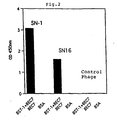
- Absorbance at 450nm was measured using a microplate reader. The results are shown in Fig. 2.
- the nucleotide sequences of the clones that bound to BST-1 in Reference Example 3 were determined.
- the phage were subjected to deproteinization by treating them with phenol and chloroform, and the DNA was precipitated by ethanol and used as a template for nucleotide sequencing.
- a primer was established on the basis of the nucleotide sequence of the vector used, Fuse 5 vector (Smith G.P. and Scott J.K.; Methods Enzymol. 21 7, 228-257, 1993), and the nucleotide sequences of the clones were determined by using the ABI PRISM dye termination cycle sequencing ready reaction kit (Applied Biosystems). The nucleotide sequences of two clones were thus determined (SN-1: SEQ ID NO: 1, SN-16: SEQ ID NO: 2).
- Two peptides each consisting of 15 amino acid residues, were synthesized by an automated peptide synthesizer on the basis of the amino acid sequences depicted in SEQ ID NOs: 1 and 2 which were deduced from the nucleotide sequences determined.
- the synthetic peptides having the sequences depicted in SEQ ID NOs : 1 and 2 were designated as SNP-1 and SNP-16, respectively.
- the peptides were found to have a purity of 95% or more by means of reverse HPLC.
- BST-1 was immobilized at 2800 resonance units (RU) on the sensor chip CM5 of a bio-sensor for analyzing protein interactions, BIACORE (Biacore K.K.), by means of an amine coupling method.
- the synthetic peptides were passed through the sensor chip at the flow rate of 40 ⁇ l/min at the concentration of 500nM.
- the amounts (RU) of the peptides bound to BST-1 are shown in Fig. 5.
- the peptides other than #1, #3, #6, #13, and #14 have lost their binding ability, which shows that the amino acid residues other than positions 1, 3, 6, 13, and 14 are important for the binding.
- the peptides #1, #3, #6, #13, and #14 have retained their binding ability, which shows that the mutations at the positions 1, 3, 6, 13, and 14 of SNP-1 would not change the binding ability to BST-1.
- the binding ability to BST-1 of SNP-1 derivative that contains a biotinylated N-terminus or C-terminus was compared with that of the prototype SNP-1.
- the biotinylated SNP-1 was prepared by binding the amino group of the N-terminus of SNP-1 with Sulfo-NHS-LC-Biotin (PIERCE) and purifying the product using reverse HPLC.
- SNP-1 having the additional amino acid residue Lys at the C-terminus was chemically synthesized, and biotin was bound to the amino group of the Lys residue.
- the SNP-1 derivative was purified by reverse HPLC and confirmed to have more than 95% purity.
- the derivative was named SNPb-1.
- BST-1 was immobilized at 2800 resonance units (RU) on the sensor chip CM5 of a bio-sensor for analyzing protein interactions, BIACORE (Biacore K.K.), by means of an amine coupling method.
- the synthetic peptides were passed through the sensor chip at a flow rate of 40 ⁇ l/min at a concentration of from 400nM to 2000nM. An equilibrium binding value was obtained for each concentration. Based on the equilibrium binding values, dissociation constants were calculated by means of the Scatchard plot method (Hulme E. C.; Receptor-binding studies, a brief outline, in Receptor Biochemistry: A Practical Approach, 303-315, IRL press, l990).
- the control peptide described in Example 2 was used as a negative control and showed no binding ability.
- the test results are shown in Fig. 6.
- Example 6 Inhibition of cADP-ribose hydrolase activity by the synthetic peptides
- BST-1 has a cADP-ribose hydrolase activity that hydrolytically converts cADP-ribose to ADP-ribose, as well as the ADP-ribosyl cyclase activity. Accordingly, it was investigated whether or not SNP-1 can also inhibit the cADP-ribose hydrolase activity of BST-1.
- the hydrolase activity was determined according to the method described in FEBS letters, 356 , 244, l994.
- BST-1 at the concentration of 50 ⁇ g/ml was mixed with the substrate cADP-ribose at a concentration of 20 ⁇ M and allowed to react at 37°C for four hours.
- the hydrolyzed product, ADP-ribose was separated by HPLC and the rate of ADP-ribose formation was determined on the basis of the peak area.
- the total peak area of cADP-ribose and ADP-ribose corresponds to 100%.
- SNP-1 was added to the reaction system at a concentration of 0.5 ⁇ M, 2.0 ⁇ M, or 20 ⁇ M to investigate the inhibition activity.
- 20 ⁇ M of the control peptide described in Example 2 were used as a control.
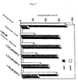
- the test results are shown in Fig. 7, which reveals that SNP-1 inhibits cADP-ribose hydrolase activity dose-dependently, while the control peptide does not inhibit the activity at all at the concentration of 20 ⁇ M.
- BST-1 was purified by affinity chromatography which takes advantage of the binding between BST-1 and SNP-1 derivative which contains biotinylated C-terminal, i.e., SNPb-1.
- an affinity column was prepared by binding 1x 10 -7 mol of SNPb-1, which was synthesized according to Example 5, to 1 ml of ultra avidin-agarose gel in a 20mM MES buffer solution, pH 6.0.
- BST-1 Fifty ml of culture supernatant containing expressed and secreted BST-1 was diluted fivefold with a 20mM acetate buffer solution, pH 5.0, and partially purified with a cation-exchange chromatography, followed by desalting by means of dialysis against a 20mM MES buffer solution, pH 6.0. Desalted BST-1 was charged into the afore-mentioned affinity chromatography column and eluted out using 0.5ml of a 20mM MES buffer (pH 6.0) containing 1.0M NaCl, to obtain purified BST-1.
- Synthetic peptide SNP-1 was mixed with a carrier therefor and encapsulated by conventionally so as to obtain capsules for treating rheumatoid arthritis.
Landscapes
- Chemical & Material Sciences (AREA)
- Health & Medical Sciences (AREA)
- Organic Chemistry (AREA)
- General Health & Medical Sciences (AREA)
- Life Sciences & Earth Sciences (AREA)
- Medicinal Chemistry (AREA)
- Nuclear Medicine, Radiotherapy & Molecular Imaging (AREA)
- Genetics & Genomics (AREA)
- General Chemical & Material Sciences (AREA)
- Animal Behavior & Ethology (AREA)
- Chemical Kinetics & Catalysis (AREA)
- Public Health (AREA)
- Veterinary Medicine (AREA)
- Biochemistry (AREA)
- Biophysics (AREA)
- Pharmacology & Pharmacy (AREA)
- Molecular Biology (AREA)
- Proteomics, Peptides & Aminoacids (AREA)
- Pain & Pain Management (AREA)
- Rheumatology (AREA)
- Peptides Or Proteins (AREA)
- Medicines That Contain Protein Lipid Enzymes And Other Medicines (AREA)
- Preparation Of Compounds By Using Micro-Organisms (AREA)
Abstract
Description
- The present invention relates to peptides binding to bone marrow stromal cell antigen-1 (referred to as "BST-1" hereinafter) and peptides that bind to the antigen BST-1 and inhibit ADP-ribosyl cyclase activity of the antigen. The peptides may be used for treating rheumatoid arthritis (sometimes referred to as "RA" hereinafter), multiple myeloma (sometimes referred to as "MM" hereinafter) and the like.
- Bone marrow stromal cell lines derived from RA patients were reported to have increased proliferation-enhancing activity on DW34 which is mouse stromal cell-dependent pre-B cell, when compared with those from normal volunteer (J. Immunol., l49, 4088-4095, l992). It was also reported that bone marrow stromal cell lines from RA and MM patients showed increased proliferation-enhancing activity on pre-B cells, and a novel bone marrow stromal cell antigen-1 was successfully isolated based on the assumption that the bone marrow stromal cells from RA and MM patients must contain some proliferation-accelerator (Proc. Natl. Acad. Sci. USA, 91, 5325-5329, l994).
- BST-1 is a glycosyl-phosphatidylinositol (GPl)-anchored membrane protein carrying a hydrophobic signal peptide at the C-terminus. BST-1 is considered to function as a signal transmitter (receptor) since intracellular proteins are phosphorylated or dephosphorylated when BST-1 is stimulated by crosslinking with its polyclonal antibodies (Biochem. Biophys. Res. Commun., 228, 838-845, l996). BST-1 shows 30% homology with human lymphocyte antigen CD38 on the amino acid level, and it is known that BST-1 has cyclic ADP-ribose hydrolase activity as well as ADP-ribosyl cyclase activity like CD38 (FEBS letters, 356, 244, l994). As is well known, the term "ADP" is an abbreviation for adenosine 5'-diphosphate, and the cyclic ADP-ribose is referred to as cADP-ribose hereinafter.
- ADP-ribosyl cyclase activity is an enzymatic action which converts nicotinamide adenine dinucleotide (NAD) to cADP-ribose, and the latter is being watched with interest as a second messenger for releasing Ca 2+ from intracellular Ca 2+ stores with a mechanism different from inositol l,4,5-triphosphate (IP3) (Science, 253, ll43-ll46, l993). Through cADP-ribose hydrolase activity, cADP-ribose is hydrolized to ADP-ribose.
- BST-1 and CD38 have their catalytic regions on the extracellular side and, therefore, it would be an interesting theme to investigate how their extracellular enzymatic activities can perform the Ca 2+ release from intracellular Ca 2+ stores, considering that the cADP-ribose can hardly cross the plasma membrane.
- It has been shown that arthrosis crevicular fluid of RA patients contains a significantly high concentration of soluble BST-1 as compared with that of normal volunteers (Arthritis. Rheum., 39, 629-637, l996), and it is suggested there may be some relation between ADP-ribosyl cyclase activity of the soluble BST-1 and pathogenesis of rheumatoid arthritis. However, unavailability of an inhibitor specifically inhibiting ADP-ribosly cyclase activity has hindered researchers from investigating this subject.
- As stated above, the precise relationship between BST-1 isolated from stromal cells of RA or MM patients and the pathogenesis has not been established yet. In particular, the relationship between ADP-ribosyl cyclase activity of BST-1 and the pathogenesis has not been clarified yet. In this situation, the inventors of the present invention considered that the use of an agent inhibiting such ADP-ribosyl cyclase activity would be effective for elucidating the relationship. Accordingly, novel peptides binding to BST-1 and, additionally, novel peptides binding to BST-1 and yet inhibiting the ADP-ribosyl cyclase activity thereof were identified. The present invention is based on these findings and data.
- Thus, the invention provides peptides binding to BST-1, in particular, peptides binding to BST-1 and yet inhibiting ADP-ribosyl cyclase activity of BST-1.
- For the purpose of identifying an agent that binds to BST-1, human BST-1 was expressed in insect cells and highly purified BST-1 was obtained in a large amount. Using purified BST-1, the inventors have selected from a phage display peptide library (Jikken Igaku (Experimental Medicine), 11, No.13, August, 95-100, 1993) two peptides consisting of l5 amino acid residues that bind to BST-1. One of them was identified to inhibit ADP-ribosyl cyclase activity. Details of the procedures are discussed below.
- Human BST-1 may be prepared by, for example, recombinant DNA technology. Host cells for the expression may be selected from E. coli, yeast, insect, and animal cells. When insect cells are used, a cDNA encoding human BST-1 (Kaisho T. et al, Proc. Natl. Acad. Sci. USA, 91, 5325-5329, 1994) is conventionally inserted downstream of a promoter which functions in insect cells, for instance, of the polyhedrine promoter (King and Possee, The baculovirus expression system, Chapman & Hall, 1992). The purification of expressed products may be accomplished by salting out, by ion-chromatography, centrifugation, and the like.
- For obtaining peptides that bind to human BST-1, the peptide library method as described below may be conveniently used.
- A random peptide phage library may be constructed by binding synthetic genes having random sequences to, for instance, coat protein genes (e.g. gene III or IIIV) of phage M13 . For this purpose, the method described in Science, 249, 386, l990, or Proc. Natl. Acad. Sci., USA, 87, 6378, l990, may be used. The size of the synthetic gene to be inserted is not limitative as long as the expressed peptide is stable. However, the a gene of preferred size will be one encoding from six (6) to fifteen (15) amino acid residues so that the resulting library may cover as many random sequences as possible and can bind to the target molecule, BST-1. The Selection of phage capable of binding to BST-1 is accomplished by immobilizing purified BST-1 on a column or plate, directly or via a linker, such as antibodies, contacting the library with the immobilized BST-1, and then washing out unbound phage . After washing, bound phage are eluted with acids, neutralized, and amplified by infecting E. coli cells. This procedure is repeated for three or four rounds to concentrate phage having affinity to BST-1. In order to obtain a single uniform phage E. coli cells are infected with concentrated phages and single colonies are allowed to form on agar plates containing antibiotics. The colony is then cultured in a liquid medium, and the phage in the supernatant is concentrated by precipitation with polyethylene glycol. Sequencing of the phage DNA reveals the amino acid sequence of the peptide bound to BST-1.
- The peptide library containing random amino acid sequences may also be prepared by chemical synthesis by means such as method employing beads (Nature, 354, 82, 1991), liquid phase focusing (Nature, 354, 84, l991) and the micro plate method.
- A large-scale production of the desired peptide may be carried out by chemical synthesis or recombinant DNA technology using E. coli, yeast, insect, or animal cells as a host. Conventional peptide synthesis may be used for the former, while solid phase synthesis is preferred. In this method, the preparation of variant peptides in which one or more amino acid residues are altered may be readily accomplished (Saibo Kogaku (cell technology), extra number, Experimental protocol for anti-peptide antibody, p26-46, Shu-jun sha, 1994). As for the latter, it is an established technique that the DNA sequence is determined according to the amino acid sequence of the peptide bound to BST-1 on the basis of codon usage, and a DNA prepared according to the DNA sequence determined is incorporated into a host cell (Maniatis et al; Molecular Cloning, Appendix D1, Cold Spring Habor Laboratory, l989). Amino acid residue(s) in the sequence can be substituted with other amino acid residue(s) by incorporation of mutations into the DNA sequence.
- When the peptide is expressed in E. coli cells, it is preferred that the resulting DNA is linked to a promoter sequence, such as the tryptophan synthetase operon (Trp) promoter or the lactose operon (lac) promoter, a ribosome-binding sequence, such as the Shine-Dalgarno sequence, and a transcription terminator recognition site, is added thereto. The resulting expression vector may be incorporated into E. coli cells according to the methods described in the aforementioned Molecular Cloning Manual. Expressed products maybe purified by, for example, various kinds of chromatography.
- The fact that the peptide thus obtained inhibits the ADP-ribosyl cyclase activity of BST-1 may be identified by comparing the ADP-ribosyl cyclase activity when measured in the absence of the peptide with the activity in the presence of the peptide. As previously mentioned, NAD is converted to cADP-ribose by ADP-ribosyl cyclase activity of BST-1, and therefore, the activity may be measured by reacting NAD with BST-1 and then quantitatively determining NAD and cADP-ribose after separation of them by anion-exchange chromatography (FEBS letters, 356, 244, l994). Alternatively, nicotinamide guanine dinucleotide (NGD) may be used as a substrate instead of NAD, which is converted to cyclic guanosine-5'-diphosphate-ribose (cGDP-ribose) that can be fluorimetrically measured with an excitation wavelength of 300 nm and an emission wavelength of 410 nm, whereby the velocity of the formation of cGDP-ribose reflects ADP-ribosyl cyclase activity (J. Biol. Chem., 48, 30260, l994).
- The present invention has enabled those skilled in the art to obtain peptides that bind to BST-1, and additional peptides that bind to BST-1 and yet specifically inhibit ADP-ribosyl cyclase activity thereof. The peptides may be used for treating rheumatoid arthritis and multiple myeloma. In addition, the peptides may be immobilized on a carrier and used as a component of a medical extraperfusion apparatus for removing BST-1 from body fluid.
- In more detail, the first object of the present invention is to provide peptides capable of binding to BST-1, which comprise amino acid sequence (1) as depicted in SEQ ID NO: 1 or an amino acid sequence (2) obtained by making deletion, substitution, or insertion of one or more amino acid residues in amino acid sequence (1). As preferred embodiments there are provided peptides having the amino acid sequence (2) which contains deletions, substitutions, or insertions of (an) amino acid residue(s) at the position of 1, 3, 6, 13, and/or 14 of the amino acid sequence of SEQ ID NO : 1.
- The second object of the invention is to provide peptides capable of binding to BST-1, which comprise an amino acid sequence (3) depicted in SEQ ID NO : 2 or an amino acid sequence (4) obtained by making deletion, substitution, or insertion of one or more amino acid residues in amino acid sequence (3).
- The third object of the invention is to provide peptides which bind to BST-1 and yet specifically inhibit ADP-ribosyl cyclase activity thereof.
- The fourth object of the invention is to provide peptides which bind to BST-1 and yet specifically inhibit cADP-ribose hydrolase activity thereof.
- The fifth embodiment of the invention is to provide a pharmaceutical formulation comprising as an essential component at least one of the peptides defined in the preceding objects.
- The sixth object of the invention is to provide a diagnostic agent for detecting BST-1, which comprises as an essential component at least one of the peptides defined in the preceding objects.
- The seventh object of the invention is to provide an adsorbing agent comprising at least one of the peptides defined in the preceding objects, said peptide(s) being immobilized on a carrier.
- The eighth object of the invention is to provide a method for the purification of BST-1 using the adsorbing agent defined above.
- The ninth object of the invention is to provide a medical extraperfusion apparatus which contains as one of the components at least one of the peptides defined in the preceding objects, said peptides being capable of inhibiting an enzymatic activities of BST-1.
- The tenth object of the invention is to provide a method of screening a substance capable of interacting with BST-1, which employs at least one of the peptides defined in the preceding objects.
- The eleventh object of the invention is to provide a method of making a pharmaceutical formulation comprising the above-mentioned method for screening for a substance capable of interacting with BST-1 and admixing the substance identified or a homologue or derivative thereof with a pharmaceutically acceptable carrier. In this context it is to be noted that the screening method referred to will often provide a so-called lead substance that is not directly used as a pharmaceutically active substance but rather used as a basis for making derivatives that have improved properties as regards their pharmaceutical activity or that is used as the basis for producing homologues that have improved pharmaceutical tolerability or activity or less side reactions. The person skilled in the art working in this field is aware of conventional methods for making such homologues or derivatives once it has identified the substance capable of interacting with BST-1 according to the present invention.
- Other objects of the present invention will be apparent to those skilled in the art from the disclosure of the specification and the drawings.
- In the accompanying drawings:
- Fig. 1 represents the ADP-ribosyl cyclase activity of purified BST-1 when measured using NGD as a substrate. The ordinate represents fluorescence intensity and the abscissa represents time course. Purified BST-1 (5 µg/ml) was mixed with 100 µM NGD, allowed to react for 20 minutes at 25 °C, and the fluorescence intensity was measured with an excitation wavelength of 300nm and an emission wavelength of 410nm. When only the substrate (100 µM NGD) was allowed to react, fluorescence did not increase due to no formation of cGDP-ribose. However, when BST-1 was added to the substrate, fluorescence was linearly increased, which confirmed the enzymatic activity of BST-1.
- Fig. 2 shows an ELISA analysis of phage obtained by screening
of a phage display peptide library with BST-1. The
ordinate represents absorbance at 450nm and the abscissa
represents the phage's names: SN-1 is a phage expressing the
peptide defined by SEQ ID NO: 1; SN-16 is a phage
expressing the peptide defined by SEQ ID NO : 2; Control is
a negative control phage having unrelated sequences. SN-1
and SN-l6 reacted with the well on which BST-1 was
immobilized via antibody BEC 7 (Okuyama Y. et al; Biochem.
Biophys. Commun. 228, 838-845, l996), while they did not
react with the well on which only
BEC 7, BST-1, or bovine serum albumin (BSA) was immobilized. This confirmed that they specifically bind to BST-1. - Fig. 3 shows an inhibition of ADP-ribosyl cyclase activity of BST-1 by synthetic peptide SNP-1. The abscissa is the time course and the ordinate is fluorescence intensity. Purified BST-1 (4 µg/ml) was mixed with NGD (300 µM), and the mixture was allowed to react at 25°C for 300 seconds, and then the synthetic peptide SNP-1 was added thereto at a concentration of 20 µM. Upon addition of the peptide, the increase of fluorescence intensity was stopped, which meant complete inhibition of ADP-ribosyl cyclase activity by the peptide. On the other hand, the addition of a negative control peptide at the concentration of 20 µM did not provide any influence on the increase of fluorescence intensity. The inset shows a complete disappearance of ADP-ribosyl cyclase activity by the addition of a reducing agent DTT at the concentration of 10mM.
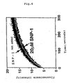
- Fig. 4 shows that the synthetic peptide SNP-1 did not inhibit the ADP-ribosyl cyclase activity of CD38. The abscissa is the time course and the ordinate is fluorescence intensity. Purified CD38 (200ng/ml) was mixed with NGD (2 µM), and allowed to react at 25°C for 300 seconds. Addition of the synthetic peptide SNP-1 at the concentration of 20 µM did not provide any influence on the increase of fluorescence intensity. Thus, SNP-1 did not inhibit the activity of CD38.
- Fig. 5 shows binding of BST-1 to variants of SNP-1.
The abscissa shows variants of SNP-1, wherein one of the
amino acid residues of SNP-1 is replaced by Ala, and the
ordinate shows a magnitude of binding (RU) when measured
in Biacore system. The
variants # 1, #3, #6, #13, and #14 retained binding ability, while the other variants lost the binding ability. The control peptide was the one prepared in Example 2. - Fig. 6 shows the binding of BST-1 with SNP-1 which was modified with biotin at the N- or C-terminus. The graphs show a Scatchard plot of prototype (original) SNP-1 (a), b SNP-1 (b) which has biotin bound at the N-terminus, and SNPb-1 (c) which has biotin bound at the C-terminus. The slope of the linear lines determined the dissociation constants of SNP-1, bSNP-1, and SNPb-1 as 510nM, 530nM, and 810nM, respectively. Absence of remarkable differences among them in terms of their dissociation constants shows that a modification of the N- or C-terminus with biotin did not result in a loss of the binding ability of SNP-1. In the drawing, Req means equilibrium binding value expressed by resonance unit (RU), and Req/C represents the value obtained by dividing the equilibrium binding value with the peptide concentration.
- Fig. 7 shows the inhibition of cADP-ribose hydrolase activity of BST-1 by SNP-1. cADP-ribose hydrolase activity of BST-1 converts cADP-ribose to ADP-ribose. After the reaction of BST-1 and cADP-ribose, two riboses were separated by HPLC, and the formation of ADP-ribose was determined based on the peak area. The total peak area of cADP-ribose and ADP-ribose corresponded to 100%. SNP-1 inhibited the formation of ADP-ribose dose-dependently, while a control peptide did not give any influence on the formation of ADP-ribose at the concentration of 20 µM. "Background" in the figure represents the results of the reaction involving only the substrate, cADP-ribose, and reflects spontaneous hydrolysis. "Peptide free" means the enzymatic reaction carried out without the peptide.
- Fig. 8 is an electrophoretic photograph which shows the results of purification of BST-1 using SNPb-1 affinity chromatography. Eluted fractions, each 20µl, were analyzed with SDS-PAGE (7.5%), and BST-1 was detected by means of silver staining. Various proteins were detected in non-purified culture supernatant, while the fractions eluted by SNPb-1 affinity chromatography contained almost complete-ly purified BST-1.
-
- The following detailed Examples and Reference Examples are presented by way of illustration of certain specific embodiments of the invention. The Examples are representative only and should not be construed as limiting the scope of the invention in any respect.
- As previously mentioned, BST-1 is a GPl-anchored membrane protein (Proc. Natl. Acad. Sci. USA, 91, 5325, l994). In order to produce a large amount of soluble BST-1 using insect cells, BST-1 cDNA in which the 298th codon (ACA) present just before the hydrophobic domain at the C-terminus has been substituted by the termination codon (TGA) was conventionally inserted at the Smal, Xbal site of an expression plasmid for insect cells, pVL1393 (PharMingen). In accordance with the conventional manner (King and Possee; The baculovirus expression system, Chapman & Hall, 1992), insect cells Sf9 (Funakoshi) were transfected with the resulting plasmid to obtain the recombinant virus. Insect cells (High five (Invitrogen)) were infected with the virus and the cells were conventionally cultured at 27°C for three days accordance with the conventional manner (King and Possee; The baculovirus expression system, Chapman & Hall, 1992). Soluble BST-1 secreted into the culture medium was identified by western blotting. The infected insect cells were cultured in a large scale, and BST-1 was purified up to 95% or more by cation-exchange chromatography and dye ligand Blue chromatography. The purified protein was confirmed to have ADP-ribosyl cyclase activity by means of the NGD method mentioned above. see Fig. 1 of the accompanying drawings.
- A phage library having random sequences consisting of 15 amino acid residues was prepared using the method described in Biochemistry, 35, 10441, 1996. A monoclonal antibody to BST-1, BEC7 (Okuyama Y. et al; Biochem. Biophys. Res. Commun. 228, 838 - 845, 1996), was diluted with 10mM phosphate buffer, pH 7.0, and coated on 96-well microtiter wells at 4°C overnight at the ratio of 3 µg/well. Purified BST-1 was added to the microtiter wells after dilution with 10mM phosphate buffer, pH 7.0, at the ratio of 5 µg/well. BST-1 was thus immobilized on the wells via the antibody. Each well was blocked with 10mM phosphate buffer, pH 7.0, containing 1% bovine serum albumin at room temperature for one hour. Subsequently, about 1012 phage library was added to 100 µl of a buffer (10mM phosphate buffer,
pH 7. 0, containing 1% bovine serum albumin, 0. 05% Tween 20) and the mixture was allowed to react with BST-1 immobilized on the wells at room temperature for one hour. The wells were washed with a washing buffer (10mM phosphate buffer, pH 7.0, and 0.05% Tween 20) ten times so as to remove unbound phage. Phage bound to BST-1 were eluted out with glycine buffer, pH 2.2, and neutralized with 1 M Tris-HCl, pH 9.5. E. coli K91 Kan (obtained from Dr. G.P. Smith of Missouri University) was infected with the phage and in order to amplify the phage the infected cells were cultured in LB medium containing tetracycline. The phages in the supernatant were concentrated by polyethylene glycol precipitation, and the concentrated phage were used in the second round. This procedure was repeated three times in total to select phage binding to BST-1. - E. coli cells K91 Kan were infected again with the phage selected in Reference Example 2 and were then cultured on LB agar plates containing tetracycline to form single colonies. The colonies were cultured overnight in LB medium containing tetracycline, and supernatants including the phage were subjected to polyethylene glycol precipitation for the purification of the phage on the next day. The phage were added to a 96-well microtiter plate, on which BST-1 had been immobilized in advance (see Reference Example 2), at the ratio of about 1010 phage/well, and allowed to react at room temperature for one hour. After washing four times with a washing buffer (10mM phosphate buffer, pH 7.0, and 0.05% Tween 20), BST-1 on the well was allowed to react with M13 phage antibody (Pharmacia) labeled with horseradish peroxidase (5000 times diluted) at room temperature for 30 minutes. After washing four times, the substrate, 3,3',5,5'-tetramethyl benzidine, was added for developing color, and the reaction was quenched by the addition of 1M H2SO4. Absorbance at 450nm was measured using a microplate reader. The results are shown in Fig. 2.
- The nucleotide sequences of the clones that bound to BST-1 in Reference Example 3 were determined. First, the phage were subjected to deproteinization by treating them with phenol and chloroform, and the DNA was precipitated by ethanol and used as a template for nucleotide sequencing. A primer was established on the basis of the nucleotide sequence of the vector used,
Fuse 5 vector (Smith G.P. and Scott J.K.; Methods Enzymol. 217, 228-257, 1993), and the nucleotide sequences of the clones were determined by using the ABI PRISM dye termination cycle sequencing ready reaction kit (Applied Biosystems). The nucleotide sequences of two clones were thus determined (SN-1: SEQ ID NO: 1, SN-16: SEQ ID NO: 2). - Two peptides, each consisting of 15 amino acid residues, were synthesized by an automated peptide synthesizer on the basis of the amino acid sequences depicted in SEQ ID NOs: 1 and 2 which were deduced from the nucleotide sequences determined. The synthetic peptides having the sequences depicted in SEQ ID NOs : 1 and 2 were designated as SNP-1 and SNP-16, respectively. The peptides were found to have a purity of 95% or more by means of reverse HPLC.
- The inhibition of ADP-ribosyl cyclase activity of BST-1 by synthetic peptides was tested by means of the NGD method previously mentioned. BST-1 at the concentration of 4 µg/ml was allowed to react with the substrate NGD at the concentration of 300 µM at 25°C for 300 seconds, which confirmed linearly increased fluorescence intensity. After 300 seconds, 20 µM of synthetic peptide SNP-1 were added to the reaction system. As a control, the same amount of a peptide encoding a sequence reverse to the sequence of SNP-1 was used. The addition of the control peptide did not show any inhibition on the activity, while SNP-1 stopped the increase of fluorescence intensity, which confirmed complete inhibition of ADP-ribosyl cyclase activity of BST-1. The results are shown in Fig. 3.
- It is known that human CD38, like BST-1, has ADP-ribosyl cyclase activity. It was therefore tested whether the synthetic peptides could inhibit ADP-ribosyl cyclase activity of CD38 or not. Thus, the substrate NGD was added to a soluble CD38 at a concentration of 200ng/ml and a final concentration of 2 µM. The mixture was allowed to react at 25°C for 300 seconds, whereby fluorescence intensity was confirmed to increase. The addition of 20 µM of SNP-1 to the reaction system did not inhibit the reaction, which showed that SNP-1 uniquely inhibits the ADP-ribosyl cyclase activity of BST-1. Fig. 4 shows the test results.
- For the purpose of identifying amino acid residues responsible for the binding of SNP-1 to BST-1, fourteen peptides which are different from SNP-1 only in that they contain Ala in place of one of the original amino acid residues of SNP-1 were chemically synthesized and given the numbers sequentially selected from #1 to #14. For instance, the variant which contains Ala substituted for the first amino acid residue at the N-terminus of SNP-1 was named #1, and the variant which contains Ala substituted for the penultimate amino acid residue was named #14. Fourteen peptides thus prepared were tested for the binding ability to BST-1.
- BST-1 was immobilized at 2800 resonance units (RU) on the sensor chip CM5 of a bio-sensor for analyzing protein interactions, BIACORE (Biacore K.K.), by means of an amine coupling method. The synthetic peptides were passed through the sensor chip at the flow rate of 40 µl/min at the concentration of 500nM. The amounts (RU) of the peptides bound to BST-1 are shown in Fig. 5. The peptides other than #1, #3, #6, #13, and #14 have lost their binding ability, which shows that the amino acid residues other than
positions peptides # 1, #3, #6, #13, and #14 have retained their binding ability, which shows that the mutations at thepositions - The binding ability to BST-1 of SNP-1 derivative that contains a biotinylated N-terminus or C-terminus was compared with that of the prototype SNP-1. The biotinylated SNP-1 was prepared by binding the amino group of the N-terminus of SNP-1 with Sulfo-NHS-LC-Biotin (PIERCE) and purifying the product using reverse HPLC. The resulting derivative, having more than 95% purity, was named bSNP-1. In order to biotinylate at the C-terminus, SNP-1 having the additional amino acid residue Lys at the C-terminus was chemically synthesized, and biotin was bound to the amino group of the Lys residue. The SNP-1 derivative was purified by reverse HPLC and confirmed to have more than 95% purity. The derivative was named SNPb-1.
- BST-1 was immobilized at 2800 resonance units (RU) on the sensor chip CM5 of a bio-sensor for analyzing protein interactions, BIACORE (Biacore K.K.), by means of an amine coupling method. The synthetic peptides were passed through the sensor chip at a flow rate of 40 µl/min at a concentration of from 400nM to 2000nM. An equilibrium binding value was obtained for each concentration. Based on the equilibrium binding values, dissociation constants were calculated by means of the Scatchard plot method (Hulme E. C.; Receptor-binding studies, a brief outline, in Receptor Biochemistry: A Practical Approach, 303-315, IRL press, l990). The control peptide described in Example 2 was used as a negative control and showed no binding ability. The test results are shown in Fig. 6. The dissociation constants (Kd) for the prototype SNP-1, bSNP-1, and SNPb-1, which were determined on the basis of the slope of the linear line, were 510nM, 530nM and 810nM, respectively. Similarity of the dissociation constants between SNP-1, bSNP-1, and SNPb-1 shows that the binding ability of SNP-1 to BST-1 is not affected by biotin-modification at the N- or C-terminal.
- BST-1 has a cADP-ribose hydrolase activity that hydrolytically converts cADP-ribose to ADP-ribose, as well as the ADP-ribosyl cyclase activity. Accordingly, it was investigated whether or not SNP-1 can also inhibit the cADP-ribose hydrolase activity of BST-1. The hydrolase activity was determined according to the method described in FEBS letters, 356, 244, l994. Thus, BST-1 at the concentration of 50 µg/ml was mixed with the substrate cADP-ribose at a concentration of 20 µM and allowed to react at 37°C for four hours. After the reaction, the hydrolyzed product, ADP-ribose, was separated by HPLC and the rate of ADP-ribose formation was determined on the basis of the peak area. The total peak area of cADP-ribose and ADP-ribose corresponds to 100%. SNP-1 was added to the reaction system at a concentration of 0.5 µM, 2.0 µM, or 20 µM to investigate the inhibition activity. As a control, 20 µM of the control peptide described in Example 2 were used. The test results are shown in Fig. 7, which reveals that SNP-1 inhibits cADP-ribose hydrolase activity dose-dependently, while the control peptide does not inhibit the activity at all at the concentration of 20 µM.
- BST-1 was purified by affinity chromatography which takes advantage of the binding between BST-1 and SNP-1 derivative which contains biotinylated C-terminal, i.e., SNPb-1. Thus, an affinity column was prepared by binding
1x 10⁻⁷ mol of SNPb-1, which was synthesized according to Example 5, to 1 ml of ultra avidin-agarose gel in a 20mM MES buffer solution, pH 6.0. - Fifty ml of culture supernatant containing expressed and secreted BST-1 was diluted fivefold with a 20mM acetate buffer solution, pH 5.0, and partially purified with a cation-exchange chromatography, followed by desalting by means of dialysis against a 20mM MES buffer solution, pH 6.0. Desalted BST-1 was charged into the afore-mentioned affinity chromatography column and eluted out using 0.5ml of a 20mM MES buffer (pH 6.0) containing 1.0M NaCl, to obtain purified BST-1.
- Synthetic peptide SNP-1 was mixed with a carrier therefor and encapsulated by conventionally so as to obtain capsules for treating rheumatoid arthritis.

Claims (12)
- A peptide capable of binding to bone marrow stromal cell antigen-1 comprising amino acid sequence (1) as depicted in SEQ ID NO: 1 or an amino acid sequence (2) obtained by deletion, substitution, or insertion of one or more amino acid residues in amino acid sequence (1).
- The peptide of claim 1, which contains a deletion, substitution, or insertion of one or more amino acid residues at position 1, 3, 6, 13 and/or 14 of amino acid sequence (1).
- A peptide capable of binding to bone marrow stromal cell antigen-1 comprising amino acid sequence (3) as depicted in SEQ ID NO: 2 or an amino acid sequence (4) obtained by deletion, substitution, or insertion or one or more amino acid residues in amino acid sequence (3).
- A peptide of any one of claims 1 to 3 which specifically inhibits the ADP-ribosyl cyclase activity of BST-1.
- A pharmaceutical formulation comprising a peptide of any one of claims 1 to 4.
- A diagnostic agent for detecting BST-1 comprising a peptide of any one of claims 1 to 4.
- An adsorbing agent comprising a peptide of any one of claims 1 to 4 immobilized on a carrier.
- A method for the purification of BST-1 using the adsorbing agent of claim 7.
- A medical extraperfusion apparatus which contains as one of the components a peptide of any one of claims 1 to 4 inhibiting an enzymatic activity of BST-1.
- A method for screening for a substance capable of interacting with BST-1, which employs a peptide of any one of claims 1 to 4.
- A method of making a pharmaceutical formulation comprising the method of claim 10 and admixing said substance identified or a homologue or derivative thereof with a pharmaceutically acceptable carrier.
- Use of a peptide of any one of claims 1 to 4 for the preparation of a pharmaceutical formulation for the treatment of rheumatoid arthritis or multiple myeloma.
Applications Claiming Priority (2)
| Application Number | Priority Date | Filing Date | Title |
|---|---|---|---|
| JP10118586A JPH11310596A (en) | 1998-04-28 | 1998-04-28 | Myeloid interstitial cell antigen binding protein |
| JP11858698 | 1998-04-28 |
Publications (2)
| Publication Number | Publication Date |
|---|---|
| EP0953572A2 true EP0953572A2 (en) | 1999-11-03 |
| EP0953572A3 EP0953572A3 (en) | 1999-11-17 |
Family
ID=14740263
Family Applications (1)
| Application Number | Title | Priority Date | Filing Date |
|---|---|---|---|
| EP99107452A Withdrawn EP0953572A3 (en) | 1998-04-28 | 1999-04-28 | Peptides binding to bone marrow stromal cell antigen |
Country Status (4)
| Country | Link |
|---|---|
| US (1) | US6414113B1 (en) |
| EP (1) | EP0953572A3 (en) |
| JP (1) | JPH11310596A (en) |
| CA (1) | CA2269103A1 (en) |
Cited By (2)
| Publication number | Priority date | Publication date | Assignee | Title |
|---|---|---|---|---|
| WO2000037089A1 (en) * | 1998-12-18 | 2000-06-29 | University Of Bath | Cyclic adenosine diphosphate ribose analogues for modulating t cell activity |
| WO2002098397A2 (en) * | 2001-06-07 | 2002-12-12 | University Of Bath | Therapeutic compositions for modulating the immune response in a mammal and use thereof |
Families Citing this family (4)
| Publication number | Priority date | Publication date | Assignee | Title |
|---|---|---|---|---|
| DE10238846A1 (en) * | 2002-08-20 | 2004-03-04 | Nemod Immuntherapie Ag | Active fusion proteins and processes for their production |
| US9175092B2 (en) | 2011-06-28 | 2015-11-03 | Oxford Biotherapeutics Ltd | Antibodies to bone marrow stromal antigen 1 |
| JP6113721B2 (en) * | 2011-06-28 | 2017-04-12 | オックスフォード ビオトヘラペウトイクス エルティーディー. | Therapeutic and diagnostic targets |
| CN111093687B (en) * | 2017-09-15 | 2024-07-09 | 凯恩塞恩斯株式会社 | Use of peptides as therapeutic agents for autoimmune diseases and bone diseases |
Citations (1)
| Publication number | Priority date | Publication date | Assignee | Title |
|---|---|---|---|---|
| WO1994017184A1 (en) * | 1993-01-29 | 1994-08-04 | Schering Corporation | Modulation of physiological responses of lymphocytes by cd38 or antibodies thereto |
-
1998
- 1998-04-28 JP JP10118586A patent/JPH11310596A/en active Pending
-
1999
- 1999-04-27 US US09/300,410 patent/US6414113B1/en not_active Expired - Fee Related
- 1999-04-28 EP EP99107452A patent/EP0953572A3/en not_active Withdrawn
- 1999-04-28 CA CA002269103A patent/CA2269103A1/en not_active Abandoned
Patent Citations (1)
| Publication number | Priority date | Publication date | Assignee | Title |
|---|---|---|---|---|
| WO1994017184A1 (en) * | 1993-01-29 | 1994-08-04 | Schering Corporation | Modulation of physiological responses of lymphocytes by cd38 or antibodies thereto |
Non-Patent Citations (5)
| Title |
|---|
| A. SATO ET AL.: "Novel peptide inhibitor of ecto-ADP-ribosyl cyclase of bone marrow stromal cell antigen (BST-1/CD157)" BIOCHEMICAL JOURNAL, vol. 337, no. 3, 1 February 1999 (1999-02-01), pages 491-496, XP002113945 * |
| M. HARA-YOKOHAMA ET AL.: "Inhibition of NAD+ Glycohydrolase and ADP-ribosyl Cyclase Activities of Leukocyte Cell Surface Antigen CD38 by Gangliosides" JOURNAL OF BIOLOGICAL CHEMISTRY, vol. 271, no. 22, 31 May 1996 (1996-05-31), pages 12951-12955, XP002113948 BALTIMORE, MD US * |
| M. TORTI ET AL.: "Cytoskeleton-dependent inhibition of the ADP-ribosyl cyclase activity of CD38 in thrombin-stimulated platelets" FEBS LETTERS, vol. 431, no. 1, 10 July 1998 (1998-07-10), pages 19-22, XP002113946 AMSTERDAM NL * |
| Y. KAJIMOTO ET AL.: "Pancreatic Islet Cells Express BST-1, a CD38-like Surface Molecule Having ADPRibosyl Cyclase Activity" BIOCHEMICAL AND BIOPHYSICAL RESEARCH COMMUNICATIONS, vol. 219, no. 3, 27 February 1996 (1996-02-27), pages 941-946, XP002113947 DULUTH, MINNESOTA US * |
| Y. OKUYAMA ET AL.: "Human BST-1 Expressed on Myeloid Cells Functions as a Receptor Molecule" BIOCHEMICAL AND BIOPHYSICAL RESEARCH COMMUNICATIONS, vol. 228, no. 3, 21 November 1996 (1996-11-21), pages 838-845, XP002113949 ORLANDO, FL US * |
Cited By (4)
| Publication number | Priority date | Publication date | Assignee | Title |
|---|---|---|---|---|
| WO2000037089A1 (en) * | 1998-12-18 | 2000-06-29 | University Of Bath | Cyclic adenosine diphosphate ribose analogues for modulating t cell activity |
| WO2002098397A2 (en) * | 2001-06-07 | 2002-12-12 | University Of Bath | Therapeutic compositions for modulating the immune response in a mammal and use thereof |
| WO2002098397A3 (en) * | 2001-06-07 | 2003-03-13 | Univ Bath | Therapeutic compositions for modulating the immune response in a mammal and use thereof |
| GB2392095A (en) * | 2001-06-07 | 2004-02-25 | Univ Bath | Therapeutic compositions for modulating the immune response in a mammal and use thereof |
Also Published As
| Publication number | Publication date |
|---|---|
| US6414113B1 (en) | 2002-07-02 |
| JPH11310596A (en) | 1999-11-09 |
| CA2269103A1 (en) | 1999-10-28 |
| EP0953572A3 (en) | 1999-11-17 |
Similar Documents
| Publication | Publication Date | Title |
|---|---|---|
| US6548634B1 (en) | Synthetic peptides having FGF receptor affinity | |
| JP4060886B2 (en) | Isolation and utilization of SH3-binding peptides | |
| US20200199179A1 (en) | Peptide library and use thereof | |
| JP2003518075A (en) | Methods and compositions for extending the elimination half-life of bioactive compounds | |
| WO1996023813A1 (en) | Peptides and compounds that bind to sh2 domains | |
| CN113150137B (en) | Preparation method and application of NDM-1 monoclonal antibody | |
| JP4604184B2 (en) | Novel sugar chain recognition protein and its gene | |
| JP4215274B2 (en) | Src SH3 binding peptide and method for separating and using the same | |
| JPH10512445A (en) | IL-2R binding polypeptide and DNA molecule encoding the same | |
| CZ151696A3 (en) | Bmp receptor encoding dna sequence | |
| EP0953572A2 (en) | Peptides binding to bone marrow stromal cell antigen | |
| JPH10509044A (en) | Peptide capable of binding to GAP protein SH3 domain, nucleotide sequence encoding the same, and method for producing and using the same | |
| Yoon et al. | Differences between AGAP1, ASAP1 and Arf GAP1 in substrate recognition: interaction with the N-terminus of Arf1 | |
| WO1998056806A1 (en) | A TRANSCRIPTION FACTOR COACTIVATOR PROTEIN, p/CIP | |
| DK1981904T3 (en) | Peptide domain from the enclosure of a HERV-W virus, which is required for reaction with a hASCT receptor | |
| US6045797A (en) | Treatment or diagnosis of diseases or conditions associated with a BLM domain | |
| JPH05271291A (en) | Functional polypeptide | |
| US6303574B1 (en) | Scr SH3 binding peptides and methods of isolating and using same | |
| US6225086B1 (en) | Polynucleotides encoding ankyrin proteins | |
| JP2006513143A (en) | Protein tyrosine phosphatase inhibitor | |
| JPH02311498A (en) | Functional polypeptide | |
| EP0862447A1 (en) | Generating d-peptides: methods and compositions | |
| TW200303919A (en) | Cytotoxic protein and the use | |
| US6812336B1 (en) | Transcription factor coactivator protein, p/CIP | |
| JPH08143597A (en) | Human neurotensin receptor protein, its production and use |
Legal Events
| Date | Code | Title | Description |
|---|---|---|---|
| PUAI | Public reference made under article 153(3) epc to a published international application that has entered the european phase |
Free format text: ORIGINAL CODE: 0009012 |
|
| PUAL | Search report despatched |
Free format text: ORIGINAL CODE: 0009013 |
|
| 17P | Request for examination filed |
Effective date: 19990525 |
|
| AK | Designated contracting states |
Kind code of ref document: A2 Designated state(s): DE FR GB |
|
| AX | Request for extension of the european patent |
Free format text: AL;LT;LV;MK;RO;SI |
|
| AK | Designated contracting states |
Kind code of ref document: A3 Designated state(s): AT BE CH CY DE DK ES FI FR GB GR IE IT LI LU MC NL PT SE |
|
| AX | Request for extension of the european patent |
Free format text: AL;LT;LV;MK;RO;SI |
|
| AKX | Designation fees paid |
Free format text: DE FR GB |
|
| 17Q | First examination report despatched |
Effective date: 20010802 |
|
| STAA | Information on the status of an ep patent application or granted ep patent |
Free format text: STATUS: THE APPLICATION IS DEEMED TO BE WITHDRAWN |
|
| 18D | Application deemed to be withdrawn |
Effective date: 20020709 |