EP0949331B1 - Established microglia - Google Patents
Established microglia Download PDFInfo
- Publication number
- EP0949331B1 EP0949331B1 EP98905808A EP98905808A EP0949331B1 EP 0949331 B1 EP0949331 B1 EP 0949331B1 EP 98905808 A EP98905808 A EP 98905808A EP 98905808 A EP98905808 A EP 98905808A EP 0949331 B1 EP0949331 B1 EP 0949331B1
- Authority
- EP
- European Patent Office
- Prior art keywords
- microglia
- cells
- brain
- csf
- cell line
- Prior art date
- Legal status (The legal status is an assumption and is not a legal conclusion. Google has not performed a legal analysis and makes no representation as to the accuracy of the status listed.)
- Expired - Lifetime
Links
Images


Classifications
-
- A—HUMAN NECESSITIES
- A61—MEDICAL OR VETERINARY SCIENCE; HYGIENE
- A61K—PREPARATIONS FOR MEDICAL, DENTAL OR TOILETRY PURPOSES
- A61K48/00—Medicinal preparations containing genetic material which is inserted into cells of the living body to treat genetic diseases; Gene therapy
-
- A—HUMAN NECESSITIES
- A61—MEDICAL OR VETERINARY SCIENCE; HYGIENE
- A61P—SPECIFIC THERAPEUTIC ACTIVITY OF CHEMICAL COMPOUNDS OR MEDICINAL PREPARATIONS
- A61P25/00—Drugs for disorders of the nervous system
-
- C—CHEMISTRY; METALLURGY
- C12—BIOCHEMISTRY; BEER; SPIRITS; WINE; VINEGAR; MICROBIOLOGY; ENZYMOLOGY; MUTATION OR GENETIC ENGINEERING
- C12N—MICROORGANISMS OR ENZYMES; COMPOSITIONS THEREOF; PROPAGATING, PRESERVING, OR MAINTAINING MICROORGANISMS; MUTATION OR GENETIC ENGINEERING; CULTURE MEDIA
- C12N5/00—Undifferentiated human, animal or plant cells, e.g. cell lines; Tissues; Cultivation or maintenance thereof; Culture media therefor
- C12N5/06—Animal cells or tissues; Human cells or tissues
- C12N5/0602—Vertebrate cells
- C12N5/0618—Cells of the nervous system
- C12N5/0622—Glial cells, e.g. astrocytes, oligodendrocytes; Schwann cells
-
- A—HUMAN NECESSITIES
- A61—MEDICAL OR VETERINARY SCIENCE; HYGIENE
- A61K—PREPARATIONS FOR MEDICAL, DENTAL OR TOILETRY PURPOSES
- A61K38/00—Medicinal preparations containing peptides
-
- C—CHEMISTRY; METALLURGY
- C12—BIOCHEMISTRY; BEER; SPIRITS; WINE; VINEGAR; MICROBIOLOGY; ENZYMOLOGY; MUTATION OR GENETIC ENGINEERING
- C12N—MICROORGANISMS OR ENZYMES; COMPOSITIONS THEREOF; PROPAGATING, PRESERVING, OR MAINTAINING MICROORGANISMS; MUTATION OR GENETIC ENGINEERING; CULTURE MEDIA
- C12N2501/00—Active agents used in cell culture processes, e.g. differentation
- C12N2501/20—Cytokines; Chemokines
- C12N2501/22—Colony stimulating factors (G-CSF, GM-CSF)
-
- C—CHEMISTRY; METALLURGY
- C12—BIOCHEMISTRY; BEER; SPIRITS; WINE; VINEGAR; MICROBIOLOGY; ENZYMOLOGY; MUTATION OR GENETIC ENGINEERING
- C12N—MICROORGANISMS OR ENZYMES; COMPOSITIONS THEREOF; PROPAGATING, PRESERVING, OR MAINTAINING MICROORGANISMS; MUTATION OR GENETIC ENGINEERING; CULTURE MEDIA
- C12N2510/00—Genetically modified cells
Definitions
- the present invention relates to a subcultivatable, established cell line of microglia, a method of separating the same, and use thereof as a pharmaceutical carrier.
- microglia cell line into which an extraneous gene or a drug was introduced, a method of introducing it, and a pharmaceutical composition comprising the same.
- hereditary diseases in the nervous system which occur due to various causative factors, for example by defect of a single enzyme, etc. or by unknown reasons. Under these circumstances, supplementary therapy is used to cope with a large number of such diseases.
- a method of using neuronphilic viruses as adenovirus vectors is devised, and a system for introducing a gene specifically into neurons is known (Kozarsky, K.F and Wilson, J. M., Curr. Opin. Genet. Dev. 3 , 499-503, 1993).
- a method of using a retrovirus vector is also devised and has succeeded in introducing a gene into hepatic cells, bloodcells, etc. (Mullingan, R. C. , Science 260 , 926-932, 1993).
- the blood-brain barrier is present, so it is difficult to conduct supplementary therapy and to introduce an effective drug, and even if a substance (e.g. anticancer drug, DNA etc.) is introduced from a peripheral position, it cannot be introduced specifically into the brain. Therefore, there was no method other than direct injection of the substance by surgical operations.
- a substance e.g. anticancer drug, DNA etc.
- the blood-brain barrier is present in the brain, so the brain is almost free of infiltration with cells or a substance from the periphery, thus making it difficult to introduce a drug or a gene into the brain.
- immunocytes such as T cells and macrophages is hardly observed.
- Microglia are cells with macrophage-like properties in the central nervous system, which not only function as immunocompetent cells in inflammatory reaction and viral infection and as phagocytes for removing cells but also play a central role in a cytokine network in the central nervous system (Sawada, M. et al. , Int. J. Dev. Neurosci., 13, 253-264, 1995). Recently, microglia have been revealed to be essential for expression of high-level brain functions such as learning and memorization and considered to be specialized cells having a role specific for the brain. Up to now, it has been considered that microglia originate from monocytes having infiltrated the brain in the perinatal period and are specialized and differentiated.
- the microglia can be obtained by primary culture of brain cells.
- the brain should be excised and purified for use, and primary culture usually requires a period of about 2 weeks, so the procedures are cumbersome.
- the cells are difficult to proliferate during culture and hard to subculture, so after primary culture, it is extremely difficult to introduce a gene into the microglia to express it therein
- microglia considered as brain-specific macrophage
- the present inventors have succeeded in obtaining highly purified microglia and examined the properties, and as a result, we found that unlike the macrophage, microglia have specific affinity for the brain.
- microglia are determinatively different from macrophage in respect of affinity for the brain and the ability to infiltrate the brain. Further, the inventors examined the distribution thereof by staining in a method capable of distinguishing the two, and found that microglia are present from an early development stage in the brain. Accordingly, it is considered that microglia are not derived from a monocyte differentiated and matured in the bone marrow, but are derived from a cell group which have a specific affinity for the brain, and infiltrate the brain at an early development stage and come to regulate high-level functions such as cerebral morphogenesis and learning and memorization.
- the inventors injected an isolated macrophage and microglia into rat peripheral arteries to compare the two for selective affinity for the brain, and as a result we found that when the microglia labeled with a fluorescent pigment was injected; many fluorescent cells were observed in the brain but hardly observed in the liver. On the other hand, when the macrophage was injected, fluorescent cells were hardly observed in the normal cell, but many fluorescent cells were observed in the liver.
- the inventors examined whether a cell strain of microglia established by the inventors can express a gene selectively in the brain by introducing a lacZ expression vector into the cell strain and injecting the resultant cells into a rat blood stream, and as a result, the inventors could detect the activity of ⁇ -galactosidase in a section of the rat brain into which the lacZ-expressing cells had been injected. From these results, it was found that the microglia unlike the macrophage is a cell having specific affinity for the brain, and by utilizing this affinity, a specific substance or gene can be introduced via a peripheral blood stream into the brain.
- the present invention relates to an established cell line of microglia having specific affinity for the brain and phagocytic ability, and having cell growth ability dependent on granulocyte-macrophage colony-stimulating factor (GM-CSF), characterised in that said cell line is subcultivatable in the presence of GM-CSF and has essentially a macrophage-like or globular form in the presence of GM-CSF and a branched form similar to branched microglia present in the brain in the absence of GM-CSF.
- GM-CSF granulocyte-macrophage colony-stimulating factor
- the present invention relates to a method of producing the established cell line of microglia which comprises the step of cloning a cell line of microglia from purified microglia through the cell culture in the presence of GM-CSF.
- the present invention relates to a pharmaceutical composition
- a pharmaceutical composition comprising the established cell line of microglia.
- the present invention relates to the pharmaceutical composition for use in a method of treating cerebral disease.
- a method of separating a subcultivatable established cell line of microglia from microglia cells in the presence of a cytokine preferably in the presence of colony-stimulating factor (CSF), more preferably in the presence of granulocyte-macrophage colony-stimulating factor (GM-CSF), still more preferably in the presence of IL-3 and/or purified astrocytes.
- CSF colony-stimulating factor
- GM-CSF granulocyte-macrophage colony-stimulating factor
- a pharmaceutical carrier comprising the established cell line of microglia described above.
- a pharmaceutical composition comprising the above-described microglia having a gene or a drug introduced into it and a pharmaceutical carrier and in particular to a pharmaceutical composition which is an agent for treatment of cerebral diseases.
- microglia having an extraneous gene introduced into it, comprising introduction of an extraneous gene and a gene expressing a fluorescent protein, preferably a fluorescent protein derived from a jellyfish, into a microglia.
- a fluorescent protein preferably a fluorescent protein derived from a jellyfish
- the conventional microglia can also be used, but the above-described established cell line of microglia is preferably used.
- the established cell line of microglia of the present invention can be purified from mouse or rat brain cells after primary culture and then separated by the following means from this purified microglia.
- a gene or drug can be introduced into the established cell line of microglia and injected into peripheral blood vessels to express the gene in the brain or to deliver the drug to the brain specifically.
- the meninges are removed from collected mouse or rat brains and divided into single cells by use of a pipette, nylon mesh, etc.
- the mouse and rat are preferably newborn.
- the mouse includes, but is not limited to, C57BL6, C3H, ICR, Balb/c etc.
- the rat includes, but is not limited to, Fisher, Wister, SD, etc.
- the resulting cells are plated on a usual animal cell culture medium (e.g. EMEM containing 10 % FCS or CS) and cultured for 10 to 14 days.
- EMEM animal cell culture medium
- FCS FCS
- CS % FCS
- the medium is exchanged with fresh one every 3 to 4 days.
- the cultured cells thus obtained are selected to prepare an established cell line in the subsequent step.
- microglia There are microglia called type I and type II, and type I is floating cells which are removed from the culture vessel upon mechanical stimulation of cells after primary culture (that is, by splashing the cells with a medium through a pipette, by shaking the culture vessel, etc.).
- Type II is cells (adherent cells) not floated by said mechanical stimulation. Since the microglia of the present invention belongs to type II, purified microglia can be selected in the following manner.
- the adherent cells which do not float by the above mechanical stimulation are treated with trypsin-EDTA, then divided into single cells, plated on a non-treated plastic dish (non-coat plastic vessel), and allowed to adhere to it.
- a conventional culture vessel is treated with chemicals so as to be positively charged, but a dish not subjected to this treatment should be used in order to obtain the adherent cells.
- the purified microglia thus obtained is of adequate purity, cell sorter, etc. can also be used to further improve purity.
- the purified microglia is cloned through culture in the presence of a cytokine, preferably colony-stimulating factor (CSF), more preferably granulocyte-macrophage colony-stimulating factor (GM-CSF).
- a cytokine preferably colony-stimulating factor (CSF), more preferably granulocyte-macrophage colony-stimulating factor (GM-CSF).
- CSF colony-stimulating factor
- GM-CSF granulocyte-macrophage colony-stimulating factor
- the cytokine used may be the naturally occurring or genetic recombinant one. Specific procedures are for example as follows:
- the purified microglia obtained in (1) above is plated on a vessel of about 10 cm diameter and cultured for 7 to 10 days in the presence of genetic recombinant granulocyte-macrophage colony-stimulating factor (rGM-CSF). After culture, the cells are recovered, and using limiting dilution, they are further cultured for 4 to 10 weeks in the presence of rGM-CSF. Then, cells having formed a single colony in each well in the test plate are released by Rubber policeman whereby the cloned cells are sorted and separated to finally give an established cell line of microglia.
- rGM-CSF genetic recombinant granulocyte-macrophage colony-stimulating factor
- the established cell line of microglia thus obtained has the following properties.
- the cells When observed after staining with a fluorescent pigment under a fluorescence microscope or without staining under a phase contract microscope, the cells have a macrophage-like or globular form in the presence of rGM-CSF, or in the absence of rGM-CSF, a branched form similar to branched microglia present in the brain. Otherwise the cells have both of the above forms.
- the established microglia of the present invention moves specifically to the brain, indicating that it has specific affinity for the brain. Further, when stimulation is given with lipopolysaccharides the microglia produces interleukin- 1 (IL-1) and interleukin-6 (IL-6). Upon stimulation with interferon ⁇ , it produces IL-5. This property is different from that of macrophages because macrophages do not express IL-5 upon stimulation with IFN- ⁇ . When the phagocytic ability of the cell line of the present invention is examined using incorporation of a fluorescent pigment as an indicator, it has a strong phagocytic ability. The established cell line of microglia of the present invention has a phagocytic ability which is hundreds to thousands times higher than that of astrocytes.
- the established microglia of the present invention is not proliferated after removal of rGM-CSF from the medium, it is proliferated depending on GM-CSF.
- a gene into the established cell line of microglia of the present invention is important for expressing the gene specifically in the brain.
- a desired gene can be obtained by known cloning means, and any commercially available gene can also be used, and the type of gene is particularly not limited.
- DOTAP Boehringer-Mannheim Co.
- DOTAP Boehringer-Mannheim Co.
- the method of introducing the gene into the microglia includes conventional means such as calcium phosphate method, DEAE dextran method, lipofection method, electroporation method, particle gum method, etc
- the most generally used method is a method wherein a drug resistance gene is introduced simultaneously with a desired gene into cells and expressed therein, and cells other than those cells coming to express the gene stably and constantly by incorporating the gene into their chromosomal DNA are perished by chemical treatment and excluded from the culture, whereby the desired cells are separated.
- the method of introducing a gene in the present invention is preferably a method wherein an expression vector modified so as to be capable of expressing a fluorescent protein, preferably green florescent protein (GFP) derived from an aurelia, in higher animals is used in place of a drug resistance gene, and by using a difference in fluorescence intensity, the microglia is separated as the desired cells exhibiting stable and constant expression.
- a fluorescent protein preferably green florescent protein (GFP) derived from an aurelia
- the cells to be introduced are stained with a fluorescent pigment specific for phagocytes. After introduction of the cells into an animal, the brain is excised and frozen, from which a section of about 8 microns in thickness is prepared and examined for fluorescent cells under a fluorescence microscope, or the section is activity-stained with a substrate for the introduced gene.
- the cells can also be confirmed using magnetic resonance image (MRI), positron emission tomography (PET) etc.
- MRI magnetic resonance image
- PET positron emission tomography
- contrast media, etc. for MRI may be incorporated into the cells which are then injected into an animal so that the cells can be monitored in the animal. According to these methods, it is not necessary to kill the animal, and the cells can be monitored easily in a non-invasive manner.
- Mi medium Eagle's MEM containing 10 % bovine serum, 0.2 % glucose and 5 ⁇ g/ml bovine insulin.
- the cells of meninges were divided into single cells with a Pasteur pipette or nylon mesh and then cultured in Mi medium.
- PBRCs phase-bright round cells
- the PBRCs were removed by mechanical shaking, and the remaining cells were removed with 200 U/ml trypsin-0.02 % EDTA and incubated in a non-coat plastic vessel at 37 °C for 30 minutes.
- the cells which adhered to the non-coat plastic vessel were washed twice with Mi medium, and the cells were then removed and recovered. The same procedure was repeated further twice to give purified microglia.
- the cells were recovered and counted, and clones were obtained from the cells in the following manner using limiting dilution.
- the cells were put to each well on a 96-well plate (Falcon Co.) at a density of 0.5 cell/well (in 100 ⁇ l) and cultured for about 3 weeks in the presence of 2 ng/ml mouse gene recombinant GM-CSF (Genzyme Co.). Each well was examined for the presence of the clone, and the target clones were separated.
- microglia Ra2 and GMI-R1
- FERM P-16109 and FERM P-16110 respectively with the National Institute of Bioscience and Human-Technology, Agency of Industrial Science and Technology, Japan.
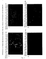
- rGM-CSF When GMI-RI incubated in the presence or absence of rGM-CSF was observed under a phase contrast microscope, it had a macrophage-like or globular form in the presence of rGM-CSF (Fig. 1 A) and a branched form in the absence of rGM-CSF (Fig. 1 B).
- the established cell line of microglia GMI-R1 of the present invention was allowed to adhere to a plastic vessel.
- a fluorescent pigment PKH26 Zynaxis Co.
- a phagocyte-staining solution Diluent B, Zynaxis Co.
- 10 % serum were mixed at a ratio of 1 : 1 and added to the above vessel, and GMI-RI was stained with the fluorescent pigment at 37 °C for 15 minutes (Ishihara. S., Sawada, M. et al., Exp. Neurol., 124 , 219-230, 1993).
- the cells were recovered and 2 ⁇ 10 6 cells were injected into an artery in the armpit of each of 5-week-old rats from the same strain (Fisher). 48 hours and 1, 2, and 3 weeks after injection, each organ was excised from the rats and frozen in a solution of OCT (Tissue Tek Co.).
- Microglia GMI-R1 and macrophages which was isolated for comparison from the abdomen of a rat of the same strain (Fisher) by washing it with cold PBS, were labeled respectively with a fluorescent pigment specific for phagocytes and then injected into armpit arteries of rats, and tissue sections were prepared for examination of the tissue orientation.
- the established cell line of microglia of the present invention possessed specific affinity for the brain.
- RNA was extracted from the cells stimulated with lipopolysaccharides for 12 hours and from the cells not stimulated by using RNeasy (Qiagen Co.), and 2 ⁇ g of the RNA was used to prepare a cDNA mixture by reverse transcriptase (BRL). PCR was carried out using the resulting cDNA as a template, where an IL-1 specific synthetic primer and IL-6 specific synthetic primer having the following sequences were used.
- IL-1 specific synthetic primer Sawada et al., Int. J. Dev. Neurosci, 13, 253-264, 1995:
- PCR In PCR, 30 cycles each consisting of reaction at 55 °C for 1 minute, 72 °C for 2 minutes and 94 °C for 1 minute were carried out (Omnigene from HYBRID Co., Ltd. was used). After PCR, the amplification product was subjected to agarose gel electrophoresis to examine gene expression.
- IL-1 and IL-6 Production of IL-1 and IL-6 was also confirmed by ELISA. Further, a culture supernatant of the established microglia of the present invention after stimulation with lipopolysaccharides was added to MH60 cells proliferating depending on IL-6 or to D10 cells proliferating depending on IL-1, followed by incubation, and whether the MH60 cells and D10 cells were proliferated or not was examined.
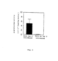
- rGM-CSF diluted 5000-fold, and 100 ⁇ /ml each of mouse IL-3, IL-4 and IL-6 were examined in a comparative test. Two days and four days after addition of the respective reagents, MTT assays were carried out (Fig. 4 B).
- Vector ptk ⁇ (Clonetech Co.) for expression of lac Z gene derived from E . coli and DOTAP lipid (Boehringer-MannheimCo.) were mixed to be a final concentration of 1 ⁇ g/ml .
- the mixture was mixed with a serum-containing medium, then added to the established cell line of microglia of the present invention and treated for 16 hours.
- Example 1 Example 1 (b-1) in order to examine whether the gene was delivered to and expressed in the brain, as follows: An artery in the left armpit of a mature rat (250 to 300 g) under anesthesia with Nembutal was exposed. After hemostatic treatment, a cannula was inserted into the artery and used to inject 1 to 2 ⁇ 10 6 cells into the rat. After injection, the incision site was sutured and the rat was allowed to recover.
- EMEM plus 10% FCS a usual medium
- b-1 fluorescent pigment as described in Example 1
- the brain was excised from the rat and 3 successive frozen sections of the brain were prepared and observed respectively under a fluorescence microscope. Further, staining and quantification of ⁇ -galactosidase activity was carried out in the following manner.
- One of the three sections was fixed in 0.5 % glutaraldehyde and subjected to activity staining with Xgal as substrate according to the method of Lim et al. (Bio Techniques 7 , 576-579, 1989). To quantify the activity, one of the sections was homogenized in a dissolving buffer by ultrasonication and examined for its activity with a commercial kit (Galacto Light; Boehringer-Mannheim).
- the fluorescent pigment PKH26 used in Example 1 forms granules in diluent B.
- the microglia cell strain of the present invention incorporates these granules specifically and transfers them to the brain, so the fluorescent pigment PKH26 was used as a model of chemical substance (anti-tumor drug).
- microglia of the present invention transfers the chemical substance (drug) into the brain specifically.
- GMI-R1 cells 1 ⁇ 10 6 GMI-R1 cells were plated on a Petri dish, and 16 hours later, 10 ⁇ g GFP expression vector pEGFP (Clonteck Co.) was added to a gene-introducing medium, with which the previous medium was then exchanged, and the cells were cultured at 37 °C for 24 hours in a CO 2 incubator and then cultured together with rGM-CSF for additional 7 days in a medium for microglia.
- pEGFP Clonteck Co.
- FACS fluorescent activated cell sorter
- Fig. 7 shows a result of FACS analysis of the cells into which the gene was not introduced
- Fig. 8 shows a result of FACS analysis of the cells into which GFP was introduced.
- an established cell line of microglia having specific affinity for the brain.
- the established cell line of microglia according to the present invention is useful not only as a carrier for introducing a gene into the brain, but also as a carrier four introducing a chemical substance such as drug specifically into the brain.
Description
- The present invention relates to a subcultivatable, established cell line of microglia, a method of separating the same, and use thereof as a pharmaceutical carrier.
- Further, we describe a microglia cell line into which an extraneous gene or a drug was introduced, a method of introducing it, and a pharmaceutical composition comprising the same.
- There are quite a number of hereditary diseases in the nervous system, which occur due to various causative factors, for example by defect of a single enzyme, etc. or by unknown reasons. Under these circumstances, supplementary therapy is used to cope with a large number of such diseases.
- A large number of studies have been made worldwide on the system of selective delivery to the brain. For introducing a gene into the brain in an animal, a method of using neuronphilic viruses as adenovirus vectors is devised, and a system for introducing a gene specifically into neurons is known (Kozarsky, K.F and Wilson, J. M., Curr. Opin. Genet. Dev. 3, 499-503, 1993). A method of using a retrovirus vector is also devised and has succeeded in introducing a gene into hepatic cells, bloodcells, etc. (Mullingan, R. C. , Science 260, 926-932, 1993).
- In the brain, however, the blood-brain barrier is present, so it is difficult to conduct supplementary therapy and to introduce an effective drug, and even if a substance (e.g. anticancer drug, DNA etc.) is introduced from a peripheral position, it cannot be introduced specifically into the brain. Therefore, there was no method other than direct injection of the substance by surgical operations.
- As a method not involving invasive means such as surgical operations, there is a method of utilizing liposomes, and liposomes rendered capable of introduction into the brain relatively easily by changing their constitutional elements were developed by a Japanese group. However, even in this method too, incorporation of liposomes into the brain is as low as about 1 % based on the injection amount, so this method cannot be said to be specific for the brain.
- As described above, the blood-brain barrier is present in the brain, so the brain is almost free of infiltration with cells or a substance from the periphery, thus making it difficult to introduce a drug or a gene into the brain. Actually, infiltration of the normal brain with immunocytes such as T cells and macrophages is hardly observed.
- Microglia are cells with macrophage-like properties in the central nervous system, which not only function as immunocompetent cells in inflammatory reaction and viral infection and as phagocytes for removing cells but also play a central role in a cytokine network in the central nervous system (Sawada, M. et al. , Int. J. Dev. Neurosci., 13, 253-264, 1995). Recently, microglia have been revealed to be essential for expression of high-level brain functions such as learning and memorization and considered to be specialized cells having a role specific for the brain. Up to now, it has been considered that microglia originate from monocytes having infiltrated the brain in the perinatal period and are specialized and differentiated.
- The microglia can be obtained by primary culture of brain cells. For this primary culture, however, the brain should be excised and purified for use, and primary culture usually requires a period of about 2 weeks, so the procedures are cumbersome. Further, the cells are difficult to proliferate during culture and hard to subculture, so after primary culture, it is extremely difficult to introduce a gene into the microglia to express it therein
- In a process of studies on microglia considered as brain-specific macrophage, the present inventors have succeeded in obtaining highly purified microglia and examined the properties, and as a result, we found that unlike the macrophage, microglia have specific affinity for the brain.
- Further, the inventors found that microglia are determinatively different from macrophage in respect of affinity for the brain and the ability to infiltrate the brain. Further, the inventors examined the distribution thereof by staining in a method capable of distinguishing the two, and found that microglia are present from an early development stage in the brain. Accordingly, it is considered that microglia are not derived from a monocyte differentiated and matured in the bone marrow, but are derived from a cell group which have a specific affinity for the brain, and infiltrate the brain at an early development stage and come to regulate high-level functions such as cerebral morphogenesis and learning and memorization.
- Accordingly, the inventors injected an isolated macrophage and microglia into rat peripheral arteries to compare the two for selective affinity for the brain, and as a result we found that when the microglia labeled with a fluorescent pigment was injected; many fluorescent cells were observed in the brain but hardly observed in the liver. On the other hand, when the macrophage was injected, fluorescent cells were hardly observed in the normal cell, but many fluorescent cells were observed in the liver.
- Then, the inventors examined whether a cell strain of microglia established by the inventors can express a gene selectively in the brain by introducing a lacZ expression vector into the cell strain and injecting the resultant cells into a rat blood stream, and as a result, the inventors could detect the activity of β-galactosidase in a section of the rat brain into which the lacZ-expressing cells had been injected. From these results, it was found that the microglia unlike the macrophage is a cell having specific affinity for the brain, and by utilizing this affinity, a specific substance or gene can be introduced via a peripheral blood stream into the brain.
- That is, the present invention relates to an established cell line of microglia having specific affinity for the brain and phagocytic ability, and having cell growth ability dependent on granulocyte-macrophage colony-stimulating factor (GM-CSF), characterised in that said cell line is subcultivatable in the presence of GM-CSF and has essentially a macrophage-like or globular form in the presence of GM-CSF and a branched form similar to branched microglia present in the brain in the absence of GM-CSF.
- Further, the present invention relates to a method of producing the established cell line of microglia which comprises the step of cloning a cell line of microglia from purified microglia through the cell culture in the presence of GM-CSF.
- Further, the present invention relates to a pharmaceutical composition comprising the established cell line of microglia.
- Further, the present invention relates to the pharmaceutical composition for use in a method of treating cerebral disease.
- We also describe a method of separating a subcultivatable established cell line of microglia from microglia cells in the presence of a cytokine, preferably in the presence of colony-stimulating factor (CSF), more preferably in the presence of granulocyte-macrophage colony-stimulating factor (GM-CSF), still more preferably in the presence of IL-3 and/or purified astrocytes.
- Also described is a pharmaceutical carrier comprising the established cell line of microglia described above.
- We also describe the above-described established cell line of microglia having a gene or a drug introduced into it.
- Also described is a pharmaceutical composition comprising the above-described microglia having a gene or a drug introduced into it and a pharmaceutical carrier and in particular to a pharmaceutical composition which is an agent for treatment of cerebral diseases.
- We also describe a method for screening or producing a microglia having an extraneous gene introduced into it, comprising introduction of an extraneous gene and a gene expressing a fluorescent protein, preferably a fluorescent protein derived from a jellyfish, into a microglia. As the microglia the conventional microglia can also be used, but the above-described established cell line of microglia is preferably used.
- We also describe using the above-described pharmaceutical composition to deliver a drug or gene specifically to the brain.
- Hereinafter, the present invention is described in more detail.
-
- Fig. 1 is a photograph showing the form of the established cell line of microglia of the present invention (morphology of the cells).
- Fig. 2 is a photograph showing the difference in tissue specificity between the established cell line of microglia of the present invention and macrophages (morphology of the cells).
- Fig. 3 shows electrophoresis indicating cytokine expression by stimulation with lipopolysaccharides.
- Fig. 4 shows GM-CSF-dependent proliferation of the established cell line of microglia of the present invention.
- Fig. 5 is a photograph indicating gene expression in a rat brain (morphology of the cells).
- Fig. 6 shows gene expression in a rat brain.
- Fig. 7 shows a result of FACS analysis of cells into which a gene was not introduced.
- Fig. 8 shows a result of FACS analysis of cells into which GFP was introduced.
- The established cell line of microglia of the present invention can be purified from mouse or rat brain cells after primary culture and then separated by the following means from this purified microglia. In addition, because the established cell line of microglia of the present invention is easily handled and has affinity for the brain, a gene or drug can be introduced into the established cell line of microglia and injected into peripheral blood vessels to express the gene in the brain or to deliver the drug to the brain specifically.
- Hereinafter, the process for producing the established cell line of microglia of the present invention is described in more detail.
- First, the meninges are removed from collected mouse or rat brains and divided into single cells by use of a pipette, nylon mesh, etc. The mouse and rat are preferably newborn. The mouse includes, but is not limited to, C57BL6, C3H, ICR, Balb/c etc., and the rat includes, but is not limited to, Fisher, Wister, SD, etc.
- The resulting cells are plated on a usual animal cell culture medium (e.g. EMEM containing 10 % FCS or CS) and cultured for 10 to 14 days. The medium is exchanged with fresh one every 3 to 4 days.
- Then, the cultured cells thus obtained are selected to prepare an established cell line in the subsequent step.
- There are microglia called type I and type II, and type I is floating cells which are removed from the culture vessel upon mechanical stimulation of cells after primary culture (that is, by splashing the cells with a medium through a pipette, by shaking the culture vessel, etc.). Type II is cells (adherent cells) not floated by said mechanical stimulation. Since the microglia of the present invention belongs to type II, purified microglia can be selected in the following manner.
- The adherent cells which do not float by the above mechanical stimulation are treated with trypsin-EDTA, then divided into single cells, plated on a non-treated plastic dish (non-coat plastic vessel), and allowed to adhere to it. Generally, a conventional culture vessel is treated with chemicals so as to be positively charged, but a dish not subjected to this treatment should be used in order to obtain the adherent cells. After incubation in a CO2 incubator at 37 °C for 1 hour, cells floating in the medium after mechanical stimulation are removed, and cells capable of proliferation on the vessel are recovered by Rubber Policeman, etc., and the same procedure is repeated twice to obtain cells to be used in establishing a cell line of microglia. Although the purified microglia thus obtained is of adequate purity, cell sorter, etc. can also be used to further improve purity.
- To separate the subcultivatable established cell line of microglia of the present invention from the purified microglia obtained in the manner described in (1) above, the purified microglia is cloned through culture in the presence of a cytokine, preferably colony-stimulating factor (CSF), more preferably granulocyte-macrophage colony-stimulating factor (GM-CSF). As the cytokine allowed to be present during culture, GM-CSF may be used singly, but IL-3 and/or a supernatant derived from purified astrocytes may be allowed to be present in addition to GM-CSF. The cytokine used may be the naturally occurring or genetic recombinant one. Specific procedures are for example as follows:
- The purified microglia obtained in (1) above is plated on a vessel of about 10 cm diameter and cultured for 7 to 10 days in the presence of genetic recombinant granulocyte-macrophage colony-stimulating factor (rGM-CSF). After culture, the cells are recovered, and using limiting dilution, they are further cultured for 4 to 10 weeks in the presence of rGM-CSF. Then, cells having formed a single colony in each well in the test plate are released by Rubber Policeman whereby the cloned cells are sorted and separated to finally give an established cell line of microglia.
- The established cell line of microglia thus obtained has the following properties.
- When observed after staining with a fluorescent pigment under a fluorescence microscope or without staining under a phase contract microscope, the cells have a macrophage-like or globular form in the presence of rGM-CSF, or in the absence of rGM-CSF, a branched form similar to branched microglia present in the brain. Otherwise the cells have both of the above forms.
- Upon administration into an artery of a mouse, the established microglia of the present invention moves specifically to the brain, indicating that it has specific affinity for the brain. Further, when stimulation is given with lipopolysaccharides the microglia produces interleukin- 1 (IL-1) and interleukin-6 (IL-6). Upon stimulation with interferon γ, it produces IL-5. This property is different from that of macrophages because macrophages do not express IL-5 upon stimulation with IFN-γ. When the phagocytic ability of the cell line of the present invention is examined using incorporation of a fluorescent pigment as an indicator, it has a strong phagocytic ability. The established cell line of microglia of the present invention has a phagocytic ability which is hundreds to thousands times higher than that of astrocytes.
- Because the established microglia of the present invention is not proliferated after removal of rGM-CSF from the medium, it is proliferated depending on GM-CSF.
- The form and characteristics of the established microglia of the present invention are exemplified in specific examples in Figs. 1 to 4.
- Introduction of a gene into the established cell line of microglia of the present invention is important for expressing the gene specifically in the brain. A desired gene can be obtained by known cloning means, and any commercially available gene can also be used, and the type of gene is particularly not limited.
- As a means of efficiently introducing a gene into the established cell line of microglia, there is for example a method of using DOTAP (Boehringer-Mannheim Co.). That is, there is a method of culturing the established cell line of microglia together with a desired gene in a gene-introducing medium containing DOTAP. They are cultured in a CO2 incubator at 37 °C for 16 to 24 hours and further cultured together with rGM-CSF for 30 to 72 hours (preferably for 48 hours) in a medium for culturing microglia.
- In addition to the above-described means, the method of introducing the gene into the microglia includes conventional means such as calcium phosphate method, DEAE dextran method, lipofection method, electroporation method, particle gum method, etc
- Out of known methods of separating a strain stably expressing an introduced gene, the most generally used method is a method wherein a drug resistance gene is introduced simultaneously with a desired gene into cells and expressed therein, and cells other than those cells coming to express the gene stably and constantly by incorporating the gene into their chromosomal DNA are perished by chemical treatment and excluded from the culture, whereby the desired cells are separated.
- When this screening method is applied to the microglia, the microglia coming to express the introduced gene stably and constantly recognize dead cells thus expressing a strong phagocytic ability and simultaneously undergoing activation to lose their cell growth ability. Accordingly, the method of introducing a gene in the present invention is preferably a method wherein an expression vector modified so as to be capable of expressing a fluorescent protein, preferably green florescent protein (GFP) derived from an aurelia, in higher animals is used in place of a drug resistance gene, and by using a difference in fluorescence intensity, the microglia is separated as the desired cells exhibiting stable and constant expression.
- Whether the cells having the gene introduced into them have reached the brain and whether the gene has been expressed can be confirmed in the following manner: The cells to be introduced are stained with a fluorescent pigment specific for phagocytes. After introduction of the cells into an animal, the brain is excised and frozen, from which a section of about 8 microns in thickness is prepared and examined for fluorescent cells under a fluorescence microscope, or the section is activity-stained with a substrate for the introduced gene.
- The cells can also be confirmed using magnetic resonance image (MRI), positron emission tomography (PET) etc. For example, contrast media, etc. for MRI may be incorporated into the cells which are then injected into an animal so that the cells can be monitored in the animal. According to these methods, it is not necessary to kill the animal, and the cells can be monitored easily in a non-invasive manner.
- Hereinafter, the present invention is described in more detail by reference to Examples.
- Brains were excised from newborn mice (C57BL6, op/op) and newborn rats (Fisher), and meninges were removed in an ice-cold microglia culture medium (referred to as Mi medium; Eagle's MEM containing 10 % bovine serum, 0.2 % glucose and 5 µg/ml bovine insulin). The cells of meninges were divided into single cells with a Pasteur pipette or nylon mesh and then cultured in Mi medium. For the cells from the mouse brains, 20 ml Mi medium was used per one brain, and for the cells from the rat brains, 40 ml Mi medium was used per one brain, and the former cells were incubated in 2 culture vessels of 10 cm diameter and the latter cells in 4 culture vessels of 10 cm diameter in a CO2 incubator (5 % CO2, 95 % air) at 37 °C for 10 to 14 days. The medium was exchanged with fresh one every 3 to 4 days
- When phase-bright round cells (PBRCs) appeared, the PBRCs were removed by mechanical shaking, and the remaining cells were removed with 200 U/ml trypsin-0.02 % EDTA and incubated in a non-coat plastic vessel at 37 °C for 30 minutes. The cells which adhered to the non-coat plastic vessel were washed twice with Mi medium, and the cells were then removed and recovered. The same procedure was repeated further twice to give purified microglia.
- 1×10⁵ purified microglia cells obtained in (1) above were plated on a 10 cm vessel and cultured for 7 days in Mi medium in the presence of rGM-CSF (GenzymeCo.).
- The cells were recovered and counted, and clones were obtained from the cells in the following manner using limiting dilution. The cells were put to each well on a 96-well plate (Falcon Co.) at a density of 0.5 cell/well (in 100 µl) and cultured for about 3 weeks in the presence of 2 ng/ml mouse gene recombinant GM-CSF (Genzyme Co.). Each well was examined for the presence of the clone, and the target clones were separated.
- As a result, five kinds of established microglia (Ra2, GMI-M6-1, GMI-M6-3, GMI-M5-2, GMI-MF11) derived from the mouse brains and one kind (GMI-Rl) from the rat brains were obtained.
- Among these, the established cell lines of microglia, Ra2 and GMI-R1, have been designated as "mouse microglia Ra2" and "rat microglia GMI-R1" and deposited as FERM P-16109 and FERM P-16110 respectively with the National Institute of Bioscience and Human-Technology, Agency of Industrial Science and Technology, Japan.
- The properties of the resulting clones were then examined.
- When GMI-RI incubated in the presence or absence of rGM-CSF was observed under a phase contrast microscope, it had a macrophage-like or globular form in the presence of rGM-CSF (Fig. 1 A) and a branched form in the absence of rGM-CSF (Fig. 1 B).
- The established cell line of microglia GMI-R1 of the present invention was allowed to adhere to a plastic vessel. Separately, a fluorescent pigment PKH26 (Zynaxis Co.) prepared in a phagocyte-staining solution (Diluent B, Zynaxis Co.) and 10 % serum were mixed at a ratio of 1 : 1 and added to the above vessel, and GMI-RI was stained with the fluorescent pigment at 37 °C for 15 minutes (Ishihara. S., Sawada, M. et al., Exp. Neurol., 124, 219-230, 1993).
- The cells were recovered and 2×10⁶ cells were injected into an artery in the armpit of each of 5-week-old rats from the same strain (Fisher). 48 hours and 1, 2, and 3 weeks after injection, each organ was excised from the rats and frozen in a solution of OCT (Tissue Tek Co.).
- Microglia GMI-R1, and macrophages which was isolated for comparison from the abdomen of a rat of the same strain (Fisher) by washing it with cold PBS, were labeled respectively with a fluorescent pigment specific for phagocytes and then injected into armpit arteries of rats, and tissue sections were prepared for examination of the tissue orientation.
- After the established cell line of microglia of the present invention was injected, many fluorescent cells were observed in normal brain cells (Fig. 2 A) but not observed in the liver (Fig. 2 B). On the other hand, after the macrophages were injected, fluorescent cells were hardly observed in normal brain cells (Fig. 2 C), while many fluorescent cells were observed in the liver (Fig. 2 D).
- Accordingly, the established cell line of microglia of the present invention possessed specific affinity for the brain. (b-2) Functional Characteristics (ability to produce IL-1 and IL-6)
- 1×10⁶ Ra2 cells were plated onto a 6 cm culture vessel, and total RNA was extracted from the cells stimulated with lipopolysaccharides for 12 hours and from the cells not stimulated by using RNeasy (Qiagen Co.), and 2 µg of the RNA was used to prepare a cDNA mixture by reverse transcriptase (BRL). PCR was carried out using the resulting cDNA as a template, where an IL-1 specific synthetic primer and IL-6 specific synthetic primer having the following sequences were used. IL-1 specific synthetic primer (Sawada et al., Int. J. Dev. Neurosci, 13, 253-264, 1995):
- Sense chain: 5'-ATGGCAACTGTTCCTGAACTCAACT-3' (SEQ ID NO: 1)
- Antisense chain: 5'-CAGGACAGGTATAGATTCTTTCCTTT-3' (SEQ ID NO: 2)
- IL-6 specific synthetic primer (Sawada et al., Brain Res. 583, 296-299, 1992):
- Sense chain: 5'-ATGAAGTTCCTCTCTGCAAGAGACT-3' (SEQ ID NO: 3)
- Antisense chain: 5'-CACTAGGTTTGCCGAGTAGATCTC-3' (SEQ ID NO: 4)
- In PCR, 30 cycles each consisting of reaction at 55 °C for 1 minute, 72 °C for 2 minutes and 94 °C for 1 minute were carried out (Omnigene from HYBRID Co., Ltd. was used). After PCR, the amplification product was subjected to agarose gel electrophoresis to examine gene expression.
- As a result, it was found that Ra2 increased expression of IL-1 and IL-6 (Fig. 3, "LPS" lanes). In Fig. 3, "M" is a molecular weight marker and "cont" is control (not stimulated).
- Production of IL-1 and IL-6 was also confirmed by ELISA. Further, a culture supernatant of the established microglia of the present invention after stimulation with lipopolysaccharides was added to MH60 cells proliferating depending on IL-6 or to D10 cells proliferating depending on IL-1, followed by incubation, and whether the MH60 cells and D10 cells were proliferated or not was examined.
- As a result, it was found that both the cells proliferated in the presence of the culture supernatant of the established microglia of the present invention stimulated with the lipopolysaccharides.
- 5×10⁴ GMI-R1 cells were plated on a 96-well test plate, and 2 µg/ml rGM-CSF was added to it so as to be diluted 1000-, 5000- and 10000-fold respectively, and the cells were incubated for 4 days and then subjected to MTT assays. As a control, 400 µ/ml human M-CSF (The Green Cross Corporation) was diluted 1000-fold and 0.1 mg/ml PMA (phorbol myristate acetate) was diluted 1000- or 5000-fold, and these were used as the control (Fig. 4 A).
- Separately, rGM-CSF diluted 5000-fold, and 100 µ/ml each of mouse IL-3, IL-4 and IL-6 (any of which were produced by Genzyme Co.), were examined in a comparative test. Two days and four days after addition of the respective reagents, MTT assays were carried out (Fig. 4 B).
- As a result, it was found that GMI-R1 was proliferated depending on rGM-CSF.
- Vector ptkβ (Clonetech Co.) for expression of lac Z gene derived from E . coli and DOTAP lipid (Boehringer-MannheimCo.) were mixed to be a final concentration of 1 µg/ml . The mixture was mixed with a serum-containing medium, then added to the established cell line of microglia of the present invention and treated for 16 hours. As the control, the established microglia of the invention into which the gene was not introduced, and macrophages obtained in the same manner as in Example 1, were used.
- Then, the cells were further cultured for 48 hours in a usual medium (EMEM plus 10% FCS) and then stained with the fluorescent pigment as described in Example 1 (b-1) in order to examine whether the gene was delivered to and expressed in the brain, as follows: An artery in the left armpit of a mature rat (250 to 300 g) under anesthesia with Nembutal was exposed. After hemostatic treatment, a cannula was inserted into the artery and used to inject 1 to 2×10⁶ cells into the rat. After injection, the incision site was sutured and the rat was allowed to recover.
- 48 hours after the cells were injected, the brain was excised from the rat and 3 successive frozen sections of the brain were prepared and observed respectively under a fluorescence microscope. Further, staining and quantification of β-galactosidase activity was carried out in the following manner.
- One of the three sections was fixed in 0.5 % glutaraldehyde and subjected to activity staining with Xgal as substrate according to the method of Lim et al. (
Bio Techniques 7, 576-579, 1989). To quantify the activity, one of the sections was homogenized in a dissolving buffer by ultrasonication and examined for its activity with a commercial kit (Galacto Light; Boehringer-Mannheim). - As a result, it could be confirmed that in case the gene was introduced into the established microglia of the present invention, lacZ-positive cells were present in the section from the rat brain (Fig. 5). Further, as a result of quantification of β-galactosidase activity by the chemiluminescence method, considerably higher activity was detected in the rat brain section into which the E. coli-derived gene lac Z expression vector was introduced than the counterpart into which the gene was not introduced (Fig. 6).
- The fluorescent pigment PKH26 used in Example 1 forms granules in diluent B. The microglia cell strain of the present invention incorporates these granules specifically and transfers them to the brain, so the fluorescent pigment PKH26 was used as a model of chemical substance (anti-tumor drug).
- As a result, when the established cell line of microglia of the present invention was introduced, many fluorescent cells were observed in normal brain cells but not observed in the liver. It therefore follows that the microglia of the present invention transfers the chemical substance (drug) into the brain specifically.
- 1×10⁶ GMI-R1 cells were plated on a Petri dish, and 16 hours later, 10 µg GFP expression vector pEGFP (Clonteck Co.) was added to a gene-introducing medium, with which the previous medium was then exchanged, and the cells were cultured at 37 °C for 24 hours in a CO2 incubator and then cultured together with rGM-CSF for additional 7 days in a medium for microglia.
- After culture for 7 days, the cells were suspended, and those cells exhibiting fluorescence intensity which was at least 100 times as high as that of the cells into which the gene had not been introduced were fractionated and concentrated by a fluorescent activated cell sorter (FACS) , FACS Calibur produced by Becton-Dickinson and then cultured together with rGM-CSF in a medium for microglia.
- The same procedure was repeated additionally twice every 7 days, whereby almost 90 % or more cells could be recovered in a fraction having about 100-fold fluorescence, and these cells were put to a TP96 test plate at a density of 1 cell/well by limiting dilution of the cells and further cultured together with rGM-CSF in a medium for microglia. The microglia coming to expressing the introduced gene pEGFP stably and constantly could thereby be separated.
- One example of results in FACS analysis as to how the cells could be separated by this method is shown in the drawings. Fig. 7 shows a result of FACS analysis of the cells into which the gene was not introduced, while Fig. 8 shows a result of FACS analysis of the cells into which GFP was introduced.
- According to the present invention, there is provided an established cell line of microglia having specific affinity for the brain. The established cell line of microglia according to the present invention is useful not only as a carrier for introducing a gene into the brain, but also as a carrier four introducing a chemical substance such as drug specifically into the brain.
-
- SEQ ID NO: 1
LENGTH: 25 base pairs
TYPE: nucleic acid
STRANDEDNESS: single
TOPOLOGY: linear
MOLECULAR TYPE: other nucleic acid (synthetic-DNA)
SEQUENCE DESCRIPTION: SEQ ID NO.I:
ATGGCAACTG TTCCTGAACT CAACT - SEQ ID NO: 2
LENGTH: 26 base pairs
TYPE: nucleic acid
STRANDEDNESS: single
TOPOLOGY: linear
MOLECULAR TYPE: other nucleic acid (synthetic DNA)
SEQUENCE DESCRIPTION: SEQ ID NO 2:
CAGGACAGGT ATAGATTCTT TCCTTT - SEQ ID NO: 3
LENGTH: 25 base pairs
TYPE: nucleic acid
STRANDEDNESS: single
TOPOLOGY: linear
MOLECULAR TYPE: other nucleic acid (synthetic DNA)
SEQUENCE DESCRIPTION: SEQ ID NO 3:
ATGAAGTTCC TCTCTGCAAG AGACT - SEQ ID NO: 4
LENGTH: 24 base pairs
TYPE: nucleic acid
STRANDEDNESS: single
TOPOLOGY: linear
MOLECULAR TYPE: other nucleic acid (synthetic DNA)
SEQUENCE DESCRIPTION: SEQ ID NO 4:
CACTAGGTTT GCCGAGTAGA TCTC.
Claims (6)
- An established cell line of microglia having specific affinity for the brain and phagocytic ability, and having cell growth ability dependent on granulocyte-macrophage colony stimulating factor (GM-CSF), characterised in that said cell line is subcultivatable in the presence of GM-CSF and has essentially macrophage-like or globular form in the presence of GM-CSF and a branched form similar to branched microglia in the brain in the absence of GM-CSF.
- A method of producing the established cell line of microglia of claim 1, which comprises the step of cloning a cell line of microglia from purified microglia through the cell culture in the presence of GM-CSF.
- The method according to claim 2, wherein the GM-CSF is a genetic recombinant one.
- The method according to claim 2 or 3, wherein the cell culture is carried out in the presence of IL-3 and/or a culture supernatant of purified astrocytes in addition to the GM-CSF.
- A pharmaceutical composition comprising an established cell line of microglia as defined in claim 1.
- The pharmaceutical composition according to claim 5 for use in a method of treating cerebral disease.
Applications Claiming Priority (3)
| Application Number | Priority Date | Filing Date | Title |
|---|---|---|---|
| JP5044897 | 1997-03-05 | ||
| JP5044897 | 1997-03-05 | ||
| PCT/JP1998/000949 WO1998039415A1 (en) | 1997-03-05 | 1998-03-05 | Established microglia |
Publications (3)
| Publication Number | Publication Date |
|---|---|
| EP0949331A1 EP0949331A1 (en) | 1999-10-13 |
| EP0949331A4 EP0949331A4 (en) | 2001-05-02 |
| EP0949331B1 true EP0949331B1 (en) | 2007-01-10 |
Family
ID=12859154
Family Applications (1)
| Application Number | Title | Priority Date | Filing Date |
|---|---|---|---|
| EP98905808A Expired - Lifetime EP0949331B1 (en) | 1997-03-05 | 1998-03-05 | Established microglia |
Country Status (5)
| Country | Link |
|---|---|
| US (3) | US6673605B2 (en) |
| EP (1) | EP0949331B1 (en) |
| JP (1) | JP3410738B2 (en) |
| DE (1) | DE69836853T2 (en) |
| WO (1) | WO1998039415A1 (en) |
Families Citing this family (5)
| Publication number | Priority date | Publication date | Assignee | Title |
|---|---|---|---|---|
| FI20040953A0 (en) * | 2004-07-08 | 2004-07-08 | Jari Koistinaho | Bone marrow cell stimulation method and bone marrow cell |
| JP4670363B2 (en) * | 2005-01-21 | 2011-04-13 | 住友ベークライト株式会社 | Method for transporting microglia cells |
| WO2006082798A1 (en) * | 2005-02-01 | 2006-08-10 | Kumamoto University | Monoclonal antibody specifically recognizing rat type i microglia |
| WO2015105201A1 (en) | 2014-01-09 | 2015-07-16 | Kyushu University, National University Corporation | Method of producing microglial cells |
| CN113122499A (en) * | 2021-04-20 | 2021-07-16 | 苏州大学 | Primary culture method of microglia capable of improving yield |
Family Cites Families (6)
| Publication number | Priority date | Publication date | Assignee | Title |
|---|---|---|---|---|
| JPH01172324A (en) | 1987-12-15 | 1989-07-07 | Armour Internatl Co | Colestyramine composition and production thereof |
| JPH02286621A (en) | 1989-04-26 | 1990-11-26 | Mitsubishi Kasei Corp | Oral cholesterol lowering agent |
| JPH0549473A (en) * | 1991-08-14 | 1993-03-02 | Mochida Pharmaceut Co Ltd | Cerebral microglia cell line of rat |
| JP3552285B2 (en) | 1993-08-03 | 2004-08-11 | 三菱化学株式会社 | Oral cholesterol lowering agent |
| TW438608B (en) | 1995-08-02 | 2001-06-07 | Hisamitsu Pharmaceutical Co | A tablet containing anion exchange resin |
| US6780641B2 (en) * | 2000-07-10 | 2004-08-24 | University Of British Columbia | Immortalized human microglia cell line |
-
1998
- 1998-03-05 EP EP98905808A patent/EP0949331B1/en not_active Expired - Lifetime
- 1998-03-05 WO PCT/JP1998/000949 patent/WO1998039415A1/en active IP Right Grant
- 1998-03-05 JP JP53838398A patent/JP3410738B2/en not_active Expired - Fee Related
- 1998-03-05 US US09/180,394 patent/US6673605B2/en not_active Expired - Lifetime
- 1998-03-05 DE DE69836853T patent/DE69836853T2/en not_active Expired - Lifetime
-
2003
- 2003-06-23 US US10/602,234 patent/US20040072346A1/en not_active Abandoned
-
2006
- 2006-06-02 US US11/445,564 patent/US20060233769A1/en not_active Abandoned
Also Published As
| Publication number | Publication date |
|---|---|
| US6673605B2 (en) | 2004-01-06 |
| US20060233769A1 (en) | 2006-10-19 |
| DE69836853D1 (en) | 2007-02-22 |
| US20040072346A1 (en) | 2004-04-15 |
| EP0949331A1 (en) | 1999-10-13 |
| DE69836853T2 (en) | 2007-06-21 |
| JP3410738B2 (en) | 2003-05-26 |
| WO1998039415A1 (en) | 1998-09-11 |
| EP0949331A4 (en) | 2001-05-02 |
| US20020115206A1 (en) | 2002-08-22 |
Similar Documents
| Publication | Publication Date | Title |
|---|---|---|
| US6184035B1 (en) | Methods for isolation and activation of, and control of differentiation from, skeletal muscle stem or progenitor cells | |
| CN101792732A (en) | Techniques for growth and differentiation of human pluripotent stem cells | |
| US20060247195A1 (en) | Method of altering cell properties by administering rna | |
| Kuang et al. | Disruption of thelama2gene in embryonic stem cells: Laminin α2 is necessary for sustenance of mature muscle cells | |
| WO1998032879A1 (en) | Method for separating cells | |
| EP0946199B1 (en) | TGF beta 1-RESPONSIVE CELLS FROM BONE MARROW | |
| JP2003524407A (en) | Identification of pluripotent pro-mesenchymal and pro-hematopoietic progenitor stem cells | |
| EP0457856A1 (en) | Isolation growth and differentiation of human muscle cells | |
| US20060233769A1 (en) | Established cell line of microglia | |
| EP1090105B1 (en) | Non-embryonic ependymal neural stem cells and method for their isolation | |
| WO2009152186A1 (en) | Methods for enhancing cell therapy efficacy including treatment with cd26 peptidase inhibitors | |
| US20040220396A1 (en) | Polypeptide having an activity to support proliferation or survival of hematopoietic stem cell and hematopoietic progenitor cell, and dna coding for the same | |
| JP4387108B2 (en) | Stock microglia | |
| WO2010147803A2 (en) | Adult cerebellum-derived neural stem cells and compositions and methods for producing oligodendrocytes | |
| US20030109037A1 (en) | Methods for application of genetically-modified endogenous or exogenous stem/progenitor or their progeny for treatment of disease | |
| US20080206202A1 (en) | Methods for application of endogenous or exogenous stem/progenitor or their progeny for treatment of disease | |
| AU1550802A (en) | TGF-beta1-responsive cells from bone marrow |
Legal Events
| Date | Code | Title | Description |
|---|---|---|---|
| PUAI | Public reference made under article 153(3) epc to a published international application that has entered the european phase |
Free format text: ORIGINAL CODE: 0009012 |
|
| 17P | Request for examination filed |
Effective date: 19981130 |
|
| AK | Designated contracting states |
Kind code of ref document: A1 Designated state(s): CH DE FR GB IT LI |
|
| A4 | Supplementary search report drawn up and despatched |
Effective date: 20010321 |
|
| AK | Designated contracting states |
Kind code of ref document: A4 Designated state(s): CH DE FR GB IT LI |
|
| 17Q | First examination report despatched |
Effective date: 20021014 |
|
| RAP1 | Party data changed (applicant data changed or rights of an application transferred) |
Owner name: JAPAN SCIENCE AND TECHNOLOGY AGENCY |
|
| GRAP | Despatch of communication of intention to grant a patent |
Free format text: ORIGINAL CODE: EPIDOSNIGR1 |
|
| GRAS | Grant fee paid |
Free format text: ORIGINAL CODE: EPIDOSNIGR3 |
|
| GRAA | (expected) grant |
Free format text: ORIGINAL CODE: 0009210 |
|
| AK | Designated contracting states |
Kind code of ref document: B1 Designated state(s): CH DE FR GB IT LI |
|
| REG | Reference to a national code |
Ref country code: GB Ref legal event code: FG4D |
|
| REF | Corresponds to: |
Ref document number: 69836853 Country of ref document: DE Date of ref document: 20070222 Kind code of ref document: P |
|
| REG | Reference to a national code |
Ref country code: CH Ref legal event code: NV Representative=s name: PATENTANWAELTE SCHAAD, BALASS, MENZL & PARTNER AG |
|
| ET | Fr: translation filed | ||
| PLBE | No opposition filed within time limit |
Free format text: ORIGINAL CODE: 0009261 |
|
| STAA | Information on the status of an ep patent application or granted ep patent |
Free format text: STATUS: NO OPPOSITION FILED WITHIN TIME LIMIT |
|
| 26N | No opposition filed |
Effective date: 20071011 |
|
| REG | Reference to a national code |
Ref country code: HK Ref legal event code: WD Ref document number: 1024715 Country of ref document: HK |
|
| REG | Reference to a national code |
Ref country code: CH Ref legal event code: PUE Owner name: ACTGEN, INC. Free format text: MAKOTO SAWADA#21, KITA 2-CHOME KIBUKI-CHO KASUGAI-CHI#AICHI-KEN 487-0015 (JP) -TRANSFER TO- ACTGEN, INC.#15-502 AKAHO#KOMAGANE, NAGANO, 399-4117 (JP) Ref country code: CH Ref legal event code: PUE Owner name: MAKOTO SAWADA Free format text: JAPAN SCIENCE AND TECHNOLOGY AGENCY#4-1-8, HONCHO#KAWAGUCHI-SHI SAITAMA (JP) -TRANSFER TO- MAKOTO SAWADA#21, KITA 2-CHOME KIBUKI-CHO KASUGAI-CHI#AICHI-KEN 487-0015 (JP) |
|
| REG | Reference to a national code |
Ref country code: GB Ref legal event code: 732E Free format text: REGISTERED BETWEEN 20110526 AND 20110601 |
|
| REG | Reference to a national code |
Ref country code: FR Ref legal event code: TP |
|
| REG | Reference to a national code |
Ref country code: DE Ref legal event code: R082 Ref document number: 69836853 Country of ref document: DE Representative=s name: RECHTS- UND PATENTANWAELTE LORENZ SEIDLER GOSS, DE |
|
| REG | Reference to a national code |
Ref country code: DE Ref legal event code: R082 Ref document number: 69836853 Country of ref document: DE Representative=s name: LORENZ SEIDLER GOSSEL RECHTSANWAELTE PATENTANW, DE Effective date: 20111130 Ref country code: DE Ref legal event code: R082 Ref document number: 69836853 Country of ref document: DE Representative=s name: RECHTS- UND PATENTANWAELTE LORENZ SEIDLER GOSS, DE Effective date: 20111130 Ref country code: DE Ref legal event code: R081 Ref document number: 69836853 Country of ref document: DE Owner name: ACTGEN, INC., KOMAGANE, JP Free format text: FORMER OWNER: JAPAN SCIENCE AND TECHNOLOGY AGENCY, KAWAGUCHI-SHI, SAITAMA, JP Effective date: 20111130 Ref country code: DE Ref legal event code: R081 Ref document number: 69836853 Country of ref document: DE Owner name: ACTGEN, INC., JP Free format text: FORMER OWNER: JAPAN SCIENCE AND TECHNOLOGY AGENCY, KAWAGUCHI-SHI, JP Effective date: 20111130 |
|
| PGFP | Annual fee paid to national office [announced via postgrant information from national office to epo] |
Ref country code: CH Payment date: 20140312 Year of fee payment: 17 |
|
| PGFP | Annual fee paid to national office [announced via postgrant information from national office to epo] |
Ref country code: FR Payment date: 20140311 Year of fee payment: 17 Ref country code: IT Payment date: 20140306 Year of fee payment: 17 |
|
| PGFP | Annual fee paid to national office [announced via postgrant information from national office to epo] |
Ref country code: GB Payment date: 20140305 Year of fee payment: 17 |
|
| PGFP | Annual fee paid to national office [announced via postgrant information from national office to epo] |
Ref country code: DE Payment date: 20140417 Year of fee payment: 17 |
|
| REG | Reference to a national code |
Ref country code: DE Ref legal event code: R119 Ref document number: 69836853 Country of ref document: DE |
|
| REG | Reference to a national code |
Ref country code: CH Ref legal event code: PL |
|
| GBPC | Gb: european patent ceased through non-payment of renewal fee |
Effective date: 20150305 |
|
| PG25 | Lapsed in a contracting state [announced via postgrant information from national office to epo] |
Ref country code: IT Free format text: LAPSE BECAUSE OF NON-PAYMENT OF DUE FEES Effective date: 20150305 |
|
| REG | Reference to a national code |
Ref country code: FR Ref legal event code: ST Effective date: 20151130 |
|
| PG25 | Lapsed in a contracting state [announced via postgrant information from national office to epo] |
Ref country code: CH Free format text: LAPSE BECAUSE OF NON-PAYMENT OF DUE FEES Effective date: 20150331 Ref country code: GB Free format text: LAPSE BECAUSE OF NON-PAYMENT OF DUE FEES Effective date: 20150305 Ref country code: DE Free format text: LAPSE BECAUSE OF NON-PAYMENT OF DUE FEES Effective date: 20151001 Ref country code: LI Free format text: LAPSE BECAUSE OF NON-PAYMENT OF DUE FEES Effective date: 20150331 |
|
| PG25 | Lapsed in a contracting state [announced via postgrant information from national office to epo] |
Ref country code: FR Free format text: LAPSE BECAUSE OF NON-PAYMENT OF DUE FEES Effective date: 20150331 |