EP0947583A2 - DNA fragments activated in expression of melanoma inhibiting protein (MIA) - Google Patents
DNA fragments activated in expression of melanoma inhibiting protein (MIA) Download PDFInfo
- Publication number
- EP0947583A2 EP0947583A2 EP99101554A EP99101554A EP0947583A2 EP 0947583 A2 EP0947583 A2 EP 0947583A2 EP 99101554 A EP99101554 A EP 99101554A EP 99101554 A EP99101554 A EP 99101554A EP 0947583 A2 EP0947583 A2 EP 0947583A2
- Authority
- EP
- European Patent Office
- Prior art keywords
- mia
- protein
- dna
- seq
- cells
- Prior art date
- Legal status (The legal status is an assumption and is not a legal conclusion. Google has not performed a legal analysis and makes no representation as to the accuracy of the status listed.)
- Withdrawn
Links
Images


Classifications
-
- C—CHEMISTRY; METALLURGY
- C07—ORGANIC CHEMISTRY
- C07K—PEPTIDES
- C07K14/00—Peptides having more than 20 amino acids; Gastrins; Somatostatins; Melanotropins; Derivatives thereof
- C07K14/435—Peptides having more than 20 amino acids; Gastrins; Somatostatins; Melanotropins; Derivatives thereof from animals; from humans
- C07K14/46—Peptides having more than 20 amino acids; Gastrins; Somatostatins; Melanotropins; Derivatives thereof from animals; from humans from vertebrates
- C07K14/47—Peptides having more than 20 amino acids; Gastrins; Somatostatins; Melanotropins; Derivatives thereof from animals; from humans from vertebrates from mammals
- C07K14/4701—Peptides having more than 20 amino acids; Gastrins; Somatostatins; Melanotropins; Derivatives thereof from animals; from humans from vertebrates from mammals not used
- C07K14/4702—Regulators; Modulating activity
- C07K14/4703—Inhibitors; Suppressors
-
- A—HUMAN NECESSITIES
- A61—MEDICAL OR VETERINARY SCIENCE; HYGIENE
- A61P—SPECIFIC THERAPEUTIC ACTIVITY OF CHEMICAL COMPOUNDS OR MEDICINAL PREPARATIONS
- A61P35/00—Antineoplastic agents
-
- A—HUMAN NECESSITIES
- A61—MEDICAL OR VETERINARY SCIENCE; HYGIENE
- A61K—PREPARATIONS FOR MEDICAL, DENTAL OR TOILETRY PURPOSES
- A61K38/00—Medicinal preparations containing peptides
-
- C—CHEMISTRY; METALLURGY
- C07—ORGANIC CHEMISTRY
- C07K—PEPTIDES
- C07K2319/00—Fusion polypeptide
Definitions
- the invention relates to a melanoma-inhibiting protein (MIA), a nucleic acid coding therefor, a method for the production and detection of this protein and its use in the manufacture of a therapeutic.
- MIA melanoma-inhibiting protein
- the regulation of cell growth is both positive controlled as well as negative factors.
- To the positive factors include the known ones Growth factors such as B. epidermal growth factor (EGF), platelet derived growth factor (PDGF), insulin and Somatomedine.
- factors include both Growth stimulator as well as growth inhibitor can act (Roberts et al., Proc. Natl. Acad. Sci.
- a growth inhibition is an anti-proliferative To understand effect.
- a suitable concentration for this is, for example, 0.1 ⁇ g MIA protein / ml culture medium.
- Higher or lower concentrations of the MIA protein are also suitable for growth inhibition, which depending on the concentration stronger or is observed less.
- This cell line was developed by a metastatic malignant Melanoma derived and in defined serum-free culture medium (50% Dulbecco's minimal essential medium, 50% F-12) with 0.8 mmol / l L-glutamine, non-essential amino acids, 10 ⁇ g / ml transferrin, 30 nmol / l sodium selenite and 4 ⁇ g / ml gentamicin as a monolayer culture under Cultivated standard culture conditions.
- the cell line was on June 23, 1993 at the German Collection for Microorganisms and Zellkulturen GmbH in Braunschweig (DSM ACC 2133). It is another object of the invention. From the culture supernatant of this cell line, the invention can Protein via a gel chromatographic Isolation of a protein fraction approximately 11 in size kD and subsequent purification of this fraction a reverse phase HPLC can be obtained.
- the protein can have its DNA sequence and those of it derived amino acid sequence can be defined.
- the MIA protein can be found in natural allelic variations can differ from individual to individual, occur (e.g. SEQ ID NO: 24). Such variations of Amino acids are usually amino acid exchanges. It can also be deletions, insertions or Act additions of amino acids to the overall sequence.
- the MIA protein according to the invention can - both in scope and Kind depending on the cell and the cell type in which it is is expressed - glycosylated or not glycosylated be.
- the protein according to the invention can also be produced recombinantly become. In the recombinant production in Prokaryotes will get non-glycosylated MIA protein.
- Nucleic acid sequences make it possible to find in genomes of any cells (e.g. in addition to human cells also in Cells of other mammals) according to the MIA gene or its To search for variants, to identify them and that isolate desired gene coding for the MIA protein.
- Such methods and the suitable ones Hybridization conditions are known to the person skilled in the art and e.g. from J. Sambrook, E.F. Fritsch and T. Maniatis, Molecular cloning, Cold Spring Harbor Laboratory, 1989 and B.D. Hames, S.G. Higgins, Nucleic acid hybridization - a practical approach (1985) IRL Press, Oxford, England. Usually the standard protocols described there for the Experiments used.
- MIA protein derivatives By using the recombinant DNA technique it is possible to produce a variety of MIA protein derivatives. Such derivatives can be modified, for example be in single or multiple amino acids Substitution, deletion or addition.
- the derivatization can, for example, via site directed mutagenesis respectively.
- Such variations are for a person skilled in the art easily feasible (J. Sambrook, B.D. Hames, Loc. Lit.). It only has to be ensured that the characteristic properties of the MIA protein (Inhibition of the cell lines mentioned) are retained.
- a protein which is encoded by the Nucleotides 40 - 432 or 112 to 432 from SEQ ID NO 1 or of DNA sequences which are due to the degeneration of the genetic codes for a polypeptide with the same amino acid sequence would encode.
- the MIA protein from HTZ 19-dM has a molecular weight from approx. 11 kD, is stable against heat (3 minutes at 100 ° C) and sensitive to proteases, such as. B. Trypsin.
- Another object of the invention are melanoma inhibiting Proteins from mammalian cells, e.g. Mouse, Rat, beef, sheep, which are essentially analogous Way the growth of cell lines HTZ19-dM and ATCC Inhibit CRL 1424, like the human MIA protein.
- These proteins which are analogous to the human MIA protein, can be obtained by using a hybridization sample, which contains sequences coding for human MIA, a cDNA library of the corresponding mammal is screened according to methods familiar to the person skilled in the art and about the sequence comparison with the DNA and protein sequence the corresponding for human and murine MIA (SEQ ID NO: 1-5) coding segment is identified.
- a preferred subject of the invention is murine MIA protein and the nucleic acid sequence coding therefor (SEQ ID NO: 4).
- the murine protein is encoded from nucleotides 110-499 or 179-499 of SEQ ID NO: 4.
- the invention Protein obtained in large quantities in a reproducible manner become.
- suitable expression vectors integrated.
- such an expression vector contains a regulatable / inducible one Promoter.
- suitable host cells such as. B. E. coli, as a prokaryotic Host cells or Saccharomyces cerevisiae, Teratocarcinoma cell line PA-1 sc 9117 (Büttner et al., Mol. Cell.
- Another object of the invention is a method to obtain a MIA protein by isolation from the Culture supernatant of the melanoma cell HTZ 19-dM over a gel chromatographic separation and purification of a Fraction having a molecular weight of approx. 11 kD (SDS-PAGE, not reduced), via reverse phase HPLC. In this way, about 0.2 ⁇ g / l culture supernatant can be preserved.
- the natural MIA protein in the isolation / purification of an acid treatment subject is provided.
- a pH value of around 2 is advantageous.
- Acetic acid for example, is suitable as the acid.
- the detection of transformed or transduced Host cells that produce the MIA protein recombinantly, and the protein are preferably purified about antibodies that bind to this protein.
- Such antibodies are with the help of the invention Protein as an antigen or immunogen in a simple way available by known methods.
- Another object of the invention is therefore Use of the protein according to the invention with melanoma inhibitors Antibody production activity, that bind to this protein.
- Another object of the invention is therefore antibodies against the MIR protein, which are obtainable by Immunization of an animal with a MIR protein and Obtaining the antibodies from the serum or spleen cells of the immunized animals.
- the MIA protein not only on melanoma cells but also to a lesser extent on other tumor cells, such as.
- This protein works in very low concentrations (nanogram range).
- This protein is therefore suitable for the manufacture of a therapeutic agent for tumor therapy.
- Such a therapeutic agent is particularly suitable for the therapy of malignant melanomas, malignant gliomas, bronchial carcinomas (in particular small cell bronchial carcinoma, SCLC) and neuroblastomas.
- the melanoma-inhibiting Protein that induced interleukin 2-dependent or phytohemagglutinin Proliferation peripheral Blood lymphocytes suppressed. Furthermore, the cytotoxicity reduced by T lymphocytes.
- the melanoma-inhibiting Protein is therefore also suitable for production a therapeutic agent that is used as an immunosuppressant finds.
- Another object of the invention is therefore Use of a protein according to the invention for the production a therapeutic agent that is used in tumor therapy or as an immunosuppressant.
- this is protein according to the invention optionally with the usual auxiliaries, fillers and / or additives used together processed in a pharmaceutical formulation.
- Another object of the invention is therefore a therapeutic composition containing an inventive Melanoma-inhibiting protein, if necessary together with the commonly used auxiliary, filling and / or Additives.
- Another object of the invention is the use of sequences of the MIA gene, preferably of for one Protein coding sequences or MIA activity activating sequences from the 5 'untranslated Field in gene therapy and especially for manufacturing of drugs for gene therapy.
- Retroviruses (Mulligan, R.C. (1991) in Nobel Symposium 8: Ethiology of human disease at the DNA level (Lindsten, J. and Pattersun Editors), pages 143 - 189, Raven Press), Adeno Associated Virus (McLughlin, J.-Virol. 62 (1988), 1963), Vaccinia Virus (Moss et al., Ann. Rev. Immunol. 5 (1987) 305) Bovine Papilloma Virus (Rasmussen et al., Methods Enzymol. 139 (1987) 642) or Viruses from the group of herpes viruses, such as Epstein Barr Virus (Margolskee et al., Mol. Cell. Biol. 8 (1988) 2937) or herpes simplex virus.
- Epstein Barr Virus Margolskee et al., Mol. Cell. Biol. 8 (1988) 2937
- herpes simplex virus such as Epstein Barr Virus (
- Non-viral delivery systems are also known.
- DNA for this "naked" nucleic acid is usually preferred DNA, used, or nucleic acid along with one Auxiliary, such as with transfer reagents (liposomes, Dendromers, polylysine-transferrin conjugates (Wagner, 1990; Felgner et al., Proc. Natl. Acad. Sci. USA 84 (1987) 7413)).
- Another preferred gene therapy method is based on homologous recombination. It can either the gene coding for the MIA protein in one or multiple copies in the genome of somatic cells be introduced and / or endogenously in the cells existing MIA gene modulated, preferably activated become.
- the targeting DNA is a DNA that is complementary (homologous) to a region of genomic DNA (preferably in or near the MIA gene). If two homologous pieces of single-stranded DNA (e.g. the Targeting DNA and the genomic DNA) in immediate Come close to each other, they hybridize and form one Double strand helix. Through a recombination event then the MIA gene fragment and the targeting DNA into the genome to get integrated. This homologous recombination can performed both in vitro and in vivo (on the patient) become.
- a DNA is used which is for a protein encoded with MIA activity, a fragment that expresses MIA inhibited (knock-out sequence), or a fragment, which is able, after integration into the genome a cell the expression of a protein with MIA activity to activate in this cell.
- a fragment can be, for example, a promoter and / or enhancer region be heterologous to the corresponding MIA region or after integration into the MIA gene, that in itself silent or low-expressed MIA gene transcriptionally and / or activated in translation.
- one or more MIA genes are in the target cell (target cell) newly introduced or that in essential transcriptionally silent gene in the genome of a Mammalian cell activated so that the mammalian cell is capable will produce endogenous MIA protein.
- a DNA construct in the genome by homologous recombination inserted this DNA construct comprising: a DNA regulatory element, which is capable of expression modulate, preferably stimulate this gene, if it is operationally linked to it; and a or several DNA target segments which are homologous to one Region in this genome that are within or in the Is close to this gene.
- This construct is in one Inserted into the genome of the mammalian cell that the regulatory segment is operatively linked to the gene which codes for the protein with MIA activity.
- the construct still includes amplifying Sequences, especially if genes are responsible for a protein encode with MIA activity, introduced into the cell become.
- the construct is a regulatory element, one or more MIA genes and one or more target segments.
- the target segments are selected so that they can be used with a suitable Hybridize region of the genome, with the inserted exogenous MIA genes expressed after homologous recombination become.
- homologous recombination can be initiated.
- the homologous recombination takes place during DNA replication or mitosis of the cells.
- Such a DNA can be used to manufacture a tumor therapeutic or Production of a homologous or heterologous MIA protein be used in a host organism.
- Nucleic acid sequences of the MIA protein it is possible to provide a test with the nucleic acids which code for MIA proteins, can be detected. Such detection can, for example, in cells or cell lysates. Such evidence can by means of nucleic acid diagnostics.
- the sample to be examined is included brought into contact with a probe which Hybridize MIA protein coding nucleic acid sequence would. Hybridization between the probe and Nucleic acids from the sample show the presence of expressed MIA proteins.
- Such procedures are Known in the art and for example in WO 89/06698, EP-A 0 200 362, USP 2915082, EP-A 0 063 879, EP-A 0 173 251, EP-A 0 128 018.
- the nucleic acid of the sample coding for an MIA protein amplified before the test for example using the known PCR technique.
- a derivatized (marked) Nucleic acid probe used. This probe comes with a denatured DNA or RNA bound to a support brought into contact with the sample and the temperature, Ionic strength, pH value and other buffer conditions - depending on the length of the nucleic acid sample and the resulting melting temperature of the expected Hybrids - chosen so that the labeled DNA or RNA can bind to homologous DNA or RNA (hybridization, see also J. Mol. Biol. 98 (1975), 503; Proc. Natl. Acad. Sci.
- Membranes or carrier materials based on nitrocellulose e.g. Schleicher and Schüll, BA 85, Amersham Hybond, C.
- nitrocellulose e.g. Schleicher and Schüll, BA 85, Amersham Hybond, C.
- reinforced or bound powdered nitrocellulose or with different functional groups e.g. Nitro group
- derivatized nylon membranes e.g. Schleicher and Schull, Nytran; NEN, Gene Screen; Amersham Hybond M .; Pall Biodyne.
- Hybridizing DNA or RNA is detected then in that the carrier after thorough washing and Saturate to prevent nonspecific binding is incubated with an antibody or antibody fragment.
- the antibody or antibody fragment is against that incorporated in the nucleic acid probe during derivatization Substance directed.
- the antibody in turn is marked. However, it can also be a directly labeled DNA be used. After incubation with the antibodies is washed again to remove only specifically bound Detect antibody conjugates. The determination is made then over the label of the antibody or antibody fragment according to known methods.
- MIA is therefore a valuable prognostic marker in the Tumor diagnosis (metastases, course).
- HTZ 19-dM cells are defined as monolayers serum-free tissue culture medium (50% Dulbecco's minimal essential medium, 50% F-12, Boehringer Mannheim GmbH) with 0.8 mmol / l L-glutamine (Gibco, U.K.), non-essential Amino acids (Gibco, U.K.), 10 ⁇ g / ml transferrin (Boehringer Mannheim GmbH, catalog no.1073974), 30 nmol / l sodium selenite (Sigma) and 4 ⁇ g / ml gentamycin (Merck) cultivated. The cell culture supernatant of this culture is taken every 3 - 4 days and stored at -70 ° C until purification.
- the cell culture supernatants are cleaned for purification 0.45 ⁇ m filter (Becton Dickinson, Heidelberg) filtered and by membrane ultrafiltration with Amicon YM 2 membranes (Exclusion limit 2000 D, Amico Danvers Massachusetts, USA) to a final volume of 1% of the original volume concentrated.
- the material obtained is used for 30 Dialysed for hours against 0.1 mol / l acetic acid (dialysis membrane with exclusion limit of 1000 D, Reichelt, Heidelberg) and then at 100,000 g for one hour 4 ° C ultracentrifuged. The pellet is discarded and the Supernatant lyophilized for further processing.
- the eluate is collected in 1.5 ml fractions. Aliquots are lyophilized and, as described in Example 5, examined for anti-tumor effects.
- the MIA amino acid sequences in the sequencer certainly.
- the C-terminal and one near the N-terminus located peptide sequence were used as the basis for synthesis two primers selected.
- the primers are to degenerate oligonucleotides with restriction enzyme sites attached.
- Upstream Primer 1 (sense) (UP 1) (SEQ ID NO: 6)
- the dash (/) means that either the base is in front or after the dash at this point.
- This Oligonucleotide is a mixture of 32 different ones Molecules, covering almost all possible codons are. Only at positions 12 and 13 are G-T Mismatches with the target sequence are possible, resulting in stability the hybrid is not increased, but also not decreased.
- An EcoRI linker also hangs at the 5 'end to be able to easily clone any product after PCR. In front of it there are 3 non-specific bases at the 5 'end, so the restriction interface is not on the edge because restriction enzymes cut less well there.
- DP 1 corresponds to the 8 C-terminal amino acids, is 8 times degenerates and contains a Sal I interface.
- the cloned insert was sequenced using the T-7 Deaza Sequencing Kit from Pharmacia.
- the T-3 and T-7 primers (Stratagene) were available as primers.
- the following picture resulted from the overlap of the read sequences (primer printed in bold):
- a plasmid obtained in this way is pbs L7MIA, which in the German collection of microorganisms and Zellkulturen GmbH (DSM) in Braunschweig, Germany on July 14, 1993 (DSM 8420).
- Pre-hybridization (2 hours) and hybridization (16 hours) were carried out at 60 ° C in 6 x SSC, 5 x Denhardt's solution, 100 ⁇ g ml salmon sperm DNA and 0.1% SDS.
- the 32 P-dCTP-labeled sample was added to the hybridization mixture in a concentration of 1 ⁇ 10 6 cpm / ml.
- the filters were then washed twice at 60 ° C. for 20 minutes in 2 ⁇ SSC, 0.1% SDS, then twice for 20 minutes in 1 ⁇ SSC, 0.1% SDS and finally twice for 20 minutes in 0.25 ⁇ SSC, 0.1% SDS.
- the filters were then dried and exposed to X-ray film for 24 to 48 hours.
- the plaques providing a positive hybridization signal in this method were isolated and confirmed by re-hybridization.
- the human genomic DNA contained as an insert in these phages was characterized by Southern hybridization using MIA cDNA samples.
- the phage DNA was cleaved with the restriction endonuclease XbaI, separated on a 0.8% agarose gel and then transferred to nitrocellulose according to Southern (J. Mol.Biol. 98 (1975), 503).
- the filters thus obtained were hybridized with the complete MIA cDNA as a sample under the conditions described above, two XbaI fragments of approximately 1.4 kb and approximately 2.2 kb providing positive signals.
- Example 2a A commercially available (Novagene, New York) cDNA library from a 13.5 day old mouse embryo in the Vector Lambda EXlox (Palazzolo et al. (1990), Gene 88, 25 - 36) was plated as described in Example 2a.
- the hybridization sample was the one coding for human MIA cDNA from Example 2a used in radioactively labeled form.
- the hybridization conditions were identical to those described in Example 2b, with the exception of the temperatures with hybridization and washing, which here is 55 ° C cheat. Those obtained in the way and by re-hybridization confirmed plaques of existing cDNA inserts were sequenced.
- the sequence of the insert with the complete coding DNA of murine MIA is in SEQ ID NO 4 shown.
- a murine genomic DNA library was created (from the liver of an adult BALB / C mouse in the vector EMBL3 (Frischholz et al. (1983), J. Mol. Biol. 170, 827), commercially available from Clontech, Palo Alto CA) in analog Using as described in Example 2b the murine MIA cDNA from Example 2c as a sample. The conditions were identical to those in Example 2b described. The further procedure also took place in analogous way.
- a concentration can be determined at which this 3 H thymidine incorporation is inhibited by 50% compared to the untreated control (IC 50 value in Table 1 below).
- IC 50 value in Table 1 below.
- Antiproliferative effect of the melanoma-inhibiting protein on various tumor cells Tumor cell line IC 50 ( ⁇ g / ml) a) Melanoma cell lines HTZ 19-dM 1.2 ATCC HTB 69 3.7 HTZ 320 1.35 HTZ 318 3.5 ATCC CRL 1424 2.1 b) Neuroblastoma lines Kelly 80 c) glioblastoma ATCC HTB17 10th d) Astrocytoma HTZ 243 5 HTZ 209 5
- a modified Boyden chamber system (Albini et al. (1987), Cancer Res. 47, 3239-3245) is used to determine the invasive activity of MIA.
- the chambers were purchased from Costar (Blind Well Chamber No. 441200).
- 52 ⁇ l of Matrigel (Becton Dickinson Cat. No. 40234) are applied to the polycarbonate filter (pore size 8 ⁇ m, Costar No. 150446).
- the lower chamber is filled with 210 ⁇ l fibroblast-conditioned medium.
- Fibroblasts from normal human skin are kept between the 10th and 20th passage for 24 hours in DMEM medium (Gibco) without the addition of fetal calf serum.
- the medium conditioned in this way is used undiluted as a chemoattractant.
- 2 ⁇ 10 5 of the tumor cells to be examined are placed in 800 ⁇ l DMEM (Gibco, without fetal calf serum) with or without MIA active ingredient in the upper chamber of the Boyden apparatus.
- Human tumor cells see example 3a or example 8
- animal tumor cells such as B16 (ATCC CRL 6322) can be tested for the inhibition of their migration behavior by MIA using the described method.
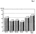
- melanoma-inhibiting protein Without the addition of the melanoma-inhibiting protein, about 10% of tumor cells migrate from the upper chamber to the lower chamber within 4 hours, where they remain attached to the underside of the Matrigel membrane. They are fixed there, colored and then counted. If the human or murine melanoma inhibiting protein MIA is added to the upper chamber, the migration of the cells is strongly inhibited. 1 shows the inhibition values obtained ( [Number of cells in the lower chamber, experiment with MIA] / [Number of cells in the lower chamber, experiment without MIA] x 100% ).
- MIA inhibits T cell mediated cytotoxic activity
- the CD4 + T cell line D7.1 which is specific against a peptide from myelin basic protein (MBP, amino acid position 87-106), is able to present both MBP and peptide 87-106 in a standard 51 Cr release assay lysing (targets: Daudi cells, R. Martin, U. Utz, JE Coligan, JR Richert, M. Flerlage, E. Robinson, R. Stone, WE Biddison, DE McFarlin, HF MacFarland, Diversity in fine specificity and T cell receptor usage of the human CD 4 + cytotoxic T-cell response specific for the immunodominant myelin basic protein peptide 87-106, J. Immunol.
- MIA inhibits the cytotoxic activity of lymphokine-activated peripheral blood lymphocytes (LAK cells)
- LAK cells are not clonally expanded, lymphokine-activated peripheral blood lymphocytes, predominantly T lymphocytes (A.A. Rayner, E.A. Grimm, M.T. Lotze, E.W. Chu, S.A. Rosenberg, Lymphokine activated killer (LAK) cells: Analysis of factors relevant for immunotherapy of human cancer, Cancer 55 (1985), 1327-1333).
- LAK cells are experimented on their cytotoxicity versus HTZ-19 melanoma cells as targets in one Microcytotoxicity assay tested.
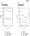
- effector-target ratios of 1: 1, 5: 1 and 10: 1 there is a maximum cytotoxicity (CTX) of just under 40%. This will after adding MIA (concentration as in Example 4 a) strongly inhibited, by a maximum of 80% with a low effector-target ratio, as it is most likely local - on the tumor itself - is to be expected (Fig. 3).
- MIA inhibits the IL-2 dependent or phytohemagglutinin dependent Lymphocyte proliferation
- PBMC Peripheral blood lymphocytes
- PHA phythaemagglutinin
- IL-2 interleukin-2
- MIA as a fusion protein with an as "Carrier” in E.coli
- suitable protein can for example the commercially available vector pQE40 (Cat. No. 33403, DIAGEN GmbH, Düsseldorf) can be used.
- pQE40 Cat. No. 33403, DIAGEN GmbH, Düsseldorf
- This vector is between the existing ones Restriction interfaces SphI and HindIII one for the mature form of MIA coding cDNA fragment inserted.
- Such a fragment is easiest by PCR amplification manufactured under known techniques under Using the cloned MIA cDNA as a template and two suitable primer (5'-GATGCATGCGGTCCTATGCCCAAGCTG-3 '(SEQ ID NO: 9) and 5'-GATAAGCTTTCACTGGCAGTAGAAATC-3 '(SEQ ID NO: 10)).
- the PCR fragment obtained is treated with SphI and HindIII cut and in the same treated vector pQE40 ligated.
- the resulting plasmid expresses a Fusion protein from DHFR (dihydrofolate reductase as "Carrier") and MIA.
- DHFR dihydrofolate reductase as "Carrier”
- MIA Magnetic Ink Characteristics
- the fusion protein carries at the N-terminus 6 histidines that follow the manufacturer's instructions according to help purify the fusion protein of Ni chelate gel materials can be used.
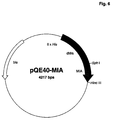
- Such processes are described in EP-A 0 282 042 and EP-A 0 253 303, the content of which is the subject of this disclosure Registration is described. 6 shows the expression plasmid pQE40-MIA.
- the content of MIA can be optimized by the Carrier protein is eliminated by DHFR and if possible by a small peptide is replaced, which has the same functions Fulfills.
- suitable peptides are, for example MetArgGlySerHisHisHisHisHisHisGlySerSerArgProPro (SEQ ID NO: 13) (this peptide can be derived from the IgA protease immediately following amino acid sequence of the mature MIA to be split off; are such procedures described in WO 91/11520, the content of which is the subject the disclosure of this application) or MetArgGlySerHisHisHisHisHisHisHisHisGlySerValAspAspAspAspLys- (SEQ ID NO: 14) (this peptide can be derived from the enterokinase immediately following amino acid sequence of the mature MIA can be split off).
- Expression plasmids the code for such MIA-peptide fusions, such as can be prepared as follows: A PCR amplification with MIA cDNA (Seq.ID No 1) as template and the primers 5'-AAAAAGGATCCAGCCGGCCGCCCGGTCCTATGCCCAAGCTGGC-3 ' (SEQ ID NO: 15) and 5'-GGCGAGCAGCCAGATCTCCATAG-3 '(SEQ ID NO: 16) a fragment which was cut with BamHI and BglII becomes.
- the expression vector pQE40-MIA is also included BamHI and BglII restricted, the smaller of the resulting Fragments are discarded and described by the PCR fragment replaced.
- the MetArgGlySerHisHisHisHisHisHisHisGlySerValAspAspAspAspLys-MIA fusion protein (SEQ ID NO: 14) is used in a completely analogous manner Primer 5'-AAAAAAGGATCCGTTGATGATGACGATAAAGGTCCTATGCCCAAGCTGGC-3 ' (SEQ ID NO: 17) and 5'-GGCGAGCAGCCAGATCTCCATAG-3 ' (SEQ ID NO: 16) received.
- fusion proteins from peptides and MIA can be made in an analogous manner and under control more suitable (preferably stronger and more inducible) Promoters in one of the numerous for E.coli described plasmids cloned and for expression to be brought.
- Another alternative is the Fusion of MIA with a peptide which is found in E. coli Secretion of the fusion protein into the periplasm with subsequent Cleavage and release of MIA leads. This method is described in WO 88/09373, the The subject matter of the disclosure of this application is.
- the expression plasmid pQE40-MIA (or a similar Expression plasmid, which for example by use of another basic vector or another "Carrier" proteins or peptides can be obtained; Such alternatives are also in Example 5a described in Methods of Enzymology 185 (Gene Expression Technology), ed. David V. Goeddel, Academic Press 1991) is transfected into a suitable E. coli strain, which sufficient expression of the lac repressor has so that an inducible expression of the MIA fusion protein can be achieved. Suitable for this is for example the strain E.coli M15 [pREP4], which together is commercially available with pQE40 (Diagen GmbH, Düsseldorf), or other E. coli strains such as E.
- E.coli UT5600 (Earhart et al. (1979), FEMS Microbiology Letters 6, 277-280) or E.coli BL21 (Grodberg and Dunn (1988), J.Bacteriol. 170, 1245-1253), previously with a lac repressor expressing helper plasmid such as pUBS520 (Brinckmann et al. (1989) Gene 85, 109-114) or pUBS500 (EP-B 0 373 365) were transfected.
- helper plasmid such as pUBS520 (Brinckmann et al. (1989) Gene 85, 109-114) or pUBS500 (EP-B 0 373 365) were transfected.
- MIA can then like can be obtained as follows: E.coli M15 [pREP4 / pQE40-MIA] on LB medium up to an optical density of 0.6 (measured at 550 nm), then IPTG (isopropyl- ⁇ -D-thiogalactopyranoside, Boehringer Mannheim GmbH) in added to a final concentration of 1 mM and then Cultivated for 4 hours. The cells will centrifuged, in 100 mM sodium phosphate buffer pH 7.5 taken up with 300 mM NaCl and by freezing three times and thawing with subsequent ultrasound treatment lysed.
- Lysate is considered Ni-NTA agarose (Diagen GmbH) the maximum binding capacity specified by the manufacturer added and overnight at room temperature incubated with mixing.
- the one so loaded with fusion protein Gel material is centrifuged at low speed, twice with 100 mM sodium phosphate buffer pH 7.5 and washed twice with sodium phosphate buffer pH 6.1. MIA is then cleaved from the fusion protein by incubating the gel material with IgA protease (Boehringer Mannheim GmbH) in 100 mM sodium phosphate buffer pH 7.5 overnight at 37 ° C. The gel material will separated by centrifugation and the MIA-containing Supernatant after sterile filtration in the in the examples 3 and 4 described activity tests used.
- the DNA sequence coding for MIA is in a way modified that an efficient expression in E.coli enables.
- a PCR amplification with human MIA cDNA (Seq. ID No 1) as template and the Primers 1 (5'-AAAAACATATGGGACCAATGCCAAAATTAGCAGATCGTAAATTATGTGCAGATCAGGAG-3 ' (SEQ ID NO: 19) and 2 (5'-AAAAAAAGCTTTCACTGGCAGTAGAAATC-3 ' (SEQ ID NO: 20)).
- Primers 1 5'-AAAAAAAGCTTTCACTGGCAGTAGAAATC-3 ' (SEQ ID NO: 20)
- Primer 1 changes the MIA coding sequence in the N-terminal area so that with unchanged MIA amino acid sequence the DNA and thus the mRNA sequence is optimized for E.coli.
- the start codon Met is added and at this point a recognition sequence the restriction endonuclease NdeI introduced, the one subsequent cloning of the MIA coding modified in this way Fragment into a vector that allows one (preferably strong and inducible) promoters and a translation initiation sequence ("Shine-Dalgarno sequence") with the NdeI interface placed below contains.
- Primer 2 serves as a 3 'counter primer and contains a HindIII interface so that the insertion of the modified MIA coding fragment in the vector can be done as a NdeI HindIII fragment.
- the so modified MIA coding sequence is in SEQ ID NO: 18 shown.
- An expression plasmid produced in this way is p11379, which is filed under the accession number DSM 9267 June 29, 1994 at the German Collection of Microorganisms and Zellkulturen GmbH, D-38124 Braunschweig has been.
- An expression plasmid for fusion-free MIA is used Expression transfected into a suitable E. coli strain.
- Expression plasmids such as p11379 are strains which have a sufficiently high intracellular concentration at the lac repressor. Such tribes can by transfecting a second plasmid such as pREP4 (Diagen GmbH), pUBS500 or pUBS520 (Brinckmann et al. (1989), Gene 85, 109-114).
- the used E. coli strains should preferably be low have cell-specific protease activity, such as this E.coli UT5600 (Earhart et al.
- MIA-containing protein aggregates from E. coli fermentations are in 6 M Guanidinium hydrochloride, 100mM TrisHCl pH 8, 1mM EDTA solubilized, then adjusted to pH 3-4 and dialyzed against 4 M guanidinium hydrochloride pH 3.5.
- the solubilized protein is then renatured in 1 M arginine pH 8, 1 mM EDTA, 5 mM GSH (glutathione reduced) and 0.5 mM GSSG (glutathione oxidized).
- MIA can, for example, add a renaturation approach of 1.4 M ammonium sulfate by adsorption on hydrophobic Gel matrices such as Fractogel TSK Butyl (E. Merck, Darmstadt) followed by elution in 20 mM TrisHCl pH 7 become.
- hydrophobic Gel matrices such as Fractogel TSK Butyl (E. Merck, Darmstadt) followed by elution in 20 mM TrisHCl pH 7 become.
- expression can also be as described in Example 9 can be achieved by homologous recombination in vitro.
- Such promoters and enhancers mostly come from Viruses such as SV40, hCMV, Polyoma or retroviruses.
- promoter-enhancer systems can also be used, those specific to a particular cell or tissue type such as WAP, MMTV or immunoglobulin promoters, or systems that are inducible, such as Metallothionein promoters.
- a vector complements the MIA cDNA (if one is used) with donor and Acceptor signals for RNA processing as well a signal for poly A addition.
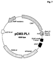
- a suitable one Vector is, for example, pCMX-pL1 (Umesono et al. (1991), Cell 65, 1255-1266) shown in Fig. 7 is.
- the expression plasmid pCMX-pL1-MIA is incorporated into the human teratocarcinoma line PA-1sc9177 (Büttner et al. (1991), Mol. Cell. Biol. 11, 3573-3583) according to the methods described (Büttner et al. (1993) Mol. Cell. Biol. 13, 4174-4185) transfected, whereby 200,000 cells per 100 mm culture dish with 5 ⁇ g DNA be transfected.
- the cells are made after transfection in MEM (Gibco) without the addition of fetal calf serum cultured, after 48 hours in the cell culture supernatant MIA is detectable.
- a coding for MIA is used DNA segment, preferably the human MIA cDNA (SEQ ID NO 1), introduced in vectors that are different from AcMNPV (Autographa californica nuclear polyhedrosis virus) or Derive BmNPV (Bombyx mori nuclear polyhedrosis virus).
- the MIA cDNA is initially under control a strong promoter suitable for insect cells brought (O'Reilly DR, Miller LK and Luckow VA 1992; Baculovirus Expression Vectors - A Laboratory Manual; W.H. Freeman & Co, New York), such as the polH promoter or of the p10 promoter.
- the polH promoter works as follows:
- a DNA fragment coding for MIA with interfaces for the restriction endonucleases EcoRI (adjacent to the 5 'end of the later MIA transcript) and PstI (adjacent to the 3' end of the later MIA transcript) is obtained by PCR amplification according to known techniques Use of the MIA cDNA as template and the primer 5'-CGTGAATTCAACATGGCCCGGTCCCTGGTGTGC-3 '(SEQ ID NO: 21) and 5'-TATCTGCAGTCACTGGCAGTAGAAATCCCA-3' (SEQ ID NO: 22).
- This fragment is cut with EcoRI and PstI (to produce the corresponding cohesive ends) and in the transfer vector pVL1393 restricted with the same endonucleases (O'Reilly DR, Miller LK and Luckow VA 1992; Baculovirus Expression Vectors - A Laboratory Manual; WHFreeman & Co, New York), which is commercially available (PharMingen, San Diego, CA or Invitrogen Corporation, San Diego, CA).
- the resulting transfer expression plasmid pVL1393-MIA is transfected for growth in E. coli K12 and plasmid DNA is prepared according to established methods.
- the transfer of the MIA DNA under control of the polH promoter from the transfer plasmid into the baculovirus vector is carried out by homologous recombination according to established methods (O'Reilly et al. 1992, see above).
- BaculoGold DNA linearized AcNPV virus DNA with lethal deletion and lacZ expression controlled by the polH promoter, commercially available from PharMingen, order number 21100D
- 2 ⁇ g pVL1393-MIA are mixed, incubated for 5 minutes at room temperature and then with 1 ml 125 mM Hepes pH 7.1, 125 mM CaCl2, 140 mM NaCl added.
- This mixture is added to 2 ⁇ 10 6 SF9 insect cells (Invitrogen, order number B825-01) in a culture dish with a diameter of 60 mm, which had previously been overlaid with 1 ml Grace's medium with 10% fetal calf serum. After 4 hours of incubation at 4 ° C, the DNA-containing medium is removed and the cells are incubated in fresh medium for 4 days at 27 ° C. The recombinant baculoviruses obtained in this way are then purified twice via plaque formation (O'Reilly et al.
- viruses which have inserted MIA by homologous recombination being distinguished by the lack of ⁇ -galactosidase activity ( optically recognizable by the absence of a blue color in the presence of 5-bromo-4-chloro-3-indolyl- ⁇ -D-galactopyranoside) from the wild-type viruses used (AcNPV with lethal deletion and lacZ expression controlled by the polH promoter, commercially available from PharMingen, order number 21100D).
- SF9 cells are infected according to established methods (O'Reilly et al.
- MIA mRNA is present in a particular cell and, accordingly, MIA mRNA is present
- nucleic acid hybridization such as Northern hybridization, in-situ hybridization, dot or slot hybridization, and the like derived diagnostic techniques (Sambrook et al. 1989, Molecular Cloning - A Laboratory Manual; Cold Spring Harbor Laboratory Press; Nucleic Acid Hybridization - A Practical Approach, ed. BD Hames and SJ Higgins (1985), IRL Press; WO 89/06698, EP-A 0 200 362, USP 2915082, EP-A 0 063 879, EP-A 0 173 251, EP-A 0 128 018).
- MIA-specific primers PCR Protocols - A Guide to Methods and Applications; ed. MA Innis, DH Gelfand, JJ Sninsky, TJ White (1990), Academic Press Inc; PCR - A Practical Approach; Ed. MJ McPherson, P Quirke, GR Taylor (1991), IRL Press).
- Tables 2A and 2B show the MIA expression in various human tumors, tumor lines and normal cells, which in this case was determined by Northern hybridization using the radiolabelled human MIA cDNA (Seq. ID No. 1). For this, the RNA was isolated from the cells listed according to the method of Chomczynski and Sacchi (Anal. Biochem.
- the hybridization was carried out at 68 ° C in 5 x SSC, 5 x Denhardt, 0.5% SDS, 10% dextran sulfate and 100 ⁇ g / ml salmon sperm DNA.
- the membranes were then washed twice in 1 ⁇ SSC at 68 ° C. for one hour each and then exposed to X-ray film.
- MIA coding nucleic acids therapeutic purposes
- MIA inhibits the proliferation and metastasis of Tumor cells.
- This effect can be in an animal model or to a patient not only through the exogenous supply of MIA protein are caused, but also by that Introduction of a DNA segment that is either for MIA encoded under a suitable promoter or one contains suitable promoter by homologous recombination before integrating the cell's own MIA gene into the genome can.
- this promoter has to Sequence sections flanked by the sequences of the human (or animal in the case of an animal model) MIA Gens in the 5'-untranslated region as far as possible are homologous or preferably identical (WO 91/09955).
- Example is the therapeutic effect of an MIA coding DNA segment shown in an animal model.
- the injection of murine B16 melanoma cells (ATCC CRL 6322) into the tail vein of C57BL mice with subsequent quantification of lung metastases represents an established in vivo metastasis model. 100,000 cells of the B16 melanoma line were retrobulbar injected into C57BL mice (Day 1, 16 animals) After 48 hours, 100 ⁇ g of the MIA expression plasmid pCMX-PL1-MIA (Example 7a) in TE (10 mM TrisCl pH 8.0, 1 mM EDTA) were mixed with DOTAP transfection reagent (Leventis and Silvius (1990) in 8 animals.
Abstract
Description
Die Erfindung betrifft ein Melanom-inhibierendes Protein (MIA), eine dafür codierende Nukleinsäure, ein Verfahren zur Gewinnung und zum Nachweis dieses Proteins sowie dessen Verwendung zur Herstellung eines Therapeutikums.The invention relates to a melanoma-inhibiting protein (MIA), a nucleic acid coding therefor, a method for the production and detection of this protein and its use in the manufacture of a therapeutic.
Die Regulation des Zellwachstums wird sowohl durch positiv als auch negativ wirkende Faktoren kontrolliert. Zu den positiv wirkenden Faktoren zählen die bekannten Wachstumsfaktoren wie z. B. epidermal growth factor (EGF), platelet derived growth factor (PDGF), Insulin und Somatomedine. Zu den negativ, d. h. inhibitorisch wirkenden Faktoren gehören neben dem TGF-β, der sowohl als Wachstumsstimulator als auch als Wachstumsinhibitor wirken kann (Roberts et al., Proc. Natl. Acad. Sci. 82 (1985), 119 - 123) die endogenen, tumorinhibierenden Faktoren aus Dickdarmkarzinomzellen (Levine et al, Cancer Research 45 (1985), 2248 - 2254), Melanomen (Bogdahn et al., Cancer Research 49 (1989), 5358 - 5363) sowie aus gesunden epithelialen Zellen von Brustdrüsen der Ratte (Ethier et al., J. Cell. Phys. 142 (1990), 15 - 20).The regulation of cell growth is both positive controlled as well as negative factors. To the positive factors include the known ones Growth factors such as B. epidermal growth factor (EGF), platelet derived growth factor (PDGF), insulin and Somatomedine. To the negative, d. H. inhibitory In addition to TGF-β, factors include both Growth stimulator as well as growth inhibitor can act (Roberts et al., Proc. Natl. Acad. Sci. 82 (1985), 119-123) the endogenous, tumor-inhibiting Factors from colon cancer cells (Levine et al, Cancer Research 45 (1985), 2248-2254), melanoma (Bogdahn et al., Cancer Research 49 (1989), 5358-5363) and from healthy rat epithelial cells from mammary glands (Ethier et al., 1990, J. Cell. Phys. 142: 15-20).
Durch Störung dieses Regulationssystems, wie z. B. durch Überproduktion von positiv wirkenden Wachstumsfaktoren oder eine verminderte Abhängigkeit veränderter Zellen von diesen Wachstumsfaktoren (Rodeck et al., International Journal of Cancer 40 (1987), 687 - 690) wird es Tumorzellen ermöglicht, sich unkontrolliert zu vermehren. Die oben genannten tumorinhibierenden Faktoren aus verschiedenen Tumorgeweben stellen interessante Verbindungen dar, um in dieses gestörte Regulationssystem therapeutisch eingreifen zu können. Für eine solche therapeutische Verwendung müssen diese Faktoren in großer Menge und reproduzierbarer Reinheit zur Verfügung gestellt werden können. Bislang sind jedoch für die meisten dieser Faktoren lediglich angereicherte Fraktionen aus Zellysaten beschrieben, die auf Grund ihrer komplexen und zum Teil unbekannten Zusammensetzung und der damit verbundenen nicht reproduzierbaren Herstellbarkeit für einen therapeutischen Einsatz nicht geeignet sind.By disturbing this regulatory system, such as. B. by Overproduction of positive growth factors or a reduced dependence of changed cells on these growth factors (Rodeck et al., International Journal of Cancer 40 (1987), 687-690) there will be tumor cells allows you to multiply uncontrollably. The tumor-inhibiting factors mentioned above from various Tumor tissues are interesting connections to be therapeutic in this disrupted regulatory system to be able to intervene. For such a therapeutic Use these factors in large quantities and reproducible purity can. So far, however, for most of these factors only enriched fractions from cell lysates described because of their complex and in part unknown composition and related non-reproducible producibility for a therapeutic Use are not suitable.
Grundlage der Erfindung ist ein neues Melanom-inhibierendes
Protein (im weiteren als MIA-Protein oder MIA
bezeichnet), welches das Wachstum der Zellinien HTZ 19-dM
und ATCC CRL 1424 inhibiert und
Unter einer Wachstuminhibierung ist eine-antiproliferative Wirkung zu verstehen. Dabei wird das Wachstum der Zellen bei Zusatz des MIA-Proteins zum Kulturmedium deutlich verlangsamt. Eine hierfür geeignete Konzentration ist beispielsweise O,1 µg MIA-Protein/ml Kulturmedium. Höhere oder geringere Konzentrationen des MIA-Proteins sind jedoch ebenfalls geeignet zur Wachstumsinhibierung, die je nach Konzentration stärker oder geringer beobachtet wird.Under a growth inhibition is an anti-proliferative To understand effect. The growth of Cells when the MIA protein is added to the culture medium significantly slowed down. A suitable concentration for this is, for example, 0.1 µg MIA protein / ml culture medium. Higher or lower concentrations of the MIA protein are also suitable for growth inhibition, which depending on the concentration stronger or is observed less.
Die Eigenschaften eines solchen Proteine sind durch die Erfinder in Cancer Research 49 (1989), 5358 - 5363, Cancer Research 50 (1990), 6981 - 6986, Melanoma Research 2 (1992), 327 - 336 beschrieben. Ein nacharbeitbares Verfahren zur Herstellung dieses Proteins ist jedoch in diesen Publikationen nicht angegeben. Das Protein ist aus der humanen Melanomazellinie HTZ 19-dM, die bisher der Öffentlichkeit noch nicht zugänglich war, erhältlich. Diese Zellinie wurde von einem metastasierenden malignen Melanom abgeleitet und in definiertem serumfreien Kulturmedium (50 % Dulbecco's minimal essential medium, 50 % F-12) mit 0,8 mmol/l L-Glutamin, nichtessentiellen Aminosäuren, 10 µg/ml Transferrin, 30 nmol/l Natriumselenit und 4 µg/ml Gentamicin als Monolayer-Kultur unter Standard-Kulturbedingungen kultiviert. Die Zellinie wurde am 23.06.93 bei der Deutschen Sammlung für Mikroorganismen und Zellkulturen GmbH in Braunschweig hinterlegt (DSM ACC 2133). Sie ist ein weiterer Gegenstand der Erfindung. Aus dem Kulturüberstand dieser Zellinie kann das erfindungsgemäße Protein über eine gelchromatographische Isolierung einer Proteinfraktion einer Größe von ca. 11 kD und anschliessende Aufreinigung dieser Fraktion über eine reverse phase HPLC gewonnen werden. The properties of such proteins are due to the Inventor in Cancer Research 49 (1989), 5358-5363, Cancer Research 50 (1990), 6981-6986, Melanoma Research 2 (1992), 327-336. A reworkable Process for the production of this protein is however in not specified in these publications. The protein is out the human melanoma cell line HTZ 19-dM, which was previously the Public was not yet available. This cell line was developed by a metastatic malignant Melanoma derived and in defined serum-free culture medium (50% Dulbecco's minimal essential medium, 50% F-12) with 0.8 mmol / l L-glutamine, non-essential amino acids, 10 µg / ml transferrin, 30 nmol / l sodium selenite and 4 µg / ml gentamicin as a monolayer culture under Cultivated standard culture conditions. The cell line was on June 23, 1993 at the German Collection for Microorganisms and Zellkulturen GmbH in Braunschweig (DSM ACC 2133). It is another object of the invention. From the culture supernatant of this cell line, the invention can Protein via a gel chromatographic Isolation of a protein fraction approximately 11 in size kD and subsequent purification of this fraction a reverse phase HPLC can be obtained.
Das Protein kann über seine DNA-Sequenz und die davon abgeleitete Aminosäuresequenz definiert werden. Das MIA-Protein kann in natürlichen allelischen Variationen, die sich von Individuum zu Individuum unterscheiden können, vorkommen (z.B. SEQ ID NO:24). Solche Variationen der Aminosäuren sind in der Regel Aminosäureaustausche. Es kann sich aber auch um Deletionen, Insertionen oder Additionen von Aminosäuren zur Gesamtsequenz handeln. Das erfindungsgemäße MIA Protein kann - sowohl in Umfang und Art abhängig von der Zelle und dem Zelltyp, in dem es exprimiert wird - glycosyliert oder nicht glycosyliert sein.The protein can have its DNA sequence and those of it derived amino acid sequence can be defined. The MIA protein can be found in natural allelic variations can differ from individual to individual, occur (e.g. SEQ ID NO: 24). Such variations of Amino acids are usually amino acid exchanges. It can also be deletions, insertions or Act additions of amino acids to the overall sequence. The MIA protein according to the invention can - both in scope and Kind depending on the cell and the cell type in which it is is expressed - glycosylated or not glycosylated be.
Das erfindungsgemäße Protein kann auch rekombinant hergestellt werden. Bei der rekombinanten Herstellung in Prokaryonten wird nichtglykosyliertes MIA-Protein erhalten. Mit Hilfe der durch die Erfindung bereitgestellten Nukleinsäuresequenzen ist es möglich, in Genomen von beliebigen Zellen (z. B. außer in Humanzellen auch in Zellen anderer Säugetiere) nach dem MIA-Gen oder seinen Varianten zu suchen, diese zu identifizieren und das gewünschte für das MIA-Protein codierende Gen zu isolieren. Derartige Verfahren und die hierfür geeigneten Hybridisierungsbedingungen sind dem Fachmann bekannt und beispielsweise bei J. Sambrook, E.F. Fritsch und T. Maniatis, Molecular cloning, Cold Spring Harbor Laboratory, 1989 und B.D. Hames, S.G. Higgins, Nucleic acid hybridisation - a practical approach (1985) IRL Press, Oxford, England, beschrieben. Üblicherweise werden hierbei die dort beschriebenen Standardprotokolle für die Experimente verwendet. The protein according to the invention can also be produced recombinantly become. In the recombinant production in Prokaryotes will get non-glycosylated MIA protein. With the help of the provided by the invention Nucleic acid sequences make it possible to find in genomes of any cells (e.g. in addition to human cells also in Cells of other mammals) according to the MIA gene or its To search for variants, to identify them and that isolate desired gene coding for the MIA protein. Such methods and the suitable ones Hybridization conditions are known to the person skilled in the art and e.g. from J. Sambrook, E.F. Fritsch and T. Maniatis, Molecular cloning, Cold Spring Harbor Laboratory, 1989 and B.D. Hames, S.G. Higgins, Nucleic acid hybridization - a practical approach (1985) IRL Press, Oxford, England. Usually the standard protocols described there for the Experiments used.
Durch die Anwendung der rekombinanten DNA-Technik ist es möglich, eine Vielzahl von MIA-Protein-Derivaten herzustellen. Derartige Derivate können beispielsweise modifiziert sein in einzelnen oder mehreren Aminosäuren durch Substitution, Deletion oder Addition. Die Derivatisierung kann beispielsweise über site directed mutagenesis erfolgen. Derartige Variationen sind für einen Fachmann ohne weiteres durchführbar (J. Sambrook, B.D. Hames, Loc. Lit.). Es muß lediglich sichergestellt sein, daß die charakteristischen Eigenschaften des MIA-Proteins (Inhibition der genannten Zellinien) erhalten bleiben.By using the recombinant DNA technique it is possible to produce a variety of MIA protein derivatives. Such derivatives can be modified, for example be in single or multiple amino acids Substitution, deletion or addition. The derivatization can, for example, via site directed mutagenesis respectively. Such variations are for a person skilled in the art easily feasible (J. Sambrook, B.D. Hames, Loc. Lit.). It only has to be ensured that the characteristic properties of the MIA protein (Inhibition of the cell lines mentioned) are retained.
Ein weiterer Gegenstand der Erfindung ist deshalb ein
MIA-Protein, welches
Bevorzugt ist ein Protein, welches codiert wird von den
Nukleotiden 40 - 432 oder 112 bis 432 aus SEQ ID NO 1
oder von DNA-Sequenzen, die aufgrund der Degeneration des
genetischen Codes für ein Polypeptid mit derselben Aminosäuresequenz
codieren würden.A protein which is encoded by the
Nucleotides 40 - 432 or 112 to 432 from
Das MIA-Protein aus HTZ 19-dM weist ein Molekulargewicht von ca. 11 kD auf, ist stabil gegen Hitze (3 Minuten bei 100° C) und sensitiv gegenüber Proteasen, wie z. B. Trypsin.The MIA protein from HTZ 19-dM has a molecular weight from approx. 11 kD, is stable against heat (3 minutes at 100 ° C) and sensitive to proteases, such as. B. Trypsin.
Gegenstand der Erfindung ist eine Nukleinsäure, welche
für ein MIA-Protein codiert und ausgewählt ist aus der
Gruppe
Ein weiterer Gegenstand der Erfindung sind melanominhibierende Proteine aus Säugerzellen, wie z.B. Maus, Ratte, Rind, Schaf, welche in im wesentlichen analoger Weise das Wachstum der Zellinien HTZ19-dM und ATCC CRL 1424 inhibieren, wie das humane MIA-Protein.Another object of the invention are melanoma inhibiting Proteins from mammalian cells, e.g. Mouse, Rat, beef, sheep, which are essentially analogous Way the growth of cell lines HTZ19-dM and ATCC Inhibit CRL 1424, like the human MIA protein.
Diese dem humanen MIA-Protein analogen Proteine können dadurch erhalten werden, daß mit einer Hybridisierungsprobe, welche für humanes MIA codierende Sequenzen enthält, eine cDNA-Bibliothek des entsprechenden Säugetiers nach dem Fachmann geläufigen Methoden gescreent wird und über den Sequenzvergleich mit der DNA und Proteinsequenz für humanes und murines MIA (SEQ ID NO:1-5) das entsprechende codierende Segment identifiziert wird.These proteins, which are analogous to the human MIA protein, can can be obtained by using a hybridization sample, which contains sequences coding for human MIA, a cDNA library of the corresponding mammal is screened according to methods familiar to the person skilled in the art and about the sequence comparison with the DNA and protein sequence the corresponding for human and murine MIA (SEQ ID NO: 1-5) coding segment is identified.
Ein bevorzugter Gegenstand der Erfindung ist das murine MIA-Protein und die hierfür codierende Nukleinsäuresequenz (SEQ ID NO:4). Das murine Protein wird codiert von den Nukleotiden 110 - 499 oder 179 - 499 von SEQ ID NO:4.A preferred subject of the invention is murine MIA protein and the nucleic acid sequence coding therefor (SEQ ID NO: 4). The murine protein is encoded from nucleotides 110-499 or 179-499 of SEQ ID NO: 4.
Mit Hilfe dieser Nukleinsäuren kann das erfindungsgemäße Protein in reproduzierbarer Weise in großen Mengen gewonnen werden. Zur Expression in prokaryontischen oder eukaryontischen Organismen, wie z.B. prokaryontischen Wirtszellen oder in eukaryontischen Wirtszellen, wird die Nukleinsäure nach dem Fachmann geläufigen Verfahren in geeignete Expressionsvektoren integriert. Vorzugsweise enthält ein solcher Expressionsvektor einen regulierbaren/induzierbaren Promotor. Zur Expression werden diese rekombinanten Vektoren dann nach bekannten Verfahren in geeignete Wirtszellen, wie z. B. E.coli, als prokaryontische Wirtszellen oder Saccharomyces cerevisiae, Teratocarcinoma-Zellinie PA-1 sc 9117 (Büttner et al., Mol. Cell. Biol. 11, 1991, 3573 - 3583), Insektenzellen, CHO- oder COS-Zellen als eukaryontische Wirtszellen eingeführt und die transformierten bzw. transduzierten Wirtszellen unter Bedingungen kultiviert, die eine Expression des heterologen Gens ermöglichen. Die Isolierung des Proteins kann aus der Wirtszelle oder dem Kulturüberstand der Wirtszelle nach bekannten Verfahren erfolgen. Derartige Verfahren sind beispielsweise bei Ausubel I., Frederick M., Current Protocols in Mol. Biol. (1992), John Wiley and Sons, New York, beschrieben. Es kann auch eine in vitro Reaktivierung des Proteins erforderlich sein.With the help of these nucleic acids, the invention Protein obtained in large quantities in a reproducible manner become. For expression in prokaryotic or eukaryotic organisms, e.g. prokaryotic Host cells or in eukaryotic host cells, the Nucleic acid according to methods familiar to the person skilled in the art suitable expression vectors integrated. Preferably such an expression vector contains a regulatable / inducible one Promoter. These become expression recombinant vectors then by known methods in suitable host cells, such as. B. E. coli, as a prokaryotic Host cells or Saccharomyces cerevisiae, Teratocarcinoma cell line PA-1 sc 9117 (Büttner et al., Mol. Cell. Biol. 11, 1991, 3573-3583), insect cells, CHO or COS cells as eukaryotic host cells introduced and the transformed or transduced Host cells cultured under conditions that a Enable expression of the heterologous gene. The insulation of the protein can come from the host cell or the culture supernatant the host cell by known methods respectively. Such methods are for example Ausubel I., Frederick M., Current Protocols in Mol. Biol. (1992), John Wiley and Sons, New York. It an in vitro reactivation of the protein may also be required be.
Vorzugsweise wird zur rekombinanten Herstellung des
erfindungsgemäßen Proteins eine DNA mit den Nukleotiden
40 - 432 oder 112 - 432 (codierende Sequenz) aus
SEQ ID NO 1 (cDNA) oder die genomische DNA gemäß SEQ ID
NO 3 verwendet.Preferably for the recombinant production of
protein according to the invention a DNA with the nucleotides
40 - 432 or 112 - 432 (coding sequence)
SEQ ID NO 1 (cDNA) or the genomic DNA according to
Ein weiterer Gegenstand der Erfindung ist ein Verfahren zur Gewinnung eines MIA-Proteins durch Isolierung aus dem Kulturüberstand der Melanomzelline HTZ 19-dM über eine gelchromatographische Auftrennung und Aufreinigung einer Fraktion, die einem Molekulargewicht von ca. 11 kD (SDS-PAGE, nicht reduziert) entspricht, über reverse phase HPLC. Auf diese Weise können ca. 0,2 µg/l Kulturüberstand erhalten werden.Another object of the invention is a method to obtain a MIA protein by isolation from the Culture supernatant of the melanoma cell HTZ 19-dM over a gel chromatographic separation and purification of a Fraction having a molecular weight of approx. 11 kD (SDS-PAGE, not reduced), via reverse phase HPLC. In this way, about 0.2 µg / l culture supernatant can be preserved.
In einer bevorzugten Ausführungsform wird das natürliche MIA-Protein bei der Isolierung/Aufreinigung einer Säurebehandlung unterworfen. Dadurch kann die MIA-Aktivität gesteigert werden. Vorteilhaft ist ein pH-Wert um ca. 2, als Säure ist beispielsweise Essigsäure geeignet. In a preferred embodiment, the natural MIA protein in the isolation / purification of an acid treatment subject. This allows the MIA activity be increased. A pH value of around 2 is advantageous. Acetic acid, for example, is suitable as the acid.
Der Nachweis von transformierten bzw. transduzierten Wirtszellen, welche das MIA-Protein rekombinant produzieren, sowie die Aufreinigung des Proteins erfolgen vorzugsweise über Antikörper, die an dieses Protein binden. Derartige Antikörper sind mit Hilfe des erfindungsgemäßen Proteins als Antigen oder Immunogen in einfacher Weise nach bekannten Verfahren erhältlich.The detection of transformed or transduced Host cells that produce the MIA protein recombinantly, and the protein are preferably purified about antibodies that bind to this protein. Such antibodies are with the help of the invention Protein as an antigen or immunogen in a simple way available by known methods.
Ein weiterer Gegenstand der Erfindung ist daher die Verwendung des erfindungsgemäßen Proteins mit Melanominhibierender Aktivität zur Herstellung von Antikörpern, die an dieses Protein binden.Another object of the invention is therefore Use of the protein according to the invention with melanoma inhibitors Antibody production activity, that bind to this protein.
Dazu werden hierzu üblicherweise verwendete Tiere, wie insbesondere Schafe, Kaninchen oder Mäuse mit dem erfindungsgemäßen Protein immunisiert und anschließend das Antiserum der immunisierten Tiere nach bekannten Verfahren gewonnen oder Milzzellen der immunisierten Tiere mit immortalen Zellen, wie z. B. Myelomzellen, gem. dem Verfahren von Köhler und Milstein (Nature 256 (1975), 495 - 497) fusioniert. Von den so erhaltenen Hybridomzellen werden dann solche ausgewählt und kloniert, welche einen monoklonalen Antikörper gegen das MIA-Protein produzieren. Die so erhaltenen monoklonalen oder polyklonalen Antikörper können für eine immunadsorptive Reinigung des Melanom-inhibierenden Proteins an ein Trägermaterial, wie z. B. Cellulose, gebunden werden. Weiterhin können derartige Antikörper für einen Nachweis des MIA-Proteins in Proben, wie etwa Gewebeschnitten oder Körperflüssigkeiten, verwendet werden.For this purpose, commonly used animals, such as in particular sheep, rabbits or mice with the invention Immunized protein and then that Antiserum of the immunized animals according to known methods obtained or using spleen cells of the immunized animals immortal cells, such as B. myeloma cells, acc. the Method by Koehler and Milstein (Nature 256 (1975), 495 - 497) merged. Of the hybridoma cells thus obtained then those are selected and cloned, which one produce monoclonal antibodies against the MIA protein. The monoclonal or polyclonal thus obtained Antibodies can be used for immunoadsorptive purification of the Melanoma-inhibiting protein on a carrier material, such as e.g. B. cellulose. Furthermore, such Antibodies for detection of the MIA protein in Samples, such as tissue sections or body fluids, be used.
Ein weiterer Gegenstand der Erfindung sind daher Antikörper gegen das MIR-Protein, welche erhältlich sind durch Immunisierung eines Tieres mit einem MIR-Protein und Gewinnung der Antikörper aus dem Serum oder Milzzellen der immunisierten Tiere.Another object of the invention is therefore antibodies against the MIR protein, which are obtainable by Immunization of an animal with a MIR protein and Obtaining the antibodies from the serum or spleen cells of the immunized animals.
Es hat sich gezeigt, daß das MIA-Protein nicht nur auf Melanomzellen sondern auch in geringerem Umfang auf andere Tumorzellen, wie z. B. Glioblastomzellen, Neuroblastome, small cell lung cancer und neuroektodermale Tumoren eine hemmende Wirkung durch Hemmung der DNA-Synthese (3H-Thymidin-Einbau (Coligan J. E., Kruisbeek A. M., Margulies D. H., Shevach E. M., Strober W., Current Protocols Immunology, NIH Monograph, J. Wiley and Sons, New York, 1992)), Hemmung einer Tumorkoloniebildung im Soft-Agar oder im Tumorstammzellen-Assay (Schlag P., Flentje D., Cancer Treatment Rev. 11: Suppl. A: 131 - 137, 1984) aufweist. Das Wachstum von normalen, nichtentarteten Zellen wird dagegen nicht inhibiert. Dabei wirkt dieses Protein bereits in sehr geringen Konzentrationen (Nanogramm-Bereich). Daher eignet sich dieses Protein für die Herstellung eines Therapeutikums zur Tumortherapie. Insbesondere ist ein solches Therapeutikum zur Therapie von malignen Melanomen, malignen Gliomen, Bronchial-Karzinomen (insbesondere kleinzelliges Bronchialkarzinom, SCLC) und Neuroblastomen geeignet.It has been shown that the MIA protein not only on melanoma cells but also to a lesser extent on other tumor cells, such as. B. Glioblastoma cells, neuroblastomas, small cell lung cancer and neuroectodermal tumors an inhibitory effect by inhibiting DNA synthesis ( 3 H-thymidine incorporation (Coligan JE, Kruisbeek AM, Margulies DH, Shevach EM, Strober W., Current Protocols Immunology, NIH Monograph, J. Wiley and Sons, New York, 1992)), inhibition of tumor colony formation in soft agar or in the tumor stem cell assay (Schlag P., Flentje D., Cancer Treatment Rev. 11: Suppl. A: 131-137 , 1984). In contrast, the growth of normal, non-degenerate cells is not inhibited. This protein works in very low concentrations (nanogram range). This protein is therefore suitable for the manufacture of a therapeutic agent for tumor therapy. Such a therapeutic agent is particularly suitable for the therapy of malignant melanomas, malignant gliomas, bronchial carcinomas (in particular small cell bronchial carcinoma, SCLC) and neuroblastomas.
Es hat sich weiterhin gezeigt, daß das Melanom-inhibierende Protein die Interleukin 2-abhängige bzw. Phytohämagglutinin-induzierte Proliferation peripherer Blutlymphozyten supprimiert. Weiterhin wird die Cytotoxizität von T-Lymphozyten reduziert. Das Melanom-inhibierende Protein eignet sich damit auch zur Herstellung eines Therapeutikums, das Anwendung als Immunsuppressivum findet. It has also been shown that the melanoma-inhibiting Protein that induced interleukin 2-dependent or phytohemagglutinin Proliferation peripheral Blood lymphocytes suppressed. Furthermore, the cytotoxicity reduced by T lymphocytes. The melanoma-inhibiting Protein is therefore also suitable for production a therapeutic agent that is used as an immunosuppressant finds.
Ein weiterer Gegenstand der Erfindung ist daher die Verwendung eines erfindungsgemäßen Proteins zur Herstellung eines Therapeutikums, das Anwendung in der Tumortherapie oder als Immunsuppressivum findet.Another object of the invention is therefore Use of a protein according to the invention for the production a therapeutic agent that is used in tumor therapy or as an immunosuppressant.
Für die genannten therapeutischen Anwendungen wird das erfindungsgemäße Protein ggf. mit den üblicherweise verwendeten Hilfs-, Füll- und/oder Zusatzstoffen zusammen in einer pharmazeutischen Formulierung verarbeitet.For the therapeutic applications mentioned, this is protein according to the invention optionally with the usual auxiliaries, fillers and / or additives used together processed in a pharmaceutical formulation.
Ein weiterer Gegenstand der Erfindung ist daher eine therapeutische Zusammensetzung, enthaltend ein erfindungsgemäßes Melanom-inhibierendes Protein, ggf. zusammen mit den üblicherweise verwendeten Hilfs-, Füll- und/oder Zusatzstoffen.Another object of the invention is therefore a therapeutic composition containing an inventive Melanoma-inhibiting protein, if necessary together with the commonly used auxiliary, filling and / or Additives.
Ein weiterer Gegenstand der Erfindung ist die Verwendung von Sequenzen des MIA-Gens, vorzugsweise von für ein Protein mit MIA-Aktivität codierenden Sequenzen oder aktivierende Sequenzen aus dem 5' untranslatierten Bereich, in der Gentherapie und insbesondere zur Herstellung von Arzneimitteln für die Gentherapie.Another object of the invention is the use of sequences of the MIA gene, preferably of for one Protein coding sequences or MIA activity activating sequences from the 5 'untranslated Field in gene therapy and especially for manufacturing of drugs for gene therapy.
Gentherapie von somatischen Zellen kann beispielsweise unter Verwendung von retroviralen Vektoren, anderen viralen Vektoren oder durch nicht-viralen Gentransfer erfolgen (zur Übersicht vgl. T. Friedmann, Science 244 (1989) 1275; Morgan 1993, RAC DATA MANAGEMENT Report, June 1993). For example, gene therapy from somatic cells using retroviral vectors, others viral vectors or by non-viral gene transfer (for an overview see T. Friedmann, Science 244 (1989) 1275; Morgan 1993, RAC DATA MANAGEMENT Report, June 1993).
Gentherapeutisch geeignete Vektorsysteme sind beispielsweise Retroviren (Mulligan, R.C. (1991) in Nobel Symposium 8: Ethiology of human disease at the DNA level (Lindsten, J. and Pattersun Editors), pages 143 - 189, Raven Press), Adeno Associated Virus (McLughlin, J.-Virol. 62 (1988), 1963), Vaccinia Virus (Moss et al., Ann. Rev. Immunol. 5 (1987) 305), Bovine Papilloma Virus (Rasmussen et al., Methods Enzymol. 139 (1987) 642) oder Viren aus der Gruppe der Herpesviren, wie Epstein Barr Virus (Margolskee et al., Mol. Cell. Biol. 8 (1988) 2937) oder Herpes Simplex Virus.Vector systems suitable for gene therapy are, for example Retroviruses (Mulligan, R.C. (1991) in Nobel Symposium 8: Ethiology of human disease at the DNA level (Lindsten, J. and Pattersun Editors), pages 143 - 189, Raven Press), Adeno Associated Virus (McLughlin, J.-Virol. 62 (1988), 1963), Vaccinia Virus (Moss et al., Ann. Rev. Immunol. 5 (1987) 305) Bovine Papilloma Virus (Rasmussen et al., Methods Enzymol. 139 (1987) 642) or Viruses from the group of herpes viruses, such as Epstein Barr Virus (Margolskee et al., Mol. Cell. Biol. 8 (1988) 2937) or herpes simplex virus.
Es sind auch nicht-virale Deliverysysteme bekannt. Hierzu wird üblicherweise "nackte" Nukleinsäure, vorzugsweise DNA, verwendet, oder Nukleinsäure zusammen mit einem Hilfsstoff, wie z.B. mit Transferreagenzien (Liposomen, Dendromere, Polylysin-Transferrin-Konjugate (Wagner, 1990; Felgner et al., Proc. Natl. Acad. Sci. USA 84 (1987) 7413)).Non-viral delivery systems are also known. For this "naked" nucleic acid is usually preferred DNA, used, or nucleic acid along with one Auxiliary, such as with transfer reagents (liposomes, Dendromers, polylysine-transferrin conjugates (Wagner, 1990; Felgner et al., Proc. Natl. Acad. Sci. USA 84 (1987) 7413)).
Ein weiteres bevorzugtes gentherapeutisches Verfahren basiert auf der homologen Rekombination. Dabei kann entweder das für das MIA-Protein codierende Gen in einer oder mehreren Kopien in das Genom von somatischen Zellen eingeführt werden und/oder das endogen in den Zellen vorhandene MIA-Gen moduliert, vorzugsweise aktiviert werden.Another preferred gene therapy method is based on homologous recombination. It can either the gene coding for the MIA protein in one or multiple copies in the genome of somatic cells be introduced and / or endogenously in the cells existing MIA gene modulated, preferably activated become.
Verfahren zur homologen Rekombination sind beispielsweise beschrieben in Kucherlapati, Proc. in Nucl. Acids Res. and Mol. Biol. 36 (1989) 301; Thomas et al., Cell 44 (1986) 419-428; Thomas und Capecchi, Cell 51 (1987) 503-512; Doetschman et al., Proc. Natl. Acad. Sci. USA 85 (1988) 8583-8587 und Doetschman et al., Nature 330 (1987) 576-578. Bei diesen Verfahren wird ein Stück DNA, das im Genom an einer bestimmte Stelle integriert werden soll (Genfragment von MIA), an eine "Targeting DNA" gebunden. Die Targeting DNA ist eine DNA, die komplementär (homolog) zu einer Region der genomischen DNA ist (vorzugsweise im oder in der Nähe des MIA-Gens). Wenn zwei homologe Stücke einer Einzelstrang-DNA (z.B. die Targeting DNA und die genomische DNA) in unmittelbare Nähe zueinander kommen, hybridisieren sie und bilden eine Doppelstrang-Helix. Durch ein Rekombinationsereignis kann dann das MIA-Genfragment und die Targeting-DNA ins Genom integriert werden. Diese homologe Rekombination kann sowohl in vitro als auch in vivo (am Patienten) durchgeführt werden.Methods for homologous recombination are, for example described in Kucherlapati, Proc. in nucl. Acids Res. and Mol. Biol. 36 (1989) 301; Thomas et al., Cell 44 (1986) 419-428; Thomas and Capecchi, Cell 51 (1987) 503-512; Doetschman et al., Proc. Natl. Acad. Sci. USA 85 (1988) 8583-8587 and Doetschman et al., Nature 330 (1987) 576-578. In this process, a piece of DNA that is in the Genome to be integrated at a certain point (MIA gene fragment) bound to a "targeting DNA". The targeting DNA is a DNA that is complementary (homologous) to a region of genomic DNA (preferably in or near the MIA gene). If two homologous pieces of single-stranded DNA (e.g. the Targeting DNA and the genomic DNA) in immediate Come close to each other, they hybridize and form one Double strand helix. Through a recombination event then the MIA gene fragment and the targeting DNA into the genome to get integrated. This homologous recombination can performed both in vitro and in vivo (on the patient) become.
Vorzugsweise wird eine DNA verwendet, die für ein Protein mit MIA-Aktivität codiert, ein Fragment, welches die MIA-Expression inhibiert (Knock-out-Sequenz), oder ein Fragment, welches in der Lage ist, nach Integration ins Genom einer Zelle die Expression eines Proteins mit MIA-Aktivität in dieser Zelle zu aktivieren. Ein solches Fragment kann zum Beispiel eine Promotor- und/oder Enhancer-Region sein, die heterolog zur entsprechenden MIA-Region ist oder nach Integration in das MIA-Gen das an sich stumme oder gering exprimierte MIA-Gen transkriptional und/oder translatorisch aktiviert.Preferably a DNA is used which is for a protein encoded with MIA activity, a fragment that expresses MIA inhibited (knock-out sequence), or a fragment, which is able, after integration into the genome a cell the expression of a protein with MIA activity to activate in this cell. Such a fragment can be, for example, a promoter and / or enhancer region be heterologous to the corresponding MIA region or after integration into the MIA gene, that in itself silent or low-expressed MIA gene transcriptionally and / or activated in translation.
Mit dieser DNA werden also ein oder mehrere MIA-Gene in die Zielzelle (Targetzelle) neu eingebracht oder das im wesentlichen transkriptional stumme Gen im Genom einer Säugerzelle so aktiviert, daß die Säugerzelle befähigt wird, endogenes MIA-Protein herzustellen. Hierzu wird ein DNA-Konstrukt in das Genom durch homologe Rekombination insertiert, wobei dieses DNA-Konstrukt umfaßt: ein DNA-Regulationselement, welches in der Lage ist, die Expression dieses Gens zu modulieren, vorzugsweise zu stimulieren, wenn es operativ daran gekoppelt ist; und ein oder mehrere DNA-Target-Segmente, welche homolog zu einer Region in diesem Genom sind, die innerhalb oder in der Nähe dieses Gens liegt. Dieses Konstrukt wird in einer Weise ins Genom der Säugerzelle insertiert, daß das regulatorische Segment operativ an das Gen gekoppelt ist, welches für das Protein mit MIA-Aktivität codiert. Vorzugsweise umfaßt das Konstrukt noch amplifizierende Sequenzen, insbesondere wenn Gene, die für ein Protein mit MIA-Aktivität codieren, in die Zelle eingebracht werden.With this DNA one or more MIA genes are in the target cell (target cell) newly introduced or that in essential transcriptionally silent gene in the genome of a Mammalian cell activated so that the mammalian cell is capable will produce endogenous MIA protein. For this, a DNA construct in the genome by homologous recombination inserted, this DNA construct comprising: a DNA regulatory element, which is capable of expression modulate, preferably stimulate this gene, if it is operationally linked to it; and a or several DNA target segments which are homologous to one Region in this genome that are within or in the Is close to this gene. This construct is in one Inserted into the genome of the mammalian cell that the regulatory segment is operatively linked to the gene which codes for the protein with MIA activity. Preferably the construct still includes amplifying Sequences, especially if genes are responsible for a protein encode with MIA activity, introduced into the cell become.
Für die Einführung der MIA-Gene in die Zielzellen umfaßt das Konstrukt ein Regulationselement, ein oder mehrere MIA-Gene und ein oder mehrere Targetsegmente. Die Targetsegmente sind so ausgewählt, daß sie mit einer geeigneten Region des Genoms hybridisieren, wobei die insertierten exogenen MIA-Gene nach homologer Rekombination exprimiert werden.Included for the introduction of the MIA genes into the target cells the construct is a regulatory element, one or more MIA genes and one or more target segments. The target segments are selected so that they can be used with a suitable Hybridize region of the genome, with the inserted exogenous MIA genes expressed after homologous recombination become.
Es sind eine Vielzahl von Verfahren bekannt, durch die homologe Rekombination initiiert werden kann. Vorzugsweise erfolgt die homologe Rekombination während der DNA-Replikation oder Mitosis der Zellen. Eine derartige DNA kann zur Herstellung eines Tumortherapeutikums oder zur Herstellung eines homologen oder heterologen MIA-Proteins in einem Wirtsorganismus verwendet werden. A variety of methods are known through which homologous recombination can be initiated. Preferably the homologous recombination takes place during DNA replication or mitosis of the cells. Such a DNA can be used to manufacture a tumor therapeutic or Production of a homologous or heterologous MIA protein be used in a host organism.
Auf Basis der durch die Erfindung bereitgestellten
Nukleinsäure-Sequenzen des MIA-Proteins ist es möglich,
einen Test bereitzustellen, mit dem Nukleinsäuren, welche
für MIA-Proteine codieren, nachgewiesen werden können.
Ein derartiger Nachweis kann beispielsweise in Zellen
oder Zell-Lysaten erfolgen. Ein solcher Nachweis kann
mittels der Nukleinsäuren-Diagnostik geführt werden.
Hierbei wird die zu untersuchende Probe (sample) mit
einer Sonde in Kontakt gebracht, welche mit der für das
MIA-Protein codierenden Nukleinsäuren-Sequenz hybridisieren
würde. Eine Hybridisierung zwischen der Sonde und
Nukleinsäuren aus der Probe zeigt das Vorhandensein von
exprimierten MIA-Proteinen. Derartige Verfahren sind dem
Fachmann bekannt und beispielsweise in der WO 89/06698,
EP-A 0 200 362, USP 2915082, EP-A 0 063 879, EP-A 0 173
251, EP-A 0 128 018 beschrieben.Based on the provided by the invention
Nucleic acid sequences of the MIA protein it is possible
to provide a test with the nucleic acids which
code for MIA proteins, can be detected.
Such detection can, for example, in cells
or cell lysates. Such evidence can
by means of nucleic acid diagnostics.
The sample to be examined is included
brought into contact with a probe which
Hybridize MIA protein coding nucleic acid sequence
would. Hybridization between the probe and
Nucleic acids from the sample show the presence of
expressed MIA proteins. Such procedures are
Known in the art and for example in WO 89/06698,
EP-
In einer bevorzugten Ausführungsform der Erfindung wird die für ein MIA-Protein codierende Nukleinsäure der Probe vor dem Test amplifiziert, beispielsweise mittels der bekannten PCR-Technik. Üblicherweise wird im Rahmen der Nukleinsäurediagnostik eine derivatisierte (markierte) Nukleinsäuresonde verwendet. Diese Sonde wird mit einer an einen Träger gebundenen denaturierten DNA oder RNA aus der Probe in Kontakt gebracht und hierbei die Temperatur, Ionenstärke, pH-Wert und sonstige Pufferbedingungen - abhängig von der Länge der Nukleinsäureprobe und der daraus resultierenden Schmelztemperatur des zu erwartenden Hybrids - so gewählt, daß die markierte DNA oder RNA an homologe DNA oder RNA binden kann (Hybridisierung, siehe auch J. Mol. Biol. 98 (1975), 503; Proc. Natl. Acad. Sci. USA 76 (1979), 3683). Als Träger geeignet sind Membranen oder Trägermaterialien auf Basis Nitrocellulose (z. B. Schleicher und Schüll, BA 85, Amersham Hybond, C.), verstärkte oder gebundene pulverförmige Nitrocellulose oder mit verschiedenen funktionellen Gruppen (z. B. Nitrogruppe) derivatisierte Nylonmembranen (z. B. Schleicher und Schüll, Nytran; NEN, Gene Screen; Amersham Hybond M.; Pall Biodyne).In a preferred embodiment of the invention the nucleic acid of the sample coding for an MIA protein amplified before the test, for example using the known PCR technique. Usually in the context of Nucleic acid diagnostics a derivatized (marked) Nucleic acid probe used. This probe comes with a denatured DNA or RNA bound to a support brought into contact with the sample and the temperature, Ionic strength, pH value and other buffer conditions - depending on the length of the nucleic acid sample and the resulting melting temperature of the expected Hybrids - chosen so that the labeled DNA or RNA can bind to homologous DNA or RNA (hybridization, see also J. Mol. Biol. 98 (1975), 503; Proc. Natl. Acad. Sci. USA 76: 3683 (1979). Are suitable as carriers Membranes or carrier materials based on nitrocellulose (e.g. Schleicher and Schüll, BA 85, Amersham Hybond, C.), reinforced or bound powdered nitrocellulose or with different functional groups (e.g. Nitro group) derivatized nylon membranes (e.g. Schleicher and Schull, Nytran; NEN, Gene Screen; Amersham Hybond M .; Pall Biodyne).
Die Detektion von hybridisierender DNA oder RNA erfolgt dann dadurch, daß der Träger nach gründlichem Waschen und Absättigen zur Verhinderung unspezifischer Bindung mit einem Antikörper oder Antikörperfragment inkubiert wird. Der Antikörper oder das Antikörperfragment ist gegen die in der Nukleinsäuresonde bei der Derivatisierung inkorporierte Substanz gerichtet. Der Antikörper wiederum ist markiert. Es kann jedoch auch eine direkt markierte DNA verwendet werden. Nach der Inkubation mit den Antikörpern wird nochmals gewaschen, um nur spezifisch gebundene Antikörperkonjugate nachzuweisen. Die Bestimmung erfolgt dann über die Markierung des Antikörpers oder Antikörperfragments nach an sich bekannten Methoden.Hybridizing DNA or RNA is detected then in that the carrier after thorough washing and Saturate to prevent nonspecific binding is incubated with an antibody or antibody fragment. The antibody or antibody fragment is against that incorporated in the nucleic acid probe during derivatization Substance directed. The antibody in turn is marked. However, it can also be a directly labeled DNA be used. After incubation with the antibodies is washed again to remove only specifically bound Detect antibody conjugates. The determination is made then over the label of the antibody or antibody fragment according to known methods.
Der Nachweis der MIA-Expression kann durchgeführt werden beispielsweise als:
- in situ-Hybridisierung mit fixierten ganzen Zellen mit fixierten Gewebesabstrichen und isolierten Metaphasen-Chromosomen,
- colony-Hybridisierung (Zellen) und plaque-Hybridisierung (Phagen und Viren),
- Northern-Hybridisierung (RNA-Nachweis),
- Serum-Analytik (z. B. Zelltypanalyse von Zellen in Serum, durch slot-blot-Analyse),
- nach Amplifikation (z.B. PCR-Technik).
- in situ hybridization with fixed whole cells with fixed tissue smears and isolated metaphase chromosomes,
- colony hybridization (cells) and plaque hybridization (phages and viruses),
- Northern hybridization (RNA detection),
- Serum analysis (e.g. cell type analysis of cells in serum, by slot blot analysis),
- after amplification (eg PCR technique).
Die Erfindung umfaßt daher ein Verfahren zum Nachweis von
Nukleinsäuren, welche für ein MIA-Protein codieren,
dadurch gekennzeichnet, daß die zu untersuchende Probe
mit einer Nukleinsäurensonde inkubiert wird, welche
ausgewählt ist aus der Gruppe
MIA ist damit ein wertvoller prognostischer Marker in der Tumordiagnostik (Metastasen, Verlauf).MIA is therefore a valuable prognostic marker in the Tumor diagnosis (metastases, course).
Die Erfindung wird durch die Sequenzprotokolle in Verbindung
mit den folgenden Beispielen und Abbildungen näher
erläutert. Dabei bedeuten:
- Fig. 1
- zeigt die invasionshemmende Wirkung von humanem MIA (Hemmung der Zellwanderung mit und ohne MIA in %). B16 + mMIA: Test mit murinem MIA.
- Fig. 2
- Inhibition von T-Cell-vermittelter cytotoxischer Aktivität durch MIA als %Lyse von CD4+ T-Zellen.
- Fig. 3
- Inhibition der cytotoxischen Aktivität von LAK-Zellen durch MIA.
- Fig. 4
- Inhibition der Phytohämagglutinin-abhängigen Lymphozytenproliferation durch MIA (MIA-Konzentration in ng/ml).
- Fig. 5
- Inhibition der IL-2-stimulierten PBMC Proliferation durch MIA (MIA-Konzentration in ng/ml).
- Fig. 6
- Plasmidkarte des Expressionsplasmids pQE40-MIA (Beispiel 5a).
- Fig. 7
- Plasmidkarte des Vektors pCMX-PL1 (Beispiel 7a).
- Fig. 1
- shows the invasive effect of human MIA (inhibition of cell migration with and without MIA in%). B16 + mMIA: test with murine MIA.
- Fig. 2
- Inhibition of T-cell mediated cytotoxic activity by MIA as% lysis of CD4 + T-cells.
- Fig. 3
- Inhibition of cytotoxic activity of LAK cells by MIA.
- Fig. 4
- Inhibition of phytohemagglutinin-dependent lymphocyte proliferation by MIA (MIA concentration in ng / ml).
- Fig. 5
- Inhibition of IL-2-stimulated PBMC proliferation by MIA (MIA concentration in ng / ml).
- Fig. 6
- Plasmid map of the expression plasmid pQE40-MIA (Example 5a).
- Fig. 7
- Plasmid map of the vector pCMX-PL1 (Example 7a).
HTZ 19-dM-Zellen werden als Monolayer in definiertem serumfreien Gewebekulturmedium (50 % Dulbecco's minimal essential medium, 50 % F-12, Boehringer Mannheim GmbH) mit 0,8 mmol/l L-Glutamin (Gibco, U.K.), nichtessentiellen Aminosäuren (Gibco, U.K.), 10 µg/ml Transferrin (Boehringer Mannheim GmbH, Katalog-Nr. 1073974), 30 nmol/l Natriumselenit (Sigma) und 4 µg/ml Gentamycin (Merck) kultiviert. Der Zellkulturüberstand dieser Kultur wird im Abstand von jeweils 3 - 4 Tagen abgenommen und bis zur Aufreinigung bei -70°C gelagert.HTZ 19-dM cells are defined as monolayers serum-free tissue culture medium (50% Dulbecco's minimal essential medium, 50% F-12, Boehringer Mannheim GmbH) with 0.8 mmol / l L-glutamine (Gibco, U.K.), non-essential Amino acids (Gibco, U.K.), 10 µg / ml transferrin (Boehringer Mannheim GmbH, catalog no.1073974), 30 nmol / l sodium selenite (Sigma) and 4 µg / ml gentamycin (Merck) cultivated. The cell culture supernatant of this culture is taken every 3 - 4 days and stored at -70 ° C until purification.
Zur Aufreinigung werden die Zellkulturüberstände durch
0,45 µm Filter (Becton Dickinson, Heidelberg) filtiert
und durch Membran-Ultrafiltration mit Amicon YM 2-Membranen
(Auschlußgrenze 2000 D, Amico Danvers Massachusetts,
USA) auf ein Endvolumen von 1 % des Ausgangsvolumens
aufkonzentriert. Das erhaltene Material wird für 30
Stunden gegen 0,1 mol/l Essigsäure dialysiert (Dialysemembran
mit Ausschlußgrenze von 1000 D, Reichelt, Heidelberg)
und anschließend bei 100 000 g für eine Stunde bei
4°C ultrazentrifugiert. Das Pellet wird verworfen und der
Überstand zur weiteren Verarbeitung lyophilisiert.The cell culture supernatants are cleaned for purification
0.45 µm filter (Becton Dickinson, Heidelberg) filtered
and by membrane ultrafiltration with
Die lyophilisierten Dialysate werden in 1 mol/l Essigsäure aufgenommen und durch Gelpermeationschromatographie an einer Biogel P-10-Säule (Pharmacia, Uppsala, 2,6 x 100 cm; Biogel P- 10, 200 - 400 Mesh, Biorad Laboratories, Richmond, California, USA) weiter aufgereinigt. Das Gelmaterial wird mit 1 mol/l Essigsäure bei 22°C äquilibriert und die Dialysate in einer Konzentration von 130 - 145 mg in 5 ml in 1 mol/l Essigsäure aufgetragen. Eluiert wird mit 1 mol/l Essigsäure bei einer Flußrate von 12 ml/Std. und das Eluat in 4 ml-Fraktionen aufgefangen. Über Bestimmung der Anti-Tumor-Aktivität (vgl. Beispiel 5) werden drei aktive Fraktionspools definiert, von denen der mittlere Pool, der einem Molekulargewicht von 8000 - 17000 D entspricht, über reverse phase HPLC weiter aufgereinigt wird. Dazu werden diese Fraktionen zunächst einmal lyophilisiert und dann in 0,1 % Trifluoressigsäure (TFA) aufgenommen. Jeweils 100 µl Aliquots der erhaltenen Lösung werden auf die reverse phase HPLC aufgetragen (Flußrate 0,5 ml/min). Es werden Fraktionen mit je 750 µl gesammelt. Weitere Daten der HPLC-Trennung:
- Gradientenprogramm:
- Lösung A: 0,06 % TFA in Wasser
Lösung B: 0,056 % TFA, 80 % Acetonitril
- Gradient program:
- Solution A: 0.06% TFA in water
Solution B: 0.056% TFA, 80% acetonitrile
Das Eluat wird in 1,5 ml-Fraktionen aufgefangen. Aliquots werden lyophilisiert und, wie in Beispiel 5 beschrieben, auf Anti-Tumor-Wirkung hin untersucht.The eluate is collected in 1.5 ml fractions. Aliquots are lyophilized and, as described in Example 5, examined for anti-tumor effects.
Auf diese Weise können ca. 1 µg Melanom-inhibierendes Protein aus 5 l Kulturüberstand gewonnen werden.In this way, approx. 1 µg melanoma-inhibiting Protein can be obtained from 5 l culture supernatant.
Nach Asp-N und Trypsin-Verdau des gereinigten Proteins und erneuter Aufreinigung der so erhaltenen Peptidfragmente werden die MIA-Aminosäuresequenzen im Sequenzierer bestimmt. Die C-terminale und eine nahe dem N-Terminus gelegene Peptidsequenz wurden als Grundlage zur Synthese zweier Primer ausgewählt. Bei den Primern handelt es sich um degenerierte Oligonukleotide mit angefügten Restriktionsenzymschnittstellen. After Asp-N and trypsin digestion of the purified protein and re-purification of the peptide fragments thus obtained the MIA amino acid sequences in the sequencer certainly. The C-terminal and one near the N-terminus located peptide sequence were used as the basis for synthesis two primers selected. The primers are to degenerate oligonucleotides with restriction enzyme sites attached.
EcoRI site5'TGTGAATTCAGTTIA / TG / CIGCIGAT / CCAA / GGAA / GTG 3 '
EcoRI site
Der Querstrich (/) bedeutet, daß entweder die Base vor oder hinter dem Querstrich an dieser Stelle steht. Dieses Oligonukleotid ist eine Mischung aus 32 verschiedenen Molekülen, wodurch fast alle möglichen Codons abgedeckt sind. Lediglich an den Positionen 12 und 13 sind G-T Mismatches mit der Zielsequenz möglich, was die Stabilität der Hybride nicht erhöht, aber auch nicht vermindert. Am 5'-Ende hängt zusätzlich ein EcoRI-Linker, um ein eventuelles Produkt nach PCR leicht umklonieren zu können. Davor liegen am 5'-Ende noch 3 unspezifische Basen, damit die Restriktionsschnittstelle nicht ganz am Rand liegt, da Restriktionsenzyme dort weniger gut schneiden.The dash (/) means that either the base is in front or after the dash at this point. This Oligonucleotide is a mixture of 32 different ones Molecules, covering almost all possible codons are. Only at positions 12 and 13 are G-T Mismatches with the target sequence are possible, resulting in stability the hybrid is not increased, but also not decreased. An EcoRI linker also hangs at the 5 'end to be able to easily clone any product after PCR. In front of it there are 3 non-specific bases at the 5 'end, so the restriction interface is not on the edge because restriction enzymes cut less well there.
SalI site5'TGTGTCGACTGTTCGTAGAAA / GTCCCATCTTA / GTC 3 '
SalI site
DP 1 entspricht den 8 C-terminalen Aminosäuren, ist 8-fach
degeneriert und enthält eine Sal I Schnittstelle.
Für die spezifische Erststrangsynthese wurde der Primer DP 1 in dem folgenden Ansatz verwendet:
- 5 µl
- 10 x PCR Puffer
- 3 µl
- HTZ-19 Gesamt-RNA
- 5 µl
- 10 x PCR buffer
- 3 µl
- HTZ-19 total RNA
wurden gemischt und 5 min auf 65°C erhitzt. Dazu wurden pipettiert:
- 1 µl
- 100 mM MgCl2
- 10 µl
- 2,5 mM dNTP
- 0,5 µl
- Placenta RNase Inhibitor
- 2 µl
- DP 1 (1 µg)
- 1 µl
- Reverse Transkriptase
- 1 µl
- 100 mM MgCl 2
- 10 µl
- 2.5 mM dNTP
- 0.5 µl
- Placenta RNase inhibitor
- 2 µl
- DP 1 (1 µg)
- 1 µl
- Reverse transcriptase
Nach 1 h Inkubation bei 37°C wurde zu obigem Ansatz folgendes zugegeben:
- 1 µl
- UP 1 (1 µg)
- 5 µl
- 10 x PCR Puffer
- 70,5 µl
- H2O
- 1 µl
- Taq-Polymerase
- 1 µl
- UP 1 (1 µg)
- 5 µl
- 10 x PCR buffer
- 70.5 µl
- H 2 O
- 1 µl
- Taq polymerase
Die Amplifikation erfolgte 30 Zyklen mit dem folgenden
Profil:
Nach dem letzten Zyklus wurde der Ansatz zur vollständigen Verlängerung aller Produkte noch 7 min. bei 72°C inkubiert. After the last cycle, the approach became complete Extension of all products for another 7 min. at 72 ° C incubated.
Nach einer Phenol/Chloroform-Extraktion und EtOH-Fällung erfolgte der Verdau des PCR-Ansatzes mit EcoRI und SalI 2 h bei 37°C. Nach Enzymaktivierung folgte die Auftrennung in einem 5 % PAA-Gel und Elution des 320 bp großen Fragmentes über Nacht. Die Hälfte der Eluats wurde über Nacht mit 100 ng EcoRI/SalI verdautem pbluescript ligiert. Von den am nächsten Tag transformierten und auf SOB-Agar-Platten mit Amp und X-Gal/IPTG ausplattierten Bakterien (E. coli DH5a) konnte eine rekombinante weiße Kolonie gepickt werden.After phenol / chloroform extraction and EtOH precipitation the PCR mixture was digested with EcoRI and SalI 2 h at 37 ° C. The separation followed after enzyme activation in a 5% PAA gel and elution of the 320 bp Fragmentes overnight. Half of the eluates were over Night with 100 ng EcoRI / SalI digested pbluescript ligated. Of the next day transformed and on Plated SOB agar plates with Amp and X-Gal / IPTG Bacteria (E. coli DH5a) could be a recombinant white Colony to be picked.
Nach Isolation des Plasmids erfolgte die Sequenzierung
des umklonierten Inserts mit dem T-7 Deaza Sequencing-Kit
von Pharmacia. Als Primer standen der T-3 und T-7 Primer
(Stratagene) zur Verfügung. Aus der Überlappung der abgelesenen
Sequenzen ergab sich folgendes Bild (Primer fett
gedruckt):
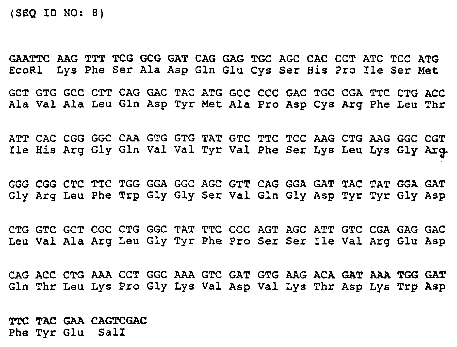
Für die Klonierung der vollständigen cDNA stand eine Lambda gt11 cDNA-Bibliothek zur Verfügung, die aus der RNA von in definiertem Medium (dM) wachsenden HTZ-19 Melanomzellen synthetisiert worden war.There was one for the cloning of the complete cDNA Lambda gt11 cDNA library available from the RNA from HTZ-19 growing in defined medium (dM) Melanoma cells had been synthesized.
Insgesamt wurden 25 Platten mit je 8000 pfu ausplattiert, je 2 Nitrocellulose-Filter aufgelegt und mit dem durch Nick-Translation markierten gereinigten MIA-PCR Insert hybridisiert (50 ml Hybridisierungslösung, 2 x 10⁶ cpm/ml). Nach zweitägiger Autoradiographie konnten mehrere Signale erhalten werden, die auf beiden Filtern ein übereinstimmendes Hybridisierungssignal ergaben. Die zugehörigen Phagen-Plaques wurden isoliert und einer Nachuntersuchung unterzogen. Dabei wurden von jedem isolierten Plaque 4 Verdünnungsstufen ausplattiert und die Platten mit 100-300 pfu für zwei weitere Hybridisierungen verwendet.A total of 25 plates with 8000 pfu each were plated out, 2 nitrocellulose filters were placed on each and hybridized with the purified MIA-PCR insert marked by nick translation (50 ml hybridization solution, 2 × 10 6 cpm / ml). After two days of autoradiography, several signals were obtained, which gave a matching hybridization signal on both filters. The associated phage plaques were isolated and subjected to a follow-up examination. Four dilution stages were plated out from each isolated plaque and the plates with 100-300 pfu were used for two further hybridizations.
Aus den so isolierten Plaques konnte nach einer 50 ml
Übernacht-Kultur die Lambda-DNA isoliert werden, 40 µg
mit EcoRI verdaut und in einem 5% PAA-Gel aufgetrennt
werden. Das Insert wurde eluiert und 8 ng für die Ligation
mit 100 ng EcoRI verdautem, dephosphoryliertem
Vektor (pbluescript, Stratagene) eingesetzt. Die Hälfte
der Ligationsansätze wurden für die Transformation kompetenter
E. coli DH5a eingesetzt, die auf SOB/Amp-Platten
mit IPTG und X-Gal für die Blau/Weiß Selektion ausplattiert
wurden. Die rekombinanten Kolonien wurden abgeimpft,
die Plasmid-DNA isoliert und die Inserts
sequenziert. Die Sequenz des Inserts mit der vollständigen
codierenden Sequenz ist unter SEQ ID NO 1 gezeigt. After a 50 ml
Overnight culture, the lambda DNA isolated, 40 µg
digested with EcoRI and separated in a 5% PAA gel
become. The insert was eluted and 8 ng for ligation
dephosphorylated with 100 ng EcoRI
Vector (pbluescript, Stratagene) used. The half
of the ligation approaches became more competent for the transformation
E. coli DH5a used on SOB / Amp plates
plated with IPTG and X-Gal for the blue / white selection
were. The recombinant colonies were vaccinated,
the plasmid DNA isolated and the inserts
sequenced. The sequence of the insert with the complete
coding sequence is shown under
Ein auf diese Weise erhaltenes Plasmid ist pbs L7MIA, welches bei der Deutschen Sammlung von Mikroorganismen und Zellkulturen GmbH (DSM) in Braunschweig, Deutschland am 14.07.93 hinterlegt wurde (DSM 8420).A plasmid obtained in this way is pbs L7MIA, which in the German collection of microorganisms and Zellkulturen GmbH (DSM) in Braunschweig, Germany on July 14, 1993 (DSM 8420).
Alle für die Klonierung verwendeten Methoden sind ausführlich in J. Sambrook, E.F. Fritsch, T. Maniatis (1989), Molecular cloning: a laboratory manual, 2nd edition, Cold Spring Harbor Laboratory Press, Cold Spring Harbor, USA, beschrieben.All methods used for cloning are detailed in J. Sambrook, E.F. Fritsch, T. Maniatis (1989) Molecular cloning: a laboratory manual, 2nd edition, Cold Spring Harbor Laboratory Press, Cold Spring Harbor, USA.
Eine humane genomische DNA Bibliothek im Bacteriophagen
Lambda FIX II (Elgin et al. (1991), Strategies 4, 8-9),
kommerziell erhältlich von Stratagene (Heidelberg), wurde
nach etablierten Methoden (Sambrook et al. Molecular
Cloning, Cold Spring Harbor Laboratory, 1989) auf Nitrocellulose-Filter
plattiert. Zur Verwendung als Hybridisierungs-Probe
wurde die für humanes MIA codierende cDNA
aus Beispiel 2a radioaktiv markiert und entsprechend
etablierten Techniken verwendet (Sambrook et al. (1989),
Molecular Cloning, Cold Spring Harbor Laboratory Press).
Vorhybridisierung (2 Stunden) und Hybridisierung (16
Stunden) erfolgten bei 60°C in 6 x SSC, 5 x Denhardt's
Lösung, 100 µg ml Lachssperma-DNA und 0.1% SDS. Die 32P-dCTP-markierte
Probe wurde dem Hybridisierungsansatz in
einer Konzentration von 1 x 10⁶ cpm/ml zugesetzt. Anschließend
wurden die Filter bei 60°C zweimal 20 Minuten
gewaschen in 2 x SSC, 0.1 % SDS, dann zweimal 20 Minuten
in 1 x SSC, 0.1 % SDS und schließlich zweimal 20 Minuten
in 0.25 x SSC, 0.1 % SDS. Die Filter wurden daraufhin
getrocknet und 24 bis 48 Stunden auf Roentgenfilm exponiert.
Die bei diesem Verfahren ein positives Hybridisierungssignal
liefernden Plaques wurden isoliert und durch
Rehybridisierung bestätigt. Die in diesen Phagen als
Insert enthaltene humane genomische DNA wurde durch
Southern-Hybridisierung unter Verwendung von MIA cDNA-Proben
charakterisiert. Hierzu wurde die Phagen DNA mit
der Restriktionsendonuklease XbaI gespalten, auf einem
0.8 % Agarose-Gel aufgetrennt und anschließend nach
Southern (J.Mol.Biol. 98 (1975), 503) auf Nitrocellulose
transferiert. Die so erhaltenen Filter wurden unter den
oben beschriebenen Bedingungen mit der kompletten MIA
cDNA als Probe hybridisiert, wobei zwei XbaI Fragmente
von ca. 1.4 kb und ca. 2.2 kb positive Signale lieferten.
Diese beiden Fragmente wurden jeweils in das zur Sequenzierung
geeignete und bei Stratagene (Heidelberg) käuflich
erhältliche Plasmid pBluescriptSK- (Short et al.
(1988), Nucl. Acids res. 16, 7583 - 7600; Alting-Meese
und Short (1989), Nucl. Acids res. 17, 9494) kloniert.
Die gesamte, für das humane MIA codierende Sequenz ist in
vier Exons lokalisiert, die in SEQ ID NO:3 mit ihren
flankierenden Sequenzen gezeigt sind. Da nicht untersucht
wurde, ob zwischen den beiden XbaI-Fragmenten, auf denen
die MIA Exons lokalisiert sind, sich eines oder sogar
mehrere weitere XbaI-Fragmente befinden, kann nicht
ausgeschlossen werden, daß Intron 2 in Wirklichkeit
wesentlich größer ist. A human genomic DNA library in the bacteriophage Lambda FIX II (Elgin et al. (1991), Strategies 4, 8-9), commercially available from Stratagene (Heidelberg), was established using established methods (Sambrook et al. Molecular Cloning, Cold Spring Harbor Laboratory, 1989) plated on nitrocellulose filters. For use as a hybridization sample, the cDNA coding for human MIA from Example 2a was radioactively labeled and used in accordance with established techniques (Sambrook et al. (1989), Molecular Cloning, Cold Spring Harbor Laboratory Press). Pre-hybridization (2 hours) and hybridization (16 hours) were carried out at 60 ° C in 6 x SSC, 5 x Denhardt's solution, 100 µg ml salmon sperm DNA and 0.1% SDS. The 32 P-dCTP-labeled sample was added to the hybridization mixture in a concentration of 1 × 10 6 cpm / ml. The filters were then washed twice at 60 ° C. for 20 minutes in 2 × SSC, 0.1% SDS, then twice for 20 minutes in 1 × SSC, 0.1% SDS and finally twice for 20 minutes in 0.25 × SSC, 0.1% SDS. The filters were then dried and exposed to X-ray film for 24 to 48 hours. The plaques providing a positive hybridization signal in this method were isolated and confirmed by re-hybridization. The human genomic DNA contained as an insert in these phages was characterized by Southern hybridization using MIA cDNA samples. For this purpose, the phage DNA was cleaved with the restriction endonuclease XbaI, separated on a 0.8% agarose gel and then transferred to nitrocellulose according to Southern (J. Mol.Biol. 98 (1975), 503). The filters thus obtained were hybridized with the complete MIA cDNA as a sample under the conditions described above, two XbaI fragments of approximately 1.4 kb and approximately 2.2 kb providing positive signals. These two fragments were each inserted into the plasmid pBluescriptSK- (Short et al. (1988), Nucl. Acids res. 16, 7583-7600; Alting-Meese and Short (1989), which is suitable for sequencing and is commercially available from Stratagene (Heidelberg), Nucl. Acids res. 17, 9494). The entire sequence coding for the human MIA is located in four exons, which are shown in SEQ ID NO: 3 with their flanking sequences. Since it was not investigated whether between the two XbaI fragments on which the MIA exons are located there are one or even more XbaI fragments, it cannot be ruled out that
Eine kommerziell erhältliche (Novagene, New York) cDNA-Bibliothek aus einem 13.5 Tage alten Maus-Embryo im Vektor Lambda EXlox (Palazzolo et al. (1990), Gene 88, 25 - 36) wurde wie in Beispiel 2a beschrieben plattiert. Als Hybridisierungsprobe wurde die für humanes MIA codierende cDNA aus Beispiel 2a in radioaktiv markierter Form eingesetzt. Die Hybridisierungsbedingungen waren identisch mit den in Beispiel 2b beschriebenen, mit Ausnahme der Temperaturen bei Hybridisierung und Waschung, die hier 55°C betrugen. Die in den so erhaltenen und durch Rehybridisierung bestätigten Plaques vorliegenden cDNA-Inserts wurden sequenziert. Die Sequenz des Inserts mit der vollständigen codierenden DNA des murinen MIA ist in SEQ ID NO 4 gezeigt.A commercially available (Novagene, New York) cDNA library from a 13.5 day old mouse embryo in the Vector Lambda EXlox (Palazzolo et al. (1990), Gene 88, 25 - 36) was plated as described in Example 2a. As The hybridization sample was the one coding for human MIA cDNA from Example 2a used in radioactively labeled form. The hybridization conditions were identical to those described in Example 2b, with the exception of the temperatures with hybridization and washing, which here is 55 ° C cheat. Those obtained in the way and by re-hybridization confirmed plaques of existing cDNA inserts were sequenced. The sequence of the insert with the complete coding DNA of murine MIA is in SEQ ID NO 4 shown.
In diesem Fall wurde eine murine genomische DNA-Bibliothek (aus der Leber einer adulten BALB/C Maus im Vektor EMBL3 (Frischauf et al. (1983), J.Mol.Biol. 170, 827), käuflich erhältlich bei Clontech, Palo Alto CA) in analoger Weise wie in Beispiel 2b beschrieben unter Verwendung der murinen MIA cDNA aus Beispiel 2c als Probe abgesucht. Die Bedingungen waren identisch zu den in Beispiel 2b beschriebenen. Auch das weitere Vorgehen erfolgte in analoger Weise. In this case, a murine genomic DNA library was created (from the liver of an adult BALB / C mouse in the vector EMBL3 (Frischauf et al. (1983), J. Mol. Biol. 170, 827), commercially available from Clontech, Palo Alto CA) in analog Using as described in Example 2b the murine MIA cDNA from Example 2c as a sample. The conditions were identical to those in Example 2b described. The further procedure also took place in analogous way.
Zur Bestimmung der antiproliferativen Wirkung des
Melanom-inhibierenden Proteins bzw. der gemäß Beispiel 1
erhaltenen Proteinfraktionen auf Melanomzellen werden
exponentiell wachsende HTZ 19-dM-Zellen für 24 Stunden in
96er Mikrokulturplatten (Costar, Zürich) in jeweils
100 µl serumfreiem Medium (s. Beispiel 1) mit einer
Dichte von 3 x 10³-Zellen pro Vertiefung ausgesät (gemäß
Chambard et al., J. Cell. Physiol. 135 (1988), 101 -
107). Die Zellen werden dann mit der zu untersuchenden
Proteinfraktion für 4 - 5 Tage bei 37°C/5 % CO2 inkubiert.
Nach Zugabe von jeweils 1 µCi 3H Thymidin (spezifische
Aktivität 23 Ci/mmol, Amersham Buchler,
Braunschweig, Deutschland) werden die Zellen 8 Stunden
unter identischen Bedingungen weiter inkubiert und anschließend
der 3H Thymidin-Einbau in die zelluläre DNA
nach Säurefällung in üblicher Weise über einen Flüssigszintillationszähler
gemessen. Die Aktivität der untersuchten
Proteinfraktion wird als Prozentsatz des 3H
Thymidin-Einbaus dieser behandelten Zellen, gegenüber dem
3H Thymidin-Einbau in unbehandelten Kontrollzellen ausgedrückt.
Unter Verwendung verschiedener Konzentrationen
des isolierten Melanom-inhibierenden Proteins kann eine
Konzentration ermittelt werden, bei welcher dieser 3H
Thymidin-Einbau um 50 %, gegenüber der unbehandelten
Kontrolle, gehemmt wird (IC 50-Wert in der folgenden
Tabelle 1).
Zur Bestimmung der invasionshemmenden Wirkung von MIA
wird ein modifiziertes Boyden-Kammer-System (Albini et
al. (1987), Cancer Res. 47, 3239 - 3245) verwendet. Die
Kammern wurden von der Firma Costar (Blind Well Chamber
No. 441200) bezogen. Zur Simulierung einer Basalmembran-ähnlichen
Barriere zwischen dem Chemoattraktans in
der unteren Kammer und den Zellen in der oberen Kammer
werden auf den Polycarbonatfilter (Porengröße 8 µm,
Costar No. 150446) 52 µl Matrigel (Becton Dickinson Cat.
No. 40234) aufgebracht. Als Chemoattraktans wird die
untere Kammer mit 210 µl Fibroblasten-konditioniertem
Medium befüllt. Dieses wird wie folgt gewonnen: Fibroblasten
aus normaler menschlicher Haut werden zwischen
der 10. und 20. Passage für 24 Stunden in DMEM-Medium
(Gibco) ohne Zusatz von foetalem Kälberserum gehalten.
Das so konditionierte Medium wird unverdünnt als Chemoattraktans
eingesetzt. In die obere Kammer der Boyden-Apparatur
werden 2 x 10⁵ der zu untersuchenden Tumorzellen
in 800 µl DMEM (Gibco, ohne foetales Kälberserum)
mit, bzw. ohne MIA-Wirkstoff gegeben. Humane Tumorzellen
(siehe Beispiel 3a oder Beispiel 8) oder tierische Tumorzellen
wie etwa B16 (ATCC CRL 6322) können mit der
beschriebenen Methode auf Inhibierung ihres Migrationsverhaltens
durch MIA getestest werden. Ohne Zugabe
des Melanom inhibierenden Proteins wandern ca 10 % Tumorzellen
innerhalb von ca 4 Stunden aus der oberen Kammer
in die untere Kammer, wo sie an der Unterseite der
Matrigel-Membran haften bleiben. Sie werden dort fixiert,
gefärbt und anschließend gezählt. Wird der oberen Kammer
das humane oder murine Melanom inhibierende Protein MIA
zugesetzt, so wird die Wanderung der Zellen stark inhibiert.
Fig. 1 zeigt die erhaltenen Hemmwerte (
Die gegen ein Peptid aus myelin basic protein (MBP, Aminosäureposition 87 - 106) spezifische CD4+ T-Zellinie D7.1 ist in der Lage, in einem Standard 51Cr-Release Assay sowohl MBP- als auch Peptid 87-106 präsentierende Targets zu lysieren (Targets: Daudi-Zellen, R. Martin, U. Utz, J.E. Coligan, J.R. Richert, M. Flerlage, E. Robinson, R. Stone, W.E. Biddison, D.E. McFarlin, H.F. MacFarland, Diversity in fine specificity and T cell receptor usage of the human CD4 + cytotoxic T-cell response specific for the immunodominant myelin basic protein peptide 87 - 106, J. Immunol. (1992), 148 (5), 1359 - 1366). Nach Zugabe von MIA (eingesetzte Menge entspricht ca. 50 - 100 ng/ml gereinigten MIA) wird die Peptid-spezifische Zytotoxizität um ca. 55 % (Fig. 2 a), die MBP-spezifische Zytotoxizität um ca. 50 % (Fig. 2 b) gehemmt. Die Hemmung zeigt erwartungsgemäß eine geringe Abhängigkeit vom Effektor-Target-Verhältnis (E:T), das hier mit 1:1 bzw. 5:1 sehr niedrig angesetzt wurde und damit hochspezifisch ist (Fig. 2).The CD4 + T cell line D7.1, which is specific against a peptide from myelin basic protein (MBP, amino acid position 87-106), is able to present both MBP and peptide 87-106 in a standard 51 Cr release assay lysing (targets: Daudi cells, R. Martin, U. Utz, JE Coligan, JR Richert, M. Flerlage, E. Robinson, R. Stone, WE Biddison, DE McFarlin, HF MacFarland, Diversity in fine specificity and T cell receptor usage of the human CD 4 + cytotoxic T-cell response specific for the immunodominant myelin basic protein peptide 87-106, J. Immunol. (1992), 148 (5), 1359-1366). After adding MIA (amount used corresponds to approx. 50-100 ng / ml of purified MIA), the peptide-specific cytotoxicity is reduced by approx. 55% (FIG. 2 a), the MBP-specific cytotoxicity by approx. 50% (FIG. 2 b) inhibited. As expected, the inhibition shows little dependence on the effector-target ratio (E: T), which was set very low here with 1: 1 or 5: 1 and is therefore highly specific (FIG. 2).
LAK-Zellen sind nicht klonal expandierte, Lymphokin-aktivierte periphere Blutlymphozyten, vorwiegend T-Lymphozyten (A.A. Rayner, E.A. Grimm, M.T. Lotze, E.W. Chu, S.A. Rosenberg, Lymphokine activated killer (LAK) cells: Analysis of factors relevant for immunotherapy of human cancer, Cancer 55 (1985), 1327 - 1333). Sie werden in vielen immunologischen Therapie-Ansätzen der Tumortherapie standardisiert eingesetzt. In dem hier gezeigten Experiment werden LAK-Zellen auf ihre Zytotoxizität gegenüber HTZ-19 Melanom-Zellen als Targets in einem Microzytotoxizitäts-Assay geprüft. In Effektor-Target-Verhältnissen von 1:1, 5:1 und 10:1 kommt es zu einer maximalen Zytotoxizität (CTX) von knapp 40 %. Diese wird nach Zugabe von MIA (Konzentration wie in Beispiel 4 a) stark gehemmt, maximal um 80 % bei niedrigem Effektor-Target-Verhältnis, wie es am ehesten lokal - am Tumor selbst - zu erwarten ist (Fig. 3). LAK cells are not clonally expanded, lymphokine-activated peripheral blood lymphocytes, predominantly T lymphocytes (A.A. Rayner, E.A. Grimm, M.T. Lotze, E.W. Chu, S.A. Rosenberg, Lymphokine activated killer (LAK) cells: Analysis of factors relevant for immunotherapy of human cancer, Cancer 55 (1985), 1327-1333). You will be in many immunological therapy approaches of tumor therapy used in a standardized manner. In the one shown here LAK cells are experimented on their cytotoxicity versus HTZ-19 melanoma cells as targets in one Microcytotoxicity assay tested. In effector-target ratios of 1: 1, 5: 1 and 10: 1 there is a maximum cytotoxicity (CTX) of just under 40%. This will after adding MIA (concentration as in Example 4 a) strongly inhibited, by a maximum of 80% with a low effector-target ratio, as it is most likely local - on the tumor itself - is to be expected (Fig. 3).
Periphere Blutlymphozyten (PBMC) lassen sich klassischerweise mit Phythämagglutinin (PHA) und Interleukin-2 (IL-2) standardisiert stimulieren (J.E. Coligan, A.M. Kruisbeek, D.H. Margulies, E.M. Shevach, W. Strober, Current protocols in immunology, NIH Monograph, J. Wiley Sons, New York, 1992). Hierbei werden mit PHA fast ausschließlich T-Zellen stimuliert, mit IL-2 vorwiegend T-Lymphozyten, aber auch B-Zellen mit IL-2-Rezeptor. Unter Co-Inkubation mit MIA läßt sich im angegebenen Dosierbereich (Proteineinheit: nach dem ersten Reinigungsschritt (Biogel P10-Säule), Aktivität ist um den Faktor 50 - 100 geringer als nach vollständiger Reinigung) eine sehr starke Hemmung der PHA-Antwort erreichen (Fig. 4). Die IL-2-Antwort wird im höheren Dosisbereicht gehemmt (Fig. 5).Peripheral blood lymphocytes (PBMC) can be classically with phythaemagglutinin (PHA) and interleukin-2 (IL-2) stimulate in a standardized manner (J.E. Coligan, A.M. Kruisbeek, D.H. Margulies, E.M. Shevach, W. Strober, Current protocols in immunology, NIH Monograph, J. Wiley Sons, New York, 1992). Here are almost exclusively with PHA T cells stimulated with IL-2 predominantly T lymphocytes, but also B cells with IL-2 receptors. Under Co-incubation with MIA can be done in the specified dosage range (Protein unit: after the first cleaning step (Biogel P10 column), activity is by the factor 50 - 100 less than after complete cleaning) one achieve very strong inhibition of the PHA response (Fig. 4). The IL-2 response is inhibited in the higher dose range (Fig. 5).
Zur Expression von MIA als Fusionsprotein mit einem als
"Carrier" in E.coli geeigneten Protein kann beispielsweise
der käuflich erhältliche Vektor pQE40 (Cat. No.
33403, DIAGEN GmbH, Düsseldorf) verwendet werden. In
diesen Vektor wird zwischen die je einmal vorhandenen
Restriktionsschnittstellen SphI und HindIII ein für die
reife Form von MIA codierendes cDNA-Fragment insertiert.
Ein derartiges Fragment wird am einfachsten durch PCR-Amplifikation
nach bekannten Techniken hergestellt unter
Verwendung der klonierten MIA cDNA als Matrize und zweier
geeigneter Primer (5'-GATGCATGCGGTCCTATGCCCAAGCTG-3' (SEQ
ID NO:9) und 5'-GATAAGCTTTCACTGGCAGTAGAAATC-3' (SEQ ID
NO:10)). Das erhaltene PCR-Fragment wird mit SphI und
HindIII geschnitten und in den ebenso behandelten Vektor
pQE40 ligiert. Das resultierende Plasmid exprimiert ein
Fusionsprotein aus DHFR (Dihydrofolat-Reduktase als
"Carrier") und MIA. Um aus diesem Fusionsprotein freies
MIA proteolytisch abspalten zu können, wird ein DNA-Segment
zwischen DHFR und MIA einkloniert, welches für
die Erkennungssequenz der IgA-Protease (Ser Arg Pro
Pro/Ser) codiert. Dies geschieht durch Öffnen des Expressionsplasmides
mit BglII (Partialrestriktion mit anschließender
Isolierung des linearisierten Vektors) und
SphI und anschließende Insertion eines Adaptors (5'-GATCTAGCCGGCCGCCCAGCCCGGCATG-3'
(SEQ ID NO:11) und 5'-CCGGGCTGGGCGGCCGGCTA-3'
(SEQ ID NO:12) hybridisiert zum
Doppelstrang). Das resultierende Expressionsplasmid
pQE40-MIA codiert für ein Fusionsprotein aus DHFR und
MIA, wobei sich zwischen DHFR und MIA eine Spaltstelle
für IgA-Protease befindet. Das Fusionsprotein trägt am N-Terminus
6 Histidine, die den Angaben des Herstellers
zufolge zur Aufreinigung des Fusionsproteins mit Hilfe
von Ni-Chelat-Gelmaterialien verwendet werden können.
Derartige Verfahren sind in EP-A 0 282 042 und EP-A 0 253
303, deren Inhalt Gegenstand der Offenbarung dieser
Anmeldung ist, beschrieben. Fig. 6 zeigt das Expressionsplasmid
pQE40-MIA. To express MIA as a fusion protein with an as
"Carrier" in E.coli suitable protein can for example
the commercially available vector pQE40 (Cat. No.
33403, DIAGEN GmbH, Düsseldorf) can be used. In
this vector is between the existing ones
Restriction interfaces SphI and HindIII one for the
mature form of MIA coding cDNA fragment inserted.
Such a fragment is easiest by PCR amplification
manufactured under known techniques under
Using the cloned MIA cDNA as a template and two
suitable primer (5'-GATGCATGCGGTCCTATGCCCAAGCTG-3 '(SEQ
ID NO: 9) and 5'-GATAAGCTTTCACTGGCAGTAGAAATC-3 '(SEQ ID
NO: 10)). The PCR fragment obtained is treated with SphI and
HindIII cut and in the same treated vector
pQE40 ligated. The resulting plasmid expresses a
Fusion protein from DHFR (dihydrofolate reductase as
"Carrier") and MIA. To get free from this fusion protein
Being able to cleave MIA proteolytically becomes a DNA segment
cloned between DHFR and MIA, which for
the recognition sequence of the IgA protease (Ser Arg Pro
Per / Ser) coded. This is done by opening the expression plasmid
with BglII (partial restriction with subsequent
Isolation of the linearized vector) and
SphI and subsequent insertion of an adapter (5'-GATCTAGCCGGCCGCCCAGCCCGGCATG-3 '
(SEQ ID NO: 11) and 5'-CCGGGCTGGGCGGCCGGCTA-3 '
(SEQ ID NO: 12) hybridizes to
Double strand). The resulting expression plasmid
pQE40-MIA codes for a fusion protein from DHFR and
MIA, with a gap between DHFR and MIA
for IgA protease. The fusion protein carries at the N-
Der Gehalt von MIA läßt sich optimieren, indem das Carrier-Protein DHFR eliminiert und durch ein möglichst kleines Peptid ersetzt wird, welches dieselben Funktionen erfüllt. Derartige geeignete Peptide sind beispielsweise MetArgGlySerHisHisHisHisHisHisGlySerSerArgProPro (SEQ ID NO:13) (dieses Peptid kann durch IgA-Protease von der unmittelbar darauf folgenden Aminosäure-Sequenz des reifen MIA abgespalten werden; derartige Verfahren sind in der WO 91/11520 beschrieben, deren Inhalt Gegenstand der Offenbarung dieser Anmeldung ist) oder MetArgGlySerHisHisHisHisHisHisGlySerValAspAspAspAspLys- (SEQ ID NO:14) (dieses Peptid kann durch Enterokinase von der unmittelbar darauf folgenden Aminosäure-Sequenz des reifen MIA abgespalten werden). Expressionsplasmide, die für derartige MIA-Peptidfusionen codieren, können wie folgt hergestellt werden: Eine PCR-Amplifikation mit MIA cDNA (Seq.ID No 1) als Matrize und den Primern 5'-AAAAAGGATCCAGCCGGCCGCCCGGTCCTATGCCCAAGCTGGC-3' (SEQ ID NO:15) und 5'-GGCGAGCAGCCAGATCTCCATAG-3' (SEQ ID NO:16) liefert ein Fragment, welches mit BamHI und BglII nachgeschnitten wird. Der Expressionsvektor pQE40-MIA wird ebenfalls mit BamHI und BglII restringiert, das kleinere der entstehenden Fragmente wird verworfen und durch das beschriebene PCR-Fragment ersetzt. Hierdurch wird ein Expressionsvektor erhalten, der nach Induktion das Fusionsprotein MetArgGlySerHisHisHisHisHisHisGlySerSerArgProPro-MIA (SEQ ID NO:13) exprimiert. Das Fusionsprotein MetArgGlySerHisHisHisHisHisHisGlySerValAspAspAspAspLys-MIA (SEQ ID NO:14) wird in völlig analoger Weise unter Verwendung der Primer 5'-AAAAAAGGATCCGTTGATGATGACGATAAAGGTCCTATGCCCAAGCTGGC-3' (SEQ ID NO:17) und 5'-GGCGAGCAGCCAGATCTCCATAG-3' (SEQ ID NO:16) erhalten. The content of MIA can be optimized by the Carrier protein is eliminated by DHFR and if possible by a small peptide is replaced, which has the same functions Fulfills. Such suitable peptides are, for example MetArgGlySerHisHisHisHisHisHisGlySerSerArgProPro (SEQ ID NO: 13) (this peptide can be derived from the IgA protease immediately following amino acid sequence of the mature MIA to be split off; are such procedures described in WO 91/11520, the content of which is the subject the disclosure of this application) or MetArgGlySerHisHisHisHisHisHisGlySerValAspAspAspAspLys- (SEQ ID NO: 14) (this peptide can be derived from the enterokinase immediately following amino acid sequence of the mature MIA can be split off). Expression plasmids, the code for such MIA-peptide fusions, such as can be prepared as follows: A PCR amplification with MIA cDNA (Seq.ID No 1) as template and the primers 5'-AAAAAGGATCCAGCCGGCCGCCCGGTCCTATGCCCAAGCTGGC-3 ' (SEQ ID NO: 15) and 5'-GGCGAGCAGCCAGATCTCCATAG-3 '(SEQ ID NO: 16) a fragment which was cut with BamHI and BglII becomes. The expression vector pQE40-MIA is also included BamHI and BglII restricted, the smaller of the resulting Fragments are discarded and described by the PCR fragment replaced. This becomes an expression vector obtained after the induction of the fusion protein MetArgGlySerHisHisHisHisHisHisGlySerSerArgProPro-MIA (SEQ ID NO: 13). The MetArgGlySerHisHisHisHisHisHisGlySerValAspAspAspAspLys-MIA fusion protein (SEQ ID NO: 14) is used in a completely analogous manner Primer 5'-AAAAAAGGATCCGTTGATGATGACGATAAAGGTCCTATGCCCAAGCTGGC-3 ' (SEQ ID NO: 17) and 5'-GGCGAGCAGCCAGATCTCCATAG-3 ' (SEQ ID NO: 16) received.
Weitere, ähnliche Fusionsproteine aus Peptiden und MIA können in analoger Weise hergestellt und unter der Kontrolle geeigneter (vorzugsweise starker und induzierbarer) Promotoren in eines der zahlreichen für E.coli beschriebenen Plasmide einkloniert und zur Expression gebracht werden. Eine weitere Alternative stellt die Fusion von MIA mit einem Peptid dar, welches in E.coli zu Sekretion des Fusionsproteines ins Periplasma mit nachfolgender Abspaltung und Freisetzung von MIA führt. Dieses Verfahren ist in WO 88/09373 beschrieben, deren Inhalt Gegenstand der Offenbarung dieser Anmeldung ist.Further, similar fusion proteins from peptides and MIA can be made in an analogous manner and under control more suitable (preferably stronger and more inducible) Promoters in one of the numerous for E.coli described plasmids cloned and for expression to be brought. Another alternative is the Fusion of MIA with a peptide which is found in E. coli Secretion of the fusion protein into the periplasm with subsequent Cleavage and release of MIA leads. This method is described in WO 88/09373, the The subject matter of the disclosure of this application is.
Das Expressionsplasmid pQE40-MIA (oder ein ähnliches
Expressionsplasmid, welches beispielsweise durch Verwendung
eines anderen Grundvektors oder eines anderen
"Carrier"-Proteins oder -Peptides erhalten werden kann;
derartige Alternativen sind außer in Beispiel 5a auch
beschrieben in Methods of Enzymology 185 (Gene Expression
Technology), Hrsg. David V. Goeddel, Academic Press 1991)
wird in einen geeigneten E.coli Stamm transfiziert, der
über eine hinreichende Expression des lac-Repressors
verfügt, so daß eine induzierbare Expression des MIA-Fusionsproteins
erzielt werden kann. Geeignet hierfür ist
beispielsweise der Stamm E.coli M15[pREP4], der zusammen
mit pQE40 käuflich erhältlich ist (Diagen GmbH, Düsseldorf),
oder aber andere E.coli-Stämme wie etwa E.coli
UT5600 (Earhart et al. (1979), FEMS Microbiology Letters
6, 277-280) oder E.coli BL21 (Grodberg und Dunn (1988),
J.Bacteriol. 170, 1245-1253), die zuvor mit einem lac-Repressor
exprimierenden Helfer-Plasmid wie etwa pUBS520
(Brinckmann et al. (1989), Gene 85, 109-114) oder pUBS500
(EP-B 0 373 365) transfiziert wurden. MIA kann dann wie
folgt gewonnen werden: E.coli M15 [pREP4/pQE40-MIA] wird
auf LB-Medium bis zu einer optischen Dichte von 0.6
(gemessen bei 550 nm) kultiviert, dann IPTG (Isopropyl-β-D-thiogalactopyranosid,
Boehringer Mannheim GmbH) in
einer Endkonzentration von 1 mM zugegeben und anschließend
4 Stunden weiterkultiviert. Die Zellen werden
abzentrifugiert, in 100 mM Natrium-Phosphat-Puffer pH 7.5
mit 300 mM NaCl aufgenommen und durch dreimaliges Einfrieren
und Auftauen mit anschließender Ultraschallbehandlung
lysiert. Zu dem durch Zentrifugation geklärten
Lysat wird Ni-NTA-Agarose (Diagen GmbH) unter Berücksichtigung
der vom Hersteller angegebenen maximalen Bindekapazität
zugegeben und über Nacht bei Raumtemperatur
unter Mischen inkubiert. Das so mit Fusionsprotein beladene
Gelmaterial wird niedertourig abzentrifugiert,
zweimal mit 100 mM Natrium-Phosphat-Puffer pH 7.5 und
zweimal mit Natrium-Phosphat-Puffer pH 6.1 gewaschen.
Anschließend wird MIA aus dem Fusionsprotein abgespalten
durch Inkubation des Gelmateriales mit IgA-Protease
(Boehringer Mannheim GmbH) in 100 mM Natrium-Phosphat-Puffer
pH 7.5 über Nacht bei 37°C. Das Gelmaterial wird
durch Zentrifugation abgetrennt und der MIA-haltige
Überstand nach Sterilfiltration in den in den Beispielen
3 und 4 beschriebenen Aktivitätstests eingesetzt. The expression plasmid pQE40-MIA (or a similar
Expression plasmid, which for example by use
of another basic vector or another
"Carrier" proteins or peptides can be obtained;
Such alternatives are also in Example 5a
described in Methods of Enzymology 185 (Gene Expression
Technology), ed. David V. Goeddel, Academic Press 1991)
is transfected into a suitable E. coli strain, which
sufficient expression of the lac repressor
has so that an inducible expression of the MIA fusion protein
can be achieved. Suitable for this is
for example the strain E.coli M15 [pREP4], which together
is commercially available with pQE40 (Diagen GmbH, Düsseldorf),
or other E. coli strains such as E. coli
UT5600 (Earhart et al. (1979),
Die für MIA codierende DNA-Sequenz wird in einer Weise
modifiziert, die eine effiziente Expression in E.coli
ermöglicht. Zu diesem Zweck wird eine PCR-Amplifikation
mit humaner MIA cDNA (Seq. ID No 1) als Matrize und den
Primern 1 (5'-AAAAACATATGGGACCAATGCCAAAATTAGCAGATCGTAAATTATGTGCAGATCAGGAG-3'
(SEQ ID NO:19) und 2 (5'-AAAAAAAGCTTTCACTGGCAGTAGAAATC-3'
(SEQ ID NO:20)) durchgeführt.
Primer 1 verändert die MIA codierende Sequenz in
dem N-terminalen Bereich so, daß bei unveränderter MIA-Aminosäure-Sequenz
die DNA- und damit die mRNA-Sequenz
für E.coli optimiert wird. Das Startcodon Met wird hinzugefügt
und an eben dieser Stelle eine Erkennungssequenz
der Restriktionsendonuklease NdeI eingebracht, die eine
anschließende Klonierung des so modifizierten MIA codierenden
Fragmentes in einen Vektor gestattet, der einen
(vorzugsweise starken und induzierbaren) Promoter und
eine Translations-Inititations-Sequenz ("Shine-Dalgarno-Sequenz")
mit nachfolgend plazierter NdeI-Schnittstelle
enthält. Primer 2 dient als 3'-Gegenprimer und enthält
eine HindIII-Schnittstelle, so daß die Insertion des
modifizierten MIA codierenden Fragmentes in den Vektor
als NdeI HindIII Fragment erfolgen kann. Die derart
modifizierte MIA codierende Sequenz ist in SEQ ID NO:18
gezeigt. Ein derart hergestelltes Expressionsplasmid ist
p11379, welches unter der Hinterlegungsnummer DSM 9267 am
29.06.94 bei der Deutschen Sammlung von Mikroorganismen
und Zellkulturen GmbH, D-38124 Braunschweig, hinterlegt
wurde. The DNA sequence coding for MIA is in a way
modified that an efficient expression in E.coli
enables. For this purpose a PCR amplification
with human MIA cDNA (Seq. ID No 1) as template and the
Primers 1 (5'-AAAAACATATGGGACCAATGCCAAAATTAGCAGATCGTAAATTATGTGCAGATCAGGAG-3 '
(SEQ ID NO: 19) and 2 (5'-AAAAAAAGCTTTCACTGGCAGTAGAAATC-3 '
(SEQ ID NO: 20)).
Ein Expressionsplasmid für fusionsfreies MIA wird zur
Expression in einen geeigneten E.coli Stamm transfiziert.
Bei Verwendung eines durch lac-Repressor kontrollierten
Expressionsplasmides wie p11379 sind dies Stämme,
die über eine hinreichend hohe intrazelluläre Konzentration
an lac-Repressor verfügen. Solche Stämme können
durch Transfektion eines zweiten Plasmides wie pREP4
(Diagen GmbH), pUBS500 oder pUBS520 (Brinckmann et al.
(1989), Gene 85, 109-114) hergestellt werden. Die verwendeten
E.coli-Stämme sollten vorzugsweise eine geringe
zelleigene Protease-Aktivität aufweisen, wie dies beispielsweise
bei E.coli UT5600 (Earhart et al. (1979),
FEMS Microbiology Letters 6:277-280), bei E.coli BL21
(Grodberg und Dunn (1988), J.Bacteriol. 170:1245-1253)
oder bei E.coli B der Fall ist. Die Expressionskultivierung
erfolgt dann analog wie in Beispiel 5b beschrieben.
Zur Gewinnung von MIA wird das als Proteinaggregat aus E.
coli erhaltene MIA nach den in EP 241 022, EP 364 926, EP
219 874 und DE 40 37 196 beschriebenen Verfahren aufgearbeitet.An expression plasmid for fusion-free MIA is used
Expression transfected into a suitable E. coli strain.
When using a lac repressor controlled
Expression plasmids such as p11379 are strains
which have a sufficiently high intracellular concentration
at the lac repressor. Such tribes can
by transfecting a second plasmid such as pREP4
(Diagen GmbH), pUBS500 or pUBS520 (Brinckmann et al.
(1989), Gene 85, 109-114). The used
E. coli strains should preferably be low
have cell-specific protease activity, such as this
E.coli UT5600 (Earhart et al. (1979),
FEMS Microbiology Letters 6: 277-280), at E.coli BL21
(Grodberg and Dunn (1988), J.Bacteriol. 170: 1245-1253)
or is the case with E.coli B. Expression cultivation
is then carried out analogously to that described in Example 5b.
To obtain MIA, this is used as a protein aggregate from E.
coli obtained MIA according to the in EP 241 022, EP 364 926, EP
219 874 and
Im Detail wird hierzu beispielsweise wie folgt vorgegangen:
MIA-haltige Proteinaggregate aus E.coli-Fermentationen
(sogenannte "Inclusion Bodies") werden in 6 M
Guanidinium-Hydrochlorid, 100 mM TrisHCl pH 8, 1 mM EDTA
solubilisiert, anschließend auf pH 3-4 eingestellt und
gegen 4 M Guanidinium-Hydrochlorid pH 3,5 dialysiert. Die
Renaturierung des solubilisierten Proteins erfolgt dann
in 1 M Arginin pH 8, 1 mM EDTA, 5 mM GSH (Glutathion
reduziert) und 0,5 mM GSSG (Glutathion oxidiert). Aus dem
Renaturierungsansatz kann MIA beispielsweise nach Zusatz
von 1,4 M Ammoniumsulfat durch Adsorption an hydrophobe
Gelmatrices wie Fractogel TSK Butyl (E. Merck, Darmstadt)
und anschließende Elution in 20 mM TrisHCl pH 7 gewonnen
werden.In detail, the procedure is as follows:
MIA-containing protein aggregates from E. coli fermentations
(so-called "Inclusion Bodies") are in 6 M
Guanidinium hydrochloride, 100mM TrisHCl pH 8, 1mM EDTA
solubilized, then adjusted to pH 3-4 and
dialyzed against 4 M guanidinium hydrochloride pH 3.5. The
The solubilized protein is then renatured
in 1 M
Hierzu werden die humane (SEQ ID NO:1) oder murine (SEQ ID NO:4) MIA cDNA oder die entsprechenden genomischen DNA-Segmente in einen Vektor ligiert, in welchem sie ausgehend von einem starken Promoter-Enhancer-System in Säugerzellen transkribiert werden (im Falle der genomischen MIA Fragmente ist dieser Schritt notwendig, da die MIA eigenen Promotoren nur in bestimmten Zelltypen, beispielsweise Melanomen, aktiv und daher nicht für eine allgemeine rekombinante Expression geeignet sind. Eine Expression kann jedoch auch wie in Beispiel 9 beschrieben durch homologe Rekombination in vitro erreicht werden. Derartige Promotoren und Enhancer stammen zumeist aus Viren wie SV40, hCMV, Polyoma oder Retroviren. Alternativ können auch Promoter-Enhancer-Systeme verwendet werden, die für einen bestimmten Zell- oder Gewebetyp spezifisch sind, wie etwa WAP-, MMTV- oder Immunglobulin-Promoter, oder aber solche Systeme, die induzierbar sind, wie etwa Metallothionein-Promoter. Ein solcher Vektor ergänzt die MIA-cDNA (falls eine solche verwendet wird) mit Donor- und Akzeptor-Signalen für die RNA-Prozessierung sowie einem Signal für poly-A-Addition. Ein solcher geeigneter Vektor ist beispielsweise pCMX-pL1 (Umesono et al. (1991), Cell 65, 1255-1266), der in Fig. 7 dargestellt ist. In die einzige EcoRI-Schnittstelle dieses Vektors wird die mit EcoRI-Linkern versehene MIA cDNA ligiert, wobei durch Restriktionsanalyse mit Hilfe der übrigen Schnittstellen im Polylinker dieses Vektors (siehe SEQ ID NO:23) sichergestellt wird, daß die Orientierung der MIA-cDNA in Ableserichtung des CMV-Promoters vorliegt. Völlig analog wird bei einer Klonierung in andere Vektoren vorgegangen, z.B. in pCDNA3 (Invitrogen, San Diego/USA) oder pSG5 (Stratagene, LaJolla/USA). Die DNA der so erhaltenen Expressionsplasmide wird aus E.coli präpariert und mit für den jeweiligen Zelltyp spezifischen Techniken in die Säugerzellen transfiziert (Methods of Enzymology 185 (Gene Expression Technology), Hrsg. David V. Goeddel, Academic Press 1991, Sektion V). Das Expressionsplasmid pCMX-pL1-MIA wird in die humane Teratocarcinoma-Linie PA-1sc9177 (Büttner et al. (1991), Mol. Cell. Biol. 11, 3573-3583) nach beschriebenen Methoden (Büttner et al. (1993), Mol. Cell. Biol. 13, 4174 - 4185) transfiziert, wobei 200 000 Zellen pro 100 mm Kulturschale mit 5 µg DNA transfiziert werden. Die Zellen werden nach der Transfektion in MEM (Gibco) ohne Zusatz von foetalem Kälberserum kultiviert, wobei nach 48 Stunden im Zellkulturüberstand MIA nachweisbar ist.For this purpose, the human (SEQ ID NO: 1) or murine (SEQ ID NO: 4) MIA cDNA or the corresponding genomic DNA segments ligated into a vector in which they based on a strong promoter-enhancer system in Mammalian cells are transcribed (in the case of genomic MIA fragments, this step is necessary because the MIA's own promoters only in certain cell types, for example melanoma, active and therefore not for one general recombinant expression are suitable. A However, expression can also be as described in Example 9 can be achieved by homologous recombination in vitro. Such promoters and enhancers mostly come from Viruses such as SV40, hCMV, Polyoma or retroviruses. Alternatively promoter-enhancer systems can also be used, those specific to a particular cell or tissue type such as WAP, MMTV or immunoglobulin promoters, or systems that are inducible, such as Metallothionein promoters. Such a vector complements the MIA cDNA (if one is used) with donor and Acceptor signals for RNA processing as well a signal for poly A addition. Such a suitable one Vector is, for example, pCMX-pL1 (Umesono et al. (1991), Cell 65, 1255-1266) shown in Fig. 7 is. In the only EcoRI site of this vector the MIA cDNA provided with EcoRI linkers is ligated, by restriction analysis with the help of the rest Interfaces in the polylinker of this vector (see SEQ ID NO: 23) ensure that the orientation of the MIA cDNA in the reading direction of the CMV promoter. Completely the same applies to cloning into other vectors proceeded, e.g. in pCDNA3 (Invitrogen, San Diego / USA) or pSG5 (Stratagene, LaJolla / USA). The DNA of that Expression plasmids obtained are prepared from E. coli and with techniques specific to each cell type transfected into the mammalian cells (Methods of Enzymology 185 (Gene Expression Technology), ed. David V. Goeddel, Academic Press 1991, Section V). The expression plasmid pCMX-pL1-MIA is incorporated into the human teratocarcinoma line PA-1sc9177 (Büttner et al. (1991), Mol. Cell. Biol. 11, 3573-3583) according to the methods described (Büttner et al. (1993) Mol. Cell. Biol. 13, 4174-4185) transfected, whereby 200,000 cells per 100 mm culture dish with 5 µg DNA be transfected. The cells are made after transfection in MEM (Gibco) without the addition of fetal calf serum cultured, after 48 hours in the cell culture supernatant MIA is detectable.
Zur Expression in Insektenzellen wird ein für MIA codierendes DNA-Segment, vorzugsweise die humane MIA cDNA (SEQ ID NO 1), in Vektoren eingebracht, die sich von AcMNPV (Autographa californica nuclear polyhedrosis virus) oder BmNPV (Bombyx mori nuclear polyhedrosis virus) ableiten. Hierzu wird die MIA cDNA zunächst unter die Kontrolle eines für Insektenzellen geeigneten, starken Promoters gebracht (O'Reilly DR, Miller LK und Luckow VA 1992; Baculovirus Expression Vectors - A Laboratory Manual; W.H.Freeman & Co, New York), wie des polH-Promoters oder des p10-Promoters. Zur Expression von MIA mit Hilfe des polH-Promoters wird wie folgt vorgegangen:For expression in insect cells, a coding for MIA is used DNA segment, preferably the human MIA cDNA (SEQ ID NO 1), introduced in vectors that are different from AcMNPV (Autographa californica nuclear polyhedrosis virus) or Derive BmNPV (Bombyx mori nuclear polyhedrosis virus). For this, the MIA cDNA is initially under control a strong promoter suitable for insect cells brought (O'Reilly DR, Miller LK and Luckow VA 1992; Baculovirus Expression Vectors - A Laboratory Manual; W.H. Freeman & Co, New York), such as the polH promoter or of the p10 promoter. To express MIA using the The polH promoter works as follows:
Ein für MIA codierendes DNA-Fragment mit Schnittstellen
für die Restriktionsendonukleasen EcoRI (benachbart dem
5'-Ende des späteren MIA-Transkriptes) und PstI (benachbart
dem 3'-Ende des späteren MIA-Transkriptes) wird
durch PCR-Amplifikation nach bekannten Techniken unter
Verwendung der MIA cDNA als Matrize und der Primer 5'-CGTGAATTCAACATGGCCCGGTCCCTGGTGTGC-3'
(SEQ ID NO:21) und
5'-TATCTGCAGTCACTGGCAGTAGAAATCCCA-3' (SEQ ID NO:22)
erhalten. Dieses Fragment wird mit EcoRI und PstI nachgeschnitten
(zur Erzeugung der entsprechenden kohäsiven
Enden) und in den mit denselben Endonukleasen restringierten
Transfer-Vektor pVL1393 (O'Reilly DR, Miller LK
und Luckow VA 1992; Baculovirus Expression Vectors - A
Laboratory Manual; W.H.Freeman & Co, New York), der
kommerziell erhältlich ist (PharMingen, San Diego, CA
oder Invitrogen Corporation, San Diego, CA), einligiert.
Das resultierende Transfer-Expressionsplasmid pVL1393-MIA
wird zur Vermehrung in E.coli K12 transfiziert und
Plasmid-DNA nach etablierten Methoden präpariert. Der
Transfer der unter Kontrolle des polH-Promoters stehenden
MIA-DNA vom Transfer-Plasmid in den Baculovirus-Vektor
erfolgt durch homologe Rekombination nach etablierten
Methoden (O'Reilly et al. 1992, siehe oben). Hierzu
werden 0.5 µg BaculoGold DNA (linearisierte AcNPV Virus-DNA
mit Lethal-Deletion und durch den polH-Promoter
gesteuerter lacZ Expression, käuflich erhältlich bei
PharMingen, Bestellnummer 21100D) und 2 µg pVL1393-MIA
gemischt, 5 Minuten bei Raumtemperatur inkubiert und
anschließend mit 1 ml 125 mM Hepes pH 7.1, 125 mM CaCl2 ,
140 mM NaCl versetzt. Diese Mischung wird zu 2 x 10⁶
SF9-Insektenzellen (Invitrogen, Bestellnummer B825-01) in
einer Kulturschale von 60 mm Durchmesser gegeben, die
zuvor mit 1 ml Grace's Medium mit 10 % foetalem Kälberserum
überschichtet worden waren. Nach 4 Stunden Inkubation
bei 4°C wird das DNA-haltige Medium entfernt und die
Zellen werden in frischem Medium für 4 Tage bei 27°C
inkubiert. Die auf diesem Weg erhaltenen rekombinanten
Baculoviren werden anschließend zweimal über Plaque-Bildung
gereinigt (O'Reilly et al. 1992, siehe oben),
wobei Viren, die MIA durch homologe Rekombination insertiert
haben, sich durch das Fehlen von β-Galactosidase-Aktivität
(optisch erkennbar durch das Fehlen einer
Blaufärbung in Gegenwart von 5-Brom-4-chlor-3-indolyl-β-D-galactopyranosid)
von den verwendeten Wildtyp-Viren
(AcNPV mit Lethal-Deletion und durch den polH-Promoter
gesteuerter lacZ Expression, käuflich erhältlich bei
PharMingen, Bestellnummer 21100D) unterscheiden. Mit
einem derart erhaltenen, MIA-exprimierenden rekombinanten
Baculovirus werden nach etablierten Methoden (O'Reilly et
al. 1992, siehe oben) SF9-Zellen infiziert (MOI = 20
pfu/Zelle) und mindestens 36 Stunden bei 27°C in serumfreiem
Medium (Cell/Perfect Bac serum free insect cell
culture medium, Stratagene, Bestellnummer 205120) weiterinkubiert.
Anschließend werden die Zellkulturüberstände
abgenommen, im Überstand befindliche Viren durch Ultrazentrifugation
entfernt (Beckmann Ti 60 Rotor, 30000 rpm)
und der Überstand anschließend durch einen Microcon 100
Filter (Amicon, Ausschlußgrenze 100 kD) filtriert. Die so
erhaltene MIA-haltige Lösung kann in den in Beispiel 3
und 4 beschriebenen Tests eingesetzt oder gemäß Beispiel
1 weiter aufgereinigt werden.A DNA fragment coding for MIA with interfaces for the restriction endonucleases EcoRI (adjacent to the 5 'end of the later MIA transcript) and PstI (adjacent to the 3' end of the later MIA transcript) is obtained by PCR amplification according to known techniques Use of the MIA cDNA as template and the primer 5'-CGTGAATTCAACATGGCCCGGTCCCTGGTGTGC-3 '(SEQ ID NO: 21) and 5'-TATCTGCAGTCACTGGCAGTAGAAATCCCA-3' (SEQ ID NO: 22). This fragment is cut with EcoRI and PstI (to produce the corresponding cohesive ends) and in the transfer vector pVL1393 restricted with the same endonucleases (O'Reilly DR, Miller LK and Luckow VA 1992; Baculovirus Expression Vectors - A Laboratory Manual; WHFreeman & Co, New York), which is commercially available (PharMingen, San Diego, CA or Invitrogen Corporation, San Diego, CA). The resulting transfer expression plasmid pVL1393-MIA is transfected for growth in E. coli K12 and plasmid DNA is prepared according to established methods. The transfer of the MIA DNA under control of the polH promoter from the transfer plasmid into the baculovirus vector is carried out by homologous recombination according to established methods (O'Reilly et al. 1992, see above). For this purpose, 0.5 µg BaculoGold DNA (linearized AcNPV virus DNA with lethal deletion and lacZ expression controlled by the polH promoter, commercially available from PharMingen, order number 21100D) and 2 µg pVL1393-MIA are mixed, incubated for 5 minutes at room temperature and then with 1 ml 125 mM Hepes pH 7.1, 125 mM CaCl2, 140 mM NaCl added. This mixture is added to 2 × 10 6 SF9 insect cells (Invitrogen, order number B825-01) in a culture dish with a diameter of 60 mm, which had previously been overlaid with 1 ml Grace's medium with 10% fetal calf serum. After 4 hours of incubation at 4 ° C, the DNA-containing medium is removed and the cells are incubated in fresh medium for 4 days at 27 ° C. The recombinant baculoviruses obtained in this way are then purified twice via plaque formation (O'Reilly et al. 1992, see above), viruses which have inserted MIA by homologous recombination being distinguished by the lack of β-galactosidase activity ( optically recognizable by the absence of a blue color in the presence of 5-bromo-4-chloro-3-indolyl-β-D-galactopyranoside) from the wild-type viruses used (AcNPV with lethal deletion and lacZ expression controlled by the polH promoter, commercially available from PharMingen, order number 21100D). With an MIA-expressing recombinant baculovirus obtained in this way, SF9 cells are infected according to established methods (O'Reilly et al. 1992, see above) (MOI = 20 pfu / cell) and at least 36 hours at 27 ° C. in serum-free medium ( Cell / Perfect Bac serum free insect cell culture medium, Stratagene, order number 205120). The cell culture supernatants are then removed, viruses in the supernatant are removed by ultracentrifugation (
Ein Nachweis, daß in einer bestimmten Zelle MIA exprimiert
wird und demzufolge MIA mRNA vorhanden ist, kann
einerseits mit den etablierten Methoden der Nucleinsäure-Hybridisierung
erfolgen, wie etwa Northern-Hybridisierung,
in-situ-Hybridisierung, Dot- oder Slot-Hybridisierung
und daraus abgeleiteten diagnostischen Techniken
(Sambrook et al. 1989, Molecular Cloning - A Laboratory
Manual; Cold Spring Harbor Laboratory Press; Nucleic Acid
Hybridisation - A Practical Approach, Hrsg. BD Hames und
SJ Higgins (1985), IRL Press; WO 89/06698, EP-A 0 200
362, USP 2915082, EP-A 0 063 879, EP-A 0 173 251, EP-A 0
128 018). Andererseits können Methoden aus dem vielfältigen
Repertoire der Amplifikationstechniken unter Verwendung
von MIA-spezifischen Primern angewandt werden (PCR
Protocols - A Guide to Methods and Applications; Hrsg. MA
Innis, DH Gelfand, JJ Sninsky, TJ White (1990), Academic
Press Inc; PCR - A Practical Approach; Hrsg. MJ
McPherson, P Quirke, GR Taylor (1991), IRL Press). Die
Tabellen 2A und 2B zeigen die MIA Expression in verschiedenen
humanen Tumoren, Tumorlinien und normalen Zellen,
die in diesem Fall durch Northern-Hybridisierung unter
Verwendung der radioaktiv markiertem humanen MIA cDNA
(Seq. ID No. 1) ermittelt wurde. Die RNA wurde hierfür
aus den aufgelisteten Zellen nach der Methode von
Chomczynski und Sacchi (Anal. Biochem. 162 (1987), 156-159)
isoliert. 20 µg Gesamt-RNA wurden auf einem 1%
Agarose-Formaldehyd-Gel aufgetrennt und auf Nylon-Membranen
(Amersham, Braunschweig) nach Standard-Methoden
übertragen (Sambrook et al. (1989); Molecular Cloning - A
Laboratory Manual; Cold Spring Harbor Laboratory Press).
Als Probe wurde die komplette humane MIA cDNA (Seq. ID
No. 1) radioaktiv markiert (Feinberg und Vogelstein
(1984), Anal. Biochem. 137, 266-267). Die Hybridisierung
erfolgte bei 68°C in 5 x SSC, 5 x Denhardt, 0.5 % SDS, 10
% Dextransulfat und 100 µg/ml Lachssperma-DNA.
Anschließend wurden die Membranen zweimal je eine Stunde
in 1 x SSC bei 68°C gewaschen und dann auf Roentgenfilm
exponiert.
MIA hemmt die Proliferation und die Metastasierung von Tumorzellen. Dieser Effekt kann in einem Tiermodell oder einem Patienten nicht nur durch die exogene Zufuhr von MIA-Protein hervorgerufen werden, sondern auch durch das Einbringen eines DNA-Segmentes, das entweder für MIA unter einem geeigneten Promoter codiert oder aber einen geeigneten Promoter enthält, der durch homologe Rekombination vor das zelleigene MIA Gen in das Genom integrieren kann. In letzterem Fall muß dieser Promoter von Sequenzabschnitten flankiert sein, die den Sequenzen des humanen (oder im Falle eines Tiermodells tierischen) MIA Gens im 5'-untranslatierten Bereich möglichst weitgehend homolog oder vorzugsweise identisch sind (WO 91/09955). MIA inhibits the proliferation and metastasis of Tumor cells. This effect can be in an animal model or to a patient not only through the exogenous supply of MIA protein are caused, but also by that Introduction of a DNA segment that is either for MIA encoded under a suitable promoter or one contains suitable promoter by homologous recombination before integrating the cell's own MIA gene into the genome can. In the latter case, this promoter has to Sequence sections flanked by the sequences of the human (or animal in the case of an animal model) MIA Gens in the 5'-untranslated region as far as possible are homologous or preferably identical (WO 91/09955).
Mit diesem Verfahren läßt sich erreichen, daß die Tumorzelle vermehrt MIA exprimiert und so die eigene Proliferation und ihre Metastasierung inhibiert, sofern die DNA-Segmente in die Tumorzelle selbst eingebracht werden. In vielen Fällen wird es jedoch nicht erforderlich sein, die entsprechenden Gensegmente spezifisch und ausschließlich in die Tumorzellen selbst einzubringen, da auch eine Expression in anderen, vorzugsweise dem Tumor benachbarten, Körperzellen über eine vermehrte MIA-Ausschüttung eine Inhibition der Tumorzellen bewirkt. Im folgenden Beispiel ist die therapeutische Wirkung eines MIA-codierenden DNA-Segmentes in einem Tiermodell gezeigt.With this method it can be achieved that the tumor cell increasingly expresses MIA and thus their own proliferation and inhibited their metastasis, provided the DNA segments be introduced into the tumor cell itself. In however, in many cases it will not be necessary to use the corresponding gene segments specifically and exclusively in the tumor cells themselves, because there is also one Expression in others, preferably adjacent to the tumor Body cells through an increased MIA release inhibits the tumor cells. Hereinafter Example is the therapeutic effect of an MIA coding DNA segment shown in an animal model.
Die Injektion von murinen B16-Melanom-Zellen (ATCC CRL
6322) in die Schwanzvene von C57BL-Mäusen mit nachfolgender
Quantifizierung von Lungenmetastasen stellt ein
etabliertes in vivo Metastasierungsmodell dar. 100.000
Zellen der Melanomlinie B16 wurden retrobulbär in C57BL-Mäuse
injiziert (Tag 1, 16 Tiere) Nach 48 Stunden wurden
bei 8 Tieren je 100 µg des MIA-Expressionsplasmides pCMX-PL1-MIA
(Beispiel 7a) in TE (10 mM TrisCl pH 8.0, 1 mM
EDTA) gemischt mit DOTAP Transfektionsreagenz (Leventis
und Silvius (1990), Biochim.Biophys.Acta 1023, 124-132,
kommerziell erhältlich bei Boehringer Mannheim, Cat.No
1202375), in die Schwanzvene injiziert. Die Kontrollgruppe
(8 Tiere) erhielt dasselbe Plasmid, jedoch ohne
MIA-Sequenzen. Nach 13 Tagen hatten in der Kontrollgruppe
ohne MIA 6 von 8 Tieren einen lokalen Tumor entwickelt;
die durchschnittliche Zahl der Metastasen in Lunge, Milz,
Niere und Leber betrug 7.8. In der Gruppe, die das MIA
codierende Plasmid erhalten hatte, entwickelten nur 4 von
8 Tieren einen lokalen Tumor und die durchschnittliche
Zahl der Metastasen betrug 2.7.
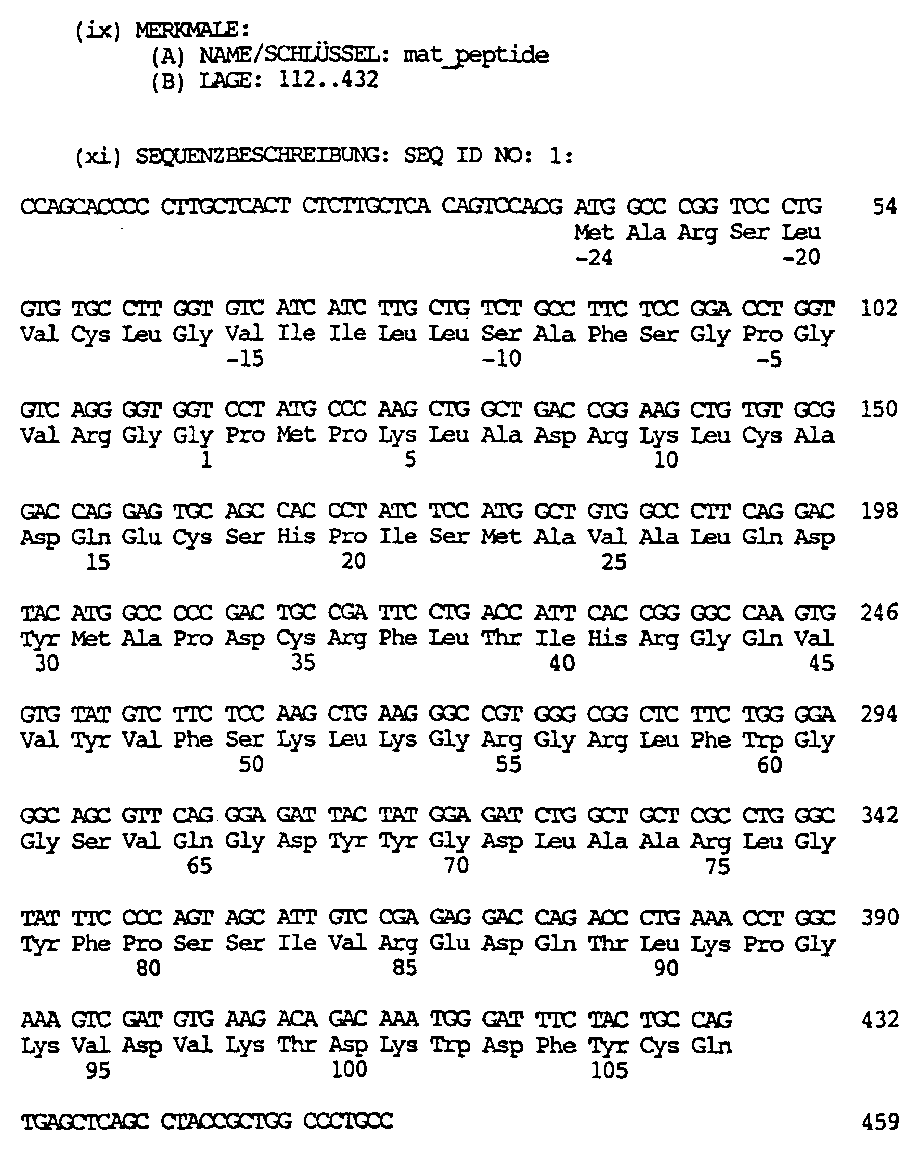
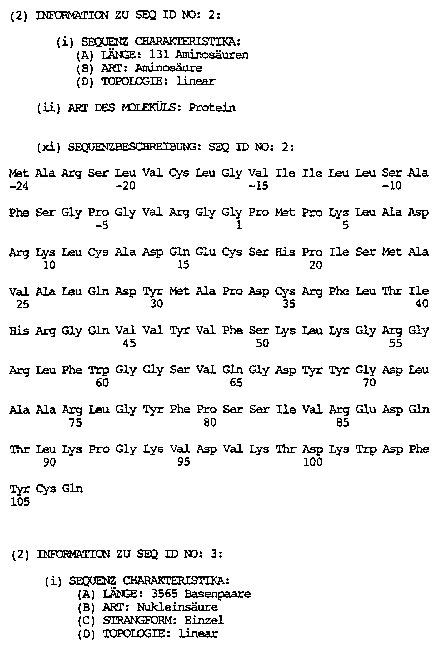


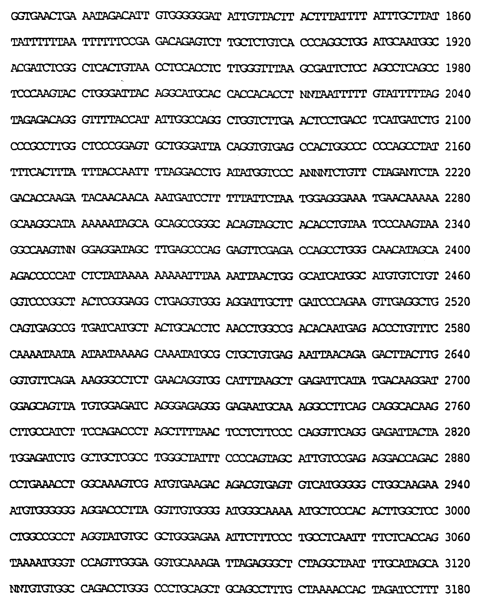


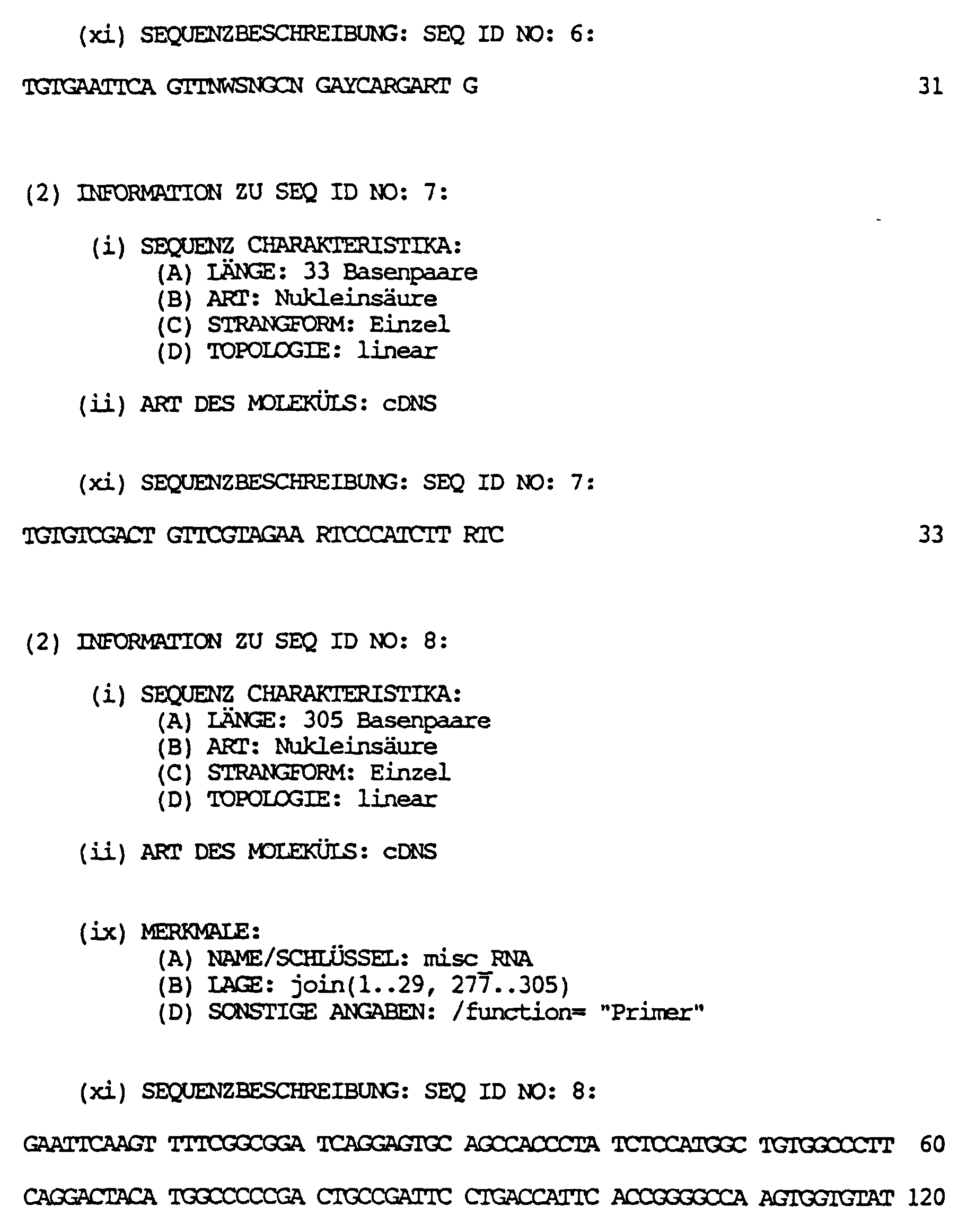


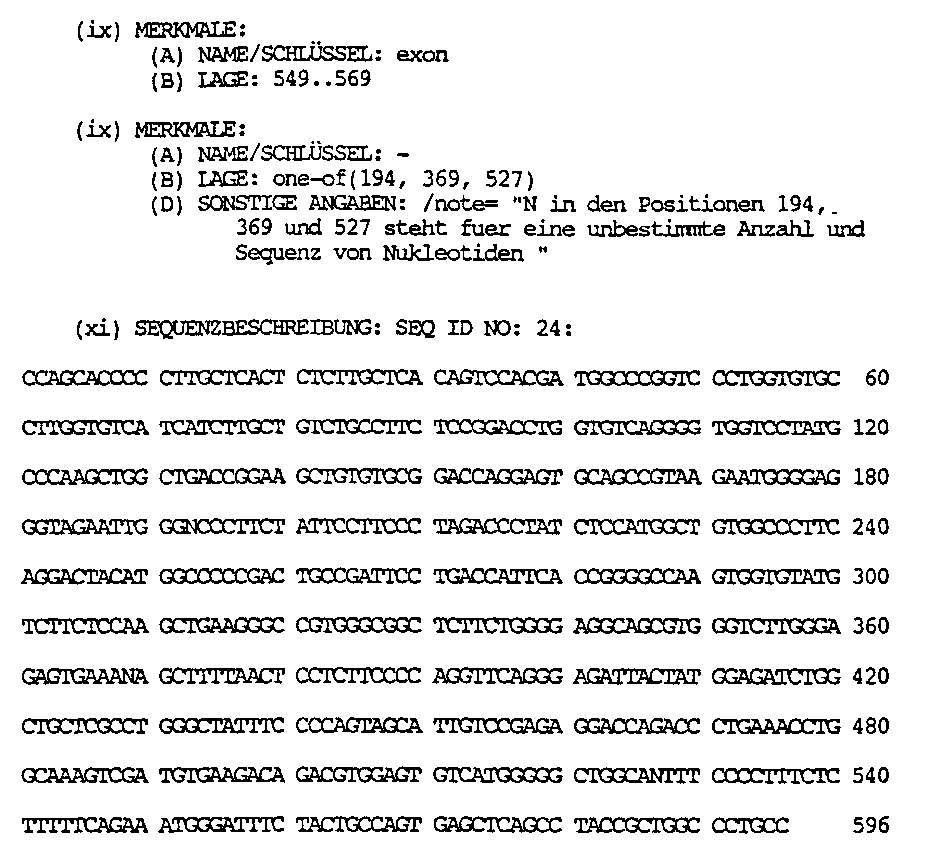
Claims (3)
Applications Claiming Priority (3)
| Application Number | Priority Date | Filing Date | Title |
|---|---|---|---|
| DE4324247 | 1993-07-20 | ||
| DE4324247 | 1993-07-20 | ||
| EP94925364A EP0710248B1 (en) | 1993-07-20 | 1994-07-19 | Melanoma-inhibiting protein |
Related Parent Applications (1)
| Application Number | Title | Priority Date | Filing Date |
|---|---|---|---|
| EP94925364.5 Division | 1995-03-10 |
Publications (2)
| Publication Number | Publication Date |
|---|---|
| EP0947583A2 true EP0947583A2 (en) | 1999-10-06 |
| EP0947583A3 EP0947583A3 (en) | 2000-05-03 |
Family
ID=6493222
Family Applications (2)
| Application Number | Title | Priority Date | Filing Date |
|---|---|---|---|
| EP94925364A Expired - Lifetime EP0710248B1 (en) | 1993-07-20 | 1994-07-19 | Melanoma-inhibiting protein |
| EP99101554A Withdrawn EP0947583A3 (en) | 1993-07-20 | 1994-07-19 | DNA fragments activated in expression of melanoma inhibiting protein (MIA) |
Family Applications Before (1)
| Application Number | Title | Priority Date | Filing Date |
|---|---|---|---|
| EP94925364A Expired - Lifetime EP0710248B1 (en) | 1993-07-20 | 1994-07-19 | Melanoma-inhibiting protein |
Country Status (14)
| Country | Link |
|---|---|
| US (1) | US5770366A (en) |
| EP (2) | EP0710248B1 (en) |
| JP (1) | JP3435163B2 (en) |
| CN (1) | CN1056617C (en) |
| AT (1) | ATE185152T1 (en) |
| AU (1) | AU7531294A (en) |
| CA (1) | CA2167693C (en) |
| DE (2) | DE4425481A1 (en) |
| DK (1) | DK0710248T3 (en) |
| ES (1) | ES2139751T3 (en) |
| GR (1) | GR3032212T3 (en) |
| IL (2) | IL154248A (en) |
| WO (1) | WO1995003328A2 (en) |
| ZA (1) | ZA945278B (en) |
Families Citing this family (20)
| Publication number | Priority date | Publication date | Assignee | Title |
|---|---|---|---|---|
| DE19653358A1 (en) * | 1996-12-20 | 1998-06-25 | Boehringer Mannheim Gmbh | Immunoassay for the detection of MIA |
| EP0972022A2 (en) | 1997-01-21 | 2000-01-19 | Human Genome Sciences, Inc. | Polynucleotides and polypeptides encoding receptors |
| USRE39920E1 (en) | 1997-05-30 | 2007-11-13 | Xenomics, Inc. | Methods for detection of nucleic acid sequences in urine |
| US6492144B1 (en) * | 1997-05-30 | 2002-12-10 | Diagen Corporation | Methods for detection of nucleic acid sequences in urine |
| EP0945507A1 (en) * | 1998-03-27 | 1999-09-29 | Boehringer Mannheim Gmbh | Tumor specific expression controll region and its use |
| US20020103360A1 (en) * | 1998-09-01 | 2002-08-01 | Yang Pan | Novel protein related to melanoma-inhibiting protein and uses thereof |
| US20030072767A1 (en) * | 1998-09-30 | 2003-04-17 | Alexander Gaiger | Compositions and methods for WT1 specific immunotherapy |
| EP1025871A1 (en) * | 1999-01-28 | 2000-08-09 | F. Hoffmann-La Roche Ag | Use of a melanoma inhibiting activity factor (MIA) for cartilage and bone repair |
| US6713293B1 (en) * | 1999-02-08 | 2004-03-30 | Friedrich Grummt | Encapsulated cells containing an amplified expression vector as a drug delivery device |
| EP1191099B1 (en) * | 1999-06-30 | 2009-10-07 | Takeda Pharmaceutical Company Limited | Novel polypeptide and dna thereof |
| WO2001055332A2 (en) * | 2000-01-25 | 2001-08-02 | Hyseq, Inc. | Methods and materials relating to novel growth regulatory-like polypeptides and polynucleotides |
| EP1133994A1 (en) * | 2000-03-11 | 2001-09-19 | Biognostik Gesellschaft für biomolekulare Diagnostik mbH | A method for reversing the immunosuppressive effects of the Melanoma Inhibitory Activity "MIA" |
| SK13692002A3 (en) * | 2000-03-23 | 2003-02-04 | Akzo Nobel N. V. | Use of MIA in immunotherapy |
| US6613534B2 (en) | 2001-03-20 | 2003-09-02 | Wake Forest University Health Sciences | MAP-2 as a determinant of metastatic potential |
| US20100212037A1 (en) * | 2002-10-16 | 2010-08-19 | Anja Katrin Bosserhoff | Mia-2 protein |
| KR20090046893A (en) * | 2006-07-28 | 2009-05-11 | 노파르티스 아게 | Use of melanoma inhibitory activity (mia) protein as an early indicator for therapeutic response in melanoma |
| ES2660785T3 (en) * | 2007-08-22 | 2018-03-26 | Trovagene, Inc. | Methods of using mRNA for the detection of cell death in vivo |
| KR101880905B1 (en) | 2010-04-27 | 2018-07-23 | 에스씨아이엘 테크놀로지 게엠베하 | Stable aqueous MIA/CD-RAP formulations |
| CA2807431A1 (en) * | 2010-08-30 | 2012-03-08 | F. Hoffmann-La Roche Ag | Prokaryotic expression construct |
| ITRM20110134A1 (en) * | 2011-03-22 | 2012-09-23 | Matteo Bordignon | INHIBITORS OF MIA (MELANOMA INHIBITORY ACTIVITY) TO IDENTIFY, PREVENT AND TREAT VITILIGINE |
-
1994
- 1994-07-19 DE DE4425481A patent/DE4425481A1/en not_active Withdrawn
- 1994-07-19 EP EP94925364A patent/EP0710248B1/en not_active Expired - Lifetime
- 1994-07-19 DE DE59408791T patent/DE59408791D1/en not_active Expired - Lifetime
- 1994-07-19 JP JP50493195A patent/JP3435163B2/en not_active Expired - Lifetime
- 1994-07-19 AU AU75312/94A patent/AU7531294A/en not_active Abandoned
- 1994-07-19 WO PCT/EP1994/002369 patent/WO1995003328A2/en active IP Right Grant
- 1994-07-19 ES ES94925364T patent/ES2139751T3/en not_active Expired - Lifetime
- 1994-07-19 AT AT94925364T patent/ATE185152T1/en active
- 1994-07-19 CA CA002167693A patent/CA2167693C/en not_active Expired - Lifetime
- 1994-07-19 US US08/578,649 patent/US5770366A/en not_active Expired - Lifetime
- 1994-07-19 CN CN94192822A patent/CN1056617C/en not_active Expired - Lifetime
- 1994-07-19 ZA ZA945278A patent/ZA945278B/en unknown
- 1994-07-19 EP EP99101554A patent/EP0947583A3/en not_active Withdrawn
- 1994-07-19 DK DK94925364T patent/DK0710248T3/en active
- 1994-07-20 IL IL154248A patent/IL154248A/en not_active IP Right Cessation
- 1994-07-20 IL IL11038594A patent/IL110385A/en not_active IP Right Cessation
-
1999
- 1999-12-22 GR GR990403299T patent/GR3032212T3/en unknown
Non-Patent Citations (2)
| Title |
|---|
| R.APFEL ET AL.: "Purification and analysis of growth regulating proteins secreted by a human melanoma cell line" MELANOMA RES., Bd. 2, Nr. 5-6, 1992, Seiten 327-36, XP002131794 * |
| U.BOGDAHN ET AL.: "Autocrine Tumor Cell Growth-inhibiting Activities from Human Malignant Melanoma" CANCER RESEARCH, Bd. 49, 1989, Seiten 5358-5363, XP002131793 * |
Also Published As
| Publication number | Publication date |
|---|---|
| IL154248A0 (en) | 2003-09-17 |
| ATE185152T1 (en) | 1999-10-15 |
| EP0947583A3 (en) | 2000-05-03 |
| CN1133049A (en) | 1996-10-09 |
| WO1995003328A3 (en) | 1995-03-16 |
| CN1056617C (en) | 2000-09-20 |
| IL154248A (en) | 2008-08-07 |
| ZA945278B (en) | 1996-01-19 |
| EP0710248B1 (en) | 1999-09-29 |
| EP0710248A1 (en) | 1996-05-08 |
| DE4425481A1 (en) | 1995-03-02 |
| GR3032212T3 (en) | 2000-04-27 |
| IL110385A0 (en) | 1994-10-21 |
| DE59408791D1 (en) | 1999-11-04 |
| AU7531294A (en) | 1995-02-20 |
| IL110385A (en) | 2003-12-10 |
| CA2167693A1 (en) | 1995-02-02 |
| JPH09500531A (en) | 1997-01-21 |
| ES2139751T3 (en) | 2000-02-16 |
| US5770366A (en) | 1998-06-23 |
| CA2167693C (en) | 2007-01-30 |
| WO1995003328A2 (en) | 1995-02-02 |
| JP3435163B2 (en) | 2003-08-11 |
| DK0710248T3 (en) | 2000-04-10 |
Similar Documents
| Publication | Publication Date | Title |
|---|---|---|
| EP0710248B1 (en) | Melanoma-inhibiting protein | |
| DE60121808T2 (en) | USE OF A POLYPEPTIDE CONTAINING THE EXTRACELLULAR DOMAIN OF IL-20RA AND IL-20RB FOR THE TREATMENT OF INFLAMMATION | |
| DE69834852T2 (en) | NL3 TIE receptor tyrosine kinase ligand homologue | |
| DE3852255T3 (en) | Tumor necrosis factor-inhibiting protein and its purification | |
| DE69534733T2 (en) | NEW ON p53 responsive GENES | |
| EP0417563B1 (en) | TNF-binding proteins | |
| WO1996001316A1 (en) | NOVEL GROWTH OR DIFFERENTIATION FACTOR OF THE TGF-β FAMILY | |
| DE69734397T2 (en) | TUMOR NEKROSIS FACTOR SIMILAR LIGAND | |
| EP0892047A2 (en) | Human and murine semaphorin L | |
| DE69434997T2 (en) | Human chondromodulin I protein | |
| US5888764A (en) | Human fas gene promoter region | |
| DE69535316T2 (en) | AS LERK6 DESIGNATED CYTOKIN | |
| WO1996022370A9 (en) | Human fas gene promoter region | |
| CA2251262C (en) | Nucleic acid encoding a nervous tissue sodium channel | |
| DE69836278T2 (en) | ALL FORMS OF HUMAN STAT3 | |
| EP1237910B1 (en) | Trp-protein-related mtr1 protein and dna sequence coding therefor | |
| WO1998045428A1 (en) | Fanconi-gen ii | |
| CA2323506C (en) | Mammalian blood loss-induced gene, kd312 | |
| DE60217081T2 (en) | GENES PARTICIPATED IN V (D) J RECOMBINATION AND / OR DNA REPAIR | |
| DE69636737T2 (en) | NEW IMMUNOREGULATORY PROTEIN LST-1 | |
| EP0613498A1 (en) | Receptor derivates with binding sites for human rhinoviruses | |
| WO2002057433A9 (en) | Homeodomain-interacting protein kinases and the use of the same for influencing cell division and cell proliferation | |
| JP2000514654A (en) | Pituitary differentiation factor and method of use | |
| WO2000046244A1 (en) | cDNA SEQUENCE OF AN INTERACTOR FANCIP1 OF THE FANCONI ANAEMIA PROTEIN OF COMPLEMENTATION GROUP A | |
| EP0939806A1 (en) | Fanconi gene 1 |
Legal Events
| Date | Code | Title | Description |
|---|---|---|---|
| PUAI | Public reference made under article 153(3) epc to a published international application that has entered the european phase |
Free format text: ORIGINAL CODE: 0009012 |
|
| AC | Divisional application: reference to earlier application |
Ref document number: 710248 Country of ref document: EP |
|
| AK | Designated contracting states |
Kind code of ref document: A2 Designated state(s): AT BE CH DE DK ES FR GB GR IE IT LI LU NL PT SE |
|
| RIN1 | Information on inventor provided before grant (corrected) |
Inventor name: KALUZA, BRIGITTE, DR. Inventor name: BUETTNER, REINHARD, DR. Inventor name: BOGDAHN, ULRICH, PROF.DR. |
|
| PUAL | Search report despatched |
Free format text: ORIGINAL CODE: 0009013 |
|
| RIN1 | Information on inventor provided before grant (corrected) |
Inventor name: KALUZA, BRIGITTE, DR. Inventor name: BUETTNER, REINHARD, DR. Inventor name: BOGDAHN, ULRICH, PROF.DR. |
|
| RIC1 | Information provided on ipc code assigned before grant |
Free format text: 7C 07K 14/52 A, 7C 12N 15/07 B, 7C 12N 15/19 B, 7A 61K 38/22 B, 7C 07K 14/47 B |
|
| AK | Designated contracting states |
Kind code of ref document: A3 Designated state(s): AT BE CH DE DK ES FR GB GR IE IT LI LU NL PT SE |
|
| RIN1 | Information on inventor provided before grant (corrected) |
Inventor name: KALUZA, BRIGITTE, DR. Inventor name: BUETTNER, REINHARD, DR. Inventor name: BOGDAHN, ULRICH, PROF.DR. |
|
| 17P | Request for examination filed |
Effective date: 20001103 |
|
| 17Q | First examination report despatched |
Effective date: 20010607 |
|
| STAA | Information on the status of an ep patent application or granted ep patent |
Free format text: STATUS: THE APPLICATION IS DEEMED TO BE WITHDRAWN |
|
| 18D | Application deemed to be withdrawn |
Effective date: 20011018 |