EP0910357B1 - Down-regulation of dna repair to enhance sensitivity to p53-mediated suppression - Google Patents
Down-regulation of dna repair to enhance sensitivity to p53-mediated suppression Download PDFInfo
- Publication number
- EP0910357B1 EP0910357B1 EP97933543A EP97933543A EP0910357B1 EP 0910357 B1 EP0910357 B1 EP 0910357B1 EP 97933543 A EP97933543 A EP 97933543A EP 97933543 A EP97933543 A EP 97933543A EP 0910357 B1 EP0910357 B1 EP 0910357B1
- Authority
- EP
- European Patent Office
- Prior art keywords
- dna
- cell
- cells
- gene
- tumor
- Prior art date
- Legal status (The legal status is an assumption and is not a legal conclusion. Google has not performed a legal analysis and makes no representation as to the accuracy of the status listed.)
- Expired - Lifetime
Links
- 102100025064 Cellular tumor antigen p53 Human genes 0.000 title claims abstract description 195
- 101000721661 Homo sapiens Cellular tumor antigen p53 Proteins 0.000 title claims abstract description 194
- 230000001404 mediated effect Effects 0.000 title claims abstract description 39
- 230000032147 negative regulation of DNA repair Effects 0.000 title description 8
- 230000001629 suppression Effects 0.000 title description 6
- 230000035945 sensitivity Effects 0.000 title description 4
- 210000004027 cell Anatomy 0.000 claims abstract description 260
- 206010028980 Neoplasm Diseases 0.000 claims abstract description 79
- 238000011282 treatment Methods 0.000 claims abstract description 68
- 210000004881 tumor cell Anatomy 0.000 claims abstract description 66
- 230000033616 DNA repair Effects 0.000 claims abstract description 65
- 238000000034 method Methods 0.000 claims abstract description 64
- 208000005017 glioblastoma Diseases 0.000 claims abstract description 45
- 230000006907 apoptotic process Effects 0.000 claims abstract description 37
- 201000011510 cancer Diseases 0.000 claims abstract description 33
- 206010006187 Breast cancer Diseases 0.000 claims abstract description 18
- 208000026310 Breast neoplasm Diseases 0.000 claims abstract description 18
- 108090000623 proteins and genes Proteins 0.000 claims description 94
- 230000014509 gene expression Effects 0.000 claims description 75
- 229960004316 cisplatin Drugs 0.000 claims description 66
- DQLATGHUWYMOKM-UHFFFAOYSA-L cisplatin Chemical compound N[Pt](N)(Cl)Cl DQLATGHUWYMOKM-UHFFFAOYSA-L 0.000 claims description 64
- 239000003795 chemical substances by application Substances 0.000 claims description 56
- 239000012623 DNA damaging agent Substances 0.000 claims description 41
- GSCPDZHWVNUUFI-UHFFFAOYSA-N 3-aminobenzamide Chemical group NC(=O)C1=CC=CC(N)=C1 GSCPDZHWVNUUFI-UHFFFAOYSA-N 0.000 claims description 23
- 102100023132 Transcription factor Jun Human genes 0.000 claims description 22
- 230000002401 inhibitory effect Effects 0.000 claims description 22
- 102000004169 proteins and genes Human genes 0.000 claims description 22
- -1 Ref-1 Proteins 0.000 claims description 19
- 238000002347 injection Methods 0.000 claims description 15
- 239000007924 injection Substances 0.000 claims description 15
- AOJJSUZBOXZQNB-TZSSRYMLSA-N Doxorubicin Chemical compound O([C@H]1C[C@@](O)(CC=2C(O)=C3C(=O)C=4C=CC=C(C=4C(=O)C3=C(O)C=21)OC)C(=O)CO)[C@H]1C[C@H](N)[C@H](O)[C@H](C)O1 AOJJSUZBOXZQNB-TZSSRYMLSA-N 0.000 claims description 14
- 230000006698 induction Effects 0.000 claims description 14
- 102000039446 nucleic acids Human genes 0.000 claims description 12
- 108020004707 nucleic acids Proteins 0.000 claims description 12
- 150000007523 nucleic acids Chemical class 0.000 claims description 12
- 102000012338 Poly(ADP-ribose) Polymerases Human genes 0.000 claims description 11
- 108010061844 Poly(ADP-ribose) Polymerases Proteins 0.000 claims description 11
- 229920000776 Poly(Adenosine diphosphate-ribose) polymerase Polymers 0.000 claims description 11
- 230000010076 replication Effects 0.000 claims description 11
- 150000004492 retinoid derivatives Chemical class 0.000 claims description 11
- 108010060248 DNA Ligase ATP Proteins 0.000 claims description 8
- RJURFGZVJUQBHK-UHFFFAOYSA-N actinomycin D Natural products CC1OC(=O)C(C(C)C)N(C)C(=O)CN(C)C(=O)C2CCCN2C(=O)C(C(C)C)NC(=O)C1NC(=O)C1=C(N)C(=O)C(C)=C2OC(C(C)=CC=C3C(=O)NC4C(=O)NC(C(N5CCCC5C(=O)N(C)CC(=O)N(C)C(C(C)C)C(=O)OC4C)=O)C(C)C)=C3N=C21 RJURFGZVJUQBHK-UHFFFAOYSA-N 0.000 claims description 8
- 101001050288 Homo sapiens Transcription factor Jun Proteins 0.000 claims description 7
- 108010040002 Tumor Suppressor Proteins Proteins 0.000 claims description 7
- 102000001742 Tumor Suppressor Proteins Human genes 0.000 claims description 7
- 239000003814 drug Substances 0.000 claims description 7
- 208000020816 lung neoplasm Diseases 0.000 claims description 7
- 208000003174 Brain Neoplasms Diseases 0.000 claims description 6
- 102000003915 DNA Topoisomerases Human genes 0.000 claims description 6
- 108090000323 DNA Topoisomerases Proteins 0.000 claims description 6
- 210000003527 eukaryotic cell Anatomy 0.000 claims description 6
- 230000002601 intratumoral effect Effects 0.000 claims description 6
- 238000004519 manufacturing process Methods 0.000 claims description 6
- 230000004936 stimulating effect Effects 0.000 claims description 6
- 102000008158 DNA Ligase ATP Human genes 0.000 claims description 5
- 102100027584 Protein c-Fos Human genes 0.000 claims description 5
- 108010071563 Proto-Oncogene Proteins c-fos Proteins 0.000 claims description 5
- 230000001419 dependent effect Effects 0.000 claims description 5
- 201000001441 melanoma Diseases 0.000 claims description 5
- 206010041823 squamous cell carcinoma Diseases 0.000 claims description 5
- 108020001738 DNA Glycosylase Proteins 0.000 claims description 4
- 102000028381 DNA glycosylase Human genes 0.000 claims description 4
- 102000005768 DNA-Activated Protein Kinase Human genes 0.000 claims description 4
- 108010006124 DNA-Activated Protein Kinase Proteins 0.000 claims description 4
- 108010092160 Dactinomycin Proteins 0.000 claims description 4
- 101150009243 HAP1 gene Proteins 0.000 claims description 4
- NWIBSHFKIJFRCO-WUDYKRTCSA-N Mytomycin Chemical compound C1N2C(C(C(C)=C(N)C3=O)=O)=C3[C@@H](COC(N)=O)[C@@]2(OC)[C@@H]2[C@H]1N2 NWIBSHFKIJFRCO-WUDYKRTCSA-N 0.000 claims description 4
- 102000006943 Uracil-DNA Glycosidase Human genes 0.000 claims description 4
- 108010072685 Uracil-DNA Glycosidase Proteins 0.000 claims description 4
- RJURFGZVJUQBHK-IIXSONLDSA-N actinomycin D Chemical compound C[C@H]1OC(=O)[C@H](C(C)C)N(C)C(=O)CN(C)C(=O)[C@@H]2CCCN2C(=O)[C@@H](C(C)C)NC(=O)[C@H]1NC(=O)C1=C(N)C(=O)C(C)=C2OC(C(C)=CC=C3C(=O)N[C@@H]4C(=O)N[C@@H](C(N5CCC[C@H]5C(=O)N(C)CC(=O)N(C)[C@@H](C(C)C)C(=O)O[C@@H]4C)=O)C(C)C)=C3N=C21 RJURFGZVJUQBHK-IIXSONLDSA-N 0.000 claims description 4
- 230000000692 anti-sense effect Effects 0.000 claims description 4
- 229960000640 dactinomycin Drugs 0.000 claims description 4
- 229960004679 doxorubicin Drugs 0.000 claims description 4
- 208000037841 lung tumor Diseases 0.000 claims description 4
- STQGQHZAVUOBTE-UHFFFAOYSA-N 7-Cyan-hept-2t-en-4,6-diinsaeure Natural products C1=2C(O)=C3C(=O)C=4C(OC)=CC=CC=4C(=O)C3=C(O)C=2CC(O)(C(C)=O)CC1OC1CC(N)C(O)C(C)O1 STQGQHZAVUOBTE-UHFFFAOYSA-N 0.000 claims description 3
- 108010001132 DNA Polymerase beta Proteins 0.000 claims description 3
- 102100034157 DNA mismatch repair protein Msh2 Human genes 0.000 claims description 3
- 206010019695 Hepatic neoplasm Diseases 0.000 claims description 3
- 101001134036 Homo sapiens DNA mismatch repair protein Msh2 Proteins 0.000 claims description 3
- ZDZOTLJHXYCWBA-VCVYQWHSSA-N N-debenzoyl-N-(tert-butoxycarbonyl)-10-deacetyltaxol Chemical compound O([C@H]1[C@H]2[C@@](C([C@H](O)C3=C(C)[C@@H](OC(=O)[C@H](O)[C@@H](NC(=O)OC(C)(C)C)C=4C=CC=CC=4)C[C@]1(O)C3(C)C)=O)(C)[C@@H](O)C[C@H]1OC[C@]12OC(=O)C)C(=O)C1=CC=CC=C1 ZDZOTLJHXYCWBA-VCVYQWHSSA-N 0.000 claims description 3
- 229930012538 Paclitaxel Natural products 0.000 claims description 3
- 206010039491 Sarcoma Diseases 0.000 claims description 3
- 208000000453 Skin Neoplasms Diseases 0.000 claims description 3
- 229940045799 anthracyclines and related substance Drugs 0.000 claims description 3
- 229960004562 carboplatin Drugs 0.000 claims description 3
- 229960004630 chlorambucil Drugs 0.000 claims description 3
- JCKYGMPEJWAADB-UHFFFAOYSA-N chlorambucil Chemical compound OC(=O)CCCC1=CC=C(N(CCCl)CCCl)C=C1 JCKYGMPEJWAADB-UHFFFAOYSA-N 0.000 claims description 3
- 208000029742 colonic neoplasm Diseases 0.000 claims description 3
- STQGQHZAVUOBTE-VGBVRHCVSA-N daunorubicin Chemical compound O([C@H]1C[C@@](O)(CC=2C(O)=C3C(=O)C=4C=CC=C(C=4C(=O)C3=C(O)C=21)OC)C(C)=O)[C@H]1C[C@H](N)[C@H](O)[C@H](C)O1 STQGQHZAVUOBTE-VGBVRHCVSA-N 0.000 claims description 3
- 229960000975 daunorubicin Drugs 0.000 claims description 3
- 229960001101 ifosfamide Drugs 0.000 claims description 3
- HOMGKSMUEGBAAB-UHFFFAOYSA-N ifosfamide Chemical compound ClCCNP1(=O)OCCCN1CCCl HOMGKSMUEGBAAB-UHFFFAOYSA-N 0.000 claims description 3
- 230000005865 ionizing radiation Effects 0.000 claims description 3
- 208000014018 liver neoplasm Diseases 0.000 claims description 3
- 229960001924 melphalan Drugs 0.000 claims description 3
- SGDBTWWWUNNDEQ-LBPRGKRZSA-N melphalan Chemical compound OC(=O)[C@@H](N)CC1=CC=C(N(CCCl)CCCl)C=C1 SGDBTWWWUNNDEQ-LBPRGKRZSA-N 0.000 claims description 3
- CFCUWKMKBJTWLW-BKHRDMLASA-N mithramycin Chemical compound O([C@@H]1C[C@@H](O[C@H](C)[C@H]1O)OC=1C=C2C=C3C[C@H]([C@@H](C(=O)C3=C(O)C2=C(O)C=1C)O[C@@H]1O[C@H](C)[C@@H](O)[C@H](O[C@@H]2O[C@H](C)[C@H](O)[C@H](O[C@@H]3O[C@H](C)[C@@H](O)[C@@](C)(O)C3)C2)C1)[C@H](OC)C(=O)[C@@H](O)[C@@H](C)O)[C@H]1C[C@@H](O)[C@H](O)[C@@H](C)O1 CFCUWKMKBJTWLW-BKHRDMLASA-N 0.000 claims description 3
- 229960004857 mitomycin Drugs 0.000 claims description 3
- OSTGTTZJOCZWJG-UHFFFAOYSA-N nitrosourea Chemical compound NC(=O)N=NO OSTGTTZJOCZWJG-UHFFFAOYSA-N 0.000 claims description 3
- 229960001592 paclitaxel Drugs 0.000 claims description 3
- 230000010412 perfusion Effects 0.000 claims description 3
- 229960003171 plicamycin Drugs 0.000 claims description 3
- CPTBDICYNRMXFX-UHFFFAOYSA-N procarbazine Chemical compound CNNCC1=CC=C(C(=O)NC(C)C)C=C1 CPTBDICYNRMXFX-UHFFFAOYSA-N 0.000 claims description 3
- 229960000624 procarbazine Drugs 0.000 claims description 3
- 208000023958 prostate neoplasm Diseases 0.000 claims description 3
- RCINICONZNJXQF-MZXODVADSA-N taxol Chemical compound O([C@@H]1[C@@]2(C[C@@H](C(C)=C(C2(C)C)[C@H](C([C@]2(C)[C@@H](O)C[C@H]3OC[C@]3([C@H]21)OC(C)=O)=O)OC(=O)C)OC(=O)[C@H](O)[C@@H](NC(=O)C=1C=CC=CC=1)C=1C=CC=CC=1)O)C(=O)C1=CC=CC=C1 RCINICONZNJXQF-MZXODVADSA-N 0.000 claims description 3
- 229940063683 taxotere Drugs 0.000 claims description 3
- NRUKOCRGYNPUPR-QBPJDGROSA-N teniposide Chemical compound COC1=C(O)C(OC)=CC([C@@H]2C3=CC=4OCOC=4C=C3[C@@H](O[C@H]3[C@@H]([C@@H](O)[C@@H]4O[C@@H](OC[C@H]4O3)C=3SC=CC=3)O)[C@@H]3[C@@H]2C(OC3)=O)=C1 NRUKOCRGYNPUPR-QBPJDGROSA-N 0.000 claims description 3
- 229960001278 teniposide Drugs 0.000 claims description 3
- 108010006654 Bleomycin Proteins 0.000 claims description 2
- CMSMOCZEIVJLDB-UHFFFAOYSA-N Cyclophosphamide Chemical compound ClCCN(CCCl)P1(=O)NCCCO1 CMSMOCZEIVJLDB-UHFFFAOYSA-N 0.000 claims description 2
- 108010068250 Herpes Simplex Virus Protein Vmw65 Proteins 0.000 claims description 2
- 208000008839 Kidney Neoplasms Diseases 0.000 claims description 2
- 229930192392 Mitomycin Natural products 0.000 claims description 2
- 206010029098 Neoplasm skin Diseases 0.000 claims description 2
- 206010029260 Neuroblastoma Diseases 0.000 claims description 2
- 206010061535 Ovarian neoplasm Diseases 0.000 claims description 2
- 102100040250 Transcription elongation factor A protein-like 1 Human genes 0.000 claims description 2
- 229960001561 bleomycin Drugs 0.000 claims description 2
- OYVAGSVQBOHSSS-UAPAGMARSA-O bleomycin A2 Chemical compound N([C@H](C(=O)N[C@H](C)[C@@H](O)[C@H](C)C(=O)N[C@@H]([C@H](O)C)C(=O)NCCC=1SC=C(N=1)C=1SC=C(N=1)C(=O)NCCC[S+](C)C)[C@@H](O[C@H]1[C@H]([C@@H](O)[C@H](O)[C@H](CO)O1)O[C@@H]1[C@H]([C@@H](OC(N)=O)[C@H](O)[C@@H](CO)O1)O)C=1N=CNC=1)C(=O)C1=NC([C@H](CC(N)=O)NC[C@H](N)C(N)=O)=NC(N)=C1C OYVAGSVQBOHSSS-UAPAGMARSA-O 0.000 claims description 2
- 229960004397 cyclophosphamide Drugs 0.000 claims description 2
- 208000032839 leukemia Diseases 0.000 claims description 2
- 208000003154 papilloma Diseases 0.000 claims description 2
- 102000001996 DNA Polymerase beta Human genes 0.000 claims 1
- 108700039887 Essential Genes Proteins 0.000 claims 1
- 190000008236 carboplatin Chemical compound 0.000 claims 1
- 125000002678 retinoid group Chemical group 0.000 claims 1
- 239000003112 inhibitor Substances 0.000 abstract description 40
- 108700025694 p53 Genes Proteins 0.000 abstract description 20
- 238000001415 gene therapy Methods 0.000 abstract description 19
- 206010053759 Growth retardation Diseases 0.000 abstract description 10
- 230000006882 induction of apoptosis Effects 0.000 abstract description 6
- 108020004414 DNA Proteins 0.000 description 84
- 241000701161 unidentified adenovirus Species 0.000 description 61
- 230000005778 DNA damage Effects 0.000 description 48
- 231100000277 DNA damage Toxicity 0.000 description 48
- 239000013598 vector Substances 0.000 description 40
- 238000012546 transfer Methods 0.000 description 38
- 230000000694 effects Effects 0.000 description 36
- 239000000203 mixture Substances 0.000 description 34
- 230000035899 viability Effects 0.000 description 29
- 150000001875 compounds Chemical class 0.000 description 27
- 239000013604 expression vector Substances 0.000 description 26
- 102200082402 rs751610198 Human genes 0.000 description 26
- 241000700159 Rattus Species 0.000 description 24
- 238000012384 transportation and delivery Methods 0.000 description 23
- 238000003556 assay Methods 0.000 description 22
- 208000015181 infectious disease Diseases 0.000 description 22
- 210000001519 tissue Anatomy 0.000 description 22
- 239000000427 antigen Substances 0.000 description 20
- 108091007433 antigens Proteins 0.000 description 20
- 102000036639 antigens Human genes 0.000 description 20
- 238000001727 in vivo Methods 0.000 description 20
- 230000035772 mutation Effects 0.000 description 20
- 235000018102 proteins Nutrition 0.000 description 20
- 230000006870 function Effects 0.000 description 19
- 239000002502 liposome Substances 0.000 description 19
- 241000700605 Viruses Species 0.000 description 18
- 230000012010 growth Effects 0.000 description 18
- 239000000463 material Substances 0.000 description 18
- 230000003612 virological effect Effects 0.000 description 18
- 230000010261 cell growth Effects 0.000 description 16
- 102000040430 polynucleotide Human genes 0.000 description 16
- 108091033319 polynucleotide Proteins 0.000 description 16
- 239000002157 polynucleotide Substances 0.000 description 16
- 108010018242 Transcription Factor AP-1 Proteins 0.000 description 15
- 230000027455 binding Effects 0.000 description 15
- 238000000338 in vitro Methods 0.000 description 15
- 239000000243 solution Substances 0.000 description 15
- 241001465754 Metazoa Species 0.000 description 14
- WQZGKKKJIJFFOK-FPRJBGLDSA-N beta-D-galactose Chemical compound OC[C@H]1O[C@@H](O)[C@H](O)[C@@H](O)[C@H]1O WQZGKKKJIJFFOK-FPRJBGLDSA-N 0.000 description 14
- 230000001965 increasing effect Effects 0.000 description 14
- 108020003175 receptors Proteins 0.000 description 14
- 102000005962 receptors Human genes 0.000 description 14
- 238000002965 ELISA Methods 0.000 description 13
- 108010005774 beta-Galactosidase Proteins 0.000 description 13
- 239000013612 plasmid Substances 0.000 description 13
- 238000013518 transcription Methods 0.000 description 13
- 230000035897 transcription Effects 0.000 description 13
- 102220497176 Small vasohibin-binding protein_T47D_mutation Human genes 0.000 description 12
- LFQSCWFLJHTTHZ-UHFFFAOYSA-N Ethanol Chemical compound CCO LFQSCWFLJHTTHZ-UHFFFAOYSA-N 0.000 description 11
- 238000001514 detection method Methods 0.000 description 11
- 210000004185 liver Anatomy 0.000 description 11
- 230000002829 reductive effect Effects 0.000 description 11
- 239000000523 sample Substances 0.000 description 11
- 239000000126 substance Substances 0.000 description 11
- 230000001225 therapeutic effect Effects 0.000 description 11
- 241001430294 unidentified retrovirus Species 0.000 description 11
- 108010076525 DNA Repair Enzymes Proteins 0.000 description 10
- 230000008265 DNA repair mechanism Effects 0.000 description 10
- 230000005764 inhibitory process Effects 0.000 description 10
- 230000007246 mechanism Effects 0.000 description 10
- 238000011275 oncology therapy Methods 0.000 description 10
- 238000002560 therapeutic procedure Methods 0.000 description 10
- 230000015572 biosynthetic process Effects 0.000 description 9
- 239000003446 ligand Substances 0.000 description 9
- 239000000047 product Substances 0.000 description 9
- 230000001177 retroviral effect Effects 0.000 description 9
- 230000008685 targeting Effects 0.000 description 9
- 238000001890 transfection Methods 0.000 description 9
- 238000005406 washing Methods 0.000 description 9
- 108091032973 (ribonucleotides)n+m Proteins 0.000 description 8
- 102000011724 DNA Repair Enzymes Human genes 0.000 description 8
- 108010091358 Hypoxanthine Phosphoribosyltransferase Proteins 0.000 description 8
- 238000007792 addition Methods 0.000 description 8
- 239000002246 antineoplastic agent Substances 0.000 description 8
- 239000002585 base Substances 0.000 description 8
- 230000003833 cell viability Effects 0.000 description 8
- 230000001413 cellular effect Effects 0.000 description 8
- 230000007423 decrease Effects 0.000 description 8
- 239000003623 enhancer Substances 0.000 description 8
- 239000012634 fragment Substances 0.000 description 8
- 239000000499 gel Substances 0.000 description 8
- 239000002773 nucleotide Substances 0.000 description 8
- 125000003729 nucleotide group Chemical group 0.000 description 8
- 239000002245 particle Substances 0.000 description 8
- 238000002360 preparation method Methods 0.000 description 8
- 230000008439 repair process Effects 0.000 description 8
- 108010035563 Chloramphenicol O-acetyltransferase Proteins 0.000 description 7
- 102000004190 Enzymes Human genes 0.000 description 7
- 108090000790 Enzymes Proteins 0.000 description 7
- 241000699670 Mus sp. Species 0.000 description 7
- 239000007864 aqueous solution Substances 0.000 description 7
- 239000000872 buffer Substances 0.000 description 7
- 230000008859 change Effects 0.000 description 7
- 238000011161 development Methods 0.000 description 7
- 229940088598 enzyme Drugs 0.000 description 7
- 210000003494 hepatocyte Anatomy 0.000 description 7
- 238000011534 incubation Methods 0.000 description 7
- 230000001939 inductive effect Effects 0.000 description 7
- 210000004072 lung Anatomy 0.000 description 7
- 210000004962 mammalian cell Anatomy 0.000 description 7
- 239000002609 medium Substances 0.000 description 7
- 150000003839 salts Chemical class 0.000 description 7
- 238000012360 testing method Methods 0.000 description 7
- PEDCQBHIVMGVHV-UHFFFAOYSA-N Glycerine Chemical compound OCC(O)CO PEDCQBHIVMGVHV-UHFFFAOYSA-N 0.000 description 6
- DNIAPMSPPWPWGF-UHFFFAOYSA-N Propylene glycol Chemical compound CC(O)CO DNIAPMSPPWPWGF-UHFFFAOYSA-N 0.000 description 6
- 230000003321 amplification Effects 0.000 description 6
- 238000006243 chemical reaction Methods 0.000 description 6
- 238000002512 chemotherapy Methods 0.000 description 6
- 238000000576 coating method Methods 0.000 description 6
- 229940127089 cytotoxic agent Drugs 0.000 description 6
- 230000000254 damaging effect Effects 0.000 description 6
- 238000012217 deletion Methods 0.000 description 6
- 230000037430 deletion Effects 0.000 description 6
- 238000009472 formulation Methods 0.000 description 6
- 230000002068 genetic effect Effects 0.000 description 6
- 230000000977 initiatory effect Effects 0.000 description 6
- 238000003199 nucleic acid amplification method Methods 0.000 description 6
- PHEDXBVPIONUQT-RGYGYFBISA-N phorbol 13-acetate 12-myristate Chemical compound C([C@]1(O)C(=O)C(C)=C[C@H]1[C@@]1(O)[C@H](C)[C@H]2OC(=O)CCCCCCCCCCCCC)C(CO)=C[C@H]1[C@H]1[C@]2(OC(C)=O)C1(C)C PHEDXBVPIONUQT-RGYGYFBISA-N 0.000 description 6
- 239000002644 phorbol ester Substances 0.000 description 6
- 238000001959 radiotherapy Methods 0.000 description 6
- 230000001105 regulatory effect Effects 0.000 description 6
- 239000002904 solvent Substances 0.000 description 6
- 238000006467 substitution reaction Methods 0.000 description 6
- 108091003079 Bovine Serum Albumin Proteins 0.000 description 5
- 102000053602 DNA Human genes 0.000 description 5
- 102100033195 DNA ligase 4 Human genes 0.000 description 5
- GHASVSINZRGABV-UHFFFAOYSA-N Fluorouracil Chemical compound FC1=CNC(=O)NC1=O GHASVSINZRGABV-UHFFFAOYSA-N 0.000 description 5
- 108700020796 Oncogene Proteins 0.000 description 5
- 102000040945 Transcription factor Human genes 0.000 description 5
- 108091023040 Transcription factor Proteins 0.000 description 5
- 102000007537 Type II DNA Topoisomerases Human genes 0.000 description 5
- 108010046308 Type II DNA Topoisomerases Proteins 0.000 description 5
- 230000008901 benefit Effects 0.000 description 5
- 230000022131 cell cycle Effects 0.000 description 5
- 239000011248 coating agent Substances 0.000 description 5
- LOKCTEFSRHRXRJ-UHFFFAOYSA-I dipotassium trisodium dihydrogen phosphate hydrogen phosphate dichloride Chemical compound P(=O)(O)(O)[O-].[K+].P(=O)(O)([O-])[O-].[Na+].[Na+].[Cl-].[K+].[Cl-].[Na+] LOKCTEFSRHRXRJ-UHFFFAOYSA-I 0.000 description 5
- 208000037265 diseases, disorders, signs and symptoms Diseases 0.000 description 5
- 239000006185 dispersion Substances 0.000 description 5
- 229940079593 drug Drugs 0.000 description 5
- 229960002949 fluorouracil Drugs 0.000 description 5
- 238000001476 gene delivery Methods 0.000 description 5
- 210000005260 human cell Anatomy 0.000 description 5
- 230000010354 integration Effects 0.000 description 5
- 230000003902 lesion Effects 0.000 description 5
- 239000003550 marker Substances 0.000 description 5
- 238000004806 packaging method and process Methods 0.000 description 5
- 239000002953 phosphate buffered saline Substances 0.000 description 5
- 230000005855 radiation Effects 0.000 description 5
- 230000009467 reduction Effects 0.000 description 5
- 231100000419 toxicity Toxicity 0.000 description 5
- 230000001988 toxicity Effects 0.000 description 5
- 239000003981 vehicle Substances 0.000 description 5
- 241000701022 Cytomegalovirus Species 0.000 description 4
- YQYJSBFKSSDGFO-UHFFFAOYSA-N Epihygromycin Natural products OC1C(O)C(C(=O)C)OC1OC(C(=C1)O)=CC=C1C=C(C)C(=O)NC1C(O)C(O)C2OCOC2C1O YQYJSBFKSSDGFO-UHFFFAOYSA-N 0.000 description 4
- MHAJPDPJQMAIIY-UHFFFAOYSA-N Hydrogen peroxide Chemical compound OO MHAJPDPJQMAIIY-UHFFFAOYSA-N 0.000 description 4
- 238000000636 Northern blotting Methods 0.000 description 4
- YJQPYGGHQPGBLI-UHFFFAOYSA-N Novobiocin Natural products O1C(C)(C)C(OC)C(OC(N)=O)C(O)C1OC1=CC=C(C(O)=C(NC(=O)C=2C=C(CC=C(C)C)C(O)=CC=2)C(=O)O2)C2=C1C YJQPYGGHQPGBLI-UHFFFAOYSA-N 0.000 description 4
- 102000007981 Ornithine carbamoyltransferase Human genes 0.000 description 4
- ISWSIDIOOBJBQZ-UHFFFAOYSA-N Phenol Chemical compound OC1=CC=CC=C1 ISWSIDIOOBJBQZ-UHFFFAOYSA-N 0.000 description 4
- 238000002105 Southern blotting Methods 0.000 description 4
- 108700019146 Transgenes Proteins 0.000 description 4
- 108700025716 Tumor Suppressor Genes Proteins 0.000 description 4
- 102000044209 Tumor Suppressor Genes Human genes 0.000 description 4
- WTIJXIZOODAMJT-WBACWINTSA-N [(3r,4s,5r,6s)-5-hydroxy-6-[4-hydroxy-3-[[5-[[4-hydroxy-7-[(2s,3r,4s,5r)-3-hydroxy-5-methoxy-6,6-dimethyl-4-(5-methyl-1h-pyrrole-2-carbonyl)oxyoxan-2-yl]oxy-8-methyl-2-oxochromen-3-yl]carbamoyl]-4-methyl-1h-pyrrole-3-carbonyl]amino]-8-methyl-2-oxochromen- Chemical compound O([C@@H]1[C@H](C(O[C@H](OC=2C(=C3OC(=O)C(NC(=O)C=4C(=C(C(=O)NC=5C(OC6=C(C)C(O[C@@H]7[C@@H]([C@H](OC(=O)C=8NC(C)=CC=8)[C@@H](OC)C(C)(C)O7)O)=CC=C6C=5O)=O)NC=4)C)=C(O)C3=CC=2)C)[C@@H]1O)(C)C)OC)C(=O)C1=CC=C(C)N1 WTIJXIZOODAMJT-WBACWINTSA-N 0.000 description 4
- 239000004480 active ingredient Substances 0.000 description 4
- 235000004279 alanine Nutrition 0.000 description 4
- 238000004458 analytical method Methods 0.000 description 4
- 230000005775 apoptotic pathway Effects 0.000 description 4
- 238000013459 approach Methods 0.000 description 4
- 239000012736 aqueous medium Substances 0.000 description 4
- 239000002775 capsule Substances 0.000 description 4
- 238000005119 centrifugation Methods 0.000 description 4
- 238000002648 combination therapy Methods 0.000 description 4
- 230000006378 damage Effects 0.000 description 4
- 230000002950 deficient Effects 0.000 description 4
- 201000010099 disease Diseases 0.000 description 4
- 231100000673 dose–response relationship Toxicity 0.000 description 4
- 238000001962 electrophoresis Methods 0.000 description 4
- VJJPUSNTGOMMGY-MRVIYFEKSA-N etoposide Chemical compound COC1=C(O)C(OC)=CC([C@@H]2C3=CC=4OCOC=4C=C3[C@@H](O[C@H]3[C@@H]([C@@H](O)[C@@H]4O[C@H](C)OC[C@H]4O3)O)[C@@H]3[C@@H]2C(OC3)=O)=C1 VJJPUSNTGOMMGY-MRVIYFEKSA-N 0.000 description 4
- 238000003018 immunoassay Methods 0.000 description 4
- 238000001990 intravenous administration Methods 0.000 description 4
- 239000007788 liquid Substances 0.000 description 4
- YJQPYGGHQPGBLI-KGSXXDOSSA-N novobiocin Chemical compound O1C(C)(C)[C@H](OC)[C@@H](OC(N)=O)[C@@H](O)[C@@H]1OC1=CC=C(C(O)=C(NC(=O)C=2C=C(CC=C(C)C)C(O)=CC=2)C(=O)O2)C2=C1C YJQPYGGHQPGBLI-KGSXXDOSSA-N 0.000 description 4
- 229960002950 novobiocin Drugs 0.000 description 4
- 210000000056 organ Anatomy 0.000 description 4
- 230000026731 phosphorylation Effects 0.000 description 4
- 238000006366 phosphorylation reaction Methods 0.000 description 4
- 230000008488 polyadenylation Effects 0.000 description 4
- 239000000843 powder Substances 0.000 description 4
- 230000008569 process Effects 0.000 description 4
- 230000035755 proliferation Effects 0.000 description 4
- 230000022532 regulation of transcription, DNA-dependent Effects 0.000 description 4
- 230000004044 response Effects 0.000 description 4
- 230000002441 reversible effect Effects 0.000 description 4
- 238000003786 synthesis reaction Methods 0.000 description 4
- 231100000331 toxic Toxicity 0.000 description 4
- 230000002588 toxic effect Effects 0.000 description 4
- 230000002103 transcriptional effect Effects 0.000 description 4
- 241001515965 unidentified phage Species 0.000 description 4
- XLYOFNOQVPJJNP-UHFFFAOYSA-N water Substances O XLYOFNOQVPJJNP-UHFFFAOYSA-N 0.000 description 4
- 229920001817 Agar Polymers 0.000 description 3
- 108700028369 Alleles Proteins 0.000 description 3
- 102100033270 Cyclin-dependent kinase inhibitor 1 Human genes 0.000 description 3
- 108010079245 Cystic Fibrosis Transmembrane Conductance Regulator Proteins 0.000 description 3
- 230000004544 DNA amplification Effects 0.000 description 3
- 230000005971 DNA damage repair Effects 0.000 description 3
- 102100033215 DNA nucleotidylexotransferase Human genes 0.000 description 3
- 208000031448 Genomic Instability Diseases 0.000 description 3
- 241000700721 Hepatitis B virus Species 0.000 description 3
- 241001135569 Human adenovirus 5 Species 0.000 description 3
- QNAYBMKLOCPYGJ-REOHCLBHSA-N L-alanine Chemical compound C[C@H](N)C(O)=O QNAYBMKLOCPYGJ-REOHCLBHSA-N 0.000 description 3
- 206010058467 Lung neoplasm malignant Diseases 0.000 description 3
- 108091054437 MHC class I family Proteins 0.000 description 3
- 239000000020 Nitrocellulose Substances 0.000 description 3
- 101710198224 Ornithine carbamoyltransferase, mitochondrial Proteins 0.000 description 3
- FAPWRFPIFSIZLT-UHFFFAOYSA-M Sodium chloride Chemical compound [Na+].[Cl-] FAPWRFPIFSIZLT-UHFFFAOYSA-M 0.000 description 3
- 108060008682 Tumor Necrosis Factor Proteins 0.000 description 3
- 108010067390 Viral Proteins Proteins 0.000 description 3
- 229940009456 adriamycin Drugs 0.000 description 3
- 239000008272 agar Substances 0.000 description 3
- 108010084541 asialoorosomucoid Proteins 0.000 description 3
- 230000033228 biological regulation Effects 0.000 description 3
- 229960002685 biotin Drugs 0.000 description 3
- 239000011616 biotin Substances 0.000 description 3
- 229940098773 bovine serum albumin Drugs 0.000 description 3
- 201000008275 breast carcinoma Diseases 0.000 description 3
- 239000000969 carrier Substances 0.000 description 3
- 230000030833 cell death Effects 0.000 description 3
- 210000000170 cell membrane Anatomy 0.000 description 3
- 238000003570 cell viability assay Methods 0.000 description 3
- 239000003153 chemical reaction reagent Substances 0.000 description 3
- 210000000349 chromosome Anatomy 0.000 description 3
- 210000001072 colon Anatomy 0.000 description 3
- 230000002301 combined effect Effects 0.000 description 3
- 239000002299 complementary DNA Substances 0.000 description 3
- 238000005520 cutting process Methods 0.000 description 3
- 230000001086 cytosolic effect Effects 0.000 description 3
- 239000002612 dispersion medium Substances 0.000 description 3
- 238000004520 electroporation Methods 0.000 description 3
- 210000000981 epithelium Anatomy 0.000 description 3
- 229960005420 etoposide Drugs 0.000 description 3
- 239000001963 growth medium Substances 0.000 description 3
- 206010073071 hepatocellular carcinoma Diseases 0.000 description 3
- 230000001900 immune effect Effects 0.000 description 3
- 238000003364 immunohistochemistry Methods 0.000 description 3
- 238000001114 immunoprecipitation Methods 0.000 description 3
- 230000002779 inactivation Effects 0.000 description 3
- 239000004615 ingredient Substances 0.000 description 3
- 238000007918 intramuscular administration Methods 0.000 description 3
- 238000007912 intraperitoneal administration Methods 0.000 description 3
- 238000010253 intravenous injection Methods 0.000 description 3
- 239000006193 liquid solution Substances 0.000 description 3
- 201000005202 lung cancer Diseases 0.000 description 3
- 108020004999 messenger RNA Proteins 0.000 description 3
- 244000005700 microbiome Species 0.000 description 3
- 229920001220 nitrocellulos Polymers 0.000 description 3
- 238000007911 parenteral administration Methods 0.000 description 3
- 239000012071 phase Substances 0.000 description 3
- 150000003904 phospholipids Chemical class 0.000 description 3
- 239000004033 plastic Substances 0.000 description 3
- 229920003023 plastic Polymers 0.000 description 3
- 229920000136 polysorbate Polymers 0.000 description 3
- 238000001556 precipitation Methods 0.000 description 3
- 239000002243 precursor Substances 0.000 description 3
- 238000012163 sequencing technique Methods 0.000 description 3
- 239000007787 solid Substances 0.000 description 3
- 241000894007 species Species 0.000 description 3
- 238000007920 subcutaneous administration Methods 0.000 description 3
- 230000002459 sustained effect Effects 0.000 description 3
- 230000009261 transgenic effect Effects 0.000 description 3
- 230000010474 transient expression Effects 0.000 description 3
- 102000003390 tumor necrosis factor Human genes 0.000 description 3
- 230000005760 tumorsuppression Effects 0.000 description 3
- 210000002845 virion Anatomy 0.000 description 3
- YBJHBAHKTGYVGT-ZKWXMUAHSA-N (+)-Biotin Chemical compound N1C(=O)N[C@@H]2[C@H](CCCCC(=O)O)SC[C@@H]21 YBJHBAHKTGYVGT-ZKWXMUAHSA-N 0.000 description 2
- FWMNVWWHGCHHJJ-SKKKGAJSSA-N 4-amino-1-[(2r)-6-amino-2-[[(2r)-2-[[(2r)-2-[[(2r)-2-amino-3-phenylpropanoyl]amino]-3-phenylpropanoyl]amino]-4-methylpentanoyl]amino]hexanoyl]piperidine-4-carboxylic acid Chemical compound C([C@H](C(=O)N[C@H](CC(C)C)C(=O)N[C@H](CCCCN)C(=O)N1CCC(N)(CC1)C(O)=O)NC(=O)[C@H](N)CC=1C=CC=CC=1)C1=CC=CC=C1 FWMNVWWHGCHHJJ-SKKKGAJSSA-N 0.000 description 2
- 206010001258 Adenoviral infections Diseases 0.000 description 2
- 241000894006 Bacteria Species 0.000 description 2
- 102100026189 Beta-galactosidase Human genes 0.000 description 2
- BPYKTIZUTYGOLE-IFADSCNNSA-N Bilirubin Chemical compound N1C(=O)C(C)=C(C=C)\C1=C\C1=C(C)C(CCC(O)=O)=C(CC2=C(C(C)=C(\C=C/3C(=C(C=C)C(=O)N\3)C)N2)CCC(O)=O)N1 BPYKTIZUTYGOLE-IFADSCNNSA-N 0.000 description 2
- 108090000565 Capsid Proteins Proteins 0.000 description 2
- 102100023321 Ceruloplasmin Human genes 0.000 description 2
- HEDRZPFGACZZDS-UHFFFAOYSA-N Chloroform Chemical compound ClC(Cl)Cl HEDRZPFGACZZDS-UHFFFAOYSA-N 0.000 description 2
- 108020004705 Codon Proteins 0.000 description 2
- 102100023419 Cystic fibrosis transmembrane conductance regulator Human genes 0.000 description 2
- 108010008286 DNA nucleotidylexotransferase Proteins 0.000 description 2
- 102100022302 DNA polymerase beta Human genes 0.000 description 2
- 230000004543 DNA replication Effects 0.000 description 2
- 241000702421 Dependoparvovirus Species 0.000 description 2
- 206010061818 Disease progression Diseases 0.000 description 2
- 241000725618 Duck hepatitis B virus Species 0.000 description 2
- 241000792859 Enema Species 0.000 description 2
- WSFSSNUMVMOOMR-UHFFFAOYSA-N Formaldehyde Chemical compound O=C WSFSSNUMVMOOMR-UHFFFAOYSA-N 0.000 description 2
- 241000233866 Fungi Species 0.000 description 2
- 102100037907 High mobility group protein B1 Human genes 0.000 description 2
- 101710168537 High mobility group protein B1 Proteins 0.000 description 2
- 108010027412 Histocompatibility Antigens Class II Proteins 0.000 description 2
- 102000018713 Histocompatibility Antigens Class II Human genes 0.000 description 2
- 241000282412 Homo Species 0.000 description 2
- 101000944380 Homo sapiens Cyclin-dependent kinase inhibitor 1 Proteins 0.000 description 2
- 101000891649 Homo sapiens Transcription elongation factor A protein-like 1 Proteins 0.000 description 2
- 241000598171 Human adenovirus sp. Species 0.000 description 2
- 102000018251 Hypoxanthine Phosphoribosyltransferase Human genes 0.000 description 2
- 102000014150 Interferons Human genes 0.000 description 2
- 108010050904 Interferons Proteins 0.000 description 2
- 102000004889 Interleukin-6 Human genes 0.000 description 2
- 108090001005 Interleukin-6 Proteins 0.000 description 2
- 101710128836 Large T antigen Proteins 0.000 description 2
- 102000003960 Ligases Human genes 0.000 description 2
- 108090000364 Ligases Proteins 0.000 description 2
- 102000043129 MHC class I family Human genes 0.000 description 2
- 231100000002 MTT assay Toxicity 0.000 description 2
- 238000000134 MTT assay Methods 0.000 description 2
- TWRXJAOTZQYOKJ-UHFFFAOYSA-L Magnesium chloride Chemical compound [Mg+2].[Cl-].[Cl-] TWRXJAOTZQYOKJ-UHFFFAOYSA-L 0.000 description 2
- 241000713333 Mouse mammary tumor virus Species 0.000 description 2
- 241000699666 Mus <mouse, genus> Species 0.000 description 2
- 108091061960 Naked DNA Proteins 0.000 description 2
- 241001028048 Nicola Species 0.000 description 2
- 108091034117 Oligonucleotide Proteins 0.000 description 2
- 102000043276 Oncogene Human genes 0.000 description 2
- 102000003992 Peroxidases Human genes 0.000 description 2
- 101000902592 Pyrococcus furiosus (strain ATCC 43587 / DSM 3638 / JCM 8422 / Vc1) DNA polymerase Proteins 0.000 description 2
- 230000006819 RNA synthesis Effects 0.000 description 2
- 241000283984 Rodentia Species 0.000 description 2
- 206010070834 Sensitisation Diseases 0.000 description 2
- MTCFGRXMJLQNBG-UHFFFAOYSA-N Serine Natural products OCC(N)C(O)=O MTCFGRXMJLQNBG-UHFFFAOYSA-N 0.000 description 2
- 108700026226 TATA Box Proteins 0.000 description 2
- 108010006785 Taq Polymerase Proteins 0.000 description 2
- 102000006601 Thymidine Kinase Human genes 0.000 description 2
- 108020004440 Thymidine kinase Proteins 0.000 description 2
- 108010078814 Tumor Suppressor Protein p53 Proteins 0.000 description 2
- 102000015098 Tumor Suppressor Protein p53 Human genes 0.000 description 2
- XSQUKJJJFZCRTK-UHFFFAOYSA-N Urea Chemical compound NC(N)=O XSQUKJJJFZCRTK-UHFFFAOYSA-N 0.000 description 2
- 241000700618 Vaccinia virus Species 0.000 description 2
- 208000036142 Viral infection Diseases 0.000 description 2
- 238000002835 absorbance Methods 0.000 description 2
- 238000010521 absorption reaction Methods 0.000 description 2
- 230000009471 action Effects 0.000 description 2
- 230000002411 adverse Effects 0.000 description 2
- 239000000443 aerosol Substances 0.000 description 2
- 125000003295 alanine group Chemical group N[C@@H](C)C(=O)* 0.000 description 2
- SHGAZHPCJJPHSC-YCNIQYBTSA-N all-trans-retinoic acid Chemical compound OC(=O)\C=C(/C)\C=C\C=C(/C)\C=C\C1=C(C)CCCC1(C)C SHGAZHPCJJPHSC-YCNIQYBTSA-N 0.000 description 2
- 235000001014 amino acid Nutrition 0.000 description 2
- 150000001413 amino acids Chemical group 0.000 description 2
- FROZIYRKKUFAOC-UHFFFAOYSA-N amobam Chemical compound N.N.SC(=S)NCCNC(S)=S FROZIYRKKUFAOC-UHFFFAOYSA-N 0.000 description 2
- 208000036878 aneuploidy Diseases 0.000 description 2
- 231100001075 aneuploidy Toxicity 0.000 description 2
- 239000003242 anti bacterial agent Substances 0.000 description 2
- 230000000844 anti-bacterial effect Effects 0.000 description 2
- 239000003429 antifungal agent Substances 0.000 description 2
- 229940121375 antifungal agent Drugs 0.000 description 2
- 238000000376 autoradiography Methods 0.000 description 2
- VSRXQHXAPYXROS-UHFFFAOYSA-N azanide;cyclobutane-1,1-dicarboxylic acid;platinum(2+) Chemical compound [NH2-].[NH2-].[Pt+2].OC(=O)C1(C(O)=O)CCC1 VSRXQHXAPYXROS-UHFFFAOYSA-N 0.000 description 2
- 230000001580 bacterial effect Effects 0.000 description 2
- 239000011324 bead Substances 0.000 description 2
- 210000004556 brain Anatomy 0.000 description 2
- 239000001506 calcium phosphate Substances 0.000 description 2
- 229910000389 calcium phosphate Inorganic materials 0.000 description 2
- 235000011010 calcium phosphates Nutrition 0.000 description 2
- 230000004663 cell proliferation Effects 0.000 description 2
- OSASVXMJTNOKOY-UHFFFAOYSA-N chlorobutanol Chemical compound CC(C)(O)C(Cl)(Cl)Cl OSASVXMJTNOKOY-UHFFFAOYSA-N 0.000 description 2
- 239000003593 chromogenic compound Substances 0.000 description 2
- 239000013599 cloning vector Substances 0.000 description 2
- 238000010276 construction Methods 0.000 description 2
- 238000007796 conventional method Methods 0.000 description 2
- 238000011443 conventional therapy Methods 0.000 description 2
- DDRJAANPRJIHGJ-UHFFFAOYSA-N creatinine Chemical compound CN1CC(=O)NC1=N DDRJAANPRJIHGJ-UHFFFAOYSA-N 0.000 description 2
- 230000034994 death Effects 0.000 description 2
- 231100000517 death Toxicity 0.000 description 2
- 230000003247 decreasing effect Effects 0.000 description 2
- 230000029087 digestion Effects 0.000 description 2
- 230000005750 disease progression Effects 0.000 description 2
- 230000005782 double-strand break Effects 0.000 description 2
- 239000003937 drug carrier Substances 0.000 description 2
- 239000007920 enema Substances 0.000 description 2
- 229940095399 enema Drugs 0.000 description 2
- 239000012091 fetal bovine serum Substances 0.000 description 2
- 210000002950 fibroblast Anatomy 0.000 description 2
- 239000012530 fluid Substances 0.000 description 2
- 108700004026 gag Genes Proteins 0.000 description 2
- 238000003384 imaging method Methods 0.000 description 2
- 230000003100 immobilizing effect Effects 0.000 description 2
- 230000001976 improved effect Effects 0.000 description 2
- 239000000411 inducer Substances 0.000 description 2
- 229940079322 interferon Drugs 0.000 description 2
- QWTDNUCVQCZILF-UHFFFAOYSA-N isopentane Chemical compound CCC(C)C QWTDNUCVQCZILF-UHFFFAOYSA-N 0.000 description 2
- 239000007951 isotonicity adjuster Substances 0.000 description 2
- 210000003734 kidney Anatomy 0.000 description 2
- 150000002632 lipids Chemical class 0.000 description 2
- 238000011068 loading method Methods 0.000 description 2
- 239000006210 lotion Substances 0.000 description 2
- 238000012423 maintenance Methods 0.000 description 2
- 230000036210 malignancy Effects 0.000 description 2
- 230000003211 malignant effect Effects 0.000 description 2
- 230000004048 modification Effects 0.000 description 2
- 238000012986 modification Methods 0.000 description 2
- 238000010369 molecular cloning Methods 0.000 description 2
- 230000017066 negative regulation of growth Effects 0.000 description 2
- 210000005170 neoplastic cell Anatomy 0.000 description 2
- 230000007935 neutral effect Effects 0.000 description 2
- 231100000590 oncogenic Toxicity 0.000 description 2
- 230000002246 oncogenic effect Effects 0.000 description 2
- 108040007629 peroxidase activity proteins Proteins 0.000 description 2
- 239000008177 pharmaceutical agent Substances 0.000 description 2
- 239000008194 pharmaceutical composition Substances 0.000 description 2
- 229920002401 polyacrylamide Polymers 0.000 description 2
- 229920001223 polyethylene glycol Polymers 0.000 description 2
- 238000003127 radioimmunoassay Methods 0.000 description 2
- 230000010837 receptor-mediated endocytosis Effects 0.000 description 2
- BOLDJAUMGUJJKM-LSDHHAIUSA-N renifolin D Natural products CC(=C)[C@@H]1Cc2c(O)c(O)ccc2[C@H]1CC(=O)c3ccc(O)cc3O BOLDJAUMGUJJKM-LSDHHAIUSA-N 0.000 description 2
- 238000011160 research Methods 0.000 description 2
- 229930002330 retinoic acid Natural products 0.000 description 2
- 238000010839 reverse transcription Methods 0.000 description 2
- 230000008313 sensitization Effects 0.000 description 2
- 238000000926 separation method Methods 0.000 description 2
- 210000002966 serum Anatomy 0.000 description 2
- 230000005783 single-strand break Effects 0.000 description 2
- 210000003491 skin Anatomy 0.000 description 2
- 239000011780 sodium chloride Substances 0.000 description 2
- 238000000527 sonication Methods 0.000 description 2
- 238000001179 sorption measurement Methods 0.000 description 2
- 210000000952 spleen Anatomy 0.000 description 2
- 230000002269 spontaneous effect Effects 0.000 description 2
- 230000010473 stable expression Effects 0.000 description 2
- 238000003860 storage Methods 0.000 description 2
- 239000004094 surface-active agent Substances 0.000 description 2
- 238000001356 surgical procedure Methods 0.000 description 2
- 239000000725 suspension Substances 0.000 description 2
- 238000011200 topical administration Methods 0.000 description 2
- 230000000699 topical effect Effects 0.000 description 2
- 230000009466 transformation Effects 0.000 description 2
- 230000001131 transforming effect Effects 0.000 description 2
- 230000001052 transient effect Effects 0.000 description 2
- 229960001727 tretinoin Drugs 0.000 description 2
- QORWJWZARLRLPR-UHFFFAOYSA-H tricalcium bis(phosphate) Chemical compound [Ca+2].[Ca+2].[Ca+2].[O-]P([O-])([O-])=O.[O-]P([O-])([O-])=O QORWJWZARLRLPR-UHFFFAOYSA-H 0.000 description 2
- GETQZCLCWQTVFV-UHFFFAOYSA-N trimethylamine Chemical compound CN(C)C GETQZCLCWQTVFV-UHFFFAOYSA-N 0.000 description 2
- 239000002753 trypsin inhibitor Substances 0.000 description 2
- 238000009281 ultraviolet germicidal irradiation Methods 0.000 description 2
- 238000011144 upstream manufacturing Methods 0.000 description 2
- VBEQCZHXXJYVRD-GACYYNSASA-N uroanthelone Chemical compound C([C@@H](C(=O)N[C@H](C(=O)N[C@@H](CS)C(=O)N[C@@H](CC(N)=O)C(=O)N[C@@H](CS)C(=O)N[C@H](C(=O)N[C@@H]([C@@H](C)CC)C(=O)NCC(=O)N[C@@H](CC=1C=CC(O)=CC=1)C(=O)N[C@@H](CO)C(=O)NCC(=O)N[C@@H](CC(O)=O)C(=O)N[C@@H](CCCNC(N)=N)C(=O)N[C@@H](CS)C(=O)N[C@@H](CCC(N)=O)C(=O)N[C@@H]([C@@H](C)O)C(=O)N[C@@H](CCCNC(N)=N)C(=O)N[C@@H](CC(O)=O)C(=O)N[C@@H](CC(C)C)C(=O)N[C@@H](CCCNC(N)=N)C(=O)N[C@@H](CC=1C2=CC=CC=C2NC=1)C(=O)N[C@@H](CC=1C2=CC=CC=C2NC=1)C(=O)N[C@@H](CCC(O)=O)C(=O)N[C@@H](CC(C)C)C(=O)N[C@@H](CCCNC(N)=N)C(O)=O)C(C)C)[C@@H](C)O)NC(=O)[C@H](CO)NC(=O)[C@H](CC(O)=O)NC(=O)[C@H](CC(C)C)NC(=O)[C@H](CO)NC(=O)[C@H](CCC(O)=O)NC(=O)[C@@H](NC(=O)[C@H](CC=1NC=NC=1)NC(=O)[C@H](CCSC)NC(=O)[C@H](CS)NC(=O)[C@@H](NC(=O)CNC(=O)CNC(=O)[C@H](CC(N)=O)NC(=O)[C@H](CC(C)C)NC(=O)[C@H](CS)NC(=O)[C@H](CC=1C=CC(O)=CC=1)NC(=O)CNC(=O)[C@H](CC(O)=O)NC(=O)[C@H](CC=1C=CC(O)=CC=1)NC(=O)[C@H](CO)NC(=O)[C@H](CO)NC(=O)[C@H]1N(CCC1)C(=O)[C@H](CS)NC(=O)CNC(=O)[C@H]1N(CCC1)C(=O)[C@H](CC=1C=CC(O)=CC=1)NC(=O)[C@H](CO)NC(=O)[C@@H](N)CC(N)=O)C(C)C)[C@@H](C)CC)C1=CC=C(O)C=C1 VBEQCZHXXJYVRD-GACYYNSASA-N 0.000 description 2
- 230000029812 viral genome replication Effects 0.000 description 2
- 230000009385 viral infection Effects 0.000 description 2
- 239000013603 viral vector Substances 0.000 description 2
- 238000001262 western blot Methods 0.000 description 2
- IIZPXYDJLKNOIY-JXPKJXOSSA-N 1-palmitoyl-2-arachidonoyl-sn-glycero-3-phosphocholine Chemical compound CCCCCCCCCCCCCCCC(=O)OC[C@H](COP([O-])(=O)OCC[N+](C)(C)C)OC(=O)CCC\C=C/C\C=C/C\C=C/C\C=C/CCCCC IIZPXYDJLKNOIY-JXPKJXOSSA-N 0.000 description 1
- SGTNSNPWRIOYBX-UHFFFAOYSA-N 2-(3,4-dimethoxyphenyl)-5-{[2-(3,4-dimethoxyphenyl)ethyl](methyl)amino}-2-(propan-2-yl)pentanenitrile Chemical compound C1=C(OC)C(OC)=CC=C1CCN(C)CCCC(C#N)(C(C)C)C1=CC=C(OC)C(OC)=C1 SGTNSNPWRIOYBX-UHFFFAOYSA-N 0.000 description 1
- QKNYBSVHEMOAJP-UHFFFAOYSA-N 2-amino-2-(hydroxymethyl)propane-1,3-diol;hydron;chloride Chemical compound Cl.OCC(N)(CO)CO QKNYBSVHEMOAJP-UHFFFAOYSA-N 0.000 description 1
- SHGAZHPCJJPHSC-ZVCIMWCZSA-N 9-cis-retinoic acid Chemical compound OC(=O)/C=C(\C)/C=C/C=C(/C)\C=C\C1=C(C)CCCC1(C)C SHGAZHPCJJPHSC-ZVCIMWCZSA-N 0.000 description 1
- 230000005730 ADP ribosylation Effects 0.000 description 1
- 229920000936 Agarose Polymers 0.000 description 1
- 102000009027 Albumins Human genes 0.000 description 1
- 102000002260 Alkaline Phosphatase Human genes 0.000 description 1
- 108020004774 Alkaline Phosphatase Proteins 0.000 description 1
- 102100033312 Alpha-2-macroglobulin Human genes 0.000 description 1
- 102100023635 Alpha-fetoprotein Human genes 0.000 description 1
- QGZKDVFQNNGYKY-UHFFFAOYSA-O Ammonium Chemical compound [NH4+] QGZKDVFQNNGYKY-UHFFFAOYSA-O 0.000 description 1
- 108020000948 Antisense Oligonucleotides Proteins 0.000 description 1
- 108020005544 Antisense RNA Proteins 0.000 description 1
- 241000271566 Aves Species 0.000 description 1
- 241000711404 Avian avulavirus 1 Species 0.000 description 1
- 102100038080 B-cell receptor CD22 Human genes 0.000 description 1
- BTBUEUYNUDRHOZ-UHFFFAOYSA-N Borate Chemical compound [O-]B([O-])[O-] BTBUEUYNUDRHOZ-UHFFFAOYSA-N 0.000 description 1
- 241000283690 Bos taurus Species 0.000 description 1
- OYPRJOBELJOOCE-UHFFFAOYSA-N Calcium Chemical compound [Ca] OYPRJOBELJOOCE-UHFFFAOYSA-N 0.000 description 1
- KLWPJMFMVPTNCC-UHFFFAOYSA-N Camptothecin Natural products CCC1(O)C(=O)OCC2=C1C=C3C4Nc5ccccc5C=C4CN3C2=O KLWPJMFMVPTNCC-UHFFFAOYSA-N 0.000 description 1
- 208000005623 Carcinogenesis Diseases 0.000 description 1
- DLGOEMSEDOSKAD-UHFFFAOYSA-N Carmustine Chemical compound ClCCNC(=O)N(N=O)CCCl DLGOEMSEDOSKAD-UHFFFAOYSA-N 0.000 description 1
- 229940123587 Cell cycle inhibitor Drugs 0.000 description 1
- 206010057248 Cell death Diseases 0.000 description 1
- 101710163595 Chaperone protein DnaK Proteins 0.000 description 1
- 208000037088 Chromosome Breakage Diseases 0.000 description 1
- 108091026890 Coding region Proteins 0.000 description 1
- 108060005980 Collagenase Proteins 0.000 description 1
- 102000029816 Collagenase Human genes 0.000 description 1
- 206010009944 Colon cancer Diseases 0.000 description 1
- 208000001333 Colorectal Neoplasms Diseases 0.000 description 1
- 241000557626 Corvus corax Species 0.000 description 1
- 102100023033 Cyclic AMP-dependent transcription factor ATF-2 Human genes 0.000 description 1
- 102000015792 Cyclin-Dependent Kinase 2 Human genes 0.000 description 1
- 108010024986 Cyclin-Dependent Kinase 2 Proteins 0.000 description 1
- 102000012605 Cystic Fibrosis Transmembrane Conductance Regulator Human genes 0.000 description 1
- WQZGKKKJIJFFOK-QTVWNMPRSA-N D-mannopyranose Chemical compound OC[C@H]1OC(O)[C@@H](O)[C@@H](O)[C@@H]1O WQZGKKKJIJFFOK-QTVWNMPRSA-N 0.000 description 1
- 108020005124 DNA Adducts Proteins 0.000 description 1
- 238000001712 DNA sequencing Methods 0.000 description 1
- 229940124087 DNA topoisomerase II inhibitor Drugs 0.000 description 1
- 241000450599 DNA viruses Species 0.000 description 1
- 102000052510 DNA-Binding Proteins Human genes 0.000 description 1
- 230000004568 DNA-binding Effects 0.000 description 1
- 101710096438 DNA-binding protein Proteins 0.000 description 1
- 238000011346 DNA-damaging therapy Methods 0.000 description 1
- 108010014303 DNA-directed DNA polymerase Proteins 0.000 description 1
- 102000016928 DNA-directed DNA polymerase Human genes 0.000 description 1
- 102000004163 DNA-directed RNA polymerases Human genes 0.000 description 1
- 108090000626 DNA-directed RNA polymerases Proteins 0.000 description 1
- AHCYMLUZIRLXAA-SHYZEUOFSA-N Deoxyuridine 5'-triphosphate Chemical compound O1[C@H](COP(O)(=O)OP(O)(=O)OP(O)(O)=O)[C@@H](O)C[C@@H]1N1C(=O)NC(=O)C=C1 AHCYMLUZIRLXAA-SHYZEUOFSA-N 0.000 description 1
- 229920002307 Dextran Polymers 0.000 description 1
- 239000006144 Dulbecco’s modified Eagle's medium Substances 0.000 description 1
- 101150029662 E1 gene Proteins 0.000 description 1
- 101150005585 E3 gene Proteins 0.000 description 1
- 108010031111 EBV-encoded nuclear antigen 1 Proteins 0.000 description 1
- KCXVZYZYPLLWCC-UHFFFAOYSA-N EDTA Chemical compound OC(=O)CN(CC(O)=O)CCN(CC(O)=O)CC(O)=O KCXVZYZYPLLWCC-UHFFFAOYSA-N 0.000 description 1
- 102000001301 EGF receptor Human genes 0.000 description 1
- 108060006698 EGF receptor Proteins 0.000 description 1
- 241000406206 Ecotropic murine leukemia virus Species 0.000 description 1
- 241000196324 Embryophyta Species 0.000 description 1
- 108010067770 Endopeptidase K Proteins 0.000 description 1
- 101710091045 Envelope protein Proteins 0.000 description 1
- 108091029865 Exogenous DNA Proteins 0.000 description 1
- 108010010803 Gelatin Proteins 0.000 description 1
- 108700028146 Genetic Enhancer Elements Proteins 0.000 description 1
- 108700039691 Genetic Promoter Regions Proteins 0.000 description 1
- 201000010915 Glioblastoma multiforme Diseases 0.000 description 1
- WQZGKKKJIJFFOK-GASJEMHNSA-N Glucose Natural products OC[C@H]1OC(O)[C@H](O)[C@@H](O)[C@@H]1O WQZGKKKJIJFFOK-GASJEMHNSA-N 0.000 description 1
- 239000004366 Glucose oxidase Substances 0.000 description 1
- 108010015776 Glucose oxidase Proteins 0.000 description 1
- 102000003886 Glycoproteins Human genes 0.000 description 1
- 108090000288 Glycoproteins Proteins 0.000 description 1
- 101710178376 Heat shock 70 kDa protein Proteins 0.000 description 1
- 101710152018 Heat shock cognate 70 kDa protein Proteins 0.000 description 1
- 208000009889 Herpes Simplex Diseases 0.000 description 1
- 101000884305 Homo sapiens B-cell receptor CD22 Proteins 0.000 description 1
- 101000974934 Homo sapiens Cyclic AMP-dependent transcription factor ATF-2 Proteins 0.000 description 1
- 101000907783 Homo sapiens Cystic fibrosis transmembrane conductance regulator Proteins 0.000 description 1
- 101000997829 Homo sapiens Glial cell line-derived neurotrophic factor Proteins 0.000 description 1
- 101001057504 Homo sapiens Interferon-stimulated gene 20 kDa protein Proteins 0.000 description 1
- 101001055144 Homo sapiens Interleukin-2 receptor subunit alpha Proteins 0.000 description 1
- 101000609255 Homo sapiens Plasminogen activator inhibitor 1 Proteins 0.000 description 1
- 108090000144 Human Proteins Proteins 0.000 description 1
- 102000003839 Human Proteins Human genes 0.000 description 1
- 241000701024 Human betaherpesvirus 5 Species 0.000 description 1
- 229920002153 Hydroxypropyl cellulose Polymers 0.000 description 1
- DGAQECJNVWCQMB-PUAWFVPOSA-M Ilexoside XXIX Chemical compound C[C@@H]1CC[C@@]2(CC[C@@]3(C(=CC[C@H]4[C@]3(CC[C@@H]5[C@@]4(CC[C@@H](C5(C)C)OS(=O)(=O)[O-])C)C)[C@@H]2[C@]1(C)O)C)C(=O)O[C@H]6[C@@H]([C@H]([C@@H]([C@H](O6)CO)O)O)O.[Na+] DGAQECJNVWCQMB-PUAWFVPOSA-M 0.000 description 1
- 108700002232 Immediate-Early Genes Proteins 0.000 description 1
- 108700005091 Immunoglobulin Genes Proteins 0.000 description 1
- 208000026350 Inborn Genetic disease Diseases 0.000 description 1
- 108090001061 Insulin Proteins 0.000 description 1
- 102100034349 Integrase Human genes 0.000 description 1
- 102000003996 Interferon-beta Human genes 0.000 description 1
- 108090000467 Interferon-beta Proteins 0.000 description 1
- 102100027268 Interferon-stimulated gene 20 kDa protein Human genes 0.000 description 1
- 101150038517 JUN gene Proteins 0.000 description 1
- HNDVDQJCIGZPNO-YFKPBYRVSA-N L-histidine Chemical compound OC(=O)[C@@H](N)CC1=CN=CN1 HNDVDQJCIGZPNO-YFKPBYRVSA-N 0.000 description 1
- 101710192606 Latent membrane protein 2 Proteins 0.000 description 1
- 239000000232 Lipid Bilayer Substances 0.000 description 1
- 206010025323 Lymphomas Diseases 0.000 description 1
- 108010031099 Mannose Receptor Proteins 0.000 description 1
- 102000000422 Matrix Metalloproteinase 3 Human genes 0.000 description 1
- 101710201349 Metallothionein B Proteins 0.000 description 1
- 102100031347 Metallothionein-2 Human genes 0.000 description 1
- 101710094505 Metallothionein-2 Proteins 0.000 description 1
- 206010027476 Metastases Diseases 0.000 description 1
- 241000713869 Moloney murine leukemia virus Species 0.000 description 1
- 241001529936 Murinae Species 0.000 description 1
- 241000699660 Mus musculus Species 0.000 description 1
- 101000596402 Mus musculus Neuronal vesicle trafficking-associated protein 1 Proteins 0.000 description 1
- 101000800539 Mus musculus Translationally-controlled tumor protein Proteins 0.000 description 1
- 108010021466 Mutant Proteins Proteins 0.000 description 1
- 102000008300 Mutant Proteins Human genes 0.000 description 1
- 229930193140 Neomycin Natural products 0.000 description 1
- 102000006570 Non-Histone Chromosomal Proteins Human genes 0.000 description 1
- 108010008964 Non-Histone Chromosomal Proteins Proteins 0.000 description 1
- 102000008297 Nuclear Matrix-Associated Proteins Human genes 0.000 description 1
- 108010035916 Nuclear Matrix-Associated Proteins Proteins 0.000 description 1
- 102000007999 Nuclear Proteins Human genes 0.000 description 1
- 108010089610 Nuclear Proteins Proteins 0.000 description 1
- 108091028043 Nucleic acid sequence Proteins 0.000 description 1
- 235000019483 Peanut oil Nutrition 0.000 description 1
- 108010089430 Phosphoproteins Proteins 0.000 description 1
- 102000007982 Phosphoproteins Human genes 0.000 description 1
- 108091000080 Phosphotransferase Proteins 0.000 description 1
- 239000002202 Polyethylene glycol Substances 0.000 description 1
- 229920002873 Polyethylenimine Polymers 0.000 description 1
- 229940123066 Polymerase inhibitor Drugs 0.000 description 1
- 241001505332 Polyomavirus sp. Species 0.000 description 1
- 239000004793 Polystyrene Substances 0.000 description 1
- ZLMJMSJWJFRBEC-UHFFFAOYSA-N Potassium Chemical compound [K] ZLMJMSJWJFRBEC-UHFFFAOYSA-N 0.000 description 1
- 108010015078 Pregnancy-Associated alpha 2-Macroglobulins Proteins 0.000 description 1
- 108010072866 Prostate-Specific Antigen Proteins 0.000 description 1
- 102100038358 Prostate-specific antigen Human genes 0.000 description 1
- 101710188315 Protein X Proteins 0.000 description 1
- 102000009572 RNA Polymerase II Human genes 0.000 description 1
- 108010009460 RNA Polymerase II Proteins 0.000 description 1
- 238000002123 RNA extraction Methods 0.000 description 1
- 241001068295 Replication defective viruses Species 0.000 description 1
- 108010034634 Repressor Proteins Proteins 0.000 description 1
- 102000009661 Repressor Proteins Human genes 0.000 description 1
- 241000712907 Retroviridae Species 0.000 description 1
- 241000714474 Rous sarcoma virus Species 0.000 description 1
- 108091006627 SLC12A9 Proteins 0.000 description 1
- 101000781972 Schizosaccharomyces pombe (strain 972 / ATCC 24843) Protein wos2 Proteins 0.000 description 1
- 108010032838 Sialoglycoproteins Proteins 0.000 description 1
- 102000007365 Sialoglycoproteins Human genes 0.000 description 1
- 241000144282 Sigmodon Species 0.000 description 1
- 241000700584 Simplexvirus Species 0.000 description 1
- 108020004682 Single-Stranded DNA Proteins 0.000 description 1
- 108010090804 Streptavidin Proteins 0.000 description 1
- 208000000389 T-cell leukemia Diseases 0.000 description 1
- 208000028530 T-cell lymphoblastic leukemia/lymphoma Diseases 0.000 description 1
- 238000012288 TUNEL assay Methods 0.000 description 1
- 101710109576 Terminal protein Proteins 0.000 description 1
- AUYYCJSJGJYCDS-LBPRGKRZSA-N Thyrolar Chemical class IC1=CC(C[C@H](N)C(O)=O)=CC(I)=C1OC1=CC=C(O)C(I)=C1 AUYYCJSJGJYCDS-LBPRGKRZSA-N 0.000 description 1
- 102000011923 Thyrotropin Human genes 0.000 description 1
- 108010061174 Thyrotropin Proteins 0.000 description 1
- 101710183280 Topoisomerase Proteins 0.000 description 1
- 239000000317 Topoisomerase II Inhibitor Substances 0.000 description 1
- 101001009610 Toxoplasma gondii Dense granule protein 5 Proteins 0.000 description 1
- 101710195626 Transcriptional activator protein Proteins 0.000 description 1
- 102000004338 Transferrin Human genes 0.000 description 1
- 108090000901 Transferrin Proteins 0.000 description 1
- 239000007983 Tris buffer Substances 0.000 description 1
- 206010053614 Type III immune complex mediated reaction Diseases 0.000 description 1
- 102000003425 Tyrosinase Human genes 0.000 description 1
- 108060008724 Tyrosinase Proteins 0.000 description 1
- 108010046334 Urease Proteins 0.000 description 1
- 102100031083 Uteroglobin Human genes 0.000 description 1
- 108090000203 Uteroglobin Proteins 0.000 description 1
- 241000251539 Vertebrata <Metazoa> Species 0.000 description 1
- 102100035071 Vimentin Human genes 0.000 description 1
- 108010065472 Vimentin Proteins 0.000 description 1
- 108020005202 Viral DNA Proteins 0.000 description 1
- 108010003533 Viral Envelope Proteins Proteins 0.000 description 1
- 108700005077 Viral Genes Proteins 0.000 description 1
- 241000269370 Xenopus <genus> Species 0.000 description 1
- VRGWBRLULZUWAJ-XFFXIZSCSA-N [(2s)-2-[(1r,3z,5s,8z,12z,15s)-5,17-dihydroxy-4,8,12,15-tetramethyl-16-oxo-18-bicyclo[13.3.0]octadeca-3,8,12,17-tetraenyl]propyl] acetate Chemical compound C1\C=C(C)/CC\C=C(C)/CC[C@H](O)\C(C)=C/C[C@@H]2C([C@@H](COC(C)=O)C)=C(O)C(=O)[C@]21C VRGWBRLULZUWAJ-XFFXIZSCSA-N 0.000 description 1
- 239000003070 absorption delaying agent Substances 0.000 description 1
- 239000002253 acid Substances 0.000 description 1
- 239000013543 active substance Substances 0.000 description 1
- 230000001154 acute effect Effects 0.000 description 1
- 208000030002 adult glioblastoma Diseases 0.000 description 1
- 239000011543 agarose gel Substances 0.000 description 1
- 210000005058 airway cell Anatomy 0.000 description 1
- 229960001445 alitretinoin Drugs 0.000 description 1
- 230000002152 alkylating effect Effects 0.000 description 1
- 230000000172 allergic effect Effects 0.000 description 1
- 108010026331 alpha-Fetoproteins Proteins 0.000 description 1
- 108010090535 alpha-albumin Proteins 0.000 description 1
- VREFGVBLTWBCJP-UHFFFAOYSA-N alprazolam Chemical compound C12=CC(Cl)=CC=C2N2C(C)=NN=C2CN=C1C1=CC=CC=C1 VREFGVBLTWBCJP-UHFFFAOYSA-N 0.000 description 1
- 230000004075 alteration Effects 0.000 description 1
- 125000003277 amino group Chemical group 0.000 description 1
- 210000004102 animal cell Anatomy 0.000 description 1
- 230000001028 anti-proliverative effect Effects 0.000 description 1
- 239000000074 antisense oligonucleotide Substances 0.000 description 1
- 238000012230 antisense oligonucleotides Methods 0.000 description 1
- 239000003443 antiviral agent Substances 0.000 description 1
- 238000003782 apoptosis assay Methods 0.000 description 1
- 230000009925 apoptotic mechanism Effects 0.000 description 1
- 239000008346 aqueous phase Substances 0.000 description 1
- 208000010668 atopic eczema Diseases 0.000 description 1
- 230000003190 augmentative effect Effects 0.000 description 1
- OHDRQQURAXLVGJ-HLVWOLMTSA-N azane;(2e)-3-ethyl-2-[(e)-(3-ethyl-6-sulfo-1,3-benzothiazol-2-ylidene)hydrazinylidene]-1,3-benzothiazole-6-sulfonic acid Chemical compound [NH4+].[NH4+].S/1C2=CC(S([O-])(=O)=O)=CC=C2N(CC)C\1=N/N=C1/SC2=CC(S([O-])(=O)=O)=CC=C2N1CC OHDRQQURAXLVGJ-HLVWOLMTSA-N 0.000 description 1
- WPIHMWBQRSAMDE-YCZTVTEBSA-N beta-D-galactosyl-(1->4)-beta-D-galactosyl-N-(pentacosanoyl)sphingosine Chemical compound CCCCCCCCCCCCCCCCCCCCCCCCC(=O)N[C@@H](CO[C@@H]1O[C@H](CO)[C@H](O[C@@H]2O[C@H](CO)[C@H](O)[C@H](O)[C@H]2O)[C@H](O)[C@H]1O)[C@H](O)\C=C\CCCCCCCCCCCCC WPIHMWBQRSAMDE-YCZTVTEBSA-N 0.000 description 1
- WQZGKKKJIJFFOK-VFUOTHLCSA-N beta-D-glucose Chemical compound OC[C@H]1O[C@@H](O)[C@H](O)[C@@H](O)[C@@H]1O WQZGKKKJIJFFOK-VFUOTHLCSA-N 0.000 description 1
- 239000003012 bilayer membrane Substances 0.000 description 1
- 239000011230 binding agent Substances 0.000 description 1
- 230000004071 biological effect Effects 0.000 description 1
- 239000012472 biological sample Substances 0.000 description 1
- 229960000074 biopharmaceutical Drugs 0.000 description 1
- 238000001574 biopsy Methods 0.000 description 1
- 235000020958 biotin Nutrition 0.000 description 1
- 230000000903 blocking effect Effects 0.000 description 1
- 210000004369 blood Anatomy 0.000 description 1
- 239000008280 blood Substances 0.000 description 1
- 230000008499 blood brain barrier function Effects 0.000 description 1
- 210000001218 blood-brain barrier Anatomy 0.000 description 1
- 210000001185 bone marrow Anatomy 0.000 description 1
- 210000000481 breast Anatomy 0.000 description 1
- 208000023913 breast extraskeletal osteosarcoma Diseases 0.000 description 1
- 201000002858 breast osteosarcoma Diseases 0.000 description 1
- 239000011575 calcium Substances 0.000 description 1
- 229910052791 calcium Inorganic materials 0.000 description 1
- 244000309466 calf Species 0.000 description 1
- VSJKWCGYPAHWDS-FQEVSTJZSA-N camptothecin Chemical compound C1=CC=C2C=C(CN3C4=CC5=C(C3=O)COC(=O)[C@]5(O)CC)C4=NC2=C1 VSJKWCGYPAHWDS-FQEVSTJZSA-N 0.000 description 1
- 229940127093 camptothecin Drugs 0.000 description 1
- 230000036952 cancer formation Effects 0.000 description 1
- 230000005907 cancer growth Effects 0.000 description 1
- 230000000711 cancerogenic effect Effects 0.000 description 1
- 239000004202 carbamide Substances 0.000 description 1
- 125000003178 carboxy group Chemical group [H]OC(*)=O 0.000 description 1
- 231100000504 carcinogenesis Toxicity 0.000 description 1
- 231100000315 carcinogenic Toxicity 0.000 description 1
- 229960005243 carmustine Drugs 0.000 description 1
- 239000005018 casein Substances 0.000 description 1
- BECPQYXYKAMYBN-UHFFFAOYSA-N casein, tech. Chemical compound NCCCCC(C(O)=O)N=C(O)C(CC(O)=O)N=C(O)C(CCC(O)=N)N=C(O)C(CC(C)C)N=C(O)C(CCC(O)=O)N=C(O)C(CC(O)=O)N=C(O)C(CCC(O)=O)N=C(O)C(C(C)O)N=C(O)C(CCC(O)=N)N=C(O)C(CCC(O)=N)N=C(O)C(CCC(O)=N)N=C(O)C(CCC(O)=O)N=C(O)C(CCC(O)=O)N=C(O)C(COP(O)(O)=O)N=C(O)C(CCC(O)=N)N=C(O)C(N)CC1=CC=CC=C1 BECPQYXYKAMYBN-UHFFFAOYSA-N 0.000 description 1
- 235000021240 caseins Nutrition 0.000 description 1
- 101150055766 cat gene Proteins 0.000 description 1
- 230000015556 catabolic process Effects 0.000 description 1
- 150000001768 cations Chemical class 0.000 description 1
- 238000004113 cell culture Methods 0.000 description 1
- 230000025084 cell cycle arrest Effects 0.000 description 1
- 230000032823 cell division Effects 0.000 description 1
- 239000013592 cell lysate Substances 0.000 description 1
- 238000001516 cell proliferation assay Methods 0.000 description 1
- 230000006364 cellular survival Effects 0.000 description 1
- 230000004700 cellular uptake Effects 0.000 description 1
- 239000003610 charcoal Substances 0.000 description 1
- 239000002738 chelating agent Substances 0.000 description 1
- 238000007385 chemical modification Methods 0.000 description 1
- 230000035572 chemosensitivity Effects 0.000 description 1
- 230000000973 chemotherapeutic effect Effects 0.000 description 1
- 208000022605 chemotherapy-induced alopecia Diseases 0.000 description 1
- 210000003837 chick embryo Anatomy 0.000 description 1
- 229960005091 chloramphenicol Drugs 0.000 description 1
- 229960004926 chlorobutanol Drugs 0.000 description 1
- ZYWFEOZQIUMEGL-UHFFFAOYSA-N chloroform;3-methylbutan-1-ol;phenol Chemical compound ClC(Cl)Cl.CC(C)CCO.OC1=CC=CC=C1 ZYWFEOZQIUMEGL-UHFFFAOYSA-N 0.000 description 1
- 230000002759 chromosomal effect Effects 0.000 description 1
- 238000010367 cloning Methods 0.000 description 1
- GVPFVAHMJGGAJG-UHFFFAOYSA-L cobalt dichloride Chemical compound [Cl-].[Cl-].[Co+2] GVPFVAHMJGGAJG-UHFFFAOYSA-L 0.000 description 1
- 229960002424 collagenase Drugs 0.000 description 1
- 201000010989 colorectal carcinoma Diseases 0.000 description 1
- 238000007398 colorimetric assay Methods 0.000 description 1
- 229940046044 combinations of antineoplastic agent Drugs 0.000 description 1
- 230000000295 complement effect Effects 0.000 description 1
- 239000003184 complementary RNA Substances 0.000 description 1
- 238000001816 cooling Methods 0.000 description 1
- 229940109239 creatinine Drugs 0.000 description 1
- WZHCOOQXZCIUNC-UHFFFAOYSA-N cyclandelate Chemical compound C1C(C)(C)CC(C)CC1OC(=O)C(O)C1=CC=CC=C1 WZHCOOQXZCIUNC-UHFFFAOYSA-N 0.000 description 1
- 230000001351 cycling effect Effects 0.000 description 1
- 230000002559 cytogenic effect Effects 0.000 description 1
- 230000009089 cytolysis Effects 0.000 description 1
- 210000001151 cytotoxic T lymphocyte Anatomy 0.000 description 1
- 231100000135 cytotoxicity Toxicity 0.000 description 1
- 230000003013 cytotoxicity Effects 0.000 description 1
- 238000002784 cytotoxicity assay Methods 0.000 description 1
- 231100000263 cytotoxicity test Toxicity 0.000 description 1
- 230000007547 defect Effects 0.000 description 1
- 230000007812 deficiency Effects 0.000 description 1
- 238000006731 degradation reaction Methods 0.000 description 1
- 230000002939 deleterious effect Effects 0.000 description 1
- 238000002716 delivery method Methods 0.000 description 1
- 238000000586 desensitisation Methods 0.000 description 1
- 238000003745 diagnosis Methods 0.000 description 1
- UGMCXQCYOVCMTB-UHFFFAOYSA-K dihydroxy(stearato)aluminium Chemical compound CCCCCCCCCCCCCCCCCC(=O)O[Al](O)O UGMCXQCYOVCMTB-UHFFFAOYSA-K 0.000 description 1
- 238000007865 diluting Methods 0.000 description 1
- 239000003085 diluting agent Substances 0.000 description 1
- 238000010790 dilution Methods 0.000 description 1
- 239000012895 dilution Substances 0.000 description 1
- AFABGHUZZDYHJO-UHFFFAOYSA-N dimethyl butane Natural products CCCC(C)C AFABGHUZZDYHJO-UHFFFAOYSA-N 0.000 description 1
- OGGXGZAMXPVRFZ-UHFFFAOYSA-M dimethylarsinate Chemical compound C[As](C)([O-])=O OGGXGZAMXPVRFZ-UHFFFAOYSA-M 0.000 description 1
- 208000035475 disorder Diseases 0.000 description 1
- 238000009826 distribution Methods 0.000 description 1
- VSJKWCGYPAHWDS-UHFFFAOYSA-N dl-camptothecin Natural products C1=CC=C2C=C(CN3C4=CC5=C(C3=O)COC(=O)C5(O)CC)C4=NC2=C1 VSJKWCGYPAHWDS-UHFFFAOYSA-N 0.000 description 1
- 239000003534 dna topoisomerase inhibitor Substances 0.000 description 1
- 238000012377 drug delivery Methods 0.000 description 1
- 238000007877 drug screening Methods 0.000 description 1
- 241001493065 dsRNA viruses Species 0.000 description 1
- 230000002500 effect on skin Effects 0.000 description 1
- 230000008030 elimination Effects 0.000 description 1
- 238000003379 elimination reaction Methods 0.000 description 1
- 210000002257 embryonic structure Anatomy 0.000 description 1
- 239000000839 emulsion Substances 0.000 description 1
- 238000005516 engineering process Methods 0.000 description 1
- 108700004025 env Genes Proteins 0.000 description 1
- YJGVMLPVUAXIQN-UHFFFAOYSA-N epipodophyllotoxin Natural products COC1=C(OC)C(OC)=CC(C2C3=CC=4OCOC=4C=C3C(O)C3C2C(OC3)=O)=C1 YJGVMLPVUAXIQN-UHFFFAOYSA-N 0.000 description 1
- 210000002919 epithelial cell Anatomy 0.000 description 1
- BEFDCLMNVWHSGT-UHFFFAOYSA-N ethenylcyclopentane Chemical compound C=CC1CCCC1 BEFDCLMNVWHSGT-UHFFFAOYSA-N 0.000 description 1
- XJRPTMORGOIMMI-UHFFFAOYSA-N ethyl 2-amino-4-(trifluoromethyl)-1,3-thiazole-5-carboxylate Chemical compound CCOC(=O)C=1SC(N)=NC=1C(F)(F)F XJRPTMORGOIMMI-UHFFFAOYSA-N 0.000 description 1
- 238000011156 evaluation Methods 0.000 description 1
- 238000011124 ex vivo culture Methods 0.000 description 1
- 238000002474 experimental method Methods 0.000 description 1
- 239000000284 extract Substances 0.000 description 1
- 238000000605 extraction Methods 0.000 description 1
- 239000012894 fetal calf serum Substances 0.000 description 1
- 239000012458 free base Substances 0.000 description 1
- 238000004108 freeze drying Methods 0.000 description 1
- 238000007710 freezing Methods 0.000 description 1
- 238000002825 functional assay Methods 0.000 description 1
- VRGWBRLULZUWAJ-UHFFFAOYSA-N fusaproliferin Natural products C1C=C(C)CCC=C(C)CCC(O)C(C)=CCC2C(C(COC(C)=O)C)=C(O)C(=O)C21C VRGWBRLULZUWAJ-UHFFFAOYSA-N 0.000 description 1
- 230000004927 fusion Effects 0.000 description 1
- 101150098622 gag gene Proteins 0.000 description 1
- 108010074605 gamma-Globulins Proteins 0.000 description 1
- 229960002963 ganciclovir Drugs 0.000 description 1
- IRSCQMHQWWYFCW-UHFFFAOYSA-N ganciclovir Chemical compound O=C1NC(N)=NC2=C1N=CN2COC(CO)CO IRSCQMHQWWYFCW-UHFFFAOYSA-N 0.000 description 1
- 239000008273 gelatin Substances 0.000 description 1
- 229920000159 gelatin Polymers 0.000 description 1
- 235000019322 gelatine Nutrition 0.000 description 1
- 235000011852 gelatine desserts Nutrition 0.000 description 1
- 238000010363 gene targeting Methods 0.000 description 1
- 238000012637 gene transfection Methods 0.000 description 1
- 238000007429 general method Methods 0.000 description 1
- 208000016361 genetic disease Diseases 0.000 description 1
- 210000004602 germ cell Anatomy 0.000 description 1
- 239000003862 glucocorticoid Substances 0.000 description 1
- 239000008103 glucose Substances 0.000 description 1
- 229940116332 glucose oxidase Drugs 0.000 description 1
- 235000019420 glucose oxidase Nutrition 0.000 description 1
- PCHJSUWPFVWCPO-UHFFFAOYSA-N gold Chemical compound [Au] PCHJSUWPFVWCPO-UHFFFAOYSA-N 0.000 description 1
- 229910052737 gold Inorganic materials 0.000 description 1
- 239000010931 gold Substances 0.000 description 1
- 210000003714 granulocyte Anatomy 0.000 description 1
- 230000009422 growth inhibiting effect Effects 0.000 description 1
- 230000009036 growth inhibition Effects 0.000 description 1
- 239000003630 growth substance Substances 0.000 description 1
- 101150028578 grp78 gene Proteins 0.000 description 1
- 229910001385 heavy metal Inorganic materials 0.000 description 1
- 230000003067 hemagglutinative effect Effects 0.000 description 1
- 210000003958 hematopoietic stem cell Anatomy 0.000 description 1
- HNDVDQJCIGZPNO-UHFFFAOYSA-N histidine Natural products OC(=O)C(N)CC1=CN=CN1 HNDVDQJCIGZPNO-UHFFFAOYSA-N 0.000 description 1
- 230000006658 host protein synthesis Effects 0.000 description 1
- 102000043283 human SERPINE1 Human genes 0.000 description 1
- 229960000027 human factor ix Drugs 0.000 description 1
- 235000011167 hydrochloric acid Nutrition 0.000 description 1
- 239000001257 hydrogen Substances 0.000 description 1
- 229910052739 hydrogen Inorganic materials 0.000 description 1
- 125000004435 hydrogen atom Chemical class [H]* 0.000 description 1
- 150000004679 hydroxides Chemical class 0.000 description 1
- 239000001863 hydroxypropyl cellulose Substances 0.000 description 1
- 235000010977 hydroxypropyl cellulose Nutrition 0.000 description 1
- 230000003463 hyperproliferative effect Effects 0.000 description 1
- 239000005457 ice water Substances 0.000 description 1
- 230000016178 immune complex formation Effects 0.000 description 1
- 238000003365 immunocytochemistry Methods 0.000 description 1
- 238000013115 immunohistochemical detection Methods 0.000 description 1
- 230000002458 infectious effect Effects 0.000 description 1
- 230000036512 infertility Effects 0.000 description 1
- 238000001802 infusion Methods 0.000 description 1
- 239000007972 injectable composition Substances 0.000 description 1
- 150000007529 inorganic bases Chemical class 0.000 description 1
- 229940100601 interleukin-6 Drugs 0.000 description 1
- 238000007917 intracranial administration Methods 0.000 description 1
- 238000010255 intramuscular injection Methods 0.000 description 1
- 239000007927 intramuscular injection Substances 0.000 description 1
- 239000007928 intraperitoneal injection Substances 0.000 description 1
- 230000001678 irradiating effect Effects 0.000 description 1
- 230000002427 irreversible effect Effects 0.000 description 1
- 238000002955 isolation Methods 0.000 description 1
- JJWLVOIRVHMVIS-UHFFFAOYSA-N isopropylamine Chemical compound CC(C)N JJWLVOIRVHMVIS-UHFFFAOYSA-N 0.000 description 1
- 108700025907 jun Genes Proteins 0.000 description 1
- 210000003292 kidney cell Anatomy 0.000 description 1
- 230000003907 kidney function Effects 0.000 description 1
- 230000002147 killing effect Effects 0.000 description 1
- 238000002372 labelling Methods 0.000 description 1
- GUBGYTABKSRVRQ-QKKXKWKRSA-N lactose group Chemical group OC1[C@H](O)[C@@H](O)[C@H](O[C@H]2[C@H](O)[C@@H](O)[C@@H](O)[C@H](O2)CO)[C@H](O1)CO GUBGYTABKSRVRQ-QKKXKWKRSA-N 0.000 description 1
- 229940067606 lecithin Drugs 0.000 description 1
- 239000000787 lecithin Substances 0.000 description 1
- 235000010445 lecithin Nutrition 0.000 description 1
- 231100001231 less toxic Toxicity 0.000 description 1
- 230000000670 limiting effect Effects 0.000 description 1
- 239000006194 liquid suspension Substances 0.000 description 1
- 210000005229 liver cell Anatomy 0.000 description 1
- 208000019423 liver disease Diseases 0.000 description 1
- 230000003908 liver function Effects 0.000 description 1
- 230000007774 longterm Effects 0.000 description 1
- 210000005265 lung cell Anatomy 0.000 description 1
- 239000006166 lysate Substances 0.000 description 1
- 229920002521 macromolecule Polymers 0.000 description 1
- 229910001629 magnesium chloride Inorganic materials 0.000 description 1
- 230000014759 maintenance of location Effects 0.000 description 1
- 210000005075 mammary gland Anatomy 0.000 description 1
- 239000011159 matrix material Substances 0.000 description 1
- 238000005259 measurement Methods 0.000 description 1
- 230000010534 mechanism of action Effects 0.000 description 1
- 239000012528 membrane Substances 0.000 description 1
- 210000004779 membrane envelope Anatomy 0.000 description 1
- 230000009401 metastasis Effects 0.000 description 1
- 230000001394 metastastic effect Effects 0.000 description 1
- 206010061289 metastatic neoplasm Diseases 0.000 description 1
- 238000000520 microinjection Methods 0.000 description 1
- 235000013336 milk Nutrition 0.000 description 1
- 239000008267 milk Substances 0.000 description 1
- 210000004080 milk Anatomy 0.000 description 1
- 150000007522 mineralic acids Chemical class 0.000 description 1
- 230000011278 mitosis Effects 0.000 description 1
- 238000002156 mixing Methods 0.000 description 1
- 230000020654 modulation by virus of host translation Effects 0.000 description 1
- 239000002991 molded plastic Substances 0.000 description 1
- 238000012544 monitoring process Methods 0.000 description 1
- 210000002864 mononuclear phagocyte Anatomy 0.000 description 1
- 239000002324 mouth wash Substances 0.000 description 1
- 210000003205 muscle Anatomy 0.000 description 1
- 230000000869 mutational effect Effects 0.000 description 1
- 101150008049 mx gene Proteins 0.000 description 1
- VMGAPWLDMVPYIA-HIDZBRGKSA-N n'-amino-n-iminomethanimidamide Chemical compound N\N=C\N=N VMGAPWLDMVPYIA-HIDZBRGKSA-N 0.000 description 1
- 230000006654 negative regulation of apoptotic process Effects 0.000 description 1
- 230000018901 negative regulation of programmed cell death Effects 0.000 description 1
- 229960004927 neomycin Drugs 0.000 description 1
- 230000001613 neoplastic effect Effects 0.000 description 1
- 208000002154 non-small cell lung carcinoma Diseases 0.000 description 1
- 230000009871 nonspecific binding Effects 0.000 description 1
- 210000000299 nuclear matrix Anatomy 0.000 description 1
- 230000005257 nucleotidylation Effects 0.000 description 1
- 239000003921 oil Substances 0.000 description 1
- 235000019198 oils Nutrition 0.000 description 1
- 210000000287 oocyte Anatomy 0.000 description 1
- 150000007524 organic acids Chemical class 0.000 description 1
- 235000005985 organic acids Nutrition 0.000 description 1
- 150000007530 organic bases Chemical class 0.000 description 1
- 230000008520 organization Effects 0.000 description 1
- 229940127084 other anti-cancer agent Drugs 0.000 description 1
- 230000002611 ovarian Effects 0.000 description 1
- 230000002018 overexpression Effects 0.000 description 1
- 239000012188 paraffin wax Substances 0.000 description 1
- 230000036961 partial effect Effects 0.000 description 1
- 230000037361 pathway Effects 0.000 description 1
- 239000000312 peanut oil Substances 0.000 description 1
- 230000002093 peripheral effect Effects 0.000 description 1
- 230000002688 persistence Effects 0.000 description 1
- 230000002085 persistent effect Effects 0.000 description 1
- 239000000546 pharmaceutical excipient Substances 0.000 description 1
- 230000000144 pharmacologic effect Effects 0.000 description 1
- 229960003742 phenol Drugs 0.000 description 1
- 150000002989 phenols Chemical class 0.000 description 1
- 229930029653 phosphoenolpyruvate Natural products 0.000 description 1
- DTBNBXWJWCWCIK-UHFFFAOYSA-N phosphoenolpyruvic acid Chemical compound OC(=O)C(=C)OP(O)(O)=O DTBNBXWJWCWCIK-UHFFFAOYSA-N 0.000 description 1
- 235000011007 phosphoric acid Nutrition 0.000 description 1
- 150000003016 phosphoric acids Chemical class 0.000 description 1
- 102000020233 phosphotransferase Human genes 0.000 description 1
- 238000007747 plating Methods 0.000 description 1
- YJGVMLPVUAXIQN-XVVDYKMHSA-N podophyllotoxin Chemical compound COC1=C(OC)C(OC)=CC([C@@H]2C3=CC=4OCOC=4C=C3[C@H](O)[C@@H]3[C@@H]2C(OC3)=O)=C1 YJGVMLPVUAXIQN-XVVDYKMHSA-N 0.000 description 1
- 229960001237 podophyllotoxin Drugs 0.000 description 1
- YVCVYCSAAZQOJI-UHFFFAOYSA-N podophyllotoxin Natural products COC1=C(O)C(OC)=CC(C2C3=CC=4OCOC=4C=C3C(O)C3C2C(OC3)=O)=C1 YVCVYCSAAZQOJI-UHFFFAOYSA-N 0.000 description 1
- 108700004029 pol Genes Proteins 0.000 description 1
- 238000003752 polymerase chain reaction Methods 0.000 description 1
- 229920005862 polyol Polymers 0.000 description 1
- 150000003077 polyols Chemical class 0.000 description 1
- 229920002223 polystyrene Polymers 0.000 description 1
- 230000032029 positive regulation of DNA repair Effects 0.000 description 1
- 230000023603 positive regulation of transcription initiation, DNA-dependent Effects 0.000 description 1
- 230000029279 positive regulation of transcription, DNA-dependent Effects 0.000 description 1
- 229910052700 potassium Inorganic materials 0.000 description 1
- 239000011591 potassium Substances 0.000 description 1
- 239000002244 precipitate Substances 0.000 description 1
- 239000003755 preservative agent Substances 0.000 description 1
- 230000002335 preservative effect Effects 0.000 description 1
- 230000002265 prevention Effects 0.000 description 1
- 210000001948 pro-b lymphocyte Anatomy 0.000 description 1
- MFDFERRIHVXMIY-UHFFFAOYSA-N procaine Chemical compound CCN(CC)CCOC(=O)C1=CC=C(N)C=C1 MFDFERRIHVXMIY-UHFFFAOYSA-N 0.000 description 1
- 229960004919 procaine Drugs 0.000 description 1
- 238000012545 processing Methods 0.000 description 1
- 230000002250 progressing effect Effects 0.000 description 1
- 229930185346 proliferin Natural products 0.000 description 1
- 230000002035 prolonged effect Effects 0.000 description 1
- 210000002307 prostate Anatomy 0.000 description 1
- 238000002731 protein assay Methods 0.000 description 1
- 238000000159 protein binding assay Methods 0.000 description 1
- 230000017854 proteolysis Effects 0.000 description 1
- 230000002685 pulmonary effect Effects 0.000 description 1
- 239000002510 pyrogen Substances 0.000 description 1
- 238000011002 quantification Methods 0.000 description 1
- 230000003439 radiotherapeutic effect Effects 0.000 description 1
- 230000008707 rearrangement Effects 0.000 description 1
- 230000006798 recombination Effects 0.000 description 1
- 238000005215 recombination Methods 0.000 description 1
- 238000011084 recovery Methods 0.000 description 1
- 230000022983 regulation of cell cycle Effects 0.000 description 1
- 230000026267 regulation of growth Effects 0.000 description 1
- 230000003362 replicative effect Effects 0.000 description 1
- 230000028617 response to DNA damage stimulus Effects 0.000 description 1
- 230000000717 retained effect Effects 0.000 description 1
- 239000003161 ribonuclease inhibitor Substances 0.000 description 1
- 238000003118 sandwich ELISA Methods 0.000 description 1
- 238000007423 screening assay Methods 0.000 description 1
- 230000028327 secretion Effects 0.000 description 1
- 238000005204 segregation Methods 0.000 description 1
- 239000008159 sesame oil Substances 0.000 description 1
- 235000011803 sesame oil Nutrition 0.000 description 1
- 238000002741 site-directed mutagenesis Methods 0.000 description 1
- 229910052708 sodium Inorganic materials 0.000 description 1
- 239000011734 sodium Substances 0.000 description 1
- 210000001082 somatic cell Anatomy 0.000 description 1
- 229940075582 sorbic acid Drugs 0.000 description 1
- 235000010199 sorbic acid Nutrition 0.000 description 1
- 239000004334 sorbic acid Substances 0.000 description 1
- 230000009870 specific binding Effects 0.000 description 1
- 238000001228 spectrum Methods 0.000 description 1
- 238000003153 stable transfection Methods 0.000 description 1
- 238000010186 staining Methods 0.000 description 1
- 230000001954 sterilising effect Effects 0.000 description 1
- 238000004659 sterilization and disinfection Methods 0.000 description 1
- 108091007196 stromelysin Proteins 0.000 description 1
- 235000000346 sugar Nutrition 0.000 description 1
- 150000008163 sugars Chemical class 0.000 description 1
- 230000008093 supporting effect Effects 0.000 description 1
- 230000004083 survival effect Effects 0.000 description 1
- 208000024891 symptom Diseases 0.000 description 1
- 230000002195 synergetic effect Effects 0.000 description 1
- 238000012385 systemic delivery Methods 0.000 description 1
- 229940037128 systemic glucocorticoids Drugs 0.000 description 1
- 238000010863 targeted diagnosis Methods 0.000 description 1
- 238000002626 targeted therapy Methods 0.000 description 1
- 125000003831 tetrazolyl group Chemical group 0.000 description 1
- 229940124788 therapeutic inhibitor Drugs 0.000 description 1
- RTKIYNMVFMVABJ-UHFFFAOYSA-L thimerosal Chemical compound [Na+].CC[Hg]SC1=CC=CC=C1C([O-])=O RTKIYNMVFMVABJ-UHFFFAOYSA-L 0.000 description 1
- 229940033663 thimerosal Drugs 0.000 description 1
- 238000004809 thin layer chromatography Methods 0.000 description 1
- 229940104230 thymidine Drugs 0.000 description 1
- 230000026727 thymocyte apoptotic process Effects 0.000 description 1
- 239000005495 thyroid hormone Substances 0.000 description 1
- 229940036555 thyroid hormone Drugs 0.000 description 1
- 230000030968 tissue homeostasis Effects 0.000 description 1
- 229940044693 topoisomerase inhibitor Drugs 0.000 description 1
- 230000005026 transcription initiation Effects 0.000 description 1
- 230000002463 transducing effect Effects 0.000 description 1
- 238000010361 transduction Methods 0.000 description 1
- 230000026683 transduction Effects 0.000 description 1
- 239000012581 transferrin Substances 0.000 description 1
- 238000011830 transgenic mouse model Methods 0.000 description 1
- 230000007704 transition Effects 0.000 description 1
- 230000014616 translation Effects 0.000 description 1
- 238000013519 translation Methods 0.000 description 1
- 230000014621 translational initiation Effects 0.000 description 1
- 230000005945 translocation Effects 0.000 description 1
- 238000011277 treatment modality Methods 0.000 description 1
- 238000011269 treatment regimen Methods 0.000 description 1
- LENZDBCJOHFCAS-UHFFFAOYSA-N tris Chemical compound OCC(N)(CO)CO LENZDBCJOHFCAS-UHFFFAOYSA-N 0.000 description 1
- 230000004565 tumor cell growth Effects 0.000 description 1
- WFKWXMTUELFFGS-UHFFFAOYSA-N tungsten Chemical compound [W] WFKWXMTUELFFGS-UHFFFAOYSA-N 0.000 description 1
- 229910052721 tungsten Inorganic materials 0.000 description 1
- 239000010937 tungsten Substances 0.000 description 1
- 230000007306 turnover Effects 0.000 description 1
- 241001529453 unidentified herpesvirus Species 0.000 description 1
- 230000003827 upregulation Effects 0.000 description 1
- 210000005166 vasculature Anatomy 0.000 description 1
- 235000015112 vegetable and seed oil Nutrition 0.000 description 1
- 239000008158 vegetable oil Substances 0.000 description 1
- 229960001722 verapamil Drugs 0.000 description 1
- 231100000747 viability assay Toxicity 0.000 description 1
- 238000003026 viability measurement method Methods 0.000 description 1
- 108700026220 vif Genes Proteins 0.000 description 1
- 210000005048 vimentin Anatomy 0.000 description 1
- 238000001429 visible spectrum Methods 0.000 description 1
Images

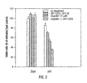

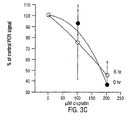
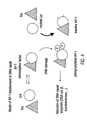
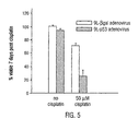


Classifications
-
- A—HUMAN NECESSITIES
- A61—MEDICAL OR VETERINARY SCIENCE; HYGIENE
- A61K—PREPARATIONS FOR MEDICAL, DENTAL OR TOILETRY PURPOSES
- A61K31/00—Medicinal preparations containing organic active ingredients
- A61K31/70—Carbohydrates; Sugars; Derivatives thereof
- A61K31/7042—Compounds having saccharide radicals and heterocyclic rings
- A61K31/7048—Compounds having saccharide radicals and heterocyclic rings having oxygen as a ring hetero atom, e.g. leucoglucosan, hesperidin, erythromycin, nystatin, digitoxin or digoxin
-
- A—HUMAN NECESSITIES
- A61—MEDICAL OR VETERINARY SCIENCE; HYGIENE
- A61K—PREPARATIONS FOR MEDICAL, DENTAL OR TOILETRY PURPOSES
- A61K31/00—Medicinal preparations containing organic active ingredients
- A61K31/16—Amides, e.g. hydroxamic acids
- A61K31/165—Amides, e.g. hydroxamic acids having aromatic rings, e.g. colchicine, atenolol, progabide
- A61K31/166—Amides, e.g. hydroxamic acids having aromatic rings, e.g. colchicine, atenolol, progabide having the carbon of a carboxamide group directly attached to the aromatic ring, e.g. procainamide, procarbazine, metoclopramide, labetalol
-
- A—HUMAN NECESSITIES
- A61—MEDICAL OR VETERINARY SCIENCE; HYGIENE
- A61K—PREPARATIONS FOR MEDICAL, DENTAL OR TOILETRY PURPOSES
- A61K31/00—Medicinal preparations containing organic active ingredients
- A61K31/185—Acids; Anhydrides, halides or salts thereof, e.g. sulfur acids, imidic, hydrazonic or hydroximic acids
- A61K31/19—Carboxylic acids, e.g. valproic acid
- A61K31/20—Carboxylic acids, e.g. valproic acid having a carboxyl group bound to a chain of seven or more carbon atoms, e.g. stearic, palmitic, arachidic acids
- A61K31/203—Retinoic acids ; Salts thereof
-
- A—HUMAN NECESSITIES
- A61—MEDICAL OR VETERINARY SCIENCE; HYGIENE
- A61K—PREPARATIONS FOR MEDICAL, DENTAL OR TOILETRY PURPOSES
- A61K31/00—Medicinal preparations containing organic active ingredients
- A61K31/70—Carbohydrates; Sugars; Derivatives thereof
- A61K31/7042—Compounds having saccharide radicals and heterocyclic rings
- A61K31/7052—Compounds having saccharide radicals and heterocyclic rings having nitrogen as a ring hetero atom, e.g. nucleosides, nucleotides
- A61K31/7056—Compounds having saccharide radicals and heterocyclic rings having nitrogen as a ring hetero atom, e.g. nucleosides, nucleotides containing five-membered rings with nitrogen as a ring hetero atom
-
- A—HUMAN NECESSITIES
- A61—MEDICAL OR VETERINARY SCIENCE; HYGIENE
- A61P—SPECIFIC THERAPEUTIC ACTIVITY OF CHEMICAL COMPOUNDS OR MEDICINAL PREPARATIONS
- A61P35/00—Antineoplastic agents
Definitions
- the present invention relates generally to the field of cancer therapy. More particularly, it concerns the use of an inhibitory agent that inhibits DNA repair for the manufacture of a medicament for the treatment of cancer by induction of p53-mediated apoptosis in a cell.
- glioblastoma multiforme is the most common intracranial brain tumor and is particularly resistant to therapy, and rapidly becomes more resistant as therapy continues.
- Glioblastoma tumors are highly vascularized, infiltrate the brain extensively and can attain a very large size. Glioblastomas are unilaterally fatal and patients have a mean survival time of about one year from the time of diagnosis.
- glioblastomas respond poorly to most chemotherapeutic agents, even though the blood brain barrier is broken down as a consequence of the disease.
- Certain chemotherapeutic agents such as cisplatin, carmustine, procarbazine and 5-fluororacil are somewhat efficacious in the treatment of glioblastoma but the tumors are never completely eradicated by these methods.
- a major reason for the failure of traditional treatment therapies of glioblastoma is the development of resistance in subsets of tumor cells. One reason for this resistance appears to be a result of genetic changes that accompany disease progression, including loss of wild-type p53 function. Mutations in p53 occur in over 50% of adult glioblastoma cases and are associated more with disease progression.
- the p53 gene is well recognized as a tumor suppressor gene (Montenarh, 1992). There is now considerable evidence linking mutations of p53 in the oncogenesis of many human cancers. There are numerous reports demonstrating that the growth of colon, glioblastoma, breast cancer, osteosarcoma and lung tumor cells can be suppressed by the expression of wild-type p53. The introduction of wild-type p53 into a wide variety of p53-mutated cells, using viral delivery methods, has resulted in the expression of the wild-type p53 transgene and a suppression of the malignant phenotype. These observations demonstrate that a high level of expression of wild-type p53 is a desirable course for the treatment of oncogenic malignancy.
- GB-A-2244646 dicloses the use of a poly(ADP-ribose) polymerase inhibitor to treat viral infections.
- p53 has been shown to be a trigger of apoptosis (Yonish-Rouach et al., 1991; Shaw et al., 1992; Lowe et al. , 1993; Lotem and Sachs, 1993; Clarke et al. , 1993) which suggests that the disruption of p53 in tumors has significant consequences for cancer therapy.
- the desensitization of tumor cells to the effects of traditional cancer therapies as a result of p53 mutation may aid in the progression of disease.
- cancer therapies such as radiotherapy and chemotherapy that induce DNA damage to a tumor cell contribute to the development of resistance of tumors.
- DNA damage results in up-regulated DNA damage repair mechanisms, which could account for increased resistance to DNA damaging therapy.
- DNA damage results in cell cycle arrest and induction of DNA repair mechanisms, so as to prevent the transfer of damaged DNA to the next generation of cells.
- Cells that sustain high levels of DNA damage such as tumor cells that exhibit high levels of karyotypic instability, or cells that are treated with DNA damaging agents, are induced to undergo apoptosis. This switch from either arrest and DNA repair or apoptosis is mediated by p53.
- This use may further relate to contacting the cell with a first stimulatory agent that increases the level of a tumor suppressor in said cell.
- the tumor suppressor may be p53, p21 or MSH-2, and preferably is p53.
- the stimulatory agent may be an expression construct that comprises a tumor suppressor gene under the control of a promoter active in eukaryotic cells.
- the expression construct may be an adenoviral expression construct.
- the said adenoviral expression construct lacks a portion of at least one gene essential to adenoviral replication, such as the E1 gene.
- a preferred promoter is the CMV promoter.
- the method advantageously employs and said inhibitory agent that inhibits the function of a protein selected from the group consisting of c-jun, c-fos, poly-ADP ribose polymerase, DNA polymerase ⁇ , topoisomerase I, d-TMP synthase, hMTII-A, uracil DNA glycosylase, alkyl-N-purine DNA glycosylase, DNA ligase IV, DNA ligase III, Hap-1, Ref-1, poly-ADP ribose polymerase and DNA-dependent protein kinase.
- a protein selected from the group consisting of c-jun, c-fos, poly-ADP ribose polymerase, DNA polymerase ⁇ , topoisomerase I, d-TMP synthase, hMTII-A, uracil DNA glycosylase, alkyl-N-purine DNA glycosylase, DNA ligase IV, DNA
- the inhibitory agent is a non-functional version of an agent involved with DNA repair.
- a mutant jun protein that competitively inhibits c-jun may be employed.
- the inhibitory agent is an antisense construct encoding at least a portion of a gene such as c-jun, c-fos, poly-ADP ribose polymerase, DNA polymerase ⁇ , topoisomerase I, d-TMP synthase, hMTII-A, uracil DNA glycosylase, alkyl-N-purine DNA glycosylase, DNA ligase IV, DNA ligase III, Hap-1, Ref-1, poly-ADP ribose polymerase and DNA-dependent protein kinase.
- the inhibitory agent is a retinoid, for example, the synthetic retinoid SR11220.
- the inhibitory agent is 3-aminobenzamide.
- the method may also comprise the step of providing a DNA-damaging agent.
- Suitable DNA-damaging agents included cisplatin, carboplatin, VP16, teniposide, daunorubicin, doxorubicin, dactinomycin, mitomycin, plicamycin, bleomycin, procarbazine, nitrosourea, cyclophosphamide, bisulfan, melphalan. chlorambucil, ifosfamide, merchlorehtamine, taxol, taxotere, anthracyclines and ionizing radiation.
- Tumor cells such as a lung tumor cell, a prostate tumor cell, a breast tumor cell, a colon tumor cell, a liver tumor cell, a brain tumor cell, a kidney tumor cell, a skin tumor cell and an ovarian tumor cell all are contemplated targets of the method.
- These tumors may be a squamous cell carcinoma, a non-squamous cell carcinoma, a glioblastoma, a sarcoma, a melanoma, a papilloma, a neuroblastoma and a leukemia cell.
- the tumors may be treated ex vivo or in a subject, such as a human subject.
- Delivery of the inhibitory agent, the stimulatory agent and/or the DNA damaging agent is advantageously via direct intratumoral injection.
- the injection comprises continuous perfusion of the tumor.
- Cancer accounts for the death of over half a million people each year in the United States alone.
- the causes for cancer are multifactorial, but it is known that aberrations in mechanisms involved with controlled cell death (apoptosis) may result in uncontrolled cell proliferation and, hence, contribute to many cancers.
- Conventional cancer therapies such as radiotherapy and chemotherapy that result in DNA damage are poorly effective or lose their effectiveness over time in some cancers.
- a consequence of the treatment of tumor cells with such DNA damaging agents is the induction of DNA repair mechanisms. Up-regulation of DNA repair mechanisms contributes to the increasing resistance of tumors to sustained therapy.
- the present invention provides a means of increasing levels of apoptosis in tumor cells through inhibition of DNA damage repair mechanisms.
- the present invention relies, in part, on the observation that agents that inhibit the expression of DNA repair enzymes sensitize tumor cells to p53-mediated apoptosis. This finding can be employed in a number of ways. First, treatment of tumors which express wild-type p53 with DNA repair inhibitory agents can sensitize the tumors to p53-mediated apoptosis. Second, the delivery of combination therapy, where p53 gene therapy is used in combination with agents that inhibit DNA repair are used to inhibit the DNA repair mechanisms, thereby inducing apoptosis.
- a "functional" p53 or p53 gene is one that confers this apoptosis-inducing ability on cells.
- a "wild-type” p53 will accomplish this function.
- WAF1 one of the targets of p53 , encodes a p21 WAF1/CIP1 protein of 21,000 Dalton molecular weight, which is an inhibitor of cyclin-dependent kinase 2 required for the G1-to-S transition (E1-Deiry et al. , 1993).
- Karyotypic instability is a hallmark of cancer and is particularly apparent in glioblastoma (Bigner et al., 1981; Bigner et al., 1990). This instability manifested by an increased level of chromosomal breakage and rearrangement, including translocations, additions, deletions, amplifications, loss of heterozygosity and aneuploidy. Tumor cells show elevated rates of spontaneous gene amplification compared to normal cells, where gene amplification is rarely detected (Tisty, 1990). Spontaneous deletions also occur with increased frequency in tumor cells (Kaden et al., 1989).
- Loss of p53 function may favor the growth of karyotypically unstable cells by removing a trigger for apoptosis that could eliminate cells with unstable genomes (Lane, 1992). Thus, loss of p53 may contribute to the marked aneuploidy and karyotypic instability observed in tumors, for example, glioblastoma.
- Studies of p53 null transgenic mice have shown that normal transgenic hematopoetic cells (Lotem and Sachs, 1993), E1A-expressing transgenic fibroblasts (Lowe et al., 1993) and transformed transgenic fibroblasts (Lowe et al., 1994) were all more resistant to apoptosis following treatment with a variety of anti-cancer agents.
- glioblastoma cells and other tumor cells lacking functional p53 are significantly more sensitive to the DNA damaging effect of cisplatin and radiation following introduction of am exogenous, wild-type p53 gene. This enhancement of cell death has been shown to be the result of apoptosis. Expression of exogenous wild-type p53 in cells containing an intact normal p53 does not affect the growth of these cells.
- wild-type p53 is widespread in cancer, the use of wild-type p53 as a therapy sensitizer may have far reaching consequences in treating p53-negative associated cancers as well as cancers that contain a functional p53 gene, including but not limited to breast, lung, prostate, colon, liver, brain, skin, ovarian, pancreatic, kidney, lymphoid and renal.
- exogenous wild-type p53 may achieve only a slowing of growth or reversible growth arrest. Transfer of wild-type p53 into T98G glioblastoma cells slows the growth of the cells but does not induce apoptosis. Similarly, GM47 glioblastoma cells which express an inducible wild-type p53 undergo reversible growth arrest at high levels of p53 induction. However, after de-induction, the cells begin cell cycling (Mercer et al., 1990).
- Tumor suppression and therapy sensitization through p53 appears to be selective for tumors cells (Baker et al., 1990), possibly because p53 levels in normal cells are kept at low level by rapid turnover (Rogel et al., 1985).
- the intrinsic genomic instability of tumor cells which would normally induce the apoptotic pathway, may also be a significant factor in contributing to the induction p53-mediated apoptosis in response to artificially induced DNA damage, such as chemotherapy or radiation therapy.
- the apparent specificity of p53 for tumor cells would allow the selective targeting of p53-specific suppression, which would be less toxic than tumor cell suppression protocols that aim at a general cell cycle block.
- tumor suppressors for example p21 and MSH-2
- p53 will function in an analogous manner to p53 for the purpose of inducing apoptosis.
- reference to the use of p53 implicitly includes reference to these and other tumor suppressors to the extent that they are involved with apoptotic mechanisms.
- DNA repair mechanisms Treatment of tumor cells with DNA damaging agents results in the induction of DNA repair mechanisms.
- the success or failure of DNA repair may have a significant role in determining the consequences of p53 expression in a cell subjected to DNA damage.
- Tumor cells that fail to repair DNA damage arising from either intrinsic genomic instability or from external DNA damaging agents, may be more susceptible to p53-mediated apoptosis.
- DNA repair enzymes including dTMP synthetase, DNA polymerase B, topoisomerase 1, and hMTII-A, is up-regulated.
- Some of these DNA repair enzymes are subject to transcriptional transactivational by the transcription factor, AP-1, which consists of subunits fos and jun.
- DNA repair in tumor cells is down-regulated by a dominant-negative inhibitor of c-jun (mutant jun or m-jun), which fails to be phosphorylated due to amino acid substitutions at two critical phosphorylation sites that are associated with cellular transformation.
- c-jun mutant jun or m-jun
- the m-jun competes with wild-type jun for binding to c-fos so that when m-jun is bound to c-fos, it forms an inactive AP-1 complex, which cannot carry out transactivation of DNA repair genes (FIG. 4.).
- m-jun does not interfere with normal cellular transcription involving AP-1.
- Cells having the mutant jun gene are more sensitive to p53-induced apoptosis.
- DNA repair inhibition include inhibitors of other transcription factors, such as Sp1, E2F-1 and jun/ATF2 that may be involved with transcription of DNA damage repair proteins.
- Tissue-specific transcription factors that are involved in DNA repair may also be targeted to provide specific therapy for a particular type of cancer.
- nucleic acids encoding antisense inhibitory agents relating to each of these targets, or any of the other transcription factors and DNA repair enzymes may be used to inhibit the induction of DNA repair.
- Organochemical compounds also may be employed to inhibit repair of DNA repair.
- Such compounds include retinoids like SR11220, and 3-aminobenzamide.
- Retinoids in general induce a block in the cell cycle.
- SR11220 is a synthetic retinoid that is specific for AP-1, and thus has less toxicity than other retinoids such as retinoic acid, which has broad specificity (Fanjul et al., 1994; incorporated herein by reference).
- retinoids which may be used to inhibit DNA repair include, but are not limited to, trans-retinoic acid, 9-cis retinoic acid, known to inhibit AP-1, and the synthetic retinoids SR11105, SR11217, SR11238, SR11235, SR11302, SR11220, SR11327, SR11228, SR11324 (Fanjul et al., 1994).
- the use of retinoids with broad specificity while, if administered intravenously could be significantly toxic, may exhibit only local toxicity if administered intratumorally or adjacent to the tumor site.
- 3-aminobenzamide is an inhibitor of poly-ADP ribose polymerase that inhibits the repair of both single- and double-strand breaks induced by DNA damaging agents. It has been shown that p53 is induced following irradiation of 3-aminobenzamide treated cells (Lane, 1992). Compounds with similar specificity could be used according to the present invention Again, although such compounds may exhibit marked toxicity if used systematically, the regional or local delivery may obviate much of the toxicity. For example, delivery of 3-aminobenzamide as a bolus injection into the tumor mass or the tumor vasculature may result in induction of apoptosis in tumor cells with only minor effects on the surrounding normal tissues.
- Useful compounds may include fragments or parts of the enzymes or factors listed above, including antisense oligonucleotides corresponding to DNA repair-related enzymes. Common antisense targets are regions involve with transcription initiation, translation initiation and splicing. Alternatively, compounds isolated from natural sources, such as animals, bacteria, fungi, plant sources, including leaves and bark, and marine samples may be assayed as potentially useful pharmaceutical agents. It will be understood that the pharmaceutical agents to be screened also could be derived or synthesized from chemical compositions or man-made compounds.
- a method for determining the ability of a candidate substance to inhibit the DNA repair activity of cancer cells and to concomitantly induce apoptosis in said cells includes generally the steps of:
- the cell is a cancer cell that has heightened DNA repair mechanisms. Because of these mechanisms, the cancer cell is resistant, to a certain extent, to p53-induced apoptosis.
- the cell may be p53-negative, but a p53 transgene may be provided in step (b) to facilitate induction of apoptosis.
- the candidate screening assay relies on the formation of lesions in DNA, such as those caused by DNA damaging agents. These lesions block the progression of the Taq polymerase used in PCR TM and thus decrease the yield of the PCRTM product. It has been shown that the level of DNA damage induced by cisplatin, for example, correlates closely with the level of amplified PCRTM product obtained. Measurement of the PCRTM signal obtained from amplification of a significantly large region of DNA, for example 2.7 kB, will decrease in relation to the signal from a small region of amplified DNA, for example 150 bp, whose signal will not be affected due to its small size, after treatment with DNA damaging agents.
- MTT assay it may be desirable simply to measure inhibition of growth of cancer cells, for example, by measuring growth according to the MTT assay.
- a significant inhibition in growth is represented by decreases of at least about 30%-40% as compared to uninhibited, and most preferably, of at least about 50%, with more significant decreases also being possible.
- Growth assays as measured by the MTT assay are well known in the art. Assays may be conducted as described by Mosmann et al., (1983) and Rubinstein et al. (1990) (incorporated herein by reference). Therefore, if a candidate substance exhibited inhibition of growth of cancer cells in this type of study, it would likely be a suitable compound for use in the present invention.
- TUNEL Terminal deoxynucleotidyl transferase mediated dUTP-biotin nick end labeling
- TdT buffer (30 mM Tris Hcl, pH 7.2; 140 mM cacodylate, 1 mM cobalt chloride) and incubated with biotinylated dUTP (Boehringer Mannheim, Indianapolis, IN) and 100 U/ml TdT enzyme (Bethesda Research Laboratory) for 1 h at 37°C.
- biotinylated dUTP Boehringer Mannheim, Indianapolis, IN
- U/ml TdT enzyme Bethesda Research Laboratory
- the avidin-biotin complex was detected using the Vectastain Elite kit (Vector Laboratory, Burlingame, CA), by the diaminobenzidine-H 2 O 2 method.
- Quantitative in vitro testing of the DNA repair inhibiting agents is not a requirement of the invention as it is generally envisioned that the agents will often be selected on the basis of their known properties or by structural and/or functional comparison to those agents already demonstrated to be effective. Therefore, the effective amounts will often be those amounts proposed to be safe for administration to animals in another context.
- p53 currently is recognized as a tumor suppressor gene (Montenarh, 1992).
- the p53 gene is a frequent target of mutational inactivation in a wide variety of human tumors and is already documented to be the most frequently-mutated gene in common human cancers (Mercer, 1992). It is mutated in over 50% of human NSCLC (Hollestein et al., 1991) and in a wide spectrum of other tumors.
- the p53 gene encodes a 393-amino-acid phosphoprotein that can form complexes with host proteins such as large-T antigen and E1B.
- the wild-type protein is found in normal tissues and cells, but at concentrations which are minute by comparison with levels of mutant protein in transformed cells or tumor tissue.
- wild-type p53 appears to be important in regulating cell growth and division.
- Overexpression of wild-type p53 has been shown in some cases to be anti-proliferative in human tumor cell lines.
- p53 can act as a negative regulator of cell growth (Weinberg, 1991) and may directly suppress uncontrolled cell growth or indirectly activate genes that suppress this growth.
- absence or inactivation of wild-type p53 may contribute to transformation.
- some studies indicate that the presence of mutant p53 may be necessary for full expression of the transforming potential of the gene.
- Wild-type p53 is recognized as an important growth regulator in many cell types. Mis-sense mutations are common for the p53 gene and are essential for the transforming ability of the oncogene. A single genetic change prompted by point mutations can create carcinogenic p53. Unlike other oncogenes, however, p53 point mutations are known to occur in at least 30 distinct codons, often creating dominant alleles that produce shifts in cell phenotype without a reduction to homozygosity. Additionally, some of these negative alleles appear to be tolerated in the organism and passed on in the germ line. Various mutant alleles appear to range from minimally dysfunctional to strongly penetrant (Weinberg, 1991).
- a patient with cancer will be treated with various agents that permit p53 induced apoptosis to occur.
- agents include DNA repair inhibitory agents and, in certain cases, DNA damaging agents.
- p53 function is necessary, it is desirable that the p53 status of the tumor cells be determined. This may be accomplished using conventional methods, examples of which are described below.
- patients will have adequate bone marrow function (defined as peripheral absolute granulocyte count of > 2,000/mm 3 and platelet count of 100,000mm 3 ), adequate liver function (bilirubin ⁇ 1.5 mg/dl) and adequate renal function (creatinine ⁇ 1.5 mg/dl).
- the patient will be treated with a pharmaceutically acceptable form of the DNA repair inhibitory agent These agents are described above.
- This administration could be in the form of, for example, an intratumoral injection, or indeed any other method of application that is routinely used and well know to one of skill in the art. A more detailed discussion of formulations and routes of administration is provided below.
- a wide variety of detection methods can be employed to detect the p53 status of a cell.
- detection methods There are numerous antibodies to the p53 protein, hence any assay that utilizes antibodies for detection, for example, ELISAs, Western Blotting, immunoassay techniques, etc.
- assays that employ nucleotide probes may be used to identify the presence/absence of p53, for example, Southern blotting, Northern blotting or PCRTM techniques. All the above techniques are well known to one of skill in the art and could be utilized in the present invention without undue experimentation.
- Immunoassays include, but are not limited to those described in U.S. Patent No. 4,367,110 (double monoclonal antibody sandwich assay) and U.S. Patent No. 4,452,901 (western blot). Other assays include immunoprecipitation of labeled ligands and immunocytochemistry, both in vitro and in vivo .
- Immunoassays generally are binding assays. Certain preferred immunoassays are the various types of enzyme linked immunosorbent assays (ELISAs) and radioimmunoassays (RIA) known in the art. Immunohistochemical detection using tissue sections is also particularly useful.
- ELISAs enzyme linked immunosorbent assays
- RIA radioimmunoassays
- the anti-p53 antibodies are immobilized on a selected surface, such as a well in a polystyrene microtiter plate, dipstick or column support. Then, a test composition suspected of containing the desired antigen, such as a clinical sample, is added to the wells. After binding and washing to remove non-specifically bound immune complexes, the bound antigen may be detected. Detection is generally achieved by the addition of another antibody, specific for the desired antigen, that is linked to a detectable label. This type of ELISA is known as a "sandwich ELISA". Detection also may be achieved by the addition of a second antibody specific for the desired antigen, followed by the addition of a third antibody that has binding affinity for the second antibody, with the third antibody being linked to a detectable label.
- the samples suspected of containing the desired antigen are immobilized onto the well surface and then contacted with the antibodies of the invention. After binding and appropriate washing, the bound immune complexes are detected. Where the initial antigen specific antibodies are linked to a detectable label, the immune complexes may be detected directly. Again, the immune complexes may be detected using a second antibody that has binding affinity for the first antigen specific antibody, with the second antibody being linked to a detectable label.
- Competition ELISAs are also possible in which test samples compete for binding with known amounts of labeled antigens or antibodies.
- the amount of reactive species in the unknown sample is determined by mixing the sample with the known labeled species before or during incubation with coated wells. The presence of reactive species in the sample acts to reduce the amount of labeled species available for binding to the well and thus reduces the ultimate signal.
- ELISAs have certain features in common, such as coating, incubating or binding, washing to remove non-specifically bound species, and detecting the bound immune complexes. These are described as below.
- Antigen or antibodies may also be linked to a solid support, such as in the form of plate, beads, dipstick, membrane or column matrix, and the sample to be analyzed applied to the immobilized antigen or antibody.
- a solid support such as in the form of plate, beads, dipstick, membrane or column matrix
- the sample to be analyzed applied to the immobilized antigen or antibody.
- a plate In coating a plate with either antigen or antibody, one will generally incubate the wells of the plate with a solution of the antigen or antibody, either overnight or for a specified period. The wells of the plate will then be washed to remove incompletely adsorbed material. Any remaining available surfaces of the wells are then "coated” with a nonspecific protein that is antigenically neutral with regard to the test antisera. These include bovine serum albumin (BSA), casein and solutions of milk powder.
- BSA bovine serum albumin
- the coating allows for blocking of nonspecific adsorption sites on the immobilizing surface and thus reduces the background caused
- a secondary or tertiary detection means rather than a direct procedure.
- the immobilizing surface is contacted with the clinical or biological sample to be tested under conditions effective to allow immune complex (antigen/antibody) formation. Detection of the immune complex then requires a labeled secondary binding ligand or antibody, or a secondary binding ligand or antibody in conjunction with a labeled tertiary antibody or third binding ligand.
- Under conditions effective to allow immune complex (antigen/antibody) formation means that the conditions preferably include diluting the antigens and antibodies with solutions such as BSA, bovine gamma globulin (BGG) and phosphate buffered saline (PBS)/Tween. These added agents also tend to assist in the reduction of nonspecific background.
- BSA bovine gamma globulin
- PBS phosphate buffered saline
- the suitable conditions also mean that the incubation is at a temperature and for a period of time sufficient to allow effective binding. Incubation steps are typically from about 1 to 2 to 4 hours, at temperatures preferably on the order of 25° to 27°C, or may be overnight at about 4°C or so.
- the contacted surface is washed so as to remove non-complexed material. Washing often includes washing with a solution of PBS/Tween, or borate buffer. Following the formation of specific immune complexes between the test sample and the originally bound material, and subsequent washing, the occurrence of even minute amounts of immune complexes may be determined.
- the second or third antibody will have an associated label to allow detection.
- this will be an enzyme that will generate color development upon incubating with an appropriate chromogenic substrate.
- a urease, glucose oxidase, alkaline phosphatase or hydrogen peroxidase-conjugated antibody for a period of time and under conditions that favor the development of further immune complex formation, e.g. , incubation for 2 hours at room temperature in a PBS-containing solution such as PBS-Tween.
- the amount of label is quantified, e.g., by incubation with a chromogenic substrate such as urea and bromocresol purple or 2,2'-azino-di-(3-ethyl-benzthiazoline-6-sulfonic acid [ABTS] and H 2 O 2 , in the case of peroxidase as the enzyme label. Quantification is then achieved by measuring the degree of color generation, e.g. , using a visible spectra spectrophotometer.
- a chromogenic substrate such as urea and bromocresol purple or 2,2'-azino-di-(3-ethyl-benzthiazoline-6-sulfonic acid [ABTS] and H 2 O 2 , in the case of peroxidase as the enzyme label.
- Quantification is then achieved by measuring the degree of color generation, e.g. , using a visible spectra spectrophotometer.
- the label may be a chemiluminescent one.
- the use of such labels is described in U.S. Patent Nos. 5,310,687, 5,238,808 and 5.221,605.
- Assays for the p53 status of the cell may be performed directly on biopsy samples.
- Methods for in vitro situ analysis are well known and involve assessing binding of antigen-specific antibodies to tissues, cells or cell extracts. These are conventional techniques well within the grasp of those skilled in the art.
- the antibodies to p53 may be used in conjunction with both fresh-frozen and formalin-fixed, paraffin-embedded tissue blocks prepared for study by immunohistochemistry (IHC). Each tissue block may consist of 50 mg of residual "pulverized" tumor.
- IHC immunohistochemistry
- Each tissue block may consist of 50 mg of residual "pulverized" tumor.
- the method of preparing tissue blocks from these particulate specimens has been successfully used in previous IHC studies of various prognostic factors, e.g. , in breast cancer, and is well known to those of skill in the art. (Abbondanzo et al., 1990; Allred et al., 1990; Brown et al., 1990).
- frozen-sections may be prepared by rehydrating 50 ng of frozen pulverized tumor at room temperature in PBS in small plastic capsules; pelleting the particles by centrifugation; resuspending them in a viscous embedding medium (OCT); inverting the capsule and pelleting again by centrifugation; snap-freezing in -70°C isopentane; cutting the plastic capsule and removing the frozen cylinder of tissue; securing the tissue cylinder on a cryostat microtome chuck; and cutting 25-50 serial sections containing an average of about 500 remarkably intact tumor cells.
- OCT viscous embedding medium
- Permanent-sections may be prepared by a similar method involving rehydration of the 50 mg sample in a plastic microfuge tube; pelleting; resuspending in 10% formalin for 4 hours fixation; washing/pelleting; resuspending in warm 2.5% agar; pelleting; cooling in ice water to harden the agar; removing the tissue/agar block from the tube; infiltrating and embedding the block in paraffin; and cutting up to 50 serial permanent sections.
- the DNA and RNA from test cells is recovered by gentle cell rupture in the presence of a cation chelator such as EDTA.
- the proteins and other cell milieu are removed by admixing with saturated phenol or phenol/chloroform and centrifugation of the emulsion.
- the DNA and RNA is in the upper aqueous phase, it is deproteinised and mixed with ethanol. This solution allows the DNA and RNA to precipitate, the DNA and RNA can then be recover using centrifugation.
- RNAse inhibitors such as DEPC are needed to prevent RNA degradation.
- Electrophoresis in agarose or polyacrylamide gels is the most usual way to separate DNA and RNA molecules.
- Southern blotting will confirm the identity of the p53 encoding DNA. This is achieved by transferring the DNA from the intact gel onto nitrocellulose paper. The nitrocellulose paper is then washed in buffer that has for example, a radiolabelled cDNA containing a sequence complementary to wild-type-P53 DNA. The probe binds specifically to the DNA that encodes a region of p53 and can be detected using autoradiography by contacting the probed nitrocellulose paper with photographic film. p53-encoding mRNA can be detected in a similar manner by a process known as Northern blotting. For a more detailed description of buffers gel preparation, electrophoresis condition etc., the skilled artisan is referred to Sambrook, 1989.
- PCR TM is a powerful tool in modern analytical biology. Short oligonucleotide sequences usually 15-35 bp in length are designed, homologous to flanking regions either side of the p53 sequences to be amplified.
- the primers are added in excess to the source DNA, in the presence of buffer, enzyme, and free nucleotides.
- the source DNA is denatured at 95°C and then cooled to 50-60°C to allow the primers to anneal. The temperature is adjusted to the optimal temperature for the polymerase for an extension phase. This cycle is repeated 25-40 times.
- PCRTM Single Strand Conformation Polymorphism
- SSCP Single Strand Conformation Polymorphism
- each exon is separately amplified by PCRTM using primers specific for the particular exon.
- the PCRTM product is denatured and separated out on a polyacrylamide gel to detect a shift in mobility due to a conformational change which resulted because of a point mutation or other small nucleotide change in the gene.
- Mutations result in a change in the physical conformation of the DNA as well as change in the electrical charge of the molecule.
- DNA that'is slightly different in shape and charge as compared to wild-type will move at a different rate and thus occupy a different position in the gel.
- the specific nucleotide changes are detected by DNA sequencing of the amplified PCRTM product. Sequencing of linear DNA breaks down the DNA molecule into its individual nucleotides in the order with which they are assembled in the intact molecule. Separation of the individual nucleotides by electrophoresis on a sequencing gel allows detection of individual nucleotide changes compared to wild-type and is used to determine homo- or heterozygocity of a mutation, which is easily distinguished by the appearance of a single or double band in the sequencing gel.
- compositions for use in the present invention will have an effective amount of a p53 expression vector or p53 protein, along with a compound that inhibits DNA repair.
- Such compositions will generally be dissolved or dispersed in a pharmaceutically acceptable carrier or aqueous medium.
- phrases "pharmaceutically or pharmacologically acceptable” refer to molecular entities and compositions that do not produce an adverse, allergic or other untoward reaction when administered to an animal, or human, as appropriate.
- pharmaceutically acceptable carrier includes any and all solvents, dispersion media, coatings, antibacterial and antifungal agents, isotonic and absorption delaying agents and the like. The use of such media and agents for pharmaceutical active substances is well known in the art. Except insofar as any conventional media or agent is incompatible with the active ingredients, its use in the therapeutic compositions is contemplated. Supplementary active ingredients, such as other anti-cancer agents, can also be incorporated into the compositions.
- other pharmaceutically acceptable forms include, e.g. , tablets or other solids for oral administration; time release capsules; and any other form currently used, including cremes, lotions, mouthwashes, inhalants and the like.
- the active compounds for use in the present invention will often be formulated for parenteral administration, e.g. , formulated for injection via the intravenous, intramuscular, sub-cutaneous, or even intraperitoneal routes.
- parenteral administration e.g. , formulated for injection via the intravenous, intramuscular, sub-cutaneous, or even intraperitoneal routes.
- the preparation of an aqueous composition that contains a compound or compounds that increase the expression of wild-type p53 will be known to those of skill in the art in light of the present disclosure.
- such compositions can be prepared as injectables, either as liquid solutions or suspensions; solid forms suitable for using to prepare solutions or suspensions upon the addition of a liquid prior to injection can also be prepared; and the preparations can also be emulsified.
- Solutions of the active compounds as free base or pharmacologically acceptable salts can be prepared in water suitably mixed with a surfactant, such as hydroxypropylcellulose. Dispersions can also be prepared in glycerol, liquid polyethylene glycols, and mixtures thereof and in oils. Under ordinary conditions of storage and use, these preparations contain a preservative to prevent the growth of microorganisms.
- the pharmaceutical forms suitable for injectable use include sterile aqueous solutions or dispersions; formulations including sesame oil, peanut oil or aqueous propylene glycol; and sterile powders for the extemporaneous preparation of sterile injectable solutions or dispersions.
- the form must be sterile and must be fluid to the extent that it may be easily injected. It also should be stable under the conditions of manufacture and storage and must be preserved against the contaminating action of microorganisms, such as bacteria and fungi.
- the active compounds may be formulated into a composition in a neutral or salt form.
- Pharmaceutically acceptable salts include the acid addition salts (formed with the free amino groups of the protein) and which are formed with inorganic acids such as, for example, hydrochloric or phosphoric acids, or such organic acids as acetic, oxalic, tartaric, mandelic, and the like. Salts formed with the free carboxyl groups can also be derived from inorganic bases such as, for example, sodium, potassium, ammonium, calcium, or ferric hydroxides, and such organic bases as isopropylamine, trimethylamine, histidine, procaine and the like.
- the carrier also can be a solvent or dispersion medium containing, for example, water, ethanol, polyol (for example, glycerol, propylene glycol, and liquid polyethylene glycol, and the like), suitable mixtures thereof, and vegetable oils.
- the proper fluidity can be maintained, for example, by the use of a coating, such as lecithin, by the maintenance of the required particle size in the case of dispersion and by the use of surfactants.
- the prevention of the action of microorganisms can be brought about by various antibacterial and antifungal agents, for example, parabens, chlorobutanol, phenol, sorbic acid, thimerosal, and the like.
- isotonic agents for example, sugars or sodium chloride.
- Prolonged absorption of the injectable compositions can be brought about by the use in the compositions of agents delaying absorption, for example, aluminum monostearate and gelatin.
- Sterile injectable solutions are prepared by incorporating the active compounds in the required amount in the appropriate solvent with various of the other ingredients enumerated above, as required, followed by filtered sterilization.
- dispersions are prepared by incorporating the various sterilized active ingredients into a sterile vehicle which contains the basic dispersion medium and the required other ingredients from those enumerated above.
- the preferred methods of preparation are vacuum-drying-and freeze-drying techniques which yield a powder of the active ingredient plus any additional desired ingredient from a previously sterile-filtered solution thereof.
- the therapeutic formulations for use in the invention also may be prepared in forms suitable for topical administration, such as in cremes and lotions. These forms may be used for treating skin-associated diseases, such as various sarcomas.
- compositions according to the use of the present invention will be via any common route so long as the target tissue is available via that route. This includes oral, nasal, buccal, rectal, vaginal or topical. Topical administration would be particularly advantageous for treatment of skin cancers, to prevent chemotherapy-induced alopecia or other dermal hyperproliferative disorder. Alternatively, administration will be by orthotopic, intradermal subcutaneous, intramuscular, intraperitoneal or intravenous injection. Such compositions would normally be administered as pharmaceutically acceptable compositions that include physiologically acceptable carriers, buffers or other excipients. For treatment of conditions of the lungs, aerosol delivery to the lung is contemplated. Volume of the aerosol is between about 0.01 ml and 0.5 ml.
- a preferred method for treatment of colon-associated disease would be via enema.
- Volume of the enema is between about 1 ml and 100 ml.
- Direct intratumoral injection is the preferred mode, with continuous intratumoral perfusion a more specific embodiment.
- aqueous solutions For parenteral administration in an aqueous solution, for example, the solution should be suitably buffered if necessary and the liquid diluent first rendered isotonic with sufficient saline or glucose.
- aqueous solutions are especially suitable for intravenous, intramuscular, subcutaneous and intraperitoneal administration.
- sterile aqueous media which can be employed will be known to those of skill in the art in light of the present disclosure.
- one dosage could be dissolved in 1 mL of isotonic NaCI solution and either added to 1000 mL of hypodermoclysis fluid or injected at the proposed site of infusion, (see for example, Remington's Pharmaceutical Sciences, 1990). Some variation in dosage will necessarily occur depending on the condition of the subject being treated. The person responsible for administration will, in any event, determine the appropriate dose for the individual subject.
- unit dose refers to physically discrete units suitable for use in a subject, each unit containing a predetermined-quantity of the therapeutic composition calculated to produce the desired responses, discussed above, in association with its administration, i.e., the appropriate route and treatment regimen.
- the quantity to be administered both according to number of treatments and unit dose, depends on the protection desired.
- the amount of therapeutic composition comprising a unit dose will range from about 5-30 mg of polynucleotide.
- kits All the essential materials and reagents required for determining wild-type p53 in a sample or for inhibiting the DNA repair mechanisms in tumor cells may be assembled together in a kit.
- the liquid solution preferably is an aqueous solution, with a sterile aqueous solution being particularly preferred.
- the kit may contain materials for PCR TM analyses, such primers, buffers and appropriate solvents.
- the kit may contain antibodies directed to the p53, secondary antibodies that binding primary antibodies, labels or signal generating compounds (either conjugated or unconjugated) and various reagents for the generation and detection of signals.
- an inhibitor of DNA repair in combination with an p53 expression vector may be formulated into a single or separate pharmaceutically acceptable syringeable composition.
- the container means may itself be an inhalant, syringe, pipette, eye dropper, or other such like apparatus, from which the formulation may be applied to an infected area of the body, such as the lungs, injected into an animal, or even applied to and mixed with the other components of the kit
- kits may also be provided in dried or lyophilized forms. When reagents or components are provided as a dried form, reconstitution generally is by the addition of a suitable solvent. It is envisioned that the solvent also may be provided in another container means.
- the kits may also include an instruction sheet defining administration of the inhibitor of DNA repair and/or the p53 status determining agents, for explaining the assays for determining p53 levels in samples.
- kits also will typically include a means for containing the vials in close confinement for commercial sale such as, e.g. , injection or blow-molded plastic containers into which the desired vials are retained.
- a means for containing the vials in close confinement for commercial sale such as, e.g. , injection or blow-molded plastic containers into which the desired vials are retained.
- the kits also may comprise, or be packaged with, an instrument for assisting with the injection(administration or placement of the ultimate complex composition within the body of an animal.
- an instrument may be an inhalant, syringe, pipette, forceps, measured spoon, eye dropper or any such medically approved delivery vehicle.
- Other instrumentation includes devices that permit the reading or monitoring of reactions in vitro .
- inhibitors of DNA repair will be used in combination with gene therapy in the treatment of those cancers that do not express a functional p53. It is clear that delivery of wild-type p53 into tumors that express a mutated p53 gene can overcome the deleterious effects of the p53 mutation.
- an inhibitor of DNA repair can be administered concurrently with the gene therapy, before the gene therapy or after the gene therapy.
- the components need for gene therapy, as well as the therapeutic inhibitors of DNA repair can be assembled in a kit form as described above.
- the inhibitors of DNA repair also have been described above, so the remaining discussion relates to the elements relating to gene delivery.
- expression construct is meant to include any type of genetic construct containing a nucleic acid coding for a p53 product.
- the polynucleotide encoding the p53 polynucleotide will be under the transcriptional control of a promoter.
- a “promoter” refers to a DNA sequence recognized by the synthetic machinery of the host cell, or introduced synthetic machinery, that is required to initiate the specific transcription of a gene.
- under transcriptional control means that the promoter is in the correct location in relation to the polynucleotide to control RNA polymerase initiation and expression of the polynucleotide.
- promoter will be used herein to refer to a group of transcriptional control modules that are clustered around the initiation site for RNA polymerase II.
- Much of the thinking about how promoters are organized derives from analyses of several viral promoters, including those for the HSV thymidine kinase (tk) and SV40 early transcription units. These studies, augmented by more recent work, have shown that promoters are composed of discrete functional modules, each consisting of approximately 7-20 bp of DNA, and containing one or more recognition sites for transcriptional activator or repressor proteins.
- At least one module in each promoter functions to position the start site for RNA synthesis.
- the best known example of this is the TATA box, but in some promoters lacking a TATA box, such as the promoter for the mammalian terminal deoxynucleotidyl transferase gene and the promoter for the SV40 late genes, a discrete element overlying the start site itself helps to fix the place of initiation.
- promoter elements regulate the frequency of transcriptional initiation. Typically, these are located in the region 30-110 bp upstream of the start site, although a number of promoters have recently been shown to contain functional elements downstream of the start site as well.
- the spacing between promoter elements frequently is flexible, so that promoter function is preserved when elements are inverted or moved relative to one another. In the tk promoter, the spacing between promoter elements can be increased to 50 bp apart before activity begins to decline. Depending on the promoter, it appears that individual elements can function either cooperatively or independently to activate transcription.
- the particular promoter that is employed to control the expression of a p53 polynucleotide is not believed to be critical, so long as it is capable of expressing the polynucleotide in the targeted cell.
- a human cell it is preferable to position the polynucleotide coding region adjacent to and under the control of a promoter that is capable of being expressed in a human cell.
- a promoter might include either a human or viral promoter.
- the human cytomegalovirus (CMV) immediate early gene promoter, the SV40 early promoter and the Rous sarcoma virus long terminal repeat can be used to obtain high-level expression of the p53 polynucleotide.
- CMV cytomegalovirus
- the use of other viral or mammalian cellular or bacterial phage promoters which are well-known in the art to achieve expression of polynucleotides is contemplated as well, provided that the levels of expression are sufficient to produce a growth inhibitory effect.
- a promoter with well-known properties, the level and pattern of expression of a polynucleotide following transfection can be optimized. For example, selection of a promoter which is active in specific cells, such as tyrosinase (melanoma), alpha-fetoprotein and albumin (liver tumors), CC10 (lung tumor) and prostate-specific antigen (prostate tumor) will permit tissue-specific expression of p53 polynucleotides.
- Table 1 lists several promoters which may be employed, in the context of the present invention, to regulate the expression of p53 constructs. This list is not intended to be exhaustive of all the possible elements involved in the promotion of p53 expression but, merely, to be exemplary thereof.
- Enhancers were originally detected as genetic elements that increased transcription from a promoter located at a distant position on the same molecule of DNA. This ability to act over a large distance had little precedent in classic studies of prokaryotic transcriptional regulation. Subsequent work showed that regions of DNA with enhancer activity are organized much like promoters. That is, they are composed of many individual elements, each of which binds to one or more transcriptional proteins.
- enhancers The basic distinction between enhancers and promoters is operational. An enhancer region as a whole must be able to stimulate transcription at a distance; this need not be true of a promoter region or its component elements. On the other hand, a promoter must have one or more elements that direct initiation of RNA synthesis at a particular site and in a particular orientation, whereas enhancers lack these specificities. Promoters and enhancers are often overlapping and contiguous, often seeming to have a very similar modular organization.
- Eukaryotic Promoter Data Base EPDB any promoter/enhancer combination (as per the Eukaryotic Promoter Data Base EPDB) could also be used to drive expression of a p53 construct.
- Use of a T3, T7 or SP6 cytoplasmic expression system is another possible embodiment.
- Eukaryotic cells can support cytoplasmic transcription from certain bacteriophage promoters if the appropriate bacteriophage polymerase is provided, either as part of the delivery complex or as an additional genetic expression vector.
- selection of a promoter that is regulated in response to specific physiologic signals can permit inducible expression of the p53 construct.
- expression is inducible by tumor necrosis factor.
- Table 2 illustrates several promoter/inducer combinations: Element Inducer MT II Phorbol Ester (TPA) Heavy metals MMTV (mouse mammary tumor virus) Glucocorticoids ß-Interferon poly(rl)X poly(rc) Adenovirus 5 E2 Ela c-jun Phorbol Ester (TPA), H 2 O 2 Collagenase Phorbol Ester (TPA) Stromelysin Phorbol Ester (TPA), II,-1 SV40 Phorbol Ester (TPA) Murine MX Gene Interferon, Newcastle Disease Virus GRP78 Gene A23187 ⁇ -2-Macroglobulin IL-6 Vimentin Serum MHC Class I Gene H-2kB Interferon HSP70 Ela, SV40 Large T Antigen Proliferin Phorbol Ester (TPA) Tumor Necrosis Factor FMA Thyroid Stimulating Hormone a Gene Thyroid Hormone
- the delivery of an expression vector in a cell may be identified in vitro or in vivo by including a marker in the expression vector.
- the marker would result in an identifiable change to the transfected cell permitting identification of expression.
- a drug selection marker aids in cloning and in the selection of transformants.
- enzymes such as herpes simplex virus thymidine kinase (tk) (eukaryotic) or chloramphenicol acetyltransferase (CAT) (prokaryotic) may be employed.
- Immunologic markers also can be employed.
- the selectable marker employed is not believed to be important, so long as it is capable of being expressed along with the polynucleotide encoding p53. Further examples of selectable markers are well known to one of skill in the art.
- the nature of the polyadenylation signal is not believed to be crucial to the successful practice of the invention, and any such sequence may be employed.
- the inventor has employed the SV40 polyadenylation signal in that it was convenient and known to function well in the target cells employed.
- a terminator is also contemplated as an element of the expression construct. These elements can serve to enhance message levels and to minimize read through from the construct into other sequences.
- the expression construct comprises a virus or engineered construct derived from a viral genome.
- viruses to enter cells via receptor-mediated endocytosis and, in some cases. integrate into the host cell chromosomes, have made them attractive candidates for gene transfer in to mammalian cells.
- expression vectors need not be viral but, instead, may be any plasmid, cosmid or phage construct that is capable of supporting expression of encoded genes in mammalian cells, such as pUC or Bluescript TM plasmid series.
- the retroviruses are a group of single-stranded RNA viruses characterized by an ability to convert their RNA to double-stranded DNA in infected cells by a process of reverse-transcription (Coffin, 1990).
- the resulting DNA then stably integrates into cellular chromosomes as a provirus and directs synthesis of viral proteins.
- the integration results in the retention of the viral gene sequences in the recipient cell and its descendants.
- the retroviral genome contains three genes - gag, pol, and env - that code for capsid proteins, polymerase enzyme, and envelope components, respectively.
- a sequence found upstream from the gag gene, termed ⁇ functions as a signal for packaging of the genome into virions.
- Two long terminal repeat (LTR) sequences are present at the 5' and 3' ends of the viral genome. These contain strong promoter and enhancer sequences and are also required for integration in the host cell genome (Coffin, 1990).
- a nucleic acid encoding a p53 is inserted into the viral genome in the place of certain viral sequences to produce a virus that is replication-defective.
- a packaging cell line containing the gag, pol and env genes but without the LTR and ⁇ components is constructed (Mann et al., 1983).
- Retroviral vectors are able to infect a broad variety of cell types. However, integration and stable expression require the division of host cells (Paskind et al., 1975).
- adenoviruses are double-stranded DNA tumor viruses with genome sizes of approximate 36 kb (Tooze, 1981).
- adenoviruses As a model system for eukaryotic gene expression, adenoviruses have been widely studied and well characterized, which makes them an attractive system for development of adenovirus as a gene transfer system. This group of viruses is relatively simple to grow and manipulate, and exhibit a broad host range in vitro and in vivo. In lytically infected cells, adenoviruses are capable of shutting off host protein synthesis, directing cellular machineries to synthesize large quantities of viral proteins, and producing copious amounts of virus.
- the E1 region of the genome includes E1A arid E1B which encode proteins responsible for transcription regulation of the viral genome, as well as a few cellular genes.
- E2 expression including E2A and E2B, allows synthesis of viral replicative functions, e.g. DNA-binding protein, DNA polymerase, and a terminal protein that primes replication.
- E3 gene products prevent cytolysis by cytotoxic T cells and tumor necrosis factor and appear to be important for viral propagation.
- Functions associated with the E4 proteins include DNA replication, late gene expression, and host cell shutoff.
- the late gene products include most of the virion capsid proteins, and these are expressed only after most of the processing of a single primary transcript from the major late promoter has occurred.
- the major late promoter exhibits high efficiency during the late phase of the infection (Stratford-Perricaudet and Perricaudet, 1991).
- adenovirus-derived vectors offer excellent potential for the substitution of large DNA fragments when used in connection with cell lines such as 293 cells.
- Ad5-transformed human embryonic kidney cell lines (Graham, et al., 1977) have been developed to provide the essential viral proteins in trans. The inventor thus reasoned that the characteristics of adenoviruses rendered them good candidates for use in targeting cancer cells in vivo (Grunhaus and Horwitz, 1992).
- an adenovirus system for delivering foreign proteins to a cell include (i) the ability to substitute relatively large pieces of viral DNA by foreign DNA; (ii) the structural stability of recombinant adenoviruses; (iii) the safety of adenoviral administration to humans; and (iv) lack of any known association of adenoviral infection with cancer or malignancies; (v) the ability to obtain high titers of the recombinant virus; and (vi) the high infectivity of adenovirus.
- adenovirus vectors over retroviruses include the higher levels of gene expression. Additionally, adenovirus replication is independent of host gene replication. unlike retroviral sequences. Because adenovirus transforming genes in the E1 region can be readily deleted and still provide efficient expression vectors, oncogenic risk from adenovirus vectors is thought to be negligible (Grunhaus and Horwitz, 1992).
- adenovirus gene transfer systems are based upon recombinant, engineered adenovirus which is rendered replication-incompetent by deletion of a portion of its genome, such as E1, and yet still retains its competency for infection. Sequences encoding relatively large foreign proteins can be expressed when additional deletions are made in the adenovirus genome. For example, adenoviruses deleted in both E1 and E3 regions are capable of carrying up to 10 Kb of foreign DNA and can be grown to high titers in 293 cells (Stratford-Perricaudet and Perricaudet, 1991). Surprisingly persistent expression of transgenes following adenoviral infection has also been reported.
- Adenovirus-mediated gene transfer has recently been investigated as a means of mediating gene transfer into eukaryotic cells and into whole animals.
- OTC rare recessive genetic disorder ornithine transcarbamylase
- viral vectors may be employed as expression constructs in the present invention.
- Vectors derived from viruses such as vaccinia virus (Ridgeway, 1988; Baichwal and Sugden, 1986; Coupar et al., 1988) adeno-associated virus (AAV) (Ridgeway, 1988; Baichwal and Sugden, 1986; Hermonat and Muzycska, 1984) and herpes viruses may be employed. These viruses offer several attractive features for various mammalian cells (Friedmann, 1989; Ridgeway, 1988; Baichwal and Sugden, 1986; Coupar et al., 1988; Horwich et al., 1990).
- the expression vector In order to effect expression of p53 constructs, the expression vector must be delivered into a cell. As described above, the preferred mechanism for delivery is via viral infection where the expression vector is encapsidated in an infectious adenovirus particle.
- the adenoviral expression vector may simply consist of naked recombinant vector. Transfer of the construct may be performed by any of the methods mentioned above which physically or chemically permeabilize the cell membrane.
- Dubensky et al. (1984) successfully injected polyomavirus DNA in the form of CaPO 4 precipitates into liver and spleen of adult and newborn mice demonstrating active viral replication and acute infection.
- Benvenisty and Neshif (1986) also demonstrated that direct intraperitoneal injection of CaPO 4 precipitated plasmids results in expression of the transfected genes. It is envisioned that DNA encoding an p53 construct may also be transferred in a similar manner in vivo.
- Another embodiment of the invention for transferring a naked DNA expression vector into cells may involve particle bombardment. This method depends on the ability to accelerate DNA coated microprojectiles to a high velocity allowing them to pierce cell membranes and enter cells without killing them (Klein et al., 1987). Several devices for accelerating small particles have been developed. One such device relies on a high voltage discharge to generate an electrical current, which in turn provides the motive force (Yang et al., 1990). The microprojectiles used have consisted of biologically inert substances such as tungsten or gold beads.
- Selected organs including the liver, skin, and muscle tissue of rats and mice have been bombarded in vivo (Yang et al., 1990; Zelenin et al., 1991). This may require surgical exposure of the tissue or cells, to eliminate any intervening tissue between the gun and the target organ. DNA encoding a p53 construct may be delivered via this method.
- the expression vector may be entrapped in a liposome.
- Liposomes are vesicular structures characterized by a phospholipid bilayer membrane and an inner aqueous medium. Multilamellar liposomes have multiple lipid layers separated by aqueous medium. Liposomes form spontaneously when phospholipids are suspended in an excess of aqueous solution. The lipid components undergo self-rearrangement before the formation of closed structures and entrap water and dissolved solutes between the lipid bilayers (Ghosh and Bachhawat, 1991). Also contemplated are lipofectamine-DNA complexes.
- Liposome-mediated polynucleotide delivery and expression of foreign DNA in vitro has been very successful.
- Wong et al. (1980) demonstrated the feasibility of liposome-mediated delivery and expression of foreign DNA in cultured chick embryo, HeLa and hepatoma cells.
- Nicolau et al (1987) accomplished successful liposome-mediated gene transfer in rats after intravenous injection.
- the liposome may be complexed with a hemagglutinating virus (HVJ). This has been shown to facilitate fusion with the cell membrane and promote cell entry of liposome-encapsulated DNA (Kaneda et al., 1989).
- the liposome may be complexed or employed in conjunction with nuclear non-histone chromosomal proteins (HMG-1) (Kato et al., 1991).
- HMG-1 nuclear non-histone chromosomal proteins
- the liposome may be complexed or employed in conjunction with both HVJ and HMG-1.
- expression vectors have been successfully employed in transfer and expression of a polynucleotide in vitro and in vivo , then they are applicable for the present invention.
- a bacteriophage promoter is employed in the DNA construct, it also will be desirable to include within the liposome an appropriate bacteriophage polymerase.
- Receptor-mediated gene targeting vehicles generally consist of two components: a cell receptor-specific ligand and a DNA-binding agent.
- ligands Several tligands have been used for receptor-mediated gene transfer. The most extensively characterized ligands are asialoorosomucoid (ASOR) (Wu and Wu, 1987) and transferrin (Wagner et al., 1993).
- neoglycoprotein which recognizes the same receptor as ASOR, has been used as a gene delivery vehicle (Ferkol et al., 1993; Perales et al., 1994) and epidermal growth factor (EGF) has also been used to deliver genes to squamous carcinoma cells (Myers, EPO 0273085).
- the delivery vehicle may comprise a ligand and a liposome.
- a ligand for example, Nicolau et al. (1987) employed lactosyl-ceramide, a galactose-terminal asialganglioside, incorporated into liposomes and observed an increase in the uptake of the insulin gene by hepatocytes.
- an adenoviral expression vector also may be specifically delivered into a cell type such as lung, epithelial or tumor cells. by any number of receptor-ligand systems, with or without liposomes.
- epidermal growth factor (EGF) may be used as the receptor for mediated delivery of p53 construct in many tumor cells that exhibit upregulation of EGF receptor.
- Mannose can be used to target the mannose receptor on liver cells.
- antibodies to CD5 (CLL), CD22 (lymphoma), CD25 (T-cell leukemia) and MAA (melanoma) can similarly be used as targeting moieties.
- gene transfer may more easily be performed under ex vivo conditions.
- Ex vivo gene therapy refers to the isolation of cells from an animal, the delivery of a polynucleotide into the cells, in vitro , and then the return of the modified cells back into an animal. This may involve the surgical removal of tissue/organs from an animal or the primary culture of cells and tissues. Anderson et al., U.S. Patent 5,399,346, and incorporated herein in its entirety, disclose ex vivo therapeutic methods. During ex vivo culture, the expression vector can express the p53 construct. Finally, the cells may be reintroduced into the original animal, or administered into a distinct animal, in a pharmaceutically acceptable form by any of the means described below.
- HS-tK herpes simplex-thymidine kinase
- apoptosis in cells such as malignant or metastatic cells
- a target cell with an inhibitor of DNA repair (and additionally with a wild-type p53 protein or expression vector containing wild-type p53 if the cell is p53-negative), and at least one DNA damaging agent.
- These compositions would be provided in a combined amount effective to induce apoptosis; this may include elimination or diminution of the related tumor burden, or it may simply inhibit proliferation of the related tumor.
- This process may involve contacting the cells with an inhibitor of DNA repair (and optionally wild-type p53 expression vector) and the DNA damaging agent(s) at the same time.
- This may be achieved by contacting the cell with a single composition or pharmacological formulation that includes both agents, or by contacting the cell with two distinct compositions or formulations, at the same time, wherein one composition includes the inhibitor of DNA repair and a wild-type p53 protein or expression vector containing wild-type p53, and the other includes the DNA damaging agent.
- the inhibitor of DNA repair and p53 gene therapy treatment may precede or follow the DNA damaging agent treatment by intervals ranging from minutes to weeks.
- the DNA damaging factor, and the inhibitor of DNA repair and p53 gene therapy are applied separately to the cell, one would generally ensure that a significant period of time did not expire between the time of each delivery, such that the DNA damaging agent, and inhibitor of DNA repair and p53 gene therapy would still be able to exert an advantageously combined effect on the cell.
- contacted and “exposed”, when applied to a cell are used herein to describe the process by which a protein or compound, such as an inhibitor of DNA repair, expression vector and a DNA damaging agent or factor are delivered to a target cell by placement in direct juxtaposition with the target cell.
- a protein or compound such as an inhibitor of DNA repair, expression vector and a DNA damaging agent or factor
- both agents are delivered to a cell in a combined amount effective to induce apoptosis of the cell.
- DNA damaging agents or factors are defined herein as any chemical compound or treatment method that induces DNA damage when applied to a cell. Such agents and factors include radiation and waves that induce DNA damage such as, ⁇ -irradiation, X-rays, UV-irradiation, microwaves, electronic emissions, and the like. A variety of chemical compounds, also described as "chemotherapeutic agents", function to induce DNA damage, all of which are intended to be of use in the combined treatment methods disclosed herein.
- Chemotherapeutic agents contemplated to be of use include, e.g., adriamycin, 5-fluorouracil (5FU), etoposide (VP-16), camptothecin, actinomycin-D, mitomycin C, cisplatin (CDDP), carboplatin, bisulphan, melphalan, chlorambucil, ifosfamide, merchlorehtamine, nitrosourea, tenoposide, daunorubicin, doxorubicin, dactinomycin, plicamycin, anthracyclines, taxol, taxotere and even hydrogen peroxide.
- adriamycin 5-fluorouracil
- VP-16 etoposide
- camptothecin actinomycin-D
- mitomycin C mitomycin C
- CDDP cisplatin
- carboplatin bisulphan
- melphalan chlorambucil
- the invention also encompasses the use of a combination of one or more DNA damaging agents, whether radiation-based or actual compounds, such as the use of X-rays with cisplatin or the use of cisplatin with etoposide.
- DNA damaging agents whether radiation-based or actual compounds, such as the use of X-rays with cisplatin or the use of cisplatin with etoposide.
- the use of cisplatin in combination with an inhibitor of DNA repair and a p53 protein or gene is particularly preferred as this compound.
- Any method may be used to contact a cell with an inhibitor of DNA repair, so long as the method results in inhibition of DNA repair within the cell.
- This includes both the direct delivery of an inhibitor of DNA repair protein to the cell and the delivery of a gene or DNA segment that encodes the inhibitor of DNA repair, which gene will direct the expression and production of the inhibitor of DNA repair within the cell.
- protein delivery is subject to such drawbacks as protein degradation and low cellular uptake, it is contemplated that the use of a recombinant vector that expresses a inhibitor of DNA repair protein will provide particular advantages.
- the tumor cells In treating cancer according to the use of the invention, one would contact the tumor cells with a DNA damaging agent in addition to the inhibitor of DNA repair. This may be achieved by irradiating the localized tumor site with DNA damaging radiation such as X-rays, UV-light, ⁇ -rays or even microwaves. Alternatively, the tumor cells may be contacted with the DNA damaging agent by administering to the subject a therapeutically effective amount of a pharmaceutical composition comprising a DNA damaging compound.
- the DNA damaging agent may be prepared and used as a combined therapeutic composition, or kit, by combining it with an inhibitor of DNA repair and p53 expression vector, as described above.
- Agents that directly cross-link nucleic acids, specifically DNA, are envisaged and are shown herein, to eventuate DNA damage leading to a synergistic anti-neoplastic combination.
- Agents such as cisplatin, and other DNA alkylating may be used.
- Cisplatin has been widely used to treat cancer, with efficacious doses used in clinical applications of 20 mg/m 2 for 5 days every three weeks for a total of three courses. Cisplatin is not absorbed orally and must therefore be delivered via injection intravenously, subcutaneously, intratumorally or intraperitoneally.
- Agents that damage DNA also include compounds that interfere with DNA replication, mitosis, and chromosomal segregation.
- chemotherapeutic compounds include adriamycin, also known as doxorubicin, etoposide, verapamil, podophyllotoxin, and the like. Widely used in clinical setting for the treatment of neoplasms, these compounds are administered through bolus injections intravenously at doses ranging from 25-75 mg/m 2 at 21 day intervals for adriamycin, to 35-50 mg/m 2 for etoposide intravenously or double the intravenous dose orally.
- nucleic acid precursors that disrupt the synthesis and fidelity of nucleic acid precursors, and subunits also lead to DNA damage.
- nucleic acid precursors have been developed.
- agents that have undergone extensive testing and are readily available are particularly useful.
- agents such as 5-fluorouracil (5-FU) are preferentially used by neoplastic tissue, making this agent particularly useful for targeting to neoplastic cells.
- 5-FU is applicable in a wide range of carriers, including topical, however intravenous administration with doses ranging from 3 to 15 mg/kg/day being commonly used.
- Dosage ranges for X-rays range from daily doses of 50 to 200 roentgens for prolonged periods of time (3 to 4 weeks), to single doses of 2000 to 6000 roentgens.
- Dosage ranges for radioisotopes vary widely, and depend on the half-life of the isotope, the strength and type of radiation emitted, and the uptake by the neoplastic cells.
- the inventor proposes that the local or regional delivery of an inhibitor of DNA repair, a DNA damaging agent and/or a gene therapy vector expressing p53 to target cancer cells will be an efficient method for therapeutic intervention.
- systemic delivery of an inhibitor of DNA repair, or the DNA damaging agent may be appropriate in certain circumstances, for example, where extensive metastasis of a has occurred.
- T98G glioblastoma cells were obtained from ATCC and cultured at 37°C 10% CO 2 in Dulbecco's Modified Eagles Medium supplemented with 10% newborn calf serum.
- 9L rat glioblastoma cells were obtained from Carol Cruse (University of Colorado) and cultured at 37° C in 10% CO 2 in Dulbecco's Modified Eagles Medium supplemented with 10% fetal Calf serum.
- the T98G-LHCmjun clone (termed T98G mutant jun or T98G-mjun), as well as control empty vector modified clone (termed T98GLHCX) were obtained from Dr.
- Cisplatin (PlatinolTM, Bristol Myers/Squibb) was obtained as a 1 mg/ml aqueous solution through local pharmacies. This solution was diluted to 0.3 mg/ml (1mM aliquoted, and stored in the dark at 20° C. 3-Aminobenzamide was purchased from Sigma Chemical Company, St. Louis, MO).
- the synthetic retinoid SR11220 was supplied by Dr. Magnus Pfahl, Sidney Kimmel Cancer Center. This retinoid has been demonstrated to have anti-AP-1 activity (Fanjul et al., 1994).
- pCEP4 was purchased from Invitrogen (San Diego, CA). When transduced into mammalian cells, this plasmid replicates as an independent cytoplasmic episome as a result of its EBV origin of replication. pCEP4 encodes the EBNA-1 protein needed for replication, and a hygromycin resistance marker. Transgene expression is driven by the CMV promoter.
- the reporter plasmid PG13 (PG 13 -CAT) was obtained from Bert Vogelstein (Johns Hopkins Oncology Center). In this plasmid, the chloramphenicol acetyl transferase (CAT) gene is under the control of a wild -type p53-specific binding site.
- Cells to be assayed for wild-type p53 expression are transduced (using Lipofectamine (GIBCO/BRL, Gaithersberg,MD) and following the manufacture's instructions) wit PG13 and incubated for two days. Two days following transfection, cell lysates are prepared and analyzed for their ability to acetylate 14 C-chloramphenicol by thin-layer chromatography. (See Ausubel et al., 1992 and Kern et al., 1992). Chromatographs were analyzed by autoradiography or quantitated with and Ambis4000 Radioanalytic Imaging system (Ambis, Inc., San Diego, CA).
- Rat 9L glioblastoma cells express p53 mutated at codon 277 ( J . Neuroonal. 19:259-268, 1994.)
- 9LpCEPp53 (or 9LpCEP 4 ) were obtained by transducing 5 X 10 5 9L cells with 15 ⁇ G of pCEPp53 (or pCEP4) using Lipofectamine (GIBCO/BRL, Gaithersberg, MD) following the manufacturer's instructions.
- Clones were selected in 50 ⁇ g/ml hygromycin and characterized for wild-type p53 expression by immunoprecipitation (immunoprecipitation procedure described in: Gjerset et al., Molec. Carcinog.
- pCEPp53 was derived by inserting the human wild-type p53 cDNA into the multicloning site of pCEP4.
- p53 adenovirus or ⁇ -galactosidase ( ⁇ -gal) adenovirus were obtained from Canji, Inc. (San Diego, CA). These are replication-defective viruses in which the early region genes E1A and E1B, which are required for viral replication, are deleted and replaced with the human wild-type p53 and ⁇ -gal sequences, respectively, each form viral CMV promoter (Wills et al. , 1994).
- Viability assays were performed in 96 wells plates 6-7 days after plating. Cells were plated at a starting cell number per well of 1000. Triplicate or quadruplicate wells were set up for each assay point. For cisplatin treatment, triplicate or quadruplicate wells were set up for each assay point. For cisplatin treatment, triplicate or quadruplicate wells were exposed for 1 hour to various concentrations of cisplatin (PlatinolTM), and then incubated in the absence of cisplatin for 7 days, during which time control (untreated) wells were in exponential phase growth.
- PlatinumTM cisplatin
- Viable cell number was based on the bioconversion of the tetrazolium compound, MTS, into formazan (Promega, Celltiter96TM Aq eous ), as determined by absorbance at 590 nm using an ELISA reader.
- 3-aminobenzamide (ABZ) treatment triplicate or quadruplicate wells were treated with either 5mM ABZ or 10mM ABX. Fresh ABZ was added every two days and viability was scored as described above at 6 days. Assays involving the synthetic retinoid SR11220 were performed in the presence of charcoal-treated fetal bovine serum. Medium was replaced every two days with medium containing fresh retinoid.
- DNA from cells was prepared immediately after a 1 hour 15 minute treatment with cisplatin (100 ⁇ M or 200 ⁇ M) and 6 hours later.
- 3-aminobenzamide (10mM) an inhibitor of ADP ribosylation and DNA repair (see Din et al., 1992) was added during the 6 hour recovery period.
- Lysates from 5 X 10 6 cells were prepared by proteinase K digestion of cells, followed by digestion with Bam H1 (which reduces viscosity but does not cut within the HPRTase gene) and Rnase, followed by phenol-chloroform-isoamyl alcohol extraction, and alcohol precipitation.
- Quantitative PCRTM was performed in 50 ⁇ l aliquots using 0.5 ⁇ g DNA, 50 pmol of forward primer for 2.7 Kb fragment, 50 pmol reverse primer, and 5 pmol of forward primer for the 150 base fragment, 50 mM KCL, 10mM Tris pH 8.3, 1.5 mM MgCl 2 , 250 mM dNTPs, 0.5 ⁇ l Tac polymerasse (Perkin Elmer), and 1 pmol radioactively end-labeled reverse primer (labeled with y- 32 P-dATP).
- Amplification conditions were as follows: 1 cycle: 94° C, 1'30; 25 cycles: 94° (1 min)-57°(1min)-70°(2'30"); 1 cynic:94°(1 min)-57°(1min)-70°(7').
- Control reactions with known amounts of DNA in two-fold dilutions were performed to insure that the extent of reaction was directly proportional to the amount of template
- 10 ⁇ l aliquots were electrophoresed on a 1 % agarose gel. The gel was vacuum-dried for two hours onto filter paper and the PCRTM-amplified 2.7 Kb and 150 bp bands were quantitated using an Ambis4000 Radioanalytic Imaging system (Ambis, Inc., San Diego, CA).
- T98G parental cell line
- T98GLHXC empty vector modified control
- T98G-mutant jun containing jun mutated at positions 63 and 73 from serine to alanine.
- the cells were infected with wild-type p53-expressing virus and the growth of the cells was measured after seven days (FIG. 1).
- the T98G-mutant jun cells had a significantly reduced viability compared to the T98G parental cells and the T98GLHXN empty vector control cells.
- Viability of the p53 adenovirus infected clones was measured relative to the viability of ⁇ gal adenovirus-infected control cells for each subclone.
- the transcriptional activation of some DNA repair enzymes are controlled by the transcription factor AP-1, which is a complex of the proteins fos and jun. 63 Ser and 73 Ser are important phosphorylation sites on jun during the induction of DNA repair in response to DNA damage.
- AP-1 transcription factor 1
- 63 Ser and 73 Ser are important phosphorylation sites on jun during the induction of DNA repair in response to DNA damage.
- mutation of these amino acids to alanine results in the decreased viability of T98G glioblastoma cells.
- the proposed mechanism of action of these mutations is through inactivation of AP-1, which then would not be able to transactivate the transcription of DNA repair enzymes (FIG. 4).
- 9L rat glioblastoma cells were stably modified with either a control vector, pCEP4, or a vector encoding human wild-type p53, pCEPp53.
- the cells were then treated with 3-aminobenzamide, an inhibitor of the DNA repair enzyme poly-ADP ribose polymerase (FIG. 6).
- 9L rat glioblastoma cells that expressed human wild-type p53 exhibited reduced viability in a dose dependent manner, with low viability at 5mM 3-aminobenzamide and even lower viability at 10mM 3-aminobenzamide.
- 9L cells stably expressing only the control vector were resistant to growth inhibition at both 5mM and 10mM 3-aminobenzamide.
- the viability of neither pCEP4 nor pCEPp53 transfected 9L glioblastoma cells was affected in the absence of 3-aminobenzamide treatment.
- cells that are inhibited for DNA repair are sensitive to p53-mediated growth suppression.
- viability of 9L rat glioblastoma cells infected with either ⁇ gal adenovirus or p53 adenovirus, and treated one day later in the absence or presence of 50 ⁇ M cisplatin was measured seven days after administration of cisplatin (FIG. 5).
- 9L rat glioblastoma cells infected with p53 adenovirus exhibited a significant decrease in viability compared to 9L cells infected with ⁇ gal adenovirus when exposed to cisplatin.
- T47D breast cancer cells were infected with ⁇ gal-adenovirus or p53 adenovirus and treated with SR11220 or cisplatin or both.
- SR11220 is a synthetic retinoid that specifically down-regulates AP-1.
- Cisplatin is a common chemotherapeutic agent that creates DNA adducts in cells.
- T47D breast cancer cells infected with p53 adenovirus were slightly sensitive to the effects of p53, in comparison to T47D breast cancer cells infected with ⁇ gal-adenovirus (FIG. 2). Upon treatment of p53 adenovirus infected T47D cells with 10 -8 M SR11220.
- DNA damage was measured by PCR TM of a 2.7 Kbp region of the HPRT gene and compared with an internal control region of 150 bp, that is too small to show significant effects of DNA damage. The data is represented as the percentage of the control PCR TM signal. DNA damage was measured in T98G parental cells at zero and six hours after treatment for 1.5 hours with varying concentrations of cisplatin (FIG. 3A.). The results show considerable DNA damage immediately after cisplatin treatment but after time for repair (6 hours), the level of DNA damage is partially reduced.
- T98G parental glioblastoma cells Treatment of T98G parental glioblastoma cells with 10 mM 3-aminobenzamide, an inhibitor of the DNA repair enzyme poly-ADP ribose polymerase and subsequent treatment with varying concentrations of cisplatin resulted in significant DNA damage after six hours post cisplatin treatment (FIG. 3B). In the absence of 3-aminobenzamide no significant DNA damage remained after six hours post-cisplatin treatment. Thus chemical inhibition of a DNA repair mechanism sensitizes T98G tumor cells to apoptosis induced by DNA damaging agents.
- T98G-mutant jun cells were exposed to varying concentrations of cisplatin for 1.5 hours and DNA damage was measured at zero and 6 hours post-cisplatin treatment (FIG. 3C).
- Expression of the mutant jun, which contains two non-phosphorylatable alanine substitutions at critical sites for induction of DNA repair induced high levels of DNA damage immediately after cisplatin treatment (zero hour) which was not able to be repaired even after six hours.
- the inhibition of DNA repair at the transcriptional level through blockage of DNA damage-induced AP-1 activity significantly sensitizes tumor cells for progression to apoptosis.
- p53-Mediated Tumor Suppression is Enhanced by Increasing Levels of Cisplatin Treatment
- T47D breast carcinoma T98G glioblastoma
- 9L rat glioblastoma T47D breast carcinoma cells after infection with wild-type p53 adenovirus and subsequent treatment with cisplatin two days post-infection.
- Cell viability decreased with increasing doses of cisplatin whereas control T47D breast carcinoma cells infected with ⁇ gal-adenovirus were significantly more refractory to the DNA damaging effects of cispiatin.
- T98G glioblastoma cells infected with wild-type p53 adenovirus sensitized the tumor cells to growth suppression in a dose dependent manner (FIG. 7B).
- T98G glioblastoma cells infected with ⁇ gal adenovirus exhibited a slightly reduce growth rate in response to increasing levels of cisplatin.
- Tumor cells stably modified with constructs expressing wild-type p53 were also sensitized to the effects of cisplatin.
- Cell viability of T98G glioblastoma cells stably modified with a vector containing wild-type p53, pLp53RNL was reduced in a dose dependent manner after treatment with increasing doses of cisplatin (FIG. 7C.)
- Control T98G glioblastoma cells stably modified with vector only, pLNRL were more resistant to the effects of cisplatin treatment.
- DNA topoisomerases assist in the separation of the DNA by catalyzing the unlinking of the double stranded DNA and by making transient strand breaks and allowing the DNA to rotate around or traverse through these breaks.
- Topoisomerase I catalyzes single strand breaks and topoisomerase II catalyzes double strand breaks.
- Novobiocin and Coumermycin A inhibit the incision step of topoisomerase II preventing the introduction of topoisomerase II-induced strand breaks in DNA, and thus block subsequent steps in repair.
- T98G glioblastoma cells express an endogenous mutant p53. Wild-type p53 activity was restored by administering to the cells 100 PFU/cell of replication deficient recombinant p53 adenovirus (Adp53). Control cells were infected with a recombinant adenovirus containing ⁇ -galactosidase (Ad ⁇ gal). Following infection with adenovirus, the cells were re-plated at low density in 96 well plates.
- the cells were treated for 24 hours with either Coumermycin A or Novobiocin at concentrations of 20 or 40 ⁇ M and 400 or 500 ⁇ M, respectively.
- viability of the cells was determined using an assay based on the bioconversion of a tetrozolium salt into a formazan compound (Promega, CellTiter 96®AQ ucous )whose concentration can be determined by absorbance at 490 nm. For each of the drug concentrations tested, the viability of the Adp53 treated cells was calculated as a percent of the viability of the Ad ⁇ gal treated cells.
Landscapes
- Health & Medical Sciences (AREA)
- Life Sciences & Earth Sciences (AREA)
- Veterinary Medicine (AREA)
- Chemical & Material Sciences (AREA)
- Medicinal Chemistry (AREA)
- Pharmacology & Pharmacy (AREA)
- Animal Behavior & Ethology (AREA)
- General Health & Medical Sciences (AREA)
- Public Health (AREA)
- Epidemiology (AREA)
- Molecular Biology (AREA)
- Nuclear Medicine, Radiotherapy & Molecular Imaging (AREA)
- General Chemical & Material Sciences (AREA)
- Chemical Kinetics & Catalysis (AREA)
- Organic Chemistry (AREA)
- Medicines That Contain Protein Lipid Enzymes And Other Medicines (AREA)
- Pharmaceuticals Containing Other Organic And Inorganic Compounds (AREA)
- Compounds Of Unknown Constitution (AREA)
- Acyclic And Carbocyclic Compounds In Medicinal Compositions (AREA)
- Plural Heterocyclic Compounds (AREA)
- Photoreceptors In Electrophotography (AREA)
- Saccharide Compounds (AREA)
- Measuring Or Testing Involving Enzymes Or Micro-Organisms (AREA)
Applications Claiming Priority (3)
| Application Number | Priority Date | Filing Date | Title |
|---|---|---|---|
| US08/675,887 US6054467A (en) | 1996-07-05 | 1996-07-05 | Down-regulation of DNA repair to enhance sensitivity to P53-mediated apoptosis |
| PCT/US1997/012542 WO1998001123A1 (en) | 1996-07-05 | 1997-07-02 | Down-regulation of dna repair to enhance sensitivity to p53-mediated suppression |
| US675887 | 2000-09-29 |
Publications (2)
| Publication Number | Publication Date |
|---|---|
| EP0910357A1 EP0910357A1 (en) | 1999-04-28 |
| EP0910357B1 true EP0910357B1 (en) | 2003-06-04 |
Family
ID=24712371
Family Applications (1)
| Application Number | Title | Priority Date | Filing Date |
|---|---|---|---|
| EP97933543A Expired - Lifetime EP0910357B1 (en) | 1996-07-05 | 1997-07-02 | Down-regulation of dna repair to enhance sensitivity to p53-mediated suppression |
Country Status (8)
Families Citing this family (23)
| Publication number | Priority date | Publication date | Assignee | Title |
|---|---|---|---|---|
| FR2729295A1 (fr) * | 1995-01-17 | 1996-07-19 | Rhone Poulenc Rorer Sa | Traitement therapeutique combine des pathologies hyperproliferatives |
| US6054467A (en) * | 1996-07-05 | 2000-04-25 | Sidney Kimmel Cancer Center | Down-regulation of DNA repair to enhance sensitivity to P53-mediated apoptosis |
| US20030064949A1 (en) * | 1998-02-17 | 2003-04-03 | Loretta Nielsen | Combined tumor suppressor gene therapy and chemotherapy in the treatment of neoplasms |
| US20030060434A1 (en) * | 1997-02-18 | 2003-03-27 | Loretta Nielsen | Combined tumor suppressor gene therapy and chemotherapy in the treatment of neoplasms |
| US6831057B2 (en) * | 1997-10-28 | 2004-12-14 | The University Of North Carolina At Chapel Hill | Use of NF-κB inhibition in combination therapy for cancer |
| US6573050B1 (en) | 1999-10-29 | 2003-06-03 | Sunnybrook & Women's College Health Sciences Centre | Treatment, diagnosis and evaluation of anti-cancer therapy resistance in melanoma |
| WO2002048344A2 (en) * | 2000-12-13 | 2002-06-20 | Georgetown University | Gene therapy for prostate cancer: sensibilization of cells to dn a damaging drugs and radiation |
| US7459153B2 (en) * | 2001-01-04 | 2008-12-02 | Wadell Goeran | Viral vectors for gene therapy |
| US6586460B1 (en) | 2001-04-02 | 2003-07-01 | The Board Of Regents For Oklahoma State University | Heteroarotinoids containing urea or thiourea linker |
| AU2002351241A1 (en) * | 2001-12-04 | 2003-06-17 | Nereus Pharmaceuticals, Inc. | Methods and apparatus for cell based screening |
| US20050032728A1 (en) * | 2002-12-17 | 2005-02-10 | Sidney Kimmel Cancer Center | Tumor suppression through bicistronic co-expression of p53 and p14ARF |
| WO2005012537A2 (en) * | 2003-07-25 | 2005-02-10 | Genvec, Inc. | Adenoviral vector-based vaccines |
| EP1694322A2 (en) * | 2003-11-26 | 2006-08-30 | Arqule, Inc. | Use of beta-lapachone for protecting against radiation injury |
| US20070281041A1 (en) * | 2004-03-02 | 2007-12-06 | Introgen Therapeutics, Inc. | Compositions and Methods Involving MDA-7 for the Treatment of Cancer |
| EP1784493A2 (en) * | 2004-09-01 | 2007-05-16 | The Government of the United States of America as Represented by The Department of Health and Human Services | Adenoviral vectors able to transduce apcs, potential use in immune response generation |
| SG158133A1 (en) * | 2004-12-31 | 2010-01-29 | Sonne Holm | Method for reversing multiple resistance in animal cells |
| JP2007119359A (ja) * | 2005-10-25 | 2007-05-17 | Fujifilm Corp | 癌抑制剤 |
| US20070280905A1 (en) * | 2006-06-01 | 2007-12-06 | Shulin Li | Prognosis and Systemic Therapy for Treating Malignancy |
| KR101587687B1 (ko) * | 2012-04-27 | 2016-01-21 | 가톨릭대학교 산학협력단 | Sr11302 화합물을 유효성분으로 포함하는 면역질환의 예방 또는 치료용 조성물 |
| WO2017085351A1 (es) * | 2015-11-20 | 2017-05-26 | Servicio Andaluz De Salud | Derivados del ácido retinoico para el tratamiento de la colestasis |
| US10722530B2 (en) * | 2016-12-23 | 2020-07-28 | Case Western Reserve University | Methods of diagnosing and treating cancer |
| US11911356B2 (en) | 2017-12-22 | 2024-02-27 | The Johns Hopkins University | Regulation of mutant TERT by BRAF V600E/map kinase pathway through FOS/GABP in human cancer |
| WO2019207167A1 (en) | 2018-04-27 | 2019-10-31 | Fondazione Telethon | Therapy of sulfatase deficiencies |
Family Cites Families (12)
| Publication number | Priority date | Publication date | Assignee | Title |
|---|---|---|---|---|
| US5532220A (en) * | 1987-08-31 | 1996-07-02 | The Regents Of The University Of California | Genetic mechanisms of tumor suppression |
| ATE161039T1 (de) * | 1988-10-31 | 1997-12-15 | Univ California | Erzeugnisse und verfahren zur regelung der suppression von neoplastischen phenotypen |
| US5527676A (en) * | 1989-03-29 | 1996-06-18 | The Johns Hopkins University | Detection of loss of the wild-type P53 gene and kits therefor |
| IE911115A1 (en) * | 1990-04-10 | 1991-10-23 | Canji Inc | Gene therapy for cell proliferative diseases |
| US5747469A (en) * | 1991-03-06 | 1998-05-05 | Board Of Regents, The University Of Texas System | Methods and compositions comprising DNA damaging agents and p53 |
| US5521291A (en) * | 1991-09-30 | 1996-05-28 | Boehringer Ingelheim International, Gmbh | Conjugates for introducing nucleic acid into higher eucaryotic cells |
| EP0931830B1 (en) * | 1993-02-16 | 2001-03-07 | Onyx Pharmaceuticals, Inc. | Cytopathic viruses for therapy and phophylaxis of neoplasia |
| FR2704234B1 (fr) * | 1993-04-22 | 1995-07-21 | Centre Nat Rech Scient | Virus recombinants, preparation et utilisation en therapie genique. |
| FR2712603B1 (fr) * | 1993-11-18 | 1996-02-09 | Centre Nat Rech Scient | Virus recombinants, préparation et utilisation en thérapie génique. |
| FR2712602B1 (fr) * | 1993-11-18 | 1996-02-09 | Centre Nat Rech Scient | Virus recombinants, préparation et utilisation en thérapie génique. |
| FR2716893B1 (fr) * | 1994-03-03 | 1996-04-12 | Rhone Poulenc Rorer Sa | Virus recombinants, leur préparation et leur utilisation thérapeutique. |
| US6054467A (en) * | 1996-07-05 | 2000-04-25 | Sidney Kimmel Cancer Center | Down-regulation of DNA repair to enhance sensitivity to P53-mediated apoptosis |
-
1996
- 1996-07-05 US US08/675,887 patent/US6054467A/en not_active Expired - Fee Related
-
1997
- 1997-07-02 WO PCT/US1997/012542 patent/WO1998001123A1/en active IP Right Grant
- 1997-07-02 CA CA002259960A patent/CA2259960C/en not_active Expired - Fee Related
- 1997-07-02 DE DE69722621T patent/DE69722621D1/de not_active Expired - Lifetime
- 1997-07-02 AU AU36705/97A patent/AU724212B2/en not_active Ceased
- 1997-07-02 EP EP97933543A patent/EP0910357B1/en not_active Expired - Lifetime
- 1997-07-02 AT AT97933543T patent/ATE241973T1/de not_active IP Right Cessation
- 1997-07-02 JP JP10505400A patent/JP2000516207A/ja active Pending
-
2004
- 2004-05-10 US US10/842,718 patent/US20050095226A1/en not_active Abandoned
Also Published As
| Publication number | Publication date |
|---|---|
| DE69722621D1 (de) | 2003-07-10 |
| CA2259960C (en) | 2008-04-29 |
| EP0910357A1 (en) | 1999-04-28 |
| CA2259960A1 (en) | 1998-01-15 |
| US20050095226A1 (en) | 2005-05-05 |
| US6054467A (en) | 2000-04-25 |
| ATE241973T1 (de) | 2003-06-15 |
| AU3670597A (en) | 1998-02-02 |
| JP2000516207A (ja) | 2000-12-05 |
| AU724212B2 (en) | 2000-09-14 |
| WO1998001123A1 (en) | 1998-01-15 |
Similar Documents
| Publication | Publication Date | Title |
|---|---|---|
| EP0910357B1 (en) | Down-regulation of dna repair to enhance sensitivity to p53-mediated suppression | |
| EP0921821B1 (en) | 2-methoxyestradiol-induced apoptosis in cancer cells | |
| EP0760675B1 (en) | Compositions comprising dna damaging agents and p53 | |
| KR100620939B1 (ko) | 신생물에 대한 종양 억제 유전자 요법 및 화학 요법의 조합 치료 방법 | |
| KR19990071795A (ko) | 암 진단 및 치료를 위한 방법과 조성물 | |
| AU724324B2 (en) | p16 expression constructs and their application in cancer therapy | |
| US20060052322A1 (en) | Combination treatment of cancer with elicitor of gene product expression and gene-product targeting agent | |
| CA2277880A1 (en) | Use of pea3 in tumor suppression | |
| AU762298B2 (en) | Repression of cell transformation with human PEA3 | |
| US7163925B1 (en) | p16 expression constructs and their application in cancer therapy | |
| US20080293652A1 (en) | Combination of Ad-P53 and Chemotherapy for the Treatment of Tumours | |
| WO1998003195A1 (en) | Methods and compositions for modulation of growth response | |
| WO1998003195A9 (en) | Methods and compositions for modulation of growth response | |
| WO2000015648A1 (en) | Adenoviral vector encoding pro-apoptotic bax gene and uses thereof | |
| US20010010814A1 (en) | Adenoviral vector encoding pro-apoptotic bax gene and uses thereof |
Legal Events
| Date | Code | Title | Description |
|---|---|---|---|
| PUAI | Public reference made under article 153(3) epc to a published international application that has entered the european phase |
Free format text: ORIGINAL CODE: 0009012 |
|
| 17P | Request for examination filed |
Effective date: 19990205 |
|
| AK | Designated contracting states |
Kind code of ref document: A1 Designated state(s): AT BE CH DE DK ES FI FR GB GR IE IT LI LU MC NL PT SE |
|
| 17Q | First examination report despatched |
Effective date: 20010704 |
|
| GRAG | Despatch of communication of intention to grant |
Free format text: ORIGINAL CODE: EPIDOS AGRA |
|
| RIC1 | Information provided on ipc code assigned before grant |
Free format text: 7A 61K 31/165 A, 7A 61K 31/20 B, 7A 61K 31/70 B, 7A 61K 31/71 B, 7A 61P 35/00 B |
|
| RIC1 | Information provided on ipc code assigned before grant |
Free format text: 7A 61K 31/165 A, 7A 61K 31/20 B, 7A 61K 31/70 B, 7A 61K 31/71 B, 7A 61P 35/00 B |
|
| RIC1 | Information provided on ipc code assigned before grant |
Free format text: 7A 61K 31/165 A, 7A 61K 31/20 B, 7A 61K 31/70 B, 7A 61K 31/71 B, 7A 61P 35/00 B |
|
| GRAG | Despatch of communication of intention to grant |
Free format text: ORIGINAL CODE: EPIDOS AGRA |
|
| GRAH | Despatch of communication of intention to grant a patent |
Free format text: ORIGINAL CODE: EPIDOS IGRA |
|
| GRAH | Despatch of communication of intention to grant a patent |
Free format text: ORIGINAL CODE: EPIDOS IGRA |
|
| GRAA | (expected) grant |
Free format text: ORIGINAL CODE: 0009210 |
|
| AK | Designated contracting states |
Designated state(s): AT BE CH DE DK ES FI FR GB GR IE IT LI LU MC NL PT SE |
|
| PG25 | Lapsed in a contracting state [announced via postgrant information from national office to epo] |
Ref country code: NL Free format text: LAPSE BECAUSE OF FAILURE TO SUBMIT A TRANSLATION OF THE DESCRIPTION OR TO PAY THE FEE WITHIN THE PRESCRIBED TIME-LIMIT Effective date: 20030604 Ref country code: LI Free format text: LAPSE BECAUSE OF FAILURE TO SUBMIT A TRANSLATION OF THE DESCRIPTION OR TO PAY THE FEE WITHIN THE PRESCRIBED TIME-LIMIT Effective date: 20030604 Ref country code: IT Free format text: LAPSE BECAUSE OF FAILURE TO SUBMIT A TRANSLATION OF THE DESCRIPTION OR TO PAY THE FEE WITHIN THE PRESCRIBED TIME-LIMIT;WARNING: LAPSES OF ITALIAN PATENTS WITH EFFECTIVE DATE BEFORE 2007 MAY HAVE OCCURRED AT ANY TIME BEFORE 2007. THE CORRECT EFFECTIVE DATE MAY BE DIFFERENT FROM THE ONE RECORDED. Effective date: 20030604 Ref country code: FR Free format text: LAPSE BECAUSE OF FAILURE TO SUBMIT A TRANSLATION OF THE DESCRIPTION OR TO PAY THE FEE WITHIN THE PRESCRIBED TIME-LIMIT Effective date: 20030604 Ref country code: FI Free format text: LAPSE BECAUSE OF FAILURE TO SUBMIT A TRANSLATION OF THE DESCRIPTION OR TO PAY THE FEE WITHIN THE PRESCRIBED TIME-LIMIT Effective date: 20030604 Ref country code: CH Free format text: LAPSE BECAUSE OF FAILURE TO SUBMIT A TRANSLATION OF THE DESCRIPTION OR TO PAY THE FEE WITHIN THE PRESCRIBED TIME-LIMIT Effective date: 20030604 Ref country code: BE Free format text: LAPSE BECAUSE OF FAILURE TO SUBMIT A TRANSLATION OF THE DESCRIPTION OR TO PAY THE FEE WITHIN THE PRESCRIBED TIME-LIMIT Effective date: 20030604 Ref country code: AT Free format text: LAPSE BECAUSE OF FAILURE TO SUBMIT A TRANSLATION OF THE DESCRIPTION OR TO PAY THE FEE WITHIN THE PRESCRIBED TIME-LIMIT Effective date: 20030604 |
|
| REG | Reference to a national code |
Ref country code: GB Ref legal event code: FG4D |
|
| RIC1 | Information provided on ipc code assigned before grant |
Ipc: 7A 61P 35/00 B Ipc: 7A 61K 31/70 B Ipc: 7A 61K 31/20 B Ipc: 7A 61K 31/165 A |
|
| REG | Reference to a national code |
Ref country code: CH Ref legal event code: EP |
|
| PG25 | Lapsed in a contracting state [announced via postgrant information from national office to epo] |
Ref country code: LU Free format text: LAPSE BECAUSE OF NON-PAYMENT OF DUE FEES Effective date: 20030702 Ref country code: IE Free format text: LAPSE BECAUSE OF NON-PAYMENT OF DUE FEES Effective date: 20030702 |
|
| REG | Reference to a national code |
Ref country code: IE Ref legal event code: FG4D |
|
| REF | Corresponds to: |
Ref document number: 69722621 Country of ref document: DE Date of ref document: 20030710 Kind code of ref document: P |
|
| PG25 | Lapsed in a contracting state [announced via postgrant information from national office to epo] |
Ref country code: MC Free format text: LAPSE BECAUSE OF NON-PAYMENT OF DUE FEES Effective date: 20030731 |
|
| PG25 | Lapsed in a contracting state [announced via postgrant information from national office to epo] |
Ref country code: SE Free format text: LAPSE BECAUSE OF FAILURE TO SUBMIT A TRANSLATION OF THE DESCRIPTION OR TO PAY THE FEE WITHIN THE PRESCRIBED TIME-LIMIT Effective date: 20030904 Ref country code: PT Free format text: LAPSE BECAUSE OF FAILURE TO SUBMIT A TRANSLATION OF THE DESCRIPTION OR TO PAY THE FEE WITHIN THE PRESCRIBED TIME-LIMIT Effective date: 20030904 Ref country code: GR Free format text: LAPSE BECAUSE OF FAILURE TO SUBMIT A TRANSLATION OF THE DESCRIPTION OR TO PAY THE FEE WITHIN THE PRESCRIBED TIME-LIMIT Effective date: 20030904 Ref country code: DK Free format text: LAPSE BECAUSE OF FAILURE TO SUBMIT A TRANSLATION OF THE DESCRIPTION OR TO PAY THE FEE WITHIN THE PRESCRIBED TIME-LIMIT Effective date: 20030904 |
|
| PG25 | Lapsed in a contracting state [announced via postgrant information from national office to epo] |
Ref country code: DE Free format text: LAPSE BECAUSE OF FAILURE TO SUBMIT A TRANSLATION OF THE DESCRIPTION OR TO PAY THE FEE WITHIN THE PRESCRIBED TIME-LIMIT Effective date: 20030905 |
|
| PG25 | Lapsed in a contracting state [announced via postgrant information from national office to epo] |
Ref country code: ES Free format text: LAPSE BECAUSE OF FAILURE TO SUBMIT A TRANSLATION OF THE DESCRIPTION OR TO PAY THE FEE WITHIN THE PRESCRIBED TIME-LIMIT Effective date: 20030915 |
|
| NLV1 | Nl: lapsed or annulled due to failure to fulfill the requirements of art. 29p and 29m of the patents act | ||
| REG | Reference to a national code |
Ref country code: CH Ref legal event code: PL |
|
| PLBE | No opposition filed within time limit |
Free format text: ORIGINAL CODE: 0009261 |
|
| STAA | Information on the status of an ep patent application or granted ep patent |
Free format text: STATUS: NO OPPOSITION FILED WITHIN TIME LIMIT |
|
| REG | Reference to a national code |
Ref country code: IE Ref legal event code: MM4A |
|
| 26N | No opposition filed |
Effective date: 20040305 |
|
| EN | Fr: translation not filed | ||
| PGFP | Annual fee paid to national office [announced via postgrant information from national office to epo] |
Ref country code: GB Payment date: 20080702 Year of fee payment: 12 |
|
| GBPC | Gb: european patent ceased through non-payment of renewal fee |
Effective date: 20090702 |
|
| PG25 | Lapsed in a contracting state [announced via postgrant information from national office to epo] |
Ref country code: GB Free format text: LAPSE BECAUSE OF NON-PAYMENT OF DUE FEES Effective date: 20090702 |