EP0906572B1 - Masking background fluorescence and luminescence in optical analysis of biomedical assays - Google Patents
Masking background fluorescence and luminescence in optical analysis of biomedical assays Download PDFInfo
- Publication number
- EP0906572B1 EP0906572B1 EP97927032A EP97927032A EP0906572B1 EP 0906572 B1 EP0906572 B1 EP 0906572B1 EP 97927032 A EP97927032 A EP 97927032A EP 97927032 A EP97927032 A EP 97927032A EP 0906572 B1 EP0906572 B1 EP 0906572B1
- Authority
- EP
- European Patent Office
- Prior art keywords
- solution
- dye
- layer
- fluorescence
- fluorescent
- Prior art date
- Legal status (The legal status is an assumption and is not a legal conclusion. Google has not performed a legal analysis and makes no representation as to the accuracy of the status listed.)
- Expired - Lifetime
Links
Images


Classifications
-
- G—PHYSICS
- G01—MEASURING; TESTING
- G01N—INVESTIGATING OR ANALYSING MATERIALS BY DETERMINING THEIR CHEMICAL OR PHYSICAL PROPERTIES
- G01N33/00—Investigating or analysing materials by specific methods not covered by groups G01N1/00 - G01N31/00
- G01N33/48—Biological material, e.g. blood, urine; Haemocytometers
- G01N33/50—Chemical analysis of biological material, e.g. blood, urine; Testing involving biospecific ligand binding methods; Immunological testing
- G01N33/53—Immunoassay; Biospecific binding assay; Materials therefor
- G01N33/536—Immunoassay; Biospecific binding assay; Materials therefor with immune complex formed in liquid phase
- G01N33/542—Immunoassay; Biospecific binding assay; Materials therefor with immune complex formed in liquid phase with steric inhibition or signal modification, e.g. fluorescent quenching
-
- G—PHYSICS
- G01—MEASURING; TESTING
- G01N—INVESTIGATING OR ANALYSING MATERIALS BY DETERMINING THEIR CHEMICAL OR PHYSICAL PROPERTIES
- G01N33/00—Investigating or analysing materials by specific methods not covered by groups G01N1/00 - G01N31/00
- G01N33/48—Biological material, e.g. blood, urine; Haemocytometers
- G01N33/50—Chemical analysis of biological material, e.g. blood, urine; Testing involving biospecific ligand binding methods; Immunological testing
- G01N33/5005—Chemical analysis of biological material, e.g. blood, urine; Testing involving biospecific ligand binding methods; Immunological testing involving human or animal cells
-
- G—PHYSICS
- G01—MEASURING; TESTING
- G01N—INVESTIGATING OR ANALYSING MATERIALS BY DETERMINING THEIR CHEMICAL OR PHYSICAL PROPERTIES
- G01N33/00—Investigating or analysing materials by specific methods not covered by groups G01N1/00 - G01N31/00
- G01N33/48—Biological material, e.g. blood, urine; Haemocytometers
- G01N33/50—Chemical analysis of biological material, e.g. blood, urine; Testing involving biospecific ligand binding methods; Immunological testing
- G01N33/53—Immunoassay; Biospecific binding assay; Materials therefor
- G01N33/5306—Improving reaction conditions, e.g. reduction of non-specific binding, promotion of specific binding
-
- G—PHYSICS
- G01—MEASURING; TESTING
- G01N—INVESTIGATING OR ANALYSING MATERIALS BY DETERMINING THEIR CHEMICAL OR PHYSICAL PROPERTIES
- G01N33/00—Investigating or analysing materials by specific methods not covered by groups G01N1/00 - G01N31/00
- G01N33/48—Biological material, e.g. blood, urine; Haemocytometers
- G01N33/50—Chemical analysis of biological material, e.g. blood, urine; Testing involving biospecific ligand binding methods; Immunological testing
- G01N33/53—Immunoassay; Biospecific binding assay; Materials therefor
- G01N33/543—Immunoassay; Biospecific binding assay; Materials therefor with an insoluble carrier for immobilising immunochemicals
- G01N33/54393—Improving reaction conditions or stability, e.g. by coating or irradiation of surface, by reduction of non-specific binding, by promotion of specific binding
-
- Y—GENERAL TAGGING OF NEW TECHNOLOGICAL DEVELOPMENTS; GENERAL TAGGING OF CROSS-SECTIONAL TECHNOLOGIES SPANNING OVER SEVERAL SECTIONS OF THE IPC; TECHNICAL SUBJECTS COVERED BY FORMER USPC CROSS-REFERENCE ART COLLECTIONS [XRACs] AND DIGESTS
- Y10—TECHNICAL SUBJECTS COVERED BY FORMER USPC
- Y10S—TECHNICAL SUBJECTS COVERED BY FORMER USPC CROSS-REFERENCE ART COLLECTIONS [XRACs] AND DIGESTS
- Y10S436/00—Chemistry: analytical and immunological testing
- Y10S436/80—Fluorescent dyes, e.g. rhodamine
-
- Y—GENERAL TAGGING OF NEW TECHNOLOGICAL DEVELOPMENTS; GENERAL TAGGING OF CROSS-SECTIONAL TECHNOLOGIES SPANNING OVER SEVERAL SECTIONS OF THE IPC; TECHNICAL SUBJECTS COVERED BY FORMER USPC CROSS-REFERENCE ART COLLECTIONS [XRACs] AND DIGESTS
- Y10—TECHNICAL SUBJECTS COVERED BY FORMER USPC
- Y10S—TECHNICAL SUBJECTS COVERED BY FORMER USPC CROSS-REFERENCE ART COLLECTIONS [XRACs] AND DIGESTS
- Y10S436/00—Chemistry: analytical and immunological testing
- Y10S436/805—Optical property
-
- Y—GENERAL TAGGING OF NEW TECHNOLOGICAL DEVELOPMENTS; GENERAL TAGGING OF CROSS-SECTIONAL TECHNOLOGIES SPANNING OVER SEVERAL SECTIONS OF THE IPC; TECHNICAL SUBJECTS COVERED BY FORMER USPC CROSS-REFERENCE ART COLLECTIONS [XRACs] AND DIGESTS
- Y10—TECHNICAL SUBJECTS COVERED BY FORMER USPC
- Y10S—TECHNICAL SUBJECTS COVERED BY FORMER USPC CROSS-REFERENCE ART COLLECTIONS [XRACs] AND DIGESTS
- Y10S436/00—Chemistry: analytical and immunological testing
- Y10S436/823—Immunogenic carrier or carrier per se
-
- Y—GENERAL TAGGING OF NEW TECHNOLOGICAL DEVELOPMENTS; GENERAL TAGGING OF CROSS-SECTIONAL TECHNOLOGIES SPANNING OVER SEVERAL SECTIONS OF THE IPC; TECHNICAL SUBJECTS COVERED BY FORMER USPC CROSS-REFERENCE ART COLLECTIONS [XRACs] AND DIGESTS
- Y10—TECHNICAL SUBJECTS COVERED BY FORMER USPC
- Y10T—TECHNICAL SUBJECTS COVERED BY FORMER US CLASSIFICATION
- Y10T436/00—Chemistry: analytical and immunological testing
- Y10T436/13—Tracers or tags
Landscapes
- Health & Medical Sciences (AREA)
- Life Sciences & Earth Sciences (AREA)
- Immunology (AREA)
- Engineering & Computer Science (AREA)
- Chemical & Material Sciences (AREA)
- Molecular Biology (AREA)
- Biomedical Technology (AREA)
- Hematology (AREA)
- Urology & Nephrology (AREA)
- Food Science & Technology (AREA)
- General Physics & Mathematics (AREA)
- Cell Biology (AREA)
- Biotechnology (AREA)
- Medicinal Chemistry (AREA)
- Physics & Mathematics (AREA)
- Analytical Chemistry (AREA)
- Biochemistry (AREA)
- General Health & Medical Sciences (AREA)
- Microbiology (AREA)
- Pathology (AREA)
- Chemical Kinetics & Catalysis (AREA)
- Tropical Medicine & Parasitology (AREA)
- Investigating Or Analysing Materials By The Use Of Chemical Reactions (AREA)
- Investigating Or Analysing Biological Materials (AREA)
- Investigating, Analyzing Materials By Fluorescence Or Luminescence (AREA)
- Measuring Or Testing Involving Enzymes Or Micro-Organisms (AREA)
Abstract
Description
Die Erfindung geht aus von einem Verfahren zur quantitativen optischen Analyse von fluoreszenzmarkierten biologischen Zellen, die mit einer Fluoreszenzfarbstofflösung in Kontakt stehen oder von lumineszenten Zellen, die in Form einer zusammenhängenden Zellschicht auf einem transparenten Träger am Boden eines Reaktionsgefäßes aufgebracht sind, oder auch von Fluoreszenz- oder Lumineszenz-markierten Reaktionspartnern in einer Lösung, in der ein fluoreszierender oder lumineszenter Ligand gelöst ist, wobei die Lösung mit einer für diesen Ligand spezifischen, am transparenten Träger am Boden des Reaktionsgefäß befindlichen Rezeptorschicht in Kontakt steht, deren für die Rezeptor-Liganden Bindung charakteristische Fluoreszenz- oder Lumineszenzstrahlung durch den transparenten Boden hindurch erfaßt und ausgewertet wird.The invention is based on a method for quantitative optical analysis of fluorescence-labeled biological cells with a Fluorescent dye solution are in contact or from luminescent cells that are in Form of a coherent cell layer on a transparent support Bottom of a reaction vessel are applied, or of fluorescence or Luminescence-labeled reactants in a solution in which a fluorescent or luminescent ligand is dissolved, the solution with a for this ligand specific, on the transparent support at the bottom of the reaction vessel located receptor layer is in contact, for the receptor ligands Binding characteristic fluorescence or luminescence radiation through the transparent floor is captured and evaluated.
Aus WO 94/02642 ist es bekannt fluoreszenz-markierte target-spezifische Moleküle zum Nachweis des entsprechenden Targets an der Zelle zu verwenden. Dazu wird die Zelle mit einer Lösung enthaltend die fluoreszenz-markierten target-spezifischen Moleküle in Kontakt gebracht. Nach einer entsprechenden Inkubationszeit wird die Lösung entfernt und die Zelle dem Anregungslicht ausgesetzt und anschließend die Fluoreszenzemission gemessen. Dabei tritt das Problem auf, daß einige der fluoreszenz-markierten target-spezifischen Moleküle auch nicht-target-spezifische Bindungen eingehen und daß eine Lichtemission von zellulären Molekülen auftritt. Um diese unerwünschte Hintergrund-Emission zu unterdrücken wird zu der Lösung in der sich die Zelle befindet eine Verbindung gegeben, die diese Hintergrund-Emission einerseits absorbiert und andererseits die nicht-target-spezifischen Bindungsstellen besetzt.From WO 94/02642 it is known fluorescence-labeled target-specific molecules to be used to prove the corresponding target on the cell. For this, the Cell with a solution containing the fluorescence-labeled target-specific Molecules contacted. After an appropriate incubation period, the Solution removed and the cell exposed to the excitation light and then the Fluorescence emission measured. The problem arises that some of the fluorescence-labeled target-specific molecules also non-target-specific Form bonds and that light emission from cellular molecules occurs. To suppress this unwanted background emission, the solution becomes in which the cell is given a connection that this background emission absorbed on the one hand and the non-target-specific on the other Binding sites occupied.
Ein Problem bei der Fluoreszenzmessung in biologisch medizinischen Assays besteht häufig darin, daß die mit der biologischen Zellaktion korrelierten Fluoreszenzänderungen klein sind gegenüber der unspezifischen Hintergrundfluoreszenz aus dem Überstand der Fluoreszenzfarbstofflösung mit der die Zellen in Kontakt stehen. Dadurch wird das Auflösungsvermögen stark eingeschränkt. Herkömmliche kommerzielle Meßsysteme (Fluoreszenzreader, Fa. Dynatech bzw. SLT) können das Problem nicht lösen, weil durch ihre optischen Meßanordnung (Anregung von 'oben' durch die fluoreszente Flüssigkeitssäule des Überstands) das Signal im Vergleich zum Hintergrund kaum detektiert werden kann. Geräte neuerer Konstruktion (Fa. Labsystems), die die Zellen von der Rückseite durch den transparenten Träger des Reaktionsgefäßes beleuchten, haben zwar den Vorteil, daß bei Eintritt des Anregungslichts die Zellen zur Fluoreszenz angeregt werden. Da das Anregungslicht aber weiter in den ebenfalls fluoreszenten Überstand eintritt, läßt es sich nicht vermeiden, daß das unspezifische Hintergrundsignal das Zellsignal verfälscht. Selbst sehr aufwendige Meßsysteme (Fa. NovelTech, FLIPR: Flurescence Imaging Plate Reader) können mit einer speziellen Laser-Beleuchtungsgeometrie (Anregung unter ca. 45°) diese Hintergrundfluoreseszenz nur vermindern. Grund für das Scheitern aller Problemlösungsversuche über die Meßgeometrie ist der Umstand, daß hierüber die eigentliche Ursache für die Hintergrundfluoreszenz nicht entscheidend beeinflußt werden kann.There is a problem with fluorescence measurement in biological medical assays often in that the fluorescence changes correlated with the biological cell action are small compared to the non-specific background fluorescence from the Supernatant of the fluorescent dye solution with which the cells are in contact. Thereby the resolution is severely restricted. Conventional commercial Measuring systems (fluorescence readers, Dynatech or SLT) can solve the problem do not solve, because of their optical measuring arrangement (excitation from 'above' the fluorescent liquid column of the supernatant) compared to the signal Background can hardly be detected. Devices of newer construction (Fa. Labsystems) that cover the cells from the back through the transparent support of the Illuminate the reaction vessel have the advantage that when the excitation light enters the cells are stimulated to fluoresce. Since the excitation light further into the fluorescent supernatant, it cannot be avoided that the non-specific background signal falsifies the cell signal. Even a lot complex measuring systems (NovelTech, FLIPR: Flurescence Imaging Plate Reader) can with a special laser lighting geometry (excitation at approx. 45 °) only reduce this background fluorescence. Reason for everyone's failure Problem-solving tests About the measurement geometry is the fact that the actual Cause for the background fluorescence cannot be influenced decisively.
Bei den bisher durchgeführten Rezeptor-Bindungstudien mit Fluoreszenz- oder Lumineszenz-markierten Liganden muß der jeweils markierte und nicht gebundene Anteil durch waschähnliche Vorgänge entfernt werden. Viele Beschichtungen sind jedoch empfindlich für diese Waschschritte. Außerdem ist das Entfemen des ungebundenen Liganden mit einem beträchtlichen Aufwand verbunden. Die direkte Messung der Rezeptor-Ligand Assoziation bzw. Dissoziation ist bei diesem Verfahren nicht möglich.In previous receptor binding studies with fluorescence or Luminescence-labeled ligands must be the labeled and unbound Share removed by washing-like processes. Many coatings are however sensitive to these washing steps. In addition, the removal of the unbound Ligands involve considerable effort. The direct measurement the receptor-ligand association or dissociation is in this method not possible.
Der Erfindung liegt die Aufgabe zugrunde, die Empfindlichkeit der optischen Analyse von fluoreszenzmarkierten oder lumineszenten Zellen in einem zellulären Assay zu verbessern, um z. B. möglichst geringe Membranpotentialänderungen auf der Basis von Fluoreszenzänderungen potentialsensitiver Farbstoffe messen zu können. Dabei soll die Empfindlichkeit des Meßsystems so hoch sein, daß sich Potentialänderungen unter 5 mV mindestens qualitativ nachweisen lassen. Im Falle von lumineszenten Zellen soll eine Steigerung in der Detektion des Lumineszenzsignals erreicht werden. Außerdem soll die Methode für ein Screening mit hohem Probendurchsatz geeignet sein.The invention has for its object the sensitivity of the optical analysis fluorescent-labeled or luminescent cells in a cellular assay improve z. B. the smallest possible changes in membrane potential on the basis to be able to measure changes in the fluorescence of potential-sensitive dyes. there the sensitivity of the measuring system should be so high that there are changes in potential below 5 mV have at least qualitative evidence. In the case of luminescent Cells are said to achieve an increase in the detection of the luminescence signal. The method is also said to be suitable for screening with high sample throughput his.
Weiterhin liegt der Erfindung die Aufgabe zugrunde, Rezeptor-Bindungsstudien auf der Basis von Fluoreszenz- bzw. Lumineszenz-markierten Liganden bzw. Rezeptoren zu vereinfachen und eine kontinuierliche Messung der Rezeptor-Bindungsinteraktion (Kinetik) zu ermöglichen. Durch die Reduzierung der erforderlichen Verfahrensschritte soll diese Methode insbesondere für ein Screening mit hohem Durchsatz und für diagnostische Anwendungen geeignet sein.Furthermore, the invention is based on the object of receptor binding studies the basis of fluorescence or luminescence-labeled ligands or receptors to simplify and continuously measure the receptor-binding interaction To enable (kinetics). By reducing the number of process steps required This method is particularly intended for high throughput screening and be suitable for diagnostic applications.
Die geforderte hohe Auflösung bei geringen Membranpotentialänderungen konnte erst erzielt werden, nachdem die Ursache für die störende Überlagerung der unspezifischen Hintergrund- und der spezifischen Fluoreszenz der Zellen beseitigt werden konnte. Das hierzu entwickelte erfindungsgemäße Verfahren beruht auf der grundsätzlich neuen Idee, die Anregungsenergie und die nicht von dem biologischen Objekt stammende Fluoreszenz zu maskieren. Hierzu wird neben dem Fluoreszenzfarbstoff ein weiterer Farbstoff hinzugefügt, der das Anregungslicht des Fluoreszenzfarbstoffs und/oder dessen Emissionslicht vollkommen absorbiert, ohne die Fluoreszenz der Zellen zu beeinflussen. Durch diese Absorption wird erreicht, daß das unspezifische Hintergrundsignal maskiert und das Zell-Nutzsignal mit einer bisher nicht möglichen Auflösung detektiert werden kann.The required high resolution with small changes in membrane potential could only be achieved be achieved after the cause of the disturbing overlay of the non-specific Background and specific fluorescence of the cells can be eliminated could. The inventive method developed for this is based on the fundamentally new idea, the excitation energy and not the biological one To mask object-derived fluorescence. In addition to the fluorescent dye Another dye is added, which is the excitation light of the fluorescent dye and / or its emission light is completely absorbed without the fluorescence to influence the cells. This absorption ensures that masked non-specific background signal and the cell useful signal with a previously not possible resolution can be detected.
Eine im Rahmen der Erfindung liegende Alternativlösung besteht darin, daß auf die Zellschicht eine für die Lösung durchlässige Trennschicht aufgebracht wird, die das Anregungslicht für den Fluoreszenzfarbstoff und/oder sein Emissionslicht absorbiert und/oder reflektiert, ohne die Zelleigenschaften negativ zu beeinflussen. Dabei wird die Dicke der Trennschicht so gewählt, daß im Lösungsansatz mit dem Fluoreszenzfarbstoff aber ohne die Zellen keine Fluoreszenz mehr nachweisbar ist.An alternative solution within the scope of the invention is that Cell layer is applied to the solution permeable separating layer that the Excitation light for the fluorescent dye and / or its emission light is absorbed and / or reflected without adversely affecting the cell properties. Doing so the thickness of the separating layer is chosen so that in the solution with the fluorescent dye but no fluorescence is detectable without the cells.
Eine weitere Variante der Erfindung besteht darin, daß die Methode der erfindungsgemäßen Trennschicht auch zur Empfindlichkeitssteigerung bei der quantitativen optischen Analyse von lumineszenten (selbstleuchtenden) biologischen Zellen verwendet wird, die in Form einer zusammenhängenden Zellschicht auf einem transparenten Träger aufgebracht sind. Zu diesem Zweck werden die optischen Eigenschaften, der für die Lösung durchlässigen Trennschicht so gewählt, daß sie das Lumineszenzlicht möglichst stark reflektiert, ohne die Zelleigenschaften negativ zu beeinflussen. Auf diese Weise kann die Lumineszenzintensität und damit der Meßeffekt beträchtlich erhöht werden.Another variant of the invention is that the method of the invention Separating layer also to increase sensitivity in the quantitative optical analysis of luminescent (self-luminous) biological cells used which is in the form of a coherent cell layer on a transparent Carrier are applied. For this purpose, the optical properties of the for the solution permeable separating layer chosen so that it contains the luminescent light reflected as strongly as possible without negatively influencing the cell properties. On in this way the luminescence intensity and thus the measuring effect can be considerable increase.
Das erfindungsgemäße Verfahren kann in ganz analoger Weise zur quantitativen optischen Analyse von Fluoreszenz- oder Lumineszenz-markierten Reaktionspartnern in einem mit einer Lösung gefüllten Reaktionsgefäß herangezogen werden, wobei der fluoreszierende oder lumineszente Ligand in gelöster Form vorliegt und die Lösung mit einer für diesen Ligand spezifischen, auf einem transparenten Träger am Boden des Reaktionsgefäß aufgebrachten oder sich darauf absetzenden Rezeptorschicht in Kontakt steht, deren für die Rezeptor-Liganden Bindung charakteristische Fluoreszenz- oder Lumineszenzstrahlung durch den transparenten Boden hindurch erfaßt und ausgewertet wird. In diesem Fall beruht die erfindungsgemäße Lösung der oben beschriebenen Aufgabe darauf, daß der sich im Überstand, d.h. in Lösung befindliche freie Ligand und dessen unspezifische Fluoreszenz oder Lumineszenz durch einen zusätzlichen Farbstoff und/oder durch eine diffus absorbierende oder reflektierende Trennschicht maskiert wird und damit die Ursache für die störende Überlagerung der unspezifischen Hintergrund- und der spezifischen Fluoreszenz der Liganden in der Lösung beseitigt wird. Da auf diese Weise der nicht gebundene Ligand maskiert wird, stellt die gemessene Fluoreszenz oder Lumineszenz ein direktes Maß für die Interaktion Ligand-Rezeptor dar. Sie kann bei diesem Verfahren zeitlich aufgelöst direkt gemessen werden.The method according to the invention can be used in a completely analogous manner to the quantitative optical analysis of fluorescence or luminescence-labeled reactants in a reaction vessel filled with a solution, the fluorescent or luminescent ligand is in solution and the solution with a specific one for this ligand, on a transparent support at the bottom of the reaction vessel applied or deposited thereon in Is in contact, the fluorescence characteristic of the receptor-ligand binding or luminescence radiation is detected through the transparent base and is evaluated. In this case, the solution according to the invention is based on the above described task that the supernatant, i.e. in solution free ligand and its nonspecific fluorescence or luminescence by one additional dye and / or by a diffusely absorbing or reflecting Interface is masked and thus the cause of the disturbing overlay of the unspecific background and the specific fluorescence of the ligands in the Solution is eliminated. Since the unbound ligand is masked in this way, the measured fluorescence or luminescence provides a direct measure of the interaction Ligand receptor. In this method, it can be temporally resolved directly be measured.
Gegenstand der Erfindung ist also bei Rezeptorstudien in Analogie zu dem oben beschriebenen Verfahren eine Verfahrensvariante, bei der der Lösung ein Maskierungsfarbstoff hinzugefügt und/oder auf die Rezeptorschicht eine für die Lösung durchlässige Trennschicht aufgebracht wird, wobei die optischen Eigenschaften des Maskierungsfarbstoffs und/oder der Trennschicht so gewählt werden, daß das Anregungslicht für den Fluoreszenzfarbstoff des in der Lösung vorhandenen Liganden und/oder sein Emissionslicht oder sein Lumineszenzlicht von der Lösung oder der Trennschicht absorbiert oder an der Trennschicht reflektiert wird. Dabei wird die Dicke der Trennschicht so gewählt, daß im Lösungsansatz mit dem Fluoreszenzfarbstoff, aber ohne die Rezeptorschicht, keine Fluoreszenz mehr nachweisbar ist.The invention therefore relates to receptor studies in analogy to the above described method, a process variant in which the solution is a masking dye added and / or on the receptor layer one for the solution permeable separating layer is applied, the optical properties of the Masking dye and / or the separating layer are chosen so that the excitation light for the fluorescent dye of the ligand present in the solution and / or its emission light or its luminescent light from the solution or the Separating layer is absorbed or reflected on the separating layer. The The thickness of the separating layer is chosen so that in the solution with the fluorescent dye, but without the receptor layer, fluorescence is no longer detectable.
Vorzugsweise besteht die Trennschicht aus polymeren Latexkügelchen (z.B. Polystyrol, Polyurethan, Butadien Acrylnitril). Die Latexkügelchen können dabei auch mit einem Maskierungsfarbstoff gefärbt sein, der in diesem Fall eine hinreichend große Polymeranfärbbarkeit aufweisen muß.The separating layer preferably consists of polymeric latex beads (e.g. polystyrene, Polyurethane, butadiene acrylonitrile). The latex beads can also be used be colored with a masking dye, which in this case is sufficiently large Must have polymer dyeability.
Bei dem zuerst erwähnten Verfahren soll sich der Maskierungsfarbstoff möglichst gut in der Lösung, die auch den Fluoreszenzfarbstoff in gelöster Form enthält, verteilen. Da das Lösungsmittel in der Regel Wasser ist, wird zweckmäßig ein Maskierungsfarbstoff eingesetzt, der eine gute Wasserlöslichkeit besitzt (>2g/ml) und keine zelltoxischen Nebeneffekte aufweist. In the method mentioned first, the masking dye should be as good as possible in the solution, which also contains the fluorescent dye in dissolved form. Since the solvent is usually water, it is advisable to use a masking dye used, which has good water solubility (> 2g / ml) and no cell toxic Has side effects.
Gemäß einer Weiterentwicklung der Erfindung wird nach dem Austausch des einen Fluoreszenzfarbstoff enthaltenden Überstandes durch eine Fluoreszenzfarbstoff-freie Lösung ein weiterer Maskierungsfarbstoff zugegeben, welcher eine unspezifische Fluoreszenz an der Reaktionsgefäßwand unterdrückt.According to a further development of the invention, after the replacement of one Supernatant containing fluorescent dye by a fluorescent dye-free Solution added another masking dye, which is a non-specific Fluorescence suppressed on the reaction vessel wall.
Mit der Erfindung werden folgende Vorteile erzielt:
Das beschriebene neue Verfahren ist nicht an ein bestimmtes Meßsystem gebunden,
sondern kann, weil es keine spezifisch technische Lösung darstellt, von vielen handelsüblichen
Geräten benutzt werden. Hierzu zählen praktisch alle Fluoreszenzreader, die
transparente Reaktionsgefäße z.B. Mikrotiterplatten von der Unterseite her beleuchten
und auch messen können. Mit einem sehr geringen Aufwand (minimale Zusatzkosten
nur für die speziellen Absorptionsfarbstoffe) wird es hierdurch erstmals möglich,
in einen Auflösungsbereich z.B. bei der Messung von Potentialänderungen in
Zellmembranen durch Messung der Fluoreszenzänderung potentialsensitiver Fluoreszenzfarbstoffe
vorzudringen, der bisher unerreicht war. Erstmals wird es möglich,
auch bei sehr kleinen Änderungen einen direkten Vergleich der Ergebnisse aus verschiedenen
Reaktionsgefäßen (z.B. verschiedene Vertiefungen in einer Mikrotiterplatte)
durchzuführen, so daß auf das aufwendige Verfahren der Bestimmung der relativen
Änderung in einem Reaktionsgefäß verzichtet werden kann. Dadurch verringert
sich die Anzahl der zu erfassenden Meßwerte z.B. für Kinetikmessungen. Der
zeitliche Aufwand für ein Meßprogramm wird deutlich reduziert und die Möglichkeit
geschaffen, durch eine simple Einzelmessung (z.B. Endpunktbestimmung) unter
Verwendung des Bezugs auf einen getrennten Kontrollansatz gleiche Resultate zu
erhalten. Die hierbei geforderte Uniformität des biologischen Ansatz (z.B. homogene
Zellschicht) ist z.B. für Mikrotiterplatten im allgemeinen gegeben.The following advantages are achieved with the invention:
The new method described is not tied to a specific measuring system, but, because it is not a specific technical solution, can be used by many commercially available devices. This includes practically all fluorescence readers that illuminate and also measure transparent reaction vessels, eg microtiter plates, from the bottom. With very little effort (minimal additional costs only for the special absorption dyes), this makes it possible for the first time to penetrate a resolution range, for example when measuring potential changes in cell membranes by measuring the change in fluorescence of potential-sensitive fluorescent dyes, which was previously unattainable. For the first time, even with very small changes, it is possible to carry out a direct comparison of the results from different reaction vessels (for example different wells in a microtiter plate), so that the complex method of determining the relative change in a reaction vessel can be dispensed with. This reduces the number of measured values to be recorded, for example for kinetic measurements. The time required for a measurement program is significantly reduced and the possibility is created to obtain the same results by means of a simple individual measurement (eg determination of the end point) using the reference to a separate control approach. The required uniformity of the biological approach (eg homogeneous cell layer) is generally given for example for microtiter plates.
Überraschenderweise zeigte die Anwendung verschiedener wasserlöslicher Farbstoffe und auch deren Mischungen in den verschiedensten getesteten Zellen keine negative Auswirkung auf die Physiologie der Zellen (z.B. Reaktion der Zellen im Vergleich zu elektrophysiologischen Messungen wie Whole-cell-patch-clamp, bzw. Effekte der untersuchten Pharmaka). Auch der Einsatz von unlöslichen Farbpigmenten bzw. anorganischen feinverteilten Teilchen wurde erstaunlich gut von den biologischen Objekten toleriert. Surprisingly, the use of various water-soluble dyes showed and also their mixtures in the various tested cells no negative Effect on the physiology of the cells (e.g. reaction of the cells compared to electrophysiological measurements such as whole cell patch clamp or effects of investigated pharmaceuticals). Also the use of insoluble color pigments or inorganic finely divided particles was amazingly good from the biological objects tolerated.
Durch das beschriebene einfache Verfahren der Maskierung der Hintergrundfluoreszenz bei der quantitativen Fluoreszenzmessung in biologisch medizinischen Assays verbunden mit einer Steigerung der Empfindlichkeit z. B. beim Einsatz von potentialsensitiven Fluoreszenzfarbstoffen und die Adaptionsfähigkeit dieses Verfahrens z.B. auf Mikrotiterplatten als Reaktionsgefäße wird der Einsatz solcher Meßtechniken das High-Throughput-Screening wesentlich vereinfachen, zumal zur Realisierung der geschilderten Vorteile kein erhöhter technischer Aufwand notwendig ist, sondern vorhandene kommerzielle Meßgeräte dazu ausreichen.By means of the described simple method of masking the background fluorescence in quantitative fluorescence measurement in biological medical assays combined with an increase in sensitivity z. B. when using potential sensitive Fluorescent dyes and the adaptability of this method e.g. on microtiter plates as reaction vessels, the use of such measurement techniques is the Simplify high-throughput screening considerably, especially to implement the described Advantages no increased technical effort is necessary, but existing ones commercial measuring instruments are sufficient.
Bei Rezeptor-Liganden Studien liegt der erfindungswesentliche Vorteil darin, daß es aufgrund der Maskierung der unspezifischen Fluoreszenz bzw. Lumineszenz nicht mehr erforderlich ist, den nicht gebundenen Anteil der Liganden zu entfernen. Dadurch werden die Testverfahren erheblich vereinfacht, Beschädigungen und Zerstörungen der empfindlichen Beschichtungen bzw. der biologischen Objekte wie z.B. Zellen, vermieden und die Empfindlichkeit und damit auch die Genauigkeit der Messung verbessert. Durch die Verwendung von Mikropartikeln kann die nutzbare Oberfläche zur Beschichtung von Fluoreszenz- oder Lumineszenz-markierten Liganden wesentlich vergrößert werden. Durch geeignete Maßnahmen, z.B. höhere spezifische Dichte bzw. die Verwendung von magnetisierbaren Partikeln. kann erreicht werden, daß sich die Mikropartikel am tranparenten Träger absetzen und anreichern. Auch in diesem Fall wird die Fluoreszenz bzw. Lumineszenz der nicht gebundenen, im Überstand befindlichen Liganden durch die Maskierung wirkungsvoll unterdrückt.In receptor-ligand studies, the advantage essential to the invention is that it due to the masking of the non-specific fluorescence or luminescence it is no longer necessary to remove the unbound portion of the ligands. This considerably simplifies the test procedures, damage and destruction sensitive coatings or biological objects such as Cells, avoided and the sensitivity and thus the accuracy of the Measurement improved. By using microparticles, the usable Surface for coating fluorescence or luminescence-labeled ligands be significantly enlarged. With suitable measures, e.g. higher specific Density or the use of magnetizable particles. can be achieved be that the microparticles settle on the transparent carrier and accumulate. In this case too, the fluorescence or luminescence of the unbound, im Supernatant ligands effectively suppressed by the masking.
Da die Interaktion zwischen dem Liganden und dem Rezeptor nicht durch das Entfernen des ungebundenen Anteils unterbrochen weden muß, kann auf diese Weise sogar in einem einzelnen Reaktionsansatz eine kontinuierliche Messung der Interaktion zwischen Ligand und Rezeptor (Kinetik) erfolgen. Because the interaction between the ligand and the receptor is not through removal the unbound portion must be interrupted in this way a continuous measurement of the interaction even in a single reaction batch between ligand and receptor (kinetics).
Im Folgenden wird die Erfindung an Hand von Ausführungsbeispielen und Zeichnungen näher erläutert: Es zeigen:
- Fig. 1
- ein Reaktionsgefäß für einen Fluoreszenzassay nach dem Stand der Technik
- Fig. 2
- die Unterdrückung der Hintergrundfluoreszenz bei einem Fluoreszenzassay mit einem Maskierungsfarbstoff im Überstand
- Fig. 3
- die spektrale Anregung und Emission für einen Verteilungsfarbstoff und die spektrale Absorption des Maskierungsfarbstoffes
- Fig. 4
- die ortsabhängige Zellfluoreszenz ohne Maskierungsfarbstoff
- Fig. 5
- die ortsabhängige Zellfluoreszenz mit Maskierungsfarbstoff
- Fig. 6
- die Unterdrückung der Hintergrundfluoreszenz bei einem Fluoreszenzassay mit Hilfe einer Trennschicht
- Fig. 7
- die Verstärkung der Lumineszenz durch Rückreflexion an einer Trennschicht
- Fig. 8
- die Wandfluoreszenz bei einem Fluoreszenzassay nach dem Stand der Technik
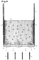
- Fig. 9
- die Unterdrückung der Wandfluoreszenz bei einem Fluoreszenzassay mit Hilfe eines Maskierungsfarbstoffes und
- Fig. 10
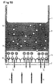
- ) die Unterdrückung der Hintergrund-fluoreszenz oder lumineszenz bei einem Fluoreszenz- oder Lumineszenzassay zur Untersuchung von Rezeptor-Ligandenbindungen mit Hilfe eines Maskierungsfarbstoffs im Überstand
- Fig. 1
- a reaction vessel for a fluorescence assay according to the prior art
- Fig. 2
- suppression of background fluorescence in a fluorescence assay with a masking dye in the supernatant
- Fig. 3
- the spectral excitation and emission for a distribution dye and the spectral absorption of the masking dye
- Fig. 4
- the location-dependent cell fluorescence without masking dye
- Fig. 5
- the location-dependent cell fluorescence with masking dye
- Fig. 6
- suppression of background fluorescence in a fluorescence assay using a separating layer
- Fig. 7
- the amplification of the luminescence by back reflection on a separation layer
- Fig. 8
- the wall fluorescence in a fluorescence assay according to the prior art
- Fig. 9
- suppression of wall fluorescence in a fluorescence assay using a masking dye and
- Fig. 10
- ) suppression of background fluorescence or luminescence in a fluorescence or luminescence assay for the investigation of receptor-ligand bonds with the aid of a masking dye in the supernatant
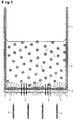
In Fig. 1 ist ein Reaktiongefäß 1 für ein Fluoreszenzassay mit einem transparenten
Boden 2 dargestellt. In dem Reaktionsgefäß 1 befindet sich eine Fluoreszenzfarbstofflösung
3, in der die Fluoreszenzfarbstoffmoleküle 4 schematisch angedeutet sind. Die
Lösung 3 wird auch als Überstand bezeichnet. Auf dem transparenten Boden 2 sind
die zu untersuchenden biologischen Zellen auf einem transparenten Träger angeordnet.
Durch den Boden 2 wird Licht (Anregungslicht) 6 eingestrahlt, um die Zellen
5 zur Fluoreszenz anzuregen. Dem von den Zellen 5 ausgestrahlten Fluoreszenzlicht 7
ist eine Hintergrundfluoreszenz-Strahlung 8 überlagert, die von den ebenfalls angeregten
Fluoreszenzfarbstoffmolekülen 4 im Überstand 3 herrührt. Für die biologischanalytische
Untersuchung und Auswertung der Zellen 5 ist aber nur das Fluoreszenzlicht
7 maßgebend. Da aber bei allen bekannten Fluoreszenz-Analysegeräten die
Hintergrundfluoreszenz 8 mit erfaßt wird, gehen kleine Fluoreszenzunterschiede der
Zellen 5 vor der starken Hintergrundfluoreszenz 8 unter, was zu einem deutlichen
Empfindlichkeitsverlust führt.In Fig. 1 is a
Dieser Nachteil kann durch das erfindungsgemäße Verfahren nach Fig. 2 dadurch
vermieden werden, daß die Hintergrundfluoreszenz durch einen Maskierungsfarbstoff
im Überstand 3 unterdrückt wird. Die in Fig. 1 vorhandene Hintergrundfluoreszenz 8
wird gem. Fig. 2 vollständig im Überstand absorbiert. Der zu dem Überstand 3 hinzugefügte
Maskierungsfarbstoff (schematisch mit 9 bezeichnet) kann entweder in gelöster
Form oder in fein verteilter disperser Phase (farbpigmentierte Systeme) vorliegen.
Bevorzugt werden jedoch lösliche Farbstoffe eingesetzt, weil hier die Zugabe
besonders einfach mit Hilfe einer Pipette erfolgen kann und weil im Gegensatz zu
einem Pigment-System die physikalischen Einflüsse der Teilchengrößenverteilung und
von Sedimentationsprozessen und Schichtdickenungleichmäßigkeiten nicht berücksichtigt
werden müssen.This disadvantage can be caused by the inventive method according to FIG. 2
avoid background fluorescence from a masking dye
is suppressed in the
An die Eigenschaften eines derartigen Farbstoffs werden folgende Anforderungen gestellt:
- bei Verwendung eines löslichen Absorptionsfarbstoffes gute Wasserlöslichkeit für den Einsatz in biologischen Assays
- keine Membrangängigkeit des Farbstoffs, um eine Anfärbung der Zellen zu vermeiden
- hohe spezifische Absorption im Excitations- und/oder Emissions-Wellenlängenbereich des Fluoreszenzfarbstoffes
- keine toxischen Nebeneffekte (Vermeidung von Zellschädigungen)
- good water solubility for use in biological assays when using a soluble absorption dye
- no dye permeability to avoid staining of the cells
- high specific absorption in the excitation and / or emission wavelength range of the fluorescent dye
- no toxic side effects (avoiding cell damage)
Als gute Wasserlöslichkeit wird eine Löslichkeit von >2 mg/ml angesehen. Die Zelltoxizität
kann mit Hilfe bekannter Testverfahren (z.B. Zytotoxizitätstest) bestimmt
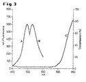
werden. Fig. 3 zeigt die optischen (spektralen) Eigenschaften eines Fluoreszenz- und
eines Maskierungsfarbstoffes in einem Diagramm. Die Kurve A zeigt die spektrale
Verteilung des Anregungslichtes, die Kurve B die spektrale Verteilung des emittierten
Fluoreszenzlichts für den handelsüblichen Verteilungsfluoreszenz-Farbstoff Bis-(1,3-dibutylbarbituricacid)trimethaneoxonol
(Dibac4(3)) und die Kurve C die spektrale
Transmission (Absorptionsspektrum) des verwendeten Maskierungsfarbstoffs (Brilliant
Black BN, C.I. 28440, Food Black 1, z.B. Sigma B-8384). Man erkennt, daß der
Maskierungsfarbstoff im Wellenlängenbereich der Anregung und Emission des Fluoreszenzfarbstoffs
fast vollständig absorbiert.A solubility of> 2 mg / ml is considered to be good water solubility. Cell toxicity can be determined using known test methods (eg cytotoxicity test). 3 shows the optical (spectral) properties of a fluorescent and a masking dye in a diagram. Curve A shows the spectral distribution of the excitation light, curve B shows the spectral distribution of the emitted fluorescent light for the commercially available distribution fluorescent dye bis- (1,3-dibutylbarbituricacid) trimethaneoxonol (Dibac 4 (3)) and curve C shows the spectral transmission ( Absorption spectrum) of the masking dye used (Brilliant Black BN, CI 28440,
Die Kontrastverbesserung bzw. Empfindlichkeitssteigerung läßt sich noch besser an Hand der Figuren 4 und 5 verstehen. Zur Demonstration der Wirkung des Maskierungsfarbstoffs auf die unspezifische Hintergrundfluoreszenz wurden zwei Videoaufnahmen von dem gleichen Bildausschnitt vor und nach Hinzufügen von 100 mg/ml des löslichen Maskierungsfarbstoffes Brilliantschwarz in Anwesenheit des potentialsensitiven Fluoreszenzfarbstoffes Dibac4(3) (5 mM) gemacht. Beide Male wurden dieselbe Videozeile bildanalytisch ausgewertet und die beiden Fluoreszenzintensitätsprofile über die identischen Ausschnitte im Reaktionsgefäß dargestellt. Der Bereich Z entspricht dabei dem Bereich, in dem sich die Zellschicht, d.h. die biologische Probe befindet, während rechts davon in der Zone Ü zum größten Teil das vom Überstand herkommende Fluoreszenzsignal gemessen wird. Der Meßbereich des Aufnahmesystems (8 bit) liegt zwischen 0 (schwarz) und 255 (weiß). Für die unmaskierte Aufnahme ergibt sich ein Kontrastverhältnis von ca. 1:3,6 (Intensitätsverhältnis der dunkelsten und hellsten Bildanteile) und im Falle der maskierten Aufnahme ein Kontrastverhältnis von ca. 1:14,4. Das entspricht einer Kontraststeigerung um den Faktor 4. The improvement in contrast or sensitivity increase can be better understood with reference to FIGS. 4 and 5. To demonstrate the effect of the masking dye on non-specific background fluorescence, two video recordings were made of the same image section before and after adding 100 mg / ml of the soluble masking dye Brilliant Black in the presence of the potential-sensitive fluorescent dye Dibac 4 (3) (5 mM). Both times, the same video line was evaluated analytically and the two fluorescence intensity profiles were shown over the identical sections in the reaction vessel. The area Z corresponds to the area in which the cell layer, ie the biological sample, is located, while the fluorescence signal from the supernatant is measured for the most part in the zone U to the right. The measuring range of the recording system (8 bit) is between 0 (black) and 255 (white). A contrast ratio of approx. 1: 3.6 (intensity ratio of the darkest and brightest parts of the image) results for unmasked recording and a contrast ratio of approx. 1: 14.4 in the case of masked recording. This corresponds to an increase in contrast by a factor of 4.
Eine alternative Möglichkeit, das Verhältnis von Nutz- zu Hintergrundsignal zu verbessern,
besteht gemäß Fig. 6 in einer Überlagerung der Zellschicht mit einer feinteiligen
optischen Trennschicht 10. Die Trennschicht 10 besteht zweckmäßig aus
einem feinteiligen anorganischen Weißpigment, wie z.B. TiO2 oder Al2O3. Hierdurch
wird nicht nur die Hintergrundfluoreszenzstrahlung aus dem Überstand 3 abgeschirmt,
sondern auch die meßbare Größe der Zellfluoreszenz durch Reflexion an den anorganischen
Teilchen verstärkt.An alternative way of improving the ratio of useful signal to background signal, according to FIG. 6, is to overlay the cell layer with a fine-particle
Alternativ kann die Trennschicht aus polymeren Latexkügelchen mit einem Durchmesser vorzugsweise im Bereich von 200 nm bis 5 mm bestehen. Geeignete Polymere sind z.B. Polystyrol, Polyurethan, Butadien Acrylnitril. Die Latexkügelchen können auch mit einem geeigneten Maskierungsfarbstoff angefärbt werden, für den die gleichen Kriterien gelten, wie für den der Lösung zugefügten Absorptionsfarbstoff (s. oben). Eine geeignete Farbstoffklasse sind z.B. ®Resoline.Alternatively, the separation layer can be made of polymeric latex beads with a diameter preferably exist in the range of 200 nm to 5 mm. Suitable polymers are e.g. Polystyrene, polyurethane, butadiene acrylonitrile. The latex beads can can also be stained with a suitable masking dye for which the same Criteria apply, such as for the absorption dye added to the solution (see above). A suitable class of dyes are e.g. ®Resoline.
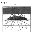
In Lumineszenz-Assays (selbstleuchtende Zellen) besteht grundsätzlich die Anforderung,
die spezifische sehr geringe Lichtintensität einer biologischen Zelle mit hoher
Empfindlichkeit zu detektieren. Durch Einbringen einer reflektierenden Trennschicht
10 gemäß Fig. 7 kann, analog zur Methode der Unterdrückung der Hintergrundfluoreszenz
(gem. Fig. 6), das Lumineszenzsignal der biologischen Zellen verstärkt werden.
Hierbei werden Strahlungsanteile 11 des ungerichteten Lumineszenzlichtes in
Richtung des Detektors reflektiert und erhöhen so das spezifische Meßsignal.In luminescence assays (self-luminous cells) there is basically the requirement
the specific very low light intensity of a biological cell with high
Detect sensitivity. By introducing a
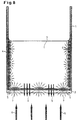
Bei einer Vielzahl anderer fluoreszenter Testverfahren an biologischen Zellen ist es im
Gegensatz zu Verteilungsfarbstoffen möglich, den Fluoreszenzfarbstoff nach Anfärbung
der Zellen durch Lösungswechsel aus dem Überstand zu entfernen. Der Fluoreszenzfarbstoff
FURA2-AM wird z.B. nach Eindringen in die Zelle in den freien Farbstoff
gespalten und verliert hierbei seine Zellmembranpermeabilität. Dadurch kommt
es zu einer Anreicherung des impermeablen Fluoreszenzfarbstoffes in der Zelle. In
diesem Fall kann der fluoreszente Überstand 3 durch eine Fluoreszenzfarbstoff-freie
Lösung 3a ausgetauscht werden, ohne die spezifische Zellfluoreszenz zu verändern.
Die unspezifische Hintergrundfluoreszenz des Überstandes wird auf diese Weise
entfernt. FURA2-AM färbt jedoch Reaktionsgefäße nachhaltig an (Wandfluoreszenz)
und erzeugt so ein anderes unspezifisches Fluoreszenzsignal, das der Hintergrund-fluoreszenz
von Verteilungsfarbstoffen vergleichbar ist. Dieser Sachverhalt ist in Fig.
8 dargestellt. In diesem Fall geht also die Hintergrundfluoreszenzstrahlung 8 auf die
an den Gefäßwänden anhaftenden Fluoreszenzfarbstoffmoleküle 4 zurück. Durch Einbringung
von Maskierungsfarbstoffen in den Fluoreszenzfarbstoff-freien Überstand 3a
kann auch dieses unspezifische Fluoreszenzsignal vollständig unterdrückt werden.In a variety of other fluorescent test methods on biological cells it is in the
Contrary to distribution dyes possible, the fluorescent dye after staining
remove the cells from the supernatant by changing the solution. The fluorescent dye
FURA2-AM is e.g. after entering the cell into the free dye
split and lose their cell membrane permeability. Because of that comes
there is an accumulation of the impermeable fluorescent dye in the cell. In
In this case, the
In Fig. 10 ist zusätzlich ein zu Fig. 2 analoges Ausführungsbeispiel dargestellt, bei
dem die auf einem transparenten Träger am Boden 2 des Reaktionsgefäßes 1 aufgebrachte
biologische Schicht aus Rezeptoren 12 besteht, die mit den im Überstand
(Lösung) 3 vorhandenen, Fluoreszenz- oder Lumineszenz-markierten Liganden 13
eine spezifische Bindung eingehen. Die gebundenen Liganden sind hier mit 14 bezeichnet.
Das durch den Boden 2 eingestrahlte Primärlicht 6 regt im Falle der nicht
maskierten Lösung die Fluoreszenz-markierten Liganden 13 und 14 zur Fluoreszenz
an. Im Falle von Lumineszenz-markierten Liganden entfällt das Primärlicht 6. Analog
zur Ausführung nach Fig. 2 wird der Lösung 3 wiederum ein Maskierungsfarbstoff
beigefügt, der dafür sorgt, daß die von den ungebundenen Liganden 13 ausgehende
Fluoreszenz- oder Lumineszenzstrahlung in der Lösung vollständig absorbiert wird.
Die am Boden 2 erfaßte Fluoreszenz- oder Lumineszenzstrahlung 15, d.h. der Meßeffekt,
geht daher ganz überwiegend auf die an die Rezeptoren 12 gebundenen Liganden
14 zurück und wird nicht durch die Hintergrundstrahlung der ungebundenen
Liganden 13 in der Lösung 3 verfälscht. Das Meßsignal ist daher ein direktes Maß für
die Stärke der Ligand-Rezeptorbindung. Dabei liegt die Schichtdicke der Rezeptorschicht
im nm-Bereich, während die Dimensionen des darüber befindlichen Überstandes
in der Größenordnung mehrerer mm liegt.FIG. 10 additionally shows an exemplary embodiment analogous to FIG. 2, at
which is applied to a transparent support at the
Gemäß Fig. 6 und 7 kann die für die Lösung durchlässige Trennschicht 10 in ganz
analoger Weise bei der Untersuchung von Liganden-Rezeptorbindungen zur Maskierung
bzw. Unterdrückung der Hintergrundfluoreszenz bzw. -lumineszenz eingesetzt
werden. In diesem Fall wird die störende Hintergrundfluoreszenz bzw. -lumineszenz
durch die Trennschicht 10 abgeschirmt und im Falle von lumineszenten Liganden
Strahlungsanteile 11 des von gebundenen Liganden stammenden Lumineszenzlichts
in Richtung auf den Detektor reflektiert und damit das Nutzsignal verstärkt. 6 and 7, the
Der in klassischen pharmakologischen Rezeptorbindungsstudien hinsichtlich seiner Bindungsstärke zu bewertende, nicht fluoreszierende bzw. lumineszierende Reaktionspartner wurde hier aus Gründen der Übersichtlichkeit nicht dargestellt.The one in classic pharmacological receptor binding studies regarding its Binding strength to be assessed, non-fluorescent or luminescent reactants was not shown here for reasons of clarity.
Claims (7)
- Process for the quantitative optical analysis of fluorescently labelled biological cells (5) which are applied to a transparent support at the bottom (2) of a reaction vessel (1) in the form of a coherent cell layer and are in contact with a solution (3) containing the fluorescent dye (4), characterized in that the fluorescent dye (4) already present in addition to a masking dye (9) which absorbs the excitation light (6) for the fluorescent dye (4) and/or its emission light (7) is added to the solution (3) and/or in that a separating layer (10) which is permeable to the solution and which absorbs and/or reflects the excitation light (6) for the fluorescent dye (4) and/or its emission light (7) is applied to the cell layer.
- Process for the quantitative optical analysis of luminescent, biological cells in the form of a coherent cell layer situated on the transparent support, characterized in that a separating layer (10) which reflects the luminescent light is applied to the cell layer.
- Process for the quantitative optical analysis of fluorescently or luminescently labelled reaction components in a reaction vessel (1) filled with a solution (3) in which a fluorescent or luminescent ligand (13) is dissolved and the solution (3) is in contact with a receptor layer (12), which is specific for this ligand (13) and is applied to a transparent support at the bottom (2) of the reaction vessel (1) or deposited thereon, whose fluorescent or luminescent radiation (7, 15), which is characteristic of the receptor-ligand binding, is detected and analysed through the transparent bottom (2), characterized in that a masking dye (9) is added to the solution (3) and/or a separating layer (10) permeable to the solution (3) is applied to the receptor layer (12), the optical properties of the masking dye (9) and/or of the separating layer (10) being selected such that the excitation light (6) for the fluorescent dye (4) of the ligand (13) present in the solution (3) and/or its fluorescent light (8) or its luminescent light is absorbed by the solution (3) or the separating layer (10) or reflected at the separating layer (10).
- Process according to Claims 1 to 3, characterized in that the separating layer (10) used is a layer of polymeric latex beads.
- Process according to Claim 4, characterized in that the polymeric latex beads are dyed with a masking dye.
- Process according to Claim 1 or 3, characterized in that a masking dye is used which possesses good water solubility and has no cytotoxic side effects.
- Process according to Claim 1 - 2, characterized in that in the case of a_ replacement of the supernatant (3) containing a fluorescent dye (4) by a fluorescent dye-free solution (3a) a masking dye is added which suppresses the non-specific fluorescence emitted from the stained reaction vessel wall.
Applications Claiming Priority (3)
| Application Number | Priority Date | Filing Date | Title |
|---|---|---|---|
| DE19621312A DE19621312A1 (en) | 1996-05-28 | 1996-05-28 | Masking of background fluorescence and signal amplification in the optical analysis of biological medical assays |
| DE19621312 | 1996-05-28 | ||
| PCT/EP1997/002662 WO1997045739A1 (en) | 1996-05-28 | 1997-05-23 | Masking background fluorescence and luminescence in optical analysis of biomedical assays |
Publications (2)
| Publication Number | Publication Date |
|---|---|
| EP0906572A1 EP0906572A1 (en) | 1999-04-07 |
| EP0906572B1 true EP0906572B1 (en) | 2002-04-03 |
Family
ID=7795451
Family Applications (1)
| Application Number | Title | Priority Date | Filing Date |
|---|---|---|---|
| EP97927032A Expired - Lifetime EP0906572B1 (en) | 1996-05-28 | 1997-05-23 | Masking background fluorescence and luminescence in optical analysis of biomedical assays |
Country Status (9)
| Country | Link |
|---|---|
| US (5) | US6420183B1 (en) |
| EP (1) | EP0906572B1 (en) |
| JP (1) | JP3452068B2 (en) |
| AT (1) | ATE215698T1 (en) |
| CA (1) | CA2256629C (en) |
| DE (2) | DE19621312A1 (en) |
| DK (1) | DK0906572T3 (en) |
| ES (1) | ES2175416T3 (en) |
| WO (1) | WO1997045739A1 (en) |
Families Citing this family (35)
| Publication number | Priority date | Publication date | Assignee | Title |
|---|---|---|---|---|
| DE19621312A1 (en) | 1996-05-28 | 1997-12-04 | Bayer Ag | Masking of background fluorescence and signal amplification in the optical analysis of biological medical assays |
| US6221612B1 (en) * | 1997-08-01 | 2001-04-24 | Aurora Biosciences Corporation | Photon reducing agents for use in fluorescence assays |
| DE19927051C2 (en) | 1999-06-14 | 2002-11-07 | November Ag Molekulare Medizin | Method and device for identifying a nucleotide sequence |
| US6181413B1 (en) * | 1999-09-03 | 2001-01-30 | Biometric Imaging, Inc. | Displacing volume in field of view |
| DE10006309A1 (en) * | 2000-02-12 | 2001-08-23 | Aventis Pharma Gmbh | Method for the identification of substances that modulate the activity of hyperpolarization-activated cation channels |
| AU2001276830A1 (en) * | 2000-06-23 | 2002-01-08 | Irm, Llc | Method and apparatus for performing an assay |
| US6552794B2 (en) * | 2001-04-04 | 2003-04-22 | Applied Spectral Imaging Ltd. | Optical detection method for improved sensitivity |
| WO2003005005A1 (en) * | 2001-07-03 | 2003-01-16 | Hitachi, Ltd. | Biological sample optical measuring method and biological sample optical measuring apparatus |
| JP4150374B2 (en) * | 2003-02-26 | 2008-09-17 | 富士通株式会社 | Arrayed waveguide type wavelength multiplexer / demultiplexer |
| US6972184B2 (en) * | 2003-12-23 | 2005-12-06 | Millipore Corporation | Cell motility assay |
| WO2005095929A1 (en) * | 2004-03-30 | 2005-10-13 | Hamamatsu Photonics K.K. | Masking member, light measuring method, light measuring kit and light measuring container |
| DE102004016361B4 (en) * | 2004-04-01 | 2006-07-06 | Cybio Ag | Optical analyzer for fluorescence measurements on multiprobe carriers |
| US7371534B2 (en) * | 2004-05-25 | 2008-05-13 | Discoverx Corporation | Sensitive intracellular calcium assay |
| FR2873445A1 (en) * | 2004-07-26 | 2006-01-27 | Genewave Soc Par Actions Simpl | DEVICE FOR DETECTING THE FLUORESCENCE EMITTED BY CHROMOPHORIC ELEMENTS IN WELLS OF A MULTI-WELL PLATE |
| US8396731B2 (en) * | 2005-12-30 | 2013-03-12 | Sap Ag | Architectural design for service procurement application software |
| EP1991873B1 (en) * | 2006-02-21 | 2013-10-16 | Harri HÄRMÄ | Separation-free assay method |
| US9097730B2 (en) | 2007-04-13 | 2015-08-04 | Aat Bioquest, Inc. | Fluorescein lactone ion indicators and their applications |
| US20080254498A1 (en) | 2007-04-13 | 2008-10-16 | Abd Bioquest, Inc. | Fluorescent ion indicators and their applications |
| US9279817B2 (en) | 2007-04-13 | 2016-03-08 | Aat Bioquest, Inc. | Carbofluorescein lactone ion indicators and their applications |
| WO2009105583A1 (en) * | 2008-02-19 | 2009-08-27 | Anstron Technologies Company | Fluorescence resonance energy transfer (fret) binding assays that are surface-based |
| US7994485B2 (en) * | 2008-04-08 | 2011-08-09 | Carestream Health, Inc. | Apparatus and method for fluorescence measurements using spatially structured illumination |
| US20090270398A1 (en) * | 2008-04-21 | 2009-10-29 | Institute For Oneworld Health | Compounds, Compositions and Methods Comprising Pyridazine Derivatives |
| US20090264433A1 (en) * | 2008-04-21 | 2009-10-22 | Institute For Oneworld Health | Compounds, Compositions and Methods Comprising Triazine Derivatives |
| WO2009131951A2 (en) * | 2008-04-21 | 2009-10-29 | Institute For Oneworld Health | Compounds, compositions and methods comprising isoxazole derivatives |
| EP2278879B1 (en) * | 2008-04-21 | 2016-06-15 | PATH Drug Solutions | Compounds, compositions and methods comprising oxadiazole derivatives |
| US20110237528A1 (en) * | 2008-09-19 | 2011-09-29 | Institute For Oneworld Health | Compositions and methods comprising imidazole and triazole derivatives |
| US8511216B2 (en) * | 2009-03-30 | 2013-08-20 | Kanzaki Kokyukoki Mfg. Co., Ltd. | Hydraulic actuator unit |
| US8343976B2 (en) * | 2009-04-20 | 2013-01-01 | Institute For Oneworld Health | Compounds, compositions and methods comprising pyrazole derivatives |
| EP2577311A1 (en) * | 2010-05-31 | 2013-04-10 | Boehringer Ingelheim Microparts GmbH | Method and device for optical examination |
| WO2012027331A1 (en) | 2010-08-27 | 2012-03-01 | Ironwood Pharmaceuticals, Inc. | Compositions and methods for treating or preventing metabolic syndrome and related diseases and disorders |
| CN104755931B (en) | 2012-05-02 | 2016-08-31 | 查尔斯河实验室公司 | For the method detecting the living cells in cell sample |
| US8993259B2 (en) | 2012-05-02 | 2015-03-31 | Charles River Laboratories, Inc. | Method of viability staining with membrane permeable fluorescent dye and membrane impermeable fluorescence quencher |
| WO2013166338A2 (en) | 2012-05-02 | 2013-11-07 | Charles River Laboratories, Inc. | Cell capture system and use thereof |
| FR3019900B1 (en) | 2014-04-09 | 2017-12-22 | Bio-Rad Innovations | USE OF ABSORBENT PARTICLES FOR IMPROVING SIGNAL DETECTION IN A METHOD OF ANALYSIS |
| US9810700B1 (en) | 2017-05-31 | 2017-11-07 | Aat Bioquest, Inc. | Fluorogenic calcium ion indicators and methods of using the same |
Family Cites Families (32)
| Publication number | Priority date | Publication date | Assignee | Title |
|---|---|---|---|---|
| US3506827A (en) * | 1968-01-22 | 1970-04-14 | James R Alburger | Method of masking fluorescence in fluorescent dye tracer inspection process materials |
| US3785735A (en) * | 1972-01-19 | 1974-01-15 | Bio Physics Systems Inc | Photoanalysis method |
| US4476231A (en) | 1981-07-22 | 1984-10-09 | International Remote Imaging Systems, Inc. | Method of analyzing the distribution of a reagent between particles and liquid in a suspension |
| DE3213183A1 (en) | 1982-04-08 | 1983-10-20 | Max Planck Gesellschaft zur Förderung der Wissenschaften e.V., 3400 Göttingen | ARRANGEMENT FOR OPTICAL MEASUREMENT OF PHYSICAL SIZES |
| JPS5934154A (en) * | 1982-08-19 | 1984-02-24 | Konishiroku Photo Ind Co Ltd | Determination by means of immunoanalytical element |
| US5082768A (en) * | 1984-06-15 | 1992-01-21 | Mast Immunosystems, Inc. | Attenuator to suppress extraneous light in luminescent specific-binding assays |
| US4665024A (en) * | 1984-10-01 | 1987-05-12 | Becton, Dickinson And Company | Fluorescent gram stain |
| US4639421A (en) * | 1984-10-01 | 1987-01-27 | Becton, Dickinson And Company | Fluorescent gram stain |
| US4716121A (en) * | 1985-09-09 | 1987-12-29 | Ord, Inc. | Fluorescent assays, including immunoassays, with feature of flowing sample |
| CA1291031C (en) * | 1985-12-23 | 1991-10-22 | Nikolaas C.J. De Jaeger | Method for the detection of specific binding agents and their correspondingbindable substances |
| US4891324A (en) * | 1987-01-07 | 1990-01-02 | Syntex (U.S.A.) Inc. | Particle with luminescer for assays |
| US5326692B1 (en) * | 1992-05-13 | 1996-04-30 | Molecular Probes Inc | Fluorescent microparticles with controllable enhanced stokes shift |
| US5830766A (en) * | 1990-05-23 | 1998-11-03 | Ares-Serono Research & Development Ltd. Partnership | Enhanced signal-to-noise ratio and sensitivity optical immunoassay |
| US5164301A (en) * | 1990-06-22 | 1992-11-17 | Difco Laboratories | Process and kit for detecting microbial metabolism |
| US5264565A (en) * | 1991-01-22 | 1993-11-23 | Affymax Technologies, N.V. | Nucleic acid encoding the D2 /Ml chimeric receptor |
| FI913443A0 (en) * | 1991-07-17 | 1991-07-17 | Rolf Kroneld | METHOD, APPARAT OCH INDIKATOR FOER ATT INDIKERA FLYKTIGA KOLVAETEN ELLER MILJOEGIFTER I VATTEN OCH VAETSKOR. |
| EP0643836B1 (en) * | 1991-12-13 | 1999-09-08 | Bion Diagnostic Sciences, Inc. | Agglutination assays and kits employing colloidal dyes |
| WO1994002642A1 (en) * | 1992-07-17 | 1994-02-03 | Aprogenex, Inc. | Background-reducing compounds for probe-mediated in-situ fluorimetric assays |
| WO1994017388A1 (en) * | 1993-01-26 | 1994-08-04 | Fci-Fiberchem, Inc. | Ion selective fluorosensor based on the inner filter effect |
| US5556764A (en) * | 1993-02-17 | 1996-09-17 | Biometric Imaging, Inc. | Method and apparatus for cell counting and cell classification |
| US5545535A (en) * | 1993-04-13 | 1996-08-13 | Molecular Probes, Inc. | Fluorescent assay for bacterial gram reaction |
| JP3326708B2 (en) * | 1993-08-31 | 2002-09-24 | 日水製薬株式会社 | Optical measuring device and method thereof |
| DE69519783T2 (en) * | 1994-04-29 | 2001-06-07 | Perkin Elmer Corp | METHOD AND DEVICE FOR REAL-TIME DETECTION OF PRODUCTS OF NUCLEIC ACID AMPLIFICATION |
| AU2636995A (en) * | 1994-05-18 | 1995-12-05 | Research And Development Institute, Inc. | Simple, rapid method for the detection, identification and enumeration of specific viable microorganisms |
| AU3961495A (en) | 1994-10-04 | 1996-04-26 | Sotomayor, Tevic Emar | Composition and treatment for hyperimmunization with non heat-killed bacteria |
| EP1089078B1 (en) * | 1994-10-20 | 2007-02-28 | Sysmex Corporation | Reagent and method for analyzing solid components in urine |
| DE19621312A1 (en) | 1996-05-28 | 1997-12-04 | Bayer Ag | Masking of background fluorescence and signal amplification in the optical analysis of biological medical assays |
| US5772999A (en) | 1996-07-30 | 1998-06-30 | Dcv Biologics, L.P. | Method of preventing, countering, or reducing NSAID-induced gastrointestinal damage by administering milk or egg products from hyperimmunized animals |
| US6200762B1 (en) | 1997-08-01 | 2001-03-13 | Aurora Biosciences Corporation | Photon reducing agents and compositions for fluorescence assays |
| US6221612B1 (en) | 1997-08-01 | 2001-04-24 | Aurora Biosciences Corporation | Photon reducing agents for use in fluorescence assays |
| US6214563B1 (en) | 1997-08-01 | 2001-04-10 | Aurora Biosciences Corporation | Photon reducing agents for reducing undesired light emission in assays |
| US6210910B1 (en) * | 1998-03-02 | 2001-04-03 | Trustees Of Tufts College | Optical fiber biosensor array comprising cell populations confined to microcavities |
-
1996
- 1996-05-28 DE DE19621312A patent/DE19621312A1/en not_active Withdrawn
-
1997
- 1997-05-23 US US09/194,099 patent/US6420183B1/en not_active Expired - Lifetime
- 1997-05-23 CA CA002256629A patent/CA2256629C/en not_active Expired - Lifetime
- 1997-05-23 ES ES97927032T patent/ES2175416T3/en not_active Expired - Lifetime
- 1997-05-23 AT AT97927032T patent/ATE215698T1/en active
- 1997-05-23 EP EP97927032A patent/EP0906572B1/en not_active Expired - Lifetime
- 1997-05-23 DE DE59706875T patent/DE59706875D1/en not_active Expired - Lifetime
- 1997-05-23 DK DK97927032T patent/DK0906572T3/en active
- 1997-05-23 WO PCT/EP1997/002662 patent/WO1997045739A1/en active IP Right Grant
- 1997-05-23 JP JP54157897A patent/JP3452068B2/en not_active Expired - Lifetime
-
2001
- 2001-09-28 US US09/966,137 patent/US7063952B2/en not_active Expired - Lifetime
- 2001-09-28 US US09/966,522 patent/US8178359B2/en not_active Expired - Fee Related
-
2002
- 2002-10-03 US US10/263,607 patent/US7138280B2/en not_active Expired - Fee Related
-
2008
- 2008-08-27 US US12/199,317 patent/US7615376B2/en not_active Expired - Fee Related
Also Published As
| Publication number | Publication date |
|---|---|
| JP3452068B2 (en) | 2003-09-29 |
| US7138280B2 (en) | 2006-11-21 |
| US20030092081A1 (en) | 2003-05-15 |
| US7063952B2 (en) | 2006-06-20 |
| DE59706875D1 (en) | 2002-05-08 |
| ATE215698T1 (en) | 2002-04-15 |
| EP0906572A1 (en) | 1999-04-07 |
| US6420183B1 (en) | 2002-07-16 |
| DE19621312A1 (en) | 1997-12-04 |
| DK0906572T3 (en) | 2002-07-08 |
| US20020022274A1 (en) | 2002-02-21 |
| US8178359B2 (en) | 2012-05-15 |
| WO1997045739A1 (en) | 1997-12-04 |
| ES2175416T3 (en) | 2002-11-16 |
| US20020015969A1 (en) | 2002-02-07 |
| US20020009754A1 (en) | 2002-01-24 |
| US7615376B2 (en) | 2009-11-10 |
| CA2256629C (en) | 2003-07-22 |
| JP2000512746A (en) | 2000-09-26 |
| US20080318270A1 (en) | 2008-12-25 |
| CA2256629A1 (en) | 1997-12-04 |
Similar Documents
| Publication | Publication Date | Title |
|---|---|---|
| EP0906572B1 (en) | Masking background fluorescence and luminescence in optical analysis of biomedical assays | |
| DE69722800T2 (en) | SAMPLE ANALYSIS METHOD BY DETERMINING A FUNCTION OF SPECIFIC BRIGHTNESS | |
| EP1000345B1 (en) | Method and device for referencing fluorescence intensity signals | |
| DE69819916T2 (en) | DEVICE AND METHOD FOR IMAGING SAMPLES MARKED WITH LIGHT-DIVERING SUBSTANCE | |
| DE19649048C1 (en) | Particle identification method for enzyme-linked immunoassay using fast Fourier transform | |
| DE60034315T2 (en) | CHEMICAL AND BIOCHEMICAL DETECTION METHOD AND DEVICE | |
| DE69829606T2 (en) | Reagent and method for classification and enumeration of leukocytes | |
| DE3322373C2 (en) | Test means and methods for the detection of antigens and / or antibodies | |
| DE2628158B2 (en) | Method for fluorescence spectroscopy of a target substance | |
| DE10223438B4 (en) | Fluorescence-measuring system | |
| DE4210970C2 (en) | Process for the simultaneous optical qualitative and quantitative detection of different molecules of a mixture marked with fluorochromes or fluorogens by means of laser spectroscopy | |
| EP0979402B1 (en) | Method for optical detection of analyte molecules in a natural biological medium | |
| EP1461600A1 (en) | Method and/or system for identifying fluorescent, luminescent and/or absorbing substances on and/or in sample carriers | |
| DE60130452T2 (en) | Reference device for evaluating the function of a confocal laser scanning microscope, and method and system for carrying out the evaluation | |
| DE19903576A1 (en) | Quantitative determination of analytes in a heterogeneous system | |
| DE19943704C1 (en) | Affinity sensor for the detection of biological and / or chemical species and its use | |
| DE19822452A1 (en) | Method and device for determining the concentration, determination of adsorption and binding kinetics and equilibrium and binding constants of molecules by luminescence measurements | |
| WO2004031747A1 (en) | Method and device for the detection of at least one luminescent substance | |
| DE69937403T2 (en) | BINDING ASSAY USING OPTICAL RESONANCE OF COLLOIDAL PARTICLES | |
| DE102020134515A1 (en) | Optical sensor element, optical oxygen sensor and method for monitoring the function of an optical oxygen sensor | |
| DE102004051830B4 (en) | Multifunctional reference system for analyte determination by fluorescence | |
| WO2001025759A1 (en) | Method and device for determining substances such as e.g., dna sequences, in a sample | |
| DE4104014A1 (en) | Determining absolute intracellular calcium ion concn. - by staining with calcium sensitive fluorescent dye then scanning fluorescence decay of complexed and free dye | |
| DE112021006503T5 (en) | OPTICAL MODULE | |
| DE102004027957A1 (en) | Investigation of interactions between biomolecules of differing types, attaches biomolecules to backlit biochip using chemical spacers, and includes measurements with total internal reflection |
Legal Events
| Date | Code | Title | Description |
|---|---|---|---|
| PUAI | Public reference made under article 153(3) epc to a published international application that has entered the european phase |
Free format text: ORIGINAL CODE: 0009012 |
|
| 17P | Request for examination filed |
Effective date: 19981228 |
|
| AK | Designated contracting states |
Kind code of ref document: A1 Designated state(s): AT BE CH DE DK ES FI FR GB IT LI NL SE |
|
| 17Q | First examination report despatched |
Effective date: 20001219 |
|
| GRAG | Despatch of communication of intention to grant |
Free format text: ORIGINAL CODE: EPIDOS AGRA |
|
| GRAG | Despatch of communication of intention to grant |
Free format text: ORIGINAL CODE: EPIDOS AGRA |
|
| GRAH | Despatch of communication of intention to grant a patent |
Free format text: ORIGINAL CODE: EPIDOS IGRA |
|
| REG | Reference to a national code |
Ref country code: GB Ref legal event code: IF02 |
|
| GRAH | Despatch of communication of intention to grant a patent |
Free format text: ORIGINAL CODE: EPIDOS IGRA |
|
| GRAA | (expected) grant |
Free format text: ORIGINAL CODE: 0009210 |
|
| AK | Designated contracting states |
Kind code of ref document: B1 Designated state(s): AT BE CH DE DK ES FI FR GB IT LI NL SE |
|
| REF | Corresponds to: |
Ref document number: 215698 Country of ref document: AT Date of ref document: 20020415 Kind code of ref document: T |
|
| REG | Reference to a national code |
Ref country code: CH Ref legal event code: NV Representative=s name: E. BLUM & CO. PATENTANWAELTE Ref country code: CH Ref legal event code: EP |
|
| REF | Corresponds to: |
Ref document number: 59706875 Country of ref document: DE Date of ref document: 20020508 |
|
| REG | Reference to a national code |
Ref country code: DK Ref legal event code: T3 |
|
| ET | Fr: translation filed | ||
| GBT | Gb: translation of ep patent filed (gb section 77(6)(a)/1977) |
Effective date: 20020625 |
|
| REG | Reference to a national code |
Ref country code: ES Ref legal event code: FG2A Ref document number: 2175416 Country of ref document: ES Kind code of ref document: T3 |
|
| PLBE | No opposition filed within time limit |
Free format text: ORIGINAL CODE: 0009261 |
|
| STAA | Information on the status of an ep patent application or granted ep patent |
Free format text: STATUS: NO OPPOSITION FILED WITHIN TIME LIMIT |
|
| 26N | No opposition filed |
Effective date: 20030106 |
|
| REG | Reference to a national code |
Ref country code: CH Ref legal event code: PUE Owner name: BAYER HEALTHCARE AG Free format text: BAYER AG##51368 LEVERKUSEN (DE) -TRANSFER TO- BAYER HEALTHCARE AG##51368 LEVERKUSEN (DE) |
|
| REG | Reference to a national code |
Ref country code: GB Ref legal event code: 732E |
|
| NLS | Nl: assignments of ep-patents |
Owner name: BAYER HEALTHCARE AG |
|
| REG | Reference to a national code |
Ref country code: FR Ref legal event code: TP |
|
| REG | Reference to a national code |
Ref country code: CH Ref legal event code: PFA Owner name: BAYER HEALTHCARE AG Free format text: BAYER HEALTHCARE AG# #51368 LEVERKUSEN (DE) -TRANSFER TO- BAYER HEALTHCARE AG# #51368 LEVERKUSEN (DE) |
|
| REG | Reference to a national code |
Ref country code: CH Ref legal event code: PUE Owner name: BAYER SCHERING PHARMA AKTIENGESELLSCHAFT Free format text: BAYER HEALTHCARE AG# #51368 LEVERKUSEN (DE) -TRANSFER TO- BAYER SCHERING PHARMA AKTIENGESELLSCHAFT#MUELLERSTRASSE 178#13353 BERLIN (DE) |
|
| NLS | Nl: assignments of ep-patents |
Owner name: BAYER SCHERING PHARMA AKTIENGESELLSCHAFT Effective date: 20090918 |
|
| REG | Reference to a national code |
Ref country code: FR Ref legal event code: TP |
|
| REG | Reference to a national code |
Ref country code: DE Ref legal event code: R081 Ref document number: 59706875 Country of ref document: DE Owner name: BAYER INTELLECTUAL PROPERTY GMBH, DE Free format text: FORMER OWNER: BAYER SCHERING PHARMA AKTIENGESELLSCHAFT, 13353 BERLIN, DE Effective date: 20120612 |
|
| REG | Reference to a national code |
Ref country code: CH Ref legal event code: PUE Owner name: BAYER INTELLECTUAL PROPERTY GMBH, DE Free format text: FORMER OWNER: BAYER SCHERING PHARMA AKTIENGESELLSCHAFT, DE |
|
| BECH | Be: change of holder |
Owner name: BAYER INTELLECTUAL PROPERTY G.M.B.H. Effective date: 20130211 |
|
| REG | Reference to a national code |
Ref country code: FR Ref legal event code: TP Owner name: BAYER INTELLECTUAL PROPERTY GMBH, DE Effective date: 20130130 Ref country code: FR Ref legal event code: CD Owner name: BAYER INTELLECTUAL PROPERTY GMBH, DE Effective date: 20130130 |
|
| REG | Reference to a national code |
Ref country code: ES Ref legal event code: PC2A Owner name: BAYER INTELLECTUAL PROPERTY GMBH Effective date: 20130319 |
|
| REG | Reference to a national code |
Ref country code: DE Ref legal event code: R081 Ref document number: 59706875 Country of ref document: DE Owner name: BAYER INTELLECTUAL PROPERTY GMBH, DE Free format text: FORMER OWNER: BAYER PHARMA AKTIENGESELLSCHAFT, 13353 BERLIN, DE Effective date: 20130410 |
|
| REG | Reference to a national code |
Ref country code: NL Ref legal event code: TD Effective date: 20130625 Ref country code: NL Ref legal event code: SD Effective date: 20130625 |
|
| REG | Reference to a national code |
Ref country code: GB Ref legal event code: 732E Free format text: REGISTERED BETWEEN 20131212 AND 20131218 |
|
| REG | Reference to a national code |
Ref country code: AT Ref legal event code: PC Ref document number: 215698 Country of ref document: AT Kind code of ref document: T Owner name: BAYER INTELLECTUAL PROPERTY GMBH, DE Effective date: 20131218 |
|
| REG | Reference to a national code |
Ref country code: FR Ref legal event code: PLFP Year of fee payment: 20 |
|
| PGFP | Annual fee paid to national office [announced via postgrant information from national office to epo] |
Ref country code: NL Payment date: 20160426 Year of fee payment: 20 |
|
| PGFP | Annual fee paid to national office [announced via postgrant information from national office to epo] |
Ref country code: FI Payment date: 20160509 Year of fee payment: 20 Ref country code: GB Payment date: 20160518 Year of fee payment: 20 Ref country code: DE Payment date: 20160518 Year of fee payment: 20 Ref country code: CH Payment date: 20160511 Year of fee payment: 20 Ref country code: ES Payment date: 20160427 Year of fee payment: 20 |
|
| PGFP | Annual fee paid to national office [announced via postgrant information from national office to epo] |
Ref country code: BE Payment date: 20160425 Year of fee payment: 20 Ref country code: IT Payment date: 20160524 Year of fee payment: 20 Ref country code: FR Payment date: 20160426 Year of fee payment: 20 Ref country code: SE Payment date: 20160511 Year of fee payment: 20 Ref country code: AT Payment date: 20160425 Year of fee payment: 20 Ref country code: DK Payment date: 20160510 Year of fee payment: 20 |
|
| REG | Reference to a national code |
Ref country code: DE Ref legal event code: R071 Ref document number: 59706875 Country of ref document: DE |
|
| REG | Reference to a national code |
Ref country code: NL Ref legal event code: MK Effective date: 20170522 |
|
| REG | Reference to a national code |
Ref country code: DK Ref legal event code: EUP Effective date: 20170523 |
|
| REG | Reference to a national code |
Ref country code: CH Ref legal event code: PL |
|
| REG | Reference to a national code |
Ref country code: GB Ref legal event code: PE20 Expiry date: 20170522 |
|
| REG | Reference to a national code |
Ref country code: SE Ref legal event code: EUG |
|
| REG | Reference to a national code |
Ref country code: AT Ref legal event code: MK07 Ref document number: 215698 Country of ref document: AT Kind code of ref document: T Effective date: 20170523 |
|
| PG25 | Lapsed in a contracting state [announced via postgrant information from national office to epo] |
Ref country code: GB Free format text: LAPSE BECAUSE OF EXPIRATION OF PROTECTION Effective date: 20170522 |
|
| REG | Reference to a national code |
Ref country code: ES Ref legal event code: FD2A Effective date: 20170830 |
|
| PG25 | Lapsed in a contracting state [announced via postgrant information from national office to epo] |
Ref country code: ES Free format text: LAPSE BECAUSE OF EXPIRATION OF PROTECTION Effective date: 20170524 |