EP0906433B1 - Protein tyrosin-phosphatasen von hematopoietischen zellen - Google Patents
Protein tyrosin-phosphatasen von hematopoietischen zellen Download PDFInfo
- Publication number
- EP0906433B1 EP0906433B1 EP97916257A EP97916257A EP0906433B1 EP 0906433 B1 EP0906433 B1 EP 0906433B1 EP 97916257 A EP97916257 A EP 97916257A EP 97916257 A EP97916257 A EP 97916257A EP 0906433 B1 EP0906433 B1 EP 0906433B1
- Authority
- EP
- European Patent Office
- Prior art keywords
- ptp
- cells
- ptp hsc
- hsc
- seq
- Prior art date
- Legal status (The legal status is an assumption and is not a legal conclusion. Google has not performed a legal analysis and makes no representation as to the accuracy of the status listed.)
- Expired - Lifetime
Links
Images


Classifications
-
- C—CHEMISTRY; METALLURGY
- C12—BIOCHEMISTRY; BEER; SPIRITS; WINE; VINEGAR; MICROBIOLOGY; ENZYMOLOGY; MUTATION OR GENETIC ENGINEERING
- C12N—MICROORGANISMS OR ENZYMES; COMPOSITIONS THEREOF; PROPAGATING, PRESERVING, OR MAINTAINING MICROORGANISMS; MUTATION OR GENETIC ENGINEERING; CULTURE MEDIA
- C12N9/00—Enzymes; Proenzymes; Compositions thereof; Processes for preparing, activating, inhibiting, separating or purifying enzymes
- C12N9/14—Hydrolases (3)
- C12N9/16—Hydrolases (3) acting on ester bonds (3.1)
-
- A—HUMAN NECESSITIES
- A61—MEDICAL OR VETERINARY SCIENCE; HYGIENE
- A61P—SPECIFIC THERAPEUTIC ACTIVITY OF CHEMICAL COMPOUNDS OR MEDICINAL PREPARATIONS
- A61P35/00—Antineoplastic agents
-
- A—HUMAN NECESSITIES
- A61—MEDICAL OR VETERINARY SCIENCE; HYGIENE
- A61P—SPECIFIC THERAPEUTIC ACTIVITY OF CHEMICAL COMPOUNDS OR MEDICINAL PREPARATIONS
- A61P35/00—Antineoplastic agents
- A61P35/02—Antineoplastic agents specific for leukemia
-
- A—HUMAN NECESSITIES
- A61—MEDICAL OR VETERINARY SCIENCE; HYGIENE
- A61P—SPECIFIC THERAPEUTIC ACTIVITY OF CHEMICAL COMPOUNDS OR MEDICINAL PREPARATIONS
- A61P43/00—Drugs for specific purposes, not provided for in groups A61P1/00-A61P41/00
-
- A—HUMAN NECESSITIES
- A61—MEDICAL OR VETERINARY SCIENCE; HYGIENE
- A61K—PREPARATIONS FOR MEDICAL, DENTAL OR TOILETRY PURPOSES
- A61K38/00—Medicinal preparations containing peptides
Definitions
- the present invention concerns novel protein tyrosine phosphatases. More particularly, the invention concerns non-receptor protein tyrosine phosphatases of hematopoietic stem cells (PTP HSC's).
- PTP HSC's hematopoietic stem cells
- the ability of the hematopoietic stem cell to function as a source of committed progenitors throughout the lifetime of the organism is, at present, a poorly understood phenomenon.
- the major characteristic of the hematopoietic stem cell is its ability to self renew in the absence of differentiation (Morrison et al ., Ann. Rev. Cell Dev. Biol. , 11 , 35-71 [1995]).
- This self renewal phenomenon is especially remarkable in light of the fact that the hematopoietic stroma, which is in close physical contact with the stem cell, is known to be a source that is rich in factors which mediate the growth and differentiation of hematopoietic progenitors (Deryugina and Muller-Sieberg, Crit. Rev. in Immunol.
- Such growth factors in addition to many others, are known to induce the expansion and differentiation of stem cells, and these endothelial cell lines induced a rapid expansion and differentiation of embryonic hematopoietic stem cells along the myeloid pathway, although very early progenitor cells are also amplified by these stromal cells (C. Fennie and L. Lasky-unpublished data).
- hematopoietic growth factors are very diverse both structurally and functionally, they are all believed to play role in mediating protein phosphorylation (Paulson and Bernstein, Semin lmmunol . 7 (4), 267-77 [1995]).
- This protein modification can occur via direct means, such as in the cases of the stem cell factor and FLT-3 receptors, both of which have intrinsic tyrosine kinase activity, or via indirect means, as is the case of the hematopoietic/cytokine growth factor receptors for, for example, IL-3, EPO and TPO.
- tyrosine phosphorylation is indirectly accomplished by the activation of the JAK kinases, which occurs after growth factor mediated receptor dimerization (Ihle et al ., Annu. Rev. Immunol. 13 , 369-398 [1995]).
- JAK kinases the JAK kinases
- diverse complex pathways of protein phosphorylation are stimulated upon receptor binding.
- the intrinsic tyrosine kinase receptors mediate their signals via an elaborate series of tyrosine phosphorylation events which ultimately activate the RAS signaling pathway (Fantl et al ., Ann. Rev. Biochem. 62, 453-481 [1993]).
- This pathway eventually leads to the activation of the serine/threonine specific MAP kinase pathway which results in transcriptional activation.
- hematopoietic growth factor-induced receptor dimerization mediates more direct activation events.
- the stimulation of the JAK kinases by receptor binding leads to the tyrosine phosphorylation and subsequent dimerization of various STAT proteins.
- These activated STAT proteins than migrate to the nucleus, bind to STAT responsive sites in the nuclear DNA and induce transcription of differentiation and growth specific genes.
- a major effect of the growth factors produced by the hematopoietic stroma is to mediate the activation of various cellular pathways by protein phosphorylation.
- PTPs contain a phosphatase domain including a subset of highly conserved amino acids, and a recent crystal structure analysis of PTP 1B complexed with a tyrosine phosphorylated peptide revealed that many of these conserved residues are involved with substrate recognition and tyrosine dephosphorylation (Jia et al ., Science 268 (5218), 1754-1758 [1995]).
- PTPs fall into two general categories: receptor type and non-receptor type.
- the receptor type PTPs have variously sized extracellular domains and, generally, two intracellular phosphatase domains Walton and Doxin, supra ; Sun and Tonks, supra.
- the extracellular domains often contain a number of motifs that are generally utilized in cell adhesion including immunoglobulin domains and fibronectin-like regions . Many of these PTP s appear to function as homotypic and heterotypic sensors of the extracellular space, and they have been hypothesized to play roles in contact inhibition, cell guidance and other intercellular functions (Brady-Kalnay and Tonks, Curr. Opin. Cell. Biol. 7 (5), 65-657 [1995]).
- the non-receptor PTPs are generally intracellular enzymes. They have various cellular localizations, depending upon the types of domains they contain, and some of the enzymes contain SH2 motifs which allow them to interact intimately with phosphotyrosine residues.
- non-receptor PTPs While many of the non-receptor PTPs are in various cytoplasmic locations, a small number of these enzymes are found in the nucleus (Flores et al ., Mol. Cell. Biol. 14 (7), 4938-46 [1994]). Many non-receptor PTPs appear to function as both activators as well as inhibitors of diverse tyrosine phosphorylated proteins. A subset appear to play important rotes in hematopoiesis.
- the motheaten mouse which has a phenotype of lethal myeloid amplification and inflammation, has been found to have a mutation in the PTP I C gene (Schulz et al ., Cell 73 (7), 1445-54 [1993]); (McCulloch and Siminovitch, Adv. Exp. Med. Biol. 365 , 145-54 [1994]).
- the level of tyrosine phosphorylation of the EPO receptor appears to be in part controlled by the PTP 1C enzyme as well (Klingmuller et al ., Cell 80 (5), 729-38 [1995]).
- PTPTY42 a non-PTP termed PTPTY42.
- WO 01/13989 describes methods and compositions for treating malignancies, using a DNA sequence encoding a PTPase.
- PTP HSC tyrosine phosphorylated proteins
- the present invention concerns an isolated non-receptor protein tyrosine phosphatase of hematopoietic stem cells (PTP HSC) as defined in claim 1 or claim 6.
- PTP HSC hematopoietic stem cells
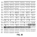
- a preferred group of the PTP HSC proteins of the present invention includes a protein comprising the amino acid sequence shown in Figure 1 (SEQ. ID. NO:2); a protein comprising the amino acid sequence shown in Figure 8 (SEQ. ID. NO: 17), a further mammalian homologue of either protein, as defined in claim 6; and derivatives of the foregoing proteins retaining the ability of tyrosine dephosphorylation in hematopoietic stem cells or progenitor cells.
- the PTP HSCs including derivatives (e.g. amino acid sequence variants) of the native proteins, preferably have an active N-terminal tyrosine phosphatase domain, retaining a serine residue at a position corresponding to amino acid position 37 in Figure 1, and retaining an active site cysteine residue at a position corresponding to amino acid position 229 in Figure 1, a region rich in serine, threonine, and proline, and a carboxy-terminal region showing at least about 80% sequence homology with the amino acid sequence between positions 430 and 451 in Figure 1.
- such derivatives have at least about 65% overall sequence homology with the amino acid sequence shown in Figure 1 or Figure 8 and retain the ability of tyrosine dephosphorylation in hematopoietic stem cells or progenitor cells.
- the present invention concerns agonists and antagonists of PTP HSCs.
- the invention concerns isolated nucleic acid molecules encoding the PTP HSCs herein.
- the invention concerns vectors comprising nucleic acid encoding the PTP HSCs herein, operably linked to control sequences recognized by a host cell transformed with the vector, and to cells transformed with such vectors.
- antibodies capable of specific binding to the PTP HSCs of this invention, and hybridoma cell lines producing such antibodies.
- the antibodies may be agonist antibodies, which stimulate the ability of the native PTP HSCs of the present invention to dephosphorylate tyrosines, or antagonist antibodies, which block this activity.
- the present invention further concerns an assay for identifying an antagonist or an agonist of a PTP HSC of the present invention, which comprises contacting the phosphatase domain of the PTP HSC with a candidate antagonist or agonist, and monitoring the ability of the phosphatase domain to dephosphorylate tyrosine residues.
- the invention further concerns a method for the differentiation of undifferentiated malignant hemopoietic (e.g. leukemia) cells in cell culture, comprising contacting said cells with an antagonist antibody of a PTP HSC of the present invention.
- undifferentiated malignant hemopoietic e.g. leukemia
- the invention concerns a method for the induction of hematopoietic stem cell differentiation in cell culture, comprising contacting said stem cells with an antagonist antibody of a PTP HSC of the present invention.
- the invention concerns a method for expansion undifferentiated hematopoietic stems cells in cell culture, comprising cultivating stem cells in the presence of a PTP HSC of the present invention or an agonist antibody specifically binding a native PTP HSC.
- the invention concerns the manufacture of a medicament, for example for the expansion of undifferentiated stem cells in vivo in one example, the medicament is for administering to a patient an agonist antibody of PTP HSC of the present invention ; and a stem cell growth factor.
- non-receptor protein tyrosine phosphatase of hematopoietic stem cells tyrosine phosphatase of hematopoietic stem cells
- PTP HSC a native intracellular protein tyrosine phosphatase which (1) is expressed predominantly in early hematopoietic stem and progenitor cells; (2) predominantly lacks expression in adult tissues; (3) comprises an N-terminal tyrosine phosphatase domain, followed by a region rich in serine, threonine, and proline, and a carboxy terminal region of about 15 to 25 amino acids rich in basic amino acid residues; and (4) is capable of tyrosine dephosphorylation in hematopoietic progenitor cells, and functional derivatives of such native tyrosine phosphatase.
- native tyrosine phosphatase in this context refers to a naturally occurring tyrosine phosphatase, having the described properties, of any human or non-human animal species, with or without the initiating methionine, whether purified from native source, synthesized, produced by recombinant DNA technology or by any combination of these and/or other methods.
- Native PTP HSCs specifically include the native murine and native human HSC proteins (SEQ. ID. NOs: 2 and , respectively).
- a “functional derivative” of a polypeptide is a compound having a qualitative biological activity in common with the native polypeptide.
- a functional derivative of a native PTP HSC polypeptide is a compound that has a qualitative biological activity in common with a native PTP HSC.
- “Functional derivatives” include, but are not limited to, fragments of native polypeptides from any animal species (including humans), derivatives of native (human and non-human) polypeptides and their fragments, and peptide and non-peptide analogs of native polypeptides, provided that they have a biological activity in common with a respective native polypeptide.
- “Fragments” comprise regions within the sequence of a mature native polypeptide.
- Non-peptide analogs are organic compounds which display substantially the same surface as peptide analogs of the native polypeptides.
- the non-peptide analogs of the native PTP HSCs of the present invention are organic compounds which display substantially the same surface as peptide analogs of the native PTP HSCs. Such compounds interact with other molecules in a similar fashion as the peptide analogs, and mimic a biological activity of native PTP HSC of the present invention.
- polypeptide functional derivatives of the native PTP HSCs of the present invention preferably have an active N-terminal tyrosine phosphatase domain, retaining a serine residue at a position corresponding to amino acid position 37 in Figure 1, and retaining an active site cysteine residue at a position corresponding to amino acid position 229 in Figure 1; a region rich in serine, threonine, and proline; and a carboxy-terminal region showing at least about 80% sequence homology with the amino acid sequence between positions 430 and 451 in Figure 1.
- such derivatives have at least about 65%, more preferably at least about 75 %, even more preferably at least about 85%, most preferably at least about 95% overall sequence homology with the amino acid sequence shown in Figure 1 (SEQ. ID. NO: 2) or Figure 8 (SEQ. ID. NO: 18) and retain the ability of tyrosine dephosphorylation in hematopoietic progenitor cells.
- biological activity in the context of the definition of functional derivatives is defined as the possession of at least one adhesive, regulatory or effector function qualitatively in common with a native polypeptide (e.g. PTP HSC).
- PTP HSC native polypeptide
- the functional derivatives of the native PTP HSCs of the present invention are unified by their qualitative ability of tyrosine dephosphorylation in hematopoietic progenitor cells.
- the functional derivatives of the native PTP HSCs herein preferably are capable of downregulating STAT activation.
- agonist is used to refer to peptide and non-peptide analogs of the native PTP HSCs of the present invention and to antibodies specifically binding such native PTP HSCs provided that they retain the qualitative ability of tyrosine dephosphorylation in hematopoietic progenitor cells.
- antagonist is used to refer to a molecule inhibiting the ability of a PTP HSC of the present invention to dephosphorylate tyrosines.
- Preferred antagonists essentially completely block tyrosine dephosphorylation caused by a PTP HSC.
- Identity or “homology” with respect to a native polypeptide and its functional derivative is defined herein as the percentage of amino acid residues in the candidate sequence that are identical with the residues of a corresponding native polypeptide, after aligning the sequences and introducing gaps, if necessary, to achieve the maximum percent homology, and not considering any conservative substitutions as part of the sequence identity. Neither N- or C-terminal extensions nor insertions shall be construed as reducing identity or homology. Methods and computer programs for the alignment are well known in the art.
- stem cell is used in the broadest sense to describe cells which are not terminally differentiated and have the ability to divide throughout the lifetime of the organism, yielding some progeny that differentiate and others that remain stem cells, including stem cells of any tissue type, such as the lining of the gut, the epidermal layer of the skin and the blood-forming tissues.
- hematopoietic stem cell is used in the broadest sense to refer to stem cells from which blood cells derive, including pluripotent stem cells, lymphoid and myeloid stem cells.
- hematopoietic progenitor cell refers to the progeny of a pluripotent hematopoietic stem cell which are committed for a particular line of differentiation. These committed progenitor cells are irreversibly determined as ancestors of only one or a few blood cell types, e.g, erythrocytes or granulocytes.
- Hematopoietic growth factors are growth factors that influence blood cell formation or differentiation in vivo , such as EPO, TPO, IL-3, IL-6, stem cell growth factor, M-CSF, G-CSF, GM-CSF, FTL 3 ligand, LIF, etc., unified by their role in mediating protein phosphorylation.
- the receptors of these growth factors are either transmembrane tyrosine kinases or are members of the cytokine receptor family.
- amino acid and “amino acids” refer to all naturally occurring L- ⁇ -amino acids.
- D-amino acids may be present in the polypeptides or peptides of the present invention in order to facilitate conformational restriction.
- a D amino acid cysteine may be provided at one or both termini of a peptide functional derivative or peptide antagonist of the native PTP HSC's of the present invention.
- amino acids are identified by either the single-letter or three-letter designations: Asp D aspartic acid Ile I isoleucine Thr T threonine Leu L leucine Ser S serine e Tyr Y tyrosine Glu E glutamic acid Phe F phenylalanine Pro P proline His H histidine Gly G glycine Lys K lysine Ala A alanine Arg R arginine Cys C cysteine Trp W tryptophan Val V valine Gln Q glutamine Met M methionine Asn N asparagine
- amino acids may be classified according to the chemical composition and properties of their side chains. They are broadly classified into two groups, charged and uncharged. Each of these groups is divided into subgroups to classify the amino acids more accurately:
- Acidic Residues aspartic acid, glutamic acid
- Hydrophilic Residues serine, threonine, asparagine, glutamine
- Non-polar Residues cysteine, methionine, proline
- Aromatic Residues phenylalanine, tyrosine, tryptophan
- amino acid sequence variant refers to molecules with some differences in their amino acid sequences as compared to a native amino acid sequence.
- Substitutional variants are those that have at least one amino acid residue in a native sequence removed and a different amino acid inserted in its place at the same position.
- the substitutions may be single, where only one amino acid in the molecule has been substituted, or they may be multiple, where two or more amino acids have been substituted in the same molecule.
- Insertional variants are those with one or more amino acids inserted immediately adjacent to an amino acid at a particular position in a native sequence. Immediately adjacent to an amino acid means connected to either the ⁇ -carboxy or ⁇ -amino functional group of the amino acid.
- Deletional variants are those with one or more amino acids in the native amino acid sequence removed. Ordinarily, deletional variants will have one or two amino acids deleted in a particular region of the molecule.
- Antibodies (Abs) and “immunoglobulins (igs)” are glycoproteins having the same structural characteristics. While antibodies exhibit binding specificity to a specific antigen, immunoglobulins include both antibodies and other antibody-like molecules which lack antigen specificity. Polypeptides of the latter kind are, for example, produced at low levels by the lymph system and at increased levels by myelomas.
- Native antibodies and immunoglobulins are usually heterotetrameric glycoproteins of about 150,000 daltons, composed of two identical light (L) chains and two identical heavy (H) chains. Each light chain is linked to a heavy chain by one covalent disulfide bond, while the number of disulfide linkages varies between the heavy chains of different immunoglobulin isotypes. Each heavy and light chain also has regularly spaced intrachain disulfide bridges. Each heavy chain has at one end a variable domain (V H ) followed by a number of constant domains.
- V H variable domain
- Each light chain has a variable domain at one and (V L ) and a constant domain at its other end; the constant domain of the light chain is aligned with the first constant domain of the heavy chain, and the light chain variable domain is aligned with the variable domain of the heavy chain.
- Particular amino acid residues are believed to form an interface between the light and heavy chain variable domains (Clothia et al ., J. Mol. Biol. 186 , 651-663 [1985]; Novomy and Haber, Proc. Natl. Acad. Sci. USA 82 , 4592-4596 [1985]).
- variable refers to the fact that certain portions of the variable domains differ extensively in sequence among antibodies and are used in the binding and specificity of each particular antibody for its particular antigen. However, the variability is not evenly distributed through the variable domains of antibodies. It is concentrated in three segments called complementarity determining regions (CDRs) or hypervariable regions both in the light chain and the heavy chain variable domains. The more highly conserved portions of variable domains are called the framework (FR).
- CDRs complementarity determining regions
- FR framework
- the variable domains of native heavy and light chains each comprise four FR regions, largely adopting a ⁇ -sheet configuration, connected by three CDRs, which form loops connecting, and in some cases forming part of, the ⁇ -sheet structure.
- the CDRs in each chain are held together in close proximity by the FR regions and, with the CDRs from the other chain, contribute to the formation of the antigen binding site of antibodies (see Kabat, E.A. et al ., Sequences of Proteins of Immunological Interest , National Institute of Health, Bethesda, MD [1991]).
- the constant domains are not involved directly in binding an antibody to an antigen, but exhibit various effector functions, such as participation of the antibody in antibody-dependent cellular toxicity.
- Papain digestion of antibodies produces two identical antigen binding fragments, called Fab fragments, each with a single antigen binding site, and a residual "Fc" fragment, whose name reflects its ability to crystallize readily. Pepsin treatment yields an F(ab') 2 fragment that has two antigen combining sites and is still capable of cross-linking antigen.
- Fv is the minimum antibody fragment which contains a complete antigen recognition and binding site. This region consists of a dimer of one heavy and one light chain variable domain in tight, non-covalent association. It is in this configuration that the three CDRs of each variable domain interact to define an antigen binding site on the surface of the V H -V L dimer. Collectively, the six CDRs confer antigen binding specificity to the antibody. However, even a single variable domain (or half of an Fv comprising only three CDRs specific for an antigen) has the ability to recognize and bind antigen, although at a lower affinity than the entire binding site.
- the Fab fragment also contains the constant domain of the light chain and the first constant domain (CH1) of the heavy chain.
- Fab' fragments differ from Fab fragments by the addition of a few residues at the carboxy terminus of the heavy chain CH1 domain including one or more cysteines from the antibody hinge region.
- Fab'-SH is the designation herein for Fab' in which the cysteine residue(s) of the constant domains bear a free thiol group.
- F(ab') 2 antibody fragments originally were produced as pairs of Fab' fragments which have hinge cysteines between them. Other, chemical couplings of antibody fragments are also known.
- the light chains of antibodies (immunoglobulins) from any vertebrate species can be assigned to one of two clearly distinct types, called kappa and lambda ( ⁇ ), based on the amino acid sequences of their constant domains.
- immunoglobulins can be assigned to different classes. There are five major classes of immunoglobulins: lgA, IgD, IgE, IgG and IgM, and several of these may be further divided into subclasses (isotypes), e.g. IgG-1, IgG-2, IgG-3, and IgG-4; IgA-1 and IgA-2.
- the heavy chain constant domains that correspond to the different classes of immunoglobulins are called ⁇ , delta, epsilon, ⁇ , and ⁇ , respectively.
- the subunit structures and three-dimensional configurations of different classes of immunoglobulins are well known.
- antibody is used in the broadest sense and specifically covers single monoclonal antibodies (including agonist and antagonist antibodies), antibody compositions with polyepitopic specificity, as well as antibody fragments (e.g., Fab, F(ab') 2 , and Fv), so long as they exhibit the desired biological activity.
- the term "monoclonal antibody” as used herein refers to an antibody obtained from a population of substantially homogeneous antibodies, i.e., the individual antibodies comprising the population are identical except for possible naturally occurring mutations that may be present in minor amounts. Monoclonal antibodies are highly specific, being directed against a single antigenic site. Furthermore, in contrast to conventional (polyclonal) antibody preparations which typically include different antibodies directed against different determinants (epitopes), each monoclonal antibody is directed against a single determinant on the antigen. In addition to their specificity, the monoclonal antibodies are advantageous in that they are synthesized by the hybridoma culture, uncontaminated by other immunoglobulins.
- the modifier "monoclonal” indicates the character of the antibody as being obtained from a substantially homogeneous population of antibodies, and is not to be construed as requiring production of the antibody by any particular method.
- the monoclonal antibodies to be used in accordance with the present invention may be made by the hybridoma method first described by Kohler & Milstein, Nature 256 :495 (1975), or may be made by recombinant DNA methods [see, e.g. U.S. Patent No. 4,816,567 (Cabilly et al .)].
- the monoclonal antibodies herein specifically include "chimeric" antibodies (immunoglobulins) in which a portion of the heavy and/or light chain is identical with or homologous to corresponding sequences in antibodies derived from a particular species or belonging to a particular antibody class or subclass, while the remainder of the chain(s) is identical with or homologous to corresponding sequences in antibodies derived from another species or belonging to another antibody class or subclass, as well as fragments of such antibodies, so long as they exhibit the desired biological activity (U.S. Patent No. 4,816,567 (Cabilly et al .; Morrison et al ., Proc. Natl. Acad. Sci. USA 81 , 6851-6855 [1984]).
- chimeric antibodies immunoglobulins in which a portion of the heavy and/or light chain is identical with or homologous to corresponding sequences in antibodies derived from a particular species or belonging to a particular antibody class or subclass, while the remainder of the chain(s
- Humanized forms of non-human (e.g. murine) antibodies are chimeric immunoglobulins, immunoglobulin chains or fragments thereof (such as Fv, Fab, Fab', F(ab') 2 or other antigen-binding subsequences of antibodies) which contain minimal sequence derived from non-human immunoglobulin.
- humanized antibodies are human immunoglobulins (recipient antibody) in which residues from a complementary determining region (CDR) of the recipient are replaced by residues from a CDR of a non-human species (donor antibody) such as mouse, rat or rabbit having the desired specificity, affinity and capacity.
- CDR complementary determining region
- humanized antibody may comprise residues which are found neither in the recipient antibody nor in the imported CDR or framework sequences. These modifications are made to further refine and optimize antibody performance.
- the humanized antibody will comprise substantially all of at least one, and typically two, variable domains, in which all or substantially all of the CDR regions correspond to those of a non-human immunoglobulin and all or substantially all of the FR regions are those of a human immunoglobulin consensus sequence.
- the humanized antibody optimally also will comprise at least a portion of an immunoglobulin constant region (Fc), typically that of a human immunoglobulin.
- Fc immunoglobulin constant region
- progeny in the context of the present invention the expressions "cell”, “cell line”, and “cell culture” are used interchangeably, and all such designations include progeny. It is also understood that all progeny may not be precisely identical in DNA content, due to deliberate or inadvertent mutations. Mutant progeny that have the same function or biological property, as screened for in the originally transformed cell, are included.
- replicable expression vector and "expression vector” refer to a piece of DNA, usually double-stranded, which may have inserted into it a piece of foreign DNA.
- Foreign DNA is defined as heterologous DNA, which is DNA not naturally found in the host cell.
- the vector is used to transport the foreign or heterologous DNA into a suitable host cell. Once in the host cell, the vector can replicate independently of the host chromosomal DNA, and several copies of the vector and its inserted (foreign) DNA may be generated.
- the vector contains the necessary elements that permit translating the foreign DNA into a polypeptide. Many molecules of the polypeptide encoded by the foreign DNA can thus be rapidly synthesized.
- Oligonucleotides are short-length, single- or double-stranded polydeoxynucleotides that are chemically synthesized by known methods [such as phosphotriester, phosphite, or phosphoramidite chemistry, using solid phase techniques such as those described in EP 266,032, published 4 May 1988, or via deoxynucleoside H-phosphanate intermediates as described by Froehler et al ., Nucl. Acids Res. 14 , 5399 (1986). They are then purified on polyacrylamide gels.
- the native PTP HSC proteins of the present invention may be isolated from relatively undifferentiated, early hematopoietic stem or progenitor cells.
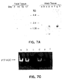
- the isolation of murine PTP HSC from the CD34 hi fraction of murine 10.5 day yolk sac or embryo cells is illustrated in the examples.
- murine PTP HSC can be isolated from CD34 hi population originated from bone marrow or fetal liver.
- the purity of these murine cells was found to be a critical step in isolating the mRNA encoding the new murine PTP HSC of the present invention. A high degree of purity was achieved by purification with a rabbit anti-murine CD34 antibody followed by a lineage depletion step and a positive selection step with the Sca antibody.
- murine PTP HSC can be detected and obtained from other relatively undifferentiated precursors of mature murine hematopoietic cells, such as, BAF 3, 32D and FDCP hematopoietic progenitor cells, available from the American Type Culture Collection (ATCC).
- Native human PTP HSC can, for example, be identified in and obtained from human CMK progenitor cells.
- the PTP HSCs enzymes have an extremely low abundance in embryonic tissues, their purification by traditional methods would be very cumbersome and inefficient.
- cDNA or genomic clones encoding the PTP HSC proteins of the present invention can be prepared using standard techniques of recombinant DNA technology.
- cDNA library can be constructed by obtaining polyadenylated mRNA from a cell line known to express the desired PTP HSC, and using the mRNA as a template to synthesize double stranded cDNA.
- exemplary human and non-human cell lines suitable for this purpose have been listed hereinabove.
- a PTP HSC polypeptide gene can also be obtained from a genomic library, such as a human genomic cosmid library.
- probes designed to identify the gene of interest or the protein encoded by it.
- suitable probes include monoclonal and polyclonal antibodies that recognize and specifically bind to a PTP HSC polypeptide.
- suitable probes include carefully selected oligonucleotide probes (usually of about 20-80 bases in length) that encode known or suspected portions of a PTP HSC polypeptide from the same or different species, and/or complementary or homologous cDNAs or fragments thereof that encode the same or a similar gene.
- Appropriate probes for screening genomic DNA libraries include, without limitation, oligonucleotides, cDNAs, or fragments thereof that encode the same or a similar gene, and/or homologous genomic DNAs or fragments thereof. Screening the cDNA or genomic library with the selected probe may be conducted using standard procedures as described in Chapters 10-12 of Sambrook et al ., Molecular Cloning: A Laboratory Manual , New York, Cold Spring Harbor Laboratory Press, 1989.
- DNA encoding an enzyme of the present invention is isolated by using carefully selected oligonucleotide sequences to screen cDNA libraries from various tissues, the oligonucleotide sequences selected as probes should be sufficient in length and sufficiently unambiguous that false positives are minimized.
- the actual nucleotide sequence(s) is/are usually designed based on regions which have the least codon redundance.
- the oligonucleotides may be degenerate at one or more positions. The use of degenerate oligonucleotides is of particular importance where a library is screened from a species in which preferential codon usage is not known.
- the oligonucleotide must be labeled such that it can be detected upon hybridization to DNA in the library being screened.
- the preferred method of labeling is to use ATP (e.g., ⁇ 32 P) and polynucleotide kinase to radiolabel the 5' end of the oligonucleotide.
- ATP e.g., ⁇ 32 P
- polynucleotide kinase to radiolabel the 5' end of the oligonucleotide.
- other methods may be used to label the oligonucleotide, including, but not limited to, biotinylation or enzyme labeling.
- cDNAs encoding PTP HSCs can also be identified and isolated by other known techniques of recombinant DNA technology, such as by direct expression cloning, or by using the polymerase chain reaction (PCR) as described in U.S. Patent No. 4,683,195, issued 28 July 1987, in section 14 of Sambrook et al ., supra , or in Chapter 15 of Current Protocols in Molecular Biology , Ausubel et al . eds., Greene Publishing Associates and Wiley-Interscience 1991.
- the use of the PCR technique for obtaining cDNA encoding murine PTP HSC or the PTP domain of this native protein is also illustrated in the examples.
- cDNAs from other species can also be obtained by cross-species hybridization.
- human or other mammalian cDNA or genomic libraries are probed by labeled oligonucleotide sequences selected from known PTP HSC sequences (such as murine PTP HSC) in accord with known criteria, among which is that the sequence should be sufficient in length and sufficiently unambiguous that false positives are minimized.
- oligonucleotide sequences selected from known PTP HSC sequences (such as murine PTP HSC) in accord with known criteria, among which is that the sequence should be sufficient in length and sufficiently unambiguous that false positives are minimized.
- a 32 P-labeled oligonucleotide having about 30 to 50 bases is sufficient, particularly if the oligonucleotide contains one or more codons for methionine or tryptophan.
- Isolated nucleic acid will be DNA that is identified and separated from contaminant nucleic acid encoding other polypeptides from the source of nucleic acid.
- Hybridization is preferably performed under "stringent conditions" which means (1) employing low ionic strength and hgh temperature for washing, for example, 0.015 sodium chloride/0.0015 M sodium citrate/0.1 % sodium dodecyl sulfate at 50 ° C, or (2) employing during hybridization a denaturing agent, such as formamide, for example, 50% (vol/vol) formamide with 0.1% bovine serum albumin/0.
- Another example is the use of 5)% formamide, 5 x SSC (0.75 M sodium chloride, 0.075 M sodium citrate), 50 mM sodium phosphate (pH 6.8), 0.1% sodium pyrophosphate, 5 x Denhardt's solution, sonicated salmon sperm DNA (50 ⁇ g/ml), 0.1% SDS, and 10% dextran sulfate at 42 ° C, with washes at 42 ° C in 0.2 x SSC and 0.1% SDS.
- the gene encoding a particular PTP HSC polypeptide can also be obtained by chemical synthesis, following one of the methods described in Engels and Uhlmann, Agnew. Chem. Int. Ed. Engl. 28 , 716 (1989). These methods include triester, phosphite, phosphoramidite and H-phosphonate methods, PCR and other autoprimer methods, and oligonucleotide syntheses on solid supports.
- nucleic acid encoding PTP HSC is available, it is generally ligated into a replicable expression vector for further cloning (amplification of the DNA), or for expression.
- Expression and cloning vectors are well known in the art and contain a nucleic acid sequence that enables the vector to replicate in one or more selected host cells. The selection of the appropriate vector will depend on 1) whether it is to be used for DNA amplification or for DNA expression, 2) the size of the DNA to be inserted into the vector, and 3) the host cell to be transformed with the vector. Each vector contains various components depending on its function (amplification of DNA of expression of DNA) and the host cell for which it is compatible.
- the vector components generally include, but are not limited to, one or more- of the following: a signal sequence, an origin of replication, one or more marker genes, an enhancer element, a promoter, and a transcription termination sequence.
- Plasmid DNA fragments are cleaved, tailored, and religated in the form desired to generate the plasmids required.
- the ligation mixtures are commonly used to transform E. coli cells, e.g. E. coli K12 strain 294 (ATCC 31,446) and successful transformants selected by ampicillin or tetracycline resistance where appropriate.
- Plasmids from the transformants are prepared, analyzed by restriction endonuclease digestion, and/or sequenced by the method of Messing et al ., Nucleic Acids Res. 9 , 309 (1981) or by the method of Maxam et al ., Methods in Enzymology 65 , 499 (1980).
- the polypeptides of the present invention may be expressed in a variety of prokaryotic and eukaryotic host cells.
- Suitable prokaryotes include gram negative or gram positive organisms, for example E . coli or bacilli.
- a preferred cloning host is E . coli 294 (ATCC 31,446) although other gram negative or gram positive prokaryotes such as E . coli B, E . coli X1776 (ATCC 31,537), E. coli W3110 (ATCC 27,325), Pseudomonas species, or Serratia Marcesans are suitable.
- eukaryotic microbes such as filamentous fungi or yeast are suitable hosts for vectors herein.
- Saccharomyces cerevisiae or common baker's yeast, is the most commonly used among lower eukaryotic host microorganisms.
- S. pombe Beach and Nurse, Nature 290 , 140 (1981)]
- Kluyveromyces lactis [Louvencourt et al ., J. Bacteriol.
- Suitable host cells may also derive from multicellular organisms. Such host cells are capable of complex processing and glycosylation activities. In principle, any higher eukaryotic cell culture is workable, whether from vertebrate or invertebrate culture, although cells from mammals such as humans are preferred. Examples of invertebrate cells include plants and insect cells. Numerous baculoviral strains and variants and corresponding permissive insect host cells from hosts such as Spodoptera frugiperda (caterpillar), Aedes aegypti (mosquito), Aedes albopictus (mosquito), Drosophila melangaster (fruitfly), and Bombyx mori host cells have been identified. See, e.g.
- Plant cell cultures of cotton, corn, potato, soybean, petunia, tomato, and tobacco can be utilized as hosts.
- plant cells are transfected by incubation with certain strains of the bacterium Agrobacterium tumefaciens , which has been previously manipulated to contain the PTP HSC DNA.
- Agrobacterium tumefaciens the DNA encoding a PTP HSC is transferred to the plant cell host such that it is transfected, and will, under appropriate conditions, express the PTP HSC DNA.
- regulatory and signal sequences compatible with plant cells are available, such as the nopaline synthase promoter and polyadenylation signal sequences. Depicker et al ., J. Mol.
- DNA segments isolated from the upstream region of the T-DNA 780 gene are capable of activating or increasing transcription levels of plant-expressible genes in recombinant DNA-containing plant tissue. See EP 321,196 published 21 June 1989.
- mice sertolli cells [TM4, Mather, Biol. Reprod . 23 , 243-251 (1980)]; monkey kidney cells (CV1 ATCC CCL 70); African green monkey kidney cells (VERO-76, ATCC CRL-1587); human cervical carcinoma cells (HELA, ATCC CCL 2); canine kidney cells (MDCK, ATCC CCL 34); buffalo rat liver cells (BRL 3A, ATCC CRL 1442); human lung cells (W138, ATCC CCL75); human liver cells (Hep G2, HB 8065); mouse mammary tumor (MMT 060562, ATCC CCL51); TRI cells [Mather et al ., Annals N.Y. Acad. Sci. 383, 44068 (1982)]; MRC 5 cells; FS4 cells; and a human hepatoma cell line (Hep G2).
- Preferred host cells are human embryonic kidney 293 and Chinese hamster ovary cells.
- transient expression involves the use of an expression vector that is able to replicate efficiently in a host cell, such that the host cell accumulates many copies of the expression vector and, in turn, synthesizes high levels of a desired polypeptide encoded by the expression vector.
- Transient systems comprising a suitable expression vector and a host cell, allow for the convenient positive identification of polypeptides encoded by clones DNAs, as well as for the rapid screening of such polypeptides for desired biological or physiological properties.
- transient expression systems are particularly useful in the invention for purposes of identifying analogs and variants of a PTP HSC.
- PTP HSC polypeptides in recombinant vertebrate cell culture are described in Getting et al ., Nature 293 , 620-625 (1981); Mantel et al ., Nature 281 , 40-46 (1979); Levinson et al .; EP 117,060 and EP 117,058.
- Particularly useful plasmids for mammalian cell culture expression of the PTP HSC polypeptides are pRK5 (EP 307,247), or pSV16B (PCT Publication No. WO 91/08291).
- PTP HSCs of the present invention are, for example, described in EP 457,758 published 27 November 1991.
- a large variety of expression vectors is now commercially available.
- An exemplary commercial yeast expression vector is pPIC.9 (Invitrogen), while an commercially available expression vector suitable for transformation of E. coli cells is PET15b (Novagen).
- Prokaryotes cells used to produced the PTP HSCs of this invention are cultured in suitable media as describe generally in Sambrook et al ., supra .
- Mammalian cells can be cultured in a variety of media.
- Commercially available media such as Ham's F10 (Sigma), Minimal Essential Medium (MEM, Sigma), RPMI-1640 (Sigma), and Dulbecco's Modified Eagle's Medium (DMEM, Sigma) are suitable for culturing the host cells.
- 30,985 may be used as culture media for the host cells. Any of these media may be supplemented as necessary with hormones and/or other growth factors (such as insulin, transferrin, or epidermal growth factor), salts (such as sodium chloride, calcium, magnesium, and phosphate), buffers (such as HEPES), nucleosides (such as adenosine and thymidine), antibiotics (such as Gentamycin TM drug) trace elements (defined as inorganic compounds usually present at final concentrations in the micromolar range), and glucose or an equivalent energy source. Any other necessary supplements may also be included at appropriate concentrations that would be known to those skilled in the art.
- the culture conditions such as temperature, pH and the like, suitably are those previously used with the host cell selected for cloning or expression, as the case may be, and will be apparent to the ordinary artisan.
- the host cells referred to in this disclosure encompass cells in in vitro cell culture as well as cells that are within a host animal or plant.
- the PTP HSCs of this invention may be produced by homologous recombination, or with recombinant production methods utilizing control elements introduced into cells already containing DNA encoding the particular PTP HSC.
- Gene amplification and/or expression may be measured in a sample directly, for example, by conventional Southern blotting, Northern blotting to quantitate the transcription of mRNA [Thomas, Proc. Natl. Acad. Sci. USA 77 , 5201-5205 (1980)], dot blotting (DNA analysis), or in situ hybridization, using an appropriately labeled probe, based on the sequences provided herein.
- Various labels may be employed. most commonly radioisotopes, particularly 32 P.
- other techniques may also be employed, such as using biotin-modified nucleotides for introduction into a polynucleotide.
- the biotin then serves as a site for binding to avidin or antibodies, which may be labeled with a wide variety of labels, such as radionuclides, fluorescers, enzymes, or the like.
- antibodies may be employed that can recognize specific duplexes, including DNA duplexes, RNA duplexes, and DNA-RNA hybrid duplexes or DNA-protein duplexes.
- the antibodies in turn may be labeled and the assay may be carried out where the duplex is bound to the surface, so that upon the formation of duplex on the surface, the presence of antibody bound to the duplex can be detected.
- Gene expression may be measured by immunological methods, such as immunohistochemical staining of tissue sections and assay of cell culture or body fluids, to quantitate directly the expression of gene product.
- immunohistochemical staining techniques a cell sample is prepared, typically by dehydration and fixation, followed by reaction with labeled antibodies specific for the gene product coupled, where the labels are usually visually detectable, such as enzymatic labels, fluorescent labels, luminescent labels, and the like.
- a particularly sensitive staining technique suitable for use in the present invention is described by Hse et al ., Am. J. Clin. Pharm. 75 , 734-738 (1980).
- Antibodies useful for immunohistochemical staining and/or assay of sample fluids may be either monoclonal or polyclonal, and may be prepared in any animal. Conveniently, the antibodies may be prepared against a native PTP HSC polypeptide, or against a synthetic peptide based on the DNA sequence provided herein as described further hereinbelow.
- Amino acid sequence variants of native PTP HSCs are prepared by methods known in the art by introducing appropriate nucleotide changes into a PTP HSC DNA, or by in vitro synthesis of the desired polypeptide. There are two principal variables in the construction of amino acid sequence variants: the location of the mutation site and the nature of the mutation. With the exception of naturally-occurring alleles, which do not require the manipulation of the DNA sequence encoding the PTP HSC, the amino acid sequence variants of PTP HSCs are preferably constructed by mutating the DNA, either to arrive at an allele or an amino acid sequence variant that does not occur in nature.
- PTP phosphatase domain of the enzymes of the present invention.
- Non-conservative substitutions within this domain may result in PTP HSC variants which loose their ability to dephosphatase tyrosines and will, therefore, be useful as antagonists of native PTP HSCs.
- PTP HSC variants mutated to enhance their enzymatic activity will be useful, for example, as more effective inhibitors of progenitor/stem cell differentiation.
- amino acid alterations can be made at sites that differ in PTP HSC proteins from various species, or in highly conserved regions, depending on the goal to be achieved. Sites at such locations will typically be modified in series, e.g. by (1) substituting first with conservative choices and then with more radical selections depending upon the results achieved, (2) deleting the target residue or residues, or (3) inserting residues of the same or different class adjacent to the located site, or combinations of options 1-3.
- One helpful technique is called “alanine scanning” (Cunningham and Wells, Science 244 , 1081-1085 [1989]).
- the gene encoding a PTP HSC variant can, for example, be obtained by chemical synthesis as hereinabove described. More preferably, DNA encoding a PTP HSC amino acid sequence variant is prepared by site-directed mutagenesis of DNA that encodes an earlier prepared variant or a nonvariant version of the PTP HSC. Site-directed (site-specific) mutagenesis allows the production of PTP HSC variants through the use of specific oligonucleotide sequences that encode the DNA sequence of the desired mutation, as well as a sufficient number of adjacent nucleotides, to provide a primer sequence of sufficient size and sequence complexity to form a stable duplex on both sides of the deletion junction being traversed.
- a primer of about 20 to 25 nucleotides in length is preferred, with about 5 to 10 residues on both sides of the junction of the sequence being altered.
- site-specific mutagenesis is well known in the art, as exemplified by publications such as, Edelman et al ., DNA 2 , 183 (1983).
- the site-specific mutagenesis technique typically employs a phage vector that exists in both a single-stranded and double-stranded form.
- Typical vectors useful in site-directed mutagenesis include vectors such as the M13 phage, for example, as disclosed by Messing et al ., Third Cleveland Symposium on Macromolecules and Recombinant DNA , A.
- phage vectors are commercially available and their use is well known to those skilled in the art.
- a versatile and efficient procedure for the construction of oligodeoxyribonucleotide directed site-specific mutations in DNA fragments using M 13 - derived vectors was published by Zoller, M.J. and Smith, M., Nucleic Acids Res. 10 , 6487-6500 [1982]).
- plasmid vectors that contain a single-stranded phage origin of replication may be employed to obtain single-stranded DNA.
- nucleotide substitutions are introduced by synthesizing the appropriate DNA fragment in vitro, and amplifying it by PCR procedures known in the art.
- PCR technique may also be used in creating amino acid sequence variants of a PTP HSC.
- template plasmid DNA (1 1 ⁇ g) is linearized by digestion with a restriction endonuclease that has a unique recognition site in the plasmid DNA outside of the region to be amplified.
- 100 ng is added to a PCR mixture containing PCR buffer, which contains the four deoxynucleotide triphosphates and is included in the GeneAmp R kits (obtained from Perkin-Elmer Cetus, Norwalk, CT and Emeryville, CA), and 25 pmole of each oligonucleotide primer, to a final volume of 50 ⁇ l.
- the reaction mixture is overlayered with 35 ⁇ l mineral oil.
- the reaction is denatured for 5 minutes at 100°C, placed briefly on ice, and then 1 ⁇ l Thermus aquaticus ( Taq ) DNA polymerase (5 units/ 1), purchased from Perkin-Elmer Cetus, Norwalk, CT and Emeryville, CA) is added below the mineral oil layer.
- the reaction mixture is then inserted into a DNA Thermal Cycler (purchased from Perkin-Elmer Cetus) programmed as follows:
- reaction vial is removed from the thermal cycler and the aqueous phase transferred to a new vial, extracted with phenol/chloroform (50:50 vol), and ethanol precipitated, and the DNA is recovered by standard procedures. This material is subsequently subjected to appropriate treatments for insertion into a vector.
- phagemid display method may be useful in making amino acid sequence variants of native or variant PTP HSCs or their fragments.
- This method involves (a) constructing a replicable expression vector comprising a first gene encoding an receptor to be mutated, a second gene encoding at least a portion of a natural or wild-type phage coat protein wherein the first and second genes are heterologous, and a transcription regulatory element operably linked to the first and second genes, thereby forming a gene fusion encoding a fusion protein; (b) mutating the vector at one or more selected positions within the first gene thereby forming a family of related plasmids; (c) transforming suitable host cells with the plasmids; (d) infecting the transformed host cells with a helper phage having a gene encoding the phage coat protein; (e) culturing the transformed infected host cells under conditions suitable for forming recombinant phagemid particles containing at least a
- Steps (d) through (g) can be repeated one or more times.
- the plasmid is under tight control of the transcription regulatory element, and the culturing conditions are adjusted so that the amount or number of phagemid particles displaying more than one copy of the fusion protein on the surface of the particle is less than about 1%.
- the amount of phagemid particles displaying more than one copy of the fusion protein is less than 10% of the amount of phagemid particles displaying a single copy of the fusion protein. Most preferably, the amount is less than 20%.
- the expression vector will further contain a secretory signal sequence fused to the DNA encoding each subunit of the polypeptide and the transcription regulatory element will be a promoter system.
- Preferred promoter systems are selected from lac Z, ⁇ PL , tac , T7 polymerase, tryptophan, and alkaline phosphatase promoters and combinations thereof.
- the method will employ a helper phage selected from M13K07, M13R408, M13-VCS, and Phi X 174.
- the preferred helper phage is M13K07
- the preferred coat protein is the M13 Phage gene III coat protein.
- the preferred host is E. coli , and protease-deficient strains of E. coli .
- Naturally-occurring amino acids are divided into groups based on common side chain properties:
- Conservative substitutions involve exchanging a member within one group for another member within the same group, whereas non-conservative substitutions will entail exchanging a member of one of these classes for another.
- Amino acid sequence deletions generally range from about 1 to 30 residues, more preferably about 1 to 10 residues, and typically are contiguous.
- Amino acid insertions include amino- and/or carboxyl-terminal fusions ranging in length from one residue to polypeptides containing a hundred or more residues, as well as intrasequence insertions of single or multiple amino acid residues.
- intrasequence insertions i.e. insertions within the PTP HSC protein amino acid sequence
- terminal insertions include the PTP HSC polypeptides with an N-terminal methionyl residue, an artifact of its direct expression in bacterial recombinant cell culture, and fusion of a heterologous N-terminal signal sequence to the N-terminus of the PTP HSC molecule to facilitate the secretion of the mature PTP HSC from recombinant host cells.
- signal sequences will generally be obtained from, and thus homologous to, the intended host cell species. Suitable sequences include STII or Ipp for E. coli, alpha factor for yeast, and viral signals such as herpes gD for mammalian cells.
- insertional variants of the native PTP HSC molecules include the fusion of the N- or C-terminus of the TRAF molecule to immunogenic polypeptides, e.g. bacterial polypeptides such as beta-lactamase or an enzyme encoded by the E. coli trp locus, or yeast protein, and C-terminal fusions with proteins having a long half life such as immunoglobulin regions (preferably immunoglobulin constant regions), albumin, or ferritin, as described in WO 89/02922 published on 6 April 1989.
- immunogenic polypeptides e.g. bacterial polypeptides such as beta-lactamase or an enzyme encoded by the E. coli trp locus, or yeast protein
- C-terminal fusions with proteins having a long half life such as immunoglobulin regions (preferably immunoglobulin constant regions), albumin, or ferritin, as described in WO 89/02922 published on 6 April 1989.
- Covalent modifications of PTP HSCs are included within the scope herein. Such modifications are traditionally introduced by reacting targeted amino acid residues of the PTP HSC polypeptides with an organic derivatizing agent that is capable of reacting with selected sides or terminal residues, or by harnessing mechanisms of post-translational modifications that function in selected recombinant host cells.
- the resultant covalent derivatives are useful in programs directed at identifying residues important for biological activity, for immunoassays of the PTP HSC, or for the preparation of anti-PTP HSC antibodies for immunoaffinity purification of the recombinant.
- Cysteinyl residues most commonly are reacted with ⁇ -haloacetates (and corresponding amines), such as chloroacetic acid or chloroacetamide, to give carboxymethyl or carboxyamidomethyl derivatives. Cysteinyl residues also are derivatized by reaction with bromotrifluoroacetone, ⁇ -bromo- ⁇ -(5-imidozoyl)propionic acid, chloroacetyl phosphate, N-alkylmaleimides, 3-nitro-2-pyridyl disulfide, methyl 2-pyridyl disulfide, p-chloromercuribenzoate, 2-chloromercuri-4-nitrophenol, or chloro-7-nitrobenzo-2-oxa- 1,3-diazole.
- Histidyl residues are derivatized by reaction with diethylpyrocarbonate at pH 5.5-7.0 because this agent is relatively specific for the histidyl side chain.
- Para-bromophenacyl bromide also is useful; the reaction is preferably performed in 0.1 M sodium cacodylate at pH 6.0.
- Lysinyl and amino terminal residues are reacted with succinic or other carboxylic acid anhydrides. Derivatization with these agents has the effect of reversing the charge of the lysinyl residues.
- Other suitable reagents for derivatizing ⁇ -amino-containing residues include imidoesters such as methyl picolinimidate; pyridoxal phosphate; pyridoxal; chloroborohydride; trinitrobenzenesulfonic acid; O-methylisourea; 2,4-pentanedione; and transaminase-catalyzed reaction with glyoxylate.
- Arginyl residues are modified by reaction with one or several conventional reagents, among them phenylglyoxal, 2,3-butanedione, 1,2-cyclohexanedione, and ninhydrin. Derivatization of arginine residues requires that the reaction be performed in alkaline conditions because of the high pK a of the guanidine functional group. Furthermore, these reagents may react with the groups of lysine as well as the arginine epsilon-amino group.
- tyrosyl residues may be made, with particular interest in introducing spectral labels into tyrosyl residues by reaction with aromatic diazonium compounds or tetranitromethane. Most commonly, N-acetylimidizole and tetranitromethane are used to form O-acetyl tyrosyl species and 3-nitro derivatives, respectively.
- Tyrosyl residues are iodinated using 125 I or 131 I to prepare labeled proteins for use in radioimmunoassay.
- Glutaminyl and asparaginyl residues are frequently deamidated to the corresponding glutamyl and aspartyl residues. Alternatively, these residues are deamidated under mildly acidic conditions. Either form of these residues falls within the scope of this invention.
- modifications include hydroxylation of proline and lysine, phosphorylation of hydroxyl groups of seryl, threonyl or tyrosyl residues, methylation of the ⁇ -amino groups of lysine, arginine, and histidine side chains (T.E. Creighton, Proteins: Structure and Molecular Properties , W.H. Freeman & Co., San Francisco, pp. 79-86 [1983]), acetylation of the N-terminal amine, and amidation of any C-terminal carboxyl group.
- the molecules may further be covalently linked to nonproteinaceous polymers, e.g.
- polyethylene glycol, polypropylene glycol or polyoxyalkylenes in the manner set forth in U.S.S.N. 07/275,296 or U.S. patents 4,640,835; 4,496,689; 4,301,144; 4,670,417; 4,791,192 or 4,179,337.
- Derivatization with bifunctional agents is useful for preparing intramolecular aggregates of the PTP HSCs with polypeptides as well as for cross-linking the PTP HSC polypeptide to a water insoluble support matrix or surface for use in assays or affinity purification.
- cross-linking agents include 1,1-bis(diazoacetyl)-2-phenylethane, glutaraldehyde, N-hydroxysuccinimide esters, homobifunctional imidoesters, and bifunctional maleimides.
- Derivatizing agents such as methyl-3-[(p-azidophenyl)dithio]propioimidate yield photoactivatable intermediates which are capable of forming cross-links in the presence of light.
- reactive water insoluble matrices such as cyanogen bromide activated carbohydrates and the systems reactive substrates described in U.S. Patent Nos. 3,959,642; 3,969,287; 3,691,016; 4,195,128; 4,247,642; 4,229,537; 4,055,635; and 4,330,440 are employed for protein immobilization and cross-linking.
- nonproteinaceous polymer ordinarily is a hydrophilic synthetic polymer, i.e. a polymer not otherwise found in nature.
- hydrophilic polyvinyl polymers fall within the scope of this invention, e.g. polyvinylalcohol and polyvinylpyrrolidone.
- Particularly useful are polyvinylalkylene ethers such a polyethylene glycol, polypropylene glycol.
- the PTP HSC polypeptides may be linked to various nonproteinaceous polymers, such as polyethylene glycol, polypropylene glycol or polyoxyalkylenes, in the manner set forth in U.S. Patent Nos. 4,640,835; 4,496,689; 4,301,144; 4,670,417; 4,791,192 or 4,179,337.
- the PTP HSCs may be entrapped in microcapsules prepared, for example, by coacervation techniques or by interfacial polymerization, in colloidal drug delivery systems (e.g. liposomes, albumin microspheres, microemulsions, nano-particles and nanocapsules), or in macroemulsions.
- colloidal drug delivery systems e.g. liposomes, albumin microspheres, microemulsions, nano-particles and nanocapsules
- macroemulsions e.g. liposomes, albumin microspheres, microemulsions, nano-particles and nanocapsules
- Polyclonal antibodies to a PTP HSC molecule generally are raised in animals by multiple subcutaneous (sc) or intraperitoneal (ip) injections of the PTP HSC and an adjuvant. It may be useful to conjugate the PTP HSC or a fragment containing the target amino acid sequence to a protein that is immunogenic in the species to be immunized, e.g.
- Animals are immunized against the immunogenic conjugates or derivatives by combining 1 mg or 1 ⁇ g of conjugate (for rabbits or mice, respectively) with 3 volumes of Freud's complete adjuvant and injecting the solution intradermally at multiple sites.
- the animals are boosted with 1/5 to 1/10 the original amount of conjugate in Freud's complete adjuvant by subcutaneous injection at multiple sites.
- 7 to 14 days later the animals are bled and the serum is assayed for anti-PTP HSC antibody titer. Animals are boosted until the titer plateaus.
- the animal boosted with the conjugate of the same PTP HSC, but conjugated to a different protein and/or through a different cross-linking reagent.
- Conjugates also can be made in recombinant cell culture as protein fusions. Also, aggregating agents such as alum are used to enhance the immune response.
- Monoclonal antibodies are obtained from a population of substantially homogeneous antibodies, i.e., the individual antibodies comprising the population are identical except for possible naturally-occurring mutations that may be present in minor amounts.
- the modifier "monoclonal" indicates the character of the antibody as not being a mixture of discrete antibodies.
- the anti-PTP HSC monoclonal antibodies of the invention may be made using the hybridoma method first described by Kohler & Milstein, Nature 256 :495 (1975), or may be made by recombinant DNA methods [Cabilly, et al ., U.S. Pat. No. 4,816,567].
- lymphocytes In the hybridoma method, a mouse or other appropriate host animal, such as hamster is immunized as hereinabove described to elicit lymphocytes that produce or are capable of producing antibodies that will specifically bind to the protein used for immunization. Alternatively, lymphocytes may be immunized in vitro. Lymphocytes then are fused with myeloma cells using a suitable fusing agent, such as polyethylene glycol, to form a hybridoma cell [Goding, Monoclonal Antibodies: Principles and Practice, pp.59-103 (Academic Press, 1986)].
- a suitable fusing agent such as polyethylene glycol
- the hybridoma cells thus prepared are seeded and grown in a suitable culture medium that preferably contains one or more substances that inhibit the growth or survival of the unfused, parental myeloma cells.
- a suitable culture medium that preferably contains one or more substances that inhibit the growth or survival of the unfused, parental myeloma cells.
- the culture medium for the hybridomas typically will include hypoxanthine, aminopterin, and thymidine (HAT medium), which substances prevent the growth of HGPRT-deficient cells.
- Preferred myeloma cells are those that fuse efficiently, support stable high level expression of antibody by the selected antibody-producing cells, and are sensitive to a medium such as HAT medium.
- preferred myeloma cell lines are murine myeloma lines, such as those derived from MOPC-21 and MPC- 11 mouse tumors available from the Salk Institute Cell Distribution Center, San Diego, California USA, and SP-2 cells available from the American Type Culture Collection, Rockville, Maryland USA.
- Human myeloma and mouse-human heteromyeloma cell lines also have been described for the production of human monoclonal antibodies [Kozbor, J. Immunol. 133 :3001 (1984); Brodeur, et al ., Monoclonal Antibody Production Techniques and Applications , pp.51-63 (Marcel Dekker, inc., New York, 1987)].
- Culture medium in which hybridoma cells are growing is assayed for production of monoclonal antibodies directed against PTP HSC.
- the binding specificity of monoclonal antibodies produced by hybridoma cells is determined by immunoprecipitation or by an in vitro binding assay, such as radioimmunoassay (RIA) or enzyme-linked immunoabsorbent assay (ELISA).
- RIA radioimmunoassay
- ELISA enzyme-linked immunoabsorbent assay
- the binding affinity of the monoclonal antibody can, for example, be determined by the Scatchard analysis of Munson & Pollard, Anal. Biochem. 107 :220 (1980).
- the clones may be subcloned by limiting dilution procedures and grown by standard methods. Goding, Monoclonal Antibodies: Principles and Practice , pp.59-104 (Academic Press, 1986). Suitable culture media for this purpose include, for example, Dulbecco's Modified Eagle's Medium or RPMI-1640 medium.
- the hybridoma cells may be grown in vivo as ascites tumors in an animal.
- the monoclonal antibodies secreted by the subclones are suitably separated from the culture medium, ascites fluid, or serum by conventional immunoglobulin purification procedures such as, for example, protein A-Sepharose, hydroxylapatite chromatography, gel electrophoresis, dialysis, or affinity chromatography.
- DNA encoding the monoclonal antibodies of the invention is readily isolated and sequenced using conventional procedures (e.g., by using oligonucleotide probes that are capable of binding specifically to genes encoding the heavy and light chains of murine antibodies).
- the hybridoma cells of the invention serve as a preferred source of such DNA.
- the DNA may be placed into expression vectors, which are then transfected into host cells such as simian COS cells, Chinese hamster ovary (CHO) cells, or myeloma cells that do not otherwise produce immunoglobulin protein, to obtain the synthesis of monoclonal antibodies in the recombinant host cells.
- the DNA also may be modified, for example, by substituting the coding sequence for human heavy and light chain constant domains in place of the homologous murine sequences, Morrison, et al ., Proc. Nat. Acad. Sci. 81 , 6851 (1984), or by covalently joining to the immunoglobulin coding sequence all or part of the coding sequence for a non-immunoglobulin polypeptide.
- “chimeric” or “hybrid” antibodies are prepared that have the binding specificity of an anti-TRAF monoclonal antibody herein.
- non-immunoglobulin polypeptides are substituted for the constant domains of an antibody of the invention, or they are substituted for the variable domains of one antigen-combining site of an antibody of the invention to create a chimeric bivalent antibody comprising one antigen-combining site having specificity for a PTP HSC and another antigen-combining site having specificity for a different antigen.
- Chimeric or hybrid antibodies also may be prepared in vitro using known methods in synthetic protein chemistry, including those involving crosslinking agents.
- immunotoxins may be constructed using a disulfide exchange reaction or by forming a thioether bond.
- suitable reagents for this purpose include iminothiolate and methyl-4-mercaptobutyrimidate.
- the antibodies of the invention typically will be labeled with a detectable moiety.
- the detectable moiety can be any one which is capable of producing, either directly or indirectly, a detectable signal.
- the detectable moiety may be a radioisotope, such as 3 H, 14 C, 32 P, 35 S, or 125 I, a fluorescent or chemiluminescent compound, such as fluorescein isothiocyanate, rhodamine, or luciferin; biotin; radioactive isotopic labels, such as, e.g., 125 I, 32 P, 14 C, or 3 H, or an enzyme, such as alkaline phosphatase, beta-galactosidase or horseradish peroxidase.
- any method known in the art for separately conjugating the antibody to the detectable moiety may be employed, including those methods described by Hunter, et al ., Nature 144 :945 (1962); David, et al ., Biochemistry 13 :1014(1974); Pain, et al ., J. Immunol. Meth. 40 :219 (1981); and Nygren, J. Histochem. and Cytochem. 30 :407 (1982).
- the antibodies of the present invention may be employed in any known assay method, such as competitive binding assays, direct and indirect sandwich assays, and immunoprecipitation assays. Zola, Monoclonal Antibodies: A Manual of Techniques, pp.147-158 (CRC Press, Inc., 1987).
- a labeled standard which may be a PTP HSC polypeptide or an immunologically reactive portion thereof
- PTP HSC test sample analyte
- the amount of PTP HSC in the test sample is inversely proportional to the amount of standard that becomes bound to the antibodies.
- the antibodies generally are insolubilized before or after the competition, so that the standard and analyte that are bound to the antibodies may conveniently be separated from the standard and analyte which remain unbound.
- Sandwich assays involve the use of two antibodies, each capable of binding to a different immunogenic portion, or epitope, of the protein to be detected.
- the test sample analyte is bound by a first antibody which is immobilized on a solid support, and thereafter a second antibody binds to the analyte, thus forming an insoluble three part complex.
- the second antibody may itself be labeled with a detectable moiety (direct sandwich assays) or may be measured using an anti-immunoglobulin antibody that is labeled with a detectable moiety (indirect sandwich assay).
- sandwich assay is an ELISA assay, in which case the detectable moiety is an enzyme.
- a humanized antibody has one or more amino acid residues introduced into it from a source which is non-human. These non-human amino acid residues are often referred to as "import" residues, which are typically taken from an "import” variable domain. Humanization can be essentially performed following the method of Winter and co-workers [Jones et al ., Nature 321 , 522-525 (1986); Riechmann et al ., Nature 332 , 323-327 (1988); Verhoeyen et al ., Science 239 , 1534-1536 (1988)], by substituting rodent CDRs or CDR sequences for the corresponding sequences of a human antibody.
- humanized antibodies are chimeric antibodies (Cabilly, supra ), wherein substantially less than an intact human variable domain has been substituted by the corresponding sequence from a non-human species.
- humanized antibodies are typically human antibodies in which some CDR residues and possibly some FR residues are substituted by residues from analogous sites in rodent antibodies.
- humanized antibodies are prepared by a process of analysis of the parental sequences and various conceptual humanized products using three dimensional models of the parental and humanized sequences.
- Three dimensional immunoglobulin models are commonly available and are familiar to those skilled in the art.
- Computer programs are available which illustrate and display probable three-dimensional conformational structures of selected candidate immunoglobulin sequences. Inspection of these displays permits analysis of the likely role of the residues in the functioning of the candidate immunoglobulin sequence, i.e. the analysis of residues that influence the ability of the candidate immunoglobulin to bind its antigen.
- FR residues can be selected and combined from the consensus and import sequence so that the desired antibody characteristic, such as increased affinity for the target antigen(s), is achieved.
- the CDR residues are directly and most substantially involved in influencing antigen binding.
- transgenic animals e.g. mice
- transgenic animals e.g. mice
- J H antibody heavy chain joining region
- transfer of the human germ-line immunoglobulin gene array in such germ-line mutant mice will result in the production of human antibodies upon antigen challenge.
- Jakobovits et al . Proc. Natl. Acad. Sci. USA 90 , 2551-255 (1993); Jakobovits et al ., Nature 362 , 255-258 (1993).
- Bispecific antibodies are monoclonal, preferably human or humanized, antibodies that have binding specificities for at least two different antigens.
- one of the binding specificities is for a PTP HSC
- the other one is for any other antigen, for example an antigen expressed on the surface of a leukemia cell, if the antibody is an antagonist of a native PTP HSC and is used to induce differentiation of undifferentiated lekemia cells.
- an agonist antibody specifically binding to a native PTP HSC is used to expand stem cells with growth factors, as hereinafter described, the second specificity could be provided by a stem cell growth factor.
- Such constructs can also be referred to as bispecific immunoadhesins. Methods for making bispecific antibodies (and bispecific immunoadhesins) are known in the art.
- bispecific antibodies are based on the coexpression of two immunoglobulin heavy chain-light chain pairs, where the two heavy chains have different specificities (Millstein and Cuello, Nature 305 , 537-539 (1983)). Because of the random assortment of immunoglobulin heavy and light chains, these hybridomas (quadromas) produce a potential mixture of 10 different antibody molecules, of which only one has the correct bispecific structure. The purification of the correct molecule, which is usually done by affinity chromatography steps, is rather cumbersome, and the product yields are low. Similar procedures are disclosed in PCT application publication No. WO 93/08829 (published 13 May 1993), and in Traunecker et al ., EMBO 10 , 3655-3659 (1991).
- antibody variable domains with the desired binding specificities are fused to immunoglobulin constant domain sequences.
- the fusion preferably is with an immunoglobulin heavy chain constant domain, comprising at least part of the hinge, and second and third constant regions of an immunoglobulin heavy chain (CH2 and CH3). It is preferred to have the first heavy chain constant region (CH 1 ) containing the site necessary for light chain binding, present in at least one of the fusions.
- DNAs encoding the immunoglobulin heavy chain fusions and, if desired, the immunoglobulin light chain are inserted into separate expression vectors, and are cotransfected into a suitable host organism.
- the bispecific antibodies are composed of hybrid immunoglobulin heavy chain with a first binding specificity in one arm, and a hybrid immunoglobulin heavy chain-light chain pair (providing a second binding specificity) in the other arm.
- Heteroconjugate antibodies are also within the scope of the present invention.
- Heteroconjugate antibodies are composed of two covalently joined antibodies. Such antibodies have, for example, been proposed to target immune system cells to unwanted cells (U.S. Patent No. 4,676,980), and for treatment of HIV infection (PCT application publication Nos. WO 91/00360 and WO 92/200373; EP 03089).
- Heteroconjugate antibodies may be made using any convenient cross-linking methods. Suitable cross-linking agents are well known in the art, and are disclosed in U.S. Patent No. 4,676,980, along with a number of cross-linking techniques.
- Peptide analogs of the PTP HSC polypeptides of the present invention are modelled based upon the three-dimensional structure of the native polypeptides.
- Peptides may be synthesized by well known techniques such as the solid-phase synthetic techniques initially described in Merrifield, J. Am. Chem. Soc. 15 , 2149-2154 (1963). Other peptide synthesis techniques are, for examples, described in Bodanszky et al ., Peptide Synthesis, John Wiley & Sons, 2nd Ed., 1976, as well as in other reference books readily available for those skilled in the art. A summary of peptide synthesis techniques may be found in Stuart and Young, Solid Phase Peptide Synthelia, Pierce Chemical Company, Rockford, IL (1984). Peptides may also be prepared by recombinant DNA technology, using a DNA sequence encoding the desired peptide.
- the present invention also contemplates non-peptide (e.g. organic) compounds which display substantially the same surface as the peptide analogs of the present invention, and therefore interact with other molecules in a similar fashion.
- non-peptide e.g. organic
- the PTP HSCs of the present invention are useful for a variety of purposes.
- native PTP HSCs are useful for the identification and isolation of a PTP HSC analog in another mammalian species.
- Native PTP HSCs and their functional equivalents are also useful in screening assays designed to identify agonist of antagonist of native PTP HSCs.
- Such assays may take the form of any conventional cell-type or biochemical binding assay, and can be performed in a variety of assay formats well known for those skilled in the art. As example is the so called “two-hybrid" assay format using the Matchmaker Two-Hybrid System (Clontech) according to the manufacturer's instructions.
- the PTP HSCs of the present invention as well as their agonists can additionally be used for the maintenance of stem/progenitor cells in cell culture.
- Agonists which inhibit differentiation but allow for hematopoietic stem cell growth are particularly useful for this purpose, since their use results in an amplification of the stem cells without differentiation (self-renewal).
- This process might be useful, as an example, for the expansion of hematopoietic stem cells prior to autologous or heterologous bone marrow transplantation.
- the same approach can be used in vivo for the expansion of stem cells with growth factors, in the absence of diferentiation.
- the native PTP HSCs of the present invention may be expressed in leukemic cells. Accordingly, antagonist of the PTP HSCs of the present invention may be used for the induction of differentiation of undifferentiated leukemia cells. This might allow for aggressive undifferentiated leukemia cells to become differentiated, which, in turn, facilitates their treatment.
- PTP HSC antagonists may also be used to induce differentiation of hematopoietic stem cells. As inhibition of the native PTP HSC enzyme might induce progenitor cells to differentiate, an antagonist of PTP HSC might act as a pan-inducer of myeloid, erythroid and lymphoid production. This use of PTP HSC antagonists may obviate or decrease the need for the use of stem cell growth factors.
- Yolk sacs or embryos were dissected from timed pregnant females at day 10.5. Fetal livers were isolated from day 13.5-14 embryos. Yolk sac and embryonic tissues were dissociated with 1% collagenase in RPMI medium at 37°C for 15 minutes. Cells were further dissociated by two passages through a 16 gauge needle. Fetal liver was only dissociated by passage through a 16 guage needle.
- Adherent cells were attached to plastic by overnight incubation, after which the non adherent hematopoietic cells were incubated with a lineage cocktail of antibodies (1 ⁇ g each of TER 119, Gr-1, Ly-1, transferrin receptor and B220) for 1 hr on ice. Cells were washed, and the lineage positive cells were depleted using magnetic beads and a Miltenyi column. Lineage negative cells were pelleted, resuspended in 2% FCS, PBS and incubated with rabbit anti-murine CD34 antibody (Baumhueter et al ., Science 262 , 436-38 [1993]) on ice for 1 hr.
- a lineage cocktail of antibodies (1 ⁇ g each of TER 119, Gr-1, Ly-1, transferrin receptor and B220) for 1 hr on ice. Cells were washed, and the lineage positive cells were depleted using magnetic beads and a Miltenyi column. Lineage negative
- Cells were washed three times in 2% FCS, PBS, resuspended in the same buffer and incubated with donkey, anti-rabbit FITC conjugated antibody and, in some cases, PE conjugated anti Sca antibody for 1 hr on ice.
- the cells were washed five times with 2% FCS, PBS, and than isolated by cell sorting on an ELITE cell sorter.
- Sense and antisense primers corresponding to the concensus PTP amino acid sequences H / D FWRM I / V W (5'-A C / T TT C / T TGG A / C GIATG A / G TITGG-3') (SEQ. ID. NO: 14, where the degenerate positions are designated by "N") and WPD F / H GVP (5'-GGIAC G / A T / A G / A G / A / A / A TCIGGCCA-3') (SEQ. ID. NO: 15, wherein the degenerate positions are designated by "N”) respectively were used.
- PCR were carry out in 1X Taq DNA polymerase buffer (GIBCO BRL) plus 0.2 mM of each dNTP, 10% DMSO and 5 units Taq polymerase (GIBCO BRL) for 25 cycles of 94° C for 1 minute, 55° C for 1 min and 72° C for 1 minute.
- the PCR products were treated with Klenow enzyme (New England Biolabs) at 30°C for 30 minutes, cloned into Smal site of pRK-5 (EP 307,247, published March 15, 1989) plasmid, and subsequently sequenced (Sequenase, USB).
- Adapter-linked double strain cDNA was prepared from A+ RNA of day-10 murine embryos (Marathon-ready cDNA synthesize kit, Clontech) using either random hexamer or oligo dT primer.
- Full-length cDNA was isolated by 5' or 3' rapid amplification of cDNA ends (RACE) of the marathon-ready cDNAs.
- RACE rapid amplification of cDNA ends
- Genomic clones encoding the PTP HSC gene were isolated using standard techniques.
- the plaque purified lambda phage DNA was digested with Not 1, and the insert fragment was directly cloned without purification into Not 1 digested Bluescript. Exons were mapped using a combination of restriction digestion and southern blotting as well as DNA sequencing using custom primers.
- cDNA sequences encode amino acid 8 to 323 containing the phosphatase domain were obtained by PCR using sense oligomer 5'-CACGGTCGACGGTGAGGAGCTTCTTTGAGCAGCTGGAGG-3' (SEQ. ID. NO: 3), and antisense oligomer 5'-GTTGCGGCCGCGATTGGAGCGCAGTTCTCCTTGAGGTTCTGG-3' (SEQ. ID. NO: 4).
- the PCR fragment was treated with Sail and Notl restriction enzyme and cloned into Sall and Notl digested pGEX-4T-1 plasmid (Pharmacia). Fusion protein was affinity purified using a glutathione sepharose column (Pharmacia).
- Tyrosine phosphatase assays on the GST-fusion protein were carried out following the manufacture's procedure using two different tyrosine phosphorylated peptides from a tyrosine phosphatase assay kit (Boehringer Mannheim).
- RNA isolated from hematopoietic cells cDNA was made from + RNA by reverse transcription (RT) with random hexamer. PCR was then used to amplified quantitatively PTP HSC cDNA and, as an internal standard, triosephosphate isomerase (TPI) cDNA.
- TPI triosephosphate isomerase
- 6 ul of the 20 ul RT reaction was brought to 50 ul so as to contain 0.3 mM of dNTPs, 4 ⁇ Ci of 32 P dATP (3,00OCi/mmol, Amersham), 100 pmol of each of the four primers, and 5 units of Taq DNA polymerase (GIBCO BRL).
- PCR primer pairs 5' RACE primers antisense primer 5'-CCTGGAGGGTCCTGAGAGTGATGTCTGCATTCAGTG-3' (SEQ. ID. NO: 5), 5'-CCTCTTGGAGCAGGGAAAGGATGACTCTTGTCTC-3' (SEQ. ID. NO: 6), 5'-CAGCTGCTCCAAGAAGCTCCTCACCAAGTC-3' (SEQ. ID. NO: 7).
- Sense primer AP1 and AP2 (Clontech).
- 3'RACE primers sense primer 5'-GGTAGAGGTGGGCAGGGTGAAGTGTTCTCGC-3' (SEQ. ID.
- the murine CD34 hi population isolated from bone marrow contains stem cells capable of reconstituting lethally irradiated animals.
- stem cells capable of reconstituting lethally irradiated animals.
- we included a lineage depletion step as well as a positive selection step with the Sca antibody (Uchida et al ., Blood 83 (12), 3758-3779 [1994]), in addition to the CD34 antibody.
- the lin lo CD34 hi Sca hi cells isolated from the early embryo contain self renewing hematopoietic stem cells (Uchida et al ., supra ; Krause et al ., supra ; Ziegler et al ., supra .
- the second PTP was found to be most homologous to two previously described non-receptor PTPs, murine PTP PEP (Matthews et al ., Mol. Cell Biol. 12 (5), 2396-2405 [1992]) and murine/human PTP PEST (Takekawa et al ., Biochem. Biophvs. Res. Commun. 189 (2), 1223-1230 [1992]; Yang et al ., J. Biol. Chem. 268 (23) 17650 [1993]; and Charest et al ., Biochem J.
- PTP PEP has also been found to be expressed in mature hematopoietic cells (Matthews et al ., supra, Flores et al ., supra) although human and murine PTP PEST appear to have a more generalized expression pattern (Yang et al ., supra ; Charest et al ., supra).
- the novel PTP reported here contains a region 3 prime of the PTP domain which is very rich in proline, serine, and threonine ( ⁇ 29%) (boxed residues in figure 1). This region lacks other significant homology with PTPs PEP and PEST, and it is also much shorter in the novel PTP described here.
- the novel PTP described here contains a serine residue at position 37 (shown started in figure 2) which is conserved in all three members of this family and which has been shown to be phosphorylated in PTP PEST by protein kinases C and A (Garton and Tonks, EMBO J. 13 (16), 3763-71 [1994]). Interestingly, increased phosphorylation at this site is inhibitory to the PTPase activity of this PTP (Banville et al ., Genomics 27 (1), 165-173 [1995]). In summary, the novel PTP described here appears to be a new member of a family of non-receptor PTPs which contain P, S and T rich regions (figure 3).
- PTP PEP nuclear localization signal
- N-terminal PTP domain contained many of the conserved amino acids found to be critical for substrate recognition and tyrosine dephosphorylation (Jia et al ., supra), it was important to demonstrate that this sequence indeed encoded an active PTP domain .
- GST glutathione-S-transferase
- the isolated GST-PTP domain fusion protein had a very high level of PTP activity, with significant dephosphorylation at only 20 picograms of enzyme per reaction, which was partially sensitive to inhibition by orthovanadate. The only partial inhibition of enzyme activity by orthovanadate was likely due to the high level of activity as well as insufficient levels of the inhibitor.
- FIG. 6A illustrates, quantitative PCR comparing the levels of the transcript encoding this PTP in the lin lo CD34 hi sca hi , a largely undifferentiated population containing hematopoietic stem cells (Spangrude et al ., supra; Krause et al ., supra; Zeigler et al ., Blood 84 (8), 2422-2430 [1994]), versus the lin lo CD34 hi sca lo population, a more differentiated cell population (Spangrude et al ., supra), containing committed progenitors, demonstrated that there was an approximately 10 fold lower level of the transcript in the more differentiated sca lo cells.
- the transcript encoding the novel PTP disappears as the cells replicate and develop, and it is completely absent after approximately 7 days in culture. These data are consistent with a role for this PTP in early stem or progenitor cells, but not in the mature, committed cell populations.
- FIG 7A The potential importance of this PTP specifically to the hematopoietic system is illustrated in figure 7A where northern blot analyses of various tissues and cell lines are shown.
- the transcript appears to be undetectable in the embryonic samples, and it is expressed at exceedingly low levels in adult lung and kidney.
- the transcript While there are clearly hematopoietic stem cells in the embryo, they must be so rare as to not allow for the direct detection of the transcript encoding the novel PTP.
- RNA isolated from the adult spleen a hematopoietic compartment that contains predominately mature, differentiated hematopoietic cells and which was previously shown to express PTP PEP (Matthews et al ., supra).
- the very faint transcripts detected in the lung have been confirmed by non-quantitative PCR analysis (J. Cheng and L. Lasky-unpublished data).
- the transcripts in the lung are very rare and may be aberrant, since screening of an adult lung library (1x10 6 clones) resulted in only two positive isolates, both of which contained introns (J. Cheng and L. Lasky-unpublished observations).
- PCR analysis of various human cell lines using the murine primer pair revealed expression of a similarly sized fragment in human CMK progenitor cells, and the sequence of this PCR fragment revealed that the human homologue is highly conserved with the murine PTP (J. Cheng, Kai Wu and L. Lasky-unpublished results).
- the novel PTP described here appears to be expressed predominately in very early hematopoietic progenitor cells, consistent with a potential role in the regulation of the differentiation state of these cells.
- the ability of the hematopoietic stem cell to self renew in the absence of differentiation is an important factor which allows for this cell to provide a large number of progeny throughout the lifetime of the organism.
- the maintenance of the undifferentiated state must occur at the same time as the stem cell replicates, since this cell type must be continually replenished.
- tyrosine phosphorylation is a critical aspect of cellular activation, based upon the results disclosed herein, it is likely that distinctive mechanisms which regulate tyrosine phosphorylation are involved with the maintenance of the self renewing stem cell.
- Such specificity can be accomplished in part by the expression of the appropriate growth factors by the hematopoietic cell stroma.
- another means by which such regulation can occur is by the dephosphorylation of a subset of tyrosine phosphorylated proteins.
- One mechanism that would allow for specific dephosphorylation is via PTPs which recognize only a fraction of the tyrosine phosphorylated proteins in the cell.
- PTPs expressed by hematopoietic stem cells might further our understanding of the mechanisms by which stem cell self renewal is attained.
- the non-receptor PTP described in the present application has some of the features that might be expected for a regulator of stem cell differentiation.
- PTP PEP which is found in the lymphoid compartment
- PTP PEST which is apparently ubiquitously expressed in a number of cell lines and tissues
- Tyrosine phosphorylation can either up- or down-regulate the activities of various proteins (Fantl et al ., supra ), so that the PTP HSC might activate or inhibit a specific subset of tyrosine phosphorylated proteins.
- this type of specific modulation would allow for the control of the phosphotyrosine levels of proteins activated by various growth factors produced by the hematopoietic stroma. Together, these data are compatible with a function for this enzyme in the modulation of development of the stem cell that is induced by the various growth factors produced by the hematopoietic microenvironment.
- PTPs such as PTP HSC are involved with the maintenance of an undifferentiated state in the hematopoietic stem cell
- PTPs such as PTP HSC
- the substrates recognized by this type of PTP have been previously characterized.
- the alpha PTP a receptor PTP
- Lar as well as CD45, are apparently involved with the regulation of the tyrosine phosphorylation levels of the insulin receptor (Kulas et al ., J. Biol. Chem.. 271 (2), 748-754 (1996); Kulas et al ., J.
- TYP 1 A recently described dual specificity phosphatase, TYP 1, related to the vaccinia virus VH 1 phosphatase, appears to be involved with the regulation of both the ERK and JNK family of mitogen activated protein kinases (King et al ., Oncogene 11 , 2553-2563 [1995]). These data suggest that several currently described phosphatases appear to play roles in the regulation of tyrosine phosphorylated nuclear proteins.
- STAT proteins Another possible substrate for both the nuclear and cytoplasmic PTP enzymes are the STAT proteins. These transcriptional activators encompass a family of at least 6 different members, all of which are activated by the JAK tyrosine kinases (Darnell et al ., Science 264 (5164), 141501421 [1994]; ihle et al ., Annu. Rev. Immunol. 13 , 369-398 [1995]). JAK phosphorylation is stimulated by the formation of receptor complexes that are stimulated by the binding of various hematopoietic and other growth factor-like molecules (Darnell et al ., supra).
- the phosphorylated STAT proteins than dimerize, migrate to the nucleus and bind specifically to various DNA elements that regulate the transcription of growth and differentiation genes (Shuai et al ., Science 261 (5129). 1744-1746 [1993]; Heim et al ., Science 267 (5202), 1347-49 [1995]).
- these transcription factors are linked with the activation of hematopoietic differentiation factors, they provide appealing targets for negative regulation in hematopoietic stem cells.
- the absolute requirement for tyrosine phosphorylation of these transcriptional activators thus suggests that the novel PTP reported here could regulate STAT activation via dephosphorylation of tyrosine residues.
- the data disclosed in this example suggest that hematopoietic stem/progenitor cells specifically express a PTP which appears to be downregulated as the cells differentiate.
- the PTP seems to be predominately specific to hematopoietic progenitor cells, suggesting an important role in the development of this cell compartment.
- these data are potentially important, a number of studies remain to be accomplished.
- the possibility that the STATs are substrates for this enzyme, the possible interaction of the enzyme with SHC, the constitutive expression of the enzyme in transfected cells and in transgenic animals, and the effects of null mutations at this locus in vivo may provide for further insights into the mechanisms by which stem cell self renewal is regulated.
- oligonucleotides (sense: 5'ACTTGGTGAGGAGCTTCTTGGAGCAGCTGGAGG3' (SEQ. ID. NO: 20), and antisense: 5'GGAATGTAACCTGGAGGGTCCTGA3' (SEQ. ID. NO: 21)) were used as PCR primers with reverse transcribed RNA isolated from human CMK hematopoietic progenitor cells.
- the conditions for PCR were identical to those described in Example 1 for the isolation of the PCR fragment encoding murine PTP HSC.
- the PCR fragment was subcloned into pBS (Bluescript) plasmid, and the DNA sequence was determined as described for the murine sequence in Example 1.
- the partial nucleotide sequence and deduced amino acid sequence of the human PTP HSC are shown in Figure 8.
- the native murine PTP HSC polypeptides are expressed in mammalian cells using standard techniques. Briefly, a DNA fragment encoding the entire PTP HSC is ligated into an expression vector (e.g. PRK5). The expression vector is then transfected into mammalian cells (e.g. embryonic kidney 292 cells), and the protein expression is determined using a monoclonal or polyclonal antibody directed against the native PTP HSC to be expressed.
- an expression vector e.g. PRK5
- mammalian cells e.g. embryonic kidney 292 cells
- PTPs expressed in lin lo CD34 hi hematopoietic progenitor cells Name (GenBank) Frequency (%) Type MMPRTYPHA ⁇ 27 receptor, single catalytic domain MUSC57B16A ⁇ 17 cytoplasmic, band 4.1 homology MUSHCPA ⁇ 14 cytoplasmic SH2 domains, hematopoietic cells MMPTPNU3 ⁇ 11 receptor MMMPTPPES ⁇ 4 cytoplasmic, pst DOMAIN MUSCPTP ⁇ 4 cytoplasmic MUSPTPA ⁇ 4 receptor, kappa, homophilic interacting MMTPBLR ⁇ 3 receptor, epithelial cells, membrane binding RNU28356 ⁇ 3 cytoplasmic RATOSTP ⁇ 1 receptor, FNIII domains MUSPTPRL 10 ⁇ 1 cytoplasmic, band 4.1 homology M60103 ⁇ 1 receptor, CD45 PTP-38 (novel) ⁇ 1 cytoplasmic, PST family related PTP-49 (nove
Landscapes
- Health & Medical Sciences (AREA)
- Life Sciences & Earth Sciences (AREA)
- Chemical & Material Sciences (AREA)
- Organic Chemistry (AREA)
- Medicinal Chemistry (AREA)
- General Health & Medical Sciences (AREA)
- Veterinary Medicine (AREA)
- Public Health (AREA)
- Bioinformatics & Cheminformatics (AREA)
- Animal Behavior & Ethology (AREA)
- Pharmacology & Pharmacy (AREA)
- Nuclear Medicine, Radiotherapy & Molecular Imaging (AREA)
- Engineering & Computer Science (AREA)
- General Chemical & Material Sciences (AREA)
- Chemical Kinetics & Catalysis (AREA)
- Genetics & Genomics (AREA)
- Wood Science & Technology (AREA)
- Zoology (AREA)
- Biotechnology (AREA)
- General Engineering & Computer Science (AREA)
- Biochemistry (AREA)
- Microbiology (AREA)
- Biomedical Technology (AREA)
- Molecular Biology (AREA)
- Hematology (AREA)
- Oncology (AREA)
- Micro-Organisms Or Cultivation Processes Thereof (AREA)
- Peptides Or Proteins (AREA)
- Measuring Or Testing Involving Enzymes Or Micro-Organisms (AREA)
- Enzymes And Modification Thereof (AREA)
- Medicines Containing Material From Animals Or Micro-Organisms (AREA)
- Investigating Or Analysing Biological Materials (AREA)
- Medicines That Contain Protein Lipid Enzymes And Other Medicines (AREA)
- Preparation Of Compounds By Using Micro-Organisms (AREA)
- Medicines Containing Antibodies Or Antigens For Use As Internal Diagnostic Agents (AREA)
Claims (22)
- Isolierte Nichtrezeptor-Proteintyrosinphosphatase von hämopoetischen Stammzellen (PTP HSC),(1) die von DNA, die unter stringenten Bedingungen mit dem Komplement der Nucleinsäure von SEQ ID NO:1 oder SEQ ID NO:16 hybridisiert, codiert wird;(2) die in frühen hämopoetischen Stammzellen oder Vorläuferzellen exprimiert wird;(3) deren Expression in differenzierten hämopoetischen Zellen herunterreguliert ist; und(4) die eine N-terminale Tyrosinphosphatasedomäne, eine anschließende an Serin, Threonin und Prolin reiche Region und eine carboxyterminale Region von etwa 15 bis 25 Aminosäuren, die an basischen Aminosäureresten reich ist, umfasst.
- PTP HSC nach Anspruch 1, die murin ist.
- PTP HSC nach Anspruch 1, die human ist.
- PTP HSC nach Anspruch 1, die die STAT-Aktivierung herunterreguliert.
- Isolierte Nichtrezeptor-Proteintyrosinphosphatase von hämopoetischen Stammzellen (PTP HSC), die ausgewählt ist aus der Gruppe von:(1) einem Protein, das die in SEQ ID NO:2 gezeigte Aminosäuresequenz umfasst;(2) einem Protein, das die in SEQ ID NO:17 gezeigte Aminosäuresequenz umfasst; und(3) einem weiteren Säugerhomologon von Protein (1) oder Protein (2), das in frühen hämopoetischen Stammzellen oder Vorläuferzellen exprimiert wird und dessen Expression in differenzierten hämopoetischen Zellen anderer Säugerarten herunterreguliert ist, das eine aktive N-terminale Tyrosinphosphatasedomäne, die einen Serinrest an einer der Aminosäureposition 37 in SEQ ID NO: 2 entsprechenden Position beibehält, eine an Serin, Threonin und Prolin reiche Region, die einen Cysteinrest eines aktiven Zentrums an einer der Aminosäureposition 229 in SEQ ID NO:2 entsprechenden Position beibehält, und eine carboxyterminale Region, die mindestens etwa 80 % Sequenzidentität mit der Aminosäuresequenz zwischen den Positionen 430 und 451 in SEQ ID NO:2 zeigt, umfasst.
- Isoliertes Nucleinsäuremolekül mit Codierung für die PTP HSC nach Anspruch 1 oder Anspruch 5.
- Vektor, der das Nucleinsäuremolekül nach Anspruch 6 in funktionaler Verknüpfung mit Kontrollsequenzen, die von einer mit dem Vektor transformierten Wirtszelle erkannt werden, umfasst.
- Wirtszelle in einer Zellkultur, die mit dem Vektor nach Anspruch 7 transformiert ist.
- Antikörper, der spezifisch an die PTP HSC nach Anspruch 1 oder Anspruch 5 binden kann.
- Antikörper nach Anspruch 9, der ein Antagonist von PTP HSC ist.
- Hybridomzelllinie, die einen Antikörper nach Anspruch 9 oder Anspruch 10 produziert.
- Test zur Identifizierung eines Antagonisten oder Agonisten einer PTP HSC nach Anspruch 1 oder Anspruch 5, der das Inkontaktbringen der Phosphatasedomäne der PTP HSC mit einem Antagonist- oder Agonistkandidaten und das Überwachen der Fähigkeit der Phosphatasedomäne zur Dephosphorylierung von Tyrosinresten umfasst.
- Verfahren zur Differenzierung undifferenzierter bösartiger hämopoetischer Zellen in Zellkultur, das das Inkontaktbringen der Zellen mit einem Antagonistantikörper einer PTP HSC nach Anspruch 1 oder Anspruch 5 umfasst.
- Verfahren nach Anspruch 13, wobei die Zellen Leukämiezellen sind.
- Verfahren zur Induktion der Differenzierung von nichtembryonalen Stammzellen in Zellkultur, das das Inkontaktbringen der Zellen mit einem Antagonistantikörper einer PTP HSC nach Anspruch 1 oder Anspruch 5 umfasst.
- Verfahren zur Expansion undifferenzierter nichtembryonaler Stammzellen in Zellkultur, das (a) die Zugabe einer PTP HSC nach Anspruch 1 oder Anspruch 5 zu der Zellkultur und (b) das Kultivieren der undifferenzierten Stammzellen umfasst.
- Verwendung eines Agonistantikörpers, der spezifisch an eine PTP HSC gemäß der Definition in Anspruch 1 oder Anspruch 5 bindet, und eines hämopoetischen Wachstumsfaktors bei der Herstellung eines Medikaments zur Expansion undifferenzierter Stammzellen bei einem Patienten.
- Verwendung eines Antagonistantikörpers einer PTP HSC nach Anspruch 1 oder Anspruch 5 bei der Herstellung eines Medikaments zur Differenzierung undifferenzierter bösartiger hämopoetischer Zellen.
- Verwendung nach Anspruch 18, wobei die Zellen Leukämiezellen sind.
- Verwendung eines Antagonistantikörpers einer PTP HSC nach Anspruch 1 oder Anspruch 5 bei der Herstellung eines Medikaments zur Induktion der Differenzierung von Stammzellen.
- Antagonistantikörper einer PTP HSC nach Anspruch 1 oder Anspruch 5 zur Verwendung in der Medizin.
- Agonistantikörper, der spezifisch an PTP HSC gemäß der Definition in Anspruch 1 oder Anspruch 5 bindet, zur Verwendung in der Medizin.
Applications Claiming Priority (3)
| Application Number | Priority Date | Filing Date | Title |
|---|---|---|---|
| US62052696A | 1996-03-22 | 1996-03-22 | |
| PCT/US1997/005278 WO1997035019A1 (en) | 1996-03-22 | 1997-03-17 | Protein tyrosine phosphatases of hematopoietic cells |
| US620526 | 2000-07-20 |
Publications (2)
| Publication Number | Publication Date |
|---|---|
| EP0906433A1 EP0906433A1 (de) | 1999-04-07 |
| EP0906433B1 true EP0906433B1 (de) | 2005-06-01 |
Family
ID=24486323
Family Applications (1)
| Application Number | Title | Priority Date | Filing Date |
|---|---|---|---|
| EP97916257A Expired - Lifetime EP0906433B1 (de) | 1996-03-22 | 1997-03-17 | Protein tyrosin-phosphatasen von hematopoietischen zellen |
Country Status (10)
| Country | Link |
|---|---|
| EP (1) | EP0906433B1 (de) |
| JP (1) | JP3496938B2 (de) |
| AT (1) | ATE296891T1 (de) |
| AU (1) | AU719909B2 (de) |
| CA (1) | CA2246430C (de) |
| DE (1) | DE69733414T2 (de) |
| IL (1) | IL125939A (de) |
| MX (1) | MXPA98007322A (de) |
| WO (1) | WO1997035019A1 (de) |
| ZA (1) | ZA972446B (de) |
Families Citing this family (6)
| Publication number | Priority date | Publication date | Assignee | Title |
|---|---|---|---|---|
| US7074589B1 (en) | 1996-06-17 | 2006-07-11 | Max-Planck-Gesellschaft Zur Forderung Der Wissenschaften E.V. | Nucleic acids encoding BDP-1 |
| EP0914452A2 (de) * | 1996-06-17 | 1999-05-12 | Max-Planck-Gesellschaft zur Förderung der Wissenschaften e.V. | Ptp-20,pcp-2bdp1, clk und sirp proteine und verwandte produkte |
| US6004791A (en) | 1996-11-13 | 1999-12-21 | Max-Planck-Gesellschaft Zur Forderung Der Wissenschaften E.V. | Protein tyrosine phosphatase PTP20 and related products and methods |
| US6541615B1 (en) | 1996-11-15 | 2003-04-01 | Max-Planck-Gellschaft Zur Foderung Der Wissenschaften E.V. | SIRP proteins and uses thereof |
| US6797513B2 (en) | 1996-12-19 | 2004-09-28 | Max-Planck-Gesellschaft Zur Forderung Der Wissenschaften E.V. | Nucleic acid encoding CLK2 protein kinases |
| CA2220853A1 (en) * | 1998-01-16 | 1999-07-16 | Hsc Research And Development Limited Partnership | Human lymphoid protein tyrosine phosphatases |
Family Cites Families (1)
| Publication number | Priority date | Publication date | Assignee | Title |
|---|---|---|---|---|
| WO1991013989A1 (en) * | 1990-03-14 | 1991-09-19 | Washington Research Foundation | Methods and compositions for the treatment of malignancies in which a protein kinase is associated |
-
1997
- 1997-03-17 AU AU23482/97A patent/AU719909B2/en not_active Ceased
- 1997-03-17 AT AT97916257T patent/ATE296891T1/de not_active IP Right Cessation
- 1997-03-17 IL IL125939A patent/IL125939A/en not_active IP Right Cessation
- 1997-03-17 CA CA002246430A patent/CA2246430C/en not_active Expired - Fee Related
- 1997-03-17 WO PCT/US1997/005278 patent/WO1997035019A1/en not_active Ceased
- 1997-03-17 MX MXPA98007322A patent/MXPA98007322A/es not_active IP Right Cessation
- 1997-03-17 EP EP97916257A patent/EP0906433B1/de not_active Expired - Lifetime
- 1997-03-17 DE DE69733414T patent/DE69733414T2/de not_active Expired - Fee Related
- 1997-03-17 JP JP53380097A patent/JP3496938B2/ja not_active Expired - Fee Related
- 1997-03-20 ZA ZA972446A patent/ZA972446B/xx unknown
Also Published As
| Publication number | Publication date |
|---|---|
| ATE296891T1 (de) | 2005-06-15 |
| DE69733414D1 (de) | 2005-07-07 |
| MXPA98007322A (es) | 2004-08-24 |
| AU719909B2 (en) | 2000-05-18 |
| AU2348297A (en) | 1997-10-10 |
| JP3496938B2 (ja) | 2004-02-16 |
| JP2000507104A (ja) | 2000-06-13 |
| IL125939A (en) | 2008-06-05 |
| CA2246430C (en) | 2008-09-02 |
| DE69733414T2 (de) | 2006-04-27 |
| IL125939A0 (en) | 1999-04-11 |
| CA2246430A1 (en) | 1997-09-25 |
| ZA972446B (en) | 1998-09-21 |
| WO1997035019A1 (en) | 1997-09-25 |
| EP0906433A1 (de) | 1999-04-07 |
Similar Documents
| Publication | Publication Date | Title |
|---|---|---|
| EP0912733B1 (de) | Kappa/mu ähnliche proteintyrosinphosphatase, proteintyrosinphosphatase lambda | |
| US6825324B2 (en) | Antibodies to receptor protein tyrosine kinases | |
| US20050196833A1 (en) | Cyclic GMP-binding, cyclic GMP-specific phosphodiesterase materials and methods | |
| WO1997044458A9 (en) | Kappa/mu-like protein tyrosine phosphatase, ptp lambda | |
| US5869612A (en) | Tumor necrosis factor receptor-associated factors | |
| EP0906433B1 (de) | Protein tyrosin-phosphatasen von hematopoietischen zellen | |
| US6238902B1 (en) | Protein tyrosine phosphatases | |
| WO1997035019A9 (en) | Protein tyrosine phosphatases of hematopoietic cells | |
| WO2002020745A1 (en) | Human soluble testicular adenylyl cyclase | |
| US6946275B2 (en) | Human testis specific serine/threonine kinase 1 and 2 | |
| JP3503951B2 (ja) | チロシンリン酸化切断溝関連タンパク質(pstpip) | |
| US6111073A (en) | Tyrosine phosphorylated cleavage furrow-associated proteins (PSTPIPs) | |
| CA2277983C (en) | Tyrosine phosphorylated cleavage furrow-associated proteins (pstpips) | |
| US6040437A (en) | Tyrosine phosphorylated cleavage furrow-associated proteins (PSTPIPS) | |
| US6887705B1 (en) | Tyrosine phosphorylated cleavage furrow-associated proteins (PSTPIPs) | |
| WO1998035037A9 (en) | TYROSINE PHOSPHORYLATED CLEAVAGE FURROW-ASSOCIATED PROTEINS (PSTPIPs) |
Legal Events
| Date | Code | Title | Description |
|---|---|---|---|
| PUAI | Public reference made under article 153(3) epc to a published international application that has entered the european phase |
Free format text: ORIGINAL CODE: 0009012 |
|
| 17P | Request for examination filed |
Effective date: 19981009 |
|
| AK | Designated contracting states |
Kind code of ref document: A1 Designated state(s): AT BE CH DE DK ES FI FR GB GR IE IT LI LU MC NL PT SE |
|
| 17Q | First examination report despatched |
Effective date: 20031215 |
|
| GRAP | Despatch of communication of intention to grant a patent |
Free format text: ORIGINAL CODE: EPIDOSNIGR1 |
|
| GRAS | Grant fee paid |
Free format text: ORIGINAL CODE: EPIDOSNIGR3 |
|
| GRAA | (expected) grant |
Free format text: ORIGINAL CODE: 0009210 |
|
| AK | Designated contracting states |
Kind code of ref document: B1 Designated state(s): AT BE CH DE DK ES FI FR GB GR IE IT LI LU MC NL PT SE |
|
| PG25 | Lapsed in a contracting state [announced via postgrant information from national office to epo] |
Ref country code: NL Free format text: LAPSE BECAUSE OF FAILURE TO SUBMIT A TRANSLATION OF THE DESCRIPTION OR TO PAY THE FEE WITHIN THE PRESCRIBED TIME-LIMIT Effective date: 20050601 Ref country code: LI Free format text: LAPSE BECAUSE OF FAILURE TO SUBMIT A TRANSLATION OF THE DESCRIPTION OR TO PAY THE FEE WITHIN THE PRESCRIBED TIME-LIMIT Effective date: 20050601 Ref country code: IT Free format text: LAPSE BECAUSE OF FAILURE TO SUBMIT A TRANSLATION OF THE DESCRIPTION OR TO PAY THE FEE WITHIN THE PRESCRIBED TIME-LIMIT;WARNING: LAPSES OF ITALIAN PATENTS WITH EFFECTIVE DATE BEFORE 2007 MAY HAVE OCCURRED AT ANY TIME BEFORE 2007. THE CORRECT EFFECTIVE DATE MAY BE DIFFERENT FROM THE ONE RECORDED. Effective date: 20050601 Ref country code: FI Free format text: LAPSE BECAUSE OF FAILURE TO SUBMIT A TRANSLATION OF THE DESCRIPTION OR TO PAY THE FEE WITHIN THE PRESCRIBED TIME-LIMIT Effective date: 20050601 Ref country code: CH Free format text: LAPSE BECAUSE OF FAILURE TO SUBMIT A TRANSLATION OF THE DESCRIPTION OR TO PAY THE FEE WITHIN THE PRESCRIBED TIME-LIMIT Effective date: 20050601 Ref country code: BE Free format text: LAPSE BECAUSE OF FAILURE TO SUBMIT A TRANSLATION OF THE DESCRIPTION OR TO PAY THE FEE WITHIN THE PRESCRIBED TIME-LIMIT Effective date: 20050601 Ref country code: AT Free format text: LAPSE BECAUSE OF FAILURE TO SUBMIT A TRANSLATION OF THE DESCRIPTION OR TO PAY THE FEE WITHIN THE PRESCRIBED TIME-LIMIT Effective date: 20050601 |
|
| REG | Reference to a national code |
Ref country code: GB Ref legal event code: FG4D |
|
| REG | Reference to a national code |
Ref country code: CH Ref legal event code: EP |
|
| REG | Reference to a national code |
Ref country code: IE Ref legal event code: FG4D |
|
| REF | Corresponds to: |
Ref document number: 69733414 Country of ref document: DE Date of ref document: 20050707 Kind code of ref document: P |
|
| PG25 | Lapsed in a contracting state [announced via postgrant information from national office to epo] |
Ref country code: SE Free format text: LAPSE BECAUSE OF FAILURE TO SUBMIT A TRANSLATION OF THE DESCRIPTION OR TO PAY THE FEE WITHIN THE PRESCRIBED TIME-LIMIT Effective date: 20050901 Ref country code: GR Free format text: LAPSE BECAUSE OF FAILURE TO SUBMIT A TRANSLATION OF THE DESCRIPTION OR TO PAY THE FEE WITHIN THE PRESCRIBED TIME-LIMIT Effective date: 20050901 Ref country code: DK Free format text: LAPSE BECAUSE OF FAILURE TO SUBMIT A TRANSLATION OF THE DESCRIPTION OR TO PAY THE FEE WITHIN THE PRESCRIBED TIME-LIMIT Effective date: 20050901 |
|
| PG25 | Lapsed in a contracting state [announced via postgrant information from national office to epo] |
Ref country code: ES Free format text: LAPSE BECAUSE OF FAILURE TO SUBMIT A TRANSLATION OF THE DESCRIPTION OR TO PAY THE FEE WITHIN THE PRESCRIBED TIME-LIMIT Effective date: 20050912 |
|
| PG25 | Lapsed in a contracting state [announced via postgrant information from national office to epo] |
Ref country code: PT Free format text: LAPSE BECAUSE OF FAILURE TO SUBMIT A TRANSLATION OF THE DESCRIPTION OR TO PAY THE FEE WITHIN THE PRESCRIBED TIME-LIMIT Effective date: 20051103 |
|
| NLV1 | Nl: lapsed or annulled due to failure to fulfill the requirements of art. 29p and 29m of the patents act | ||
| REG | Reference to a national code |
Ref country code: CH Ref legal event code: PL |
|
| ET | Fr: translation filed | ||
| PG25 | Lapsed in a contracting state [announced via postgrant information from national office to epo] |
Ref country code: MC Free format text: LAPSE BECAUSE OF NON-PAYMENT OF DUE FEES Effective date: 20060331 Ref country code: LU Free format text: LAPSE BECAUSE OF NON-PAYMENT OF DUE FEES Effective date: 20060331 |
|
| PLBE | No opposition filed within time limit |
Free format text: ORIGINAL CODE: 0009261 |
|
| STAA | Information on the status of an ep patent application or granted ep patent |
Free format text: STATUS: NO OPPOSITION FILED WITHIN TIME LIMIT |
|
| 26N | No opposition filed |
Effective date: 20060302 |
|
| PGFP | Annual fee paid to national office [announced via postgrant information from national office to epo] |
Ref country code: IE Payment date: 20080313 Year of fee payment: 12 Ref country code: GB Payment date: 20080312 Year of fee payment: 12 |
|
| PGFP | Annual fee paid to national office [announced via postgrant information from national office to epo] |
Ref country code: FR Payment date: 20080311 Year of fee payment: 12 Ref country code: DE Payment date: 20080313 Year of fee payment: 12 |
|
| GBPC | Gb: european patent ceased through non-payment of renewal fee |
Effective date: 20090317 |
|
| REG | Reference to a national code |
Ref country code: FR Ref legal event code: ST Effective date: 20091130 |
|
| REG | Reference to a national code |
Ref country code: IE Ref legal event code: MM4A |
|
| PG25 | Lapsed in a contracting state [announced via postgrant information from national office to epo] |
Ref country code: IE Free format text: LAPSE BECAUSE OF NON-PAYMENT OF DUE FEES Effective date: 20090317 Ref country code: DE Free format text: LAPSE BECAUSE OF NON-PAYMENT OF DUE FEES Effective date: 20091001 |
|
| PG25 | Lapsed in a contracting state [announced via postgrant information from national office to epo] |
Ref country code: GB Free format text: LAPSE BECAUSE OF NON-PAYMENT OF DUE FEES Effective date: 20090317 Ref country code: FR Free format text: LAPSE BECAUSE OF NON-PAYMENT OF DUE FEES Effective date: 20091123 |