1. FIELD OF THE INVENTION
The present invention in the field of immunology and
medicine is directed to monoclonal antibodies which
recognize defined regions of the T cell antigen receptor.
These monoclonal antibodies are useful in the diagnosis and
therapy of a variety of immune-related diseases and are
useful tools for study of the immune system.
2. BACKGROUND OF THE INVENTION
2.1. THE T CELL ANTIGEN RECEPTOR
T lymphocytes interact with antigens through the T
cell antigen receptor (TCR) complex. The TCR is a clone-specific
heterodimer on T cells, which recognizes its target
antigen in association with a major histocompatibility
antigen. The TCR has been shown to be noncovalently
associated with the CD3 complex. TCR is highly polymorphic
between T cells of different specificities. Approximately
90 percent of peripheral blood T cells express a TCR
consisting of an a polypeptide and a β polypeptide. A small
percentage of T cells have been shown to express a TCR
consisting of a γ polypeptide and a δ polypeptide.
(Regarding TCR molecules, see Davis and Bjorkman,
1988, Nature 334:395-402; Marrack and Kappler, 1986,
Sci. Amer. 254:36; Meuer et al., 1984, Ann. Rev.
Immunol. 2:23-50; Brenner et al., 1986, Nature
322:145-159; Krangel et al., 1987, Science 237:1051-1055;
Hata et al., 1987, Science 238:678-682;
Hochstenbach et al., 1988, J. Exp. Med. 168:761-776).
The chains of the T cell antigen receptor of a T cell
clone are each composed of a unique combination of
domains designated variable (V), [diversity (D),]
joining (J), and constant (C) (Siu et al., 1984, Cell
37:393; Yanagi et al., 1985, Proc. Natl. Acad. Sci.
USA 82:3430). Hypervariable regions have been
identified (Patten et al., 1984, Nature 312:40; Becker
et al., 1985, Nature 317:430). In each T cell clone,
the combination of V, D and J domains of both the
alpha and the beta chains or of both the delta and
gamma chains participates in antigen recognition in a
manner which is uniquely characteristic of that T cell
clone and defines a unique binding site, also known as
the idiotype of the T cell clone. In contrast, the C
domain does not participate in antigen binding.
2.2. T CELL ANTIGEN RECEPTOR GENES
TCR genes, like immunoglobulin genes, consist of
regions which rearrange during T cell ontogeny (Chien
et al., 1984, Nature 312:31-35; Hedrick et al., 1984,
Nature 308:149-153; Yanagi et al., 1984, Nature
308:145-149). In genomic DNA, each TCR gene has V, J,
and C regions; TCR β and δ polypeptides also have D
regions. The V, D,J, and C regions are separated from
one another by spacer regions in the genomic DNA.
There are usually many variable region segments and
somewhat fewer diversity, junctional, and constant
region segments. As a lymphocyte matures, these
various segments are spliced together to create a
continuous gene sequence consisting of one V, (D), J,
and C region.
TCR diversity, and thereby T cell specificity, is
derived from several sources (Barth et al., 1985,
Nature 316:517-523; Fink et al., 1986, Nature 321:219-225):
a multiplicity of germline gene segments (Chien
et al., 1984, Nature 309:322-326; Malissen et al.,
1984, Cell 37:1101-1110; Gascoigne et al., 1984,
Nature 310:387-391; Kavaler et al., 1984, Nature
310:421-423; Siu et al., 1984, Nature 311:344-349;
Patten et al., 1984, Nature 312:40-46); combinatorial
diversity through the assembly of different V, D, J,
and C segments (Siu et al., 1984, Cell 37:393-401;
Goverman et al., 1985, Cell 40:859-867); and
junctional flexibility, N-region diversity and the use
of either multiple D regions or any of the three
translational reading frames for Dβ segments. TCR
diversity does not appear to arise from the somatic
hypermutation mechanism observed for immunoglobulins
(Barth et al., supra). As a result of these
mechanisms, TCRs are generated which differ in their
amino-terminal, or N-terminal, domains (called
variable, or V regions, constructed from combinations
of V, D, and J gene segments) but are similar
elsewhere, including their carboxy-terminal, or C-terminal
domains (called constant regions).
Accordingly, an almost limitless repertoire of TCR is
established.
The β gene of the TCR appears to resemble most
closely the immunoglobulin V gene in that it has three
gene segments, Vβ, Dβ and Jβ which rearrange to form a
contiguous β gene (Siu et al., 1984, Cell 37:393-401).
The β locus has been well characterized in mice, where
it spans 700-800 kilobases of DNA and is comprised of
two nearly identical C regions tandemly arranged with
one D element and a cluster of 5-6 J elements 5' to
each (Kronenberg et al., 1986, Ann. Rev. Immunol.
3:537-560). Approximately twenty to thirty Vβ regions
are located upstream (5') to the D, J, and C elements
(Behlke et al., 1985, Science 229:566-570) although Vβ
genes may also be located 3' to the murine Cβ genes
(Malissen et al., 1986, Nature 319:28). Study of the
structure and diversity of the human TCR β-chain
variable region genes has led to the grouping of genes
into distinct Vβ subfamilies (Tillinghast et al.,
1986, Science 233:879-883; Concannon et al., 1986,
Proc. Natl. Acad. Sci. USA 83:6598-6602; Borst et al.,
1987, J. Immunol. 139:1952-1959).
The γ TCR gene was identified, first in mice
(Saito et al., 1984, Nature 309:757-762; Kranz et al.,
1985, Nature 313:762-755; Hayday et al., 1985, Cell
40:259-269) and then in humans (Lefranc et al., 1985,
Nature 316:464-466; Murre et al., 1985, Nature
316:549-552). The human γ TCR locus appears to
consist of between five and ten variable, five
joining, and two constant region genes (Dialynas et
al., 1986, Proc. Natl. Acad. Sci. USA 83:2619).
The TCR α and δ locus are next to one another on
human chromosome 14. TCR δ coding segments are
located entirely within the α gene locus
(Satyanarayana et al., 1988, Proc. Natl. Acad. Sci.
USA 85:8166-8170; Chien et al., 1987, Nature 330:722-727;
Elliot et al., 1988, Nature 331:627-631). It is
estimated that there are a minimum of 45-50 Vα regions
(Becker et al., Nature 317:430-434) whereas there are
only approximately 10 Vδ regions (Chien et al., 1987,
supra). In peripheral blood, two predominant Vδ genes
appear to be expressed, namely, Vδ1 and Vδ2,
identifiable by monoclonal antibodies, δ TCS1 and BB3,
respectively. Nucleic acid sequences of TCR α genes
have been reported (Sim et al., 1984, Nature 312:771-775;
Yanagi et al., 1985, Proc. Natl. Acad. Sci. USA
82:3430-3434; Berkout et al., 1988, Nucl. Acids Res.
16:5208).
2.3. ANTIBODIES TO THE T CELL ANTIGEN RECEPTOR
Clonotypic antibodies react only with a
particular clone of T cells. Acuto et al. produced
clonotypic monoclonal antibodies against a human
thymocyte cell line, and thereby identified the TCR in
relatively undifferentiated T3+ cells (1983, Cell
34:717-726). Meuer et al. showed that anti-TCR
clonotypic monoclonal antibodies coupled to sepharose
beads could induce production of interleukin-2 (1984,
Proc. Natl. Acad. Sci. USA 81:1509-1513). Anti-TCR
clonotypic antibody directed toward the CT8 cell line
could only block cytotoxic effector cell function of
that T cell line (Meuer et al., 1984, Ann. Rev.
Immunol. 2:23-50). Antibodies which recognize TCR
from many T cell lines recognize shared epitopes, or
framework regions, of TCR proteins. Brenner et al.
found that different cloned T cell lines shared
antigenic determinants, none of which appeared to be
accessible at the cell surface (1984, J. Exp. Med.
160:541-551). β-Framework-1 (βF1) monoclonal
antibody reacts with a "hidden determinant" on the
surface of viable T cells, and recognizes the TCR β
polypeptide in Western blots (Brenner et al., 1987, J.
Immunol. 138:1502-1509). Another antibody, WT31,
originally thought to be a framework reagent is useful
in cell binding, but is inefficient in
immunoprecipitation studies (Spits et al., 1985, J.
Immunol. 135:1922-1928). WT31 now appears to
recognize a CD3 determinant or perhaps a combined αβ
TCR: CD3 epitope.
2.4. RHEUMATOID ARTHRITIS
Rheumatoid arthritis (RA), a chronic, recurrent,
inflammatory disease primarily involving joints,
affects 1-3% of North Americans, with a female to male
ratio of 3:1. Severe RA patients tend to exhibit
extra-articular manifestations including vasculitis,
muscle atrophy, subcutaneous nodules, lymphadenopathy,
splenomegaly and leukopenia. Spontaneous remission
may occur; other patients have brief episodes of acute
arthritis with longer periods of low-grade activity;
still others progress to severe deformity of joints.
In some patients with rheumatoid arthritis,
particularly those with long-standing disease, a
constellation of symptoms called "Felty's syndrome"
develops, in which the typical arthropathy is
accompanied by splenomegaly and neutropenia. It is
estimated that about 15% of RA patients (severe RA and
Felty's syndrome) become completely incapacitated
("Primer on the Rheumatic Diseases", 8th edition,
1983, Rodman, G.P. & Schumacher, H.R., Eds., Zvaifler,
N.J., Assoc. Ed., Arthritis Foundations, Atlanta,
Georgia).
The antigenic stimulus initiating the immune
response and consequent inflammation is unknown.
Certain HLA types (DR4, Dw4, Dw14 and DR1) have an
increased prevalence in RA, perhaps leading to a
genetic susceptibility to an unidentified factor which
initiates the disease process. The association with
DR4 is highest for Felty's Disease and severe RA
(Westedt, M.L., et al., Annals of Rheumatic Diseases,
1986, 45:534-538). Relationships between Epstein Barr
virus and RA have been suggested. Synovial
lymphocytes produce IgG that is recognized as foreign
and stimulates a local immune response with production
of anti-IgG-antibodies (rheumatoid factors). Immune
complexes are formed by activation of the complement
system which results in inflammation including
activation of lysozyme and other enzymes. Helper T
cell infiltration of the synovium and liberation of
lymphokines such as IL6 lead to further accumulation
of macrophages and slowly progressing joint
destruction (erosions).
The approach to drug treatment in rheumatoid
arthritis has been described as a pyramid ("Primer on
the Rheumatic Diseases", supra). First line agents
include aspirin and NSAIDS (non-steroidal anti-inflammatory
drugs). When these agents fail, gold
salts, penicillamine, methotrexate, or antimalarials,
known as conventional second line drugs, are
considered. Finally, steroids or cytotoxics are tried
in patients with serious active disease that is
refractory to first and second line treatment.
Cyclosporine is now suggested to have a role in the
treatment of patients whose disease is unresponsive to
aspirin, NSAIDS, gold or penicillamine. However, the
current experimental drugs to treat severe RA patients
may prove too toxic even if they are effective.
Numerous efforts have been directed to developing
safer and more efficacious immunotherapy to replace
these toxic drugs. Severe RA patients who were
treated with total lymphoid irradiation or thoracic
duct drainage experienced significant improvement of
disease symptoms. These procedures are not suitable
for routine application. Due to these encouraging
findings, however, and to the demonstration of the
presence of T cells in the synovial infiltrate, it is
possible to design new immunotherapies to specifically
eliminate T cells. Most of these new experimental
immunotherapies are targeted toward all or the bulk of
T cells, and thus may produce significant side
effects. A better approach for selective
immunotherapy may be to eliminate only the subset of T
cells that are involved in RA.
2.5. ROLE OF T CELLS IN RHEUMATOID ARTHRITIS
Evidence has accumulated supporting a role for T
cells in the pathogenesis of rheumatoid arthritis
(RA). The synovial tissue and surrounding synovial
fluid of patients with rheumatoid arthritis (RA) are
infiltrated with large numbers of cells. Activated
and resting T cells can mediate tissue damage by a
variety of mechanisms including the direct
cytotoxicity of target cells expressing specific
antigen in combination with the appropriate HLA
restricting elements. The strong association of
certain HLA products with RA has led researchers to
implicate T cells in the autoimmune destruction of RA
patient joints. In fact, HLA DR4, Dw4 and Dw14 gene
products are among the major class II molecules that
contribute significantly to disease susceptibility in
RA patients (Nepom, B., et al., 1987, "Abstracts of
Amer. Rheumatism Assoc.", p. S25; Todd, J.A., et al.,
1988, Science 240:1003-1009), and they are capable of
restricting antigen recognition of CD4+ T cells,
primarily. Other autoimmune diseases also show a high
correlation between disease susceptibility and HLA
expression.
This genetic aspect of disease risk has
encouraged the phenotypic analysis of the T cells
found within diseased joints. Previously, comparisons
of T cells from RA joints and RA peripheral blood (PB)
demonstrated significant differences in CD4 or CD8
phenotype, therefore implying a selection of T cells
involved in disease activity. Most studies agree that
synovial tissue-infiltrated T cells were mostly CD4+
helper-inducer (4B4+) cells (Duke, O., et al., 1987,
Arth. Rheum., 30, 849) while the PB usually contained
a mixture of CD4+ and CD8+ cells including both
helper-inducer cells and suppressor-inducer cells
(2H4+) (Emery, P., et al., 1987, Arth. Rheum., 30,
849). In contrast, there is additional evidence that
the CD4+ infiltrate may be predominantly suppressor-inducer
cells (2H4+) (Mikasaka, N., et al., 1987,
Amer. Rheum. Abstracts, p. S39).
3. SUMMARY OF THE INVENTION
The invention is directed to monoclonal
antibodies reactive with a member of the Vβ3 family
variable region of the beta chain of the TCR. More
particularly, the invention provides for detection of
the Vβ3.1 subfamily. In a specific embodiment the
invention provides for detection of Vβ3.1, without
cross-reacting with other Vβ3 family variable regions.
In a specific embodiment, the monoclonal antibodies of
the invention do not react with Vβ3.2. In particular,
the invention provides monoclonal antibodies, termed
5E4 and 8F10, which react with the variable region of
a member of the Vβ3 family. In various embodiments of
the invention, these antibodies or fragments or
derivatives thereof, can be used to bind with a member
of the Vβ3 family TCR variable region amino acid
sequences, either as part of an intact TCR or peptide,
or T cell-surface molecule, or a fragment thereof.
In another specific embodiment, the invention is
directed to monoclonal antibodies 5E4 and 8F10 as
produced by hybridomas deposited with the ATCC and
assigned accession numbers HB 11020 and HB 11021
respectively.
The present invention is also directed to a
fragment of any of the above antibodies, preferably
selected from the group consisting of a Fv fragment, a
Fab fragment, a Fab' fragment and a F(ab'), fragment.
The invention is further directed to a hybridoma
cell line producing any of the above mAbs.
The monoclonal antibodies of the invention have
value in the diagnosis and therapy of conditions and
diseases affecting the immune system.
In particular embodiments of the invention,
rheumatoid arthritis may be diagnosed by detecting
increased percentages of total T cells which express
certain beta chain T cell receptor variable region
genes in a patient sample. In specific embodiments of
the invention, rheumatoid arthritis may be diagnosed
by detecting increased percentages of total T cells
that express Vβ3, Vβ9, or Vβ10 T cell receptor
variable regions in a patient sample. The present
invention provides for detection of T cells that
express a member of the Vβ3 family, in particular
Vβ3.1.
In yet another embodiment, the present invention
provides a method for diagnosing the immune-related
disease rheumatoid arthritis in a subject, wherein the
disease is associated with a preferential usage of a
Vβ3 family variable region of a T cell antigen
receptor, the method comprising:
In the above method, the sample may be contacted
in vitro or in vivo. In the in vitro method, the
sample is preferably a body fluid, tissue or a
histologic specimen.
A preferred method, as described above, is useful
for diagnosing rheumatoid arthritis (RA) in a subject.
In diagnosing RA, the sample is preferably selected
from the group consisting of peripheral blood,
synovial tissue, and synovial fluid.
In yet another embodiment, the invention provides
a method of treating an immune-related disease or
disorder in a subject, in particular rheumatoid
arthritis, comprising administering to the subject a
therapeutically effective amount of an antibody, or a
fragment or derivative thereof, as described above.
Also provided herein is a pharmaceutical
composition useful for diagnosing or treating an
immune-related disease or disorder, in particular
rheumatoid arthritis, comprising an antibody, fragment
or derivative, as above, and a pharmaceutically
acceptable excipient. Preferably the antibody is
detectably labeled.
In further particular embodiments of the
invention, rheumatoid arthritis may be treated by
administering a therapeutically effective amount of a
monoclonal antibody, or fragment or derivative
thereof, which recognizes an epitope of the Vβ3 family
variable region of the beta chain of a T cell antigen
receptor, in particular an epitope unique to the Vβ3.1
variable region of the beta chain. According to
specific embodiments, monoclonal antibodies which
recognize epitopes of the Vβ3 family, in particular
Vβ3.1, alone or in combination with antibodies which
recognize epitopes of Vβ9 or Vβ10 variable regions of
the T cell antigen receptor may be used to treat
rheumatoid arthritis.
The present invention is also directed to a
method of increasing the number of T cells expressing
a member of the Vβ3 family, in particular a Vβ3.1, T
cell antigen receptor variable region, comprising
exposing T lymphocytes to an effective concentration
of an antibody or fragment or derivative thereof
reactive, as described above. The exposing may be
performed in vivo or in vitro.
3.1. ABBREVIATIONS AND DEFINITIONS
As used herein, the following terms will have the
meanings indicated:
- C =
- constant
- D =
- diversity
- J =
- joining
- V =
- variable
- ELISA =
- enzyme linked immunosorbent assay
- mAb =
- monoclonal antibody
- PBL =
- peripheral blood lymphocytes
- PMA =
- phorbol 12-myristate 13-acetate
- PBS =
- phosphate buffered saline
- SDS-PAGE =
- sodium dodecylsulfate polyacrylamide
gel electrophoresis
- TCR =
- T cell antigen receptor
- RES =
- reticuloendothelial system
- RA =
- rheumatoid arthritis
- ST-line =
- RA synovial tissue-derived T cells
- FS =
- Felty's Syndrome
- EBV =
- Epstein-Barr virus
- HLA =
- human leukocyte antigen
- FCS =
- fetal calf serum
- anti-clonotypic antibody =
- an antibody that reacts solely with the
T cell clone against which it was
raised. Also referred to as an anti-idiotypic
antibody.
- anti-minor framework antibody =
- an antibody that reacts with a minor
framework determinant present on a
subset of T cells. Anti-minor
framework antibodies recognize small
percentages of PBLs, i.e., less than
20% in a normal subject. Anti-minor
framework antibodies can be used to
define closely related TCRs or TCR
families.
- anti-major framework antibody =
- an antibody that reacts with a major
framework determinant present on a
large population of T cells.
4. BRIEF DESCRIPTION OF THE DRAWINGS
Figure 1. A series of graphs showing flow
cytometry cytometric analysis of the reactivity of
murine mAbs 5E4 and 8F10 specific for human TCR Vβ3
epitopes with murine T cells transfected with human
Vβ3. Transfectants (panels B, C, E, & F) were stained
with a mAb specific for murine T3 (2C11; panel E), 5E4
(panel C), or 8F10 (panel F) by indirect
immunofluorescence. Binding of the two murine
anti-Vβ3 mAbs was detected with a FITC-conjugated goat
anti-mouse Ig reagent. Hamster mAb 2C11 binding was
detected with a FITC-conjugated anti-hamster Ig
reagent (panel B). Also shown is the control
reactivity of the untransfected parental mutant line
within the anti-transfer Ig reagent (panel A) and 2C11
(panel D).
Figure 2. Shows SDS-PAGE pattern of
immunoprecipitation of TCR by 5E4 and 8F10 mAbs. Lane
1 shows an SDS-PAGE of immunoprecipitates of 125I-labeled
murine cells expressing human Vβ3.1 cell
surface proteins using αF1 as negative control (lane
1), βF1 as positive control (lane 2); mAb 5E4 (lane 3)
and mAb 8F10 (lane 4). The positions of 14C-labeled
molecular weight markers are denoted on the right.
Figure 3. Reactivity of 5E4 and 8F10 with human
PBL. (A) Two color immunofluorescence analysis using
anti-CD3 phycoerythrin and either negative control
IgG1 mAb, 5E4 or 8F10 as indicated. Binding of the
latter 3 mAb was detected using FITC-conjugateed goat
anti-mouse IgG; (B) 5E4 and 8F10 expression on CD4+ or
CD8+ T Cells. Two-color immunofluorescence employed
either phycoerythrin conjugated antibodies to CD4 or
CD8 with 5E4 or 8F10. The percentage of 5E4- or 8F10-reactive
T cells that were also CD4 or CD8 positive is
shown.
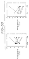
Figure 4. Shows the V-D-J junctional sequences of
human T cells stimulated by 5E4 (5E4+ cells) or by
8F10 (8F10+ cells). The nucleotide sequences of the
V-D-J junctional region of eight 5E4 reactive and six
8F10 reactive T cells are shown in comparison to the
analogous region from the Vβ3.1 clone PL4.4. The 5'
end of the Jβ sequences are identified by comparison
with germ line sequences.
Figure 5. Reactivity of FITC conjugated 5E4 and
8F10 with cynomolgus macaque PBL. Panel 1 is a graph
of 2-color immunofluorescence analysis using a
combination of anti-CD4 and anti-CD8 phycoerythrin and
negative control mAb. Panel 2 is a graph of 2-color
immunofluorescence analysis using a combination of
phycoerythrin conjugated anti-CD4 and anti-CD8 and
8F10 to stain cynomolgous macaque PBL. Panel 3 uses
5E4 as in panel 2 in place of 8F10. 8F10 stains a
subpopulation of macaque PBL consisting of 1.4%. 5E4
stains a subpopulation consisting of 1.46%.
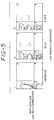
Figure 6. Analysis of Vβ gene usage in synovial
tissue derived T cell line. Line ST-2, derived from
the synovial membrane-infiltrating cells of a RA
patient, was analyzed for TCR Vβ expression using the
cDNA, PCR amplification, slot blot hybridization
protocol. The left panel represents the
autoradiograph obtained when the panel of Vβ genes was
hybridized with the ST-2 amplified TCR specific CDNA
probe. The right panel of the figure is the
densitometry trace of the autoradiograph.
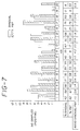
Figure 7. Shows detection of Vβ gene usage in RA
T cells. Shown are tabulated results of the
expression of the panel of Vβ genes in 12 paired
synovial tissue-derived and peripheral blood-derived T
cell lines from RA patients. For the top part of the
figure, the vertical axis represents the number of
samples that were positive for a particular Vβ. The
individual Vβ genes are indicated on the horizontal
axis. Data derived from synovial T cells and
peripheral blood T cells are plotted in pairs as open
and crosshatched bars, respectively. For the bottom
part of the figure, the frequencies of the individual
Vβ genes in the 12 patient samples are shown (%
synovial and % PBT). To indicate preferential usage
of Vβ genes the synovial/peripheral blood ratio is
shown.
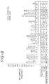
Figure 8. Detection of dominant Vβ gene usage in
RA T cells. This figure is similar to Figure 7,
except that the tabulated data includes only the
expression of the most frequently occurring Vβ genes
as determined by the densitometry trace. The most
frequent or dominant Vβ was determined from the
greatest peak height which was used as a standard.
Any Vβ gene with a corresponding densitometry peak
with height greater than 50% of the standard was used
in the tabulation.
5. DETAILED DESCRIPTION OF THE INVENTION
In the following description, reference will be
made to various methodologies known to those of skill
in the art of immunology, cell biology, and molecular
biology. Publications and other materials setting
forth such known methodologies to which reference is
made are incorporated herein by reference in their
entireties as though set forth in full.
5.1. IMMUNE RELATED DISEASES
The term "immune-related disease" as used herein
refers to a disease in which the immune system is
involved in the pathogenesis of the disease, or in
which appropriate stimulation of the immune system can
result in protection from the disease. Relevant
diseases include, but are not limited, to autoimmune
diseases, neoplastic diseases, infectious diseases,
hypersensitivity, transplantation, graft-versus-host
disease, and degenerative nervous system diseases.
Autoimmune diseases include, but are not limited to,
arthritis, such as rheumatoid arthritis, type I
diabetes, juvenile diabetes, multiple sclerosis,
autoimmune thyroiditis (Hashimoto's thyroiditis),
myasthenia gravis, systemic lupus erthematosis (SLE),
Sjogren's syndrome, Grave's disease, Addison's
disease, Goodpasture's syndrome, scleroderma,
dermatomyositis, myxoedeman, polymyositis, pernicious
anemia, inflammatory bowel disease including Crohn's
disease and autoimmune atrophic gastritis, and
autoimmune hemolytic anemia. Neoplastic diseases
include, but are not limited to, lymphoproliferative
diseases such as leukemias, lymphomas, Non-Hodgkin's
lymphoma, and Hodgkin's lymphoma, and cancers such as
cancer of the breast, colon, lung, liver, pancreas,
etc. Infectious diseases include but are not limited
to viral infections caused by viruses such as HIV,
HSV, EBV, CMB, Influenza, Hepatitis A, B, or C; fungal
infections such as those caused by the yeast genus
Candida; parasitic infections such as those caused by
schistosomes, filaria, nematodes, trichinosis or
protozoa such as trypanosomes causing sleeping
sickness, plasmodium causing malaria or leishmania
causing leischmaniasis; and bacterial infections such
as those caused by mycobacterium, corynebacterium, or
staphylococcus. Hypersensitivity diseases include but
are not limited to Type I hypersensitivities such as
contact with allergens that lead to allergies, Type II
hypersensitivities such as those present in
Goodpasture's syndrome, myasthenia gravis, and
autoimmune hemolytic anemia, and Type IV
hypersensitivities such as those manifested in
leprosy, tuberculosis, sarcoidosis and
schistosomiasis. Degenerative nervous system diseases
include, but are not limited to, multiple sclerosis
and Alzheimer's disease.
Also intended as immune-related diseases as used
herein are malignancies wherein the tumor cell carries
a tumor marker, such as a tumor antigen, capable of
being recognized and responded to by the immune
system. The TCR can serve as a tumor marker on T cell
leukemia or T cell lymphoma cells.
In addition to humans, other preferred animals
for the present invention include domesticated animals
such as equine, bovine, porcine, canine, feline and
murine species. Autoimmune diseases in non-human
species may be analogous to those identified in humans
or may be uniquely characterized for a particular
species or group of species. Thus, the methods of the
present invention are useful in diagnosis and therapy
in human and veterinary medicine.
5.2. ANTIBODIES OF THE INVENTION
The present invention is directed to an antibody,
or a fragment, derivative, or analogue thereof,
specific for an epitope of the Vβ3 region of a human
TCR β chain, preferably a human TCR β chain, may be
utilized in the diagnosis and therapy of an autoimmune
disease, preferably RA. In a preferred embodiment,
the antibodies are specific for the Vβ3.1 chain. The
cDNA sequence for β3.1 is known (Concannon, et al.,
1986, Proc. Nat'l. Acad. Sci. USA, 83:6548-6602) as
are other Vβ3 subfamilies (Toyonaga et al., 1987, Ann.
Rev. Immunol. 5:585-620) The antibodies of the
present invention are useful in diagnosis and therapy.
The term "antibody" is meant to include
polyclonal antibodies, monoclonal antibodies (mAbs),
and chimeric antibodies (see below), (Idiotypy in
Biology and Medicine, Academic Press, New York, 1984).
Preferred antibodies are mAbs. Such antibodies may be
of any immunoglobulin class including IgG, IgM, IgE,
IgA, and any subclass or isotype thereof. Preferred
antibodies for therapeutic use include antibodies of
the IgG2a isotype or IgG2b isotype (Rashid et al.,
1992, J. Immunol. 148: 1382-1388).
The term "antibody" is also meant to include both
intact molecules as well as fragments thereof which
bind the antigen, such as, for example, F(ab')2, Fab',
Fab and Fv. These fragments lack the Fc fragment of
an intact antibody molecule, clear more rapidly from
the circulation, and may have less non-specific tissue
binding than an intact antibody (Wahl et al., 1983, J.
Nucl. Med. 24:316-325), properties which may be
desirable for particular therapeutic or diagnostic
utilities. It will be appreciated that these antigen-binding
fragments of the antibodies useful in the
present invention may be used for the detection and
quantitation of TCR proteins or peptides as disclosed
herein for intact antibody molecules. Such fragments
are typically produced by proteolytic cleavage, using
enzymes such as papain (to produce Fab fragments) or
pepsin (to produce F(ab')2 fragments) or by reducing
the disulfide bridges.
The monoclonal antibodies of the invention are
reactive with a variable region of the vβ3 family of
the beta chain of the T cell antigen receptor. In
particular, such an anti-TCRβ mAb can recognize Vβ3.1.
In a specific embodiment, the invention is directed to
monoclonal antibodies 5E4 and 8F10, as deposited with
the ATCC and assigned accession numbers HB 11020 and
HB 11021, respectively. The Vβ3.1 specific monoclonal
antibodies of the present invention enables the
analysis of the expression of the Vβ3.1 gene in a
biological sample.
Various chemical or biochemical derivatives of
the antibodies or antibody fragments of the present
invention can also be produced using known methods.
One type of derivative which is diagnostically useful
is an immunoconjugate comprising an antibody molecule,
or an antigen-binding fragment thereof, to which is
conjugated a detectable label such as a radioisotope
or other tracer molecule. A therapeutically useful
immunoconjugate comprises an antibody molecule, or an
antigen-binding fragment thereof, conjugated to a
therapeutically useful molecule such as a cytotoxic
drug or a toxic protein (see, for review: Dillman,
R.O., Ann. Int. Med. 111:592-603 (1989)). Such
antibody derivatives are discussed in more detail
below.
The antibody, fragment or derivative of the
present invention, may be prepared by using any of a
number of techniques well-known in the art. For
producing a mAb, any method which provides for the
production of antibody molecules by continuous cell
lines in culture may be used. These methods include,
but are not limited to,the hybridoma technique
originally described by Kohler and Milstein (1975,
Nature 256:495-497), and the more recent human B cell
hybridoma technique (Kozbor et al., 1983, Immunology
Today 4:72), EBV-hybridoma technique (Cole et al.,
1985, Monoclonal Antibodies and Cancer Therapy, Alan
R. Liss, Inc., pp. 77-96), and trioma techniques. A
hybridoma of rodent origin producing the mAbs of this
invention may be cultivated in vitro or in vivo. For
an overview of antibody production methods, see:
Hartlow, E. et al., Antibodies: A Laboratory Manual,
Cold Spring Harbor Laboratory Press, Cold Spring
Harbor, NY, 1988.
In one embodiment, the antibody of the present
invention is a human mAb. Human mAbs may be made by
any of a number of techniques known in the art (e.g.,
Teng et al., 1983, Proc. Natl. Acad. Sci. U.S.A.
80:7308-7312; Kozbor et al., supra; Olsson et al.,
1982, Meth. Enzymol. 92:3-16).
In another embodiment, the antibody is a chimeric
antibody, preferably a mouse-human chimeric antibody,
wherein the heavy and light chain variable regions are
derived from a murine mAb and the constant regions are
of human origin. The chimeric antibodies of this
invention have both the TCR-recognizing specificity of
the mouse Mab and the biological properties of human
antibodies, which include resistance to clearance in
the human and lower immunogenicity for humans,
allowing multiple treatments. Method for producing
chimeric antibody molecules are disclosed, for
example, in Gorman et al., PCT Pub. WO9206193
(4/16/92); Cabilly et al., U.S. Patent 4,816,567
(3/28/89) and Eur. Patent Pub. EP125023 (11/14/84);
Taniguchi et al., Eur. Patent Pub. EP171496 (2/19/86);
Morrison et al., Eur. Patent Pub. EP173494 (3/5/86);
Neuberger et al., PCT Pub. WO8601533 (3/13/86); Kudo
et al., Eur. Patent Pub. EP184187 (6/11/86); Robinson
et al., PCT Pub. WO 8702671 (5/7/87); Cabilly et al.,
Proc. Natl. Acad. Sci. USA 81:3273-3277 (1984);
Morrison et al., Proc. Natl. Acad. Sci. USA
81:6851-6855 (1984); Boulianne et al., Nature 312:643-646
(1984); Morrison, Science, 229:1202-1207 (1985);
Neuberger et al., Nature 314:268-270 (1985); Takeda et
al., Nature 314:452-454 (1985); Oi et al.,
BioTechniques 4:214 (1986); and Liu et al., Proc.
Natl. Acad. Sci. USA 84:3439-3443 (1987).
For human therapeutic purposes, mAbs or chimeric
antibodies can be "humanized" by producing human
constant region chimeras, where even parts of the
variable regions, in particular the conserved or
framework regions of the antigen-binding domain, are
of human origin, and only the hypervariable regions
are non-human. See for example, UK Patent Publication
GB 2188638 A entitled "Chimeric Antibodies", Harris et
al., PCT Publication WO 9204381, published March 19,
1992, entitled "Novel Antibodies for Treatment and
Prevention of Infection in Animals and Man," and
Riechmann et al, 1988, Nature 332:323-327.
In a further embodiment, the antibody is a single
chain antibody formed by linking the heavy and light
chain fragment of the Fv region via an amino acid
bridge, producing a single chain polypeptide (Bird,
1988, Science 242:423-426; Huston et al, 1988,
Proc.Natl.Acad.Sci. USA 85:5879-5883: and Ward et al,
1989, Nature 34:544-546).
Antibody molecules or fragments may be purified
by known techniques, e.g., immunoabsorption or
immunoaffinity chromatography, chromatographic methods
such as HPLC (high performance liquid chromatography),
or a combination thereof, etc.
Once antibodies of the desired specificity are
generated, they may be used to identify and select
other antibodies having the same or cross-reactive
epitope specificity. For example, a new antibody is
tested by measuring its ability to inhibit the binding
of an antibody of known specificity to its epitope.
Various competitive binding assays known in the art
can be used.
The isotype of the antibody can be selected
during hybridoma production or by appropriate
recombinant methods well-known in the art to achieve a
desired effector function mediated by the Fc portion
of the immunoglobulin heavy chain. For example,
certain isotypes, such as IgG2a, have superior
activity in antibody-dependent cellular cytotoxicity.
Likewise, certain isotypes, such as IgG2a, are more
readily eliminated from the circulation through Fc
receptors on cells of the reticuloendothelial system
and are therefore more efficient at removing an
undesired antigen or target cell from sites of active
disease (Rashid, et al., 1992. J. Immunol. 148: 1382-1388).
Accordingly, depending on the intended use, a
particular antibody isotype may be preferable to
others, as can be readily ascertained by one of
ordinary skill in the art without undue
experimentation.
To identify a hybridoma producing an antibody of
a particular isotype, or to switch an isotype of an
antibody, the hybridoma supernatants may be screened
for production of TCR-specific mAbs using an ELISA
which tests for the immunoglobulin isotype. What
follows is an example of a method for selecting a
desired isotype switch from IgG1 to IgG2a. Hybridoma
cells are grown in the logarithmic phase for a 2-3
week period prior and then subjected to negative
selection using antibody-coated magnetic beads. Super
paramagnetic iron oxide particles coated with a goat
anti-mouse antibody preparation including all IgG
isotype classes (Biomag® beads purchased from Advanced
Magnetics, Inc.) may be used. For switching an
isotype from IgG1 to IgG2a, it is necessary to block
the IgG2a binding sites on the antibody-coated beads
by incubating with immunoglobulins (of irrelevant
specificity) having the IgG2a isotype. About 108
hybridoma cells expressing a variety of isotypes are
incubated with such IgG2a-blocked beads. Cells
expressing IgG1, IgG2b and IgG3 isotypes bind and are
removed magnetically from the population. Such a
negative selection step is preferably repeated several
times.
The remaining cell population, depleted of IgG1,
IgG2b and IgG3 bearing cells, and conversely enriched
for IgG2a-bearing cells, is plated in microplates at a
cell density of about 1000 cells/well. Using
commercially available anti-isotype reagents in an
ELISA assay, the wells are screened for IgG2a
production; positive clones are replated at 0.3
cells/well followed by another round of screening and
re-plating. Using such an approach, approximately 1-5
of 107 cells which have switched isotype are optimally
selected. Cells which have switched from IgM to IgG
can be selected using a similar approach with the
appropriate antibody-coated beads.
As used herein, an antibody reactive with the "V
region" of the TCR shall be construed to be an
antibody reactive with an epitope of the V region, a
combination epitope of the V region, or a combination
epitope of the V-D or V-D-J regions. An antibody
reactive with a V region of a TCR may recognize an
idiotypic determinant, a clonotypic determinant, or,
preferably, may recognize a minor framework region
expressed by a discrete subset of T lymphocytes.
Preferably, such an antibody is reactive with a unique
epitope on a Vβ3.1 variable region of the β chain of
the T cell antigen receptor.
The term "minor framework region" refers to a
region of the TCR which is not shared by all TCR
molecules, but is also not unique to a particular T
cell clone. Preferred anti-TCR β mAbs recognize
members of the Vβ3 family, most preferably, Vβ3.1.
5.3. METHODS OF GENERATING AND CHARACTERIZING
ANTIBODIES OF THE INVENTION
This invention provides specific monoclonal
antibodies reactive with defined regions of the
members of the Vβ3 family variable regions of the β T
cell antigen receptor. Over the last several years,
since the cloning of the genes encoding for the TCR,
surprisingly few antibodies have been generated
against the different variable, diversity, joining or
constant regions of the receptor. This indicates that
the knowledge of the amino acid sequence of a
particular region of interest has not been sufficient
to allow the reproducible production of specific
antibodies. Several groups have been able to generate
one or two specific antibodies, but no one seems to
have been able to generate antibodies at will. Over
several years, Present inventors have generated
several TCR antibodies by numerous methods and now
have a preferred method to maximize success in
generating antibodies against a defined region of
interest.
The major elements of this improved protocol
include 1) the use of purified protein as the
preferred immunogen; 2) effectively monitoring the
mice by tailbleeds and prescreening during
immunization to detect positive antibody responses; 3)
using purified protein during the screening of
hybridoma supernatants to minimize the number of
hybridomas to be screened to detect positive ones and
to maximize the positive signal over background in the
screening procedure so that true positives are not
missed due to weak signals; and 4) better
characterizing the resultant positives to determine
their true specificity.
5.3.1. HOST REQUIREMENTS
Various host animals, including but not limited
to mice, rats or rabbits can be used in the practice
of this invention. However, some hosts are preferred,
as discussed infra. Several years of experience in
generating TCR antibodies has resulted in the
observation that the TCR proteins are not very
immunogenic. This is perhaps due to the pivotal role
the proteins themselves play in the immune system.
Only those TCRs that recognize "non-self" are allowed
to exit the thymus during ontogeny. In addition, the
proteins are highly conserved evolutionarily and are
members of the immunoglobulin supergene family.
Members of this supergene family share not just
sequence homologies, but structural similarities as
well. It is now known that some human-mouse homologs
exist that are very similar in sequence (Wilson et
al., 1988, Immunol. Reviews 101: 149-172). This was
first proposed as the variable genes were being mapped
for their relative position along the chromosome (Lai
et al., 1988, Nature 331: 543-546). Thus, some human
Vβ and murine Vβ sequences are more similar to one
another than either one is to other variable regions
in the species. The practical result of this
similarity is that human TCR proteins used as
immunogens in mice to produce an antibody response
yield variable results. It generally takes several
weeks or months to generate a response in the mice,
requiring multiple boostings. Although a better
immune response may be generated in other hosts,
chickens, for example, the lack of reliable myeloma
fusion partners needed to create the immortal fused
hybridomas for chicken spleen cells limits the
usefulness of these hosts.
In order to maximize the antibody response in
mice to human immunogens, various groups have proposed
using host animals deficient in the TCR homolog of
interest. For example, mice strains such as SJL, RIII
S/J (H-2'), C57L, C57BR and SWR (Behlke et al., 1986,
Proc. Natl. Acad. Sci USA 83:767-771; Haqqi et al., J.
Exp. Med., 1989, 169: 1903-1909; Jouvin-Marche, 1989,
Eur. J. Immunol. 19: 1921-1926) that have deleted
major portions of the murine Vβ locus may be
advantageous for generating antibodies against the
corresponding human homologs. Other murine strains
such as nude mice or SCID mice may be useful for
similar reasons. Taking this idea one step further,
one group has proposed genetically engineering mouse
cells to produce a chimeric TCR where every portion of
the TCR is murine, except the portion (e.g. the human
variable region) to be used to elicit antibodies.
This procedure has worked in at least one instance to
generate Vβ13.1 and Vβ13.2 specific antibodies (Intl.
Publ. No. WO92/02629, published February 20, 1992),
but is not the complete answer to the problem of
creating antibodies at will, for the reasons indicated
below under screening.
5.3.2. PREFERRED IMMUNOGENS
There are a large number of different immunogens
representing defined regions of the TCR that can be
used to generate antibodies. Some of these include
peptides; conjugated peptides; partially purified TCR
protein by immunoprecipitation, for example; more
fully purified protein; T cell clones; transfected
cells; soluble recombinant receptor protein; or
combinations of these. TCR antibodies that exist today
have been generated on a hit-or-miss basis for each of
these immunogens. However, a careful understanding of
the relative merits of each immunogen was necessary in
order to develop the present inventors' preferred
procedure to yield a desired antibody.
Peptide immunogens: Several groups have used
chemically synthesized TCR peptides to generate
numerous anti-peptide antibodies to TCR.
Unfortunately, these anti-peptide antibodies very
rarely interact with intact receptor on cells. Thus,
they have no therapeutic or in vivo diagnostic
utility. Often such anti-peptide antibodies react
well with the peptide to which they were raised, but
react poorly with TCR proteins, even when the protein
is denatured, by Western blotting for example.
Further complications include the observations that
not all peptides are soluble and that the peptides
sometimes lack the carbohydrate groups present on the
native version of the receptor. In summary, although
anti-peptide antibodies can be generated, the
resulting antibodies often have little utility for
diagnostic and therapeutic applications, and the use
of peptides as immunogens has not given rise to
anti-minor framework or V region specific antibodies.
Whole Cell Immunogens: Immunogen on the surface
of T cells or on transfected T cells have been used to
generate specific antibodies. Various cell lines
including, but not limited to, those disclosed herein
and cell lines disclosed elsewhere (see, for example,
Toyonaga et al., 1987, Ann. Rev. Immunol. 5: 585-620)
can be used as immunogens to generate mAbs specific
for the human TCR V region. Any T cell line
expressing a TCR, a TCR chain or fragment on the cell
surface may serve as an immunogen. Note that
antibodies to known V, D, J, DJ, VJ, VDJ or
combinations thereof can also be generated by
immunizing with such cells.
The expression of DNA encoding the V, D, J, and C
regions of any TCR chain can be determined in any cell
line by well-known procedures including cDNA
sequencing, in situ hybridization, polymerase chain
reaction (PCR) analysis, Northern analysis, Southern
analysis, immunoassay, or flow cytometry cytometry, to
name but a few. The V specificity of the resultant
antibody can be determined from knowing the sequence
of the TCRs expressed by the immunizing cell.
Whole cells that can be used as immunogens to
produce a TCR-specific antibodies of the present
invention include not only T cells which naturally
express a TCR, but also cells transfected with a
recombinant DNA construct which encodes a particular
TCR chain or chains, or a fragment thereof. For
example, β- Jurkat cells which do not produce a
functional TCR can be "reconstituted" by transfection
with TCR β cDNA to produce intact αβ TCR on the cell
surface (Ohashi, P.S. et al., Nature, [1985] 316:606-609).
Such transfected cells would then be used as
immunogens for inducing antibodies specific for an α
or β TCR epitope. Additional examples of such
transfected cells have been reported (Kaye, J. et al.,
1988, Nature 336:580-583; Dembic, Z., et al., 1986,
Nature 320: 232-238; Saito, T., et al., 1987, Nature
325:125-130). According to the present invention, any
procedure that results in expression of a transfected
TCR gene on the cell surface could be used to produce
a whole cell immunogen.
Immunogens can also be produced as proteins or
cells derived from eukaryotic expression systems in
which a TCR protein or peptide is attached to the cell
membrane via an enzymatically cleavable phospholipid
anchor domain (Int'l Patent Application
PCT/US88/02648, published February 9, 1989).
Many of the antibodies produced using these whole
cell immunogens are anti-idiotypic or anti-clonotypic,
but some anti-minor framework or variable region
specific antibodies have been generated, as well.
Although not impossible, it is rare that anti-constant
region antibodies are produced using whole cell
immunogens, since the constant regions of the TCR
chains appear to be masked by one another and by T3
polypeptides also present in the TCR:T3 complex on the
surface of cells.
The major drawback of using cells as immunogen
for producing anti-TCR antibodies is that many other
non-TCR proteins on the cell are also immunogenic. The
antibodies specific to the TCR represent a minor
subset of the total antibody response generated in the
host. As a result, large numbers of hybridomas need to
be screened to detect the ones producing TCR specific
antibodies. Even when the cells used to immunize are
murine cells expressing only one defined human region,
the screening procedure is not optimal. For example,
in order to prescreen the mice to determine if they
are making specific antibody prior to isolation of the
spleen cells and fusion with the myeloma partner, it
is necessary to try to titrate mouse serum by dilution
until it is possible to see the difference between a
negative versus a positive result. Since mouse serum
has an extremely high protein concentration, the
difference in a positive hybridoma supernatant can not
always be seen above the background created from the
serum itself. When hybridoma supernatants are
screened by Flow cytometry analysis by detecting
binding to the same transfected cells used as
immunogen or by their ability to generate an IL-2
response, the same problem of detecting a positive
difference over the background of the testing
procedures exist. The observed signals are small
relative to the background ones.
Purified Protein Immunogens: When purified
protein is used as an immunogen, a less heterogeneous,
more efficient antibody response is generated in the
host. This results in an increased relative
proportion of cells producing antibodies against the
desired TCR antigens over the other contaminating
antigens present in the immunogen. This is especially
important for enhancing TCR specific antibody
responses, since TCR are not very immunogenic to begin
with. In addition, an increased proportion of desired
cells minimizes the number of hybridoma cells that
must be screened to find positives. The "purer" the
protein used as immunogen, the better the chances for
success. Present inventors prefer using purified,
soluble, recombinant protein as immunogen, if
available, since a large quantity of material is
available to use first as immunogen and later in
improved prescreening and screening procedures. With a
purified protein it is possible to maximize the signal
to noise ratio of the prescreening and screening
methods, so it is easier and more reliable to detect
positive hybridomas over assay background. A purified
protein can also be used to generate all types of TCR
antibody, e.g. it is possible to identify
anti-idiotypic, anti-variable or anti-minor framework,
and anti-constant or anti-major framework
specificities.
The present invention in a preferred embodiment
thus uses a soluble Vβ3 TCR polypeptide that can be
used 1) as immunogen to generate specific antibodies
and 2) in screening protocols to enhance signal to
background ratios to identify and characterize said
antibodies.
5.3.3. IMPROVED SCREENING PROCEDURES
Screening procedures that can be used to screen
hybridoma cells expressing different anti-TCR
antibodies include but are not limited to (1) enzyme
linked immunosorbent assays (ELISA), (2) flow
cytometry analysis, (3) immunoprecipitation, (4)
Western blotting and (5) the ability to comodulate the
CD3 antigen (part of the TCR-CD3 complex present on
the surface of the T cells) off of the surface of
cells. The comodulation and flow cytometry screening
procedures are preferred for the selection of
antibodies potentially useful in therapy since these
procedures select antibodies that are able to
recognize intact TCR on live cells.
Many different immunoassay formats including but
not limited to ELISA, EIA, and RIA formats of an ELISA
that can be used to screen for anti-TCR antibodies as
can be envisioned by one skilled in the art.
Many additional screening assays, such as those
based upon competition with anti-TCR antibodies of
known specificity or the ability to cause T cells
expressing known TCRs to proliferate in culture, will
be known to those skilled in the art and can be used
for the selection of appropriate antibodies.
Initially it was difficult to obtain antibodies
reactive with T cell receptor V regions. This problem
arose when the signal observed by the antibody binding
to PBLs by flow cytometry analysis was very low
relative to the background of Flow cytometry itself.
Since this signal to background level was so low, and
was many times obscured by the variability of the
assay itself, it was concluded that the antibodies
were reacting positively only with the T cell clone
used to generate them. As more became known about the
number of T cell receptor genes for the β chain
(60-100 total human genes in about 20 variable region
families) and the α chain (100 or so human genes), it
was easier to interpret the flow cytometry data more
accurately. For example, a Vβ region specific antibody
would be expected to react with about 5% of PBLs on
average. Later studies of the percentages of families
of Vβs in normal PBLs indicate that families are
actually present at 1% to 8% depending upon the
individual family. Depending upon the actual
instrument, its calibration, the skill of the operator
and the inherent interassay variability of flow
cytometry analysis, background levels in flow
cytometry can easily be in the 3-4% range. Thus,
ironically, the same problem that confounded
investigators trying to make specific TCR antibodies
7-8 years ago still exists today: the ability to
distinguish a true positive hydridoma by a screening
method is diminished in assays that provide low
signals with high backgrounds.
Present inventors have improved the chances of
success of identifying desired hybridomas, by
providing improved screening assays with greater
signal to noise ratios. One such assay is an ELISA
format using purified protein, better yet purified
soluble, recombinant protein, to bind to antibodies in
hybridoma supernatants to generate strong positive
signals. Since the "analyte" in these assays is a
known, purified protein, the signals are higher than
those obtained if cell lysates or other partially
purified protein preparations containing numerous
contaminants are used. These more effective and
efficient screening assays enhance the ability to
prescreen mouse serum to monitor effective antibody
responses in the host following immunization and to
screen pooled hybridoma supernatants during initial
screening of many hybridoma candidates. Another
advantage of the ELISA assays is that the choice of
the plating antibody (pan-Ig or IgG2b specific, for
example) can positively select for TCR specific
antibodies of a preferred isotype. Thus, once a
desired TCR antibody is identified, it is not
necessary to then switch its isotype. With soluble,
purified protein it is also possible to bind antigen
directly to the plate rather than using sandwich
immunoassay formats.
5.3.4. IMPROVED CHARACTERIZATION PROCEDURES
After a positive hybridoma has been detected by
the initial screening protocol, it is then further
screened and analyzed to determine its specificity.
Such characterization includes, for example 1)
immunoprecipitation followed by SDS gel
electrophoresis to determine if the antibody
precipitates proteins of sizes expected for TCRs, with
or without coprecipitation of the T3 polypeptides; 2)
the ability of the antibody to comodulate the T3
receptor off the surface of cells, indicating that the
antibody is reacting with the TCR; 3) determining the
percentage of CD3 positive PBLs the antibody binds to
by flow cytometry analysis to determine whether it is
anti-idiotypic, anti-variable or anti-constant region
specific; 4) Western blot analysis to determine β
chain specificity, 5) blocking experiments with other
known antibodies or peptides to determine epitope
specificity or similarity to known antibodies; and
6) analysis against a panel of cells expressing known
TCRs to determine variable or constant region
reactivity.
In addition to these procedures the present
inventors have found that the following procedure
allows even better antibody characterization. Once an
antibody has been generated and characterized as
indicated above, it is then used to stimulate PBLs to
expand in culture. The antibody specifically
interacts with the subset of PBLs expressing the
epitope reactive with the antibody and stimulates a
mitogenic response. As cycles of this mitogenic
response occur, the antibody is selectively amplifying
these cells until it is possible to generate cell
lines that are up to 95% reactive with the antibody.
This results in a very high positive signal that it
easy to distinguish over background, for example a 95%
positive signal in flow cytometry over a background
signal of 2-4%. Using this procedure it was possible
to distinguish the true specificity of antibody W112
(Vβ5.3) over antibody 1C1 (Vβ5.2 and Vβ5.3) (Boylston
et al., 1986, Immunol. 137(2): 741). In addition,
the present inventors were able to determine the fine
specificity of antibodies 5E4 and 8F10 as being
variable region specific and not a combined variable
and joining region specificity. A further advantage of
this amplification method is that it is simple to
distinguish a true anti-idiotypic antibody from one
that recognizes one of the less expressed V region
families. The anti-variable region antibody will
productively drive PBLs through mitogenic cycles to
generate an amplified cell line. Since it is extremely
unlikely that an anti-idiotypic antibody will find its
cognate antigen in a population of PBLs, it will not
generate amplified cell lines.
The present inventors have further characterized
the ability of specific antibodies to interact with
monkey cells, e.g. the presence of a monkey homolog.
Since anti-human TCR antibodies do not cross react
with non-primate TCR, an important advantage of the
present invention is to enable a primate model for
safety, toxicity and efficacy studies. While some
anti-TCR V Region antibodies react with monkey cells
and others do not (See Axberg et al., 1991, J. Clin.
Immunol. 11, 1-12), the present inventors have shown
that 5E4 and 8F10 do react with monkey cells. It is
possible to further define the fine specificities of
these antibodies, since similar antibodies by other
characterization procedures may interact with
different percentages of monkey cells. Thus, although
they recognize similar antigens, they may recognize
different epitopes or combined epitopes.
5.4. DIAGNOSTIC UTILITY OF THE
ANTIBODIES OF THE INVENTION
The antibodies and fragments of the present
invention can be employed for diagnostic or research
purposes in various immunoassays well-known in the
art. The antibodies, or fragments of antibodies,
useful in the present invention may be used to
quantitatively or qualitatively detect the presence of
cells which express a member of the TCR Vβ3 family,
particularly Vβ3.1, or the levels of TCR protein
present in a sample. This can be accomplished by
immunofluorescence techniques employing a
fluorescently labeled antibody (see below) coupled
with light microscopic, flow cytometry cytometric, or
fluorimetric detection.
The antibodies (or fragments thereof) useful in
the present invention may be employed histologically,
as in immunofluorescence or immunoelectron microscopy,
for in situ detection of the TCR molecule.
One way of measuring the reactivity of a T cell
receptor epitope with a specific antibody of the
present invention is by enzyme immunoassay (EIA) such
as an enzyme-linked immunosorbent assay (ELISA)
(Voller, A. et al., J. Clin. Pathol. 31:507-520
(1978); Butler, J.E., Meth. Enzymol. 73:482-523
(1981); Maggio, E. (ed.), Enzyme Immunoassay, CRC
Press, Boca Raton, FL, 1980). The enzyme, either
conjugated to the antibody of the invention, or to a
binding partner for the antibody, when later exposed
to an appropriate substrate, will react with the
substrate in such a manner as to produce a chemical
moiety which can be detected, for example, by
spectrophotometric, fluorimetric or by visual means.
A preferred method of enumerating total T Cell
receptor β chain or Total V region subset TCR chain is
performed using detergent treated whole blood samples.
In particular Vβ3.1 subset may be detected from a
sample or whole blood samples are added to wells in a
96 well plate previously coated with 5 µg/ml of
coating antibody. Coating antibody is either a
negative control, an anti-major framework antibody
such as W76 (to detect total β chain) on a TCR v
region special monoclonal antibody such as those of
the present invention (to detect a subset of TCR β
chain). An HRP conjugated BF1 antibody, which
recognizes a different epitope of the β chain C region
than W76 is used as a detection antibody. The assay
can be used to detect total TCR β chain as well as β
chain subsets such as Vβ3.1. The assay format is
described in Rittershaus C.W. PCT Publication
WO9208981, published May 29, 1992 entitled,
"Therapeutic and diagnostic methods using total
Leukocyte surface antigens."
Detection of a member of the TCR Vβ3 family
protein or cells may be accomplished using any of a
variety of other immunoassays. For example it is
possible to detect antibody binding to TCR V region
through the use of a radioimmunoassay (RIA) (see, for
example, Weintraub, B., Principles of
Radioimmunoassays, Seventh Training Course on
Radioligand Assay Techniques, The Endocrine Society,
March, 1986, pp. 1-5, 46-49 and 68-78; Work, T.S. et
al., Laboratory Techniques and Biochemistry in
Molecular Biology, North Holland Publishing Company,
New York, 1978).
It is also possible to label the antibody in
which binding is measured using radioactive,
fluorescent, chemiluminescent or bioluminescent
conjugated antibodies.
A variety of immunoassay formats is available,
for either EIA or RIA systems. For example, assays
may be competitive or non-competitive. Two site or
sandwich assays may be used, either "forward",
"simultaneous" or "reverse" assays, which are well-known
in the art.
Additional types of immunoassays include
precipitin reactions, gel diffusion precipitin
reactions, immunodiffusion assays, agglutination
assays, complement fixation assays, immunoradiometric
assays, protein A immunoassays, and
immunoelectrophoresis assays.
Binding of the antibody, or fragment or
derivative thereof to the TCR epitope for which it is
specific may be accomplished and/or detected in vitro
or in vivo. In vitro binding, as described above, may
be performed using histologic specimens, or fractions
or extracts of tissue or fluid, including
substantially purified T cells. In vivo binding may
be achieved by administering the antibody (or fragment
or derivative) by any route or means known in the art,
including but not limited to intravenous,
intraperitoneal, intranasal, and intraarterial, such
that specific binding may be detected.
For diseases involving joints, such as RA,
intraarticular administration of a labelled antibody
(or derivative or fragment) may also be utilized as a
diagnostic procedure.
Imaging techniques can be used in vivo, wherein
the antibody, derivative or fragment is bound to a
detectable label capable of in vivo localization.
Many different labels and methods of labeling are
known in the art.
The present invention provides method for
diagnosing an immune-related disease, including a
lymphatic malignancy, based on detecting the specific
binding of a mAb, or a derivative or fragment thereof,
to a defined region of a TCR in a biological sample
from a subject suspected of having the disease or
disorder. Biological samples which may be tested
according to the present invention include any body
fluid, such as peripheral blood, plasma, cerebrospinal
fluid, lymph, peritoneal fluid, or pleural fluid, and
the like, or any body tissue or extract thereof.
According to the present invention, RA may be
diagnosed in a subject by detecting the increased
presence of T cells expressing Vβ3, in particular,
Vβ3.1, alone or concomitant with increased presence of
T cells expressing Vβ9, or Vβ10, in a biological
sample from the subject as compared to baseline
samples. Such diagnosis may be achieved by the use of
a mAb, fragment or derivative, specific for the
particular TCR V region associated with the disease,
as described above. As used herein, the term
"baseline sample" refers to a sample from a normal,
healthy individual who does not have rheumatoid
arthritis, or a sample from the subject prior to onset
of the disease or at a time of remission of the
disease. In a further aspect, a baseline sample may
be a mixture or average of samples from a general
population. In a special embodiment the biological
sample being tested in from the site of disease and
the base line is the peripheral blood.
Alternatively, such diagnosis may be achieved by
detection of the presence of nucleic acid sequences
characteristic of these TCR V regions using molecular
techniques. Preferably, such molecular diagnosis is
achieved by detecting the presence of nucleic acid
sequences homologous to a gene encoding a defined TCR
or Vβ3, preferably Vβ3.1, region in mRNA in a patient
sample. The nucleic acid and amino acid sequence for
the Vβ3 gene family members and gene products are
known (Toyonaga et al., 1987 Ann. Rev. Immunol.,
5:595-620). One skilled in the art could readily
design diagnostic tests to detect the Vβ3 elevation
described here. Molecular approaches used to
correlate TCR gene expression with disease include:
It should be understood that the diagnostic
methods of the present invention are best used along
with other known diagnostic methods to obtain a
comprehensive patient diagnosis. For example, a
diagnosis of RA may be made based on the methods of
the present invention together with conventional
diagnostic recognition of the clinical features of RA,
such as:
See, for example, Fishman
et al., Medicine, Second
Ed., J. B. Lippincott Company, Philadelphia, PA, pp.
340-346. As with any diagnostic criteria, the
parameters disclosed in the present invention may not
be sole determinants, or pathognomonic, of a
particular disorder.
5.5. THERAPEUTIC UTILITY OF THE
ANTIBODIES OF THE INVENTION
As mentioned above, the present invention is also
useful in the therapy of an immune-related disease,
preferably an autoimmune disease or a lymphatic
malignancy. The therapeutic embodiments of the
present invention are best applied once a correlation
has been established between a disease of interest,
for example RA, and preferential use of a particular
TCR V gene in T cells associated with the disease, for
example genes encoding a member of the Vβ3 family, in
particular Vβ3.1, alone or concomitant with use of
genes encoding Vβ9, or Vβ10.
The particular TCR which is expressed on or
"marks" those T cells mediating the autoimmune process
is designated, a "marker TCR." In a preferred
embodiment, the marker TCR for rheumatoid arthritis is
Vβ3.1. The antibodies, fragments or derivatives of
the present invention are therapeutically useful in
part because of their ability to interfere with the
binding of the T cell, via its TCR, to the MHC/antigen
complex (or the antigen alone) needed for initiation
or propagation of the autoimmune process.
Marker TCRs associated with a given disease are
identified using any of a variety of techniques well-known
in the art. Marker TCR V genes for RA are
exemplified in more detail below. A genetic approach
using patients known to have Myasthenia gravis or
multiple sclerosis was described by Oksenberg, J.R.,
et al., Proc. Natl. Acad. Sci. USA 86:988-992 (1989).
Sequences of the appropriate TCR β chain are obtained
by genomic analysis using restriction fragment length
polymorphisms found in families having a prevalence of
the particular autoimmune disease, for example, as
described by Seboun, E., et al., Cell 87:1095-1100
(1989); Burns, F.R., et al., J. Exp. Med. 169:27-39
(1989)). Thus it is within the level of ordinary
skill in the art to identify other diseases associated
with expression of Vβ3, in particular Vβ3.1.
It should be appreciated that, for the purposes
of the present invention, determination of the marker
TCR associated with an autoimmune disease does not
require that the "autoantigen" be characterized. It
is sufficient that the autoimmune disease involves a T
cell-mediated immune response as a necessary part of
the pathogenetic process. In fact, as is known in the
art, the autoimmune disease may not involve a true
autoantigen at the inductive stage, but rather, may
represent a response to an exogenous antigen, such as
a bacterial or viral antigen, which is cross-reactive
with self antigens, or results in an immunopathologic
response directed to the exogenous antigen present in
the host.
T cells of the subset associated with the
autoimmune disease, which may recognize a true
autoantigen or an autoimmune disease-associated
antigen (such as certain viral or bacterial antigens)
may be cloned and expanded or immortalized in culture
by methods well-known in the art. For example, the T
cells may be fused to an immortalizing cell, e.g., a
long term T cell line, a T cell lymphoma line or a T
cell hybridoma, and then grown in culture. The
cultured cells serve as the source of cell-surface TCR
chains which are analyzed using the antibodies of the
present invention, or as the source of cDNA encoding
the appropriate TCR for molecular identification of
TCR usage. Such CDNA is cloned and expressed by
methods well known in the art. (See, for example,
Sambrook, J. et al. (Molecular Cloning: A Laboratory
Manual, 2nd Edition, Cold Spring Harbor Press, Cold
Spring Harbor, NY (1989)). T cells may be isolated
from humans who are susceptible to an autoimmune
disease, preferably from susceptible individuals who
have the autoimmune disease, and are expanded in
culture using known techniques (see, for example,
Zamvil et al., Nature 317:355-358 (1985);
Nature 324:258-260 (1986)).
An absolute correlation between an autoimmune
disease and the usage of a particular TCR is neither
expected nor necessary for every individual to
successfully practice the present invention.
Correlations have been shown for some individuals with
Rheumatoid arthritis and the expression or presence of
Vβ3 in the synovial tissue relative to peripheral
blood. The correlation also exists for increased
expression of Vβ9 or Vβ10 relative to peripheral
blood, or both, especially when detected in addition
to Vβ3.
The TCR expressed by T cells specifically
associated with a disease can be identified using TCR-specific
antibodies, either polyclonal, monoclonal or
chimeric, such as those described herein.
Specifically antibodies for an epitope of a member of
the TCR Vβ3 family, specifically Vβ3.1, can be used to
detect surface expression, employing techniques of
fluorescence microscopy, flow cytometry cytometry,
immunocytochemistry, or other techniques known in the
art. Such antibodies are described herein, and have
been reported by others for a number of TCR αβ chain V
regions in rodent systems (See, for example, Ohashi,
M., et al., J. Exp. Med. 168:2153-2164 (1988);
Gascoigne, N.R.J., et al., Proc. Natl. Acad. Sci., USA
84:2936 (1987); Kappler, J.W., et al., Nature 332:35
(1988); Kappler, J.W., et al., Cell 49:263 (1987);
MacDonald, H.R., et at., Nature 332:40 (1988)).
As an alternative to analysis of the cell surface
using antibodies, DNA or MRNA of the T cells can be
probed directly, or after amplification by the
polymerase chain reaction (Synha et al., Science
239:1026 (1988); Saiki et al., Nature 324:163 (1986),
by specific hybridization with nucleic acid probes for
the various TCR gene families, using hybridization
methods well known in the art (See examples, below).
Where no specific autoantigen has been
identified, the oligoclonality of T cells in the
anatomic region associated with the disease can be
used as a basis for enrichment of reactive T cells.
For instance, cells uniquely associated with RA are
found in the synovial fluid of the joint; cells
uniquely associated with MS are found in the
cerebrospinal fluid (CSF); and disease-associated T
cells infiltrate the thyroid tissue in Hashimoto's
thyroiditis and in Graves' disease. In these
instances, T cells may be isolated from the relevant
anatomical location and expanded in culture. (See,
for example, Londei, M. et al., Science 228:85-89
(1985); Stamenkovic, I. et al. Proc. Natl. Acad. Sci.
USA 85:1179-1183 (1988); Oksenberg, J.R., et al.,
supra).
Treatment of an individual using the antibodies,
fragments or derivatives of this invention comprises
parenterally administering a single dose or multiple
doses of the antibody, fragment or derivative. The
effective dose is a function of the individual
antibody (or fragment or derivative), the presence and
nature of a conjugated therapeutic agent, the subject
and his clinical status. Effective doses of the
antibodies, fragments or derivatives of this invention
for use in preventing, suppressing, or treating an
immune-related disease are in the range of about 1 ng
to 100 mg/kg body weight. A preferred dose range is
between about 10 ng and 10 mg/kg. A more preferred
dose range is between about 100 ng and 1 mg/kg.
The route of administration may include
intravenous (IV), subcutaneous (SC), intramuscular,
intrapulmonary, intraperitoneal (IP), intranasal,
intracerebroventricular, intrathecal, intradermal, or
other known routes.
As mentioned above, the antibody or antigen-binding
fragment thereof can be coupled to cytotoxic
proteins, including ribosomal inhibitory proteins such
as Ricin-A, Pseudomonas toxin, and Diphtheria toxin,
as well as other proteins such as tumor necrosis
factor. Toxins conjugated to antibodies or other
ligands, are known in the art (see, for example,
Olsnes, S. et al., Immunol. Today 10:291-295 (1989)).
Since antibody to a TCR epitope will react with a much
smaller proportion of total lymphocytes than the more
broadly-reactive immunotoxins used to date, higher
doses of a toxin-conjugated anti-TCR antibody will be
tolerated by patients, or conversely, lower doses will
be effective.
In a preferred embodiment, ricin A chain is
conjugated to a TCR-specific antibody of the present
invention, i.e., an anti-Vβ3 antibody, resulting in an
immunoconjugate capable of binding to the TCR of
lymphocytes which are a causative agent of an
autoimmune disorder, such as RA, and destroying the
cells, thereby treating the disease. Effective doses
of a ricin A conjugated monoclonal anti-TCR antibody
are in the range of about 0.005 to 0.5 mg/kg/day, with
the preferred dose in the range of about 0.05 to 0.2
mg/kg/day.
The anti-TCR antibodies of this invention can be
conjugated to additional types of therapeutic moieties
including, but not limited to, radionuclides and
cytotoxic drugs, to treat individuals with
autoimmunity or with malignant or lymphoproliferative
disease. Non-limiting examples of radionuclides which
can be coupled to antibodies and delivered in vivo to
sites of antigen include 212Bi, 131I, 186Re, and 90Y. Such
radionuclides exert their cytotoxic effect by locally
irradiating the cells, leading to various
intracellular lesions, as is well-known in the art of
radiotherapy.
Cytotoxic drugs which can be conjugated to
antibodies and subsequently used for in vivo therapy
include, but are not limited to, daunorubicin,
doxorubicin, methotrexate, and mitomycin C. For a
fuller exposition of these classes of drugs which are
known in the art, and their mechanisms of action, see
Goodman, A.G., et al., Goodman and Gilman's The
Pharmacological Basis of Therapeutics, 7th Ed.,
Macmillan Publishing Co., (1985).
The therapeutic approaches disclosed herein are
based on any of a number of possible mechanisms by
which the antibodies, fragments or derivatives of the
present invention may act to achieve the therapeutic
benefits. The present inventors do not intend to be
bound by any particular theory as to mechanism of
action.
In one embodiment, if a particular Vβ3 TCR-specific
antibody is capable of inducing T cell
proliferation in vitro (see Examples, below), it may
be administered therapeutically to induce activation
and/or proliferation of T cells bearing a member of
the Vβ3 TCR family, leading to specific cell-mediated
immunity.
In another embodiment, an antibody directed
against a marker Vβ3 TCR associated with disease-causing
T cells can be used, alone or conjugated to a
toxic agent, to remove the undesired T cells.
In yet another embodiment, the Vβ3 TCR-specific
antibody is administered therapeutically to block the
interaction of effector T cells with the antigen for
which they are specific, thereby modulating a
deleterious immune response.
An administered antibody, fragment or derivative
of the present invention may act by binding a Vβ3 TCR
molecule in vivo and marking the T cell bearing that
Vβ3 TCR for elimination by the one or another host
defense system such as the reticuloendothelial system,
or through antibody-dependent cellular cytotoxicity.
Typically, when the antibody is used to stimulate
the T cell subpopulation bearing the particular Vβ3
TCR, the antibody is administered at a lower
concentration known as a "mitogenic" concentration.
When the antibody is being used for T cell
elimination, it is administered at a much higher
concentration.
For the antibody, fragment or derivative of the
present invention to be useful in therapy, it must
have the ability to recognize and either modulate or
lead to the destruction of a specific disease-related
T cell subset. The exact nature of this therapeutic
modulation, whether by stimulation, blocking, or
elimination of T cells, depends upon the disease being
treated and the nature of the specific T cell
subset(s) involved.
First generation treatments based on anti-Vβ3 TCR
antibody therapeutics may be developed using knowledge
of the correlation between a disease and the
expression of a specific TCR Vβ3 region gene subfamily
in subjects having the disease. Such therapeutics
offer an improvement over known procedures, such as
the use of anti-CD3 antibody in the treatment of renal
transplant rejection, wherein the broad reactivity of
the anti-CD3 with all T cells results in modulation of
the entire T cell population. The therapeutic methods
of the present invention result in modulation of only
the particular T cell subset expressing the TCR V
region subfamily of interest.
In addition, reagents directed to TCR Vβ3 region
subfamilies, as disclosed herein, are applicable to
the treatment of groups of patients showing similar V
region expression. This contrasts with therapy
directed to a unique clonal Vβ3 (V-D-J-C) chain which
should be useful only in those individual patients in
which that precise TCR Vβ3 chain is expressed.
Second generation anti-TCR Vβ3 antibody-based
therapeutics are expected to rely on further
refinements of our knowledge of the association of
particular V, D, and J regions of β TCR genes with
specific disease states. For example, patients may be
further subdivided into groups for treatment based
upon the Vβ3 TCR V, D and J regions involved. The
objective remains the modulation of only the disease-related
T cells, while sparing or not affecting other
T cells in the subject to achieve a greater
specificity of therapy.
According to the present invention, where Vβ3 is
discussed above, it refers to any member of the Vβ3
family; preferably the subfamily is Vβ3.1.
5.6. PHARMACEUTICAL COMPOSITIONS OF THE INVENTION
The preclinical and clinical therapeutic use of
the present invention in the treatment of immune-related
diseases or disorders will be best
accomplished by those of skill, employing accepted
principles of diagnosis and treatment. Such
principles are known in the art, and are set forth,
for example, in Braunwald, E. et al., eds., Harrison's
Principles of Internal Medicine, 11th Ed., McGraw-Hill,
publisher, New York, N.Y. (1987).
The antibodies, fragments and derivatives of the
present invention, are well suited for the preparation
of pharmaceutical compositions. The pharmaceutical
compositions of the invention may be administered to
any animal which may experience the beneficial effects
of the compositions of the invention. Foremost among
such animals are humans, although the invention is not
intended to be so limited.
The pharmaceutical compositions of the present
invention may be administered by any means that
achieve their intended purpose. For example,
administration may be by parenteral, subcutaneous,
intravenous, intradermal, intramuscular, intraperitoneal,
transdermal, or buccal routes. Alternatively,
or concurrently, administration may be by the oral
route. The pharmaceutical compositions can be administered
parenterally by bolus injection or by gradual
perfusion over time.
The dosage administered will be dependent upon
the age, sex, health, and weight of the recipient,
kind of concurrent treatment, if any, frequency of
treatment, and the nature of the effect desired. The
dose ranges for the administration of the compositions
of the present invention are those large enough to
produce the desired effect. The doses should not be
so large as to cause adverse side effects, such as unwanted
cross reactions, generalized immunosuppression,
anaphylactic reactions and the like.
Preferred doses for humans range between about
0.0001 - 25 mg of antibody, fragment or derivative per
kg body weight.
In addition to antibody, fragment, or derivative
of the invention which is itself pharmacologically
active, pharmaceutical compositions preferably contain
suitable pharmaceutically acceptable carriers
comprising excipients and auxiliaries which facilitate
processing of the active compounds into preparations
which can be used pharmaceutically. Such
pharmaceutically acceptable carrier are sterile.
Moreover, as used herein, the term pharmaceutically
acceptable carriers does not include cell culture
media, or any components not approved for use in
humans.
Suitable formulations for parenteral
administration include aqueous solutions of the
antibody in water-soluble form, for example, water-soluble
salts. In addition, suspensions of the
antibody as appropriate oily injection suspensions may
be administered. Suitable lipophilic solvents or
vehicles include fatty oils, for example, sesame oil,
or synthetic fatty acid esters, for example, ethyl
oleate or triglycerides. Aqueous injection
suspensions may contain substances which increase the
viscosity of the suspension include, for example,
sodium carboxymethyl cellulose, sorbitol, and/or
dextran. Optionally, the suspension may also contain
stabilizers. The antibodies, fragments or derivatives
of the invention are preferably formulated in purified
form substantially free of aggregates and other
protein materials, preferably at concentrations of
about 1.0 ng/ml to 100 mg/ml.
The compositions are formulated using
conventional pharmaceutically acceptable parenteral
vehicles for administration by injection. These
vehicles are nontoxic and therapeutic, and a number of
formulations are set forth in Remington's
Pharmaceutical Sciences, 16th ed., Mack Publishing
Co., Easton, PA (1980). Non-limiting examples of
excipients are water, saline, Ringer's solution,
dextrose solution and Hank's balanced salt solution.
Formulations according to the invention may also
contain minor amounts of additives such as substances
that maintain isotonicity, physiological Ph, and
stability.
To enhance delivery or bioactivity, the
antibodies, fragment or derivative thereof, can be
incorporated into liposomes using methods and
compounds known in the art.
Having now generally described the invention, the
same will be more readily understood through reference
to the following examples which are provided by way of
illustration, and are not intended to be limiting of
the present invention, unless specified.
6. EXAMPLE: PRODUCTION AND CHARACTERIZATION OF
MONOCLONAL ANTIBODIES TO Vβ3.1
6.1. METHODS
6.1.1. IMMUNIZATION AND SCREENING FOR HYBRIDOMAS
Soluble purified Vβ3.1 protein (20µg) was
obtained as described in Section 6.1.7. and emulsified
in complete Freund's adjuvant before injection
intraperitoneally (i.p.) and subcutaneously (s.c.)
into Balb/c mice. At 3-4 week intervals, the mice
were immunized i.p. with 20 µg Vβ3.1 emulsified in
Freund's adjuvant. Four weeks after the third
immunization, one mouse was boosted intravenously
(i.v.) with 20 µg Vβ3.1 and the spleen removed 4 days
later. Splenocytes were fused with P3X63-Ag8.653
myeloma cells by addition of 1 ml 50% (w/v) PEG-1500
(Boehringer Mannheim, Indianapolis IN), then diluted
with 20 ml serum-free Opti-MEM medium (GIBCO). After
fusion, the cells were plated into wells of 96-well
flat-bottomed plates and selected in Opti-MEM-I medium
containing 7.5% FCS, 10% calf serum (Hyclone), 5%
Origen (IGEN, Rockville, MD) and HAT (GIBCO). Wells
with growth were screened for the production of
anti-TCR mAbs using a "TCR capture" screening ELISA.
Wells of flat bottomed 96 well plates were coated
overnight at 4°C with 100 µl of goat anti-mouse IgG
Fc-specific antibodies (Cappel, Westchester, PA) at 2
µg/ml in PBS. After blocking plates for 1 hr with a
solution of 1% BSA in PBS, plates were washed with
wash buffer (0.05% Tween-20 in PBS). Hybridoma
culture supernatant (100 µl) was added and incubated
for 1-2 hrs at room temperature. The wells were then
washed with wash buffer, and 100 µl of a solution
containing approximately 1 µg/ml soluble Vβ3.1,
horseradish peroxidase-conjugated W76-Fab fragment (an
antibody which reacts with Cβ regions), 2% normal
mouse serum, 0.25% NP-40, and 25% FCS in wash buffer
was added and incubated for 2 hr. After washing, the
reactions were developed. Isotype analysis was done
using a commercial kit (Boehringer Mannheim).
6.1.2. RADIOLABELING AND IMMUNOPRECIPITATION
Surface proteins were radiolabeled with 125I by the
lactoperoxidase technique. Briefly, 5 x 10⁷ cells
were washed 3 times with PBS and incubated 30 minutes
at room temperature in 1 ml PBS with 100 ng
lactoperoxidase, 2 ng glucose oxidase, 1 mCi Na125I and
10 mM glucose. The cells were washed twice in PBS
with 10mM NaI and lysed for 1 hour at 4°C in 1.0 ml of
a solution containing 10mM Tris-HCl, pH 8.0, 0.15M
NaCl, 1% NP-40, 1mM PMSF, 0.6 U/ml aprotinin, 1 mM
EDTA, and 10 mM iodoacetamide. Nuclei from lysed
cells were pelleted and discarded. Hemoglobin was
added to a final concentration of 0.5% and the lysate
was then purified on a NAP-5 Sephadex G25 column
(Pharmacia) to remove free 125I.
Prior to immunoprecipitation, the lysate was
precleared by incubation with 0.2ml Pansorbin
(Calbiochem, La Jolla, CA) per ml lysate for 1 hour at
4°C. Control mAbs or 50 µl of mAb culture supernatant
was added to 2 x 10⁶ cpm of lysate and incubated
overnight at 4°C. Immune complexes were precipitated
by addition of 5-10 µg goat anti-mouse IgG-Sepharose
(Cappel) and incubated 4 hrs at 4°C. After washing 4
times in lysis buffer, the immunoprecipitates were
eluted from the Sepharose by boiling for 5 min in
SDS-PAGE sample buffer and analyzed under reducing
conditions on an 11% SDS polyacrylamide gel. The gel
was dried under vacuum and analyzed via
autoradiography.
6.1.3. FLOW CYTOMETRY METHODS
For single color immunofluorescence, 5 x 10⁵ cells
were incubated with hybridoma supernatants or control
mAbs for 30 minutes on ice. The cells were then
pelleted, washed twice with flow cytometry buffer (PBS
with 0.02% NaN3 and 2% FCS) and incubated with
fluorescein-conjugated goat anti-mouse IgG F(ab)'2
(Cappel) for 30 minutes on ice. When the hamster mAb
2C11 was tested, a fluorescein-conjugated rabbit
anti-hamster IgG F(ab)'2 (Jackson ImmunoResearch, West
Grove, PA) was the detecting reagent. After 2 washes
with flow cytometry buffer, the cells were resuspended
in 1.5 ml flow cytometry buffer and analyzed on a
FACScan cytofluorograph (Becton Dickinson, Mountain
View, CA). For two color immunofluorescence, 100 µl
heparinized or EDTA-treated whole blood from normal
human donors was incubated with hybridoma supernatants
or control mAbs for 30 minutes on ice. The cells
were then pelleted, washed twice with flow cytometry
buffer and incubated with fluorescein-conjugated goat
anti-mouse IgG F(ab)'2 (Cappel) for 30 minutes on ice.
After 1 wash with flow cytometry buffer, the cells
were incubated with phycoerythrin-conjugated mAb
specific for CD4, CD8 or CD3 for 30 min. Erythrocytes
were then lysed by incubating 5 min with 2 ml of
lysing solution (0.16 M NH4Cl, 0.01 M KHCO3, 0.1 mM
EDTA) at ambient temperature. The remaining cells
were pelleted, washed once with flow cytometry buffer
and resuspended in 0.5 ml flow cytometry buffer. Flow
cytometry analysis was performed after gating on the
lymphocyte population.
6.1.4. STIMULATION OF T CELL PROLIFERATION
USING ANTI-TCR MABS
Heparinized blood from normal human donors was
diluted 2-fold with PBS, and mononuclear cells were
isolated by centrifugation on Ficoll-Hypaque
(Pharmacia, Piscataway, NJ). After washing 2-3 times
with PBS to remove platelets, the cells were
resuspended at 1 x 10⁶ cells/ml in RPMI-1640 medium
supplemented with 10% normal human serum and cultured
on mAb-coated plates prepared as follows. Wells of
24-well plates were incubated overnight at 4°C with
goat anti-mouse IgG-Fc specific antibody (Cappel)
diluted to 1 µg/ml in PBS. After 1 wash with PBS, 100
ml of one of the anti-Vβ3 (5E4 or 8F10) culture
supernatants was added and incubated 1 hour at 37°C.
After the plates were washed several times with
RPMI-1640, cells were added and cultured for 48 hr at
37°C in a 5% CO2 atmosphere. Recombinant IL-2 (Amgen,
Thousand Oaks, CA) diluted to 20 U/ml in medium
lacking mAbs was then added at 3-5 day intervals.
After 7 days the cells were restimulated by incubating
on mAb-coated plates. Cells were analyzed by flow
cytometry cytometry and harvested for RNA preparation
5-7 days after the last stimulation.
6.1.5. PCR ANALYSIS OF Vβ3 MAB-STIMULATED PBL
Total RNA was prepared by the acid-phenol-guanidinium
thiocyanate technique Clontech
Laboratories, Palo Alto, CA). cDNA was synthesized
from 1 µg of RNA using the Geneamp® RNA PCR kit
(Perkin Elmer Cetus, Norwalk, CT) and oligo dT for
priming. PCR was performed using a sense Vβ3-specific
primer (3-1 primer 5'GGAGATATTCCTGAGGGGTAC3' or 3-2
primer 5'GATGTGAAAGTAACCCAGAGC3') and an antisense Cβ
primer (5'CTGATGGCTCAAACACAGCGACCTCG3') using 0.5 µM
of each primer, 2.5 U AmpliTaq DNA polymerase (Perkin
Elmer Cetus), 50 mM KCl, 10mM Tris-HCl, pH 8.3, 2 mM
MgCl2 and 0.2 mM of each deoxynucleoside triphosphate.
The PCR profile used for the amplifications with
the 3-1 primer was: denaturation at 94°C for 1 min,
annealing at 60°C for 2 min, and extension at 72°C for
2 min for 25 cycles. When the 3-2 primer was used,
PCR conditions were the same except that annealing was
done at 56°C. After amplification, the reaction was
made 5 mM in MgCl2 and PCR products were blunt-ended by
incubating 30 min at room temperature with 1-2 U
Klenow fragment. Half of the PCR product was purified
from a 1% low melting point agarose gel,
phosphorylated with T4 DNA kinase as described and
cloned into SmaI-digested, pUC18 which had been
treated with calf intestinal phosphatase (Boehringer
Mannheim, Indianapolis, IN). This DNA was used to
transform DH5α competent cells and individual colonies
were isolated and plasmid DNA was sequenced with T7
DNA polymerase (Sequenase, U.S. Biochemical,
Cleveland, OH).
6.1.6. FLOW CYTOMETRY METHODS
USING MONKEY CELLS
For two color immunofluorescence, 100 µl
heparinized or EDTA-treated whole blood from
cynomolgous macaques (TSI Mason, Research) was
incubated with FITC-labelled 8F10 and 5E4 and with a
mixture of phycoerythrin-conjugated mAb specific for
CD4 and CD8 for 30 minutes. Erythrocytes were then
lysed by incubating 5 minutes with 2 ml of lysing
solution (0.16 M NH4Cl, 0.01 M KHCO3, 0.1 mM EDTA) at
ambient temperature. The remaining cells were
pelleted, washed once with flow cytometry buffer and
resuspended in 0.5 ml flow cytometry buffer. Flow
cytometry analysis was Performed after gating on the
lymphocyte population.
6.1.7. PRODUCTION OF SOLUBLE Vβ3.1 PROTEIN (SVβ3.1)
The 1.3 kilobase β-chain insert of YT35 (Yanagi
et al., 1984, Nature 308: 148-149) was subcloned into
pUC18, digested with StuI and SphI, and the large
fragment containing the vector and most of the β-chain
coding region was purified. This large fragment was
then ligated to two complementary oligomers (5'
CCTGGGGTAGAGCAGACTGTTCGTAGCATG 3' and 5'
CTACGAACAGTCTGCTCTACCCCAGG 3', New England Biolabs,
Beverley, MA) which had been annealed by incubating at
65°C for 15 minutes and then slowly cooled to room
temperature. This yielded a human TCR β-chain
construct designated sVβ8.1, similar to the previously
described murine soluble construct (Gascoigne, et al.,
1990, J. Biol. Chem., 265: 9296-9301) with a glycine
to serine substitution at amino acid 258 after the
initiation codon followed by an in-frame stop codon.
The cDNA clone PL4.4 (Concannon, et al., 1986,
Proc. Natl. Acad. Sci. USA, 83: 6598-6602) was used as
a template for the Vβ3.1 coding sequence. Since this
clone lacked DNA encoding a full-length signal or
leader sequence, the DNA encoding the leader sequence
from the cDNA clone YT35 was joined to the DNA
encoding the V, D and J regions of PL4.4 using site-directed
mutagenesis. The TCR coding region of YT35
(Vβ8.1, Cβ1) was cloned into pUC18 and digested with
HincII and BglII. The large fragment, encompassing
pUC18, the entire TCR leader (L) and V regions, and
the Cβ1 region 3' of the BglII site, was ligated to a
StuI/BglII fragment from the clone PL4.4 (Vβ3.1, Cβ2),
encompassing the entire V, D, and J regions of PL4.4
and the Cβ2 region 5' of the BglII site. The BglII
site is common to both the Cβ1 and Cβ2 sequences.
This cloning intermediate contained the entire V, D, J
and C region of PL4.4 properly fused with the Cβ1 of
YT35 at the 3' end, but out of frame with Vβ8.1 of
YT35 at the 5' end. Site-directed mutagenesis with
the oligomer 5' GCATACAGATGCTAAAGTAACCCAGAGAG 3' was
used to loop out the unwanted Vβ8.1 sequence and
restore the open reading frame. The first 13
nucleotides of this oligomer hybridizes to DNA
encoding the Vβ8.1 L region up to the L-Vβ8.1 junction
while the remainder of the oligomer binds to the N-terminal
coding region of Vβ3.1. Hybridization of
this oligomer to the intermediate caused the
intervening unwanted Vβ8 sequence to be "looped out".
Briefly, the Vβ3.1 intermediate DNA (1 µg) was
linearized with XmnI (this enzyme cuts in the
ampicillin resistance gene of pUC18), dephosphorylated
with calf intestinal phosphatase (Boehringer
Mannheim), and mixed with 1 µg of EcoRI/HindIII
digested and dephosphorylated pUC18 in a final volume
of 25 µl. The DNA mixture was denatured by incubating
5 minutes with 25 µl 0.4 M NaOH, neutralized by the
addition of 450 µl of 0.1 M Tris-HCl, pH7.5 and then
heated at 68°C for 2 hours. This created a small
proportion of heteroduplex DNA consisting of one
strand of pUC18 only and one strand of pUC18 plus
Vβ3.1 intermediate DNA. The mutagenic oligomer (50
pmoles) was phosphorylated, annealed to 45 µl of
heteroduplex DNA for 20 minutes at 60°C and cooled at
room temperature for 10 minutes. The gap was filled
and the plasmid closed by the addition of 10 mM
deoxynucleotides, 5 U of the Klenow fragment of E.
coli DNA Polymerase I, 4 mM dithiothreitol, 0.2 mM
adenosine triphosphate, and 100 U T4 DNA ligase (all
enzymes were purchased from New England Biolabs,
Beverly, MA). Dilutions of the ligation mixture were
used to transform DH5α competent cells (BRL).
Ampicillin resistant colonies were transferred to
nylon membranes (Colony/Plaque Screen, New England
Nuclear, Boston, MA) and those harboring the mutation
were detected by colony hybridization with the 5'-end
labeled (γ32P-ATP, New England Nuclear) mutagenic
oligomer. Hybridization was performed at 37°C in 0.9
M NaCl, 1.0 M sodium citrate. Filters were washed at
65°C, a temperature which favors hybridization of the
oligomer to correctly mutagenized DNA. DNA from
positive colonies was analyzed by restriction
digestion and DNA sequencing to confirm that it
encoded a full-length Vβ3.1 chain. This construct was
designated flVβ3.1.
To generate sVβ3.1, the small EcoRI/BglII
fragment from flVβ3.1 was ligated to the large
EcoRI/BglII fragment from sVβ8.1. This ligated
fragment was then cloned into the XhoI site of the
mammalian expression vector pTCSgpt (see Intl. Patent
Publ. #WO 91/05047, published April 18, 1991) to
generate sVβ3.1-pTCSgpt.
Calcium phosphate-mediated cotransfection of CHO
DUX B11 cells was performed using 3 µg pSV2 DHFR and
30 µg sVβ3-pTCSgpt as described (Graham and van der
Erb, 1973, Virol. 52: 456-467). Cells were selected
by growth in media containing methotrexate (Lederle,
White Plains, NY) as described (Kaufman, et al., 1985,
Molec. and Cell. Biol. 5: 1750-1759). Subclones were
analyzed for TCR β-chain production with a dual mAb
sandwich ELISA specific for TCR β-chain in which
lysates of the TCR-positive cell line Jurkat served as
standards. Briefly, 96 well flat-bottomed plates
(Immulon 2, Dynatech Laboratories, Chantilly, VA) were
coated overnight at room temperature in a humidified
chamber with 100 µl anti-TCR Cβ mAb W76 at 2 µg/ml in
PBS. After incubating the plates for 2 hours at 37°C
in blocking buffer (1% BSA, 0.05% Tween-20, 0.15 M
NaCl, 0.025 M Tris-HCl, pH 7.4, 0.01% thimerosal), the
samples to be tested or Jurkat lysate standards were
diluted in blocking buffer containing 0.25% NP-40 and
the plates were incubated 1.5 hours at 37°C. The
plates were washed 3 times with wash buffer (0.05%
Tween-20, 0.01% thimerosal in PBS) and 100 µl of
horseradish peroxidase-conjugated βF1 diluted in
blocking buffer containing 50% FCS was added and
allowed to incubate 1.5 hours at 37°C. After 3
washes, the plates were developed using 100 µl
substrate solution (0.2% o-phenylenediamine, 0.017 M
sodium citrate, 0.065 M Na2HPO4, 0.04% H2O2). This
reaction was stopped by addition of 50 µl 2 N H2SO4 and
the absorbance of each well was read at 490 nm on a
microtiter plate reader (Dynatech). Positive
subclones from the cotransfection were identified
using the ELISA described above and were subsequently
amplified by culturing the cells in increasing
concentrations of methotrexate (Kaufman, et al.,
Molec. Cell. Biol., 5: 1750-1759). A subclone
cultured in 250 Nm methotrexate was selected for
further study and designated CHO-sVβ3.1.
For large-scale culture, the CHO-sVβ3.1 cells
were grown on microcarriers (Ventregel II, Ventrex
Laboratories, Inc., Portland, ME) in CHO-SFM media
(Gibco) containing 1% dialyzed FCS (Gibco). The
supernatant from 6 liters of culture was harvested at
confluence, concentrated 30-fold by ultrafiltration
(Benchmark Vortex Flow cytometry System, Membrex, Inc.
Fairfield, NJ) and the sVβ3.1 protein was purified by
affinity chromatography on a column of the anti-TCR β
chain mAb W76 coupled to Affigel-10 (Bio-Rad,
Richmond, CA). The column was eluted with 50 mM
diethylamine, Ph 11.5 and fractions were neutralized
with 1/10th volume 0.5 M HEPES, pH 7.0. Protein
concentration was estimated using the BCA protein
reagent assay (Pierce, Rockford, IL) versus a BSA
standard curve and purity was assessed by Coomassie
Blue and silver staining of material run on SDS-PAGE.
To create a T cell line expressing human Vβ3,
GLS5hβ- murine T hybridoma cells were transfected by
electroporation with the full-length Vβ3.1 insert from
flVβ3.1 that had been cloned into the BamH1 site of
the mammalian expression vector pFneo (Ohashi et al.,
1985, Nature 316: 606-607). Five million (5 x 10⁶)
GLS5hβ- cells suspended in 0.8 ml Opti-MEM-I medium
(Gibco) were mixed with 20 µg plasmid DNA and
subjected to 200 volts, 960 µF using a Genepulser
electroporation apparatus (BioRad). After 2 days of
culture, the cells were resuspended in media
containing 1 mg/ml G-418 (Gibco). After approximately
2 weeks, the cultures were analyzed for TCR expression
by flow cytometry cytometry with anti-murine CD3 Mab
2C11. Limiting dilution cloning yielded subclones
which were chosen on the basis of positive reactivity
with 2C11 and negative reactivity with H57-597, a Mab
specific for the murine TCR β-chain. A positive
subclone was designated GLS5hβ-/hVβ3.1.
6.2. RESULTS
One fusion as described above resulted in 6 Mabs
which were strongly reactive with Vβ3 in the TCR
capture screening ELISA. Two of these, designated 5E4
and 8F10, were also reactive with normal human PBL
(see below) by flow cytometry cytometry and were
selected for further analysis. Both Mabs were of the
IgG1 isotype. To confirm that 5E4 and 8F10 recognized
cell-surface TCR β chains, the mAbs were tested for
their ability to bind to the surface of a murine T
cell hybridoma expressing human Vβ3.1. The results
appear in Figure 1. Both mAbs reacted with the murine
transfectant, although 5E4 exhibited weaker
fluorescence intensity than either 8F10 or the
positive control mAb specific for murine CD3, 2C11.
None of these mAbs reacted with the TCR-negative
mutant cell line.
Experiments were then performed to determine the
biochemical characteristics of the molecule recognized
by 5E4 and 8F10. Each mAb was used to
immunoprecipitate 125I-labeled surface proteins from the
murine T cell transfectant. The immunoprecipitates
were subjected to SDS-PAGE under reducing conditions
and the gels were analyzed by autoradiography (Figure
2). Both 5E4 and 8F10 mAbs immunoprecipitated a
molecule of identical size to that immunoprecipitated
by mAb βF1, a TCR Cβ specific antibody, indicating
that the new mAbs bound to a TCR-like molecule. As
expected, a mAb specific for the constant region of
the human TCR α chain, αF1, did not react with the
murine α chain expressed by the transfectant and so
served as a negative control.
PBL from several normal human donors were
analyzed for reactivity with 5E4 and 8F10. To
delineate which T cell population was stained, the
samples were also reacted with phycoerythrin
conjugates of either Leu-4 (anti-CD3) or with a
mixture of Leu-3a (anti-CD4) and Leu-2a (anti-CD8)
mAbs. Figure 3A shows representative results obtained
from one donor. Analysis of reactivity with PBL from
19 donors showed that 5E4 reacted with 0.7 - 8.1%
(mean = 4.0 %) of peripheral blood T cells. 8F10
reacted with 0.9 - 9.9% (mean = 4.9%) of peripheral
blood T cells. These results indicate that
individuals vary considerably in expression of the Vβ3
epitope or epitopes recognized by 5E4 and 8F10.
Family studies in humans using the available
anti-TCR V region antibodies have recently
demonstrated an influence of MHC on TCR α and β chain
expression as, MHC-identical individuals appeared to
have a more similar pattern of V region usage than did
MHC-mismatched siblings. The discovery of the mAbs of
the present invention will facilitate study of the
relationship between MHC and Vβ3 expression.
To determine whether Vβ3.1 was preferentially
expressed in CD4+ or CD8+ T cells, PBL from 5 donors
having 5E4 or 8F10 reactive cells were analyzed by
2-color immunofluorescence for reactivity with either
anti-CD4 or anti-CD8 mAbs (Figure 3B). PBL from 2
donors exhibited much higher 5E4 and 8F10 reactivity
in the CD8-reactive T cells (closed squares) as
compared to the CD4 reactive T cells (open squares).
PBL from a different donor showed higher 8F10
reactivity in the CD4+ population. Thus, though
certain individuals exhibited an uneven distribution
among CD4 and CD8 positive T cells of the Vβ3.1
epitopes detected by the new mAbs, there was no
apparent bias toward either population in the limited
sample presented here.
The skewing of Vβ3 expression to either CD8+ or
CD4+ T cells observed in individuals could reflect the
influences of different Class I or Class II MHC gene
products on selection of Vβ3 cells. However, the
contribution of antigenic influences to Vβ3 expression
cannot be ruled out, since Vβ3 expression in an
antigenically "naive" populations, such as cord blood
lymphocytes, has not been studied.
To study the fine specificity of the 5E4 and 8F10
mAbs, the mAbs were used to stimulate proliferation of
human PBL cell lines, which were then subjected to PCR
analysis of expressed Vβ3 sequences using Cβ- and
Vβ3.1-specific primers. Ficoll-Hypaque purified PBMC
from 2 donors were stimulated with either 5E4 or 8F10.
After approximately 2.5 weeks of culture, the cells
were harvested for flow cytometry cytometric analysis
with the mAbs and for RNA preparation.
In two 5E4-stimulated lines, approximately 70% of
the cells stained with 5E4. In the two
8F10-stimulated lines, approximately 83% of the cells
were reactive with 8F10. Lines derived by stimulating
with either mAb were equally reactive with the other
mAb, suggesting that the two mAbs recognize nearly
identical cell populations though not necessarily the
same epitope.
To determine if these antibody-stimulated cell
lines expressed Vβ3 subfamily genes distinct from
Vβ3.1, RNA from the 4 PBL lines described above was
reverse transcribed using an oligo dT primer and
subjected to PCR analysis with a Cβ primer and the 3-2
primer (which can detect Vβ3.1 and a published Vβ3.2
sequences). The PCR products were then cloned and
sequenced. A total of 6 sequences from individual
bacterial colonies were obtained for each mAb-stimulated
T cell group (4 from 5E4-stimulated cells
of donor 1, 2 from 5E4-stimulated cell from donor 2,
and 3 each from 8F10-stimulated cells from both
donors). All 12 sequences were identical to the Vβ3.1
sequence of clone PL4.4, with one exception: one
sequence from the donor 1 5E4-stimulated line
contained a single nucleotide change at base pair #118
in the PL4.4 sequence, T->C. This nucleotide change
could simply represent a PCR artifact or could result
from an allelic difference in Vβ3.1. Since no Vβ3.2
sequences were found, it is likely that 5E4 and 8F10
reactivity is restricted to cells expressing genes of
the Vβ3.1 subfamily.
Cloning and sequencing of PCR products was
performed to detect any restriction in D or J segment
usage in the Vβ3.1 sequences expressed by the above
mAb-stimulated cells. The PCR analysis was performed
as above using the 3-2 V region primer (hybridizing
about 100 b.p. upstream from the V-D-J junction of
clone PL4.4) and the Cβ primer in order to generate
short fragments which could be completely sequenced
with relative ease. Vβ3 sequences of 5E4- and 8F10-stimulated
PBL from only one donor were analyzed.
Fourteen independent DNA sequences from this analysis
(8 from the 5E4-stimulated line and 6 from the 8F10-stimulated
line) are shown in Figure 4 which also
shows the V-D-J junction of PL4.4 (the cDNA encoding
the Vβ3.1 protein used as immunogen for the new mAbs).
There were no discernible differences in the
specificity of the two mAbs (5E4 and 8F10) in that
sequences from the cell lines derived using either mAb
exhibited a comparable degree of variability in J
region usage.
Of note is that 9 (5 of the 5E4-stimulated type
and 4 of the 8F10-stimulated type) of the 14 Vβ3.1
clones represented here utilize the Jβ2.7 gene
segment. Also, 12/14 rearranged to the Cβ2 gene
segment. The Jβ2.7 gene was frequently utilized by
both the 5E4- and 8F10-stimulated PBL. Despite the
apparent preferential usage of Jβ2.7 in the Vβ3.1
sequences of 5E4- and 8F10-stimulated PBL, other J
gene segments were also used, albeit at a much lower
frequencies. While it is possible that the epitope(s)
recognized by the two mAbs is formed by shared regions
of identity or homology among the various Jβ
subfamilies, the variety of J regions observed
suggests that the mAbs are not J region-specific.
It is possible that rearrangements involving the
Vβ3.1 gene preferentially utilize the Jβ2.7 gene.
Some J segments have been reported to be utilized more
frequently though not in association with a particular
V gene segment.
In conclusion, PCR analysis of mAb-stimulated
human PBL showed that 5E4 and 8F10 were specific for
Vβ3.1-encoded TCR β chains.
The mAbs 5E4 and 8F10 are therefore useful for
confirming these observations and for further analysis
and modulation of Vβ3 cells in normal and disease
conditions.
PBL from 3 cynomolgous macaques were analyzed for
reactivity with 5E4 and 8F10. To delineate which T
cell population was stained, the samples were also
reacted with phycoerythrin conjugates of a mixture of
Leu-3a (anti-CD4) and Leu-2a (anti-CD8) mAbs, both of
which have cross reactivity with macaque PBL. Figure
5 shows representative results obtained from
cynomolgous macaque showing that 5E4 reacted with 0.2
to 1.46% (mean = 0.83%) of peripheral blood CD4+/CD8+
T cells. 8F10 reacted with 0.6 - 1.40% (mean = 1.07%)
of macaque peripheral blood T cells. These results
indicate that the mAbs of the present invention cross
react with cynomolgous macaque peripheral blood T
cells.
7. EXAMPLE: MONOCLONAL ANTIBODIES REACTIVE WITH
THE VARIABLE REGIONS OF α, β HUMAN
T CELL ANTIGEN RECEPTOR ARE USEFUL IN
THE TREATMENT OF RHEUMATOID ARTHRITIS
The first step needed in the development of T
cell receptor specific therapeutics is to correlate
specific T cell receptor gene usage with disease.
Once it is known which T cell receptors (TCRs) are
primarily involved in the disease, specific
therapeutics can be produced.
A panel of TCR variable region genes was used to
determine which variable regions correlate with
rheumatoid arthritis. The data presented infra
involves the analysis of rheumatoid arthritis patient
samples using Vα and Vβ TCR gene probes. Similar
analysis could also be done using Vγ and Vδ genes as
well.
7.1. MATERIALS AND METHODS
SAMPLES: Paired synovial membrane derived T cell
lines and peripheral blood T cell lines were prepared
from 12 patients with RA. Peripheral blood lines were
also obtained from 5 normal individuals for controls
using cell culture procedures.
T CELL RECEPTOR VARIABLE REGION GENE PROBES:
There are 17 human Vα and 24 human Vβ subfamilies that
have been identified to date (Robinson, M.A., 1991 J.
Immunol. 146 4392-4397). These Vβ subfamilies are
named Vβ1 to Vβ24. As additional variable regions
become available, they may similarly be tested. Once
correlations between disease and specific TCR V
subfamilies have been identified, the specific member
of the subfamily responsible for the correlation can
also be identified.
RNA PREPARATIONS: RNA was isolated by the
guanidinium isothiocyanate cesium chloride procedure
(Maniatis, T., et al., 1982, In "Molecular Cloning: A
Laboratory Manual", Cold Spring Harbor Laboratories,
NY). Total RNA was precipitated twice in 0.3 M sodium
acetate and 2.5 volumes of ethanol. On average, 5 to
10 µg of total RNA was obtained from 10 million
cultured T cells.
T CELL ANTIGEN RECEPTOR USAGE ANALYSIS: The
usage of T cell antigen receptor α and β chains in the
T cell lines was determined using 3 major steps; i)
cDNA synthesis; ii) polymerase chain reaction
amplification; and iii) DNA slot blot analysis.
cDNA SYNTHESIS: Five µg of total RNA from each
sample was primed for cDNA synthesis using Cα and Cβ
oligonucleotides. To analyze TCR γ,δ gene usage, Cγ
and Cδ primers could be used in an analogous fashion.
Both Cα and Cβ primers were 18-mers synthesized by New
England Biolabs, Beverly, MA using the published
sequences of the α and β constant regions (Yanagi, Y.,
et al., 1984, Nature, 308, 145-149). The sequence of
the Cα primer (5'-TTAGAGTCTCTCAGCTGG-3') is located 31
nucleotides 3' from the NH2 terminus of the α chain
constant region. The sequence for the Cβ primer (5'-TTCTGATGGCTCAAACAC-3')
is located 36 nucleotides 3'
from the NH2 terminus of the β chain constant region.
The Cβ oligonucleotide primed cDNA synthesis from both
β chain constant regions (Yanagi, Y., et al., 1984,
Nature, 308, 145-149; Jones, N., et al., 1984, Nature,
227, 311-314). The location of these primers was
chosen such that the synthesized CDNA would comprise
the variable, diversity, and joining regions of the T
cell receptor MRNA and only a small portion of the
constant region.
First strand DNA synthesis was performed
according to published procedures (Okayama, H. and
Berg, P., 1982, Mol. Cell. Biol., 2, 161-170; Gubler,
U. and Hoffman, B.J., 1983, Gene, 25, 263-269) except
that the reaction was terminated prior to synthesis of
the second strand. The resulting templates were in
the form of RNA:DNA hybrids. These duplexes were then
used in an oligo-dG tailing reaction (Deng, G-R. and
Wu, R., 1983, Meth, in Enzymol., 100, 96-117) which
preferentially tails the 3' end of the CDNA strand
over the RNA strand.
POLYMERASE CHAIN REACTION (PCR)AMPLIFICATION:
The PCR reaction was performed in a thermocycler
(Perkin-Elmer, Norwalk, CT) using recombinant Taq DNA
polymerase (Cetus Corp., Emeryville, CA).
Oligonucleotides, d(C)10, and Cα and Cβ, were used as
primers for amplification. The PCR amplification
procedure of Loh, E.Y., et al. (1989, Nature, 243,
217-243) was used with the following modifications.
PCR amplification was done for 30 cycles with each
cycle comprising incubations at 92°C for 1 minute, 50°C
for 1.5 minutes and 72°C for 2.5 minutes. The last
extension reaction was for 10 minutes at 72°C. All
samples were amplified a total of 3 times with
isolation of the amplified DNA fragment of about 300-400
base pairs between each round. The final
amplified DNA samples were then precipitated with
spermine to remove free nucleotides, before labeling
with 32P radiolabeled nucleotides. Labeling was done
during 5 cycles of PCR amplification using all four 32P
labeled nucleotides at a ratio of 1:10 non-radiolabeled
nucleotides. The resulting 32P labeled
DNAs were purified on elute-tip™ columns (Schleicher &
Schuell, Keene, NH) to remove non-incorporated 32P
nucleotides.
DNA SLOT BLOT ANALYSIS: DNA slot blots were
prepared using a slot blot apparatus (Schleicher &
Schuell, Keene, NH) and nylon membranes (Oncor,
Gaithersburg, MD) according to manufacturer's
protocols. A panel of cDNA subclones comprising the
variable region of α and β chain TCR genes was spotted
in duplicate on each slot blot (3 µg per slot). After
the blots had been prepared containing the panel of
TCR V region DNAs, individual blots were then
hybridized to the 32P labeled T cell derived cDNA
generated in step #2. Individual patient samples were
hybridized to duplicate blots. Hybridization
condition and washes (Southern, E., 1979, J. Mol.
Biol., 98, 503-517) were chosen to ensure no cross-hybridization
between members of different
subfamilies. The wash steps were performed at 42°C in
0.2X SSC (30mM sodium chloride, 3 Mm sodium citrate,
pH 7.4) with 0.1% sodium dodecyl sulfate using 4
washes of 20 minutes each. Following washing, the
blots were blotted dry, and then autoradiographed at -
70°C for 2-6 days using Eastman Kodak, X-Omat X-ray
film (Rochester, NY). The developed autoradiographs
were than scanned for intensity using a video
densitometer (Model 620, Biorad Corp., Richmond, CA).
7.2. RESULTS
Even in a normal disease free state, the
expression of TCRs varies for the different
subfamilies. Some subfamilies, e.g. Vβ8, Vβ6 and
Vα10, are expressed quite frequently and the
expression of others is fairly rare. For disease
correlation, the increased levels of expression in
disease are determined relative to these base levels.
Using cDNA synthesis, PCR amplification and slot
blot hybridization procedures, paired RA samples
including peripheral blood and synovial tissue derived
T cell lines from each of 12 patients were analyzed
relative to the expression in 5 normal peripheral
blood controls. One basic assumption in this analysis
is that the disease related T cells are more abundant
at the site of the disease, e.g. the synovial membrane
of patients with rheumatoid arthritis, than in the
periphery.
An example of this analysis is shown in Figure 6.
The left panel of Figure 6 shows the autoradiograph
obtained when the T cell line ST-2 obtained from
synovial tissue infiltrating lymphocytes was analyzed
with the panel of Vβ TCR genes. The right side of
this figure shows the densitometry trace. In this
cell line, it is clear that several TCR Vβ genes (Vβ's
2, 4, 6, 7, 8, 11 and 18) are expressed with Vβ4 being
expressed in highest amounts. To determine which of
these correlate with disease, this pattern of
expression was compared to the pattern of expression
observed in the peripheral blood derived T cell lines
(see Figures 7 and 8).
Figure 7 tabulates the results observed for Vβ
gene expression in each of the paired synovium tissue
and peripheral blood derived T cell lines from the 12
RA patients analyzed. The Y axis represents the
number of patient samples (12 total) where a Vβ was
observed by the densitometry analysis as illustrated
in Figure 6 for the ST-2 cell line. The X axis
represents each of the 16 Vβ gene probes tested.
Peripheral blood data is represented by a crosshatched
bar and synovial tissue data is represented by an open
bar for each Vβ. From this figure, it can be
determined that in the 12 RA patient samples analyzed,
Vβ3, Vβ9, Vβ10 and Vβ12 were expressed more often in
the synovial tissue derived T cell lines than in the
peripheral blood derived T cell lines. For example,
the ratio of presence in synovium to presence in the
peripheral blood sample was found to be 1.4 for Vβ3.
By this analysis, the most frequently expressed Vβ
genes in the synovium relative to the peripheral blood
of rheumatoid arthritis patients were Vβ3, Vβ9, Vβ10
and Vβ12.
When the same data was analyzed as shown in
Figure 8, the frequently used genes were Vβ1 (ratio =
4.0), Vβ3 (ratio = infinity), Vβ6 (ratio = 3.0), Vβ9
(ratio = infinity), and Vβ10 (ratio = infinity). For
the analysis in Figure 8 only the dominant Vβ in each
sample as determined by the densitometry trace was
used, the assumption being that although the T cell
line may contain varying subpopulations of T cells,
the dominant subpopulation would be the most relevant
one. The frequencies of Vβ3, Vβ9, and Vβ10 were high
when the data from the 12 patients was analyzed either
for total expression or dominant expression.
7.3. SUMMARY
This analysis has shown that T cell populations
at the site of disease, e.g. the joint synovial
membrane, appear to predominantly express specific Vβ
chains. One mechanism of autoimmunity may be that
disease-related autoantigens are recognized by the
body's own T cells via specific T cell antigen
receptor α, β, γ and δ chains. After antigen
recognition, these T cells clonally expand to give
rise to an oligoclonal population of disease-related T
cells. Other mechanisms that may be involved include
recruitment of specific cells to the disease site
which would then represent an oligoclonal population
of cells. In the total population of cells present at
the disease site, the oligoclonal cells can be
detected, as they will be using the TCR variable
regions that are most frequently expressed. To date,
our study has shown that the most frequently expressed
Vβ genes in the synovial membrane of 12 RA patients
were Vβ3, Vβ9, and Vβ10. To refine this correlation
even more, patient HLA type, disease state and
expression of TCR genes for α, β, γ and δ chains and
for TCR Diversity-Joining region expression may be
determined. It is expected that as patients are
subgrouped by HLA type, the correlations of TCR gene
usage and disease will become even stronger.
7.4. TREATMENT OF RHEUMATOID ARTHRITIS PATIENTS
WITH TCR α, β SPECIFIC REAGENTS
Once a disease correlation has been made between
a disease state and specific TCR gene expression, then
the next step is to develop TCR specific therapeutics.
One class of such therapeutics are anti-TCR
antibodies.
For the analysis presented supra on the
preferential use of Vβ3.1 genes in rheumatoid
arthritis patients, it is envisioned that a specific
therapeutic could involve anti-TCR antibodies specific
for Vβ3, in particular Vβ3.1. In a further
embodiment, a multiple antibody cocktail of antibodies
to Vβ3, in particular Vβ3.1, and Vβ9 or Vβ10, or both,
can be used in a therapeutic. Such therapeutics would
target only the T cell subsets expressing these Vβ
TCRs and not effect other non-expressing T cells. Vβ3
specific antibodies have been produced as shown in
Section 6, supra.
8. DEPOSIT OF HYBRIDOMAS
The following hybridoma cell lines, producing the
indicated monoclonal antibody, have been deposited
with the American Type Culture Collection, Rockville,
Maryland, under the provisions of the Budapest Treaty
on the International Recognition of the Deposit of
Nueroorganisms for the Purposes of Patent Procedures,
and have been assigned the listed accession numbers:
| Hybridoma | Monoclonal Antibody | Accession Number |
| 5E4.A6 | 5E4 | HB 11020 |
| 8F10.B2 | 8F10 | HB 11021 |
The references cited above are all incorporated
by reference herein, whether specifically incorporated
or not.
Having now fully described this invention, it
will be appreciated by those skilled in the art that
the same can be performed within a wide range of
equivalent parameters, concentrations, and conditions
without departing from the spirit and scope of the
invention and without undue experimentation.
While this invention has been described in
connection with specific embodiments thereof. The
invention is not to be limited in scope by the
microorganisms deposited or the specific embodiments
described herein, since such embodiments are intended
as but single illustrations of one aspect of the
invention, and microorganisms that are functionally
equivalent are within the scope of the invention. It
will be understood that the invention is capable of
further modifications. This application is intended
to cover any variations, uses, or adaptations of the
inventions following, in general, the principles of
the invention and including such departures from the
present disclosure as come within known or customary
practice within the art to which the invention
pertains and as may be applied to the essential
features hereinbefore set forth as follows in the
scope of the appended claims.