EP0843961A1 - Chimeric animal and method for constructing the same - Google Patents
Chimeric animal and method for constructing the same Download PDFInfo
- Publication number
- EP0843961A1 EP0843961A1 EP96928694A EP96928694A EP0843961A1 EP 0843961 A1 EP0843961 A1 EP 0843961A1 EP 96928694 A EP96928694 A EP 96928694A EP 96928694 A EP96928694 A EP 96928694A EP 0843961 A1 EP0843961 A1 EP 0843961A1
- Authority
- EP
- European Patent Office
- Prior art keywords
- human
- cell
- fragment
- chromosome
- gene
- Prior art date
- Legal status (The legal status is an assumption and is not a legal conclusion. Google has not performed a legal analysis and makes no representation as to the accuracy of the status listed.)
- Granted
Links
Images


Classifications
-
- A—HUMAN NECESSITIES
- A01—AGRICULTURE; FORESTRY; ANIMAL HUSBANDRY; HUNTING; TRAPPING; FISHING
- A01K—ANIMAL HUSBANDRY; CARE OF BIRDS, FISHES, INSECTS; FISHING; REARING OR BREEDING ANIMALS, NOT OTHERWISE PROVIDED FOR; NEW BREEDS OF ANIMALS
- A01K67/00—Rearing or breeding animals, not otherwise provided for; New breeds of animals
- A01K67/027—New breeds of vertebrates
- A01K67/0275—Genetically modified vertebrates, e.g. transgenic
-
- C—CHEMISTRY; METALLURGY
- C07—ORGANIC CHEMISTRY
- C07K—PEPTIDES
- C07K16/00—Immunoglobulins [IGs], e.g. monoclonal or polyclonal antibodies
-
- C—CHEMISTRY; METALLURGY
- C07—ORGANIC CHEMISTRY
- C07K—PEPTIDES
- C07K16/00—Immunoglobulins [IGs], e.g. monoclonal or polyclonal antibodies
- C07K16/18—Immunoglobulins [IGs], e.g. monoclonal or polyclonal antibodies against material from animals or humans
-
- C—CHEMISTRY; METALLURGY
- C12—BIOCHEMISTRY; BEER; SPIRITS; WINE; VINEGAR; MICROBIOLOGY; ENZYMOLOGY; MUTATION OR GENETIC ENGINEERING
- C12N—MICROORGANISMS OR ENZYMES; COMPOSITIONS THEREOF; PROPAGATING, PRESERVING, OR MAINTAINING MICROORGANISMS; MUTATION OR GENETIC ENGINEERING; CULTURE MEDIA
- C12N15/00—Mutation or genetic engineering; DNA or RNA concerning genetic engineering, vectors, e.g. plasmids, or their isolation, preparation or purification; Use of hosts therefor
- C12N15/09—Recombinant DNA-technology
- C12N15/63—Introduction of foreign genetic material using vectors; Vectors; Use of hosts therefor; Regulation of expression
- C12N15/79—Vectors or expression systems specially adapted for eukaryotic hosts
- C12N15/85—Vectors or expression systems specially adapted for eukaryotic hosts for animal cells
- C12N15/8509—Vectors or expression systems specially adapted for eukaryotic hosts for animal cells for producing genetically modified animals, e.g. transgenic
-
- C—CHEMISTRY; METALLURGY
- C12—BIOCHEMISTRY; BEER; SPIRITS; WINE; VINEGAR; MICROBIOLOGY; ENZYMOLOGY; MUTATION OR GENETIC ENGINEERING
- C12N—MICROORGANISMS OR ENZYMES; COMPOSITIONS THEREOF; PROPAGATING, PRESERVING, OR MAINTAINING MICROORGANISMS; MUTATION OR GENETIC ENGINEERING; CULTURE MEDIA
- C12N15/00—Mutation or genetic engineering; DNA or RNA concerning genetic engineering, vectors, e.g. plasmids, or their isolation, preparation or purification; Use of hosts therefor
- C12N15/09—Recombinant DNA-technology
- C12N15/87—Introduction of foreign genetic material using processes not otherwise provided for, e.g. co-transformation
- C12N15/90—Stable introduction of foreign DNA into chromosome
-
- A—HUMAN NECESSITIES
- A01—AGRICULTURE; FORESTRY; ANIMAL HUSBANDRY; HUNTING; TRAPPING; FISHING
- A01K—ANIMAL HUSBANDRY; CARE OF BIRDS, FISHES, INSECTS; FISHING; REARING OR BREEDING ANIMALS, NOT OTHERWISE PROVIDED FOR; NEW BREEDS OF ANIMALS
- A01K2217/00—Genetically modified animals
- A01K2217/05—Animals comprising random inserted nucleic acids (transgenic)
-
- A—HUMAN NECESSITIES
- A01—AGRICULTURE; FORESTRY; ANIMAL HUSBANDRY; HUNTING; TRAPPING; FISHING
- A01K—ANIMAL HUSBANDRY; CARE OF BIRDS, FISHES, INSECTS; FISHING; REARING OR BREEDING ANIMALS, NOT OTHERWISE PROVIDED FOR; NEW BREEDS OF ANIMALS
- A01K2217/00—Genetically modified animals
- A01K2217/07—Animals genetically altered by homologous recombination
- A01K2217/075—Animals genetically altered by homologous recombination inducing loss of function, i.e. knock out
-
- A—HUMAN NECESSITIES
- A01—AGRICULTURE; FORESTRY; ANIMAL HUSBANDRY; HUNTING; TRAPPING; FISHING
- A01K—ANIMAL HUSBANDRY; CARE OF BIRDS, FISHES, INSECTS; FISHING; REARING OR BREEDING ANIMALS, NOT OTHERWISE PROVIDED FOR; NEW BREEDS OF ANIMALS
- A01K2227/00—Animals characterised by species
- A01K2227/10—Mammal
- A01K2227/105—Murine
-
- A—HUMAN NECESSITIES
- A01—AGRICULTURE; FORESTRY; ANIMAL HUSBANDRY; HUNTING; TRAPPING; FISHING
- A01K—ANIMAL HUSBANDRY; CARE OF BIRDS, FISHES, INSECTS; FISHING; REARING OR BREEDING ANIMALS, NOT OTHERWISE PROVIDED FOR; NEW BREEDS OF ANIMALS
- A01K2267/00—Animals characterised by purpose
- A01K2267/01—Animal expressing industrially exogenous proteins
-
- C—CHEMISTRY; METALLURGY
- C12—BIOCHEMISTRY; BEER; SPIRITS; WINE; VINEGAR; MICROBIOLOGY; ENZYMOLOGY; MUTATION OR GENETIC ENGINEERING
- C12N—MICROORGANISMS OR ENZYMES; COMPOSITIONS THEREOF; PROPAGATING, PRESERVING, OR MAINTAINING MICROORGANISMS; MUTATION OR GENETIC ENGINEERING; CULTURE MEDIA
- C12N2517/00—Cells related to new breeds of animals
- C12N2517/02—Cells from transgenic animals
Definitions
- the present invention relates to chimeric non-human animals, a method for producing the same and a method for using the same.
- the present invention allows chimeric non-human animals to retain a foreign giant DNA fragment(s) of at least 1 Mb and to express the gene(s) on such a fragment(s), which was impossible heretofore. Hence, the following becomes possible by using the method.
- Techniques of expressing foreign genes in animals are used not only for obtaining information on the gene's functions in living bodies but also for identifying DNA sequences that regulate the expression of the genes (e.g., Magram et al., Nature, 315:338, 1985), for developing model animals with human diseases (Yamamura et al., "Manual of model mice with diseases” published by Nakayama Shoten, 1994), for breeding farm animals (e.g., Muller et al., Experientia, 47:923, 1991) and for producing useful substances with these animals (e.g., Velander et al., P. N.A.S., 89:12003, 1992).
- Mice have been used the most frequently as hosts for gene transfer. Since mice have been studied in detail as experimental animals and the embryo manipulating techniques for mice have been established, they are the most appropriate kind of mammals for gene transfer.
- mice Two methods are known for transferring foreign genes into mice. One is by injecting DNA into a pronucleus of a fertilized egg (Gordon et al., P.N.A.S., 77:7380, 1980). The other is by transferring DNA into a pluripotent embryonic stem cell (hereinafter referred to as "ES cell") to produce a chimeric mouse (Takahashi et al., Development, 102:259, 1988). In the latter method, the transferred gene is retained only in ES cell-contributing cells and tissues of chimeric mice whereas it is retained in all cells and tissues of progenies obtained via ES cell-derived germ cells. These techniques have been used to produce a large number of transgenic mice up to now.
- ES cell pluripotent embryonic stem cell
- a metaphase chromosome from a cultured human cell was dissected under observation with a microscope and the fragment (presumably having a length of at least 10 Mb) was injected into a mouse fertilized egg (Richa et al., Science, 245:175, 1989).
- a human specific DNA sequence Alu sequence
- the procedure used in this method to prepare chromosomes causes unavoidable fragmentation of DNA into small fragments due to the use of acetic acid and methanol in fixing the chromosome on slide glass and the possibility that the injected DNA exists as an intact sequence is small.
- mice Useful and interesting human genes which are desirably transferred into mice, such as genes for antibody (Cook et al., Nature Genetics, 7: 162, 1994), for T cell receptor (Hood et al., Cold Spring Harbor Symposia on Quantitative Biology, Vol. LVIII, 339, 1993), for histocompatibility antigen (Carrol et al., P.N.A.S, 84:8535, 1987), for dystrophin (Den Dunnen et al., supra) are known to be such that their coding regions have sizes of at least 1 Mb.
- mice which retain and express full lengths of genes for human immunoglobulin heavy chains ( ⁇ 1.5 Mb, Cook et al., supra), and light chain ⁇ ( ⁇ 3 Mb, Zachau, Gene, 135:167, 1993), and light chain ⁇ ( ⁇ 1.5 Mb, Frippiat et al., Hum. Mol. Genet., 4:983, 1995) is desired but this is impossible to achieve by the state-of-the-art technology (Nikkei Biotec, July, 5, 1993).
- DNA longer than 1 Mb can be transferred into cultured animal cells by the techniques available today. Such transfer is carried out predominantly by using a chromosome as a mediator. In the case of human, chromosomes have sizes of about 50-300 Mb. Some methods for chromosome transfer into cells have been reported (e.g., McBride et al., P.N.A.S., 70:1258, 1973). Among them, microcell fusion (Koi et al., Jpn. J. Cancer Res., 80:413, 1989) is the best method for selective transfer of a desired chromosome.
- the microcell is a structural body in which one to several chromosomes are encapsulated with a nuclear membrane and a plasma membrane.
- a few chromosomes can be transferred by inducing a microcell with an agent that inhibits the formation of spindle in a specific kind of cell, separating the microcell and fusing it with a recipient cell.
- the resulting libraries of monochromosomal hybrid cells containing only one human chromosome have been used for mapping known genes and specifying the chromosomes on which unknown tumor-suppressor genes and cellular senescence genes exist (e.g., Saxon et al., EMBO J., 5:3461, 1986).
- microcell fusion is considered to be an appropriate method for transferring DNA of at least 1 Mb into a cultured animal cell.
- an object of the present invention is to provide chimeric non-human animals which retain foreign chromosomes or fragments thereof and express genes on the chromosomes or fragments, and their progenies, and a method for producing the same.
- Another object of the present invention is to provide tissues and cells derived from the chimeric non-human animals and their progenies.
- a further object of the present invention is to provide hybridomas prepared by fusing the cells derived from the chimeric non-human animals and their progenies with myeloma cells.
- a still further object of the present invention is to provide a method for producing a biologically active substance that is an expression product of the gene on a foreign chromosome or a fragment thereof by using the chimeric non-human animals or their progenies, or their tissues or cells.
- the inventors succeeded in transferring chromosomes or fragments thereof derived from human normal fibroblast cells into mouse ES cells and obtaining clones which were capable of stable retention of the chromosomes or fragments. Moreover, they produced from these ES clones those chimeric mice which retained human chromosomes in normal tissues and which expressed several human genes including human antibody heavy-chain genes. It has become possible to make that animals retain and express giant DNA fragments by the series of these techniques, although this has been impossible by conventional techniques.
- Fig.1 shows the results of PCR analysis of an A9 cell retaining human chromosome #2 (fragment).
- Fig.2 shows that human chromosome #22 (fragment) is retained in an E14 drug resistant cell (PCR analysis).
- Fig. 3 is a photograph of electrophoresis patterns showing that human L1 sequence is retained in a chimeric mouse produced from a human chromosome #22-transferred ES cell (Southern analysis).
- Fig. 4 is a photograph of electrophoresis patterns showing the presence of a human chromosome in organs of a human chromosome #22 transferred chimeric mouse (PCR analysis).
- Fig. 5 is a photograph of electrophoresis patterns showing the results of the expression of human genes in a human chromosome #22 transferred chimeric mouse (RT-PCR).
- Fig. 6 is a photograph of electrophoresis patterns showing the results of the expression of human genes in organs of a human chromosome #22 transferred chimeric mouse (RT-PCR).
- Fig. 7 shows that human chromosome #4 (fragment) is retained in an E14 drug resistant cell (PCR analysis).
- Fig. 8 is a photograph of electrophoresis patterns showing the detection of human L1 sequence in a human chromosome #4-transferred E14 cell clone (Southern analysis).
- Fig. 9 is a photograph of electrophoresis patterns showing that human L1 sequence is retained in a chimeric mouse produced from a human chromosome #4-transferred ES cell (Southern analysis).
- Fig. 10 shows that human chromosome #14 (fragment) is retained in a TT2 drug resistant cell (PCR analysis).
- Fig. 11 is a photograph of electrophoresis patterns showing the presence of a human chromosome in organs of a chimeric mouse produced from a human chromosome #14 transferred ES cell (PCR analysis).
- Fig. 12 shows the results of a test on a tail-derived fibroblast cell for resistance to G418.
- Fig. 13 shows the concentration of human antibody IgM in a serum of a human serum albumin (hereinafter referred to as "HSA")-immunized chimeric mouse (ELISA).
- HSA human serum albumin
- Fig. 14 shows the concentration of human antibody IgG in a serum of an HSA-immunized chimeric mouse (ELISA).
- Fig. 15 shows the results of ELISA of hybridoma clone H4B7 capable of producing human IgM.
- Fig. 16 is a photograph of the results of FISH analysis of a mouse ES cell clone (TT2 cell clone PG15) retaining partial fragments of human chromosomes #2 and 14.
- Fig. 17 shows that the antibody titer of anti-HSA human IgG is increased in a serum of an HSA-immunized chimeric mouse.
- Fig. 18 shows that the antibody titer of anti-HSA human Ig ⁇ is increased in a serum of an HSA-immunized chimeric mouse.
- Fig. 19 is a photograph of electrophoresis patterns showing the detection of human L1 sequence in a human chromosome #22-transferred TT2 cell clone (Southern analysis).
- Fig. 20 shows that the antibody titer of anti-HSA human Ig ⁇ is increased in a serum of an HSA-immunized chimeric mouse.
- Fig. 21 shows that a partial fragment of human chromosome #2 is retained in a progeny of a chimeric mouse into which a partial fragment of a human chromosome #2 was transferred (PCR analysis).
- Fig. 22 shows the presence of a cell expressing human ⁇ chain on the cell surface in a spleen of a human chromosome #14-transferred chimeric mouse (flow cytometry analysis).
- Fig. 23 shows the structure of LoxP-pstNEO plasmid DNA.
- Fig. 24 shows the structure of genomic DNA carrying a mouse antibody heavy chain C ⁇ gene.
- Fig. 25 shows the structure of genomic DNA carrying a mouse antibody light-chain ⁇ gene.
- Fig. 26 shows the structures of a mouse antibody heavy-chain targeting vector and a probe for Southern blotting, as well as a DNA fragment to be detected in homologous recombinants.
- Fig. 27 shows the structures of a mouse antibody light-chain ⁇ targeting vector and a probe for Southern blotting, as well as a DNA fragment to be detected in homologous recombinants.
- Fig. 28 is a photograph of electrophoresis patterns showing the results of Southern blot analysis of mouse antibody heavy-chain homologous recombinants and high concentration G418 resistant clones derived therefrom.
- a non-human animal that retains a human chromosome(s) or a fragment(s) thereof and which expresses the gene on the chromosome(s) or fragment(s) thereof can be produced by
- a mouse is used as a non-human animal that retains a human chromosome or a fragment thereof and which expresses the gene on the chromosome or fragment thereof (the mouse is hereinafter referred to as a "human chromosome transferred mouse").
- human chromosome means a naturally occurring complex which consists of nucleic acids and proteins that are derived from human cells. There are 46 normal human chromosomes of 23 kinds (24 kinds in male), each of which contains DNAs of about 50-300 Mb in size.
- the human chromosome includes not only partial fragments which can be stably replicated and segregated as independent chromosomes but also fragments that are translocated on mouse chromosomes and which are retained stably.
- the size of the DNA is usually at least 1 Mb and in some cases, it is smaller than 1 Mb.
- the feature of the present invention resides in that a mouse can retain and express the foreign gene on a foreign chromosome as a mediator without treatments such as cloning in an E. coli or yeast cell, or extraction of the DNA from a cell.
- human chromosome transferred mouse means a mouse retaining a human chromosome(s) or a fragment(s) thereof in all or part of its normal somatic cells.
- the mouse expresses the gene(s) on a human chromosome(s) or a fragment(s) thereof in all or part of its normal somatic cells.
- a desired chromosome donor cell 1) retains a human chromosome(s) labeled with a marker also available for selection of recipient cells; 2) does not contain other human chromosomes; and 3) has a higher ability to form a microcell.
- Any human-derived cell lines, cancer cells and primary culture cells can be used as materials for providing human chromosomes.
- normal fibroblast cells are suitable because they have a low possibility of abnormality such as deletion and amplification of chromosomes and can be readily cultured.
- human cells can be transformed with vectors that express genes for markers such as drug-resistance (e.g., G418-, puromycin-, hygromycin- or blasticidin-resistance).
- Promoters operating efficiently not only in human cells but also in recipient cells such as mouse ES cells are desirably used to regulate the expression of the marker used.
- herpes simplex virus thymidine kinase promoter linked with SV 40 enhancer Kerpes simplex virus thymidine kinase promoter linked with SV 40 enhancer (Katoh et al., Cell Struct. Funct., 12:575, 1987), mouse PGK-1 promoter (Soriano et al., Cell, 64:693, 1991) and the like can be used.
- a library of human cell transformants in which the introduced marker genes have been inserted into 46 human chromosomes of 23 kinds at random can be prepared by transformation through electroporation (Ishida et al., "Cell Technology Experiment Manual", published by Kodansha, 1992) and the like and subsequent selection of transformants.
- the whole cell of the transformant may be fused with a cell having a high ability to form microcells such as mouse A9 cell (Oshimura, M., Environ. Health Perspect., 93:57, 1991) so as to provide the transformed cell with an ability to form microcells.
- mouse A9 cell Oshimura, M., Environ. Health Perspect., 93:57, 1991
- human chromosomes selectively disappear.
- the fused cell selected by the marker can retain stably the marked human chromosome.
- the marked human chromosomes can be identified in the finally obtained mouse-human monochromosomal hybrid cells by PCR (Polymerase Chain Reaction, Saiki et al., Science, 239:487, 1988), Southern blot analysis (Ausubel et al., Current protocols in molecular biology, John Wiley & Sons, Inc., 1994), FISH analysis (Fluorescence In Situ Hybridization, Lawrence et al., Cell, 52: 51, 1988) and the like.
- the above procedures are applied to each of many human cell transformant clones to select a clone in which a chromosome of interest is marked.
- the above procedures are applied to a mixture of human cell transformant clones and the identification of human chromosomes is carried out on a large number of the resulting mouse-human monochromosome hybrid cells.
- a marker gene can be inserted into a desired site by homologous recombination of a specific DNA sequence on the chromosome which is to be transferred (Thomas et al., Cell, 51:503, 1987).
- a microcell prepared from the mouse-human hybrid cell may be irradiated with ⁇ -rays such that the marked human chromosome is fragmented and transferred into a mouse A9 cell. Even if the microcell is not irradiated with ⁇ -rays, a partially fragmented human chromosome may be transferred at a certain frequency. In these cases, the resulting microcell fused clones retain partial fragments of the marked human chromosomes. These clones can be used when it is desired to transfer the partial fragments into recipient cells.
- an embyonic carcinoma cell (EC cell, Hanaoka et al., Differentiation, 48:83, 1991), an embyonic stem cell (ES cell, Evans, Nature, 292:154, 1981) or an embyonic germ cell (EG cell, Matsui et al., Cell, 70:841, 1992) that are derived from various strains of mice contribute to the normal somatic cells in mice, or are capable of the production of chimeric mice, by injection into or coculturing with a mouse early embryo.
- ES and EG cells have a very high ability in this respect and in many cases, they also contribute to germ cells thereby making it possible to produce progenies derived from the cells.
- EC cells can be obtained predominantly from teratocarcinoma; ES cells from the inner cell masses of blastocysts; and EG cells from primordial germ cells appearing at the early stage of embryogeny. These cell lines and their mutants, and any undifferentiated cells that are capable of differentiation into all or part of the normal somatic cells in mice can be used as recipient cells for the transfer of human chromosome in the present invention.
- the microcells prepared from the human chromosome donor cells or the microcells irradiated with ⁇ -rays can be used as materials for the transfer of human chromosomes into the recipient cells.
- the human chromosome can be transferred into the recipient cell through fusion of the recipient cell with the microcell by the method described in Motoyuki Shimizu, "Cell Technology Handbook", published by Yodosha, 1992.
- the microcell donor cells retain markers by which human chromosomes or fragments thereof can be selected in the recipient cells.
- the clone containing a gene, a chromosome or a fragment of interest can be selected by PCR, Southern blot analysis, FISH method or the like in the same manner as in (1), thus all kinds of human chromosomes or fragments thereof can be transferred. Moreover, if several chromosomes or fragments thereof which contain different selection markers are transferred sequentially, a recipient cell retaining these chromosomes or fragments at the same time can be obtained. In addition, clones having an increased number of the transferred chromosome can be selected from the clones into which the human chromosome has been transferred. Such selection can be accomplished by increasing the concentration of a selection drug to be added to a culture medium.
- the following confirmative techniques may be employed: Southern blot analysis using the genomic DNA extracted from the selected recipient cell, with a human specific repeated sequence (L1, Alu, etc.: Korenberg et al., Cell, 53:391, 1988) or a human gene used as a probe; and chromosome analysis such as PCR method using a human gene specific primer or FISH method using a human chromosome specific probe (Lichter et al., Human Genetics, 80:224, 1988).
- a human specific repeated sequence L1, Alu, etc.: Korenberg et al., Cell, 53:391, 1988
- chromosome analysis such as PCR method using a human gene specific primer or FISH method using a human chromosome specific probe (Lichter et al., Human Genetics, 80:224, 1988).
- 8 cell stage embryos derived from Balb/c (albino, CREA JAPAN, INC.) or ICR (albino, CREA JAPAN, INC.) are desirably used for CBAxC57BL/6 F1-derived TT2 cell (agouti, Yagi et al., Analytical Biochemistry, 214:70, 1993).
- the contribution of the ES cells in mice produced from the embryos into which ES cells were injected can be roughly judged by the color of their coat. However, it should be noted that the total absence of contribution to the coat color does not always lead to the conclusion that there is no contribution to other tissues.
- the detailed information on the retention of the human chromosome in various tissues of the chimeric mice can be obtained by Southern blot analysis using the genomic DNA extracted from various tissues, by PCR or the like.
- the expression of the gene on the transferred human chromosome can be confirmed by the following methods.
- the expression of mRNA transcribed from the human chromosome can be detected by RT-PCR method or northern blotting (Ausubel et al., supra) using RNAs derived from various tissues (Kawasaki et al., P.N.A.S., 85:5698, 1988).
- the expression at the protein level can be detected by enzyme immunoassay using an anti-human protein antibody that is rendered minimal in its ability to enter into a cross reaction with mouse homologous proteins (ELISA, Toyama and Ando, "Monoclonal Antibody Experiment Manual”, published by Kodansha Scientific, 1987; Ishikawa, "Enzyme immunoassay with Superhigh Sensitivity”, published by Gakkai Shuppan Center, 1993), western blotting (Ausuel et al., supra), isozyme analysis utilizing the difference in electrophoretic mobility (Koi et al., Jpn. J. Cancer Res., 80:413, 1989) or the like.
- the retention of the human chromosome in the chimeric mice and the expression of the gene on the human chromosome can be confirmed by the appearance of the cells expressing a drug resistance marker gene in primary culture cells derived from the chimeric mice.
- human IgM, IgG, IgA and the like in sera of the chimeric mice which are produced from ES cells retaining human chromosome #14 on which a gene for human immunoglobulin heavy chain exists can be detected by enzyme immunoassay using an anti-human Ig antibody that is rendered minimal in its ability to enter into cross reaction with mouse antibody.
- Hybridomas capable of producing a human immonoglobulin heavy chain can be obtained by ELISA screening of hybridomas prepared by immunizing the chimeric mouse with a human-derived antigen (e.g., HSA) and fusing the spleen cells of the immunized mice with mouse myeloma cells (Toyama and Ando, "Monoclonal Antibody Experiment Manual", published by Kodansha Scientific, 1987).
- a human-derived antigen e.g., HSA
- chromosomes or fragments thereof to be transferred into chimeric non-human animals are not limited to those derived from humans but include any foreign chromosomes and fragments thereof.
- foreign chromosome means a chromosome which is transferred into a pluripotent cell and, subsequently, the gene on which (or a fragment thereof) is expressed in a chimeric non-human animal.
- the organism species from which the foreign chromosome is derived is not particularly limited.
- Other kinds of chimeric animals such as chimeric rat and pig can be produced by the method of the present invention.
- ES cells or ES-like cells derived from animals other than mouse were established with rat (Iannaccone et al., Dev. Biol., 163, 288-, 1994), pig (Wheeler et al., Reprod. Fertil. Dev., 6, 563-, 1994) and bovine (Sims et al., Proc. Natl. Acad. Sci.
- ES cells and ES-like cells can be used as recipient cells in the transfer of foreign chromosomes to produce chimeric non-human animals retaining the foreign chromosomes or fragments thereof and expressing the genes on the chromosomes or fragments thereof in the same manner as in the case of mouse.
- pluripotent cells into which a foreign chromosome(s) or a fragment(s) thereof are transferred are not limited to the ES cells, EC cells and EG cells mentioned above.
- an ES cell retaining a foreign chromosome(s) or a fragment(s) thereof is differentiated to a germ cell in the chimeric non-human animal, reproduced progenies will retain the transferred chromosome(s) or fragment(s) thereof and express the gene(s) on the chromosome(s) or fragment(s) thereof.
- the chimeric non-human animals or their progenies can be used to express the gene on the foreign chromosome or fragment thereof and to recover the expression product, thereby producing a biologically active substance. More specifically, the chimeric non-human animals or their progenies can be bred under the conditions for expressing the gene on the foreign chromosome or fragment thereof to recover the expression product from the blood, ascites and the like of the animals. Alternatively, the tissues or cells of the chimeric non-human animal, or immortalized cells derived therefrom (e.g., hybridomas immortalized by fusion with myeloma cells) can be cultured under the conditions for expressing the gene on the foreign chromosome or fragment thereof and the expression product is thereafter recovered from the culture.
- immortalized cells derived therefrom e.g., hybridomas immortalized by fusion with myeloma cells
- a foreign chromosome(s) or a fragment(s) thereof which was extracted from tissues or cells of these chimeric non-human animals or their progenies, or from immortalized cells derived therefrom; the DNA which is a component of said foreign chromosome(s) or fragment(s) thereof; or cDNA derived from the foreign chromosome(s) or fragment(s) thereof retained in tissues or cells of the chimeric non-human animals or their progenies, or in immortalized cells derived therefrom may be used to transform animal cells or insect cells (e.g., CHO cells, BHK cells, hepatoma cells, myeloma cells, SF9 cells) and the transformed cells may be cultured under the conditions for expressing the gene on the foreign chromosome(s) or fragment(s) thereof to recover the expression product (e.g., an antibody protein specific to a particular antigen) from the culture.
- the expression product e.g., an antibody protein specific to a particular antigen
- the expression product can be collected by known techniques such as centrifugation and purified by known techniques such as ammonium sulfate fractionation, partition chromatography 1 gel filtration chromatography, adsorption chromatography, preparative thin-layer chromatography and the like.
- the biologically active substance includes any kinds of substances encoded on foreign chromosomes, for example, antibodies, particularly human antibodies.
- the human antibody gene on the foreign chromosome can be cloned from spleen cells of the chimeric non-human animal or immortalized cells such as hybridomas derived therefrom and transferred into Chinese hamster ovary cells (CHO), myeloma cells or the like to produce a human antibody (Lynette et al., Biotechnology, 10:1121-, 1992; Bebbington et al., Biotechnology, 10:169-, 1992).
- CHO Chinese hamster ovary cells
- mice or their progenies that retain human chromosomes #2, 14 and/or 22 (or fragments thereof) which can be produced by the method of the present invention can retain the greater part of the functional sequences of respective genes for human antibody heavy chain on chromosome #14, light chain ⁇ on chromosome #2 and light chain ⁇ on chromosome #22.
- they can produce a wide repertory of antibodies which are more similar to human antibody repertory, compared with known transgenic mice into which parts of human antibody gene have been transferred by using yeast artificial chromosomes and the like (Green et al., Nature Genetics, 7, 13-, 1994; Lonberg et al., Nature, 368, 856-, 1994).
- mice and their progenies retaining two human chromosomes (or fragments) of #2+#14, #22+#14 or other combination and the mice and their progenies retaining three human chromosomes (or fragments) of #2+#14+#22 or other combination which are obtainable by mating said chimeric mice and their progenies retaining two human chromosomes (or fragments), as produced by the method of the invention, can produce complete human antibodies both heavy- and light-chains of which are derived from human. These mice can recognize human-derived antigens as foreign substances to cause an immunoreaction with the antigens, thereby producing antigen-specific human antibodies.
- Method A and Method B may be used not only to yield human antibodies but also to yield products of any genes located on a foreign chromosome efficiently.
- Plasmid pSTneoB containing a G418 resistance gene was linearized with restriction enzyme SalI (TAKARA SHUZO CO., LTD.) and introduced into human normal fibroblast cell HFL-1 (obtained from RIKEN Cell Bank, RCB0251).
- the HFL-1 cells were treated with trypsin and suspended in Dulbecco's phosphate-buffered saline (PBS) at a concentration of 5x10 6 cells/ml, followed by electroporation using a Gene Pulser (Bio-Rad Laboratories, Inc.) in the presence of 10 ⁇ g of DNA (Ishida et al., "Cell Technology Experiment Procedure Manual", published by Kodansha, 1992). A voltage of 1000 V was applied at a capacitance of 25 ⁇ F with an Electroporation Cell of 4 mm in length (165-2088, Bio-Rad Laboratories, Inc.) at room temperature.
- PBS Dulbecco's phosphate-buffered saline
- the electroporated cells were inoculated into an Eagle's F12 medium (hereinafter referred to as "F12") supplemented with 15% fetal bovine serum (FBS) in 3-6 tissue culture plastic plates (Corning) of 100 mm ⁇ . After one day, the medium was replaced with a F12 supplemented with 15% FBS and containing 200 ⁇ g/ml of G418 (GENENTICIN, Sigma). The colonies formed after 2-3 weeks were collected in 52 groups each consisting of about 100 colonies. The colonies of each group were inoculated again into a plate of 100 mm ⁇ and cultured.
- F12 Eagle's F12 medium
- FBS fetal bovine serum
- Mouse A9 cells (Oshimura, Environ. Health Perspect., 93:57, 1991; JCRB 0211) were cultured in Dulbecco's modified Eagle's medium (hereinafter referred to as "DMEM") supplemented with 10% FBS in plates of 100 mm ⁇ .
- DMEM Dulbecco's modified Eagle's medium
- the G418 resistant HFL-1 cells of 52 groups were cultured in F12 supplemented with 15% FBS and 200 ⁇ g/ml of G418 in plates of 100 mm ⁇ .
- the mouse A9 cells and HFL-1 cells were treated with trypsin and one fourth to one half of both cells were mixed.
- the mixed cells were inoculated into a plate of 100 mm ⁇ and cultured in a mixture of equal amounts of DMEM containing 10% FBS and F12 containing 15% FBS for a period ranging from a half day to one day.
- Cell fusion was carried out in accordance with the method described in Shimizu et al., "Cell Technology Handbook", published by Yodosha, p.127-, 1992.
- the cell surface was washed twice with DMEM and then treated sequentially with 2 ml of a PEG (1:1.4) solution for 1 minute and with 2 ml of PEG (1:3) for 1 minute.
- the cells were dispersed by treatment with trypsin and two groups of the cells were collected, followed by cultivation in 6 centrifuge flasks (Coaster, 3025) of 25 cm 2 until the cell density reached 70-80% confluence.
- the medium was replaced with a medium (20% FBS supplemented DMEM) containing Colcemid (0.05 ⁇ g/ml, Demecolcine, Wako Pure Chemicals Co., Ltd) and the cells were cultured for 2 days to form microcells.
- cytochalasin B (10 ⁇ g/ml, Sigma) solution preliminarily warmed at 37°C was filled in the 25 cm 2 centrifuge flask, which were inserted into an acryl centrifuge container, followed by centrifugation at 34°C at 8,000 rpm for 1 hour.
- the microcells were suspended in a serum-free medium and purified by passage through a filter.
- the purified micorcells were added and the two kinds of cells were fused with a PEG solution. The fused cells were cultured in a G418 containing selective medium and colonies formed were isolated.
- the isolated cells were cultured and genomic DNA was extracted from the cells with a Puregene DNA Isolation kit (Gentra System Co.). PCR was performed using the genomic DNA as a template with human chromosome specific primers to select the clones retaining human chromosome #2, 4, 14 or 22. The PCR amplification was conducted with about 0.1 ⁇ g of the genomic DNA as a template, using a thermal cycler (GeneAmp 9600, Perkin-Elmer Corp.) in accordance with the method described in Innis et al., "PCR Experiment Manual", published by HBJ Publication Office, 1991. Taq polymerase was purchased from Perkin-Elmer Corp.
- the primers for the genes were prepared on the basis of nucleotide sequences obtained from data bases such as GenBank, EMBL and the like.
- the names of the polymorphic primers and the sequences of the primers for the genes will be shown for the respective chromosomes in the following examples (#2, Example 1; #4, Example 6, #14, Example 9; #22, Example 2).
- the following genetic markers and polymorphic makers (Polymorphic STS Primer Pairs: D2S207, D2S177, D2S156 and D2S159, BIOS Laboratories, Inc.) were used to identify chromosome #2.
- FISH analysis was conducted with probes specific to human chromosomes #2, 4, 14 and 22 (CHROMOSOME PAINTING SYSTEM, Cambio Ltd.) in accordance with the method described in Matsubara et al., "FISH Experiment Protocol", published by Shujunsha, 1994.
- At least one clone retaining chromosome #2 was obtained in 10 groups out of 26 groups (745 clones). Among them, only 5 clones were positive to all the used primers specific to chromosome #2. FISH analysis was conducted with these clones. FISH analysis was conducted with probes specific to human chromosomes #2 (CHROMOSOME PAINTING SYSTEM, Cambio Ltd.) in accordance with the method described in Matsubara et al., "FISH Experiment Protocol", published by Shujunsha, 1994. In the cells positive to all the primers, an intact form of human chromosome #2 was observed.
- Fig. 1 In some of the clones positive to part of the primers, an independent chromosome smaller than human chromosome #2 was observed or a cell having a chromosome in a form fusing with chromosomes other than human chromosome #2 was observed (Fig. 1).
- Fig. 1 the names of the clones are shown in the horizontal line and the primers used in the PCR are shown in the left longitudinal line.
- ⁇ shows positive clones and X shows negative clones.
- the forms of human chromosome #2 observed by FISH are shown in the bottom line. No description means no performance of experiment.
- A9 cells retaining human chromosomes #4, 14 and 22 were obtained by the same procedure.
- mice A9 cell clones retaining human chromosome #22 (hereinafter referred to as "A9/#22") from Example 1 were used as chromosome donor cells.
- Mouse ES cell line E14 (obtained from Martin L. Hooper; Hooper et al., Nature, 326:292, 1987) was used as a chromosome recipient cell.
- E14 cells were cultured in accordance with the method described in Aizawa Shinichi, "Biomanual Series 8, Gene Targeting", published by Yodosha, 1995 and G418 resistant STO cell line (obtained from Prof. Kondo Hisato, Osaka University) treated with mitomycin C (Sigma) was used as a feeder cell.
- microcells were prepared from about 10 8 cells of A9/#22 in accordance with the method reported by Shimizu et al. "Cell Technology Handbook", published by Yodosha, 1992. The total amount of the resulting microcells were suspended in 5 ml of DMEM. About 10 7 cells of E14 were dispersed with trypsin and washed three times with DMEM and suspended in 5 ml of DMEM. The cells were then mixed with the microcells and the mixture was centrifuged at 1,250 rpm for 10 minutes to remove the supernatant.
- the precipitate was dispersed by tapping and 0.5 ml of a PEG solution (1: 1.4) [5 g of PEG 1000 (Wako Pure Chemicals Co., Ltd.) and 1 ml of DMSO (Sigma) as dissolved in 6 ml of DMEM] was added. The mixture was left to stand at room temperature for 1 minute and 30 seconds and 10 ml of DMEM was added slowly. Immediately thereafter, the resulting mixture was centrifuged at 1,250 rpm for 10 minutes to remove the supernatant. The precipitate was suspended in 30 ml of a medium for ES cells and inoculated into 3 tissue culture plastic plates (Corning) of 100 mm in diameter into which feeder cells were inoculated.
- Human chromosome #22 was fragmented by irradiating the microcells with ⁇ rays (Koi et al., Science, 260:361, 1993).
- the microcells obtained from about 10 8 cells of A9/#22 were suspended in 5 ml of DMEM and irradiated with ⁇ rays of 60 Gy on ice with a Gammacell 40 (Canadian Atomic Energy Public Corporation) at 1.2 Gy/min for 50 minutes.
- the fusion of ⁇ ray-irradiated microcells and the selection of drug resistant clones were conducted by the same procedure as in the case of the unirradiated microcells.
- the frequency of the appearance of the drug resistant clones was 1-7 per 10 7 of E14 cells.
- the drug resistant clones were stored frozen and DNA was prepared from the clones by the same procedure as in the case of the unirradiated microcells.
- PCR amplification (Innis et al., supra) was conducted by using about 0.1 ⁇ g of the genomic DNA as a template with the above 10 kinds of the primers. As a result, amplification products having expected lengths were detected with all the primers in the case of the two unirradiated clones and with part of the primers in the case of the ⁇ ray-irradiated two clones. The results are shown in Fig. 2. In Fig. 2, a schematic chromosome map based on the G bands of human chrosome #22 and the location of some markers on bands are shown at the left side (O'Brien, GENETIC MAPS, 6th edition, BOOK 5, etc.).
- the arrangement of the genetic and polymorphic markers shows approximate positional relationships on the basis of the presently available information (Science, HUMAN GENETIC MAP, 1994; Nature Genetics, 7:22, 1994; Nature 359:794, 1992, etc.) and the order is not necessarily correct.
- the markers for which the expected amplification products were detected by PCR are shown by ⁇ and the markers for which the expected amplification products were not detected are shown by ⁇ .
- the results of the observation by FISH analysis are shown at the bottom side.
- A9/#22 is a chromosome donor cell.
- Southern blot analysis of about 2 ⁇ g of the genomic DNA digested with restriction enzyme BglII was conducted by using human specific repeated sequence L1 (10 4 -10 5 copies were present per haploid genome, obtained from RIKEN DNA Bank; Nucleic acids research, 13;7813, 1985; pUK19A derived EcoRI-BamHI fragment of 1.4 kb) as a probe in accordance with the method described in Ausubel et al., Current Protocols in Molecular Biology, John Wiley & Sons, Inc., 1994. As a result, a large number of bands hybridized with the human L1 sequence were detected in DNA of each drug resistant clone.
- FISH analysis was conducted with probes specific to human chromosomes #22 (CHROMOSOME PAINTING SYSTEM, Cambio Ltd.) in accordance with the method described in Matsubara et al., "FISH Experiment Protocol", published by Shujunsha, 1994.
- human chromosome #22 was detected in the form of translocation to the mouse chromosome with respect to E14/#22-9 and in the form of an independent chromosome with respect to the three other clones.
- mice ES cell clone E14/#22-9 retaining human chromosome #22 maintains the ability to produce chimera, that is, the ability to differentiate into normal tissues of mouse.
- Example 3 In addition to the determination of coat color in Example 3, the retention of the transferred chromosome was confirmed by PCR analysis using a template genomic DNA prepared from the tail of the chimeric mouse.
- the tail was obtained from the chimeric mouse at least 3 weeks old in accordance with the method described in Motoya Katsuki, "Development Technology Experiment Manual", published by Kodansha Scientific, 1987.
- Genomic DNA was extracted from the tail with a Puregene DNA Isolation Kit.
- PVALB and D22S278 were used, with the extracted genomic DNA as a template, to confirm the amplification products.
- the analysis was conducted with 10 of the mice in which the contribution to coat color was observed. As a result, the products of amplification with at least either of the primers were detected in all the mice.
- the lanes represent the genomic DNA derived from the tails of the chimeric mice (K22-6, 7, 8, 9, 10, 11 and 12; 9 is the non-chimeric mouse) and control DNA (C which is a mixture of E14/#22-9 genomic DNA and E14 genomic DNA at a weight ratio of 1:9) as counted from the right.
- the DNA molecular weights are shown at the left side and chimerism in the chimeric mice at the right side (-: 0%, +: ⁇ 10%, and ++: 10-30%).
- genomic DNA was obtained from the brain, liver, muscle, heart, spleen, thymus, ovary and kidney with an ISOGEN (Nippon Gene Co.).
- ISOGEN Natural Gene Co.
- PCR analysis was conducted with MB and D1A1 selected from the primers for the genes used in Example 2.
- both primers gave expected amplification products in all the tissues.
- the results of PCR analysis using D1A1 primer are shown in Fig. 4.
- the PCR products were electrophoresed on a 2% agarose gel and stained with ethidium bromide for detection.
- B brain
- L liver
- SM skeletal muscle
- H heart
- Sp Spleen
- Th thymus
- Ov ovary
- K kidney
- nc non-chimeric mouse tail-derived DNA (negative control)
- pc human fibroblast cell (HFL-1) DNA (positive control).
- the tail of the mouse (K22-7) having about 5% contribution to coat color was frozen with liquid nitrogen and then disrupted for use as a sample for confirming the expression of the human genes.
- the sample was a mixture of tissues such as skin, bones, muscles, blood and the like.
- Total RNA was extracted from the sample with an ISOGEN (Nippon Gene Co.) and used in an RT-PCR method to detect mRNAs of human myoglobin (MB) and human cytochrome b5 reductase (D1A1).
- the RT-PCR was performed in accordance with the method described in Innis et al., "PCR Experiment Manual", published by HBJ Publication Office, 1991.
- Randam hexamer oligonucleotides (final concentration: 100 pmol, TAKARA SHUZO CO. , LTD.) were used as primers for reverse transcription and Super Script (BRL Co.) as reverse transcriptase.
- the following primers were used for amplification using cDNA as a template.
- amplification products specific to mRNAs of both genes were detected (Fig. 5).
- the RT-PCR products were electrophoresed on a 2% agarose gel and stained with ethidium bromide for detection.
- M is a marker (HindIII digested ⁇ DNA + HaeIII digested ⁇ X174DNA, TAKARA SHUZO CO., LTD.); MB, human myoglobin; D1A1, human cytochrome b5 reductase; and WT, a wild-type C3H mouse.
- expected products of amplification with D1A1 were observed in all the organs and those with MB were observed only in the heart and skeletal muscle (Fig. 6).
- Myoglobin is known to be expressed specifically in muscle cells (Bassel-Duby et al., MCB, 12:5024, 1992).
- the above results show that the gene on the transferred human chromosome can be subjected to the normal tissue-specific regulation in the mouse.
- PCR products were electrophoresed on a 2% agarose gel and stained with ethidium bromide for detection.
- the lanes represent the following from the left: B, brain; H, heart; Th, thymus; L, liver; Sp, spleen; K, kidney; Ov, ovary; SM, skeletal muscle; and M, marker (supra).
- the lower band observed in the results of MB are believed to represent non-specific products.
- the mouse A9 cell clone retaining human chromosome #4 (hereinafter referred to as "A9/#4") from Example 1 was used as a chromosome donor cell.
- Mouse ES cell line E14 (see Example 2) was used as a chromosome recipient cell.
- the microcell fusion and the selection of G418 resistant clones were conducted by the same procedures as in Example 2.
- the frequency of the appearance of the drug resistant clones was 1-2 per 10 7 of E14 cells.
- the drug resistant clones were stored frozen and genomic DNA were prepared by the same procedures as in Example 2.
- the retention of the transferred human chromosome #4 or fragments thereof in the drug resistant clones E14/#4-4, E14/#4-7 and E14/#4-11 was confirmed by the methods described in (1)-(3) below.
- PCR amplification was conducted with the above 11 kinds of the primers. As a result, the amplification products having expected lengths were detected with all or part of the primers in all the three clones. In the E14/#4-4 and E14/#4-7 clones, the deletion of partial regions was observed. The results are shown in Fig. 7.
- Fig. 7 a schematic chromosome map based on the G bands of human chromosome #4 and the location of some markers on bands are shown at the left side (see Example 2). The arrangement of the genetic and polymorphic markers shows approximate positional relationships on the basis of the presently available information (see Example 2) and the order is not necessarily correct.
- A9/#4 is a chromosome donor cell.
- the lanes represent the following as counted from the left: 1, A9/#4 (chromosome donor cell); 2, A9/#4+A9 (1:2); 3, A9/#4+A9 (1:9); 4, A9; 5, E14/#4-7; and 6, E14/#4-4.
- Lanes 2 and 3 represent mixtures of two kinds of DNAs at the ratios shown in parentheses. The molecular weights of DNAs are shown at the left side.
- FISH analysis was conducted with probes specific to human chromosomes #4 (CHROMOSOME PAINTING SYSTEM, Cambio Ltd.) by the same procedure as in Example 2.
- human chromosome #4 or partial fragments thereof were detected in the form of translocation to the mouse chromosome with respect to E14/#4-4 and in the form of an independent chromosome with respect to the two other clones.
- the relative sizes of the observed human chromosome were consistent with those presumed from the results of the PCR analysis.
- the cells in frozen stocks of the G418 resistant ES cell clones E14/#4-4 and E14/#4-7 which were confirmed to retain partial fragments of human chromosome #4 were thawed, started to culture, and injected into blastcyst stage embryos obtained by the same method as in Example 3; the injection rate was 10-15 cells per embryo.
- Two and half days after a foster mother [ICR or MCH(ICR)] mouse was subjected to a pseudopregnant treatment, about ten of the ES cell-injected embryos were transplanted to each side of the uterus of the foster mother. The results are shown in Table 2.
- mice ES cell clones E14/#4-4 and E14/#4-7 which retain fragments of human chromosome #4 maintain the ability to produce chimera, that is, the ability to differentiate into normal tissues of mouse.
- genomic DNAs were prepared from the tails of one individual derived from E14/#4-7 (K#4-7-1: about 5% chimerism) and one individual derived from E14/#4-4 (K#4-4-41: about 5% chimerism) by the same procedure as in Example 4. These DNAs were used as templates to conduct PCR analysis using polymorphic marker F11 for chromosome #4 analysis (see Example 6) which was detected in E14/#4-7 and E14/#4-4. As a result, expected amplification products were detected in both mice.
- Fibroblast cells were prepared from the tails of one individual derived from E14/#4-7 (K#4-7-1: about 5% chimerism) and one individual derived from E14/#4-4 (K#4-4-41: about 5% chimerism).
- the tail of each mouse was cut at a length of 5-10 mm and washed several times with PBS/1mM EDTA, followed by notching of the tail with a knife.
- the outer skin layer was removed and the inner tissues were cut into fine pieces.
- the fine pieces of tissues were transferred into a tube containing 5 ml of PBS/1 mM EDTA and left to stand for 30 minutes to 1 hour at room temperature.
- the supernatant was removed leaving a 1 ml portion of the PBS/EDTA behind, and 1 ml of 0.25% trypsin/PBS was added.
- the tissues were dispersed thoroughly by tapping or pipetting at room temperature for 5-10 minutes. After centrifugation at 1,000 rpm for 10 minutes, the precipitate was suspended in 2 ml of DMEM (10% FCS) and inoculated into a 35 mm plate. After cultivation for 7-10 days, the cells were treated with trypsin and about 10 4 cells per plate were inoculated into two 35 mm plates. G418 was added to the medium in one plate at a final concentration of 400 ⁇ g/ml.
- the cells were cultured for 5-7 days and the appearance of viable cells in each plate were examined. Under these conditions, 100% of the wild-type ICR mouse-derived fibroblast cells were killed in the presence of G418. As a result, G418 resistant fibroblast cells was present in both mice.
- the mouse A9 cell clone retaining human chromosome #14 (hereinafter referred to as "A9/#14") from Example 1 was used as a chromosome donor cell.
- Mouse ES cell line TT2 (purchased from Lifetech Oriental Co., Yagi et al., Analytical Biochem., 214:70, 1993) was used as a chromosome recipient cell.
- the TT2 cells were cultured in accordance with the method described in Aizawa Shinichi, "Biomanual Series 8, Gene Targeting", published by Yodosha, 1995 and G418 resistant primary culture cells (purchased from Lifetech Oriental Co.) treated with mitomycin C (Sigma) were used as feeder cells.
- microcell fusion and the selection of G418 resistant clones were conducted by the same procedures as in Example 2.
- the frequency of the appearance of the drug resistant clones was 3-6 per 10 7 of TT2 cells.
- the drug resistant clones were stored frozen and genomic DNA was prepared by the same procedures as in Example 2.
- Human chromosome #14 was fragmented by irradiating the microcells with ⁇ -rays (Koi et al., Science, 260:361, 1993).
- the microcells obtained from about 10 8 cells of A9/#14 were suspended in 5 ml of DMEM and irradiated with ⁇ -rays of 30 Gy on ice with a Gammacell 40 (Canadian Atomic Energy Public Corporation) at 1.2 Gy/min for 25 minutes.
- the fusion of ⁇ ray-irradiated microcells and the selection of drug resistant clones were conducted by the same procedure as in the case of the unirradiated micorcells. As a result, the frequency of the appearance of the drug resistant clones was 3 per 10 7 of TT2 cells.
- the drug resistant clones were frozen stored and DNA was prepared by the same procedure as in Example 2.
- PCR amplification was conducted using the genomic DNAs of the 4 drug resistant clones as templates with the above 18 kinds of the primers by the same procedure as in Example 2. As a result, expected amplification products were detected with all or part of the primers.
- the drug resistant clones 3-1 and 3-2 obtained by using the ⁇ -ray irradiated microcells, a tendency for the deletion of partial regions of chromosome #14 was observed.
- deletion was observed as in the case of the 1-4 clone. The results are shown in Fig. 10. In Fig.
- Example 10 a schematic chromosome map based on the G bands of human chromosome #14 and the location of some markers on bands are shown at the left side (see Example 2).
- the arrangement of the genetic and polymorphic markers shows approximate positional relationships on the basis of the presently available information (see Example 2) and the order is not necessarily correct.
- the markers for which the expected amplification products were detected are shown by ⁇ and the markers for which the expected amplification products were not detected are shown by ⁇ .
- A9/#14 is a chromosome donor cell. The results of Example 11 (1) are shown at the right side.
- FISH analysis was conducted with probes specific to human chromosomes #14 (CHROMOSOME PAINTING SYSTEM, Cambio Ltd.) in accordance with the method described in Matsubara et al., "FISH Experiment Protocol", published by Shujunsha, 1994.
- human chromosome #14 or partial fragments thereof were detected in the form of an independent chromosome.
- the relative sizes of the observed human chromosome were consistent with those presumed from the results of the PCR analysis.
- the embryos were cultured in an ES medium overnight to develop to blastocysts.
- Genomic DNA was extracted from the tail of one individual derived from 3-1 (K3-1-1: about 25% chimerism) by the same procedure as in Example 4. The DNA was used as a template to conduct PCR analysis using all of the 14 primers for chromosome #14 analysis which were detected in 3-1. As a result, expected amplification products were detected with all the 14 primers. (Fig. 10)
- genomic DNA was obtained from the brain, kidney, spleen, heart, liver and thymus with a Puregene DNA Isolation Kit.
- PCR analysis was conducted with IGM primers (see Example 9).
- expected amplification products were detected in all the tissues (Fig. 11).
- the PCR products were electrophoresed on a 2% agarose gel and stained with ethidium bromide for detection.
- Fig. 11 The PCR products were electrophoresed on a 2% agarose gel and stained with ethidium bromide for detection.
- the lanes represent the following as counted from the left: B, brain; K, kidney; Sp, Spleen; H, heart; L, liver; Th, thymus; pc, human fibroblast cell (HFL-1) DNA (positive control); nc, non-chimeric mouse tail DNA (negative control); and M, marker (HindIII digested ⁇ DNA + HaeIII digested ⁇ X174 DNA, TAKARA SHUZO CO., LTD.).
- Fibroblast cells were prepared from the tails of two individuals derived from 3-2 (K3-2-1: about 25% chimerism, and K3-2-3: about 50% chimerism) and one individual derived from 1-4 (K1-4-1: about 80% chimerism).
- the tail of each chimeric mouse of 3-6 weeks was cut at a length of 5-10 mm and washed several times with PBS/1mM EDTA, followed by notching of the tail with a knife. The outer layer was removed and the inner tissues were cut into fine pieces. The fine pieces of tissues were transferred into a tube containing 5 ml of PBS/1 mM EDTA and left to stand for 30 minutes to 1 hour at room temperature.
- the supernatant was removed leaving a 1 ml portion of the PBS/EDTA behind, and 1 ml of 0.25% trypsin/PBS was added.
- the tissues were dispersed thoroughly by tapping or pipetting at room temperature for 5-10 minutes. After centrifugation at 1,000 rpm for 10 minutes, the precipitate was suspended in 2 ml of DMEM (10% FCS) and inoculated into a 35 mm plate. After cultivation for 7-10 days, the cells were treated with trypsin and about 10 4 cells per plate were inoculated into four 35 mm plates. G418 was added to the medium in two of the plates at a final concentration of 400 ⁇ g/ml.
- the cells were cultured for 5-7 days and the viable cells in each plate were counted. Under these conditions, 100% of the wild-type ICR mouse-derived fibroblast cells were killed in the presence of G418. Assuming the same growth rate of the G418 resistant fibroblast in the non-selective and selective media, the ratio of the viable cells in the selective medium to those in the non-selective medium is believed to reflect the contribution in the fibroblast cell populations of the G418 resistant ES cell-drived fiblablast. As a result, the presence of G418 resistant fibroblast cells was observed in all the three individuals as shown in Fig. 12. In Fig. 12, % resistance is an average of 2 pairs of the selective/non-selective 35 mm plates for each mouse. ICR refers to the wild-type ICR mice.
- the mouse A9 cell W23 retaining a human chromosome #2 fragment (hereinafter referred to as "A9/#2 W23") from Example 1 was used as a chromosome donor cell.
- Mouse ES cell line TT2 (see Example 9) was used as a chromosome recipient cell.
- the microcell fusion and the selection of G418 resistant clones were conducted by the same procedures as in Example 2.
- the frequency of the appearance of the drug resistant clones was 1-3 per 10 7 of TT2 cells.
- the drug resistant clones were stored frozen and genomic DNA was prepared by the same procedures as in Example 2.
- the retention of partial fragments of human chromosome #2 in drug resistant clones 5-1, 5-2 and 5-3 was confirmed by the methods described in (1) and (2) below.
- FISH analysis was conducted with probes specific to human chromosome #2 (CHROMOSOME PAINTING SYSTEM, Cambio Ltd.) by the same method as in Example 2.
- CHROMOSOME PAINTING SYSTEM Cambio Ltd.
- the sizes of the observed human chromosome were the same as those observed in A9/#2 W23.
- the cells in a frozen stock of the G418 resistant ES cell clone 5-1 that was obtained in Example 12 and which was confirmed to retain human chromosome #2 was thawed, started to culture and injected into 8-cell stage embryos obtained by mating ICR or MCH(ICR) male and female mice (CREA JAPAN, INC.); the injection rate was 10-12 cells per embryo.
- the embryos were cultured in an ES medium (Example 9) overnight to develop to blastocysts.
- Two and half days after a foster mother ICR mouse (CREA JAPAN, INC.) was subjected to a pseudopregnant treatment, about ten of the injected embryos were transplanted to each side of the uterus of the foster mother.
- the concentrations of human antibody in the sera were determined by enzyme-linked immunosorbent assay (ELISA).
- ELISA enzyme-linked immunosorbent assay
- the ELISA for human antibody was performed in accordance with the method described in Toyama and Ando, "Monoclonal Antibody Experiment Manual”, published by Kodansha, 1987; Andou and Chiba, “Monoclonal Antibody Experiment Procedure Manual”, published by Kodansha Scientific, 1991; Ishikawa, "Super High Sensitivity Enzyme Immuno Assay”, published by Gakkaisyuppan center, 1993; Ed Harlow and David Lane, “Antibodies A Laboratory Manual”, published by Cold Spring Harbor Laboratory, 1988 and A. Doyle and J. B.
- Anti-human IgM mouse monoclonal antibody (Sigma, I6385) was diluted with 50 mM carbonate-bicartonate buffer (pH 9.6) and absorbed to the 96-well microtiter plates.
- the serum samples diluted with mouse serum (Sigma, M5905) supplemented PBS were added to the plates.
- peroxidase-labeled anti-human IgM goat antibody (Tago, 2392) was added and the plates were incubated.
- ABTS substrate Karlegaard & Perry Laboratories Inc., 506200
- enzyme activity was determined by absorbance measurement at 405 nm.
- the spleen was removed from the human albumin-immunized chimeric mouse (K3-1-1, Example 14) at day 44 after birth.
- the spleen cell was fused with a myeloma cell to produce a hybridoma.
- the hybridoma was produced using a myeloma cell P3X63Ag8.653 (DAINIPPON PHARMACEUTICAL CO., LTD., 05-565) by the method described in Ando and Chiba, "Monoclonal Antibody Experimental Procedure Manual", published by Kodansha Scientific, 1991.
- the hybridomas were inoculated into ten 96-well plates and cultured for 1 week. The culture supernatant was analyzed by ELISA.
- the ELISA procedure was conducted by using anti-human IgM mouse monoclonal antibody (Sigma, I6385) immobilized on ELISA plate in the same manner as in Example 14 to give 6 positive clones.
- HSA antigen
- peroxidase-labeled anti-human IgA+IgG+IgM goat antibodies Karlkegaard & Perry Laboratories Inc., 04-10-17 were used for detection of HSA-specific human antibody.
- Fig. 15 the dilution of the culture supernatant samples is plotted on the horizontal axis and the absorbance at 405 nm is plotted on the vertical axis.
- the A9 cells retaining the G418 resistance-marked human chromosome #2 fragment (W23) were cultured in a G418 (800 ⁇ g/ml) containing selective medium (10% FBS, DMEM) in a 100 mm plate.
- Plasmid pPGKPuro (provided by Dr. Peter W. Laird (WHITEHEAD INSTITUTE)) containing puromycin resistance gene was linearized with restriction enzyme SalI (TAKARA SHUZO CO., LTD.) before transfection.
- the cells were treated with trypsin and suspended in Dulbecco's phosphate buffered saline (PBS) at a concentration of 5 x 10 6 cells/ml, followed by electroporation using a Gene Pulser (Bio-Rad Laboratories, Inc.) in the presence of 10 ⁇ g of DNA in the same manner as in Example 1.
- a voltage of 1000 V was applied at a capacitance of 25 ⁇ F with an Electroporation Cell of 4 mm in length (Example 1) at room temperature.
- the electroporated cells were inoculated into media in 3-6 plates of 100 mm ⁇ .
- the medium was replaced with a double-selective medium containing 10 ⁇ g/ml of puromycin (Sigma, P-7255) and 800 ⁇ g/ml of G418.
- the colonies formed after 2-3 weeks were collected in groups each consisting of about 200 colonies.
- the cells of each of the three groups were cultured in two or three 25 cm 2 flasks to form microcells.
- the mouse A9 cells were cultured in a 25 cm 2 flask and fused with the microcells by the same procedure as in Example 1.
- the fused cells were transferred into two 100 mm plates and cultured in the double-selective medium containing G418 and puromycin.
- One of the three groups gave two double-drug resistant clones. In these clones, it was most likely that puromycin resistance marker had been introduced into human chromosome #2 fragment.
- the ES cell clone retaining the G418 resistance marked human chromosome #14 fragment was cultured in a medium containing G418 at a high concentration to give ES cell clones in which the human chromosome was duplicated ("Biomanual Series 8, Gene Targeting", published by Yodosha, 1995).
- G418 resistant mouse primary cells purchased from Lifetech Oriental
- the E14/#14-36 cells were inoculated into the 100 mm plate and after half a day, the medium was replaced with a medium containing G418 at a concentration of 16 mg/ml. The medium was replaced every 1-2 days.
- the G418 concentration was changed to 10 mg/ml one week later and the cultivation was continued. Among the colonies formed, 15 were picked up and cultured, followed by FISH analysis of chromosome using human chromosome #14 specific probes (see Example 9). As a result, human chromosome #14 fragment was found to have duplicated in the 8 clones.
- FISH analysis was conducted by using pSTneoB (see Example 1) as a probe in the case of the A9/#2 W23 clone having only G418 resistance (see Example 16) and by using pPGKPuro (see Example 16) as a probe in the case of the PG1 clone in accordance with the method described in Matsubara et al., "FISH Experiment Protocol", published by Shujunsha, 1994.
- pSTneoB see Example 1
- pPGKPuro see Example 16
- the PG1 cell clone was used as a chromosome donor cell to prepare a mouse ES cell retaining both a human chromosome #2 partial fragment and a human chromosome #14 partial fragment.
- the G418 resistant TT2 cell clone 1-4 already retaining the human chromosome #14 partial fragment was used as a chromosome recipient cell.
- the microcell fusion and the selection of puromycin resistant cells were carried out by the same methods as in the selection of the G418 resistant clones in Example 9 except that the concentration of puromycin was 0.75 ⁇ g/ml.
- the frequency of the appearance of the resulting puromycin resistant clones was 3-7 per 10 7 of 1-4 cells.
- G418 resistance in these puromycin resistant clones was confirmed from the fact that they were grown in the presence of 300 ⁇ g/ml of G418.
- the double-drug resistant clones were stored frozen and genomic DNA was prepared by the same methods as in Example 2.
- the retention of the human chromosome #2 partial fragment and human chromosome #14 partial fragment was confirmed by the method described in (1) in the case of double-drug resistant clones PG5, PG15 and PG16 and by the method described in (2) in the case of the clone PG15.
- Genomic DNAs of the double-drug resistant clones were used as templates in the PCR amplifications.
- the primers whose presence in the A9/#2 W23 clone was confirmed in Example 12 and those whose presence in the TT2/#14 1-4 clone was confirmed in Example 9 were used. All the primers gave expected amplification products in all the three clones.
- FISH analysis was conducted by using FITC-labeled human total DNA as a probe in the same manner as in Example 11.
- two (large and small) human chromosome fragments were detected.
- the large fragment had the same size as that of the partial fragment detected by using the human chromosome #14 specific probes in the case of the TT2/#14 1-4 clone in Example 9 and the small fragment had the same size as that of the partial fragment detected by using the human chromosome #2 specific probes in the case of the TT2/#2 5-1 in Example 12.
- the results are shown in Fig. 16.
- the less bright chromosome was derived from the mouse.
- the two (large and small) chromosome fragments of high brightness due to FITC fluorescence as shown by arrows were derived from the human, which are believed to correspond to the human chromosome #14 and #2 partial fragments.
- the cells in frozen stocks of the G418 and puromycin double-resistant TT2 cell clones PG5, PG15 and PG16 from Example 18 which were confirmed to retain human chromosome #2 partial fragments and human chromosome #14 partial fragments were thawed, started to culture and injected into 8-cell stage embryos obtained by mating ICR or MCH(ICR) male and female mice (CREA JAPAN, INC.); the injection rate was 10-12 cells per embryo.
- the embryos were cultured in a medium for ES cells (see Example 9) overnight to develop to blastocysts.
- the two KPG-15 (9 weeks old; derived from the PG-5 clone, 10% chimerism) and KPG-18 (5 weeks old; derived from the PG-5 clone, 10% chimerism) chimeric mice from Example 19 were immunized with 0.2 ml of a solution of human serum albumin (HSA, Sigma, A3782) and adjuvant (MPL+TDM Emulsion, RIBI Immunochem Research Inc.) at a HSA concentration of 0.25 mg/ml.
- HSA human serum albumin
- MPL+TDM Emulsion RIBI Immunochem Research Inc.
- TMBZ 3,3',5,5'-tetramethylbenzidine
- ML-1120T 3,3',5,5'-tetramethylbenzidine
- enzyme activity was determined by absorbance measurement at 450 nm.
- Purified human IgG antibody having ⁇ chain (Sigma, I-3889) was used as standard. The standard was diluted stepwise with mouse serum-supplemented PBS. In the case of ⁇ chain, 96-well microtiter plates were coated with anti-human antibody ⁇ chain mouse monoclonal antibody (Sigma, I-6385) diluted with 50 mM carbonate-bicarbonate buffer (pH 9.6) and then a serum sample was added.
- peroxidase-labeled anti-human antibody ⁇ chain mouse antibody (The Binding Site Limited, MP008) was added to the plates and incubated. After TMBZ (Sumitomo Bakelite, ML-1120T) was added, enzyme activity was determined by absorbance measurement at 450 nm. Purified human IgM antibody having ⁇ chain (CAPPEL, 6001-1590) was used as standard. The standard was diluted stepwise with mouse serum-supplemented PBS. As a result, both the human antibody ⁇ and ⁇ chains were detected in both individuals. The concentrations of these human antibodies in the sera increased after the immunization (Tables 7 and 8).
- mice retaining human chromosome #14 fragments which were produced by the same method as in Example 10 were immunized with HSA either 4 times at days 79, 93, 107 and 133 after birth (K9) or 3 times at days 74, 88 and 111 after birth (K11) by the same method as in Example 20.
- Antibodies including human ⁇ chain against human serum albumin in the sera of the chimeric mice were detected by ELISA.
- the chimeric mouse K22-7 from Example 3 (9 weeks old; 10% chimerism) was bled and human antibody ⁇ chain in the serum was detected by ELISA (see Example 14).
- ELISA ELISA
- Ninety six-well microtiter plates were coated with anti-human antibody ⁇ chain goat antibody (VECTOR LABORATORIES INC., AI-3070) diluted with 50 mM carbonate-bicarbonate buffer (pH 9.6) and then a serum sample was added. Subsequently, biotin-labeled anti-human antibody ⁇ chain goat antibody (VECTOR LABORATORIES INC., BA-3070) was added to the plates and incubated.
- the chimeric mouse K2-8 from Example 13 (5 weeks old; 70% chimerism) and the chimeric mice K2-3, K2-4 and K2-12 from Example 13 (9 weeks old; chimerisms was 50%, 20% and 80%, respectively) were bled and human antibody ⁇ chain in the sera was detected by ELISA (see Example 14).
- ELISA Western Immunosorbent assay
- Ninety six-well microtiter plates were coated with anti-human antibody ⁇ chain goat antibody (VECTOR LABORATORIES INC., AI-3060) diluted with 50 mM carbonate-bicarbonate buffer (pH 9.6) and then a serum sample was added.
- the chimeric mice K2-3 and K2-4 retaining human chromosome #2 fragments from Example 13 were immunized with HSA, 3 times at days 66, 80 and 102 after birth by the same method as in Example 20.
- the chimeric mouse K2-12 was immunized with HSA, 4 times at days 63, 77, 91 and 116 after birth by the same method as in Example 20.
- Human antibody ⁇ chain against HSA in the sera of the chimeric mice was detected by ELISA (see Example 14).
- HSA Sigma, A 3782
- 50 mM carbonate-bicarbonate buffer pH 9.6
- biotin-labeled anti-human antibody ⁇ chain goat antibody VECTOR LABORATORIES, INC., BA-3060
- a complex of biotinylated horseradish peroxidase and avidin DH VECTOR LABORATORIES, INC., Vectastain ABC Kit
- OPD Quantumitomo Bakelite, ML-1130O
- enzyme activity was determined by absorbance measurement at 490 nm.
- the titer of the anti-HSA human ⁇ chain in the sera of the chimeric mice immunized with HSA increased after the immunization.
- control ICR mouse gave a background level of the anti-HSA human ⁇ chain titer after the immunization.
- the results are shown in Fig. 18.
- Fig. 18 the number of days after the first immunization of the chimeric mice with HSA is plotted on the horizontal axis and the absorbance at 490 nm is plotted on the vertical axis.
- the spleen was removed from the HSA-immunized chimeric mouse K9 (see Example 21) at day 136 after birth.
- a spleen cell was fused with a myeloma cell to produce a hybridoma.
- the hybridoma was produced using a myeloma cell Sp-2/0-Ag14 (Dainippon Pharmaceutical Co., Ltd., 05-554) by the method described in Toyama and Ando, "Monoclonal Antibody Experiment Procedure Manual", published by Kodansha Scientific, 1991.
- the cells were inoculated into a medium containing 10% ORIGEN Hybridoma Cloning Factor (HCF, Bokusui Brown) in eight 96-well plates and G418 was added after 3 days at a concentration of 1 mg/ml, followed by cultivation for 1-3 weeks.
- the culture supernatant was analyzed by ELISA.
- Ninety six-well microtiter plates were coated with anti-human ⁇ chain mouse monoclonal antibody (Sigma, I-6385) diluted with 50 mM carbonate-bicarbonate buffer (pH 9.6) and a sample diluted with PBS was added.
- the spleen was removed from the HSA-immunized chimeric mouse K2-3 (see Example 23) at day 105 after birth.
- a spleen cell was fused with a myeloma cell to produce a hybridoma.
- the hybridoma was produced using a myeloma cell P3X63Ag8.653 (Dainippon Pharmaceutical Co., Ltd., 05-565) by the method described in Toyama and Ando, "Monoclonal Antibody Experiment Procedure Manual", published by Kodansha Scientific, 1991.
- the cells were inoculated into a medium containing 10% HCF (Bokusui Brown) in ten 96-well plates and G418 was added after 3 days at a concentration of 1 mg/ml, followed by cultivation for 1-3 weeks.
- the culture supernatant was assayed by ELISA.
- the ELISA analysis was conducted by the same method as in Example 23 and two human antibody ⁇ chain-positive clones were obtained.
- the A9 cells retaining the G418 resistance-marked human chromosome #22 (A9/#22 ⁇ 2) from Example 1 were re-marked with puromycin resistance by the same method as in Example 16.
- About 200 colonies of double-drug resistant clones obtained by electroporation of the ⁇ 2 cells with pPGKPuro were collected as one group and three such groups (P1, P2 and P3) were used as donor cells to perform microcell transfer into wild-type mouse A9 cells.
- 6, 1 and 3 of double-drug resistant clones were obtained from the groups P1, P2 and P3, respectively.
- the clone 6-1 from group P3 was used as a microcell donor cell and a wild-type A9 cell as a recipient cell to perform a microcell transfer experiment (see Example 18).
- the human chromosome #22 was confirmed to have been further marked with a puromycin resistance gene.
- the preparation of microcells and the fusion with A9 cells were conducted by the same methods as in Example 1.
- twenty eight G418 resistant colonies appeared 11 days after the microcell transfer. After the medium for these colonies was changed to one containing 8 ⁇ g/ml puromycin, these colonies were cultured for 3 days to give 21 (75%) viable colonies.
- In many cases of microcell fusion only one or few chromosomes are transferred into a recipient cell.
- cotransfer of both the resistance genes at a high frequency shows that the G418 resistance-labeled chromosome #22 retained in the 6-1 clone was marked with the puromycin resistance gene.
- H4B7 (HSA-specific) and H8F9 (non-specific) hybridomas were selected.
- Total RNAs were obtained from these hybridomas using ISOGEN (Nippon Gene).
- ISOGEN Natural Gene
- the synthesis of cDNA from 5 ⁇ g each of the total RNAs was conducted with a Ready-To-Go T-primed 1st strand Kit (Pharmacia Co.).
- PCR was performed to amplify a human antibody heavy chain variable region.
- the first run of PCR was performed by using three kinds of primer combinations of HS1xCM1, HS2xCM1 and HS3xCM1 in 40 cycles at 94 °C for 1 minute, 50 °C for 2 minutes and 72 °C for 3 minutes with a Thermal Cycler 140 (Perkin-Elmer Corp.).
- the PCR products were amplified again under the same temperature conditions in 30 cycles using HS1xCM2, HS2xCM2 and HS3xCM2 primers, respectively.
- the amplification products were electrophoresed on a 1.5% agarose gel and detected by staining with ethidium bromide.
- an amplification product of about 490 bp was detected with the HS3xCM2 primer in the case of the H4B7 hybridoma.
- a slight band was detected at the same site with the HS3xCM2 primer.
- the band in the case of H8F9 was amplified again with the HS3xCM2 primer in 30 cycles under the same temperature conditions as above.
- the amplification product was detected as a very intensive signal.
- PCR products were cloned into a pBlueScriptII SK+ (Stratagene Ltd.) at a SmaI site in accordance with the method described in Ishida et al., "Gene Expression Experiment Manual", published by Kodansha Scientific, 1995.
- plasmids #2, #3, #4(H4B7), #11, #13 and #14 were selected and the nucleotide sequences of the amplification products were determined with a Fluorescence Autosequencer (Applied Biosystems Inc.).
- cDNA was prepared from the spleen of the chimeric mouse K2-8 from Example 13 which was confirmed to express human antibody ⁇ chain in Example 23.
- PCR was performed to amplify human antibody ⁇ chain variable region.
- cDNA from the liver of the chimeric mouse K2-8 and cDNA from the spleen of the chimeric mouse K3-2-2 derived from the TT2/#14 3-2 clone (see Example 10) were used as negative controls.
- PCR was performed by using primer combinations of KVMIXxKC2 and KVMIXxKC3 in 40 cycles at 94 °C for 15 seconds, 55 °C for 15 seconds and 72°C for 20 seconds with a Thermal Cycler 9600 (Perkin-Elmer Corp.).
- the amplification products were electrophoresed on a 1.5% agarose gel and detected by staining with ethidium bromide.
- expected amplification products of about 420 bp (KC2) and about 450 bp (KC3) were detected. In the case of the two negative controls, no specific amplification product was detected.
- mice K15A and K16A from Example 10 (derived from the 1-4 clone, with chimerism of 70% and 50%, respectively) of 11 weeks after birth were bled and human antibody ⁇ chain subclasses and ⁇ chain in the sera were detected by the same ELISA method as in Example 14.
- Anti-human IgG3 antibody (Sigma, I-7260) was diluted with 100 mM glycine-HCl buffer (pH 2.5) and incubated for 5 minutes at room temperature, followed by 10-fold dilution with 100 mM phosphate buffer (pH 7.0).
- a serum sample was added.
- peroxidase-labeled anti-human IgG antibody (Pharmingen, 08007E) was added to the plates and incubated. After TMBZ (Sumitomo Bakelite, ML-1120T) was added, enzyme activity was determined by absorbance measurement at 450 nm.
- Purified human IgG3 antibody (Sigma, I-4389) was used as standard. The standard was diluted stepwise with mouse serum-supplemented PBS.
- Anti-human IgG4 antibody (Sigma, I-7635) was diluted with 100 mM glycine-HCl buffer (pH 2.5) and incubated for 5 minutes at room temperature, followed by 10-fold dilution with 100 mM phosphate buffer (pH 7.0).
- a serum sample was added.
- peroxidase-labeled anti-human IgG antibody (Pharmingen, 08007E) was added to the plates and incubated. After TMBZ (Sumitomo Bakelite, ML-1120T) was added, enzyme activity was determined by absorbance measurement at 450 nm.
- Purified human IgG4 antibody (Sigma, I-4639) was used as standard. The standard was diluted stepwise with mouse serum-supplemented PBS.
- the cell clone 6-1 (A9/#22, G418 and puromycin resistant) from Example 26 was used as a chromosome donor cell for the preparation of mouse ES cell (TT2) retaining human chromosome #22.
- a wild-type TT2 cell line (see Example 9) was used as a chromosome recipient cell.
- the microcell fusion and the selection of puromycin resistant clones were conducted by the same procedures as in the selection of G418 resistant clones in Example 9 except that the concentration of puromycin was 0.75 ⁇ g/ml.
- the frequency of the appearance of the puromycin resistant clones was 1-2 per 10 7 of TT2 cells.
- the puromycin resistant clones were stored frozen and genomic DNA was prepared by the same methods as in Example 2.
- the retention of human chromosome #22 in the puromycin resistant clone PG22-1 was confirmed by the methods described in (1) and (2) below.
- Genomic DNA of the puromycin resistant clone was used as a template in PCR amplification.
- ten primers whose presence in the A9/#22 clone was confirmed in Example 2 were used in the PCR amplification. All the markers which existed in the A9/#22 clone (see Example 2) were detected.
- the cells in a frozen stock of the puromycin resistant TT2 cell clone PG22-1 from Example 30 which was confirmed to retain human chromosome #22 were thawed, started to culture and injected into 8-cell stage embryos obtained by mating ICR or MCH(ICR) male and female mice (CREA JAPAN, INC.); the injection rate was 10-12 cells per embryo .
- the embryos were cultured in a medium for ES cells (see Example 9) overnight to develop to blastocysts. Two and a half day after a foster mother ICR mouse was subjected to a pseudopregnant treatment, about ten of the injected embryos were transplanted to each side of the uterus of the foster mother. The results are shown in Table 11.
- the concentration of human antibody ⁇ in the sera of the chimeric mice KPG22-1, 2 and 3 from Example 31 was determined by ELISA in accordance with the same procedure as in Example 14.
- the chimeric mice of 2 months after birth were bled and human antibody ⁇ chain in the sera was detected by ELISA.
- Ninety six-well microtiter plates were coated with anti-human immunoglobulin ⁇ chain antibody (VECTOR LABORATORIES INC., IA-3070) diluted with PBS and then a serum sample was added. Subsequently, biotin-labeled anti-human immunoglobulin ⁇ chain antibody (VECTOR LABORATORIES INC., BA-3070) was added to the plates and incubated.
- the chimeric mouse KPG22-3 from Example 31 was immunized with HSA, 3 times at days 79, 94 and 110 after birth by the same method as in Example 20.
- Human antibody ⁇ chain in the serum of the chimeric mouse was detected by ELISA in accordance with the same procedure as in Example 14.
- HSA Sigma, A 3782
- 50 mM carbonate-bicarbonate buffer pH 9.6
- Biotinylated anti-human Ig ⁇ antibody VECTOR LABORATORIES INC., BA-3070 was added.
- the spleen was removed from the mouse KPG22-3 (see Example 33) at day 113 after birth by the same method as in Example 25.
- a spleen cell was fused with a myeloma cell to produce a hybridoma.
- the hybridoma was produced using a myeloma cell SP-2/0-Ag14 (Dainippon Pharmaceutical Co., Ltd., 05-554) by the method described in Toyama and Ando, "Monoclonal Antibody Experiment Manual", published by Kodansha Scientific, 1991.
- the cells were inoculated into a medium containing 10% HCF (Air Brown) in five 96-well plates and cultured for 1-3 weeks.
- the supernatant of the culture solution in colony-positive wells was analyzed by ELISA.
- the ELISA analysis was conducted by the same method as in Example 33 and four human antibody ⁇ chain-positive clones were obtained.
- the 6-1 cell clone from Example 26 (A9/#22, G418 and puromycin resistant) was used as a chromosome donor cell for the preparation of mouse ES cells retaining both a human chromosome #22 partial fragment and a human chromosome #14 partial fragment.
- the G418 resistant TT2 cell clone 1-4 retaining a human chromosome #14 partial fragment from Example 9 was used as a chromosome recipient cell.
- the experiment of microcell fusion and the selection of puromycin resistant cells were carried out by the same methods as in the selection of the G418 resistant clones in Example 9 except that the concentration of puromycin was 0.75 ⁇ g/ml.
- the frequency of the appearance of the puromycin resistant clones was 1-2 per 10 7 of 1-4 cells.
- the retention of G418 resistance in the puromycin resistant clones was confirmed from the fact that these clones were grown in the presence of 300 ⁇ g/ml G418.
- the double-drug resistant clones were stored frozen and genomic DNAs were prepared by the same methods as in Example 2.
- the retention of human chromosome #22 and a human chromosome #14 partial fragment in the double-drug resistant clone PG22-5 was confirmed by PCR analysis.
- PCR amplification was conducted using primers whose presence on chromosome #22 was confirmed in Example 2 (A9/#22) and primers whose presence on chromosome #14 was confirmed in Example 9 (TT2/#14 1-4); as a result, three markers (D22S275, D22S315 and Ig ⁇ ) of the ten markers on chromosome #22 and all of the markers on chromosome #14 in the TT2/#14 1-4 clone were detected.
- the cells in a frozen stock of the G418 and puromycin double-resistant TT2 cell clone PG22-5 from Example 35 which was confirmed to retain a human chromosome #22 partial fragment and a human chromosome #14 partial fragment were thawed, started to culture and injected into 8-cell stage embryos obtained by mating ICR or MCH(ICR) male and female mice (CREA JAPAN, INC.); the injection rate was 10-12 cells per embryo.
- the embryos were cultured in a medium for ES cells (see Example 9) overnight to develop to blastocysts.
- the chimeric mice KPG22-9, 10 and 12 from Example 36 were immunized with HSA.
- the chimeric mice KPG22-9 and 10 were immunized 11 weeks after birth and bled 2 weeks after the immunization.
- the chimeric mouse KPG22-12 was immunized twice at 7 and 11 weeks after birth and bled 2 weeks after the second immunization.
- a serum human antibody ⁇ chain, a serum human antibody ⁇ chain, and a serum antibody having both human antibody ⁇ and ⁇ chains were detected by ELISA in accordance with Example 14.
- 96-well microtiter plates were coated with anti-human immunoglobulin ⁇ chain antibody (Kirkegaard & Perry Laboratories Inc., 01-10-11) diluted with PBS and a serum sample was added. Subsequently, peroxidase-labeled anti-human immunoglobulin ⁇ chain antibody (The Binding Site Limited, MP008) was added to the plates and incubated. After TMBZ (Sumitomo Bakelite, ML-1120T) was added as a peroxidase substrate, enzyme activity was determined by absorbance measurement at 450 nm. Purified human IgM antibody having ⁇ chain (Dainippon Pharmaceutical Co., Ltd., U13200) was used as standard.
- Both ⁇ and ⁇ chains were detected in the chimeric mice.
- An antibody molecule having both human antibody ⁇ and ⁇ chains was detected.
- the human antibody ⁇ chain gene and human antibody ⁇ chain gene can function at the same time in the chimeric mice derived from the ES cells retaining human chromosome #22 partial fragments and human chromosome #14 partial fragments; and a complete antibody containing both human heavy and light chains was produced in part of the B cells.
- mice that is, the chimeric mouse K9 retaining only human chromosome #14 from Example 10 and the chimeric mouse KG22-2 retaining only human chromosome #22 from Example 31, gave background levels of an antibody having both human antibody ⁇ and ⁇ chains in the sera. It was confirmed that in these detection systems, only a complete antibody molecule having human ⁇ and ⁇ chains was detected.
- the chimeric mouse KPG-15 (derived from the TT2ES clone PG5, 10% chimerism) was immunized during 2-3 months after birth 3 times with 0.2 ml of a solution of human serum albumin (HSA, Sigma, A3782) and adjuvant (MPL+TDM Emulsion, RIBI Immunochem Research Inc.) in PBS at a HSA concentration of 0.25 mg/ml and bled (see Example 15).
- the chimeric mouse KPG-26 (derived from the TT2ES clone PG6, 40% chimerism) of 6 weeks after birth was bled. The concentration of a complete human antibody molecule in the sera was determined by ELISA in accordance with Example 14.
- a antibody molecule having both human antibody ⁇ and ⁇ chains was detected.
- the control mice that is, the chimeric mouse K9 retaining only human chromosome #14 from Example 10 and the chimeric mouse K2-9 retaining only human chromosome #2 from Example 13, gave background levels ( ⁇ 0.002 mg/ml) of an antibody having human antibody ⁇ and ⁇ chains in the sera.
- the human antibody ⁇ chain gene and human antibody ⁇ chain gene can function at the same time in the chimeric mice derived from the ES cells retaining both human chromosome #2 partial fragments and human chromosome #14 partial fragments; and a complete antibody molecule containing both human heavy and light chains was produced in part of the B cells.
- the cell clone PG1 from Example 16 was used as a chromosome donor cell for the preparation of a mouse ES cell (XO) retaining a human chromosome #2 partial fragment.
- a TT2F cell purchased from Lifetec Oriental Co. having a karyotype of (39, XO), which was reported to differentiate efficiently into an oocyte in chimeric mice (Shinichi Aizawa, "Biomanual Series 8, Gene Targeting" published by Yodosha, 1995), was used as a chromosome recipient cell.
- the experiment of microcell fusion and the selection of puromycin resistant cells were carried out by the same methods as in the selection of the G418 resistant clones in Example 9 except that the concentration of puromycin was 0.75 ⁇ g/ml.
- the frequency of the appearance of the puromycin resistant clones was 5 per 10 7 of TT2F cells.
- the puromycin resistant clones were stored frozen and genomic DNAs were prepared from the clones by the same methods as in Example 2. The retention of human chromosome #2 partial fragments in the drug resistant clones P-20 and P-21 was confirmed by PCR analysis.
- the cells in a frozen stock of the puromycin resistant TT2F cell clone P-21 from Example 39 which was confirmed to retain a human chromosome #2 partial fragment were thawed, started to culture and injected into 8-cell stage embryos obtained by mating ICR or MCH(ICR) male and female mice (CREA JAPAN, INC.); the injection rate was 10-12 cells per embryo.
- the embryos were cultured in a medium for ES cells (see Example 9) overnight to develop to blastocysts. Two and a half day after a foster mother ICR mouse was subjected to a pseudopregnant treatment, about ten of the injected embryos were transplanted to each side of the uterus of the foster mother. The results are shown in Table 16.
- mice K2-1F, 2F, 3F and 4F (derived from the P-21 clone, 100% chimerism) from Example 40 of about 1 month after birth were bled and the concentration of human antibody ⁇ chain in the sera was determined quantitatively by ELISA in the same manner as in Example 20.
- markers ⁇ X174/HaeIII fragment, Nippongene
- the DNA molecular weights of main bands are shown at the right side and the lengths of expected products of amplification with the respective primers are shown by arrows at the left side.
- tail-derived DNA of the mother chimeric mice K2-1F and K2-4F positive controls
- ES cell-contributed offspring mice would be produced by mating K2-18 (70% chimeric male mouse from Example 13) with K2-19 (60% chimeric female mouse of Example 13) or non-chimeric female littermates. Since TT2 cell has the karyotype of (40, XY), it may differentiate into a functional sperm in the male chimeric mouse K2-18. If this is the case, offspring mice of an agouti coat color should be reproduced from ICR (albino, recessive)-derived oocytes fertilized with sperms from TT2 cell (agouti color, dominant) in the chimeric mice.
- ICR albino, recessive
- the concentration of human antibody ⁇ chain in the sera of the offspring mice K2-1F-1 ⁇ 10 and K2-4F-1 ⁇ 5 from Example 42 was determined quantitatively by ELISA.
- the mice of about 4-6 months after birth were bled and the concentration of human antibody ⁇ chain in the sera was determined by ELISA in the same manner as in Example 20.
- the spleen was removed from the chimeric mouse KPG06 (derived from the PG16 clone, 30% chimerism) from Example 19 of six months after birth and treated with an aqueous solution of ammonium chloride.
- the spleen cells were stained with fluorescein isothiocyanate (FITC)-labeled anti-mouse CD45R (B220) antibody (Pharmingen, 01124A) in PBS containing 1% rat serum.
- FITC fluorescein isothiocyanate
- the human IgM is plotted on the horizontal axis and the CD45R (B220) is plotted on the vertical axis.
- a population of cells positive to both B cell marker CD45R (FITC) and human IgM (PE) increased by 4%, indicating that cells expressing human antibody ⁇ chain on the cell surfaces were present in the chimeric mice.
- cDNAs were synthesized from RNAs extracted from the spleens of the chimeric mice K15A (derived from the 1-4 clone, prepared by the method described in Example 10), K2-8 prepared in Example 13 and KPG22-2 prepared in Example 31, all of which were confirmed to express human antibody heavy chain, ⁇ and ⁇ chains in Examples 29, 23 and 32, respectively.
- PCR was performed using the respective cDNAs and the following primers to amplify the variable regions of respective human antibody.
- cDNA derived from the spleen of a non-chimeric mouse ICR was used as a negative control.
- Those primers set forth below without indication of reference literature were designed on the basis of the nucleotide sequences obtained from data bases such as Genebank and the like.
- the PCR was performed with combinations of the primers for constant regions with those for variable regions (3 primer pairs each for heavy chain, ⁇ and ⁇ chains) at 94 °C for 15 seconds, at the annealing temperatures shown with respect to the respective primers for variable region for 15 seconds, at 72 °C for 20 seconds in the cycle numbers shown with respect to the respective primers for variable region using a Thermal Cycler 9600 (Perkin-Elmer Corp.).
- the amplification products of the first run of PCR were amplified again with a combination of the two primers H1GMEX1-1 and VH3BACK.
- the obtained amplification products were extracted from the agarose gel using prep.A.gene (Bio-Rad Laboratories, Inc.), treated with restriction enzymes (heavy chain, HindIII and PstI; light chain ⁇ , HindIII and PvuII; and light chain ⁇ , HindIII and EcoRI), and cloned into pUC119 (TAKARA SHUZO CO., LTD.) at the sites of HindIII/PstI (heavy chain), HindIII/HincII ( ⁇ chain) and HindIII/EcoRI ( ⁇ chain).
- the nucleotide sequences of the products that were amplified with the following primers and which were cloned into the plasmids were determined with a Fluorescence Autosequencer (Applied Biosystems Inc.).
- the obtained nucleotide sequences were analyzed with DNASIS (Hitachi Software Engineering Co., Ltd.). The results show that all of the sequences were derived from human and that they were functional sequences which did not contain a termination codon at any site between an initiation codon and a constant region: this was true with all of the ⁇ and ⁇ chains and with 21 out of a total of 23 heavy chains. When the same sequences were removed from the determined sequences, unique variable region sequences were identified. as follows: 17 heavy chains, 11 ⁇ chains, and 12 ⁇ chains.
- Example 46 The nucleotide sequences determined in Example 46 (heavy chain, 17 clones; ⁇ chain, 11 clones; and ⁇ chain, 12 clones) were analyzed in the following points.
- VH segments, V ⁇ segments and V ⁇ segments are shown in Table 19 together with the family names of the respective V fragments in accordance with the conventions described in Cook et al., Nature genetics, 7, 162-, 1994 (VH fragments), Klein et al., Eur. J. Immunol, 23, 3248-, 1993 (V ⁇ fragments) and Williams et al. (supra) (V ⁇ fragments), respectively.
- mice which are produced from these human chromosomes #14+#2 or #14+#22-transferred, mouse antibody genes (heavy-chain, light-chain ⁇ )-disrupted TT2 (or TT2F) ES cells according to the method of Example 19 (heavy-chain + ⁇ chain) or Example 36 (heavy-chain + ⁇ chain) are expected to produce antibodies both heavy- and light-chains of which are mainly derived from humans.
- the abbreviations of the restriction enzymes, etc. appearing in Figures 23-27 are as follows.
- LoxP sequence-containing plasmid DNA pBS246 (Plasmid pBS246, loxP2 Cassette Vector, U. S. Patent 4959317) was purchased from GIBCO BRL. XhoI linker DNAs were inserted into the EcoRI and SpeI restriction sites of this plasmid. The pstNEO DNA fragment described above was inserted into the EcoRV restriction site of the thus modified pBS246 to give plasmid LoxP-pstNEO (Fig. 23).
- TT2 (or TT2F) cells were derived from F1 mice between C56BL/6 mice and CBA mice, the inventors have decided to prepare vectors for antibody gene knockout using genomic DNA clones derived from a C57BL/6 mouse.
- genomic DNA library an adult C57BL/6N male liver-derived ⁇ DNA library from Clontech was used.
- probe for screening the following synthetic DNA sequences (60 mers) were used.
- the ⁇ clones were isolated and analyzed to subclone those DNA fragments containing heavy-chain C ⁇ or light-chain J ⁇ -C ⁇ into plasmid pBluescript SKII(+) (Stratagene) (heavy-chain C ⁇ : Fig. 24; light-chain J ⁇ -C ⁇ : Fig. 25). These DNA fragments were used to prepare targeting vectors for disrupting mouse antibody genes in TT2 (or TT2F) cells as described below.
- the purified plasmid DNA was cleaved at one site with restriction enzyme SacII and used for transfection of TT2 (or TT2F) ES cells.
- SacII restriction enzyme
- the genomic DNA fragment prepared in 2 above contains the J region and constant region of mouse antibody light-chain ⁇ .
- a DNA fragment (EcoRI-SacII) containing the J region (J1-J5) was replaced with the LoxP-pstNEO gene prepared in 1 above (Fig. 27).
- the direction of transcription of pstNEO was the same as that of the antibody gene.
- This plasmid DNA was amplified using E. coli JM109 and purified by cesium chloride equilibrium centrifugation. The purified plasmid DNA was cleaved at one site with restriction enzyme KpnI and used for transfection of TT2 (or TT2F) ES cells.
- the antibody heavy-chain targeting vector prepared in Section 3 Example 48 was linearized with restriction enzyme SacII (Takara Shuzo), and transferred into mouse TT2F ES cells according to the method described by Shinichi Aizawa, "Biomanual Series 8, Gene Targeting", published by Yodosha, 1995.
- the TT2F cells were treated with trypsin and suspended in HBS at a concentration of 2.5x10 7 cells/ml. To the cell suspension, 5 ⁇ g of DNA was added.
- electroporation was performed with a gene pulser (Bio-Rad Laboratories, Inc.; resistor unit not connected). A voltage of 250 V was applied at a capacitance of 960 ⁇ F using an electroporation cell of 4 mm in length at room temperature.
- the electroporated cells were suspended in 20 ml of an ES medium and inoculated into two tissue culture plastic plates (Corning) of 100 mm into which feeder cells were seeded preliminarily. Similarly, experiments using 10 and 15 ⁇ g of DNA were also conducted. After one day, the medium was replaced with a medium containing 300 ⁇ g/ml of G418 (GENETICIN; Sigma). Seven to nine days thereafter, a total of 176 colonies formed were picked up.
- G418 G418
- Each colony was grown up to confluence in a 12-well plate, and then four fifths of the culture was suspended in 0.2 ml of a preservation medium [ES medium + 10% DMSO (Sigma)] and stored frozen at -80°C. The remaining one fifth was inoculated into a 12-well gelatin coated plate and cultured for 2 days. Then, genomic DNA was obtained by the method described in Example 2. These genomic DNAs from G418 resistant TT2F cells were digested with restriction enzymes EcoRI and XhoI (Takara Shuzo) and separated by agarose gel electrophoresis. Then, Southern blotting was performed to detect homologous recombinants with the probe described in Section 3, Example 48.
- the cells in a frozen stock of the antibody heavy-chain homologous recombinant TT2F cell clone #131 from Example 49 were thawed, started to culture and injected into 8-cell stage embryos obtained by mating a male and a female mouse of ICR or MCH(ICR) (CREA JAPAN, INC.); the injection rate was 10-12 cells per embryo. After the embryos were cultured overnight in the medium for ES cells (see Example 9) to develop into blastocysts, about ten of the TT2F cell-injected embryos were transplanted to each side of the uterus of a foster mother ICR mouse (CREA JAPAN, INC.; 2.5 days after pseudopregnant treatment).
- each clone was inoculated into ten 35 mm plates at a rate of about 100 cells per plate (in this Example, G418 resistant primary culture cells which were not treated with mitomycin were used as feeder cells)(see Example 9).
- the cells were cultured in an ES medium containing 0, 0.5, 1, 2, 3, 5, 8, 10, 15 and 20 mg/ml of G418 (GENETICIN, Sigma) for 10 days.
- ES medium containing 0, 0.5, 1, 2, 3, 5, 8, 10, 15 and 20 mg/ml of G418 (GENETICIN, Sigma) for 10 days.
- GENETICIN G418
- G418 resistant clones were selected at concentrations of 4, 5, 6, 7 and 8 mg/ml.
- cells were inoculated into ten 100 mm plates at a rate of about 10 6 cells per plate and cultured in an ES medium containing G418 at each of the concentrations described above (5 grades; two plates for each concentration). Twelve days after the start of culture, definite colonies (#131: 12 clones; #141: 10 clones) were picked up from plates of 7 mg/ml and 8 mg/ml in G418 concentration. These clones were stored frozen and genomic DNA was prepared by the same procedures as in Example 49.
- the genomic DNAs from these high concentration G418 resistant clones were digested with restriction enzymes EcoRI and XhoI (Takara Shuzo) and separated by agarose gel electrophoresis. Then, Southern blotting was performed to detect with the probe from Section 3, Example 48 those clones in which both alleles have been disrupted. As a result, one clone derived from #131 (#131-3) was found to be both alleles-distrupted clone. The results of Southern blot analysis of 6 clones derived from #131 are shown in Fig. 28. In wild-type TT2F cells, two wild-type bands (a, b) are detected after the EcoRI and XhoI digestion.
- the G418 resistance marker gene in the antibody heavy-chain both alleles-disrupted clone (high concentration G418 resistant clone #131-3) from Example 51 was removed by the following procedures.
- An expression vector, pBS185 (BRL) containing Cre recombinase gene which causes a site-specific recombination between the two LoxP sequences inserted at both the ends of the G418 resistance gene was transferred into #131-3 clone according to the methods described in Shinichi Aizawa, "Biomanual Series 8, Gene Targeting", published by Yodosha, 1995 and Seiji Takatsu et al., "Experimental Medicine (extra number): Basic Technologies in Immunological Researches", p.
- #131-3 cells were treated with trypsin and suspended in HBS to give a concentration of 2.5x10 7 cells/ml.
- 30 ⁇ g of pBS185 DNA was added.
- electroporation was performed with a gene pulser (Bio-Rad Laboratories, Inc.; resistor unit not connected).
- a voltage of 250 V was applied at a capacitance of 960 ⁇ F using an electroporation cell of 4 mm in length (see Example 1).
- the electroporated cells were suspended in 5 ml of an ES medium and inoculated into a tissue culture plastic plate (Corning) of 60 mm in which feeder cells were seeded preliminarily.
- the cells were treated with trypsin and reinoculated into three 100 mm plates (preliminarily seeded with feeder cells) such that the three plates have 100, 200 and 300 cells, respectively.
- a similar experiment was also conducted under the same conditions except that the setting of the gene pulser was changed (resistor unit connected; resistance value infinite).
- a total of 96 colonies formed were picked up and treated with trypsin. Then, the colonies were divided into two groups; one was inoculated into a 48-well plate preliminarily seeded with feeder cells and the other was inoculated into a 48-well plate coated with gelatin alone.
- Human chromosome #14 (containing an antibody heavy-chain gene) marked with a G418 resistance gene is transferred by microcell fusion as described in Example 9 into the mouse ES cell clone (from TT2F, G418 sensitive) obtained in Example 52 which is deficient in an endogenous antibody heavy-chain.
- the retention of human chromosome #14 (fragment) containing a human antibody heavy-chain gene is confirmed by PCR analysis or the like (see Example 9).
- a human chromosome #2 fragment (containing the antibody heavy-chain ⁇ gene) or human chromosome #22 (containing the antibody heavy-chain ⁇ gene) marked with a puromycin resistance gene is transferred into the antibody heavy-chain-deficient mouse ES cell clone retaining a human chromosome #14 partial fragment (G418 resistant) from Example 53 by microcell fusion as described in Examples 18 and 35.
- the retention of the human chromosome #14 (fragment) and human chromosome #2 fragment or #22 (fragment) is confirmed by PCR analysis or the like (see Examples 18 and 35).
- Chimeric mice from the endogenous antibody heavy-chain gene-deficient mouse ES cell clone obtained in Example 53 retaining human chromosome #14 (fragment) containing a human antibody heavy-chain gene are produced by the same procedures as in Example 10.
- a human antibody heavy-chain produced in the ES cell clone-derived B cells is detected by the method described in Example 14. Since antibody heavy-chain genes functional in the ES cell clone-derived B cells are only the human-derived gene on the transferred chromosome, many of the ES cell clone-derived B cells produce human antibody heavy-chain.
- Chimeric mice are produced by the same procedures as in Examples 19, 36, etc from the endogenous antibody heavy-chain gene-deficient mouse ES cell clone retaining human chromosomes #14+#2 (fragments) or #14+#22 (fragments) obtained in Example 54.
- human antibody heavy-chain and light-chain ⁇ or ⁇ are detected in the ES cell clone-derived B cells according to the method described in Examples 14, 23 and 32.
- antibody heavy-chain genes functional in the ES cell clone-derived B cells are only the human-derived gene on the transferred chromosome.
- many of the ES cell clone-derived B cells produce human heavy-chains.
- complete human antibody molecules both heavy and light-chains which are derived from humans are also detected by the method described in Examples 37 and 38.
- the chimeric mice from Example 56 are immunized with an antigen of interest in the same manner as in Examples 15, 25 and 34.
- the spleen is isolated from each mice and the spleen cells are fused with myeloma cells to produce hybridomas. After cultivation for 1-3 weeks, the culture supernatant is analyzed by ELISA.
- the ELISA is performed by the method described in Examples 14, 15, 21, 24, 25, 33, 34, 37 and 38. As a result, human antibody positive clones and clones which are human antibody positive and specific to the antigen used in the immunization are obtained.
- a homologous recombinant, which has further disruption in an antibody light-chain gene in the antibody heavy-chain-deficient homozygote TT2F cell clone (G418 sensitive) obtained in Example 52 is produced by the following procedures. Briefly, the antibody light-chain targeting vector prepared in Section 4, Example 48 is linearized with restriction enzyme KpnI (Takara Shuzo), and transferred into the above TT2F cell clone (G418 sensitive) according to the method described in Shinichi Aizawa, "Biomanual Series 8: Gene Targeting", published by Yodosha, 1995. After 7-9 days, colonies formed are picked up. They are stored frozen and genomic DNA is prepared in the same manner as in Example 49.
- Genomic DNAs from G418 resistant clones are digested with restriction enzymes EcoRI and NotI (Takara Shuzo) and separated by agarose gel electrophoresis. Then, Southern blot analysis is performed to detect homologous recombinants with the probe described in Section 4, Example 48.
- a clone in which both alleles of a light-chain gene are disrupted is prepared from the TT2F antibody light-chain homologous recombinant (and antibody heavy-chain-deficient homozygote) clone from Example 58 by the procedures described below. Briefly, a high concentration G418 resistant clone is prepared and stored frozen, and DNA is prepared in the same manner as in Example 51. Genomic DNA from the high concentration G418 resistant clone is digested with restriction enzymes EcoRI and NotI (Takara Shuzo) and separated by agarose gel electrophoresis. Then, Southern blot analysis is performed to detect those clones in which both alleles have been disrupted, with the probe from Section 4, Example 48.
- the G418 resistance marker gene in the antibody light-chain both alleles-disrupted clone (high concentration G418 resistant clone) obtained in Example 59 is removed by the same procedures as in Example 52. Briefly, an expression vector, pBS185 (BRL), containing Cre recombinase gene which causes a site-specific recombination between the two loxP sequences inserted at both the ends of the G418 resistance gene (Section 1, Example 48) was transferred into the above clone according to the method described in Example 52.
- the resultant G418 sensitive clones are grown to confluence in 35 mm plates, and 4/5 of the resultant culture was suspended in 0.5 ml of a preservation medium [ES medium + 10% DMSO (Sigma)] and stored frozen at -80°C by the same procedures as in Example 52. The remaining 1/5 was inoculated into a 12-well gelatin coated plate.
- genomic DNA is prepared by the method described in Example 2. These genomic DNAs from G418 sensitive TT2F clones are digested with restriction enzyme EcoRI (Takara Shuzo) and separated by agarose gel electrophoresis. Then, Southern blotting is performed to confirm the removal of the G418 resistance gene using a 3.2 kb XhoI fragment from G418 resistance gene-containing pSTneoB as a probe.
- Human chromosome #14 (containing a human antibody heavy-chain gene) marked with a G418 resistance gene is transferred by microcell fusion as described in Example 9 into the mouse ES cell clone (from TT2F, G418 sensitive) obtained in Example 60 which is deficient in both endogenous antibody heavy-chain and light-chain.
- the retention of human chromosome #14 (fragment) containing a human antibody heavy-chain gene is confirmed by PCR analysis, etc. (see Example 9).
- a partial fragment of human chromosome #2 (containing human antibody light-chain ⁇ gene) marked with a puromycin resistance gene is transferred by microcell fusion as described in Example 18 into the mouse ES cell clone (from TT2F, G418 resistant) obtained in Example 61 which is deficient in endogenous antibody heavy-chain and light-chain and which retains human chromosome #14.
- the retention of the human chromosome #14 (fragment) and human chromosome #2 fragment is confirmed by PCR analysis etc. (see Example 18).
- Human chromosome #22 (containing human antibody light-chain ⁇ gene) marked with a puromycin resistance gene is transferred by microcell fusion as described in Example 35 into the mouse ES cell clone (from TT2F, G418 resistant) obtained in Example 61 which is deficient in endogenous antibody heavy-chain and light-chain and which retains human chromosome #14.
- the retention of human chromosome #14 (fragment) and human chromosome #22 (fragment) is confirmed by PCR analysis etc. (see Example 35).
- human chromosome #2 or #22 is marked by inserting therein a marker gene such as blasticidin resistance (Izumi et al., Exp. Cell. Res., 197:229, 1991), hygromycin resistance (Wind et al., Cell, 82:321-, 1995), etc. This marking is performed according to the method described in Examples 16 and 26.
- Human chromosome #22 (containing human antibody light-chain ⁇ gene) marked with blasticidin resistance, hygromycin resistance, etc.
- Example 62 is transferred into the mouse ES cell clone (from TT2F, G418 resistant, puromycin resistant) obtained in Example 62 which is deficient in endogenous antibody heavy-chain & light-chain and which retains both human chromosome #14 (fragment) and human chromosome #2 (partial fragment), according to the method described in Example 9.
- As feeder cells for culturing ES cells appropriate cells are selected depending on the selection marker used. When a hygromycin resistance marker is used, primary culture fibroblasts obtained from a transgenic mouse strain which retains and expresses the marker (Johnson et al., Nucleic Acids Research, vol. 23, No. 7, 1273-, 1995) are used. It is confirmed by PCR analysis, etc.
- Chimeric mice are produced by the same procedures as in Example 10, etc from the endogenous antibody heavy-chain & light-chain genes-deficient mouse ES cell clones retaining human chromosomes (fragments) containing human antibody genes obtained in Examples 61, 62, 63 and 64.
- mouse antibodies produced in host embryo-derived B cells and human antibodies produced mainly in ES cell clone-derived B cells are detected by the method described in Examples 14, 23 and 32.
- antibody heavy-chain genes and light-chain ⁇ genes functional in the ES cell clone-derived B cells are only human-derived genes on the transferred chromosomes, many of the ES cell clone-derived B cells produce human antibody heavy-chain and light-chain ⁇ (Lonberg et al., Nature, 368:856-, 1994). Furthermore, complete human antibody molecules both heavy and light-chains which are derived from humans are also detected by the method described in Examples 37 and 38.
- the chimeric mice from Example 65 are immunized with an antigen of interest in the same manner as in Example 25.
- the spleen is isolated from each mice and the spleen cells are fused with myeloma cells to produce hybridomas. After cultivation for 1-3 weeks, the culture supernatant is analyzed by ELISA.
- the ELISA is performed by the method described in Examples 14, 15, 21, 22, 23, 24, 25, 33, 34, 37 and 38. As a result, human antibody positive clones and clones which are human antibody positive and specific to the antigen used in the immunization are obtained.
- mice retaining the disrupted allele are selected by Southern blot analysis, PCR (Example 49) or the like (the expected possibility is 1/2).
- Offsprings born by the mating of those antibody heavy-chain-deficient heterozygous male and female mice are subjected to Southern blot analysis (see Example 49) and analysis of ⁇ chain expression on the surface of lymphocytes (Kitamura et al., Nature, 350:423-, 1991), etc.
- mice are produced by the same procedures as in Example 10, etc. using the mouse ES cell clone from Examples 62, 63 or 64 which is deficient in endogenous antibody heavy-chain & light-chain and which retains human chromosomes #14+#2, #14+#22 or #14+#2+#22 (fragments).
- the resultant chimeric mice produce antibodies consisting mainly of human heavy-chain and light-chain by the expression of human antibody heavy-chain (on chromosome #14), light-chain ⁇ (on chromosome #2) and light-chain ⁇ (on chromosome #22) genes functional in ES cell-derived B cells.
- Human chromosome #14 (fragment) retaining chimeric mice obtained by the method described in Example 9 or a modified method thereof in which mouse ES cells in Example 9 are replaced with TT2F (39, XO, see Example 39) are mixed and mated with wild-type ICR mice (albino, CREA JAPAN INC. ) to produce offsprings. Genomic DNA is prepared from the tail of those offspring mice exhibiting agouti coat color. The DNA is subjected to PCR to examine the retention of human chromosome #14 (fragment) (see Examples 9, 42 and 43).
- Mouse ES cell clones retaining human chromosome #14 (fragment) differentiate into functional oocytes or sperms in chimeric mice, as described in Examples 42 and 43, and thus human chromosome #14 (fragment) can be transmitted to the progenies derived from such oocytes or sperms. This means that it is possible to establish a mouse strain retaining human chromosome #14 (fragment) containing a human antibody heavy-chain gene which can transmit the transferred chromosome to its progeny.
- human chromosome #14 means one containing a human antibody heavy-chain gene
- human chromosome #2 means one containing the human antibody ⁇ chain gene
- human chromosome #22 means one containing the human antibody ⁇ chain gene.
- Human chromosome #22 (fragment)-retaining chimeric mice obtained by the method described in Example 30 or a modified method thereof in which mouse ES cells in Example 30 are replaced with TT2F (39, XO) are mixed and mated with ICR mice to produce offsprings.
- Genomic DNA is prepared from the tail of those offspring mice exhibiting agouti coat color. The DNA is subjected to PCR to examine the retention of human chromosome #22 (fragment) (see Examples 30, 42 and 43).
- Mouse ES cell clones retaining human chromosome #22 (fragment) differentiate into functional oocytes or sperms in chimeric mice, as described in Examples 42 and 43, and thus human chromosome #22 (fragment) can be transmitted to the progenies derived from such oocytes or sperms. This means that it is possible to establish a mouse strain retaining human chromosome #22 (fragment) containing the human antibody light-chain ⁇ chain gene which can transmit the transferred chromosome to its progeny.
- the human chromosome #2 (fragment) retaining mouse strain from Example 42 or 43 is mated with the human chromosome #14 (fragment) retaining mouse strain from Example 68 to produce offsprings.
- Genomic DNA is prepared from tails of the offspring mice. The DNA is analyzed by PCR, etc. (Examples 9, 42 and 43) to confirm those mice which retain both human chromosome #2 partial fragment and human chromosome #14 (fragment).
- the human chromosome #22 (fragment) retaining mouse strain from Example 69 is mated with the human chromosome #14 (fragment) retaining mouse strain from Example 68 to produce offsprings.
- Genomic DNA is prepared from tails of the offspring mice. The DNA is analyzed by PCR, etc. (Examples 30, 42 and 43) to confirm those mice which retain both human chromosome #22 (fragment) and #14 (fragment).
- the mouse strain retaining both human chromosome #2 (fragment) and #14 (fragment) obtained in Example 71 is mated with the mouse strain retaining a human chromosome #2 fragment obtained in Example 42 or 43 to produce offsprings.
- Genomic DNA is prepared from tails of the offspring mice. The DNA is analyzed by PCR, etc. (Examples 9, 30, 42 and 43) to confirm those mice which retain all of the three human chromosomes, #22 (fragment), #14 (fragment) and #2 (fragment).
- mice retaining all of the above three human chromosomes may also be obtained by mating the mouse strain retaining both human chromosome #2 (fragment) and #14 (fragment) from Example 70, with the mouse strain retaining a human chromosome #22 fragment from Example 69.
- mice retaining human chromosomes #2+#14 (Example 70), #14+#22 (Example 71) and #2+#14+#22 (Example 72), respectively, are repeatedly mated with mouse strains deficient in endogenous antibody heavy-chain (Example 67; Kitamura et al., Nature, 350:423-, 1991) or light-chain ⁇ (Zou et al., EMBO J., 12:811-, 1993; Chen et al., EMBO J., 12:821-, 1993) gene. From the resultant offsprings, those mice retaining human chromosomes #2+#14, #14+#22 or #2+#14+#22 are selected by PCR analysis, etc.
- mice (Examples 9, 30, 42 and 43) to thereby establish a mouse strain which produces mainly complete human (Green et al., Nature Genetics, 7:13-, 1994; Lonberg et al., Nature, 368:856-, 1994).
- mice retaining a human chromosome(s) containing a human antibody gene(s) obtained in Example 42, 43, 68, 69, 70, 71, 72 or 73 are immunized with an antigen of interest in the same manner as in Example 25.
- the spleen is isolated from each mice and the spleen cells are fused with myeloma cells to produce hybridomas. After cultivation for 1-3 weeks, the culture supernatant is analyzed by ELISA.
- the ELISA is performed by the method described in Examples 14, 15, 21, 22, 25, 33, 34, 37 and 38. As a result, human antibody positive clones and clones which are human antibody positive and specific to the antigen used in the immunization are obtained.
- mice born in the same manner as in Example 40 from the mouse antibody heavy-chain both alleles-disrupted TT2F cell clone (#131-3) from Example 51, three mice having chimerisms of 0%, 50% and 99%, respectively, were selected.
- Mouse IgM in their sera was detected and determined. Briefly, the chimeric mice of about 2 weeks after birth were bled and mouse IgM concentration in the sera was determined by ELIZA by the same procedures as in Example 14. A PBS-diluted anti-mouse IgM antibody (Kirkegaard & Perry Laboratories Inc., 01-18-03) was fixed, and then a PBS-diluted serum sample supplemented with 5% FBS was added thereto.
- Peroxidase-labeled anti-mouse IgM antibody (Kirkegaard & Perry Laboratories Inc., 074-1803) was added thereto and absorbance at 450 nm was determined using TMBZ as a substrate.
- Purified mouse IgM (Fermingen, 0308ID) was used as a standard. This standard was diluted stepwise with FBS-supplemented PBS. The results are shown in Table 20. Of the chimeric mice derived from the mouse antibody heavy-chain both alleles-disrupted TT2F cells, the mouse having a chimerism of 99% showed a low mouse IgM concentration. Thus, it was confirmed that the mouse heavy-chain gene from the ES cells hardly functions in this mouse. Concentration of Mouse IgM in Chimeric Mice (ELISA) Chimerism % IgM (mg/l) 0 12 50 11 99 1.5
- a chimeric non-human animal retaining a foreign chromosome(s) or a fragment(s) thereof and expressing the gene(s) on the chromosome(s) or fragment(s) is provided. It is possible to produce biologically active substances using the chimeric non-human animal of the present invention.
- a pluripotent cell retaining a foreign chromosome(s) or a fragment(s) thereof and expressing the gene(s) on the chromosome(s) or fragment(s) is provided.
- the pluripotent cell can be used for treatment of hereditary diseases, for example, by bone marrow transplantation.
Abstract
Description
- Production of animals which retain and express a full length of a gene encoding a biologically active substance, for example, a full length of human antibody gene. The biologically active substance, for example, a human-type antibody is useful as a pharmaceutical product.
- Analysis of functions of human giant genes (e.g., histocompatibility antigen, dystrophin, etc.) in animals.
- Production of model animals with human dominant hereditary disease and a disease due to chromosomal aberration.
- The transfer of a human chromosome into a mouse ES cell of a normal
karyotype as a recipient cell would be a kind of transfer of
chromosomal aberration. Up to now, it has been believed that genetic
aberration at chromosomal levels which is large enough to be
recognizable with microscopes is generally fatal to the embryogeny in
mice (Gropp et al., J. Exp. Zool., 228:253, 1983 and Shinichi Aizawa,
"Biotechnology Manual
Series 8, Gene Targeting", published by Yodosha, 1995). - Available human chromosomes are usually derived from finitely proliferative normal fibroblasts or differentiated somatic cells such as cancer cells and the like. It was believed that if a chromosome derived from such a somatic cell was transferred into an undifferentiated ES cell, the transferred chromosome might cause differentiation of the ES cell or its senescence (Muller et al., Nature, 311:438, 1984; Sugawara, Science, 247:707, 1990).
- Only few studies have been reported as to whether a somatic cell-derived chromosome introduced into an early embryo can function in the process of embryonic development as normally as a germ cell-derived chromosome to ensure the expression of a specific gene in various kinds of tissues and cells. One of the big differences between the two chromosomes is assumed to concern methylation of the chromosomal DNA. The methylation is changed according to differentiation of cells and its important role in the expression of tissue-specific genes has been suggested (Ceder, Cell, 53:3, 1988). For example, it has been reported that if a methylated DNA substrate is introduced into a B cell, the methylated DNA is maintained after replication and suppresses a site-directed recombination reaction which is essential to the activation of an antibody gene (Hsieh et al., EMBO J., 11:315, 1992). In addition, it was reported that higher levels of de novo methylation occurred in established cell lines than in vivo (Antequera et al., Cell, 62:503, 1990). On the basis of the studies reported, it could not be easily expected that an antibody gene in a human fibroblast or a human-mouse hybrid cell which was likely to be methylated at a high level would be normally expressed in a mouse B cell.
"Experimental Medicine (extra number): Basic Technologies in Immunological Researches", p. 234-, published by Yodosha, 1995]. Since those B lymphocytes are deficient in mouse heavy-chain and light-chain κ, they produce human antibodies alone mainly as a result of the expression of the functional human antibody genes on the transferred chromosomes.
| Production of chimeric mice from the ES cells retaining human chromosome #22 (fragments) | |||||||
| ES cell clone/human chromosome | G418 resistant clone No. | Number of ES cell-injected blastocyst stage embryos | Number of offspring mice | Number of chimeric mice | Contribution to coat color | ||
| <-10% | 10-30% | 30%< | |||||
| E14/#22 | 9 | 166 | 29 | 16 | 7 | 3 | 6 |
| Production of chimeric mice from the E14 cell clones retaining human chromosome #4 (fragments) | |||||||
| ES cell clone/human chromosome | G418 resistant clone No. | Number of ES cell-injected blastocyst stage embryos | Number of offspring mice | Number of chimeric mice | Contribution to coat color | ||
| <10% | 10-30% | 30%< | |||||
| E14/#4 | 4 | 160 | 8 | 5 | 5 | - | - |
| 7 | 80 | 5 | 2 | 1 | 1 | - |
| Production of chimeric mice from the TT2 cell clones retaining human chromosome #14 (fragments) | |||||||
| ES cell clone/human chromosome | G418 resistant clone No. | Number of ES cell-injected 8-cell stage embryos | Number of offspring mice | Number of chimeric mice | Contribution to coat color | ||
| <20% | 20-50% | 50-80% | |||||
| TT2/#14 | 1-4 | 98 | 20 | 1 | - | - | 1 |
| 1-5 | 110 | 14 | 2 | 1 | - | 1 | |
| 3-1 | 103 | 11 | 2 | 1 | 1 | - | |
| 3-2 | 183 | 19 | 3 | - | 2 | 1 |
| Production of chimeric mice from the TT2 cell clone retaining human chromosome #2 (fragments) | |||||||
| ES cell clone/human chromosome | G418 resistant clone No. | Number of ES cell-injected 8 cell stage embryos | Number of offspring mice | Number of chimeric mice | Contribution to coat color | ||
| <20% | 20-50% | 50-80% | |||||
| TT2/#2 (W23) | 5-1 | 264 | 51 | 18 | 7 | 5 | 6 |
| Concentrations of Human Antibodies in Chimeric Mouse Sera (ELISA) | ||
| Chimeric Mouse | IgG (mg/l) | IgM (mg/l) |
| K3-1-2 | 0.37 | 3.7 |
| K3-2-2 | 0.33 | 5.9 |
| K3-2-3 | 0.51 | 3.4 |
| Production of chimeric mice from the mouse ES cell clones
retaining both | |||||||
| ES cell clone/human chromosome | Double-drug resistant clone No. | Number of ES cell-injected 8-cell stage embryos | Number of offspring mice | Number of chimeric mice | Contribution to coat color | ||
| <10% | 10-50% | 50%< | |||||
| TT2/#14+#2 | PG5 | 160 | 26 | 8 | 7 | 1 | - |
| PG15 | 168 | 15 | 3 | 1 | 2 | - | |
| PG16 | 223 | 32 | 12 | 3 | 6 | 3 |
| Concentrations of Human Antibodies in Chimeric Mouse KPG15 (ELISA) | ||
| IgM (mg/l) | Igκ (mg/l) | |
| Before Immunization | 0.19 | 1.6 |
| 8 Days After Immunization | 0.75 | 1.7 |
| Concentrations of Human Antibodies in Chimeric Mouse KPG18 (ELISA) | ||
| IgM (mg/l) | Igκ (mg/l) | |
| Before Immunization | 0.29 | 0.57 |
| 8 Days After Immunization | 3.4 | 0.87 |
| Concentration of Human Antibody κ Chain in Chimeric Mouse (ELISA) | |
| Chimeric Mouse | Ig κ (mg/l) |
| K2-3 | 124 |
| K2-4 | 85 |
| K2-8 | 25 |
| K2-12 | 56 |
* ( ) means that any one of the bases therein should be selected.
* ( ) means that any one of the bases therein should be selected.
| Concentrations of Human antibody IgG Subclasses and IgM in the Chimeric Mice (ELISA) | |||||
| Chimeric mouse | IgG1 | IgG2 | IgG3 | IgG4 | IgM (mg/l) |
| K15A | 2.25 | 1.96 | 0.17 | 0.43 | 7.09 |
| K16A | 0.30 | 0.69 | 0.10 | 0.07 | 0.87 |
| Production of chimeric mice from the TT2 cell clone retaining
| |||||||
| ES cell clone/human chromosome | Puromycin resistant clone No. | Number of ES cell-injected 8-cell stage embryos | Number of offspring mice | Number chimeric mice | Contribution to coat color | ||
| <20% | 20-50% | 50-80% | |||||
| TT2/#22 | PG22-1 | 266 | 36 | 8 | 4 | 1 | 3 |
| Concentration of Human Antibody λ Chain in Chimeric Mice (ELISA) | ||
| Chimeric Mouse | %Chimerism | Igλ (mg/l) |
| KPG22-1 | 50 | 12 |
| KPG22-2 | 50 | 18 |
| KPG22-3 | 20 | 24 |
| Production of the chimeric mouse from the mouse ES cell clone
retaining both a | |||||||
| ES cell clone/human chromosome | Double-drug resistant clone No. | Number of ES cell-injected 8-cell stage embryos | Number of offspring mice | Number of chimeric mice | Contribution to coat color | ||
| <20% | 20-50% | 50-80% | |||||
| TT2/#22+#14 | PG22-5 | 302 | 16 | 5 | 3 | 2 | 0 |
| Concentrations of Human Antibodies in Chimeric Mice (ELISA) | |||||
| ES clone | Chimeric mouse | Chimerism (%) | IgM (mg/l) | Igλ (mg/l) | IgM, λ (mg/l) |
| PG22-5 | KPG22-9 | 30 | 2.54 | 9.9 | 0.043 |
| PG22-5 | KPG22-10 | 5 | 4.96 | 21.5 | 0.333 |
| PG22-5 | KPG22-12 | 40 | 3.71 | 7.0 | 0.048 |
| 3-2 | | 50 | 6.66 | - | < 0.003 |
| PG22-1 | KPG22-2 | 50 | - | 17.6 | < 0.003 |
| Concentrations of Human Antibodies in Chimeric Mice (ELISA) | |||||
| ES clone | Chimeric mouse | Chimerism (%) | IgM (mg/l) | Igκ (mg/l) | IgM, κ (mg/l) |
| PG-5 | | 10 | 0.18 | 1.01 | 0.075 |
| PG-6 | | 40 | 1.52 | 1.26 | 0.018 |
| 3-2 | | 50 | 6.66 | - | < 0.002 |
| 5-1 | K2-9 | 40 | - | 135 | < 0.002 |
| Production of the chimeric mice from the TT2F cell clone
retaining a | ||||||||
| ES cell clone/human chromosome | Puromycin resistant clone No. | Number of ES cell-injected 8-cell stage embryos | Number of offspring mice | Number of chimeric mice | Contribution to coat color | |||
| <20% | 20-50% | 50-90% | 100% | |||||
| TT2F/#2fg. | P-21 | 141 | 20 | 9 | 0 | 2 | 3 | 4 |
| Concentration of Human Antibody κ Chain in Chimeric Mice (ELISA) | ||
| Chimeric mouse | %Chimerism | Ig κ (mg/l) |
| K2- | 100 | 66 |
| K2- | 100 | 156 |
| K2- | 100 | 99 |
| K2- | 100 | 20 |
| Concentration of Human Antibody κ Chain in Offspring Mice (ELISA) | ||||
| Mother mouse | Number of mouse | Presence of | Ig κ (mg/l) | |
| K2-1F | #1 | - | 0.58 | |
| K2- | # | 2 | + | 84.1 |
| K2- | # | 3 | + | 12.8 |
| K2- | # | 4 | + | 15.1 |
| K2-1F | #5 | - | 0.52 | |
| K2-1F | #6 | - | 0.58 | |
| K2-1F | #7 | - | 1.30 | |
| K2-1F | #8 | - | 0.90 | |
| K2-1F | #9 | - | 0.56 | |
| K2- | # | 10 | + | 28.8 |
| K2-4F | #1 | - | < 0.04 | |
| K2- | # | 2 | + | 23.3 |
| K2- | # | 3 | + | 11.8 |
| K2-4F | #4 | - | 0.08 | |
| K2-4F | #5 | - | 0.06 |
- K15A (heavy chain)
For constant region: - HIGMEX1-2 : 5'-CCAAGCTTCAGGAGAAAGTGATGGAGTC (SEQ ID NO:53)
- HIGMEX1-1 : 5'-CCAAGCTTAGGCAGCCAACGGCCACGCT (used in 2nd PCR of
VH3BACK) (SEQ ID NO:54)
For variable region: - VH1/5BACK (59°C, 35 cycles, Marks et al., Eur. J. Immnol., 21, 985-, 1991),
- VH4BACK (59°C, 35 cycles, Marks et al., supra), and
- VH3BACK (1st PCR:59 °C, 35 cycles; 2nd PCR:59°C, 35 cycles, Marks et al., supra)
- K2-8 (light chain κ)
For constant region: - KC2H: 5'-CCAAGCTTCAGAGGCAGTTCCAGATTTC (SEQ ID NO:55) For variable region:
- Vk1/4BACK (55°C, 40 cycles, Marks et al., Eur. J. Immnol., 21, 985-, 1991),
- Vk2BACK (55°C, 40 cycles, Marks et al., supra), and
- Vk3BACK (55°C, 40 cycles, Marks et al., supra)
- KPG22-2 (light chain λ)
For constant region: - Cλ MIX (a mixture of the following three kinds of primers at an equal molar ratio)
- IGL1-CR : 5'-GGGAATTCGGGTAGAAGTCACTGATCAG (SEQ ID NO:56)
- IGL2-CR : 5'-GGGAATTCGGGTAGAAGTCACTTATGAG (SEQ ID NO:57)
- IGL7-CR : 5'-GGGAATTCGGGTAGAAGTCACTTACGAG (SEQ ID NO:58) For variable region:
- Vλ 1LEA1 (55°C, 40 cycles, Williams et al., Eur. J. Immunol., 23, 1456-, 1993),
- Vλ 2MIX (55°C, 40 cycles, a mixture of
Vλ 2 LEA1 andVλ 2 JLEAD (Williams et al. (supra)) at an equal molar ratio) - Vλ 3MIX (55°C, 40 cycles, a mixture of Vλ 3LEA1, Vλ 3JLEAD and Vλ 3BACK4, which were reported in Williams et al. (supra) at an equal molar ratio.
- HIGMEX1-2× VH1/5BACK: 10 clones
- HIGMEX1-2× VH4BACK: 8 clones
- HIGMEX1-2 (2nd PCR, HIGMEX1-1)× VH3BACK: 5 clones
-
KC2H× Vκ 1/4BACK: 6 clones - KC2H× Vκ 2BACK: 7 clones
- KC2H× Vκ 3BACK: 4 clones
- Cλ MIX× Vλ 1LEA1: 5 clones
- Cλ MIX× Vλ 2MIX: 6 clones
- Cλ MIX× Vλ 3MIX: 5 clones
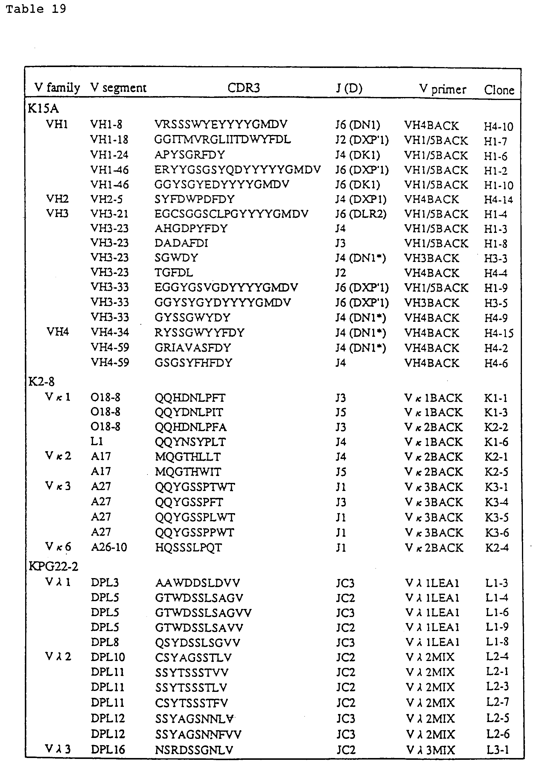
| Concentration of Mouse IgM in Chimeric Mice (ELISA) | |
| Chimerism % | IgM (mg/ℓ) |
| 0 | 12 |
| 50 | 11 |
| 99 | 1.5 |


Claims (31)
- A method for producing a chimeric non-human animal, which comprises preparing a microcell containing a foreign chromosome(s) or a fragment(s) thereof and transferring the foreign chromosome(s) or fragment(s) into a pluripotent cell by fusion with the microcell.
- A method for producing a pluripotent cell containing a foreign chromosome(s) or a fragment(s) thereof, which comprises preparing a microcell containing a foreign chromosome(s) or a fragment(s) thereof and transferring the foreign chromosome(s) or fragment(s) thereof into a pluripotent cell by fusion with the microcell.
- The method of claim 1 or 2, wherein the foreign chromosome(s) or fragment(s) thereof has(have) a base length not less than 1 Mb (1 million bp).
- The method of claim 1 or 2, wherein the foreign chromosome or fragment thereof contains a region encoding an antibody.
- The method of claim 1 or 2, wherein the microcell containing a foreign chromosome(s) or a fragment(s) thereof is induced from a hybrid cell prepared by the fusion of a cell from which the foreign chromosome(s) or fragment(s) thereof is(are) derived, with a cell having a high ability to form a microcell.
- The method of claim 5, wherein the microcell containing a foreign chromosome(s) or a fragment(s) thereof is induced from a cell prepared by a further fusion of the microcell induced from said hybrid cell with a cell having a high ability to form a microcell.
- The method of claim 5, wherein the cell from which the foreign chromosome(s) or fragment(s) thereof is(are) derived is a human normal diploid cell.
- The method of any one of claims 5-7, wherein the cell having a high ability to form a microcell is a mouse A9 cell.
- The method of claim 1 or 2, wherein the pluripotent cell is an ES cell.
- The method of claim 1 or 2, wherein the foreign chromosome or fragment thereof contains a gene of interest and the pluripotent cell has a disrupted gene identical with or homologous to said gene of interest on the foreign chromosome or fragment thereof.
- The method of claim 1, wherein a foreign chromosome or a fragment thereof containing a gene of interest is transferred into a pluripotent cell having a disrupted gene identical with or homologous to said gene of interest and then, a chimera is produced from the pluripotent cell by using an embryo of a non-human animal in a strain deficient in said gene of interest or a gene homologous thereto.
- The method of claim 11, wherein the non-human animal in a strain deficient in the gene of interest or a gene homologous thereto is produced by homologous recombination in gene targeting.
- The method of claim 1, wherein the chimeric non-human animal retains a foreign chromosome(s) or a fragment(s) thereof, expresses the gene(s) on the foreign chromosome(s) or fragment(s) thereof, and can transmit the foreign chromosome(s) or fragment(s) thereof to its progeny.
- The method of claim 1, wherein the chimeric non-human animal is a mammal.
- The method of claim 14, wherein the mammal is a mouse.
- A pluripotent cell containing a foreign chromosome(s) or a fragment(s) thereof, which can be prepared by the method of claim 2.
- Use of the pluripotent cell of claim 16 for producing a chimeric non-human animal.
- A chimeric non-human animal retaining a foreign chromosome(s) or a fragment(s) thereof and expressing a gene(s) on the foreign chromosome(s) or fragment(s) thereof which can be produced by the method of any one of claims 1 and 3-15, or its progeny retaining the foreign chromosome(s) or fragment(s) thereof and expressing the gene(s) on the foreign chromosome(s) or fragment(s) thereof.
- A non-human animal which can be produced by mating the chimeric non-human animals or its progenies of claim 18, said non-human animal retaining a foreign chromosome(s) or a fragment(s) thereof and expressing the gene(s) on the foreign chromosome(s) or fragment(s) thereof, or its progeny retaining the foreign chromosome(s) or fragment(s) thereof and expressing the gene(s) on the foreign chromosome(s) or fragment(s) thereof.
- A tissue from the chimeric non-human animal or its progeny of claim 18 or from the non-human animal or its progeny of claim 19.
- A cell from the chimeric non-human animal or its progeny of claim 18 or from the non-human animal or its progeny of claim 19.
- The cell of claim 21, which is a B cell.
- A hybridoma prepared by the fusion of the cell of claim 21 with a myeloma cell.
- A non-human animal retaining the foreign chromosome(s) or fragment(s) thereof and expressing a gene(s) on the foreign chromosome(s) or fragment(s) thereof, which can be produced by mating the chimeric non-human animal or its progeny of claim 18 or the non-human animal or its progeny of claim 19, with a non-human animal in a strain deficient in said gene(s) or a gene homologous thereto, or its progeny retaining the foreign chromosome(s) or fragment(s) thereof and expressing the gene(s) on the foreign chromosome(s) or fragment(s) thereof.
- A method for producing a biologically active substance, which comprises expressing the gene(s) on a foreign chromosome(s) or a fragment(s) thereof in the chimeric non-human animal or its progeny of claim 18 or the non-human animal or its progeny of claim 19, or a tissue or a cell thereof, and recovering the biologically active substance as an expression product.
- The method of claim 25, wherein the cell of the chimeric non-human animal is a B cell.
- The method of claim 26, wherein the B cell is immortalized by fusion with a myeloma cell.
- The method of any one of claims 25-27, wherein the biologically active substance is an antibody.
- The method of claim 28, wherein the antibody is an antibody of a mammal.
- The method of claim 29, wherein the antibody of a mammal is a human antibody.
- A method for producing a biologically active substance, which comprises: mating the chimeric non-human animal or its progeny of claim 18 or the non-human animal or its progeny of claim 19, with a non-human animal in a strain deficient in the gene(s) or a gene homologous thereto to thereby produce an offspring; expressing the gene(s) on the foreign chromosome(s) or fragment(s) thereof in the offspring, or a tissue or a cell thereof; and recovering the biologically active substance as an expression product.
Priority Applications (1)
| Application Number | Priority Date | Filing Date | Title |
|---|---|---|---|
| EP07001391A EP1916300A1 (en) | 1995-08-29 | 1996-08-29 | Chimeric animal and method for producing the same |
Applications Claiming Priority (5)
| Application Number | Priority Date | Filing Date | Title |
|---|---|---|---|
| JP24234095 | 1995-08-29 | ||
| JP242340/95 | 1995-08-29 | ||
| JP27940/96 | 1996-02-15 | ||
| JP2794096 | 1996-02-15 | ||
| PCT/JP1996/002427 WO1997007671A1 (en) | 1995-08-29 | 1996-08-29 | Chimeric animal and method for constructing the same |
Related Child Applications (1)
| Application Number | Title | Priority Date | Filing Date |
|---|---|---|---|
| EP07001391A Division EP1916300A1 (en) | 1995-08-29 | 1996-08-29 | Chimeric animal and method for producing the same |
Publications (3)
| Publication Number | Publication Date |
|---|---|
| EP0843961A1 true EP0843961A1 (en) | 1998-05-27 |
| EP0843961A4 EP0843961A4 (en) | 2000-08-09 |
| EP0843961B1 EP0843961B1 (en) | 2007-01-24 |
Family
ID=26365944
Family Applications (3)
| Application Number | Title | Priority Date | Filing Date |
|---|---|---|---|
| EP07001391A Withdrawn EP1916300A1 (en) | 1995-08-29 | 1996-08-29 | Chimeric animal and method for producing the same |
| EP96113839A Withdrawn EP0773288A3 (en) | 1995-08-29 | 1996-08-29 | Chimeric animal and method for producing the same |
| EP96928694A Expired - Lifetime EP0843961B1 (en) | 1995-08-29 | 1996-08-29 | Chimeric animal and method for constructing the same |
Family Applications Before (2)
| Application Number | Title | Priority Date | Filing Date |
|---|---|---|---|
| EP07001391A Withdrawn EP1916300A1 (en) | 1995-08-29 | 1996-08-29 | Chimeric animal and method for producing the same |
| EP96113839A Withdrawn EP0773288A3 (en) | 1995-08-29 | 1996-08-29 | Chimeric animal and method for producing the same |
Country Status (12)
| Country | Link |
|---|---|
| EP (3) | EP1916300A1 (en) |
| JP (1) | JP4318736B2 (en) |
| KR (1) | KR100308764B1 (en) |
| CN (2) | CN101333516A (en) |
| AT (1) | ATE352613T1 (en) |
| AU (1) | AU718138B2 (en) |
| CA (1) | CA2230759C (en) |
| DE (1) | DE69636868T2 (en) |
| DK (1) | DK0843961T3 (en) |
| HK (1) | HK1008472A1 (en) |
| TW (1) | TW448032B (en) |
| WO (1) | WO1997007671A1 (en) |
Cited By (121)
| Publication number | Priority date | Publication date | Assignee | Title |
|---|---|---|---|---|
| EP0972445A1 (en) * | 1997-02-28 | 2000-01-19 | Kirin Beer Kabushiki Kaisha | Pluripotent cells having disrupted intrinsic genes |
| EP1106061A1 (en) * | 1998-08-21 | 2001-06-13 | Kirin Beer Kabushiki Kaisha | Method for modifying chromosomes |
| WO2001077357A2 (en) * | 2000-04-07 | 2001-10-18 | Vlaams Interuniversitair Instituut Voor Biotechnologie Vzw | Chromosomal vectors and uses thereof |
| WO2004050683A2 (en) | 2002-12-02 | 2004-06-17 | Abgenix, Inc. | Antibodies directed to tumor necrosis factor and uses thereof |
| WO2005016111A2 (en) | 2003-08-08 | 2005-02-24 | Abgenix, Inc. | Antibodies directed to parathyroid hormone (pth) and uses thereof |
| WO2005092926A2 (en) | 2004-03-19 | 2005-10-06 | Amgen Inc. | Reducing the risk of human and anti-human antibodies through v gene manipulation |
| US7041870B2 (en) | 2000-11-30 | 2006-05-09 | Medarex, Inc. | Transgenic transchromosomal rodents for making human antibodies |
| WO2006055638A2 (en) | 2004-11-17 | 2006-05-26 | Abgenix, Inc. | Fully human monoclonal antibodies to il-13 |
| WO2006068975A2 (en) | 2004-12-20 | 2006-06-29 | Abgenix, Inc. | Binding proteins specific for human matriptase |
| WO2007068895A1 (en) | 2005-12-15 | 2007-06-21 | Astrazeneca Ab | Combination of angiopoietin-2 antagonist and of vegf-a, kdr and/or flt1 antagonist for treating cancer |
| WO2007070750A1 (en) | 2005-12-13 | 2007-06-21 | Eli Lilly And Company | Anti-il-17 antibodies |
| WO2007077028A2 (en) | 2005-12-30 | 2007-07-12 | U3 Pharma Ag | Antibodies directed to her-3 and uses thereof |
| US7358054B2 (en) | 2001-04-13 | 2008-04-15 | Biogen Idec Ma Inc. | Antibodies to VLA-1 |
| US7402729B2 (en) | 2001-05-11 | 2008-07-22 | Kirin Pharma Kabushiki Kaisha | Human artificial chromosome containing human antibody λ light chain gene and non-human animal containing the human artificial chromosome capable of genetic transmission |
| WO2008112193A1 (en) | 2007-03-12 | 2008-09-18 | Dana-Farber Cancer Institute | Prognostic, diagnostic, and cancer therapeutic uses of fanci and fanci modulating agents |
| EP1994824A1 (en) * | 2001-11-15 | 2008-11-26 | Kirin Pharma Kabushiki Kaisha | Chimeric non-human animal |
| US7462353B2 (en) | 1999-06-01 | 2008-12-09 | Biogen Idec Ma Inc. | Method for the treatment of inflammatory disorders |
| US7465449B2 (en) | 2002-03-13 | 2008-12-16 | Biogen Idec Ma Inc. | Anti-αvβ6 antibodies |
| EP2042594A1 (en) | 2000-11-17 | 2009-04-01 | Kirin Pharma Kabushiki Kaisha | Expression of xenogenous (human) immunoglobulins in cloned, transgenic ungulates |
| WO2009040134A1 (en) | 2007-09-26 | 2009-04-02 | U3 Pharma Gmbh | Heparin-binding epidermal growth factor-like growth factor antigen binding proteins |
| WO2009054873A2 (en) | 2007-08-02 | 2009-04-30 | Novimmune S.A. | Anti-rantes antibodies and methods of use thereof |
| EP2110434A1 (en) | 2002-02-25 | 2009-10-21 | Genentech, Inc. | Type-1 cytokine receptor GLM-R |
| US7642341B2 (en) | 2003-12-18 | 2010-01-05 | Merck Serono S.A. | Angiogenesis inhibiting molecules, their selection, production and their use in the treatment of cancer |
| WO2010072740A2 (en) | 2008-12-23 | 2010-07-01 | Astrazeneca Ab | TARGETED BINDING AGENTS DIRECTED TO α5β1 AND USES THEREOF |
| US7790863B2 (en) | 2003-11-19 | 2010-09-07 | Merck Serono S.A. | Angiogenesis inhibiting molecules and their use in the treatment and diagnosis of cancer |
| US7807863B2 (en) | 2002-11-08 | 2010-10-05 | Kyowa Hakko Kirin Co., Ltd. | Transgenic bovine having reduced prion protein activity and uses thereof |
| EP2239271A1 (en) | 2003-06-13 | 2010-10-13 | Biogen Idec MA Inc. | Aglycosyl anti-CD154 (CD40 ligand) antibodies and uses thereof |
| WO2010128407A2 (en) | 2009-05-05 | 2010-11-11 | Novimmune S.A. | Anti-il-17f antibodies and methods of use thereof |
| EP2284194A1 (en) | 2004-12-21 | 2011-02-16 | AstraZeneca AB | Antibodies directed to angiopoietin-2 and uses thereof |
| US7928285B2 (en) | 2004-04-22 | 2011-04-19 | Kyowa Hakko Kirin Co., Ltd. | Method of producing xenogenous antibodies using a bovine |
| WO2011060206A2 (en) | 2009-11-13 | 2011-05-19 | U3 Pharma Gmbh | Material and methods for treating or preventing her-3 associated diseases |
| EP2357202A1 (en) | 2006-04-10 | 2011-08-17 | AstraZeneca AB | Targeted binding agents directed to Upar and uses thereof |
| US8007797B2 (en) | 2006-09-28 | 2011-08-30 | Merck Serono S.A. | Junctional adhesion molecule-C (JAM-C) binding compounds and methods of their use |
| EP2361933A2 (en) | 2005-01-26 | 2011-08-31 | Amgen Fremont Inc. | Antibodies against interleukin-1 beta |
| EP2399936A2 (en) | 2004-07-26 | 2011-12-28 | Biogen Idec MA Inc. | Anti-CD154 antibodies |
| EP2404616A2 (en) | 2005-12-13 | 2012-01-11 | AstraZeneca AB | Binding proteins specific for insulin-like growth factors and uses thereof |
| WO2012018404A2 (en) | 2010-08-06 | 2012-02-09 | U3 Pharma Gmbh | Use of her3 binding agents in prostate treatment |
| EP2420513A1 (en) | 2006-08-03 | 2012-02-22 | MedImmune Limited | Targeted binding agents directed to PDGFR-alpha and uses thereof |
| EP2457586A1 (en) | 2003-06-27 | 2012-05-30 | Amgen Fremont Inc. | Antibodies directed to the deletion mutants of epidermal growth factor receptor and uses thereof |
| EP2457439A1 (en) | 1999-06-10 | 2012-05-30 | Amgen Fremont Inc. | Transgenic non-human animals for producing specific isotypes of human antibodies via non-cognate switch regions |
| WO2012102679A1 (en) | 2011-01-24 | 2012-08-02 | National University Of Singapore | Pathogenic mycobacteria-derived mannose-capped lipoarabinomannan antigen binding proteins |
| WO2012138997A1 (en) | 2011-04-07 | 2012-10-11 | Amgen Inc. | Novel egfr binding proteins |
| WO2012154999A1 (en) | 2011-05-10 | 2012-11-15 | Amgen Inc. | Methods of treating or preventing cholesterol related disorders |
| EP2527456A1 (en) | 2004-10-22 | 2012-11-28 | Revivicor Inc. | Transgenic porcines lacking endogenous immunoglobulin light chain |
| US8398975B2 (en) | 2006-08-03 | 2013-03-19 | Medimmune Limited | Antibodies directed to αVβ6 and uses thereof |
| WO2013067055A1 (en) | 2011-11-01 | 2013-05-10 | Bionomics, Inc. | Methods of blocking cancer stem cell growth |
| WO2013067054A1 (en) | 2011-11-01 | 2013-05-10 | Bionomics, Inc. | Antibodies and methods of treating cancer |
| WO2013067057A1 (en) | 2011-11-01 | 2013-05-10 | Bionomics, Inc. | Anti-gpr49 antibodies |
| WO2013067060A1 (en) | 2011-11-01 | 2013-05-10 | Bionomics, Inc. | Anti-gpr49 antibodies |
| WO2013075048A1 (en) | 2011-11-16 | 2013-05-23 | Amgen Inc. | Methods of treating epidermal growth factor deletion mutant viii related disorders |
| WO2013166448A1 (en) | 2012-05-03 | 2013-11-07 | Amgen Inc. | Stable formulations containing anti-pcsk9 antibodies |
| USRE44681E1 (en) | 2006-07-10 | 2013-12-31 | Biogen Idec Ma Inc. | Compositions and methods for inhibiting growth of SMAD4-deficient cancers |
| WO2014114801A1 (en) | 2013-01-25 | 2014-07-31 | Amgen Inc. | Antibodies targeting cdh19 for melanoma |
| WO2014114800A1 (en) | 2013-01-25 | 2014-07-31 | Amgen Research (Munich) Gmbh | Antibody constructs for cdh19 and cd3 |
| WO2014123580A1 (en) | 2013-02-06 | 2014-08-14 | Inhibrx Llc | Non-platelet depleting and non-red blood cell depleting cd47 antibodies and methods of use thereof |
| WO2014140358A1 (en) | 2013-03-15 | 2014-09-18 | Amgen Research (Munich) Gmbh | Single chain binding molecules comprising n-terminal abp |
| WO2014140368A1 (en) | 2013-03-15 | 2014-09-18 | Amgen Research (Munich) Gmbh | Antibody constructs for influenza m2 and cd3 |
| EP2816058A1 (en) | 2005-10-12 | 2014-12-24 | Eli Lilly and Company | Anti-myostatin antibodies |
| WO2014209384A1 (en) | 2013-06-28 | 2014-12-31 | Amgen Inc. | Methods for treating homozygous familial hypercholesterolema |
| EP2839843A1 (en) | 2006-05-25 | 2015-02-25 | Biogen Idec MA Inc. | VLA-1 antagonist for use in treating stroke |
| US8992924B2 (en) | 2005-07-08 | 2015-03-31 | Biogen Idec Ma Inc. | Anti-ανβ6 antibodies and uses thereof |
| US9045541B2 (en) | 2012-02-06 | 2015-06-02 | Inhibrx Llc | CD47 antibodies and methods of use thereof |
| EP2918605A1 (en) | 2007-11-12 | 2015-09-16 | U3 Pharma GmbH | Axl antibodies |
| EP2927244A1 (en) | 2008-09-19 | 2015-10-07 | MedImmune, LLC | Antibodies directed to DLL4 and uses thereof |
| DE202008018562U1 (en) | 2007-08-23 | 2015-11-02 | Amgen Inc. | Antigen-binding Proteins Against Proprotein Convertase Subtilisin Kexin Type 9 (PCSK9) |
| WO2015195917A1 (en) | 2014-06-18 | 2015-12-23 | Mersana Therapeutics, Inc. | Monoclonal antibodies against her2 epitope and methods of use thereof |
| WO2016016415A1 (en) | 2014-07-31 | 2016-02-04 | Amgen Research (Munich) Gmbh | Bispecific single chain antibody construct with enhanced tissue distribution |
| WO2016016412A1 (en) | 2014-07-31 | 2016-02-04 | Amgen Research (Munich) Gmbh | Antibody constructs for cdh19 and cd3 |
| WO2016016859A1 (en) | 2014-07-31 | 2016-02-04 | Amgen Research (Munich) Gmbh | Optimized cross-species specific bispecific single chain antibody constructs |
| WO2016166360A1 (en) | 2015-04-17 | 2016-10-20 | Bayer Pharma Aktiengesellschaft | Bispecific antibody constructs for cdh3 and cd3 |
| WO2017021370A1 (en) | 2015-07-31 | 2017-02-09 | Amgen Research (Munich) Gmbh | Bispecific antibody constructs binding egfrviii and cd3 |
| WO2017021349A1 (en) | 2015-07-31 | 2017-02-09 | Amgen Research (Munich) Gmbh | Bispecific antibody constructs binding dll3 and cd3 |
| WO2017021354A1 (en) | 2015-07-31 | 2017-02-09 | Amgen Research (Munich) Gmbh | Antibody constructs for cd70 and cd3 |
| WO2017021362A1 (en) | 2015-07-31 | 2017-02-09 | Amgen Research (Munich) Gmbh | Antibody constructs for flt3 and cd3 |
| WO2017021356A1 (en) | 2015-07-31 | 2017-02-09 | Amgen Research (Munich) Gmbh | Bispecific antibody constructs binding mesothelin and cd3 |
| US9585374B2 (en) | 2004-10-22 | 2017-03-07 | Revivicor, Inc. | Ungulates with genetically modified immune systems |
| WO2017160754A1 (en) | 2016-03-15 | 2017-09-21 | Mersana Therapeutics,Inc. | Napi2b-targeted antibody-drug conjugates and methods of use thereof |
| WO2017182427A1 (en) | 2016-04-19 | 2017-10-26 | Amgen Research (Munich) Gmbh | Administration of a bispecific construct binding to cd33 and cd3 for use in a method for the treatment of myeloid leukemia |
| EP3279215A1 (en) | 2009-11-24 | 2018-02-07 | MedImmune Limited | Targeted binding agents against b7-h1 |
| WO2018049248A1 (en) | 2016-09-09 | 2018-03-15 | Icellhealth Consulting Llc | Oncolytic virus equipped with bispecific engager molecules |
| US10035859B2 (en) | 2013-03-15 | 2018-07-31 | Biogen Ma Inc. | Anti-alpha V beta 6 antibodies and uses thereof |
| US10035860B2 (en) | 2013-03-15 | 2018-07-31 | Biogen Ma Inc. | Anti-alpha V beta 6 antibodies and uses thereof |
| WO2018141910A1 (en) | 2017-02-02 | 2018-08-09 | Amgen Research (Munich) Gmbh | Low ph pharmaceutical composition comprising t cell engaging antibody constructs |
| US10077304B2 (en) | 2013-08-14 | 2018-09-18 | The Governing Council Of The University Of Toronto | Antibodies against frizzled receptor |
| WO2018204907A1 (en) | 2017-05-05 | 2018-11-08 | Amgen Inc. | Pharmaceutical composition comprising bispecific antibody constructs for improved storage and administration |
| US10149461B2 (en) | 2008-10-27 | 2018-12-11 | Revivicor, Inc. | Immunocompromised ungulates |
| US10160808B2 (en) | 2012-02-16 | 2018-12-25 | Santarus, Inc. | Anti-VLA1 (CD49A) antibody pharmaceutical compositions |
| US10301391B2 (en) | 2016-02-03 | 2019-05-28 | Amgen Research (Munich) Gmbh | BCMA and CD3 bispecific T cell engaging antibody constructs |
| WO2019118426A1 (en) | 2017-12-11 | 2019-06-20 | Amgen Inc. | Continuous manufacturing process for bispecific antibody products |
| WO2019133961A1 (en) | 2017-12-29 | 2019-07-04 | Amgen Inc. | Bispecific antibody construct directed to muc17 and cd3 |
| EP3524626A1 (en) | 2007-03-22 | 2019-08-14 | Biogen MA Inc. | Binding proteins, including antibodies, antibody derivatives and antibody fragments, that specifically bind cd154 and uses thereof |
| WO2020025532A1 (en) | 2018-07-30 | 2020-02-06 | Amgen Research (Munich) Gmbh | Prolonged administration of a bispecific antibody construct binding to cd33 and cd3 |
| WO2020025792A1 (en) | 2018-08-03 | 2020-02-06 | Amgen Research (Munich) Gmbh | Antibody constructs for cldn18.2 and cd3 |
| WO2020043670A1 (en) | 2018-08-27 | 2020-03-05 | Affimed Gmbh | Cryopreserved nk cells preloaded with an antibody construct |
| WO2020077212A1 (en) | 2018-10-11 | 2020-04-16 | Amgen Inc. | Downstream processing of bispecific antibody constructs |
| US10745487B2 (en) | 2016-03-22 | 2020-08-18 | Bionomics Limited | Method of treating cancer by administering an anti-LGR5 monoclonal antibody |
| US10781264B2 (en) | 2016-02-03 | 2020-09-22 | Amgen Research (Munich) Gmbh | PSMA and CD3 bispecific T cell engaging antibody constructs |
| WO2020252442A1 (en) | 2019-06-13 | 2020-12-17 | Amgen Inc. | Automated biomass-based perfusion control in the manufacturing of biologics |
| WO2021021676A1 (en) | 2019-07-26 | 2021-02-04 | Amgen Inc. | Anti-il13 antigen binding proteins |
| WO2021050640A1 (en) | 2019-09-10 | 2021-03-18 | Amgen Inc. | Purification method for bispecific antigen-binding polypeptides with enhanced protein l capture dynamic binding capacity |
| WO2021097344A1 (en) | 2019-11-13 | 2021-05-20 | Amgen Inc. | Method for reduced aggregate formation in downstream processing of bispecific antigen-binding molecules |
| WO2021127528A1 (en) | 2019-12-20 | 2021-06-24 | Amgen Inc. | Mesothelin-targeted cd40 agonistic multispecific antibody constructs for the treatment of solid tumors |
| WO2021130383A1 (en) | 2019-12-27 | 2021-07-01 | Affimed Gmbh | Method for the production of bispecific fcyriii x cd30 antibody construct |
| WO2021150824A1 (en) | 2020-01-22 | 2021-07-29 | Amgen Research (Munich) Gmbh | Combinations of antibody constructs and inhibitors of cytokine release syndrome and uses thereof |
| WO2021183861A1 (en) | 2020-03-12 | 2021-09-16 | Amgen Inc. | Method for treatment and prophylaxis of crs in patients comprising a combination of bispecifc antibodies binding to cds x cancer cell and tnfalpha or il-6 inhibitor |
| WO2021188851A1 (en) | 2020-03-19 | 2021-09-23 | Amgen Inc. | Antibodies against mucin 17 and uses thereof |
| WO2021236638A1 (en) | 2020-05-19 | 2021-11-25 | Amgen Inc. | Mageb2 binding constructs |
| WO2022074206A1 (en) | 2020-10-08 | 2022-04-14 | Affimed Gmbh | Trispecific binders |
| WO2022096698A1 (en) | 2020-11-06 | 2022-05-12 | Amgen Inc. | Polypeptide constructs binding to cd3 |
| WO2022096700A1 (en) | 2020-11-06 | 2022-05-12 | Amgen Research (Munich) Gmbh | Polypeptide constructs selectively binding to cldn6 and cd3 |
| WO2022096704A1 (en) | 2020-11-06 | 2022-05-12 | Amgen Inc. | Antigen binding domain with reduced clipping rate |
| WO2022096716A2 (en) | 2020-11-06 | 2022-05-12 | Amgen Inc. | Multitargeting bispecific antigen-binding molecules of increased selectivity |
| US11434302B2 (en) | 2016-02-03 | 2022-09-06 | Amgen Research (Munich) Gmbh | Bispecific T cell engaging antibody constructs |
| WO2022212831A1 (en) | 2021-04-02 | 2022-10-06 | Amgen Inc. | Mageb2 binding constructs |
| WO2022234102A1 (en) | 2021-05-06 | 2022-11-10 | Amgen Research (Munich) Gmbh | Cd20 and cd22 targeting antigen-binding molecules for use in proliferative diseases |
| WO2023007023A1 (en) | 2021-07-30 | 2023-02-02 | Affimed Gmbh | Duplexbodies |
| WO2023079493A1 (en) | 2021-11-03 | 2023-05-11 | Affimed Gmbh | Bispecific cd16a binders |
| WO2023078968A1 (en) | 2021-11-03 | 2023-05-11 | Affimed Gmbh | Bispecific cd16a binders |
| EP4248976A2 (en) | 2007-08-23 | 2023-09-27 | Amgen Inc. | Antigen binding proteins to proprotein convertase subtilisin kexin type 9 (pcsk9) |
| WO2023218027A1 (en) | 2022-05-12 | 2023-11-16 | Amgen Research (Munich) Gmbh | Multichain multitargeting bispecific antigen-binding molecules of increased selectivity |
| WO2024059675A2 (en) | 2022-09-14 | 2024-03-21 | Amgen Inc. | Bispecific molecule stabilizing composition |
Families Citing this family (47)
| Publication number | Priority date | Publication date | Assignee | Title |
|---|---|---|---|---|
| GB8908127D0 (en) * | 1989-04-11 | 1989-05-24 | Shaw Francis & Co Ltd | Mixer and a method of a mixer control |
| US7084260B1 (en) | 1996-10-10 | 2006-08-01 | Genpharm International, Inc. | High affinity human antibodies and human antibodies against human antigens |
| US5770429A (en) * | 1990-08-29 | 1998-06-23 | Genpharm International, Inc. | Transgenic non-human animals capable of producing heterologous antibodies |
| AU7057398A (en) * | 1997-04-11 | 1998-11-11 | Imperial College Of Science, Technology And Medicine | Chromosome transfer (xmmct) to es cells, induced during the exposure of microcells to radiation |
| JP4689781B2 (en) * | 1998-09-03 | 2011-05-25 | 独立行政法人科学技術振興機構 | Amino acid transport protein and its gene |
| EP1206906A4 (en) * | 1999-08-13 | 2004-12-29 | Kirin Brewery | Mouse having human cytochrome p450 transferred therein |
| WO2001014424A2 (en) * | 1999-08-24 | 2001-03-01 | Medarex, Inc. | Human ctla-4 antibodies and their uses |
| US6835370B2 (en) * | 1999-11-08 | 2004-12-28 | Rhode Island Hospital | Diagnosis and treatment of malignant neoplasms |
| US7414170B2 (en) | 1999-11-19 | 2008-08-19 | Kirin Beer Kabushiki Kaisha | Transgenic bovines capable of human antibody production |
| US7820878B2 (en) | 1999-11-19 | 2010-10-26 | Kyowa Hakko Kirin Co., Ltd. | Production of ungulates, preferably bovines that produce human immunoglobulins |
| CN102504024A (en) * | 2000-02-10 | 2012-06-20 | 雅培制药有限公司 | Antibodies that bind human interleukin-18 and methods of making and using |
| EP1409646B1 (en) * | 2000-12-19 | 2012-06-13 | Altor BioScience Corporation | Transgenic animals comprising a humanized immune system |
| JP2002316944A (en) * | 2001-04-17 | 2002-10-31 | Chemo Sero Therapeut Res Inst | Human polyclonal antibody composition |
| ES2384168T3 (en) | 2002-04-12 | 2012-07-02 | Medarex, Inc. | Treatment procedures using CTLA-4 antibodies |
| NZ536341A (en) * | 2002-05-17 | 2008-05-30 | Kirin Holdings Kk | Transgenic ungulates capable of human antibody production |
| US7595048B2 (en) | 2002-07-03 | 2009-09-29 | Ono Pharmaceutical Co., Ltd. | Method for treatment of cancer by inhibiting the immunosuppressive signal induced by PD-1 |
| EP2277543B1 (en) * | 2002-09-06 | 2015-12-16 | Amgen, Inc | Therapeutic anti-IL-1R1 monoclonal antibody |
| CN1956738B (en) | 2004-01-09 | 2013-05-29 | 辉瑞大药厂 | Antibodies to MAdCAM |
| CN105315373B (en) | 2005-05-09 | 2018-11-09 | 小野药品工业株式会社 | The human monoclonal antibodies of programmed death-1 (PD-1) and the method for carrying out treating cancer using anti-PD-1 antibody |
| KR101607288B1 (en) | 2005-07-01 | 2016-04-05 | 이. 알. 스퀴부 앤드 선즈, 엘.엘.씨. | Human monoclonal antibodies to programmed death ligand 1(pd-l1) |
| NZ594784A (en) | 2005-08-18 | 2013-06-28 | Genmab As | Therapy with CD4 binding peptides and radiation |
| WO2007067959A2 (en) | 2005-12-07 | 2007-06-14 | Medarex, Inc. | Ctla-4 antibody dosage escalation regimens |
| JP2009531273A (en) * | 2006-02-28 | 2009-09-03 | オンコセラピー・サイエンス株式会社 | Method for damaging cells using effector function of anti-EphA4 antibody |
| CA2658204A1 (en) | 2006-07-07 | 2008-01-31 | Kyowa Hakko Kirin Co., Ltd. | Human artificial chromosome (hac) vector and human cell medicine comprising same |
| RU2448979C2 (en) * | 2006-12-14 | 2012-04-27 | Ридженерон Фармасьютикалз, Инк. | Human antibodies to delta-like human ligand-4 |
| EP2006304A1 (en) * | 2007-06-22 | 2008-12-24 | Stiftung Für Diagnostische Forschung | Anti-HPA-5b monoclonal antibody |
| EA018396B1 (en) * | 2007-10-01 | 2013-07-30 | Бристоль-Мейерз Сквибб Компани | Human antibodies that bind mesothelin, and uses thereof |
| GB0720552D0 (en) * | 2007-10-19 | 2007-11-28 | Iti Scotland Ltd | Multiple cluster knockout |
| CN102741404B (en) * | 2009-11-17 | 2017-03-08 | Sab有限责任公司 | human artificial chromosome vector |
| CN107903325B (en) | 2011-05-16 | 2021-10-29 | 埃泰美德(香港)有限公司 | Multispecific FAB fusion proteins and methods of use thereof |
| MY172730A (en) | 2012-03-16 | 2019-12-11 | Regeneron Pharma | Histidine engineered light chain antibodies and genetically modified non-human animals for generating the same |
| JP6133544B2 (en) * | 2012-03-16 | 2017-05-24 | 康宏 香月 | Microcell storage method |
| US9801362B2 (en) | 2012-03-16 | 2017-10-31 | Regeneron Pharmaceuticals, Inc. | Non-human animals expressing pH-sensitive immunoglobulin sequences |
| CN103387955B (en) * | 2012-05-10 | 2015-04-29 | 上海南方模式生物科技发展有限公司 | Making method and use of non-human mammal B lymphocyte defect animal model |
| EP3049442A4 (en) | 2013-09-26 | 2017-06-28 | Costim Pharmaceuticals Inc. | Methods for treating hematologic cancers |
| JOP20200094A1 (en) | 2014-01-24 | 2017-06-16 | Dana Farber Cancer Inst Inc | Antibody molecules to pd-1 and uses thereof |
| JOP20200096A1 (en) | 2014-01-31 | 2017-06-16 | Children’S Medical Center Corp | Antibody molecules to tim-3 and uses thereof |
| CN112877292A (en) | 2014-05-02 | 2021-06-01 | 凯奥目生物科学株式会社 | Human antibody producing cell |
| EP3659621A1 (en) | 2014-09-13 | 2020-06-03 | Novartis AG | Combination therapies for cancer |
| US10870701B2 (en) | 2016-03-15 | 2020-12-22 | Generon (Shanghai) Corporation Ltd. | Multispecific fab fusion proteins and use thereof |
| EP3533867A4 (en) * | 2016-10-31 | 2020-07-22 | National University Corporation Tottori University | Human antibody producing non-human animal and method for preparing human antibodies using same |
| CN109136261B (en) | 2017-06-19 | 2021-03-16 | 百奥赛图(北京)医药科技股份有限公司 | Preparation method and application of humanized CD28 gene modified animal model |
| WO2018233608A1 (en) * | 2017-06-19 | 2018-12-27 | Beijing Biocytogen Co., Ltd | Genetically modified non-human animal with human or chimeric cd28 |
| CR20200076A (en) | 2017-07-14 | 2020-06-10 | Pfizer | Antibodies to madcam |
| CN109837307B (en) * | 2017-11-24 | 2023-09-19 | 中国科学院脑科学与智能技术卓越创新中心 | Method for establishing embryonic stem cells containing exogenous chromosomes |
| WO2020169022A1 (en) | 2019-02-18 | 2020-08-27 | Beijing Biocytogen Co., Ltd | Genetically modified non-human animals with humanized immunoglobulin locus |
| WO2023090361A1 (en) | 2021-11-16 | 2023-05-25 | 国立大学法人鳥取大学 | Mammalian artificial chromosomal vector having human immunoglobulin heavy chain locus containing modified d region, and cell or non-human animal holding said vector |
Family Cites Families (1)
| Publication number | Priority date | Publication date | Assignee | Title |
|---|---|---|---|---|
| ZA874410B (en) * | 1986-06-24 | 1988-04-27 | Merrell Dow Pharma | Fusion products |
-
1996
- 1996-08-29 EP EP07001391A patent/EP1916300A1/en not_active Withdrawn
- 1996-08-29 AT AT96928694T patent/ATE352613T1/en not_active IP Right Cessation
- 1996-08-29 DK DK96928694T patent/DK0843961T3/en active
- 1996-08-29 KR KR1019980701537A patent/KR100308764B1/en active IP Right Grant
- 1996-08-29 EP EP96113839A patent/EP0773288A3/en not_active Withdrawn
- 1996-08-29 DE DE69636868T patent/DE69636868T2/en not_active Expired - Lifetime
- 1996-08-29 CN CNA2008100099774A patent/CN101333516A/en active Pending
- 1996-08-29 CA CA2230759A patent/CA2230759C/en not_active Expired - Lifetime
- 1996-08-29 EP EP96928694A patent/EP0843961B1/en not_active Expired - Lifetime
- 1996-08-29 AU AU68376/96A patent/AU718138B2/en not_active Expired
- 1996-08-29 WO PCT/JP1996/002427 patent/WO1997007671A1/en active IP Right Grant
- 1996-08-29 CN CNB961977876A patent/CN100379857C/en not_active Expired - Lifetime
- 1996-09-25 TW TW085111701A patent/TW448032B/en not_active IP Right Cessation
-
1998
- 1998-07-29 HK HK98109505A patent/HK1008472A1/en not_active IP Right Cessation
-
2008
- 2008-03-10 JP JP2008060174A patent/JP4318736B2/en not_active Expired - Lifetime
Non-Patent Citations (14)
Cited By (205)
| Publication number | Priority date | Publication date | Assignee | Title |
|---|---|---|---|---|
| EP0972445A4 (en) * | 1997-02-28 | 2000-12-27 | Kirin Brewery | Pluripotent cells having disrupted intrinsic genes |
| EP0972445A1 (en) * | 1997-02-28 | 2000-01-19 | Kirin Beer Kabushiki Kaisha | Pluripotent cells having disrupted intrinsic genes |
| US8124406B2 (en) | 1998-08-21 | 2012-02-28 | Kyowa Hakko Kirin Co., Ltd | Method for modifying chromosomes |
| EP1106061A1 (en) * | 1998-08-21 | 2001-06-13 | Kirin Beer Kabushiki Kaisha | Method for modifying chromosomes |
| US7868223B2 (en) | 1998-08-21 | 2011-01-11 | Kyowa Hakko Kirin Co., Ltd. | Method for modifying chromosomes |
| US7371568B1 (en) | 1998-08-21 | 2008-05-13 | Kirin Pharma Kabushiki Kaisha | Method for modifying chromosomes |
| EP1106061A4 (en) * | 1998-08-21 | 2004-04-14 | Kirin Brewery | Method for modifying chromosomes |
| US8557240B2 (en) | 1999-06-01 | 2013-10-15 | Biogen Idec Ma Inc. | Method for the treatment of inflammatory disorders |
| US8084031B2 (en) | 1999-06-01 | 2011-12-27 | Biogen Idec Ma Inc. | Method for the treatment of inflammatory disorders |
| US9902774B2 (en) | 1999-06-01 | 2018-02-27 | Biogen Ma Inc. | Method for the treatment of inflammatory disorders |
| US7462353B2 (en) | 1999-06-01 | 2008-12-09 | Biogen Idec Ma Inc. | Method for the treatment of inflammatory disorders |
| EP2457439A1 (en) | 1999-06-10 | 2012-05-30 | Amgen Fremont Inc. | Transgenic non-human animals for producing specific isotypes of human antibodies via non-cognate switch regions |
| WO2001077357A3 (en) * | 2000-04-07 | 2002-04-18 | Vlaams Interuniv Inst Biotech | Chromosomal vectors and uses thereof |
| WO2001077357A2 (en) * | 2000-04-07 | 2001-10-18 | Vlaams Interuniversitair Instituut Voor Biotechnologie Vzw | Chromosomal vectors and uses thereof |
| EP2042594A1 (en) | 2000-11-17 | 2009-04-01 | Kirin Pharma Kabushiki Kaisha | Expression of xenogenous (human) immunoglobulins in cloned, transgenic ungulates |
| US10076103B2 (en) | 2000-11-30 | 2018-09-18 | Kyowa Hakko Kirin Co., Ltd. | Transgenic transchromosomal rodents for making human antibodies |
| US7576258B2 (en) | 2000-11-30 | 2009-08-18 | Medarex, Inc. | Transgenic transchromosomal rodents for making human antibodies |
| US8835712B2 (en) | 2000-11-30 | 2014-09-16 | Medarex, L.L.C. | Transgenic trasnchromosomal rodents for making human antibodies |
| US9426970B2 (en) | 2000-11-30 | 2016-08-30 | E. R. Squibb & Sons, L.L.C. | Transgenic transchromosomal rodents for making human antibodies |
| US7816578B2 (en) | 2000-11-30 | 2010-10-19 | Kyowa Hakko Kirin Co., Ltd. | Transgenic transchromosomal rodents for making human antibodies |
| US7041870B2 (en) | 2000-11-30 | 2006-05-09 | Medarex, Inc. | Transgenic transchromosomal rodents for making human antibodies |
| US7910099B2 (en) | 2001-04-13 | 2011-03-22 | Biogen Idec Ma Inc. | Antibodies to VLA-1 |
| US9644030B2 (en) | 2001-04-13 | 2017-05-09 | Biogen Ma Inc. | Antibodies to VLA-1 |
| US7358054B2 (en) | 2001-04-13 | 2008-04-15 | Biogen Idec Ma Inc. | Antibodies to VLA-1 |
| US8084028B2 (en) | 2001-04-13 | 2011-12-27 | Biogen Idec Ma Inc. | Antibodies to VLA-1 |
| US7723073B2 (en) | 2001-04-13 | 2010-05-25 | Biogen Idec Ma Inc. | Antibodies to VLA-1 |
| US7476536B2 (en) | 2001-05-11 | 2009-01-13 | Kirin Pharma Kabushiki Kaisha | Artificial human chromosome containing human antibody a light chain gene |
| US10448622B2 (en) | 2001-05-11 | 2019-10-22 | E. R. Squibb & Sons, L.L.C. | Human artificial chromosome containing human antibody lambda light chain gene and non-human animal containing the human artificial chromosome capable of genetic transmission |
| US9499838B2 (en) | 2001-05-11 | 2016-11-22 | Kyowa Hakko Kirin Co., Ltd. | Human artificial chromosome containing human antibody λ light chain gene and non-human animal containing the human artificial chromosome capable of genetic transmission |
| US7402729B2 (en) | 2001-05-11 | 2008-07-22 | Kirin Pharma Kabushiki Kaisha | Human artificial chromosome containing human antibody λ light chain gene and non-human animal containing the human artificial chromosome capable of genetic transmission |
| EP1994824A1 (en) * | 2001-11-15 | 2008-11-26 | Kirin Pharma Kabushiki Kaisha | Chimeric non-human animal |
| EP2110434A1 (en) | 2002-02-25 | 2009-10-21 | Genentech, Inc. | Type-1 cytokine receptor GLM-R |
| US9745376B2 (en) | 2002-03-13 | 2017-08-29 | Biogen Ma Inc. | Anti-ανβ6 antibodies |
| US7465449B2 (en) | 2002-03-13 | 2008-12-16 | Biogen Idec Ma Inc. | Anti-αvβ6 antibodies |
| US8153126B2 (en) | 2002-03-13 | 2012-04-10 | Violette Shelia M | Anti-αvβ6 antibodies |
| EP2287198A2 (en) | 2002-03-13 | 2011-02-23 | Biogen Idec MA Inc. | Anti-alpha V beta 6 antibodies |
| EP2287199A2 (en) | 2002-03-13 | 2011-02-23 | Biogen Idec MA Inc. | Anti-alpha V beta 6 antibodies |
| EP2336185A1 (en) | 2002-03-13 | 2011-06-22 | Biogen Idec Inc. | Anti- alpha v beta 6 antibodies |
| US7807863B2 (en) | 2002-11-08 | 2010-10-05 | Kyowa Hakko Kirin Co., Ltd. | Transgenic bovine having reduced prion protein activity and uses thereof |
| WO2004050683A2 (en) | 2002-12-02 | 2004-06-17 | Abgenix, Inc. | Antibodies directed to tumor necrosis factor and uses thereof |
| EP2239271A1 (en) | 2003-06-13 | 2010-10-13 | Biogen Idec MA Inc. | Aglycosyl anti-CD154 (CD40 ligand) antibodies and uses thereof |
| EP3011971A1 (en) | 2003-06-27 | 2016-04-27 | Amgen Fremont Inc. | Antibodies directed to the deletion mutants of epidermal growth factor receptor and uses thereof |
| EP2457586A1 (en) | 2003-06-27 | 2012-05-30 | Amgen Fremont Inc. | Antibodies directed to the deletion mutants of epidermal growth factor receptor and uses thereof |
| EP3037105A1 (en) | 2003-06-27 | 2016-06-29 | Amgen Fremont Inc. | Antibodies directed to the deletion mutants of epidermal growth factor receptor and uses thereof |
| EP2457587A1 (en) | 2003-06-27 | 2012-05-30 | Amgen Fremont Inc. | Anitbodies directed to the deletion mutants of epidermal growth factor receptor and uses thereof |
| EP3679951A1 (en) | 2003-06-27 | 2020-07-15 | Amgen Fremont Inc. | Antibodies directed to the deletion mutants of epidermal growth factor receptor and uses thereof |
| WO2005016111A2 (en) | 2003-08-08 | 2005-02-24 | Abgenix, Inc. | Antibodies directed to parathyroid hormone (pth) and uses thereof |
| US7790863B2 (en) | 2003-11-19 | 2010-09-07 | Merck Serono S.A. | Angiogenesis inhibiting molecules and their use in the treatment and diagnosis of cancer |
| US8093010B2 (en) | 2003-12-18 | 2012-01-10 | Merck Serono S.A. | Angiogenesis inhibiting molecules, their selection, production and their use in the treatment of cancer |
| US7642341B2 (en) | 2003-12-18 | 2010-01-05 | Merck Serono S.A. | Angiogenesis inhibiting molecules, their selection, production and their use in the treatment of cancer |
| EP2418224A2 (en) | 2004-03-19 | 2012-02-15 | Amgen Inc. | Reducing the risk of human and anti-human antibodies through V gene manipulation |
| WO2005092926A2 (en) | 2004-03-19 | 2005-10-06 | Amgen Inc. | Reducing the risk of human and anti-human antibodies through v gene manipulation |
| US7928285B2 (en) | 2004-04-22 | 2011-04-19 | Kyowa Hakko Kirin Co., Ltd. | Method of producing xenogenous antibodies using a bovine |
| EP2399936A2 (en) | 2004-07-26 | 2011-12-28 | Biogen Idec MA Inc. | Anti-CD154 antibodies |
| US11085054B2 (en) | 2004-10-22 | 2021-08-10 | Revivicor, Inc. | Ungulates with genetically modified immune systems |
| EP2527456A1 (en) | 2004-10-22 | 2012-11-28 | Revivicor Inc. | Transgenic porcines lacking endogenous immunoglobulin light chain |
| US9585374B2 (en) | 2004-10-22 | 2017-03-07 | Revivicor, Inc. | Ungulates with genetically modified immune systems |
| WO2006055638A2 (en) | 2004-11-17 | 2006-05-26 | Abgenix, Inc. | Fully human monoclonal antibodies to il-13 |
| WO2006068975A2 (en) | 2004-12-20 | 2006-06-29 | Abgenix, Inc. | Binding proteins specific for human matriptase |
| EP2284194A1 (en) | 2004-12-21 | 2011-02-16 | AstraZeneca AB | Antibodies directed to angiopoietin-2 and uses thereof |
| EP3699191A1 (en) | 2004-12-21 | 2020-08-26 | MedImmune Limited | Antibodies directed to angiopoietin-2 and uses thereof |
| EP2361933A2 (en) | 2005-01-26 | 2011-08-31 | Amgen Fremont Inc. | Antibodies against interleukin-1 beta |
| US8992924B2 (en) | 2005-07-08 | 2015-03-31 | Biogen Idec Ma Inc. | Anti-ανβ6 antibodies and uses thereof |
| EP2816058A1 (en) | 2005-10-12 | 2014-12-24 | Eli Lilly and Company | Anti-myostatin antibodies |
| WO2007070750A1 (en) | 2005-12-13 | 2007-06-21 | Eli Lilly And Company | Anti-il-17 antibodies |
| EP2481753A1 (en) | 2005-12-13 | 2012-08-01 | Eli Lilly and Company | Anti-IL-17 Antibodies |
| EP3808769A1 (en) | 2005-12-13 | 2021-04-21 | Eli Lilly And Company | Anti-il-17 antibodies |
| EP2404616A2 (en) | 2005-12-13 | 2012-01-11 | AstraZeneca AB | Binding proteins specific for insulin-like growth factors and uses thereof |
| EP2518083A2 (en) | 2005-12-15 | 2012-10-31 | Medimmune Limited | Combination of angiopoietin-2 antagonist and of VEGF-A, KDR and/or FLTL antagonist for treating cancer |
| WO2007068895A1 (en) | 2005-12-15 | 2007-06-21 | Astrazeneca Ab | Combination of angiopoietin-2 antagonist and of vegf-a, kdr and/or flt1 antagonist for treating cancer |
| WO2007077028A2 (en) | 2005-12-30 | 2007-07-12 | U3 Pharma Ag | Antibodies directed to her-3 and uses thereof |
| EP3950715A1 (en) | 2005-12-30 | 2022-02-09 | Amgen Inc. | Antibodies directed to her-3 and uses thereof |
| EP2993187A2 (en) | 2005-12-30 | 2016-03-09 | U3 Pharma GmbH | Antibodies directed to her-3 and uses thereof |
| EP3196213A2 (en) | 2005-12-30 | 2017-07-26 | Daiichi Sankyo Europe GmbH | Antibodies directed to her-3 and uses thereof |
| EP2357202A1 (en) | 2006-04-10 | 2011-08-17 | AstraZeneca AB | Targeted binding agents directed to Upar and uses thereof |
| US10119979B2 (en) | 2006-05-25 | 2018-11-06 | Biogen Ma Inc. | Methods of treating stroke and traumatic brain injury using humanized AQC2 anti-VLA-1 antibodies |
| EP2839843A1 (en) | 2006-05-25 | 2015-02-25 | Biogen Idec MA Inc. | VLA-1 antagonist for use in treating stroke |
| USRE44681E1 (en) | 2006-07-10 | 2013-12-31 | Biogen Idec Ma Inc. | Compositions and methods for inhibiting growth of SMAD4-deficient cancers |
| EP2420514A1 (en) | 2006-08-03 | 2012-02-22 | MedImmune Limited | Targeted binding agents directed to PDGFR-alpha and uses thereof |
| US8398975B2 (en) | 2006-08-03 | 2013-03-19 | Medimmune Limited | Antibodies directed to αVβ6 and uses thereof |
| EP2420513A1 (en) | 2006-08-03 | 2012-02-22 | MedImmune Limited | Targeted binding agents directed to PDGFR-alpha and uses thereof |
| US8007797B2 (en) | 2006-09-28 | 2011-08-30 | Merck Serono S.A. | Junctional adhesion molecule-C (JAM-C) binding compounds and methods of their use |
| WO2008112193A1 (en) | 2007-03-12 | 2008-09-18 | Dana-Farber Cancer Institute | Prognostic, diagnostic, and cancer therapeutic uses of fanci and fanci modulating agents |
| EP3524626A1 (en) | 2007-03-22 | 2019-08-14 | Biogen MA Inc. | Binding proteins, including antibodies, antibody derivatives and antibody fragments, that specifically bind cd154 and uses thereof |
| WO2009054873A2 (en) | 2007-08-02 | 2009-04-30 | Novimmune S.A. | Anti-rantes antibodies and methods of use thereof |
| DE202008018562U1 (en) | 2007-08-23 | 2015-11-02 | Amgen Inc. | Antigen-binding Proteins Against Proprotein Convertase Subtilisin Kexin Type 9 (PCSK9) |
| EP4248976A2 (en) | 2007-08-23 | 2023-09-27 | Amgen Inc. | Antigen binding proteins to proprotein convertase subtilisin kexin type 9 (pcsk9) |
| EP3666797A1 (en) | 2007-08-23 | 2020-06-17 | Amgen, Inc | Antigen binding proteins to proprotein convertase subtilisin kexin type 9 (pcsk9) |
| WO2009040134A1 (en) | 2007-09-26 | 2009-04-02 | U3 Pharma Gmbh | Heparin-binding epidermal growth factor-like growth factor antigen binding proteins |
| EP2497783A2 (en) | 2007-09-26 | 2012-09-12 | U3 Pharma GmbH | Heparin-binding epidermal growth factor-like growth factor antigen binding proteins |
| EP2918605A1 (en) | 2007-11-12 | 2015-09-16 | U3 Pharma GmbH | Axl antibodies |
| EP2927244A1 (en) | 2008-09-19 | 2015-10-07 | MedImmune, LLC | Antibodies directed to DLL4 and uses thereof |
| US10149461B2 (en) | 2008-10-27 | 2018-12-11 | Revivicor, Inc. | Immunocompromised ungulates |
| WO2010072740A2 (en) | 2008-12-23 | 2010-07-01 | Astrazeneca Ab | TARGETED BINDING AGENTS DIRECTED TO α5β1 AND USES THEREOF |
| WO2010128407A2 (en) | 2009-05-05 | 2010-11-11 | Novimmune S.A. | Anti-il-17f antibodies and methods of use thereof |
| EP3351558A2 (en) | 2009-11-13 | 2018-07-25 | Daiichi Sankyo Europe GmbH | Material and methods for treating or preventing her-3 associated diseases |
| EP3670539A1 (en) | 2009-11-13 | 2020-06-24 | Daiichi Sankyo Europe GmbH | Material and methods for treating or preventing her-3 associated diseases |
| WO2011060206A2 (en) | 2009-11-13 | 2011-05-19 | U3 Pharma Gmbh | Material and methods for treating or preventing her-3 associated diseases |
| EP2896632A2 (en) | 2009-11-13 | 2015-07-22 | U3 Pharma GmbH | Material and methods for treating or preventing HER-3 associated diseases |
| EP2719708A2 (en) | 2009-11-13 | 2014-04-16 | U3 Pharma GmbH | Material and methods for treating or preventing HER-3 associated diseases |
| EP3279215A1 (en) | 2009-11-24 | 2018-02-07 | MedImmune Limited | Targeted binding agents against b7-h1 |
| WO2012018404A2 (en) | 2010-08-06 | 2012-02-09 | U3 Pharma Gmbh | Use of her3 binding agents in prostate treatment |
| WO2012102679A1 (en) | 2011-01-24 | 2012-08-02 | National University Of Singapore | Pathogenic mycobacteria-derived mannose-capped lipoarabinomannan antigen binding proteins |
| WO2012138997A1 (en) | 2011-04-07 | 2012-10-11 | Amgen Inc. | Novel egfr binding proteins |
| WO2012154999A1 (en) | 2011-05-10 | 2012-11-15 | Amgen Inc. | Methods of treating or preventing cholesterol related disorders |
| EP3597218A1 (en) | 2011-05-10 | 2020-01-22 | Amgen, Inc | Methods of treating or preventing cholesterol related disorders |
| WO2013067055A1 (en) | 2011-11-01 | 2013-05-10 | Bionomics, Inc. | Methods of blocking cancer stem cell growth |
| US10598653B2 (en) | 2011-11-01 | 2020-03-24 | Bionomics Inc. | Methods of blocking cancer stem cell growth |
| WO2013067054A1 (en) | 2011-11-01 | 2013-05-10 | Bionomics, Inc. | Antibodies and methods of treating cancer |
| WO2013067057A1 (en) | 2011-11-01 | 2013-05-10 | Bionomics, Inc. | Anti-gpr49 antibodies |
| WO2013067060A1 (en) | 2011-11-01 | 2013-05-10 | Bionomics, Inc. | Anti-gpr49 antibodies |
| US10196442B2 (en) | 2011-11-01 | 2019-02-05 | Bionomics Inc. | Methods of inhibiting growth of a colon cancer tumor in a subject by administering monoclonal antibodies to G protein-coupled receptor 49 (GPR49) |
| WO2013075048A1 (en) | 2011-11-16 | 2013-05-23 | Amgen Inc. | Methods of treating epidermal growth factor deletion mutant viii related disorders |
| US9663575B2 (en) | 2012-02-06 | 2017-05-30 | Inhibrx, Lp | CD47 antibodies and methods of use thereof |
| EP3578569A1 (en) | 2012-02-06 | 2019-12-11 | Inhibrx, Inc. | Cd47 antibodies and methods of use thereof |
| US9045541B2 (en) | 2012-02-06 | 2015-06-02 | Inhibrx Llc | CD47 antibodies and methods of use thereof |
| US10160808B2 (en) | 2012-02-16 | 2018-12-25 | Santarus, Inc. | Anti-VLA1 (CD49A) antibody pharmaceutical compositions |
| US10316095B2 (en) | 2012-02-16 | 2019-06-11 | Santarus, Inc. | Antibody formulations |
| WO2013166448A1 (en) | 2012-05-03 | 2013-11-07 | Amgen Inc. | Stable formulations containing anti-pcsk9 antibodies |
| EP3656399A1 (en) | 2012-05-03 | 2020-05-27 | Amgen, Inc | Stable formulations containing anti-pcsk9 antibodies |
| EP3699194A1 (en) | 2013-01-25 | 2020-08-26 | Amgen Research (Munich) GmbH | Antibody constructs for cdh19 and cd3 |
| WO2014114801A1 (en) | 2013-01-25 | 2014-07-31 | Amgen Inc. | Antibodies targeting cdh19 for melanoma |
| WO2014114800A1 (en) | 2013-01-25 | 2014-07-31 | Amgen Research (Munich) Gmbh | Antibody constructs for cdh19 and cd3 |
| US11498964B2 (en) | 2013-01-25 | 2022-11-15 | Amgen Research (Munich) Gmbh | Antibody constructs for CDH19 and CD3 |
| EP4137518A1 (en) | 2013-02-06 | 2023-02-22 | Inhibrx, Inc. | Non-platelet depleting and non-red blood cell depleting cd47 antibodies and methods of use thereof |
| WO2014123580A1 (en) | 2013-02-06 | 2014-08-14 | Inhibrx Llc | Non-platelet depleting and non-red blood cell depleting cd47 antibodies and methods of use thereof |
| WO2014140358A1 (en) | 2013-03-15 | 2014-09-18 | Amgen Research (Munich) Gmbh | Single chain binding molecules comprising n-terminal abp |
| US10035860B2 (en) | 2013-03-15 | 2018-07-31 | Biogen Ma Inc. | Anti-alpha V beta 6 antibodies and uses thereof |
| US10035859B2 (en) | 2013-03-15 | 2018-07-31 | Biogen Ma Inc. | Anti-alpha V beta 6 antibodies and uses thereof |
| WO2014140368A1 (en) | 2013-03-15 | 2014-09-18 | Amgen Research (Munich) Gmbh | Antibody constructs for influenza m2 and cd3 |
| EP3653642A1 (en) | 2013-03-15 | 2020-05-20 | Amgen Research (Munich) GmbH | Single chain binding molecules comprising n-terminal abp |
| WO2014209384A1 (en) | 2013-06-28 | 2014-12-31 | Amgen Inc. | Methods for treating homozygous familial hypercholesterolema |
| US10077304B2 (en) | 2013-08-14 | 2018-09-18 | The Governing Council Of The University Of Toronto | Antibodies against frizzled receptor |
| EP3705494A2 (en) | 2013-08-14 | 2020-09-09 | Sachdev Sidhu | Antibodies against frizzled proteins and methods of use thereof |
| US10689442B2 (en) | 2013-08-14 | 2020-06-23 | Sachdev Sidhu | Antibodies against Frizzled receptor |
| EP4285917A2 (en) | 2014-06-18 | 2023-12-06 | Mersana Therapeutics, Inc. | Monoclonal antibodies against her2 epitope and methods of use thereof |
| WO2015195917A1 (en) | 2014-06-18 | 2015-12-23 | Mersana Therapeutics, Inc. | Monoclonal antibodies against her2 epitope and methods of use thereof |
| WO2016016412A1 (en) | 2014-07-31 | 2016-02-04 | Amgen Research (Munich) Gmbh | Antibody constructs for cdh19 and cd3 |
| US11661462B2 (en) | 2014-07-31 | 2023-05-30 | Amgen Research (Munich) Gmbh | Optimized cross-species specific bispecific single chain antibody contructs |
| US9765157B2 (en) | 2014-07-31 | 2017-09-19 | Amgen Research (Munich) Gmbh | Antibody constructs for CDH19 and CD3 |
| WO2016016415A1 (en) | 2014-07-31 | 2016-02-04 | Amgen Research (Munich) Gmbh | Bispecific single chain antibody construct with enhanced tissue distribution |
| WO2016016859A1 (en) | 2014-07-31 | 2016-02-04 | Amgen Research (Munich) Gmbh | Optimized cross-species specific bispecific single chain antibody constructs |
| WO2016166360A1 (en) | 2015-04-17 | 2016-10-20 | Bayer Pharma Aktiengesellschaft | Bispecific antibody constructs for cdh3 and cd3 |
| EP4276116A2 (en) | 2015-04-17 | 2023-11-15 | Amgen Research (Munich) GmbH | Bispecific antibody constructs for cdh3 and cd3 |
| US10851170B2 (en) | 2015-07-31 | 2020-12-01 | Amgen Research (Munich) Gmbh | Antibody constructs for CD70 and CD3 |
| EP3865514A1 (en) | 2015-07-31 | 2021-08-18 | Amgen Research (Munich) GmbH | Bispecific antibody constructs binding dll3 and cd3 |
| WO2017021362A1 (en) | 2015-07-31 | 2017-02-09 | Amgen Research (Munich) Gmbh | Antibody constructs for flt3 and cd3 |
| US11447567B2 (en) | 2015-07-31 | 2022-09-20 | Amgen Research (Munich) Gmbh | Antibody constructs for FLT3 and CD3 |
| US10683351B2 (en) | 2015-07-31 | 2020-06-16 | Amgen Research (Munich) Gmbh | Antibody constructs for DLL3 and CD3 |
| EP3912999A1 (en) | 2015-07-31 | 2021-11-24 | Amgen Research (Munich) GmbH | Bispecific antibody constructs binding egfrviii and cd3 |
| US11155629B2 (en) | 2015-07-31 | 2021-10-26 | Amgen Research (Munich) Gmbh | Method for treating glioblastoma or glioma with antibody constructs for EGFRVIII and CD3 |
| WO2017021354A1 (en) | 2015-07-31 | 2017-02-09 | Amgen Research (Munich) Gmbh | Antibody constructs for cd70 and cd3 |
| US11591396B2 (en) | 2015-07-31 | 2023-02-28 | Amgen Research (Munich) Gmbh | Antibody constructs for DLL3 and CD3 |
| EP4327885A2 (en) | 2015-07-31 | 2024-02-28 | Amgen Research (Munich) GmbH | Antibody constructs for msln and cd3 |
| WO2017021356A1 (en) | 2015-07-31 | 2017-02-09 | Amgen Research (Munich) Gmbh | Bispecific antibody constructs binding mesothelin and cd3 |
| US10519241B2 (en) | 2015-07-31 | 2019-12-31 | Amgen Research (Munich) Gmbh | Antibody constructs for EGFRVIII and CD3 |
| US10294300B2 (en) | 2015-07-31 | 2019-05-21 | Amgen Research (Munich) Gmbh | Antibody constructs for DLL3 and CD3 |
| EP4219562A2 (en) | 2015-07-31 | 2023-08-02 | Amgen Research (Munich) GmbH | Antibody constructs for flt3 and cd3 |
| WO2017021349A1 (en) | 2015-07-31 | 2017-02-09 | Amgen Research (Munich) Gmbh | Bispecific antibody constructs binding dll3 and cd3 |
| US11884720B2 (en) | 2015-07-31 | 2024-01-30 | Amgen Research (Munich) Gmbh | Antibody constructs for MSLN and CD3 |
| WO2017021370A1 (en) | 2015-07-31 | 2017-02-09 | Amgen Research (Munich) Gmbh | Bispecific antibody constructs binding egfrviii and cd3 |
| US11352433B2 (en) | 2016-02-03 | 2022-06-07 | Amgen Research (Munich) Gmbh | BCMA and CD3 bispecific T cell engaging antibody constructs |
| EP4039709A1 (en) | 2016-02-03 | 2022-08-10 | Amgen Research (Munich) GmbH | Bcma and cd3 bispecific t cell engaging antibody constructs |
| US11434302B2 (en) | 2016-02-03 | 2022-09-06 | Amgen Research (Munich) Gmbh | Bispecific T cell engaging antibody constructs |
| EP4206228A1 (en) | 2016-02-03 | 2023-07-05 | Amgen Research (Munich) GmbH | Psma and cd3 bispecific t cell engaging constructs |
| US10781264B2 (en) | 2016-02-03 | 2020-09-22 | Amgen Research (Munich) Gmbh | PSMA and CD3 bispecific T cell engaging antibody constructs |
| US10301391B2 (en) | 2016-02-03 | 2019-05-28 | Amgen Research (Munich) Gmbh | BCMA and CD3 bispecific T cell engaging antibody constructs |
| WO2017160754A1 (en) | 2016-03-15 | 2017-09-21 | Mersana Therapeutics,Inc. | Napi2b-targeted antibody-drug conjugates and methods of use thereof |
| US10745487B2 (en) | 2016-03-22 | 2020-08-18 | Bionomics Limited | Method of treating cancer by administering an anti-LGR5 monoclonal antibody |
| WO2017182427A1 (en) | 2016-04-19 | 2017-10-26 | Amgen Research (Munich) Gmbh | Administration of a bispecific construct binding to cd33 and cd3 for use in a method for the treatment of myeloid leukemia |
| US11053318B2 (en) | 2016-04-19 | 2021-07-06 | Amgen Research (Munich) Gmbh | Administration of a bispecific construct binding to CD33 and CD3 for use in a method for the treatment of myeloid leukemia |
| WO2018049261A1 (en) | 2016-09-09 | 2018-03-15 | Icellhealth Consulting Llc | Oncolytic virus expressing immune checkpoint modulators |
| WO2018049248A1 (en) | 2016-09-09 | 2018-03-15 | Icellhealth Consulting Llc | Oncolytic virus equipped with bispecific engager molecules |
| WO2018141910A1 (en) | 2017-02-02 | 2018-08-09 | Amgen Research (Munich) Gmbh | Low ph pharmaceutical composition comprising t cell engaging antibody constructs |
| WO2018204907A1 (en) | 2017-05-05 | 2018-11-08 | Amgen Inc. | Pharmaceutical composition comprising bispecific antibody constructs for improved storage and administration |
| US11918650B2 (en) | 2017-05-05 | 2024-03-05 | Amgen Inc. | Pharmaceutical composition comprising bispecific antibody constructs for improved storage and administration |
| WO2019118426A1 (en) | 2017-12-11 | 2019-06-20 | Amgen Inc. | Continuous manufacturing process for bispecific antibody products |
| WO2019133961A1 (en) | 2017-12-29 | 2019-07-04 | Amgen Inc. | Bispecific antibody construct directed to muc17 and cd3 |
| WO2020025532A1 (en) | 2018-07-30 | 2020-02-06 | Amgen Research (Munich) Gmbh | Prolonged administration of a bispecific antibody construct binding to cd33 and cd3 |
| WO2020025792A1 (en) | 2018-08-03 | 2020-02-06 | Amgen Research (Munich) Gmbh | Antibody constructs for cldn18.2 and cd3 |
| US11692031B2 (en) | 2018-08-03 | 2023-07-04 | Amgen Research (Munich) Gmbh | Antibody constructs for CLDN18.2 and CD3 |
| WO2020043670A1 (en) | 2018-08-27 | 2020-03-05 | Affimed Gmbh | Cryopreserved nk cells preloaded with an antibody construct |
| WO2020077212A1 (en) | 2018-10-11 | 2020-04-16 | Amgen Inc. | Downstream processing of bispecific antibody constructs |
| WO2020252442A1 (en) | 2019-06-13 | 2020-12-17 | Amgen Inc. | Automated biomass-based perfusion control in the manufacturing of biologics |
| WO2021021676A1 (en) | 2019-07-26 | 2021-02-04 | Amgen Inc. | Anti-il13 antigen binding proteins |
| WO2021050640A1 (en) | 2019-09-10 | 2021-03-18 | Amgen Inc. | Purification method for bispecific antigen-binding polypeptides with enhanced protein l capture dynamic binding capacity |
| WO2021097344A1 (en) | 2019-11-13 | 2021-05-20 | Amgen Inc. | Method for reduced aggregate formation in downstream processing of bispecific antigen-binding molecules |
| WO2021127528A1 (en) | 2019-12-20 | 2021-06-24 | Amgen Inc. | Mesothelin-targeted cd40 agonistic multispecific antibody constructs for the treatment of solid tumors |
| WO2021130383A1 (en) | 2019-12-27 | 2021-07-01 | Affimed Gmbh | Method for the production of bispecific fcyriii x cd30 antibody construct |
| WO2021150824A1 (en) | 2020-01-22 | 2021-07-29 | Amgen Research (Munich) Gmbh | Combinations of antibody constructs and inhibitors of cytokine release syndrome and uses thereof |
| WO2021183861A1 (en) | 2020-03-12 | 2021-09-16 | Amgen Inc. | Method for treatment and prophylaxis of crs in patients comprising a combination of bispecifc antibodies binding to cds x cancer cell and tnfalpha or il-6 inhibitor |
| WO2021188851A1 (en) | 2020-03-19 | 2021-09-23 | Amgen Inc. | Antibodies against mucin 17 and uses thereof |
| WO2021236638A1 (en) | 2020-05-19 | 2021-11-25 | Amgen Inc. | Mageb2 binding constructs |
| WO2022074206A1 (en) | 2020-10-08 | 2022-04-14 | Affimed Gmbh | Trispecific binders |
| WO2022096700A1 (en) | 2020-11-06 | 2022-05-12 | Amgen Research (Munich) Gmbh | Polypeptide constructs selectively binding to cldn6 and cd3 |
| WO2022096704A1 (en) | 2020-11-06 | 2022-05-12 | Amgen Inc. | Antigen binding domain with reduced clipping rate |
| WO2022096698A1 (en) | 2020-11-06 | 2022-05-12 | Amgen Inc. | Polypeptide constructs binding to cd3 |
| WO2022096716A2 (en) | 2020-11-06 | 2022-05-12 | Amgen Inc. | Multitargeting bispecific antigen-binding molecules of increased selectivity |
| WO2022212831A1 (en) | 2021-04-02 | 2022-10-06 | Amgen Inc. | Mageb2 binding constructs |
| WO2022234102A1 (en) | 2021-05-06 | 2022-11-10 | Amgen Research (Munich) Gmbh | Cd20 and cd22 targeting antigen-binding molecules for use in proliferative diseases |
| WO2023007023A1 (en) | 2021-07-30 | 2023-02-02 | Affimed Gmbh | Duplexbodies |
| WO2023078968A1 (en) | 2021-11-03 | 2023-05-11 | Affimed Gmbh | Bispecific cd16a binders |
| WO2023079493A1 (en) | 2021-11-03 | 2023-05-11 | Affimed Gmbh | Bispecific cd16a binders |
| WO2023218027A1 (en) | 2022-05-12 | 2023-11-16 | Amgen Research (Munich) Gmbh | Multichain multitargeting bispecific antigen-binding molecules of increased selectivity |
| WO2024059675A2 (en) | 2022-09-14 | 2024-03-21 | Amgen Inc. | Bispecific molecule stabilizing composition |
Also Published As
| Publication number | Publication date |
|---|---|
| AU718138B2 (en) | 2000-04-06 |
| CN100379857C (en) | 2008-04-09 |
| DE69636868T2 (en) | 2007-10-25 |
| EP1916300A1 (en) | 2008-04-30 |
| CA2230759C (en) | 2012-02-21 |
| HK1008472A1 (en) | 1999-11-12 |
| JP2008200042A (en) | 2008-09-04 |
| AU6837696A (en) | 1997-03-19 |
| KR100308764B1 (en) | 2001-12-17 |
| TW448032B (en) | 2001-08-01 |
| EP0843961A4 (en) | 2000-08-09 |
| CA2230759A1 (en) | 1997-03-06 |
| EP0773288A2 (en) | 1997-05-14 |
| CN101333516A (en) | 2008-12-31 |
| DK0843961T3 (en) | 2007-05-21 |
| EP0843961B1 (en) | 2007-01-24 |
| EP0773288A3 (en) | 1997-07-09 |
| CN1200014A (en) | 1998-11-25 |
| JP4318736B2 (en) | 2009-08-26 |
| DE69636868D1 (en) | 2007-03-15 |
| KR19990044300A (en) | 1999-06-25 |
| WO1997007671A1 (en) | 1997-03-06 |
| ATE352613T1 (en) | 2007-02-15 |
Similar Documents
| Publication | Publication Date | Title |
|---|---|---|
| AU718138B2 (en) | Chimeric animal and method for constructing the same | |
| US8110671B2 (en) | Isolated human chromosome 14 fragment encoding immunoglobulin genes | |
| EP1707051B1 (en) | Human chromosome fragment, and vector containing it | |
| US8124406B2 (en) | Method for modifying chromosomes | |
| KR100882030B1 (en) | Expression of Xenogenous (Human) Immunoglobulins in Cloned, Transgenic Ungulates | |
| CA2214018C (en) | An immortalized mammal hybridoma fusion partner | |
| EP1500329B1 (en) | Human antibodies that specifically bind human TNF alpha | |
| US7435871B2 (en) | Transgenic animals bearing human Igλ light chain genes | |
| JP2007312792A (en) | Chimeric animals and method for producing the same | |
| JP3030092B2 (en) | Chimeric animal and method for producing the same | |
| TWI223585B (en) | Chimeric animal and method for producing the same | |
| JPH11313576A (en) | Chimera animal and its preparation |
Legal Events
| Date | Code | Title | Description |
|---|---|---|---|
| PUAI | Public reference made under article 153(3) epc to a published international application that has entered the european phase |
Free format text: ORIGINAL CODE: 0009012 |
|
| 17P | Request for examination filed |
Effective date: 19980227 |
|
| AK | Designated contracting states |
Kind code of ref document: A1 Designated state(s): AT BE CH DE DK ES FI FR GB GR IE IT LI LU MC NL PT SE |
|
| RIC1 | Information provided on ipc code assigned before grant |
Free format text: 7C 12N 15/00 A, 7A 01K 67/027 B, 7C 12N 15/90 B, 7C 12N 5/10 B, 7C 07K 16/00 B, 7C 12N 15/08 B, 7C 12N 15/06 B, 7A 61K 48/00 B |
|
| A4 | Supplementary search report drawn up and despatched |
Effective date: 20000615 |
|
| AK | Designated contracting states |
Kind code of ref document: A4 Designated state(s): AT BE CH DE DK ES FI FR GB GR IE IT LI LU MC NL PT SE |
|
| 17Q | First examination report despatched |
Effective date: 20010517 |
|
| TPAD | Observations filed by third parties |
Free format text: ORIGINAL CODE: EPIDOS TIPA |
|
| GRAP | Despatch of communication of intention to grant a patent |
Free format text: ORIGINAL CODE: EPIDOSNIGR1 |
|
| GRAS | Grant fee paid |
Free format text: ORIGINAL CODE: EPIDOSNIGR3 |
|
| GRAA | (expected) grant |
Free format text: ORIGINAL CODE: 0009210 |
|
| RIN1 | Information on inventor provided before grant (corrected) |
Inventor name: ISHIDA, ISAO,KIRIN BEER KABUSHIKI KAISHA KIBAN Inventor name: OSHIMURA, MITSUO,TOTTORI DAIGAKU IGAKU-BU Inventor name: HANAOKA, KAZUNORI,KITASATO DAIGAKU RIGAKU-BU Inventor name: YOSHIDA, HITOSHI,KIRIN BEER KABUSHIKI KAISHA Inventor name: TOMIZUKA, KAZUMA,KIRIN BEER KABUSHIKI KAISHA |
|
| AK | Designated contracting states |
Kind code of ref document: B1 Designated state(s): AT BE CH DE DK ES FI FR GB GR IE IT LI LU MC NL PT SE |
|
| PG25 | Lapsed in a contracting state [announced via postgrant information from national office to epo] |
Ref country code: NL Free format text: LAPSE BECAUSE OF FAILURE TO SUBMIT A TRANSLATION OF THE DESCRIPTION OR TO PAY THE FEE WITHIN THE PRESCRIBED TIME-LIMIT Effective date: 20070124 Ref country code: FI Free format text: LAPSE BECAUSE OF FAILURE TO SUBMIT A TRANSLATION OF THE DESCRIPTION OR TO PAY THE FEE WITHIN THE PRESCRIBED TIME-LIMIT Effective date: 20070124 Ref country code: AT Free format text: LAPSE BECAUSE OF FAILURE TO SUBMIT A TRANSLATION OF THE DESCRIPTION OR TO PAY THE FEE WITHIN THE PRESCRIBED TIME-LIMIT Effective date: 20070124 |
|
| RAP1 | Party data changed (applicant data changed or rights of an application transferred) |
Owner name: KIRIN BEER KABUSHIKI KAISHA |
|
| REG | Reference to a national code |
Ref country code: GB Ref legal event code: FG4D |
|
| REG | Reference to a national code |
Ref country code: CH Ref legal event code: EP |
|
| REG | Reference to a national code |
Ref country code: IE Ref legal event code: FG4D |
|
| REF | Corresponds to: |
Ref document number: 69636868 Country of ref document: DE Date of ref document: 20070315 Kind code of ref document: P |
|
| REG | Reference to a national code |
Ref country code: HK Ref legal event code: GR Ref document number: 1008472 Country of ref document: HK |
|
| REG | Reference to a national code |
Ref country code: CH Ref legal event code: NV Representative=s name: NOVAGRAAF INTERNATIONAL SA |
|
| PG25 | Lapsed in a contracting state [announced via postgrant information from national office to epo] |
Ref country code: SE Free format text: LAPSE BECAUSE OF FAILURE TO SUBMIT A TRANSLATION OF THE DESCRIPTION OR TO PAY THE FEE WITHIN THE PRESCRIBED TIME-LIMIT Effective date: 20070424 |
|
| PG25 | Lapsed in a contracting state [announced via postgrant information from national office to epo] |
Ref country code: ES Free format text: LAPSE BECAUSE OF FAILURE TO SUBMIT A TRANSLATION OF THE DESCRIPTION OR TO PAY THE FEE WITHIN THE PRESCRIBED TIME-LIMIT Effective date: 20070505 |
|
| REG | Reference to a national code |
Ref country code: DK Ref legal event code: T3 |
|
| PG25 | Lapsed in a contracting state [announced via postgrant information from national office to epo] |
Ref country code: PT Free format text: LAPSE BECAUSE OF FAILURE TO SUBMIT A TRANSLATION OF THE DESCRIPTION OR TO PAY THE FEE WITHIN THE PRESCRIBED TIME-LIMIT Effective date: 20070625 |
|
| ET | Fr: translation filed | ||
| NLV1 | Nl: lapsed or annulled due to failure to fulfill the requirements of art. 29p and 29m of the patents act | ||
| PLBE | No opposition filed within time limit |
Free format text: ORIGINAL CODE: 0009261 |
|
| STAA | Information on the status of an ep patent application or granted ep patent |
Free format text: STATUS: NO OPPOSITION FILED WITHIN TIME LIMIT |
|
| REG | Reference to a national code |
Ref country code: CH Ref legal event code: PUE Owner name: KIRIN PHARMA KABUSHIKI KAISHA Free format text: KIRIN HOLDINGS KABUSHIKI KAISHA#10-1, SHINKAWA 2-CHOME, CHUO-KU#TOKYO (JP) -TRANSFER TO- KIRIN PHARMA KABUSHIKI KAISHA#26-1, JINGUMAE 6-CHOME SHIBUYA-KU#TOKYO (JP) Ref country code: CH Ref legal event code: PFA Owner name: KIRIN HOLDINGS KABUSHIKI KAISHA Free format text: KIRIN BEER KABUSHIKI KAISHA#10-1, SHINKAWA 2-CHOME, CHUO-KU#TOKYO 104-8288 (JP) -TRANSFER TO- KIRIN HOLDINGS KABUSHIKI KAISHA#10-1, SHINKAWA 2-CHOME, CHUO-KU#TOKYO (JP) |
|
| 26N | No opposition filed |
Effective date: 20071025 |
|
| PG25 | Lapsed in a contracting state [announced via postgrant information from national office to epo] |
Ref country code: BE Free format text: LAPSE BECAUSE OF FAILURE TO SUBMIT A TRANSLATION OF THE DESCRIPTION OR TO PAY THE FEE WITHIN THE PRESCRIBED TIME-LIMIT Effective date: 20070124 |
|
| REG | Reference to a national code |
Ref country code: GB Ref legal event code: 732E |
|
| REG | Reference to a national code |
Ref country code: FR Ref legal event code: TP Ref country code: FR Ref legal event code: CD |
|
| PG25 | Lapsed in a contracting state [announced via postgrant information from national office to epo] |
Ref country code: MC Free format text: LAPSE BECAUSE OF NON-PAYMENT OF DUE FEES Effective date: 20070831 Ref country code: IT Free format text: LAPSE BECAUSE OF FAILURE TO SUBMIT A TRANSLATION OF THE DESCRIPTION OR TO PAY THE FEE WITHIN THE PRESCRIBED TIME-LIMIT Effective date: 20070124 Ref country code: GR Free format text: LAPSE BECAUSE OF FAILURE TO SUBMIT A TRANSLATION OF THE DESCRIPTION OR TO PAY THE FEE WITHIN THE PRESCRIBED TIME-LIMIT Effective date: 20070425 |
|
| REG | Reference to a national code |
Ref country code: CH Ref legal event code: NV Representative=s name: NOVAGRAAF INTERNATIONAL SA |
|
| PG25 | Lapsed in a contracting state [announced via postgrant information from national office to epo] |
Ref country code: IE Free format text: LAPSE BECAUSE OF NON-PAYMENT OF DUE FEES Effective date: 20070829 |
|
| REG | Reference to a national code |
Ref country code: GB Ref legal event code: 732E Free format text: REGISTERED BETWEEN 20090409 AND 20090415 |
|
| PG25 | Lapsed in a contracting state [announced via postgrant information from national office to epo] |
Ref country code: LU Free format text: LAPSE BECAUSE OF NON-PAYMENT OF DUE FEES Effective date: 20070829 |
|
| REG | Reference to a national code |
Ref country code: CH Ref legal event code: PFA Owner name: KYOWA HAKKO KIRIN CO., LTD. Free format text: KIRIN PHARMA KABUSHIKI KAISHA#26-1, JINGUMAE 6-CHOME SHIBUYA-KU#TOKYO (JP) -TRANSFER TO- KYOWA HAKKO KIRIN CO., LTD.#1-6-1, OHTEMACHI CHIYODA-KU#TOKYO 100-8185 (JP) |
|
| REG | Reference to a national code |
Ref country code: FR Ref legal event code: TP |
|
| REG | Reference to a national code |
Ref country code: CH Ref legal event code: PFA Owner name: KYOWA HAKKO KIRIN CO., LTD. Free format text: KYOWA HAKKO KIRIN CO., LTD.#1-6-1, OHTEMACHI CHIYODA-KU#TOKYO 100-8185 (JP) -TRANSFER TO- KYOWA HAKKO KIRIN CO., LTD.#1-6-1, OHTEMACHI CHIYODA-KU#TOKYO 100-8185 (JP) |
|
| REG | Reference to a national code |
Ref country code: FR Ref legal event code: PLFP Year of fee payment: 20 |
|
| PGFP | Annual fee paid to national office [announced via postgrant information from national office to epo] |
Ref country code: GB Payment date: 20150826 Year of fee payment: 20 Ref country code: DK Payment date: 20150811 Year of fee payment: 20 Ref country code: DE Payment date: 20150825 Year of fee payment: 20 Ref country code: CH Payment date: 20150811 Year of fee payment: 20 |
|
| PGFP | Annual fee paid to national office [announced via postgrant information from national office to epo] |
Ref country code: FR Payment date: 20150629 Year of fee payment: 20 |
|
| REG | Reference to a national code |
Ref country code: DE Ref legal event code: R071 Ref document number: 69636868 Country of ref document: DE |
|
| REG | Reference to a national code |
Ref country code: DK Ref legal event code: EUP Effective date: 20160829 |
|
| REG | Reference to a national code |
Ref country code: CH Ref legal event code: PL |
|
| REG | Reference to a national code |
Ref country code: GB Ref legal event code: PE20 Expiry date: 20160828 |
|
| PG25 | Lapsed in a contracting state [announced via postgrant information from national office to epo] |
Ref country code: GB Free format text: LAPSE BECAUSE OF EXPIRATION OF PROTECTION Effective date: 20160828 |