EP2481753A1 - Anti-IL-17 Antibodies - Google Patents
Anti-IL-17 Antibodies Download PDFInfo
- Publication number
- EP2481753A1 EP2481753A1 EP11192240A EP11192240A EP2481753A1 EP 2481753 A1 EP2481753 A1 EP 2481753A1 EP 11192240 A EP11192240 A EP 11192240A EP 11192240 A EP11192240 A EP 11192240A EP 2481753 A1 EP2481753 A1 EP 2481753A1
- Authority
- EP
- European Patent Office
- Prior art keywords
- antibody
- human
- seq
- antibodies
- binding
- Prior art date
- Legal status (The legal status is an assumption and is not a legal conclusion. Google has not performed a legal analysis and makes no representation as to the accuracy of the status listed.)
- Granted
Links
- 241000282414 Homo sapiens Species 0.000 claims abstract description 145
- 230000027455 binding Effects 0.000 claims abstract description 128
- 101000998146 Homo sapiens Interleukin-17A Proteins 0.000 claims abstract description 94
- 239000000427 antigen Substances 0.000 claims abstract description 70
- 108091007433 antigens Proteins 0.000 claims abstract description 70
- 102000036639 antigens Human genes 0.000 claims abstract description 70
- 239000012634 fragment Substances 0.000 claims abstract description 35
- 230000002757 inflammatory effect Effects 0.000 claims abstract description 11
- 102000013691 Interleukin-17 Human genes 0.000 claims description 279
- 108050003558 Interleukin-17 Proteins 0.000 claims description 279
- 108090000765 processed proteins & peptides Proteins 0.000 claims description 106
- 102000004196 processed proteins & peptides Human genes 0.000 claims description 63
- 238000003556 assay Methods 0.000 claims description 49
- 208000037265 diseases, disorders, signs and symptoms Diseases 0.000 claims description 49
- 125000003275 alpha amino acid group Chemical group 0.000 claims description 45
- 229920001184 polypeptide Polymers 0.000 claims description 42
- 238000011282 treatment Methods 0.000 claims description 28
- 201000006417 multiple sclerosis Diseases 0.000 claims description 27
- 238000000338 in vitro Methods 0.000 claims description 22
- 208000035475 disorder Diseases 0.000 claims description 18
- 239000000203 mixture Substances 0.000 claims description 16
- 108090001007 Interleukin-8 Proteins 0.000 claims description 15
- 239000003814 drug Substances 0.000 claims description 11
- 108010054477 Immunoglobulin Fab Fragments Proteins 0.000 claims description 10
- 102000001706 Immunoglobulin Fab Fragments Human genes 0.000 claims description 10
- 108010067060 Immunoglobulin Variable Region Proteins 0.000 claims description 7
- 102000017727 Immunoglobulin Variable Region Human genes 0.000 claims description 7
- 238000002360 preparation method Methods 0.000 claims description 6
- 201000004681 Psoriasis Diseases 0.000 claims description 5
- 238000010494 dissociation reaction Methods 0.000 claims description 4
- 230000005593 dissociations Effects 0.000 claims description 4
- 206010039073 rheumatoid arthritis Diseases 0.000 claims description 4
- 239000003937 drug carrier Substances 0.000 claims description 3
- 208000012239 Developmental disease Diseases 0.000 abstract description 3
- 230000002062 proliferating effect Effects 0.000 abstract description 3
- 230000001363 autoimmune Effects 0.000 abstract description 2
- 229940127121 immunoconjugate Drugs 0.000 abstract description 2
- 210000004027 cell Anatomy 0.000 description 145
- 108090000623 proteins and genes Proteins 0.000 description 75
- 102100035360 Cerebellar degeneration-related antigen 1 Human genes 0.000 description 62
- 238000000034 method Methods 0.000 description 60
- 102000004169 proteins and genes Human genes 0.000 description 41
- 239000013598 vector Substances 0.000 description 39
- 235000018102 proteins Nutrition 0.000 description 38
- 235000001014 amino acid Nutrition 0.000 description 37
- 241001529936 Murinae Species 0.000 description 34
- 229940024606 amino acid Drugs 0.000 description 32
- 150000001413 amino acids Chemical class 0.000 description 31
- 201000010099 disease Diseases 0.000 description 30
- 238000002965 ELISA Methods 0.000 description 26
- 241000699670 Mus sp. Species 0.000 description 25
- 230000014509 gene expression Effects 0.000 description 25
- BAAVRTJSLCSMNM-CMOCDZPBSA-N (2s)-2-[[(2s)-2-[[(2s)-2-[[(2s)-2-amino-3-(4-hydroxyphenyl)propanoyl]amino]-4-carboxybutanoyl]amino]-3-(4-hydroxyphenyl)propanoyl]amino]pentanedioic acid Chemical compound C([C@H](N)C(=O)N[C@@H](CCC(O)=O)C(=O)N[C@@H](CC=1C=CC(O)=CC=1)C(=O)N[C@@H](CCC(O)=O)C(O)=O)C1=CC=C(O)C=C1 BAAVRTJSLCSMNM-CMOCDZPBSA-N 0.000 description 24
- 101000998145 Mus musculus Interleukin-17A Proteins 0.000 description 24
- 108010032276 tyrosyl-glutamyl-tyrosyl-glutamic acid Proteins 0.000 description 24
- 239000000872 buffer Substances 0.000 description 23
- 239000013604 expression vector Substances 0.000 description 23
- 108010014419 Chemokine CXCL1 Proteins 0.000 description 22
- 102000016950 Chemokine CXCL1 Human genes 0.000 description 22
- 150000007523 nucleic acids Chemical class 0.000 description 22
- 241000699666 Mus <mouse, genus> Species 0.000 description 21
- 108020004414 DNA Proteins 0.000 description 20
- 230000000694 effects Effects 0.000 description 20
- 239000000243 solution Substances 0.000 description 20
- 239000012911 assay medium Substances 0.000 description 19
- 230000001225 therapeutic effect Effects 0.000 description 19
- 210000004602 germ cell Anatomy 0.000 description 18
- 241000282567 Macaca fascicularis Species 0.000 description 17
- 210000001519 tissue Anatomy 0.000 description 17
- 108060003951 Immunoglobulin Proteins 0.000 description 16
- 241001465754 Metazoa Species 0.000 description 16
- 102000018358 immunoglobulin Human genes 0.000 description 16
- 238000001727 in vivo Methods 0.000 description 16
- 238000004519 manufacturing process Methods 0.000 description 16
- 102000039446 nucleic acids Human genes 0.000 description 16
- 108020004707 nucleic acids Proteins 0.000 description 16
- 239000002953 phosphate buffered saline Substances 0.000 description 16
- 108091033319 polynucleotide Proteins 0.000 description 16
- 102000040430 polynucleotide Human genes 0.000 description 16
- 239000002157 polynucleotide Substances 0.000 description 16
- 238000012360 testing method Methods 0.000 description 16
- 101000998143 Rattus norvegicus Interleukin-17A Proteins 0.000 description 15
- 102000004890 Interleukin-8 Human genes 0.000 description 14
- 206010028980 Neoplasm Diseases 0.000 description 14
- 241000700159 Rattus Species 0.000 description 14
- 239000007924 injection Substances 0.000 description 14
- 238000002347 injection Methods 0.000 description 14
- 239000008194 pharmaceutical composition Substances 0.000 description 14
- 238000003259 recombinant expression Methods 0.000 description 14
- 241000124008 Mammalia Species 0.000 description 13
- 241000283973 Oryctolagus cuniculus Species 0.000 description 13
- 125000000539 amino acid group Chemical group 0.000 description 13
- 239000001963 growth medium Substances 0.000 description 13
- 229910052739 hydrogen Inorganic materials 0.000 description 12
- 230000028327 secretion Effects 0.000 description 12
- 102000004127 Cytokines Human genes 0.000 description 11
- 108090000695 Cytokines Proteins 0.000 description 11
- 102000004554 Interleukin-17 Receptors Human genes 0.000 description 11
- 230000003472 neutralizing effect Effects 0.000 description 11
- 102000039989 IL-17 family Human genes 0.000 description 10
- 206010061218 Inflammation Diseases 0.000 description 10
- 108010076504 Protein Sorting Signals Proteins 0.000 description 10
- FAPWRFPIFSIZLT-UHFFFAOYSA-M Sodium chloride Chemical compound [Na+].[Cl-] FAPWRFPIFSIZLT-UHFFFAOYSA-M 0.000 description 10
- 230000000890 antigenic effect Effects 0.000 description 10
- 230000004071 biological effect Effects 0.000 description 10
- 230000007423 decrease Effects 0.000 description 10
- 238000005516 engineering process Methods 0.000 description 10
- 210000004408 hybridoma Anatomy 0.000 description 10
- 230000004054 inflammatory process Effects 0.000 description 10
- 239000000463 material Substances 0.000 description 10
- BDAGIHXWWSANSR-UHFFFAOYSA-N methanoic acid Natural products OC=O BDAGIHXWWSANSR-UHFFFAOYSA-N 0.000 description 10
- 230000001105 regulatory effect Effects 0.000 description 10
- 230000004044 response Effects 0.000 description 10
- 238000006467 substitution reaction Methods 0.000 description 10
- CURLTUGMZLYLDI-UHFFFAOYSA-N Carbon dioxide Chemical compound O=C=O CURLTUGMZLYLDI-UHFFFAOYSA-N 0.000 description 9
- 241000282412 Homo Species 0.000 description 9
- 108091069193 IL-17 family Proteins 0.000 description 9
- 208000033068 episodic angioedema with eosinophilia Diseases 0.000 description 9
- 230000002163 immunogen Effects 0.000 description 9
- 102000005962 receptors Human genes 0.000 description 9
- 108020003175 receptors Proteins 0.000 description 9
- KRKNYBCHXYNGOX-UHFFFAOYSA-K Citrate Chemical compound [O-]C(=O)CC(O)(CC([O-])=O)C([O-])=O KRKNYBCHXYNGOX-UHFFFAOYSA-K 0.000 description 8
- 238000012286 ELISA Assay Methods 0.000 description 8
- 208000009386 Experimental Arthritis Diseases 0.000 description 8
- 108010017525 Interleukin-17 Receptors Proteins 0.000 description 8
- 108091028043 Nucleic acid sequence Proteins 0.000 description 8
- 210000004369 blood Anatomy 0.000 description 8
- 239000008280 blood Substances 0.000 description 8
- -1 e.g. Proteins 0.000 description 8
- 230000006870 function Effects 0.000 description 8
- 208000002551 irritable bowel syndrome Diseases 0.000 description 8
- 239000002609 medium Substances 0.000 description 8
- 210000005087 mononuclear cell Anatomy 0.000 description 8
- 230000001175 peptic effect Effects 0.000 description 8
- MZOFCQQQCNRIBI-VMXHOPILSA-N (3s)-4-[[(2s)-1-[[(2s)-1-[[(1s)-1-carboxy-2-hydroxyethyl]amino]-4-methyl-1-oxopentan-2-yl]amino]-5-(diaminomethylideneamino)-1-oxopentan-2-yl]amino]-3-[[2-[[(2s)-2,6-diaminohexanoyl]amino]acetyl]amino]-4-oxobutanoic acid Chemical compound OC[C@@H](C(O)=O)NC(=O)[C@H](CC(C)C)NC(=O)[C@H](CCCN=C(N)N)NC(=O)[C@H](CC(O)=O)NC(=O)CNC(=O)[C@@H](N)CCCCN MZOFCQQQCNRIBI-VMXHOPILSA-N 0.000 description 7
- NFGXHKASABOEEW-UHFFFAOYSA-N 1-methylethyl 11-methoxy-3,7,11-trimethyl-2,4-dodecadienoate Chemical compound COC(C)(C)CCCC(C)CC=CC(C)=CC(=O)OC(C)C NFGXHKASABOEEW-UHFFFAOYSA-N 0.000 description 7
- 241000282693 Cercopithecidae Species 0.000 description 7
- 102000019034 Chemokines Human genes 0.000 description 7
- 108010012236 Chemokines Proteins 0.000 description 7
- 102000008394 Immunoglobulin Fragments Human genes 0.000 description 7
- 108010021625 Immunoglobulin Fragments Proteins 0.000 description 7
- 108060008682 Tumor Necrosis Factor Proteins 0.000 description 7
- 102000000852 Tumor Necrosis Factor-alpha Human genes 0.000 description 7
- 238000007792 addition Methods 0.000 description 7
- 206010003246 arthritis Diseases 0.000 description 7
- 210000003169 central nervous system Anatomy 0.000 description 7
- 239000003153 chemical reaction reagent Substances 0.000 description 7
- 238000001514 detection method Methods 0.000 description 7
- 239000003085 diluting agent Substances 0.000 description 7
- 238000001990 intravenous administration Methods 0.000 description 7
- 238000002703 mutagenesis Methods 0.000 description 7
- 231100000350 mutagenesis Toxicity 0.000 description 7
- 229910052757 nitrogen Inorganic materials 0.000 description 7
- 241000894007 species Species 0.000 description 7
- 239000000758 substrate Substances 0.000 description 7
- 102000004190 Enzymes Human genes 0.000 description 6
- 108090000790 Enzymes Proteins 0.000 description 6
- 102100033096 Interleukin-17D Human genes 0.000 description 6
- 108010066979 Interleukin-27 Proteins 0.000 description 6
- 102100023302 Myelin-oligodendrocyte glycoprotein Human genes 0.000 description 6
- 210000001744 T-lymphocyte Anatomy 0.000 description 6
- 230000008901 benefit Effects 0.000 description 6
- 230000037396 body weight Effects 0.000 description 6
- 210000004978 chinese hamster ovary cell Anatomy 0.000 description 6
- 210000001072 colon Anatomy 0.000 description 6
- 229920003045 dextran sodium sulfate Polymers 0.000 description 6
- 239000003623 enhancer Substances 0.000 description 6
- 229940088598 enzyme Drugs 0.000 description 6
- 210000002919 epithelial cell Anatomy 0.000 description 6
- 230000013595 glycosylation Effects 0.000 description 6
- 238000006206 glycosylation reaction Methods 0.000 description 6
- 239000006166 lysate Substances 0.000 description 6
- 210000002966 serum Anatomy 0.000 description 6
- 238000010561 standard procedure Methods 0.000 description 6
- UCSJYZPVAKXKNQ-HZYVHMACSA-N streptomycin Chemical compound CN[C@H]1[C@H](O)[C@@H](O)[C@H](CO)O[C@H]1O[C@@H]1[C@](C=O)(O)[C@H](C)O[C@H]1O[C@@H]1[C@@H](NC(N)=N)[C@H](O)[C@@H](NC(N)=N)[C@H](O)[C@H]1O UCSJYZPVAKXKNQ-HZYVHMACSA-N 0.000 description 6
- XLYOFNOQVPJJNP-UHFFFAOYSA-N water Substances O XLYOFNOQVPJJNP-UHFFFAOYSA-N 0.000 description 6
- OSWFIVFLDKOXQC-UHFFFAOYSA-N 4-(3-methoxyphenyl)aniline Chemical compound COC1=CC=CC(C=2C=CC(N)=CC=2)=C1 OSWFIVFLDKOXQC-UHFFFAOYSA-N 0.000 description 5
- 108091006020 Fc-tagged proteins Proteins 0.000 description 5
- 101000998181 Homo sapiens Interleukin-17B Proteins 0.000 description 5
- 102100033461 Interleukin-17A Human genes 0.000 description 5
- JGSARLDLIJGVTE-MBNYWOFBSA-N Penicillin G Chemical compound N([C@H]1[C@H]2SC([C@@H](N2C1=O)C(O)=O)(C)C)C(=O)CC1=CC=CC=C1 JGSARLDLIJGVTE-MBNYWOFBSA-N 0.000 description 5
- 239000004743 Polypropylene Substances 0.000 description 5
- 108020004511 Recombinant DNA Proteins 0.000 description 5
- 238000013459 approach Methods 0.000 description 5
- 239000011324 bead Substances 0.000 description 5
- 230000015572 biosynthetic process Effects 0.000 description 5
- 150000001875 compounds Chemical class 0.000 description 5
- 238000011161 development Methods 0.000 description 5
- 230000018109 developmental process Effects 0.000 description 5
- 239000000539 dimer Substances 0.000 description 5
- 235000019253 formic acid Nutrition 0.000 description 5
- 230000003053 immunization Effects 0.000 description 5
- 238000002649 immunization Methods 0.000 description 5
- 230000006698 induction Effects 0.000 description 5
- 230000005764 inhibitory process Effects 0.000 description 5
- 230000003993 interaction Effects 0.000 description 5
- 239000003550 marker Substances 0.000 description 5
- 108020004999 messenger RNA Proteins 0.000 description 5
- 239000013642 negative control Substances 0.000 description 5
- 238000002823 phage display Methods 0.000 description 5
- 229920001155 polypropylene Polymers 0.000 description 5
- 230000000069 prophylactic effect Effects 0.000 description 5
- 230000002829 reductive effect Effects 0.000 description 5
- 239000011780 sodium chloride Substances 0.000 description 5
- 210000000952 spleen Anatomy 0.000 description 5
- 230000009261 transgenic effect Effects 0.000 description 5
- YBJHBAHKTGYVGT-ZKWXMUAHSA-N (+)-Biotin Chemical compound N1C(=O)N[C@@H]2[C@H](CCCCC(=O)O)SC[C@@H]21 YBJHBAHKTGYVGT-ZKWXMUAHSA-N 0.000 description 4
- 208000023275 Autoimmune disease Diseases 0.000 description 4
- 206010051728 Bone erosion Diseases 0.000 description 4
- 108091035707 Consensus sequence Proteins 0.000 description 4
- 241000701022 Cytomegalovirus Species 0.000 description 4
- 101150074155 DHFR gene Proteins 0.000 description 4
- 238000008157 ELISA kit Methods 0.000 description 4
- DHMQDGOQFOQNFH-UHFFFAOYSA-N Glycine Chemical compound NCC(O)=O DHMQDGOQFOQNFH-UHFFFAOYSA-N 0.000 description 4
- XLYOFNOQVPJJNP-ZSJDYOACSA-N Heavy water Chemical compound [2H]O[2H] XLYOFNOQVPJJNP-ZSJDYOACSA-N 0.000 description 4
- 241000829100 Macaca mulatta polyomavirus 1 Species 0.000 description 4
- 108010081690 Pertussis Toxin Proteins 0.000 description 4
- 102000004142 Trypsin Human genes 0.000 description 4
- 108090000631 Trypsin Proteins 0.000 description 4
- 230000003321 amplification Effects 0.000 description 4
- 230000000903 blocking effect Effects 0.000 description 4
- 210000004556 brain Anatomy 0.000 description 4
- 238000011088 calibration curve Methods 0.000 description 4
- 201000011510 cancer Diseases 0.000 description 4
- 125000000151 cysteine group Chemical group N[C@@H](CS)C(=O)* 0.000 description 4
- 238000010790 dilution Methods 0.000 description 4
- 239000012895 dilution Substances 0.000 description 4
- 239000012636 effector Substances 0.000 description 4
- 230000005713 exacerbation Effects 0.000 description 4
- 238000002474 experimental method Methods 0.000 description 4
- 239000012091 fetal bovine serum Substances 0.000 description 4
- 210000003958 hematopoietic stem cell Anatomy 0.000 description 4
- 238000004128 high performance liquid chromatography Methods 0.000 description 4
- 238000011813 knockout mouse model Methods 0.000 description 4
- 230000003902 lesion Effects 0.000 description 4
- 238000005259 measurement Methods 0.000 description 4
- 238000006386 neutralization reaction Methods 0.000 description 4
- 238000003199 nucleic acid amplification method Methods 0.000 description 4
- 239000008188 pellet Substances 0.000 description 4
- 239000000546 pharmaceutical excipient Substances 0.000 description 4
- 239000000047 product Substances 0.000 description 4
- 230000010076 replication Effects 0.000 description 4
- 238000012552 review Methods 0.000 description 4
- DAEPDZWVDSPTHF-UHFFFAOYSA-M sodium pyruvate Chemical compound [Na+].CC(=O)C([O-])=O DAEPDZWVDSPTHF-UHFFFAOYSA-M 0.000 description 4
- 230000009870 specific binding Effects 0.000 description 4
- 238000007920 subcutaneous administration Methods 0.000 description 4
- 208000024891 symptom Diseases 0.000 description 4
- 238000003786 synthesis reaction Methods 0.000 description 4
- 239000012588 trypsin Substances 0.000 description 4
- 238000013414 tumor xenograft model Methods 0.000 description 4
- 239000011534 wash buffer Substances 0.000 description 4
- MTCFGRXMJLQNBG-REOHCLBHSA-N (2S)-2-Amino-3-hydroxypropansäure Chemical compound OC[C@H](N)C(O)=O MTCFGRXMJLQNBG-REOHCLBHSA-N 0.000 description 3
- WEVYAHXRMPXWCK-UHFFFAOYSA-N Acetonitrile Chemical compound CC#N WEVYAHXRMPXWCK-UHFFFAOYSA-N 0.000 description 3
- 201000001320 Atherosclerosis Diseases 0.000 description 3
- 108010074051 C-Reactive Protein Proteins 0.000 description 3
- 102100032752 C-reactive protein Human genes 0.000 description 3
- 206010052360 Colorectal adenocarcinoma Diseases 0.000 description 3
- 102000053602 DNA Human genes 0.000 description 3
- KCXVZYZYPLLWCC-UHFFFAOYSA-N EDTA Chemical compound OC(=O)CN(CC(O)=O)CCN(CC(O)=O)CC(O)=O KCXVZYZYPLLWCC-UHFFFAOYSA-N 0.000 description 3
- 241000196324 Embryophyta Species 0.000 description 3
- 101001010626 Homo sapiens Interleukin-22 Proteins 0.000 description 3
- 108700005091 Immunoglobulin Genes Proteins 0.000 description 3
- 108010005716 Interferon beta-1a Proteins 0.000 description 3
- FBOZXECLQNJBKD-ZDUSSCGKSA-N L-methotrexate Chemical compound C=1N=C2N=C(N)N=C(N)C2=NC=1CN(C)C1=CC=C(C(=O)N[C@@H](CCC(O)=O)C(O)=O)C=C1 FBOZXECLQNJBKD-ZDUSSCGKSA-N 0.000 description 3
- 102000057297 Pepsin A Human genes 0.000 description 3
- 108090000284 Pepsin A Proteins 0.000 description 3
- 206010034649 Peritoneal abscess Diseases 0.000 description 3
- 108010067787 Proteoglycans Proteins 0.000 description 3
- 240000004808 Saccharomyces cerevisiae Species 0.000 description 3
- MTCFGRXMJLQNBG-UHFFFAOYSA-N Serine Natural products OCC(N)C(O)=O MTCFGRXMJLQNBG-UHFFFAOYSA-N 0.000 description 3
- 208000037883 airway inflammation Diseases 0.000 description 3
- 230000004075 alteration Effects 0.000 description 3
- 238000004458 analytical method Methods 0.000 description 3
- 230000033115 angiogenesis Effects 0.000 description 3
- 230000009286 beneficial effect Effects 0.000 description 3
- 210000000988 bone and bone Anatomy 0.000 description 3
- 150000001720 carbohydrates Chemical group 0.000 description 3
- 235000011089 carbon dioxide Nutrition 0.000 description 3
- 239000006143 cell culture medium Substances 0.000 description 3
- 239000000919 ceramic Substances 0.000 description 3
- 210000001175 cerebrospinal fluid Anatomy 0.000 description 3
- 238000010367 cloning Methods 0.000 description 3
- 238000007796 conventional method Methods 0.000 description 3
- 230000006378 damage Effects 0.000 description 3
- 230000003247 decreasing effect Effects 0.000 description 3
- 238000012217 deletion Methods 0.000 description 3
- 230000037430 deletion Effects 0.000 description 3
- 229910052805 deuterium Inorganic materials 0.000 description 3
- UREBDLICKHMUKA-CXSFZGCWSA-N dexamethasone Chemical compound C1CC2=CC(=O)C=C[C@]2(C)[C@]2(F)[C@@H]1[C@@H]1C[C@@H](C)[C@@](C(=O)CO)(O)[C@@]1(C)C[C@@H]2O UREBDLICKHMUKA-CXSFZGCWSA-N 0.000 description 3
- 230000029087 digestion Effects 0.000 description 3
- 230000009266 disease activity Effects 0.000 description 3
- 231100000673 dose–response relationship Toxicity 0.000 description 3
- 229940079593 drug Drugs 0.000 description 3
- 230000002255 enzymatic effect Effects 0.000 description 3
- 238000009472 formulation Methods 0.000 description 3
- 230000004927 fusion Effects 0.000 description 3
- 230000005484 gravity Effects 0.000 description 3
- 230000036541 health Effects 0.000 description 3
- 239000001257 hydrogen Substances 0.000 description 3
- 230000001900 immune effect Effects 0.000 description 3
- 230000028993 immune response Effects 0.000 description 3
- 229940072221 immunoglobulins Drugs 0.000 description 3
- 230000002401 inhibitory effect Effects 0.000 description 3
- 102000053460 interleukin-17 receptor activity proteins Human genes 0.000 description 3
- 108040001304 interleukin-17 receptor activity proteins Proteins 0.000 description 3
- 238000007912 intraperitoneal administration Methods 0.000 description 3
- 238000002955 isolation Methods 0.000 description 3
- 210000001503 joint Anatomy 0.000 description 3
- 210000004072 lung Anatomy 0.000 description 3
- 210000004962 mammalian cell Anatomy 0.000 description 3
- 239000011159 matrix material Substances 0.000 description 3
- 230000001404 mediated effect Effects 0.000 description 3
- 229960000485 methotrexate Drugs 0.000 description 3
- 201000008482 osteoarthritis Diseases 0.000 description 3
- 239000005022 packaging material Substances 0.000 description 3
- 230000036961 partial effect Effects 0.000 description 3
- 230000001575 pathological effect Effects 0.000 description 3
- 229940111202 pepsin Drugs 0.000 description 3
- 229910052698 phosphorus Inorganic materials 0.000 description 3
- 238000003752 polymerase chain reaction Methods 0.000 description 3
- 230000002265 prevention Effects 0.000 description 3
- 125000002924 primary amino group Chemical group [H]N([H])* 0.000 description 3
- 230000000770 proinflammatory effect Effects 0.000 description 3
- 239000002904 solvent Substances 0.000 description 3
- 210000000278 spinal cord Anatomy 0.000 description 3
- 239000012089 stop solution Substances 0.000 description 3
- 238000003860 storage Methods 0.000 description 3
- 229960005322 streptomycin Drugs 0.000 description 3
- 238000010254 subcutaneous injection Methods 0.000 description 3
- 239000007929 subcutaneous injection Substances 0.000 description 3
- 239000000126 substance Substances 0.000 description 3
- 239000006228 supernatant Substances 0.000 description 3
- 239000004094 surface-active agent Substances 0.000 description 3
- 230000001839 systemic circulation Effects 0.000 description 3
- 238000013518 transcription Methods 0.000 description 3
- 230000035897 transcription Effects 0.000 description 3
- 238000011830 transgenic mouse model Methods 0.000 description 3
- 241000701161 unidentified adenovirus Species 0.000 description 3
- 210000005253 yeast cell Anatomy 0.000 description 3
- 102000002260 Alkaline Phosphatase Human genes 0.000 description 2
- 108020004774 Alkaline Phosphatase Proteins 0.000 description 2
- 101100227726 Arabidopsis thaliana FRL3 gene Proteins 0.000 description 2
- 102100026189 Beta-galactosidase Human genes 0.000 description 2
- 108091003079 Bovine Serum Albumin Proteins 0.000 description 2
- 101100120609 Caenorhabditis elegans frh-1 gene Proteins 0.000 description 2
- 108091026890 Coding region Proteins 0.000 description 2
- 206010009900 Colitis ulcerative Diseases 0.000 description 2
- 102000000503 Collagen Type II Human genes 0.000 description 2
- 108010041390 Collagen Type II Proteins 0.000 description 2
- 206010009944 Colon cancer Diseases 0.000 description 2
- 108010062580 Concanavalin A Proteins 0.000 description 2
- 208000011231 Crohn disease Diseases 0.000 description 2
- KDXKERNSBIXSRK-RXMQYKEDSA-N D-lysine Chemical compound NCCCC[C@@H](N)C(O)=O KDXKERNSBIXSRK-RXMQYKEDSA-N 0.000 description 2
- SRBFZHDQGSBBOR-IOVATXLUSA-N D-xylopyranose Chemical compound O[C@@H]1COC(O)[C@H](O)[C@H]1O SRBFZHDQGSBBOR-IOVATXLUSA-N 0.000 description 2
- YZCKVEUIGOORGS-OUBTZVSYSA-N Deuterium Chemical compound [2H] YZCKVEUIGOORGS-OUBTZVSYSA-N 0.000 description 2
- 229920002307 Dextran Polymers 0.000 description 2
- BWGNESOTFCXPMA-UHFFFAOYSA-N Dihydrogen disulfide Chemical compound SS BWGNESOTFCXPMA-UHFFFAOYSA-N 0.000 description 2
- 239000006144 Dulbecco’s modified Eagle's medium Substances 0.000 description 2
- 101150023991 FMNL1 gene Proteins 0.000 description 2
- 101150005226 FRL1 gene Proteins 0.000 description 2
- 101150065691 FRL2 gene Proteins 0.000 description 2
- 102100028930 Formin-like protein 1 Human genes 0.000 description 2
- 102100032789 Formin-like protein 3 Human genes 0.000 description 2
- WQZGKKKJIJFFOK-GASJEMHNSA-N Glucose Chemical compound OC[C@H]1OC(O)[C@H](O)[C@@H](O)[C@@H]1O WQZGKKKJIJFFOK-GASJEMHNSA-N 0.000 description 2
- 239000004471 Glycine Substances 0.000 description 2
- 239000012981 Hank's balanced salt solution Substances 0.000 description 2
- 108010001336 Horseradish Peroxidase Proteins 0.000 description 2
- ZDXPYRJPNDTMRX-VKHMYHEASA-N L-glutamine Chemical compound OC(=O)[C@@H](N)CCC(N)=O ZDXPYRJPNDTMRX-VKHMYHEASA-N 0.000 description 2
- 229930182816 L-glutamine Natural products 0.000 description 2
- 102000005741 Metalloproteases Human genes 0.000 description 2
- 108010006035 Metalloproteases Proteins 0.000 description 2
- 230000004989 O-glycosylation Effects 0.000 description 2
- 108700026244 Open Reading Frames Proteins 0.000 description 2
- 108090000526 Papain Proteins 0.000 description 2
- 102000004160 Phosphoric Monoester Hydrolases Human genes 0.000 description 2
- 108090000608 Phosphoric Monoester Hydrolases Proteins 0.000 description 2
- 206010035226 Plasma cell myeloma Diseases 0.000 description 2
- 239000004365 Protease Substances 0.000 description 2
- 238000002123 RNA extraction Methods 0.000 description 2
- 208000007400 Relapsing-Remitting Multiple Sclerosis Diseases 0.000 description 2
- 241000283984 Rodentia Species 0.000 description 2
- VYPSYNLAJGMNEJ-UHFFFAOYSA-N Silicium dioxide Chemical compound O=[Si]=O VYPSYNLAJGMNEJ-UHFFFAOYSA-N 0.000 description 2
- CDBYLPFSWZWCQE-UHFFFAOYSA-L Sodium Carbonate Chemical compound [Na+].[Na+].[O-]C([O-])=O CDBYLPFSWZWCQE-UHFFFAOYSA-L 0.000 description 2
- 239000004473 Threonine Substances 0.000 description 2
- 201000006704 Ulcerative Colitis Diseases 0.000 description 2
- 238000002835 absorbance Methods 0.000 description 2
- 239000013543 active substance Substances 0.000 description 2
- 230000001668 ameliorated effect Effects 0.000 description 2
- 230000005875 antibody response Effects 0.000 description 2
- 210000000628 antibody-producing cell Anatomy 0.000 description 2
- 210000001188 articular cartilage Anatomy 0.000 description 2
- 125000000613 asparagine group Chemical group N[C@@H](CC(N)=O)C(=O)* 0.000 description 2
- 208000006673 asthma Diseases 0.000 description 2
- 229940003504 avonex Drugs 0.000 description 2
- 230000001580 bacterial effect Effects 0.000 description 2
- 108010005774 beta-Galactosidase Proteins 0.000 description 2
- 229960002685 biotin Drugs 0.000 description 2
- 235000020958 biotin Nutrition 0.000 description 2
- 239000011616 biotin Substances 0.000 description 2
- 230000002051 biphasic effect Effects 0.000 description 2
- 210000004899 c-terminal region Anatomy 0.000 description 2
- 239000001506 calcium phosphate Substances 0.000 description 2
- 229910000389 calcium phosphate Inorganic materials 0.000 description 2
- 235000011010 calcium phosphates Nutrition 0.000 description 2
- 238000000423 cell based assay Methods 0.000 description 2
- 238000004113 cell culture Methods 0.000 description 2
- 238000005119 centrifugation Methods 0.000 description 2
- 238000003776 cleavage reaction Methods 0.000 description 2
- 239000011248 coating agent Substances 0.000 description 2
- 238000000576 coating method Methods 0.000 description 2
- 238000004132 cross linking Methods 0.000 description 2
- 238000012258 culturing Methods 0.000 description 2
- 235000018417 cysteine Nutrition 0.000 description 2
- 230000001419 dependent effect Effects 0.000 description 2
- 238000004807 desolvation Methods 0.000 description 2
- 230000001627 detrimental effect Effects 0.000 description 2
- 229940042399 direct acting antivirals protease inhibitors Drugs 0.000 description 2
- 230000008030 elimination Effects 0.000 description 2
- 238000003379 elimination reaction Methods 0.000 description 2
- 238000010828 elution Methods 0.000 description 2
- 210000002889 endothelial cell Anatomy 0.000 description 2
- 210000002950 fibroblast Anatomy 0.000 description 2
- GNBHRKFJIUUOQI-UHFFFAOYSA-N fluorescein Chemical compound O1C(=O)C2=CC=CC=C2C21C1=CC=C(O)C=C1OC1=CC(O)=CC=C21 GNBHRKFJIUUOQI-UHFFFAOYSA-N 0.000 description 2
- MHMNJMPURVTYEJ-UHFFFAOYSA-N fluorescein-5-isothiocyanate Chemical compound O1C(=O)C2=CC(N=C=S)=CC=C2C21C1=CC=C(O)C=C1OC1=CC(O)=CC=C21 MHMNJMPURVTYEJ-UHFFFAOYSA-N 0.000 description 2
- 101150029401 fmnl3 gene Proteins 0.000 description 2
- 108020001507 fusion proteins Proteins 0.000 description 2
- 102000037865 fusion proteins Human genes 0.000 description 2
- 238000010353 genetic engineering Methods 0.000 description 2
- 102000005396 glutamine synthetase Human genes 0.000 description 2
- 108020002326 glutamine synthetase Proteins 0.000 description 2
- 229960002449 glycine Drugs 0.000 description 2
- 238000003306 harvesting Methods 0.000 description 2
- HNDVDQJCIGZPNO-UHFFFAOYSA-N histidine Natural products OC(=O)C(N)CC1=CN=CN1 HNDVDQJCIGZPNO-UHFFFAOYSA-N 0.000 description 2
- 230000036039 immunity Effects 0.000 description 2
- 230000001939 inductive effect Effects 0.000 description 2
- 230000008595 infiltration Effects 0.000 description 2
- 238000001764 infiltration Methods 0.000 description 2
- 208000027866 inflammatory disease Diseases 0.000 description 2
- 238000001802 infusion Methods 0.000 description 2
- 210000000936 intestine Anatomy 0.000 description 2
- 238000007918 intramuscular administration Methods 0.000 description 2
- 150000002500 ions Chemical class 0.000 description 2
- 210000003734 kidney Anatomy 0.000 description 2
- 239000012669 liquid formulation Substances 0.000 description 2
- 230000002934 lysing effect Effects 0.000 description 2
- 210000002540 macrophage Anatomy 0.000 description 2
- 238000013507 mapping Methods 0.000 description 2
- 238000004949 mass spectrometry Methods 0.000 description 2
- 238000001819 mass spectrum Methods 0.000 description 2
- 230000035800 maturation Effects 0.000 description 2
- IBIKHMZPHNKTHM-RDTXWAMCSA-N merck compound 25 Chemical compound C1C[C@@H](C(O)=O)[C@H](O)CN1C(C1=C(F)C=CC=C11)=NN1C(=O)C1=C(Cl)C=CC=C1C1CC1 IBIKHMZPHNKTHM-RDTXWAMCSA-N 0.000 description 2
- 229910052751 metal Inorganic materials 0.000 description 2
- 239000002184 metal Substances 0.000 description 2
- 230000002025 microglial effect Effects 0.000 description 2
- 230000004048 modification Effects 0.000 description 2
- 238000012986 modification Methods 0.000 description 2
- 201000000050 myeloid neoplasm Diseases 0.000 description 2
- 210000000440 neutrophil Anatomy 0.000 description 2
- 238000011587 new zealand white rabbit Methods 0.000 description 2
- 238000005457 optimization Methods 0.000 description 2
- 210000000056 organ Anatomy 0.000 description 2
- 210000001672 ovary Anatomy 0.000 description 2
- 229940094443 oxytocics prostaglandins Drugs 0.000 description 2
- 229940055729 papain Drugs 0.000 description 2
- 235000019834 papain Nutrition 0.000 description 2
- 239000002245 particle Substances 0.000 description 2
- 230000007170 pathology Effects 0.000 description 2
- 229940056360 penicillin g Drugs 0.000 description 2
- 239000000137 peptide hydrolase inhibitor Substances 0.000 description 2
- 210000003819 peripheral blood mononuclear cell Anatomy 0.000 description 2
- 239000012071 phase Substances 0.000 description 2
- 239000013612 plasmid Substances 0.000 description 2
- 239000013641 positive control Substances 0.000 description 2
- 230000008569 process Effects 0.000 description 2
- 230000000750 progressive effect Effects 0.000 description 2
- 150000003180 prostaglandins Chemical class 0.000 description 2
- 210000002307 prostate Anatomy 0.000 description 2
- 238000000746 purification Methods 0.000 description 2
- 239000012857 radioactive material Substances 0.000 description 2
- 238000003127 radioimmunoassay Methods 0.000 description 2
- 230000007115 recruitment Effects 0.000 description 2
- 230000009467 reduction Effects 0.000 description 2
- 210000005084 renal tissue Anatomy 0.000 description 2
- 238000011160 research Methods 0.000 description 2
- 230000002441 reversible effect Effects 0.000 description 2
- PYWVYCXTNDRMGF-UHFFFAOYSA-N rhodamine B Chemical compound [Cl-].C=12C=CC(=[N+](CC)CC)C=C2OC2=CC(N(CC)CC)=CC=C2C=1C1=CC=CC=C1C(O)=O PYWVYCXTNDRMGF-UHFFFAOYSA-N 0.000 description 2
- 230000007017 scission Effects 0.000 description 2
- 238000012216 screening Methods 0.000 description 2
- 238000002864 sequence alignment Methods 0.000 description 2
- 238000013207 serial dilution Methods 0.000 description 2
- 230000011664 signaling Effects 0.000 description 2
- 210000003491 skin Anatomy 0.000 description 2
- 229940054269 sodium pyruvate Drugs 0.000 description 2
- 239000003381 stabilizer Substances 0.000 description 2
- 238000012409 standard PCR amplification Methods 0.000 description 2
- 238000002198 surface plasmon resonance spectroscopy Methods 0.000 description 2
- 210000005222 synovial tissue Anatomy 0.000 description 2
- 210000002437 synoviocyte Anatomy 0.000 description 2
- 238000011287 therapeutic dose Methods 0.000 description 2
- 238000001890 transfection Methods 0.000 description 2
- 238000012546 transfer Methods 0.000 description 2
- QORWJWZARLRLPR-UHFFFAOYSA-H tricalcium bis(phosphate) Chemical compound [Ca+2].[Ca+2].[Ca+2].[O-]P([O-])([O-])=O.[O-]P([O-])([O-])=O QORWJWZARLRLPR-UHFFFAOYSA-H 0.000 description 2
- 230000003612 virological effect Effects 0.000 description 2
- 238000005406 washing Methods 0.000 description 2
- HDTRYLNUVZCQOY-UHFFFAOYSA-N α-D-glucopyranosyl-α-D-glucopyranoside Natural products OC1C(O)C(O)C(CO)OC1OC1C(O)C(O)C(O)C(CO)O1 HDTRYLNUVZCQOY-UHFFFAOYSA-N 0.000 description 1
- KYBXNPIASYUWLN-WUCPZUCCSA-N (2s)-5-hydroxypyrrolidine-2-carboxylic acid Chemical compound OC1CC[C@@H](C(O)=O)N1 KYBXNPIASYUWLN-WUCPZUCCSA-N 0.000 description 1
- JKMHFZQWWAIEOD-UHFFFAOYSA-N 2-[4-(2-hydroxyethyl)piperazin-1-yl]ethanesulfonic acid Chemical compound OCC[NH+]1CCN(CCS([O-])(=O)=O)CC1 JKMHFZQWWAIEOD-UHFFFAOYSA-N 0.000 description 1
- IVLXQGJVBGMLRR-UHFFFAOYSA-N 2-aminoacetic acid;hydron;chloride Chemical compound Cl.NCC(O)=O IVLXQGJVBGMLRR-UHFFFAOYSA-N 0.000 description 1
- NKDFYOWSKOHCCO-YPVLXUMRSA-N 20-hydroxyecdysone Chemical compound C1[C@@H](O)[C@@H](O)C[C@]2(C)[C@@H](CC[C@@]3([C@@H]([C@@](C)(O)[C@H](O)CCC(C)(O)C)CC[C@]33O)C)C3=CC(=O)[C@@H]21 NKDFYOWSKOHCCO-YPVLXUMRSA-N 0.000 description 1
- XZKIHKMTEMTJQX-UHFFFAOYSA-N 4-Nitrophenyl Phosphate Chemical compound OP(O)(=O)OC1=CC=C([N+]([O-])=O)C=C1 XZKIHKMTEMTJQX-UHFFFAOYSA-N 0.000 description 1
- 229940117976 5-hydroxylysine Drugs 0.000 description 1
- CJIJXIFQYOPWTF-UHFFFAOYSA-N 7-hydroxycoumarin Natural products O1C(=O)C=CC2=CC(O)=CC=C21 CJIJXIFQYOPWTF-UHFFFAOYSA-N 0.000 description 1
- 102000012440 Acetylcholinesterase Human genes 0.000 description 1
- 108010022752 Acetylcholinesterase Proteins 0.000 description 1
- 108010062271 Acute-Phase Proteins Proteins 0.000 description 1
- 102000011767 Acute-Phase Proteins Human genes 0.000 description 1
- 102000009091 Amyloidogenic Proteins Human genes 0.000 description 1
- 108010048112 Amyloidogenic Proteins Proteins 0.000 description 1
- 208000032116 Autoimmune Experimental Encephalomyelitis Diseases 0.000 description 1
- 108090001008 Avidin Proteins 0.000 description 1
- 238000009020 BCA Protein Assay Kit Methods 0.000 description 1
- 208000006386 Bone Resorption Diseases 0.000 description 1
- 102100021935 C-C motif chemokine 26 Human genes 0.000 description 1
- 238000011740 C57BL/6 mouse Methods 0.000 description 1
- BVKZGUZCCUSVTD-UHFFFAOYSA-L Carbonate Chemical compound [O-]C([O-])=O BVKZGUZCCUSVTD-UHFFFAOYSA-L 0.000 description 1
- 108020004705 Codon Proteins 0.000 description 1
- 206010009895 Colitis ischaemic Diseases 0.000 description 1
- 108010035532 Collagen Proteins 0.000 description 1
- 102000008186 Collagen Human genes 0.000 description 1
- 208000001333 Colorectal Neoplasms Diseases 0.000 description 1
- 108010047041 Complementarity Determining Regions Proteins 0.000 description 1
- 241000699800 Cricetinae Species 0.000 description 1
- 241000699802 Cricetulus griseus Species 0.000 description 1
- IGXWBGJHJZYPQS-SSDOTTSWSA-N D-Luciferin Chemical compound OC(=O)[C@H]1CSC(C=2SC3=CC=C(O)C=C3N=2)=N1 IGXWBGJHJZYPQS-SSDOTTSWSA-N 0.000 description 1
- XPDXVDYUQZHFPV-UHFFFAOYSA-N Dansyl Chloride Chemical compound C1=CC=C2C(N(C)C)=CC=CC2=C1S(Cl)(=O)=O XPDXVDYUQZHFPV-UHFFFAOYSA-N 0.000 description 1
- CYCGRDQQIOGCKX-UHFFFAOYSA-N Dehydro-luciferin Natural products OC(=O)C1=CSC(C=2SC3=CC(O)=CC=C3N=2)=N1 CYCGRDQQIOGCKX-UHFFFAOYSA-N 0.000 description 1
- 208000016192 Demyelinating disease Diseases 0.000 description 1
- 206010012735 Diarrhoea Diseases 0.000 description 1
- 102100024746 Dihydrofolate reductase Human genes 0.000 description 1
- 206010061818 Disease progression Diseases 0.000 description 1
- 206010058838 Enterocolitis infectious Diseases 0.000 description 1
- YQYJSBFKSSDGFO-UHFFFAOYSA-N Epihygromycin Natural products OC1C(O)C(C(=O)C)OC1OC(C(=C1)O)=CC=C1C=C(C)C(=O)NC1C(O)C(O)C2OCOC2C1O YQYJSBFKSSDGFO-UHFFFAOYSA-N 0.000 description 1
- 108010008165 Etanercept Proteins 0.000 description 1
- 102000009109 Fc receptors Human genes 0.000 description 1
- 108010087819 Fc receptors Proteins 0.000 description 1
- 241000724791 Filamentous phage Species 0.000 description 1
- BJGNCJDXODQBOB-UHFFFAOYSA-N Fivefly Luciferin Natural products OC(=O)C1CSC(C=2SC3=CC(O)=CC=C3N=2)=N1 BJGNCJDXODQBOB-UHFFFAOYSA-N 0.000 description 1
- 208000007882 Gastritis Diseases 0.000 description 1
- 108090000288 Glycoproteins Proteins 0.000 description 1
- 102000003886 Glycoproteins Human genes 0.000 description 1
- 102100034221 Growth-regulated alpha protein Human genes 0.000 description 1
- 239000007995 HEPES buffer Substances 0.000 description 1
- 241000590002 Helicobacter pylori Species 0.000 description 1
- 101000897493 Homo sapiens C-C motif chemokine 26 Proteins 0.000 description 1
- 101001069921 Homo sapiens Growth-regulated alpha protein Proteins 0.000 description 1
- 101001019598 Homo sapiens Interleukin-17 receptor A Proteins 0.000 description 1
- 101000998176 Homo sapiens Interleukin-17D Proteins 0.000 description 1
- 101000998151 Homo sapiens Interleukin-17F Proteins 0.000 description 1
- 101000853002 Homo sapiens Interleukin-25 Proteins 0.000 description 1
- UFHFLCQGNIYNRP-UHFFFAOYSA-N Hydrogen Chemical compound [H][H] UFHFLCQGNIYNRP-UHFFFAOYSA-N 0.000 description 1
- 108010009817 Immunoglobulin Constant Regions Proteins 0.000 description 1
- 102000009786 Immunoglobulin Constant Regions Human genes 0.000 description 1
- 102000012745 Immunoglobulin Subunits Human genes 0.000 description 1
- 108010079585 Immunoglobulin Subunits Proteins 0.000 description 1
- 206010062016 Immunosuppression Diseases 0.000 description 1
- 206010022004 Influenza like illness Diseases 0.000 description 1
- 108010005714 Interferon beta-1b Proteins 0.000 description 1
- 108090000467 Interferon-beta Proteins 0.000 description 1
- 102000003996 Interferon-beta Human genes 0.000 description 1
- 102000003777 Interleukin-1 beta Human genes 0.000 description 1
- 108090000193 Interleukin-1 beta Proteins 0.000 description 1
- 102100035018 Interleukin-17 receptor A Human genes 0.000 description 1
- 108090001005 Interleukin-6 Proteins 0.000 description 1
- 102000004889 Interleukin-6 Human genes 0.000 description 1
- 108020004684 Internal Ribosome Entry Sites Proteins 0.000 description 1
- 206010023203 Joint destruction Diseases 0.000 description 1
- AYFVYJQAPQTCCC-GBXIJSLDSA-N L-threonine Chemical compound C[C@@H](O)[C@H](N)C(O)=O AYFVYJQAPQTCCC-GBXIJSLDSA-N 0.000 description 1
- DDWFXDSYGUXRAY-UHFFFAOYSA-N Luciferin Natural products CCc1c(C)c(CC2NC(=O)C(=C2C=C)C)[nH]c1Cc3[nH]c4C(=C5/NC(CC(=O)O)C(C)C5CC(=O)O)CC(=O)c4c3C DDWFXDSYGUXRAY-UHFFFAOYSA-N 0.000 description 1
- 208000005777 Lupus Nephritis Diseases 0.000 description 1
- 229920000426 Microplastic Polymers 0.000 description 1
- 241000699660 Mus musculus Species 0.000 description 1
- 241000187479 Mycobacterium tuberculosis Species 0.000 description 1
- 108010000123 Myelin-Oligodendrocyte Glycoprotein Proteins 0.000 description 1
- 230000004988 N-glycosylation Effects 0.000 description 1
- 241001045988 Neogene Species 0.000 description 1
- 108091005461 Nucleic proteins Proteins 0.000 description 1
- 206010033799 Paralysis Diseases 0.000 description 1
- 229930182555 Penicillin Natural products 0.000 description 1
- BELBBZDIHDAJOR-UHFFFAOYSA-N Phenolsulfonephthalein Chemical compound C1=CC(O)=CC=C1C1(C=2C=CC(O)=CC=2)C2=CC=CC=C2S(=O)(=O)O1 BELBBZDIHDAJOR-UHFFFAOYSA-N 0.000 description 1
- 108010004729 Phycoerythrin Proteins 0.000 description 1
- 239000002202 Polyethylene glycol Substances 0.000 description 1
- 241001505332 Polyomavirus sp. Species 0.000 description 1
- 241000288906 Primates Species 0.000 description 1
- ONIBWKKTOPOVIA-UHFFFAOYSA-N Proline Natural products OC(=O)C1CCCN1 ONIBWKKTOPOVIA-UHFFFAOYSA-N 0.000 description 1
- 239000012980 RPMI-1640 medium Substances 0.000 description 1
- 206010038063 Rectal haemorrhage Diseases 0.000 description 1
- 241000219061 Rheum Species 0.000 description 1
- 241000701062 Saimiriine gammaherpesvirus 2 Species 0.000 description 1
- 229920002684 Sepharose Polymers 0.000 description 1
- 108010090804 Streptavidin Proteins 0.000 description 1
- 229930006000 Sucrose Natural products 0.000 description 1
- CZMRCDWAGMRECN-UGDNZRGBSA-N Sucrose Chemical compound O[C@H]1[C@H](O)[C@@H](CO)O[C@@]1(CO)O[C@@H]1[C@H](O)[C@@H](O)[C@H](O)[C@@H](CO)O1 CZMRCDWAGMRECN-UGDNZRGBSA-N 0.000 description 1
- 230000006044 T cell activation Effects 0.000 description 1
- 241000327799 Thallomys paedulcus Species 0.000 description 1
- AYFVYJQAPQTCCC-UHFFFAOYSA-N Threonine Natural products CC(O)C(N)C(O)=O AYFVYJQAPQTCCC-UHFFFAOYSA-N 0.000 description 1
- 108010022394 Threonine synthase Proteins 0.000 description 1
- 240000007591 Tilia tomentosa Species 0.000 description 1
- HDTRYLNUVZCQOY-WSWWMNSNSA-N Trehalose Natural products O[C@@H]1[C@@H](O)[C@@H](O)[C@@H](CO)O[C@@H]1O[C@@H]1[C@H](O)[C@@H](O)[C@@H](O)[C@@H](CO)O1 HDTRYLNUVZCQOY-WSWWMNSNSA-N 0.000 description 1
- 239000007983 Tris buffer Substances 0.000 description 1
- 230000001594 aberrant effect Effects 0.000 description 1
- 230000002159 abnormal effect Effects 0.000 description 1
- 229940022698 acetylcholinesterase Drugs 0.000 description 1
- 239000002253 acid Substances 0.000 description 1
- 230000004913 activation Effects 0.000 description 1
- 239000004480 active ingredient Substances 0.000 description 1
- 230000001154 acute effect Effects 0.000 description 1
- 239000000654 additive Substances 0.000 description 1
- 239000002671 adjuvant Substances 0.000 description 1
- 230000002411 adverse Effects 0.000 description 1
- 230000009824 affinity maturation Effects 0.000 description 1
- 238000012867 alanine scanning Methods 0.000 description 1
- 239000013566 allergen Substances 0.000 description 1
- HDTRYLNUVZCQOY-LIZSDCNHSA-N alpha,alpha-trehalose Chemical compound O[C@@H]1[C@@H](O)[C@H](O)[C@@H](CO)O[C@@H]1O[C@@H]1[C@H](O)[C@@H](O)[C@H](O)[C@@H](CO)O1 HDTRYLNUVZCQOY-LIZSDCNHSA-N 0.000 description 1
- WQZGKKKJIJFFOK-PHYPRBDBSA-N alpha-D-galactose Chemical compound OC[C@H]1O[C@H](O)[C@H](O)[C@@H](O)[C@H]1O WQZGKKKJIJFFOK-PHYPRBDBSA-N 0.000 description 1
- 125000003368 amide group Chemical group 0.000 description 1
- 150000001408 amides Chemical group 0.000 description 1
- 150000001412 amines Chemical class 0.000 description 1
- 238000012870 ammonium sulfate precipitation Methods 0.000 description 1
- 239000012491 analyte Substances 0.000 description 1
- 238000012436 analytical size exclusion chromatography Methods 0.000 description 1
- 210000004102 animal cell Anatomy 0.000 description 1
- 238000010171 animal model Methods 0.000 description 1
- 238000005571 anion exchange chromatography Methods 0.000 description 1
- 230000003042 antagnostic effect Effects 0.000 description 1
- 230000003466 anti-cipated effect Effects 0.000 description 1
- 230000000259 anti-tumor effect Effects 0.000 description 1
- 230000010056 antibody-dependent cellular cytotoxicity Effects 0.000 description 1
- 239000007864 aqueous solution Substances 0.000 description 1
- PYMYPHUHKUWMLA-UHFFFAOYSA-N arabinose Natural products OCC(O)C(O)C(O)C=O PYMYPHUHKUWMLA-UHFFFAOYSA-N 0.000 description 1
- 210000003050 axon Anatomy 0.000 description 1
- 238000002819 bacterial display Methods 0.000 description 1
- SRBFZHDQGSBBOR-UHFFFAOYSA-N beta-D-Pyranose-Lyxose Natural products OC1COC(O)C(O)C1O SRBFZHDQGSBBOR-UHFFFAOYSA-N 0.000 description 1
- 229940021459 betaseron Drugs 0.000 description 1
- 230000001588 bifunctional effect Effects 0.000 description 1
- 239000012148 binding buffer Substances 0.000 description 1
- 230000000975 bioactive effect Effects 0.000 description 1
- 230000033228 biological regulation Effects 0.000 description 1
- 210000001124 body fluid Anatomy 0.000 description 1
- 239000010839 body fluid Substances 0.000 description 1
- 230000024279 bone resorption Effects 0.000 description 1
- 210000000481 breast Anatomy 0.000 description 1
- 208000035269 cancer or benign tumor Diseases 0.000 description 1
- 125000003178 carboxy group Chemical group [H]OC(*)=O 0.000 description 1
- 210000000845 cartilage Anatomy 0.000 description 1
- 230000015556 catabolic process Effects 0.000 description 1
- 230000020411 cell activation Effects 0.000 description 1
- 239000013592 cell lysate Substances 0.000 description 1
- 239000006285 cell suspension Substances 0.000 description 1
- 230000036755 cellular response Effects 0.000 description 1
- 208000015114 central nervous system disease Diseases 0.000 description 1
- 230000008859 change Effects 0.000 description 1
- 238000012512 characterization method Methods 0.000 description 1
- 238000012412 chemical coupling Methods 0.000 description 1
- 238000006243 chemical reaction Methods 0.000 description 1
- 239000003795 chemical substances by application Substances 0.000 description 1
- 238000011210 chromatographic step Methods 0.000 description 1
- 238000004587 chromatography analysis Methods 0.000 description 1
- 210000000349 chromosome Anatomy 0.000 description 1
- 230000001684 chronic effect Effects 0.000 description 1
- 208000037976 chronic inflammation Diseases 0.000 description 1
- 208000037893 chronic inflammatory disorder Diseases 0.000 description 1
- 229920001436 collagen Polymers 0.000 description 1
- 208000029742 colonic neoplasm Diseases 0.000 description 1
- 201000010989 colorectal carcinoma Diseases 0.000 description 1
- 238000004440 column chromatography Methods 0.000 description 1
- 230000002860 competitive effect Effects 0.000 description 1
- 230000006957 competitive inhibition Effects 0.000 description 1
- 230000000295 complement effect Effects 0.000 description 1
- 230000004540 complement-dependent cytotoxicity Effects 0.000 description 1
- 230000009918 complex formation Effects 0.000 description 1
- 238000004590 computer program Methods 0.000 description 1
- 239000012141 concentrate Substances 0.000 description 1
- 239000003636 conditioned culture medium Substances 0.000 description 1
- 230000001268 conjugating effect Effects 0.000 description 1
- 239000000356 contaminant Substances 0.000 description 1
- 230000008878 coupling Effects 0.000 description 1
- 238000010168 coupling process Methods 0.000 description 1
- 238000005859 coupling reaction Methods 0.000 description 1
- 230000009260 cross reactivity Effects 0.000 description 1
- 239000013078 crystal Substances 0.000 description 1
- XUJNEKJLAYXESH-UHFFFAOYSA-N cysteine Natural products SCC(N)C(O)=O XUJNEKJLAYXESH-UHFFFAOYSA-N 0.000 description 1
- 102000003675 cytokine receptors Human genes 0.000 description 1
- 108010057085 cytokine receptors Proteins 0.000 description 1
- 230000009089 cytolysis Effects 0.000 description 1
- 230000002354 daily effect Effects 0.000 description 1
- 230000006240 deamidation Effects 0.000 description 1
- YSMODUONRAFBET-UHFFFAOYSA-N delta-DL-hydroxylysine Natural products NCC(O)CCC(N)C(O)=O YSMODUONRAFBET-UHFFFAOYSA-N 0.000 description 1
- 238000013461 design Methods 0.000 description 1
- ZBCBWPMODOFKDW-UHFFFAOYSA-N diethanolamine Chemical compound OCCNCCO ZBCBWPMODOFKDW-UHFFFAOYSA-N 0.000 description 1
- 230000004069 differentiation Effects 0.000 description 1
- 102000004419 dihydrofolate reductase Human genes 0.000 description 1
- 108020001096 dihydrofolate reductase Proteins 0.000 description 1
- LOKCTEFSRHRXRJ-UHFFFAOYSA-I dipotassium trisodium dihydrogen phosphate hydrogen phosphate dichloride Chemical compound P(=O)(O)(O)[O-].[K+].P(=O)(O)([O-])[O-].[Na+].[Na+].[Cl-].[K+].[Cl-].[Na+] LOKCTEFSRHRXRJ-UHFFFAOYSA-I 0.000 description 1
- 230000005750 disease progression Effects 0.000 description 1
- 208000037765 diseases and disorders Diseases 0.000 description 1
- VIYFPAMJCJLZKD-UHFFFAOYSA-L disodium;(4-nitrophenyl) phosphate Chemical compound [Na+].[Na+].[O-][N+](=O)C1=CC=C(OP([O-])([O-])=O)C=C1 VIYFPAMJCJLZKD-UHFFFAOYSA-L 0.000 description 1
- 239000002270 dispersing agent Substances 0.000 description 1
- 238000004090 dissolution Methods 0.000 description 1
- 238000009826 distribution Methods 0.000 description 1
- 239000003651 drinking water Substances 0.000 description 1
- 235000020188 drinking water Nutrition 0.000 description 1
- 238000004520 electroporation Methods 0.000 description 1
- 238000002330 electrospray ionisation mass spectrometry Methods 0.000 description 1
- 239000000839 emulsion Substances 0.000 description 1
- 229940073621 enbrel Drugs 0.000 description 1
- 239000006167 equilibration buffer Substances 0.000 description 1
- 230000003628 erosive effect Effects 0.000 description 1
- YSMODUONRAFBET-UHNVWZDZSA-N erythro-5-hydroxy-L-lysine Chemical compound NC[C@H](O)CC[C@H](N)C(O)=O YSMODUONRAFBET-UHNVWZDZSA-N 0.000 description 1
- 210000003527 eukaryotic cell Anatomy 0.000 description 1
- 238000011156 evaluation Methods 0.000 description 1
- 238000001704 evaporation Methods 0.000 description 1
- 230000008020 evaporation Effects 0.000 description 1
- 230000003203 everyday effect Effects 0.000 description 1
- 230000000763 evoking effect Effects 0.000 description 1
- 230000001747 exhibiting effect Effects 0.000 description 1
- 208000012997 experimental autoimmune encephalomyelitis Diseases 0.000 description 1
- 239000000284 extract Substances 0.000 description 1
- 239000012530 fluid Substances 0.000 description 1
- 238000001943 fluorescence-activated cell sorting Methods 0.000 description 1
- 238000004108 freeze drying Methods 0.000 description 1
- 229930182830 galactose Natural products 0.000 description 1
- 239000007789 gas Substances 0.000 description 1
- 238000001502 gel electrophoresis Methods 0.000 description 1
- 239000011521 glass Substances 0.000 description 1
- 239000008103 glucose Substances 0.000 description 1
- 239000006481 glucose medium Substances 0.000 description 1
- 229960001269 glycine hydrochloride Drugs 0.000 description 1
- 150000002337 glycosamines Chemical group 0.000 description 1
- 239000003102 growth factor Substances 0.000 description 1
- 229940037467 helicobacter pylori Drugs 0.000 description 1
- 208000010726 hind limb paralysis Diseases 0.000 description 1
- 102000053162 human IL17A Human genes 0.000 description 1
- 102000056373 human IL17D Human genes 0.000 description 1
- 102000056946 human IL17F Human genes 0.000 description 1
- 102000055377 human IL25 Human genes 0.000 description 1
- 210000005260 human cell Anatomy 0.000 description 1
- 229940048921 humira Drugs 0.000 description 1
- 230000002209 hydrophobic effect Effects 0.000 description 1
- 238000004191 hydrophobic interaction chromatography Methods 0.000 description 1
- 125000002349 hydroxyamino group Chemical group [H]ON([H])[*] 0.000 description 1
- 239000005457 ice water Substances 0.000 description 1
- 210000000987 immune system Anatomy 0.000 description 1
- 238000003018 immunoassay Methods 0.000 description 1
- 230000002998 immunogenetic effect Effects 0.000 description 1
- 230000016784 immunoglobulin production Effects 0.000 description 1
- 238000003364 immunohistochemistry Methods 0.000 description 1
- 238000001114 immunoprecipitation Methods 0.000 description 1
- 230000001506 immunosuppresive effect Effects 0.000 description 1
- 238000002513 implantation Methods 0.000 description 1
- 230000006872 improvement Effects 0.000 description 1
- 230000002779 inactivation Effects 0.000 description 1
- 208000027139 infectious colitis Diseases 0.000 description 1
- 208000015181 infectious disease Diseases 0.000 description 1
- 210000004969 inflammatory cell Anatomy 0.000 description 1
- 208000030603 inherited susceptibility to asthma Diseases 0.000 description 1
- 239000003112 inhibitor Substances 0.000 description 1
- 238000003780 insertion Methods 0.000 description 1
- 230000037431 insertion Effects 0.000 description 1
- 229960001388 interferon-beta Drugs 0.000 description 1
- 239000007928 intraperitoneal injection Substances 0.000 description 1
- 238000005342 ion exchange Methods 0.000 description 1
- 230000000155 isotopic effect Effects 0.000 description 1
- 238000005304 joining Methods 0.000 description 1
- 210000002510 keratinocyte Anatomy 0.000 description 1
- 210000003127 knee Anatomy 0.000 description 1
- 238000002372 labelling Methods 0.000 description 1
- 210000000867 larynx Anatomy 0.000 description 1
- 239000003446 ligand Substances 0.000 description 1
- 208000027905 limb weakness Diseases 0.000 description 1
- 231100000861 limb weakness Toxicity 0.000 description 1
- 230000000670 limiting effect Effects 0.000 description 1
- 210000003141 lower extremity Anatomy 0.000 description 1
- HWYHZTIRURJOHG-UHFFFAOYSA-N luminol Chemical compound O=C1NNC(=O)C2=C1C(N)=CC=C2 HWYHZTIRURJOHG-UHFFFAOYSA-N 0.000 description 1
- 208000020816 lung neoplasm Diseases 0.000 description 1
- 208000037841 lung tumor Diseases 0.000 description 1
- 210000001165 lymph node Anatomy 0.000 description 1
- 210000004698 lymphocyte Anatomy 0.000 description 1
- 239000012931 lyophilized formulation Substances 0.000 description 1
- 239000012139 lysis buffer Substances 0.000 description 1
- 108010082117 matrigel Proteins 0.000 description 1
- 239000012528 membrane Substances 0.000 description 1
- MYWUZJCMWCOHBA-VIFPVBQESA-N methamphetamine Chemical compound CN[C@@H](C)CC1=CC=CC=C1 MYWUZJCMWCOHBA-VIFPVBQESA-N 0.000 description 1
- 238000010208 microarray analysis Methods 0.000 description 1
- 210000000274 microglia Anatomy 0.000 description 1
- 238000000386 microscopy Methods 0.000 description 1
- 238000001823 molecular biology technique Methods 0.000 description 1
- 238000010369 molecular cloning Methods 0.000 description 1
- 210000001616 monocyte Anatomy 0.000 description 1
- 229940126619 mouse monoclonal antibody Drugs 0.000 description 1
- 239000003471 mutagenic agent Substances 0.000 description 1
- 231100000707 mutagenic chemical Toxicity 0.000 description 1
- 230000035772 mutation Effects 0.000 description 1
- 210000003007 myelin sheath Anatomy 0.000 description 1
- ZTLGJPIZUOVDMT-UHFFFAOYSA-N n,n-dichlorotriazin-4-amine Chemical compound ClN(Cl)C1=CC=NN=N1 ZTLGJPIZUOVDMT-UHFFFAOYSA-N 0.000 description 1
- 210000003739 neck Anatomy 0.000 description 1
- 230000011234 negative regulation of signal transduction Effects 0.000 description 1
- 101150091879 neo gene Proteins 0.000 description 1
- 230000009871 nonspecific binding Effects 0.000 description 1
- 238000011275 oncology therapy Methods 0.000 description 1
- 230000003287 optical effect Effects 0.000 description 1
- 230000001590 oxidative effect Effects 0.000 description 1
- 238000007911 parenteral administration Methods 0.000 description 1
- 230000008506 pathogenesis Effects 0.000 description 1
- 230000003950 pathogenic mechanism Effects 0.000 description 1
- 230000037361 pathway Effects 0.000 description 1
- 229940049954 penicillin Drugs 0.000 description 1
- 230000000737 periodic effect Effects 0.000 description 1
- 230000002093 peripheral effect Effects 0.000 description 1
- 230000000144 pharmacologic effect Effects 0.000 description 1
- 229960003531 phenolsulfonphthalein Drugs 0.000 description 1
- 239000002504 physiological saline solution Substances 0.000 description 1
- 239000004033 plastic Substances 0.000 description 1
- 229920003023 plastic Polymers 0.000 description 1
- 230000008488 polyadenylation Effects 0.000 description 1
- 239000004417 polycarbonate Substances 0.000 description 1
- 229920000515 polycarbonate Polymers 0.000 description 1
- 229920001223 polyethylene glycol Polymers 0.000 description 1
- 239000000244 polyoxyethylene sorbitan monooleate Substances 0.000 description 1
- 235000010482 polyoxyethylene sorbitan monooleate Nutrition 0.000 description 1
- 229920000136 polysorbate Polymers 0.000 description 1
- 229920000053 polysorbate 80 Polymers 0.000 description 1
- 229940068968 polysorbate 80 Drugs 0.000 description 1
- 230000004481 post-translational protein modification Effects 0.000 description 1
- 239000000843 powder Substances 0.000 description 1
- 238000001556 precipitation Methods 0.000 description 1
- 239000002243 precursor Substances 0.000 description 1
- 239000003755 preservative agent Substances 0.000 description 1
- 238000007639 printing Methods 0.000 description 1
- 210000001236 prokaryotic cell Anatomy 0.000 description 1
- 238000002731 protein assay Methods 0.000 description 1
- 238000000159 protein binding assay Methods 0.000 description 1
- 238000002818 protein evolution Methods 0.000 description 1
- 238000000751 protein extraction Methods 0.000 description 1
- 230000017854 proteolysis Effects 0.000 description 1
- 230000002685 pulmonary effect Effects 0.000 description 1
- 238000002708 random mutagenesis Methods 0.000 description 1
- 230000008707 rearrangement Effects 0.000 description 1
- 229940038850 rebif Drugs 0.000 description 1
- 238000010188 recombinant method Methods 0.000 description 1
- 238000011084 recovery Methods 0.000 description 1
- 230000008929 regeneration Effects 0.000 description 1
- 238000011069 regeneration method Methods 0.000 description 1
- 229940116176 remicade Drugs 0.000 description 1
- 230000004043 responsiveness Effects 0.000 description 1
- 230000000717 retained effect Effects 0.000 description 1
- 230000001177 retroviral effect Effects 0.000 description 1
- 238000002702 ribosome display Methods 0.000 description 1
- 238000011808 rodent model Methods 0.000 description 1
- 239000012146 running buffer Substances 0.000 description 1
- 238000003118 sandwich ELISA Methods 0.000 description 1
- 230000035945 sensitivity Effects 0.000 description 1
- 238000000926 separation method Methods 0.000 description 1
- 230000019491 signal transduction Effects 0.000 description 1
- 230000007781 signaling event Effects 0.000 description 1
- 239000011734 sodium Substances 0.000 description 1
- 229910000029 sodium carbonate Inorganic materials 0.000 description 1
- 239000001509 sodium citrate Substances 0.000 description 1
- NLJMYIDDQXHKNR-UHFFFAOYSA-K sodium citrate Chemical compound O.O.[Na+].[Na+].[Na+].[O-]C(=O)CC(O)(CC([O-])=O)C([O-])=O NLJMYIDDQXHKNR-UHFFFAOYSA-K 0.000 description 1
- 239000012064 sodium phosphate buffer Substances 0.000 description 1
- 239000007790 solid phase Substances 0.000 description 1
- 230000000392 somatic effect Effects 0.000 description 1
- 210000004988 splenocyte Anatomy 0.000 description 1
- 239000008227 sterile water for injection Substances 0.000 description 1
- 230000000638 stimulation Effects 0.000 description 1
- 239000005720 sucrose Substances 0.000 description 1
- 235000000346 sugar Nutrition 0.000 description 1
- 150000008163 sugars Chemical class 0.000 description 1
- 239000000829 suppository Substances 0.000 description 1
- 230000001629 suppression Effects 0.000 description 1
- 230000008409 synovial inflammation Effects 0.000 description 1
- 230000002194 synthesizing effect Effects 0.000 description 1
- 238000012385 systemic delivery Methods 0.000 description 1
- 201000000596 systemic lupus erythematosus Diseases 0.000 description 1
- 238000010257 thawing Methods 0.000 description 1
- 229940124597 therapeutic agent Drugs 0.000 description 1
- 125000000341 threoninyl group Chemical group [H]OC([H])(C([H])([H])[H])C([H])(N([H])[H])C(*)=O 0.000 description 1
- 210000001685 thyroid gland Anatomy 0.000 description 1
- 210000002105 tongue Anatomy 0.000 description 1
- 230000000699 topical effect Effects 0.000 description 1
- 231100000331 toxic Toxicity 0.000 description 1
- 231100000816 toxic dose Toxicity 0.000 description 1
- 230000002588 toxic effect Effects 0.000 description 1
- 239000003053 toxin Substances 0.000 description 1
- 231100000765 toxin Toxicity 0.000 description 1
- 230000002103 transcriptional effect Effects 0.000 description 1
- 238000010361 transduction Methods 0.000 description 1
- 230000026683 transduction Effects 0.000 description 1
- 238000013519 translation Methods 0.000 description 1
- 102000027257 transmembrane receptors Human genes 0.000 description 1
- 108091008578 transmembrane receptors Proteins 0.000 description 1
- 238000012384 transportation and delivery Methods 0.000 description 1
- LENZDBCJOHFCAS-UHFFFAOYSA-N tris Chemical compound OCC(N)(CO)CO LENZDBCJOHFCAS-UHFFFAOYSA-N 0.000 description 1
- 229940079023 tysabri Drugs 0.000 description 1
- ORHBXUUXSCNDEV-UHFFFAOYSA-N umbelliferone Chemical compound C1=CC(=O)OC2=CC(O)=CC=C21 ORHBXUUXSCNDEV-UHFFFAOYSA-N 0.000 description 1
- HFTAFOQKODTIJY-UHFFFAOYSA-N umbelliferone Natural products Cc1cc2C=CC(=O)Oc2cc1OCC=CC(C)(C)O HFTAFOQKODTIJY-UHFFFAOYSA-N 0.000 description 1
- 241000701447 unidentified baculovirus Species 0.000 description 1
- 238000011144 upstream manufacturing Methods 0.000 description 1
- 239000003981 vehicle Substances 0.000 description 1
- 239000013603 viral vector Substances 0.000 description 1
- 238000010792 warming Methods 0.000 description 1
- 239000003643 water by type Substances 0.000 description 1
- 238000002424 x-ray crystallography Methods 0.000 description 1
- 238000012447 xenograft mouse model Methods 0.000 description 1
Images


Classifications
-
- C—CHEMISTRY; METALLURGY
- C07—ORGANIC CHEMISTRY
- C07K—PEPTIDES
- C07K16/00—Immunoglobulins [IGs], e.g. monoclonal or polyclonal antibodies
- C07K16/18—Immunoglobulins [IGs], e.g. monoclonal or polyclonal antibodies against material from animals or humans
- C07K16/24—Immunoglobulins [IGs], e.g. monoclonal or polyclonal antibodies against material from animals or humans against cytokines, lymphokines or interferons
- C07K16/244—Interleukins [IL]
-
- C—CHEMISTRY; METALLURGY
- C07—ORGANIC CHEMISTRY
- C07K—PEPTIDES
- C07K16/00—Immunoglobulins [IGs], e.g. monoclonal or polyclonal antibodies
- C07K16/18—Immunoglobulins [IGs], e.g. monoclonal or polyclonal antibodies against material from animals or humans
- C07K16/24—Immunoglobulins [IGs], e.g. monoclonal or polyclonal antibodies against material from animals or humans against cytokines, lymphokines or interferons
-
- A—HUMAN NECESSITIES
- A61—MEDICAL OR VETERINARY SCIENCE; HYGIENE
- A61K—PREPARATIONS FOR MEDICAL, DENTAL OR TOILETRY PURPOSES
- A61K39/00—Medicinal preparations containing antigens or antibodies
- A61K39/395—Antibodies; Immunoglobulins; Immune serum, e.g. antilymphocytic serum
-
- A—HUMAN NECESSITIES
- A61—MEDICAL OR VETERINARY SCIENCE; HYGIENE
- A61P—SPECIFIC THERAPEUTIC ACTIVITY OF CHEMICAL COMPOUNDS OR MEDICINAL PREPARATIONS
- A61P11/00—Drugs for disorders of the respiratory system
-
- A—HUMAN NECESSITIES
- A61—MEDICAL OR VETERINARY SCIENCE; HYGIENE
- A61P—SPECIFIC THERAPEUTIC ACTIVITY OF CHEMICAL COMPOUNDS OR MEDICINAL PREPARATIONS
- A61P11/00—Drugs for disorders of the respiratory system
- A61P11/06—Antiasthmatics
-
- A—HUMAN NECESSITIES
- A61—MEDICAL OR VETERINARY SCIENCE; HYGIENE
- A61P—SPECIFIC THERAPEUTIC ACTIVITY OF CHEMICAL COMPOUNDS OR MEDICINAL PREPARATIONS
- A61P17/00—Drugs for dermatological disorders
- A61P17/06—Antipsoriatics
-
- A—HUMAN NECESSITIES
- A61—MEDICAL OR VETERINARY SCIENCE; HYGIENE
- A61P—SPECIFIC THERAPEUTIC ACTIVITY OF CHEMICAL COMPOUNDS OR MEDICINAL PREPARATIONS
- A61P19/00—Drugs for skeletal disorders
- A61P19/02—Drugs for skeletal disorders for joint disorders, e.g. arthritis, arthrosis
-
- A—HUMAN NECESSITIES
- A61—MEDICAL OR VETERINARY SCIENCE; HYGIENE
- A61P—SPECIFIC THERAPEUTIC ACTIVITY OF CHEMICAL COMPOUNDS OR MEDICINAL PREPARATIONS
- A61P19/00—Drugs for skeletal disorders
- A61P19/08—Drugs for skeletal disorders for bone diseases, e.g. rachitism, Paget's disease
-
- A—HUMAN NECESSITIES
- A61—MEDICAL OR VETERINARY SCIENCE; HYGIENE
- A61P—SPECIFIC THERAPEUTIC ACTIVITY OF CHEMICAL COMPOUNDS OR MEDICINAL PREPARATIONS
- A61P25/00—Drugs for disorders of the nervous system
-
- A—HUMAN NECESSITIES
- A61—MEDICAL OR VETERINARY SCIENCE; HYGIENE
- A61P—SPECIFIC THERAPEUTIC ACTIVITY OF CHEMICAL COMPOUNDS OR MEDICINAL PREPARATIONS
- A61P29/00—Non-central analgesic, antipyretic or antiinflammatory agents, e.g. antirheumatic agents; Non-steroidal antiinflammatory drugs [NSAID]
-
- A—HUMAN NECESSITIES
- A61—MEDICAL OR VETERINARY SCIENCE; HYGIENE
- A61P—SPECIFIC THERAPEUTIC ACTIVITY OF CHEMICAL COMPOUNDS OR MEDICINAL PREPARATIONS
- A61P35/00—Antineoplastic agents
-
- A—HUMAN NECESSITIES
- A61—MEDICAL OR VETERINARY SCIENCE; HYGIENE
- A61P—SPECIFIC THERAPEUTIC ACTIVITY OF CHEMICAL COMPOUNDS OR MEDICINAL PREPARATIONS
- A61P37/00—Drugs for immunological or allergic disorders
- A61P37/02—Immunomodulators
-
- A—HUMAN NECESSITIES
- A61—MEDICAL OR VETERINARY SCIENCE; HYGIENE
- A61P—SPECIFIC THERAPEUTIC ACTIVITY OF CHEMICAL COMPOUNDS OR MEDICINAL PREPARATIONS
- A61P37/00—Drugs for immunological or allergic disorders
- A61P37/02—Immunomodulators
- A61P37/06—Immunosuppressants, e.g. drugs for graft rejection
-
- A—HUMAN NECESSITIES
- A61—MEDICAL OR VETERINARY SCIENCE; HYGIENE
- A61P—SPECIFIC THERAPEUTIC ACTIVITY OF CHEMICAL COMPOUNDS OR MEDICINAL PREPARATIONS
- A61P9/00—Drugs for disorders of the cardiovascular system
-
- A—HUMAN NECESSITIES
- A61—MEDICAL OR VETERINARY SCIENCE; HYGIENE
- A61P—SPECIFIC THERAPEUTIC ACTIVITY OF CHEMICAL COMPOUNDS OR MEDICINAL PREPARATIONS
- A61P9/00—Drugs for disorders of the cardiovascular system
- A61P9/10—Drugs for disorders of the cardiovascular system for treating ischaemic or atherosclerotic diseases, e.g. antianginal drugs, coronary vasodilators, drugs for myocardial infarction, retinopathy, cerebrovascula insufficiency, renal arteriosclerosis
-
- C—CHEMISTRY; METALLURGY
- C07—ORGANIC CHEMISTRY
- C07K—PEPTIDES
- C07K16/00—Immunoglobulins [IGs], e.g. monoclonal or polyclonal antibodies
-
- A—HUMAN NECESSITIES
- A61—MEDICAL OR VETERINARY SCIENCE; HYGIENE
- A61K—PREPARATIONS FOR MEDICAL, DENTAL OR TOILETRY PURPOSES
- A61K39/00—Medicinal preparations containing antigens or antibodies
- A61K2039/505—Medicinal preparations containing antigens or antibodies comprising antibodies
-
- C—CHEMISTRY; METALLURGY
- C07—ORGANIC CHEMISTRY
- C07K—PEPTIDES
- C07K2317/00—Immunoglobulins specific features
- C07K2317/30—Immunoglobulins specific features characterized by aspects of specificity or valency
- C07K2317/34—Identification of a linear epitope shorter than 20 amino acid residues or of a conformational epitope defined by amino acid residues
-
- C—CHEMISTRY; METALLURGY
- C07—ORGANIC CHEMISTRY
- C07K—PEPTIDES
- C07K2317/00—Immunoglobulins specific features
- C07K2317/70—Immunoglobulins specific features characterized by effect upon binding to a cell or to an antigen
- C07K2317/76—Antagonist effect on antigen, e.g. neutralization or inhibition of binding
-
- C—CHEMISTRY; METALLURGY
- C07—ORGANIC CHEMISTRY
- C07K—PEPTIDES
- C07K2317/00—Immunoglobulins specific features
- C07K2317/90—Immunoglobulins specific features characterized by (pharmaco)kinetic aspects or by stability of the immunoglobulin
- C07K2317/92—Affinity (KD), association rate (Ka), dissociation rate (Kd) or EC50 value
Definitions
- the present invention is in the field of medicine, particularly in the field of monoclonal antibodies against human IL-17.
- the invention relates to neutralizing anti-IL-17 monoclonal antibodies that bind with high affinity to an IL-17 non-linear or conformational antigenic epitope comprising amino acids DGNVDYH.
- the antibodies of the invention may be chimeric, humanized or human antibodies, immunoconjugates of the antibodies or antigen-binding fragments thereof and are useful as a medicament for the treatment of autoimmune, inflammatory, cell proliferative and developmental disorders.
- the IL-17 family of cytokines presently includes IL-17A, IL-17B, IL-17C, IL-17D, IL-17E and IL-17F. All IL-17 family members have four highly conserved cysteine residues that are involved in the formation of intrachain disulfide linkages and have two or more cysteine residues that may be involved in interchain disulfide linkages. Members of the IL-17 family have no sequence similarity to any other known cytokines. However, a viral homologue of IL-17A was found in open reading frame 13 of herpesvirus saimiri ( Yao, Z. et al., Immunity, 3:811, 1995 ) and has 72% amino acid residue identity to human IL-17A. Multiple functions have been reported for the IL-17 family members that mainly involve regulation of the immune response.
- Interleukin 17 is a 20-30 kD homodimeric glycoprotein produced predominantly by activated CD4+ T cells and functions as a proinflammatory cytokine.
- IL-17 family member
- IL-17A Interleukin 17
- IL-17R type I transmembrane receptor
- IL-17 has multiple biologic properties including upregulating adhesion molecules and inducing the production of multiple inflammatory cytokines and chemokines from various cell types including synoviocytes, chondroctes, fibroblasts, endothelial cells, epithelial cells, keratinocytes, and macrophages. Also, IL-17 induces recruitment of neutrophils to an inflammatory site through induction of chemokine release, stimulates production of prostaglandins and metalloproteinases, and inhibits proteoglycan synthesis. Furthermore, IL-17 plays an important role in the maturation of hematopoietic progenitor cells.
- IL-17 has signaling roles in different organs and tissues including lung, articular cartilage, bone, brain, hematopoietic cells, kidney, skin and intestine.
- organs and tissues including lung, articular cartilage, bone, brain, hematopoietic cells, kidney, skin and intestine.
- IL-17 bioactivity see, e.g., Kolls and Linden, Immunity 21:467-476, 2004 , or Fossiez, et al. Int. Rev. Immunol. 16:541, 1998 .
- IL-17 i.e., IL-17A
- RA rheumatoid arthritis
- IBD inflammatory bowel disorder
- MS multiple sclerosis
- Blocking an IL-17 bioactivity by binding an IL-17 specific antibody or soluble receptor to IL-17 reduces inflammation and bone erosion in various animal arthritis models.
- IL-17 has IL-1 ⁇ independent effects on collagen matrix breakdown and inflammation and joint damage, while IL-17 has synergy with TNF- ⁇ to amplify inflammation.
- IL-17 appears to be a novel target for the treatment of RA and other inflammatory or autoimmune diseases with a potentially greater safety profile than drugs that target the systemic circulation of pro-inflammatory cytokines such as TNF- ⁇ .
- Current FDA approved bioproducts ENBREL ® , REMICADE ® and HUMIRA ® antibodies
- that bind to and neutralize TNF- ⁇ have demonstrated efficacy in reducing signs and symptoms of RA and in slowing progression of the disease in a subset of RA patients.
- not all RA patients respond equally to inhibition of a TNF- ⁇ bioactivity with these bioproducts.
- IL-17 mRNA is increased in multiple sclerosis lesions and in mononuclear cells in the blood and cerebrospinal fluid of MS patients, particularly during clinical exacerbation. Accordingly, there is a need for compositions that antagonize or neutralize the activity of IL-17 in order to treat disorders, diseases or conditions wherein the presence of IL-17 bioactivity causes or contributes to an undesirable pathological effect or wherein a decrease in IL-17 bioactivity contributes to a desirable therapeutic effect, including inflammatory disorders, cell proliferative and developmental disorders and autoimmune disorders such as RA and MS and IBD.
- a neutralizing anti-IL-17 antibody that specifically binds IL-17 of human origin as well as IL-17 of a non-human mammal thereby allowing the antibody to be used in preclinical and clinical in vivo studies. Furthermore, there is a need for an IL-17-specific antibody which binds IL-17 with a high affinity and/or has a slow off rate thereby allowing the effective therapeutic dose to be minimized resulting in less frequent dosing with such an antibody than with an antibody that binds IL-17 with a lesser affinity (i.e., a higher K D ) and/or has a faster off rate.
- a high affinity IL-17-specific antibody is also desirable in that it may allow the antibody to be administered to a patient subcutaneously rather than intravenously.
- an IL-17-specific antibody with a low IC 50 value in an IL-17 bioactivity assay in order to generate a therapeutic anti-IL-17 antibody with a minimum effective therapeutic dose.
- the present invention satisfies these needs and provides related advantages.
- Antibodies of the invention are chimeric, humanized, or fully human anti-IL-17 monoclonal antibodies, and antigen-binding portions thereof, that bind a non-linear epitope comprising IL-17 amino acids DGNVDYH (SEQ ID NO: 276) and antagonize or neutralize at least one in vitro or in vivo biological activity associated with IL-17 or a portion thereof.
- antibodies of the invention have an IC 50 of less than or equal to about 1 nM, 900 pM, 800 pM, 700 pM, 600pM, 560 pM or 500 pM in an in vitro IL-8 reporter assay as described, for example, in Example 6A herein or less than or equal to 560 pM in an in vitro GRO ⁇ Reporter Assay as described, for example, in Example 6B herein.
- antibodies of the invention are characterized by a strong binding affinity (K D ) for human IL-17, i.e., less than about 7 pM, 6.5 pM, 6.0 pM, 5.5 pM, 5.0 pM, 4.5 pM or 4.0 pM.
- the antibodies of the invention are characterized by a K D for human IL-17 of no greater than about 7 pM, 6.5 pM, 6.0 pM, 5.5 pM, 5.0 pM, 4.5 pM or preferably no greater than about 4.0 pM.
- the antibodies of the invention are further characterized with a k off rate from human IL-17 of less than 2 x 10 -5 s -1 .
- an anti-IL-17 antibody of the invention is characterized by specifically binding human IL-17 as well as cynomolgus monkey IL-17 while not binding mouse or rat IL-17 at levels greater than background. Additionally, an anti-IL-17 antibody of the invention binds human IL-17 (i.e., IL-17A) but does not bind human IL-17B, C, D, E or F.
- an anti-IL-17 monoclonal antibody of the invention comprises a light chain variable region ("LCVR") polypeptide comprising 3 CDR sequences which are present together in a Fab listed in Table 3 hereinbelow and which are present in the antibody of the invention in the same CDR position as in the Fab listed in Table 3.
- LCVR light chain variable region
- an anti-IL-17 monoclonal antibody of the invention comprises a LCVR polypeptide with an amino acid sequence selected from the group consisting of SEQ ID NOs: 178-243.
- an anti-IL-17 monoclonal antibody of the invention comprises a heavy chain variable region ("HCVR") polypeptide comprising 3 CDRs which are present together in a Fab listed in Table 2 hereinbelow and which are present in the antibody of the invention in the same CDR position as in the Fab listed in Table 2.
- HCVR heavy chain variable region
- an anti-IL-17 monoclonal antibody of the invention comprises a HCVR polypeptide with an amino acid sequence selected from the group consisting of SEQ ID NOs: 56-121.
- an anti-IL-17 monoclonal antibody of the invention comprises a LCVR polypeptide comprising 3 CDRs which are present together in a Fab listed in Table 3 and which are present in the antibody of the invention in the same CDR position as in the Fab listed in Table 3 and further comprises a HCVR polypeptide comprising 3 CDRs which are present together in a Fab listed in Table 2 and which are present in the antibody of the invention in the same CDR position as in the Fab listed in Table 2.
- the 6 CDRs of an antibody of the invention, or functional fragment thereof exist together in a Fab listed in Table 1 hereinbelow and are present in the antibody of the invention in the same CDR position as in the Fab listed in Table 1.
- an anti-IL-17 monoclonal antibody of the invention comprises (i) a LCVR polypeptide with an amino acid sequence selected from the group consisting of SEQ ID NOs: 178-243 and (ii) a HCVR polypeptide with an amino acid sequence selected from the group consisting of SEQ ID NOs: 56-121.
- an antibody of the invention comprising an LCVR polypeptide with an amino acid sequence selected from the group consisting of SEQ ID NOs: 178-243 further comprises the HCVR polypeptide selected from the group consisting of SEQ ID NOs: 56-121 that is present in a Fab listed in Table 1 that comprises the particular LCVR present in the antibody.
- a monoclonal antibody of the invention is one which can compete for binding to human IL-17, or a portion of human IL-17, with a competing antibody wherein the competing antibody comprises two polypeptides with the amino acid sequences of SEQ ID NOs: 241 and 118.
- a LCVR of an anti-IL-17 monoclonal antibody of the invention comprises 1, 2 or 3 peptides, preferably 3 peptides, selected from the group consisting of peptides with a sequence as shown in (a) SEQ ID NOs: 122-149; (b) SEQ ID NOs: 150-167, and (c) SEQ ID NOs:168-177 (i.e., one peptide from (a), one peptide from (b) and one peptide from (c) for an antibody comprising 3 said peptides).
- a peptide with the sequence shown in SEQ ID NOs: 122-149, when present in an antibody of the invention, is at CDRL1.
- a HCVR of an anti-IL-17 monoclonal antibody of the invention comprises 1, 2 or 3 peptides, preferably 3 peptides, selected from the group consisting of peptides with a sequence as shown in (a) SEQ ID NOs: 11-28; (b) SEQ ID NOs: 29-32, and (c) SEQ ID NOs: 33-55 and 261 (i.e., one peptide from (a), one peptide from (b) and one peptide from (c) for an antibody comprising 3 said peptides).
- a peptide with the sequence shown in SEQ ID NOs: 11-28, when present in said antibody, is at CDRH1.
- the present invention further provides an anti-IL-17 monoclonal antibody comprising six peptides selected from the group consisting of peptides with a sequence as shown in (a) SEQ ID NOs: 122-149; (b) SEQ ID NOs: 150-167, (c) SEQ ID NOs:168-177, (d) SEQ ID NOs: 11-28; (e) SEQ ID NOs: 29-32, and (f) SEQ ID NOs: 33-55 and 261 (i.e., one peptide from each of (a-f)); preferably the six peptides coexist in a Fab listed in Table 1 herein.
- a peptide with the sequence shown in SEQ ID NOs: 11-28, when present in said antibody, is at CDRH1.
- the present invention further provides an anti-IL-17 monoclonal antibody comprising the six peptides with the sequences as shown in SEQ ID NOs: 247, 248, 249, 244, 245 and 246.
- the peptide with the sequence shown in SEQ ID NO: 247 is at CDRL1.
- the peptide with the sequence shown in SEQ ID NO: 248 is at CDRL2.
- the peptide with the sequence shown in SEQ ID NO: 249 is at CDRL3.
- the peptide with the sequence shown in SEQ ID NO: 244 is at CDRH1.
- the peptide with the sequence shown in SEQ ID NO: 245 is at CDRH2.
- the peptide with the sequence shown in SEQ ID NO: 246 is at CDRH3.
- An anti-IL-17 monoclonal antibody of the invention may comprise or consist of an intact antibody (i.e., full length), a substantially intact antibody or an antigen-binding portion thereof, e.g., a Fab fragment, a F(ab') 2 fragment or a single chain Fv fragment.
- an antibody of the invention may be labeled with a detectable label, immobilized on a solid phase and/or conjugated with a heterologous compound, e.g., an enzyme, toxin or polyethylene glycol molecule.
- the invention provides a method of preparing an anti-IL-17 monoclonal antibody of the invention comprising maintaining a host cell of the invention (i.e., host cell that has been transformed, transduced or infected with a vector (or vectors) of the invention expressing an antibody of the invention) under conditions appropriate for expression of a monoclonal antibody of the invention, whereby such antibody is expressed.
- the method may further comprise the step of isolating the monoclonal antibody of the invention from the cell or preferably from the culture media in which the cell is grown.
- the invention provides a method for determining the level of IL-17 protein in a sample comprising exposing a sample to be tested to an anti-IL-17 antibody of the invention under binding conditions and determining specific binding of the antibody to the sample.
- An anti-IL-17 antibody of the invention may be used to determine the levels of IL-17 in test samples by comparing test sample values to a standard curve generated by binding said antibody to samples with known amounts of IL-17.
- the invention further provides a kit comprising an antibody of the invention and, preferably, instructions for using the antibody to detect IL-17 protein in a sample.
- the invention provides a composition, preferably a pharmaceutical composition, comprising an anti-IL-17 monoclonal antibody of the invention.
- the pharmaceutical composition of the invention may further comprise a pharmaceutically acceptable carrier, excipient and/or diluent.
- the anti-IL-17 monoclonal antibody of the invention is the sole active ingredient.
- the pharmaceutical composition comprises a homogeneous or substantially homogeneous population of an anti-IL-17 monoclonal antibody of the invention.
- the composition for therapeutic use is physiologically compatible, sterile and may be lyophilized and optionally supplied with an appropriate diluent.
- the invention provides a method of inhibiting at least one IL-17 bioactivity in an animal, preferably a mammal, more preferably a human, in need thereof comprising administering a therapeutically effective amount, or IL-17 neutralizing amount, of an anti-IL-17 monoclonal antibody of the invention to said animal.
- the invention further provides a method of treating a disease or disorder ameliorated by neutralizing or antagonizing an IL-17 bioactivity, e.g., inhibition of signal transduction resulting from the binding of IL-17 to its receptor, that comprises administering to a patient (e.g., a human) in need of such treatment or prevention a therapeutically effective amount of IL-17 neutralizing amount, of a monoclonal antibody of the invention.
- the invention embodies an anti-IL-17 monoclonal antibody of the invention for use in the manufacture of a medicament for administration to a mammal, preferably a human, for the treatment of, e.g., an autoimmune disorder or inflammation disorder or cell-proliferation disorder.
- the invention further embodies an article of manufacture comprising a packaging material and an antibody of the invention contained within said packaging material, wherein the packaging material comprises a package insert which indicates that the antibody specifically neutralizes an IL-17 activity or decreases the level of functional IL-17 present in the system.
- the invention further provides isolated nucleic acid molecules encoding an antibody of the invention or light chain or heavy chain thereof; a vector (or vectors) comprising said nucleic acid, optionally operably linked to control sequences recognized by a host cell transformed with the vector; a host cell comprising that vector; a process for producing an antibody of the invention comprising culturing the host cell so that the nucleic acid is expressed and, optionally, recovering the antibody from the host cell culture medium.
- the invention further provides isolated nucleic acid molecules encoding cynomolgus monkey IL-17 (SEQ ID NO: 253) or rabbit IL-17 (SEQ ID NO: 251); the IL-17 protein encoded by the monkey or rabbit nucleic acid (SEQ ID NOs: 10 or 9 respectively); vectors comprising said nucleic acid molecule; host cell comprising said vector; and a process for producing cynomolgus monkey IL-17 or rabbit IL-17.
- the invention presents chimeric, humanized or fully human anti-IL-17 monoclonal antibodies, or antigen-binding portions thereof, able to neutralize or antagonize at least one IL-17 activity in vitro and/or in vivo.
- such antibodies of the invention are further characterized as having an IC 50 less than about 600 or 560 pM in e.g., an in vitro IL-8 reporter assay or GRO ⁇ reporter assay (see, e.g., Example 6) and/or preferably having a strong binding affinity with IL-17 of less than 4 pM.
- the antibodies of the invention are further characterized in that they specifically bind human and cynomolgus monkey IL-17 (SEQ ID NOs: 1 and 10 respectively) but do not bind murine or rat IL-17 (SEQ ID NOs: 7 and 8 respectively).
- the antigenic epitope to which monoclonal antibodies of the invention bind is a non-linear epitope of human (and monkey) IL-17 and comprises residues DGNVDYH (SEQ ID NO: 276) of IL-17.
- An antibody of the invention makes contact with the peptide DGNVDYH when it is in the context of the full-length IL-17.
- Interleukin 17 also referred to as “IL-17” or “IL-17A” is a 20-30 kD glycosylated homodimeric protein.
- the human IL-17 gene codes for a 155 amino acid protein that has a 19 amino acid signal sequence and a 136 amino acid mature segment.
- Human IL-17 shows amino acid sequence identity of 62.5% and 58% to the mouse and rat amino acid IL-17 sequences, respectively as shown in Figure 2 .
- Human IL-17 shows amino acid sequence identity of 97.4% to the cynomolgus monkey IL-17.
- a full-length antibody as it exists naturally is an immunoglobulin molecule comprised of four peptide chains, two heavy (H) chains (about 50-70 kDa when full length) and two light (L) chains (about 25 kDa when full length) interconnected by disulfide bonds.
- the amino terminal portion of each chain includes a variable region of about 100-110 or more amino acids primarily responsible for antigen recognition.
- the carboxy-terminal portion of each chain defines a constant region primarily responsible for effector function.
- Light chains are classified as kappa or lambda and characterized by a particular constant region. Each light chain is comprised of an N-terminal light chain variable region (herein "LCVR") and a light chain constant region comprised of one domain, CL. Heavy chains are classified as gamma, mu, alpha, delta, or epsilon, and define the antibody's isotype as IgG, IgM, IgA, IgD, and IgE, respectively and several of these may be further divided into subclasses (isotypes) e.g., IgG 1 , IgG 2 , IgG 3 , IgG 4 , IgA 1 and IgA 2 . Each heavy chain type is characterized by a particular constant region.
- LCVR N-terminal light chain variable region
- CL light chain constant region
- Heavy chains are classified as gamma, mu, alpha, delta, or epsilon, and define the antibody's isotype as IgG, I
- Each heavy chain is comprised of an N-terminal heavy chain variable region (herein "HCVR") and a heavy chain constant region.
- the heavy chain constant region is comprised of three domains (CH1, CH2, and CH3) for IgG, IgD, and IgA; and 4 domains (CH1, CH2, CH3, and CH4) for IgM and IgE.
- the HCVR and LCVR regions can be further subdivided into regions of hypervariability, termed complementarity determining regions ("CDRs"), interspersed with regions that are more conserved, termed framework regions ("FR").
- CDRs complementarity determining regions
- FR framework regions
- Each HCVR and LCVR is composed of three CDRs and four FRs, arranged from amino-terminus to carboxy-terminus in the following order: FR1, CDR1, FR2, CDR2, FR3, CDR3, FR4.
- the light chains preferably comprise, downstream of FR4, a polypeptide with the sequence shown in SEQ ID NO: 277.
- the heavy chains preferably comprise, downstream of FR4, a polypeptide with the sequence shown in SEQ ID NO: 278.
- CDRH1, CDRH2, and CDRH3 the 3 CDRs of the light chain are referred to as "CDRL1, CDRL2 and CDRL3."
- CDRL1, CDRL2 and CDRL3 The CDRs contain most of the residues which form specific interactions with the antigen.
- the numbering and positioning of CDR amino acid residues within the HCVR and LCVR regions is in accordance with the well-known Kabat numbering convention.
- antibody in reference to an anti-IL-17 monoclonal antibody of the invention (or simply “antibody of the invention”), as used herein, refers to a monoclonal antibody.
- Monoclonal antibodies of the invention can be produced using e.g., hybridoma techniques well known in the art, as well as recombinant technologies, phage display technologies, synthetic or recombinant technologies or combinations of such technologies readily known in the art.
- the term "monoclonal antibody” as used herein is not limited to antibodies produced through hybridoma technology.
- “Monoclonal antibody” refers to an antibody that is derived from a single copy or clone, including e.g., eukaryotic, prokaryotic, or phage clone, and not the method by which it is produced.
- a “monoclonal antibody” can be an intact antibody (comprising a complete or full-length Fc region), a substantially intact antibody, or a portion or fragment of an antibody comprising an antigen-binding portion, e.g., a Fab fragment, Fab' fragment or F(ab') 2 fragment of a murine antibody or of a chimeric, humanized or human antibody.
- the “Fab” fragment contains a variable and constant domain of the light chain and a variable domain and the first constant domain (CH1) of the heavy chain.
- “F(ab') 2 " antibody fragments comprise a pair of Fab fragments which are generally covalently linked near their carboxy termini by hinge cysteines between them. Other chemical couplings of antibody fragments are also known in the art.
- variable region of each light-heavy chain pair forms an antigen-binding site of the antibody.
- an intact IgG antibody has two binding sites. Except in bifunctional or bispecific antibodies, the two antigen-binging sites of the antibody are the same.
- the "antigen-binding portion” or “antigen-binding region” or “antigen-binding domain” refers interchangeably to that portion of an antibody molecule which contains the amino acid residues that interact with an antigen and confer on the antibody its specificity and affinity for the antigen. This antibody portion includes the "framework" amino acid residues necessary to maintain the proper conformation of the antigen-binding residues.
- the CDRs of the antigen-binding region of the antibodies of the invention are entirely or substantially of murine origin, optionally with certain amino acid residues altered, e.g., substituted with a different amino acid residue, (see e.g., Tables 2 and 3) to optimize a particular property of the antibody, e.g., K D , k off , IC 50 .
- the framework regions of antibodies of the invention are of human origin or substantially of human origin (at least 80%, 85%, 90%, 95%, 96%, 97%, 98% or 99% of human origin.
- Preferred framework regions of antibodies of the invention have the following sequences: SEQ ID NOs: 262 (HCVR FR1), 263 (HCVR FR2), 264 (HCVR FR3), 265 (HCVR FR4), 266 (LCVR FR1), 267 (LCVR FR2), 268 (LCVR FR3), 269 (LCVR FR4) and follow Kabat numbering.
- the antigen-binding region of an IL-17 antibody of the invention can be derived from other non-human species including, but not limited to, rabbit, rat or hamster.
- the antigen-binding region can be derived from human sequence.
- a "monoclonal antibody” as used herein can be a single chain Fv fragment that may be produced by joining the DNA encoding a LCVR and the DNA encoding a HCVR with a linker sequence.
- a linker sequence See, Pluckthun, The Pharmacology of Monoclonal Antibodies, vol. 113, Rosenburg and Moore eds., Springer-Verlag, New York, pp 269-315, 1994 ). It is understood that regardless of whether fragments are specified, the term “antibody” as used herein includes such fragments as well as single chain forms.
- Antibodies may or may not be glycosylated and still fall within the bounds of the invention.
- a population of "monoclonal antibodies,” refers to a homogeneous or substantially homogeneous antibody population (i . e ., at least about 85%, 90%, 91%, 92%, 93%, 94%, 95%, 96%, more preferably at least about 97% or 98% or most preferably at least 99% of the antibodies in the population would compete in an ELISA assay for the same antigen or epitope or more preferably the antibodies are identical in amino acid sequence.
- Antibodies may or may not be glycosylated and still fall within the bounds of the invention.
- Monoclonal antibodies may be homogeneous if they have identical amino acid sequence although they may differ in a post-translational modification, e.g., glycosylation pattern.
- a “variant” antibody refers herein to a molecule which differs in amino acid sequence from a "parent” antibody amino acid sequence by virtue of addition, deletion and/or substitution of one or more amino acid residue(s) of the parent antibody sequence.
- the variant antibody comprises at least one amino acid (e.g., from one to about ten, and preferably 2, 3, 4, 5, 6, 7 or 8) addition, deletion and/or substitution in the CDR regions of the parent antibody.
- Identity or homology with respect to the variant antibody sequence is defined herein as the percentage of amino acid residues in the variant antibody sequence that are identical with the parent antibody residues after aligning the sequences and introducing gaps, if necessary, to achieve the maximum percent sequence identity.
- the variant antibody retains the ability to bind the antigen, or preferably, the epitope, to which the parent antibody binds and preferably has at least one property or bioactivity that is superior to that of the parent antibody.
- the variant antibody preferably has stronger binding affinity, slower off-rate, lower IC 50 or enhanced ability to inhibit an antigen bioactivity than does the parent antibody.
- a variant antibody of particular interest herein is one which displays at least about 2-fold, preferably at least about 5-fold, 10-fold or 20-fold enhancement in a property or bioactivity when compared to the parent antibody.
- the "parent” antibody herein is one which is encoded by an amino acid sequence used for the preparation of a variant antibody.
- the parent antibody may have framework sequence of murine origin, but preferably the framework sequence is entirely or substantially of human origin.
- the parent antibody may be a murine, chimeric, humanized or human antibody.
- telomere binding domain a monoclonal antibody of the invention specifically binds human IL-17 (i.e., IL-17A) while it does not specifically bind human IL-17B, IL-17C, IL-17D, IL-17E, IL-17F.
- a monoclonal antibody of the invention specifically binds human IL-17 and cynomolgus monkey IL-17 but does not specifically bind rat IL-17 or murine IL-17. Further a monoclonal antibody of the invention specifically binds a non-linear or conformational human IL-17 epitope comprising amino acids DGNDYH but does not bind a human IL-17 epitope which does not comprise amino acids DGNDYH
- An antibody may preferentially bind one epitope within an antigen over a different epitope within the same antigen. Accordingly an antibody of the invention preferentially binds human IL-17 over rabbit IL-17.
- epitope refers to that portion of a molecule capable of being recognized by and bound by an antibody at one or more of the antibody's antigen-binding regions. Epitopes often consist of a chemically active surface grouping of molecules such as amino acids or sugar side chains and have specific three-dimensional structural characteristics as well as specific charge characteristics. By “inhibiting epitope” and/or “neutralizing epitope” is intended an epitope, which when in the context of the intact antigenic molecule and when bound by an antibody specific to the epitope, results in loss or diminution of a biological activity of the molecule in vivo or in vitro or in an organism containing the molecule.
- epitope further refers to a portion of a polypeptide having antigenic and/or immunogenic activity in an animal, preferably a mammal, e.g., a mouse or a human.
- antigenic epitope is defined as a portion of a polypeptide to which an antibody can specifically bind as determined by any method well known in the art, for example, by conventional immunoassays. Antigenic epitopes need not necessarily be immunogenic, buy may be immunogenic.
- An “immunogenic epitope,” as used herein, is defined as a portion of a polypeptide that elicits an antibody response in an animal, as determined by any method known in the art.
- a “nonlinear epitope” or “conformational epitope” comprises noncontiguous polypeptides (or amino acids) within the antigenic protein to which an antibody specific to the epitope binds.
- biological property or “biological characteristic,” or the terms “activity” or “bioactivity,” in reference to an antibody of the present invention, are used interchangeably herein and include, but are not limited to, epitope/antigen affinity and specificity, ability to neutralize or antagonize an activity of IL-17 in vivo or in vitro, IC 50 , in vivo stability of the antibody and the immunogenic properties of the antibody.
- Other identifiable biological properties or characteristics of an antibody recognized in the art include, for example, cross-reactivity, ( i . e ., with non-human homologs of the targeted peptide, or with other proteins or tissues, generally), and ability to preserve high expression levels of protein in mammalian cells.
- ELISA ELISA
- competitive ELISA BIACORE
- KINEXA surface plasmon resonance analysis in vitro or in vivo neutralization assays without limit, receptor binding, cytokine or growth factor production and/or secretion, signal transduction and immunohistochemistry with tissue sections from different sources including human, primate, or any other source.
- inhibitor or “neutralize” as used herein with respect to an activity of an antibody of the invention means the ability to substantially antagonize, prohibit, prevent, restrain, slow, disrupt, eliminate, stop, or reverse e.g., progression or severity of that which is being inhibited including, but not limited to, a biological activity (e.g., an IL-17 activity) or property, a disease or a condition.
- a biological activity e.g., an IL-17 activity
- the inhibition or neutralization of an IL-17 activity resulting from binding of an antibody of the invention with IL-17 is preferably at least about 20%, 30%, 40%, 50%, 60%, 70%, 80%, 90%, 95% or higher.
- nucleic acid or protein refers to a nucleic acid molecule or protein that is identified and separated from at least one contaminant with which it is ordinarily associated in its natural source.
- an "isolated antibody” is an antibody that is substantially free of other antibodies having different antigenic specificities (e.g., pharmaceutical compositions of the invention comprise an isolated antibody that specifically binds IL-17 and is substantially free of antibodies that specifically bind antigens other than IL-17).
- Kabat numbering and “Kabat labeling” are used interchangeably herein. These terms, which are recognized in the art, refer to a system of numbering amino acid residues which are more variable ( i . e ., hypervariable) than other amino acid residues in the heavy and light chain variable regions of an antibody ( Kabat, et al., Ann. NY Acad. Sci. 190:382-93 (1971 ); Kabat, et al., Sequences of Proteins of Immunological Interest, Fifth Edition, U. S. Department of Health and Human Services, NIH Publication No. 91-3242 (1991 )).
- a polynucleotide is "operably linked" to another polynucleotide when it is placed into a functional relationship with the other polynucleotide.
- a promoter or enhancer is operably linked to a coding sequence if it affects the transcription of the sequence.
- a peptide is "operably linked" to another peptide when the polynucleotides encoding them are operably linked, preferably they are in the same open reading frame.
- the terms "individual,” “subject,” and “patient,” used interchangeably herein, refer to a mammal, including, but not limited to, murines, simians, humans, mammalian farm animals, mammalian sport animals, and mammalian pets; preferably the term refers to humans.
- the subject preferably a mammal, preferably a human, is further characterized with a disease or disorder or condition that would benefit from a decreased bioactivity of IL-17.
- vector includes a nucleic acid molecule capable of transporting another nucleic acid to which it has been linked including, but not limited to, plasmids and viral vectors. Certain vectors are capable of autonomous replication in a host cell into which they are introduced while other vectors can be integrated into the genome of a host cell upon introduction into the host cell, and thereby, are replicated along with the host genome. Moreover, certain vectors are capable of directing the expression of genes to which they are operably linked. Such vectors are referred to herein as “recombinant expression vectors" (or simply "expression vectors”) and exemplary vectors are well known in the art.
- the expressions "cell,” “host cell,” “cell line,” and “cell culture” are used interchangeably and include an individual cell or cell culture that is a recipient of any isolated polynucleotide of the invention or any recombinant vector(s) comprising a sequence encoding a HCVR, LCVR or monoclonal antibody of the invention.
- Host cells include progeny of a single host cell, and the progeny may not necessarily be completely identical (in morphology or in total DNA complement) to the original parent cell due to natural, accidental, or deliberate mutation and/or change.
- a host cell includes cells transformed, transduced or infected with a recombinant vector or a polynucleotide expressing a monoclonal antibody of the invention or a light chain or heavy chain thereof.
- a host cell which comprises a recombinant vector of the invention, either stably incorporated into the host chromosome or not, may also be referred to as a "recombinant host cell”.
- Preferred host cells for use in the invention are CHO cells (e.g., ATCC CRL-9096), NS0 cells, SP2/0 cells, COS cells (ATCC e.g., CRL-1650, CRL-1651) and HeLa (ATCC CCL-2).
- Additional host cells for use in the invention include plant cells, yeast cells, other mammalian cells and prokaryotic cells.
- the present invention relates to isolated, monoclonal antibodies that specifically bind human IL-17 (i.e., IL-17A) with high affinity.
- the antibodies of the invention are preferably chimeric, humanized or human antibodies or antigen-binding portions thereof.
- antibodies of the invention neutralize or antagonize at least one IL-17 biological activity in vivo and/or in vitro.
- Specific binding of an anti-IL-17 monoclonal antibody of the invention, (including antigen-binding portions thereof) to IL-17 allows said antibody to be used as a therapeutic for IL-17-associated diseases and disorders, i.e., conditions, diseases or disorders which benefit from inhibition of an IL-17 biological activity.
- the antigenic IL-17 epitope to which the antibodies of the invention bind is a non-linear epitope that comprises amino acids ADGNVDYHMN (SEQ ID NO: 266), more preferably amino acids DGNVDYH (SEQ ID NO: 267) of human IL-17.
- the monoclonal antibodies of the invention bind human IL-17 at least 5, 10, 20, 30, 40, 50, 60, 70, 80, 90, or 100-fold greater (e.g., greater affinity or greater specificity) than with which it binds murine IL-17 or rat IL-17; more preferably at least 150, 200, 250, 300, 350, 400, 450, 500, 550 or 600-fold greater than with which it binds murine IL-17 or rat IL-17 even more preferably it does not bind murine IL-17 or rat IL-17 at levels greater than background levels as determined e.g., by ELISA assay, competition ELISA assay or K D values in a BIACORE or KINEXA assay.
- the invention provides an anti-IL-17 monoclonal antibody that possesses a strong binding affinity for human IL-17, i.e., binds human IL-17, or a portion thereof comprising DGNVDYH (SEQ ID NO: 267) [i.e., antibody contacts the DGNVDYH polypeptide], with a binding affinity (K D ) for human IL-17 of less than about 7 pM, 6.5 pM or 6 pM, preferably less than about 5.5 pM, 5 pM or 4.5 pM and most preferably less than about 4 pM.
- K D binding affinity
- the antibodies of the invention are characterized by a K D for human IL-17 of no greater than about 7 pM, 6.5 pM or 6 pM, preferably no greater than about 5.5 pM, 5 pM or 4.5 pM and most preferably no greater than about 4 pM.
- Antibody affinities may be determined as described in the examples hereinbelow or other methods available in the art.
- the anti-IL-17 antibodies of the invention which possess a strong binding affinity as described above also bind a non-linear human IL-17 epitope that comprises amino acids ADGNVDYHMN (SEQ ID NO: 266), more preferably amino acids DGNVDYH (SEQ ID NO: 267), wherein the antibody makes contact with the polypeptide DGNVDYH
- the antibodies of the invention have an off rate (k off ) for human IL-17 of less than 5 x 10 -5 , 4 x 10 -5 , 3 x 10 -5 or 2 x 10 -5 s -1 .
- the antibodies of the invention characterized by possessing a strong binding affinity for human IL-17 as described above also have an off rate (k off ) for human IL-17 of less than 5 x 10 -5 , 4 x 10 -5 , 3 x 10 -5 or 2 x 10 -5 s -1 and even more preferably also bind a non-linear human IL-17 epitope that comprises amino acids ADGNVDYHMN (SEQ ID NO: 266), more preferably amino acids DGNVDYH (SEQ ID NO: 267) of human IL-17.
- the antibodies of the invention have an IC 50 of less than 1nM, 900 pM, 800 pM, 700 pM, 650 pM, 600 pM, 560 pM, 550 pM or 500 pM in e.g., an in vitro IL-8 reporter assay or less than about 560 pM in a GRO ⁇ reporter assay (see Example 6).
- the antibodies of the invention are characterized by possessing a strong binding affinity for human IL-17 as described above (K D less than about 7 pM or 6 pM, preferably less than about 5 pM or 4.5 pM and most preferably less than about 4 pM) and also have an IC 50 of less than 1nM, 900 pM, 800 pM, 700 pM, 650 pM, 600 pM, 560 pM, 550 pM or 500 pM in e.g., an in vitro IL-8 reporter assay or less than about 560 pM in a GRO ⁇ reporter assay and even more preferably also have an off rate (k off ) for human IL-17 of less than 5 x 10 -5 , 4 x 10 -5 , 3 x 10 -5 or 2 x 10 -5 s -1 and even more preferably also bind a non-linear human IL-17 epitope that comprises amino acids DGNVDYH (SEQ ID NOSEQ ID NO
- the most preferred embodiment of the invention is an anti-IL-17 antibody comprising a light chain amino acid sequence consisting of SEQ ID NO: 279 and a heavy chain amino acid sequence consisting of SEQ ID NO: 280.
- this antibody comprises two identical light chains and two identical heavy chains.
- the light chain with amino acid sequence as shown in SEQ ID NO: 279 is encoded by a nucleic acid comprising the sequence shown in SEQ ID NO: 281 (including signal sequence) or SEQ ID NO: 283 (without the signal sequence).
- the heavy chain with amino acid sequence as shown in SEQ ID NO: 280 is encoded by a nucleic acid comprising the sequence shown in SEQ ID NO: 282 (including the signal sequence) or SEQ ID NO: 284 (without the signal sequence).
- Monoclonal antibodies may be made using the hybridoma method widely known in the art (see e.g., Kohler et al., Nature, 256:495, 1975 ) or may be made by recombinant DNA methods (e.g., as in U.S. Patent No. 4,816,567 ).
- a hybridoma can be produced by fusing a suitable immortal cell line (e.g., a myeloma cell line such as SP2/0) with antibody producing cells of the immunized animal.
- the antibody producing cell preferably those of the spleen or lymph nodes, are obtained from animals immunized with the antigen of interest.
- the fused cells can be isolated using selective culture conditions, and cloned by limiting dilution.
- Culture medium in which hybridoma cells are growing is assayed for production of monoclonal antibodies directed against the antigen.
- the binding specificity of mAbs produced by hybridoma cells is determined by immunoprecipitation or by an in vitro binding assay, such as radioimmunoassay or ELISA.
- Cells which produce antibodies with the desired binding properties can be selected by a suitable assay. Methods for such isolation and screening are well known in the art.
- Suitable methods of producing or isolating antibodies of the invention can be used, including, for example, methods which select a recombinant antibody (e.g., single chain Fv or Fab) from a library, or which rely upon immunization of transgenic animals (e.g., mice) capable of producing a repertoire of human antibodies (see e.g., Jakobovits et al., Proc. Natl. Acad. Sci. USA, 90:2551-2555, 1993 ; Jakobovits et al., Nature, 362:255-258, 1993 ; U.S. Patent Numbers 5,545,806 and 5,545,807 ).
- a recombinant antibody e.g., single chain Fv or Fab
- transgenic animals e.g., mice
- Single chain antibodies, and chimeric, humanized or primatized (CDR-grafted) antibodies, as well as chimeric or CDR-grafted single chain antibodies, and the like, comprising portions derived from different species, are also encompassed by the present invention and the term "antibody".
- the various portions of these antibodies can be joined together chemically by conventional techniques, synthetically, or can be prepared as a contiguous protein using genetic engineering techniques. For example, nucleic acids encoding a chimeric or humanized chain can be expressed to produce a contiguous protein. See e.g., U.S. Patent No. 4,816,567 ; European Patent No. 125,023 B1 ; U.S. Patent No. 4,816,397 ; European Patent No.
- Suitable functional fragments of antibodies can also be produced and fall within the scope of the invention.
- Preferred functional fragments retain an antigen-binding function of a corresponding full-length antibody.
- Particularly preferred functional fragments retain the ability to inhibit one or more functions or bioactivities characteristic of a mammalian mature IL-17, preferably human IL-17, such as a binding activity, a signaling activity, and/or stimulation or inhibition of a cellular response.
- a functional fragment can inhibit the interaction of mature IL-17 with its receptor and/or can inhibit one or more receptor-mediated functions.
- Antibody portions capable of binding to human IL-17 include, but are not limited to, Fv, Fab, Fab' and F(ab') 2 fragments and are encompassed by the invention. Such fragments can be produced by enzymatic cleavage or by recombinant techniques. For instance, papain or pepsin cleavage of an intact antibody can generate Fab or F(ab') 2 fragments, respectively. Papain digestion of antibodies produces two identical antigen-binding fragments, called "Fab" fragments, each with a single antigen-binding site. The Fab fragment also contains the constant domain of the light chain and the first constant domain (CH1) of the heavy chain. Pepsin treatment yields an F(ab') 2 fragment that has two antigen-combining sites and is still capable of cross-linking antigen.
- Fv is the minimum antibody fragment which contains a complete antigen-recognition and -binding site. This region consists of a dimer of one heavy- and one light-chain variable domain in tight, non-covalent association. It is in this configuration that the three CDRs of each variable domain interact to define an antigen-binding site on the surface of the V H -V L dimer. Collectively, the six CDRs confer antigen-binding specificity to the antibody.
- a single chain Fv fragment can be constructed in which a flexible and adequately long polypeptide links either the C-terminus of the HCVR to the N-terminus of the LCVR or the C-terminus of the LCVR to the N-terminus of the HCVR.
- a commonly used linker is a 15-residue (Gly 4 Ser) 3 peptide.
- Antibodies can also be produced in a variety of truncated forms using antibody genes in which one or more stop codons has been introduced upstream of the natural stop site.
- a chimeric gene encoding a F(ab') 2 heavy chain portion can be designed to include DNA sequences encoding the CH 1 domain and hinge region of the heavy chain.
- a murine monoclonal antibody or a human antibody (produced e.g., in a transgenic mouse) raised against IL-17 may be a parent antibody.
- a parent antibody may be further altered to create a chimeric or humanized form of the antibody or other variant form of the antibody using methods available in the art, e.g., PCR mutagenesis.
- Such chimeric, humanized, or otherwise variant antibodies may serve as parent antibodies for further variation or mutagenesis.
- Parent antibodies of the invention may be mutagenized, e.g., within the CDR domain(s) (see, e.g., Tables 2 and 3) to create variant antibodies that may be screened for presence of a property of interest, e.g., binding affinity (lower K D ), IC 50 , specificity, preferential binding, etc.
- a property of interest e.g., binding affinity (lower K D ), IC 50 , specificity, preferential binding, etc.
- the property of interest in the variant antibody is an improvement over that property in the parent antibody.
- An amino acid substitution variant antibody is preferred and has at least 1, 2, 3, 4, 5, 6, 7, 8, 9 or 10 amino acid residue(s) of the parent antibody molecule removed and a different residue inserted in its place.
- the site of greatest interest for substitutional mutagenesis is one or more CDR regions, but FR alterations are also contemplated. Conservative amino acid substitutions are preferred; although, for more substantial changes, non-conservative amino acid changes may be introduced and the
- a convenient way for generating substitution variants of a parent antibody is affinity maturation using phage display. Briefly, a polynucleotide molecule encoding a parent antibody is mutated within one or more CDR regions to generate all possible amino acid substitutions at each amino acid residue at which a substitution is desired. The antibody variants thus generated are displayed in a monovalent fashion from filamentous phage particles as fusions to the gene III product of M13 packaged within each particle. The phage-displayed variant antibodies are then screened for their biological activity (e.g., binding affinity, specificity, IC 50 ). In order to identify candidate CDR region sites for modification, alanine scanning mutagens can be performed to identify CDR region residues contributing significantly to antigen binding.
- alanine scanning mutagens can be performed to identify CDR region residues contributing significantly to antigen binding.
- a crystal structure of the antigen-antibody complex may be identified contact points between the antibody and IL-17.
- Such contact residues and neighboring residues are candidates for substitution according to the techniques elaborated herein or known in the art.
- random mutagenesis or point mutagenesis may be performed, on one or more polynucleotide molecules encoding at least one CDR.
- the mutagenesis may be performed, at one or more positions, either while the CDR is operably linked to the framework region within the variable region or while the CDR is independent of other variable region sequence and then the altered CDR returned to the variable region using recombinant DNA technology.
- cysteine residues not involved in maintaining the proper conformation of an anti-IL-17 antibody of the invention may be substituted, generally with serine, to improve the oxidative stability of the molecule and prevent aberrant crosslinking.
- cysteine bond(s) may be added to the antibody to improve its stability (particularly where the antibody is an antibody fragment such as an Fv fragment).
- Another type of amino acid variant of the antibody alters the original glycosylation pattern of the antibody. By altering is meant deleting one or more carbohydrate moieties found in the antibody, and/or adding one or more glycosylation sites that are not present in the parent antibody.
- Glycosylation of antibodies is typically either N-linked or O-linked. N-linked refers to the attachment of the carbohydrate moiety to the side chain of an asparagine residue.
- the tripeptide sequences asparagine-X-serine and asparagine-X-threonine, where X is any amino acid except proline, are the recognition sequences for enzymatic attachment of the carbohydrate moiety to the asparagines side chain.
- O-linked glycosylation refers to the attachment of one of the sugars N-aceylgalactosamine, galactose, or xylose to a hydroxyamino acid, most commonly serine or threonine, although 5-hydroxyproline or 5-hydroxylysine may also be used.
- glycosylation sites to the antibody is conveniently accomplished by altering the amino acid sequence such that it contains one or more of the above-described tripeptide sequences (for N-linked glycosylation sites).
- the alteration may also be made by the addition of, or substitution by, one or more serine or threonine residues to the sequence of the original antibody (for O-linked glycosylation sites).
- a preferred monoclonal antibody of the invention comprises a LCVR comprising a peptide with a sequence selected from the group consisting of SEQ ID NOs: 178-243 and/or a HCVR comprising a peptide with a sequence selected from the group consisting of SEQ ID NOs: 56-121.
- an antibody of the invention comprises a LCVR comprising a peptide with a sequence selected from the group consisting of SEQ ID NOs: 178-243 and further comprises a HCVR comprising a peptide with a sequence selected from the group consisting of SEQ ID NOs: 56-121, wherein the HCVR and LCVR present in an antibody of the invention exist together in a Fab listed in Table 1.
- an antibody of the invention comprising an LCVR polypeptide with an amino acid sequence of SEQ ID NO: 178, preferably further comprises a HCVR polypeptide comprising an amino acid sequence selected from the group consisting of SEQ ID NOs: 56, 60, 68-93 and 95.
- an antibody of the invention comprising an LCVR polypeptide with an amino acid sequence of SEQ ID NO: 241, preferably further comprises a HCVR polypeptide comprising an amino acid sequence selected from the group consisting of SEQ ID NOs: 118 and 106.
- the antibodies of the invention are not limited to the specific sequences of HCVR and LCVR as listed in Table 1 herein, but also include variants of these sequences that, when present in an anti-IL-17 antibody of the invention, retain or improve antigen binding ability and at least one other functional property of the parent antibody, e.g., epitope specificity, ability to compete with the parent antibody for binding to IL-17, IC 50 and/or K D or k off values for binding human IL-17.
- a monoclonal antibody of the invention is one that is competitively inhibited from binding human IL-17 (or a portion thereof comprising DGNVDYH) by a competing monoclonal antibody wherein the competing monoclonal antibody comprises two polypeptides with the amino acid sequences shown in SEQ ID NOs: 241 (LCVR) and 118 (HCVR).
- LCVR amino acid sequences shown in SEQ ID NOs: 241
- HCVR e.g., a competition ELISA assay.
- an antibody of the invention which competes with the competing antibody defined above is further characterized by specifically binding human IL-17 but not binding human IL-17B, IL-17C, IL-17D, IL-17E or IL-17F. Additionally, the antibody is further characterized by specifically binding human IL-17 and cynomolgus monkey IL-17 but not binding rat IL-17 or mouse IL-17 at levels greater than background.
- an antibody of the invention which competes for binding to human IL-17 with a competing antibody comprising amino acid sequences shown in SEQ ID NOs: 241 and 118 is further characterized by binding a human IL-17 nonlinear epitope comprising amino acids DGNVDYH (SEQ ID NO: 276).
- an antibody of the invention which competes for binding to human IL-17 with a competing antibody comprising amino acid sequences shown in SEQ ID NOs: 241 and 118 is further characterized by having a K D for human IL-17 of less than about 7 pM, 6.5 pM or 6 pM, preferably less than about 5.5 pM, 5 pM or 4.5 pM and most preferably less than about 4 pM and/or is characterized by an IC 50 , preferably in an in vitro IL-8 reporter assay, that is less than 700 pM, 650 pM, 600 pM, 560 pM, 550 pM or 500 pM, or by an IC 50 in an in vitro GRO ⁇ reporter assay of less than about 560 pM, and/or has an off rate (k off ) for human IL-17 of less than 5 x 10 -5 , 4 x 10 -5 , 3 x 10 -5 or 2 x 10 -5 s -1
- an anti-IL-17 antibody of the invention has a heavy chain variable region and a light chain variable region, wherein the heavy chain variable region comprises CDR regions with the following amino acid sequences: CDRH1 (SEQ ID NO: 244), CDRH2 (SEQ ID NO: 245), and CDRH3 (SEQ ID NO: 246); and/or wherein the light chain variable region comprises CDR regions with the following amino acid sequences: CDRL1 (SEQ ID NO: 247), CDRL2 (SEQ ID NO: 248) and CDRL3 (SEQ ID NO: 249).
- the six CDRs of an antibody of the invention exist together as in a Fab listed in Table 1 herein.
- the heavy chain CDRS are in the context of the following framework sequences: FR1 with SEQ ID NO: 262, FR2 with SEQ ID NO: 263, FR3 with SEQ ID NO: 264 and FR4 with SEQ ID NO: 265 and the light chain CDRs are in the context of the following framework sequences: FR1 with SEQ ID NO: 266, FR2 with SEQ ID NO: 267, FR3 with SEQ ID NO: 268 and FR4 with SEQ ID NO: 269, wherein the order from the amino terminus is FR1-CDR1-FR2-CDR2-FR3-CDR3-FR4.
- an anti-IL-17 antibody of the invention comprises a HCVR comprising a CDRH1 comprising a sequence selected from the group consisting of SEQ ID NOs: 11-28, and/or a CDRH2 comprising a sequence selected from the group consisting of SEQ ID NOs: 29-32, and/or a CDRH3 comprising a sequence selected from the group consisting of SEQ ID NOs: 33-55 and 261.
- an anti-IL-17 antibody of the invention comprises a LCVR comprising a CDRL1 comprising a sequence selected from the group consisting of SEQ ID NOs: 122-149, and/or a CDRL2 comprising a sequence selected from the group consisting of SEQ ID NOs: 150-167, and/or a CDRL3 comprising a sequence selected from the group consisting of SEQ ID NOs: 168-177.
- an anti-IL-17 antibody of the invention comprises a HCVR comprising a CDRH1 comprising a sequence selected from the group consisting of SEQ ID NOs: 11-28, and/or a CDRH2 comprising a sequence selected from the group consisting of SEQ ID NOs: 29-32, and/or a CDRH3 comprising a sequence selected from the group consisting of SEQ ID NOs: 33-55 and 261, and further comprises a LCVR comprising a CDRL1 comprising a sequence selected from the group consisting of SEQ ID NOs: 122-149, and/or a CDRL2 comprising a sequence selected from the group consisting of SEQ ID NOs: 150-167, and/or a CDRL3 comprising a sequence selected from the group consisting of SEQ ID NOs: 168-177.
- the composition comprising a CDR of the invention will generally be an antibody heavy or light chain sequence or a substantial portion thereof, in which the CDR is located at a location consistent with Kabat numbering.
- the three CDR regions for each chain, heavy an light are provided in a framework region as a contiguous sequence represented by the following formula: FR1-CDR1-FR2-CDR2-FR3-CDR3-FR4.
- the heavy chain or light chain FR1, FR2, FR3 and FR4 combine to form the complete framework region of an antibody when arranged as a contiguous sequence with the CDRs in the order stated.
- the framework regions of an antibody of the invention are of human origin or substantially of human origin (i.e., greater than about 80, 82, 85, 87, 90, 92, 95, 97%).
- the framework sequence is preferably entirely or substantially of human origin.
- the light chain framework region of a humanized, human or chimeric antibody of the invention comprises FR1 with SEQ ID NO: 266, FR2 with SEQ ID NO: 267, FR3 with SEQ ID NO: 268 and FR4 with SEQ ID NO: 269.
- the heavy chain framework region of a humanized, human or chimeric antibody of the invention comprises FR1 with SEQ ID NO: 262, FR2 with SEQ ID NO: 263, FR3 with SEQ ID NO: 264, and FR4 with SEQ ID NO: 265.
- LCVR of antibody 126 of the invention comprises (polypeptides in order from N-terminus) FR1 with SEQ ID NO: 266, CDR1 with SEQ ID NO: 131, FR2 with SEQ ID NO: 267, CDR2 with SEQ ID NO: 167, FR3 with SEQ ID NO: 268, CDR3 with SEQ ID NO: 168 and FR4 with SEQ ID NO: 269.
- the entire LCVR sequence, operably linked to a human kappa constant region is as shown in SEQ ID NO: 274.
- a preferred embodiment of HCVR of antibody 126 of the invention comprises (in order from N-terminus) FR1 with SEQ ID NO: 262, CDR1 with SEQ ID NO: 26, FR2 with SEQ ID NO: 262, CDR2 with SEQ ID NO: 30, FR3 with SEQ ID NO: 264, CDR3 with SEQ ID NO: 52 and FR4 with SEQ ID NO: 265.
- the entire HCVR sequence, operably linked to a human IgG 4 Fc region is as shown in SEQ ID NO: 273.
- an anti-IL-17 antibody of the invention wherein all or a portion of the variable region is limited by a particular sequence as shown by a SEQ ID NO herein (see, e.g., Tables 1-3) is further characterized by being a chimeric, humanized, or fully human antibody or antigen-binding portion thereof that antagonizes or neutralizes at least one human IL-17 activity in vivo or in vitro.
- An IL-17 antibody of the invention, wherein all or a portion of the variable region is limited by a particular sequence as shown by a SEQ ID NO herein is further characterized by specifically binding human IL-17 but not binding human IL-17B, IL-17C, IL-17D, IL-17E or IL-17F. Additionally, the antibody is further characterized by specifically binding human IL-17 and cynomolgus monkey IL-17 but not binding rat IL-17 or mouse IL-17 at levels greater than background.
- such antibody is further characterized by binding a human IL-17 nonlinear epitope comprising amino acids DGNVDYH (SEQ ID NO: 276) wherein the antibody makes contact with the polypeptide with SEQ ID NO: 276.
- such antibody is further characterized by having a K D for human IL-17 of less than about 7 pM, 6.5 pM or 6 pM, preferably less than about 5.5 pM, 5 pM or 4.5 pM and most preferably less than about 4 pM and/or is characterized by an IC 50 , preferably in an in vitro IL-8 reporter assay, that is less than 700 pM, 650 pM, 600 pM, 560 pM, 550 pM or 500 pM, or an IC 50 in an in vitro GRO ⁇ reporter assay of less than about 560 pM, and/or has an off rate (k off ) for human IL-17 of less than 5 x 10 -5 , 4
- the present invention is also directed to cell lines that express an anti-IL-17 monoclonal antibody of the invention or portion thereof. Creation and isolation of cell lines producing a monoclonal antibody of the invention can be accomplished using standard techniques known in the art. Preferred cell lines include COS, CHO, SP2/0, NS0 and yeast (available from public repositories such as ATCC, American Type Culture Collection, Manassas, VA).
- a wide variety of host expression systems can be used to express an antibody of the present invention including prokaryotic and eukaryotic expression systems (such as yeast, baculovirus, plant, mammalian and other animal cells, transgenic animals, and hybridoma cells), as well as phage display expression systems.
- prokaryotic and eukaryotic expression systems such as yeast, baculovirus, plant, mammalian and other animal cells, transgenic animals, and hybridoma cells
- phage display expression systems e.g., phage display expression systems.
- An example of a suitable bacterial expression vector is pUC119 and a suitable eukaryotic expression vector is a modified pcDNA3.1 vector with a weakened dhfr selection system.
- Other antibody expression systems are also known in the art and are contemplated herein.
- An antibody of the invention can be prepared by recombinant expression of immunoglobulin light and heavy chain genes in a host cell.
- a host cell is transformed, transduced, infected or the like with one or more recombinant expression vectors carrying DNA fragments encoding the immunoglobulin light and/or heavy chains of the antibody such that the light and/or heavy chains are expressed in the host cell.
- the heavy chain and the light chain may be expressed independently from different promoters to which they are operably linked in one vector or, alternatively, the heavy chain and the light chain may be expressed independently from different promoters to which they are operably linked in two vectors - one expressing the heavy chain and one expressing the light chain.
- the heavy chain and light chain may be expressed in different host cells.
- the recombinant antibodies are secreted into the medium in which the host cells are cultured, from which the antibodies can be recovered or purified.
- Standard recombinant DNA methodologies are used to obtain antibody heavy and light chain genes, incorporate these genes into recombinant expression vectors, and introduce the vectors into host cells.
- standard recombinant DNA technologies are described, for example, in Sambrook, Fritsch, and Maniatis (Eds.), Molecular Cloning; A Laboratory Manual, Second Edition, Cold Spring Harbor, N.Y., 1989 ; Ausubel, et al (Eds.) Current Protocols in Molecular Biology, Greene Publishing Associates, 1989 .
- An isolated DNA encoding a HCVR region can be converted to a full-length heavy chain gene by operably linking the HCVR-encoding DNA to another DNA molecule encoding heavy chain constant regions (CH1, CH2, and CH3).
- the sequences of human heavy chain constant region genes are known in the art. See, e.g., Kabat, et al., Sequences of Proteins of Immunological Interest, Fifth Edition, U.S. Department of Health and Human Services, NIH Publication No. 91-3242 (1991 ). DNA fragments encompassing these regions can be obtained e.g., by standard PCR amplification.
- the heavy chain constant region can be of any type, (e.g., IgG, IgA, IgE, IgM or IgD), class (e.g., IgG 1 , IgG 2 , IgG 3 and IgG 4 ) or subclass constant region and any allotypic variant thereof as described in Kabat ( supra ).
- the antigen binding portion can be a Fab fragment, Fab' fragment, F(ab') 2 fragment, Fd, or a single chain Fv fragment (scFv).
- the HCVR-encoding DNA may be operably linked to another DNA molecule encoding only a heavy chain CH1 constant region.
- An isolated DNA encoding a LCVR region may be converted to a full-length light chain gene (as well as to a Fab light chain gene) by operably linking the LCVR-encoding DNA to another DNA molecule encoding a light chain constant region, CL.
- the sequences of human light chain constant region genes are known in the art. See, e . g ., Kabat, supra . DNA fragments encompassing these regions can be obtained by standard PCR amplification.
- the light chain constant region can be a kappa or lambda constant region.
- the HCVR- and LCVR-encoding DNA fragments are operably linked to another fragment encoding a flexible linker, e.g., encoding the amino acid sequence (Gly 4 -Ser) 3 , such that the HCVR and LCVR sequences can be expressed as a contiguous single-chain protein, with the LCVR and HCVR regions joined by the flexible linker.
- a flexible linker e.g., encoding the amino acid sequence (Gly 4 -Ser) 3
- the HCVR and LCVR sequences can be expressed as a contiguous single-chain protein, with the LCVR and HCVR regions joined by the flexible linker.
- the invention provides a vector, preferably (but not limited to) a plasmid, a recombinant expression vector, a yeast expression vector, or a retroviral expression vector comprising a polynucleotide encoding an anti-IL-17 monoclonal antibody of the invention.
- a vector of the invention comprises a polynucleotide encoding an LCVR and/or a polynucleotide encoding an HCVR of the invention.
- both an LCVR and an HCVR encoding sequence are present in the same vector, they may be transcribed independently, each from a separate promoter to which it is operably linked.
- the LCVR may be 5' to the HCVR or the LCVR may be 3' to the HCVR, furthermore, the LCVR and HCVR coding region in the vector may be separated by a linker sequence of any size or content, preferably such linker, when present, is a polynucleotide encoding an internal ribosome entry site.
- a DNA encoding a partial or full-length light and/or heavy chain, obtained as described above, are inserted into an expression vector such that the gene is operably linked to transcriptional and translational control sequences.
- the expression vector and expression control sequences are chosen to be compatible with the expression host cell used.
- the antibody light chain gene and the antibody heavy chain gene can be inserted into separate vectors or, more typically, both genes are inserted into the same expression vector.
- the antibody genes are inserted into the expression vector by standard methods.
- the recombinant expression vector can encode a signal peptide that facilitates secretion of the anti-IL-17 monoclonal antibody light and/or heavy chain from a host cell.
- the anti-IL-17 monoclonal antibody light and/or heavy chain gene can be cloned into the vector such that the signal peptide is operably linked in-frame to the amino terminus of the antibody chain gene.
- the signal peptide can be an immunoglobulin signal peptide or a heterologous signal peptide.
- a recombinant expression vector of the invention carries regulatory sequences that control the expression of the antibody chain gene(s) in a host cell.
- regulatory sequence is intended to include promoters, enhancers and other expression control elements (e.g., polyadenylation signals), as needed, that control the transcription or translation of the antibody chain gene(s).
- the design of the expression vector, including the selection of regulatory sequences may depend on such factors as the choice of the host cell to be transformed, the level of expression of protein desired.
- Preferred regulatory sequences for mammalian host cell expression include viral elements that direct high levels of protein expression in mammalian cells, such as promoters and/or enhancers derived from cytomegalovirus (CMV), Simian Virus 40 (SV40), adenovirus, ( e . g ., the adenovirus major late promoter (AdMLP)) and polyoma virus.
- CMV cytomegalovirus
- SV40 Simian Virus 40
- AdMLP adenovirus major late promoter
- the recombinant expression vectors of the invention may carry additional sequences, such as sequences that regulate replication of the vector in host cells (e.g., origins of replication) and one or more selectable marker genes.
- the selectable marker gene facilitates selection of host cells into which the vector has been introduced.
- the selectable marker gene confers resistance to drugs, such as G418, hygromycin, or methotrexate, on a host cell into which the vector has been introduced.
- Preferred selectable marker genes include the dihydrofolate reductase ( dhfr ) gene (for use in dhfr- minus host cells with methotrexate selection/amplification), the neo gene (for G418 selection), and glutamine synthetase (GS) in a GS-negative cell line (such as NS0) for selection/amplification.
- dhfr dihydrofolate reductase
- GS glutamine synthetase
- the expression vector(s) encoding the heavy and/or light chains is introduced into a host cell by standard techniques e . g ., electroporation, calcium phosphate precipitation, DEAE-dextran transfection, transduction, infection and the like.
- electroporation e.g ., electroporation, calcium phosphate precipitation, DEAE-dextran transfection, transduction, infection and the like.
- eukaryotic cells are preferred, and most preferably mammalian host cells, because such cells are more likely to assemble and secrete a properly folded and immunologically active antibody.
- Preferred mammalian host cells for expressing the recombinant antibodies of the invention include Chinese Hamster Ovary (CHO cells) [including dhfr minus CHO cells, described in Urlaub and Chasin, Proc. Natl. Acad. Sci. USA 77:4216-20, 1980 , used with a DHFR selectable marker, e.g., as described in Kaufman and Sharp, J. Mol. Biol. 159:601-21, 1982 ] NS0 myeloma cells, COS cells, and SP2/0 cells.
- Chinese Hamster Ovary CHO cells
- dhfr minus CHO cells described in Urlaub and Chasin, Proc. Natl. Acad. Sci. USA 77:4216-20, 1980 , used with a DHFR selectable marker, e.g., as described in Kaufman and Sharp, J. Mol. Biol. 159:601-21, 1982
- NS0 myeloma cells COS cells, and SP2/0 cells.
- the antibodies When recombinant expression vectors encoding antibody genes are introduced into mammalian host cells, the antibodies are produced by culturing the host cells for a period of time sufficient to allow for expression of the antibody in the host cells or, more preferably, secretion of the antibody into the culture medium in which the host cells are grown under appropriate conditions known in the art. Antibodies can be recovered from the host cell and/or the culture medium using standard purification methods.
- Host cells can also be used to produce portions, or fragments, of intact antibodies, e.g., Fab fragments or scFv molecules by techniques that are conventional. It will be understood by a skilled artisan that variations on the above procedure are within the scope of the present invention. For example, it may be desirable to transfect a host cell with DNA encoding either the light chain or the heavy chain of an antibody of this invention. Recombinant DNA technology may also be used to remove some or all the DNA encoding either or both of the light and heavy chains that is not necessary for binding to IL-17. The molecules expressed from such truncated DNA molecules are also encompassed by the antibodies of the invention.
- the invention provides a host cell comprising a nucleic acid molecule of the present invention.
- a host cell of the invention comprises one or more vectors or constructs comprising a nucleic acid molecule of the present invention.
- the host cell of the invention is a cell into which a vector of the invention has been introduced, said vector comprising a polynucleotide encoding a LCVR of an antibody of the invention and/or a polynucleotide encoding a HCVR of the invention.
- the invention also provides a host cell into which two vectors of the invention have been introduced; one comprising a polynucleotide encoding a LCVR of an antibody of the invention and one comprising a polynucleotide encoding a HCVR present in an antibody of the invention and each operably linked to a promoter sequence.
- the host cell types include mammalian, bacterial, plant and yeast cells.
- the host cell is a CHO cell, a COS cell, a SP2/0 cell, a NS0 cell, a yeast cell or a derivative or progeny of any preferred cell type.
- a recombinant expression vector encoding both the antibody heavy chain and the antibody light chain is introduced into dhfr- minus CHO cells by e.g., calcium phosphate-mediated transfection.
- the antibody heavy and light chain genes are each operably linked to enhancer/promoter regulatory elements (e.g., derived from SV40, CMV, adenovirus and the like, such as a CMV enhancer/AdMLP promoter regulatory element or an SV40 enhancer/AdMLP promoter regulatory element) to drive high levels of transcription of the genes.
- enhancer/promoter regulatory elements e.g., derived from SV40, CMV, adenovirus and the like, such as a CMV enhancer/AdMLP promoter regulatory element or an SV40 enhancer/AdMLP promoter regulatory element
- the recombinant expression vector also carries a dhfr gene, which allows for selection of CHO cells that have been transfected with the vector using methotrexate selection/amplification.
- the selected transformant host cells are cultured to allow for expression of the antibody heavy and light chains and intact antibody is recovered from the culture medium.
- Standard molecular biology techniques are used to prepare the recombinant expression vector, transfect the host cells, select for transformants, culture the host cells and recover the antibody from the culture medium.
- Antibodies, or antigen-binding portions thereof, of the invention can be expressed in an animal (e . g ., a mouse) that is transgenic for human immunoglobulin genes (see, e.g., Taylor, et al., Nucleic Acids Res. 20:6287-95, 1992 ).
- the intact antibodies, their dimers, individual light and heavy chains, or other immunoglobulin forms of the present invention can be purified according to standard procedures of the art, including ammonium sulfate precipitation, ion exchange, affinity, reverse phase, hydrophobic interaction column chromatography, gel electrophoresis and the like. Substantially pure immunoglobulins of at least about 90%, 92%, 94% or 96% homogeneity are preferred, and 98 to 99% or more homogeneity most preferred, for pharmaceutical uses. Once purified, partially or to homogeneity as desired, the peptides may then be used therapeutically or prophylactically, as directed herein.
- chimeric antibody includes monovalent, divalent or polyvalent immunoglobulins.
- a monovalent chimeric antibody is a dimer formed by a chimeric heavy chain associated through disulfide bridges with a chimeric light chain.
- a divalent chimeric antibody is a tetramer formed by two heavy chain-light chain dimers associated through at least one disulfide bridge.
- a chimeric heavy chain of an antibody comprises an antigen-binding region derived from the heavy chain of a non-human antibody specific for IL-17, which is operably linked to at least a portion of a human, or substantially human (or species different from that from which the antigen-binding region was derived), heavy chain constant region such as CH1 or CH2, or preferably to a full-length heavy chain constant region.
- a chimeric light chain of an antibody for use in humans comprises an antigen-binding region derived entirely or substantially from the light chain of a non-human antibody specific for IL-17, operably linked to at least a portion of a human, or substantially human (or species different from that from which the antigen-binding region was derived), light chain constant region (CL), or preferably to a full-length light chain constant region.
- Antibodies, fragments or derivatives having chimeric heavy chains and light chains of the same or different variable region binding specificity can also be prepared by appropriate association of the individual polypeptide chains, according to known method steps.
- hosts expressing chimeric heavy chains are separately cultured from hosts expressing chimeric light chains, and the immunoglobulin chains are separately recovered and then associated.
- the hosts can be co-cultured and the chains allowed to associate spontaneously in the culture medium, followed by recovery of the assembled immunoglobulin or fragment.
- Methods for producing chimeric antibodies are known in the art (see, e . g ., U.S. Patent Nos.: 6,284,471 ; 5,807,715 ; 4,816,567 ; and 4,816,397 ).
- an antibody of the invention to be used for therapeutic purposes would have the sequence of the framework and constant region (to the extent it exists in the antibody) derived from the mammal in which it would be used as a therapeutic so as to decrease the possibility that the mammal would illicit an immune response against the therapeutic antibody.
- Humanized antibodies are of particular interest since they are considered to be valuable for therapeutic application and avoid the human anti-mouse antibody response frequently observed with rodent antibodies. Additionally, in humanized antibodies the effector portion of the antibody is of human origin so it may interact better with the other parts of the human immune system (e.g., destroy the target cells more efficiently by complement-dependent cytotoxicity or antibody-dependent cellular cytotoxicity).
- injected humanized antibodies may have a half-life more like that of naturally occurring human antibodies than do e.g., murine antibodies, thereby allowing smaller and less frequent doses to be given.
- humanized antibody refers to an antibody comprising portions of antibodies of different origin, wherein at least one portion is of human origin.
- the humanized antibody can comprise portions derived from an antibody of nonhuman origin with the requisite specificity, such as a mouse, and from an antibody of human origin, joined together chemically by conventional techniques (e.g., synthetic) or prepared as a contiguous polypeptide using genetic engineering techniques.
- a "humanized antibody” has CDRs that originate (or substantially originate) from a non-human antibody (preferably a mouse monoclonal antibody) while framework and constant region, to the extent it is present, (or a significant or substantial portion thereof, i.e., at least about 90%, 92%, 94%, 95%, 96%, 97%, 98% or 99%) are encoded by nucleic acid sequence information that occurs in the human germline immunoglobulin region (see, e.g., the International ImMunoGeneTics Database) or in recombined or mutated forms thereof whether or not said antibodies are produced in human cell.
- a non-human antibody preferably a mouse monoclonal antibody
- framework and constant region to the extent it is present, (or a significant or substantial portion thereof, i.e., at least about 90%, 92%, 94%, 95%, 96%, 97%, 98% or 99%) are encoded by nucleic acid sequence information that occurs in the human germline immunoglobulin region
- the CDRs of a humanized antibody may be altered or optimized from the CDRs of a non-human parent antibody from which they originated to generate desired properties, e.g., specificity, affinity and/or preferential binding.
- Altered or optimized CDRs may have amino acid substitutions, additions and/or deletions when compared to a parent CDRs, preferably about 1, 2, 3, 4, 5, 6, 7, 8, 9 or 10 total within the six CDR domains.
- the amino acid positions of CDRs that are underlined and in bold print in Tables 2 and 3 are positions which have been altered from the CDRs as shown in Fab 1 of Tables 2 and 3.
- murine antibody 2321 may be a parent antibody for comparison of CDRs of an antibody of the invention.
- Humanized forms of non-human (e.g., murine) antibodies include an intact antibody, a substantially intact antibody, a portion of an antibody comprising an antigen-binding site, or a portion of an antibody comprising a Fab fragment, Fab' fragment, F(ab') 2 , or a single chain Fv fragment.
- Humanized antibodies preferably contain minimal sequence derived from non-human immunoglobulin. Humanized antibodies may also comprise residues which are found neither in the recipient antibody nor in the imported CDR or framework sequences.
- the humanized antibody will comprise substantially all of at least one, and typically two, variable domains, in which all or substantially all of the amino acids in the CDR regions correspond to those of a non-human immunoglobulin and all or substantially all of the amino acids in the FR regions are those of a human immunoglobulin consensus sequence.
- the humanized antibody optimally also comprises at least a portion of an immunoglobulin constant region (Fc), typically that of a human immunoglobulin.
- Fc immunoglobulin constant region
- Humanized antibodies may be subjected to in vitro mutagenesis using methods of routine use in the art (or, when an animal transgenic for human Ig sequences is used, in vivo somatic mutagenesis) and, thus, the framework region amino acid sequences of the HCVR and LCVR regions of the humanized recombinant antibodies are sequences that, while derived from those related to human germline HCVR and LCVR sequences, may not naturally exist within the human antibody germline repertoire in vivo. It is contemplated that such amino acid sequences of the HCVR and LCVR framework regions of the humanized recombinant antibodies are at least 90%, 92%, 94%, 95%, 96%, 98% or most preferably at least 99% identical to a human germline sequence.
- those framework residues of the parent antibody e.g., murine antibody or generally the antibody from which the humanized antibody is derived
- These residues may be identified e.g., by X-ray crystallography of the parent antibody or Fab fragment, thereby identifying the three-dimensional structure of the antigen-binding site.
- One strategy to humanize antibodies is to choose a human germline sequence with the greatest homology to the framework of the parent antibody as the framework to receive the donor CDRs. This germline approach is based on the same rationale as the best-fit strategy, but only germline sequences are searched in the databases.
- the humanized antibody of the present invention may comprise or be derived from a human germline light chain framework.
- the light chain germline sequence is selected from human VK sequences including, but not limited to, A1, A10, A11, A14, A17, A18, A19, A2, A20, A23, A26, A27, A3, A30, A5, A7, B2, B3, L1, L10, L11, L12, L14, L 15, L16, L 18, L 19, L2, L20, L22, L23, L24, L25, L4/18a, L5, L6, L8, L9, O1 O11, O12, O14, O18, 02, 04, and 08.
- this light chain human germline framework is selected from V1-11, V1-13, V1-16, V1-17, V1-18, V1-19, V1-2, V1-20, V1-22, V1-3, V1-4, V1-5, V1-7, V1-9, V2-1, V2-11, V2-13, V2-14, V2-15, V2-17, V2-19, V2-6, V2-7, V2-8, V3-2, V3-3, V3-4, V4-1, V4-2, V4-3, V4-4, V4-6, V5-1, V5-2, V5-4, and V5-6.
- the humanized antibody of the present invention may comprise or be derived from a human germline heavy chain framework.
- this heavy chain human germline framework is selected from VH1-18, VH1-2, VH1-24, VH1-3, VH1-45, VH1-46, VH1-58, VH1-69, VH1-8, VH2-26, VH2-5, VH2-70, VH3-11, VH3-13, VH3-15, VH3-16, VH3-20, VH3-21, VH3-23, VH3-30, VH3-33, VH3-35, VH3-38, VH3-43, VH3-48, VH3-49, VH3-53, VH3-64, VH3-66, VH3-7, VH3-72, VH3-73, VH3-74, VH3-9, VH4-28, VH4-31, VH4-34, VH4-39, VH4-4, VH4-59, VH4-61, VH5-
- the light chain variable region and/or heavy chain variable region comprises a framework region or at least a portion of a framework region (e.g., containing 2 or 3 subregions, such as FR2 and FR3).
- at least FRL1, FRL2, FRL3, or FRL4 is fully human.
- at least FRH1, FRH2, FRH3, or FRH4 is fully human.
- at least FRL1, FRL2, FRL3, or FRL4 is a germline sequence (e.g., human germline) or comprises human consensus sequences for the particular framework.
- At least FRH1, FRH2, FRH3, or FRH4 is a germline sequence (e.g., human germline) or comprises human consensus sequences for the particular framework. In preferred embodiments, all of the framework region is human framework region.
- humanized antibodies may be produced by obtaining nucleic acid sequences encoding the HCVR and LCVR of an antibody, e.g., a murine antibody or antibody made by a hybridoma, which binds a IL-17 epitope of the invention, identifying the CDRs in said HCVR and LCVR (nonhuman), and grafting such CDR-encoding nucleic acid sequences onto selected human framework-encoding nucleic acid sequences.
- a CDR region may be optimized by mutagenizing randomnly or at particular locations in order to substitute one or more amino acids in the CDR with a different amino acid prior to grafting the CDR region into the framework region.
- a CDR region may be optimized subsequent to insertion into the human framework region using methods available to one of skill in the art.
- the human framework amino acid sequences are selected such that the resulting antibody is likely to be suitable for in vivo administration in humans. This can be determined, e.g., based on previous usage of antibodies containing such human framework sequence.
- the human framework sequence will not itself be significantly immunogenic.
- the amino acid sequences of the frameworks for the antibody to be humanized may be compared to those of known human framework sequences the human framework sequences to be used for CDR-grafting and selected based on their comprising sequences highly similar to those of the parent antibody, e.g., a murine antibody which binds IL-17 (e.g., an antibody comprising a HCVR with SEQ ID NO: 270 and further comprising a LCVR with SEQ ID NO: 271). Numerous human framework sequences have been isolated and their sequences reported in the art.
- the resultant CDR-grafted humanized antibody which contains CDRs of the parent (e.g., murine) or optimized CDRs of the parent antibody grafted onto selected human frameworks (and possibly also the human constant region) will substantially retain the antigen binding structure and thus retain the binding affinity of the parent antibody.
- the selected human framework regions will preferably be those that are expected to be suitable for in vivo administration, i.e., not immunogenic.
- the DNA sequence encoding the HCVR and LCVR regions of the preferably murine anti-IL-17 antibody are obtained.
- Methods for cloning nucleic acid sequences encoding immunoglobulins are known in the art. Such methods may, for example, involve the amplification of the immunoglobulin-encoding sequences to be cloned using appropriate primers by polymerase chain reaction (PCR). Primers suitable for amplifying immunoglobulin nucleic acid sequences, and specifically murine HCVR and LCVR sequences have been reported in the literature. After such immunoglobulin-encoding sequences have been cloned, they will be sequences by methods well known in the art.
- the resultant DNA sequences encoding the "humanized" variable heavy and variable light sequences are then expressed to produce a humanized Fv or humanized antibody that binds IL-17.
- the humanized HCVR and LCVR may be expressed as part of a whole anti-IL-17 antibody molecule, i.e., as a fusion protein with human constant domain sequences whose encoding DNA sequences have been obtained from a commercially available library or which have been obtained using, e.g., one of the above described methods for obtaining DNA sequences, or are in the art.
- HCVR and LCVR sequences can also be expressed in the absence of constant sequences to produce a humanized anti-IL-17 Fv. Nevertheless, fusion of human constant sequences onto the variable region is potentially desirable because the resultant humanized anti-IL-17antibody may possess human effector functions.
- DNA sequences which encode the subject humanized HCVR and LCVR sequences are synthesized, and then expressed in a vector system suitable for expression of recombinant antibodies. This may be effected in any vector system which provides for the subject humanized HCVR and LCVR sequences to be expressed as a fusion protein with human constant domain sequences and to associate to produce functional (antigen binding) antibodies or antibody fragments.
- Human constant domain sequences are known in the art, and have been reported in the literature.
- Preferred human constant light chain sequences include the kappa and lambda constant light chain sequences.
- Preferred human constant heavy chain sequences include human IgG 1 , human IgG 2 , human IgG 3 , human IgG 4 (see, e.g., Seq ID NOs: 257-260 respectively) and mutated versions thereof which provide for altered effector function, e.g., enhanced in vivo half-life, reduced Fc receptor binding, altered deamidation profile and the like.
- human framework regions are preferably derived from a human antibody variable region having sequence similarity to the analogous or equivalent region of the antigen binding region donor (i.e., the parent antibody).
- Other sources of framework regions for portions of human origin of a humanized antibody include human variable consensus sequences (see e.g., Kettleborough, C.A. et al. Protein Engineering 4:773-783 (1991 ); Carter et al., WO 94/04679 .
- the sequence of the antibody or variable region used to obtain the nonhuman portion can be compared to human sequences as described in Kabat et al. Sequences of Proteins of Immunological Interest, Fifth Edition, NIH, U.S. Government Printing Office (1991 ).
- the framework regions of a humanized antibody chain are derived from a human variable region having at least about 60% overall sequence identity, preferably at least about 70%, 80%, or 90% overall sequence identity and more preferably at least about 85% overall sequence identity, with the variable region of the nonhuman donor.
- a human portion can also be derived from a human antibody having at least about 65% sequence identity, and preferably at least about 70% sequence identity, within the particular portion ( e.g., FR) being used, when compared to the equivalent portion ( e.g., FR) of the nonhuman donor.
- references further describing methods involved in humanizing a mouse antibody are e.g., Queen et al., Proc. Natl. Acad. Sci. USA 88:2869, 1991 ; U.S. Pat. No. 5,693,761 ; U.S. Pat. No. 4,816,397 ; U.S. Pat. No. 5,225,539 ; computer programs ABMOD and ENCAD as described in Levitt, M., J. Mol. Biol.
- humanization can be essentially performed following the method of Winter and coworkers ( Jones et al., Nature, 321:522-525, 1986 ; Riechmann et al., Nature, 332:323-327, 1988 ; Verhoeyen et al., Science, 239:1534-1536, 1988 ).
- human antibodies can be generated.
- Human antibodies can be produced using various techniques known in the art, including phage display libraries ( Hoogenboom and Winter, J. Mol. Biol., 227:381, 1991 ; Marks et al., J. Mol. Biol., 222:581, 1991 ).
- the techniques of Cole et al. and Boerner et al. are also available for the preparation of human monoclonal antibodies ( Cole et al., Monoclonal Antibodies and Cancer Therapy, Alan R. Liss, p. 77 (1985 ) and Boerner et al., J. Immunol., 147:86-95, 1991 ).
- human antibodies can be made by introducing human immunoglobulin loci into transgenic animals, e.g., mice in which the endogenous immunoglobulin genes have been partially or complete inactivated.
- transgenic animals e.g., mice in which the endogenous immunoglobulin genes have been partially or complete inactivated.
- immunization e.g., with an antigen comprising an immunogenic epitope of the invention
- a full repertoire of human antibody production is obtained, which closely resembles that seen in humans in all respects, including gene rearrangement, assembly and antibody repertoire. This approach is described, for example, in U.S. Pat. Nos.
- transgenic mouse capable of responding to immunization with antibodies having human sequences may be used to produce an anti-IL-17 antibody of the invention when using methods available to one skilled in the art, e.g., when such mouse is immunized with a polypeptide comprising an immunogenic epitope of the invention.
- Antibodies of the present invention are useful in therapeutic, prophylactic, diagnostic and research applications as described herein.
- An antibody of the invention may be used to diagnose a disorder or disease associated with the expression of human IL-17.
- the antibody of the invention can be used in an assay to monitor IL-17 levels in a subject being tested for an IL-17-associated condition.
- Research applications include methods that utilize the antibody of the invention and a label to detect IL-17 in a sample, e.g., in a human body fluid or in a cell or tissue extract.
- Antibodies of the invention may by used with or without modification, and are labeled by covalent or non-covalent attachment of a detectable moiety.
- the detectable moiety can be any one that is capable of producing, either directly or indirectly, a detectable signal.
- the detectable moiety may be a radioisotope such as, e.g., 3 H, 14 C, 32 P, 35 S, or 125 I, a fluorescent or chemiluminescent compound, such as fluorescein isothiocyanate, rhodamine, or luciferin; or an enzyme, such as alkaline phosphatase, beta-galactosidase, or horseradish peroxidase.
- any method known in the art for separately conjugating the antibody to the detectable moiety may be employed, including those methods described by Hunter, et al., Nature 144:945, 1962 ; David, et al., Biochemistry 13: 1014, 1974 ; Pain, et al., J. Immunol. Meth. 40: 219, 1981 ; and Nygren, J. Histochem. and Cytochem. 30: 407, 1982 .
- a variety of conventional protocols for measuring IL-17 including e.g., ELISAs, RIAs, and FACS, are known in the art and provide a basis for diagnosing altered or abnormal levels of IL-17 expression.
- Normal or standard expression values are established using any art known technique, e.g., by combining a sample comprising a IL-17 polypeptide with, e.g., antibodies under conditions suitable to form a antigen:antibody complex.
- the antibody is directly or indirectly labeled with a detectable substance to facilitate detection of the bound or unbound antibody.
- Suitable detectable substances include various enzymes, prosthetic groups, fluorescent materials, luminescent materials and radioactive materials.
- suitable enzymes include horseradish peroxidase, alkaline phosphatase, ⁇ -galactosidase, or acetylcholinesterase;
- suitable prosthetic group complexes include streptavidin/biotin and avidin/biotin;
- suitable fluorescent materials include umbelliferone, fluorescein, fluorescein isothiocyanate, rhodamine, dichlorotriazinylamine fluorescein, dansyl chloride or phycoerythrin; an example of a luminescent material includes luminol; and examples of a radioactive material include 125 I, 131 I, 35 S, or 3 H. (See, e .
- the amount of a standard complex formed is quantitated by various methods, such as, e.g., photometric means. Amounts of IL-17 polypeptide expressed in samples are then compared with the standard values.
- the antibody of the present invention can be provided in a kit, a packaged combination of reagents in predetermined amounts with instructions for performing the diagnostic assay.
- the kit will include substrates and cofactors required by the enzyme (e.g., a substrate precursor which provides the detectable chromophore or fluorophore).
- substrates and cofactors required by the enzyme e.g., a substrate precursor which provides the detectable chromophore or fluorophore
- other additives may be included such as stabilizers, buffers (e.g., a blocking buffer or lysis buffer) and the like.
- the relative amounts of the various reagents may be varied widely to provide for concentrations in solution of the reagents which substantially optimize the sensitivity of the assay.
- the reagents may be provided as dry powders, usually lyophilized, including excipients which on dissolution will provide a reagent solution having the appropriate concentration.
- IL-17 is a pro-inflammatory cytokine secreted by activated T cells at sites of inflammation, not in the systemic circulation; it is not readily detectable in the serum or tissues of a healthy person.
- IL-17 upregulates adhesion molecules, induces production of multiple inflammatory cytokines and chemokines from various cell types including synoviocytes, chondroctes, fibroblasts, endothelial cells, epithelial cells, thereby inducing recruitment of neutrophils to an inflammatory site, stimulates the production of prostaglandins and metalloproteinases, and inhibits proteoglycan synthesis.
- IL-17 plays an important role in the maturation of hematopoietic progenitor cells.
- IL-17 has signaling roles in different organs and tissues including lung, articular cartilage, bone, brain, hematopoietic cells, kidney, skin and intestine.
- IL-17 shares 15-27% amino acid homology with IL-17 B, C and E and 44-50% amino acid homology with IL-17 D and F.
- IL-17 binds to the IL-17 receptor with low affinity (about 1 nM), while other IL-17 family members do not bind to the IL-17 receptor.
- IL-17 i.e., IL-17A
- IL-17A Increased levels of IL-17
- IL-17A have been associated with several conditions including airway inflammation, RA, osteoarthritis, bone erosion, intraperitoneal abscesses and adhesions, IBD, allograft rejection, psoriasis, certain types of cancer, angiogenesis, atherosclerosis and MS.
- Both IL-17 and IL-17R are up-regulated in the synovial tissue of RA patients.
- IL-17 exerts its role in pathogenesis of RA through IL-1- ⁇ and TNF- ⁇ dependent and independent pathways.
- IL-17 stimulates secretion of other cytokines and chemokines, e.g., TNF- ⁇ , IL-1 ⁇ , IL-6, IL-8 and Gro- ⁇ IL-17 directly contributes to disease progression in RA. Injection of IL-17 into the mouse knee promotes joint destruction independently of IL-1 ⁇ activity ( Ann. Rheum. Dis. 59:529-32, 2000 ). Anti-IL-1 ⁇ antibody has no effect on IL-17 induced inflammation and joint damage ( J. Immunol. 167:1004-1013, 2001 ). In an SCW-induced murine arthritis model, IL-17 induced inflammatory cell infiltration and proteoglycan depletion in wild-type and IL-1 ⁇ knock out and TNF- ⁇ knock out mice. Il-17 knock out mice are phenotypically normal in the absence of antigenic challenge, but have markedly reduced arthritis following type II collagen immunization ( J. Immunol. 171:6173-6177, 2003 ).
- cytokines and chemokines e.
- MS Multiple sclerosis
- CNS central nervous system
- AVONEX Interferon- ⁇
- BETASERON BETASERON
- REBIF Interferon- ⁇
- IL-17 mRNA is increased in MS lesions and in mononuclear cells (MNC) in blood and cerebrospinal fluid from MS patients. Higher numbers of IL-17 mRNA-expressing blood MNC are detected during MS clinical exacerbation compared to remission ( Multiple Sclerosis, 5:101-104, 1999 ). Furthermore, experimental autoimmune encephalomyelitis ("EAE”), a preclinical animal model for MS is significantly suppressed in IL-17 knockout mice.
- EAE experimental autoimmune encephalomyelitis
- a pharmaceutical composition comprising an anti-IL-17 monoclonal antibody of the invention may be useful for the treatment or prevention of conditions wherein the presence of IL-17 causes or contributes to undesirable pathological effects or decrease of IL-17 activity has a therapeutic benefit in mammals, preferably humans, including, but not limited to, airway inflammation, asthma, RA, osteoarthritis, bone erosion, intraperitoneal abscesses and adhesions, IBD, allograft rejection, psoriasis, certain types of cancer, angiogenesis, atherosclerosis and MS, as well as other inflammatory disorders, conditions, diseases or states including without limit: erythematosus, response to allergen exposure, Helicobacter pylori associated gastritis, bronchial asthma, and allograft rejection (e.g., renal), systemic lupus erythematosis and lupus nephritis.
- mammals preferably humans, including, but not limited to, airway inflammation, asthma, RA, osteoarthritis
- an anti-IL-17 monoclonal antibody of the present invention for treating or preventing of at least one of the aforementioned disorders in which IL-17 activity is detrimental or which benefits for decreased levels of bioactive IL-17 is contemplated herein. Additionally, the use of an anti-IL-17 monoclonal antibody of the present invention for use in the manufacture of a medicament for the treatment of at least one of the aforementioned disorders is contemplated.
- treatment refers to obtaining a desired pharmacologic and/or physiologic effect.
- the effect may be prophylactic in terms of completely or partially preventing a disease or symptom thereof and/or may be therapeutic in terms of a partial or complete cure for a disease and/or adverse affect attributable to the disease.
- Treatment includes administration of a compound of the present invention for treatment of a disease or condition in a mammal, particularly in a human, and includes: (a) preventing the disease from occurring in a subject which may be predisposed to the disease but has not yet been diagnosed as having it; (b) inhibiting the disease, i.e., arresting its development; and (c) relieving the disease, i.e., causing regression of the disease or disorder or alleviating symptoms or complications thereof. Dosage regimens may be adjusted to provide the optimum desired response (e . g ., a therapeutic or prophylactic response). For example, a single bolus may be administered, several divided doses may be administered over time or the dose may be proportionally reduced or increased as indicated by the exigencies of the therapeutic situation.
- a single bolus may be administered, several divided doses may be administered over time or the dose may be proportionally reduced or increased as indicated by the exigencies of the therapeutic situation.
- An antibody of the invention can be incorporated into pharmaceutical compositions suitable for administration to a subject (see, e.g., Example 14).
- the compounds of the invention may be administered alone or in combination with a pharmaceutically acceptable carrier, diluent, and/or excipient, in single or multiple doses.
- the pharmaceutical compositions for administration are designed to be appropriate for the selected mode of administration, and pharmaceutically acceptable diluent, carrier, and/or excipients such as dispersing agents, buffers, surfactants, preservatives, solubilizing agents, isotonicity agents, stabilizing agents and the like are used as appropriate (See, e.g., Example 14 herein).
- compositions are designed in accordance with conventional techniques as in e.g., Remington, The Science and Practice of Pharmacy, 19th Edition, Gennaro, Ed., Mack Publishing Co., Easton, PA 1995 which provides a compendium of formulation techniques as are generally known to practitioners.
- a pharmaceutical composition comprising an anti-IL-17 monoclonal antibody of the present invention can be administered to a subject at risk for or exhibiting pathologies as described herein using standard administration techniques including oral, intravenous, intraperitoneal, subcutaneous, pulmonary, transdermal, intramuscular, intranasal, buccal, sublingual, or suppository administration.
- a pharmaceutical composition of the invention preferably is a "therapeutically effective amount” or a “prophylactically effective amount” of an antibody of the invention.
- a “therapeutically effective amount” refers to an amount effective, at dosages and for periods of time necessary, to achieve the desired therapeutic result.
- a therapeutically effective amount of the antibody may vary according to factors such as the disease state, age, sex, and weight of the individual, and the ability of the antibody or antibody portion to elicit a desired response in the individual.
- a therapeutically effective amount is also one in which any toxic or detrimental effect of the antibody, are outweighed by the therapeutically beneficial effects.
- a “prophylactically effective amount” refers to an amount effective, at dosages and for periods of time necessary, to achieve the desired prophylactic result. Typically, since a prophylactic dose is used in subjects prior to or at an earlier stage of disease, the prophylactically effective amount will be less than the therapeutically effective amount.
- a therapeutically-effective or prophylactically-effective amount is at least the minimal dose, but less than a toxic dose, of an active agent which is necessary to impart therapeutic benefit to a subject.
- a therapeutically-effective amount of an antibody of the invention is an amount which in mammals, preferably humans, decreases an IL-17 bioactivity, e.g., binding to IL17R, wherein the presence of IL-17 causes or contributes to undesirable pathological effects or decrease in IL-17 levels results in a beneficial therapeutic effect in a mammal, preferably a human.
- the route of administration of an antibody of the present invention may be oral, parenteral, by inhalation, or topical.
- the antibodies of the invention can be incorporated into a pharmaceutical composition suitable for parenteral administration.
- parenteral as used herein includes intravenous, intramuscular, subcutaneous, rectal, vaginal, or intraperitoneal administration. Peripheral systemic delivery by intravenous or intraperitoneal or subcutaneous injection is preferred. Suitable vehicles for such injections are straightforward in the art.
- compositions typically must be sterile and stable under the conditions of manufacture and storage in the container provided, including e.g., a sealed vial or syringe. Therefore, pharmaceutical compositions may be sterile filtered after making the formulation, or otherwise made microbiologically acceptable.
- a typical composition for intravenous infusion could have a volume as much as 250-1000 ml of fluid, such as sterile Ringer's solution, physiological saline, dextrose solution and Hank's solution and a therapeutically effective dose, ( e . g ., 1 to 100 mg/ml, or more) of antibody concentration. Dose may vary depending on the type and severity of the disease.
- dosages for any one subject depends upon many factors, including the patient's size, body surface area, age, the particular compound to be administered, sex, time and route of administration, general health, and other drugs being administered concurrently.
- a typical dose can be, for example, in the range of 0.001 to 1000 ⁇ g; however, doses below or above this exemplary range are envisioned, especially considering the aforementioned factors.
- the daily parenteral dosage regimen can be about 0.1 ⁇ g/kg to about 100 mg/kg of total body weight, preferably from about 0.3 ⁇ g/kg to about 10 mg/kg and more preferably from about 1 ⁇ g/kg to 1 mg/kg, even more preferably from about 0.5 to 10 mg/kg body weight per day.
- the desired dosage can be delivered by a single bolus administration, by multiple bolus administrations, or by continuous infusion administration of antibody, depending on the pattern of pharmacokinetic decay that the practitioner wishes to achieve.
- Therapeutic agents of the invention may be frozen or lyophilized for storage and reconstituted in a suitable sterile carrier prior to use. Lyophilization and reconstitution can lead to varying degrees of antibody activity loss. Dosages may have to be adjusted to compensate. Generally, pH between 6 and 8 is preferred.
- an article of manufacture containing materials useful for the treatment or prevention of the disorders or conditions described above comprises a container and a label.
- Suitable containers include, for example, bottles, vials, syringes, and test tubes.
- the containers may be formed from a variety of materials such as glass or plastic.
- the container holds a composition of an antibody of the invention which is effective for preventing or treating the disorder or condition and may have a sterile access port (for example the container may be an intravenous solution bag or a vial having a stopper pierceable by a hypodermic injection needle).
- the active agent in the composition is an anti-IL-17 antibody of the invention.
- the label on, or associated with, the container indicates that the composition is used for treating the condition of choice.
- the article of manufacture may further comprise a second container comprising a pharmaceutically-acceptable buffer, such as phosphate-buffered saline, Ringer's solution and dextrose solution. It may further include other materials desirable from a commercial and user standpoint, including other buffers, diluents, filters, needles, syringes, and package inserts with instructions for use.
- An exemplary ELISA assay for measuring binding of antibodies to IL-17 uses sealed Costar 3366 microtiter plates that are coated overnight at 4°C with 50 ⁇ l of 1.0 ⁇ g/ml human IL-17 per well (R&D Systems, #317-IL/CF) in carbonate coating buffer (50 mM sodium carbonate, pH 9.0).
- carbonate coating buffer 50 mM sodium carbonate, pH 9.0.
- mouse, rat, rabbit or cynomolgus monkey IL-17 are used.
- Human IL-22 (R&D Systems) is used as a control antigen.
- Rabbit and cynomolgus monkey IL-17 are not commercially available and therefore require cloning and expression, or artificial synthesis, according to methods known in the art making use of the amino acid sequences for IL-17 of the various species provided in Figure 2 (Seq ID NOs: 9 and 10). Exemplary nucleotide sequences encoding IL-17 of the various species are shown in SEQ ID NOs: 250-254.
- the plate is subsequently blocked by adding 100 ⁇ l blocking buffer (Pierce #37515).
- the plate is incubated for 1 hour at 37°C then washed three times in wash buffer (PBS pH 7.4 and 0.05% Tween).
- 50 ⁇ l of either sample antibody or control antibody (diluted to various concentrations in PBS pH 7.4, e.g., 2, 0.4, 0.08, 0.016, 0.0032 and 0 ⁇ g/ml) is added to each well and the plate is further incubated for 1 hour at 37°C.
- the plate is then washed three times with wash buffer before adding 50 ⁇ l per well of anti-human kappa-alkaline phosphatase conjugated diluted to 1:1000 in PBS pH 7.4.
- test samples are incubated for 1 hour at 37°C. Then p-nitrophenyl phosphate disodium salt (PNPP, Pierce #37620) is freshly made by dissolving in diethanolamine substrate buffer according to manufacturer's instruction and 50 ⁇ l is added to each well. Color development is allowed to proceed for about 10 minutes at room temperature then color signal is measured at an absorbance of 405 nm using any appropriate ELISA plate reader. The degree of binding is proportional to color signal production.
- PNPP p-nitrophenyl phosphate disodium salt
- Antibodies of the invention bind human IL-17 in an ELISA assay as described herein, but do not bind rat or mouse IL-17. It is anticipated, given the Biacore data of Example 4 demonstrating that antibodies of the invention bind human and monkey IL-17, that the antibodies of the invention would also demonstrate binding to monkey IL-17 in an ELISA assay as described herein.
- An ELISA is used to measure whether antibodies of the invention selectively and/or preferentially bind particular human IL-17 members (e.g., IL-17A, IL-17B, IL-17C, IL-17D, IL-17E or IL-17F) or human IL-22 (negative control).
- human IL-17 members e.g., IL-17A, IL-17B, IL-17C, IL-17D, IL-17E or IL-17F
- human IL-22 negative control
- ELISA plate wells (Nunc Immuno Maxisorp) are coated with 100 ⁇ l (0.5 ⁇ g/ml in 1X coating buffer (BioFx)) of IL-17 family member proteins (R&D Systems) sealed and incubated overnight at 4°C. The solution in the well is removed by flicking and blocking buffer (200 ⁇ l 1.5% BSA in PBS) is added. The plates are incubated on a rotating shaker for 30 minutes at room temperature. Then 100 ⁇ l of an antibody to be tested is added per well at varying concentrations (e.g., 2, 0.4, 0.08, 0.016, 0.0032 and 0 ⁇ g/ml).
- concentrations e.g., 2, 0.4, 0.08, 0.016, 0.0032 and 0 ⁇ g/ml.
- the color signal is measured at 450 nm absorbance using any appropriate ELISA plate reader.
- the degree of binding is proportional to color signal production.
- Antibodies of the invention e.g., Fabs 103, 104, 118, 121, 126 and 131 as described in Table 1
- Fabs 103, 104, 118, 121, 126 and 131 as described in Table 1
- IL-17A specifically bind human IL-17 (i.e., IL-17A), but, under similar conditions, do not bind at greater than background levels to human IL-17B, human IL-17C, human IL-17D, human IL-17E, human IL-17F, murine IL-17 or human IL-22.
- PBMC peripheral blood mononuclear cells
- a BIACORE ® 2000 instrument is used to measure antigen-antibody binding kinetics and affinity.
- the instrument utilizes the optical properties of surface plasmon resonance to detect alteration in protein concentration of interacting molecules within a dextran biosensor matrix. Except as noted, all reagents and materials are purchased from BIACORE ® AB. All measurements are performed at 25°C. Samples are resuspended in HBS-EP buffer to a final concentration of 2 ⁇ g/ml (150 mM sodium chloride, 3 mM EDTA, 0.005% (w/v) surfactant P-20, and 10 mM HEPES, pH 7.4). Protein A is immobilized on flow cells 1 to 4 of a CM4 sensor chip at a level of 500 response units using an amine coupling kit.
- Binding is evaluated using multiple analytical cycles. Each cycle is performed at a flow rate of 50 ⁇ l/minute and consists of the following steps: injection of about 20 ⁇ l of an antibody composition at 2 ⁇ g/ml aiming at a capture of 100-200 response units, injection of 250 ⁇ l of human IL-17, Cynomalgus monkey IL-17, New Zealand white rabbit IL-17, rat IL-17 or mouse IL-17 (starting at 10 nM and using two-fold serial dilutions for each cycle) followed by 20 minutes for dissociation, and regeneration using 30 ⁇ l of 10 mM glycine hydrochloride, pH 1.5. Association and dissociation rates for each cycle are evaluated using a "1:1 with mass transfer" binding model in the BIAevaluation software.
- Full-length mAbs 103, 104, 118, 121, 126 and 131 exhibit high affinity binding to human IL-17 and to monkey IL-17 with a K D less than 5 pM, a K off slower than 2 x 10 -5 s -1 and a K on of at least 5 x 10 6 M -1 s -1 .
- the K D and k off are improved (i.e., lower K D , slower k off ) in these variant mAbs over Fab 2321 mAb (parent Fab of e.g., Fab 103 and 104) comprising a murine variable region [Seq ID Nos: 261 (VH of 2321), 262 (VL of 2321)], a human IgG 4 heavy chain constant region (SEQ ID NO: 260) and a kappa light chain constant regions (SEQ ID NO: 272).
- Antibodies of the invention exhibit binding no greater than background levels to mouse IL-17 or rat IL-17; no binding is detected up to 200 nM mouse IL-17 and no binding is detected up to 1 ⁇ M rat IL-17.
- Fc regions other than that of IgG 4 would not significantly affect K D and k off Table 4 HUMAN IL-17 k on (M -1 s -1 ) k off (s -1 ) K D (pM) mAb 103 11 ( ⁇ 2) x 10 6 1.5 ( ⁇ 0.7) x 10 -5 1.4 ( ⁇ 0.7) mAb 104 7.7 ( ⁇ 0.6) x 10 6 1.1 ( ⁇ 0.5) x 10 -5 1.7 ( ⁇ 0.9) mAb 118 5 x 10 6 2 x 10 -5 3.9 mAb 121 10 ( ⁇ 0.9) x 10 6 1.5 ( ⁇ 0.3) x 10 -5 1.6 ( ⁇ 0.4) mAb 126 7.5 ( ⁇ 0.4) x 10 6 1.3 ( ⁇ 0.2) x 10 -5 1.8 ( ⁇ 0.3) mAb 131 5.4 x 10 6 1.6 x 10 -5 2.9 * Parent 2321 mAb 2.7 x 10 6 6 x 10 -5 7
- BIACORE binding studies are performed using the IL-17 receptor Fc-fusion protein (R&D #177-IR).
- R&D #177-IR IL-17 receptor Fc-fusion protein
- a BIACORE assay is performed in BIACORE binding buffer (HBS-EP) + 1 mg/ml BSA at 25°C on a BIACORE 2000 instrument.
- a CM4 chip is used with approximately 600 response units of Protein A immobilized on flow cells 1, 2 and 3 of the chip.
- Approximately 100 response units of IL-17 receptor Fc-fusion protein is captured on flow cell 2 of the chip.
- Human IL-17 is then exposed to flow cells 1 and 2 in concentrations ranging from 600 nM to 9.4 nM.
- Binding for the competition experiment is also measured in HBS-EP + 1 mg/ml BSA at 25°C on a BIACORE 2000 instrument with a CM4 chip. Approximately 1000 response units of an antibody of the invention is immobilized on flow cells 2, 3 and 4 of the chip; flow cell 1 is left blank. Using a flow rate of 50 ⁇ l/ml, 25 ⁇ l of 500 nM human IL-17 is injected over all four flow cells, forming the antibody:antigen complex on the surface of the chip. After the injection is complete and the complex formed, 250 ⁇ l of 500 nM human IL-17 receptor Fc fusion protein is injected over all four flow cells. At the end of this injection a 25 ⁇ l injection 100 mM glycine pH 1.5 is used to regenerate the chip. The same binding experiment is then repeated using a 250 ⁇ l injection of buffer rather than IL-17 receptor Fc fusion protein.
- the binding profiles for both the receptor injection over the antibody:antigen complex and for the buffer control injection over the antibody:antigen complex are identical. This indicates that there are no binding sites available for the dimeric IL-17 to bind to its receptor once it is bound to an antibody of the invention. This result also indicates that the receptor is not able to "pull" IL-17 away from any of the antibodies once the complex is formed. These antibodies can inhibit human IL-17 from binding to its receptor, therefore neutralizing biological activity of human IL-17.
- IL-8 reporter assay described herein. This approach can also be used to determine the potency of Fabs or mAbs of the invention in a cell-based assay.
- the human HS27 cell line (ATCC #CRL-1634) secretes IL-8 in response to IL-17.
- the IL-17-induced IL-8 secretion is inhibited by neutralizing anti-IL-17 antibodies (See, e.g., J. Imm. 155:5483-5486, 1995 or Cytokine 9:794-800, 1997 ). Accordingly, IL-17-induced IL-8 secretion should proceed unconstrained if sufficient IL-17 is added to HS27 cells in the absence of neutralizing anti-IL-17 antibody.
- HS27 cells are maintained in assay medium: DMEM high glucose medium lacking phenol red (Invitrogen #31053-028) with 10% fetal bovine serum, 4 mM L-glutamine, 1 mM sodium pyruvate, penicillin G (100 U/500 ml) and streptomycin (100 ⁇ g/500 ml). Cells are grown in T150 flasks until they are about 80-90% confluent the day of the assay. Human IL-17 (R&D Systems, #317-IL-050) is reconstituted in sterile PBS without Ca 2+ and Mg 2+ stored frozen, freshly thawed for use and diluted to 200 ng/ml in assay medium.
- a 50 ⁇ l aliquot of the diluted IL-17 is added to each well of a 96-well flat-bottom tissue culture plate (Falcon #35-3072) with the outer wells left empty. Duplicate wells are used for a media-only control (100 ⁇ l/well) and IL-17-only control (100 ⁇ l/well). Testing is carried out in duplicate or triplicate. Sterile full-length mAb proteins are diluted to a maximum concentration of 24 ⁇ g/ml in assay media. Serial dilutions (typically 1:5) are made in a separate assay plate and 50 ⁇ l of the Fab samples at the various dilutions are added to the wells containing IL-17 then incubated at 37°C for 1 hour. Assay medium alone is used as a negative control.
- HS27 cells typically about 20,000 cells in 100 ⁇ l assay medium
- Fab + IL-17 or controls
- the media supernatants are then collected after centrifugation of the 96 well plates for 5 minutes at 500 times gravity and diluted 1:15 or 1:10 in assay media.
- the level of IL-17 neutralization is measured by determination of IL-8 amounts in supernatant using a commercial ELISA kit according to manufacturer's instruction except assay medium is substituted for standard diluent and substrate volume is 100 ⁇ l/well (R&D Systems, ELISA D-8000C or R&D DuoSet ELISA #DY208hIL-8).
- ELISA measurements (450 nm) are taken on a microplate reader.
- Calibration curves are obtained using a 4-parameter logistic fit with IL-8 values (pg/ml) determined from the calibration curves using standard statistical techniques.
- IC 50 values are obtained using standard statistical techniques.
- Full-length mabs 103, 104, 121 and 126 of the invention when tested in the assay described (2-4 replications), have an average IC 50 (based on an estimated molecular weight of 150 kD for each mAb) of between 450 and 500 pM with the range of all measured values between 365 and 618 pM.
- IL-17 can stimulate epithelial cells and other cells to secrete GRO ⁇ .
- the ability of an antibody of the invention to neutralize IL-17 - induced GRO ⁇ secretion from the human colorectal adenocarcinoma epithelial cell line HT-29 is tested in this assay.
- recombinant IL-17 (R&D Systems #317-IL-050/CF; reconstituted in sterile Dulbecco's PBS without Ca 2+ and Mg 2+ (D-PBS)) is diluted (to 4.5 ⁇ g/ml; 3X the highest test concentration) in assay/culture medium (McCoy's 5A (Invitrogen); 10% FBS (Invitrogen); penicillin G (100 U/500 ml); and streptomycin (100 ⁇ g/500 ml. IL-17 is further diluted serially (1:5) in assay medium.
- IL-17 0.096 ng/ml - 1,500 ng/ml; 3.0 pM - 46,875 pM
- Assay medium 50 ⁇ l
- Assay medium 50 ⁇ l
- Testing is carried out in triplicate (3 wells per treatment).
- the plate containing IL-17 in assay medium is incubated for approx. 60-90 minutes at 37°C, 5% CO 2 , before the addition of HT-29 cells.
- a concentration of IL-17 that gave approximately 70% of maximal GRO ⁇ secretion from HT-29 cells is used (60 ng/ml).
- Recombinant human IL-17 R&D Systems
- Diluted IL-17 is dispensed (50 ⁇ l) into 60 separate inner wells of tissue-culture treated 96-well plates (Becton Dickinson Falcon #35-3072).
- Assay medium 50 ⁇ l is dispensed into 3 wells for a "medium alone" treatment.
- a dose range of an antibody of the invention to be tested is typically from 2.56 - 40,000 pM.
- the antibody of the invention and control antibody sterile, in 1X PBS, pH 7.4 are diluted to 160,000 pM in assay medium.
- the antibody of the invention and control antibody are further diluted serially (1:5) in assay medium.
- Each test concentration of the antibody of the invention to be tested is then added (50 ⁇ l) to wells containing IL-17. Testing is typically carried out in triplicate.
- Assay medium alone (50 ⁇ l) is used for "medium alone” and "IL-17 alone” controls. Plates containing IL-17 and antibody of the invention mixtures are incubated for 60-90 minutes at 37°C, 5% CO 2 , before the addition of HT-29 cells.
- HT-29 cells human colorectal adenocarcinoma epithelial cells, ATCC #HTB-38
- ATCC #HTB-38 human colorectal adenocarcinoma epithelial cells, ATCC #HTB-38
- HT-29 cells are maintained in culture/assay medium in tissue culture-treated flasks using standard techniques.
- HT-29 cells are grown in tissue culture flasks until they were 50-80% confluent on the day of the assay.
- the cells are rinsed with HBSS (Cambrex #CC-5024) and detached from the culture flasks with trypsin + EDTA. The trypsin is inactivated with complete assay medium.
- HT-29 cells are then centrifuged at 500Xg for 5 min. at RT.
- the cell pellet is then re-suspended in assay medium and 20,000 HT-29 cells (in 100 ⁇ l) are added to each treatment well of the 96-well plates.
- An equal volume of D-PBS is added to each of the unused edge wells (without cells) to reduce edge effects resulting from evaporation.
- the 96-well plates were placed in a tissue culture incubator (37°C, 5% CO 2 ) for approximately 48 hours.
- the plates are centrifuged (500Xg for 5 min. at RT), and the cell culture media is transferred to polypropylene 96-well plates.
- GRO ⁇ levels are measured with a GRO ⁇ sandwich ELISA (R+D Systems DuoSet #DY275), as per the manufacturer's instructions, except for: using assay medium as the standard diluent, using 1X ELISA wash buffer from BioFX Labs, using a sample and standard volume of 50 ⁇ l per well, using a substrate from BioFX Labs (HRP substrate, #TMBW-1000-01), and using a stop solution from BioFX Labs (#LSTP-1000-01) (100 ⁇ l per well).
- Human IL-17 is able to bind and stimulate the mouse IL-17 receptor, leading to an elevation and subsequent secretion of mouse KC (CXCL1) chemokine.
- CXCL1 mouse KC
- Time and dose ranging experiments are undertaken to identify the optimal dose of human IL-17 and the optimal time for induction of mouse KC. These experiments indicate that a 150 ⁇ g/kg dose of human IL-17 and a time of 2 hours post IL-17 administration gives maximal levels of KC in mouse serum.
- Full-length antibodies of the present invention are administered intravenously to mice at 1, 10, 100 and 1000 ⁇ g/kg, one hour prior to a subcutaneous injection of human IL-17.
- mice are sacrificed and KC levels are determined by ELISA using a commercially available kit according to manufacturer's instruction (KC Quantikine, R&D). Isotype matched antibodies are used as negative controls.
- the antibodies block the ability of human IL-17 to stimulate the mouse IL-17 receptor, leading to inhibition of an elevation of mouse KC, in a dose dependent manner.
- Mab126 (a full length antibody comprising Fab 126), at a dose of 20 ⁇ g/mouse under the conditions described, decreases the mean KC level by approximately four-fold compared to a control antibody which had no effect.
- Mab 121 at a dose of 20 ⁇ g/mouse under the conditions described, decreases the mean KC level by approximately three-fold compared to a control antibody.
- Two of the anti-IL-17 antibodies are used to determine that the humanization and optimization of the parent murine Fab (2321, SEQ ID NOs: 261 and 262) do not alter the epitope-binding ability of the Fabs resulting from humanization and optimization of the parent.
- the humanized, optimized Fabs bind to the same epitope as does the parent murine Fab as determined by a standard competition ELISA or by H-D exchange and mass spec analysis for epitope mapping (See, e.g., Hoofnagle, A., et al., Methods Mol. Biol. 250:283-298, 2004 ; Hoofnagle, A., et al. , Ann. Rev. Biophys. Biomol.
- H-D exchange and mass spec assay H/DXMS
- amino acids between 80 and 89 [ADGNVDYHMN (SEQ ID NO: 266)] of human IL-17 (SEQ ID NO: 1) are comprised within the discontinuous epitope to which the antibodies of the invention bind.
- DGNVDYH SEQ ID NO: 267) is an essential sequence comprised within the discontinuous epitope to which antibodies of the invention bind based on comparison of sequence variation of IL-17 among different species and binding capability. Changing the amino acid sequence of SEQ ID NO: 267 within the context of entire IL-17 sequence, results in no detectable binding to the altered IL-17 by an antibody of the invention. Antibodies of the invention do not bind to rat or mouse IL-17 at levels greater than control antibody.
- H/DXMS assay is used to identify regions of IL-17 to which antibodies of the invention bind.
- the rate of amide hydrogen exchange rate is dependent on the structure and solvent accessibility of the amid hydrogen.
- Free IL-17 or IL-17 antibody complex in water is mixed with deuterated water (D 2 O) to allow exchange of amide protons by deuterium. Those backbone amide groups that participate in protein binding are protected from exchange and remain protonated. These regions are then identified by peptic proteolysis, coupled with LC and electrospray ionization mass spectrometry.
- Human IL-17 containing a C-terminal His and Flag tag (IL-17-Flis) is expressed and purified from GS-CHO cells using an IMAC column.
- the protein portion is collected and the final volume is adjusted to 50 ⁇ l (complex) or 80 ⁇ l (IL-17-Flis only) with 1xPBS.
- Six microliters of IL-17-Flis or complex of IL-17-Flis and mAb complex are transferred into a micro plastic vial, and 14 ⁇ l of 100% D 2 O is added into it, resulting in 70% D 2 O in the sample.
- the solution is incubated at ambient temperature for 10 min.
- the exchange is immediately quenched, digested by adding 20 ⁇ l of 1% formic acid solution and 2 ⁇ l of 2 mg/ml pepsin solution, and incubated at ambient temperature for 30 sec or at 0°C for 10 min.
- the digest is immediately injected onto a column manually.
- HPLC stream from HPLC pump is connected to a metal tube (about 1 ml), to manual injector, to a Zorbax C18 column (2.1 ⁇ 50mm) running under these settings (Column Temperature:0°C; Mobile Phase C: 0.15% formic acid in H2O, D: 0.12% Formic acid in ACN; Run Time:23 min).
- the column is equilibrated with 98% A (0.15% formic acid aqueous solution) and 2% B (0.12% formic acid in acetonitrile) at a flow rate of 0.2 ml/min.
- a gradient elution is performed from 2% to 10% B over 0.5 min, then to 40% B over 14.5 min, then to 90% B over 1 min with 2 min hold, and then returned to 2%B in 1 min.).
- the sample from HPLC is analyzed by mass spectrometer operated with these settings (Ion Mode: Positive; Mass Scan Range: 300-2000; Sample Cone Voltage: 80; Desolvation Gas Flow (L/Hr):700; Desolvation Temp: 300°C).
- the metal tube, injector loop and column are submerged in ice water throughout the assay.
- Mass spectrum of each peptic peptide of IL-17 is obtained after H/D exchange with or without an anti-IL-17 mAb tested.
- the average mass of each peptide is calculated based on its isotopic ions and intensities.
- the average masses are obtained from deconvoluted mass spectra after internal calibration.
- the antibodies of the invention do not bind to other member of IL-17 family (IL-17 B,C,D,E, and F) and they also do not bind to mouse or rat IL-17.
- Tissues (approximately 50-100 mg piece) are snap-frozen on dry ice, thawed on ice and lysed in 350 ⁇ l TPER buffer (Pierce #78510) including protease inhibitors (Pierce #78410) and phosphatase inhibitors in tubes containing ceramic lysing beads (Qbiogene #6913-050; 1.4 mm ceramic beads in 2.0 ml tubes).
- TPER buffer Pierce #78510
- protease inhibitors (Pierce #78410) and phosphatase inhibitors in tubes containing ceramic lysing beads (Qbiogene #6913-050; 1.4 mm ceramic beads in 2.0 ml tubes).
- the tubes are placed on ice for 5-10 min then centrifuged at 13,000 x gravity for 10 min at 4°C and the material transferred to new tubes to remove debris. Recentrifuge as described and transfer to new tube.
- Protein concentration is determined using a standard BSA method.
- the samples are analyzed for IL-17 using a commercial IL-17 ELISA kit according to manufacturer's instructions (R&D #DY317 using wash buffer, substrate solution and stop solution from BioFX Labs).
- IL-17 levels are normalized to total protein concentration.
- IL-17 levels are increased between two- and three-fold in cancerous colon tissue (60 samples tested) as compared to normal colon tissue (63 samples tested).
- IL-17 levels are increased on average three- to four-fold in cancerous kidney tissue (21 samples tested) as compared to normal kidney tissue (21 samples tested).
- IL-17 levels in cancerous prostate tissue (44 samples tested) are increased as compared to the normal prostate tissue (7 samples tested).
- the IL-17 levels were not elevated in other types of tumor tissue tested including breast, neck, lung, larynx, thyroid, tongue, ovary and brain.
- IL-17 induces a murine brain microglia cell line (BV-2) to secrete IFNand IL-12p70.
- BV-2 murine microglial cell line obtained from Scios, with permission from Elisabeta Blasi (Microbiology University of Perugia, Italy) who originally isolated them ( E. Blasi et al., J.
- BV-2 cells are rinsed (Dulbecco's PBS without Ca2+ and Mg2+; Invitrogen), detached (0.25% trypsin + EDTA) followed by trypsin inactivation then centrifuged (500Xg 5 min. at RT). The resulting cell pellet is re-suspended to a cell density of ⁇ 7,000 cells/100 ⁇ l culture medium. 100 ⁇ l of cell suspension is dispensed into 60 separate inner wells of poly-D-lysine coated tissue-culture treated 96-well plates. Plates are incubated as described, for approx. 48 hrs before treatment with IL-17.
- mouse IL-17 (carrier-free; R&D Systems); reconstituted in sterile Dulbecco's PBS without Ca2+ and Mg2+ is diluted in a polypropylene plate to 1.5 ⁇ g/ml (the highest test concentration) in culture medium.
- Mouse IL-17 is further serially diluted in the polypropylene plate.
- a positive control is LPS diluted in culture medium to 1 ⁇ g/ml (the highest test concentration).
- Assay medium is used as a negative control.
- Medium is gently aspirated from the cells, before adding treatments (150 ⁇ l/well). Testing is carried out in triplicate (3 wells per treatment). Separate replicate plates are incubated for either 24 hr. or 48 hr. at 37°C, 5% CO 2 .
- Standard curves are obtained using a four- or five-parameter logistic fit.
- IFN ⁇ and IL-12p70 values are determined from the standard curves using standard statistical techniques.
- Table 8 24 hours after treatment with IL-17 conc. of mIL-17, ⁇ g/ml AVG. IFN ⁇ , pg/ml AVG.
- IL-12p70 pg/ml 1.5 125.87 65.58 0.375 123.89 59.63 0.0938 125.61 67.87 0.0059 58.91 38.12 0.0015 18.78 12.34 medium only control below detection limit below detection limit LPS, 1 ⁇ g/ml 5.11 51.11 LPS, 0.25 ⁇ g/ml 5.07 49.00 48 hours after treatment with IL-17 conc. of mIL-17, ⁇ g/ml AVG. IFN ⁇ , pg/ml AVG.
- IBD is a chronic inflammatory disease which includes Crohn's Disease and Ulcerative Colitis.
- IL-17 protein levels are significantly elevated in the sera and in colon tissues from both ulcerative colitis and Crohn's disease patients.
- IL-17 is not detectable in the sera from normal individuals, or patients with infectious colitis or ischaemic colitis
- the DSS (Dextran Sodium Sulfate) model is one of the oldest and most representative pre-clinical model for irritable bowel disease (IBD). In the DSS model (see, e.g., FASEB Journal. 2004;18:1550-1552 ) both acute and chronic inflammatory lesions are induced. Mice have a high degree of uniformity of the legions with losing body weight and colon length.
- mice receive 5% DSS 30-40 Kd) in drinking water for 7 days.
- Disease Activity Index including hemoccult positive or rectal bleeding, loose stool and loss of body weight (5-8%) is observed at about day 8.
- Body weights of mice are monitored every day for 2 weeks. Mice are sacrificed from about day 12 to about day 15.
- IL-17 protein is significantly increased in DSS-treated colon versus naive colon. Treatment with IL-17 antibody may reduce the disease activity index.
- EAE is a CD4+ T cell-mediated demyelinating disease of the central nervous system (CNS) that serves as a model for MS in humans.
- CNS central nervous system
- the pathogenic mechanisms of EAE development include antigen-specific T cell activation and Th1 differentiation followed by T cell and macrophage infiltration into the CNS.
- IL-17 contributes to the pathology of multiple sclerosis (MS).
- MS multiple sclerosis
- Microarray analysis of MS lesions of human patients have demonstrated an increase of IL-17 ( Lock, et al. Nat. Med. 8:500-508, 2002 ).
- IL-17 mRNA-expressing mononuclear cells (MNC) in blood and cerebrospinal fluid are significantly elevated in number in MS patients and higher numbers of IL-17 mRNA-expressing blood MNC were detected during MS clinical exacerbation compared to remission ( Matusevicius, et al. Multiple Sclerosis. 5:1-1-104, 1999 ).
- EAE is significantly suppressed in IL-17 knockout mice ( Nakae et al., J. Immun. 171:6173-6177 ).
- IL-17 protein is increased in the spinal cord of EAE mice and treatment with an anti-murine IL-17 antibody reduces the EAE score in the active EAE model.
- 8-9 week old female C57BL/6 mice are subcutaneously immunized on day 0 with either (i) 200 ⁇ l of 5 mg/ml pertussis toxin (PT) and Complete Freund's Adjuvant (CFA) or (ii) PT, CFA and 300 ⁇ g/200 ⁇ l of MOG35-55 (myelin oligodendrocyte glycoprotein emulsified in CFA containing 5 mg/ml of heat inactivated Mycobacterium tuberculosis). On day 2, mice are treated again with PT.
- PT pertussis toxin
- CFA Complete Freund's Adjuvant
- MOG35-55 myelin oligodendrocyte glycoprotein emulsified in CFA containing 5 mg/ml of heat inactivated Mycobacterium tuberculosis
- mice are scored throughout the study for levels of paralysis. Disease is expected in the group receiving MOG.
- a rat anti-murine IL-17 monoclonal IgG 1 antibody or isotype control antibody is administered to mice on days 1, 7 and 15 (BD Biosciences for rat anti-murine IL-17 antibody).
- Mice receiving MOG are sacrificed when clinical score reaches between 1-3 (on a scale of 0-4); this is between days 14-31 for study 1 in Table 9 below and between days 14-16 for study 2 in Table 10 below. Clinical signs of disease develop about day 10.
- Individual animals are subjectively scored by at least 2 scorers independently and blinded to the identity of treatment groups according to clinical CNS disease severity.
- Grade 0 is normal; Grade 1 is completely limp tail; Grade 2 is unilateral partial hind limb weakness; Grade 3 is complete hind limb paralysis; and Grade 4 is moribund. (see J. Exp. Med. 194: 873-881, 2001 ).
- a control mouse is sacrificed on the same day as a MOG-treated mouse. Spinal cords are isolated at the time of sacrifice and flash frozen to be used for IL-17 protein analysis by ELISA. IL-17 antibody treatment group has significantly lower disease scores as compared to isotype control group.
- Lysates of each whole spinal cord are made in 1 ml (study 1 in Table 9 below) or 0.4 ml (study 2 in Table 7 below) TPER protein extraction reagent (Pierce #78510) with complete protease inhibitors (Roche Applied Science #11697498), in 2 ml tubes containing ceramic beads (lysing matrix D, QBiogene #6913050), and FastPrep instrument (Bio101) for 30 seconds at a scale of 5.5. After lysis, samples are centrifuged (5 min. at 14,000 rpm in a microfuge) to remove debris. Supernatants are transferred to new microfuge tubes. Total protein concentration in each lysate is determined with a BCA protein assay kit (Pierce #23225), using the microplate protocol of the manufacturer. Lysates are frozen and stored at -80°C.
- mice IL-17 levels are measured in undiluted samples by ELISA (R&D Quantikine #M1700) as per manufacturer's instructions. Standard curves are obtained using a four-parameter logistic fit. IL-17 values are determined from the standard curves using standard statistical techniques. IL-17 levels are normalized to protein concentration in each sample and expressed as pg IL-17/ml total protein in each lysate in Tables 9 and 10 below. As demonstrated by the data in the tables, increased levels of IL-17 were detected in EAE mice. Table 9 STUDY 1 GROUP RANGE OF mIL-17 VALUES, pg/mg AVG.
- Collagen-induced arthritis is a widely used rodent model for rheumatoid arthritis ("RA") and has histopathological features in common with human RA.
- Experimental arthritis induced in DBA/1 mice by immunization and boosting with emulsions of type II collagen, is a polyarthritic disease characterized by inflammation of the small joints and progressive erosion of cartilage and bone ( Trentham, D, et al, J. Exp. Med. 146:857-858, 1977 ).
- mice given a single injection of rat anti-murine IL-17 IgG2a mAb intraperitoneally (8mg/kg R&D, MAB421 clone 50104.11) show significantly lower clinical scores than mice injected with 16 mg/kg of control rat IgG2a.
- the acute phase reactant, C-reactive protein (CRP) is an accepted index of disease activity in RA patients.
- CRP C-reactive protein
- SAP murine serum amyloid protein
- a vector expressing a mAb of the invention is stably incorporated into an appropriate host cell, (e.g., CHO DG44 (dhfr-) cells (Chasin) or NSO cells) using standard procedures and purified using Protein A affinity column. Briefly, clarified conditioned media is applied to a 5 ml HiTrap rProtein A Sepharose FF column (Amersham Biosciences) that has been equilibrated with PBS (pH 7.4). The column is washed with 5 column volumes of equilibration buffer at a flow rate of 110 cm/hr to wash out nonspecific binding components.
- an appropriate host cell e.g., CHO DG44 (dhfr-) cells (Chasin) or NSO cells
- clarified conditioned media is applied to a 5 ml HiTrap rProtein A Sepharose FF column (Amersham Biosciences) that has been equilibrated with PBS (pH 7.4). The column is was
- the bound antibody is eluted using a linear pH gradient (0.1 M sodium phosphate buffer pH 6.8 to 0.1 M sodium citrate buffer pH 2.5).
- the main protein peak in the elution is collected and its pH adjusted to neutrality with 1 M Tris buffer (pH 8.5).
- the protein pool is concentrated to 1-2 mg/ml using 10K Vivaspin membrane (Vivasciences) and sterile filtered (0.45 ⁇ m) before storage at 4°C.
- the cell free concentrate is purified over three sequential chromatography columns (Protein A, Anion Exchange, and Hydrophobic Interaction chromatography). The purity of the mAb after these chromatography steps is greater than 99% as assessed by analytical size exclusion chromatography.
- the mAb is exchanged into a buffer as listed below depending upon the concentration of the antibody.
- Chemical stability results indicate a preferred pH between 6.0 and 7.0 (inclusive); although for 20 mg/ml preparations, the pH may be between 5.5 and 7.0 (inclusive, e.g., 5.5., 5.6, 5.7., 5.8, 5.9, 6.0, 6.1, 6.2, 6.3, 6.4, 6.5, 6.6, 6.7, 6.8, 6., or 7.0).
- a sodium chloride level of 90-30 mM (90, 85, 80, 75, 70, 65, 60, 55, 50, 45, 40, 35 or 30 mM or any value between 30 and 90 mM) is preferred, while for a liquid formulation (e.g., to be administered subcutaneously) a sodium chloride level of 100 - 150 mM (100, 110, 120, 130, 140, or 150 mM or any value between 100 and 150 mM) is preferred.
- the product is then concentrated to a final concentration of about 10, 20 or 25 mg/ml (alternatively higher, 30, 40, 50, 60, 70, 80, 90, 100, 110, 120, 130, 140, 150 mg/ml or higher) and sterile filtered.
- the filtered product may be immediately frozen at -70°C or may be lyophilized.
- a minimal weight ratio of 1:2 for antibody to lyoprotectant, e.g., sucrose or trehalose
- lyoprotectant e.g., sucrose or trehalose
- surfactant w/v
- polysorbate-80 is added for both solution formulations and solutions to be lyophilized.
- the lyophilized material is resuspended in sterile Water for Injection or sterile 0.9% sodium chloride prior to administration. Table 11 mAb conc.
- Serum pharmacokinetics of antibodies of the invention are determined after intravenous or subcutaneous administration in male cynomolgus monkeys. Concentrations of the antibodies in the serum are determined using a standard antigen-capture ELISA assay in which plates are coated with human IL-17 and bound serum antibody is detected using an anti-IgG 4 secondary antibody. Following intravenous administration of 1 mg/kg, mAb 126 is eliminated with a mean half-life of 6.5 days and mAb 121 is eliminated with a mean half-life of about 11 days. Following subcutaneous administration of 1 mg/kg, mAb 126 has a mean elimination half-life of 10.3 days and mAb 121 has a mean elimination half-life of 13 days.
- HCT116 colorectal carcinoma cells are mixed with Matrigel and subcutaneously injected into the left flank of 56-week-old female athymic (nu/nu) mice ( Charles River laboratories, Wilmington. MA). Mice are treated by subcutaneous injection every 7 days with control antibodies (e.g., human IgG4 and mouse IgG1), 4 mg/kg anti-human-IL-17, 8 mg/kg anti-mouse IL-17, or combination of 4 mg/kg anti-human-IL-17 and 8 mg/kg anti-mouse-IL-17 for 4 weeks.
- the first antibody administration starts one day prior to implanting the cells.
- mice are measured twice each week with a caliper and body weight is monitored twice a week.
- Plasma is collected from each mouse at day 34 and KC levels are measured using a KC ELISA kit according to manufacturer's instructions (R&D System).
- mice treated with the combination of anti-human IL-17 antibody and anti-mouse-IL-17 antibody have significantly reduced tumor volume.
- mice treated with both anti-human IL17 antibody and anti-mouse IL17 antibody have dramatically decreased plasma KC.
- the mice treated with either 4 mg/kg anti-human IL17 antibody or 8 mg/kg anti-mouse IL17 antibody revealed no significant reduction in tumor volumes and plasma KC levels. Data are shown in Tables 12 and 13 herein.
- tumors from mouse xenograft models are prepared largely as described in Example 9.
- tumor lysates are diluted 1:10 in TPER + 1X Halt in a polypropylene 96-well dilution plate. Protein concentration is determined by using the microplate protocol of the Coomassie Plus Protein Assay (Pierce #23236). BSA standard is diluted in TPER + Halt.
- IL-17 protein levels are determined using human and mouse IL-17 ELISA kits from R&D System as per manufacturer's instructions (human IL-17 DuoSet ELISA, R+D Systems, Cat. #DY317; mouse IL-17 ELISA, R+D System, Cat. # 421).
- KC pg/ml (+/- SE) Rat IgG1 + human IgG4 isotype controls 76.3 - 168.4 112.5 +/- 10.0 Anti-mouse IL-17 + anti-human IL-17 55.6 - 110.5 84.7 +/- 5.7 PERCENT AVG.
- KC DIFFERENCE anti-IL-17 group compared to isotype control group: -24.7%
Abstract
Description
- The present invention is in the field of medicine, particularly in the field of monoclonal antibodies against human IL-17. The invention relates to neutralizing anti-IL-17 monoclonal antibodies that bind with high affinity to an IL-17 non-linear or conformational antigenic epitope comprising amino acids DGNVDYH. The antibodies of the invention may be chimeric, humanized or human antibodies, immunoconjugates of the antibodies or antigen-binding fragments thereof and are useful as a medicament for the treatment of autoimmune, inflammatory, cell proliferative and developmental disorders.
- The IL-17 family of cytokines presently includes IL-17A, IL-17B, IL-17C, IL-17D, IL-17E and IL-17F. All IL-17 family members have four highly conserved cysteine residues that are involved in the formation of intrachain disulfide linkages and have two or more cysteine residues that may be involved in interchain disulfide linkages. Members of the IL-17 family have no sequence similarity to any other known cytokines. However, a viral homologue of IL-17A was found in open reading frame 13 of herpesvirus saimiri (Yao, Z. et al., Immunity, 3:811, 1995) and has 72% amino acid residue identity to human IL-17A. Multiple functions have been reported for the IL-17 family members that mainly involve regulation of the immune response.
- Interleukin 17 (IL-17, also referred to as IL-17A) is a 20-30 kD homodimeric glycoprotein produced predominantly by activated CD4+ T cells and functions as a proinflammatory cytokine. When a particular IL-17 family member is referred to simply as "IL-17," it is understood that the family member referred to is IL-17A. IL-17 is secreted by activated T cells at sites of inflammation not in the systemic circulation. IL-17 binds to a type I transmembrane receptor termed IL-17R which is a large ubiquitously expressed protein that demonstrates no significant sequence similarity to other known cytokine receptors. IL-17 has multiple biologic properties including upregulating adhesion molecules and inducing the production of multiple inflammatory cytokines and chemokines from various cell types including synoviocytes, chondroctes, fibroblasts, endothelial cells, epithelial cells, keratinocytes, and macrophages. Also, IL-17 induces recruitment of neutrophils to an inflammatory site through induction of chemokine release, stimulates production of prostaglandins and metalloproteinases, and inhibits proteoglycan synthesis. Furthermore, IL-17 plays an important role in the maturation of hematopoietic progenitor cells. It has been demonstrated that IL-17 has signaling roles in different organs and tissues including lung, articular cartilage, bone, brain, hematopoietic cells, kidney, skin and intestine. For a review of IL-17 bioactivity see, e.g., Kolls and Linden, Immunity 21:467-476, 2004, or Fossiez, et al. Int. Rev. Immunol. 16:541, 1998.
- Increased levels of IL-17 (i.e., IL-17A) have been associated with several conditions, diseases or disorders including airway inflammation, rheumatoid arthritis ("RA"), osteoarthritis, bone erosion, intraperitoneal abscesses and adhesions, inflammatory bowel disorder ("IBD"), allograft rejection, psoriasis, certain types of cancer, angiogenesis, atherosclerosis and multiple sclerosis ("MS") (for a review see Witkowski, et al, Cell. Mol. Life Sci. 61:567-579, 2004). Both IL-17 and IL-17R are up-regulated in the synovial tissue of RA patients. Blocking an IL-17 bioactivity by binding an IL-17 specific antibody or soluble receptor to IL-17 reduces inflammation and bone erosion in various animal arthritis models. (See, e.g., Lubberts et al, Arthritis & Rheumatism, 50:650-659, 2004). Furthermore, IL-17 has IL-1β independent effects on collagen matrix breakdown and inflammation and joint damage, while IL-17 has synergy with TNF-α to amplify inflammation.
- Thus, given its localized distribution at the site of inflammation, IL-17 appears to be a novel target for the treatment of RA and other inflammatory or autoimmune diseases with a potentially greater safety profile than drugs that target the systemic circulation of pro-inflammatory cytokines such as TNF-α. Current FDA approved bioproducts (ENBREL®, REMICADE® and HUMIRA® antibodies) that bind to and neutralize TNF-α have demonstrated efficacy in reducing signs and symptoms of RA and in slowing progression of the disease in a subset of RA patients. However, not all RA patients respond equally to inhibition of a TNF-α bioactivity with these bioproducts. Additionally, IL-17 mRNA is increased in multiple sclerosis lesions and in mononuclear cells in the blood and cerebrospinal fluid of MS patients, particularly during clinical exacerbation. Accordingly, there is a need for compositions that antagonize or neutralize the activity of IL-17 in order to treat disorders, diseases or conditions wherein the presence of IL-17 bioactivity causes or contributes to an undesirable pathological effect or wherein a decrease in IL-17 bioactivity contributes to a desirable therapeutic effect, including inflammatory disorders, cell proliferative and developmental disorders and autoimmune disorders such as RA and MS and IBD.
- There is a need for a neutralizing anti-IL-17 antibody that specifically binds IL-17 of human origin as well as IL-17 of a non-human mammal thereby allowing the antibody to be used in preclinical and clinical in vivo studies. Furthermore, there is a need for an IL-17-specific antibody which binds IL-17 with a high affinity and/or has a slow off rate thereby allowing the effective therapeutic dose to be minimized resulting in less frequent dosing with such an antibody than with an antibody that binds IL-17 with a lesser affinity (i.e., a higher KD) and/or has a faster off rate. A high affinity IL-17-specific antibody is also desirable in that it may allow the antibody to be administered to a patient subcutaneously rather than intravenously. There is also a need for an IL-17-specific antibody with a low IC50 value in an IL-17 bioactivity assay in order to generate a therapeutic anti-IL-17 antibody with a minimum effective therapeutic dose. It is also desirable to provide an antibody specific to IL-17 where an immune response to the antibody evoked by a patient receiving the antibody is reduced to a minimum. The present invention satisfies these needs and provides related advantages.
- Antibodies of the invention are chimeric, humanized, or fully human anti-IL-17 monoclonal antibodies, and antigen-binding portions thereof, that bind a non-linear epitope comprising IL-17 amino acids DGNVDYH (SEQ ID NO: 276) and antagonize or neutralize at least one in vitro or in vivo biological activity associated with IL-17 or a portion thereof.
- In one embodiment, antibodies of the invention have an IC50 of less than or equal to about 1 nM, 900 pM, 800 pM, 700 pM, 600pM, 560 pM or 500 pM in an in vitro IL-8 reporter assay as described, for example, in Example 6A herein or less than or equal to 560 pM in an in vitro GROα Reporter Assay as described, for example, in Example 6B herein.
- In another embodiment, antibodies of the invention are characterized by a strong binding affinity (KD) for human IL-17, i.e., less than about 7 pM, 6.5 pM, 6.0 pM, 5.5 pM, 5.0 pM, 4.5 pM or 4.0 pM. Alternatively, the antibodies of the invention are characterized by a KD for human IL-17 of no greater than about 7 pM, 6.5 pM, 6.0 pM, 5.5 pM, 5.0 pM, 4.5 pM or preferably no greater than about 4.0 pM. Preferably the antibodies of the invention are further characterized with a koff rate from human IL-17 of less than 2 x 10⁻⁵ s-1.
- In another embodiment, an anti-IL-17 antibody of the invention is characterized by specifically binding human IL-17 as well as cynomolgus monkey IL-17 while not binding mouse or rat IL-17 at levels greater than background. Additionally, an anti-IL-17 antibody of the invention binds human IL-17 (i.e., IL-17A) but does not bind human IL-17B, C, D, E or F.
- In one embodiment, an anti-IL-17 monoclonal antibody of the invention comprises a light chain variable region ("LCVR") polypeptide comprising 3 CDR sequences which are present together in a Fab listed in Table 3 hereinbelow and which are present in the antibody of the invention in the same CDR position as in the Fab listed in Table 3. Preferably an anti-IL-17 monoclonal antibody of the invention comprises a LCVR polypeptide with an amino acid sequence selected from the group consisting of SEQ ID NOs: 178-243.
- In another embodiment, an anti-IL-17 monoclonal antibody of the invention comprises a heavy chain variable region ("HCVR") polypeptide comprising 3 CDRs which are present together in a Fab listed in Table 2 hereinbelow and which are present in the antibody of the invention in the same CDR position as in the Fab listed in Table 2. Preferably an anti-IL-17 monoclonal antibody of the invention comprises a HCVR polypeptide with an amino acid sequence selected from the group consisting of SEQ ID NOs: 56-121.
- In another embodiment, an anti-IL-17 monoclonal antibody of the invention comprises a LCVR polypeptide comprising 3 CDRs which are present together in a Fab listed in Table 3 and which are present in the antibody of the invention in the same CDR position as in the Fab listed in Table 3 and further comprises a HCVR polypeptide comprising 3 CDRs which are present together in a Fab listed in Table 2 and which are present in the antibody of the invention in the same CDR position as in the Fab listed in Table 2. Preferably the 6 CDRs of an antibody of the invention, or functional fragment thereof, exist together in a Fab listed in Table 1 hereinbelow and are present in the antibody of the invention in the same CDR position as in the Fab listed in Table 1.
- In a preferred embodiment, an anti-IL-17 monoclonal antibody of the invention comprises (i) a LCVR polypeptide with an amino acid sequence selected from the group consisting of SEQ ID NOs: 178-243 and (ii) a HCVR polypeptide with an amino acid sequence selected from the group consisting of SEQ ID NOs: 56-121. In a more preferred embodiment, an antibody of the invention comprising an LCVR polypeptide with an amino acid sequence selected from the group consisting of SEQ ID NOs: 178-243 further comprises the HCVR polypeptide selected from the group consisting of SEQ ID NOs: 56-121 that is present in a Fab listed in Table 1 that comprises the particular LCVR present in the antibody.
- In another embodiment, a monoclonal antibody of the invention is one which can compete for binding to human IL-17, or a portion of human IL-17, with a competing antibody wherein the competing antibody comprises two polypeptides with the amino acid sequences of SEQ ID NOs: 241 and 118.
- In another embodiment, a LCVR of an anti-IL-17 monoclonal antibody of the invention comprises 1, 2 or 3 peptides, preferably 3 peptides, selected from the group consisting of peptides with a sequence as shown in (a) SEQ ID NOs: 122-149; (b) SEQ ID NOs: 150-167, and (c) SEQ ID NOs:168-177 (i.e., one peptide from (a), one peptide from (b) and one peptide from (c) for an antibody comprising 3 said peptides). A peptide with the sequence shown in SEQ ID NOs: 122-149, when present in an antibody of the invention, is at CDRL1. A peptide with the sequence shown in SEQ ID NOs: 150-167, when present in an antibody of the invention, is at CDRL2. A peptide with the sequence shown in SEQ ID NOs: 150-167, when present in an antibody of the invention, is at CDRL3.
- In another embodiment, a HCVR of an anti-IL-17 monoclonal antibody of the invention comprises 1, 2 or 3 peptides, preferably 3 peptides, selected from the group consisting of peptides with a sequence as shown in (a) SEQ ID NOs: 11-28; (b) SEQ ID NOs: 29-32, and (c) SEQ ID NOs: 33-55 and 261 (i.e., one peptide from (a), one peptide from (b) and one peptide from (c) for an antibody comprising 3 said peptides). A peptide with the sequence shown in SEQ ID NOs: 11-28, when present in said antibody, is at CDRH1. A peptide with the sequence shown in SEQ ID NOs: 29-32, when present in said antibody, is at CDRH2. A peptide with the sequence shown in SEQ ID NOs: 33-55 and 261, when present in said antibody, is at CDRH3.
- The present invention further provides an anti-IL-17 monoclonal antibody comprising six peptides selected from the group consisting of peptides with a sequence as shown in (a) SEQ ID NOs: 122-149; (b) SEQ ID NOs: 150-167, (c) SEQ ID NOs:168-177, (d) SEQ ID NOs: 11-28; (e) SEQ ID NOs: 29-32, and (f) SEQ ID NOs: 33-55 and 261 (i.e., one peptide from each of (a-f)); preferably the six peptides coexist in a Fab listed in Table 1 herein. A peptide with the sequence shown in SEQ ID NOs: 122-149, when present in an antibody of the invention, is at CDRL1. A peptide with the sequence shown in SEQ ID NOs: 150-167, when present in an antibody of the invention, is at CDRL2. A peptide with the sequence shown in SEQ ID NOs: 150-167, when present in an antibody of the invention, is at CDRL3. A peptide with the sequence shown in SEQ ID NOs: 11-28, when present in said antibody, is at CDRH1. A peptide with the sequence shown in SEQ ID NOs: 29-32, when present in said antibody, is at CDRH2. A peptide with the sequence shown in SEQ ID NOs: 33-55 and 261, when present in said antibody, is at CDRH3.
- The present invention further provides an anti-IL-17 monoclonal antibody comprising the six peptides with the sequences as shown in SEQ ID NOs: 247, 248, 249, 244, 245 and 246. The peptide with the sequence shown in SEQ ID NO: 247 is at CDRL1. The peptide with the sequence shown in SEQ ID NO: 248 is at CDRL2. The peptide with the sequence shown in SEQ ID NO: 249 is at CDRL3. The peptide with the sequence shown in SEQ ID NO: 244 is at CDRH1. The peptide with the sequence shown in SEQ ID NO: 245 is at CDRH2. The peptide with the sequence shown in SEQ ID NO: 246 is at CDRH3.
- An anti-IL-17 monoclonal antibody of the invention may comprise or consist of an intact antibody (i.e., full length), a substantially intact antibody or an antigen-binding portion thereof, e.g., a Fab fragment, a F(ab')2 fragment or a single chain Fv fragment. Furthermore, an antibody of the invention may be labeled with a detectable label, immobilized on a solid phase and/or conjugated with a heterologous compound, e.g., an enzyme, toxin or polyethylene glycol molecule.
- In another embodiment, the invention provides a method of preparing an anti-IL-17 monoclonal antibody of the invention comprising maintaining a host cell of the invention (i.e., host cell that has been transformed, transduced or infected with a vector (or vectors) of the invention expressing an antibody of the invention) under conditions appropriate for expression of a monoclonal antibody of the invention, whereby such antibody is expressed. The method may further comprise the step of isolating the monoclonal antibody of the invention from the cell or preferably from the culture media in which the cell is grown.
- Diagnostic uses for monoclonal antibodies of the invention are contemplated. In one diagnostic application, the invention provides a method for determining the level of IL-17 protein in a sample comprising exposing a sample to be tested to an anti-IL-17 antibody of the invention under binding conditions and determining specific binding of the antibody to the sample. An anti-IL-17 antibody of the invention may be used to determine the levels of IL-17 in test samples by comparing test sample values to a standard curve generated by binding said antibody to samples with known amounts of IL-17. The invention further provides a kit comprising an antibody of the invention and, preferably, instructions for using the antibody to detect IL-17 protein in a sample.
- The invention provides a composition, preferably a pharmaceutical composition, comprising an anti-IL-17 monoclonal antibody of the invention. The pharmaceutical composition of the invention may further comprise a pharmaceutically acceptable carrier, excipient and/or diluent. In said pharmaceutical composition, the anti-IL-17 monoclonal antibody of the invention is the sole active ingredient. Preferably the pharmaceutical composition comprises a homogeneous or substantially homogeneous population of an anti-IL-17 monoclonal antibody of the invention. The composition for therapeutic use is physiologically compatible, sterile and may be lyophilized and optionally supplied with an appropriate diluent.
- The invention provides a method of inhibiting at least one IL-17 bioactivity in an animal, preferably a mammal, more preferably a human, in need thereof comprising administering a therapeutically effective amount, or IL-17 neutralizing amount, of an anti-IL-17 monoclonal antibody of the invention to said animal. The invention further provides a method of treating a disease or disorder ameliorated by neutralizing or antagonizing an IL-17 bioactivity, e.g., inhibition of signal transduction resulting from the binding of IL-17 to its receptor, that comprises administering to a patient (e.g., a human) in need of such treatment or prevention a therapeutically effective amount of IL-17 neutralizing amount, of a monoclonal antibody of the invention.
- The invention embodies an anti-IL-17 monoclonal antibody of the invention for use in the manufacture of a medicament for administration to a mammal, preferably a human, for the treatment of, e.g., an autoimmune disorder or inflammation disorder or cell-proliferation disorder.
- The invention further embodies an article of manufacture comprising a packaging material and an antibody of the invention contained within said packaging material, wherein the packaging material comprises a package insert which indicates that the antibody specifically neutralizes an IL-17 activity or decreases the level of functional IL-17 present in the system.
- The invention further provides isolated nucleic acid molecules encoding an antibody of the invention or light chain or heavy chain thereof; a vector (or vectors) comprising said nucleic acid, optionally operably linked to control sequences recognized by a host cell transformed with the vector; a host cell comprising that vector; a process for producing an antibody of the invention comprising culturing the host cell so that the nucleic acid is expressed and, optionally, recovering the antibody from the host cell culture medium.
- The invention further provides isolated nucleic acid molecules encoding cynomolgus monkey IL-17 (SEQ ID NO: 253) or rabbit IL-17 (SEQ ID NO: 251); the IL-17 protein encoded by the monkey or rabbit nucleic acid (SEQ ID NOs: 10 or 9 respectively); vectors comprising said nucleic acid molecule; host cell comprising said vector; and a process for producing cynomolgus monkey IL-17 or rabbit IL-17.
-
-
FIG. 1 shows the amino acid sequence alignment of members of the human IL-17 family of proteins (IL-17, IL-17B, IL-17C, IL-17D, IL-17E and IL-17F). -
FIG. 2 shows the amino acid sequence alignment of IL-17 from human, rabbit, rat, cynomolgus monkey and murine species. - The invention presents chimeric, humanized or fully human anti-IL-17 monoclonal antibodies, or antigen-binding portions thereof, able to neutralize or antagonize at least one IL-17 activity in vitro and/or in vivo. Preferably, such antibodies of the invention are further characterized as having an IC50 less than about 600 or 560 pM in e.g., an in vitro IL-8 reporter assay or GROα reporter assay (see, e.g., Example 6) and/or preferably having a strong binding affinity with IL-17 of less than 4 pM. The antibodies of the invention are further characterized in that they specifically bind human and cynomolgus monkey IL-17 (SEQ ID NOs: 1 and 10 respectively) but do not bind murine or rat IL-17 (SEQ ID NOs: 7 and 8 respectively). The antigenic epitope to which monoclonal antibodies of the invention bind is a non-linear epitope of human (and monkey) IL-17 and comprises residues DGNVDYH (SEQ ID NO: 276) of IL-17. An antibody of the invention makes contact with the peptide DGNVDYH when it is in the context of the full-length IL-17.
- "
Interleukin 17" also referred to as "IL-17" or "IL-17A" is a 20-30 kD glycosylated homodimeric protein. The human IL-17 gene codes for a 155 amino acid protein that has a 19 amino acid signal sequence and a 136 amino acid mature segment. Human IL-17 shows amino acid sequence identity of 62.5% and 58% to the mouse and rat amino acid IL-17 sequences, respectively as shown inFigure 2 . Human IL-17 shows amino acid sequence identity of 97.4% to the cynomolgus monkey IL-17. - A full-length antibody as it exists naturally is an immunoglobulin molecule comprised of four peptide chains, two heavy (H) chains (about 50-70 kDa when full length) and two light (L) chains (about 25 kDa when full length) interconnected by disulfide bonds. The amino terminal portion of each chain includes a variable region of about 100-110 or more amino acids primarily responsible for antigen recognition. The carboxy-terminal portion of each chain defines a constant region primarily responsible for effector function.
- Light chains are classified as kappa or lambda and characterized by a particular constant region. Each light chain is comprised of an N-terminal light chain variable region (herein "LCVR") and a light chain constant region comprised of one domain, CL. Heavy chains are classified as gamma, mu, alpha, delta, or epsilon, and define the antibody's isotype as IgG, IgM, IgA, IgD, and IgE, respectively and several of these may be further divided into subclasses (isotypes) e.g., IgG1, IgG2, IgG3, IgG4, IgA1 and IgA2. Each heavy chain type is characterized by a particular constant region. Each heavy chain is comprised of an N-terminal heavy chain variable region (herein "HCVR") and a heavy chain constant region. The heavy chain constant region is comprised of three domains (CH1, CH2, and CH3) for IgG, IgD, and IgA; and 4 domains (CH1, CH2, CH3, and CH4) for IgM and IgE.
- The HCVR and LCVR regions can be further subdivided into regions of hypervariability, termed complementarity determining regions ("CDRs"), interspersed with regions that are more conserved, termed framework regions ("FR"). Each HCVR and LCVR is composed of three CDRs and four FRs, arranged from amino-terminus to carboxy-terminus in the following order: FR1, CDR1, FR2, CDR2, FR3, CDR3, FR4. For full-length antibodies of the invention the light chains preferably comprise, downstream of FR4, a polypeptide with the sequence shown in SEQ ID NO: 277. For full-length antibodies of the invention the heavy chains preferably comprise, downstream of FR4, a polypeptide with the sequence shown in SEQ ID NO: 278. Herein the 3 CDRs of the heavy chain are referred to as "CDRH1, CDRH2, and CDRH3" and the 3 CDRs of the light chain are referred to as "CDRL1, CDRL2 and CDRL3." The CDRs contain most of the residues which form specific interactions with the antigen. The numbering and positioning of CDR amino acid residues within the HCVR and LCVR regions is in accordance with the well-known Kabat numbering convention.
- The term "antibody," in reference to an anti-IL-17 monoclonal antibody of the invention (or simply "antibody of the invention"), as used herein, refers to a monoclonal antibody. A "monoclonal antibody" as used herein refers to a rodent, preferably murine antibody, a chimeric antibody, a humanized antibody or a fully human antibody, unless otherwise indicated herein. Monoclonal antibodies of the invention can be produced using e.g., hybridoma techniques well known in the art, as well as recombinant technologies, phage display technologies, synthetic or recombinant technologies or combinations of such technologies readily known in the art. The term "monoclonal antibody" as used herein is not limited to antibodies produced through hybridoma technology. "Monoclonal antibody" refers to an antibody that is derived from a single copy or clone, including e.g., eukaryotic, prokaryotic, or phage clone, and not the method by which it is produced. A "monoclonal antibody" can be an intact antibody (comprising a complete or full-length Fc region), a substantially intact antibody, or a portion or fragment of an antibody comprising an antigen-binding portion, e.g., a Fab fragment, Fab' fragment or F(ab')2 fragment of a murine antibody or of a chimeric, humanized or human antibody. The "Fab" fragment contains a variable and constant domain of the light chain and a variable domain and the first constant domain (CH1) of the heavy chain. "F(ab')2" antibody fragments comprise a pair of Fab fragments which are generally covalently linked near their carboxy termini by hinge cysteines between them. Other chemical couplings of antibody fragments are also known in the art.
- The variable region of each light-heavy chain pair forms an antigen-binding site of the antibody. Thus, an intact IgG antibody has two binding sites. Except in bifunctional or bispecific antibodies, the two antigen-binging sites of the antibody are the same. As used herein, the "antigen-binding portion" or "antigen-binding region" or "antigen-binding domain" refers interchangeably to that portion of an antibody molecule which contains the amino acid residues that interact with an antigen and confer on the antibody its specificity and affinity for the antigen. This antibody portion includes the "framework" amino acid residues necessary to maintain the proper conformation of the antigen-binding residues. Preferably, the CDRs of the antigen-binding region of the antibodies of the invention are entirely or substantially of murine origin, optionally with certain amino acid residues altered, e.g., substituted with a different amino acid residue, (see e.g., Tables 2 and 3) to optimize a particular property of the antibody, e.g., KD, koff, IC50. Preferably the framework regions of antibodies of the invention are of human origin or substantially of human origin (at least 80%, 85%, 90%, 95%, 96%, 97%, 98% or 99% of human origin. Preferred framework regions of antibodies of the invention have the following sequences: SEQ ID NOs: 262 (HCVR FR1), 263 (HCVR FR2), 264 (HCVR FR3), 265 (HCVR FR4), 266 (LCVR FR1), 267 (LCVR FR2), 268 (LCVR FR3), 269 (LCVR FR4) and follow Kabat numbering. In other embodiments, the antigen-binding region of an IL-17 antibody of the invention can be derived from other non-human species including, but not limited to, rabbit, rat or hamster. Alternatively, the antigen-binding region can be derived from human sequence.
- Furthermore, a "monoclonal antibody" as used herein can be a single chain Fv fragment that may be produced by joining the DNA encoding a LCVR and the DNA encoding a HCVR with a linker sequence. (See, Pluckthun, The Pharmacology of Monoclonal Antibodies, vol. 113, Rosenburg and Moore eds., Springer-Verlag, New York, pp 269-315, 1994). It is understood that regardless of whether fragments are specified, the term "antibody" as used herein includes such fragments as well as single chain forms. As long as the protein retains the ability to specifically or preferentially bind its intended target (i.e., epitope or antigen), it is included within the term "antibody." Antibodies may or may not be glycosylated and still fall within the bounds of the invention.
- A population of "monoclonal antibodies," refers to a homogeneous or substantially homogeneous antibody population (i.e., at least about 85%, 90%, 91%, 92%, 93%, 94%, 95%, 96%, more preferably at least about 97% or 98% or most preferably at least 99% of the antibodies in the population would compete in an ELISA assay for the same antigen or epitope or more preferably the antibodies are identical in amino acid sequence. Antibodies may or may not be glycosylated and still fall within the bounds of the invention. Monoclonal antibodies may be homogeneous if they have identical amino acid sequence although they may differ in a post-translational modification, e.g., glycosylation pattern.
- A "variant" antibody, refers herein to a molecule which differs in amino acid sequence from a "parent" antibody amino acid sequence by virtue of addition, deletion and/or substitution of one or more amino acid residue(s) of the parent antibody sequence. In a preferred embodiment, the variant antibody comprises at least one amino acid (e.g., from one to about ten, and preferably 2, 3, 4, 5, 6, 7 or 8) addition, deletion and/or substitution in the CDR regions of the parent antibody. Identity or homology with respect to the variant antibody sequence is defined herein as the percentage of amino acid residues in the variant antibody sequence that are identical with the parent antibody residues after aligning the sequences and introducing gaps, if necessary, to achieve the maximum percent sequence identity. The variant antibody retains the ability to bind the antigen, or preferably, the epitope, to which the parent antibody binds and preferably has at least one property or bioactivity that is superior to that of the parent antibody. For example, the variant antibody preferably has stronger binding affinity, slower off-rate, lower IC50 or enhanced ability to inhibit an antigen bioactivity than does the parent antibody. A variant antibody of particular interest herein is one which displays at least about 2-fold, preferably at least about 5-fold, 10-fold or 20-fold enhancement in a property or bioactivity when compared to the parent antibody.
- The "parent" antibody herein is one which is encoded by an amino acid sequence used for the preparation of a variant antibody. The parent antibody may have framework sequence of murine origin, but preferably the framework sequence is entirely or substantially of human origin. The parent antibody may be a murine, chimeric, humanized or human antibody.
- The term "specifically binds" as used herein refers to the situation in which one member of a specific binding pair does not significantly bind to molecules other than its specific binding partner(s). The term is also applicable where e.g., an antigen-binding domain of an antibody of the invention is specific for a particular epitope that is carried by a number of antigens, in which case the specific antibody carrying the antigen-binding domain will be able to bind to the various antigens carrying the epitope. Accordingly a monoclonal antibody of the invention specifically binds human IL-17 (i.e., IL-17A) while it does not specifically bind human IL-17B, IL-17C, IL-17D, IL-17E, IL-17F. Further, a monoclonal antibody of the invention specifically binds human IL-17 and cynomolgus monkey IL-17 but does not specifically bind rat IL-17 or murine IL-17. Further a monoclonal antibody of the invention specifically binds a non-linear or conformational human IL-17 epitope comprising amino acids DGNDYH but does not bind a human IL-17 epitope which does not comprise amino acids DGNDYH
- The term "preferentially binds" as used herein, refers to the situation in which an antibody binds a specific antigen at least about 20% greater, preferably at least about 50%, 2-fold, 20-fold, 50-fold or 100-fold greater than it binds a different antigen as measured by a technique available in the art, e.g., competition ELISA or KD measurement with a BIACORE or KINEXA assay. An antibody may preferentially bind one epitope within an antigen over a different epitope within the same antigen. Accordingly an antibody of the invention preferentially binds human IL-17 over rabbit IL-17.
- The term "epitope" refers to that portion of a molecule capable of being recognized by and bound by an antibody at one or more of the antibody's antigen-binding regions. Epitopes often consist of a chemically active surface grouping of molecules such as amino acids or sugar side chains and have specific three-dimensional structural characteristics as well as specific charge characteristics. By "inhibiting epitope" and/or "neutralizing epitope" is intended an epitope, which when in the context of the intact antigenic molecule and when bound by an antibody specific to the epitope, results in loss or diminution of a biological activity of the molecule in vivo or in vitro or in an organism containing the molecule.
- The term "epitope," as used herein, further refers to a portion of a polypeptide having antigenic and/or immunogenic activity in an animal, preferably a mammal, e.g., a mouse or a human. The term "antigenic epitope," as used herein, is defined as a portion of a polypeptide to which an antibody can specifically bind as determined by any method well known in the art, for example, by conventional immunoassays. Antigenic epitopes need not necessarily be immunogenic, buy may be immunogenic. An "immunogenic epitope," as used herein, is defined as a portion of a polypeptide that elicits an antibody response in an animal, as determined by any method known in the art. A "nonlinear epitope" or "conformational epitope" comprises noncontiguous polypeptides (or amino acids) within the antigenic protein to which an antibody specific to the epitope binds.
- The phrases "biological property" or "biological characteristic," or the terms "activity" or "bioactivity," in reference to an antibody of the present invention, are used interchangeably herein and include, but are not limited to, epitope/antigen affinity and specificity, ability to neutralize or antagonize an activity of IL-17 in vivo or in vitro, IC50, in vivo stability of the antibody and the immunogenic properties of the antibody. Other identifiable biological properties or characteristics of an antibody recognized in the art include, for example, cross-reactivity, (i.e., with non-human homologs of the targeted peptide, or with other proteins or tissues, generally), and ability to preserve high expression levels of protein in mammalian cells. The aforementioned properties or characteristics can be observed, measured or assessed using art-recognized techniques including, but not limited to, ELISA, competitive ELISA, BIACORE or KINEXA surface plasmon resonance analysis, in vitro or in vivo neutralization assays without limit, receptor binding, cytokine or growth factor production and/or secretion, signal transduction and immunohistochemistry with tissue sections from different sources including human, primate, or any other source.
- The term "inhibit" or "neutralize" as used herein with respect to an activity of an antibody of the invention means the ability to substantially antagonize, prohibit, prevent, restrain, slow, disrupt, eliminate, stop, or reverse e.g., progression or severity of that which is being inhibited including, but not limited to, a biological activity (e.g., an IL-17 activity) or property, a disease or a condition. The inhibition or neutralization of an IL-17 activity resulting from binding of an antibody of the invention with IL-17 is preferably at least about 20%, 30%, 40%, 50%, 60%, 70%, 80%, 90%, 95% or higher.
- The term "isolated" when used in relation to a nucleic acid or protein (e.g., an antibody) refers to a nucleic acid molecule or protein that is identified and separated from at least one contaminant with which it is ordinarily associated in its natural source. Preferably, an "isolated antibody" is an antibody that is substantially free of other antibodies having different antigenic specificities (e.g., pharmaceutical compositions of the invention comprise an isolated antibody that specifically binds IL-17 and is substantially free of antibodies that specifically bind antigens other than IL-17).
- The terms "Kabat numbering" and "Kabat labeling" are used interchangeably herein. These terms, which are recognized in the art, refer to a system of numbering amino acid residues which are more variable (i.e., hypervariable) than other amino acid residues in the heavy and light chain variable regions of an antibody (Kabat, et al., Ann. NY Acad. Sci. 190:382-93 (1971); Kabat, et al., Sequences of Proteins of Immunological Interest, Fifth Edition, U. S. Department of Health and Human Services, NIH Publication No. 91-3242 (1991)).
- A polynucleotide is "operably linked" to another polynucleotide when it is placed into a functional relationship with the other polynucleotide. For example, a promoter or enhancer is operably linked to a coding sequence if it affects the transcription of the sequence. A peptide is "operably linked" to another peptide when the polynucleotides encoding them are operably linked, preferably they are in the same open reading frame.
- The terms "individual," "subject," and "patient," used interchangeably herein, refer to a mammal, including, but not limited to, murines, simians, humans, mammalian farm animals, mammalian sport animals, and mammalian pets; preferably the term refers to humans. In a certain embodiment, the subject, preferably a mammal, preferably a human, is further characterized with a disease or disorder or condition that would benefit from a decreased bioactivity of IL-17.
- The term "vector" includes a nucleic acid molecule capable of transporting another nucleic acid to which it has been linked including, but not limited to, plasmids and viral vectors. Certain vectors are capable of autonomous replication in a host cell into which they are introduced while other vectors can be integrated into the genome of a host cell upon introduction into the host cell, and thereby, are replicated along with the host genome. Moreover, certain vectors are capable of directing the expression of genes to which they are operably linked. Such vectors are referred to herein as "recombinant expression vectors" (or simply "expression vectors") and exemplary vectors are well known in the art.
- As used herein, the expressions "cell," "host cell," "cell line," and "cell culture" are used interchangeably and include an individual cell or cell culture that is a recipient of any isolated polynucleotide of the invention or any recombinant vector(s) comprising a sequence encoding a HCVR, LCVR or monoclonal antibody of the invention. Host cells include progeny of a single host cell, and the progeny may not necessarily be completely identical (in morphology or in total DNA complement) to the original parent cell due to natural, accidental, or deliberate mutation and/or change. A host cell includes cells transformed, transduced or infected with a recombinant vector or a polynucleotide expressing a monoclonal antibody of the invention or a light chain or heavy chain thereof. A host cell which comprises a recombinant vector of the invention, either stably incorporated into the host chromosome or not, may also be referred to as a "recombinant host cell". Preferred host cells for use in the invention are CHO cells (e.g., ATCC CRL-9096), NS0 cells, SP2/0 cells, COS cells (ATCC e.g., CRL-1650, CRL-1651) and HeLa (ATCC CCL-2). Additional host cells for use in the invention include plant cells, yeast cells, other mammalian cells and prokaryotic cells.
- The present invention relates to isolated, monoclonal antibodies that specifically bind human IL-17 (i.e., IL-17A) with high affinity. The antibodies of the invention are preferably chimeric, humanized or human antibodies or antigen-binding portions thereof. Furthermore, antibodies of the invention neutralize or antagonize at least one IL-17 biological activity in vivo and/or in vitro. Specific binding of an anti-IL-17 monoclonal antibody of the invention, (including antigen-binding portions thereof) to IL-17 allows said antibody to be used as a therapeutic for IL-17-associated diseases and disorders, i.e., conditions, diseases or disorders which benefit from inhibition of an IL-17 biological activity.
- The antigenic IL-17 epitope to which the antibodies of the invention bind is a non-linear epitope that comprises amino acids ADGNVDYHMN (SEQ ID NO: 266), more preferably amino acids DGNVDYH (SEQ ID NO: 267) of human IL-17. Antibodies which bind said epitope, specifically and preferentially bind human IL-17 and cynomolgus monkey IL-17 as compared to their binding murine IL-17 or rat IL-17. The monoclonal antibodies of the invention bind human IL-17 at least 5, 10, 20, 30, 40, 50, 60, 70, 80, 90, or 100-fold greater (e.g., greater affinity or greater specificity) than with which it binds murine IL-17 or rat IL-17; more preferably at least 150, 200, 250, 300, 350, 400, 450, 500, 550 or 600-fold greater than with which it binds murine IL-17 or rat IL-17 even more preferably it does not bind murine IL-17 or rat IL-17 at levels greater than background levels as determined e.g., by ELISA assay, competition ELISA assay or KD values in a BIACORE or KINEXA assay.
- In a preferred embodiment, the invention provides an anti-IL-17 monoclonal antibody that possesses a strong binding affinity for human IL-17, i.e., binds human IL-17, or a portion thereof comprising DGNVDYH (SEQ ID NO: 267) [i.e., antibody contacts the DGNVDYH polypeptide], with a binding affinity (KD) for human IL-17 of less than about 7 pM, 6.5 pM or 6 pM, preferably less than about 5.5 pM, 5 pM or 4.5 pM and most preferably less than about 4 pM. Alternatively, the antibodies of the invention are characterized by a KD for human IL-17 of no greater than about 7 pM, 6.5 pM or 6 pM, preferably no greater than about 5.5 pM, 5 pM or 4.5 pM and most preferably no greater than about 4 pM. Antibody affinities may be determined as described in the examples hereinbelow or other methods available in the art. Preferably the anti-IL-17 antibodies of the invention which possess a strong binding affinity as described above also bind a non-linear human IL-17 epitope that comprises amino acids ADGNVDYHMN (SEQ ID NO: 266), more preferably amino acids DGNVDYH (SEQ ID NO: 267), wherein the antibody makes contact with the polypeptide DGNVDYH
- In one embodiment, the antibodies of the invention have an off rate (koff) for human IL-17 of less than 5 x 10⁻⁵, 4 x 10⁻⁵, 3 x 10⁻⁵ or 2 x 10⁻⁵ s-1. In a preferred embodiment, the antibodies of the invention characterized by possessing a strong binding affinity for human IL-17 as described above (KD less than about 7 pM or 6 pM, preferably less than about 5 pM or 4.5 pM and most preferably less than about 4 pM) also have an off rate (koff) for human IL-17 of less than 5 x 10⁻⁵, 4 x 10⁻⁵, 3 x 10⁻⁵ or 2 x 10⁻⁵ s-1 and even more preferably also bind a non-linear human IL-17 epitope that comprises amino acids ADGNVDYHMN (SEQ ID NO: 266), more preferably amino acids DGNVDYH (SEQ ID NO: 267) of human IL-17.
- In another embodiment, the antibodies of the invention have an IC50 of less than 1nM, 900 pM, 800 pM, 700 pM, 650 pM, 600 pM, 560 pM, 550 pM or 500 pM in e.g., an in vitro IL-8 reporter assay or less than about 560 pM in a GROα reporter assay (see Example 6). In a preferred embodiment, the antibodies of the invention are characterized by possessing a strong binding affinity for human IL-17 as described above (KD less than about 7 pM or 6 pM, preferably less than about 5 pM or 4.5 pM and most preferably less than about 4 pM) and also have an IC50 of less than 1nM, 900 pM, 800 pM, 700 pM, 650 pM, 600 pM, 560 pM, 550 pM or 500 pM in e.g., an in vitro IL-8 reporter assay or less than about 560 pM in a GROα reporter assay and even more preferably also have an off rate (koff) for human IL-17 of less than 5 x 10⁻⁵, 4 x 10⁻⁵, 3 x 10⁻⁵ or 2 x 10⁻⁵ s-1 and even more preferably also bind a non-linear human IL-17 epitope that comprises amino acids DGNVDYH (SEQ ID NO: 267) of human IL-17 wherein the antibody contacts the DGNVDYH polypeptide.
- The most preferred embodiment of the invention is an anti-IL-17 antibody comprising a light chain amino acid sequence consisting of SEQ ID NO: 279 and a heavy chain amino acid sequence consisting of SEQ ID NO: 280. Preferably this antibody comprises two identical light chains and two identical heavy chains. Preferably the light chain with amino acid sequence as shown in SEQ ID NO: 279 is encoded by a nucleic acid comprising the sequence shown in SEQ ID NO: 281 (including signal sequence) or SEQ ID NO: 283 (without the signal sequence). Preferably the heavy chain with amino acid sequence as shown in SEQ ID NO: 280 is encoded by a nucleic acid comprising the sequence shown in SEQ ID NO: 282 (including the signal sequence) or SEQ ID NO: 284 (without the signal sequence).
- Monoclonal antibodies ("mAbs") may be made using the hybridoma method widely known in the art (see e.g., Kohler et al., Nature, 256:495, 1975) or may be made by recombinant DNA methods (e.g., as in
U.S. Patent No. 4,816,567 ). Generally, a hybridoma can be produced by fusing a suitable immortal cell line (e.g., a myeloma cell line such as SP2/0) with antibody producing cells of the immunized animal. The antibody producing cell, preferably those of the spleen or lymph nodes, are obtained from animals immunized with the antigen of interest. The fused cells (hybridomas) can be isolated using selective culture conditions, and cloned by limiting dilution. Culture medium in which hybridoma cells are growing is assayed for production of monoclonal antibodies directed against the antigen. Preferably, the binding specificity of mAbs produced by hybridoma cells is determined by immunoprecipitation or by an in vitro binding assay, such as radioimmunoassay or ELISA. Cells which produce antibodies with the desired binding properties can be selected by a suitable assay. Methods for such isolation and screening are well known in the art. - Other suitable methods of producing or isolating antibodies of the invention, including human or artificial antibodies, can be used, including, for example, methods which select a recombinant antibody (e.g., single chain Fv or Fab) from a library, or which rely upon immunization of transgenic animals (e.g., mice) capable of producing a repertoire of human antibodies (see e.g., Jakobovits et al., Proc. Natl. Acad. Sci. USA, 90:2551-2555, 1993; Jakobovits et al., Nature, 362:255-258, 1993;
U.S. Patent Numbers 5,545,806 and5,545,807 ). - Single chain antibodies, and chimeric, humanized or primatized (CDR-grafted) antibodies, as well as chimeric or CDR-grafted single chain antibodies, and the like, comprising portions derived from different species, are also encompassed by the present invention and the term "antibody". The various portions of these antibodies can be joined together chemically by conventional techniques, synthetically, or can be prepared as a contiguous protein using genetic engineering techniques. For example, nucleic acids encoding a chimeric or humanized chain can be expressed to produce a contiguous protein. See e.g.,
U.S. Patent No. 4,816,567 ; European Patent No.125,023 B1 U.S. Patent No. 4,816,397 ; European Patent No.120,694 B1 WO 86/01533 194,276 B1 U.S. Patent No. 5,225, 539 ; European Patent No.239,400 B 1U.S. Patent Nos. 5,585,089 and5,698,762 . - In addition, functional fragments of antibodies (i.e., antigen-binding fragments), including fragments of chimeric, humanized, primatized or single chain antibodies, can also be produced and fall within the scope of the invention. Preferred functional fragments retain an antigen-binding function of a corresponding full-length antibody. Particularly preferred functional fragments retain the ability to inhibit one or more functions or bioactivities characteristic of a mammalian mature IL-17, preferably human IL-17, such as a binding activity, a signaling activity, and/or stimulation or inhibition of a cellular response. For example, in one embodiment, a functional fragment can inhibit the interaction of mature IL-17 with its receptor and/or can inhibit one or more receptor-mediated functions.
- Antibody portions capable of binding to human IL-17 include, but are not limited to, Fv, Fab, Fab' and F(ab')2 fragments and are encompassed by the invention. Such fragments can be produced by enzymatic cleavage or by recombinant techniques. For instance, papain or pepsin cleavage of an intact antibody can generate Fab or F(ab')2 fragments, respectively. Papain digestion of antibodies produces two identical antigen-binding fragments, called "Fab" fragments, each with a single antigen-binding site. The Fab fragment also contains the constant domain of the light chain and the first constant domain (CH1) of the heavy chain. Pepsin treatment yields an F(ab')2 fragment that has two antigen-combining sites and is still capable of cross-linking antigen.
- "Fv" is the minimum antibody fragment which contains a complete antigen-recognition and -binding site. This region consists of a dimer of one heavy- and one light-chain variable domain in tight, non-covalent association. It is in this configuration that the three CDRs of each variable domain interact to define an antigen-binding site on the surface of the VH-VL dimer. Collectively, the six CDRs confer antigen-binding specificity to the antibody. To overcome the tendency of non-covalently linked HCVR and LCVR domains in the Fv to dissociate when co-expressed in a host cell, a single chain Fv fragment (scFv) can be constructed in which a flexible and adequately long polypeptide links either the C-terminus of the HCVR to the N-terminus of the LCVR or the C-terminus of the LCVR to the N-terminus of the HCVR. A commonly used linker is a 15-residue (Gly4Ser)3 peptide. For a review of sFv, see Pluckthun in The Pharmacology of Monoclonal Antibodies, vol. 113, Rosenburg and Moore eds., Springer-Verlag, New York, pp. 269-315 (1994). Antibodies can also be produced in a variety of truncated forms using antibody genes in which one or more stop codons has been introduced upstream of the natural stop site. For example, a chimeric gene encoding a F(ab')2 heavy chain portion can be designed to include DNA sequences encoding the CH1 domain and hinge region of the heavy chain.
- Selection of antibody fragments from libraries using enrichment technologies such as phage-display (Matthews DJ and Wells JA. Science. 260:1113-7, 1993), ribosome display (Hanes, et al., Proc. Natl. Acad. Sci. (USA) 95:14130-5, 1998), bacterial display (Samuelson P., et al., Journal of Biotechnology. 96:129-54, 2002) or yeast display (Kieke M.C., et al., Protein Engineering, 10:1303-10, 1997) have proven to be successful alternatives to classical hybridoma technology (Review: Little M. et al., Immunology Today, 21:364-70, 2000).
- A murine monoclonal antibody or a human antibody (produced e.g., in a transgenic mouse) raised against IL-17 may be a parent antibody. A parent antibody may be further altered to create a chimeric or humanized form of the antibody or other variant form of the antibody using methods available in the art, e.g., PCR mutagenesis. Such chimeric, humanized, or otherwise variant antibodies, may serve as parent antibodies for further variation or mutagenesis. Parent antibodies of the invention may be mutagenized, e.g., within the CDR domain(s) (see, e.g., Tables 2 and 3) to create variant antibodies that may be screened for presence of a property of interest, e.g., binding affinity (lower KD), IC50, specificity, preferential binding, etc. Preferably the property of interest in the variant antibody is an improvement over that property in the parent antibody. An amino acid substitution variant antibody is preferred and has at least 1, 2, 3, 4, 5, 6, 7, 8, 9 or 10 amino acid residue(s) of the parent antibody molecule removed and a different residue inserted in its place. The site of greatest interest for substitutional mutagenesis is one or more CDR regions, but FR alterations are also contemplated. Conservative amino acid substitutions are preferred; although, for more substantial changes, non-conservative amino acid changes may be introduced and the resulting antibodies screened for the property of interest.
- A convenient way for generating substitution variants of a parent antibody is affinity maturation using phage display. Briefly, a polynucleotide molecule encoding a parent antibody is mutated within one or more CDR regions to generate all possible amino acid substitutions at each amino acid residue at which a substitution is desired. The antibody variants thus generated are displayed in a monovalent fashion from filamentous phage particles as fusions to the gene III product of M13 packaged within each particle. The phage-displayed variant antibodies are then screened for their biological activity (e.g., binding affinity, specificity, IC50). In order to identify candidate CDR region sites for modification, alanine scanning mutagens can be performed to identify CDR region residues contributing significantly to antigen binding.
- Alternatively, or in addition, it may be beneficial to analyze a crystal structure of the antigen-antibody complex to identify contact points between the antibody and IL-17. Such contact residues and neighboring residues are candidates for substitution according to the techniques elaborated herein or known in the art. Alternatively, or in addition, random mutagenesis or point mutagenesis may be performed, on one or more polynucleotide molecules encoding at least one CDR. The mutagenesis may be performed, at one or more positions, either while the CDR is operably linked to the framework region within the variable region or while the CDR is independent of other variable region sequence and then the altered CDR returned to the variable region using recombinant DNA technology. Once such variant antibodies are generated the panel of variants is subjected to screening for a property or activity of interest and antibodies with superior properties in one or more relevant assays may be selected for further development.
- Any cysteine residue not involved in maintaining the proper conformation of an anti-IL-17 antibody of the invention may be substituted, generally with serine, to improve the oxidative stability of the molecule and prevent aberrant crosslinking. Conversely, cysteine bond(s) may be added to the antibody to improve its stability (particularly where the antibody is an antibody fragment such as an Fv fragment).
- Another type of amino acid variant of the antibody alters the original glycosylation pattern of the antibody. By altering is meant deleting one or more carbohydrate moieties found in the antibody, and/or adding one or more glycosylation sites that are not present in the parent antibody. Glycosylation of antibodies is typically either N-linked or O-linked. N-linked refers to the attachment of the carbohydrate moiety to the side chain of an asparagine residue. The tripeptide sequences asparagine-X-serine and asparagine-X-threonine, where X is any amino acid except proline, are the recognition sequences for enzymatic attachment of the carbohydrate moiety to the asparagines side chain. Thus the presence of either of these tripeptide sequences in a polypeptide creates a potential glycosylation site. O-linked glycosylation refers to the attachment of one of the sugars N-aceylgalactosamine, galactose, or xylose to a hydroxyamino acid, most commonly serine or threonine, although 5-hydroxyproline or 5-hydroxylysine may also be used.
- Addition of glycosylation sites to the antibody is conveniently accomplished by altering the amino acid sequence such that it contains one or more of the above-described tripeptide sequences (for N-linked glycosylation sites). The alteration may also be made by the addition of, or substitution by, one or more serine or threonine residues to the sequence of the original antibody (for O-linked glycosylation sites).
- A preferred monoclonal antibody of the invention comprises a LCVR comprising a peptide with a sequence selected from the group consisting of SEQ ID NOs: 178-243 and/or a HCVR comprising a peptide with a sequence selected from the group consisting of SEQ ID NOs: 56-121. In a preferred embodiment, an antibody of the invention comprises a LCVR comprising a peptide with a sequence selected from the group consisting of SEQ ID NOs: 178-243 and further comprises a HCVR comprising a peptide with a sequence selected from the group consisting of SEQ ID NOs: 56-121, wherein the HCVR and LCVR present in an antibody of the invention exist together in a Fab listed in Table 1. For example, an antibody of the invention comprising an LCVR polypeptide with an amino acid sequence of SEQ ID NO: 178, preferably further comprises a HCVR polypeptide comprising an amino acid sequence selected from the group consisting of SEQ ID NOs: 56, 60, 68-93 and 95. Furthermore, an antibody of the invention comprising an LCVR polypeptide with an amino acid sequence of SEQ ID NO: 241, preferably further comprises a HCVR polypeptide comprising an amino acid sequence selected from the group consisting of SEQ ID NOs: 118 and 106. The skilled artisan will appreciate that the antibodies of the invention are not limited to the specific sequences of HCVR and LCVR as listed in Table 1 herein, but also include variants of these sequences that, when present in an anti-IL-17 antibody of the invention, retain or improve antigen binding ability and at least one other functional property of the parent antibody, e.g., epitope specificity, ability to compete with the parent antibody for binding to IL-17, IC50 and/or KD or koff values for binding human IL-17.
- Furthermore, a monoclonal antibody of the invention is one that is competitively inhibited from binding human IL-17 (or a portion thereof comprising DGNVDYH) by a competing monoclonal antibody wherein the competing monoclonal antibody comprises two polypeptides with the amino acid sequences shown in SEQ ID NOs: 241 (LCVR) and 118 (HCVR). Such competitive inhibition between antibodies may be measured by assays in the art, e.g., a competition ELISA assay.
- Preferably, an antibody of the invention which competes with the competing antibody defined above is further characterized by specifically binding human IL-17 but not binding human IL-17B, IL-17C, IL-17D, IL-17E or IL-17F. Additionally, the antibody is further characterized by specifically binding human IL-17 and cynomolgus monkey IL-17 but not binding rat IL-17 or mouse IL-17 at levels greater than background.
- More preferably, an antibody of the invention which competes for binding to human IL-17 with a competing antibody comprising amino acid sequences shown in SEQ ID NOs: 241 and 118 is further characterized by binding a human IL-17 nonlinear epitope comprising amino acids DGNVDYH (SEQ ID NO: 276). Even more preferably, an antibody of the invention which competes for binding to human IL-17 with a competing antibody comprising amino acid sequences shown in SEQ ID NOs: 241 and 118 is further characterized by having a KD for human IL-17 of less than about 7 pM, 6.5 pM or 6 pM, preferably less than about 5.5 pM, 5 pM or 4.5 pM and most preferably less than about 4 pM and/or is characterized by an IC50, preferably in an in vitro IL-8 reporter assay, that is less than 700 pM, 650 pM, 600 pM, 560 pM, 550 pM or 500 pM, or by an IC50 in an in vitro GROα reporter assay of less than about 560 pM, and/or has an off rate (koff) for human IL-17 of less than 5 x 10⁻⁵, 4 x 10⁻⁵, 3 x 10⁻⁵ or 2 x 10⁻⁵ s-1.
- In one embodiment, an anti-IL-17 antibody of the invention has a heavy chain variable region and a light chain variable region, wherein the heavy chain variable region comprises CDR regions with the following amino acid sequences: CDRH1 (SEQ ID NO: 244), CDRH2 (SEQ ID NO: 245), and CDRH3 (SEQ ID NO: 246); and/or wherein the light chain variable region comprises CDR regions with the following amino acid sequences: CDRL1 (SEQ ID NO: 247), CDRL2 (SEQ ID NO: 248) and CDRL3 (SEQ ID NO: 249). Preferably, the six CDRs of an antibody of the invention exist together as in a Fab listed in Table 1 herein. Even more preferably, the heavy chain CDRS are in the context of the following framework sequences: FR1 with SEQ ID NO: 262, FR2 with SEQ ID NO: 263, FR3 with SEQ ID NO: 264 and FR4 with SEQ ID NO: 265 and the light chain CDRs are in the context of the following framework sequences: FR1 with SEQ ID NO: 266, FR2 with SEQ ID NO: 267, FR3 with SEQ ID NO: 268 and FR4 with SEQ ID NO: 269, wherein the order from the amino terminus is FR1-CDR1-FR2-CDR2-FR3-CDR3-FR4.
- It is further contemplated that an anti-IL-17 antibody of the invention comprises a HCVR comprising a CDRH1 comprising a sequence selected from the group consisting of SEQ ID NOs: 11-28, and/or a CDRH2 comprising a sequence selected from the group consisting of SEQ ID NOs: 29-32, and/or a CDRH3 comprising a sequence selected from the group consisting of SEQ ID NOs: 33-55 and 261. In another embodiment, an anti-IL-17 antibody of the invention comprises a LCVR comprising a CDRL1 comprising a sequence selected from the group consisting of SEQ ID NOs: 122-149, and/or a CDRL2 comprising a sequence selected from the group consisting of SEQ ID NOs: 150-167, and/or a CDRL3 comprising a sequence selected from the group consisting of SEQ ID NOs: 168-177. In a preferred embodiment, an anti-IL-17 antibody of the invention comprises a HCVR comprising a CDRH1 comprising a sequence selected from the group consisting of SEQ ID NOs: 11-28, and/or a CDRH2 comprising a sequence selected from the group consisting of SEQ ID NOs: 29-32, and/or a CDRH3 comprising a sequence selected from the group consisting of SEQ ID NOs: 33-55 and 261, and further comprises a LCVR comprising a CDRL1 comprising a sequence selected from the group consisting of SEQ ID NOs: 122-149, and/or a CDRL2 comprising a sequence selected from the group consisting of SEQ ID NOs: 150-167, and/or a CDRL3 comprising a sequence selected from the group consisting of SEQ ID NOs: 168-177.
- The composition comprising a CDR of the invention will generally be an antibody heavy or light chain sequence or a substantial portion thereof, in which the CDR is located at a location consistent with Kabat numbering. The three CDR regions for each chain, heavy an light, are provided in a framework region as a contiguous sequence represented by the following formula: FR1-CDR1-FR2-CDR2-FR3-CDR3-FR4. The heavy chain or light chain FR1, FR2, FR3 and FR4 combine to form the complete framework region of an antibody when arranged as a contiguous sequence with the CDRs in the order stated. Preferably the framework regions of an antibody of the invention are of human origin or substantially of human origin (i.e., greater than about 80, 82, 85, 87, 90, 92, 95, 97%).
- In a humanized antibody for therapeutic use in humans, the framework sequence is preferably entirely or substantially of human origin. Preferably the light chain framework region of a humanized, human or chimeric antibody of the invention comprises FR1 with SEQ ID NO: 266, FR2 with SEQ ID NO: 267, FR3 with SEQ ID NO: 268 and FR4 with SEQ ID NO: 269. Preferably the heavy chain framework region of a humanized, human or chimeric antibody of the invention comprises FR1 with SEQ ID NO: 262, FR2 with SEQ ID NO: 263, FR3 with SEQ ID NO: 264, and FR4 with SEQ ID NO: 265. For example, a preferred embodiment of LCVR of antibody 126 of the invention, as described in Tables 1, 2 and 3 herein comprises (polypeptides in order from N-terminus) FR1 with SEQ ID NO: 266, CDR1 with SEQ ID NO: 131, FR2 with SEQ ID NO: 267, CDR2 with SEQ ID NO: 167, FR3 with SEQ ID NO: 268, CDR3 with SEQ ID NO: 168 and FR4 with SEQ ID NO: 269. The entire LCVR sequence, operably linked to a human kappa constant region is as shown in SEQ ID NO: 274. Further, a preferred embodiment of HCVR of antibody 126 of the invention comprises (in order from N-terminus) FR1 with SEQ ID NO: 262, CDR1 with SEQ ID NO: 26, FR2 with SEQ ID NO: 262, CDR2 with SEQ ID NO: 30, FR3 with SEQ ID NO: 264, CDR3 with SEQ ID NO: 52 and FR4 with SEQ ID NO: 265. The entire HCVR sequence, operably linked to a human IgG4 Fc region is as shown in SEQ ID NO: 273.
- In one embodiment, an anti-IL-17 antibody of the invention, wherein all or a portion of the variable region is limited by a particular sequence as shown by a SEQ ID NO herein (see, e.g., Tables 1-3) is further characterized by being a chimeric, humanized, or fully human antibody or antigen-binding portion thereof that antagonizes or neutralizes at least one human IL-17 activity in vivo or in vitro. An IL-17 antibody of the invention, wherein all or a portion of the variable region is limited by a particular sequence as shown by a SEQ ID NO herein is further characterized by specifically binding human IL-17 but not binding human IL-17B, IL-17C, IL-17D, IL-17E or IL-17F. Additionally, the antibody is further characterized by specifically binding human IL-17 and cynomolgus monkey IL-17 but not binding rat IL-17 or mouse IL-17 at levels greater than background.
- More preferably, such antibody is further characterized by binding a human IL-17 nonlinear epitope comprising amino acids DGNVDYH (SEQ ID NO: 276) wherein the antibody makes contact with the polypeptide with SEQ ID NO: 276. Even more preferably, such antibody is further characterized by having a KD for human IL-17 of less than about 7 pM, 6.5 pM or 6 pM, preferably less than about 5.5 pM, 5 pM or 4.5 pM and most preferably less than about 4 pM and/or is characterized by an IC50, preferably in an in vitro IL-8 reporter assay, that is less than 700 pM, 650 pM, 600 pM, 560 pM, 550 pM or 500 pM, or an IC50 in an in vitro GROα reporter assay of less than about 560 pM, and/or has an off rate (koff) for human IL-17 of less than 5 x 10⁻⁵, 4 x 10⁻⁵, 3 x 10⁻⁵ or 2 x 10⁻⁵ s-1.
- The present invention is also directed to cell lines that express an anti-IL-17 monoclonal antibody of the invention or portion thereof. Creation and isolation of cell lines producing a monoclonal antibody of the invention can be accomplished using standard techniques known in the art. Preferred cell lines include COS, CHO, SP2/0, NS0 and yeast (available from public repositories such as ATCC, American Type Culture Collection, Manassas, VA).
- A wide variety of host expression systems can be used to express an antibody of the present invention including prokaryotic and eukaryotic expression systems (such as yeast, baculovirus, plant, mammalian and other animal cells, transgenic animals, and hybridoma cells), as well as phage display expression systems. An example of a suitable bacterial expression vector is pUC119 and a suitable eukaryotic expression vector is a modified pcDNA3.1 vector with a weakened dhfr selection system. Other antibody expression systems are also known in the art and are contemplated herein.
- An antibody of the invention can be prepared by recombinant expression of immunoglobulin light and heavy chain genes in a host cell. To express an antibody recombinantly, a host cell is transformed, transduced, infected or the like with one or more recombinant expression vectors carrying DNA fragments encoding the immunoglobulin light and/or heavy chains of the antibody such that the light and/or heavy chains are expressed in the host cell. The heavy chain and the light chain may be expressed independently from different promoters to which they are operably linked in one vector or, alternatively, the heavy chain and the light chain may be expressed independently from different promoters to which they are operably linked in two vectors - one expressing the heavy chain and one expressing the light chain. Optionally the heavy chain and light chain may be expressed in different host cells. Preferably, the recombinant antibodies are secreted into the medium in which the host cells are cultured, from which the antibodies can be recovered or purified. Standard recombinant DNA methodologies are used to obtain antibody heavy and light chain genes, incorporate these genes into recombinant expression vectors, and introduce the vectors into host cells. Such standard recombinant DNA technologies are described, for example, in Sambrook, Fritsch, and Maniatis (Eds.), Molecular Cloning; A Laboratory Manual, Second Edition, Cold Spring Harbor, N.Y., 1989; Ausubel, et al (Eds.) Current Protocols in Molecular Biology, Greene Publishing Associates, 1989.
- An isolated DNA encoding a HCVR region can be converted to a full-length heavy chain gene by operably linking the HCVR-encoding DNA to another DNA molecule encoding heavy chain constant regions (CH1, CH2, and CH3). The sequences of human heavy chain constant region genes are known in the art. See, e.g., Kabat, et al., Sequences of Proteins of Immunological Interest, Fifth Edition, U.S. Department of Health and Human Services, NIH Publication No. 91-3242 (1991). DNA fragments encompassing these regions can be obtained e.g., by standard PCR amplification. The heavy chain constant region can be of any type, (e.g., IgG, IgA, IgE, IgM or IgD), class (e.g., IgG1, IgG2, IgG3 and IgG4) or subclass constant region and any allotypic variant thereof as described in Kabat (supra). Alternatively, the antigen binding portion can be a Fab fragment, Fab' fragment, F(ab')2 fragment, Fd, or a single chain Fv fragment (scFv). For a Fab fragment heavy chain gene, the HCVR-encoding DNA may be operably linked to another DNA molecule encoding only a heavy chain CH1 constant region.
- An isolated DNA encoding a LCVR region may be converted to a full-length light chain gene (as well as to a Fab light chain gene) by operably linking the LCVR-encoding DNA to another DNA molecule encoding a light chain constant region, CL. The sequences of human light chain constant region genes are known in the art. See, e.g., Kabat, supra. DNA fragments encompassing these regions can be obtained by standard PCR amplification. The light chain constant region can be a kappa or lambda constant region.
- To create an scFv gene, the HCVR- and LCVR-encoding DNA fragments are operably linked to another fragment encoding a flexible linker, e.g., encoding the amino acid sequence (Gly4-Ser)3, such that the HCVR and LCVR sequences can be expressed as a contiguous single-chain protein, with the LCVR and HCVR regions joined by the flexible linker. See, e.g., Bird, et al., Science 242:423-6, 1988; Huston, et al., Proc. Natl. Acad. Sci. USA 85:5879-83, 1988; McCafferty, et al., Nature 348:552-4, 1990.
- In one embodiment, the invention provides a vector, preferably (but not limited to) a plasmid, a recombinant expression vector, a yeast expression vector, or a retroviral expression vector comprising a polynucleotide encoding an anti-IL-17 monoclonal antibody of the invention. Alternatively, a vector of the invention comprises a polynucleotide encoding an LCVR and/or a polynucleotide encoding an HCVR of the invention. When both an LCVR and an HCVR encoding sequence are present in the same vector, they may be transcribed independently, each from a separate promoter to which it is operably linked. If the sequences encoding the LCVR and HCVR are present in the same vector and transcribed from one promoter to which they are both operably linked, the LCVR may be 5' to the HCVR or the LCVR may be 3' to the HCVR, furthermore, the LCVR and HCVR coding region in the vector may be separated by a linker sequence of any size or content, preferably such linker, when present, is a polynucleotide encoding an internal ribosome entry site.
- To express an antibody of the invention, a DNA encoding a partial or full-length light and/or heavy chain, obtained as described above, are inserted into an expression vector such that the gene is operably linked to transcriptional and translational control sequences. The expression vector and expression control sequences are chosen to be compatible with the expression host cell used. The antibody light chain gene and the antibody heavy chain gene can be inserted into separate vectors or, more typically, both genes are inserted into the same expression vector. The antibody genes are inserted into the expression vector by standard methods. Additionally, the recombinant expression vector can encode a signal peptide that facilitates secretion of the anti-IL-17 monoclonal antibody light and/or heavy chain from a host cell. The anti-IL-17 monoclonal antibody light and/or heavy chain gene can be cloned into the vector such that the signal peptide is operably linked in-frame to the amino terminus of the antibody chain gene. The signal peptide can be an immunoglobulin signal peptide or a heterologous signal peptide.
- In addition to the antibody heavy and/or light chain gene(s), a recombinant expression vector of the invention carries regulatory sequences that control the expression of the antibody chain gene(s) in a host cell. The term "regulatory sequence" is intended to include promoters, enhancers and other expression control elements (e.g., polyadenylation signals), as needed, that control the transcription or translation of the antibody chain gene(s). The design of the expression vector, including the selection of regulatory sequences may depend on such factors as the choice of the host cell to be transformed, the level of expression of protein desired. Preferred regulatory sequences for mammalian host cell expression include viral elements that direct high levels of protein expression in mammalian cells, such as promoters and/or enhancers derived from cytomegalovirus (CMV), Simian Virus 40 (SV40), adenovirus, (e.g., the adenovirus major late promoter (AdMLP)) and polyoma virus.
- In addition to the antibody heavy and/or light chain genes and regulatory sequences, the recombinant expression vectors of the invention may carry additional sequences, such as sequences that regulate replication of the vector in host cells (e.g., origins of replication) and one or more selectable marker genes. The selectable marker gene facilitates selection of host cells into which the vector has been introduced. For example, typically the selectable marker gene confers resistance to drugs, such as G418, hygromycin, or methotrexate, on a host cell into which the vector has been introduced. Preferred selectable marker genes include the dihydrofolate reductase (dhfr) gene (for use in dhfr-minus host cells with methotrexate selection/amplification), the neo gene (for G418 selection), and glutamine synthetase (GS) in a GS-negative cell line (such as NS0) for selection/amplification.
- For expression of the light and/or heavy chains, the expression vector(s) encoding the heavy and/or light chains is introduced into a host cell by standard techniques e.g., electroporation, calcium phosphate precipitation, DEAE-dextran transfection, transduction, infection and the like. Although it is theoretically possible to express the antibodies of the invention in either prokaryotic or eukaryotic host cells, eukaryotic cells are preferred, and most preferably mammalian host cells, because such cells are more likely to assemble and secrete a properly folded and immunologically active antibody. Preferred mammalian host cells for expressing the recombinant antibodies of the invention include Chinese Hamster Ovary (CHO cells) [including dhfr minus CHO cells, described in Urlaub and Chasin, Proc. Natl. Acad. Sci. USA 77:4216-20, 1980, used with a DHFR selectable marker, e.g., as described in Kaufman and Sharp, J. Mol. Biol. 159:601-21, 1982] NS0 myeloma cells, COS cells, and SP2/0 cells. When recombinant expression vectors encoding antibody genes are introduced into mammalian host cells, the antibodies are produced by culturing the host cells for a period of time sufficient to allow for expression of the antibody in the host cells or, more preferably, secretion of the antibody into the culture medium in which the host cells are grown under appropriate conditions known in the art. Antibodies can be recovered from the host cell and/or the culture medium using standard purification methods.
- Host cells can also be used to produce portions, or fragments, of intact antibodies, e.g., Fab fragments or scFv molecules by techniques that are conventional. It will be understood by a skilled artisan that variations on the above procedure are within the scope of the present invention. For example, it may be desirable to transfect a host cell with DNA encoding either the light chain or the heavy chain of an antibody of this invention. Recombinant DNA technology may also be used to remove some or all the DNA encoding either or both of the light and heavy chains that is not necessary for binding to IL-17. The molecules expressed from such truncated DNA molecules are also encompassed by the antibodies of the invention.
- The invention provides a host cell comprising a nucleic acid molecule of the present invention. Preferably a host cell of the invention comprises one or more vectors or constructs comprising a nucleic acid molecule of the present invention. The host cell of the invention is a cell into which a vector of the invention has been introduced, said vector comprising a polynucleotide encoding a LCVR of an antibody of the invention and/or a polynucleotide encoding a HCVR of the invention. The invention also provides a host cell into which two vectors of the invention have been introduced; one comprising a polynucleotide encoding a LCVR of an antibody of the invention and one comprising a polynucleotide encoding a HCVR present in an antibody of the invention and each operably linked to a promoter sequence. The host cell types include mammalian, bacterial, plant and yeast cells. Preferably the host cell is a CHO cell, a COS cell, a SP2/0 cell, a NS0 cell, a yeast cell or a derivative or progeny of any preferred cell type.
- In a preferred system for recombinant expression of an antibody of the invention, a recombinant expression vector encoding both the antibody heavy chain and the antibody light chain is introduced into dhfr-minus CHO cells by e.g., calcium phosphate-mediated transfection. Within the recombinant expression vector, the antibody heavy and light chain genes are each operably linked to enhancer/promoter regulatory elements (e.g., derived from SV40, CMV, adenovirus and the like, such as a CMV enhancer/AdMLP promoter regulatory element or an SV40 enhancer/AdMLP promoter regulatory element) to drive high levels of transcription of the genes. The recombinant expression vector also carries a dhfr gene, which allows for selection of CHO cells that have been transfected with the vector using methotrexate selection/amplification. The selected transformant host cells are cultured to allow for expression of the antibody heavy and light chains and intact antibody is recovered from the culture medium. Standard molecular biology techniques are used to prepare the recombinant expression vector, transfect the host cells, select for transformants, culture the host cells and recover the antibody from the culture medium. Antibodies, or antigen-binding portions thereof, of the invention can be expressed in an animal (e.g., a mouse) that is transgenic for human immunoglobulin genes (see, e.g., Taylor, et al., Nucleic Acids Res. 20:6287-95, 1992).
- Once expressed, the intact antibodies, their dimers, individual light and heavy chains, or other immunoglobulin forms of the present invention can be purified according to standard procedures of the art, including ammonium sulfate precipitation, ion exchange, affinity, reverse phase, hydrophobic interaction column chromatography, gel electrophoresis and the like. Substantially pure immunoglobulins of at least about 90%, 92%, 94% or 96% homogeneity are preferred, and 98 to 99% or more homogeneity most preferred, for pharmaceutical uses. Once purified, partially or to homogeneity as desired, the peptides may then be used therapeutically or prophylactically, as directed herein.
- As used herein, the term "chimeric antibody" includes monovalent, divalent or polyvalent immunoglobulins. A monovalent chimeric antibody is a dimer formed by a chimeric heavy chain associated through disulfide bridges with a chimeric light chain. A divalent chimeric antibody is a tetramer formed by two heavy chain-light chain dimers associated through at least one disulfide bridge.
- A chimeric heavy chain of an antibody comprises an antigen-binding region derived from the heavy chain of a non-human antibody specific for IL-17, which is operably linked to at least a portion of a human, or substantially human (or species different from that from which the antigen-binding region was derived), heavy chain constant region such as CH1 or CH2, or preferably to a full-length heavy chain constant region. A chimeric light chain of an antibody for use in humans comprises an antigen-binding region derived entirely or substantially from the light chain of a non-human antibody specific for IL-17, operably linked to at least a portion of a human, or substantially human (or species different from that from which the antigen-binding region was derived), light chain constant region (CL), or preferably to a full-length light chain constant region. Antibodies, fragments or derivatives having chimeric heavy chains and light chains of the same or different variable region binding specificity, can also be prepared by appropriate association of the individual polypeptide chains, according to known method steps.
- With this approach, hosts expressing chimeric heavy chains are separately cultured from hosts expressing chimeric light chains, and the immunoglobulin chains are separately recovered and then associated. Alternatively, the hosts can be co-cultured and the chains allowed to associate spontaneously in the culture medium, followed by recovery of the assembled immunoglobulin or fragment. Methods for producing chimeric antibodies are known in the art (see, e.g.,
U.S. Patent Nos.: 6,284,471 ;5,807,715 ;4,816,567 ; and4,816,397 ). - Preferably an antibody of the invention to be used for therapeutic purposes, would have the sequence of the framework and constant region (to the extent it exists in the antibody) derived from the mammal in which it would be used as a therapeutic so as to decrease the possibility that the mammal would illicit an immune response against the therapeutic antibody. Humanized antibodies are of particular interest since they are considered to be valuable for therapeutic application and avoid the human anti-mouse antibody response frequently observed with rodent antibodies. Additionally, in humanized antibodies the effector portion of the antibody is of human origin so it may interact better with the other parts of the human immune system (e.g., destroy the target cells more efficiently by complement-dependent cytotoxicity or antibody-dependent cellular cytotoxicity). Also, injected humanized antibodies may have a half-life more like that of naturally occurring human antibodies than do e.g., murine antibodies, thereby allowing smaller and less frequent doses to be given. The term "humanized antibody" as used herein refers to an antibody comprising portions of antibodies of different origin, wherein at least one portion is of human origin. For example, the humanized antibody can comprise portions derived from an antibody of nonhuman origin with the requisite specificity, such as a mouse, and from an antibody of human origin, joined together chemically by conventional techniques (e.g., synthetic) or prepared as a contiguous polypeptide using genetic engineering techniques.
- Preferably, a "humanized antibody" has CDRs that originate (or substantially originate) from a non-human antibody (preferably a mouse monoclonal antibody) while framework and constant region, to the extent it is present, (or a significant or substantial portion thereof, i.e., at least about 90%, 92%, 94%, 95%, 96%, 97%, 98% or 99%) are encoded by nucleic acid sequence information that occurs in the human germline immunoglobulin region (see, e.g., the International ImMunoGeneTics Database) or in recombined or mutated forms thereof whether or not said antibodies are produced in human cell.
- The CDRs of a humanized antibody may be altered or optimized from the CDRs of a non-human parent antibody from which they originated to generate desired properties, e.g., specificity, affinity and/or preferential binding. Altered or optimized CDRs may have amino acid substitutions, additions and/or deletions when compared to a parent CDRs, preferably about 1, 2, 3, 4, 5, 6, 7, 8, 9 or 10 total within the six CDR domains. For example, the amino acid positions of CDRs that are underlined and in bold print in Tables 2 and 3 are positions which have been altered from the CDRs as shown in
Fab 1 of Tables 2 and 3. Alternatively murine antibody 2321 may be a parent antibody for comparison of CDRs of an antibody of the invention. - Humanized forms of non-human (e.g., murine) antibodies include an intact antibody, a substantially intact antibody, a portion of an antibody comprising an antigen-binding site, or a portion of an antibody comprising a Fab fragment, Fab' fragment, F(ab')2, or a single chain Fv fragment. Humanized antibodies preferably contain minimal sequence derived from non-human immunoglobulin. Humanized antibodies may also comprise residues which are found neither in the recipient antibody nor in the imported CDR or framework sequences. In general, the humanized antibody will comprise substantially all of at least one, and typically two, variable domains, in which all or substantially all of the amino acids in the CDR regions correspond to those of a non-human immunoglobulin and all or substantially all of the amino acids in the FR regions are those of a human immunoglobulin consensus sequence. The humanized antibody optimally also comprises at least a portion of an immunoglobulin constant region (Fc), typically that of a human immunoglobulin. [Jones et al., Nature, 321:522-525, 1986; Riechmann et al., Nature, 332:323-329, 1988; and Presta, Curr. Op. Struct. Biol., 2:593-596, 1992.]
- Humanized antibodies may be subjected to in vitro mutagenesis using methods of routine use in the art (or, when an animal transgenic for human Ig sequences is used, in vivo somatic mutagenesis) and, thus, the framework region amino acid sequences of the HCVR and LCVR regions of the humanized recombinant antibodies are sequences that, while derived from those related to human germline HCVR and LCVR sequences, may not naturally exist within the human antibody germline repertoire in vivo. It is contemplated that such amino acid sequences of the HCVR and LCVR framework regions of the humanized recombinant antibodies are at least 90%, 92%, 94%, 95%, 96%, 98% or most preferably at least 99% identical to a human germline sequence. Preferably, those framework residues of the parent antibody (e.g., murine antibody or generally the antibody from which the humanized antibody is derived) which maintain or affect combining-site structures will be retained. These residues may be identified e.g., by X-ray crystallography of the parent antibody or Fab fragment, thereby identifying the three-dimensional structure of the antigen-binding site. One strategy to humanize antibodies is to choose a human germline sequence with the greatest homology to the framework of the parent antibody as the framework to receive the donor CDRs. This germline approach is based on the same rationale as the best-fit strategy, but only germline sequences are searched in the databases.
- The humanized antibody of the present invention may comprise or be derived from a human germline light chain framework. In particular embodiments, the light chain germline sequence is selected from human VK sequences including, but not limited to, A1, A10, A11, A14, A17, A18, A19, A2, A20, A23, A26, A27, A3, A30, A5, A7, B2, B3, L1, L10, L11, L12, L14, L 15, L16, L 18, L 19, L2, L20, L22, L23, L24, L25, L4/18a, L5, L6, L8, L9, O1 O11, O12, O14, O18, 02, 04, and 08. In certain embodiments, this light chain human germline framework is selected from V1-11, V1-13, V1-16, V1-17, V1-18, V1-19, V1-2, V1-20, V1-22, V1-3, V1-4, V1-5, V1-7, V1-9, V2-1, V2-11, V2-13, V2-14, V2-15, V2-17, V2-19, V2-6, V2-7, V2-8, V3-2, V3-3, V3-4, V4-1, V4-2, V4-3, V4-4, V4-6, V5-1, V5-2, V5-4, and V5-6.
- In other embodiments, the humanized antibody of the present invention may comprise or be derived from a human germline heavy chain framework. In particular embodiments, this heavy chain human germline framework is selected from VH1-18, VH1-2, VH1-24, VH1-3, VH1-45, VH1-46, VH1-58, VH1-69, VH1-8, VH2-26, VH2-5, VH2-70, VH3-11, VH3-13, VH3-15, VH3-16, VH3-20, VH3-21, VH3-23, VH3-30, VH3-33, VH3-35, VH3-38, VH3-43, VH3-48, VH3-49, VH3-53, VH3-64, VH3-66, VH3-7, VH3-72, VH3-73, VH3-74, VH3-9, VH4-28, VH4-31, VH4-34, VH4-39, VH4-4, VH4-59, VH4-61, VH5-51, VH6-1, and VH7-81. See
PCT WO 2005/005604 for a description of the different germline sequences. - In particular embodiments, the light chain variable region and/or heavy chain variable region comprises a framework region or at least a portion of a framework region (e.g., containing 2 or 3 subregions, such as FR2 and FR3). In certain embodiments, at least FRL1, FRL2, FRL3, or FRL4 is fully human. In other embodiments, at least FRH1, FRH2, FRH3, or FRH4 is fully human. In some embodiments, at least FRL1, FRL2, FRL3, or FRL4 is a germline sequence (e.g., human germline) or comprises human consensus sequences for the particular framework. In other embodiments, at least FRH1, FRH2, FRH3, or FRH4 is a germline sequence (e.g., human germline) or comprises human consensus sequences for the particular framework. In preferred embodiments, all of the framework region is human framework region.
- In general, humanized antibodies may be produced by obtaining nucleic acid sequences encoding the HCVR and LCVR of an antibody, e.g., a murine antibody or antibody made by a hybridoma, which binds a IL-17 epitope of the invention, identifying the CDRs in said HCVR and LCVR (nonhuman), and grafting such CDR-encoding nucleic acid sequences onto selected human framework-encoding nucleic acid sequences. Optionally, a CDR region may be optimized by mutagenizing randomnly or at particular locations in order to substitute one or more amino acids in the CDR with a different amino acid prior to grafting the CDR region into the framework region. Alternatively, a CDR region may be optimized subsequent to insertion into the human framework region using methods available to one of skill in the art. Preferably, the human framework amino acid sequences are selected such that the resulting antibody is likely to be suitable for in vivo administration in humans. This can be determined, e.g., based on previous usage of antibodies containing such human framework sequence. Preferably, the human framework sequence will not itself be significantly immunogenic.
- Alternatively, the amino acid sequences of the frameworks for the antibody to be humanized may be compared to those of known human framework sequences the human framework sequences to be used for CDR-grafting and selected based on their comprising sequences highly similar to those of the parent antibody, e.g., a murine antibody which binds IL-17 (e.g., an antibody comprising a HCVR with SEQ ID NO: 270 and further comprising a LCVR with SEQ ID NO: 271). Numerous human framework sequences have been isolated and their sequences reported in the art. This enhances the likelihood that the resultant CDR-grafted humanized antibody, which contains CDRs of the parent (e.g., murine) or optimized CDRs of the parent antibody grafted onto selected human frameworks (and possibly also the human constant region) will substantially retain the antigen binding structure and thus retain the binding affinity of the parent antibody. To retain a significant degree of antigen binding affinity, the selected human framework regions will preferably be those that are expected to be suitable for in vivo administration, i.e., not immunogenic.
- In either method, the DNA sequence encoding the HCVR and LCVR regions of the preferably murine anti-IL-17 antibody are obtained. Methods for cloning nucleic acid sequences encoding immunoglobulins are known in the art. Such methods may, for example, involve the amplification of the immunoglobulin-encoding sequences to be cloned using appropriate primers by polymerase chain reaction (PCR). Primers suitable for amplifying immunoglobulin nucleic acid sequences, and specifically murine HCVR and LCVR sequences have been reported in the literature. After such immunoglobulin-encoding sequences have been cloned, they will be sequences by methods well known in the art.
- After the CDR-encoding sequences are grafted onto the selected human framework encoding sequences, the resultant DNA sequences encoding the "humanized" variable heavy and variable light sequences are then expressed to produce a humanized Fv or humanized antibody that binds IL-17. The humanized HCVR and LCVR may be expressed as part of a whole anti-IL-17 antibody molecule, i.e., as a fusion protein with human constant domain sequences whose encoding DNA sequences have been obtained from a commercially available library or which have been obtained using, e.g., one of the above described methods for obtaining DNA sequences, or are in the art. However, the HCVR and LCVR sequences can also be expressed in the absence of constant sequences to produce a humanized anti-IL-17 Fv. Nevertheless, fusion of human constant sequences onto the variable region is potentially desirable because the resultant humanized anti-IL-17antibody may possess human effector functions.
- Methods for synthesizing DNA encoding a protein of known sequence are well known in the art. Using such methods, DNA sequences which encode the subject humanized HCVR and LCVR sequences (with or without constant regions) are synthesized, and then expressed in a vector system suitable for expression of recombinant antibodies. This may be effected in any vector system which provides for the subject humanized HCVR and LCVR sequences to be expressed as a fusion protein with human constant domain sequences and to associate to produce functional (antigen binding) antibodies or antibody fragments.
- Human constant domain sequences are known in the art, and have been reported in the literature. Preferred human constant light chain sequences include the kappa and lambda constant light chain sequences. Preferred human constant heavy chain sequences include human IgG1, human IgG2, human IgG3, human IgG4 (see, e.g., Seq ID NOs: 257-260 respectively) and mutated versions thereof which provide for altered effector function, e.g., enhanced in vivo half-life, reduced Fc receptor binding, altered deamidation profile and the like.
- If present, human framework regions are preferably derived from a human antibody variable region having sequence similarity to the analogous or equivalent region of the antigen binding region donor (i.e., the parent antibody). Other sources of framework regions for portions of human origin of a humanized antibody include human variable consensus sequences (see e.g., Kettleborough, C.A. et al. Protein Engineering 4:773-783 (1991);
Carter et al., WO 94/04679 - References further describing methods involved in humanizing a mouse antibody that may be used are e.g., Queen et al., Proc. Natl. Acad. Sci. USA 88:2869, 1991;
U.S. Pat. No. 5,693,761 ;U.S. Pat. No. 4,816,397 ;U.S. Pat. No. 5,225,539 ; computer programs ABMOD and ENCAD as described in Levitt, M., J. Mol. Biol. 168:595-620, 1983; humanization can be essentially performed following the method of Winter and coworkers (Jones et al., Nature, 321:522-525, 1986; Riechmann et al., Nature, 332:323-327, 1988; Verhoeyen et al., Science, 239:1534-1536, 1988). - As an alternative to humanization, human antibodies can be generated. Human antibodies can be produced using various techniques known in the art, including phage display libraries (Hoogenboom and Winter, J. Mol. Biol., 227:381, 1991; Marks et al., J. Mol. Biol., 222:581, 1991). The techniques of Cole et al. and Boerner et al. are also available for the preparation of human monoclonal antibodies (Cole et al., Monoclonal Antibodies and Cancer Therapy, Alan R. Liss, p. 77 (1985) and Boerner et al., J. Immunol., 147:86-95, 1991). Similarly, human antibodies can be made by introducing human immunoglobulin loci into transgenic animals, e.g., mice in which the endogenous immunoglobulin genes have been partially or complete inactivated. Upon immunization, e.g., with an antigen comprising an immunogenic epitope of the invention, a full repertoire of human antibody production is obtained, which closely resembles that seen in humans in all respects, including gene rearrangement, assembly and antibody repertoire. This approach is described, for example, in
U.S. Pat. Nos. 5,545,807 ;5,545,806 ;5,569,825 ;5,589,369 ;5,591,669 ;5,625,126 ;5,633,425 ;5,661,016 , and in the following scientific publications: Marks et al., BioTechnology 10:779-783, 1992; Lonberg et al., Nature 368: 856-859, 1994; Morrison, Nature 368: 812-13, 1994; Fishwild et al., Nature Biotechnology 14:845-51, 1996; Neuberger, Nature Biotechnology 14: 826 (1996); Lonberg and Huszar, Intern. Rev. Immunol. 13: 65-93 (1995) and Jobkobovits et al., Proc. Natl. Acad Sci. USA, 90:2551, 1993. - Human immunoglobulin genes introduced into the mouse, thus creating transgenic mice capable of responding to antigens with antibodies having human sequences are also described in Bruggemann et al. (Proc. Nat'l. Acad Sci. USA 86:6709-6713, 1989). There are several strategies that exist for the generation of mammals that produce human antibodies. In particular, there is the "minilocus" approach in which an exogenous Ig locus is mimicked through the inclusion of pieces (e.g., individual genes) from the Ig locus (see, e.g.,
U.S. Pat. NOs. 5,545,807 ,5,545,806 ,5,625,825 ,5,625,126 ,5,633,425 ,5,661,016 ,5,770,429 ,5,789,650 , and5,814,318 ,5,612,205 ,5,721,367 ,5,789,215 ), YAC introduction of large and substantially germline fragments of the Ig loci (See Mendez et al. Nature Genetics 15:146-156, 1997; Green and Jakobovits J. Exp. Med. 188:483-495, 1998), and introduction of entire or substantially entire loci through the use of microcell fusion (see European Patent Application No.EP 0 843 961 A1 ). - Any transgenic mouse capable of responding to immunization with antibodies having human sequences may be used to produce an anti-IL-17 antibody of the invention when using methods available to one skilled in the art, e.g., when such mouse is immunized with a polypeptide comprising an immunogenic epitope of the invention.
- Antibodies of the present invention are useful in therapeutic, prophylactic, diagnostic and research applications as described herein. An antibody of the invention may be used to diagnose a disorder or disease associated with the expression of human IL-17. In a similar manner, the antibody of the invention can be used in an assay to monitor IL-17 levels in a subject being tested for an IL-17-associated condition. Research applications include methods that utilize the antibody of the invention and a label to detect IL-17 in a sample, e.g., in a human body fluid or in a cell or tissue extract. Antibodies of the invention may by used with or without modification, and are labeled by covalent or non-covalent attachment of a detectable moiety. The detectable moiety can be any one that is capable of producing, either directly or indirectly, a detectable signal. For example, the detectable moiety may be a radioisotope such as, e.g., 3H, 14C, 32P, 35S, or 125I, a fluorescent or chemiluminescent compound, such as fluorescein isothiocyanate, rhodamine, or luciferin; or an enzyme, such as alkaline phosphatase, beta-galactosidase, or horseradish peroxidase. Any method known in the art for separately conjugating the antibody to the detectable moiety may be employed, including those methods described by Hunter, et al., Nature 144:945, 1962; David, et al., Biochemistry 13: 1014, 1974; Pain, et al., J. Immunol. Meth. 40: 219, 1981; and Nygren, J. Histochem. and Cytochem. 30: 407, 1982.
- A variety of conventional protocols for measuring IL-17, including e.g., ELISAs, RIAs, and FACS, are known in the art and provide a basis for diagnosing altered or abnormal levels of IL-17 expression. Normal or standard expression values are established using any art known technique, e.g., by combining a sample comprising a IL-17 polypeptide with, e.g., antibodies under conditions suitable to form a antigen:antibody complex. The antibody is directly or indirectly labeled with a detectable substance to facilitate detection of the bound or unbound antibody. Suitable detectable substances include various enzymes, prosthetic groups, fluorescent materials, luminescent materials and radioactive materials. Examples of suitable enzymes include horseradish peroxidase, alkaline phosphatase, β-galactosidase, or acetylcholinesterase; examples of suitable prosthetic group complexes include streptavidin/biotin and avidin/biotin; examples of suitable fluorescent materials include umbelliferone, fluorescein, fluorescein isothiocyanate, rhodamine, dichlorotriazinylamine fluorescein, dansyl chloride or phycoerythrin; an example of a luminescent material includes luminol; and examples of a radioactive material include 125I, 131I, 35S, or 3H. (See, e.g., Zola, Monoclonal Antibodies: A Manual of Techniques, CRC Press, Inc. (1987)).The amount of a standard complex formed is quantitated by various methods, such as, e.g., photometric means. Amounts of IL-17 polypeptide expressed in samples are then compared with the standard values.
- As a matter of convenience, the antibody of the present invention can be provided in a kit, a packaged combination of reagents in predetermined amounts with instructions for performing the diagnostic assay. Where the antibody is labeled with an enzyme, the kit will include substrates and cofactors required by the enzyme (e.g., a substrate precursor which provides the detectable chromophore or fluorophore). In addition, other additives may be included such as stabilizers, buffers (e.g., a blocking buffer or lysis buffer) and the like. The relative amounts of the various reagents may be varied widely to provide for concentrations in solution of the reagents which substantially optimize the sensitivity of the assay. Particularly, the reagents may be provided as dry powders, usually lyophilized, including excipients which on dissolution will provide a reagent solution having the appropriate concentration.
- IL-17 is a pro-inflammatory cytokine secreted by activated T cells at sites of inflammation, not in the systemic circulation; it is not readily detectable in the serum or tissues of a healthy person. IL-17 upregulates adhesion molecules, induces production of multiple inflammatory cytokines and chemokines from various cell types including synoviocytes, chondroctes, fibroblasts, endothelial cells, epithelial cells, thereby inducing recruitment of neutrophils to an inflammatory site, stimulates the production of prostaglandins and metalloproteinases, and inhibits proteoglycan synthesis. Furthermore, IL-17 plays an important role in the maturation of hematopoietic progenitor cells. IL-17 has signaling roles in different organs and tissues including lung, articular cartilage, bone, brain, hematopoietic cells, kidney, skin and intestine. IL-17 shares 15-27% amino acid homology with IL-17 B, C and E and 44-50% amino acid homology with IL-17 D and F. IL-17 binds to the IL-17 receptor with low affinity (about 1 nM), while other IL-17 family members do not bind to the IL-17 receptor.
- Increased levels of IL-17 (i.e., IL-17A) have been associated with several conditions including airway inflammation, RA, osteoarthritis, bone erosion, intraperitoneal abscesses and adhesions, IBD, allograft rejection, psoriasis, certain types of cancer, angiogenesis, atherosclerosis and MS. Both IL-17 and IL-17R are up-regulated in the synovial tissue of RA patients. IL-17 exerts its role in pathogenesis of RA through IL-1-β and TNF-α dependent and independent pathways. IL-17 stimulates secretion of other cytokines and chemokines, e.g., TNF-α, IL-1β, IL-6, IL-8 and Gro-α IL-17 directly contributes to disease progression in RA. Injection of IL-17 into the mouse knee promotes joint destruction independently of IL-1β activity (Ann. Rheum. Dis. 59:529-32, 2000). Anti-IL-1β antibody has no effect on IL-17 induced inflammation and joint damage (J. Immunol. 167:1004-1013, 2001). In an SCW-induced murine arthritis model, IL-17 induced inflammatory cell infiltration and proteoglycan depletion in wild-type and IL-1β knock out and TNF-α knock out mice. Il-17 knock out mice are phenotypically normal in the absence of antigenic challenge, but have markedly reduced arthritis following type II collagen immunization (J. Immunol. 171:6173-6177, 2003).
- Multiple sclerosis ("MS") is an autoimmune disease characterized by central nervous system ("CNS") inflammation with damage to the myelin sheath surrounding axons. A hallmark of MS is that T cells infiltrate into the CNS. MS affects more than 350,000 persons in the U.S. and 2. 5 million worldwide. There are many forms and the most common is relapsing/remitting disease ("RRMS) followed by a secondary progressive stage. Current therapeutics consist of Interferon-β (AVONEX, BETASERON and REBIF) that reduces the relapse/exacerbation rate by 31%-34%, but may produce flu-like symptoms and/or synthesis of neutralizing antibodies (e.g., about 15% of patients receiving AVONEX produce neutralizing antibodies in 18 months. TYSABRI, approved by the FDA for RRMS was subsequently removed from the market due to CNS immunosuppression concerns. There is still an unmet medical need in the treatment of MS. IL-17 mRNA is increased in MS lesions and in mononuclear cells (MNC) in blood and cerebrospinal fluid from MS patients. Higher numbers of IL-17 mRNA-expressing blood MNC are detected during MS clinical exacerbation compared to remission (Multiple Sclerosis, 5:101-104, 1999). Furthermore, experimental autoimmune encephalomyelitis ("EAE"), a preclinical animal model for MS is significantly suppressed in IL-17 knockout mice.
- Therefore, a pharmaceutical composition comprising an anti-IL-17 monoclonal antibody of the invention may be useful for the treatment or prevention of conditions wherein the presence of IL-17 causes or contributes to undesirable pathological effects or decrease of IL-17 activity has a therapeutic benefit in mammals, preferably humans, including, but not limited to, airway inflammation, asthma, RA, osteoarthritis, bone erosion, intraperitoneal abscesses and adhesions, IBD, allograft rejection, psoriasis, certain types of cancer, angiogenesis, atherosclerosis and MS, as well as other inflammatory disorders, conditions, diseases or states including without limit: erythematosus, response to allergen exposure, Helicobacter pylori associated gastritis, bronchial asthma, and allograft rejection (e.g., renal), systemic lupus erythematosis and lupus nephritis. The use of an anti-IL-17 monoclonal antibody of the present invention for treating or preventing of at least one of the aforementioned disorders in which IL-17 activity is detrimental or which benefits for decreased levels of bioactive IL-17 is contemplated herein. Additionally, the use of an anti-IL-17 monoclonal antibody of the present invention for use in the manufacture of a medicament for the treatment of at least one of the aforementioned disorders is contemplated.
- As used herein, the terms "treatment", "treating", and the like, refer to obtaining a desired pharmacologic and/or physiologic effect. The effect may be prophylactic in terms of completely or partially preventing a disease or symptom thereof and/or may be therapeutic in terms of a partial or complete cure for a disease and/or adverse affect attributable to the disease. "Treatment", as used herein, includes administration of a compound of the present invention for treatment of a disease or condition in a mammal, particularly in a human, and includes: (a) preventing the disease from occurring in a subject which may be predisposed to the disease but has not yet been diagnosed as having it; (b) inhibiting the disease, i.e., arresting its development; and (c) relieving the disease, i.e., causing regression of the disease or disorder or alleviating symptoms or complications thereof. Dosage regimens may be adjusted to provide the optimum desired response (e.g., a therapeutic or prophylactic response). For example, a single bolus may be administered, several divided doses may be administered over time or the dose may be proportionally reduced or increased as indicated by the exigencies of the therapeutic situation.
- An antibody of the invention can be incorporated into pharmaceutical compositions suitable for administration to a subject (see, e.g., Example 14). The compounds of the invention may be administered alone or in combination with a pharmaceutically acceptable carrier, diluent, and/or excipient, in single or multiple doses. The pharmaceutical compositions for administration are designed to be appropriate for the selected mode of administration, and pharmaceutically acceptable diluent, carrier, and/or excipients such as dispersing agents, buffers, surfactants, preservatives, solubilizing agents, isotonicity agents, stabilizing agents and the like are used as appropriate (See, e.g., Example 14 herein). Said compositions are designed in accordance with conventional techniques as in e.g., Remington, The Science and Practice of Pharmacy, 19th Edition, Gennaro, Ed., Mack Publishing Co., Easton, PA 1995 which provides a compendium of formulation techniques as are generally known to practitioners.
- A pharmaceutical composition comprising an anti-IL-17 monoclonal antibody of the present invention can be administered to a subject at risk for or exhibiting pathologies as described herein using standard administration techniques including oral, intravenous, intraperitoneal, subcutaneous, pulmonary, transdermal, intramuscular, intranasal, buccal, sublingual, or suppository administration.
- A pharmaceutical composition of the invention preferably is a "therapeutically effective amount" or a "prophylactically effective amount" of an antibody of the invention. A "therapeutically effective amount" refers to an amount effective, at dosages and for periods of time necessary, to achieve the desired therapeutic result. A therapeutically effective amount of the antibody may vary according to factors such as the disease state, age, sex, and weight of the individual, and the ability of the antibody or antibody portion to elicit a desired response in the individual. A therapeutically effective amount is also one in which any toxic or detrimental effect of the antibody, are outweighed by the therapeutically beneficial effects. A "prophylactically effective amount" refers to an amount effective, at dosages and for periods of time necessary, to achieve the desired prophylactic result. Typically, since a prophylactic dose is used in subjects prior to or at an earlier stage of disease, the prophylactically effective amount will be less than the therapeutically effective amount.
- A therapeutically-effective or prophylactically-effective amount is at least the minimal dose, but less than a toxic dose, of an active agent which is necessary to impart therapeutic benefit to a subject. Stated another way, a therapeutically-effective amount of an antibody of the invention is an amount which in mammals, preferably humans, decreases an IL-17 bioactivity, e.g., binding to IL17R, wherein the presence of IL-17 causes or contributes to undesirable pathological effects or decrease in IL-17 levels results in a beneficial therapeutic effect in a mammal, preferably a human.
- The route of administration of an antibody of the present invention may be oral, parenteral, by inhalation, or topical. Preferably, the antibodies of the invention can be incorporated into a pharmaceutical composition suitable for parenteral administration. The term parenteral as used herein includes intravenous, intramuscular, subcutaneous, rectal, vaginal, or intraperitoneal administration. Peripheral systemic delivery by intravenous or intraperitoneal or subcutaneous injection is preferred. Suitable vehicles for such injections are straightforward in the art.
- The pharmaceutical composition typically must be sterile and stable under the conditions of manufacture and storage in the container provided, including e.g., a sealed vial or syringe. Therefore, pharmaceutical compositions may be sterile filtered after making the formulation, or otherwise made microbiologically acceptable. A typical composition for intravenous infusion could have a volume as much as 250-1000 ml of fluid, such as sterile Ringer's solution, physiological saline, dextrose solution and Hank's solution and a therapeutically effective dose, (e.g., 1 to 100 mg/ml, or more) of antibody concentration. Dose may vary depending on the type and severity of the disease. As is well known in the medical arts, dosages for any one subject depends upon many factors, including the patient's size, body surface area, age, the particular compound to be administered, sex, time and route of administration, general health, and other drugs being administered concurrently. A typical dose can be, for example, in the range of 0.001 to 1000 µg; however, doses below or above this exemplary range are envisioned, especially considering the aforementioned factors. The daily parenteral dosage regimen can be about 0.1 µg/kg to about 100 mg/kg of total body weight, preferably from about 0.3 µg/kg to about 10 mg/kg and more preferably from about 1 µg/kg to 1 mg/kg, even more preferably from about 0.5 to 10 mg/kg body weight per day. Progress may be monitored by periodic assessment. For repeated administrations over several days or longer, depending on the condition, the treatment is repeated until a desired suppression of disease symptoms occurs. However, other dosage regimens may be useful and are not excluded herefrom. The desired dosage can be delivered by a single bolus administration, by multiple bolus administrations, or by continuous infusion administration of antibody, depending on the pattern of pharmacokinetic decay that the practitioner wishes to achieve.
- These suggested amounts of antibody are subject to a great deal of therapeutic discretion. The key factor in selecting an appropriate dose and scheduling is the result obtained. Factors for consideration in this context include the particular disorder being treated, the particular mammal being treated, the clinical condition of the individual patient, the cause of the disorder, the site of delivery of the antibody, the particular type of antibody, the method of administration, the scheduling of administration, and other factors known to medical practitioners.
- Therapeutic agents of the invention may be frozen or lyophilized for storage and reconstituted in a suitable sterile carrier prior to use. Lyophilization and reconstitution can lead to varying degrees of antibody activity loss. Dosages may have to be adjusted to compensate. Generally, pH between 6 and 8 is preferred.
- In another embodiment of the invention, an article of manufacture containing materials useful for the treatment or prevention of the disorders or conditions described above is provided. The article of manufacture comprises a container and a label. Suitable containers include, for example, bottles, vials, syringes, and test tubes. The containers may be formed from a variety of materials such as glass or plastic. The container holds a composition of an antibody of the invention which is effective for preventing or treating the disorder or condition and may have a sterile access port (for example the container may be an intravenous solution bag or a vial having a stopper pierceable by a hypodermic injection needle). The active agent in the composition is an anti-IL-17 antibody of the invention. The label on, or associated with, the container indicates that the composition is used for treating the condition of choice. The article of manufacture may further comprise a second container comprising a pharmaceutically-acceptable buffer, such as phosphate-buffered saline, Ringer's solution and dextrose solution. It may further include other materials desirable from a commercial and user standpoint, including other buffers, diluents, filters, needles, syringes, and package inserts with instructions for use.
- The following examples are offered for illustrative purposes only, and are not intended to limit the scope of the present invention in any way.
TABLE 1 SEQ ID NUMBERS Fab # LCVR Light CDR1 Light CDR2 Light CDR3 HCVR H-CDR1 H-CDR2 H-CDR3 1 178 122 150 168 56 11 29 33 2 179 122 150 169 57 11 29 34 3 180 123 150 168 56 11 29 33 4 181 124 150 168 58 11 29 35 5 179 122 150 169 59 11 29 36 6 182 124 150 169 60 11 29 37 7 183 125 150 170 56 11 29 33 8 184 124 150 171 61 11 29 38 9 185 124 150 170 62 11 29 39 10 178 122 150 168 60 11 29 37 11 181 124 150 168 61 11 29 38 12 186 124 150 172 63 11 29 40 13 187 123 150 169 64 11 29 41 14 188 123 150 173 65 11 29 42 15 189 124 150 174 66 11 29 43 16 181 124 150 168 62 11 29 39 17 187 123 150 169 61 11 29 38 18 181 124 150 168 67 11 29 44 19 190 124 150 175 56 11 29 33 20 178 122 150 168 68 12 29 33 21 178 122 150 168 69 13 29 33 22 178 122 150 168 70 14 29 33 23 178 122 150 168 71 15 29 33 24 178 122 150 168 72 16 29 33 25 178 122 150 168 73 17 29 33 26 178 122 150 168 74 18 29 33 27 178 122 150 168 75 19 29 33 28 178 122 150 168 76 20 29 33 29 178 122 150 168 77 21 29 33 30 178 122 150 168 78 22 29 33 31 178 122 150 168 79 23 29 33 32 178 122 150 168 80 24 29 33 33 178 122 150 168 81 11 30 33 34 178 122 150 168 82 11 31 33 35 178 122 150 168 83 11 32 33 36 178 122 150 168 58 11 29 35 37 178 122 150 168 84 11 29 45 38 178 122 150 168 85 11 29 261 39 178 122 150 168 86 11 29 47 40 178 122 150 168 87 11 29 48 41 178 122 150 168 88 11 29 49 42 178 122 150 168 89 11 29 50 43 178 122 150 168 90 11 29 51 44 178 122 150 168 91 11 29 52 45 178 122 150 168 92 11 29 53 46 178 122 150 168 93 11 29 54 47 191 125 150 168 56 11 29 33 48 192 126 150 168 56 11 29 33 49 193 127 150 168 56 11 29 33 50 194 128 150 168 56 11 29 33 51 195 129 150 168 56 11 29 33 52 196 130 150 168 56 11 29 33 53 197 131 150 168 56 11 29 33 54 198 132 150 168 56 11 29 33 55 199 133 150 168 56 11 29 33 56 200 134 150 168 56 11 29 33 57 201 135 150 168 56 11 29 33 58 202 136 150 168 56 11 29 33 59 203 137 150 168 56 11 29 33 60 204 138 150 168 56 11 29 33 61 205 139 150 168 56 11 29 33 62 206 140 150 168 56 11 29 33 63 199 133 150 168 56 11 29 33 64 207 141 150 168 56 11 29 33 65 208 142 150 168 56 11 29 33 66 209 143 150 168 56 11 29 33 67 210 144 150 168 56 11 29 33 68 211 122 151 168 56 11 29 33 69 212 122 150 176 56 11 29 33 70 213 122 150 177 56 11 29 33 71 214 145 150 168 94 25 29 46 72 191 125 150 168 95 26 29 46 73 215 146 150 168 96 26 29 55 74 199 133 150 168 97 26 29 48 75 178 122 150 168 95 26 29 46 76 199 133 150 168 95 26 29 46 78 178 122 150 168 98 26 29 47 79 195 129 150 168 99 27 29 46 80 195 129 150 168 97 26 29 48 82 199 133 150 168 98 26 29 47 84 199 133 150 168 100 26 29 52 85 191 125 150 168 98 26 29 47 86 191 125 150 168 95 26 29 46 87 216 147 150 168 95 26 29 46 88 199 133 150 168 94 25 29 46 89 196 130 150 168 100 26 29 52 91 195 129 150 168 97 26 29 48 92 216 147 150 168 97 26 29 48 93 195 129 150 168 101 27 29 48 94 199 133 150 168 95 26 29 46 95 217 130 152 168 98 26 29 47 96 218 125 153 168 102 26 32 46 97 219 145 154 168 97 26 29 48 98 199 133 150 168 98 26 29 47 99 199 133 150 168 95 26 29 46 100 220 125 155 168 103 26 32 33 101 221 133 156 168 95 26 29 46 102 222 148 157 168 95 26 29 46 103 223 130 158 168 104 26 32 46 104 224 145 159 168 104 26 32 46 105 225 130 150 169 105 26 32 47 106 226 133 160 168 106 26 29 47 107 227 130 161 169 107 25 32 48 108 228 133 162 169 108 25 29 47 109 229 130 163 168 109 27 32 46 110 230 131 164 169 100 26 29 52 111 231 146 165 168 95 26 29 46 112 232 125 166 168 97 26 29 48 113 199 133 150 168 110 27 29 47 114 233 129 159 168 106 26 29 47 115 234 133 167 168 106 26 29 47 116 235 149 167 168 110 27 29 47 117 236 125 167 168 111 28 32 53 118 234 133 167 168 112 26 30 53 119 237 122 167 168 100 26 29 52 120 238 122 167 169 112 28 30 46 121 239 147 167 169 113 28 32 46 122 237 122 167 168 114 26 30 53 123 236 125 167 168 115 26 29 53 124 240 131 167 168 116 11 30 53 125 178 122 150 168 117 26 32 52 126 241 131 167 168 118 26 30 52 127 241 131 167 168 106 26 29 47 128 242 129 167 169 119 27 30 47 129 236 125 167 168 120 26 30 52 130 243 129 167 168 119 27 30 47 131 236 125 167 168 117 26 32 52 132 236 125 167 168 121 27 30 52 Table 2 Heavy Chain CDR Alignments Fab# CDR1 SEQ ID No. CDR2 Seq ID No. CDR3 Seq ID No. 1 GYSFTDYNMN 11 VINPNYGTTDYNQRFKG 29 YDYATGTGAY 33 2 GYSFTDYNMN 11 VINPNYGTTDYNQRFKG 29 YDY W TGTG G Y 34 3 GYSFTDYNMN 11 VINPNYGTTDYNQRFKG 29 YDYATGTGAY 33 4 GYSFTDYNMN 11 VINPNYGTTDYNQRFKG 29 YDY W TGTGAY 35 5 GYSFTDYNMN 11 VINPNYGTTDYNQRFKG 29 YDYATGTG L Y 36 6 GYSFTDYNMN 11 VINPNYGTTDYNQRFKG 29 YDYATGTG G Y 37 7 GYSFTDYNMN 11 VINPNYGTTDYNQRFKG 29 YDYATGTGAY 33 8 GYSFTDYNMN 11 VINPNYGTTDYNQRFKG 29 YDYATGTG V Y 38 9 GYSFTDYNMN 11 VINPNYGTTDYNQRFKG 29 YDY H TGTG G Y 39 10 GYSFTDYNMN 11 VINPNYGTTDYNQRFKG 29 YDYATGTG G Y 37 11 GYSFTDYNMN 11 VINPNYGTTDYNQRFKG 29 YDYATGTG V Y 38 12 GYSFTDYNMN 11 VINPNYGTTDYNQRFKG 29 YDYATGTG T Y 40 13 GYSFTDYNMN 11 VINPNYGTTDYNQRFKG 29 YDY F TGTG G Y 41 14 GYSFTDYNMN 11 VINPNYGTTDYNQRFKG 29 YDY F TGTG P Y 42 15 GYSFTDYNMN 11 VINPNYGTTDYNQRFKG 29 YDY Y TGTG G Y 43 16 GYSFTDYNMN 11 VINPNYGTTDYNQRFKG 29 YDY H TGTG G Y 39 17 GYSFTDYNMN 11 VINPNYGTTDYNQRFKG 29 YDYATGTG V Y 38 18 GYSFTDYNMN 11 VINPNYGTTDYNQRFKG 29 YDY S TGTG G Y 44 19 GYSFTDYNMN 11 VINPNYGTTDYNQRFKG 29 YDYATGTGAY 33 20 GYSFTDYN I N 12 VINPNYGTTDYNQRFKG 29 YDYATGTGAY 33 21 GYSFTDYN L N 13 VINPNYGTTDYNQRFKG 29 YDYATGTGAY 33 22 GYSF G DYNMN 14 VINPNYGTTDYNQRFKG 29 YDYATGTGAY 33 23 GYSF R DYNMN 15 VINPNYGTTDYNQRFKG 29 YDYATGTGAY 33 24 GYSFT W YNMN 16 VINPNYGTTDYNQRFKG 29 YDYATGTGAY 33 25 GYSF N DYNMN 17 VINPNYGTTDYNQRFKG 29 YDYATGTGAY 33 26 GYSFTDYNM S 18 VINPNYGTTDYNQRFKG 29 YDYATGTGAY 33 27 GYSFTDYN T N 19 VINPNYGTTDYNQRFKG 29 YDYATGTGAY 33 28 GYSF P DYNMN 20 VINPNYGTTDYNQRFKG 29 YDYATGTGAY 33 29 H YSFTDYNMN 21 VINPNYGTTDYNQRFKG 29 YDYATGTGAY 33 30 GY H FTDYNMN 22 VINPNYGTTDYNQRFKG 29 YDYATGTGAY 33 31 GY P FTDYNMN 23 VINPNYGTTDYNQRFKG 29 YDYATGTGAY 33 32 GYSFTD F NMN 24 VINPNYGTTDYNQRFKG 29 YDYATGTGAY 33 33 GYSFTDYNMN 11 VINP M YGTTDYNQRFKG 30 YDYATGTGAY 33 34 GYSFTDYNMN 11 VINP A YGTTDYNQRFKG 31 YDYATGTGAY 33 35 GYSFTDYNMN 11 VINP E YGTTDYNQRFKG 32 YDYATGTGAY 33 36 GYSFTDYNMN 11 VINPNYGTTDYNQRFKG 29 YDY W TGTGAY 35 37 GYSFTDYNMN 11 VINPNYGTTDYNQRFKG 29 YDY S TGTGAY 45 38 GYSFTDYNMN 11 VINPNYGTTDYNQRFKG 29 YD AF TGTGAY 261 39 GYSFTDYNMN 11 VINPNYGTTDYNQRFKG 29 YDY Y TGTGAY 47 40 GYSFTDYNMN 11 VINPNYGTTDYNQRFKG 29 YDY H TGTGAY 48 41 GYSFTDYNMN 11 VINPNYGTTDYNQRFKG 29 YDY L TGTGAY 49 42 GYSFTDYNMN 11 VINPNYGTTDYNQRFKG 29 YDYAT S TGAY 50 43 GYSFTDYNMN 11 VINPNYGTTDYNQRFKG 29 YDYA P GTGAY 51 44 GYSFTDYNMN 11 VINPNYGTTDYNQRFKG 29 YDY F TGTG V Y 52 45 GYSFTDYNMN 11 VINPNYGTTDYNQRFKG 29 YDY Y TGTG V Y 53 46 GYSFTDYNMN 11 VINPNYGTTDYNQRFKG 29 YD P ATGTGAY 54 47 GYSFTDYNMN 11 VINPNYGTTDYNQRFKG 29 YDYATGTGAY 33 48 GYSFTDYNMN 11 VINPNYGTTDYNQRFKG 29 YDYATGTGAY 33 49 GYSFTDYNMN 11 VINPNYGTTDYNQRFKG 29 YDYATGTGAY 33 50 GYSFTDYNMN 11 VINPNYGTTDYNQRFKG 29 YDYATGTGAY 33 51 GYSFTDYNMN 11 VINPNYGTTDYNQRFKG 29 YDYATGTGAY 33 52 GYSFTDYNMN 11 VINPNYGTTDYNQRFKG 29 YDYATGTGAY 33 53 GYSFTDYNMN 11 VINPNYGTTDYNQRFKG 29 YDYATGTGAY 33 54 GYSFTDYNMN 11 VINPNYGTTDYNQRFKG 29 YDYATGTGAY 33 55 GYSFTDYNMN 11 VINPNYGTTDYNQRFKG 29 YDYATGTGAY 33 56 GYSFTDYNMN 11 VINPNYGTTDYNQRFKG 29 YDYATGTGAY 33 57 GYSFTDYNMN 11 VINPNYGTTDYNQRFKG 29 YDYATGTGAY 33 58 GYSFTDYNMN 11 VINPNYGTTDYNQRFKG 29 YDYATGTGAY 33 59 GYSFTDYNMN 11 VINPNYGTTDYNQRFKG 29 YDYATGTGAY 33 60 GYSFTDYNMN 11 VINPNYGTTDYNQRFKG 29 YDYATGTGAY 33 61 GYSFTDYNMN 11 VINPNYGTTDYNQRFKG 29 YDYATGTGAY 33 62 GYSFTDYNMN 11 VINPNYGTTDYNQRFKG 29 YDYATGTGAY 33 63 GYSFTDYNMN 11 VINPNYGTTDYNQRFKG 29 YDYATGTGAY 33 64 GYSFTDYNMN 11 VINPNYGTTDYNQRFKG 29 YDYATGTGAY 33 65 GYSFTDYNMN 11 VINPNYGTTDYNQRFKG 29 YDYATGTGAY 33 66 GYSFTDYNMN 11 VINPNYGTTDYNQRFKG 29 YDYATGTGAY 33 67 GYSFTDYNMN 11 VINPNYGTTDYNQRFKG 29 YDYATGTGAY 33 68 GYSFTDYNMN 11 VINPNYGTTDYNQRFKG 29 YDYATGTGAY 33 69 GYSFTDYNMN 11 VINPNYGTTDYNQRFKG 29 YDYATGTGAY 33 70 GYSFTDYNMN 11 VINPNYGTTDYNQRFKG 29 YDYATGTGAY 33 71 GYSFTDY HLG 25 VINPNYGTTDYNQRFKG 29 YDY F TGTGAY 46 72 GYSFTDY HIH 26 VINPNYGTTDYNQRFKG 29 YDY F TGTGAY 46 73 GYSFTDY HIH 26 VINPNYGTTDYNQRFKG 29 YDY H TGTG V Y 55 74 GYSFTDY HIH 26 VINPNYGTTDYNQRFKG 29 YDY H TGTGAY 48 75 GYSFTDY HIH 26 VINPNYGTTDYNQRFKG 29 YDY F TGTGAY 46 76 GYSFTDY HIH 26 VINPNYGTTDYNQRFKG 29 YDY F TGTGAY 46 78 GYSFTDY HIH 26 VINPNYGTTDYNQRFKG 29 YDY Y TGTGAY 47 79 GYSFTDY H M S 27 VINPNYGTTDYNQRFKG 29 YDY F TGTGAY 46 80 GYSFTDY HIH 26 VINPNYGTTDYNQRFKG 29 YDY H TGTGAY 48 82 GYSFTDY HIH 26 VINPNYGTTDYNQRFKG 29 YDY Y TGTGAY 47 84 GYSFTDY HIH 26 VINPNYGTTDYNQRFKG 29 YDY F TGTG V Y 52 85 GYSFTDY HIH 26 VINPNYGTTDYNQRFKG 29 YDY Y TGTGAY 47 86 GYSFTDY HIH 26 VINPNYGTTDYNQRFKG 29 YDY F TGTGAY 46 87 GYSFTDY HIH 26 VINPNYGTTDYNQRFKG 29 YDY F TGTGAY 46 88 GYSFTDY HLG 25 VINPNYGTTDYNQRFKG 29 YDY F TGTGAY 46 89 GYSFTDY HIH 26 VINPNYGTTDYNQRFKG 29 YDY F TGTG V Y 52 91 GYSFTDY HIH 26 VINPNYGTTDYNQRFKG 29 YDY H TGTGAY 48 92 GYSFTDY HIH 26 VINPNYGTTDYNQRFKG 29 YDY H TGTGAY 48 93 GYSFTDY H M S 27 VINPNYGTTDYNQRFKG 29 YDY H TGTGAY 48 94 GYSFTDY HIH 26 VINPNYGTTDYNQRFKG 29 YDY F TGTGAY 46 95 GYSFTDY HIH 26 VINPNYGTTDYNQRFKG 29 YDY Y TGTGAY 47 96 GYSFTDY HIH 26 VINP E YGTTDYNQRFKG 32 YDY F TGTGAY 46 97 GYSFTDY HIH 26 VINPNYGTTDYNQRFKG 29 YDY H TGTGAY 48 98 GYSFTDY HIH 26 VINPNYGTTDYNQRFKG 29 YDY Y TGTGAY 47 99 GYSFTDY HIH 26 VINPNYGTTDYNQRFKG 29 YDY F TGTGAY 46 100 GYSFTDY HIH 26 VINP E YGTTDYNQRFKG 32 YDYATGTGAY 33 101 GYSFTDY HIH 26 VINPNYGTTDYNQRFKG 29 YDY F TGTGAY 46 102 GYSFTDY HIH 26 VINPNYGTTDYNQRFKG 29 YDY F TGTGAY 46 103 GYSFTDY HIH 26 VINP E YGTTDYNQRFKG 32 YDY F TGTGAY 46 104 GYSFTDY HIH 26 VINP E YGTTDYNQRFKG 32 YDY F TGTGAY 46 105 GYSFTDY HIH 26 VINP E YGTTDYNQRFKG 32 YDY Y TGTGAY 47 106 GYSFTDY HIH 26 VINPNYGTTDYNQRFKG 29 YDY Y TGTGAY 47 107 GYSFTDY HLG 25 VINP E YGTTDYNQRFKG 32 YDY H TGTGAY 48 108 GYSFTDY HLG 25 VINPNYGTTDYNQRFKG 29 YDY Y TGTGAY 47 109 GYSFTDY H M S 27 VINP E YGTTDYNQRFKG 32 YDY F TGTGAY 46 110 GYSFTDY HIH 26 VINPNYGTTDYNQRFKG 29 YDY F TGTG V Y 52 111 GYSFTDY HIH 26 VINPNYGTTDYNQRFKG 29 YDY F TGTGAY 46 112 GYSFTDY HIH 26 VINPNYGTTDYNQRFKG 29 YDY H TGTGAY 48 113 GYSFTDY H M S 27 VINPNYGTTDYNQRFKG 29 YDY Y TGTGAY 47 114 GYSFTDY HIH 26 VINPNYGTTDYNQRFKG 29 YDY Y TGTGAY 47 115 GYSFTDY HIH 26 VINPNYGTTDYNQRFKG 29 YDY Y TGTGAY 47 116 GYSFTDY H M S 27 VINPNYGTTDYNQRFKG 29 YDY Y TGTGVY 47 117 GYSFTDY HIS 28 VINP E YGTTDYNQRFKG 32 YDY Y TGTG V Y 53 118 GYSFTDY HIH 26 VINP M YGTTDYNQRFKG 30 YDY Y TGTG V Y 53 119 GYSFTDY HIH 26 VINPNYGTTDYNQRFKG 29 YDY F TGTG V Y 52 120 GYSFTDY HIS 28 VINP M YGTTDYNQRFKG 30 YDY F TGTGAY 46 121 GYSFTDY HIS 28 VINP E YGTTDYNQRFKG 32 YDY F TGTGAY 46 122 GYSFTDY HIH 26 VINP M YGTTDYNQRFKG 30 YDY Y TGTG V Y 53 123 GYSFTDY HIH 26 VINPNYGTTDYNQRFKG 29 YDY Y TGTG V Y 53 124 GYSFTDYNMN 11 VINP M YGTTDYNQRFKG 30 YDY Y TGTG V Y 53 125 GYSFTDY HIH 26 VINP E YGTTDYNQRFKG 32 YDY F TGTG V Y 52 126 GYSFTDY HIH 26 VINP M YGTTDYNQRFKG 30 YDY F TGTG V Y 52 127 GYSFTDY HIH 26 VINPNYGTTDYNQRFKG 29 YDY Y TGTGAY 47 128 GYSFTDY H M S 27 VINP M YGTTDYNQRFKG 30 YDY Y TGTGAY 47 129 GYSFTDY HIH 26 VINP M YGTTDYNQRFKG 30 YDY F TGTG V Y 52 130 GYSFTDY H M S 27 VINP M YGTTDYNQRFKG 30 YDY Y TGTGAY 47 131 GYSFTDY HIH 26 VINP E YGTTDYNQRFKG 32 YDY F TGTG V Y 52 132 GYSFTDY H M S 27 VINP M YGTTDYNQRFKG 30 YDY F TGTG V Y 52 Consensus: SEQ ID NO: 244 SEQ ID NO: 245 SEQ ID NO: 246 X1YX3FX5X6X7X8X9X10 VINPX5YGTTDYNQRFKG YDX3X4X5X6TGX9Y X1 is H or G X5 is N,A,M or E X3 is Y,A or P X3 is S,H or P X4 is Y,F,H,S,W,L or A X5 is G,R,T,N or P X5 is T or P X6 is D or W X6 is G or S X7 is Y or F X9 is A,G,L,V or T X8 is N or H X9 is M,T,L or I X10 is N,G,H or S Table 3 Light Chain CDR Alignments Fab# CDR1 SEQ ID NO: CDR2 SEQ ID NO: CDR3 SEQ ID NO: 1 RSSQSLVHSRGNTYLH 122 KVSNRFS 150 SQSTHLPFT 168 2 RSSQSLVHSRGNTYLH 122 KVSNRFS 150 SQSTH Y PFT 169 3 RSSQSLVHS H GNTYLH 123 KVSNRFS 150 SQSTHLPFT 168 4 RSSQSLVHS N GNTYLH 124 KVSNRFS 150 SQSTHLPFT 168 5 RSSQSLVHSRGNTYLH 122 KVSNRFS 150 SQSTH Y PFT 169 6 RSSQSLVHS N GNTYLH 124 KVSNRFS 150 SQSTH Y PFT 169 7 RSSQSLVHS Y GNTYLH 125 KVSNRFS 150 SQSTH I PFT 170 8 RSSQSLVHS N GNTYLH 124 KVSNRFS 150 SQS L H V PFT 171 9 RSSQSLVHS N GNTYLH 124 KVSNRFS 150 SQSTH I PFT 170 10 RSSQSLVHSRGNTYLH 122 KVSNRFS 150 SQSTHLPFT 168 11 RSSQSLVHS N GNTYLH 124 KVSNRFS 150 SQSTHLPFT 168 12 RSSQSLVHS N GNTYLH 124 KVSNRFS 150 SQSTH E PFT 172 13 RSSQSLVHS H GNTYLH 123 KVSNRFS 150 SQSTH Y PFT 169 14 RSSQSLVHS H GNTYLH 123 KVSNRFS 150 N QSTH V PFT 173 15 RSSQSLVHS N GNTYLH 124 KVSNRFS 150 SQ T TH V PFT 174 16 RSSQSLVHS N GNTYLH 124 KVSNRFS 150 SQSTHLPFT 168 17 RSSQSLVHS H GNTYLH 123 KVSNRFS 150 SQSTH Y PFT 169 18 RSSQSLVHS N GNTYLH 124 KVSNRFS 150 SQSTHLPFT 168 19 RSSQSLVHS N GNTYLH 124 KVSNRFS 150 SQS M H V PFT 175 20 RSSQSLVHSRGNTYLH 122 KVSNRFS 150 SQSTHLPFT 168 21 RSSQSLVHSRGNTYLH 122 KVSNRFS 150 SQSTHLPFT 168 22 RSSQSLVHSRGNTYLH 122 KVSNRFS 150 SQSTHLPFT 168 23 RSSQSLVHSRGNTYLH 122 KVSNRFS 150 SQSTHLPFT 168 24 RSSQSLVHSRGNTYLH 122 KVSNRFS 150 SQSTHLPFT 168 25 RSSQSLVHSRGNTYLH 122 KVSNRFS 150 SQSTHLPFT 168 26 RSSQSLVHSRGNTYLH 122 KVSNRFS 150 SQSTHLPFT 168 27 RSSQSLVHSRGNTYLH 122 KVSNRFS 150 SQSTHLPFT 168 28 RSSQSLVHSRGNTYLH 122 KVSNRFS 150 SQSTHLPFT 168 29 RSSQSLVHSRGNTYLH 122 KVSNRFS 150 SQSTHLPFT 168 30 RSSQSLVHSRGNTYLH 122 KVSNRFS 150 SQSTHLPFT 168 31 RSSQSLVHSRGNTYLH 122 KVSNRFS 150 SQSTHLPFT 168 32 RSSQSLVHSRGNTYLH 122 KVSNRFS 150 SQSTHLPFT 168 33 RSSQSLVHSRGNTYLH 122 KVSNRFS 150 SQSTHLPFT 168 34 RSSQSLVHSRGNTYLH 122 KVSNRFS 150 SQSTHLPFT 168 35 RSSQSLVHSRGNTYLH 122 KVSNRFS 150 SQSTHLPFT 168 36 RSSQSLVHSRGNTYLH 122 KVSNRFS 150 SQSTHLPFT 168 37 RSSQSLVHSRGNTYLH 122 KVSNRFS 150 SQSTHLPFT 168 38 RSSQSLVHSRGNTYLH 122 KVSNRFS 150 SQSTHLPFT 168 39 RSSQSLVHSRGNTYLH 122 KVSNRFS 150 SQSTHLPFT 168 40 RSSQSLVHSRGNTYLH 122 KVSNRFS 150 SQSTHLPFT 168 41 RSSQSLVHSRGNTYLH 122 KVSNRFS 150 SQSTHLPFT 168 42 RSSQSLVHSRGNTYLH 122 KVSNRFS 150 SQSTHLPFT 168 43 RSSQSLVHSRGNTYLH 122 KVSNRFS 150 SQSTHLPFT 168 44 RSSQSLVHSRGNTYLH 122 KVSNRFS 150 SQSTHLPFT 168 45 RSSQSLVHSRGNTYLH 122 KVSNRFS 150 SQSTHLPFT 168 46 RSSQSLVHSRGNTYLH 122 KVSNRFS 150 SQSTHLPFT 168 47 RSS K SLVHSRGNTYLH 125 KVSNRFS 150 SQSTHLPFT 168 48 RSSQS V VHSRGNTYLH 126 KVSNRFS 150 SQSTHLPFT 168 49 V SSQSLVHSRGNTYLH 127 KVSNRFS 150 SQSTHLPFT 168 50 RSS A SLVHSRGNTYLH 128 KVSNRFS 150 SQSTHLPFT 168 51 RSSQSL K HSRGNTYLH 129 KVSNRFS 150 SQSTHLPFT 168 52 RSSQSL R HSRGNTYLH 130 KVSNRFS 150 SQSTHLPFT 168 53 RSS R SLVHSRGNTYLH 131 KVSNRFS 150 SQSTHLPFT 168 54 RS H QSLVHSRGNTYLH 132 KVSNRFS 150 SQSTHLPFT 168 55 RSSQSLVHSRGNT F LH 133 KVSNRFS 150 SQSTHLPFT 168 56 RSSQSLVH N RGNTYLH 134 KVSNRFS 150 SQSTHLPFT 168 57 RSSQSLVHSRG R TYLH 135 KVSNRFS 150 SQSTHLPFT 168 58 RSSQSLVH R RGNTYLH 136 KVSNRFS 150 SQSTHLPFT 168 59 RSSQSLVHSRGNTY T H 137 KVSNRFS 150 SQSTHLPFT 168 60 RSSQSLVHSRGNTY S H 138 KVSNRFS 150 SQSTHLPFT 168 61 RSSQSLVHSRGNTY H H 139 KVSNRFS 150 SQSTHLPFT 168 62 RSSQSLVH A RGNTYLH 140 KVSNRFS 150 SQSTHLPFT 168 63 RSSQSLVHSRGNTY F H 133 KVSNRFS 150 SQSTHLPFT 168 64 RSSQSLVHSRGNT W LH 141 KVSNRFS 150 SQSTHLPFT 168 65 RSSQSLVHSRGN V YLH 142 KVSNRFS 150 SQSTHLPFT 168 66 RSSQSLVHSRG K TYLH 143 KVSNRFS 150 SQSTHLPFT 168 67 RSSQSLVH L RGNTYLH 144 KVSNRFS 150 SQSTHLPFT 168 68 RSSQSLVHSRGNTYLH 122 KVSNRF I 167 SQSTHLPFT 168 69 RSSQSLVHSRGNTYLH 122 KVSNRFS 150 SQ T THLPFT 176 70 RSSQSLVHSRGNTYLH 122 KVSNRFS 150 SQST S LPFT 177 71 RSS K SLVHSRGNT F LH 145 KVSNRFS 150 SQSTHLPFT 168 72 RSS K SLVHSRGNTYLH 125 KVSNRFS 150 SQSTHLPFT 168 73 RSSQSL R HSRGNT F LH 146 KVSNRFS 150 SQSTHLPFT 168 74 RSSQSLVHSRGNT F LH 133 KVSNRFS 150 SQSTHLPFT 168 75 RSSQSLVHSRGNTYLH 122 KVSNRFS 150 SQSTHLPFT 168 76 RSSQSLVHSRGNT F LH 133 KVSNRFS 150 SQSTHLPFT 168 78 RSSQSLVHSRGNTYLH 122 KVSNRFS 150 SQSTHLPFT 168 79 RSSQSL K HSRGNTYLH 129 KVSNRFS 150 SQSTHLPFT 168 80 RSSQSL K HSRGNTYLH 129 KVSNRFS 150 SQSTHLPFT 168 82 RSSQSLVHSRGNT F LH 133 KVSNRFS 150 SQSTHLPFT 168 84 RSSQSLVHSRGNT F LH 133 KVSNRFS 150 SQSTHLPFT 168 85 RSS K SLVHSRGNTYLH 125 KVSNRFS 150 SQSTHLPFT 168 86 RSS K SLVHSRGNTYLH 125 KVSNRFS 150 SQSTHLPFT 168 87 RSSQSL K HSRGNT F LH 147 KVSNRFS 150 SQSTHLPFT 168 88 RSSQSLVHSRGNT F LH 133 KVSNRFS 150 SQSTHLPFT 168 89 RSSQSL R HSRGNTYLH 130 KVSNRFS 150 SQSTHLPFT 168 91 RSSQSL K HSRGNTYLH 129 KVSNRFS 150 SQSTHLPFT 168 92 RSSQSL K HSRGNT F LH 147 KVSNRFS 150 SQSTHLPFT 168 93 RSSQSL K HSRGNTYLH 129 KVSNRFS 150 SQSTHLPFT 168 94 RSSQSLVHSRGNT F LH 133 KVSNRFS 150 SQSTHLPFT 168 95 RSSQSL R HSRGNTYLH 130 KVSNRF H 152 SQSTHLPFT 168 96 RSS K SLVHSRGNTYLH 125 KV A NRFS 153 SQSTHLPFT 168 97 RSS K SLVHSRGNT F LH 145 KVS V RFS 154 SQSTHLPFT 168 98 RSSQSLVHSRGNT F LH 133 KVSNRFS 150 SQSTHLPFT 168 99 RSSQSLVHSRGNT F LH 133 KVSNRFS 150 SQSTHLPFT 168 100 RSS K SLVHSRGNTYLH 125 KVSN N FS 155 SQSTHLPFT 168 101 RSSQSLVHSRGNT F LH 133 KV D NRFS 156 SQSTHLPFT 168 102 RSS R SLVHSRGNT F LH 148 KV T NRFS 157 SQSTHLPFT 168 103 RSSQSL R HSRGNTYLH 130 KVSN I FS 158 SQSTHLPFT 168 104 RSS K SLVHSRGNT F LH 145 KVS T RFS 159 SQSTHLPFT 168 105 RSSQSL R HSRGNTYLH 130 KVSNRFS 150 SQSTH Y PFT 169 106 RSSQSLVHSRGNT F LH 133 KV R NRFS 160 SQSTHLPFT 168 107 RSSQSL R HSRGNTYLH 130 KV P NRFS 161 SQSTH Y PFT 169 108 RSSQSLVHSRGNT F LH 133 KVSNRF V 162 SQSTH Y PFT 169 109 RSSQSL R HSRGNTYLH 130 KVSNR I S 163 SQSTHLPFT 168 110 RSS R SLVHSRGNTYLH 131 KVSNRF T 164 SQSTH Y PFT 169 111 RSSQSL R HSRGNT F LH 146 KVSNR N S 165 SQSTHLPFT 168 112 RSS K SLVHSRGNTYLH 125 KV H NRFS 166 SQSTHLPFT 168 113 RSSQSLVHSRGNT F LH 133 KVSNRFS 150 SQSTHLPFT 168 114 RSSQSL K HSRGNTYLH 129 KVS T RFS 159 SQSTHLPFT 168 115 RSSQSLVHSRGNT F LH 133 KVSNRF I 167 SQSTHLPFT 168 116 RSSQSL K HS H GNTYLH 149 KVSNRF I 167 SQSTHLPFT 168 117 RSS K SLVHSRGNTYLH 125 KVSNRF I 167 SQSTHLPFT 168 118 RSSQSLVHSRGNT F LH 133 KVSNRF I 167 SQSTHLPFT 168 119 RSSQSLVHSRGNTYLH 122 KVSNRF I 167 SQSTHLPFT 168 120 RSSQSLVHSRGNTYLH 122 KVSNRF I 167 SQSTH Y PFT 169 121 RSSQSL K HSRGNT F LH 147 KVSNRF I 167 SQSTH Y PFT 169 122 RSSQSLVHSRGNTYLH 122 KVSNRF I 167 SQSTHLPFT 168 123 RSS K SLVHSRGNTYLH 125 KVSNRF I 167 SQSTHLPFT 168 124 RSS R SLVHSRGNTYLH 131 KVSNRF I 167 SQSTHLPFT 168 125 RSSQSLVHSRGNTYLH 122 KVSNRFS 150 SQSTHLPFT 168 126 RSS R SLVHSRGNTYLH 131 KVSNRF I 167 SQSTHLPFT 168 127 RSS R SLVHSRGNTYLH 131 KVSNRF I 167 SQSTHLPFT 168 128 RSSQSL K HSRGNTYLH 129 KVSNRF I 167 SQSTH Y PFT 169 129 RSS K SLVHSRGNTYLH 125 KVSNRF I 167 SQSTHLPFT 168 130 RSSQSL K HSRGNTYLH 129 KVSNRF I 167 SQSTHLPFT 168 131 RSS K SLVHSRGNTYLH 125 KVSNRF I 167 SQSTHLPFT 168 132 RSS K SLVHSRGNTYLH 125 KVSNRF I 167 SQSTHLPFT 168 Consensus: SEQ ID NO: 247 SEQ ID NO: 248 SEQ ID NO: 249 X1SX3X4SX6X7HX9X10GX12X13X14X15H X1VX3X4RX6X7 X1QX3X4X5X6PFT X1 is R or V X3 is K or I X1 is S or N X3 is S or H X3 is S, A, D, T, R, H or P X3 is S or T X4 is Q, K, R or A X4 is N,V or T X4 is T,L or M X6 is V or L X5 is R, I or N X5 is H or S X7 is R,V or K X6 is F, I or N or X6 is L,I,V,E or Y X9 is S,N,R,A or L X7 is S, H, I, T or V X10 is H,R,N or Y X12 is N,K or R X13 is T or V X14 is F,Y or W X15 is L, T, S, H or F - An exemplary ELISA assay for measuring binding of antibodies to IL-17 uses sealed Costar 3366 microtiter plates that are coated overnight at 4°C with 50 µl of 1.0 µg/ml human IL-17 per well (R&D Systems, #317-IL/CF) in carbonate coating buffer (50 mM sodium carbonate, pH 9.0). Alternatively, mouse, rat, rabbit or cynomolgus monkey IL-17 are used. Human IL-22 (R&D Systems) is used as a control antigen. Rabbit and cynomolgus monkey IL-17 are not commercially available and therefore require cloning and expression, or artificial synthesis, according to methods known in the art making use of the amino acid sequences for IL-17 of the various species provided in
Figure 2 (Seq ID NOs: 9 and 10). Exemplary nucleotide sequences encoding IL-17 of the various species are shown in SEQ ID NOs: 250-254. - The plate is subsequently blocked by adding 100 µl blocking buffer (Pierce #37515). The plate is incubated for 1 hour at 37°C then washed three times in wash buffer (PBS pH 7.4 and 0.05% Tween). Then, 50 µl of either sample antibody or control antibody (diluted to various concentrations in PBS pH 7.4, e.g., 2, 0.4, 0.08, 0.016, 0.0032 and 0 µg/ml) is added to each well and the plate is further incubated for 1 hour at 37°C. The plate is then washed three times with wash buffer before adding 50 µl per well of anti-human kappa-alkaline phosphatase conjugated diluted to 1:1000 in PBS pH 7.4. The test samples are incubated for 1 hour at 37°C. Then p-nitrophenyl phosphate disodium salt (PNPP, Pierce #37620) is freshly made by dissolving in diethanolamine substrate buffer according to manufacturer's instruction and 50 µl is added to each well. Color development is allowed to proceed for about 10 minutes at room temperature then color signal is measured at an absorbance of 405 nm using any appropriate ELISA plate reader. The degree of binding is proportional to color signal production.
- Antibodies of the invention bind human IL-17 in an ELISA assay as described herein, but do not bind rat or mouse IL-17. It is anticipated, given the Biacore data of Example 4 demonstrating that antibodies of the invention bind human and monkey IL-17, that the antibodies of the invention would also demonstrate binding to monkey IL-17 in an ELISA assay as described herein.
- An ELISA is used to measure whether antibodies of the invention selectively and/or preferentially bind particular human IL-17 members (e.g., IL-17A, IL-17B, IL-17C, IL-17D, IL-17E or IL-17F) or human IL-22 (negative control).
- In an exemplary assay, ELISA plate wells (Nunc Immuno Maxisorp) are coated with 100 µl (0.5 µg/ml in 1X coating buffer (BioFx)) of IL-17 family member proteins (R&D Systems) sealed and incubated overnight at 4°C. The solution in the well is removed by flicking and blocking buffer (200 µl 1.5% BSA in PBS) is added. The plates are incubated on a rotating shaker for 30 minutes at room temperature. Then 100 µl of an antibody to be tested is added per well at varying concentrations (e.g., 2, 0.4, 0.08, 0.016, 0.0032 and 0 µg/ml). The plates are again incubated overnight (4°C) followed by warming on a rotating shaker (60 min room temp). Each plate-well is then washed five times with buffer (1X Ish buffer, BioFX). After washing, an appropriate commercially available HRP-conjugated secondary antibody (1:2000 in PBS with 1.5% BSA) is added (100 µl/well). Plates are re-incubated on a rotating shaker (60 min. room temp.) followed by buffer washing (5X) as described above. The colorimetric signal is developed by adding TMB (100 µl/well) until saturation (approx 3-5 min.) then further development is ended by adding stop solution (100 µl/well, BioFX). The color signal is measured at 450 nm absorbance using any appropriate ELISA plate reader. The degree of binding is proportional to color signal production. Antibodies of the invention (e.g.,
Fabs 103, 104, 118, 121, 126 and 131 as described in Table 1) specifically bind human IL-17 (i.e., IL-17A), but, under similar conditions, do not bind at greater than background levels to human IL-17B, human IL-17C, human IL-17D, human IL-17E, human IL-17F, murine IL-17 or human IL-22. - Using sterile forceps and scissors, remove spleen of a rat sacrificed by CO2 inhalation and put the spleen into a tube containing 5 ml RPMI 1640 media + 10% fetal bovine serum and penicillin/streptomycin (media solution). Pour contents of the tube into a 10 cm Petri dish and remove fat from spleen. Homogenize the spleen gently using a pair of fully frosted, pre-autoclaved microscopy slides. Wash cells off slides using media solution, pipette a few times and filter cells through cell strainer (Fisher Scientific). Wash cells once with media solution, count cells and resuspend them to a final concentration of 2 x 10⁷ cells/ml in 80 ml. Add cell solution to a T150 flask, add Concanavalin A to a final concentration of 3 µg/ml and incubate at 37°C for about 15 hours. Harvest the cells, wash with PBS, freeze the cell pellet on dry ice and proceed immediately to standard RNA isolation procedures.
- Load about 7 ml whole blood from cynomolgus monkey or 10 ml whole blood from New Zealand white rabbit into a BD Vacutainer™ CPT™ System for separation of mononuclear cells from whole blood. Centrifuge the CPT cell preparation tube for 20 min at 1500 x gravity in a horizontal swinging bucket rotor. Collect lymphocytes and monocytes at the interface, wash twice with media solution, count and resuspend in media solution at a final concentration of 106 cells/ml. Add Concanavalin A to a final concentration of 3 µg/ml and incubate at 37°C for about 15 hours. Harvest the cells, wash with PBS, freeze the cell pellet on dry ice and proceed immediately to standard RNA isolation procedures.
- A BIACORE® 2000 instrument is used to measure antigen-antibody binding kinetics and affinity. The instrument utilizes the optical properties of surface plasmon resonance to detect alteration in protein concentration of interacting molecules within a dextran biosensor matrix. Except as noted, all reagents and materials are purchased from BIACORE® AB. All measurements are performed at 25°C. Samples are resuspended in HBS-EP buffer to a final concentration of 2 µg/ml (150 mM sodium chloride, 3 mM EDTA, 0.005% (w/v) surfactant P-20, and 10 mM HEPES, pH 7.4). Protein A is immobilized on
flow cells 1 to 4 of a CM4 sensor chip at a level of 500 response units using an amine coupling kit. - Binding is evaluated using multiple analytical cycles. Each cycle is performed at a flow rate of 50 µl/minute and consists of the following steps: injection of about 20 µl of an antibody composition at 2 µg/ml aiming at a capture of 100-200 response units, injection of 250 µl of human IL-17, Cynomalgus monkey IL-17, New Zealand white rabbit IL-17, rat IL-17 or mouse IL-17 (starting at 10 nM and using two-fold serial dilutions for each cycle) followed by 20 minutes for dissociation, and regeneration using 30 µl of 10 mM glycine hydrochloride, pH 1.5. Association and dissociation rates for each cycle are evaluated using a "1:1 with mass transfer" binding model in the BIAevaluation software.
- Full-
length mAbs 103, 104, 118, 121, 126 and 131 (see Table 1) having an IgG4 Fc region exhibit high affinity binding to human IL-17 and to monkey IL-17 with a KD less than 5 pM, a Koff slower than 2 x 10⁻⁵s-1 and a Kon of at least 5 x 10⁶ M-1s-1. The KD and koff are improved (i.e., lower KD, slower koff) in these variant mAbs over Fab 2321 mAb (parent Fab of e.g., Fab 103 and 104) comprising a murine variable region [Seq ID Nos: 261 (VH of 2321), 262 (VL of 2321)], a human IgG4 heavy chain constant region (SEQ ID NO: 260) and a kappa light chain constant regions (SEQ ID NO: 272). Antibodies of the invention exhibit binding no greater than background levels to mouse IL-17 or rat IL-17; no binding is detected up to 200 nM mouse IL-17 and no binding is detected up to 1 µM rat IL-17. When the full-length mAbs 103, 104, 121 and 126 are tested, under the same conditions described above, for binding to cynomolgus monkey IL-17 and rabbit IL-17; binding to rabbit IL-17 is weak and biphasic while binding to monkey IL-17 is similar to binding to human. Specific values for certain mabs (values are reported as mean ± standard error of mean) of the invention when tested in this assay are listed in Table 4 below. It is contemplated that Fc regions other than that of IgG4 would not significantly affect KD and koffTable 4 HUMAN IL-17 kon (M-1 s-1) koff (s-1) KD (pM) mAb 103 11 (± 2) x 106 1.5 (± 0.7) x 10-5 1.4 (± 0.7) mAb 104 7.7 (± 0.6) x 106 1.1 (± 0.5) x 10-5 1.7 (± 0.9) mAb 118 5 x 10⁶ 2 x 10⁻⁵ 3.9 mAb 121 10 (± 0.9) x 106 1.5 (± 0.3) x 10-5 1.6 (± 0.4) mAb 126 7.5 (± 0.4) x 106 1.3 (± 0.2) x 10-5 1.8 (± 0.3) mAb 131 5.4 x 10⁶ 1.6 x 10⁻⁵ 2.9 *Parent 2321 mAb 2.7 x 10⁶ 6 x 10⁻⁵ 7 CYNO IL-17 kon (M-1 s-1) koff (s-1) KD (pM) mAb 103 8.8 x 10⁶ 1.1 x 10⁻⁵ 1.3 mAb 104 9.4 x 10⁶ 0.5 x 10⁻⁵ 0.5 mAb 121 7.8 (± 0.3) x 106 0.7 (± 0.2) x 10-5 1.1 (± 0.04) mAb 126 7.9 (± 0.3) x 106 0.7 (± 0.6) x 10-5 0.8 (± 0.8) RABBIT IL-17 a kon (M-1 s-1) koff (s-1) KD (pM) mAb 103 1.8 x 10⁵ 3.6 x 10⁻⁴ 2 10.6 x 10⁶ 19.2 x 10⁻² 18.1 mAb 104 1.0 (± 0.1) x 105 1.8 (± 1.0) x 10-4 1.9 (± 1.3) 4.0 (± 2) x 106 7.0 (± 2) x 10-2 20 (± 6) mAb 121 8 (± 6) x 105 4 (± 3) x 10-4 0.51 (± 0.13) 17 (± 11) x 106 2.1 (± 0.2) x 10-2 1.5 (± 1.0) mAb 126 1.5 (± 0.6) x 105 1.7 (± 0.5) x 10-4 1.3 (± 0.6) 9 (± 3) x 106 11 (± 2) x 10-2 14 (± 4.0) a Binding is biphasic and data fit with heterogeneous ligand binding model resulting two affinities. - This example demonstrates that the antibodies of the invention compete for binding to IL-17 with the IL-17 receptor.
- BIACORE binding studies are performed using the IL-17 receptor Fc-fusion protein (R&D #177-IR). To demonstrate that the IL-17 receptor Fc-fusion protein binds human IL-17, a BIACORE assay is performed in BIACORE binding buffer (HBS-EP) + 1 mg/ml BSA at 25°C on a BIACORE 2000 instrument. A CM4 chip is used with approximately 600 response units of Protein A immobilized on
flow cells flow cell 2 of the chip. Human IL-17 is then exposed to flowcells cell 1 is used as a reference flow cell. The data is fit using the "Bivalent analyte" model in the BIAevaluation Version 3.2 software. The results indicate that this interaction has an onrate of 1.06 x 10⁵ M-1s-1, a fast off-rate of 20.3 s-1 and a slow off-rate of 1.63 x 10⁻⁴ s-1. Therefore, this interaction has a KD or binding affinity of 1.5 nM and 0.19 mM which is much weaker than the binding affinities of the antibodies of the invention to human IL-17. - Binding for the competition experiment is also measured in HBS-EP + 1 mg/ml BSA at 25°C on a BIACORE 2000 instrument with a CM4 chip. Approximately 1000 response units of an antibody of the invention is immobilized on
flow cells cell 1 is left blank. Using a flow rate of 50 µl/ml, 25 µl of 500 nM human IL-17 is injected over all four flow cells, forming the antibody:antigen complex on the surface of the chip. After the injection is complete and the complex formed, 250 µl of 500 nM human IL-17 receptor Fc fusion protein is injected over all four flow cells. At the end of this injection a 25 µl injection 100 mM glycine pH 1.5 is used to regenerate the chip. The same binding experiment is then repeated using a 250 µl injection of buffer rather than IL-17 receptor Fc fusion protein. - The binding profiles for both the receptor injection over the antibody:antigen complex and for the buffer control injection over the antibody:antigen complex are identical. This indicates that there are no binding sites available for the dimeric IL-17 to bind to its receptor once it is bound to an antibody of the invention. This result also indicates that the receptor is not able to "pull" IL-17 away from any of the antibodies once the complex is formed. These antibodies can inhibit human IL-17 from binding to its receptor, therefore neutralizing biological activity of human IL-17.
- To test the ability of an antibody of the invention to neutralize or antagonize an IL-17 bioactivity, one can utilize the IL-8 reporter assay described herein. This approach can also be used to determine the potency of Fabs or mAbs of the invention in a cell-based assay. The human HS27 cell line (ATCC #CRL-1634) secretes IL-8 in response to IL-17. The IL-17-induced IL-8 secretion is inhibited by neutralizing anti-IL-17 antibodies (See, e.g., J. Imm. 155:5483-5486, 1995 or Cytokine 9:794-800, 1997). Accordingly, IL-17-induced IL-8 secretion should proceed unconstrained if sufficient IL-17 is added to HS27 cells in the absence of neutralizing anti-IL-17 antibody.
- HS27 cells are maintained in assay medium: DMEM high glucose medium lacking phenol red (Invitrogen #31053-028) with 10% fetal bovine serum, 4 mM L-glutamine, 1 mM sodium pyruvate, penicillin G (100 U/500 ml) and streptomycin (100 µg/500 ml). Cells are grown in T150 flasks until they are about 80-90% confluent the day of the assay. Human IL-17 (R&D Systems, #317-IL-050) is reconstituted in sterile PBS without Ca2+ and Mg2+ stored frozen, freshly thawed for use and diluted to 200 ng/ml in assay medium. A 50 µl aliquot of the diluted IL-17 is added to each well of a 96-well flat-bottom tissue culture plate (Falcon #35-3072) with the outer wells left empty. Duplicate wells are used for a media-only control (100 µl/well) and IL-17-only control (100 µl/well). Testing is carried out in duplicate or triplicate. Sterile full-length mAb proteins are diluted to a maximum concentration of 24 µg/ml in assay media. Serial dilutions (typically 1:5) are made in a separate assay plate and 50 µl of the Fab samples at the various dilutions are added to the wells containing IL-17 then incubated at 37°C for 1 hour. Assay medium alone is used as a negative control.
- HS27 cells (typically about 20,000 cells in 100 µl assay medium) are added to each well of the plate containing Fab + IL-17 (or controls) and incubated for about 48 hours at 37°C. The media supernatants are then collected after centrifugation of the 96 well plates for 5 minutes at 500 times gravity and diluted 1:15 or 1:10 in assay media. The level of IL-17 neutralization is measured by determination of IL-8 amounts in supernatant using a commercial ELISA kit according to manufacturer's instruction except assay medium is substituted for standard diluent and substrate volume is 100 µl/well (R&D Systems, ELISA D-8000C or R&D DuoSet ELISA #DY208hIL-8). ELISA measurements (450 nm) are taken on a microplate reader. Calibration curves are obtained using a 4-parameter logistic fit with IL-8 values (pg/ml) determined from the calibration curves using standard statistical techniques. IC50 values are obtained using standard statistical techniques.
- Full-
length mabs 103, 104, 121 and 126 of the invention (with IgG4 Fc region), when tested in the assay described (2-4 replications), have an average IC50 (based on an estimated molecular weight of 150 kD for each mAb) of between 450 and 500 pM with the range of all measured values between 365 and 618 pM. - To test the ability of an antibody of the invention to neutralize or antagonize an IL-17 bioactivity, one can utilize the following cell-based assay. IL-17 can stimulate epithelial cells and other cells to secrete GROα. The ability of an antibody of the invention to neutralize IL-17 - induced GROα secretion from the human colorectal adenocarcinoma epithelial cell line HT-29 is tested in this assay.
- To test whether human IL-17 dose-dependently induced GROα secretion from HT-29 cells, recombinant IL-17 (R&D Systems #317-IL-050/CF; reconstituted in sterile Dulbecco's PBS without Ca2+ and Mg2+ (D-PBS)) is diluted (to 4.5 µg/ml; 3X the highest test concentration) in assay/culture medium (McCoy's 5A (Invitrogen); 10% FBS (Invitrogen); penicillin G (100 U/500 ml); and streptomycin (100 µg/500 ml. IL-17 is further diluted serially (1:5) in assay medium. Various concentrations of IL-17 (0.096 ng/ml - 1,500 ng/ml; 3.0 pM - 46,875 pM) are dispensed (50 µl each) into inner wells of a tissue-culture treated 96-well plate. Assay medium (50 µl) is dispensed into 3 wells for a "medium alone" treatment. Testing is carried out in triplicate (3 wells per treatment). The plate containing IL-17 in assay medium is incubated for approx. 60-90 minutes at 37°C, 5% CO2, before the addition of HT-29 cells.
- For evaluation of an antibody of the invention, e.g., mAb 126 with an IgG4 Fc region, a concentration of IL-17 that gave approximately 70% of maximal GROα secretion from HT-29 cells is used (60 ng/ml). Recombinant human IL-17 (R&D Systems) is diluted (to 240 ng/ml; 4X working concentration) in assay/culture medium. Diluted IL-17 is dispensed (50 µl) into 60 separate inner wells of tissue-culture treated 96-well plates (Becton Dickinson Falcon #35-3072). Assay medium (50 µl) is dispensed into 3 wells for a "medium alone" treatment.
- A dose range of an antibody of the invention to be tested is typically from 2.56 - 40,000 pM. In a separate dilution plate, the antibody of the invention and control antibody (sterile, in 1X PBS, pH 7.4) are diluted to 160,000 pM in assay medium. The antibody of the invention and control antibody are further diluted serially (1:5) in assay medium. Each test concentration of the antibody of the invention to be tested is then added (50 µl) to wells containing IL-17. Testing is typically carried out in triplicate. Assay medium alone (50 µl) is used for "medium alone" and "IL-17 alone" controls. Plates containing IL-17 and antibody of the invention mixtures are incubated for 60-90 minutes at 37°C, 5% CO2, before the addition of HT-29 cells.
- HT-29 cells (human colorectal adenocarcinoma epithelial cells, ATCC #HTB-38), are maintained in culture/assay medium in tissue culture-treated flasks using standard techniques. HT-29 cells are grown in tissue culture flasks until they were 50-80% confluent on the day of the assay. On the day of the assay, the cells are rinsed with HBSS (Cambrex #CC-5024) and detached from the culture flasks with trypsin + EDTA. The trypsin is inactivated with complete assay medium. HT-29 cells are then centrifuged at 500Xg for 5 min. at RT. The cell pellet is then re-suspended in assay medium and 20,000 HT-29 cells (in 100 µl) are added to each treatment well of the 96-well plates. An equal volume of D-PBS is added to each of the unused edge wells (without cells) to reduce edge effects resulting from evaporation. The 96-well plates were placed in a tissue culture incubator (37°C, 5% CO2) for approximately 48 hours.
- At the end of the assay, the plates are centrifuged (500Xg for 5 min. at RT), and the cell culture media is transferred to polypropylene 96-well plates. GROα levels are measured with a GROα sandwich ELISA (R+D Systems DuoSet #DY275), as per the manufacturer's instructions, except for: using assay medium as the standard diluent, using 1X ELISA wash buffer from BioFX Labs, using a sample and standard volume of 50 µl per well, using a substrate from BioFX Labs (HRP substrate, #TMBW-1000-01), and using a stop solution from BioFX Labs (#LSTP-1000-01) (100 µl per well). At the end of the ELISA reactions, plates are read at 450 nm on a microplate reader. Calibration curves for GROα are obtained by performing a 4-parameter logistic fit. GROα values (concentration in pg/ml) for the samples are obtained from the calibration curves. The human colorectal adenocarcinoma epithelial cell line HT-29 secreted GROα when stimulated with IL-17, in a dose-dependent manner (Table 5). Control human IgG4 did not cause a decrease in IL-17 - induced GROα secretion. These results (Table 6) demonstrate that mAb 126 is able to completely neutralize human IL-17 - induced GROα secretion from HT-29 cells in vitro using the conditions described. The IC50 value for mAb 126 in this assay is approximately 560 pM.
Table 5 Human IL-17 (ng/ml) AVG GROα (pg/ml) STDEV 1,500.00 2,420.4 311.8 300.00 2,047.5 509.9 60.00 1,556.0 209.0 12.00 960.0 24.9 2.40 502.5 12.3 0.48 297.9 6.3 0.10 205.8 4.8 0 149.2 16.7 Abbreviations: AVG = average; STDEV = Standard deviation. Table 6 mAb 126 IgG4 negative control Antibody AVG GROα, STDEV AVG GROα, STDEV conc., pM pg/ml pg/ml 40,000.0 123.8 1.4 1,297.3 29.4 8,000.0 134.1 6.4 1,419.9 133.4 1,600.0 151.3 9.5 1,370.4 114.7 320.0 1,170.6 56.0 1,388.6 54.1 64.0 1,340.8 59.1 1,380.4 36.0 12.8 1,362.0 21.1 1,346.2 81.6 2.56 1,280.9 56.1 1,243,4 118.3 0 (IL-17 1,201.4 66.1 alone) Medium 117.2 10.0 alone Abbreviations: conc. = concentration; AVG = average; STDEV = Standard deviation. - Human IL-17 is able to bind and stimulate the mouse IL-17 receptor, leading to an elevation and subsequent secretion of mouse KC (CXCL1) chemokine. Time and dose ranging experiments are undertaken to identify the optimal dose of human IL-17 and the optimal time for induction of mouse KC. These experiments indicate that a 150 µg/kg dose of human IL-17 and a time of 2 hours post IL-17 administration gives maximal levels of KC in mouse serum. Full-length antibodies of the present invention (e.g., Fab 126 or
Fab 121 with HCVR operably linked to human IgG4 Fc, SEQ ID NO:260 [or SEQ ID NO: 278] and the LCVR operably linked to a human kappa constant region, SEQ ID NO: 263 [or SEQ ID NO: 277]) are administered intravenously to mice at 1, 10, 100 and 1000 µg/kg, one hour prior to a subcutaneous injection of human IL-17. At two hours after human IL-17 administration, the mice are sacrificed and KC levels are determined by ELISA using a commercially available kit according to manufacturer's instruction (KC Quantikine, R&D). Isotype matched antibodies are used as negative controls. The antibodies block the ability of human IL-17 to stimulate the mouse IL-17 receptor, leading to inhibition of an elevation of mouse KC, in a dose dependent manner. Mab126 (a full length antibody comprising Fab 126), at a dose of 20 µg/mouse under the conditions described, decreases the mean KC level by approximately four-fold compared to a control antibody which had no effect.Mab 121, at a dose of 20 µg/mouse under the conditions described, decreases the mean KC level by approximately three-fold compared to a control antibody. - Two of the anti-IL-17 antibodies (Fab 126 and Fab 104) are used to determine that the humanization and optimization of the parent murine Fab (2321, SEQ ID NOs: 261 and 262) do not alter the epitope-binding ability of the Fabs resulting from humanization and optimization of the parent. The humanized, optimized Fabs bind to the same epitope as does the parent murine Fab as determined by a standard competition ELISA or by H-D exchange and mass spec analysis for epitope mapping (See, e.g., Hoofnagle, A., et al., Methods Mol. Biol. 250:283-298, 2004; Hoofnagle, A., et al. , Ann. Rev. Biophys. Biomol. Struct., 32:1-25, 2003; Baerga-Ortiz, A., et al., Protein Sci. 11:1300-8, 2002) therefore, Fabs 1-132 of the invention derived from the same parent Fab, would be expected to bind the same epitope.
- Using the H-D exchange and mass spec assay (H/DXMS) to map the epitope, it is determined that amino acids between 80 and 89 [ADGNVDYHMN (SEQ ID NO: 266)] of human IL-17 (SEQ ID NO: 1) are comprised within the discontinuous epitope to which the antibodies of the invention bind. DGNVDYH (SEQ ID NO: 267) is an essential sequence comprised within the discontinuous epitope to which antibodies of the invention bind based on comparison of sequence variation of IL-17 among different species and binding capability. Changing the amino acid sequence of SEQ ID NO: 267 within the context of entire IL-17 sequence, results in no detectable binding to the altered IL-17 by an antibody of the invention. Antibodies of the invention do not bind to rat or mouse IL-17 at levels greater than control antibody.
- H/DXMS assay is used to identify regions of IL-17 to which antibodies of the invention bind. The rate of amide hydrogen exchange rate is dependent on the structure and solvent accessibility of the amid hydrogen. Free IL-17 or IL-17: antibody complex in water is mixed with deuterated water (D2O) to allow exchange of amide protons by deuterium. Those backbone amide groups that participate in protein binding are protected from exchange and remain protonated. These regions are then identified by peptic proteolysis, coupled with LC and electrospray ionization mass spectrometry. Human IL-17 containing a C-terminal His and Flag tag (IL-17-Flis) is expressed and purified from GS-CHO cells using an IMAC column. Two 10 µg aliquots (7.7 µl) of IL-17-Flis solution is transferred into 2 Microcon, and 100 µg of either mAb 104 or mAb 126 (molar ratio of IL-17/Mab = 1/2) is added into Microcon. Twenty µg of IL-17-Flis solution is transferred into another Microcon and no antibody added. Then 1x PBS buffer is added into each Microcon to the final volume of∼180 µl and centrifuged at 14,000g for 14 min. Then 150 ml of 1x PBS buffer is added into each Microcon and centrifuged at 14,000g for 14 min. These steps are necessary to ensure the free antigen and the antigen:antibody complex are in identical buffer conditions.
- The protein portion is collected and the final volume is adjusted to 50 µl (complex) or 80 µl (IL-17-Flis only) with 1xPBS. Six microliters of IL-17-Flis or complex of IL-17-Flis and mAb complex are transferred into a micro plastic vial, and 14 µl of 100% D2O is added into it, resulting in 70% D2O in the sample. The solution is incubated at ambient temperature for 10 min. The exchange is immediately quenched, digested by adding 20 µl of 1% formic acid solution and 2 µl of 2 mg/ml pepsin solution, and incubated at ambient temperature for 30 sec or at 0°C for 10 min. The digest is immediately injected onto a column manually. Waters 2795 HPLC and Micromass LTC Premier are used for all assays. HPLC stream from HPLC pump is connected to a metal tube (about 1 ml), to manual injector, to a Zorbax C18 column (2.1×50mm) running under these settings (Column Temperature:0°C; Mobile Phase C: 0.15% formic acid in H2O, D: 0.12% Formic acid in ACN; Run Time:23 min). The column is equilibrated with 98% A (0.15% formic acid aqueous solution) and 2% B (0.12% formic acid in acetonitrile) at a flow rate of 0.2 ml/min. A gradient elution is performed from 2% to 10% B over 0.5 min, then to 40% B over 14.5 min, then to 90% B over 1 min with 2 min hold, and then returned to 2%B in 1 min.). The sample from HPLC is analyzed by mass spectrometer operated with these settings (Ion Mode: Positive; Mass Scan Range: 300-2000; Sample Cone Voltage: 80; Desolvation Gas Flow (L/Hr):700; Desolvation Temp: 300°C). The metal tube, injector loop and column are submerged in ice water throughout the assay. Mass spectrum of each peptic peptide of IL-17 is obtained after H/D exchange with or without an anti-IL-17 mAb tested. For small peptides, the average mass of each peptide is calculated based on its isotopic ions and intensities. For larger peptides, the average masses are obtained from deconvoluted mass spectra after internal calibration.
- When antibody forms a complex with IL-17, the binding region (epitope) of IL-17 is protected from solvent. This leads to slower amide hydrogen exchange rates when compared to those of IL-17 alone. By comparing the mass of peptides from the free and from the complex after deuterium exchange, the peptides protected by complex formation should be different from the corresponding peptide in free IL-17. Table 7 below lists mass differences that are obtained by H/DXMS for peptic peptides of IL-17. These peptic peptides cover the whole sequence of IL-17-Flis. As the data in the table demonstrate, the mass difference of IL-17-Flis peptide between the complex and itself is similar for both antibodies tested, i.e. they bind the same epitope. A major mass difference is found for the peptic peptide 24-87+117-133 (i.e., amino acids 24 to 87 and 117 to 133 of IL-17) (these two peptides are connected through disulfide bond) and 66-87+117-134, suggesting residues within these regions are involved in binding. Since those peptic peptides are quite large, other enzymatic digests are necessary to narrow down specific amino acid residues involved in binding. In addition to this data, the antibodies of the invention do not bind to other member of IL-17 family (IL-17 B,C,D,E, and F) and they also do not bind to mouse or rat IL-17. These data together with sequence comparison and examination of IL-17 homology structural model suggest that residues 80-89 are comprised within a non-linear epitope of IL-17 to which the antibodies of the invention bind.
Table 7 IL-17-Flis +mAb 104 IL-17-Flis+ mAb 126 Peptic Peptide Ave (n=3) SD Ave (n=3) SD 1-23+98 -116 -0.36 0.61 -0.78 0.59 24-43 -0.79 0.13 -0.44 0.65 27-42 -0.56 0.17 -0.56 0.38 24-65 -1.32 0.54 -1.17 0.19 54 to 65 -0.17 0.37 -0.53 0.25 24-87+117-133 -3.60 0.38 -4.09 0.29 66- 87+117-134* -1.94 -2.38 88 -97 -0.30 0.08 -0.29 0.14 111-116 -0.08 0.07 -0.17 0.08 135 -151 -0.14 0.03 -0.12 0.12 Note: delta Mass is obtained by subtracting a peptic peptide average mass ofIL-17-Flis only from the corresponding peptide average mass of IL-17-Flis and antibody complex.
* This data is from a 10 min digestion at 0°C (n=1). All others are from ambient digestion for 0.5 min. - Various human non-cancerous and cancerous cell lysates are tested for the presence of IL-17 protein. Tissues (approximately 50-100 mg piece) are snap-frozen on dry ice, thawed on ice and lysed in 350 µl TPER buffer (Pierce #78510) including protease inhibitors (Pierce #78410) and phosphatase inhibitors in tubes containing ceramic lysing beads (Qbiogene #6913-050; 1.4 mm ceramic beads in 2.0 ml tubes). The tubes are placed on ice for 5-10 min then centrifuged at 13,000 x gravity for 10 min at 4°C and the material transferred to new tubes to remove debris. Recentrifuge as described and transfer to new tube. Protein concentration is determined using a standard BSA method. The samples are analyzed for IL-17 using a commercial IL-17 ELISA kit according to manufacturer's instructions (R&D #DY317 using wash buffer, substrate solution and stop solution from BioFX Labs). IL-17 levels are normalized to total protein concentration. IL-17 levels are increased between two- and three-fold in cancerous colon tissue (60 samples tested) as compared to normal colon tissue (63 samples tested). IL-17 levels are increased on average three- to four-fold in cancerous kidney tissue (21 samples tested) as compared to normal kidney tissue (21 samples tested). IL-17 levels in cancerous prostate tissue (44 samples tested) are increased as compared to the normal prostate tissue (7 samples tested). The IL-17 levels were not elevated in other types of tumor tissue tested including breast, neck, lung, larynx, thyroid, tongue, ovary and brain.
- IL-17 induces a murine brain microglia cell line (BV-2) to secrete IFNand IL-12p70. The BV-2 murine microglial cell line [obtained from Scios, with permission from Elisabeta Blasi (Microbiology University of Perugia, Italy) who originally isolated them (E. Blasi et al., J. Immunology 1990, 27:229-237)] are cultured on poly-D-lysine coated tissue culture flasks, to no greater than 60% confluence in high-glucose DMEM (Invitrogen #31053-028) with 2 mM L-glutamine (Invitrogen/GIBCO #25030-081), 10% FBS (heat inactivated; Invitrogen/GIBCO #10082-147), 1 mM sodium pyruvate (Invitrogen/GIBCO #11360-070), 100 µg/ml Normocin (InvivoGen) at 37°C, 5% CO2.
- On day 0 of the assay, BV-2 cells are rinsed (Dulbecco's PBS without Ca2+ and Mg2+; Invitrogen), detached (0.25% trypsin + EDTA) followed by trypsin inactivation then centrifuged (
500Xg 5 min. at RT). The resulting cell pellet is re-suspended to a cell density of∼7,000 cells/100 µl culture medium. 100 µl of cell suspension is dispensed into 60 separate inner wells of poly-D-lysine coated tissue-culture treated 96-well plates. Plates are incubated as described, for approx. 48 hrs before treatment with IL-17. - On
day 2 of the assay, recombinant mouse IL-17 (mIL-17) (carrier-free; R&D Systems); reconstituted in sterile Dulbecco's PBS without Ca2+ and Mg2+ is diluted in a polypropylene plate to 1.5 µg/ml (the highest test concentration) in culture medium. Mouse IL-17 is further serially diluted in the polypropylene plate. A positive control is LPS diluted in culture medium to 1 µg/ml (the highest test concentration). Assay medium is used as a negative control. Medium is gently aspirated from the cells, before adding treatments (150 µl/well). Testing is carried out in triplicate (3 wells per treatment). Separate replicate plates are incubated for either 24 hr. or 48 hr. at 37°C, 5% CO2. - On
days 3 andday 4 of the assay, plates are centrifuged (500Xg for 5 min. RT), then cell culture media is transferred to polypropylene 96-well plates, which are sealed and frozen (-80°C). Media samples are thawed and assayed for cytokine and chemokine levels with a murine 22-plex multiplex kit (Linco), as per the manufacturer's instructions (except: a black-walled polycarbonate filter plate (Millipore) replaces the filter plate included in the kit). Fluorescence is read on a Luminex® instrument (50 beads per bead set, low RP1 gain setting). Data is shown in Table 8 below. - Standard curves are obtained using a four- or five-parameter logistic fit. IFNγ and IL-12p70 values (pg/ml) are determined from the standard curves using standard statistical techniques.
Table 8 24 hours after treatment with IL-17 conc. of mIL-17, µg/ml AVG. IFNγ, pg/ml AVG. IL-12p70, pg/ml 1.5 125.87 65.58 0.375 123.89 59.63 0.0938 125.61 67.87 0.0059 58.91 38.12 0.0015 18.78 12.34 medium only control below detection limit below detection limit LPS, 1 µg/ml 5.11 51.11 LPS, 0.25 µg/ml 5.07 49.00 48 hours after treatment with IL-17 conc. of mIL-17, µg/ml AVG. IFNγ, pg/ml AVG. IL-12p70, pg/ml 1.5 134.38 61.48 0.375 124.99 58.65 0.0938 119.96 58.15 0.0059 47.07 27.87 0.0015 13.97 9.44 medium only control below detection limit below detection limit LPS, 1 µg/ml 5.20 46.37 LPS, 0.25 µg/ml 4.30 36.36 - IBD is a chronic inflammatory disease which includes Crohn's Disease and Ulcerative Colitis. IL-17 protein levels are significantly elevated in the sera and in colon tissues from both ulcerative colitis and Crohn's disease patients. However, IL-17 is not detectable in the sera from normal individuals, or patients with infectious colitis or ischaemic colitis The DSS (Dextran Sodium Sulfate) model is one of the oldest and most representative pre-clinical model for irritable bowel disease (IBD). In the DSS model (see, e.g., FASEB Journal. 2004;18:1550-1552) both acute and chronic inflammatory lesions are induced. Mice have a high degree of uniformity of the legions with losing body weight and colon length. It is reproducible in respect of time course and severity among individual mice. For disease induction, mice receive 5% DSS 30-40 Kd) in drinking water for 7 days. Disease Activity Index (DAI) including hemoccult positive or rectal bleeding, loose stool and loss of body weight (5-8%) is observed at about
day 8. Body weights of mice are monitored every day for 2 weeks. Mice are sacrificed from about day 12 to about day 15. IL-17 protein is significantly increased in DSS-treated colon versus naive colon. Treatment with IL-17 antibody may reduce the disease activity index. - EAE is a CD4+ T cell-mediated demyelinating disease of the central nervous system (CNS) that serves as a model for MS in humans. The pathogenic mechanisms of EAE development include antigen-specific T cell activation and Th1 differentiation followed by T cell and macrophage infiltration into the CNS. IL-17 contributes to the pathology of multiple sclerosis (MS). Microarray analysis of MS lesions of human patients have demonstrated an increase of IL-17 (Lock, et al. Nat. Med. 8:500-508, 2002). IL-17 mRNA-expressing mononuclear cells (MNC) in blood and cerebrospinal fluid are significantly elevated in number in MS patients and higher numbers of IL-17 mRNA-expressing blood MNC were detected during MS clinical exacerbation compared to remission (Matusevicius, et al. Multiple Sclerosis. 5:1-1-104, 1999). EAE is significantly suppressed in IL-17 knockout mice (Nakae et al., J. Immun. 171:6173-6177).
- The example described here demonstrates that IL-17 protein is increased in the spinal cord of EAE mice and treatment with an anti-murine IL-17 antibody reduces the EAE score in the active EAE model. For disease induction, 8-9 week old female C57BL/6 mice are subcutaneously immunized on day 0 with either (i) 200 µl of 5 mg/ml pertussis toxin (PT) and Complete Freund's Adjuvant (CFA) or (ii) PT, CFA and 300 µg/200 µl of MOG35-55 (myelin oligodendrocyte glycoprotein emulsified in CFA containing 5 mg/ml of heat inactivated Mycobacterium tuberculosis). On
day 2, mice are treated again with PT. Mice are scored throughout the study for levels of paralysis. Disease is expected in the group receiving MOG. A rat anti-murine IL-17 monoclonal IgG1 antibody or isotype control antibody is administered to mice ondays study 1 in Table 9 below and between days 14-16 forstudy 2 in Table 10 below. Clinical signs of disease develop about day 10. Individual animals are subjectively scored by at least 2 scorers independently and blinded to the identity of treatment groups according to clinical CNS disease severity. Grade 0 is normal;Grade 1 is completely limp tail;Grade 2 is unilateral partial hind limb weakness;Grade 3 is complete hind limb paralysis; andGrade 4 is moribund. (see J. Exp. Med. 194: 873-881, 2001). A control mouse is sacrificed on the same day as a MOG-treated mouse. Spinal cords are isolated at the time of sacrifice and flash frozen to be used for IL-17 protein analysis by ELISA. IL-17 antibody treatment group has significantly lower disease scores as compared to isotype control group. - Lysates of each whole spinal cord are made in 1 ml (
study 1 in Table 9 below) or 0.4 ml (study 2 in Table 7 below) TPER protein extraction reagent (Pierce #78510) with complete protease inhibitors (Roche Applied Science #11697498), in 2 ml tubes containing ceramic beads (lysing matrix D, QBiogene #6913050), and FastPrep instrument (Bio101) for 30 seconds at a scale of 5.5. After lysis, samples are centrifuged (5 min. at 14,000 rpm in a microfuge) to remove debris. Supernatants are transferred to new microfuge tubes. Total protein concentration in each lysate is determined with a BCA protein assay kit (Pierce #23225), using the microplate protocol of the manufacturer. Lysates are frozen and stored at -80°C. - After thawing lysates on ice, and clarifying by centrifugation, mouse IL-17 levels are measured in undiluted samples by ELISA (R&D Quantikine #M1700) as per manufacturer's instructions. Standard curves are obtained using a four-parameter logistic fit. IL-17 values are determined from the standard curves using standard statistical techniques. IL-17 levels are normalized to protein concentration in each sample and expressed as pg IL-17/ml total protein in each lysate in Tables 9 and 10 below. As demonstrated by the data in the tables, increased levels of IL-17 were detected in EAE mice.
Table 9 STUDY 1GROUP RANGE OF mIL-17 VALUES, pg/mg AVG. mIL-17, pg/mg (+/- SE) RANGE OF CLINICAL SCORES AT SACRIFICE AVG. CLIN. SCORE AT SACRIFICE (+/- SE) Naive (n=7) 3.63 - 10.06 5.19 +/- 0.87 N/A N/A CFA (n=14) 3.16 - 7.51 4.31 +/- 0.33 N/A N/A CFA+MOG (n=14) 4.12 - 16.62 8.57 +/- 1.01 0.9 - 3.0 1.74 +/- 0.20 All IL-17 ELISA values were within the detection range of the ELISA., average of duplicates Table 10 STUDY 2GROUP RANGE OF mIL-17 VALUES, pg/mg AVG. mIL-17, pg/mg (+/- SE) RANGE OF CLINICAL SCORES AT SACRIFICE AVG. CLIN. SCORE AT SACRIFICE (+/- SE) CFA (n=6) 1.88 - 2.78 2.24 +/- 0.14 N/A N/A CFA+MOG (n=8) 1.78 - 5.42 3.34 +/- 0.45 2.75 - 3.20 2.94 +/- 0.06 All IL-17 ELISA values were within the detection range of the ELISA., average of duplicates - Collagen-induced arthritis (CIA) is a widely used rodent model for rheumatoid arthritis ("RA") and has histopathological features in common with human RA. Experimental arthritis, induced in DBA/1 mice by immunization and boosting with emulsions of type II collagen, is a polyarthritic disease characterized by inflammation of the small joints and progressive erosion of cartilage and bone (Trentham, D, et al, J. Exp. Med. 146:857-858, 1977). Recently, Lubberts, et al, (Arthritis & Rheumatism, 50:650-659, 2004; incorporated herein) demonstrated that polyclonal rabbit anti-murine IL-17 antibody, administered either at the onset or at a later stage of murine CIA, ameliorated clinical signs of arthritis.
- In the CIA model, mice given a single injection of rat anti-murine IL-17 IgG2a mAb intraperitoneally (8mg/kg R&D, MAB421 clone 50104.11) show significantly lower clinical scores than mice injected with 16 mg/kg of control rat IgG2a. The acute phase reactant, C-reactive protein (CRP), is an accepted index of disease activity in RA patients. Similar to CRP, murine serum amyloid protein (SAP) serves as an indicator of disease in the murine CIA model (Bliven, M., et al, Arthritis & Rheumatism, 29:1131-1138, 1986). In animals treated with 8 mg/kg of anti-murine IL-17, SAP levels were significantly lower than in those treated with control antibody. Moreover, the decrease in clinical scores and SAP values are comparable to an anti-mouse IL-1β group (8 mg/kg) used as a positive control. Finally, significant reduction in synovial inflammation at 8mg/kg antibody and bone resorption at 16 mg/kg antibody is present compared to that of mice treated with control antibody. A dose response study may be conducted in the CIA model with anti-murine IL-17 antibody (e.g., at 0.1, 1 and 8 mg/kg). The clinical scores for rat anti-murine IL-17 display a trend for dose responsiveness. A similar assay may be performed in cynomolgus monkeys as a model for RA using an antibody of the invention.
- A vector expressing a mAb of the invention is stably incorporated into an appropriate host cell, (e.g., CHO DG44 (dhfr-) cells (Chasin) or NSO cells) using standard procedures and purified using Protein A affinity column. Briefly, clarified conditioned media is applied to a 5 ml HiTrap rProtein A Sepharose FF column (Amersham Biosciences) that has been equilibrated with PBS (pH 7.4). The column is washed with 5 column volumes of equilibration buffer at a flow rate of 110 cm/hr to wash out nonspecific binding components. The bound antibody is eluted using a linear pH gradient (0.1 M sodium phosphate buffer pH 6.8 to 0.1 M sodium citrate buffer pH 2.5). The main protein peak in the elution is collected and its pH adjusted to neutrality with 1 M Tris buffer (pH 8.5). The protein pool is concentrated to 1-2 mg/ml using 10K Vivaspin membrane (Vivasciences) and sterile filtered (0.45 µm) before storage at 4°C.
- For large preparations of a mAb of the invention, the cell free concentrate is purified over three sequential chromatography columns (Protein A, Anion Exchange, and Hydrophobic Interaction chromatography). The purity of the mAb after these chromatography steps is greater than 99% as assessed by analytical size exclusion chromatography. The mAb is exchanged into a buffer as listed below depending upon the concentration of the antibody. Chemical stability results indicate a preferred pH between 6.0 and 7.0 (inclusive); although for 20 mg/ml preparations, the pH may be between 5.5 and 7.0 (inclusive, e.g., 5.5., 5.6, 5.7., 5.8, 5.9, 6.0, 6.1, 6.2, 6.3, 6.4, 6.5, 6.6, 6.7, 6.8, 6., or 7.0). For lyophilized product, a sodium chloride level of 90-30 mM (90, 85, 80, 75, 70, 65, 60, 55, 50, 45, 40, 35 or 30 mM or any value between 30 and 90 mM) is preferred, while for a liquid formulation (e.g., to be administered subcutaneously) a sodium chloride level of 100 - 150 mM (100, 110, 120, 130, 140, or 150 mM or any value between 100 and 150 mM) is preferred. The product is then concentrated to a final concentration of about 10, 20 or 25 mg/ml (alternatively higher, 30, 40, 50, 60, 70, 80, 90, 100, 110, 120, 130, 140, 150 mg/ml or higher) and sterile filtered. The filtered product may be immediately frozen at -70°C or may be lyophilized. A minimal weight ratio of 1:2 for antibody to lyoprotectant, (e.g., sucrose or trehalose) is needed for stable lyophilized formulation but is not required for a liquid formulation. Additionally, 0.02% surfactant (w/v), i.e., polysorbate-80, is added for both solution formulations and solutions to be lyophilized. The lyophilized material is resuspended in sterile Water for Injection or sterile 0.9% sodium chloride prior to administration.
Table 11 mAb conc. Buffer pH NaCl (mM) 10 mg/ml 10 mM citrate (Na) 6.0 30, 50-150 20 mg/ml 10 mM citrate 5.5 50-150 20 mg/ml 10 mM citrate 6.0 50-150 20 mg/ml 10 mM citrate 6.5 50-150 20 mg/ml 10 mM citrate 7.0 50-150 20 mg/ml 10 mM histidine 6.5 150 >50 mg/ml 10 mM citrate 5.5 50-150 >50 mg/ml 10 mM citrate 6.0 50-150 >50 mg/ml 10 mM citrate 6.5 50-150 >50 mg/ml 10 mM histidine 6.5 150 - Serum pharmacokinetics of antibodies of the invention (e.g., mAb 126 and 121 [IgG4 Fc region with
Fab 126 or 121 respectively]) are determined after intravenous or subcutaneous administration in male cynomolgus monkeys. Concentrations of the antibodies in the serum are determined using a standard antigen-capture ELISA assay in which plates are coated with human IL-17 and bound serum antibody is detected using an anti-IgG4 secondary antibody. Following intravenous administration of 1 mg/kg, mAb 126 is eliminated with a mean half-life of 6.5 days andmAb 121 is eliminated with a mean half-life of about 11 days. Following subcutaneous administration of 1 mg/kg, mAb 126 has a mean elimination half-life of 10.3 days andmAb 121 has a mean elimination half-life of 13 days. - To establish tumor xenograft models with which to test the antitumor activity of anti-IL-17 antibodies of the invention, 5 million HCT116 colorectal carcinoma cells are mixed with Matrigel and subcutaneously injected into the left flank of 56-week-old female athymic (nu/nu) mice ( Charles River laboratories, Wilmington. MA). Mice are treated by subcutaneous injection every 7 days with control antibodies (e.g., human IgG4 and mouse IgG1), 4 mg/kg anti-human-IL-17, 8 mg/kg anti-mouse IL-17, or combination of 4 mg/kg anti-human-IL-17 and 8 mg/kg anti-mouse-IL-17 for 4 weeks. The first antibody administration starts one day prior to implanting the cells. Tumors are measured twice each week with a caliper and body weight is monitored twice a week. Plasma is collected from each mouse at day 34 and KC levels are measured using a KC ELISA kit according to manufacturer's instructions (R&D System). In comparison with control IgG-injected mice, mice treated with the combination of anti-human IL-17 antibody and anti-mouse-IL-17 antibody have significantly reduced tumor volume. Furthermore, mice treated with both anti-human IL17 antibody and anti-mouse IL17 antibody have dramatically decreased plasma KC. The mice treated with either 4 mg/kg anti-human IL17 antibody or 8 mg/kg anti-mouse IL17 antibody revealed no significant reduction in tumor volumes and plasma KC levels. Data are shown in Tables 12 and 13 herein.
- To measure IL17 level in the tumors, the tumors from mouse xenograft models are prepared largely as described in Example 9. For protein measurement, tumor lysates are diluted 1:10 in TPER + 1X Halt in a polypropylene 96-well dilution plate. Protein concentration is determined by using the microplate protocol of the Coomassie Plus Protein Assay (Pierce #23236). BSA standard is diluted in TPER + Halt. IL-17 protein levels are determined using human and mouse IL-17 ELISA kits from R&D System as per manufacturer's instructions (human IL-17 DuoSet ELISA, R+D Systems, Cat. #DY317; mouse IL-17 ELISA, R+D System, Cat. # 421). Both human and mouse IL-17 were increased in tumors from HCT116 and HT29 colon tumor xenograft models as compared to H460 lung tumor xenograft model.
Table 12 Tumor Volume (n=10) Time, days (post implantation of HCT-116 cells) rat IgG1 + human IgG4 isotype controls (MEAN +/- SE) anti-mouse IL-17 + anti-human IL-17 (MEAN +/- SE) 8 101.4 +/- 6.7 91.5 +/- 9.4 14 149.2 +/- 9.2 123.9 +/- 16.2 17 162.1 +/- 12.4 134.6 +/- 14.7 20 177.7 +/- 17.1 152.8 +/- 18.7 24 279.2 +/- 22.8 222.4 +/- 35.4 28 323.3 +/- 22.5 244.6 +/- 32.8 31 405.8 +/- 33.4 275.1 +/- 36.6 34 537.7 +/- 50.7 339.8 +/- 46.3 Tumor volume is calculated using the LogVol,AR method Table 13 KC Chemokine Levels in Plasma (n=10) GROUP RANGE OF KC VALUES, pg/ml AVG. KC, pg/ml (+/- SE) Rat IgG1 + human IgG4 isotype controls 76.3 - 168.4 112.5 +/- 10.0 Anti-mouse IL-17 + anti-human IL-17 55.6 - 110.5 84.7 +/- 5.7 PERCENT AVG. KC DIFFERENCE (anti-IL-17 group compared to isotype control group): -24.7% 


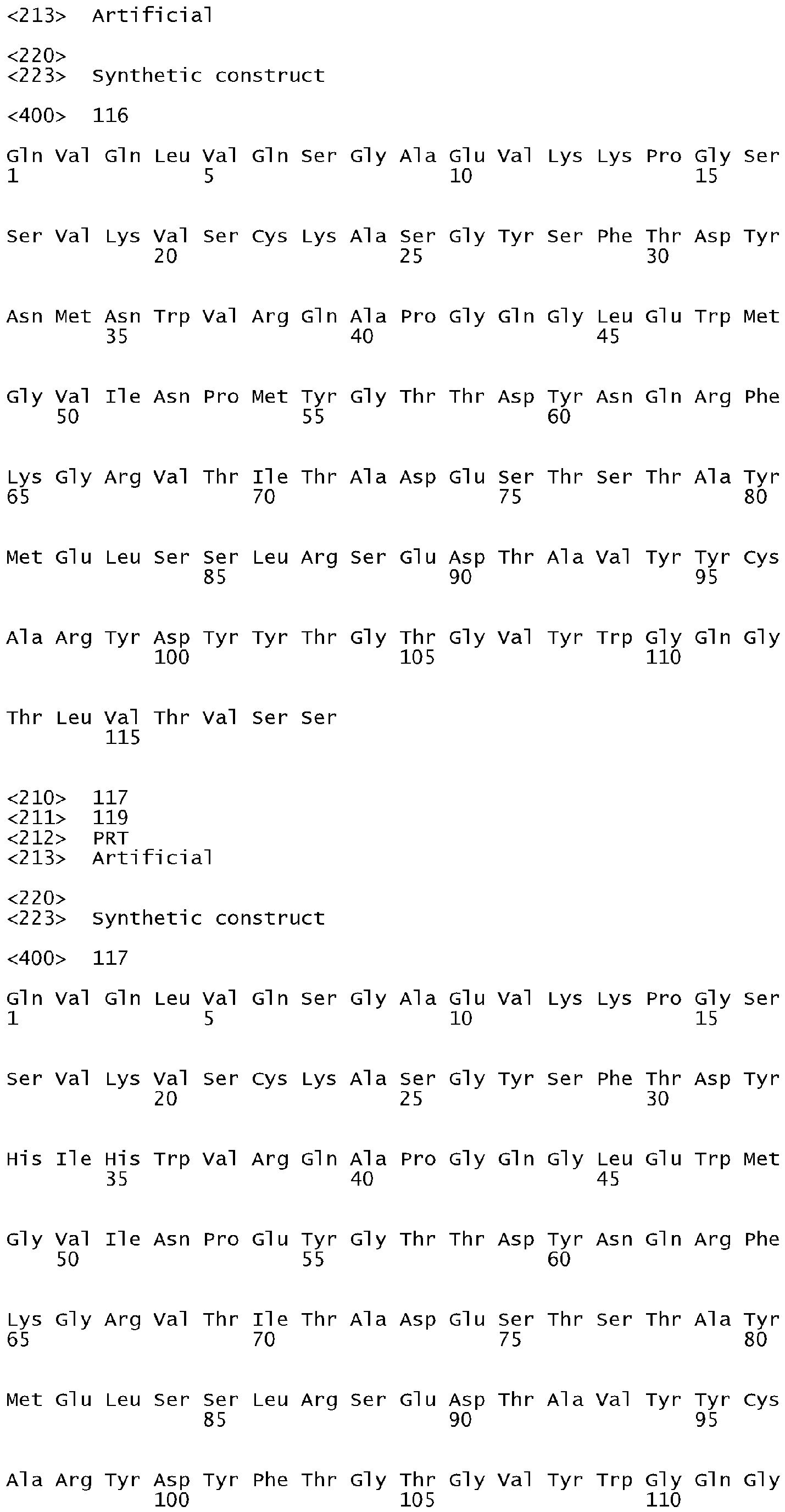


Claims (15)
- A humanized anti-IL-17 monoclonal antibody, wherein the antibody binds human IL-17 with a KD less than 7.0 pM.
- The antibody of claim 1, wherein said antibody further has a dissociation rate constant or koff rate of less than 5 x 10⁻⁵ s-1.
- The antibody according to any one of claims 1 to 2, wherein said antibody specifically binds a non-linear epitope of human IL-17, wherein the epitope comprises a polypeptide with SEQ ID NO: 276.
- The antibody according to any one of claims 1 to 3, wherein said antibody neutralizes IL-17 bioactivity in an in vitro IL-8 reporter assay at an IC50 of less than 1 nM.
- The antibody according to any one of claims 1 to 4, wherein said antibody competes for binding to human IL-17 with a humanized anti-IL-17 antibody comprising two polypeptides with the amino acid sequences shown in SEQ ID NO: 241 and SEQ ID NO: 118.
- The antibody according to any one of claims 1 to 5, wherein said antibody comprises a heavy chain variable region (HCVR) comprising a polypeptide with an amino acid sequence selected from the group consisting of SEQ ID NOs: 56-121.
- The antibody according to any one of claims 1 to 6, wherein said antibody comprises a light chain variable region (LCVR) comprising a polypeptide with an amino acid sequence selected from the group consisting of SEQ ID NOs: 178-243.
- The antibody according to any one of claims 1 to 4, wherein said antibody comprises a CDRL1 polypeptide with SEQ ID NO: 247, a CDRL2 polypeptide with SEQ ID NO: 248, a CDRL3 polypeptide with SEQ ID NO: 249, a CDRH1 polypeptide with SEQ ID NO: 244, a CDRH2 polypeptide SEQ ID NO: 245, and a CDRH3 polypeptide with SEQ ID NO: 246.
- The humanized monoclonal antibody according to any one of claims 1 to 8, wherein said antibody is a humanized or fully human antibody or antigen-binding portion thereof.
- The antibody according to any one of claims 1 to 9, wherein the antibody is a humanized full-length antibody, a humanized substantially intact antibody, a Fab fragment, a F(ab')2 fragment or a single chain Fv fragment.
- The antibody according to any one of claims 1 to 10, wherein the antibody further comprises a heavy chain constant region selected from the group consisting of IgG1, IgG2, IgG3, IgG4, IgA, IgE, IgM and IgD.
- A composition comprising the antibody according to any one of claims 1 to 11 and wherein said composition further comprises a pharmaceutically acceptable carrier.
- An antibody according to any one of claims 1 to 11, for use as a medicament.
- Use of an effective amount of the antibody according to any one of claims 1 to 11 in the preparation of a medicament for the treatment of one or more conditions selected from the group consisting of rheumatoid arthritis, inflammatory bowel disorder, psoriasis and multiple sclerosis.
- An antibody according to any one of claims 1 to 11 for use in the treatment of one or more conditions selected from the group consisting of rheumatoid arthritis, inflammatory bowel disorder, psoriasis and multiple sclerosis.
Priority Applications (6)
| Application Number | Priority Date | Filing Date | Title |
|---|---|---|---|
| PL11192240T PL2481753T3 (en) | 2005-12-13 | 2006-12-05 | Anti-IL-17 Antibodies |
| EP18160779.7A EP3366702B1 (en) | 2005-12-13 | 2006-12-05 | Anti-il-17 antibodies |
| SI200632258T SI2481753T1 (en) | 2005-12-13 | 2006-12-05 | Anti-IL-17 Antibodies |
| RS20180601A RS57255B1 (en) | 2005-12-13 | 2006-12-05 | Anti-il-17 antibodies |
| EP20190492.7A EP3808769A1 (en) | 2005-12-13 | 2006-12-05 | Anti-il-17 antibodies |
| HRP20180941TT HRP20180941T1 (en) | 2005-12-13 | 2018-06-15 | Anti-il-17 antibodies |
Applications Claiming Priority (3)
| Application Number | Priority Date | Filing Date | Title |
|---|---|---|---|
| US74995305P | 2005-12-13 | 2005-12-13 | |
| US80194806P | 2006-05-19 | 2006-05-19 | |
| EP06846464.3A EP1963368B3 (en) | 2005-12-13 | 2006-12-05 | Anti-il-17 antibodies |
Related Parent Applications (3)
| Application Number | Title | Priority Date | Filing Date |
|---|---|---|---|
| EP06846464.3A Division EP1963368B3 (en) | 2005-12-13 | 2006-12-05 | Anti-il-17 antibodies |
| EP06846464.3A Division-Into EP1963368B3 (en) | 2005-12-13 | 2006-12-05 | Anti-il-17 antibodies |
| EP06846464.3 Division | 2006-12-05 |
Related Child Applications (3)
| Application Number | Title | Priority Date | Filing Date |
|---|---|---|---|
| EP20190492.7A Division EP3808769A1 (en) | 2005-12-13 | 2006-12-05 | Anti-il-17 antibodies |
| EP18160779.7A Division-Into EP3366702B1 (en) | 2005-12-13 | 2006-12-05 | Anti-il-17 antibodies |
| EP18160779.7A Division EP3366702B1 (en) | 2005-12-13 | 2006-12-05 | Anti-il-17 antibodies |
Publications (2)
| Publication Number | Publication Date |
|---|---|
| EP2481753A1 true EP2481753A1 (en) | 2012-08-01 |
| EP2481753B1 EP2481753B1 (en) | 2018-04-18 |
Family
ID=38008638
Family Applications (4)
| Application Number | Title | Priority Date | Filing Date |
|---|---|---|---|
| EP06846464.3A Active EP1963368B3 (en) | 2005-12-13 | 2006-12-05 | Anti-il-17 antibodies |
| EP20190492.7A Pending EP3808769A1 (en) | 2005-12-13 | 2006-12-05 | Anti-il-17 antibodies |
| EP11192240.7A Active EP2481753B1 (en) | 2005-12-13 | 2006-12-05 | Anti-IL-17 Antibodies |
| EP18160779.7A Active EP3366702B1 (en) | 2005-12-13 | 2006-12-05 | Anti-il-17 antibodies |
Family Applications Before (2)
| Application Number | Title | Priority Date | Filing Date |
|---|---|---|---|
| EP06846464.3A Active EP1963368B3 (en) | 2005-12-13 | 2006-12-05 | Anti-il-17 antibodies |
| EP20190492.7A Pending EP3808769A1 (en) | 2005-12-13 | 2006-12-05 | Anti-il-17 antibodies |
Family Applications After (1)
| Application Number | Title | Priority Date | Filing Date |
|---|---|---|---|
| EP18160779.7A Active EP3366702B1 (en) | 2005-12-13 | 2006-12-05 | Anti-il-17 antibodies |
Country Status (23)
| Country | Link |
|---|---|
| US (2) | US7838638B2 (en) |
| EP (4) | EP1963368B3 (en) |
| JP (1) | JP5063612B2 (en) |
| KR (1) | KR101158959B1 (en) |
| CN (1) | CN101326195B9 (en) |
| AU (1) | AU2006325860B2 (en) |
| CA (1) | CA2631938C (en) |
| CY (3) | CY1113097T1 (en) |
| DK (2) | DK1963368T6 (en) |
| EA (1) | EA014298B1 (en) |
| ES (2) | ES2389780T7 (en) |
| HK (1) | HK1120275A1 (en) |
| HR (2) | HRP20120606T4 (en) |
| HU (2) | HUE039353T2 (en) |
| IL (1) | IL191217A (en) |
| LT (2) | LT2481753T (en) |
| LU (1) | LU93189I2 (en) |
| NO (2) | NO340827B1 (en) |
| PL (2) | PL2481753T3 (en) |
| PT (2) | PT1963368E (en) |
| RS (2) | RS52509B2 (en) |
| SI (2) | SI1963368T1 (en) |
| WO (1) | WO2007070750A1 (en) |
Cited By (2)
| Publication number | Priority date | Publication date | Assignee | Title |
|---|---|---|---|---|
| US8715669B2 (en) | 2008-05-05 | 2014-05-06 | Novimmune Sa | Anti-IL-17A/IL-17F cross-reactive antibodies |
| US11299540B2 (en) | 2017-06-18 | 2022-04-12 | Kindred Biosciences, Inc. | IL17A antibodies and antagonists for veterinary use |
Families Citing this family (115)
| Publication number | Priority date | Publication date | Assignee | Title |
|---|---|---|---|---|
| ES2305879T3 (en) | 2003-11-21 | 2008-11-01 | Ucb Pharma, S.A. | METHOD FOR THE TREATMENT OF MULTIPLE SCLEROSIS BY INHIBITION OF ACTIVITY IL-17. |
| PT1963368E (en) * | 2005-12-13 | 2012-09-14 | Lilly Co Eli | Anti-il-17 antibodies |
| US7790163B2 (en) | 2006-03-10 | 2010-09-07 | Zymogenetics, Inc. | Antibodies that bind both IL-17A and IL-17F and methods of using the same |
| WO2007106769A2 (en) | 2006-03-10 | 2007-09-20 | Zymogenetics, Inc. | Antibodies that bind both il-17a and il-17f and methods of using the same |
| US7910703B2 (en) * | 2006-03-10 | 2011-03-22 | Zymogenetics, Inc. | Antagonists to IL-17A, IL-17F, and IL-23P19 and methods of use |
| TW200815469A (en) * | 2006-06-23 | 2008-04-01 | Astrazeneca Ab | Compounds |
| GB0612928D0 (en) | 2006-06-29 | 2006-08-09 | Ucb Sa | Biological products |
| KR101218484B1 (en) | 2006-08-11 | 2013-01-04 | 머크 샤프 앤드 돔 코포레이션 | Antibodies to il-17a |
| GB0620729D0 (en) | 2006-10-18 | 2006-11-29 | Ucb Sa | Biological products |
| AU2008226067B2 (en) | 2007-03-12 | 2012-11-08 | Esbatech, An Alcon Biomedical Research Unit Llc | Sequence based engineering and optimization of single chain antibodies |
| DK2164961T3 (en) | 2007-06-25 | 2015-03-02 | Esbatech Alcon Biomed Res Unit | Sequence based forward position and optimization of single chain antibodies |
| DK2158315T3 (en) * | 2007-06-25 | 2016-06-06 | Esbatech Alcon Biomed Res Unit | Methods of modifying antibodies and modified antibodies with improved functional properties |
| WO2009026412A1 (en) * | 2007-08-21 | 2009-02-26 | Children's Medical Center Corporation | Treatment of airway hyperreactivity |
| WO2009082624A2 (en) * | 2007-12-10 | 2009-07-02 | Zymogenetics, Inc. | Antagonists of il-17a, il-17f, and il-23 and methods of using the same |
| GB0807413D0 (en) | 2008-04-23 | 2008-05-28 | Ucb Pharma Sa | Biological products |
| CN102164959A (en) * | 2008-09-29 | 2011-08-24 | 罗氏格黎卡特股份公司 | Antibodies against human IL 17 and uses thereof |
| JP2012508563A (en) * | 2008-11-12 | 2012-04-12 | シェーリング コーポレイション | ΒG1-IGG intron for enhanced anti-IGF1R expression |
| US8211434B2 (en) | 2008-11-26 | 2012-07-03 | Allergan, Inc. | KLK-13 antibody inhibitor for treating dry eye |
| WO2010062858A1 (en) * | 2008-11-26 | 2010-06-03 | Allergan, Inc. | Il-17 antibody inhibitor for treating dry eye |
| JP5836807B2 (en) * | 2009-03-05 | 2015-12-24 | アッヴィ・インコーポレイテッド | IL-17 binding protein |
| CN102458437B (en) | 2009-05-05 | 2015-06-10 | 诺维莫尼公司 | Anti-il-17f antibodies and methods of use thereof |
| US8519107B2 (en) | 2009-10-30 | 2013-08-27 | Janssen Biotech, Inc. | IL-17A antibodies |
| JO3417B1 (en) | 2010-01-08 | 2019-10-20 | Regeneron Pharma | Stabilized formulations containing anti-interleukin-6 receptor (il-6r) antibodies |
| WO2011141823A2 (en) | 2010-05-14 | 2011-11-17 | Orega Biotech | Methods of treating and/or preventing cell proliferation disorders with il-17 antagonists |
| BR112013002831A2 (en) * | 2010-08-05 | 2016-11-01 | Anaptysbio Inc | antibodies directed against il-17 |
| TW201302793A (en) | 2010-09-03 | 2013-01-16 | Glaxo Group Ltd | Novel antigen binding proteins |
| EP4137514A1 (en) | 2010-10-08 | 2023-02-22 | Novartis AG | Methods of treating psoriasis using il-17 antagonists |
| US10208349B2 (en) | 2011-01-07 | 2019-02-19 | Ucb Biopharma Sprl | Lipocalin 2 as a biomarker for IL-17 inhibitor therapy efficacy |
| GB201100282D0 (en) | 2011-01-07 | 2011-02-23 | Ucb Pharma Sa | Biological methods |
| SG10201510762YA (en) | 2011-01-14 | 2016-01-28 | Ucb Pharma Sa | Antibody molecules which bind il-17a and il-17f |
| US9943594B2 (en) | 2011-10-11 | 2018-04-17 | Sanofi Biotechnology | Methods for the treatment of rheumatoid arthritis |
| TWI589299B (en) | 2011-10-11 | 2017-07-01 | 再生元醫藥公司 | Compositions for the treatment of rheumatoid arthritis and methods of using same |
| JP2015504430A (en) | 2011-11-21 | 2015-02-12 | ノバルティス アーゲー | Methods of treating psoriatic arthritis (PSA) using IL-7 antagonists and PSA-responsive or non-responsive alleles |
| EP2809660B1 (en) | 2012-02-02 | 2016-01-20 | Ensemble Therapeutics Corporation | Macrocyclic compounds for modulating il-17 |
| EP3156073A1 (en) | 2012-03-07 | 2017-04-19 | Eli Lilly and Company | Il-17 antibody formulation |
| AR090626A1 (en) * | 2012-04-20 | 2014-11-26 | Lilly Co Eli | ANTI-BAFF-ANTI-IL-17 BIESPECIFIC ANTIBODIES |
| BR112014025768A2 (en) | 2012-04-20 | 2017-07-04 | Novartis Ag | methods of treating ankylosing spondylitis using il-17 antagonists |
| EP2852615B1 (en) * | 2012-05-22 | 2018-12-05 | Bristol-Myers Squibb Company | Il-17a/f il-23 bispecific antibodies and their uses |
| EP2864355B1 (en) | 2012-06-25 | 2016-10-12 | Orega Biotech | Il-17 antagonist antibodies |
| WO2014066726A2 (en) | 2012-10-26 | 2014-05-01 | Ensemble Therapeutics Corporation | Compounds for modulating il-17 |
| EP2925360A4 (en) * | 2012-11-29 | 2016-07-13 | Bayer Healthcare Llc | Humanized monoclonal antibodies against activated protein c and uses thereof |
| EP2953969B1 (en) | 2013-02-08 | 2019-08-28 | Novartis AG | Anti-il-17a antibodies and their use in treating autoimmune and inflammatory disorders |
| TW201444867A (en) * | 2013-03-08 | 2014-12-01 | Lilly Co Eli | Anti-TNF-anti-IL-17 bispecific antibodies |
| WO2014155278A2 (en) | 2013-03-26 | 2014-10-02 | Novartis Ag | Methods of treating autoimmune diseases using il-17 antagonists |
| US9700485B2 (en) | 2013-04-24 | 2017-07-11 | Corning Incorporated | Delamination resistant pharmaceutical glass containers containing active pharmaceutical ingredients |
| US20150004133A1 (en) * | 2013-06-07 | 2015-01-01 | The Regents Of The University Of California | Compositions And Methods For Treating Steatohepatitis, Liver Fibrosis, and Hepatocellular Carcinoma (HCC) |
| US9603882B2 (en) | 2013-08-13 | 2017-03-28 | Industrial Technology Research Institute | Method for modulating Th17 cells and method for treating a disease related to modulation of Th17 cells |
| JP6255099B2 (en) | 2013-08-15 | 2017-12-27 | ノバルティス アーゲー | Methods of treating generalized pustular psoriasis (GPP) using IL-17 antagonists |
| PL3072905T3 (en) | 2013-11-18 | 2021-03-08 | Shanghai Hengrui Pharmaceutical Co., Ltd. | Il-17a binding agent and uses thereof |
| CN106132433B (en) * | 2013-12-27 | 2021-04-06 | 国立大学法人大阪大学 | Vaccine targeting IL-17A |
| NZ720353A (en) * | 2013-12-30 | 2019-12-20 | Epimab Biotherapeutics Inc | Fabs-in-tandem immunoglobulin and uses thereof |
| BR112016018980A2 (en) | 2014-02-21 | 2017-10-10 | Genentech Inc | method of treating a disorder, multispecific antibody, isolated nucleic acid, host cell, methods of producing an antibody, producing an antibody half or multispecific antibody, and producing a multispecific, immunoconjugate antibody and pharmaceutical formulation |
| RU2577228C2 (en) * | 2014-03-14 | 2016-03-10 | Закрытое Акционерное Общество "Биокад" | Anti-il-17 antibodies, methods of their production and application |
| TWI713453B (en) | 2014-06-23 | 2020-12-21 | 美商健生生物科技公司 | Interferon alpha and omega antibody antagonists |
| RU2697383C2 (en) | 2014-09-10 | 2019-08-13 | Новартис Аг | Use of il-17 antagonists for inhibition of structural damage progression in patients with psoriatic arthritis |
| MA41044A (en) | 2014-10-08 | 2017-08-15 | Novartis Ag | COMPOSITIONS AND METHODS OF USE FOR INCREASED IMMUNE RESPONSE AND CANCER TREATMENT |
| JP6877339B2 (en) | 2014-10-14 | 2021-05-26 | ノバルティス アーゲー | Antibody molecule against PD-L1 and its use |
| RU2749712C2 (en) | 2015-01-12 | 2021-06-16 | Аффибоди Аб | Il-17a-binding polypeptides |
| CN107407677B (en) | 2015-01-28 | 2020-07-17 | 豪夫迈·罗氏有限公司 | Gene expression markers and treatment of multiple sclerosis |
| CN106336459B (en) * | 2015-07-13 | 2020-12-08 | 三生国健药业(上海)股份有限公司 | Human interleukin-17A resisting monoclonal antibody, preparation method and application thereof |
| AU2016294332A1 (en) | 2015-07-16 | 2017-11-30 | Eli Lilly And Company | Treatment of pruritus |
| EP3964528A1 (en) | 2015-07-29 | 2022-03-09 | Novartis AG | Combination therapies comprising antibody molecules to lag-3 |
| EP3878465A1 (en) | 2015-07-29 | 2021-09-15 | Novartis AG | Combination therapies comprising antibody molecules to tim-3 |
| US11077187B2 (en) | 2015-11-17 | 2021-08-03 | Oklahoma Medical Research Foundation | Epitope of optimized humanized monoclonal antibodies against activated protein C and uses thereof |
| MX2018007423A (en) | 2015-12-17 | 2018-11-09 | Novartis Ag | Antibody molecules to pd-1 and uses thereof. |
| US11421028B2 (en) | 2016-02-06 | 2022-08-23 | Epimab Biotherapeutics, Inc. | Fabs-in-tandem immunoglobulin and uses thereof |
| CN107488227A (en) * | 2016-06-12 | 2017-12-19 | 三生国健药业(上海)股份有限公司 | Anti-human IL-17 A monoclonal antibodies, its preparation method and application |
| WO2017221174A1 (en) | 2016-06-22 | 2017-12-28 | Novartis Ag | Methods of treating vitiligo using interleukin-17 (il-17) antibodies |
| CN109476733A (en) | 2016-07-19 | 2019-03-15 | 诺华股份有限公司 | The method for newly sending out psoriasis in plaques using IL-17 antagonist for treating |
| JP6802381B2 (en) * | 2016-09-14 | 2020-12-16 | ベイジン・ハンミ・ファーマシューティカル・カンパニー・リミテッドBeijing Hanmi Pharmaceutical Co., Ltd. | Possible antibodies that specifically bind to IL-17A and their functional fragments |
| JP2019505516A (en) | 2016-11-28 | 2019-02-28 | ノバルティス アーゲー | Method of treating acne using an interleukin-17 (IL-17) antagonist |
| CN110494453B (en) | 2017-02-10 | 2023-05-26 | 豪夫迈·罗氏有限公司 | Anti-tryptase antibodies, compositions thereof and uses thereof |
| WO2018158741A1 (en) | 2017-03-03 | 2018-09-07 | Novartis Ag | Psoriasis disease modification following long-term treatment with an il-17 antagonist |
| JP6876156B2 (en) * | 2017-03-10 | 2021-05-26 | スージョウ カノヴァ バイオファーマシューティカル カンパニーリミテッドSuzhou Kanova Biopharmaceutical Co., Ltd. | Monoclonal antibodies that react with IL-17A and IL-17F, and their use |
| US10941178B2 (en) * | 2017-03-17 | 2021-03-09 | Gilead Sciences, Inc. | Method of purifying an antibody |
| US10676522B2 (en) | 2017-05-05 | 2020-06-09 | Novartis Ag | Methods of selectively treating asthma using IL-17 antagonists |
| WO2019027828A1 (en) | 2017-08-04 | 2019-02-07 | Eli Lilly And Company | Anti-il-17 antibody dose regimen |
| WO2019040230A1 (en) | 2017-08-23 | 2019-02-28 | Eli Lilly And Company | Treatment of genital psoriasis |
| US20210179702A1 (en) | 2017-11-02 | 2021-06-17 | Novartis Ag | Method of treating tendinopathy using interleukin-17 (il-17) |
| KR102048475B1 (en) * | 2017-11-10 | 2019-11-26 | 주식회사 와이바이오로직스 | Antibodies Specifically Binding Against IL-17A and Uses Thereof |
| US20200347130A1 (en) * | 2017-11-10 | 2020-11-05 | Jiangsu Hengrui Medicine Co., Ltd. | CD96 Antibody, Antigen-Binding Fragment and Pharmaceutical use Thereof |
| US20200277369A1 (en) | 2017-11-20 | 2020-09-03 | Novartis Ag | Method of treating hidradentitis suppurativa with il-17 antagonists |
| AU2018375738A1 (en) | 2017-11-30 | 2020-06-11 | Novartis Ag | BCMA-targeting chimeric antigen receptor, and uses thereof |
| US11619832B2 (en) | 2018-03-08 | 2023-04-04 | Coopervision International Limited | Identification of contact lens wearers predisposed to contact lens discomfort |
| JP6930778B2 (en) * | 2018-05-17 | 2021-09-01 | 江蘇▲筌▼信生物医薬有限公司Qyuns Therapeutics Co., Ltd. | Anti-human interleukin 17A monoclonal antibody and its use |
| PE20210320A1 (en) | 2018-06-01 | 2021-02-16 | Novartis Ag | BINDING MOLECULES AGAINST BCMA AND THE USES OF THEM |
| GB201810327D0 (en) | 2018-06-22 | 2018-08-08 | Bicycletx Ltd | Peptide ligands for binding to IL-17 |
| SG11202101674QA (en) | 2018-08-29 | 2021-03-30 | Regeneron Pharma | Methods and compositions for treating subjects having rheumatoid arthritis |
| WO2020064702A1 (en) | 2018-09-25 | 2020-04-02 | INSERM (Institut National de la Santé et de la Recherche Médicale) | Use of antagonists of th17 cytokines for the treatment of bronchial remodeling in patients suffering from allergic asthma |
| GB201820316D0 (en) | 2018-12-13 | 2019-01-30 | Bicyclerd Ltd | Bicyclic peptide ligands specific for IL-17 |
| CN109369806B (en) * | 2019-01-14 | 2019-04-19 | 迈威(上海)生物科技有限公司 | The minimizing technology of cysteine variant in Su Jin monoclonal antibody product |
| MA54857A (en) | 2019-01-31 | 2021-12-08 | Numab Therapeutics AG | MULTISPECIFIC ANTIBODIES HAVING SPECIFICITY FOR TNFA AND IL-17A, ANTIBODIES TARGETING IL-17A, AND METHODS OF USE THEREOF |
| EP3689907A1 (en) | 2019-01-31 | 2020-08-05 | Numab Therapeutics AG | Antibodies targeting il-17a and methods of use thereof |
| CN114206442A (en) | 2019-01-31 | 2022-03-18 | 赛诺菲生物技术公司 | anti-IL-6 receptor antibodies for the treatment of juvenile idiopathic arthritis |
| CN113474360A (en) * | 2019-02-18 | 2021-10-01 | 伊莱利利公司 | Therapeutic antibody formulations |
| KR102400401B1 (en) * | 2019-05-22 | 2022-05-24 | (주)셀인바이오 | Anti-inflammatory peptide and anti-inflammatory composition |
| AU2020322569A1 (en) | 2019-07-26 | 2022-02-17 | Sinocelltech Ltd | Humanized anti-IL17A antibody and use thereof |
| US20220267432A1 (en) | 2019-07-30 | 2022-08-25 | Jiangsu Hengrui Medicine Co., Ltd. | Method for treating autoimmune disease by il-17 antagonist |
| CN114981302A (en) | 2019-09-20 | 2022-08-30 | 诺华股份有限公司 | Methods of treating autoimmune diseases using interleukin-17 (IL-17) antagonists |
| MX2022004311A (en) | 2019-10-15 | 2022-05-10 | Lilly Co Eli | Recombinantly engineered, lipase/esterase-deficient mammalian cell lines. |
| EP4061418A1 (en) | 2019-11-19 | 2022-09-28 | Novartis AG | Methods of treating lupus nephritis using interleukin-17 (il-17) antagonists |
| CA3162052A1 (en) | 2019-12-06 | 2021-06-10 | Novartis Ag | Methods of treating lichen planus using interleukin-17 (il-17) antagonists |
| GB201918559D0 (en) | 2019-12-16 | 2020-01-29 | Bicycle Tx Ltd | Bicyclic peptide ligands specific for IL-17 |
| JP2023506083A (en) | 2019-12-16 | 2023-02-14 | バイスクルテクス・リミテッド | Bicyclic peptide ligands specific for IL-17 |
| GB201918558D0 (en) | 2019-12-16 | 2020-01-29 | Bicycle Tx Ltd | Bicyclic peptide ligands specific for IL-17 |
| GB201918557D0 (en) | 2019-12-16 | 2020-01-29 | Bicycle Tx Ltd | Bicyclic peptide ligands specific for IL-17 |
| JP2023527827A (en) | 2020-05-27 | 2023-06-30 | サノフイ | IL-17A modulator |
| US20230286943A1 (en) | 2020-05-27 | 2023-09-14 | Sanofi | Il-17a modulators |
| CA3187136A1 (en) | 2020-06-23 | 2021-12-30 | Novartis Ag | Methods of treating thyroid eye disease and graves' orbitopahy using interleukin-17 (il-17) antagonists |
| WO2022097060A1 (en) | 2020-11-06 | 2022-05-12 | Novartis Ag | Cd19 binding molecules and uses thereof |
| CN115746132B (en) * | 2021-09-03 | 2023-09-08 | 三优生物医药(上海)有限公司 | anti-IL-17A antibodies and uses thereof |
| IT202100023816A1 (en) * | 2021-09-16 | 2023-03-16 | Takis S R L | Antibody against SARS-CoV-2 and its uses in the medical field. |
| CN113896793B (en) * | 2021-09-30 | 2023-05-26 | 港科鹏禾生物(苏州)有限公司 | Monoclonal antibody for resisting human IL-17RC and application thereof |
| WO2023223211A1 (en) | 2022-05-16 | 2023-11-23 | Novartis Ag | Methods of treating giant cell arteritis using interleukin-17 (il-17) antagonists |
| WO2023223263A1 (en) | 2022-05-18 | 2023-11-23 | Novartis Ag | Methods of selectively treating tendinopathy using interleukin-17 (il-17) antagonists |
Citations (29)
| Publication number | Priority date | Publication date | Assignee | Title |
|---|---|---|---|---|
| WO1986001533A1 (en) | 1984-09-03 | 1986-03-13 | Celltech Limited | Production of chimeric antibodies |
| US4816397A (en) | 1983-03-25 | 1989-03-28 | Celltech, Limited | Multichain polypeptides or proteins and processes for their production |
| US4816567A (en) | 1983-04-08 | 1989-03-28 | Genentech, Inc. | Recombinant immunoglobin preparations |
| US5225539A (en) | 1986-03-27 | 1993-07-06 | Medical Research Council | Recombinant altered antibodies and methods of making altered antibodies |
| WO1994004679A1 (en) | 1991-06-14 | 1994-03-03 | Genentech, Inc. | Method for making humanized antibodies |
| EP0239400B1 (en) | 1986-03-27 | 1994-08-03 | Medical Research Council | Recombinant antibodies and methods for their production |
| US5545807A (en) | 1988-10-12 | 1996-08-13 | The Babraham Institute | Production of antibodies from transgenic animals |
| US5545806A (en) | 1990-08-29 | 1996-08-13 | Genpharm International, Inc. | Ransgenic non-human animals for producing heterologous antibodies |
| US5569825A (en) | 1990-08-29 | 1996-10-29 | Genpharm International | Transgenic non-human animals capable of producing heterologous antibodies of various isotypes |
| US5585089A (en) | 1988-12-28 | 1996-12-17 | Protein Design Labs, Inc. | Humanized immunoglobulins |
| US5589369A (en) | 1992-02-11 | 1996-12-31 | Cell Genesys Inc. | Cells homozygous for disrupted target loci |
| US5591669A (en) | 1988-12-05 | 1997-01-07 | Genpharm International, Inc. | Transgenic mice depleted in a mature lymphocytic cell-type |
| US5612205A (en) | 1990-08-29 | 1997-03-18 | Genpharm International, Incorporated | Homologous recombination in mammalian cells |
| US5625126A (en) | 1990-08-29 | 1997-04-29 | Genpharm International, Inc. | Transgenic non-human animals for producing heterologous antibodies |
| US5625825A (en) | 1993-10-21 | 1997-04-29 | Lsi Logic Corporation | Random number generating apparatus for an interface unit of a carrier sense with multiple access and collision detect (CSMA/CD) ethernet data network |
| US5633425A (en) | 1990-08-29 | 1997-05-27 | Genpharm International, Inc. | Transgenic non-human animals capable of producing heterologous antibodies |
| US5661016A (en) | 1990-08-29 | 1997-08-26 | Genpharm International Inc. | Transgenic non-human animals capable of producing heterologous antibodies of various isotypes |
| US5698762A (en) | 1994-12-09 | 1997-12-16 | Dauerman; Leonard | Microwave-assisted pyrolysis of waste polyaromatic hydrocarbons |
| EP0843961A1 (en) | 1995-08-29 | 1998-05-27 | Kirin Beer Kabushiki Kaisha | Chimeric animal and method for constructing the same |
| US5770429A (en) | 1990-08-29 | 1998-06-23 | Genpharm International, Inc. | Transgenic non-human animals capable of producing heterologous antibodies |
| US5789215A (en) | 1991-08-20 | 1998-08-04 | Genpharm International | Gene targeting in animal cells using isogenic DNA constructs |
| US5789650A (en) | 1990-08-29 | 1998-08-04 | Genpharm International, Inc. | Transgenic non-human animals for producing heterologous antibodies |
| US5807715A (en) | 1984-08-27 | 1998-09-15 | The Board Of Trustees Of The Leland Stanford Junior University | Methods and transformed mammalian lymphocyte cells for producing functional antigen-binding protein including chimeric immunoglobulin |
| US5814318A (en) | 1990-08-29 | 1998-09-29 | Genpharm International Inc. | Transgenic non-human animals for producing heterologous antibodies |
| US6284471B1 (en) | 1991-03-18 | 2001-09-04 | New York University Medical Center | Anti-TNFa antibodies and assays employing anti-TNFa antibodies |
| WO2004106377A1 (en) * | 2003-05-30 | 2004-12-09 | Celltech R & D Limited | Methods for producing antibodies |
| WO2005005604A2 (en) | 2003-06-30 | 2005-01-20 | Centocor, Inc. | Engineered anti-target immunoglobulin derived proteins, compositions, methods and uses |
| WO2006013107A1 (en) * | 2004-08-05 | 2006-02-09 | Novartis Ag | Il-17 antagonistic antibodies |
| WO2006054059A1 (en) * | 2004-11-19 | 2006-05-26 | Ucb Pharma S.A. | Neutralising antibody molecules having specificity for human il-17 |
Family Cites Families (6)
| Publication number | Priority date | Publication date | Assignee | Title |
|---|---|---|---|---|
| US539A (en) | 1837-12-26 | Standard measurer for taking measure for coats | ||
| US5225A (en) | 1847-08-07 | Latch | ||
| US6274711B1 (en) * | 1993-06-14 | 2001-08-14 | Inserm, Institut National De La Sante Et De La Recherche Medicale | Purified mammalian CTLA-8 antigens and related reagents |
| US6562333B1 (en) | 1993-06-14 | 2003-05-13 | Schering Corporation | Purified mammalian CTLA-8 antigens and related reagents |
| ES2305879T3 (en) | 2003-11-21 | 2008-11-01 | Ucb Pharma, S.A. | METHOD FOR THE TREATMENT OF MULTIPLE SCLEROSIS BY INHIBITION OF ACTIVITY IL-17. |
| PT1963368E (en) * | 2005-12-13 | 2012-09-14 | Lilly Co Eli | Anti-il-17 antibodies |
-
2006
- 2006-12-05 PT PT06846464T patent/PT1963368E/en unknown
- 2006-12-05 EP EP06846464.3A patent/EP1963368B3/en active Active
- 2006-12-05 DK DK06846464.3T patent/DK1963368T6/en active
- 2006-12-05 CN CN200680046605.8A patent/CN101326195B9/en active Active
- 2006-12-05 HU HUE11192240A patent/HUE039353T2/en unknown
- 2006-12-05 CA CA2631938A patent/CA2631938C/en active Active
- 2006-12-05 US US12/095,398 patent/US7838638B2/en active Active
- 2006-12-05 WO PCT/US2006/061586 patent/WO2007070750A1/en active Application Filing
- 2006-12-05 PL PL11192240T patent/PL2481753T3/en unknown
- 2006-12-05 ES ES06846464T patent/ES2389780T7/en active Active
- 2006-12-05 EA EA200870050A patent/EA014298B1/en active Protection Beyond IP Right Term
- 2006-12-05 RS RS20120418A patent/RS52509B2/en unknown
- 2006-12-05 JP JP2008545908A patent/JP5063612B2/en active Active
- 2006-12-05 PL PL06846464T patent/PL1963368T6/en unknown
- 2006-12-05 DK DK11192240.7T patent/DK2481753T3/en active
- 2006-12-05 AU AU2006325860A patent/AU2006325860B2/en active Active
- 2006-12-05 RS RS20180601A patent/RS57255B1/en unknown
- 2006-12-05 SI SI200631427T patent/SI1963368T1/en unknown
- 2006-12-05 KR KR1020087014147A patent/KR101158959B1/en active Protection Beyond IP Right Term
- 2006-12-05 SI SI200632258T patent/SI2481753T1/en unknown
- 2006-12-05 ES ES11192240.7T patent/ES2672221T3/en active Active
- 2006-12-05 PT PT111922407T patent/PT2481753T/en unknown
- 2006-12-05 EP EP20190492.7A patent/EP3808769A1/en active Pending
- 2006-12-05 LT LTEP11192240.7T patent/LT2481753T/en unknown
- 2006-12-05 EP EP11192240.7A patent/EP2481753B1/en active Active
- 2006-12-05 EP EP18160779.7A patent/EP3366702B1/en active Active
-
2008
- 2008-05-01 IL IL191217A patent/IL191217A/en active Protection Beyond IP Right Term
- 2008-06-26 NO NO20082905A patent/NO340827B1/en active Protection Beyond IP Right Term
-
2009
- 2009-02-04 HK HK09101005.2A patent/HK1120275A1/en unknown
-
2010
- 2010-10-21 US US12/908,892 patent/US8110191B2/en active Active
-
2012
- 2012-07-23 HR HRP20120606TT patent/HRP20120606T4/en unknown
- 2012-09-07 CY CY20121100807T patent/CY1113097T1/en unknown
-
2016
- 2016-08-24 CY CY2016030C patent/CY2016030I2/en unknown
- 2016-08-25 LT LTPA2016026C patent/LTC1963368I2/en unknown
- 2016-08-31 LU LU93189C patent/LU93189I2/en unknown
- 2016-09-07 HU HUS1600037C patent/HUS1600037I1/en unknown
-
2017
- 2017-08-18 NO NO2017044C patent/NO2017044I2/en unknown
-
2018
- 2018-06-15 HR HRP20180941TT patent/HRP20180941T1/en unknown
- 2018-06-27 CY CY20181100670T patent/CY1120365T1/en unknown
Patent Citations (34)
| Publication number | Priority date | Publication date | Assignee | Title |
|---|---|---|---|---|
| EP0120694B1 (en) | 1983-03-25 | 1993-07-21 | Celltech Therapeutics Limited | Processes for the production of multichain polypeptides or proteins |
| US4816397A (en) | 1983-03-25 | 1989-03-28 | Celltech, Limited | Multichain polypeptides or proteins and processes for their production |
| US4816567A (en) | 1983-04-08 | 1989-03-28 | Genentech, Inc. | Recombinant immunoglobin preparations |
| EP0125023B1 (en) | 1983-04-08 | 1991-06-05 | Genentech, Inc. | Recombinant immunoglobulin preparations, methods for their preparation, dna sequences, expression vectors and recombinant host cells therefor |
| US5807715A (en) | 1984-08-27 | 1998-09-15 | The Board Of Trustees Of The Leland Stanford Junior University | Methods and transformed mammalian lymphocyte cells for producing functional antigen-binding protein including chimeric immunoglobulin |
| EP0194276B1 (en) | 1984-09-03 | 1993-08-11 | Celltech Therapeutics Limited | Production of chimeric antibodies |
| WO1986001533A1 (en) | 1984-09-03 | 1986-03-13 | Celltech Limited | Production of chimeric antibodies |
| US5225539A (en) | 1986-03-27 | 1993-07-06 | Medical Research Council | Recombinant altered antibodies and methods of making altered antibodies |
| EP0239400B1 (en) | 1986-03-27 | 1994-08-03 | Medical Research Council | Recombinant antibodies and methods for their production |
| US5545807A (en) | 1988-10-12 | 1996-08-13 | The Babraham Institute | Production of antibodies from transgenic animals |
| US5591669A (en) | 1988-12-05 | 1997-01-07 | Genpharm International, Inc. | Transgenic mice depleted in a mature lymphocytic cell-type |
| US5693761A (en) | 1988-12-28 | 1997-12-02 | Protein Design Labs, Inc. | Polynucleotides encoding improved humanized immunoglobulins |
| US5585089A (en) | 1988-12-28 | 1996-12-17 | Protein Design Labs, Inc. | Humanized immunoglobulins |
| US5625126A (en) | 1990-08-29 | 1997-04-29 | Genpharm International, Inc. | Transgenic non-human animals for producing heterologous antibodies |
| US5770429A (en) | 1990-08-29 | 1998-06-23 | Genpharm International, Inc. | Transgenic non-human animals capable of producing heterologous antibodies |
| US5612205A (en) | 1990-08-29 | 1997-03-18 | Genpharm International, Incorporated | Homologous recombination in mammalian cells |
| US5569825A (en) | 1990-08-29 | 1996-10-29 | Genpharm International | Transgenic non-human animals capable of producing heterologous antibodies of various isotypes |
| US5814318A (en) | 1990-08-29 | 1998-09-29 | Genpharm International Inc. | Transgenic non-human animals for producing heterologous antibodies |
| US5633425A (en) | 1990-08-29 | 1997-05-27 | Genpharm International, Inc. | Transgenic non-human animals capable of producing heterologous antibodies |
| US5661016A (en) | 1990-08-29 | 1997-08-26 | Genpharm International Inc. | Transgenic non-human animals capable of producing heterologous antibodies of various isotypes |
| US5545806A (en) | 1990-08-29 | 1996-08-13 | Genpharm International, Inc. | Ransgenic non-human animals for producing heterologous antibodies |
| US5789650A (en) | 1990-08-29 | 1998-08-04 | Genpharm International, Inc. | Transgenic non-human animals for producing heterologous antibodies |
| US5721367A (en) | 1990-08-29 | 1998-02-24 | Pharming B.V. | Homologous recombination in mammalian cells |
| US6284471B1 (en) | 1991-03-18 | 2001-09-04 | New York University Medical Center | Anti-TNFa antibodies and assays employing anti-TNFa antibodies |
| WO1994004679A1 (en) | 1991-06-14 | 1994-03-03 | Genentech, Inc. | Method for making humanized antibodies |
| US5789215A (en) | 1991-08-20 | 1998-08-04 | Genpharm International | Gene targeting in animal cells using isogenic DNA constructs |
| US5589369A (en) | 1992-02-11 | 1996-12-31 | Cell Genesys Inc. | Cells homozygous for disrupted target loci |
| US5625825A (en) | 1993-10-21 | 1997-04-29 | Lsi Logic Corporation | Random number generating apparatus for an interface unit of a carrier sense with multiple access and collision detect (CSMA/CD) ethernet data network |
| US5698762A (en) | 1994-12-09 | 1997-12-16 | Dauerman; Leonard | Microwave-assisted pyrolysis of waste polyaromatic hydrocarbons |
| EP0843961A1 (en) | 1995-08-29 | 1998-05-27 | Kirin Beer Kabushiki Kaisha | Chimeric animal and method for constructing the same |
| WO2004106377A1 (en) * | 2003-05-30 | 2004-12-09 | Celltech R & D Limited | Methods for producing antibodies |
| WO2005005604A2 (en) | 2003-06-30 | 2005-01-20 | Centocor, Inc. | Engineered anti-target immunoglobulin derived proteins, compositions, methods and uses |
| WO2006013107A1 (en) * | 2004-08-05 | 2006-02-09 | Novartis Ag | Il-17 antagonistic antibodies |
| WO2006054059A1 (en) * | 2004-11-19 | 2006-05-26 | Ucb Pharma S.A. | Neutralising antibody molecules having specificity for human il-17 |
Non-Patent Citations (75)
| Title |
|---|
| "Current Protocols in Molecular Biology", 1989, GREENE PUBLISHING ASSOCIATES |
| "Molecular Cloning; A Laboratory Manual", 1989, COLD SPRING HARBOR |
| ANN. RHEUM. DIS., vol. 59, 2000, pages 529 - 32 |
| BAERGA-ORTIZ, A. ET AL., PROTEIN SCI., vol. 11, 2002, pages 1300 - 8 |
| BIRD ET AL., SCIENCE, vol. 242, 1988, pages 423 - 6 |
| BLIVEN, M. ET AL., ARTHRITIS & RHEUMATISM, vol. 29, 1986, pages 1131 - 1138 |
| BOERNER ET AL., J IMMUNOL., vol. 147, 1991, pages 86 - 95 |
| BRUGGEMANN ET AL., PROC. NAT'L. ACAD SCI. USA, vol. 86, 1989, pages 6709 - 6713 |
| COLE ET AL.: "Monoclonal Antibodies and Cancer Therapy", 1985, ALAN R. LISS, pages: 77 |
| CYTOKINE, vol. 9, 1997, pages 794 - 800 |
| DAVID ET AL., BIOCHEMISTRY, vol. 13, 1974, pages 1014 |
| E. BLASI ET AL., J IMMUNOLOGY, vol. 27, 1990, pages 229 - 237 |
| FASEB JOURNAL., vol. 18, 2004, pages 1550 - 1552 |
| FISHWILD ET AL., NATURE BIOTECHNOLOGY, vol. 14, 1996, pages 845 - 51 |
| FOSSIEZ ET AL., INT. REV. IMMUNOL., vol. 16, 1998, pages 541 |
| GIAVEDONI L D: "Simultaneous detection of multiple cytokines and chemokines from nonhuman primates using luminex technology", JOURNAL OF IMMUNOLOGICAL METHODS, ELSEVIER SCIENCE PUBLISHERS B.V.,AMSTERDAM, NL, vol. 301, no. 1-2, June 2005 (2005-06-01), pages 89 - 101, XP004976653, ISSN: 0022-1759 * |
| GREEN; JAKOBOVITS, J EXP. MED., vol. 188, 1998, pages 483 - 495 |
| HANES ET AL., PROC. NATL. ACAD SCI. (USA, vol. 95, 1998, pages 14130 - 5 |
| HOFSTETTER ET AL: "Therapeutic efficacy of IL-17 neutralization in murine experimental autoimmune encephalomyelitis", CELLULAR IMMUNOLOGY, ACADEMIC PRESS, SAN DIEGO, CA, US, vol. 237, no. 2, October 2005 (2005-10-01), pages 123 - 130, XP005253007, ISSN: 0008-8749 * |
| HOOFNAGLE, A. ET AL., ANN. REV. BIOPHYS. BIOMOL. STRUCT., vol. 32, 2003, pages 1 - 25 |
| HOOFNAGLE, A. ET AL., METHODS MOL. BIOL., vol. 250, 2004, pages 283 - 298 |
| HOOGENBOOM; WINTER, J MOL. BIOL., vol. 227, 1991, pages 381 |
| HUNTER ET AL., NATURE, vol. 144, 1962, pages 945 |
| HUSTON ET AL., PROC. NATL. ACAD SCI. USA, vol. 85, 1988, pages 5879 - 83 |
| I EXP. MED., vol. 194, 2001, pages 873 - 881 |
| J IMM., vol. 155, 1995, pages 5483 - 5486 |
| J. IMMUNOL., vol. 167, 2001, pages 1004 - 1013 |
| J. IMMUNOL., vol. 171, 2003, pages 6173 - 6177 |
| JAKOBOVITS ET AL., NATURE, vol. 362, 1993, pages 255 - 258 |
| JAKOBOVITS ET AL., PROC. NATL. ACAD SCI. USA, vol. 90, 1993, pages 2551 - 2555 |
| JOBKOBOVITS ET AL., PROC. NATL. ACAD SCI. USA, vol. 90, 1993, pages 2551 |
| JONES ET AL., NATURE, vol. 321, 1986, pages 522 - 525 |
| KABAT ET AL., ANN. AT ACAD. SCI., vol. 190, 1971, pages 382 - 93 |
| KABAT ET AL.: "Sequences of Proteins of Immunological Interest", 1991, NIH |
| KABAT ET AL.: "Sequences of Proteins of Immunological Interest", 1991, NIH PUBLICATION NO. 91-3242 |
| KAUFMAN; SHARP, J MOL. BIOL., vol. 159, 1982, pages 601 - 21 |
| KETTLEBOROUGH, C.A. ET AL., PROTEIN ENGINEERING, vol. 4, 1991, pages 773 - 783 |
| KIEKE M.C. ET AL., PROTEIN ENGINEERING, vol. 10, 1997, pages 1303 - 10 |
| KOHLER ET AL., NATURE, vol. 256, 1975, pages 495 |
| KOLLS; LINDEN, IMMUNITY, vol. 21, 2004, pages 467 - 476 |
| LEVITT, M., V., MOL. BIOL., vol. 168, 1983, pages 595 - 620 |
| LITTLE M. ET AL., IMMUNOLOGY TODAY, vol. 21, 2000, pages 364 - 70 |
| LOCK ET AL., NAT. MED., vol. 8, 2002, pages 500 - 508 |
| LONBERG ET AL., NATURE, vol. 368, 1994, pages 856 - 859 |
| LONBERG; HUSZAR, INTERN. REV. IMMUNOL., vol. 13, 1995, pages 65 - 93 |
| LUBBERTS E: "THE ROLE OF IL-17 AND FAMILY MEMBERS IN THE PATHOGENESIS OF ARTHRITIS", CURRENT OPINION IN INVESTIGATIONAL DRUGS, PHARMAPRESS, US, vol. 4, no. 5, May 2003 (2003-05-01), pages 572 - 577, XP009056201, ISSN: 1472-4472 * |
| LUBBERTS ET AL., ARTHRITIS & RHEUMATISM, vol. 50, 2004, pages 650 - 659 |
| MARKS ET AL., BIOTECHNOLOGY, vol. 10, 1992, pages 779 - 783 |
| MARKS ET AL., J MOL. BIOL., vol. 222, 1991, pages 581 |
| MATTHEWS DJ; WELLS JA., SCIENCE., vol. 260, 1993, pages 1113 - 7 |
| MATUSEVICIUS ET AL., MULTIPLE SCLEROSIS., vol. 5, 1999, pages 1 - 1,104 |
| MCCAFFERTY ET AL., NATURE, vol. 348, 1990, pages 552 - 4 |
| MENDEZ ET AL., NATURE GENETICS, vol. 15, 1997, pages 146 - 156 |
| MORRISON, NATURE, vol. 368, 1994, pages 812 - 13 |
| MOSELEY T A ET AL: "Interleukin-17 family and IL-17 receptors", CYTOKINE AND GROWTH FACTOR REVIEWS, OXFORD, GB, vol. 14, no. 2, April 2003 (2003-04-01), pages 155 - 174, XP002321611, ISSN: 1359-6101 * |
| MULTIPLE SCLEROSIS, vol. 5, 1999, pages 101 - 104 |
| NAKAE ET AL., J IMMUN., vol. 171, pages 6173 - 6177 |
| NEUBERGER, NATURE BIOTECHNOLOGY, vol. 14, 1996, pages 826 |
| NYGREN, J., HISTOCHEM. AND CYTOCHEM., vol. 30, 1982, pages 407 |
| PAIN ET AL., J IMMUNOL. METH., vol. 40, 1981, pages 219 |
| PLUCKTHUN: "The Pharmacology of Monoclonal Antibodies", vol. 113, 1994, SPRINGER-VERLAG, pages: 269 - 315 |
| PLUCKTHUN: "The Pharmacology ofallonoclonal Antibodies", vol. 113, 1994, SPRINGER-VERLAG, pages: 269 - 315 |
| PRESTA, CURR. OP. STRUCT. BIOL., vol. 2, 1992, pages 593 - 596 |
| QUEEN ET AL., PROC. NATL. ACAD SCI. USA, vol. 88, 1991, pages 2869 |
| REMINGTON: "The Science and Practice of Pharmacy", 1995, MACK PUBLISHING CO. |
| RIECHMANN ET AL., NATURE, vol. 332, 1988, pages 323 - 327 |
| RIECHMANN ET AL., NATURE, vol. 332, 1988, pages 323 - 329 |
| SAMUELSON P. ET AL., JOURNAL OF BIOTECHNOLOGY., vol. 96, 2002, pages 129 - 54 |
| TAYLOR ET AL., NUCLEIC ACIDS RES., vol. 20, 1992, pages 6287 - 95 |
| TRENTHAM, D ET AL., J EXP. MED., vol. 146, 1977, pages 857 - 858 |
| URLAUB; CHASIN, PROC. NATL. ACAD. SCI. USA, vol. 77, 1980, pages 4216 - 20 |
| VERHOEYEN ET AL., SCIENCE, vol. 239, 1988, pages 1534 - 1536 |
| WITKOWSKI ET AL., CELL. MOL. LIFE SCI., vol. 61, 2004, pages 567 - 579 |
| YAO, Z. ET AL., IMMUNITY, vol. 3, 1995, pages 811 |
| ZOLA: "Monoclonal Antibodies: A Manual of Techniques", 1987, CRC PRESS, INC. |
Cited By (4)
| Publication number | Priority date | Publication date | Assignee | Title |
|---|---|---|---|---|
| US8715669B2 (en) | 2008-05-05 | 2014-05-06 | Novimmune Sa | Anti-IL-17A/IL-17F cross-reactive antibodies |
| US8771697B2 (en) | 2008-05-05 | 2014-07-08 | Novimmune Sa | Methods of treatment using anti-IL-17A/IL-17F cross-reactive antibodies |
| US9650437B2 (en) | 2008-05-05 | 2017-05-16 | Novimmune S.A. | Nucleic acid encoding and method of producing anti-IL-17A/IL-17F cross-reactive antibodies |
| US11299540B2 (en) | 2017-06-18 | 2022-04-12 | Kindred Biosciences, Inc. | IL17A antibodies and antagonists for veterinary use |
Also Published As
Similar Documents
| Publication | Publication Date | Title |
|---|---|---|
| EP1963368B1 (en) | Anti-il-17 antibodies | |
| CA2624935C (en) | Anti-myostatin antibodies | |
| JP2009519348A5 (en) | ||
| US7635760B2 (en) | Anti-myostatin antibodies | |
| WO2007076336A1 (en) | Treatment of migraine with anti-cgrp antibodies | |
| BRPI0619792B1 (en) | HUMANIZED ANTI-IL-17 MONOCLONAL ANTIBODY, COMPOSITION AND USE OF SUCH ANTIBODY |
Legal Events
| Date | Code | Title | Description |
|---|---|---|---|
| PUAI | Public reference made under article 153(3) epc to a published international application that has entered the european phase |
Free format text: ORIGINAL CODE: 0009012 |
|
| AC | Divisional application: reference to earlier application |
Ref document number: 1963368 Country of ref document: EP Kind code of ref document: P |
|
| AK | Designated contracting states |
Kind code of ref document: A1 Designated state(s): AT BE BG CH CY CZ DE DK EE ES FI FR GB GR HU IE IS IT LI LT LU LV MC NL PL PT RO SE SI SK TR |
|
| AX | Request for extension of the european patent |
Extension state: AL BA HR MK RS |
|
| 17P | Request for examination filed |
Effective date: 20130201 |
|
| 17Q | First examination report despatched |
Effective date: 20140414 |
|
| STAA | Information on the status of an ep patent application or granted ep patent |
Free format text: STATUS: EXAMINATION IS IN PROGRESS |
|
| GRAP | Despatch of communication of intention to grant a patent |
Free format text: ORIGINAL CODE: EPIDOSNIGR1 |
|
| STAA | Information on the status of an ep patent application or granted ep patent |
Free format text: STATUS: GRANT OF PATENT IS INTENDED |
|
| INTG | Intention to grant announced |
Effective date: 20171110 |
|
| GRAS | Grant fee paid |
Free format text: ORIGINAL CODE: EPIDOSNIGR3 |
|
| GRAA | (expected) grant |
Free format text: ORIGINAL CODE: 0009210 |
|
| STAA | Information on the status of an ep patent application or granted ep patent |
Free format text: STATUS: THE PATENT HAS BEEN GRANTED |
|
| AC | Divisional application: reference to earlier application |
Ref document number: 1963368 Country of ref document: EP Kind code of ref document: P |
|
| AK | Designated contracting states |
Kind code of ref document: B1 Designated state(s): AT BE BG CH CY CZ DE DK EE ES FI FR GB GR HU IE IS IT LI LT LU LV MC NL PL PT RO SE SI SK TR |
|
| AX | Request for extension of the european patent |
Extension state: AL BA HR MK RS |
|
| REG | Reference to a national code |
Ref country code: GB Ref legal event code: FG4D |
|
| REG | Reference to a national code |
Ref country code: CH Ref legal event code: EP Ref country code: CH Ref legal event code: NV Representative=s name: E. BLUM AND CO. AG PATENT- UND MARKENANWAELTE , CH |
|
| REG | Reference to a national code |
Ref country code: NL Ref legal event code: FP |
|
| REG | Reference to a national code |
Ref country code: AT Ref legal event code: REF Ref document number: 990361 Country of ref document: AT Kind code of ref document: T Effective date: 20180515 |
|
| REG | Reference to a national code |
Ref country code: IE Ref legal event code: FG4D |
|
| REG | Reference to a national code |
Ref country code: DE Ref legal event code: R096 Ref document number: 602006055206 Country of ref document: DE |
|
| REG | Reference to a national code |
Ref country code: ES Ref legal event code: FG2A Ref document number: 2672221 Country of ref document: ES Kind code of ref document: T3 Effective date: 20180613 |
|
| REG | Reference to a national code |
Ref country code: HR Ref legal event code: TUEP Ref document number: P20180941 Country of ref document: HR |
|
| REG | Reference to a national code |
Ref country code: DK Ref legal event code: T3 Effective date: 20180611 |
|
| REG | Reference to a national code |
Ref country code: SE Ref legal event code: TRGR |
|
| REG | Reference to a national code |
Ref country code: RO Ref legal event code: EPE |
|
| REG | Reference to a national code |
Ref country code: PT Ref legal event code: SC4A Ref document number: 2481753 Country of ref document: PT Date of ref document: 20180713 Kind code of ref document: T Free format text: AVAILABILITY OF NATIONAL TRANSLATION Effective date: 20180709 |
|
| REG | Reference to a national code |
Ref country code: HR Ref legal event code: T1PR Ref document number: P20180941 Country of ref document: HR |
|
| REG | Reference to a national code |
Ref country code: EE Ref legal event code: FG4A Ref document number: E015673 Country of ref document: EE Effective date: 20180614 |
|
| REG | Reference to a national code |
Ref country code: SK Ref legal event code: T3 Ref document number: E 27687 Country of ref document: SK |
|
| REG | Reference to a national code |
Ref country code: HR Ref legal event code: ODRP Ref document number: P20180941 Country of ref document: HR Payment date: 20181127 Year of fee payment: 13 |
|
| REG | Reference to a national code |
Ref country code: HU Ref legal event code: AG4A Ref document number: E039353 Country of ref document: HU |
|
| REG | Reference to a national code |
Ref country code: DE Ref legal event code: R097 Ref document number: 602006055206 Country of ref document: DE |
|
| REG | Reference to a national code |
Ref country code: GR Ref legal event code: EP Ref document number: 20180401730 Country of ref document: GR Effective date: 20190109 |
|
| PLBE | No opposition filed within time limit |
Free format text: ORIGINAL CODE: 0009261 |
|
| STAA | Information on the status of an ep patent application or granted ep patent |
Free format text: STATUS: NO OPPOSITION FILED WITHIN TIME LIMIT |
|
| 26N | No opposition filed |
Effective date: 20190121 |
|
| REG | Reference to a national code |
Ref country code: HR Ref legal event code: ODRP Ref document number: P20180941 Country of ref document: HR Payment date: 20191127 Year of fee payment: 14 |
|
| REG | Reference to a national code |
Ref country code: AT Ref legal event code: UEP Ref document number: 990361 Country of ref document: AT Kind code of ref document: T Effective date: 20180418 |
|
| REG | Reference to a national code |
Ref country code: HR Ref legal event code: ODRP Ref document number: P20180941 Country of ref document: HR Payment date: 20201127 Year of fee payment: 15 |
|
| REG | Reference to a national code |
Ref country code: HR Ref legal event code: ODRP Ref document number: P20180941 Country of ref document: HR Payment date: 20211202 Year of fee payment: 16 |
|
| REG | Reference to a national code |
Ref country code: HR Ref legal event code: ODRP Ref document number: P20180941 Country of ref document: HR Payment date: 20221201 Year of fee payment: 17 |
|
| PGFP | Annual fee paid to national office [announced via postgrant information from national office to epo] |
Ref country code: IT Payment date: 20221122 Year of fee payment: 17 |
|
| PGFP | Annual fee paid to national office [announced via postgrant information from national office to epo] |
Ref country code: PL Payment date: 20221123 Year of fee payment: 17 Ref country code: BE Payment date: 20221122 Year of fee payment: 17 |
|
| PGFP | Annual fee paid to national office [announced via postgrant information from national office to epo] |
Ref country code: ES Payment date: 20230102 Year of fee payment: 17 Ref country code: CH Payment date: 20230101 Year of fee payment: 17 |
|
| PGFP | Annual fee paid to national office [announced via postgrant information from national office to epo] |
Ref country code: CY Payment date: 20221128 Year of fee payment: 17 |
|
| P01 | Opt-out of the competence of the unified patent court (upc) registered |
Effective date: 20230516 |
|
| REG | Reference to a national code |
Ref country code: HR Ref legal event code: ODRP Ref document number: P20180941 Country of ref document: HR Payment date: 20231123 Year of fee payment: 18 |
|
| PGFP | Annual fee paid to national office [announced via postgrant information from national office to epo] |
Ref country code: NL Payment date: 20231121 Year of fee payment: 18 Ref country code: LU Payment date: 20231121 Year of fee payment: 18 |
|
| PGFP | Annual fee paid to national office [announced via postgrant information from national office to epo] |
Ref country code: SK Payment date: 20231128 Year of fee payment: 18 |
|
| PGFP | Annual fee paid to national office [announced via postgrant information from national office to epo] |
Ref country code: GR Payment date: 20231123 Year of fee payment: 18 Ref country code: GB Payment date: 20231121 Year of fee payment: 18 |
|
| PGFP | Annual fee paid to national office [announced via postgrant information from national office to epo] |
Ref country code: MC Payment date: 20231124 Year of fee payment: 18 |
|
| PGFP | Annual fee paid to national office [announced via postgrant information from national office to epo] |
Ref country code: IS Payment date: 20231129 Year of fee payment: 18 |
|
| PGFP | Annual fee paid to national office [announced via postgrant information from national office to epo] |
Ref country code: TR Payment date: 20231124 Year of fee payment: 18 Ref country code: SI Payment date: 20231129 Year of fee payment: 18 Ref country code: SE Payment date: 20231121 Year of fee payment: 18 Ref country code: RO Payment date: 20231129 Year of fee payment: 18 Ref country code: PT Payment date: 20231122 Year of fee payment: 18 Ref country code: LV Payment date: 20231121 Year of fee payment: 18 Ref country code: LT Payment date: 20231121 Year of fee payment: 18 Ref country code: IE Payment date: 20231123 Year of fee payment: 18 Ref country code: HU Payment date: 20231204 Year of fee payment: 18 Ref country code: FR Payment date: 20231122 Year of fee payment: 18 Ref country code: FI Payment date: 20231121 Year of fee payment: 18 Ref country code: EE Payment date: 20231122 Year of fee payment: 18 Ref country code: DK Payment date: 20231121 Year of fee payment: 18 Ref country code: DE Payment date: 20231121 Year of fee payment: 18 Ref country code: CZ Payment date: 20231124 Year of fee payment: 18 Ref country code: BG Payment date: 20231128 Year of fee payment: 18 Ref country code: AT Payment date: 20231123 Year of fee payment: 18 |
|
| PGFP | Annual fee paid to national office [announced via postgrant information from national office to epo] |
Ref country code: PL Payment date: 20231123 Year of fee payment: 18 Ref country code: BE Payment date: 20231121 Year of fee payment: 18 |