EP0837932B1 - Macrophage activating factors derived from cloned vitamin d binding protein - Google Patents
Macrophage activating factors derived from cloned vitamin d binding protein Download PDFInfo
- Publication number
- EP0837932B1 EP0837932B1 EP96921279A EP96921279A EP0837932B1 EP 0837932 B1 EP0837932 B1 EP 0837932B1 EP 96921279 A EP96921279 A EP 96921279A EP 96921279 A EP96921279 A EP 96921279A EP 0837932 B1 EP0837932 B1 EP 0837932B1
- Authority
- EP
- European Patent Office
- Prior art keywords
- cloned
- protein
- gcmaf
- domain iii
- cells
- Prior art date
- Legal status (The legal status is an assumption and is not a legal conclusion. Google has not performed a legal analysis and makes no representation as to the accuracy of the status listed.)
- Expired - Lifetime
Links
- 102000007436 Macrophage-Activating Factors Human genes 0.000 title description 7
- 108010086123 Macrophage-Activating Factors Proteins 0.000 title description 7
- 102000050760 Vitamin D-binding protein Human genes 0.000 title description 4
- 101710179590 Vitamin D-binding protein Proteins 0.000 title description 4
- 108090000623 proteins and genes Proteins 0.000 claims abstract description 76
- 210000002540 macrophage Anatomy 0.000 claims abstract description 73
- 102000004169 proteins and genes Human genes 0.000 claims abstract description 66
- 239000013598 vector Substances 0.000 claims abstract description 33
- 230000003213 activating effect Effects 0.000 claims abstract description 23
- 241000701447 unidentified baculovirus Species 0.000 claims abstract description 20
- 238000000034 method Methods 0.000 claims abstract description 14
- 150000001413 amino acids Chemical class 0.000 claims abstract description 10
- 108010005774 beta-Galactosidase Proteins 0.000 claims abstract description 10
- 238000010367 cloning Methods 0.000 claims abstract description 10
- 102000005348 Neuraminidase Human genes 0.000 claims abstract description 8
- 108010006232 Neuraminidase Proteins 0.000 claims abstract description 8
- 238000000338 in vitro Methods 0.000 claims abstract description 6
- 230000008569 process Effects 0.000 claims abstract description 6
- 102000005936 beta-Galactosidase Human genes 0.000 claims abstract 2
- 210000004027 cell Anatomy 0.000 claims description 60
- 206010028980 Neoplasm Diseases 0.000 claims description 43
- 201000011510 cancer Diseases 0.000 claims description 27
- 241000238631 Hexapoda Species 0.000 claims description 19
- 239000002299 complementary DNA Substances 0.000 claims description 15
- 239000012634 fragment Substances 0.000 claims description 14
- 230000004927 fusion Effects 0.000 claims description 13
- 102000004190 Enzymes Human genes 0.000 claims description 10
- 108090000790 Enzymes Proteins 0.000 claims description 10
- 239000002671 adjuvant Substances 0.000 claims description 7
- 125000003275 alpha amino acid group Chemical group 0.000 claims description 7
- 108010076504 Protein Sorting Signals Proteins 0.000 claims description 6
- 238000011282 treatment Methods 0.000 claims description 6
- 208000034454 F12-related hereditary angioedema with normal C1Inh Diseases 0.000 claims description 5
- 102000003960 Ligases Human genes 0.000 claims description 5
- 108090000364 Ligases Proteins 0.000 claims description 5
- 108091008324 binding proteins Proteins 0.000 claims description 5
- 208000016861 hereditary angioedema type 3 Diseases 0.000 claims description 5
- 239000011647 vitamin D3 Substances 0.000 claims description 5
- QYSXJUFSXHHAJI-YRZJJWOYSA-N vitamin D3 Chemical compound C1(/[C@@H]2CC[C@@H]([C@]2(CCC1)C)[C@H](C)CCCC(C)C)=C\C=C1\C[C@@H](O)CCC1=C QYSXJUFSXHHAJI-YRZJJWOYSA-N 0.000 claims description 5
- 230000029087 digestion Effects 0.000 claims description 4
- 102000023732 binding proteins Human genes 0.000 claims 4
- 108020001580 protein domains Proteins 0.000 claims 2
- 241000725303 Human immunodeficiency virus Species 0.000 claims 1
- 230000001902 propagating effect Effects 0.000 claims 1
- 102000001708 Protein Isoforms Human genes 0.000 abstract 2
- 108010029485 Protein Isoforms Proteins 0.000 abstract 2
- 230000000694 effects Effects 0.000 description 46
- 241000699666 Mus <mouse, genus> Species 0.000 description 34
- 241000699670 Mus sp. Species 0.000 description 30
- 238000001994 activation Methods 0.000 description 30
- 230000004913 activation Effects 0.000 description 29
- 241000700605 Viruses Species 0.000 description 25
- 102100031317 Alpha-N-acetylgalactosaminidase Human genes 0.000 description 24
- 108010015684 alpha-N-Acetylgalactosaminidase Proteins 0.000 description 24
- 210000002381 plasma Anatomy 0.000 description 20
- 108090000765 processed proteins & peptides Proteins 0.000 description 20
- 108020004414 DNA Proteins 0.000 description 19
- 210000002997 osteoclast Anatomy 0.000 description 14
- 239000002243 precursor Substances 0.000 description 14
- OUUQCZGPVNCOIJ-UHFFFAOYSA-M Superoxide Chemical compound [O-][O] OUUQCZGPVNCOIJ-UHFFFAOYSA-M 0.000 description 11
- 210000002966 serum Anatomy 0.000 description 11
- 230000001225 therapeutic effect Effects 0.000 description 11
- 238000001890 transfection Methods 0.000 description 10
- 210000000952 spleen Anatomy 0.000 description 9
- 102100026189 Beta-galactosidase Human genes 0.000 description 8
- 238000002649 immunization Methods 0.000 description 8
- 230000003053 immunization Effects 0.000 description 8
- 208000001132 Osteoporosis Diseases 0.000 description 7
- 210000000628 antibody-producing cell Anatomy 0.000 description 7
- 210000004369 blood Anatomy 0.000 description 7
- 239000008280 blood Substances 0.000 description 7
- 208000015181 infectious disease Diseases 0.000 description 7
- 230000000242 pagocytic effect Effects 0.000 description 7
- 210000001539 phagocyte Anatomy 0.000 description 7
- 208000030507 AIDS Diseases 0.000 description 6
- 208000003468 Ehrlich Tumor Carcinoma Diseases 0.000 description 6
- 125000000539 amino acid group Chemical group 0.000 description 6
- 230000003248 secreting effect Effects 0.000 description 6
- 238000011725 BALB/c mouse Methods 0.000 description 5
- 206010060862 Prostate cancer Diseases 0.000 description 5
- 208000000236 Prostatic Neoplasms Diseases 0.000 description 5
- 239000000427 antigen Substances 0.000 description 5
- 102000036639 antigens Human genes 0.000 description 5
- 108091007433 antigens Proteins 0.000 description 5
- 210000003719 b-lymphocyte Anatomy 0.000 description 5
- 210000001185 bone marrow Anatomy 0.000 description 5
- 230000013595 glycosylation Effects 0.000 description 5
- 238000006206 glycosylation reaction Methods 0.000 description 5
- 210000003024 peritoneal macrophage Anatomy 0.000 description 5
- 239000013612 plasmid Substances 0.000 description 5
- 230000003389 potentiating effect Effects 0.000 description 5
- 230000003612 virological effect Effects 0.000 description 5
- 206010006187 Breast cancer Diseases 0.000 description 4
- 208000026310 Breast neoplasm Diseases 0.000 description 4
- 206010009944 Colon cancer Diseases 0.000 description 4
- 241000701044 Human gammaherpesvirus 4 Species 0.000 description 4
- AVKUERGKIZMTKX-NJBDSQKTSA-N ampicillin Chemical compound C1([C@@H](N)C(=O)N[C@H]2[C@H]3SC([C@@H](N3C2=O)C(O)=O)(C)C)=CC=CC=C1 AVKUERGKIZMTKX-NJBDSQKTSA-N 0.000 description 4
- 229960000723 ampicillin Drugs 0.000 description 4
- 230000015572 biosynthetic process Effects 0.000 description 4
- 208000029742 colonic neoplasm Diseases 0.000 description 4
- 230000003247 decreasing effect Effects 0.000 description 4
- 230000007547 defect Effects 0.000 description 4
- 238000011161 development Methods 0.000 description 4
- 230000018109 developmental process Effects 0.000 description 4
- 230000003902 lesion Effects 0.000 description 4
- 210000004185 liver Anatomy 0.000 description 4
- 239000006166 lysate Substances 0.000 description 4
- 239000002609 medium Substances 0.000 description 4
- 210000005259 peripheral blood Anatomy 0.000 description 4
- 239000011886 peripheral blood Substances 0.000 description 4
- 210000003200 peritoneal cavity Anatomy 0.000 description 4
- 239000002356 single layer Substances 0.000 description 4
- 238000002560 therapeutic procedure Methods 0.000 description 4
- 238000002255 vaccination Methods 0.000 description 4
- 241000201370 Autographa californica nucleopolyhedrovirus Species 0.000 description 3
- 101000956004 Homo sapiens Vitamin D-binding protein Proteins 0.000 description 3
- 206010062016 Immunosuppression Diseases 0.000 description 3
- 206010061218 Inflammation Diseases 0.000 description 3
- 241001494479 Pecora Species 0.000 description 3
- 102000007056 Recombinant Fusion Proteins Human genes 0.000 description 3
- 108010008281 Recombinant Fusion Proteins Proteins 0.000 description 3
- 241000256251 Spodoptera frugiperda Species 0.000 description 3
- 108091081024 Start codon Proteins 0.000 description 3
- 210000000481 breast Anatomy 0.000 description 3
- 230000001413 cellular effect Effects 0.000 description 3
- 238000010276 construction Methods 0.000 description 3
- 230000007423 decrease Effects 0.000 description 3
- 230000002255 enzymatic effect Effects 0.000 description 3
- 210000003743 erythrocyte Anatomy 0.000 description 3
- 230000006870 function Effects 0.000 description 3
- 239000001963 growth medium Substances 0.000 description 3
- 230000001506 immunosuppresive effect Effects 0.000 description 3
- 238000011534 incubation Methods 0.000 description 3
- 238000002347 injection Methods 0.000 description 3
- 239000007924 injection Substances 0.000 description 3
- 238000002955 isolation Methods 0.000 description 3
- 239000002502 liposome Substances 0.000 description 3
- 210000004072 lung Anatomy 0.000 description 3
- 210000004698 lymphocyte Anatomy 0.000 description 3
- 239000000047 product Substances 0.000 description 3
- 210000002307 prostate Anatomy 0.000 description 3
- 238000000746 purification Methods 0.000 description 3
- 238000012163 sequencing technique Methods 0.000 description 3
- 239000012679 serum free medium Substances 0.000 description 3
- 239000006228 supernatant Substances 0.000 description 3
- 210000002303 tibia Anatomy 0.000 description 3
- 210000004881 tumor cell Anatomy 0.000 description 3
- 230000003442 weekly effect Effects 0.000 description 3
- 208000006386 Bone Resorption Diseases 0.000 description 2
- 241000588724 Escherichia coli Species 0.000 description 2
- 208000031886 HIV Infections Diseases 0.000 description 2
- 208000037357 HIV infectious disease Diseases 0.000 description 2
- 102000035195 Peptidases Human genes 0.000 description 2
- 108091005804 Peptidases Proteins 0.000 description 2
- 210000001744 T-lymphocyte Anatomy 0.000 description 2
- 238000003556 assay Methods 0.000 description 2
- 230000024279 bone resorption Effects 0.000 description 2
- 210000004556 brain Anatomy 0.000 description 2
- 238000012761 co-transfection Methods 0.000 description 2
- 230000022811 deglycosylation Effects 0.000 description 2
- 230000004064 dysfunction Effects 0.000 description 2
- 210000002472 endoplasmic reticulum Anatomy 0.000 description 2
- 208000033519 human immunodeficiency virus infectious disease Diseases 0.000 description 2
- 230000001939 inductive effect Effects 0.000 description 2
- 230000004054 inflammatory process Effects 0.000 description 2
- 238000003780 insertion Methods 0.000 description 2
- 230000037431 insertion Effects 0.000 description 2
- 210000003734 kidney Anatomy 0.000 description 2
- 230000003211 malignant effect Effects 0.000 description 2
- 238000004519 manufacturing process Methods 0.000 description 2
- 230000001404 mediated effect Effects 0.000 description 2
- 108020004999 messenger RNA Proteins 0.000 description 2
- 210000001616 monocyte Anatomy 0.000 description 2
- 210000000056 organ Anatomy 0.000 description 2
- 230000037361 pathway Effects 0.000 description 2
- 210000003819 peripheral blood mononuclear cell Anatomy 0.000 description 2
- 230000002980 postoperative effect Effects 0.000 description 2
- 238000001959 radiotherapy Methods 0.000 description 2
- 230000028327 secretion Effects 0.000 description 2
- 238000001356 surgical procedure Methods 0.000 description 2
- 238000004114 suspension culture Methods 0.000 description 2
- 210000001519 tissue Anatomy 0.000 description 2
- 238000012546 transfer Methods 0.000 description 2
- 238000002054 transplantation Methods 0.000 description 2
- 108020005029 5' Flanking Region Proteins 0.000 description 1
- 102000007469 Actins Human genes 0.000 description 1
- 108010085238 Actins Proteins 0.000 description 1
- 229920001817 Agar Polymers 0.000 description 1
- 102100024321 Alkaline phosphatase, placental type Human genes 0.000 description 1
- 206010003445 Ascites Diseases 0.000 description 1
- 241000894006 Bacteria Species 0.000 description 1
- 208000020084 Bone disease Diseases 0.000 description 1
- 101000956003 Bos taurus Vitamin D-binding protein Proteins 0.000 description 1
- 208000003174 Brain Neoplasms Diseases 0.000 description 1
- 125000001433 C-terminal amino-acid group Chemical group 0.000 description 1
- 102000014914 Carrier Proteins Human genes 0.000 description 1
- 108091026890 Coding region Proteins 0.000 description 1
- 238000011537 Coomassie blue staining Methods 0.000 description 1
- 241001131785 Escherichia coli HB101 Species 0.000 description 1
- 208000000461 Esophageal Neoplasms Diseases 0.000 description 1
- 208000007514 Herpes zoster Diseases 0.000 description 1
- 241000282412 Homo Species 0.000 description 1
- 101001045218 Homo sapiens Peroxisomal multifunctional enzyme type 2 Proteins 0.000 description 1
- XQFRJNBWHJMXHO-RRKCRQDMSA-N IDUR Chemical compound C1[C@H](O)[C@@H](CO)O[C@H]1N1C(=O)NC(=O)C(I)=C1 XQFRJNBWHJMXHO-RRKCRQDMSA-N 0.000 description 1
- 208000008839 Kidney Neoplasms Diseases 0.000 description 1
- 239000006137 Luria-Bertani broth Substances 0.000 description 1
- 241000124008 Mammalia Species 0.000 description 1
- 229930192392 Mitomycin Natural products 0.000 description 1
- 208000003445 Mouth Neoplasms Diseases 0.000 description 1
- 241000699660 Mus musculus Species 0.000 description 1
- NWIBSHFKIJFRCO-WUDYKRTCSA-N Mytomycin Chemical compound C1N2C(C(C(C)=C(N)C3=O)=O)=C3[C@@H](COC(N)=O)[C@@]2(OC)[C@@H]2[C@H]1N2 NWIBSHFKIJFRCO-WUDYKRTCSA-N 0.000 description 1
- OVRNDRQMDRJTHS-CBQIKETKSA-N N-Acetyl-D-Galactosamine Chemical compound CC(=O)N[C@H]1[C@@H](O)O[C@H](CO)[C@H](O)[C@@H]1O OVRNDRQMDRJTHS-CBQIKETKSA-N 0.000 description 1
- MBLBDJOUHNCFQT-UHFFFAOYSA-N N-acetyl-D-galactosamine Natural products CC(=O)NC(C=O)C(O)C(O)C(O)CO MBLBDJOUHNCFQT-UHFFFAOYSA-N 0.000 description 1
- 241000906034 Orthops Species 0.000 description 1
- 108010058846 Ovalbumin Proteins 0.000 description 1
- 206010061535 Ovarian neoplasm Diseases 0.000 description 1
- 239000004365 Protease Substances 0.000 description 1
- 208000000453 Skin Neoplasms Diseases 0.000 description 1
- 208000000102 Squamous Cell Carcinoma of Head and Neck Diseases 0.000 description 1
- 208000005718 Stomach Neoplasms Diseases 0.000 description 1
- 208000007097 Urinary Bladder Neoplasms Diseases 0.000 description 1
- 208000002495 Uterine Neoplasms Diseases 0.000 description 1
- 108020005202 Viral DNA Proteins 0.000 description 1
- 208000036142 Viral infection Diseases 0.000 description 1
- 229930003316 Vitamin D Natural products 0.000 description 1
- 102100038611 Vitamin D-binding protein Human genes 0.000 description 1
- QYSXJUFSXHHAJI-XFEUOLMDSA-N Vitamin D3 Natural products C1(/[C@@H]2CC[C@@H]([C@]2(CCC1)C)[C@H](C)CCCC(C)C)=C/C=C1\C[C@@H](O)CCC1=C QYSXJUFSXHHAJI-XFEUOLMDSA-N 0.000 description 1
- 238000009825 accumulation Methods 0.000 description 1
- 239000008272 agar Substances 0.000 description 1
- 238000004458 analytical method Methods 0.000 description 1
- 238000010171 animal model Methods 0.000 description 1
- 230000036436 anti-hiv Effects 0.000 description 1
- 210000000612 antigen-presenting cell Anatomy 0.000 description 1
- 230000001580 bacterial effect Effects 0.000 description 1
- 230000003796 beauty Effects 0.000 description 1
- 230000008901 benefit Effects 0.000 description 1
- 230000004071 biological effect Effects 0.000 description 1
- 238000004820 blood count Methods 0.000 description 1
- 210000000988 bone and bone Anatomy 0.000 description 1
- 230000022534 cell killing Effects 0.000 description 1
- 238000006243 chemical reaction Methods 0.000 description 1
- 239000003795 chemical substances by application Substances 0.000 description 1
- 210000001072 colon Anatomy 0.000 description 1
- 238000011109 contamination Methods 0.000 description 1
- 230000034994 death Effects 0.000 description 1
- 230000007812 deficiency Effects 0.000 description 1
- 230000002950 deficient Effects 0.000 description 1
- 230000003111 delayed effect Effects 0.000 description 1
- 231100000673 dose–response relationship Toxicity 0.000 description 1
- 230000002124 endocrine Effects 0.000 description 1
- 238000001952 enzyme assay Methods 0.000 description 1
- 210000003238 esophagus Anatomy 0.000 description 1
- 239000013604 expression vector Substances 0.000 description 1
- 230000037406 food intake Effects 0.000 description 1
- 102000037865 fusion proteins Human genes 0.000 description 1
- 108020001507 fusion proteins Proteins 0.000 description 1
- 239000000499 gel Substances 0.000 description 1
- 238000012215 gene cloning Methods 0.000 description 1
- 230000012010 growth Effects 0.000 description 1
- 210000004349 growth plate Anatomy 0.000 description 1
- 230000036732 histological change Effects 0.000 description 1
- 230000006801 homologous recombination Effects 0.000 description 1
- 238000002744 homologous recombination Methods 0.000 description 1
- 102000052021 human DBP Human genes 0.000 description 1
- 102000051433 human GC Human genes 0.000 description 1
- 230000028709 inflammatory response Effects 0.000 description 1
- 230000000977 initiatory effect Effects 0.000 description 1
- 238000011081 inoculation Methods 0.000 description 1
- 238000007912 intraperitoneal administration Methods 0.000 description 1
- 230000003907 kidney function Effects 0.000 description 1
- 101150066555 lacZ gene Proteins 0.000 description 1
- 206010023841 laryngeal neoplasm Diseases 0.000 description 1
- 210000000867 larynx Anatomy 0.000 description 1
- 208000032839 leukemia Diseases 0.000 description 1
- 230000003908 liver function Effects 0.000 description 1
- 208000014018 liver neoplasm Diseases 0.000 description 1
- 238000011068 loading method Methods 0.000 description 1
- 208000020816 lung neoplasm Diseases 0.000 description 1
- 239000011159 matrix material Substances 0.000 description 1
- 230000007246 mechanism Effects 0.000 description 1
- 229960004857 mitomycin Drugs 0.000 description 1
- 239000000203 mixture Substances 0.000 description 1
- 230000004048 modification Effects 0.000 description 1
- 238000012986 modification Methods 0.000 description 1
- 230000035772 mutation Effects 0.000 description 1
- 238000011580 nude mouse model Methods 0.000 description 1
- 229920001542 oligosaccharide Polymers 0.000 description 1
- 150000002482 oligosaccharides Chemical class 0.000 description 1
- 238000011275 oncology therapy Methods 0.000 description 1
- 201000002740 oral squamous cell carcinoma Diseases 0.000 description 1
- 230000001599 osteoclastic effect Effects 0.000 description 1
- 230000002611 ovarian Effects 0.000 description 1
- 239000002245 particle Substances 0.000 description 1
- 108010031345 placental alkaline phosphatase Proteins 0.000 description 1
- 229920002401 polyacrylamide Polymers 0.000 description 1
- 238000002360 preparation method Methods 0.000 description 1
- 102000004196 processed proteins & peptides Human genes 0.000 description 1
- 230000000644 propagated effect Effects 0.000 description 1
- 235000019833 protease Nutrition 0.000 description 1
- 239000011541 reaction mixture Substances 0.000 description 1
- 230000010076 replication Effects 0.000 description 1
- 108091008146 restriction endonucleases Proteins 0.000 description 1
- 230000011664 signaling Effects 0.000 description 1
- 210000002784 stomach Anatomy 0.000 description 1
- 238000006467 substitution reaction Methods 0.000 description 1
- 230000008093 supporting effect Effects 0.000 description 1
- 230000036346 tooth eruption Effects 0.000 description 1
- 238000003151 transfection method Methods 0.000 description 1
- 230000014616 translation Effects 0.000 description 1
- 241001515965 unidentified phage Species 0.000 description 1
- 238000011144 upstream manufacturing Methods 0.000 description 1
- 210000003932 urinary bladder Anatomy 0.000 description 1
- 208000024719 uterine cervix neoplasm Diseases 0.000 description 1
- 210000004291 uterus Anatomy 0.000 description 1
- 229960005486 vaccine Drugs 0.000 description 1
- 230000009385 viral infection Effects 0.000 description 1
- 230000000007 visual effect Effects 0.000 description 1
- 229930003231 vitamin Natural products 0.000 description 1
- 239000011782 vitamin Substances 0.000 description 1
- 229940088594 vitamin Drugs 0.000 description 1
- 235000013343 vitamin Nutrition 0.000 description 1
- 239000011710 vitamin D Substances 0.000 description 1
- 235000019166 vitamin D Nutrition 0.000 description 1
- 150000003710 vitamin D derivatives Chemical class 0.000 description 1
- 229940046008 vitamin d Drugs 0.000 description 1
- 150000003722 vitamin derivatives Chemical class 0.000 description 1
Images

Classifications
-
- C—CHEMISTRY; METALLURGY
- C07—ORGANIC CHEMISTRY
- C07K—PEPTIDES
- C07K14/00—Peptides having more than 20 amino acids; Gastrins; Somatostatins; Melanotropins; Derivatives thereof
- C07K14/435—Peptides having more than 20 amino acids; Gastrins; Somatostatins; Melanotropins; Derivatives thereof from animals; from humans
- C07K14/52—Cytokines; Lymphokines; Interferons
- C07K14/555—Interferons [IFN]
- C07K14/57—IFN-gamma
-
- A—HUMAN NECESSITIES
- A61—MEDICAL OR VETERINARY SCIENCE; HYGIENE
- A61P—SPECIFIC THERAPEUTIC ACTIVITY OF CHEMICAL COMPOUNDS OR MEDICINAL PREPARATIONS
- A61P19/00—Drugs for skeletal disorders
-
- A—HUMAN NECESSITIES
- A61—MEDICAL OR VETERINARY SCIENCE; HYGIENE
- A61P—SPECIFIC THERAPEUTIC ACTIVITY OF CHEMICAL COMPOUNDS OR MEDICINAL PREPARATIONS
- A61P19/00—Drugs for skeletal disorders
- A61P19/08—Drugs for skeletal disorders for bone diseases, e.g. rachitism, Paget's disease
-
- A—HUMAN NECESSITIES
- A61—MEDICAL OR VETERINARY SCIENCE; HYGIENE
- A61P—SPECIFIC THERAPEUTIC ACTIVITY OF CHEMICAL COMPOUNDS OR MEDICINAL PREPARATIONS
- A61P19/00—Drugs for skeletal disorders
- A61P19/08—Drugs for skeletal disorders for bone diseases, e.g. rachitism, Paget's disease
- A61P19/10—Drugs for skeletal disorders for bone diseases, e.g. rachitism, Paget's disease for osteoporosis
-
- A—HUMAN NECESSITIES
- A61—MEDICAL OR VETERINARY SCIENCE; HYGIENE
- A61P—SPECIFIC THERAPEUTIC ACTIVITY OF CHEMICAL COMPOUNDS OR MEDICINAL PREPARATIONS
- A61P31/00—Antiinfectives, i.e. antibiotics, antiseptics, chemotherapeutics
-
- A—HUMAN NECESSITIES
- A61—MEDICAL OR VETERINARY SCIENCE; HYGIENE
- A61P—SPECIFIC THERAPEUTIC ACTIVITY OF CHEMICAL COMPOUNDS OR MEDICINAL PREPARATIONS
- A61P31/00—Antiinfectives, i.e. antibiotics, antiseptics, chemotherapeutics
- A61P31/12—Antivirals
-
- A—HUMAN NECESSITIES
- A61—MEDICAL OR VETERINARY SCIENCE; HYGIENE
- A61P—SPECIFIC THERAPEUTIC ACTIVITY OF CHEMICAL COMPOUNDS OR MEDICINAL PREPARATIONS
- A61P35/00—Antineoplastic agents
-
- C—CHEMISTRY; METALLURGY
- C07—ORGANIC CHEMISTRY
- C07K—PEPTIDES
- C07K14/00—Peptides having more than 20 amino acids; Gastrins; Somatostatins; Melanotropins; Derivatives thereof
- C07K14/435—Peptides having more than 20 amino acids; Gastrins; Somatostatins; Melanotropins; Derivatives thereof from animals; from humans
- C07K14/46—Peptides having more than 20 amino acids; Gastrins; Somatostatins; Melanotropins; Derivatives thereof from animals; from humans from vertebrates
- C07K14/47—Peptides having more than 20 amino acids; Gastrins; Somatostatins; Melanotropins; Derivatives thereof from animals; from humans from vertebrates from mammals
-
- C—CHEMISTRY; METALLURGY
- C12—BIOCHEMISTRY; BEER; SPIRITS; WINE; VINEGAR; MICROBIOLOGY; ENZYMOLOGY; MUTATION OR GENETIC ENGINEERING
- C12N—MICROORGANISMS OR ENZYMES; COMPOSITIONS THEREOF; PROPAGATING, PRESERVING, OR MAINTAINING MICROORGANISMS; MUTATION OR GENETIC ENGINEERING; CULTURE MEDIA
- C12N9/00—Enzymes; Proenzymes; Compositions thereof; Processes for preparing, activating, inhibiting, separating or purifying enzymes
- C12N9/14—Hydrolases (3)
- C12N9/24—Hydrolases (3) acting on glycosyl compounds (3.2)
- C12N9/2402—Hydrolases (3) acting on glycosyl compounds (3.2) hydrolysing O- and S- glycosyl compounds (3.2.1)
-
- C—CHEMISTRY; METALLURGY
- C12—BIOCHEMISTRY; BEER; SPIRITS; WINE; VINEGAR; MICROBIOLOGY; ENZYMOLOGY; MUTATION OR GENETIC ENGINEERING
- C12Q—MEASURING OR TESTING PROCESSES INVOLVING ENZYMES, NUCLEIC ACIDS OR MICROORGANISMS; COMPOSITIONS OR TEST PAPERS THEREFOR; PROCESSES OF PREPARING SUCH COMPOSITIONS; CONDITION-RESPONSIVE CONTROL IN MICROBIOLOGICAL OR ENZYMOLOGICAL PROCESSES
- C12Q1/00—Measuring or testing processes involving enzymes, nucleic acids or microorganisms; Compositions therefor; Processes of preparing such compositions
- C12Q1/34—Measuring or testing processes involving enzymes, nucleic acids or microorganisms; Compositions therefor; Processes of preparing such compositions involving hydrolase
-
- C—CHEMISTRY; METALLURGY
- C12—BIOCHEMISTRY; BEER; SPIRITS; WINE; VINEGAR; MICROBIOLOGY; ENZYMOLOGY; MUTATION OR GENETIC ENGINEERING
- C12Y—ENZYMES
- C12Y302/00—Hydrolases acting on glycosyl compounds, i.e. glycosylases (3.2)
- C12Y302/01—Glycosidases, i.e. enzymes hydrolysing O- and S-glycosyl compounds (3.2.1)
- C12Y302/01049—Alpha-N-acetylgalactosaminidase (3.2.1.49)
-
- G—PHYSICS
- G01—MEASURING; TESTING
- G01N—INVESTIGATING OR ANALYSING MATERIALS BY DETERMINING THEIR CHEMICAL OR PHYSICAL PROPERTIES
- G01N33/00—Investigating or analysing materials by specific methods not covered by groups G01N1/00 - G01N31/00
- G01N33/48—Biological material, e.g. blood, urine; Haemocytometers
- G01N33/50—Chemical analysis of biological material, e.g. blood, urine; Testing involving biospecific ligand binding methods; Immunological testing
- G01N33/53—Immunoassay; Biospecific binding assay; Materials therefor
- G01N33/569—Immunoassay; Biospecific binding assay; Materials therefor for microorganisms, e.g. protozoa, bacteria, viruses
- G01N33/56983—Viruses
- G01N33/56988—HIV or HTLV
-
- G—PHYSICS
- G01—MEASURING; TESTING
- G01N—INVESTIGATING OR ANALYSING MATERIALS BY DETERMINING THEIR CHEMICAL OR PHYSICAL PROPERTIES
- G01N33/00—Investigating or analysing materials by specific methods not covered by groups G01N1/00 - G01N31/00
- G01N33/48—Biological material, e.g. blood, urine; Haemocytometers
- G01N33/50—Chemical analysis of biological material, e.g. blood, urine; Testing involving biospecific ligand binding methods; Immunological testing
- G01N33/53—Immunoassay; Biospecific binding assay; Materials therefor
- G01N33/573—Immunoassay; Biospecific binding assay; Materials therefor for enzymes or isoenzymes
-
- G—PHYSICS
- G01—MEASURING; TESTING
- G01N—INVESTIGATING OR ANALYSING MATERIALS BY DETERMINING THEIR CHEMICAL OR PHYSICAL PROPERTIES
- G01N33/00—Investigating or analysing materials by specific methods not covered by groups G01N1/00 - G01N31/00
- G01N33/48—Biological material, e.g. blood, urine; Haemocytometers
- G01N33/50—Chemical analysis of biological material, e.g. blood, urine; Testing involving biospecific ligand binding methods; Immunological testing
- G01N33/53—Immunoassay; Biospecific binding assay; Materials therefor
- G01N33/574—Immunoassay; Biospecific binding assay; Materials therefor for cancer
- G01N33/57484—Immunoassay; Biospecific binding assay; Materials therefor for cancer involving compounds serving as markers for tumor, cancer, neoplasia, e.g. cellular determinants, receptors, heat shock/stress proteins, A-protein, oligosaccharides, metabolites
-
- A—HUMAN NECESSITIES
- A61—MEDICAL OR VETERINARY SCIENCE; HYGIENE
- A61K—PREPARATIONS FOR MEDICAL, DENTAL OR TOILETRY PURPOSES
- A61K38/00—Medicinal preparations containing peptides
-
- A—HUMAN NECESSITIES
- A61—MEDICAL OR VETERINARY SCIENCE; HYGIENE
- A61K—PREPARATIONS FOR MEDICAL, DENTAL OR TOILETRY PURPOSES
- A61K39/00—Medicinal preparations containing antigens or antibodies
-
- C—CHEMISTRY; METALLURGY
- C07—ORGANIC CHEMISTRY
- C07K—PEPTIDES
- C07K2319/00—Fusion polypeptide
-
- C—CHEMISTRY; METALLURGY
- C12—BIOCHEMISTRY; BEER; SPIRITS; WINE; VINEGAR; MICROBIOLOGY; ENZYMOLOGY; MUTATION OR GENETIC ENGINEERING
- C12N—MICROORGANISMS OR ENZYMES; COMPOSITIONS THEREOF; PROPAGATING, PRESERVING, OR MAINTAINING MICROORGANISMS; MUTATION OR GENETIC ENGINEERING; CULTURE MEDIA
- C12N2799/00—Uses of viruses
- C12N2799/02—Uses of viruses as vector
- C12N2799/021—Uses of viruses as vector for the expression of a heterologous nucleic acid
- C12N2799/026—Uses of viruses as vector for the expression of a heterologous nucleic acid where the vector is derived from a baculovirus
-
- G—PHYSICS
- G01—MEASURING; TESTING
- G01N—INVESTIGATING OR ANALYSING MATERIALS BY DETERMINING THEIR CHEMICAL OR PHYSICAL PROPERTIES
- G01N2333/00—Assays involving biological materials from specific organisms or of a specific nature
- G01N2333/005—Assays involving biological materials from specific organisms or of a specific nature from viruses
- G01N2333/08—RNA viruses
- G01N2333/15—Retroviridae, e.g. bovine leukaemia virus, feline leukaemia virus, feline leukaemia virus, human T-cell leukaemia-lymphoma virus
- G01N2333/155—Lentiviridae, e.g. visna-maedi virus, equine infectious virus, FIV, SIV
- G01N2333/16—HIV-1, HIV-2
-
- Y—GENERAL TAGGING OF NEW TECHNOLOGICAL DEVELOPMENTS; GENERAL TAGGING OF CROSS-SECTIONAL TECHNOLOGIES SPANNING OVER SEVERAL SECTIONS OF THE IPC; TECHNICAL SUBJECTS COVERED BY FORMER USPC CROSS-REFERENCE ART COLLECTIONS [XRACs] AND DIGESTS
- Y10—TECHNICAL SUBJECTS COVERED BY FORMER USPC
- Y10S—TECHNICAL SUBJECTS COVERED BY FORMER USPC CROSS-REFERENCE ART COLLECTIONS [XRACs] AND DIGESTS
- Y10S436/00—Chemistry: analytical and immunological testing
- Y10S436/811—Test for named disease, body condition or organ function
- Y10S436/813—Cancer
Definitions
- This invention relates to potent macrophage activating factors, prepared by oligosaccharide digestion of the cloned vitamin D binding protein domain III, and the use of these macrophage activating factors for various cancers, HIV-infection and osteoporosis, and as a adjuvant for immunization and vaccination.
- TABLE OF TERMS Gc protein Vitamin D 3 binding protein MAF macrophage activating factor GcMAF
- Gc protein-derived macrophage activating protein GcMAFc cloned
- the small domain of vitamin D-binding protein (Gc protein) (approximately 18% length of the Gc peptide also known as domain III) was cloned via a baculovirus vector.
- the cloned domain (Cd) peptide was treated with immobilized ⁇ -galactosidase and sialidase to yield macrophage activating factor, CdMAF.
- the cloned macrophage activating factor and GcMAF is to be used for cancer therapy, HIV-infection and osteoporosis, and may also be used as a adjuvant for immunization and vaccination.
- Inflammation results in the activation of macrophages. Inflamed lesions release lysophospholipids.
- the administration into mice of small doses (5-20 tg/mouse) of lyso phosphatidyicholine (lyso-Pc) and other lysophospholipids induced a greatly enhanced phagocytic and superoxide generating capacity of macrophages (Ngwenya and Yamamoto, Proc. Soc. Exp. Biol. Med 193 :118, 1990; Yamamoto et al., Inf. Imm 61 :5388, 1993; Yamamoto et al., Inflammation 18 :311, 1994).
- Gc protein B and T lymphocytes and serum vitamin D binding protein (DBP; human DBP is known as Gc protein)
- DBP serum vitamin D binding protein
- Gc protein a protein with N-acetylgalactosamine as the remaining sugar moiety
- GcMAF titered MAF
- peripheral blood monocytes/macrophages designated as macrophages hereafter
- the macrophages of all cancer patients were activated for phagocytic and superoxide generating capacity.
- This observation indicates that cancer patient macrophages are capable of being activated.
- the MAP precursor activity of plasma Gc protein was lost or reduced in approximately 70% of this cancer patient population. Lost or reduced precursor activity of Gc protein was found to be due to deglycosylation of plasma Gc protein by ⁇ -N-acetylgalactosaminidase detected in the cancer patient's blood stream. Deglycosylated Gc protein cannot be converted to MAF (Fig. 1b).
- peripheral blood macrophages of 196 HIV-infected/AIDS patients were treated in vitro with 100 pg GcMAF/ml, the macrophages of all patients were activated for phagocytic and superoxide generating capacity.
- the MAF precursor activity of plasma Gc protein was low in approximately 35% of the HIV infected patient population.
- these patients' plasma Gc protein is deglycosylated by ⁇ -N-acctylgalactosaminidase detected in HIV-infected patients. This mechanism explains why advanced HIV-infected (AIDS) patients are severely immunosuppressed.
- ⁇ -N-acetylgalactosaminidase activity was found in plasmas of all cancer and HIV-infected patients and had an inverse correlation with the precursor activity of their plasma Gc protein.
- increase in patient serum or plasma ⁇ -N-acetylgalactosaminidase activity is responsible for decrease in the precursor activity of plasma Gc protein.
- serum or plasma ⁇ -N acetylgalactosaminidase plays a role in immunosuppression in cancer and HP/-infected/AIDS patients.
- patient serum or plasma ⁇ -N-acetylgalactosaminidase activity can serve as a diagnostic and prognostic index.
- the inflammation-primed macrophage activation process appears to be the major macrophage activation cascade, which is shared by other phagocytes such as osteoclast and monocytes.
- a defect in the inducible ⁇ -galactosidase of B lymphocytes in the macrophage activation cascade causes dysfunctional osteoclasts leading to manifestation of osteoporosis (Yamamoto et al.; J. Immunol . 152 :5100, 1994), lacking or delaying bone marrow formation in newborn.
- GcMAF and its cloned derivatives bypass the functions of lymphocytes and Gc protein and act directly on macrophages and osteoclasts
- administration of these factors into osteopetrotic hosts should rectify the bone disorder.
- GcMAFc cloned macrophage activating factor
- GcMAF bypasses the functions of lymphocytes and Gc protein and acts directly on macrophages (or osteoclasts) for activation.
- Macrophages have a potential to eliminate cancerous cells and HIV-infected cells when activated.
- GcMAF showed remarkable curative effects on a variety of human cancer indiscriminately.
- GcMAF GcMAF
- the cloned Gc protein requires an eukaryotic vector/host capable of the glycosylation of the cloned products.
- the Gc protein with a molecular weight of approximately 52000 and 458 amino acid residues) is a multi-functional protein and carries three distinct domains (Cooke and Haddad, Endocrine Rev. 10 :294, 1989).
- Domain I interacts with vitamin D while domain III interacts with actin (Haddad et al., Biochem. 31 :7174, 1992).
- Chemically and proteolytically fragmented Gc protein enabled me to indicate that the smallest domain, domain III, contains 80 amino acid residues including the site for glycosylation, an essential peptide for macrophage activation.
- I cloned the entire domain III peptide by the use of a baculovirus vector and an insect host, and treated it with the immobilized ⁇ -galactosidase and sialidase to yield a potent macrophage activating factor, designated CdMAF.
- the cloned CdMAF appears to have remarkably curative effects on a variety of cancers.
- Macrophages are antigen presenting cells. Macrophages activated by GcMAF rapidly phagocytize target antigens or cells and presented the processed antigens to antibody producing cells. I observed a rapid development of a large amount of antibody secreting cells immediately (1 to 4 days) after inoculation of small amount of GcMAF (100 pg/mouse) and sheep erythrocytes (SRBC). This finding indicates that GcMAF and its cloned derivative CdMAF should serve as a potent adjuvant for immunization and vaccination.
- a full length cDNA encoding the human Gc protein was isolated from a human liver cDNA library in bacteriophage ) ⁇ gtII (Clontech, Palo Alto, CA) by the use of a pico BlueTM immunoscreening kit available from Stratagene of La Jolla, CA.
- the baculoviral expression system in the insect cells takes advantages of several facts about the polyhedron protein: (a) it is expressed to very high levels in infected cells where it constitutes more than half of the total cellular protein late in the infection cycle; (b) it is nonessential for infection or replication of the virus, meaning that the recombinant virus does not require any helper function; (c) viruses lacking the polyhedron gene have distinct plaque morphology from viruses containing the cloned gene; and d) unlike bacterial cells, the insect cell efficiently glycosylates the cloned gene products.
- the polyhedron protein is produced at very high levels in the nuclei of infected cells late in the viral infection cycle. Accumulated polyhedron protein forms occlusion bodies that also contain embedded virus particles. These occlusion bodies, up to 15 ⁇ m in size, are highly refractile, giving them a bright shiny appearance that is readily visualized under a light microscope. Cells infected with recombinant viruses lack occlusion bodies.
- the transfection supernatant recombinant containing virus lysate
- Plaques are then screened under a light microscope for the presence (indicative of wild- type virus) or absence (indicative of recombinant virus) of occlusion bodies.
- the baculoviral expression system uses a helper-independent virus that can be propagated to high titers in insect cells adapted for growth in suspension cultures, making it possible to obtain large amounts of recombinant protein with relative case.
- the majority of the overproduced protein remains soluble in insect cells by contrast with the insoluble proteins often obtained from bacteria.
- the viral genome is large (130 kbp) and thus can accommodate large segments of foreign DNA.
- baculoviral vectors are pUC-based and confer ampicillin resistance. Each contains the polyhedron gene promoter, variable lengths of polyhedron coding sequence, and insertion site(s) for cloning the foreign gene of interest flanked by viral sequences that lie 5' to the promoter and 3' to the foreign gene insert. These flanking sequences facilitate homologous recombination between the vector and wild-type baculoviral DNA (Ausubel et al.; Current Protocols in Mol. Biol. 1990). The major consideration when choosing the appropriate baculoviral expression vector is whether to express the recombinant as a fusion or non-fusion protein.
- the cDNA containing initiation codon (-16 Met) through the leader sequence to the + 1 amino acid (leu) of the native Gc protein should be introduced to the non-fusion vector with a polylinker carrying the EcoRI site, pLV1393 (Invitrogen, San Diego, CA).
- a monolayer (2.5x10 6 cells in each of 25-cm 2 flasks) of Spodoptera frugiperda (Sf9) cells was co-transfected with a cloned plasmid (vector) DNA (2 ⁇ g) and a wild-type (Ac. MNPV) baculoviral DNA (10 ⁇ g) in 950 ⁇ l transfection buffer (Ausubel et al., Curr Protocols in Mol. Biol . 1990). When the cells were cultured for 4 or 5 days, the transfection supernatant contained recombinant viruses.
- the co-transfection lysates were diluted 10 4 , 10 5 or 10 6 and plated on Sf9 cells for cultivation for 4 to 6 days. Alter the plaques were well formed, plaques containing occlusion-negative cells were identified at a frequency of 1.3%.
- Several putative recombinant viral plaques were isolated and twice re-plaqued for purification. Pure recombinant viral plaque clones were isolated.
- An insect cell Sf9 monolayer (2.5x10 6 cells in each of 25-cm 2 flasks) was infected with a recombinant virus clone and cultured in 5 ml GIBCO serum-free medium (from GIBCO Biochemicals, Rockville, MD) or medium supplemented with 0.1 % egg albumin to avoid contamination of serum bovine vitamin D binding protein.
- the culture flasks were incubated at 27°C and monitored daily for signs of infection. After 4 to 5 days, the cells were harvested by gently dislodging them from the flask and the cells and culture medium were transferred to centrifuge tubes and centrifuged for 10 mi at 1000 x g, 4°C.
- Sf9 cells were grown in a 100-ml spinner suspension culture flask with 50 ml complete medium up to about 2 x 10 6 cells/ml. The cells were harvested, centrifuged at 1000 x g for 10 mm and re suspended in 10 to 20 ml serum-free medium containing recombinant virus at a multiplicity of infection (MOI) of 10. After 1 hour of incubation at room temperature, the infected cells were transferred to a 200-ml spinner flask containing 100 ml serum-free medium and incubated for 40 hr. More than 40% of the protein secreted was the protein of interest. The protein in the supernatant was isolated.
- MOI multiplicity of infection
- Coomassie Blue staining of the SDS-polyacrylamide gel loading 20 to 40 ⁇ g total cell protein per lane, was used to estimate the quantity of expressed protein. Because the samples contain cellular proteins, the recombinant protein was readily detected by comparison with uninfected cellular proteins.
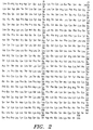
- the cloned Gc protein (2 ⁇ g) with a molecular weight of 52,000 and 458 amino acid residues (Fig. 2) was isolated by a vitamin D-affinity column (Link et al. Anal. Biochem 157 :262, 1986) and treated with immobilized ⁇ -galactosidase and sialidase.
- the resultant cloned macrophage activating factor (GcMAFc) was added to mouse and human macrophages and assayed for phagocytic and superoxide generating capacity. Incubation of macrophages with 10 pg GcMAFc/ml for 3 hours resulted in a 5-fold increased phagocytic activity and a 15-fold increase in the superoxide generating capacity of macrophages.
- domain III (approximately 80 amino acid residues) should be subcloned into an insect virus where I anticipate the efficient production and glycosylation of the peptide in the infected cells.
- the DNA segment of EI containing the initiation codon (-16 Met) through the leader sequence to the +1 amino acid (Leu) of the native Gc protein should be introduced into the vector. Because this segment carries the initiation codon for the Gc protein, non-fusion vector, pVLI393 (Invitrogen, San Diego, CA) was used.
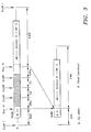
- a segment containing the initiation codon-leader sequence of the cDNA clone EI and a cDNA segment coding for 85 C-terminal amino acids (the entire domain III) plus 3' non-coding stretch of the cDNA clone E4 were ligated together and cloned into the EcoRI site of the insect virus pVL vector.
- both EI and E4 DNA were fragmented with HaeIII to yield two fragments each; Elhl (87 bp), EIhs (33 bp) and E4hs (298 bp), E4hl (450 bp), respectively. Both the larger fragments EIhl and E4hl were isolated electrophoretically, mixed with EcoRI.
- CdMAF cloned macrophage activating factor
- a baculovirus fusion vector contains human placental alkaline phosphatase secretory signal sequences that direct the nascent cloned peptide toward the secretory pathway of the cells leading to secretion into culture media.
- the signal sequence is cleaved off by signal-sequence peptidase as the nascent cloned peptide is channeled toward the secretory pathway of the host insect cells leading to secretion of the cloned domain (Cd) peptide.
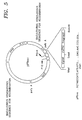
- Fig. 5 depicts that the vector carries the stuffer fragment for gene substitution and lacZ gene for identification of the gene insertion.
- the stuffer fragment of pPbac vector was excised by digesting the vector DNA with restriction enzymes SmaI and BamHI and was removed by electroelution.
- the E4 cDNA fragment of the Gc protein was digested with HaeIII and BamHI, yielding a fragment practically the same as E4h1 (see Fig. 3).
- This fragment was mixed with the above pPbac vector and ligated with T4 ligase. This strategy not only fixes the orientation of ligation but also fuses the fragment with the reading frame.
- the E. coli DH5aF cells were transformed with the reaction mixture.
- the cloned DNA insert was isolated from a number of colonies after digestion with HaeIII and BamEL The insert was confirmed by sequencing. The sequence confirmed the correct orientation.
- CdMAF 2 cloned domain peptide
- CdMAF 2 enzymatic generation of the cloned macrophage activating factor designated CdMAF 2 were described as in the Cloning Procedure I.
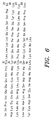
- This CdMAF is composed of 94 amino acid residues as shown in Fig. 6, including 9 amino acids from the fusion vector and is referred to herein as CdMAF 2
- CdMAF 2 has five amino acids more than the CdMAF 1 peptide derived from the non-fusion vector, they exhibited the same biological activities.
- peritoneal macrophages were isolated and assayed for superoxide generating capacity. As shown in Table 2, the macrophages were efficiently activated. These results are similar to those of macrophage activation with GcMAF (Naraparaju and Yamamoto, Immunol. Lett 43 :143, 1994). Activation of mouse pcritoneal macrophages by administration of cloned GcMAF derivatives.
- mice When BALB/c mice were administered with GcMAF, GcMAFc or CdMAF (100 pg/mouse) and received 10 5 Ehrlich ascites tumor cells/mouse, they survived for at least 5 weeks. All the control mice received only the ascites tumor and died in approximately 2 weeks. When mice were administered with an additional GcMAF, GcMAFc or CdMAF (100 pg/mouse) 4 days post-transplantation, the tumor cells were completely eliminated (Table 3). Therapeutic effects of GcMAF and cloned derivatives on mice bearing Ehrlich ascites tumor. Group No. of mice Post transplantation treatment No.
- mice survived/period Group 1 6 mice Untreated control 6 /13 ⁇ 3 days 10 mice Day 0 100 pg GcMAF/mouse 10 / 36 ⁇ 7 days Group 2 6 mice Untreated control 6 / 14 ⁇ 4 days 10 mice Day 0 100 pg GcMAFc/mouse 10 / 35 ⁇ 6 days Group 3 6 mice Untreated control 6 / 14 ⁇ 5 days 10 mice Day 0 100 pg CdMAF/mouse 10 / 34 ⁇ 3 days Group 4 8 mice Untreated control 8 / 15 ⁇ 5 days 12 mice Day 0 100 pg GcMAF/mouse 12 / > 65 days Day 4 100 pg GcMAF/mouse Group 5 8 mice Untreated control 8 / 14 ⁇ 5 days 12 mice Day 0 100 pg GcMAFc/mouse 12 / > 65 days Day 4 100 pg GcMAFc/mouse Group 6 8 mice Untreated control 8 / 14 ⁇ 5 days 12 mice Day 0 100
- the administration of GcMAFc or CdMAF to new born litters of osteopetrotic op/op mouse was performed by the weekly injection of 100 picograms for four weeks beginning from a day after birth. Mice were sacrificed at 28 days. The tibiae were removed from the treated and untreated control mice, longitudinally bisected, and examined under a dissecting microscope to measure the size of the bone marrow cavity. The cavity size was expressed as a percentage of the distance between the epiphyseal plates of the tibia. The untreated mouse group formed bone marrow with 30% of the total length of tibia. The treated mouse group experienced a 20% increased bone marrow formation over that of the untreated mouse group. This increased bone marrow cavity formation is an indication of osteoclast activation and increased osteoclastic bone resorption.
- the source of the plasma ⁇ -N-acctylgalactosaminidasc in cancer patients appeared to be cancerous cells.
- High ⁇ -N-acetylgalactosaminidase activities were detected in tumor tissue homogenates of various organs, including eleven different tumor tissues including 4 lung, 3 breast, 3 colon and 1 cervix tumors, though the ⁇ -N-acetylgalac tosaminidase activity varied from 15.9 to 50.8 nmoles/mg/min.
- Surgical removal of malignant lesions in human cancer results in the subtle decrease of plasma ⁇ -N acetylgalactosaminidase activity with the concomitant increase in the precursor activity, particularly if malignant cells are localized.
- mice were transplanted with 5 x 10 5 Ehrlich ascites tumor cells/mice into peritoneal cavity and analyzed for serum ⁇ -N acetylgalactosaminidase activity.
- serum enzyme levels were measured as transplanted Ehrlich ascites tumor grew in the mouse peritoneal cavity, the enzyme activity was directly proportional to the tumor burden (the total cell counts in peritoneal cavity) as shown in Fig. 7.
- nude mice transplanted with KB cells human oral squamous cell carcinoma cell line.
- Serum ⁇ -N-acetylgalactosaminidase activity increased as tumor size (measured by weight) of the solid tumor increased.
- I have been using plasma ⁇ -N-acetylgalactosaminidase activity as a prognostic index to monitor the progress of therapy.
- HIV-infected cells appeared to secrete ⁇ -N-acctylgalactosaminidase into the blood stream.
- peripheral blood mononuclear cells PBMC
- mitomycin as a provirus inducing agent
- ⁇ -N-acetylgalactosaminidase was secreted into the culture media.
- GcMAF 100 and 500 ng/human
- the administration of GcMAF resulted in the greatly enhanced activation of macrophages as measured by the 7-fold enhanced phagocytic capacity and the 15-fold superoxide generating capacity of macrophages.
- the administration of GcMAF showed no signs of any side effects to the recipients.
- Administration of various doses (100 pg to 10 ng/mouse) to a number of mice produced neither ill effects nor histological changes in various organs including liver, lung, kidney, spleen, brain, etc.
- GcMAF 100 ng/week
- GcMAF tumor specific serum ⁇ -N-acetylgalactosaminidase activity because the serum enzyme level is proportional to the total amount of cancerous cells (tumor burden).
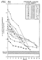
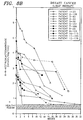
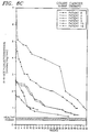
- Curative effects of GcMAF on prostate, breast and colon cancer are illustrated in Figs. 8a to 8c. After 25 weekly administrations of 100 ng GcMAF the majority (>90%) of prostate and breast cancer patients exhibited insignificantly low levels of the serum enzyme. A similar result was also observed after 35 GcMAF administrations to colon cancer patients.
- GcMAF glomerular mesenchymal fibroblasts
- Similar curative effects of GcMAF on lung, liver, stomach, brain, bladder, kidney, uterus, ovarian, larynx, esophagus, oral and skin cancers were observed.
- GcMAF appeared to be effective on a variety of cancers indiscriminately.
- GcMAF showed no evidence of side effects in patients after more than 6 months of therapy. This was also confirmed by blood cell count profile, liver and kidney functions, etc.
- peripheral blood macrophages of HIV-infected/AIDS patients with 100 pg GcMAF/mi resulted in a greatly enhanced macrophage activation (Yamamoto et al., AIDS Res. Human Ret 11 :1373, 1995).
- HIV-infected patients carry anti-HIV antibodies.
- HIV-infected cells express the viral antigens on the cell surface.
- macrophages have a potential to eliminate the infected cells via Fc-rcccptor mediated cell-killing/ingestion when activated.
- mice were inoculated with SRBC 6 hours after the intraperitoneal administration of 50 pg GcMAF/mouse.
- IgM-antibody secreting cells in the spleen were determined using the Jerne plaque assay (Jerne et al., Cell-bound antibodies, Wistar Institute Press, 1963).
- One day post-administration of GcMAF and SRBC produced 1.35 x 10 4 PFC/spleen.
- Two days after administration of GcMAF and SRBC the number of antibody secreting cells had increased to 8.2 x 10 4 PFC/spleen.
- mice that received an injection of SRBC alone produced about 3.8 x 10 4 PFC/spleen, 4 days after SRBC-injection.
- mice were injected with SRBC 6 hours after administration of various doses of GcMAF ranging from 1 to 50 pg/mouse.
- the number of antibody secreting cells per spleen was determined by the Jerne plaque assay.
- (3cMAF is a potent adjuvant for immunization.
- SRBC sheep erythrocytes
- SRBC erythrocytes
- SRBC SRBC
- SRBC SRBC 1 0.01 ⁇ 0.002 1.35 ⁇ 0.21 2 0.08 ⁇ 0.02 8.28 ⁇ 1.26 3 1.18 ⁇ 0.42 14.42 ⁇ 2.32 4 3.86 ⁇ 0.95 23.68 ⁇ 6.05 5 2.15 ⁇ 0.63 18.63 ⁇ 3.43
Landscapes
- Health & Medical Sciences (AREA)
- Life Sciences & Earth Sciences (AREA)
- Chemical & Material Sciences (AREA)
- Engineering & Computer Science (AREA)
- Immunology (AREA)
- Organic Chemistry (AREA)
- Molecular Biology (AREA)
- General Health & Medical Sciences (AREA)
- Biomedical Technology (AREA)
- Medicinal Chemistry (AREA)
- Biochemistry (AREA)
- Hematology (AREA)
- Urology & Nephrology (AREA)
- Microbiology (AREA)
- Zoology (AREA)
- Biotechnology (AREA)
- Physics & Mathematics (AREA)
- Genetics & Genomics (AREA)
- Analytical Chemistry (AREA)
- Cell Biology (AREA)
- Bioinformatics & Cheminformatics (AREA)
- Food Science & Technology (AREA)
- Wood Science & Technology (AREA)
- Pathology (AREA)
- General Physics & Mathematics (AREA)
- Virology (AREA)
- Proteomics, Peptides & Aminoacids (AREA)
- Public Health (AREA)
- Animal Behavior & Ethology (AREA)
- Veterinary Medicine (AREA)
- Biophysics (AREA)
- General Engineering & Computer Science (AREA)
- Pharmacology & Pharmacy (AREA)
- Nuclear Medicine, Radiotherapy & Molecular Imaging (AREA)
- Oncology (AREA)
- Physical Education & Sports Medicine (AREA)
- Chemical Kinetics & Catalysis (AREA)
- General Chemical & Material Sciences (AREA)
- Gastroenterology & Hepatology (AREA)
- Hospice & Palliative Care (AREA)
Abstract
Description
- This invention relates to potent macrophage activating factors, prepared by oligosaccharide digestion of the cloned vitamin D binding protein domain III, and the use of these macrophage activating factors for various cancers, HIV-infection and osteoporosis, and as a adjuvant for immunization and vaccination.
TABLE OF TERMS Gc protein Vitamin D3 binding protein MAF macrophage activating factor GcMAF Gc protein-derived macrophage activating protein GcMAFc cloned Gc protein-derived macrophage activating factor Gc domain III domain III region of Gc protein CdMAF cloned domain III-derived macrophage activating factor NagAg α-N-acetylgalactosaminidase as an antigen - The small domain of vitamin D-binding protein (Gc protein) (approximately 18% length of the Gc peptide also known as domain III) was cloned via a baculovirus vector. The cloned domain (Cd) peptide was treated with immobilized β-galactosidase and sialidase to yield macrophage activating factor, CdMAF. The cloned macrophage activating factor and GcMAF is to be used for cancer therapy, HIV-infection and osteoporosis, and may also be used as a adjuvant for immunization and vaccination.
- Other objects and many attendant features of this invention will become readily appreciated as the same becomes better understood by reference to the following detailed description when considered in connection with the accompanying drawings wherein:
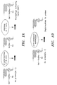
- Fig. 1a is a schematic illustration of the formation of macrophage activating factor (MAF).
- Fig. 1b is a schematic illustration of the deglycosylation of Gc protein in a cancer or HIV-infected patient's blood stream.
- Fig. 2 shows the amino acid sequence of cloned GcMAF which is SEQ II) NO:I which is the entire cloned Gc protein (and is not within the present invention).
- Fig. 3 shows the construction of the DNA fragment encoding the leader sequence of ECORI fragment E1 and domain III regions of the Gc protein; A, the entire cDNA for Gc protein; B, the construct to be inserted into the non-fusion vector; the shaded area indicates the compressed regions of about 1,000 base pairs (bp).
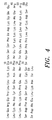
- Fig. 4 shows the 89 amino acid sequence, SEQ ID NO:2, of the cloned domain m (CdMAF1) using the non-fusion vector.
- Fig. 5 shows the baculovirus fusion vector for cloning the domain III of Gc protein.
- Fig. 6 shows the 94 amino acid sequence, SEQ ID NO:3, of the cloned domain III (CdMAF2) using the fusion vector.
- Fig. 7 shows the correlation between plasma α-N-acetylgalactosaminidase activity and tumor burden (total cell counts) in the peritoneal cavity of Ehrlich ascites tumor.
- Fig. 8A shows the therapeutic effect of GcMAF in accordance with the present invention on adult persons suffering from prostate cancer.
- Fig. 8B shows the therapeutic effect of GcMAF in accordance with the present invention on adult persons suffering from breast cancer.
- Fig. 8C shows the therapeutic effect of GcMAF in accordance with the present invention on adult persons suffering from colon cancer.
-
- Inflammation results in the activation of macrophages. Inflamed lesions release lysophospholipids. The administration into mice of small doses (5-20 tg/mouse) of lyso phosphatidyicholine (lyso-Pc) and other lysophospholipids induced a greatly enhanced phagocytic and superoxide generating capacity of macrophages (Ngwenya and Yamamoto, Proc. Soc. Exp. Biol. Med 193:118, 1990; Yamamoto et al., Inf. Imm 61:5388, 1993; Yamamoto et al., Inflammation 18:311, 1994).
- This macrophage activation requires participation of B and T lymphocytes and serum vitamin D binding protein (DBP; human DBP is known as Gc protein), in vitro activation of mouse peritoneal macrophages by lyso-Pc requires the step-wise modification of Gc protein by β-galactosidase of lyso-Pc-treated B cells and sialidase of T cells to generate the macrophage activating factor (MAF), a protein with N-acetylgalactosamine as the remaining sugar moiety (Fig 1a Yamamoto et al., Proc. Natl. Acad. Sci. USA 88:8539, 1991; Yamamoto et al., J. Immunol 151:2794, 1993; Naraparaju and Yamamoto, Immunol. Lett 43:143, 1994). Thus, Gc protein is a precursor for MAF.
- Incubation of Gc protein with immobilized β-galactosidase and sialidase generates a remarkably high titered MAF (GcMAF) (Yamamoto et al., Proc. Natl. Acad. Sci. USA 88:8539, 1991; Yamamoto et al., J. Immunol 151:2794, 1993; Naraparaju and Yamamoto, Immunol Lett. 43:143, 1994; US Patent # 5,177,002; US Patent # 5,326,749). Administration of a minute amount (10 pg/mouse; 100 ng/human) of GcMAF resulted in greatly enhanced phagocytic and superoxide generating capacities of macrophages.
- When peripheral blood monocytes/macrophages (designated as macrophages hereafter) of 326 patients bearing various types of cancer were treated in vitro with 100 pg GcMAF/ml, the macrophages of all cancer patients were activated for phagocytic and superoxide generating capacity. This observation indicates that cancer patient macrophages are capable of being activated. However, the MAP precursor activity of plasma Gc protein was lost or reduced in approximately 70% of this cancer patient population. Lost or reduced precursor activity of Gc protein was found to be due to deglycosylation of plasma Gc protein by α-N-acetylgalactosaminidase detected in the cancer patient's blood stream. Deglycosylated Gc protein cannot be converted to MAF (Fig. 1b). Thus, the loss of the MAF precursor activity of Gc protein prevents generation of MAF. Therefore, macrophage activation cannot develop in certain cancer patients. Since macrophage activation is the first step in the immune development cascade, such cancer patients become immunosuppressed. This may explain at least in part why cancer patients die from overwhelming infection.
- Similarly, when peripheral blood macrophages of 196 HIV-infected/AIDS patients were treated in vitro with 100 pg GcMAF/ml, the macrophages of all patients were activated for phagocytic and superoxide generating capacity. However, the MAF precursor activity of plasma Gc protein was low in approximately 35% of the HIV infected patient population. As in cancer patients, these patients' plasma Gc protein is deglycosylated by α-N-acctylgalactosaminidase detected in HIV-infected patients. This mechanism explains why advanced HIV-infected (AIDS) patients are severely immunosuppressed.
- Both cancer and HIV-infected patients having severely decreased precursor activity of plasma Gc protein carried large amounts of α-N-acetylgalactosaminidase while patients having moderately decreased precursor activity had moderate levels of plasma α-N-acetylgalactosaminidase activities. Patients with high precursor activity, including asymptomatic HIV-infected patients, had low but significant levels of plasma α-N acetylgalactosaminidase activity. Since a large amount (260 pg of Gc protein exists in the blood stream, a low level of the enzyme does not affect the precursor activity. Nevertheless, α-N-acetylgalactosaminidase activity was found in plasmas of all cancer and HIV-infected patients and had an inverse correlation with the precursor activity of their plasma Gc protein. Thus, increase in patient serum or plasma α-N-acetylgalactosaminidase activity is responsible for decrease in the precursor activity of plasma Gc protein. These observations led me to propose that serum or plasma α-N acetylgalactosaminidase plays a role in immunosuppression in cancer and HP/-infected/AIDS patients. Thus, patient serum or plasma α-N-acetylgalactosaminidase activity can serve as a diagnostic and prognostic index.
- The inflammation-primed macrophage activation process appears to be the major macrophage activation cascade, which is shared by other phagocytes such as osteoclast and monocytes. A defect in the inducible β-galactosidase of B lymphocytes in the macrophage activation cascade causes dysfunctional osteoclasts leading to manifestation of osteoporosis (Yamamoto et al.; J. Immunol. 152:5100, 1994), lacking or delaying bone marrow formation in newborn.
- Autosomal recessive osteoporosis is characterized by an excess accumulation of bone throughout the skeleton as a result of dysfunctional osteoclasts, resulting in reduced bone resorption (Marks, Clin. Orthop 189:239, 1984). In animal models of osteoporosis, depending on the degree of osteoclast dysfunction, marrow cavity development and tooth eruption are either delayed or more commonly absent (Marks, Am. J. Med. Genet. 34:43, 1989). In human infantile osteoporosis, death occurs within the first decade of life usually overwhelming infection (Reeves, Pediatrics 64:202., 1979), indicating immunosuppression. Accumulated evidence suggests that deficient or dysfunctional osteoclasts in osteopetrotic mammals including humans are often accompanied by deficiencies or dysfunctions of macrophages. The studies of the present inventor on the activation of both osteoclasts and macrophages in the osteopetrotic mutations revealed that osteoclasts and macrophages can be activated by a common signaling factor, the macrophage activating factor and that a defect in β-galactosidase of B cells incapacitates the generation process of macrophage activating factor (Yamamoto et al., J. Immunol 152:5100, 1994). Since GcMAF and its cloned derivatives bypass the functions of lymphocytes and Gc protein and act directly on macrophages and osteoclasts, administration of these factors into osteopetrotic hosts should rectify the bone disorder. In fact the present inventor has recently found that four administrations of GcMAF or cloned macrophage activating factor (GcMAFc) (100 pg/week) to the mutant mice beginning at birth for four weeks resulted in the activation of both macrophages and osteoclasts and subsequent resorption of the excess skeletal matrix and increased size of marrow cavity.
- Despite defects in the macrophage activation cascade in cancer, HI V-infected and osteopetrotic patients, GcMAF bypasses the functions of lymphocytes and Gc protein and acts directly on macrophages (or osteoclasts) for activation. Macrophages have a potential to eliminate cancerous cells and HIV-infected cells when activated. When cancer patients were treated with 100 ng GcMAF/patient weekly for several months, GcMAF showed remarkable curative effects on a variety of human cancer indiscriminately.
- Instead of obtaining of GcMAF from human blood source, it can be obtained from the cloned Gc protein or its small domain responsible for macrophage activation. The cloned Gc protein requires an eukaryotic vector/host capable of the glycosylation of the cloned products. The Gc protein with a molecular weight of approximately 52000 and 458 amino acid residues) is a multi-functional protein and carries three distinct domains (Cooke and Haddad, Endocrine Rev. 10:294, 1989).
- Domain I interacts with vitamin D while domain III interacts with actin (Haddad et al., Biochem. 31:7174, 1992). Chemically and proteolytically fragmented Gc protein enabled me to indicate that the smallest domain, domain III, contains 80 amino acid residues including the site for glycosylation, an essential peptide for macrophage activation. Accordingly, I cloned the entire domain III peptide, by the use of a baculovirus vector and an insect host, and treated it with the immobilized β-galactosidase and sialidase to yield a potent macrophage activating factor, designated CdMAF. Like GcMAF, the cloned CdMAF appears to have remarkably curative effects on a variety of cancers.
- Macrophages are antigen presenting cells. Macrophages activated by GcMAF rapidly phagocytize target antigens or cells and presented the processed antigens to antibody producing cells. I observed a rapid development of a large amount of antibody secreting cells immediately (1 to 4 days) after inoculation of small amount of GcMAF (100 pg/mouse) and sheep erythrocytes (SRBC). This finding indicates that GcMAF and its cloned derivative CdMAF should serve as a potent adjuvant for immunization and vaccination.
- A full length cDNA encoding the human Gc protein was isolated from a human liver cDNA library in bacteriophage ) λgtII (Clontech, Palo Alto, CA) by the use of a pico Blue™ immunoscreening kit available from Stratagene of La Jolla, CA. The baculoviral expression system in the insect cells takes advantages of several facts about the polyhedron protein: (a) it is expressed to very high levels in infected cells where it constitutes more than half of the total cellular protein late in the infection cycle; (b) it is nonessential for infection or replication of the virus, meaning that the recombinant virus does not require any helper function; (c) viruses lacking the polyhedron gene have distinct plaque morphology from viruses containing the cloned gene; and d) unlike bacterial cells, the insect cell efficiently glycosylates the cloned gene products.
- One of the beauties of this expression system is a visual screen allowing recombinant viruses to be distinguished and quantified. The polyhedron protein is produced at very high levels in the nuclei of infected cells late in the viral infection cycle. Accumulated polyhedron protein forms occlusion bodies that also contain embedded virus particles. These occlusion bodies, up to 15 µm in size, are highly refractile, giving them a bright shiny appearance that is readily visualized under a light microscope. Cells infected with recombinant viruses lack occlusion bodies. To distinguish the recombinant virus from a wild-type virus, the transfection supernatant (recombinant containing virus lysate) is plaqued onto a monolayer of insect cells. Plaques are then screened under a light microscope for the presence (indicative of wild- type virus) or absence (indicative of recombinant virus) of occlusion bodies.
- In addition, the baculoviral expression system uses a helper-independent virus that can be propagated to high titers in insect cells adapted for growth in suspension cultures, making it possible to obtain large amounts of recombinant protein with relative case. The majority of the overproduced protein remains soluble in insect cells by contrast with the insoluble proteins often obtained from bacteria. Furthermore, the viral genome is large (130 kbp) and thus can accommodate large segments of foreign DNA.
- All available baculoviral vectors are pUC-based and confer ampicillin resistance. Each contains the polyhedron gene promoter, variable lengths of polyhedron coding sequence, and insertion site(s) for cloning the foreign gene of interest flanked by viral sequences that lie 5' to the promoter and 3' to the foreign gene insert. These flanking sequences facilitate homologous recombination between the vector and wild-type baculoviral DNA (Ausubel et al.; Current Protocols in Mol. Biol. 1990). The major consideration when choosing the appropriate baculoviral expression vector is whether to express the recombinant as a fusion or non-fusion protein. Since glycosylation of the Gc peptide requires a leader signal sequence for transfer of the peptide into the endoplasmic reticulum, the cDNA containing initiation codon (-16 Met) through the leader sequence to the + 1 amino acid (leu) of the native Gc protein should be introduced to the non-fusion vector with a polylinker carrying the EcoRI site, pLV1393 (Invitrogen, San Diego, CA).
- During partial digestion of the cDNA for Gc protein in λgt11 with EcoRI enzyme, a full length Gc cDNA with EcoRI termini was isolated electrophoretically, mixed with EcoRI-cut pVL1393, and ligated with T4 ligase. This construct in the correct orientation should express the entire Gc peptide, a total of 458 amino acids (Fig. 2). To obtain the correct construction, competent E. coli HB101 cells were transformed with pVL vector and selected for transformants on Luria broth agar plates containing ampicillin (LB/ampicillin plates). The DNA was prepared for the sequencing procedure to determine which colony contains the insert or gene with the proper reading orientation, by first searching for the 3' poly A stretch. The clones with 3' ply A (from the poly A tail of mRNA) were then sequenced from the 5' end to confirm the correct orientation of the full length DNA for the Gc peptide.
- A monolayer (2.5x10⁶ cells in each of 25-cm2 flasks) of Spodoptera frugiperda (Sf9) cells was co-transfected with a cloned plasmid (vector) DNA (2 µg) and a wild-type (Ac. MNPV) baculoviral DNA (10 µg) in 950 µℓ transfection buffer (Ausubel et al., Curr Protocols in Mol. Biol. 1990). When the cells were cultured for 4 or 5 days, the transfection supernatant contained recombinant viruses.
- The co-transfection lysates were diluted 104, 105 or 106 and plated on Sf9 cells for cultivation for 4 to 6 days. Alter the plaques were well formed, plaques containing occlusion-negative cells were identified at a frequency of 1.3%. Several putative recombinant viral plaques were isolated and twice re-plaqued for purification. Pure recombinant viral plaque clones were isolated.
- An insect cell Sf9 monolayer (2.5x10⁶ cells in each of 25-cm2 flasks) was infected with a recombinant virus clone and cultured in 5 ml GIBCO serum-free medium (from GIBCO Biochemicals, Rockville, MD) or medium supplemented with 0.1 % egg albumin to avoid contamination of serum bovine vitamin D binding protein. The culture flasks were incubated at 27°C and monitored daily for signs of infection. After 4 to 5 days, the cells were harvested by gently dislodging them from the flask and the cells and culture medium were transferred to centrifuge tubes and centrifuged for 10 mi at 1000 x g, 4°C. To maximize infection for recombinant protein production, Sf9 cells were grown in a 100-ml spinner suspension culture flask with 50 ml complete medium up to about 2 x 10⁶ cells/ml. The cells were harvested, centrifuged at 1000 x g for 10 mm and re suspended in 10 to 20 ml serum-free medium containing recombinant virus at a multiplicity of infection (MOI) of 10. After 1 hour of incubation at room temperature, the infected cells were transferred to a 200-ml spinner flask containing 100 ml serum-free medium and incubated for 40 hr. More than 40% of the protein secreted was the protein of interest. The protein in the supernatant was isolated.
- Coomassie Blue staining of the SDS-polyacrylamide gel, loading 20 to 40 µg total cell protein per lane, was used to estimate the quantity of expressed protein. Because the samples contain cellular proteins, the recombinant protein was readily detected by comparison with uninfected cellular proteins.
- The cloned Gc protein (2 µg) with a molecular weight of 52,000 and 458 amino acid residues (Fig. 2) was isolated by a vitamin D-affinity column (Link et al. Anal. Biochem 157:262, 1986) and treated with immobilized β-galactosidase and sialidase. The resultant cloned macrophage activating factor (GcMAFc) was added to mouse and human macrophages and assayed for phagocytic and superoxide generating capacity. Incubation of macrophages with 10 pg GcMAFc/ml for 3 hours resulted in a 5-fold increased phagocytic activity and a 15-fold increase in the superoxide generating capacity of macrophages.
- The entire cDNA sequence for Gc protein in λgtII, including 76 bp of the upstream 5' flanking region and 204 bp of the 3' flanking stretch, was fragmented by EcoRI to yield four restriction fragments designated EI, 120; E2, 314; E3, 482; and E4, 748 bp, respectively. Each was cloned into the EcoRI site of the plasmid pSP65 from Promega (Madison, WI) by the method of Cooke and David ( J. Clin. Invest 76 2420, 1985). Although I found that a region less than one half of the domain HI was found to be responsible for macrophage activation, small segments less than 40 amino acid residues cannot be expressed in the insect cells. Moreover, short peptides arc rapidly degraded by proteases in human plasma, and thus are not clinically useful. Accordingly, the entire domain III (approximately 80 amino acid residues) should be subcloned into an insect virus where I anticipate the efficient production and glycosylation of the peptide in the infected cells.
- Since the glycosylation of a peptide requires a leader signal sequence for transfer of the peptide into the endoplasmic reticulum, the DNA segment of EI containing the initiation codon (-16 Met) through the leader sequence to the +1 amino acid (Leu) of the native Gc protein should be introduced into the vector. Because this segment carries the initiation codon for the Gc protein, non-fusion vector, pVLI393 (Invitrogen, San Diego, CA) was used. A segment containing the initiation codon-leader sequence of the cDNA clone EI and a cDNA segment coding for 85 C-terminal amino acids (the entire domain III) plus 3' non-coding stretch of the cDNA clone E4 were ligated together and cloned into the EcoRI site of the insect virus pVL vector. To achieve this construct, both EI and E4 DNA were fragmented with HaeIII to yield two fragments each; Elhl (87 bp), EIhs (33 bp) and E4hs (298 bp), E4hl (450 bp), respectively. Both the larger fragments EIhl and E4hl were isolated electrophoretically, mixed with EcoRI. cut pVL, and ligated with T4 ligase, as shown in Fig. 3. This construct in the correct orientation should express the entire domain III, a total of 89 amino acids, including the 4 amino acids of EIhl, also referred to herein as CdMAF as shown in Fig. 4. To obtain the correct construction, competent E. coli HB1O1 cells are transformed with the pVL vector and selected for transformants on LB/ampicillin plates. DNA was prepared for sequencing procedures to determine which colony contains the construct with proper reading orientation by first searching for the 3' poly dA stretch. Those clones with 3' poly dA (from the poly A tail of mRNA) were then sequenced from the 5' end to confirm the correct orientation of the Elhl fragment. I found that the vector contains the entire construct (domain III) in the correct orientation.
- Monolayers (2.5x10⁶ cells in each of 25-cm2 flasks) of Spodoptera frugiperda (Sf9) cells were co-transfected with cloned plasmid DNA (2 µg) and wild-type (AcMNPV) baculoviral DNA (10 µg) in 950 µℓ transfection buffer. Recombinant baculovirus plaques were isolated and used for production of the Gc domain III peptide in insect cells. This cloned domain with a molecular weight (MW) of 10,000 and 89 amino acids as shown in Fig. 4, was purified by actin-affinity column (Haddad et al.; J. Biochemistry 31:7174, 1992). Two µg of the cloned domain (Cd1) peptide was treated with immobilized β-galactosidase and sialidase to yield a cloned macrophage activating factor, designated as CdMAF.
- A baculovirus fusion vector, pPbac vector (Stratagene, La Jolla, CA), contains human placental alkaline phosphatase secretory signal sequences that direct the nascent cloned peptide toward the secretory pathway of the cells leading to secretion into culture media. The signal sequence is cleaved off by signal-sequence peptidase as the nascent cloned peptide is channeled toward the secretory pathway of the host insect cells leading to secretion of the cloned domain (Cd) peptide. Fig. 5 depicts that the vector carries the stuffer fragment for gene substitution and lacZ gene for identification of the gene insertion.
- The stuffer fragment of pPbac vector was excised by digesting the vector DNA with restriction enzymes SmaI and BamHI and was removed by electroelution. The E4 cDNA fragment of the Gc protein was digested with HaeIII and BamHI, yielding a fragment practically the same as E4h1 (see Fig. 3). This fragment was mixed with the above pPbac vector and ligated with T4 ligase. This strategy not only fixes the orientation of ligation but also fuses the fragment with the reading frame. The E. coli DH5aF cells were transformed with the reaction mixture. The cloned DNA insert was isolated from a number of colonies after digestion with HaeIII and BamEL The insert was confirmed by sequencing. The sequence confirmed the correct orientation.
- For the transfection of insect cells (Spodoptera frugiperda Sf9), linear wild type (AcMNPV) baculoviral DNA and insectin liposomes (Invitrogen, San Diego, CA) have been used. Liposome-mediated transfection of insect cells is the most efficient transfection method available. For transfection to a monolayer of Sf9 cells (2 X 10⁶) in a 60 mm dish, a mixture of the following was gently added:
- 3 µg cloned plasmid DNA
- 10 µℓ linear wild type baculovirus (AcMNPV) DNA (0.1 µg/µℓ)
- 1 ml medium
- 29 µℓ insectin liposomes
-
- The dishes were incubated at room temperature for 4 hours with slow rocking. After transfection, the 1 ml of medium was added and incubated at 27°C in a humidified environment for 48 hours. The resultant transfection lysate was plaque assayed. Purification of recombinant virus, isolation of the cloned domain peptide (Cd2) and enzymatic generation of the cloned macrophage activating factor designated CdMAF2 were described as in the Cloning Procedure I. This CdMAF is composed of 94 amino acid residues as shown in Fig. 6, including 9 amino acids from the fusion vector and is referred to herein as CdMAF2 Although CdMAF2 has five amino acids more than the CdMAF1 peptide derived from the non-fusion vector, they exhibited the same biological activities.
- The three hour treatment of human macrophages and osteoclasts with picogram quantities (pg) of the cloned macrophage activating factors, GcMAFc and CdMAF, resulted in a greatly enhanced superoxide generating capacity of these phagocytes as shown in Table 1. The levels of the phagocyte activation are similar to those of macrophage activation by GcMAF (Yamamoto et al., AIDS Res. Human Ref 11:1373, 1995).
Activation of phagocytes by in vitro treatment with GcMAF and its cloned derivatives. nmole of superoxide produced/min/106 phagocytes Conc (pg/ml) Human macrophages Mouse peritoneal macrophages Human osteoclasts GcMAFc 0 0.07 0.06 0.03 10 3.20 3.46 2.56 100 5.18 5.08 4.22 CdMAF 0 0.01 0.02 0.08 10 2.96 2.87 2.43 100 4.26 4.53 4.09 - One day post-administration of a picogram quantity (10 and 100 pg/mouse) of GcMAFc or CdMAF to BALB/c mice, peritoneal macrophages were isolated and assayed for superoxide generating capacity. As shown in Table 2, the macrophages were efficiently activated. These results are similar to those of macrophage activation with GcMAF (Naraparaju and Yamamoto, Immunol. Lett 43:143, 1994).
Activation of mouse pcritoneal macrophages by administration of cloned GcMAF derivatives. Dosage (pg/mouse) nmole of superoxide produced/min/106 phagocytes Mouse peritoneal macrophages GcMAFc 0 0.05 10 3.18 100 5.23 CdMAF 0 0.03 10 2.54 100 4.23 - When BALB/c mice were administered with GcMAF, GcMAFc or CdMAF (100 pg/mouse) and received 105 Ehrlich ascites tumor cells/mouse, they survived for at least 5 weeks. All the control mice received only the ascites tumor and died in approximately 2 weeks. When mice were administered with an additional GcMAF, GcMAFc or CdMAF (100 pg/mouse) 4 days post-transplantation, the tumor cells were completely eliminated (Table 3).
Therapeutic effects of GcMAF and cloned derivatives on mice bearing Ehrlich ascites tumor. Group No. of mice Post transplantation treatment No. of mice survived/ period Group 1 6 mice Untreated control 6 /13 ± 3 days 10 mice Day 0 100 pg GcMAF/ mouse 10 / 36 ± 7 days Group 2 6 mice Untreated control 6 / 14 ± 4 days 10 mice Day 0 100 pg GcMAFc/ mouse 10 / 35 ± 6 days Group 3 6 mice Untreated control 6 / 14 ± 5 days 10 mice Day 0 100 pg CdMAF/ mouse 10 / 34 ± 3 days Group 4 8 mice Untreated control 8 / 15 ± 5 days 12 mice Day 0 100 pg GcMAF/ mouse 12 / > 65 days Day 4 100 pg GcMAF/ mouse Group 5 8 mice Untreated control 8 / 14 ± 5 days 12 mice Day 0 100 pg GcMAFc/ mouse 12 / > 65 days Day 4 100 pg GcMAFc/ mouse Group 6 8 mice Untreated control 8 / 14 ± 5 days 12 mice Day 0 100 pg CdMAF/ mouse 12 / > 65 days Day 4 100 pg CdMAF/mouse - The administration of GcMAFc or CdMAF to new born litters of osteopetrotic op/op mouse was performed by the weekly injection of 100 picograms for four weeks beginning from a day after birth. Mice were sacrificed at 28 days. The tibiae were removed from the treated and untreated control mice, longitudinally bisected, and examined under a dissecting microscope to measure the size of the bone marrow cavity. The cavity size was expressed as a percentage of the distance between the epiphyseal plates of the tibia. The untreated mouse group formed bone marrow with 30% of the total length of tibia. The treated mouse group experienced a 20% increased bone marrow formation over that of the untreated mouse group. This increased bone marrow cavity formation is an indication of osteoclast activation and increased osteoclastic bone resorption.
- The source of the plasma α-N-acctylgalactosaminidasc in cancer patients appeared to be cancerous cells. High α-N-acetylgalactosaminidase activities were detected in tumor tissue homogenates of various organs, including eleven different tumor tissues including 4 lung, 3 breast, 3 colon and 1 cervix tumors, though the α-N-acetylgalac tosaminidase activity varied from 15.9 to 50.8 nmoles/mg/min. Surgical removal of malignant lesions in human cancer results in the subtle decrease of plasma α-N acetylgalactosaminidase activity with the concomitant increase in the precursor activity, particularly if malignant cells are localized.
- In a preclinical mouse tumor model, BALB/c mice were transplanted with 5 x 10⁵ Ehrlich ascites tumor cells/mice into peritoneal cavity and analyzed for serum α-N acetylgalactosaminidase activity. When serum enzyme levels were measured as transplanted Ehrlich ascites tumor grew in the mouse peritoneal cavity, the enzyme activity was directly proportional to the tumor burden (the total cell counts in peritoneal cavity) as shown in Fig. 7. This was also confirmed with nude mice transplanted with KB cells (human oral squamous cell carcinoma cell line). Serum α-N-acetylgalactosaminidase activity increased as tumor size (measured by weight) of the solid tumor increased. Thus, I have been using plasma α-N-acetylgalactosaminidase activity as a prognostic index to monitor the progress of therapy.
- Radiation therapy of human cancer decreased plasma α-N-acetylgalactosaminidase activity with a concomitant increase of precursor activity. This implies that radiation therapy decreases the number of cancerous cells capable of secreting α-N acetylgalactosaminidase. These results also confirmed that plasma α-N acetylgalactosaminidase activity has an inverse correlation with the MAF precursor activity of Gc protein. Even after surgical removal of tumor lesions in cancer patients, most post-operative patients carried significant amounts of α-N-acctylgalactosaminidase activity in their blood stream. The remnant cancerous lesions in these post-operative patients cannot be detectable by any other procedures, e.g., X-ray, scintigraphy, etc. I have been using this most sensitive enzyme assay as a prognostic index during the course of GcMAF therapy.
- HIV-infected cells appeared to secrete α-N-acctylgalactosaminidase into the blood stream. When peripheral blood mononuclear cells (PBMC) of HIV-infected patients were cultured and treated with mitomycin as a provirus inducing agent (Sato Arch. Virol 54:333, 1977), α-N-acetylgalactosaminidase was secreted into the culture media. These results led me to suggest that α-N-acetylgalactosaminidase is a virus coded product. In fact, HIV-envelope protein gpl2O appears to carry the α-N acetylgalactosaminidase activity.
- The administration of GcMAF (100 and 500 ng/human) to healthy volunteers resulted in the greatly enhanced activation of macrophages as measured by the 7-fold enhanced phagocytic capacity and the 15-fold superoxide generating capacity of macrophages. The administration of GcMAF showed no signs of any side effects to the recipients. Administration of various doses (100 pg to 10 ng/mouse) to a number of mice produced neither ill effects nor histological changes in various organs including liver, lung, kidney, spleen, brain, etc. When patients with various types of cancer were treated with GcMAF (100 ng/week), remarkable curative effects on various types of cancer were observed. The therapeutic efficacy of GcMAF on patients bearing various types of cancers was assessed by tumor specific serum α-N-acetylgalactosaminidase activity because the serum enzyme level is proportional to the total amount of cancerous cells (tumor burden). Curative effects of GcMAF on prostate, breast and colon cancer are illustrated in Figs. 8a to 8c. After 25 weekly administrations of 100 ng GcMAF the majority (>90%) of prostate and breast cancer patients exhibited insignificantly low levels of the serum enzyme. A similar result was also observed after 35 GcMAF administrations to colon cancer patients. Similar curative effects of GcMAF on lung, liver, stomach, brain, bladder, kidney, uterus, ovarian, larynx, esophagus, oral and skin cancers were observed. Thus, GcMAF appeared to be effective on a variety of cancers indiscriminately. However, GcMAF showed no evidence of side effects in patients after more than 6 months of therapy. This was also confirmed by blood cell count profile, liver and kidney functions, etc.
- When GcMAFc (100 ng/week) and CdMAF (100 ng/week) were administered to two prostate cancer patients each, curative effects similar to those of GcMAF were observed.
- Treatment of peripheral blood macrophages of HIV-infected/AIDS patients with 100 pg GcMAF/mi resulted in a greatly enhanced macrophage activation (Yamamoto et al., AIDS Res. Human Ret 11:1373, 1995). HIV-infected patients carry anti-HIV antibodies. HIV-infected cells express the viral antigens on the cell surface. Thus, macrophages have a potential to eliminate the infected cells via Fc-rcccptor mediated cell-killing/ingestion when activated.
- Similarly, treatment of peripheral blood macrophages of patients chronically infected with Epstein-Barr virus (EBV) and with herpes zostcr with 100 ng GcMAF/ml resulted in a greatly enhanced macrophage activation. Like HIV, EBV infects lymphocytes (B cells). Since these enveloped viruses code for α-N acetylgalactosaminidase and infected cells secrete it into blood stream, this enzyme activity in patient scra can be used as a prognostic index during therapy. After approximately 25 administrations of GcMAF (100 ng/week) to patients chronically infected with EBV and with herpes zoster, the enzyme activity decreased to that of healthy control levels. When GcMAFc (100 ng/week) and CdMAF (100 ng/week) were administered to EBV-infected patients, curative effects similar to those of GcMAF were observed.
- BALB/c mice were inoculated with
SRBC 6 hours after the intraperitoneal administration of 50 pg GcMAF/mouse. At various intervals (1-5 days) after immunization, IgM-antibody secreting cells in the spleen were determined using the Jerne plaque assay (Jerne et al., Cell-bound antibodies, Wistar Institute Press, 1963). One day post-administration of GcMAF and SRBC produced 1.35 x 10⁴ PFC/spleen. Two days after administration of GcMAF and SRBC, the number of antibody secreting cells had increased to 8.2 x 10⁴ PFC/spleen. By the 4th day the number of antibody secreting cells reached the maximal level (about 23.6 x 10⁴ PFC/spleen), as shown in Table 4. In contrast, mice that received an injection of SRBC alone produced about 3.8 x 10⁴ PFC/spleen, 4 days after SRBC-injection. - To ascertain the dose response, mice were injected with
SRBC 6 hours after administration of various doses of GcMAF ranging from 1 to 50 pg/mouse. On the 4th day post-administration of GcMAF and SRBC, the number of antibody secreting cells per spleen was determined by the Jerne plaque assay. On the 4th day post-administration there was a commensurate increase in the number of plaque forming cells as the concentration of GcMAF was increased above 1 pg per mouse. At a GcMAF dose of 5, 10 and 50 pg/mouse, I detected 12.6 x 10⁴, 20.2 x 10⁴ and 24.3 x 10⁴ PFC/spleen, respectively. Therefore, (3cMAF is a potent adjuvant for immunization.Time course studies on development of cells secreting antibody against sheep erythrocytes (SRBC) in BALB/c mice after administration of GcMAF and SRBC Antibody secreting cells/spleen (x 104) After SRBC immunization (days) SRBC only GcMAF + SRBC 1 0.01 ± 0.002 1.35 ± 0.21 2 0.08 ± 0.02 8.28 ± 1.26 3 1.18 ± 0.42 14.42 ± 2.32 4 3.86 ± 0.95 23.68 ± 6.05 5 2.15 ± 0.63 18.63 ± 3.43 - The following references are cited and their entire text is incorporated fully herein as arc all references set forth above in the specification.
- U.S. Patent Nos. 5,177,001, 5,177,002 and 5,326,749 (Yamamoto).
-
- 1. Jerne, N. K., Nordin, A. A. and Henry, C., The agar plaque technique for recognizing antibody producing cells. In Amos and Koprowski (cds). Cell-bound Antibody. Wistar Institute Press, Philadelphia, PA (1963).
- 2. Sato, M., Tanaka, H., Yamada, T. and Yamamoto, N., Persistent infection of BHK/WI-2 cells with rubella virus and characterization of rubella variants. Arch. Virology 54:333-343 (1977).
- 3. Reeves, J. D., August, C. S., Humbert, J. R., Weston, W. L., Host defense in infantile osteoporosis. Pediatrics. 64:202- (1979).
- 4. Marks, S. C., Jr., Congenital osteopetrotic mutations as probes of the origin, structure and function of ostcoclasts. Clin. Orthop. 189:239- (1984).
- 5. Link, R. P., Perlman, K. L, Pierce, E. A., Schnoes, H. K., DeLuca, H. F. Purification of human serum vitamin D-binding protein by 25-hydroxyvitamin D3,-Sepharose chromatography. Anal. Biochem. 157:262-269 (1986).
- 6. Cooke, N. E. and Haddad, J. G., Vitamin D binding protein (Gc-globulin). Endocrine Rev. 10:294-307 (1989).
- 7. Marks, S. C., Jr., Osteoclast biology: Lessons from mammalian mutations. Am. J. Med. Genet. 34:43-54 (1989).
- 8. Ngwenya, B.Z. and Yamamoto, N., Contribution of lysophosphatidylcholine treated nonadherent cells to mechanism of macrophage stimulation. Proc. Soc. Exp. Biol. Med. 193:118-124 (1990).
- 9. Ausubel, F. A, Brent, R., Kingston, R. E., Moore, D. D., Seidman, J. G., Smith, J. A. and Struhl, K. (eds.), Expression of proteins in insect cells using baculoviral vectors. Current Protocols in Molecular Biology. Sections 16.8.1-16.11.7. Greene Publishing and Wiley-Interscience, New York (1990).
- 10. Yamamoto, N. and Homma, S., Vitamin D3 binding protein (group-specific component, Gc) is a precursor for the macrophage activating signal from lysophosphatidylcholine-treated lymphocytes. Proc. Natl. Acad. Sci. USA. 88:8539-8543 (1991).
- 11. Cooke, N. E. and David, E. V., Serum vitamin D-binding protein is a third member of the albumin and alpha-fetoprotein gene family. J. Clin. Invest. 76:2420-2424 (1985).
- 12. Haddad, J. G., Hu, Y. Z., Kowalski, M. A., Laramore, C., Ray, K., Robzyk, P. and Cooke, N. E., Identification of the sterol- and actin-binding domains of plasma vitamin D binding protein (Gc-globulin). Biochemistry 31:7174-7181 (1992).
- 13. Yamamoto, N. and Kumashiro, R., Conversion of vitamin D3 binding protein (Group-specific component) to a macrophage activating factor by stepwise action of β-galactosidase of B cells and sialidase of T cells. J. Immunol. 151:27-94-2902 (1993).
- 14. Yamamoto, N., Kumashiro, R., Yamamoto, M., Willett, N. P. and Lindsay, D.D., Regulation of inflammation-primed activation of macrophages by two serum factors, vitamin D3-binding protein and albumin. Inf. Imm. 61:5388-5391 (1993).
- 15. Yamamoto, N., Lindsay, D. D., Naraparaju, V. R., Irelaind, R. A. and Popoff, S.M., A defect in the inflammation-primed macrophage activation cascade in osteopetrotic (op) rats. J. Immunol. 152:5100-5107 (1994).
- 16. Yamamoto, N., Willett, N.P. and Lindsay, D.D., Participation of serum proteins in the inflammation-primed activation of macrophages. Inflammation. 18:311-322 (1994).
- 17. Naraparaju, V. R. and Yamamoto, N., Roles of β-galactosidase of B lymphocytes and sialidase of T lymphocytes in inflammation-primed activation of macrophages. Immunol. Lett. 43:143-148 (1994).
- 18. Yamamoto, N., Naraparaju, V.R. and Srinivasula, S.M., Structural modification of serum vitamin D3-binding protein and immunosuppression in HIV-infected patients. AIDS Res. Human Ret. 11:1373-1378 (1995).
-
Claims (7)
- A process for cloning vitamin D3-binding protein domain III into baculovirus comprising the step of selecting and using a baculovirus vector to clone the vitamin D3-binding protein domain III:(a) isolating an initiation-leader sequence plus 4 amino acids of the 5' terminal end of the Gc protein DNA and the Gc protein domain III DNA by digestion of the Gc protein DNA with EcoRI and HaeIII enzymes and inserting into the EcoRI site of a non-fusion baculovirus vector by the use of ligase; or isolating the Gc protein domain III cDNA using HaeIII and BamHI enzymes and inserting into a fusion baculovirus vector pPbac by substituting a SmaI-BamHI stuffer fragment downstream of the signal sequence by the use of ligase;(b) isolating recombinant baculovirus carrying the domain III of Gc protein cDNA by cotransfecting the insect cells with vector DNA and wild type baculovirus DNA; and(c) propagating the recombinant baculovirus in insect cells and purifying the cloned domain protein.
- A process for producing a cloned macrophage activating factor comprising contacting cloned Gc domain III in vitro with immobilized β-galactosidase and sialidase and obtaining the cloned macrophage activating factor.
- A macrophage activating factor having amino acid sequence of SEQ ID NO:2 or SEQ ID NO:3 produced by the process of claim 2.
- A macrophage activating factor having the amino acid sequence of SEQ ID NO:2 or SEQ ID NO:3 for use in the treatment of cancer and human immunodeficiency virus.
- An adjuvant comprising a macrophage activating factor, obtained by the process according to Claim 2.
- A cloned vitamin D3-binding protein domain III having the amino acid sequence of SEQ ID.NO:2.
- A cloned vitamin D3-binding protein domain III having the amino acid sequence of SEQ ID.NO:3.
Priority Applications (2)
| Application Number | Priority Date | Filing Date | Title |
|---|---|---|---|
| EP03075633A EP1340815A3 (en) | 1995-06-07 | 1996-06-05 | Determination of alpha-N-acetylgalactosaminidase activity |
| EP04076092A EP1443112A3 (en) | 1995-06-07 | 1996-06-05 | Macrophage activating factors derived from cloned vitamin D binding protein |
Applications Claiming Priority (5)
| Application Number | Priority Date | Filing Date | Title |
|---|---|---|---|
| US478121 | 1995-06-07 | ||
| US08/478,121 US5620846A (en) | 1995-06-07 | 1995-06-07 | Diagnostic and prognostic indices for cancer and aids |
| US618485 | 1996-03-19 | ||
| US08/618,485 US6410269B1 (en) | 1995-06-07 | 1996-03-19 | Preparation of potent macrophage activating factors derived from cloned vitamin D binding protein and its domain and their therapeutic usage for cancer, HIV-infection and osteopetrosis |
| PCT/US1996/008867 WO1996040903A1 (en) | 1995-06-07 | 1996-06-05 | Macrophage activating factors derived from cloned vitamin d binding protein |
Related Child Applications (2)
| Application Number | Title | Priority Date | Filing Date |
|---|---|---|---|
| EP04076092A Division EP1443112A3 (en) | 1995-06-07 | 1996-06-05 | Macrophage activating factors derived from cloned vitamin D binding protein |
| EP03075633A Division EP1340815A3 (en) | 1995-06-07 | 1996-06-05 | Determination of alpha-N-acetylgalactosaminidase activity |
Publications (3)
| Publication Number | Publication Date |
|---|---|
| EP0837932A1 EP0837932A1 (en) | 1998-04-29 |
| EP0837932A4 EP0837932A4 (en) | 2001-02-28 |
| EP0837932B1 true EP0837932B1 (en) | 2005-10-26 |
Family
ID=27045792
Family Applications (3)
| Application Number | Title | Priority Date | Filing Date |
|---|---|---|---|
| EP03075633A Withdrawn EP1340815A3 (en) | 1995-06-07 | 1996-06-05 | Determination of alpha-N-acetylgalactosaminidase activity |
| EP96921279A Expired - Lifetime EP0837932B1 (en) | 1995-06-07 | 1996-06-05 | Macrophage activating factors derived from cloned vitamin d binding protein |
| EP04076092A Withdrawn EP1443112A3 (en) | 1995-06-07 | 1996-06-05 | Macrophage activating factors derived from cloned vitamin D binding protein |
Family Applications Before (1)
| Application Number | Title | Priority Date | Filing Date |
|---|---|---|---|
| EP03075633A Withdrawn EP1340815A3 (en) | 1995-06-07 | 1996-06-05 | Determination of alpha-N-acetylgalactosaminidase activity |
Family Applications After (1)
| Application Number | Title | Priority Date | Filing Date |
|---|---|---|---|
| EP04076092A Withdrawn EP1443112A3 (en) | 1995-06-07 | 1996-06-05 | Macrophage activating factors derived from cloned vitamin D binding protein |
Country Status (12)
| Country | Link |
|---|---|
| US (3) | US6410269B1 (en) |
| EP (3) | EP1340815A3 (en) |
| JP (1) | JPH11511962A (en) |
| CN (2) | CN1510147A (en) |
| AT (1) | ATE307885T1 (en) |
| AU (1) | AU6253596A (en) |
| BR (1) | BR9609072A (en) |
| CA (1) | CA2223940A1 (en) |
| DE (2) | DE69635346D1 (en) |
| ES (1) | ES2252756T3 (en) |
| NO (1) | NO323155B1 (en) |
| WO (1) | WO1996040903A1 (en) |
Families Citing this family (29)
| Publication number | Priority date | Publication date | Assignee | Title |
|---|---|---|---|---|
| US5985545A (en) * | 1996-03-19 | 1999-11-16 | Yamamoto; Nobuto | Diagnostic and prognostic ELISA assays of serum α-N-acetylgalactosaminidase for AIDS |
| AU2001261378B2 (en) * | 2000-05-09 | 2005-03-17 | Children's Medical Center Corporation | Method and composition for the treatment of angiogenesis |
| WO2002021133A2 (en) * | 2000-09-07 | 2002-03-14 | The Brigham And Women's Hospital, Inc. | Methods of detecting cancer based on prostasin |
| US20050009120A1 (en) * | 2000-09-07 | 2005-01-13 | The Brigham And Women's Hospital, Inc. | Methods of detecting ovarian cancer based on osteopontin |
| AU2002246849A1 (en) * | 2000-11-09 | 2002-08-06 | Northeastern Ohio Universities College Of Medicine | Agents and methods for promoting bone growth |
| WO2002075314A2 (en) * | 2000-11-30 | 2002-09-26 | Matritech, Inc. | Detection and treatment of prostate cancer |
| US7112408B2 (en) * | 2001-06-08 | 2006-09-26 | The Brigham And Women's Hospital, Inc. | Detection of ovarian cancer based upon alpha-haptoglobin levels |
| US6806355B2 (en) | 2001-08-14 | 2004-10-19 | Statens Serum Institut | Purification process for large scale production of Gc-globulin, the Gc-globulin produced hereby, a use of Gc.globulin and a Gc-globulin medicinal product |
| US7288383B2 (en) * | 2003-01-15 | 2007-10-30 | The Brigham And Women's Hospital, Inc. | Eosinophil-derived neurotoxin as a marker for ovarian cancer |
| US8034773B2 (en) | 2004-02-05 | 2011-10-11 | Arizona Biomedical Research Commission | Immunostimulatory compositions and uses thereof |
| US20050186577A1 (en) * | 2004-02-20 | 2005-08-25 | Yixin Wang | Breast cancer prognostics |
| US8178649B2 (en) | 2004-12-07 | 2012-05-15 | Arizona Biomedical Research Commission | Immunostimulatory compositions and uses thereof |
| US7838497B2 (en) | 2006-12-13 | 2010-11-23 | Susavion Biosciences, Inc. | Pro-angiogenic peptides |
| US7811995B2 (en) | 2006-12-13 | 2010-10-12 | Susavion Biosciences, Inc. | Therapeutic and diagnostic peptides |
| USRE46425E1 (en) | 2006-12-13 | 2017-06-06 | Susavion Biosciences, Inc. | Pro-angiogenic peptides and uses thereof |
| US8460697B2 (en) | 2006-12-13 | 2013-06-11 | Susavion Biosciences, Inc. | Pro-angiogenic peptides and uses thereof |
| US8496942B2 (en) | 2006-12-13 | 2013-07-30 | Susavion Biosciences, Inc. | Therapeutic peptides and uses thereof |
| IN2012DN02200A (en) * | 2009-08-22 | 2015-08-21 | Charles Knezevich | |
| JP5860401B2 (en) | 2010-09-03 | 2016-02-16 | 森田薬品工業株式会社 | Process for producing novel Gc globulin galactose deglycosides |
| CN103547280B (en) * | 2011-04-07 | 2017-06-09 | 艾弗兰纳特有限公司 | For the macrophage activating factor (MAF) of pharmaceutical composition |
| EP2561879A1 (en) | 2011-08-23 | 2013-02-27 | Protea Biopharma N.V. | Macrophage activating factor for use in the treatment of chronic fatigue syndrome (CFS) and CFS-related diseases and disorders |
| WO2013130953A2 (en) * | 2012-03-01 | 2013-09-06 | The Trustees Of Columbia University In The City Of New York | Autism-associated biomarkers and uses thereof |
| EP2687264A1 (en) | 2012-07-17 | 2014-01-22 | Protea Biopharma N.V. | Macrophage Activating Factor for treatment of endometriosis |
| CA2896899A1 (en) * | 2013-01-18 | 2014-07-24 | Ellis KLINE | Selective glycosidase regimen for immune programming and treatment of cancer |
| WO2014199373A1 (en) * | 2013-06-09 | 2014-12-18 | Efranat Ltd. | Compositions comprising gc- macrophage activating factor and uses thereof |
| AT518622A1 (en) * | 2016-04-21 | 2017-11-15 | Hg Pharma Gmbh | Dimer complex of selectively deglycosylated vitamin D binding protein (GcMAF) and cholecalciferol (calciol) and process for its preparation |
| CN106577294B (en) * | 2016-12-24 | 2018-08-24 | 河南中医药大学 | A kind of method for in-vitro rapid propagation of Ji Yu dew |
| EP3725883B1 (en) * | 2017-12-15 | 2024-01-24 | Foundation for Biomedical Research and Innovation at Kobe | Method for producing active gcmaf |
| AU2021398261A1 (en) | 2020-12-08 | 2023-07-06 | Saisei Pharma Co., Ltd. | Macrophage activator |
Family Cites Families (5)
| Publication number | Priority date | Publication date | Assignee | Title |
|---|---|---|---|---|
| US5326749A (en) | 1989-11-20 | 1994-07-05 | Nobuto Yamamoto | Macrophage activating factor from vitamin D binding protein |
| US5177001A (en) | 1989-11-20 | 1993-01-05 | Nobuto Yamamoto | In vitro enzymatic conversion of glycosylated mammalian vitamin D-binding protein to a potent macrophage activating factor |
| US5177002A (en) * | 1989-11-20 | 1993-01-05 | Nobuto Yamamoto | In vitro enzymatic conversion of glycosylated human vitamin D binding protein to a potent macrophage activating factor |
| US5516657A (en) * | 1992-05-11 | 1996-05-14 | Cambridge Biotech Corporation | Baculovirus vectors for expression of secretory and membrane-bound proteins |
| US5652352A (en) * | 1994-03-31 | 1997-07-29 | Amgen Inc. | Afamin: a human serum albumin-like gene |
-
1996
- 1996-03-19 US US08/618,485 patent/US6410269B1/en not_active Expired - Lifetime
- 1996-06-05 BR BR9609072-3A patent/BR9609072A/en not_active Application Discontinuation
- 1996-06-05 CA CA002223940A patent/CA2223940A1/en not_active Abandoned
- 1996-06-05 CN CNA031480365A patent/CN1510147A/en active Pending
- 1996-06-05 AU AU62535/96A patent/AU6253596A/en not_active Abandoned
- 1996-06-05 DE DE69635346A patent/DE69635346D1/en not_active Expired - Lifetime
- 1996-06-05 DE DE69635346T patent/DE69635346T4/en not_active Expired - Lifetime
- 1996-06-05 WO PCT/US1996/008867 patent/WO1996040903A1/en active IP Right Grant
- 1996-06-05 ES ES96921279T patent/ES2252756T3/en not_active Expired - Lifetime
- 1996-06-05 JP JP9501337A patent/JPH11511962A/en not_active Ceased
- 1996-06-05 AT AT96921279T patent/ATE307885T1/en not_active IP Right Cessation
- 1996-06-05 EP EP03075633A patent/EP1340815A3/en not_active Withdrawn
- 1996-06-05 EP EP96921279A patent/EP0837932B1/en not_active Expired - Lifetime
- 1996-06-05 CN CNB961958081A patent/CN1174097C/en not_active Expired - Fee Related
- 1996-06-05 EP EP04076092A patent/EP1443112A3/en not_active Withdrawn
-
1997
- 1997-01-06 US US08/779,729 patent/US5712104A/en not_active Expired - Lifetime
- 1997-12-08 NO NO19975771A patent/NO323155B1/en not_active IP Right Cessation
-
2001
- 2001-04-05 US US09/826,463 patent/US20020055140A1/en not_active Abandoned
Also Published As
| Publication number | Publication date |
|---|---|
| NO975771D0 (en) | 1997-12-08 |
| ATE307885T1 (en) | 2005-11-15 |
| EP1443112A3 (en) | 2004-11-03 |
| US20020055140A1 (en) | 2002-05-09 |
| CN1510147A (en) | 2004-07-07 |
| EP0837932A4 (en) | 2001-02-28 |
| EP0837932A1 (en) | 1998-04-29 |
| DE69635346T4 (en) | 2007-11-08 |
| JPH11511962A (en) | 1999-10-19 |
| CN1174097C (en) | 2004-11-03 |
| EP1340815A2 (en) | 2003-09-03 |
| US5712104A (en) | 1998-01-27 |
| NO975771L (en) | 1998-01-30 |
| US6410269B1 (en) | 2002-06-25 |
| CA2223940A1 (en) | 1996-12-19 |
| WO1996040903A1 (en) | 1996-12-19 |
| CN1191567A (en) | 1998-08-26 |
| BR9609072A (en) | 2000-10-24 |
| DE69635346D1 (en) | 2005-12-01 |
| EP1443112A2 (en) | 2004-08-04 |
| EP1340815A3 (en) | 2003-09-17 |
| ES2252756T3 (en) | 2006-05-16 |
| DE69635346T2 (en) | 2006-07-27 |
| NO323155B1 (en) | 2007-01-08 |
| AU6253596A (en) | 1996-12-30 |
Similar Documents
| Publication | Publication Date | Title |
|---|---|---|
| EP0837932B1 (en) | Macrophage activating factors derived from cloned vitamin d binding protein | |
| EP1538202B1 (en) | Production of human alpha-galactosidase A | |
| US7718428B2 (en) | Myeloid colony stimulating factor and uses thereof | |
| Sugiura et al. | Expression and characterization of murine osteoblast-specific factor 2 (OSF-2) in a baculovirus expression system | |
| US20060014143A1 (en) | Preparation of potent macrophage activating factors derived from cloned vitamin D binding protein and its domain and their therapeutic usage for cancer, HIV-infection and osteopetrosis | |
| RU2198218C2 (en) | Method of vitamin d3-binding protein (protein gc) cloning, method of cloned macrophage activation factor preparing, macrophage activation factor (versions), adjuvant (versions), cloned vitamin d3- binding protein (gc-protein), cloned domain iii of vitamin d3-binding protein (versions) | |
| AU784155B2 (en) | Myeloid colony stimulating factor and uses thereof | |
| FI104424B (en) | Process for the preparation of an oncostatin M mutant | |
| US6800479B2 (en) | Recombinant adenoviruses expressing interleukin-18 protein and gene therapy using them | |
| JP2000508531A (en) | Extracellular / epidermal growth factor-like protein | |
| AU2012227349B2 (en) | Therapy for alpha-galactosidase A deficiency | |
| Moguilevsky et al. | Recombinant human proapolipoprotein AI: experimental strategies for the production of an authentic molecule | |
| JP2003000247A (en) | TRANSFORMING GROWTH FACTOR alphaHII | |
| JP2001046072A (en) | Ace-like gene |
Legal Events
| Date | Code | Title | Description |
|---|---|---|---|
| PUAI | Public reference made under article 153(3) epc to a published international application that has entered the european phase |
Free format text: ORIGINAL CODE: 0009012 |
|
| 17P | Request for examination filed |
Effective date: 19980106 |
|
| AK | Designated contracting states |
Kind code of ref document: A1 Designated state(s): AT BE CH DE DK ES FI FR GB IE IT LI NL PT SE |
|
| RBV | Designated contracting states (corrected) |
Designated state(s): AT BE CH DE DK ES FI FR GB IE IT LI NL PT SE |
|
| RIC1 | Information provided on ipc code assigned before grant |
Free format text: 7C 12N 15/10 A, 7C 07K 14/435 B, 7C 12N 15/12 B, 7C 07K 14/47 B, 7G 01N 33/569 B, 7C 12Q 1/34 B, 7G 01N 33/573 B, 7A 61K 38/17 B, 7C 12N 15/86 B |
|
| A4 | Supplementary search report drawn up and despatched |
Effective date: 20010112 |
|
| AK | Designated contracting states |
Kind code of ref document: A4 Designated state(s): AT BE CH DE DK ES FI FR GB IE IT LI NL PT SE |
|
| 17Q | First examination report despatched |
Effective date: 20020529 |
|
| GRAP | Despatch of communication of intention to grant a patent |
Free format text: ORIGINAL CODE: EPIDOSNIGR1 |
|
| GRAS | Grant fee paid |
Free format text: ORIGINAL CODE: EPIDOSNIGR3 |
|
| GRAA | (expected) grant |
Free format text: ORIGINAL CODE: 0009210 |
|
| AK | Designated contracting states |
Kind code of ref document: B1 Designated state(s): AT BE CH DE DK ES FI FR GB IE IT LI NL PT SE |
|
| PG25 | Lapsed in a contracting state [announced via postgrant information from national office to epo] |
Ref country code: NL Free format text: LAPSE BECAUSE OF FAILURE TO SUBMIT A TRANSLATION OF THE DESCRIPTION OR TO PAY THE FEE WITHIN THE PRESCRIBED TIME-LIMIT Effective date: 20051026 Ref country code: IT Free format text: LAPSE BECAUSE OF FAILURE TO SUBMIT A TRANSLATION OF THE DESCRIPTION OR TO PAY THE FEE WITHIN THE PRE;WARNING: LAPSES OF ITALIAN PATENTS WITH EFFECTIVE DATE BEFORE 2007 MAY HAVE OCCURRED AT ANY TIME BEFORE 2007. THE CORRECT EFFECTIVE DATE MAY BE DIFFERENT FROM THE ONE RECORDED.SCRIBED TIME-LIMIT Effective date: 20051026 Ref country code: FI Free format text: LAPSE BECAUSE OF FAILURE TO SUBMIT A TRANSLATION OF THE DESCRIPTION OR TO PAY THE FEE WITHIN THE PRESCRIBED TIME-LIMIT Effective date: 20051026 Ref country code: BE Free format text: LAPSE BECAUSE OF FAILURE TO SUBMIT A TRANSLATION OF THE DESCRIPTION OR TO PAY THE FEE WITHIN THE PRESCRIBED TIME-LIMIT Effective date: 20051026 Ref country code: AT Free format text: LAPSE BECAUSE OF FAILURE TO SUBMIT A TRANSLATION OF THE DESCRIPTION OR TO PAY THE FEE WITHIN THE PRESCRIBED TIME-LIMIT Effective date: 20051026 |
|
| REG | Reference to a national code |
Ref country code: GB Ref legal event code: FG4D |
|
| REG | Reference to a national code |
Ref country code: CH Ref legal event code: EP |
|
| REG | Reference to a national code |
Ref country code: IE Ref legal event code: FG4D |
|
| REF | Corresponds to: |
Ref document number: 69635346 Country of ref document: DE Date of ref document: 20051201 Kind code of ref document: P |
|
| PG25 | Lapsed in a contracting state [announced via postgrant information from national office to epo] |
Ref country code: SE Free format text: LAPSE BECAUSE OF FAILURE TO SUBMIT A TRANSLATION OF THE DESCRIPTION OR TO PAY THE FEE WITHIN THE PRESCRIBED TIME-LIMIT Effective date: 20060126 Ref country code: DK Free format text: LAPSE BECAUSE OF FAILURE TO SUBMIT A TRANSLATION OF THE DESCRIPTION OR TO PAY THE FEE WITHIN THE PRESCRIBED TIME-LIMIT Effective date: 20060126 |
|
| PG25 | Lapsed in a contracting state [announced via postgrant information from national office to epo] |
Ref country code: PT Free format text: LAPSE BECAUSE OF FAILURE TO SUBMIT A TRANSLATION OF THE DESCRIPTION OR TO PAY THE FEE WITHIN THE PRESCRIBED TIME-LIMIT Effective date: 20060327 |
|
| REG | Reference to a national code |
Ref country code: CH Ref legal event code: NV Representative=s name: R. A. EGLI & CO. PATENTANWAELTE |
|
| NLV1 | Nl: lapsed or annulled due to failure to fulfill the requirements of art. 29p and 29m of the patents act | ||
| REG | Reference to a national code |
Ref country code: ES Ref legal event code: FG2A Ref document number: 2252756 Country of ref document: ES Kind code of ref document: T3 |
|
| PG25 | Lapsed in a contracting state [announced via postgrant information from national office to epo] |
Ref country code: IE Free format text: LAPSE BECAUSE OF NON-PAYMENT OF DUE FEES Effective date: 20060605 |
|
| PG25 | Lapsed in a contracting state [announced via postgrant information from national office to epo] |
Ref country code: ES Free format text: LAPSE BECAUSE OF NON-PAYMENT OF DUE FEES Effective date: 20060606 |
|
| ET | Fr: translation filed | ||
| PLBE | No opposition filed within time limit |
Free format text: ORIGINAL CODE: 0009261 |
|
| STAA | Information on the status of an ep patent application or granted ep patent |
Free format text: STATUS: NO OPPOSITION FILED WITHIN TIME LIMIT |
|
| 26N | No opposition filed |
Effective date: 20060727 |
|
| REG | Reference to a national code |
Ref country code: IE Ref legal event code: MM4A |
|
| REG | Reference to a national code |
Ref country code: GB Ref legal event code: 711B |
|
| REG | Reference to a national code |
Ref country code: ES Ref legal event code: FD2A Effective date: 20060606 |
|
| REG | Reference to a national code |
Ref country code: GB Ref legal event code: 711G |
|
| PGFP | Annual fee paid to national office [announced via postgrant information from national office to epo] |
Ref country code: CH Payment date: 20120625 Year of fee payment: 17 Ref country code: DE Payment date: 20120627 Year of fee payment: 17 |
|
| PGFP | Annual fee paid to national office [announced via postgrant information from national office to epo] |
Ref country code: FR Payment date: 20120705 Year of fee payment: 17 Ref country code: GB Payment date: 20120625 Year of fee payment: 17 |
|
| REG | Reference to a national code |
Ref country code: CH Ref legal event code: PL |
|
| GBPC | Gb: european patent ceased through non-payment of renewal fee |
Effective date: 20130605 |
|
| REG | Reference to a national code |
Ref country code: DE Ref legal event code: R119 Ref document number: 69635346 Country of ref document: DE Effective date: 20140101 |
|
| REG | Reference to a national code |
Ref country code: FR Ref legal event code: ST Effective date: 20140228 |
|
| PG25 | Lapsed in a contracting state [announced via postgrant information from national office to epo] |
Ref country code: LI Free format text: LAPSE BECAUSE OF NON-PAYMENT OF DUE FEES Effective date: 20130630 Ref country code: GB Free format text: LAPSE BECAUSE OF NON-PAYMENT OF DUE FEES Effective date: 20130605 Ref country code: CH Free format text: LAPSE BECAUSE OF NON-PAYMENT OF DUE FEES Effective date: 20130630 Ref country code: DE Free format text: LAPSE BECAUSE OF NON-PAYMENT OF DUE FEES Effective date: 20140101 |
|
| PG25 | Lapsed in a contracting state [announced via postgrant information from national office to epo] |
Ref country code: FR Free format text: LAPSE BECAUSE OF NON-PAYMENT OF DUE FEES Effective date: 20130701 |