EP0809979A1 - Sheet or membrane for covering bone defect sites, process for manufacturing the sheet and nail for the position fixing of such a sheet - Google Patents
Sheet or membrane for covering bone defect sites, process for manufacturing the sheet and nail for the position fixing of such a sheet Download PDFInfo
- Publication number
- EP0809979A1 EP0809979A1 EP97890084A EP97890084A EP0809979A1 EP 0809979 A1 EP0809979 A1 EP 0809979A1 EP 97890084 A EP97890084 A EP 97890084A EP 97890084 A EP97890084 A EP 97890084A EP 0809979 A1 EP0809979 A1 EP 0809979A1
- Authority
- EP
- European Patent Office
- Prior art keywords
- film
- bone
- sheet
- nail
- foil
- Prior art date
- Legal status (The legal status is an assumption and is not a legal conclusion. Google has not performed a legal analysis and makes no representation as to the accuracy of the status listed.)
- Granted
Links
- 210000000988 bone and bone Anatomy 0.000 title claims abstract description 34
- 239000012528 membrane Substances 0.000 title claims abstract description 11
- 230000007547 defect Effects 0.000 title claims description 17
- 238000000034 method Methods 0.000 title claims description 6
- 238000004519 manufacturing process Methods 0.000 title description 2
- RTAQQCXQSZGOHL-UHFFFAOYSA-N Titanium Chemical compound [Ti] RTAQQCXQSZGOHL-UHFFFAOYSA-N 0.000 claims abstract description 27
- 239000011888 foil Substances 0.000 claims abstract description 17
- 239000000126 substance Substances 0.000 claims abstract description 15
- 239000010936 titanium Substances 0.000 claims abstract description 13
- 229910052719 titanium Inorganic materials 0.000 claims abstract description 13
- KRHYYFGTRYWZRS-UHFFFAOYSA-N Fluorane Chemical compound F KRHYYFGTRYWZRS-UHFFFAOYSA-N 0.000 claims description 18
- 238000005096 rolling process Methods 0.000 claims description 10
- 241000587161 Gomphocarpus Species 0.000 claims description 4
- 238000002604 ultrasonography Methods 0.000 claims description 3
- 230000010478 bone regeneration Effects 0.000 claims description 2
- 230000010355 oscillation Effects 0.000 abstract description 2
- 239000000725 suspension Substances 0.000 abstract description 2
- 239000000463 material Substances 0.000 description 11
- 239000007943 implant Substances 0.000 description 8
- 210000001519 tissue Anatomy 0.000 description 7
- 239000002253 acid Substances 0.000 description 4
- 238000001035 drying Methods 0.000 description 4
- 230000008468 bone growth Effects 0.000 description 3
- 239000013078 crystal Substances 0.000 description 3
- 239000008187 granular material Substances 0.000 description 3
- 229910052588 hydroxylapatite Inorganic materials 0.000 description 3
- XYJRXVWERLGGKC-UHFFFAOYSA-D pentacalcium;hydroxide;triphosphate Chemical compound [OH-].[Ca+2].[Ca+2].[Ca+2].[Ca+2].[Ca+2].[O-]P([O-])([O-])=O.[O-]P([O-])([O-])=O.[O-]P([O-])([O-])=O XYJRXVWERLGGKC-UHFFFAOYSA-D 0.000 description 3
- 239000010453 quartz Substances 0.000 description 3
- 238000007789 sealing Methods 0.000 description 3
- VYPSYNLAJGMNEJ-UHFFFAOYSA-N silicon dioxide Inorganic materials O=[Si]=O VYPSYNLAJGMNEJ-UHFFFAOYSA-N 0.000 description 3
- 230000007704 transition Effects 0.000 description 3
- 238000005520 cutting process Methods 0.000 description 2
- 230000000694 effects Effects 0.000 description 2
- 238000004049 embossing Methods 0.000 description 2
- 239000004744 fabric Substances 0.000 description 2
- 239000000835 fiber Substances 0.000 description 2
- 230000012010 growth Effects 0.000 description 2
- 230000011164 ossification Effects 0.000 description 2
- 238000005488 sandblasting Methods 0.000 description 2
- 238000009958 sewing Methods 0.000 description 2
- 210000004872 soft tissue Anatomy 0.000 description 2
- 238000004381 surface treatment Methods 0.000 description 2
- 238000001356 surgical procedure Methods 0.000 description 2
- 239000004753 textile Substances 0.000 description 2
- 206010052428 Wound Diseases 0.000 description 1
- 208000027418 Wounds and injury Diseases 0.000 description 1
- 238000002679 ablation Methods 0.000 description 1
- 238000005299 abrasion Methods 0.000 description 1
- 238000010306 acid treatment Methods 0.000 description 1
- 230000006978 adaptation Effects 0.000 description 1
- 238000000137 annealing Methods 0.000 description 1
- 230000015572 biosynthetic process Effects 0.000 description 1
- 230000037176 bone building Effects 0.000 description 1
- 239000004566 building material Substances 0.000 description 1
- 230000003750 conditioning effect Effects 0.000 description 1
- 238000001816 cooling Methods 0.000 description 1
- CGMRCMMOCQYHAD-UHFFFAOYSA-J dicalcium hydroxide phosphate Chemical compound [OH-].[Ca++].[Ca++].[O-]P([O-])([O-])=O CGMRCMMOCQYHAD-UHFFFAOYSA-J 0.000 description 1
- 239000013013 elastic material Substances 0.000 description 1
- 238000005516 engineering process Methods 0.000 description 1
- 238000010438 heat treatment Methods 0.000 description 1
- 238000000465 moulding Methods 0.000 description 1
- 230000035699 permeability Effects 0.000 description 1
- 230000001681 protective effect Effects 0.000 description 1
- 238000007788 roughening Methods 0.000 description 1
- 238000007493 shaping process Methods 0.000 description 1
- 239000007787 solid Substances 0.000 description 1
- 239000003356 suture material Substances 0.000 description 1
- 238000007669 thermal treatment Methods 0.000 description 1
- 238000005406 washing Methods 0.000 description 1
Images
Classifications
-
- A—HUMAN NECESSITIES
- A61—MEDICAL OR VETERINARY SCIENCE; HYGIENE
- A61B—DIAGNOSIS; SURGERY; IDENTIFICATION
- A61B17/00—Surgical instruments, devices or methods
- A61B17/56—Surgical instruments or methods for treatment of bones or joints; Devices specially adapted therefor
- A61B17/58—Surgical instruments or methods for treatment of bones or joints; Devices specially adapted therefor for osteosynthesis, e.g. bone plates, screws or setting implements
- A61B17/68—Internal fixation devices, including fasteners and spinal fixators, even if a part thereof projects from the skin
- A61B17/80—Cortical plates, i.e. bone plates; Instruments for holding or positioning cortical plates, or for compressing bones attached to cortical plates
-
- A—HUMAN NECESSITIES
- A61—MEDICAL OR VETERINARY SCIENCE; HYGIENE
- A61C—DENTISTRY; APPARATUS OR METHODS FOR ORAL OR DENTAL HYGIENE
- A61C8/00—Means to be fixed to the jaw-bone for consolidating natural teeth or for fixing dental prostheses thereon; Dental implants; Implanting tools
- A61C8/0003—Not used, see subgroups
- A61C8/0004—Consolidating natural teeth
- A61C8/0006—Periodontal tissue or bone regeneration
-
- A—HUMAN NECESSITIES
- A61—MEDICAL OR VETERINARY SCIENCE; HYGIENE
- A61F—FILTERS IMPLANTABLE INTO BLOOD VESSELS; PROSTHESES; DEVICES PROVIDING PATENCY TO, OR PREVENTING COLLAPSING OF, TUBULAR STRUCTURES OF THE BODY, e.g. STENTS; ORTHOPAEDIC, NURSING OR CONTRACEPTIVE DEVICES; FOMENTATION; TREATMENT OR PROTECTION OF EYES OR EARS; BANDAGES, DRESSINGS OR ABSORBENT PADS; FIRST-AID KITS
- A61F2/00—Filters implantable into blood vessels; Prostheses, i.e. artificial substitutes or replacements for parts of the body; Appliances for connecting them with the body; Devices providing patency to, or preventing collapsing of, tubular structures of the body, e.g. stents
- A61F2/02—Prostheses implantable into the body
- A61F2/28—Bones
- A61F2/2846—Support means for bone substitute or for bone graft implants, e.g. membranes or plates for covering bone defects
-
- A—HUMAN NECESSITIES
- A61—MEDICAL OR VETERINARY SCIENCE; HYGIENE
- A61B—DIAGNOSIS; SURGERY; IDENTIFICATION
- A61B17/00—Surgical instruments, devices or methods
- A61B17/56—Surgical instruments or methods for treatment of bones or joints; Devices specially adapted therefor
- A61B17/58—Surgical instruments or methods for treatment of bones or joints; Devices specially adapted therefor for osteosynthesis, e.g. bone plates, screws or setting implements
- A61B17/68—Internal fixation devices, including fasteners and spinal fixators, even if a part thereof projects from the skin
- A61B17/80—Cortical plates, i.e. bone plates; Instruments for holding or positioning cortical plates, or for compressing bones attached to cortical plates
- A61B17/808—Instruments for holding or positioning bone plates, or for adjusting screw-to-plate locking mechanisms
-
- A—HUMAN NECESSITIES
- A61—MEDICAL OR VETERINARY SCIENCE; HYGIENE
- A61B—DIAGNOSIS; SURGERY; IDENTIFICATION
- A61B17/00—Surgical instruments, devices or methods
- A61B17/56—Surgical instruments or methods for treatment of bones or joints; Devices specially adapted therefor
- A61B17/58—Surgical instruments or methods for treatment of bones or joints; Devices specially adapted therefor for osteosynthesis, e.g. bone plates, screws or setting implements
- A61B17/68—Internal fixation devices, including fasteners and spinal fixators, even if a part thereof projects from the skin
- A61B17/84—Fasteners therefor or fasteners being internal fixation devices
- A61B17/842—Flexible wires, bands or straps
-
- A—HUMAN NECESSITIES
- A61—MEDICAL OR VETERINARY SCIENCE; HYGIENE
- A61B—DIAGNOSIS; SURGERY; IDENTIFICATION
- A61B90/00—Instruments, implements or accessories specially adapted for surgery or diagnosis and not covered by any of the groups A61B1/00 - A61B50/00, e.g. for luxation treatment or for protecting wound edges
- A61B90/08—Accessories or related features not otherwise provided for
- A61B2090/0815—Implantable devices for insertion in between organs or other soft tissues
-
- A—HUMAN NECESSITIES
- A61—MEDICAL OR VETERINARY SCIENCE; HYGIENE
- A61F—FILTERS IMPLANTABLE INTO BLOOD VESSELS; PROSTHESES; DEVICES PROVIDING PATENCY TO, OR PREVENTING COLLAPSING OF, TUBULAR STRUCTURES OF THE BODY, e.g. STENTS; ORTHOPAEDIC, NURSING OR CONTRACEPTIVE DEVICES; FOMENTATION; TREATMENT OR PROTECTION OF EYES OR EARS; BANDAGES, DRESSINGS OR ABSORBENT PADS; FIRST-AID KITS
- A61F2/00—Filters implantable into blood vessels; Prostheses, i.e. artificial substitutes or replacements for parts of the body; Appliances for connecting them with the body; Devices providing patency to, or preventing collapsing of, tubular structures of the body, e.g. stents
- A61F2/02—Prostheses implantable into the body
- A61F2/30—Joints
- A61F2/30721—Accessories
- A61F2/30744—End caps, e.g. for closing an endoprosthetic cavity
-
- A—HUMAN NECESSITIES
- A61—MEDICAL OR VETERINARY SCIENCE; HYGIENE
- A61F—FILTERS IMPLANTABLE INTO BLOOD VESSELS; PROSTHESES; DEVICES PROVIDING PATENCY TO, OR PREVENTING COLLAPSING OF, TUBULAR STRUCTURES OF THE BODY, e.g. STENTS; ORTHOPAEDIC, NURSING OR CONTRACEPTIVE DEVICES; FOMENTATION; TREATMENT OR PROTECTION OF EYES OR EARS; BANDAGES, DRESSINGS OR ABSORBENT PADS; FIRST-AID KITS
- A61F2/00—Filters implantable into blood vessels; Prostheses, i.e. artificial substitutes or replacements for parts of the body; Appliances for connecting them with the body; Devices providing patency to, or preventing collapsing of, tubular structures of the body, e.g. stents
- A61F2/02—Prostheses implantable into the body
- A61F2/30—Joints
- A61F2/30721—Accessories
- A61F2/30749—Fixation appliances for connecting prostheses to the body
-
- A—HUMAN NECESSITIES
- A61—MEDICAL OR VETERINARY SCIENCE; HYGIENE
- A61F—FILTERS IMPLANTABLE INTO BLOOD VESSELS; PROSTHESES; DEVICES PROVIDING PATENCY TO, OR PREVENTING COLLAPSING OF, TUBULAR STRUCTURES OF THE BODY, e.g. STENTS; ORTHOPAEDIC, NURSING OR CONTRACEPTIVE DEVICES; FOMENTATION; TREATMENT OR PROTECTION OF EYES OR EARS; BANDAGES, DRESSINGS OR ABSORBENT PADS; FIRST-AID KITS
- A61F2/00—Filters implantable into blood vessels; Prostheses, i.e. artificial substitutes or replacements for parts of the body; Appliances for connecting them with the body; Devices providing patency to, or preventing collapsing of, tubular structures of the body, e.g. stents
- A61F2/02—Prostheses implantable into the body
- A61F2/30—Joints
- A61F2/30767—Special external or bone-contacting surface, e.g. coating for improving bone ingrowth
- A61F2/30771—Special external or bone-contacting surface, e.g. coating for improving bone ingrowth applied in original prostheses, e.g. holes or grooves
-
- A—HUMAN NECESSITIES
- A61—MEDICAL OR VETERINARY SCIENCE; HYGIENE
- A61F—FILTERS IMPLANTABLE INTO BLOOD VESSELS; PROSTHESES; DEVICES PROVIDING PATENCY TO, OR PREVENTING COLLAPSING OF, TUBULAR STRUCTURES OF THE BODY, e.g. STENTS; ORTHOPAEDIC, NURSING OR CONTRACEPTIVE DEVICES; FOMENTATION; TREATMENT OR PROTECTION OF EYES OR EARS; BANDAGES, DRESSINGS OR ABSORBENT PADS; FIRST-AID KITS
- A61F2/00—Filters implantable into blood vessels; Prostheses, i.e. artificial substitutes or replacements for parts of the body; Appliances for connecting them with the body; Devices providing patency to, or preventing collapsing of, tubular structures of the body, e.g. stents
- A61F2/02—Prostheses implantable into the body
- A61F2/30—Joints
- A61F2/3094—Designing or manufacturing processes
-
- A—HUMAN NECESSITIES
- A61—MEDICAL OR VETERINARY SCIENCE; HYGIENE
- A61F—FILTERS IMPLANTABLE INTO BLOOD VESSELS; PROSTHESES; DEVICES PROVIDING PATENCY TO, OR PREVENTING COLLAPSING OF, TUBULAR STRUCTURES OF THE BODY, e.g. STENTS; ORTHOPAEDIC, NURSING OR CONTRACEPTIVE DEVICES; FOMENTATION; TREATMENT OR PROTECTION OF EYES OR EARS; BANDAGES, DRESSINGS OR ABSORBENT PADS; FIRST-AID KITS
- A61F2/00—Filters implantable into blood vessels; Prostheses, i.e. artificial substitutes or replacements for parts of the body; Appliances for connecting them with the body; Devices providing patency to, or preventing collapsing of, tubular structures of the body, e.g. stents
- A61F2/02—Prostheses implantable into the body
- A61F2/28—Bones
- A61F2002/2835—Bone graft implants for filling a bony defect or an endoprosthesis cavity, e.g. by synthetic material or biological material
-
- A—HUMAN NECESSITIES
- A61—MEDICAL OR VETERINARY SCIENCE; HYGIENE
- A61F—FILTERS IMPLANTABLE INTO BLOOD VESSELS; PROSTHESES; DEVICES PROVIDING PATENCY TO, OR PREVENTING COLLAPSING OF, TUBULAR STRUCTURES OF THE BODY, e.g. STENTS; ORTHOPAEDIC, NURSING OR CONTRACEPTIVE DEVICES; FOMENTATION; TREATMENT OR PROTECTION OF EYES OR EARS; BANDAGES, DRESSINGS OR ABSORBENT PADS; FIRST-AID KITS
- A61F2/00—Filters implantable into blood vessels; Prostheses, i.e. artificial substitutes or replacements for parts of the body; Appliances for connecting them with the body; Devices providing patency to, or preventing collapsing of, tubular structures of the body, e.g. stents
- A61F2/02—Prostheses implantable into the body
- A61F2/30—Joints
- A61F2002/30001—Additional features of subject-matter classified in A61F2/28, A61F2/30 and subgroups thereof
- A61F2002/30316—The prosthesis having different structural features at different locations within the same prosthesis; Connections between prosthetic parts; Special structural features of bone or joint prostheses not otherwise provided for
- A61F2002/30329—Connections or couplings between prosthetic parts, e.g. between modular parts; Connecting elements
- A61F2002/30331—Connections or couplings between prosthetic parts, e.g. between modular parts; Connecting elements made by longitudinally pushing a protrusion into a complementarily-shaped recess, e.g. held by friction fit
- A61F2002/30332—Conically- or frustoconically-shaped protrusion and recess
-
- A—HUMAN NECESSITIES
- A61—MEDICAL OR VETERINARY SCIENCE; HYGIENE
- A61F—FILTERS IMPLANTABLE INTO BLOOD VESSELS; PROSTHESES; DEVICES PROVIDING PATENCY TO, OR PREVENTING COLLAPSING OF, TUBULAR STRUCTURES OF THE BODY, e.g. STENTS; ORTHOPAEDIC, NURSING OR CONTRACEPTIVE DEVICES; FOMENTATION; TREATMENT OR PROTECTION OF EYES OR EARS; BANDAGES, DRESSINGS OR ABSORBENT PADS; FIRST-AID KITS
- A61F2/00—Filters implantable into blood vessels; Prostheses, i.e. artificial substitutes or replacements for parts of the body; Appliances for connecting them with the body; Devices providing patency to, or preventing collapsing of, tubular structures of the body, e.g. stents
- A61F2/02—Prostheses implantable into the body
- A61F2/30—Joints
- A61F2002/30001—Additional features of subject-matter classified in A61F2/28, A61F2/30 and subgroups thereof
- A61F2002/30316—The prosthesis having different structural features at different locations within the same prosthesis; Connections between prosthetic parts; Special structural features of bone or joint prostheses not otherwise provided for
- A61F2002/30329—Connections or couplings between prosthetic parts, e.g. between modular parts; Connecting elements
- A61F2002/30405—Connections or couplings between prosthetic parts, e.g. between modular parts; Connecting elements made by screwing complementary threads machined on the parts themselves
-
- A—HUMAN NECESSITIES
- A61—MEDICAL OR VETERINARY SCIENCE; HYGIENE
- A61F—FILTERS IMPLANTABLE INTO BLOOD VESSELS; PROSTHESES; DEVICES PROVIDING PATENCY TO, OR PREVENTING COLLAPSING OF, TUBULAR STRUCTURES OF THE BODY, e.g. STENTS; ORTHOPAEDIC, NURSING OR CONTRACEPTIVE DEVICES; FOMENTATION; TREATMENT OR PROTECTION OF EYES OR EARS; BANDAGES, DRESSINGS OR ABSORBENT PADS; FIRST-AID KITS
- A61F2/00—Filters implantable into blood vessels; Prostheses, i.e. artificial substitutes or replacements for parts of the body; Appliances for connecting them with the body; Devices providing patency to, or preventing collapsing of, tubular structures of the body, e.g. stents
- A61F2/02—Prostheses implantable into the body
- A61F2/30—Joints
- A61F2002/30001—Additional features of subject-matter classified in A61F2/28, A61F2/30 and subgroups thereof
- A61F2002/30316—The prosthesis having different structural features at different locations within the same prosthesis; Connections between prosthetic parts; Special structural features of bone or joint prostheses not otherwise provided for
- A61F2002/30329—Connections or couplings between prosthetic parts, e.g. between modular parts; Connecting elements
- A61F2002/30462—Connections or couplings between prosthetic parts, e.g. between modular parts; Connecting elements retained or tied with a rope, string, thread, wire or cable
-
- A—HUMAN NECESSITIES
- A61—MEDICAL OR VETERINARY SCIENCE; HYGIENE
- A61F—FILTERS IMPLANTABLE INTO BLOOD VESSELS; PROSTHESES; DEVICES PROVIDING PATENCY TO, OR PREVENTING COLLAPSING OF, TUBULAR STRUCTURES OF THE BODY, e.g. STENTS; ORTHOPAEDIC, NURSING OR CONTRACEPTIVE DEVICES; FOMENTATION; TREATMENT OR PROTECTION OF EYES OR EARS; BANDAGES, DRESSINGS OR ABSORBENT PADS; FIRST-AID KITS
- A61F2/00—Filters implantable into blood vessels; Prostheses, i.e. artificial substitutes or replacements for parts of the body; Appliances for connecting them with the body; Devices providing patency to, or preventing collapsing of, tubular structures of the body, e.g. stents
- A61F2/02—Prostheses implantable into the body
- A61F2/30—Joints
- A61F2/30767—Special external or bone-contacting surface, e.g. coating for improving bone ingrowth
- A61F2002/30906—Special external or bone-contacting surface, e.g. coating for improving bone ingrowth shot- sand- or grit-blasted
-
- A—HUMAN NECESSITIES
- A61—MEDICAL OR VETERINARY SCIENCE; HYGIENE
- A61F—FILTERS IMPLANTABLE INTO BLOOD VESSELS; PROSTHESES; DEVICES PROVIDING PATENCY TO, OR PREVENTING COLLAPSING OF, TUBULAR STRUCTURES OF THE BODY, e.g. STENTS; ORTHOPAEDIC, NURSING OR CONTRACEPTIVE DEVICES; FOMENTATION; TREATMENT OR PROTECTION OF EYES OR EARS; BANDAGES, DRESSINGS OR ABSORBENT PADS; FIRST-AID KITS
- A61F2220/00—Fixations or connections for prostheses classified in groups A61F2/00 - A61F2/26 or A61F2/82 or A61F9/00 or A61F11/00 or subgroups thereof
- A61F2220/0008—Fixation appliances for connecting prostheses to the body
-
- A—HUMAN NECESSITIES
- A61—MEDICAL OR VETERINARY SCIENCE; HYGIENE
- A61F—FILTERS IMPLANTABLE INTO BLOOD VESSELS; PROSTHESES; DEVICES PROVIDING PATENCY TO, OR PREVENTING COLLAPSING OF, TUBULAR STRUCTURES OF THE BODY, e.g. STENTS; ORTHOPAEDIC, NURSING OR CONTRACEPTIVE DEVICES; FOMENTATION; TREATMENT OR PROTECTION OF EYES OR EARS; BANDAGES, DRESSINGS OR ABSORBENT PADS; FIRST-AID KITS
- A61F2220/00—Fixations or connections for prostheses classified in groups A61F2/00 - A61F2/26 or A61F2/82 or A61F9/00 or A61F11/00 or subgroups thereof
- A61F2220/0025—Connections or couplings between prosthetic parts, e.g. between modular parts; Connecting elements
- A61F2220/0033—Connections or couplings between prosthetic parts, e.g. between modular parts; Connecting elements made by longitudinally pushing a protrusion into a complementary-shaped recess, e.g. held by friction fit
-
- A—HUMAN NECESSITIES
- A61—MEDICAL OR VETERINARY SCIENCE; HYGIENE
- A61F—FILTERS IMPLANTABLE INTO BLOOD VESSELS; PROSTHESES; DEVICES PROVIDING PATENCY TO, OR PREVENTING COLLAPSING OF, TUBULAR STRUCTURES OF THE BODY, e.g. STENTS; ORTHOPAEDIC, NURSING OR CONTRACEPTIVE DEVICES; FOMENTATION; TREATMENT OR PROTECTION OF EYES OR EARS; BANDAGES, DRESSINGS OR ABSORBENT PADS; FIRST-AID KITS
- A61F2220/00—Fixations or connections for prostheses classified in groups A61F2/00 - A61F2/26 or A61F2/82 or A61F9/00 or A61F11/00 or subgroups thereof
- A61F2220/0025—Connections or couplings between prosthetic parts, e.g. between modular parts; Connecting elements
- A61F2220/0075—Connections or couplings between prosthetic parts, e.g. between modular parts; Connecting elements sutured, ligatured or stitched, retained or tied with a rope, string, thread, wire or cable
-
- A—HUMAN NECESSITIES
- A61—MEDICAL OR VETERINARY SCIENCE; HYGIENE
- A61F—FILTERS IMPLANTABLE INTO BLOOD VESSELS; PROSTHESES; DEVICES PROVIDING PATENCY TO, OR PREVENTING COLLAPSING OF, TUBULAR STRUCTURES OF THE BODY, e.g. STENTS; ORTHOPAEDIC, NURSING OR CONTRACEPTIVE DEVICES; FOMENTATION; TREATMENT OR PROTECTION OF EYES OR EARS; BANDAGES, DRESSINGS OR ABSORBENT PADS; FIRST-AID KITS
- A61F2310/00—Prostheses classified in A61F2/28 or A61F2/30 - A61F2/44 being constructed from or coated with a particular material
- A61F2310/00005—The prosthesis being constructed from a particular material
- A61F2310/00011—Metals or alloys
- A61F2310/00023—Titanium or titanium-based alloys, e.g. Ti-Ni alloys
-
- A—HUMAN NECESSITIES
- A61—MEDICAL OR VETERINARY SCIENCE; HYGIENE
- A61F—FILTERS IMPLANTABLE INTO BLOOD VESSELS; PROSTHESES; DEVICES PROVIDING PATENCY TO, OR PREVENTING COLLAPSING OF, TUBULAR STRUCTURES OF THE BODY, e.g. STENTS; ORTHOPAEDIC, NURSING OR CONTRACEPTIVE DEVICES; FOMENTATION; TREATMENT OR PROTECTION OF EYES OR EARS; BANDAGES, DRESSINGS OR ABSORBENT PADS; FIRST-AID KITS
- A61F2310/00—Prostheses classified in A61F2/28 or A61F2/30 - A61F2/44 being constructed from or coated with a particular material
- A61F2310/00005—The prosthesis being constructed from a particular material
- A61F2310/00179—Ceramics or ceramic-like structures
- A61F2310/00293—Ceramics or ceramic-like structures containing a phosphorus-containing compound, e.g. apatite
Definitions
- the invention relates to a film or membrane for covering bone defects, e.g. defect fractures, alveoli or the like. and for guided bone regeneration, the film consisting of inelastic, tension-free and permanently deformable titanium, as well as a process for producing the film and a nail for fixing the position of such a film.
- EP 622 052 A1 discloses a method for forming a film to promote bone growth in the dental field. An image of the defect is recorded digitally and a model is then produced, whereby a rigid film is brought into the desired shape by the model. Dimensionally stable titanium with a thickness of 0.3 mm is used as the foil material.
- DE-PS 43 02 709 C1 relates to a covering device with a covering membrane for temporarily covering a defect site filled with bone building material, such as hyroxylapatite granulate.
- the covering membrane is supple and flexible and requires a covering membrane for stiffening, which can be designed as a solid, rigid titanium foil with a thickness of 0.3 to 1 mm with perforations. Titanium is also used in surgery for implants, nails, plates or splints. The tolerance is excellent and X-ray images are not significantly shadowed by titanium, particularly with thin walls.
- the invention aims to improve a film or membrane for covering bone defect sites, in particular to design it in a practice-oriented manner, so that it can also be used in small areas, for example in jaw surgery.
- This is achieved in that the film has a thickness of less than 0.1 mm, in particular less than 0.025 mm, and at least one of the surfaces is roughened or corrugated.
- the titanium foil is then still strong enough for the purposes mentioned and has no spring action, which makes positioning on the bone difficult.
- the roughened or corrugated surface of the titanium foil represents the outside, ie the side facing away from the bone and towards the tissue, during surgical use.
- the tissue has a good hold on the foil. Ingrowth of the film is promoted in the gum area.
- the roughening can be done by sandblasting or embossing and the corrugation by electroerosion or by laser processing as well as by chemical surface treatment.
- a special embodiment of the film is characterized in that it is cut into pieces of, for example, 3 ⁇ 4 cm and that a cut burr is formed which projects beyond one of the surfaces on all sides. The ridge is aligned in one direction and forms a pronounced sealing edge on the bone, which prevents the soft tissue from growing in.
- a bandage e.g. made of suture material, is attached to the bone in the contact area of the film so that a tight connection is guaranteed.
- a titanium foil is brought into the range of the desired material thickness by rolling.
- the rolling process entails that the surface structure is compacted and therefore a spring action occurs when the bone is adapted. This is of course undesirable because the material should remain in the desired position free of tension and permanently deformable.
- a thermal treatment process can be used to make a titanium foil stress-free and permanently deformable.
- a chemical treatment preferably with hydrofluoric acid
- the edge layers of the titanium foil are removed by the chemical treatment, i.e. those layers that justify the spring action.
- a washing process and drying for example in a drying oven, are carried out at 100 ° C. Any residual stresses are largely neutralized.
- the tension-free titanium foil can be easily and irreversibly brought into the desired shape.
- titanium nails are used, which are characterized in that the head is curved in the manner of a spherical cap and, as the outer edge facing the pin, has a circular contact edge or annular surface such that the head has a rounded shape in the interior of the spherical cavity passes over the pin and that the pin is formed with a sawtooth-like cross-sectional contour of adjoining truncated cone surfaces.
- the nail With the outer edge of the nail head, the nail seals against a titanium foil. A hole in the foil is created by driving in or pushing in the nail.
- the nail pen which penetrates the hole, displaces the foil material, which lies in the cavity of the spherical cap-like curvature of the nail head.
- the transition from the nail head to the pin is rounded.
- the pin itself is formed by the adjacent truncated cone surfaces with multiple barbs.
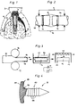
- FIG. 1 shows a cross section through a jawbone with implant and cover sheet
- FIG. 2 shows a schematic illustration of a defect fracture with cover sheet
- FIG. 3 shows the procedure for producing a sheet
- FIG. 4 shows a nail for fastening the sheet, partly in section .
- a primary part 2 of an implant is drilled into a jawbone 1.
- a permanently deformable, inelastic, tension-free titanium foil 5 with a material thickness of less than 0.1 mm, here for example 0.025 mm, is arranged in a jacket-like manner between the screw cap 4 and the jaw bone 1, which the tissue 6 from Bones 1 keeps and thus prevents the tissue 6 from growing on the bone 1.
- Bone substance builds up in the space between the film 5 and the jaw bone 1. The bone growth takes place very quickly under the film 5, since it is not hindered by the tissue 6.
- the cavity below the film 5 can, as shown on the right in FIG. 1, be filled with hydroxyapatite 7.
- This fulfills a "place keeping function" for the bone formation under the film 5.
- the hydroxylapatite 7 is rapidly grown through by the bone and is completely embedded in the bone substance.
- a tight connection of the film 5 is required both to the primary part 2 of the implant and to the jawbone 1;
- the film 5 is provided with a small central hole and the shaft of the screw cap 4 is pushed through this.
- the screw cap 4 is conical and sits on a cone of the implant.
- the film 5 is clamped between the cap cone and the implant cone and then - with the introduction of the hydroxyapatite 7 onto the jaw bone 1 in the form of a jacket.
- the position fixation on the bone 1 is done by nails 8, which are shown in Fig. 4.
- the titanium foil 5 allows permanent molding on the bones 1.
- the shape of the jacket is achieved by forming folds.
- the titanium foil 5 is used with completely inelastic technological properties, so that there is no springback in the shaping described above or in the adaptation to the implant 2 and the jawbone 1.
- the outer surface of the film 5 is rough so that the fabric 6 finds a firm hold there.
- the required degree of roughness can be achieved, for example, by sandblasting, embossing, chemical surface treatment or laser processing, for example by corrugation.
- the foil 5 made of titanium sheet remains in place and does not need to be removed. X-ray images are hardly shadowed to any significant extent, so that changes in the area of the implant can also be clearly recognized in the x-ray image below the film 5.
- FIG. 2 shows the use of a film 9 in connection with a defect fracture of a bone 10.
- the bone 10 or the fracture is e.g. by plates or nails, which are symbolically represented by brackets with arrows 11, 12, set up.
- the fracture area, which pieces of bone are missing, is encased in a sleeve-like manner by the inelastic, tension-free, permanently deformable film 9 made of titanium sheet and fixed and sealed in the connecting area to the bone with a bandage 13.
- the film 9 keeps the tissue away from the bone 11 in the fracture area, so that the growth of the bone can take place undisturbed.
- a bone matter gain of 1 mm is expected in 10 days.
- FIG. 3 shows a method for producing a foil 14 from titanium sheet 15.
- the titanium sheet 15 is brought to a thickness of approximately 0.025 mm by at least one rolling process (rolling 16, 17).
- the surface areas 18, 19 have a compacted structure, which results in a spring effect of the rolled material.
- a subsequent chemical bath (acid bath) 20 the surface 18, 19 of cut film pieces 21 is removed on both sides.
- the use of hydrofluoric acid is advisable for this.
- the arrangement in the chemical bath (acid bath) achieves the desired film thickness of, for example, 0.015 to 0.020 mm and at the same time the absence of tension or the inelastic, permanently deformable material property. It is not the aim to make the film as thin as possible, but to give the above-mentioned material properties with maximum strength of the film.
- a titanium foil which was brought to a foil thickness of 0.2 mm by rolling, is immersed in a hydrofluoric acid bath and remains there until a foil thickness of 0.1 mm is reached.
- the removal of the surface of 0.05 mm on both sides covers the area which is undesirable due to the rolling process elastic material properties justified.
- the film reduced from 0.2 mm to 0.1 mm in thickness by chemical ablation, is inelastic, tension-free and permanently deformable without springback.
- a rolled foil with a thickness of 0.2 mm is placed in a hydrofluoric acid bath and a standing wave is generated in the ultrasound range with a quartz crystal. Resonance can be determined in the electrical part of the oscillation circuit.
- the standing wave leads to the formation of structures on the surface of the titanium foil, which lead to uniform corrugation as a result of different abrasion in the hydrofluoric acid bath.
- a final film thickness between 0.08 and 0.03 mm was achieved.
- a titanium foil was reduced from 0.08 to a thickness of 0.02 mm by hydrofluoric acid treatment. This film thickness is ideally suited in the dental field, because in particular good permeability to X-rays is guaranteed.
- a quartz crystal 30 is shown in a circuit 31, the adjustable frequency of the film 14 is supplied. The adjustment takes place in such a way that the film 14 follows a standing wave between the hooking or clamping points 32, so that the desired surface structure results in the hydrofluoric acid bath.
- the film 14 is cut into rectangular pieces 21, perforated on one narrow side and hung as a strip in the chemical bath.
- the ultrasound or the high frequency of the quartz crystal can be attached via the suspension 30 are coupled, so that the structuring of the Eolien foundedes 21 results in the acid bath.
- the drying process takes place in a drying chamber 22 at, for example, 100 ° C.
- Thermal conditioning can neutralize residual stresses.
- the surface is sandblasted or corrugated using laser technology and cut to commercial dimensions (for example 3 ⁇ 4 cm), the cutting being carried out in such a way that a ridge 23 is formed all around on a surface. This ridge 23 is opposite to the rough surface 24 and comes into effect when the film 14 is used against the bone as a sealing strip.
- Other treatment methods such as annealing with subsequent cooling, can be carried out with good results, but are not optimal in all respects.
- a nail 8 made of titanium is provided according to FIG. 4.
- the nail 8 has a spherical cap-shaped button 25, the concave underside of which merges with a pronounced curve 26 into a pin 27 which is designed as a multiple barb with a truncated cone surface 28.
- the head 25 has a circular border 29 which, when the nail 8 is driven in, represents a sealing edge to the film 5.
- the concave cavity under the head 25 serves to accommodate the curved transition area between pin 27 and head 25, so that the curved transition area penetrates as little as possible into the bone and furthermore to accommodate displaced foil and bone material as a result of the pierced hole in the foil 5 and the Bone substance around pin 27.
Landscapes
- Health & Medical Sciences (AREA)
- Orthopedic Medicine & Surgery (AREA)
- Life Sciences & Earth Sciences (AREA)
- Veterinary Medicine (AREA)
- Animal Behavior & Ethology (AREA)
- Engineering & Computer Science (AREA)
- Biomedical Technology (AREA)
- Public Health (AREA)
- General Health & Medical Sciences (AREA)
- Transplantation (AREA)
- Surgery (AREA)
- Heart & Thoracic Surgery (AREA)
- Oral & Maxillofacial Surgery (AREA)
- Molecular Biology (AREA)
- Nuclear Medicine, Radiotherapy & Molecular Imaging (AREA)
- Neurology (AREA)
- Dentistry (AREA)
- Medical Informatics (AREA)
- Epidemiology (AREA)
- Developmental Biology & Embryology (AREA)
- Cardiology (AREA)
- Vascular Medicine (AREA)
- Prostheses (AREA)
- Materials For Medical Uses (AREA)
- Dental Prosthetics (AREA)
- Surgical Instruments (AREA)
Abstract
Description
Die Erfindung betrifft eine Folie oder Membran zur Abdekkung von Knochendefektstellen, z.B. von Defektfrakturen, Alveolen od.dgl. und zur geführten Knochenregeneration, wobei die Folie aus unelastisch spannungsfreiem und bleibend verformbarem Titan besteht, sowie ein Verfahren zur Herstellung der Folie und einen Nagel zur Lagefixierung einer solchen Folie.The invention relates to a film or membrane for covering bone defects, e.g. defect fractures, alveoli or the like. and for guided bone regeneration, the film consisting of inelastic, tension-free and permanently deformable titanium, as well as a process for producing the film and a nail for fixing the position of such a film.
Um Knochensubstanz bei Defekten wieder aufzubauen ist es bekannt, die Defektstelle durch eine Folie abzudecken und das Weichgewebe durch die Folie vom Hineinwachsen in die Defektstelle fernzuhalten. Es ist ferner bekannt, den Bereich, der für eine Knochenneubildung vorgesehen ist, durch ein Granulat auszufüllen und das geschaffene Volumen durch die Folie abzudecken. Das Granulat, meist Hydroxylapatit übt "Platzhalterfunktion" für den heranwachsenden knochen aus. Folien bzw. Membrane werden meist aus textilem Material hergestellt, wobei man jene Fasern einsetzt, die sich jahrelang für das Nähen von Wunden bewährt haben. Diese Gewebe haben schützende Wirkung, wenn sie in die gewünschte Position gebracht und dort z.B. durch Vernähen lagefixiert wurden. Die Faserstruktur, die Randbereiche beim Zuschneiden, z.B. während einer Operation und die mechanischen Eigenschaften des textilen Materials, insbesondere die Elastizität, können bei vielen Anwendungsfällen nicht als ideal bezeichnet werden.In order to rebuild bone substance in the case of defects, it is known to cover the defect site with a film and to keep the soft tissue from growing into the defect site through the film. It is also known to fill the area intended for new bone formation with a granulate and to cover the created volume with the film. The granulate, mostly hydroxylapatite, acts as a "placeholder" for the growing bone. Foils and membranes are mostly made of textile material, using the fibers that have proven themselves over many years for sewing wounds. These fabrics have a protective effect when they are brought into the desired position and e.g. were fixed in place by sewing. The fiber structure, the edge areas when cutting, e.g. during an operation and the mechanical properties of the textile material, especially the elasticity, cannot be described as ideal in many applications.
Aus der EP 622 052 Al ist ein Verfahren zum Formen einer Folie zur Förderung des Knochenwachstums im Dentalbereich bekannt. Dabei wird auf digitalem Weg ein Abbild der Defektstelle aufgenommen und ein Modell danach hergestellt, wobei eine steife Folie durch das Modell in die gewünschte Form gebracht wird. Als Folienmaterial wird formstabiles Titan in einer Stärke von 0,3 mm herangezogen. Die DE-PS 43 02 709 C1 betrifft eine Abdeckeinrichtung mit einer Abdeckmembran zum vorübergehenden Abdecken einer mit Knochenaufbaumaterial, wie Hyroxylapatitgranulat, gefüllten Defektstelle. Die Abdeckmembran ist geschmeidig und biegsam und erfordert zur Versteifung eine Abdeckmembran, die als feste, starre Titanfolie einer Stärke von 0,3 bis 1 mm mit Perforationen ausgebildet sein kann. Auch für Implantate, Nägel, Platten oder Schienen wird in der Chirurgie das Metall Titan verwendet. Die Verträglichkeit ist ausgezeichnet und Röntgenbilder werden insbesondere bei geringen Wandstärken durch Titan nicht wesentlich abgeschattet.EP 622 052 A1 discloses a method for forming a film to promote bone growth in the dental field. An image of the defect is recorded digitally and a model is then produced, whereby a rigid film is brought into the desired shape by the model. Dimensionally stable titanium with a thickness of 0.3 mm is used as the foil material. DE-PS 43 02 709 C1 relates to a covering device with a covering membrane for temporarily covering a defect site filled with bone building material, such as hyroxylapatite granulate. The covering membrane is supple and flexible and requires a covering membrane for stiffening, which can be designed as a solid, rigid titanium foil with a thickness of 0.3 to 1 mm with perforations. Titanium is also used in surgery for implants, nails, plates or splints. The tolerance is excellent and X-ray images are not significantly shadowed by titanium, particularly with thin walls.
Die Erfindung zielt darauf ab, eine Folie oder Membran zur Abdeckung von Knochendefektstellen zu verbessern, insbesondere praxisgerecht auszubilden, sodaß auch die Anwendung in kleinen Bereichen, etwa in der Kieferchirurgie möglich ist. Dies wird dadurch erreicht, daß die Folie eine Dicke von weniger als 0,1 mm, insbesondere weniger als 0,025 mm aufweist und mindestens eine der Oberflächen dcaufgerauht bzw. geriffelt ausgebildet ist. Die Titanfolie ist für die genannten Zwecke dann noch fest genug und hat keine Federwirkung, die ein Positonieren am Knochen erschwert. Die aufgerauhte oder geriffelte Oberfläche der Titanfolie stellt bei der chirurgischen Anwendung die Außenseite, also die dem Knochen abgewandte und dem Gewebe zugewandte Seite dar. Das Gewebe findet auf der Folie einen guten Halt. Ein Einwachsen der Folie wird dadurch etwa im Zahnfleischbereich begünstigt. Die Aufrauhung kann durch Sandstrahlen oder durch Prägen und die Riffelung durch Elektroerosion bzw. durch Laserbearbeitung sowie durch chemische Oberflächenbehandlung erfolgen. Eine besondere Ausführungsform der Folie ist dadurch gekennzeichnet, daß sie in Stücke von beispielsweise 3 x 4 cm zugeschnitten ist und daß ein Schnittgrat ausgebildet ist, der allseitig eine der Oberflächen überragt. Der Grat ist nach einer Richtung ausgerichtet und bildet eine ausgeprägte Dichtkante am Knochen, die ein Einwachsen des Weichgewebes verhindert. Eine Bandage, z.B. aus Nahtmaterial, wird im Anlagebereich der Folie am Knochen so angebracht, daß ein dichter Anschluß gewährleistet ist.The invention aims to improve a film or membrane for covering bone defect sites, in particular to design it in a practice-oriented manner, so that it can also be used in small areas, for example in jaw surgery. This is achieved in that the film has a thickness of less than 0.1 mm, in particular less than 0.025 mm, and at least one of the surfaces is roughened or corrugated. The titanium foil is then still strong enough for the purposes mentioned and has no spring action, which makes positioning on the bone difficult. The roughened or corrugated surface of the titanium foil represents the outside, ie the side facing away from the bone and towards the tissue, during surgical use. The tissue has a good hold on the foil. Ingrowth of the film is promoted in the gum area. The roughening can be done by sandblasting or embossing and the corrugation by electroerosion or by laser processing as well as by chemical surface treatment. A special embodiment of the film is characterized in that it is cut into pieces of, for example, 3 × 4 cm and that a cut burr is formed which projects beyond one of the surfaces on all sides. The ridge is aligned in one direction and forms a pronounced sealing edge on the bone, which prevents the soft tissue from growing in. A bandage, e.g. made of suture material, is attached to the bone in the contact area of the film so that a tight connection is guaranteed.
Eine Titanfolie wird durch Walzen in den Bereich der gewünschten Materialdicke gebracht. Der Walzvorgang bringt es mit sich, daß die Oberflächenstruktur verdichtet ist und sich daher eine Federwirkung bei einer Anpassung an den Knochen einstellt. Diese ist natürlich unerwünscht, denn es soll das Material spannungsfrei und bleibend verformbar in der gewünschten Position verharren. Um eine Titanfolie spannungsfrei und bleibend verformbar auszubilden, kann ein thermisches Behandlungsverfahren angewendet werden. Es ist jedoch im konkreten Fall bei den gewünschten Wanddicken vorteilhaft, wenn nach dem Walzen eine chemische Behandlung, vorzugsweise mit Flußsäure, zum Abtragen der insbesondere beim Walzen vorgespannten Oberfläche und zur Reduzierung der Foliendicke erfolgt. Durch die chemische Behandlung werden die Randschichten der Titanfolie abgetragen, also jene Schichten, die die Federwirkung begründen. Nach Erreichen der gewünschten Foliendicke von etwa 0,1 mm oder darunter erfolgt ein Waschvorgang sowie das Trocknen, z.B. im Trockenofen, bei 100°C. Eventuelle Restspannungen werden dadurch weitgehend neutralisiert.A titanium foil is brought into the range of the desired material thickness by rolling. The rolling process entails that the surface structure is compacted and therefore a spring action occurs when the bone is adapted. This is of course undesirable because the material should remain in the desired position free of tension and permanently deformable. A thermal treatment process can be used to make a titanium foil stress-free and permanently deformable. However, it is In the specific case with the desired wall thicknesses advantageous if a chemical treatment, preferably with hydrofluoric acid, is carried out after the rolling in order to remove the surface, in particular that which is prestressed during rolling, and to reduce the film thickness. The edge layers of the titanium foil are removed by the chemical treatment, i.e. those layers that justify the spring action. After reaching the desired film thickness of about 0.1 mm or less, a washing process and drying, for example in a drying oven, are carried out at 100 ° C. Any residual stresses are largely neutralized.
Die spannungsfreie Titanfolie kann leicht und irreversibel in die gewünschte Form gebracht werden. Zur Lagefixierung dieser Folien für chirurgische Zwecke werden Titannägel verwendet, die dadurch gekennzeichnet sind, daß der Kopf kugelkalottenartig gewölbt ist und als äußere, dem Stift zugewandte Berandung, eine kreisförmige Anlagekante bzw. Kreisringfläche aufweist, daß der Kopf im Inneren des Kalottenhohlraumes mit einer Rundung in den Stift übergeht und daß der Stift mit einer sägezahnartigen Querschnittskontur von aneinanderschließenden Kegelstumpfflächen ausgebildet ist. Der Nagel legt sich mit der Außenberandung des Nagelkopfes dichtend an eine Titanfolie an. Durch das Einschlagen bzw. Eindrücken des Nagels wird ein Loch in der Folie geschaffen. Der Nagelstift, der das Loch durchdringt, verdrängt das Folienmaterial, welches sich in den Hohlraum der kugelkalottenartigen Wölbung des Nagelkopfes legt. Zur besonders guten Anpassung ist der Übergang vom Nagelkopf zum Stift gerundet. Der Stift selbst ist durch die aneinanderschließenden Kegelstumpfflächen mit mehrfachen Widerhaken ausgebildet.The tension-free titanium foil can be easily and irreversibly brought into the desired shape. To fix these foils for surgical purposes, titanium nails are used, which are characterized in that the head is curved in the manner of a spherical cap and, as the outer edge facing the pin, has a circular contact edge or annular surface such that the head has a rounded shape in the interior of the spherical cavity passes over the pin and that the pin is formed with a sawtooth-like cross-sectional contour of adjoining truncated cone surfaces. With the outer edge of the nail head, the nail seals against a titanium foil. A hole in the foil is created by driving in or pushing in the nail. The nail pen, which penetrates the hole, displaces the foil material, which lies in the cavity of the spherical cap-like curvature of the nail head. For a particularly good adjustment, the transition from the nail head to the pin is rounded. The pin itself is formed by the adjacent truncated cone surfaces with multiple barbs.
Ausführungsbeispiele des Erfindungsgegenstandes sind in den Zeichnungen dargestellt. Fig. 1 zeigt einen Querschnitt durch einen Kieferknochen mit Implantat und Abdeckfolie, Fig. 2 eine schematische Darstellung einer Defektfraktur mit Abdeckfolie, Fig. 3 den Ablauf des Verfahrens zur Herstellung einer Folie und Fig. 4 einen Nagel zur Befestigung der Folie, teilweise im Schnitt.Embodiments of the subject matter of the invention are shown in the drawings. 1 shows a cross section through a jawbone with implant and cover sheet, FIG. 2 shows a schematic illustration of a defect fracture with cover sheet, FIG. 3 shows the procedure for producing a sheet and FIG. 4 shows a nail for fastening the sheet, partly in section .
Gemäß Fig. 1 ist in einem Kieferknochen 1 ein Primärteil 2 eines Implantates eingebohrt. Die obere Öffnung, in welche nach dem Einwachsen des Primärteiles ein Sekundärteil mit einem Zahn 3 eingesetzt wird, ist durch eine Schraubkappe 4 verschlossen. Um Knochensubstanz längs des Primärteiles 2 hochzuziehen, ist eine bleibend verformbare, unelastische, spannungsfreie Titanfolie 5 einer Materialdicke von weniger als 0,1 mm, hier beispielsweise von 0,025 mm, zwischen der Schraubkappe 4 und dem Kieferknochen 1 mantelförmig angeordnet, die das Gewebe 6 vom Knochen 1 abhält und so ein Heranwachsen des Gewebes 6 an den Knochen 1 verhindert. Knochensubstanz baut sich in dem Raum zwischen der Folie 5 und dem Kieferknochen 1 auf. Das Knochenwachstum erfolgt unter der Folie 5 sehr rasch, da es durch das Gewebe 6 nicht behindert wird. Der Hohlraum unterhalb der Folie 5 kann, wie rechts in Fig. 1 dargestellt, durch Hydroxylapatit 7 ausgefüllt werden. Dieses erfüllt eine "Platzhaltefunktion" für die Knochenbildung unter der Folie 5. Das Hydroxylapatit 7 wird vom Knochen rasch durchwachsen und lagert sich vollständig in die Knochensubstanz ein.1, a primary part 2 of an implant is drilled into a
Ein dichter Anschluß der Folie 5 ist sowohl an den Primärteil 2 des Implantates als auch an den Kieferknochen 1 erforderlich; dazu wird die Folie 5 mit einem kleinen zentralen Loch versehen und durch dieses der Schaft der Schraubkappe 4 durchgesteckt. Die Schraubkappe 4 ist konisch ausgebildet und sitzt auf einem Konus des Implantates. Die Folie 5 wird zwischen dem Kappenkonus und dem Implantatkonus eingeklemmt und sodann - unter Einbringung des Hydroxylapatits 7 an den Kieferknochen 1 mantelförmig angelegt. Die Lagefixierung am Knochen 1 erfolgt durch Nägel 8, die in Fig. 4 dargestellt sind. Die Titanfolie 5 erlaubt ein bleibendes Anformen an den Knochen 1. Durch Faltenbildung wird die Mantelform erreicht. Die Titanfolie 5 wird mit vollkommen unelastischen technologischen Eigenschaften eingesetzt, sodaß sich kein Rückfedern bei der oben beschriebenen Formgebung bzw. bei der Anpassung an das Implantat 2 und an den Kieferknochen 1 ergibt.A tight connection of the
Die Außenfläche der Folie 5 ist rauh, damit das Gewebe 6 dort einen festen Halt findet. Der nötige Rauheitsgrad kann z.B. durch Sandstrahlen, Prägen, durch eine chemische Oberflächenbehandlung oder durch eine Laserbearbeitung, etwa durch eine Riffelung, erreicht werden. Die Folie 5 aus Titanblech verbleibt in ihrer Position und muß nicht entfernt werden. Röntgenbilder werden kaum nennenswert abgeschattet, sodaß man Veränderungen im Bereich des Implantates auch unterhalb der Folie 5 im Röntgenbild gut erkennen kann.The outer surface of the
In Fig. 2 ist der Einsatz einer Folie 9 im Zusammenhang mit einer Defektfraktur eines Knochens 10 dargestellt. Der Knochen 10 bzw. die Fraktur wird z.B. durch Platten oder Nägel, die symbolhaft durch Klammern mit Pfeilen 11, 12 dargestellt sind, eingerichtet. Der Frakturbereich, dem Knochenstücke fehlen, wird durch die unelastische, spannungsfreie, bleibend verformbare Folie 9 aus Titanblech manschettenartig ummantelt und im Anschlußbereich an den Knochen mit einer Bandage 13 fixiert und abgedichtet. Die Folie 9 hält das Gewebe vom Knochen 11 im Frakturbereich fern, sodaß das Wachstum des Knochens ungestört erfolgen kann. Man rechnet mit einem Knochensubstanzzuwachs von 1 mm in 10 Tagen.2 shows the use of a
Fig. 3 zeigt ein Verfahren zur Herstellung einer Folie 14 aus Titanblech 15. Durch mindestens einen Walzvorgang (Walzen 16, 17) wird das Titanblech 15 auf eine Dicke von etwa 0,025 mm gebracht. Die Oberflächenbereiche 18, 19 weisen eine verdichtete Struktur auf, wodurch sich eine Federwirkung des gewalzten Materials ergibt. In einem anschließenden chemischen Bad (Säurebad) 20 wird die Oberfläche 18, 19 von zugeschnittenen Folienstücken 21 beiderseits abgetragen. Dazu ist die Anwendung von Flußsäure zweckmäßig. Durch die Anordnung im chemischen Bad (Säurebad) erreicht man die gewünschte Foliendicke von beispielsweise 0,015 bis 0,020 mm und zugleich die Spannungsfreiheit bzw. die unelastische, bleibend verformbare Materialeigenschaft. Es ist nicht das Ziel, die Folie möglichst dünn auszubilden, sondern bei maximaler Festigkeit der Folie die vorgenannten Materialeigenschaften zu verleihen.3 shows a method for producing a
Eine Titanfolie, die durch Walzen auf eine Foliendicke von 0,2 mm gebracht wurde, wird in ein Flußsäurebad getaucht und verharrt dort, bis eine Foliendicke von 0,1 mm erreicht ist. Die beiderseitige Abtragung der Oberfläche von 0,05 mm erfaßt den Bereich, der infolge des Walzvorganges die hier unerwünschten elastischen Materialeigenschaften begründet. Die von 0,2 mm auf 0,1 mm Dicke durch chemische Abtragung reduzierte Folie ist unelastisch, spannungsfrei und ohne Rückfedern bleibend verformbar.A titanium foil, which was brought to a foil thickness of 0.2 mm by rolling, is immersed in a hydrofluoric acid bath and remains there until a foil thickness of 0.1 mm is reached. The removal of the surface of 0.05 mm on both sides covers the area which is undesirable due to the rolling process elastic material properties justified. The film, reduced from 0.2 mm to 0.1 mm in thickness by chemical ablation, is inelastic, tension-free and permanently deformable without springback.
Durch Verlängerung der Verweildauer im Flußsäurebad wurde eine Folie von ursprünglich 0,203 mm auf 0,08 mm Dicke gebraucht. Damit wurde gleichzeitig eine Rauhtiefe erreicht, die bei 0,003 bis 0,007 mm liegt und für das Anwachsen von Gewebe hervorragend geeignet ist.By extending the residence time in the hydrofluoric acid bath, a film from originally 0.203 mm to 0.08 mm thick was used. A roughness depth of 0.003 to 0.007 mm was achieved at the same time and is excellently suited for the growth of tissue.
Eine gewalzte Folie mit einer Dicke von 0,2 mm wird in ein Flußsäurebad gebracht und dort mit einem Schwingquarz eine stehende Welle im Ultraschallbereich erzeugt. Resonanz ist im elektrischen Teil der Schwingschaltung feststellbar. Die stehende Welle führt dazu, daß sich Strukturen an der Oberfläche der Titanfolie bilden, die zu einer gleichmäßigen Riffelung infolge unterschiedlicher Abtragung im Flußsäurebad führen. Eine endgültige Foliendicke zwischen 0,08 und 0,03 mm wurde erreicht.A rolled foil with a thickness of 0.2 mm is placed in a hydrofluoric acid bath and a standing wave is generated in the ultrasound range with a quartz crystal. Resonance can be determined in the electrical part of the oscillation circuit. The standing wave leads to the formation of structures on the surface of the titanium foil, which lead to uniform corrugation as a result of different abrasion in the hydrofluoric acid bath. A final film thickness between 0.08 and 0.03 mm was achieved.
Durch Flußsäurebehandlung wurde eine Titanfolie von 0,08 auf eine Dicke von 0,02 mm reduziert. Diese Foliendicke eignet sich im Dentalbereich hervorragend, weil insbesondere gute Durchlässigkeit für Röntgenstrahlen gewährleistet ist.A titanium foil was reduced from 0.08 to a thickness of 0.02 mm by hydrofluoric acid treatment. This film thickness is ideally suited in the dental field, because in particular good permeability to X-rays is guaranteed.
In Fig. 3 ist ein Schwingquarz 30 in einem Stromkreis 31 dargestellt, dessen einstellbare Frequenz der Folie 14 zugeführt wird. Die Abstimmung erfolgt so, daß die Folie 14 zwischen den Einhänge- bzw. Einspannstellen 32 einer stehenden Welle folgt, sodaß sich im Flußsäurebad die gewünschte Oberflächenstruktur ergibt.In Fig. 3, a
Die Folie 14 wird in der Praxis zu Folienstücken 21 rechteckig zugeschnitten, an einer Schmalseite gelocht und als Streifen in das chemische Bad gehängt. Über die Aufhängung kann der Ultraschall bzw. die Hochfrequenz des Schwingquarzes 30 angekoppelt werden, sodaß sich im Säurebad die Strukturierung des Eolienstückes 21 ergibt. Nach dem Auswaschen der Säure vom Titanstreifen erfolgt der Trockenvorgang in einer Trockenkammer 22 bei beispielsweise 100°C. Die thermische Konditionierung kann letzte Reste von Spannungen neutralisieren. Vor der Wärmebehandlung erfolgt ein Sandstrahlen der Oberfläche oder ein Riffeln mittels Lasertechnik und ein Zuschneiden auf handelsübliche Maße (z.B. 3 x 4 cm), wobei das Schneiden so erfolgt, daß sich ein Grat 23 ringsum an einer Oberfläche bildet. Dieser Grat 23 liegt der rauhen Oberfläche 24 gegenüber und kommt bei der Anwendung der Folie 14 gegen den Knochen als Dichtleiste zur Wirkung. Andere Behandlungsverfahren wie etwa das Ausglühen mit anschließendem Erkalten sind mit guten Ergebnissen ausführbar, jedoch nicht in allen Punkten optimal.In practice, the
Zur Lagefixierung einer zur Abdeckung von Knochendefektstellen, z.B. von Defektfrakturen, Alveolen od.dgl. vorgesehene Folie, die dem forcierten, gezielten Knochenwachstum dient, ist ein Nagel 8 aus Titan gemäß Fig. 4 vorgesehen. Der Nagel 8 hat einen kugelkalottenförmigen Knopf 25, dessen konkave Unterseite mit einer ausgeprägten Rundung 26 in einen Stift 27 übergeht, der mit Kegelstumpfflächen 28 sägezahnartig als mehrfacher Widerhaken ausgebildet ist. Der Kopf 25 weist eine kreisförmige Berandung 29 auf, die bei eingeschlagenem Nagel 8 eine Dichtkante zur Folie 5 darstellt. Der konkave Hohlraum unter dem Kopf 25 dient zur Aufnahme des gekrümmten Übergangsbereiches zwischen Stift 27 und Kopf 25, sodaß der gekrümmte Übergangsbereich möglichst wenig in den Knochen eindringt und ferner zur Aufnahme von verdrängtem Folien- und Knochenmaterial infolge des gestochenen Loches in der Folie 5 und der Knochensubstanz rings um den Stift 27.To fix a position to cover bone defects, e.g. defect fractures, alveoli or the like. provided film, which is used for forced, targeted bone growth, a
Claims (4)
Applications Claiming Priority (3)
| Application Number | Priority Date | Filing Date | Title |
|---|---|---|---|
| AT933/96 | 1996-05-29 | ||
| AT93396 | 1996-05-29 | ||
| AT0093396A AT403002B (en) | 1996-05-29 | 1996-05-29 | FILM OR MEMBRANE FOR COVERING BONE DEFECTS, METHOD FOR PRODUCING THE FILM AND NAIL FOR FIXING THE POSITION OF SUCH A FILM |
Publications (3)
| Publication Number | Publication Date |
|---|---|
| EP0809979A1 true EP0809979A1 (en) | 1997-12-03 |
| EP0809979B1 EP0809979B1 (en) | 2002-07-31 |
| EP0809979B2 EP0809979B2 (en) | 2008-04-09 |
Family
ID=3503191
Family Applications (1)
| Application Number | Title | Priority Date | Filing Date |
|---|---|---|---|
| EP97890084A Expired - Lifetime EP0809979B2 (en) | 1996-05-29 | 1997-05-06 | Sheet for covering bone defect sites and process for manufacturing the sheet |
Country Status (10)
| Country | Link |
|---|---|
| US (1) | US5976140A (en) |
| EP (1) | EP0809979B2 (en) |
| JP (1) | JPH1052441A (en) |
| CN (1) | CN1136817C (en) |
| AT (1) | AT403002B (en) |
| AU (1) | AU2348497A (en) |
| BR (1) | BR9703411A (en) |
| DE (1) | DE59707835D1 (en) |
| ES (1) | ES2180916T3 (en) |
| ID (1) | ID17709A (en) |
Cited By (5)
| Publication number | Priority date | Publication date | Assignee | Title |
|---|---|---|---|---|
| EP0867193A3 (en) * | 1997-03-27 | 1999-04-28 | Friatec Aktiengesellschaft | Foil for medical use |
| EP1745759A3 (en) * | 2000-05-29 | 2007-05-09 | CelGen AG | Devices and medical membranes for bone regeneration |
| WO2011100951A1 (en) * | 2010-02-19 | 2011-08-25 | Marcus Seiler | Device for covering and/or reconstructing a bone defect site, and method for production thereof |
| WO2017089381A1 (en) | 2015-11-26 | 2017-06-01 | Syntellix Ag | Bioresorable fixation nail |
| US10390956B2 (en) | 2015-05-08 | 2019-08-27 | Reoss Gmbh | Device for covering and/or reconstructing a bone defect site, method for producing a cap for a cover for a bone defect site |
Families Citing this family (34)
| Publication number | Priority date | Publication date | Assignee | Title |
|---|---|---|---|---|
| US5839899A (en) * | 1996-03-01 | 1998-11-24 | Robinson; Dane Q. | Method and apparatus for growing jaw bone utilizing a guided-tissue regeneration plate support and fixation system |
| NL1021137C2 (en) * | 2002-07-23 | 2004-01-27 | Fondel Finance B V | Support element for attachment to bone. |
| US7476228B2 (en) * | 2002-10-11 | 2009-01-13 | Abdou M Samy | Distraction screw for skeletal surgery and method of use |
| WO2004062482A2 (en) * | 2003-01-10 | 2004-07-29 | Abdou Samy M | Plating system for bone fixation and subsidence and method of implantation |
| ITBS20030028A1 (en) * | 2003-03-13 | 2004-09-14 | Physioplant S R L | MICRO NAIL AND METHOD FOR ITS USE IN THE FIXING OF A PROTECTIVE MEMBRANE FOR DENTAL USE. |
| JP5089169B2 (en) * | 2003-06-13 | 2012-12-05 | ザ・ユニバ−シティ・オブ・コネチカット | Structural / biological implant systems |
| US8979846B2 (en) * | 2004-04-12 | 2015-03-17 | Navin N Thakkar | Flexible nail assembly for fractures of long bones |
| WO2006058221A2 (en) | 2004-11-24 | 2006-06-01 | Abdou Samy M | Devices and methods for inter-vertebral orthopedic device placement |
| US20060154204A1 (en) * | 2005-01-12 | 2006-07-13 | Reggie John A | Dental implants with improved loading properties |
| US7862588B2 (en) | 2005-02-18 | 2011-01-04 | Samy Abdou | Devices and methods for dynamic fixation of skeletal structure |
| US20070162017A1 (en) * | 2005-11-28 | 2007-07-12 | Gambale Michael A | Method and apparatus for treatment of bones |
| US8876874B2 (en) | 2006-08-21 | 2014-11-04 | M. Samy Abdou | Bone screw systems and methods of use |
| US9655699B2 (en) * | 2009-07-23 | 2017-05-23 | Lieh-Tang Chen | Artificial teethridge and fang |
| US8764806B2 (en) | 2009-12-07 | 2014-07-01 | Samy Abdou | Devices and methods for minimally invasive spinal stabilization and instrumentation |
| FR2962641B1 (en) | 2010-07-13 | 2013-08-02 | Univ Paris Diderot Paris 7 | IMPLANT DEVICE AND INSTALLATION TOOL |
| TWI475985B (en) * | 2011-02-14 | 2015-03-11 | Marcus Seiler | Device for covering and/or reconstructing a bone defect site; method of manufacturing an attachment for a covering device for a bone defect site |
| US8845728B1 (en) | 2011-09-23 | 2014-09-30 | Samy Abdou | Spinal fixation devices and methods of use |
| WO2013095077A1 (en) | 2011-12-23 | 2013-06-27 | 오스템임플란트 주식회사 | Dental membrane |
| US20130226240A1 (en) | 2012-02-22 | 2013-08-29 | Samy Abdou | Spinous process fixation devices and methods of use |
| US9198767B2 (en) | 2012-08-28 | 2015-12-01 | Samy Abdou | Devices and methods for spinal stabilization and instrumentation |
| US9320617B2 (en) | 2012-10-22 | 2016-04-26 | Cogent Spine, LLC | Devices and methods for spinal stabilization and instrumentation |
| ES2610823B1 (en) * | 2015-04-23 | 2018-03-01 | Mauricio Alberto LIZARAZO ROZO | Method of manufacturing an occlusive barrier for bone regeneration and the occlusive barrier obtained by said method |
| US20160331539A1 (en) * | 2015-05-12 | 2016-11-17 | Elwha Llc | Modifiable implants |
| US10857003B1 (en) | 2015-10-14 | 2020-12-08 | Samy Abdou | Devices and methods for vertebral stabilization |
| CN208989194U (en) * | 2015-12-08 | 2019-06-18 | 费莫斯股份有限公司 | Film nail and its installation tool case |
| US10973648B1 (en) | 2016-10-25 | 2021-04-13 | Samy Abdou | Devices and methods for vertebral bone realignment |
| US10744000B1 (en) | 2016-10-25 | 2020-08-18 | Samy Abdou | Devices and methods for vertebral bone realignment |
| USD816843S1 (en) * | 2017-06-07 | 2018-05-01 | Alevio, Llc | Orthopedic implant |
| US11179248B2 (en) | 2018-10-02 | 2021-11-23 | Samy Abdou | Devices and methods for spinal implantation |
| CN112076002B (en) * | 2020-10-13 | 2021-06-15 | 四川大学 | GBR middle periosteum nail position and direction positioning guide plate and manufacturing method thereof |
| CN112716643A (en) * | 2020-12-28 | 2021-04-30 | 浙江大学 | Regional function specificity clinical periodontal defect repair module |
| US20240074787A1 (en) * | 2021-01-28 | 2024-03-07 | Ezriel Kornel | Working channel for use in a method and system for percutaneous procedures |
| CN113040889B (en) * | 2021-03-25 | 2022-08-02 | 西安市红会医院 | Bone fracture plate fixing system matched with bone healing process |
| KR20230094139A (en) * | 2021-12-20 | 2023-06-27 | 문천호 | Screw for dental implant |
Citations (10)
| Publication number | Priority date | Publication date | Assignee | Title |
|---|---|---|---|---|
| WO1992010218A1 (en) * | 1990-12-06 | 1992-06-25 | W.L. Gore & Associates, Inc. | Implantable bioabsorbable article |
| WO1994003121A1 (en) * | 1992-08-05 | 1994-02-17 | Guidor Ab | Surgical element and method for selective tissue regeneration |
| DE4302709C1 (en) * | 1993-02-01 | 1994-07-28 | Kirsch Axel | Cover device with cover membrane |
| DE4313192C1 (en) * | 1993-04-22 | 1994-09-15 | Kirsch Axel | Cuff for accelerating healing of bone defects |
| US5348788A (en) * | 1991-01-30 | 1994-09-20 | Interpore Orthopaedics, Inc. | Mesh sheet with microscopic projections and holes |
| EP0621013A2 (en) * | 1993-02-22 | 1994-10-26 | I. SUGHERI S.r.L. | Periodontal tissue regrowth guiding implant |
| EP0622052A1 (en) * | 1993-04-28 | 1994-11-02 | Ceka N.V. | Method for manufacturing a membrane for controlled bone regeneration |
| DE9413963U1 (en) * | 1993-12-28 | 1995-04-20 | Institut Straumann Ag, Waldenburg | For the attachment of a denture to the jaw serving implant |
| DE4414675C1 (en) * | 1994-04-27 | 1995-09-28 | Kirsch Axel | Covering device for bone defects and method for their production |
| DE29513342U1 (en) * | 1995-08-19 | 1995-11-16 | Biovision GmbH Entwicklung, Herstellung und Vertrieb von Biomaterialien, 98693 Ilmenau | Fastening element, in particular for fastening membranes made of bioabsorbable polymers |
Family Cites Families (6)
| Publication number | Priority date | Publication date | Assignee | Title |
|---|---|---|---|---|
| US4636215A (en) * | 1984-01-11 | 1987-01-13 | Rei, Inc. | Combination tray and condylar prosthesis for mandibular reconstruction and the like |
| SU1572608A1 (en) † | 1988-01-22 | 1990-06-23 | Полтавский медицинский стоматологический институт | Endoprosthesis of ridge of nose |
| ATE121609T1 (en) * | 1991-03-11 | 1995-05-15 | Straumann Inst Ag | AID FOR ATTACHING AND HOLDING A COVER TO A JAW BONE. |
| EP0566427B1 (en) † | 1992-04-17 | 2000-03-15 | Kyocera Corporation | A prothesis and a method of making the same |
| US5564926A (en) * | 1992-11-26 | 1996-10-15 | Medevelop Ab | Anchoring element for anchorage in bone tissue |
| FR2713090A1 (en) † | 1993-12-03 | 1995-06-09 | Scortecci Gerard | Bone tissue regeneration promotion device, esp. for dental surgery |
-
1996
- 1996-05-29 AT AT0093396A patent/AT403002B/en not_active IP Right Cessation
-
1997
- 1997-05-06 ES ES97890084T patent/ES2180916T3/en not_active Expired - Lifetime
- 1997-05-06 EP EP97890084A patent/EP0809979B2/en not_active Expired - Lifetime
- 1997-05-06 DE DE59707835T patent/DE59707835D1/en not_active Expired - Fee Related
- 1997-05-20 AU AU23484/97A patent/AU2348497A/en not_active Abandoned
- 1997-05-26 JP JP13460897A patent/JPH1052441A/en active Pending
- 1997-05-27 CN CNB971121370A patent/CN1136817C/en not_active Expired - Fee Related
- 1997-05-28 ID IDP971790A patent/ID17709A/en unknown
- 1997-05-29 US US08/865,240 patent/US5976140A/en not_active Expired - Fee Related
- 1997-05-30 BR BR9703411A patent/BR9703411A/en not_active Application Discontinuation
Patent Citations (10)
| Publication number | Priority date | Publication date | Assignee | Title |
|---|---|---|---|---|
| WO1992010218A1 (en) * | 1990-12-06 | 1992-06-25 | W.L. Gore & Associates, Inc. | Implantable bioabsorbable article |
| US5348788A (en) * | 1991-01-30 | 1994-09-20 | Interpore Orthopaedics, Inc. | Mesh sheet with microscopic projections and holes |
| WO1994003121A1 (en) * | 1992-08-05 | 1994-02-17 | Guidor Ab | Surgical element and method for selective tissue regeneration |
| DE4302709C1 (en) * | 1993-02-01 | 1994-07-28 | Kirsch Axel | Cover device with cover membrane |
| EP0621013A2 (en) * | 1993-02-22 | 1994-10-26 | I. SUGHERI S.r.L. | Periodontal tissue regrowth guiding implant |
| DE4313192C1 (en) * | 1993-04-22 | 1994-09-15 | Kirsch Axel | Cuff for accelerating healing of bone defects |
| EP0622052A1 (en) * | 1993-04-28 | 1994-11-02 | Ceka N.V. | Method for manufacturing a membrane for controlled bone regeneration |
| DE9413963U1 (en) * | 1993-12-28 | 1995-04-20 | Institut Straumann Ag, Waldenburg | For the attachment of a denture to the jaw serving implant |
| DE4414675C1 (en) * | 1994-04-27 | 1995-09-28 | Kirsch Axel | Covering device for bone defects and method for their production |
| DE29513342U1 (en) * | 1995-08-19 | 1995-11-16 | Biovision GmbH Entwicklung, Herstellung und Vertrieb von Biomaterialien, 98693 Ilmenau | Fastening element, in particular for fastening membranes made of bioabsorbable polymers |
Cited By (10)
| Publication number | Priority date | Publication date | Assignee | Title |
|---|---|---|---|---|
| EP0867193A3 (en) * | 1997-03-27 | 1999-04-28 | Friatec Aktiengesellschaft | Foil for medical use |
| EP1745759A3 (en) * | 2000-05-29 | 2007-05-09 | CelGen AG | Devices and medical membranes for bone regeneration |
| WO2011100951A1 (en) * | 2010-02-19 | 2011-08-25 | Marcus Seiler | Device for covering and/or reconstructing a bone defect site, and method for production thereof |
| CN102821794A (en) * | 2010-02-19 | 2012-12-12 | 瑞欧斯股份有限公司 | Device for covering and/or reconstructing a bone defect site, and method for production thereof |
| CN102821794B (en) * | 2010-02-19 | 2014-12-10 | 瑞欧斯股份有限公司 | Device for covering and/or reconstructing a bone defect site, and method for production thereof |
| US9017406B2 (en) | 2010-02-19 | 2015-04-28 | Reoss Gmbh | Device for covering and/or reconstructing a bone defect site, and method for production thereof |
| EA028390B1 (en) * | 2010-02-19 | 2017-11-30 | Реосс Гмбх | Device for covering and/or reconstructing a bone defect site, and method for production thereof |
| US10390956B2 (en) | 2015-05-08 | 2019-08-27 | Reoss Gmbh | Device for covering and/or reconstructing a bone defect site, method for producing a cap for a cover for a bone defect site |
| WO2017089381A1 (en) | 2015-11-26 | 2017-06-01 | Syntellix Ag | Bioresorable fixation nail |
| DE102015120514A1 (en) | 2015-11-26 | 2017-06-01 | Syntellix Ag | Bioresorbable fixation nail |
Also Published As
| Publication number | Publication date |
|---|---|
| ID17709A (en) | 1998-01-22 |
| ATA93396A (en) | 1997-03-15 |
| AU2348497A (en) | 1997-12-04 |
| AT403002B (en) | 1997-10-27 |
| EP0809979B1 (en) | 2002-07-31 |
| DE59707835D1 (en) | 2002-09-05 |
| CN1136817C (en) | 2004-02-04 |
| JPH1052441A (en) | 1998-02-24 |
| US5976140A (en) | 1999-11-02 |
| EP0809979B2 (en) | 2008-04-09 |
| ES2180916T3 (en) | 2003-02-16 |
| CN1181228A (en) | 1998-05-13 |
| BR9703411A (en) | 1998-09-15 |
Similar Documents
| Publication | Publication Date | Title |
|---|---|---|
| AT403002B (en) | FILM OR MEMBRANE FOR COVERING BONE DEFECTS, METHOD FOR PRODUCING THE FILM AND NAIL FOR FIXING THE POSITION OF SUCH A FILM | |
| DE69121587T3 (en) | IMPLANTABLE BIORESORBABLE ITEMS | |
| DE68922319T2 (en) | Bioabsorbable surgical material for the treatment of nerve defects and method for its preparation. | |
| DE69018457T2 (en) | OBJECTS FOR TREATING PERIODONTITIS AND BONE DEFECTS. | |
| DE69206693T2 (en) | Resorbable tendon and bone reinforcement device | |
| DE60111029T2 (en) | RESORBABLE MICROMEMBRANES FOR THE REDUCTION OF SCARS | |
| DE69923167T2 (en) | Membrane with corrugated surface for guiding the fabric | |
| DE69026100T2 (en) | Biodegradable and resorbable molded body for surgical use | |
| DE69626996T2 (en) | DEGRADABLE MATERIAL AND METHOD FOR THE PRODUCTION THEREOF | |
| DE60019153T2 (en) | DEVICE FOR PROTECTING NERVES AFTER SURGICAL TREATMENT | |
| EP0609667B1 (en) | Covering membrane | |
| DE2628284C2 (en) | Endoprosthesis for use in a bone shaft | |
| EP0758872A1 (en) | Covering system for defective points in bones and process for producing it | |
| DE102013004574A1 (en) | Surgical implant | |
| DE3875096T2 (en) | ABSORBENT MATERIAL FOR FIXING FABRICS. | |
| DE102004047974A1 (en) | Surgical hook | |
| WO2012076161A1 (en) | Improved distraction membrane | |
| DE9115341U1 (en) | Support grid for receiving particulate bone replacement materials | |
| DE602005003830T2 (en) | DEVICE FOR FIXING BONE LABELS AFTER A CRANIOTOMY | |
| AT515384B1 (en) | Preconnected multilayer film for covering a bone defect site | |
| WO2001072244A1 (en) | Implant for cranioplastics | |
| EP0867193B1 (en) | Foil for medical use | |
| DE4118201C2 (en) | Device for joining bones | |
| WO2016019404A1 (en) | Method for producing a multilayer film | |
| WO1996041595A1 (en) | Process and device for the culture of bone replacement material |
Legal Events
| Date | Code | Title | Description |
|---|---|---|---|
| PUAI | Public reference made under article 153(3) epc to a published international application that has entered the european phase |
Free format text: ORIGINAL CODE: 0009012 |
|
| AK | Designated contracting states |
Kind code of ref document: A1 Designated state(s): CH DE ES FR GB IT LI NL SE |
|
| 17P | Request for examination filed |
Effective date: 19971125 |
|
| 17Q | First examination report despatched |
Effective date: 20000918 |
|
| RTI1 | Title (correction) |
Free format text: SHEET FOR COVERING BONE DEFECT SITES AND PROCESS FOR MANUFACTURING THE SHEET |
|
| GRAG | Despatch of communication of intention to grant |
Free format text: ORIGINAL CODE: EPIDOS AGRA |
|
| RTI1 | Title (correction) |
Free format text: SHEET FOR COVERING BONE DEFECT SITES AND PROCESS FOR MANUFACTURING THE SHEET |
|
| GRAG | Despatch of communication of intention to grant |
Free format text: ORIGINAL CODE: EPIDOS AGRA |
|
| GRAH | Despatch of communication of intention to grant a patent |
Free format text: ORIGINAL CODE: EPIDOS IGRA |
|
| GRAH | Despatch of communication of intention to grant a patent |
Free format text: ORIGINAL CODE: EPIDOS IGRA |
|
| GRAA | (expected) grant |
Free format text: ORIGINAL CODE: 0009210 |
|
| AK | Designated contracting states |
Kind code of ref document: B1 Designated state(s): CH DE ES FR GB IT LI NL SE |
|
| REG | Reference to a national code |
Ref country code: GB Ref legal event code: FG4D Free format text: NOT ENGLISH Ref country code: CH Ref legal event code: EP |
|
| REF | Corresponds to: |
Ref document number: 59707835 Country of ref document: DE Date of ref document: 20020905 |
|
| REG | Reference to a national code |
Ref country code: CH Ref legal event code: NV Representative=s name: PATENTANWAELTE FELDMANN & PARTNER AG |
|
| GBT | Gb: translation of ep patent filed (gb section 77(6)(a)/1977) |
Effective date: 20021119 |
|
| REG | Reference to a national code |
Ref country code: ES Ref legal event code: FG2A Ref document number: 2180916 Country of ref document: ES Kind code of ref document: T3 |
|
| ET | Fr: translation filed | ||
| PLBQ | Unpublished change to opponent data |
Free format text: ORIGINAL CODE: EPIDOS OPPO |
|
| PLBI | Opposition filed |
Free format text: ORIGINAL CODE: 0009260 |
|
| 26 | Opposition filed |
Opponent name: FRIADENT GMBH Effective date: 20030429 |
|
| PLAX | Notice of opposition and request to file observation + time limit sent |
Free format text: ORIGINAL CODE: EPIDOSNOBS2 |
|
| NLR1 | Nl: opposition has been filed with the epo |
Opponent name: FRIADENT GMBH |
|
| PLBB | Reply of patent proprietor to notice(s) of opposition received |
Free format text: ORIGINAL CODE: EPIDOSNOBS3 |
|
| PLCK | Communication despatched that opposition was rejected |
Free format text: ORIGINAL CODE: EPIDOSNREJ1 |
|
| RTI2 | Title (correction) |
Free format text: SHEET FOR COVERING BONE DEFECT SITES AND PROCESS FOR MANUFACTURING THE SHEET |
|
| APBP | Date of receipt of notice of appeal recorded |
Free format text: ORIGINAL CODE: EPIDOSNNOA2O |
|
| APBQ | Date of receipt of statement of grounds of appeal recorded |
Free format text: ORIGINAL CODE: EPIDOSNNOA3O |
|
| APAA | Appeal reference recorded |
Free format text: ORIGINAL CODE: EPIDOS REFN |
|
| APAH | Appeal reference modified |
Free format text: ORIGINAL CODE: EPIDOSCREFNO |
|
| APBU | Appeal procedure closed |
Free format text: ORIGINAL CODE: EPIDOSNNOA9O |
|
| PGFP | Annual fee paid to national office [announced via postgrant information from national office to epo] |
Ref country code: FR Payment date: 20060428 Year of fee payment: 10 |
|
| PGFP | Annual fee paid to national office [announced via postgrant information from national office to epo] |
Ref country code: SE Payment date: 20060512 Year of fee payment: 10 |
|
| PGFP | Annual fee paid to national office [announced via postgrant information from national office to epo] |
Ref country code: GB Payment date: 20060515 Year of fee payment: 10 |
|
| PGFP | Annual fee paid to national office [announced via postgrant information from national office to epo] |
Ref country code: ES Payment date: 20060516 Year of fee payment: 10 |
|
| PGFP | Annual fee paid to national office [announced via postgrant information from national office to epo] |
Ref country code: NL Payment date: 20060530 Year of fee payment: 10 |
|
| PGFP | Annual fee paid to national office [announced via postgrant information from national office to epo] |
Ref country code: IT Payment date: 20060531 Year of fee payment: 10 |
|
| PGFP | Annual fee paid to national office [announced via postgrant information from national office to epo] |
Ref country code: DE Payment date: 20060727 Year of fee payment: 10 |
|
| PGFP | Annual fee paid to national office [announced via postgrant information from national office to epo] |
Ref country code: CH Payment date: 20060825 Year of fee payment: 10 |
|
| REG | Reference to a national code |
Ref country code: CH Ref legal event code: PFA Owner name: MKE METALL- UND KUNSTSTOFFWAREN ERZEUGUNGSGESELLS Free format text: MKE METALL- UND KUNSTSTOFFWAREN ERZEUGUNGSGESELLSCHAFT M.B.H.#BAHNHOFSTRASSE 31#3860 HEIDENREICHSTEIN (AT) -TRANSFER TO- MKE METALL- UND KUNSTSTOFFWAREN ERZEUGUNGSGESELLSCHAFT M.B.H.#BAHNHOFSTRASSE 31#3860 HEIDENREICHSTEIN (AT) |
|
| EUG | Se: european patent has lapsed | ||
| REG | Reference to a national code |
Ref country code: CH Ref legal event code: PL |
|
| GBPC | Gb: european patent ceased through non-payment of renewal fee |
Effective date: 20070506 |
|
| PG25 | Lapsed in a contracting state [announced via postgrant information from national office to epo] |
Ref country code: NL Free format text: LAPSE BECAUSE OF NON-PAYMENT OF DUE FEES Effective date: 20071201 |
|
| NLV4 | Nl: lapsed or anulled due to non-payment of the annual fee |
Effective date: 20071201 |
|
| PG25 | Lapsed in a contracting state [announced via postgrant information from national office to epo] |
Ref country code: CH Free format text: LAPSE BECAUSE OF NON-PAYMENT OF DUE FEES Effective date: 20070531 Ref country code: LI Free format text: LAPSE BECAUSE OF NON-PAYMENT OF DUE FEES Effective date: 20070531 |
|
| PUAH | Patent maintained in amended form |
Free format text: ORIGINAL CODE: 0009272 |
|
| REG | Reference to a national code |
Ref country code: FR Ref legal event code: ST Effective date: 20080131 |
|
| STAA | Information on the status of an ep patent application or granted ep patent |
Free format text: STATUS: PATENT MAINTAINED AS AMENDED |
|
| 27A | Patent maintained in amended form |
Effective date: 20080409 |
|
| AK | Designated contracting states |
Kind code of ref document: B2 Designated state(s): CH DE ES FR GB IT LI NL SE |
|
| PG25 | Lapsed in a contracting state [announced via postgrant information from national office to epo] |
Ref country code: DE Free format text: LAPSE BECAUSE OF NON-PAYMENT OF DUE FEES Effective date: 20071201 |
|
| PG25 | Lapsed in a contracting state [announced via postgrant information from national office to epo] |
Ref country code: GB Free format text: LAPSE BECAUSE OF NON-PAYMENT OF DUE FEES Effective date: 20070506 |
|
| PG25 | Lapsed in a contracting state [announced via postgrant information from national office to epo] |
Ref country code: SE Free format text: LAPSE BECAUSE OF NON-PAYMENT OF DUE FEES Effective date: 20070507 |
|
| PG25 | Lapsed in a contracting state [announced via postgrant information from national office to epo] |
Ref country code: FR Free format text: LAPSE BECAUSE OF NON-PAYMENT OF DUE FEES Effective date: 20070531 |
|
| PG25 | Lapsed in a contracting state [announced via postgrant information from national office to epo] |
Ref country code: ES Free format text: LAPSE BECAUSE OF FAILURE TO SUBMIT A TRANSLATION OF THE DESCRIPTION OR TO PAY THE FEE WITHIN THE PRESCRIBED TIME-LIMIT Effective date: 20080720 |
|
| EN | Fr: translation not filed | ||
| PG25 | Lapsed in a contracting state [announced via postgrant information from national office to epo] |
Ref country code: ES Free format text: LAPSE BECAUSE OF FAILURE TO SUBMIT A TRANSLATION OF THE DESCRIPTION OR TO PAY THE FEE WITHIN THE PRESCRIBED TIME-LIMIT Effective date: 20070531 |
|
| PG25 | Lapsed in a contracting state [announced via postgrant information from national office to epo] |
Ref country code: IT Free format text: LAPSE BECAUSE OF NON-PAYMENT OF DUE FEES Effective date: 20070506 |