EP0806960B1 - Extracts of shark cartilage, process of production and uses thereof - Google Patents
Extracts of shark cartilage, process of production and uses thereof Download PDFInfo
- Publication number
- EP0806960B1 EP0806960B1 EP95935309A EP95935309A EP0806960B1 EP 0806960 B1 EP0806960 B1 EP 0806960B1 EP 95935309 A EP95935309 A EP 95935309A EP 95935309 A EP95935309 A EP 95935309A EP 0806960 B1 EP0806960 B1 EP 0806960B1
- Authority
- EP
- European Patent Office
- Prior art keywords
- extract
- cartilage
- kda
- molecular weight
- activity
- Prior art date
- Legal status (The legal status is an assumption and is not a legal conclusion. Google has not performed a legal analysis and makes no representation as to the accuracy of the status listed.)
- Expired - Lifetime
Links
Images


Classifications
-
- A—HUMAN NECESSITIES
- A61—MEDICAL OR VETERINARY SCIENCE; HYGIENE
- A61K—PREPARATIONS FOR MEDICAL, DENTAL OR TOILETRY PURPOSES
- A61K8/00—Cosmetics or similar toiletry preparations
- A61K8/18—Cosmetics or similar toiletry preparations characterised by the composition
- A61K8/96—Cosmetics or similar toiletry preparations characterised by the composition containing materials, or derivatives thereof of undetermined constitution
- A61K8/98—Cosmetics or similar toiletry preparations characterised by the composition containing materials, or derivatives thereof of undetermined constitution of animal origin
- A61K8/987—Cosmetics or similar toiletry preparations characterised by the composition containing materials, or derivatives thereof of undetermined constitution of animal origin of species other than mammals or birds
-
- A—HUMAN NECESSITIES
- A61—MEDICAL OR VETERINARY SCIENCE; HYGIENE
- A61K—PREPARATIONS FOR MEDICAL, DENTAL OR TOILETRY PURPOSES
- A61K33/00—Medicinal preparations containing inorganic active ingredients
-
- A—HUMAN NECESSITIES
- A61—MEDICAL OR VETERINARY SCIENCE; HYGIENE
- A61K—PREPARATIONS FOR MEDICAL, DENTAL OR TOILETRY PURPOSES
- A61K33/00—Medicinal preparations containing inorganic active ingredients
- A61K33/06—Aluminium, calcium or magnesium; Compounds thereof, e.g. clay
-
- A—HUMAN NECESSITIES
- A61—MEDICAL OR VETERINARY SCIENCE; HYGIENE
- A61K—PREPARATIONS FOR MEDICAL, DENTAL OR TOILETRY PURPOSES
- A61K33/00—Medicinal preparations containing inorganic active ingredients
- A61K33/24—Heavy metals; Compounds thereof
- A61K33/30—Zinc; Compounds thereof
-
- A—HUMAN NECESSITIES
- A61—MEDICAL OR VETERINARY SCIENCE; HYGIENE
- A61K—PREPARATIONS FOR MEDICAL, DENTAL OR TOILETRY PURPOSES
- A61K35/00—Medicinal preparations containing materials or reaction products thereof with undetermined constitution
- A61K35/56—Materials from animals other than mammals
- A61K35/60—Fish, e.g. seahorses; Fish eggs
-
- A—HUMAN NECESSITIES
- A61—MEDICAL OR VETERINARY SCIENCE; HYGIENE
- A61K—PREPARATIONS FOR MEDICAL, DENTAL OR TOILETRY PURPOSES
- A61K38/00—Medicinal preparations containing peptides
- A61K38/02—Peptides of undefined number of amino acids; Derivatives thereof
-
- A—HUMAN NECESSITIES
- A61—MEDICAL OR VETERINARY SCIENCE; HYGIENE
- A61P—SPECIFIC THERAPEUTIC ACTIVITY OF CHEMICAL COMPOUNDS OR MEDICINAL PREPARATIONS
- A61P17/00—Drugs for dermatological disorders
-
- A—HUMAN NECESSITIES
- A61—MEDICAL OR VETERINARY SCIENCE; HYGIENE
- A61P—SPECIFIC THERAPEUTIC ACTIVITY OF CHEMICAL COMPOUNDS OR MEDICINAL PREPARATIONS
- A61P17/00—Drugs for dermatological disorders
- A61P17/02—Drugs for dermatological disorders for treating wounds, ulcers, burns, scars, keloids, or the like
-
- A—HUMAN NECESSITIES
- A61—MEDICAL OR VETERINARY SCIENCE; HYGIENE
- A61P—SPECIFIC THERAPEUTIC ACTIVITY OF CHEMICAL COMPOUNDS OR MEDICINAL PREPARATIONS
- A61P17/00—Drugs for dermatological disorders
- A61P17/06—Antipsoriatics
-
- A—HUMAN NECESSITIES
- A61—MEDICAL OR VETERINARY SCIENCE; HYGIENE
- A61P—SPECIFIC THERAPEUTIC ACTIVITY OF CHEMICAL COMPOUNDS OR MEDICINAL PREPARATIONS
- A61P17/00—Drugs for dermatological disorders
- A61P17/10—Anti-acne agents
-
- A—HUMAN NECESSITIES
- A61—MEDICAL OR VETERINARY SCIENCE; HYGIENE
- A61P—SPECIFIC THERAPEUTIC ACTIVITY OF CHEMICAL COMPOUNDS OR MEDICINAL PREPARATIONS
- A61P17/00—Drugs for dermatological disorders
- A61P17/16—Emollients or protectives, e.g. against radiation
-
- A—HUMAN NECESSITIES
- A61—MEDICAL OR VETERINARY SCIENCE; HYGIENE
- A61P—SPECIFIC THERAPEUTIC ACTIVITY OF CHEMICAL COMPOUNDS OR MEDICINAL PREPARATIONS
- A61P17/00—Drugs for dermatological disorders
- A61P17/18—Antioxidants, e.g. antiradicals
-
- A—HUMAN NECESSITIES
- A61—MEDICAL OR VETERINARY SCIENCE; HYGIENE
- A61P—SPECIFIC THERAPEUTIC ACTIVITY OF CHEMICAL COMPOUNDS OR MEDICINAL PREPARATIONS
- A61P19/00—Drugs for skeletal disorders
- A61P19/02—Drugs for skeletal disorders for joint disorders, e.g. arthritis, arthrosis
-
- A—HUMAN NECESSITIES
- A61—MEDICAL OR VETERINARY SCIENCE; HYGIENE
- A61P—SPECIFIC THERAPEUTIC ACTIVITY OF CHEMICAL COMPOUNDS OR MEDICINAL PREPARATIONS
- A61P27/00—Drugs for disorders of the senses
- A61P27/02—Ophthalmic agents
-
- A—HUMAN NECESSITIES
- A61—MEDICAL OR VETERINARY SCIENCE; HYGIENE
- A61P—SPECIFIC THERAPEUTIC ACTIVITY OF CHEMICAL COMPOUNDS OR MEDICINAL PREPARATIONS
- A61P27/00—Drugs for disorders of the senses
- A61P27/02—Ophthalmic agents
- A61P27/06—Antiglaucoma agents or miotics
-
- A—HUMAN NECESSITIES
- A61—MEDICAL OR VETERINARY SCIENCE; HYGIENE
- A61P—SPECIFIC THERAPEUTIC ACTIVITY OF CHEMICAL COMPOUNDS OR MEDICINAL PREPARATIONS
- A61P29/00—Non-central analgesic, antipyretic or antiinflammatory agents, e.g. antirheumatic agents; Non-steroidal antiinflammatory drugs [NSAID]
-
- A—HUMAN NECESSITIES
- A61—MEDICAL OR VETERINARY SCIENCE; HYGIENE
- A61P—SPECIFIC THERAPEUTIC ACTIVITY OF CHEMICAL COMPOUNDS OR MEDICINAL PREPARATIONS
- A61P3/00—Drugs for disorders of the metabolism
- A61P3/08—Drugs for disorders of the metabolism for glucose homeostasis
- A61P3/10—Drugs for disorders of the metabolism for glucose homeostasis for hyperglycaemia, e.g. antidiabetics
-
- A—HUMAN NECESSITIES
- A61—MEDICAL OR VETERINARY SCIENCE; HYGIENE
- A61P—SPECIFIC THERAPEUTIC ACTIVITY OF CHEMICAL COMPOUNDS OR MEDICINAL PREPARATIONS
- A61P31/00—Antiinfectives, i.e. antibiotics, antiseptics, chemotherapeutics
- A61P31/04—Antibacterial agents
-
- A—HUMAN NECESSITIES
- A61—MEDICAL OR VETERINARY SCIENCE; HYGIENE
- A61P—SPECIFIC THERAPEUTIC ACTIVITY OF CHEMICAL COMPOUNDS OR MEDICINAL PREPARATIONS
- A61P35/00—Antineoplastic agents
-
- A—HUMAN NECESSITIES
- A61—MEDICAL OR VETERINARY SCIENCE; HYGIENE
- A61P—SPECIFIC THERAPEUTIC ACTIVITY OF CHEMICAL COMPOUNDS OR MEDICINAL PREPARATIONS
- A61P39/00—General protective or antinoxious agents
-
- A—HUMAN NECESSITIES
- A61—MEDICAL OR VETERINARY SCIENCE; HYGIENE
- A61P—SPECIFIC THERAPEUTIC ACTIVITY OF CHEMICAL COMPOUNDS OR MEDICINAL PREPARATIONS
- A61P43/00—Drugs for specific purposes, not provided for in groups A61P1/00-A61P41/00
-
- A—HUMAN NECESSITIES
- A61—MEDICAL OR VETERINARY SCIENCE; HYGIENE
- A61P—SPECIFIC THERAPEUTIC ACTIVITY OF CHEMICAL COMPOUNDS OR MEDICINAL PREPARATIONS
- A61P7/00—Drugs for disorders of the blood or the extracellular fluid
- A61P7/02—Antithrombotic agents; Anticoagulants; Platelet aggregation inhibitors
-
- A—HUMAN NECESSITIES
- A61—MEDICAL OR VETERINARY SCIENCE; HYGIENE
- A61P—SPECIFIC THERAPEUTIC ACTIVITY OF CHEMICAL COMPOUNDS OR MEDICINAL PREPARATIONS
- A61P9/00—Drugs for disorders of the cardiovascular system
- A61P9/10—Drugs for disorders of the cardiovascular system for treating ischaemic or atherosclerotic diseases, e.g. antianginal drugs, coronary vasodilators, drugs for myocardial infarction, retinopathy, cerebrovascula insufficiency, renal arteriosclerosis
-
- A—HUMAN NECESSITIES
- A61—MEDICAL OR VETERINARY SCIENCE; HYGIENE
- A61P—SPECIFIC THERAPEUTIC ACTIVITY OF CHEMICAL COMPOUNDS OR MEDICINAL PREPARATIONS
- A61P9/00—Drugs for disorders of the cardiovascular system
- A61P9/14—Vasoprotectives; Antihaemorrhoidals; Drugs for varicose therapy; Capillary stabilisers
-
- A—HUMAN NECESSITIES
- A61—MEDICAL OR VETERINARY SCIENCE; HYGIENE
- A61Q—SPECIFIC USE OF COSMETICS OR SIMILAR TOILETRY PREPARATIONS
- A61Q17/00—Barrier preparations; Preparations brought into direct contact with the skin for affording protection against external influences, e.g. sunlight, X-rays or other harmful rays, corrosive materials, bacteria or insect stings
-
- A—HUMAN NECESSITIES
- A61—MEDICAL OR VETERINARY SCIENCE; HYGIENE
- A61Q—SPECIFIC USE OF COSMETICS OR SIMILAR TOILETRY PREPARATIONS
- A61Q19/00—Preparations for care of the skin
-
- A—HUMAN NECESSITIES
- A61—MEDICAL OR VETERINARY SCIENCE; HYGIENE
- A61Q—SPECIFIC USE OF COSMETICS OR SIMILAR TOILETRY PREPARATIONS
- A61Q19/00—Preparations for care of the skin
- A61Q19/08—Anti-ageing preparations
Definitions
- This invention relates to the use of a shark cartilage extract in the making of a medication for treating diseases or disorders.
- Cartilage is an avascularized tissue and has been studied as a potential candidate containing anti-angiogenic factors. It is also a tissue which is relatively resistant to tumor development.
- the tumor associated with cartilage, chondrosarcoma is the least vascularized of solid tumors.
- Angiogenesis is one of the important factors in the development of a tumor. Discrete solid tumoral masses appear if the tumor cells can provoke the adjacent vascular network to expand to supply their nutritional needs. Therefore, the factors involved in the stimulation of angiogenesis have been studied for their role in the development of tumor and anti-angiogenic factors as well as drugs having an angiogenic inhibitory activity have been also investigated as tools for controlling the growth or for effecting regression of tumors.
- scapular cartilage in calves contains a substance that inhibits the vascularization of solid tumors (Langer et al., 1976). Because of its encouraging potential as anti-tumor agent, sources of greater supply of cartilage have been looked for.
- Sharks are animals being a potential source of this kind of angiogenesis inhibitor because their endoskeleton is composed entirely of cartilage (6% of their body weight versus 0.6% in calves). Sharks have also as an interesting characteristic a low propensity to developing tumors. Many hypotheses have been elaborated to explain this low probability of developing tumors in sharks. Marchalonis et al. (1990) have shown IgM antibodies able to readily attack any aggressing agent. McKinney et al. (1990) have shown that sharks have macrophages capable of differentiating normal cells from neoplastic cells and of destroying the latter.
- the anti-angiogenic substance isolated from shark cartilage by Oikawa et al. is restricted to a molecule having a molecular weight ranging from 1000 to 10,000 Daltons.
- Schinitsky (US-A-4 473 551) has described a water extract of crude powdered shark cartilage which fraction of more than 100,000 Daltons has an anti-inflammatory activity alone or in combination with glucosamine. No suggestion of a component of this extract having an anti-angiogenic or anti-tumor activity is made in this patent.
- Kuetner et al. US-A-4 746729
- PMN polymorphonuclear neutrophil
- This inhibitor has been obtained from an aqueous extract of cartilage from which molecules of a molecular weight of less than 50,000 Daltons have been retained. Fractionation on Sephacryl S-200 has given numerous fractions from which those of 10-40 KDa have been pooled after they have demonstrated an anti-elastase activity.
- the active component has an isoelectric point of 9.5 and might have a molecular weight of about 15,000 Daltons. Kuetner et al. (US-A-4 042 457) have also shown that bovine cartilage has a component of a molecular weight of less than 50,000 Daltons which has a cell proliferation inhibitory activity without any activity on endothelial cell growth. Balassa et al.
- Calf and shark cartilage contain many biological activities such as pro-inflammatory activity, anti-inflammatory activity, anti-angiogenic activity, lysozyme activity, cell growth-promoting activity, inhibitory activity against types I and IV collagenase, elastase, and other proteases like trypsin, chymotrypsin and plasmin.
- pro-inflammatory activity anti-inflammatory activity
- anti-angiogenic activity anti-angiogenic activity
- lysozyme activity cell growth-promoting activity
- inhibitory activity against types I and IV collagenase elastase
- other proteases like trypsin, chymotrypsin and plasmin.
- Oikawa explains how guanidine extraction and crude fractionation of Japanese shark cartilage by ultrafiltration on a molecular weight basis were conducted and the antiangiogenic activities were assayed as to the inhibitions of tumor and embryonic angiogenesis. A significant inhibition of angiogenesis was found.
- the document is silent about the isolation of an anti-collagenolytic and anti-inflammatory activity.
- Oikawa's product is not a 0 - 500 KDa fraction of a supernatant.
- Luer mentions inhibitors of angiogenesis from shark cartilage.
- Luer's extract comprises anti-protease components in fractions of a molecular weight higher than 10 KDa.
- Luer's abstract due to the scarce information given by Luer's abstract on his proprietary process, it is almost impossible to exactly reproduce Luer's extract. Therefore, there is no indication that Luer would have succeeded in obtaining an extract comprising all the anti-collagenolytic and anti-inflammatory molecules of the present extract.
- Shinitsky discloses anti-inflammatory compositions.
- the first composition comprises a shark cartilage powder.
- the second composition comprises an aqueous extract of said powder.
- the third compound is a combination of a shark cartillage extract and glucosamine.
- Schinitsky shows a good level of activity but only in the presence of glucosamine.
- Schinitsky also shows a cartilage anti-inflammatory activity only for the powdered cartilage, the cartilage powder being merely a starting material which is equivalent to using crude cartilage.
- WO-A-95/32722 (E-document) is also to be mentioned, for which is admitted that the cartilage extract disclosed therein, which is the same as the one used in the use of the present invention, is used for treating cancer, psoriasis, acne and arthritis. These target diseases or disorders are expressly excluded from the use claimed in the present invention.
- the above-cited methods have several drawbacks. They may denature some valuable components. When such might not be the case, they have the disadvantage of being too lenghty to be of a practical purpose. Moreover, the lenghty methods do not necessarily yield sufficient amounts of active components, and among the recovered components, some are not recovered at all or in unsufficient yield to show detectable activity or some have been disregarded by focusing on the obtention of specific activities.
- Angiogenesis is not only involved in cancer development. Many diseases or conditions affecting different physiological systems (indicated in parentheses) are angiogenesis-dependent among which the following examples: arthritis and atherosclerotic plaques (bone and ligaments), diabetic retinopathy, neovascular glaucoma, trachoma and corneal graft neovascularization (eye), psoriasis, scleroderma, hemangioma and hypertrophic scarring (skin), vascular adhesions and angiofibroma (blood system). Therefore, any new and potent anti-angiogenic "factor" could find a use in the treatment of these diseases as well as in cancer therapy.
- any new and potent anti-inflammatory "factor” could find a use in the treatment of these diseases and conditions as well as of any other inflammatory diseases or conditions.
- proteases like collagenases are involved in a diversity of diseases and conditions like cancer and premature aging because of its collagen " degrading activity, a new and potent anti-collagenolytic "factor” could find a use in . the treatment of diseases or conditions having a collagenolytic component. Because angiogenesis, inflammation and proteases like collagenases may be encountered alone or in combination in a large variety of diseases or conditions, a product capable of antagonizing at least all these activities without affecting normal body functions would be of a great therapeutic value.
- the object of the present invention is a new use of a shark cartilage extract in the making of a medication for treating a disease or disorder having a component selected from collagenolysis and inflammation, as defined in claim 1.
- the use of the present invention is linked to a method of producing cartilage extracts which have the advantage of containing a multiplicity of therapeutically valuable activities.
- anti-angiogenic, anti-inflammatory, anti-collagenolytic, in vivo anti-tumor proliferating and direct in vitro anti-tumor proliferating activities have been confirmed to be present in satisfying concentrations in a shark cartilage extract.
- Other activities await identification or confirmation.
- the effect measured in tumor cell lines was indicating that beside a direct anti-tumor proliferating activities, a cytotoxic activity appears to be present. All activities have been obtained in a liquid extract of shark cartilage, and some of them have been obtained or verified in a solid extract of the same.
- the use of the present invention is linked to a process for the obtention of a liquid extract of cartilage having a substantial portion of the biologically active hydrosoluble components present in intact cartilage, which comprises the following steps:
- This process has the advantage of being easy to perform and efficient.
- High yields of cartilage extract have been obtained, which extract, particularly obtained from shark cartilage, contains at least all the above-mentioned biological activities. It is preferably performed at cold temperature (about 0 to 10°C), in non-denaturing conditions (preferably in pure water), at a near neutral pH (about 6 to 8) to maximize the probability of recovering compounds of unknown physico-chemical characteristics.
- cartilage components can be extracted in a low volume of solution (as low as 1 L for 1 Kg of cartilage), and after a short period of homogenization (as short as 10 to 15 minutes).
- the same process is used, except that the pellet is recovered and lyophilized, disregarding the supernatant.
- This invention relates to cartilage extracts, particularly to extracts providing from elasmobranch species, more particularly from shark.
- the solid extract has shown activity. It may contain collagen and non-hydrosoluble components. It may also contain a residual activity of what was extracted in the total liquid extract.
- the total liquid extract is very rich in activity. It can be used as such or it can be concentrated. A concentration step which favorizes the maintenance of biological activities has been priviledged. Recourse to methods which could deteriorate the active components like heat-evaporation has been avoided by caution. Ultrafiltration on a membrane having a molecular weight cut-off value of about 1 KDa has been used to concentrate the liquid extract of this invention.
- a concentrated extract containing molecules of a molecular weight comprised between about 1 and about 500 KDa was obtained and tested.
- the total liquid extract (0 to 500 KDa) has been further fractionated to characterize the active components thereof. Numerous fractions have been obtained by different methods. Some of them tested on tumor cell lines have been grossly characterized by their molecular weight and isoelectric point. Others have been assigned an activity, particularly anti-collagenolytic or anti-angiogenic activities. These fractions await complete characterization and identification. Therefore, valuable activities are recovered in a total liquid extract and fractions thereof, which may be advantageously used. In lieu of administering high amounts of powdered cartilage, a more acceptable and enriched extract may now be administered.
- the use of the present invention is also linked to therapeutic or cosmetic compositions comprising as an active ingredient one of the above-cartilage extracts.
- Most interest has been drawn to topical compositions for use in dermatology and cosmetology. This interest comes from the observed activities of the cartilage extracts.
- the observed anti-collagenolytic and anti-inflammatory activities, and the antagonistic effect of cellular differenciation mediated by the induction of Protein Kinase C in keratinocytes have been considered as opening avenues to the use of the shark cartilage extracts in compositions and methods for the reduction of inflammation, the regulation of wrinkle or skin atrophy, the retardation of premature aging, the reduction of acne, the improvement of skin barrier function, the reduction of inflammation or irritation and a skin soothing effect.
- cartilage has been obtained from healthy sharks Black Spiny Dog Fish and Common Spiny Dog Fish. Any muscular and connective tissue has been removed by scraping with ethanol-treated scalpels and scissors. The cartilage was then vacuum-packed in plastic bags and frozen to -20°C for further use. In the present process any source of cartilage may be used.
- shark cartilage for reasons enunciated in the BACKGROUND section. It is believed that starting from elasmobranch cartilage (which includes sharks and rays as animal species of this group), near equivalent products would be obtained. The products will most probably be different if mammalian source of cartilage are used.
- any variation in the preparation of cartilage prior to its extraction may be used as long as it does not substantially affect the activity of the product of interest (a total liquid extract or a particular fraction thereof, for example).
- Some active components may resist to proteolytic digestion as taught by Balassa et al. (USP 4,822,607) to rid the cartilage of any surrounding tissues, while others may not resist to such treatment.
- One of the activities which do not appear to resist to such pre-treatment is the anti-angiogenic activity ( Figure 15). Therefore if one wants to produce a liquid extract containing as much as possible of all the hydrosoluble active components to which are assigned separate activities, such a digestion step should be avoided or carefully monitored to prevent extensive hydrolysis or proteolysis.
- Clean cartilage was used fresh or thawed to 4°C. Cartilage was then passed numerous times (more particularly three times) through the pores of an ethanol-treated meat chopper together with a adequate volume of water (an equal quantity (weight/volume) is about a minimal volume but can be increased without bearing any effect on the yield of recovery of valuable components).
- a low volume is preferred since it is more convenient to manipulate than unnecessary high volumes, from a practical point of view.
- water has been purified by inverse osmosis and filtration on a 0.1 ⁇ m filter. Many aqueous solutions (containing salts, for example) could be used in lieu of water.
- the blend cartilage/water was then made homogenized by an agitation at a maximal speed in an kitchen blender at about 4°C during ten minutes.
- the speed of the agitation as well as the volume of aqueous solution may influence the time of extraction. Therefore, a reasonable range of homogenization time could be as low as about 10 minutes to as high as 24 hours, preferably between about 10 and 60 minutes.
- the temperature should be maintained to below about 10°C, to avoid any degradation of active components by endogenous enzymes, when no enzyme inhibitors are used. Ideally, a temperature close to 0°C should be sought. Since normally such experimentation is made in a cold room, wherein the temperature can be maintained between 4 and 10°C, this range of temperature is acceptable in the present process. For sake of clarity and brevity, the terms "about 4°C" is hereinbelow used to designate this acceptable range of temperatures.
- a liquefaction of this homogenate can be further obtained by submitting the latter to Polytron disintegrator during 10 minutes at 4°C if the blender did not sufficiently reduce the size of the particles.
- the blend can be simply homogenized in a more performing a blender-desintegrator which, in our hands, saved the 10 min liquefaction step.
- residual particle size is less than about 500 ⁇ m.
- the size of the particles after homogenization does not need to be very small. Therefore, the need to pulverize the cartilage before extraction can be avoided. Indeed, pulverization of cartilage in the form of a powder before aqueous extraction may denature valuable activities, when such pulverization is performed in a freeze-dry state or in a heat-dry state.
- the homogenate was centrifuged at 13,600 x g during 15 minutes at 4°C, which step is one way to separate quickly and efficiently a supernatant from a pellet. Variation and adjustment of these parameters are well within the knowledge of the skilled artisan, merely depending on the volume of homogenate and of the used equipment.
- the resulting pellet was lyophilized for 24 to 48 hours.
- This first fraction will hereinbelow be defined as the lyophilizate or a solid extract.
- the supernatant can be filtered on a 24 ⁇ m Whatman filter, if necessary, to get rid of particles susceptible to affect the performance of an ultrafiltration column.
- the filtrated material was then ultrafiltrated at about 4°C on an tangential flow filtration column having a porosity of about 500 000 Daltons, which allows a first crude permeate to be obtained comprising hydrosoluble molecules of a molecular weight comprised between 0 and about 500 KDa.
- This crude permeating extract was sterile filtered on 0.22 ⁇ m filter, and aliquoted in sterile bottles for further use. This fraction will be further referred to as the crude permeate or the total liquid extract.
- the lyophilizate and the total liquid extract may have the following approximate composition which grossly takes into account the variations observed from batch to batch, and when using different material: LYOPHILIZATE: Lipids 7.35% Proteins 46.2% Humidity 20.4% Sodium 4.16 mg/g Potassium 2.64 mg/g Calcium 114 mg/g Magnesium 1.49 mg/g Zinc and iron traces TOTAL LIQUID EXTRACT: Lipids 0.10 - 0.20% Proteins 8 - 25 mg/ml Humidity 97 - 99% Sodium 30 - 220 mg/100 g Potassium 30 - 40 mg/100 g Calcium 2.0 mg/100 g Magnesium 1.1 mg/100 g Zinc and iron traces
- the protein content is evaluated by the Kjeldahl method, which indeed measures organic nitrogen (N).
- Carbohydrates being not detectable, one can presume that they are in one or another extract but under the form of proteoglycanes and/or mucopolysaccharides. It is possible that these compounds are included in the measured level of humidity.
- the lyophilizate contains an unexpected level of humidity which was measured by the OH- groups. Since the 20% water content is close to the percentage of carbohydrates normally retrieved in cartilage while the humidity of a lyophilizate should be close to 0%, this hypothesis remains to be verified.
- this solution must be protected from light to preserve photolabile substances. This solution was filtered, distributed in 500 mL sterile bottles and stored at 4°C for a maximal period of three months.
- Basal RPMI medium was supplemented with 10% (v/v) FBS (fetal bovine serum), 100 U penicillin G/50 ⁇ g streptomycin sulfate (Sigma P0906)/ml medium, 2 mM L-Glutamine (Sigma G1517) and 1 nM E 2 ( ⁇ -estradiol Sigma E8875).
- Basal RPMI medium was supplemented with 5% FBSA (fetal bovine serum adsorbed on dextran-charcoal), 2 mM L-Glutamine, 100 U penicillin G/50 ⁇ g streptomycin sulfate/ml medium and 50 ng/mL insulin (Sigma). To this medium was added increasing concentrations of the above-described lyophilizate as well as different concentrations of estradiol (10 -12 to -5 M).
- FBSA fetal bovine serum adsorbed on dextran-charcoal
- L-Glutamine 100 U penicillin G/50 ⁇ g streptomycin sulfate/ml medium
- 50 ng/mL insulin Sigma
- DME-F12 medium (without bicarbonate and without red phenol; Sigma) was reconstituted following the manufacturer's directives in pure water. For one litre, 1.2 g of sodium bicarbonate was added and the pH made to 7.40 with NaOH/HCl. This solution was filtered, distributed in 500 mL sterile bottles and stored at 4°C for a maximal period of three months.
- Basal DME-F12 medium was supplemented with 10% (v/v) FBS (fetal bovine serum), 100 U penicillin G/50 ⁇ g streptomycin sulfate/ml medium, 2 mM L-Glutamine (Sigma) and 1 nM E 2 (estradiol).
- FBS fetal bovine serum
- penicillin G/50 ⁇ g streptomycin sulfate/ml medium 100 U penicillin G/50 ⁇ g streptomycin sulfate/ml medium
- 2 mM L-Glutamine Sigma
- 1 nM E 2 estradiol
- Basal DME-F12 medium was supplemented with 5% FBSA (fetal bovine serum adsorbed on dextran-charcoal), 2 mM L-Glutamine, 100 U penicillin G/50 ⁇ g streptomycin sulfate/ml medium and 50 ng/mL insulin (Sigma).
- FBSA fetal bovine serum adsorbed on dextran-charcoal
- L-Glutamine 100 U penicillin G/50 ⁇ g streptomycin sulfate/ml medium
- 50 ng/mL insulin Sigma.
- lyophilizate and estradiol were added at the same concentrations.
- Fetal bovine serum was mixed with 1% (w/v) charcoal (carbon decolorizing alkaline).
- a solution of dextran T70 was added to the charcoal-serum solution to achieve a concentration of 0.1% (w/v).
- the mixture was agitated overnight at 4°c. After centrifugation at 4°C for 30 minutes at 10,000 x g, the serum was decanted, mixed again with the same proportions of charcoal and dextran, agitated at room temperature for three hours and recentrifuged. The serum was then heat-inactivated at 56°C for 20 minutes, sterile filtered and aliquoted in sterile conical Falcon tubes.
- ZR75-1 and MCF-7 cells were grown to reach a density of population of 20 000 cells/well on 24-well plaques or 150 000 cells/well on 6-well plaques, and treated in the presence or absence of different concentrations of lyophilizate as prepared above. To this effect, the lyophilizate is resuspended in culture medium and sterile filtered, so that hydrosoluble components thereof are recovered and tested. All experiments have been performed in triplicates. Culture media have been withdrawn and replaced by fresh media every two days. Cells were grown in an incubator under a constantly humidified atmosphere containing 5% CO 2 , at 37°C, for 17, 7, 3 or 3 days, corresponding to the first, second, third or fourth experiment, respectively.
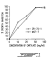
- Figure 1 shows that doses of 50 and 100 mg/mL of the lyophilizate clearly provoke hypoplasia on these cell lines, after three days of treatment.
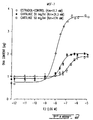
- Figure 2 shows that, in the presence of 10 -12 to 10 -9 M estradiol, treated cells respond like control cells by being non-stimulated by these hormone dosage rates. However, above 1 nM, control cells are strongly stimulated, and concentration of DNA reach 3.75 ug in the presence of 10 7 M estradiol (versus 0.69 ug in control without estradiol). In cells treated with 30 and 50 mg/mL of lyophilizate, DNA measured at the maximal stimulation is 1.9 and 1.8 ⁇ g, respectively
- Figure 2 shows that the affinity constant (Km) of the treated cells for estradiol is 3 and 16 times higher (31.3 nM and 174.0 nM) than the value of Km of the control cells (11.7 nM), in the presence of 30 and 50 mg/mL, respectively.
- Km affinity constant
- the rats of the treated groups were also given a daily dose of the lyophilizate in 3 mL of water combined with or without the supernatant, for ten weeks while the control group received the same volume of water. Only one sub-group of the second group of rats treated with a concentration of 3000 mg/Kg/day of the lyophilizate and 3 mL of the supernatant was also given an intraperitoneal (i.p.) injection of a smaller dose of the supernatant (about 8 mg of protein in 1 mL of water).
- Rats were weighing 151-175 g at the beginning of the two experiments and received food and water ad libitum .
- the first group of rats had tumors of average diameter of 0.9 cm while the second group of rats had a tumor of average diameter of 0.6 cm.
- the lyophilizate contains an active component which is absorbed in the gastro-intestinal tract and which has an effect on tumor size. This effect might be a direct effect on tumor cells or an anti-angiogenesis mediated effect.
- the lyophilizate may contain active components that are not hydrosoluble and/or that it may contain residual hydrosoluble. Therefore, in the last eventuality, one may consider that the pellet could be re-extracted in an aqueous solution to recover hydrosoluble components maximally, if the yield can be still improved.
- the animals used in the above in vivo experiments were killed by decapitation and the following tissues were taken for analysis: liver, lung, kidneys, heart, brain, muscle and mammary gland. Fat was taken out of these tissues, after what they were fixated for two days in Bouin fluid. After dehydration in ethanol, the fixated tissues were embedded in paraffin. Sections thereof were obtained and mounted on glass slides, colored with haematoxylin and visualized under microscope.
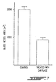
- Figure 7 shows that, when a combination of lyophilizate (p.o.)-supernatant (p.o. + i.p.) was used (refer to Figures 6a and b), a decrease of 55% of the blood vessel area was observed in the tumor.
- the diminution of the tumor size might be due to an important decrease in its vascularization, to a direct effect on tumor cells, or a combination of both phenomenons.
- the anti-angiogenic effect of these extracts is well depicted above.
- the direct hypoplasiant effect has been observed in vitro on the hormono-dependent cells, which remains to be confirmed in vivo.
- the supernatant will be hereinbelow referred to a crude permeate, e.g. the product after the ultrafiltration.
- the elution buffer was phosphate buffer saline (PBS) filtered and degazed during 15 minutes.
- the volume of the loaded sample was usually 3.2 mL (could be up to 13 mL).
- the flow rate was 1 mL/minute. Fractions of 10 mL were collected.
- the eluted compounds were detected by their U.V. absorbance (280 nm).
- a calibration chart was obtained by using the MW-GF-1000 calibration kit from Sigma, this calibration sample having the same volume as the loaded sample to analyse (3.2 mL).
- the elution volume of a sample was deduced from the plotting of the molecular weight of the compounds of the calibration kit versus their elution volume to which was subtracted the void volume of the column.
- the fractions were tested on ZR75-1 cells for their activity. The fractions of interest were identified and their characteristics were corroborated by further study (hereinbelow).
- a preparation of shark cartilage (46 mL of permeate 1 Kg/L) was dialysed overnight against 4 litres of pure water containing 5% glycerin at 4°C using a membrane Spectra pore #7 MWCO 3500 KD (Spectrum 132110).
- the dialyzed solution was mixed with 2.75 mL of ampholytes (Pharmacia #80-1125-87) pH 3.5-10.0 and 0.5 g CHAPS (Sigma C3023; 3-[(3-Cholamidopropyl) -dimethylammonio]-1-propane-sulfonate).
- the volume was completed to 55 mL with pure water.
- the solution was loaded on Rotofor.
- Isoelectrofocalization was conducted at 4°C, at a constant power of 12 watts (3000 xi power supply Biorad 165-0554), under constant water circulation for insuring maintenance of the temperature.
- the voltage was 380 volts and the amperage 31 mA.
- the amperage was stabilized (at 14 mA), the voltage read 870 volts.
- the isoelectrofocalization was stopped and 20 fractions were collected.
- the permeate was lyophilized, resuspended in PBS, and run on FPLC. No hypoplasiant activity was detectable (data not shown).
- FPLC fractions 6 and 7 contain active components of a very small molecular weight: 1 to 2.5 KD.
- the hypoplasiant effect of the fractions can be up to 33 000 times higher than the one observed with the lyophilizate.
- the above results show that lyophilization appears to provoke some loss of the direct anti-tumoral activity of the proteins contained in the eluate while no such abolition occurred with the lyophilization of the solid extract.
- the active fractions (tested on ZR75-1 cells) are retrieved in the following range of molecular weights, determined by another type of purification starting with the same permeate (1 Kg/L) on a 10 mm diameter x 30 cm length Superose-12 column using the FPLC and rotofor procedures described above. A flow rate of 1 mL/minute was selected. 45 fractions of 1 mL were collected.
- HEFs human TENON fibroblasts
- the cells were rinsed and dissociated by the afore-mentioned procedure and counted again.
- the number of cells was expressed as a percentage of that obtained in the "absolute" controls.
- GFs growth factors
- cartilage permeate was compared to the stimulation of the same by 5% FBS.
- GFs porcine platelet-derived growth factor (pPDGF) and human recombinant basic fibroblast growth factor (hr bFGF) (gift to Dr. P. Brazeau from Farmitalia Carlo Erba, Milan, Italy) were added in concentrations of 10 to 100 ng/mL in 1% FBS, respectively. Forty-eight hours after the onset of the experiment, the cells were dispersed by Trypsin-EDTA and counted on the Coulter counter. All triplicate values (columns 1, 2 and 3) appearing below equal one twentieth of counts per well.
- the column was calibrated with the following standards (MW in daltons): catalase (232,000), aldolase (158,000), albumin (56,000), ovalbumin (44,000), chymotrypsin (25,700), ribonuclease (13,700), insulin (5,700), insulin B chain (3500), insulin A chain (2500) bacitracin (1450), vitamin B-12 (1355).
- Total column volume (V T ) was 21.93 mL as determined using cytidine (246 Da). Void volume (V 0 ) was determined to be 8.38 mL with blue dextran (2 X 10 6 Da).
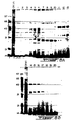
- Balassa's sample BAL had a small peak (1) eluting near the V 0 of the column (8.4 mL), a peak (2) at 18.5 mL (4000 Da) and two peaks eluting after the V t, (3) 22.6 min and (4) 28.2 mL.
- Oikawa's sample OIK also had a small peak (1) at the V 0 , peak (2) at 18.9 mL (3300 Da), peak (3) at 21.5 mL (1000 Da) and small peak (4) at 27.3 mL.
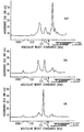
- OD210 and OD280 were monitored simultaneously. 50 ⁇ l aliquots of centrifuged samples (all at 12 ⁇ g/ ⁇ l) were loaded and eluted with 100% H 2 O. Peaks for each chromatogram labelled according to OD210 (eg. 1) and corresponding OD280 peaks are noted by '(eg. 1)'. The V 0 of this column was 5.5 mL (1.4 min).
- DUP had 3 major peaks which were observed via OD210 (1,2,3) and 2 minor peaks (4,5). Two side peaks were observed off of peak 1, labelled 1a and 1b. Significant OD280 absorbances were associated peaks 1, 1a 1b and 3. In comparison, the corresponding OD280 absorption for peak 2 is much smaller relative to the OD210.
- BAL showed more OD210 peaks, but the intensities were lower relative to the DUP peaks. As far as overlap of peaks could give an indication of identity of molecules, only peaks 3 and 7 in the Balassa sample appear to correlate with the retention times of peaks in the DUP sample (peak 1a or 1b and peak 4, respectively).
- CAM tests were first performed using different concentrations of protamine (37, 75 and 150 ⁇ g).
- a methylcellulose disk containing water and another disk containing protamine, as positive control, were placed on the chorloallantoic membrane of a chick embryo (n number of embryos analysed).
- An O-ring was placed on each disk to ease their localization.
- the crude permeate obtained after ultrafiltration was 2 and 20 fold-concentrated, providing enriched active permeate. These levels of concentration were obtained on a tangential flow filtration column having a porosity of 1000 Daltons, which reduced the volume of the eluate by 2 and 20 times.
- the concentrated permeate was filtered of a millipore filter of a porosity of 0.22 ⁇ m.
- the cartilage was processed with the alternative centrifuge method (using the CEPA centrifuge with a membrane of a porosity of 30 ⁇ M), a ten-fold concentration achieved the obtention of a concentrated extract having almost the same proteic level as the above 20-fold concentrated extract, e.g.
- the concentrated permeate was used for treating angiogenesis-dependent diseases.
- Three different types representative of angiogenesis-dependent diseases were tested in the practice in human; the first type being cancer (prostate cancer), the second type being dermatological disorders (psoriasis), and the third type being arthritis.
- the examples below will illustrate and indicate at least the antiangiogenic activity of the liquid extract.
- the keratosis component is the formation of cornified envelope in the form of a plaque.
- a plaque is a physical barrier which impedes the efficient penetration of the active ingredients towards the blood vessels.
- a patient suffering of a prostate cancer has voluntary tried a 10 fold-concentrated cartilage permeate. This patient underwent a series of successive conventional therapies that were temporarily successful. He recently began to consume the cartilage extract after his cancer showed recidivism.
- compositions may be administered by different routes e.g. topical, oral, sublingual, rectal, intravenous, intramuscular, by diffusion, etc.
- flavoring agents or fragrances may be added to these compositions to encourage patient's compliancy.
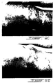
- Photographs of parts of two patients' bodies are shown in Figures 9a) and 9b).
- Figure 9a it is demonstrated that a patient affected by psoriasis with hyperkeratosis has nevertheless shown a very significant reduction of the erythema, associated with no prurit, after only one month of treatment. The hyperkeratosis remained, however, important.
- Photographs of the second patient suffering of psoriasis not complicated with hyperkeratosis Figure 9b) show a much better improvement after a three month-treatment. Since psoriasis appears to be a multifactorial disease, it is assumed that the response of the patients depends on the importance of the involvement of components like angiogenesis and inflammation in the establishment and in the perpetuation of this condition.
- the anti-angiogenic activity is indeed present in our extract, as shown in DMBA-treated rats and CAM-tests.
- the anti-inflammatory activity has also been verified (discussion below). It is probable that better results might be obtained if this kind of formulation is complemented with other therapeutic agents addressing to other factors involved (keratolytic agents, additional anti-inflammatory agents, antihistaminics, immunosuppressors, etc.).
- This complementation may take the form of amending the formulation to include an effective amount of a keratolytic agent, for example. It could also be achieved by the separate administration of such a complementary therapeutic agent, concurrently or in alternation with the application of the present topical formulation. Furthermore, the complementary medication does not need to be administered by the same route.
- the above formulation has shown no systemic effect (the effect being limited to the treated areas) and no secondary effect despite the high proportions in cartilage extract.
- the dosage rate of the liquid extract was about 190-220 mg of proteins/Kg of body weight, which presumably had a great contribution to the reduction of the area of cancer blood vessels (55% when combined with a much larger protein quantities of lyophilizate). It is therefore assumed that a dose of about 0.1 to about 200 mg/Kg of body weight per day is an approximative reasonable range of median doses (ED 50 ) for treating cancer, at least partly by reducing or abolishing angiogenesis.
- ED 50 median doses
- the 2 X cartilage extract contains 9-12 mg/mL of proteins. This formulation shows a remarkable improvement of the aspect of the skin of patients affected by more or less severe forms of acne (inflammatory acne and kystic acne; data not shown).
- results may be due to an anti-angiogenic effect (thus revealing an angiogenic component in acnae), or they may be due to active ingredients that have an effect other than anti-angiogenic (an anti-inflammatory effect, for example, see discussion below).
- the topical and all other compositions may contain a wide range of doses of the cartilage extract.
- very different dosages and/or formulations have been used.
- a minimal final protein concentration of the total liquid extract could be very low (from about 0.1 mg/mL).
- This lower range of doses depends on the accessibility and on the permeation of the active ingredients to the site of action as well as on the efficient capture of these ingredients and the sensitivity or response of the tissue to angiogenic inhibitors.
- the higher limit of the final protein concentration in formulations for some applications is not currently known.
- the highest final concentrations tried were of about 9 mg/mL of proteins in the formulation for the psoriasis cases and about 12 mg/mL in the dose unit of 7 mL administered in the prostate cancer case.
- the shark cartilage liquid extract may lose some of its activities when lyophilized in aqueous solution.
- the addition of stabilizers or protective agents as known in the art prior to lyophilization may preserve sensitive activities and render possible the administration of higher doses of the cartilage extract in the dry state.
- cartilage extract Since the cartilage extract has been shown very promising in the treatment of psoriasis, its effect has been tested in keratinocytes which PKC is activated by triphorbol acetate (TPA), a known agonist of this cellular transduction pathway.
- TPA triphorbol acetate
- Figure 16 shows that the level of differentiation of the keratinocytes was increased 5-fold by TPA.
- Shark cartilage by itself had no effect on cornified envelope formation.
- addition of the shark cartilage extract inhibited TPA-induced cornified envelope formation by more than about 60%.
- TPA-induction mimics psoriatic keratinocytes. If such is the case, these results suggest that cartilage may have no effect on normal keratinocytes in vivo , while it may have an effect on psoriatic (or activated) keratinocytes.
- ANTI-INFLAMMATORY ACTIVITY IS REMOTE FROM ANTI-ANGIOGINIC ACTIVITY IN SHARK CARTILAGE EXTRACT:
- the 4 test compounds were applied on the ventral forearms of the panelists.
- the material was allowed to absorb for twenty minutes and then Balsam of Peru, an irritant, was applied on the test sites. Skin irritation was measured in terms of increase in skin redness. The degree of redness was measured with a Minolta Chromameter and compared with the positive and negative controls.
- the positive control was the color of skin treated with Balsam of Peru alone and the negative control was a skin site treated with cola solution and challenged like the test products.
- Statistical significance was calculated via two tailed probability T-test.
- Figure 17 shows that cola at 10% was 70% active.
- Shark cartilage was 58% and 60% as anti-irritant at 20% and 10% concentrations, respectively. There was no dose-response effect.
- a 980 ml sample of liquid extract (DUP) was filtered through a 10 KDa cutoff membrane in a tangential flow ultrafiltration unit (PELLICONTM, Millipore). The unit was rinsed first with 1 L of H 2 O. Final yields were 480 mL of > 10 KDa fraction and 1.8 L of ⁇ 10 KDa fraction. The ⁇ 10 KDa was concentrated by cold-finger evaporation to 180 mL ( ⁇ 10-10X).
- the first three fractions include at least a major peak.
- the collagenase assays were run on these samples using recombinant human skin collagenase, type 1 (MMP1) using a fluorogenic peptide substrate (assay 1) and a collagen substrate (assay 2).
- the method utilizes a fluorogenic peptide substrate (Mca-pro-leu-glu-leu-Dpa-ala-arg-NH 2 ) mimicking the active site of metalloproteinases.
- This substrate has a fluorescent group (Mca) at one end and a fluorescence quenching group (Dpa) at the other. In the intact substrate, the quenching group effectively masks the fluorescence. Upon enzyme cleavage of the substrate the fluorescence in the test tube increases.
- Collagenase activation is described in Weinberg et al. (1985) Biochemistry 24, 6730. 1 ⁇ g was diluted to 100 ⁇ l with 50 mM Tris-HCl, 10 mM CaCl 2 . pH 7.5, 1 ⁇ l at 10 mg/ml solution of trypsin (in 1 nM HCl) was added and incubated for 15 min at 20°C. Activation was terminated by adding 10 ⁇ l of Soybean trypsin inhibitor (SBTI, 5 mg/ml). To each microcuvette was added:
- Fr1 is the most active fraction to inhibit the collagenase (Fig. 19). A lower level of activity is present in all other fractions.
- the enzyme was significantly inhibited by the shark cartilage extract (EC50 of about 10-20 ⁇ g/mL).
- This assay is described in Welgus et al. (1979) JBC 256, 9511-9516.
- the method uses SDS-PAGE to examine cleavage by collagenase, type 1 CMMP1). Collagenase type 1 makes a single cut in the native collagen molecule giving two fragments of 75% and 25% the size of the original collagen. After cleavage for several hours, the reaction is monitored by separating the products from the substrate by SDS-PAGE. The ratio of cleaved to uncleaved collagen is assessed visually after staining the gels with Comassie blue (or silver stain).
- EDTA 40 mM inhibited collagenase.
- the total liquid extract DUP showed a low anti-collagenolytic activity. Fractions 1 to 5 were active; the best active was fraction 1. The fraction of a molecular weight higher than 10 KDa showed no significant inhibitory activity.
- the cartilage extract of this invention may find numerous medical applications. Among the diverse activities recovered in this extract, anti-collagenolytic, anti-inflammatory and inhibitory effect on PKC-induced differentiation are particularly desirable in cosmetic applications. Since the cartilage extract of the present invention has shown an antagonist effect of PKC-mediated cellular events, and since such antagonist effect is suggested in the art as one improving the skin barrier repair function, a method for improving the barrier repair function in mammalian skin which comprises the step of applying to the skin a composition which comprises the cartilage extract and a pharmaceutically acceptable carrier, and such a composition are under the scope of this invention. Other or similar compositions can also be conceived to be used in a method for soothing skin or for reducing inflammation in mammalian skin.
- Inflammation can be caused by various agents such as chemical irritant, physical abrasion and exposure to ultraviolet radiation.
- Compositions and methods for inhibiting collagenase in skin are also contemplated. Collagenase and inflammation are linked to premature aging (degradation of collagen), and therefore the antagonist activities recovered in the cartilage extract could also be put to contribution in compositions and methods for retarding premature aging, and for regulating wrinkles or atrophy in mammalian skin. As causes of wrinkles or atrophy are listed, by way of examples, age, exposure to ultraviolet radiation or to environmental pollutant.
- Topical compositions may comprise an effective amount of shark cartilage, to be determined for each specific application.
- these compositions may contain from about 0.1 to about 75 weight percent of a liquid 1X to 20X cartilage extract and from about 50 to 99.9 weight percent of a pharmaceutically acceptable vehicle.
- These compositions may contain an anti-oxidant such as an agent which prevents the formation of lipid peroxides in skin. Examples of such anti-oxidant are tocopherol, tocopherol derivatives, ascorbic acid, ascorbic acid derivatives and BHT.
- the compositions can be complemented with anti-inflammatory agents like a phospholipase A2 inhibitor or the botanically-derived anti-irritants cola and green tea extract.
- Topical compositions may take diverse forms such as solutions, suspensions, lotions, tinctures, gels, creams, sprays, emulsions, sticks, ointments or liposomes (at least a portion of the liquid cartilage extract being present in liposomes).
- the process of the present invention has been demonstrated as one that provides for the production of cartilage extracts of a great clinical value.
- the shark cartilage extracts produced by this novel process comprises a multiplicity of activities that are recovered in good yields.
- the cartilage extracts, particularly the liquid extract and fractions thereof have a great potential since they are non-toxic to normal cells while they are effective in a large variety of diseases or conditions.
Landscapes
- Health & Medical Sciences (AREA)
- Life Sciences & Earth Sciences (AREA)
- Veterinary Medicine (AREA)
- Public Health (AREA)
- General Health & Medical Sciences (AREA)
- Animal Behavior & Ethology (AREA)
- Chemical & Material Sciences (AREA)
- Medicinal Chemistry (AREA)
- Pharmacology & Pharmacy (AREA)
- Organic Chemistry (AREA)
- Nuclear Medicine, Radiotherapy & Molecular Imaging (AREA)
- Chemical Kinetics & Catalysis (AREA)
- General Chemical & Material Sciences (AREA)
- Bioinformatics & Cheminformatics (AREA)
- Engineering & Computer Science (AREA)
- Dermatology (AREA)
- Epidemiology (AREA)
- Diabetes (AREA)
- Ophthalmology & Optometry (AREA)
- Inorganic Chemistry (AREA)
- Toxicology (AREA)
- Hematology (AREA)
- Vascular Medicine (AREA)
- Cardiology (AREA)
- Rheumatology (AREA)
- Immunology (AREA)
- Heart & Thoracic Surgery (AREA)
- Endocrinology (AREA)
- Gerontology & Geriatric Medicine (AREA)
- Oncology (AREA)
- Obesity (AREA)
- Emergency Medicine (AREA)
- Orthopedic Medicine & Surgery (AREA)
- Physical Education & Sports Medicine (AREA)
- Biochemistry (AREA)
- Zoology (AREA)
- Birds (AREA)
- Pain & Pain Management (AREA)
- Marine Sciences & Fisheries (AREA)
- Urology & Nephrology (AREA)
Abstract
Description
- This invention relates to the use of a shark cartilage extract in the making of a medication for treating diseases or disorders.
- Cartilage is an avascularized tissue and has been studied as a potential candidate containing anti-angiogenic factors. It is also a tissue which is relatively resistant to tumor development. The tumor associated with cartilage, chondrosarcoma, is the least vascularized of solid tumors. Angiogenesis is one of the important factors in the development of a tumor. Discrete solid tumoral masses appear if the tumor cells can provoke the adjacent vascular network to expand to supply their nutritional needs. Therefore, the factors involved in the stimulation of angiogenesis have been studied for their role in the development of tumor and anti-angiogenic factors as well as drugs having an angiogenic inhibitory activity have been also investigated as tools for controlling the growth or for effecting regression of tumors.
- It has been discovered that scapular cartilage in calves contains a substance that inhibits the vascularization of solid tumors (Langer et al., 1976). Because of its encouraging potential as anti-tumor agent, sources of greater supply of cartilage have been looked for.
- Sharks are animals being a potential source of this kind of angiogenesis inhibitor because their endoskeleton is composed entirely of cartilage (6% of their body weight versus 0.6% in calves). Sharks have also as an interesting characteristic a low propensity to developing tumors. Many hypotheses have been elaborated to explain this low probability of developing tumors in sharks. Marchalonis et al. (1990) have shown IgM antibodies able to readily attack any aggressing agent. McKinney et al. (1990) have shown that sharks have macrophages capable of differentiating normal cells from neoplastic cells and of destroying the latter. Rosen and Woodhead (1980) have postulated that the rarity of tumors in elasmobranchs (a group to which pertain sharks and rays) might be due to the high ionic strength of their tissues, which is equivalent to a high body temperature. In these conditions, these authors believe that the immune system exerts a close to 100% immunological surveillance. Moore et al. (1993) have discovered that sharks produce an aminosterol having antibacterial and antiprotozoal properties. Finally, Lee and Langer in Science-Vol. 221, pages 1185-1187 (1983) and Folkman and Klagsbrun (1987) have shown that sharks produce a substance which inhibits neovascularization. Lee and Langer (op.cit.) have isolated this substance by extracting it from shark cartilage in denaturing conditions (guanidine extraction). This process of extraction is however very long (41 days) might generate extracts having denatured factors and the yield of active components is far from excellent. While the active substance isolated from calves has a molecular weight of about 16 kilodaltons (KDa), the same group of researchers have not given a precise molecular weight to the one retrieved in sharks. This substance is only defined has having a molecular weight higher than 3500 Daltons. Oikawa et al. (1990) have applied the same method of extraction as the one described by Lee and Langer, but of a much shorter duration (2 days instead of 41 days). The anti-angiogenic substance isolated from shark cartilage by Oikawa et al. is restricted to a molecule having a molecular weight ranging from 1000 to 10,000 Daltons. Schinitsky (US-A-4 473 551) has described a water extract of crude powdered shark cartilage which fraction of more than 100,000 Daltons has an anti-inflammatory activity alone or in combination with glucosamine. No suggestion of a component of this extract having an anti-angiogenic or anti-tumor activity is made in this patent. Kuetner et al. (US-A-4 746729) have isolated a polymorphonuclear neutrophil (PMN) elastase inhibitor from bovine cartilage. This inhibitor has been obtained from an aqueous extract of cartilage from which molecules of a molecular weight of less than 50,000 Daltons have been retained. Fractionation on Sephacryl S-200 has given numerous fractions from which those of 10-40 KDa have been pooled after they have demonstrated an anti-elastase activity. The active component has an isoelectric point of 9.5 and might have a molecular weight of about 15,000 Daltons. Kuetner et al. (US-A-4 042 457) have also shown that bovine cartilage has a component of a molecular weight of less than 50,000 Daltons which has a cell proliferation inhibitory activity without any activity on endothelial cell growth. Balassa et al. (US-A-4 822 607) have obtained a cartilage extract in an aqueous solution, which extract has an anti-tumoral activity. However, we have observed no anti-angiogenic activity in an extract obtained by reproducing Balassa's method. Spilburg et al. (US-A-4 243 582) have isolated two glycoproteins of molecular weight of 65 KDa and of pI 3.8 from bovine cartilage (guanidine-extraction) which show anti-trypsin activity and an endothelial cell growth inhibitory activity.
- Calf and shark cartilage contain many biological activities such as pro-inflammatory activity, anti-inflammatory activity, anti-angiogenic activity, lysozyme activity, cell growth-promoting activity, inhibitory activity against types I and IV collagenase, elastase, and other proteases like trypsin, chymotrypsin and plasmin. However, nobody has yet obtained a cartilage extract which comprise a pool of clinically valuable activities.
- In Cancer Letters, Vol. 51, pages 181-186 (1990), Oikawa explains how guanidine extraction and crude fractionation of Japanese shark cartilage by ultrafiltration on a molecular weight basis were conducted and the antiangiogenic activities were assayed as to the inhibitions of tumor and embryonic angiogenesis. A significant inhibition of angiogenesis was found. Anyway, the document is silent about the isolation of an anti-collagenolytic and anti-inflammatory activity. Further, Oikawa's product is not a 0 - 500 KDa fraction of a supernatant.
- In Science, Vol. 221, pages 1185-1187 (1983), Lee and Langer explain that shark cartilages contain inhibitors of tumor angiogenesis.
- In Fed. Proc. 45,949 (1986) Luer mentions inhibitors of angiogenesis from shark cartilage. Luer's extract comprises anti-protease components in fractions of a molecular weight higher than 10 KDa. Anyway, due to the scarce information given by Luer's abstract on his proprietary process, it is almost impossible to exactly reproduce Luer's extract. Therefore, there is no indication that Luer would have succeeded in obtaining an extract comprising all the anti-collagenolytic and anti-inflammatory molecules of the present extract.
- In US-A-4 473 551, Shinitsky discloses anti-inflammatory compositions. The first composition comprises a shark cartilage powder. The second composition comprises an aqueous extract of said powder. The third compound is a combination of a shark cartillage extract and glucosamine. Schinitsky shows a good level of activity but only in the presence of glucosamine. Schinitsky also shows a cartilage anti-inflammatory activity only for the powdered cartilage, the cartilage powder being merely a starting material which is equivalent to using crude cartilage.
- All of the four above documents disclose methods wherein specific fractions are recovered. There is no teaching to use cartilage extracts that include a plurality of fractions, and thus a plurality of ingredients coming from the shark cartilage.
- WO-A-95/32722 (E-document) is also to be mentioned, for which is admitted that the cartilage extract disclosed therein, which is the same as the one used in the use of the present invention, is used for treating cancer, psoriasis, acne and arthritis. These target diseases or disorders are expressly excluded from the use claimed in the present invention.
- Shark cartilage anti-angiogenic component(s) have been generally tested in rabbit corneal pocket assay or in chick chorioallantoic membrane (CAM) assay. Up to date, whole powdered cartilage has been tested directly on tumors in vivo, on human melanoma xenograft implanted in nude mice (US-A-5 075 112), as well as tested in CAM tests for its anti-angiogenic effect. Even though an anti-tumoral effect has been assigned to cartilage extracts, this effect has most often been attributed to the anti-angiogenic component which deprives the tumor of blood supply. Up to now, there is no evidence that a shark cartilage has a direct effect on tumor cell proliferation.
- A few methods of obtaining a shark cartilage extracts and fractions are already known. Some of them produce a powdered crude cartilage without any extraction (US-A-5 075 112). Others use denaturing agents like guanidine (US-A-4 243 582). Others perform a pre-treatment of cartilage by way of an enzymatic digestion to get rid of any muscular, nervous or vascular structures surrounding the cartilage, which pre-treatment step is followed by the elimination of fats in organic solvents, and then the active components are extracted in an aqueous phase. (Balassa et al.
US Patents No 3 478 146, 4 350 682, 4 656 137 and 4 822 607). The effect of such pre-treatment on the preservation of the integrity of the biologically active cartilage components is not known. If too extensive, an enzyme digestion may hydrolyse active proteic components Balassa's method does not include a fractionation step which would enrich an extract in active components. Others simply produce aqueous extracts (in water (US-A-4 473 551) or salt solutions (US-A-4 746 729)) of cartilage by eliminating the unsolubilized material. Among the latter, specific fractions of specific molecular weights have been particularly retained for further study and purification (see discussion above). - The above-cited methods have several drawbacks. They may denature some valuable components. When such might not be the case, they have the disadvantage of being too lenghty to be of a practical purpose. Moreover, the lenghty methods do not necessarily yield sufficient amounts of active components, and among the recovered components, some are not recovered at all or in unsufficient yield to show detectable activity or some have been disregarded by focusing on the obtention of specific activities.
- Angiogenesis is not only involved in cancer development. Many diseases or conditions affecting different physiological systems (indicated in parentheses) are angiogenesis-dependent among which the following examples: arthritis and atherosclerotic plaques (bone and ligaments), diabetic retinopathy, neovascular glaucoma, trachoma and corneal graft neovascularization (eye), psoriasis, scleroderma, hemangioma and hypertrophic scarring (skin), vascular adhesions and angiofibroma (blood system). Therefore, any new and potent anti-angiogenic "factor" could find a use in the treatment of these diseases as well as in cancer therapy. Moreover, since many of the above - mentioned diseases and conditions also have an inflammatory component, any new and potent anti-inflammatory "factor" could find a use in the treatment of these diseases and conditions as well as of any other inflammatory diseases or conditions. Furthermore, since proteases like collagenases are involved in a diversity of diseases and conditions like cancer and premature aging because of its collagen " degrading activity, a new and potent anti-collagenolytic "factor" could find a use in . the treatment of diseases or conditions having a collagenolytic component. Because angiogenesis, inflammation and proteases like collagenases may be encountered alone or in combination in a large variety of diseases or conditions, a product capable of antagonizing at least all these activities without affecting normal body functions would be of a great therapeutic value.
- The object of the present invention is a new use of a shark cartilage extract in the making of a medication for treating a disease or disorder having a component selected from collagenolysis and inflammation, as defined in
claim 1. - Some additional features of the above new use of a shark cartilage extract are defined in the dependent claims.
- The use of the present invention is linked to a method of producing cartilage extracts which have the advantage of containing a multiplicity of therapeutically valuable activities. Among those, anti-angiogenic, anti-inflammatory, anti-collagenolytic, in vivo anti-tumor proliferating and direct in vitro anti-tumor proliferating activities have been confirmed to be present in satisfying concentrations in a shark cartilage extract. Other activities await identification or confirmation. The effect measured in tumor cell lines was indicating that beside a direct anti-tumor proliferating activities, a cytotoxic activity appears to be present. All activities have been obtained in a liquid extract of shark cartilage, and some of them have been obtained or verified in a solid extract of the same.
- Then, the use of the present invention is linked to a process for the obtention of a liquid extract of cartilage having a substantial portion of the biologically active hydrosoluble components present in intact cartilage, which comprises the following steps:
- a) homogenizing the cartilage in an aqueous solution in conditions compatible with the preservation of the integrity of said biologically active components until the cartilage is reduced to particles whose size is lower than or equal to about 500 µm, resulting in a mixture of particles and of a crude liquid extract having said biologically active components;
- b) centrifuging said homogenate to separate particles from the crude liquid extract; and
- c) further separating the crude liquid extract so as to obtain a final liquid extract containing cartilage molecules having a molecular weight lower than or equal to about 500 Kilodaltons.
-
- This process has the advantage of being easy to perform and efficient. High yields of cartilage extract have been obtained, which extract, particularly obtained from shark cartilage, contains at least all the above-mentioned biological activities. It is preferably performed at cold temperature (about 0 to 10°C), in non-denaturing conditions (preferably in pure water), at a near neutral pH (about 6 to 8) to maximize the probability of recovering compounds of unknown physico-chemical characteristics. According to this process, cartilage components can be extracted in a low volume of solution (as low as 1 L for 1 Kg of cartilage), and after a short period of homogenization (as short as 10 to 15 minutes). For the recovery of a solid extract, the same process is used, except that the pellet is recovered and lyophilized, disregarding the supernatant.
- This invention relates to cartilage extracts, particularly to extracts providing from elasmobranch species, more particularly from shark. The solid extract has shown activity. It may contain collagen and non-hydrosoluble components. It may also contain a residual activity of what was extracted in the total liquid extract. The total liquid extract is very rich in activity. It can be used as such or it can be concentrated. A concentration step which favorizes the maintenance of biological activities has been priviledged. Recourse to methods which could deteriorate the active components like heat-evaporation has been avoided by caution. Ultrafiltration on a membrane having a molecular weight cut-off value of about 1 KDa has been used to concentrate the liquid extract of this invention. As a result, a concentrated extract containing molecules of a molecular weight comprised between about 1 and about 500 KDa was obtained and tested. The total liquid extract (0 to 500 KDa) has been further fractionated to characterize the active components thereof. Numerous fractions have been obtained by different methods. Some of them tested on tumor cell lines have been grossly characterized by their molecular weight and isoelectric point. Others have been assigned an activity, particularly anti-collagenolytic or anti-angiogenic activities. These fractions await complete characterization and identification. Therefore, valuable activities are recovered in a total liquid extract and fractions thereof, which may be advantageously used. In lieu of administering high amounts of powdered cartilage, a more acceptable and enriched extract may now be administered.
- The use of the present invention is also linked to therapeutic or cosmetic compositions comprising as an active ingredient one of the above-cartilage extracts. Most interest has been drawn to topical compositions for use in dermatology and cosmetology. This interest comes from the observed activities of the cartilage extracts. In this respect, the observed anti-collagenolytic and anti-inflammatory activities, and the antagonistic effect of cellular differenciation mediated by the induction of Protein Kinase C in keratinocytes have been considered as opening avenues to the use of the shark cartilage extracts in compositions and methods for the reduction of inflammation, the regulation of wrinkle or skin atrophy, the retardation of premature aging, the reduction of acne, the improvement of skin barrier function, the reduction of inflammation or irritation and a skin soothing effect.
- The present invention will be more readily understood by way of the specific embodiments shown in the appended figures, which purpose is to illustrate the invention rather than to limit its scope:
- The data concerning cancer, psoriasis, acne and arthritis are indicated for illustrative purposes only and do not form part of the invention as claimed.
- Figure 1 shows the inhibitory activity of increasing doses of shark cartilage (solid extract) on ZR75-1 and MCF-7 cells.
- Figure 2 illustrates dose-response curves of the quantity of MCF-7 cells measured by their DNA content in the presence of increasing concentrations of estradiol with or without two concentrations of cartilage lyophilizate.
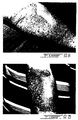
- Figures 3a) and b) show a comparison of liver sections of rats having developed a mammary gland cancer which have been administered by gavage a combination of cartilage lyophilizate and supernatant and those which have been administered only water.
- Figures 4a) and b) show a comparison of kidney sections of rats having developed a mammary gland cancer which have been administered by gavage a combination of cartilage lyophilizate and supernatant and those which have been administered only water.
- Figures 5a) and b) show a comparison of lung sections of rats having developed a mammary gland cancer which have been administered by gavage a combination of cartilage lyophilizate and supernatant and those which have been administered only water.
- Figures 6a) and b) show a comparison of mammary gland tumor sections of rats having developed such a tumor which have been administered by gavage a combination of cartilage lyophilizate and supernatant and those which have been administered only water.
- Figure 7 is an histogram derived from Figures 6a) and b), illustrating the effect of cartilage extract on blood vessel area in tumors.
- Figure 8 represents the electrophoretic profile in non-denaturing conditions of liquid fractions separated on Rotofor; molecular weight markers appear at the left followed by a sample of the crude permeate before fractionation, for comparison with the isolated fractions.
- Figures 9a) and 9b) illustrate the significant improvement of the condition of two patients suffering of psoriasis, one with hyperkeratosis 9a), and the other one without hyperkeratosis 9b), when treated with a topical composition containing an effective amount of concentrated liquid cartilage extract (lower photographs) compared with their initial condition (upper photographs).
- Figure 10 shows a PPLC migration pattern of three different extracts of shark cartilage. In panel A, DUP stands for a cartilage liquid extract according to this invention. In panels B and C, BAL and OIK stand for extracts of the prior art, Balassa et al. and Oikawa et al., respectively.
- Figure 11 shows a HPLC migration pattern of the same extracts defined in Figure 11.
- Figure 12 shows the results of CAM-tests performed using different concentrations of protamine, an anti-angiogenic reference compound, when compared to control.
- Figure 13 shows the results of CAM-tests performed with two fractions of the present total liquid extract of shark cartilage of our invention (DUP), one having molecular weight lower than 10,000 Daltons, the other one having molecules higher than 10,000 Daltons.
- Figure 14 shows the results of CAM-tests performed with the total liquid extract of our invention (DUP) when compared to an equivalent concentration of a product made by the process of Balassa (BAL).
- Figure 15 shows the results of CAM-tests performed with the total liquid extract of our invention (DUP) when compared to a sample providing from an equal quantity of dry matter weight of a product made by the process of Oikawa (OIK).
- Figure 16 shows the effect of TPA on keratinocytes when compared to a DMSO control, both measured in the presence or absence of a total shark cartilage liquid extract prepared in accordance with the present invention.
- Figure 17 shows the anti-inflammatory effect of the total liquid extract of our invention on a model of skin irritation.
- Figure 18 shows another HPLC migration pattern of a fraction of the total liquid extract of this invention having molecular weight lower than 10,000 Daltons, which fraction has been concentrated and separated in five sub-fractions.
- Figure 19 shows the anti-collagenolytic effect of each sub-fraction shown in Figure 18 at different tested volumes.
-
- In a specific embodiment, cartilage has been obtained from healthy sharks Black Spiny Dog Fish and Common Spiny Dog Fish. Any muscular and connective tissue has been removed by scraping with ethanol-treated scalpels and scissors. The cartilage was then vacuum-packed in plastic bags and frozen to -20°C for further use. In the present process any source of cartilage may be used. We have chosen shark cartilage for reasons enunciated in the BACKGROUND section. It is believed that starting from elasmobranch cartilage (which includes sharks and rays as animal species of this group), near equivalent products would be obtained. The products will most probably be different if mammalian source of cartilage are used.
- Any variation in the preparation of cartilage prior to its extraction may be used as long as it does not substantially affect the activity of the product of interest (a total liquid extract or a particular fraction thereof, for example). Some active components may resist to proteolytic digestion as taught by Balassa et al. (USP 4,822,607) to rid the cartilage of any surrounding tissues, while others may not resist to such treatment. One of the activities which do not appear to resist to such pre-treatment is the anti-angiogenic activity (Figure 15). Therefore if one wants to produce a liquid extract containing as much as possible of all the hydrosoluble active components to which are assigned separate activities, such a digestion step should be avoided or carefully monitored to prevent extensive hydrolysis or proteolysis.
- Clean cartilage was used fresh or thawed to 4°C. Cartilage was then passed numerous times (more particularly three times) through the pores of an ethanol-treated meat chopper together with a adequate volume of water (an equal quantity (weight/volume) is about a minimal volume but can be increased without bearing any effect on the yield of recovery of valuable components). A low volume is preferred since it is more convenient to manipulate than unnecessary high volumes, from a practical point of view. In the practice, water has been purified by inverse osmosis and filtration on a 0.1µm filter. Many aqueous solutions (containing salts, for example) could be used in lieu of water. When recovery of a plurality of hydrosoluble activities is contemplated, working at a near neutral pH and non-denaturing conditions are preferred to avoid lysis or denaturation of some of the cartilage components. The behavior of unknown proteins in aqueous solvents is not predictable; some may be more "comfortable" in an acidic pH, some at a basic pH. Furthermore, some proteins may be extractable in mild denaturing conditions, if such denaturation does not irreversibly affect the re-naturation of these proteins in aqueous solutions. Therefore, taking all these factors in consideration, performing a process of extraction of cartilage active components in pure water has been shown to be a judicious choice to recover with a very good yield, components having an unknown structure and behaviour.
- The blend cartilage/water was then made homogenized by an agitation at a maximal speed in an kitchen blender at about 4°C during ten minutes. Of course, the speed of the agitation as well as the volume of aqueous solution may influence the time of extraction. Therefore, a reasonable range of homogenization time could be as low as about 10 minutes to as high as 24 hours, preferably between about 10 and 60 minutes. The temperature should be maintained to below about 10°C, to avoid any degradation of active components by endogenous enzymes, when no enzyme inhibitors are used. Ideally, a temperature close to 0°C should be sought. Since normally such experimentation is made in a cold room, wherein the temperature can be maintained between 4 and 10°C, this range of temperature is acceptable in the present process. For sake of clarity and brevity, the terms "about 4°C" is hereinbelow used to designate this acceptable range of temperatures.
- A liquefaction of this homogenate can be further obtained by submitting the latter to Polytron disintegrator during 10 minutes at 4°C if the blender did not sufficiently reduce the size of the particles. Alternatively, the blend can be simply homogenized in a more performing a blender-desintegrator which, in our hands, saved the 10 min liquefaction step. At the end of the completed homogenisation step, residual particle size is less than about 500µm. Of course, the same acceptable ranges of time and temperature discussed for the obtention of the first grinded cartilage equally apply. The size of the particles after homogenization does not need to be very small. Therefore, the need to pulverize the cartilage before extraction can be avoided. Indeed, pulverization of cartilage in the form of a powder before aqueous extraction may denature valuable activities, when such pulverization is performed in a freeze-dry state or in a heat-dry state.
- The homogenate was centrifuged at 13,600 x g during 15 minutes at 4°C, which step is one way to separate quickly and efficiently a supernatant from a pellet. Variation and adjustment of these parameters are well within the knowledge of the skilled artisan, merely depending on the volume of homogenate and of the used equipment.
- The resulting pellet was lyophilized for 24 to 48 hours. This first fraction will hereinbelow be defined as the lyophilizate or a solid extract.
- The supernatant can be filtered on a 24 µm Whatman filter, if necessary, to get rid of particles susceptible to affect the performance of an ultrafiltration column. The filtrated material was then ultrafiltrated at about 4°C on an tangential flow filtration column having a porosity of about 500 000 Daltons, which allows a first crude permeate to be obtained comprising hydrosoluble molecules of a molecular weight comprised between 0 and about 500 KDa. This crude permeating extract was sterile filtered on 0.22 µm filter, and aliquoted in sterile bottles for further use. This fraction will be further referred to as the crude permeate or the total liquid extract.
- An alternative, higher performing centrifuging procedure has been developed to obtain the pellet and the supernatant. The step of centrifuging at 13600 x g for 15 minutes followed by a gross filtration on Whatman filters has been replaced by a centrifugation in a CEPA centrifuge equipped with a nylon pocket of a porosity of 30 µM, at 3000-4000 x g. A 25 kg/25 L preparation can be centrifuged in that manner within 30 minutes and provide 29 liters of supernatant. The aqueous volume obtained is higher than the starting volume of water, suggesting that a part of the water content of the cartilage itself has been harvested. The lyophilizate and the total liquid extract may have the following approximate composition which grossly takes into account the variations observed from batch to batch, and when using different material:
LYOPHILIZATE: Lipids 7.35% Proteins 46.2% Humidity 20.4% Sodium 4.16 mg/g Potassium 2.64 mg/g Calcium 114 mg/g Magnesium 1.49 mg/g Zinc and iron traces TOTAL LIQUID EXTRACT: Lipids 0.10 - 0.20% Proteins 8 - 25 mg/ml Humidity 97 - 99% Sodium 30 - 220 mg/100 g Potassium 30 - 40 mg/100 g Calcium 2.0 mg/100 g Magnesium 1.1 mg/100 g Zinc and iron traces - The protein content is evaluated by the Kjeldahl method, which indeed measures organic nitrogen (N). Organic nitrogen is converted to equivalent protein by using the following equation:
- Carbohydrates being not detectable, one can presume that they are in one or another extract but under the form of proteoglycanes and/or mucopolysaccharides. It is possible that these compounds are included in the measured level of humidity. The lyophilizate contains an unexpected level of humidity which was measured by the OH- groups. Since the 20% water content is close to the percentage of carbohydrates normally retrieved in cartilage while the humidity of a lyophilizate should be close to 0%, this hypothesis remains to be verified.
- Sterility has been controlled, applying USP XXIII specifications by:
- 1) Laboratoire de genie sanitaire du Québec Inc.
1090, l'Escarbot, Centre Industriel St-Malo, Québec GlN 4J4; and - 2) Northview Laboratories Inc.
1880, Holste Road, Northbrook, IL, 60062 U.S.A. FDA registration no. 14-18028 -
- These assays have been conducted on the hormono-dependent cancer cell lines MCF-7 and ZR75-1 (ATCC (R) numbers 22-HTB and 1500-CRL, respectively).
- 52 g RPMI 1640 without phenol red (Sigma R8755), 17.875 g Hepes (free acid; Sigma H0763), 0.55 g sodium pyruvate (Sigma P5280) and 10 g NaHCO3 were mixed in 5 L of pure water and made pH 7.40 with NaOH.
- If not used immediately, this solution must be protected from light to preserve photolabile substances. This solution was filtered, distributed in 500 mL sterile bottles and stored at 4°C for a maximal period of three months.
- Basal RPMI medium was supplemented with 10% (v/v) FBS (fetal bovine serum), 100 U penicillin G/50 µg streptomycin sulfate (Sigma P0906)/ml medium, 2 mM L-Glutamine (Sigma G1517) and 1 nM E2 (β-estradiol Sigma E8875).
- Basal RPMI medium was supplemented with 5% FBSA (fetal bovine serum adsorbed on dextran-charcoal), 2 mM L-Glutamine, 100 U penicillin G/50 µg streptomycin sulfate/ml medium and 50 ng/mL insulin (Sigma). To this medium was added increasing concentrations of the above-described lyophilizate as well as different concentrations of estradiol (10-12 to -5 M).
- DME-F12 medium (without bicarbonate and without red phenol; Sigma) was reconstituted following the manufacturer's directives in pure water. For one litre, 1.2 g of sodium bicarbonate was added and the pH made to 7.40 with NaOH/HCl. This solution was filtered, distributed in 500 mL sterile bottles and stored at 4°C for a maximal period of three months.
- Basal DME-F12 medium was supplemented with 10% (v/v) FBS (fetal bovine serum), 100 U penicillin G/50 µg streptomycin sulfate/ml medium, 2 mM L-Glutamine (Sigma) and 1 nM E2 (estradiol).
- Basal DME-F12 medium was supplemented with 5% FBSA (fetal bovine serum adsorbed on dextran-charcoal), 2 mM L-Glutamine, 100 U penicillin G/50 µg streptomycin sulfate/ml medium and 50 ng/mL insulin (Sigma). As described for the ZR75-1 cells, lyophilizate and estradiol were added at the same concentrations.
- Fetal bovine serum was mixed with 1% (w/v) charcoal (carbon decolorizing alkaline). A solution of dextran T70 was added to the charcoal-serum solution to achieve a concentration of 0.1% (w/v). The mixture was agitated overnight at 4°c. After centrifugation at 4°C for 30 minutes at 10,000 x g, the serum was decanted, mixed again with the same proportions of charcoal and dextran, agitated at room temperature for three hours and recentrifuged. The serum was then heat-inactivated at 56°C for 20 minutes, sterile filtered and aliquoted in sterile conical Falcon tubes.
- ZR75-1 and MCF-7 cells were grown to reach a density of population of 20 000 cells/well on 24-well plaques or 150 000 cells/well on 6-well plaques, and treated in the presence or absence of different concentrations of lyophilizate as prepared above. To this effect, the lyophilizate is resuspended in culture medium and sterile filtered, so that hydrosoluble components thereof are recovered and tested. All experiments have been performed in triplicates. Culture media have been withdrawn and replaced by fresh media every two days. Cells were grown in an incubator under a constantly humidified atmosphere containing 5% CO2, at 37°C, for 17, 7, 3 or 3 days, corresponding to the first, second, third or fourth experiment, respectively. Cell growth inhibition was measured by direct counting of the cells or by measuring the total DNA content of a well.
Concentration of lyophilizate Cell Inhibition (%) MCF-7 ZR75-1 1st experiment: 17 days 1 mg/mL 1.5 2.00 5 mg/mL 14.33 33.6 10 mg/mL 62.66 90.8 2nd experiment: 7 days 1 mg/mL 3.73 0.97 5 mg/mL 15.7 29.00 10 mg/mL 68.37 66.00 3rd experiment: 3 days 50 mg/mL 95.8 95.00 100 mg/mL 94.6 98.00 4th experiment: 3 days 10 mg/mL 34.4 51.5 20 mg/mL 62.5 70.5 50 mg/mL 95.8 95 100 mg/mL 94.6 98 - The above percentages of inhibition of cell growth demonstrate that the lyophilizate can inhibit in a dose-dependent manner the growth of the cells of these two cell lines.
- Figure 1 shows that doses of 50 and 100 mg/mL of the lyophilizate clearly provoke hypoplasia on these cell lines, after three days of treatment.
- Figure 2 shows that, in the presence of 10-12 to 10-9 M estradiol, treated cells respond like control cells by being non-stimulated by these hormone dosage rates. However, above 1 nM, control cells are strongly stimulated, and concentration of DNA reach 3.75 ug in the presence of 107 M estradiol (versus 0.69 ug in control without estradiol). In cells treated with 30 and 50 mg/mL of lyophilizate, DNA measured at the maximal stimulation is 1.9 and 1.8 µg, respectively
- Figure 2 shows that the affinity constant (Km) of the treated cells for estradiol is 3 and 16 times higher (31.3 nM and 174.0 nM) than the value of Km of the control cells (11.7 nM), in the presence of 30 and 50 mg/mL, respectively. This means that higher concentrations of estradiol are necessary to achieve the same growth of the cells when cartilage lyophilized solid extract is present. Therefore, this extract diminishes the maximal response (90% inhibition thereof) and increases the affinity constant of the treated cells to estradiol.
- Four hundred 40 day old female Sprague-Dawley rats (purchased from Charles River Co., St-Constant, Quebec) where adapted to their environment for 12 days. At that time, 20 mg DMBA/1 mL corn oil (9, 10-Dimethyl-1, 2-Benzanthracene; purchased from Sigma Chemical Co.) was administered by gavage. Three months after this treatment, 240 rats having developed a mammary breast cancer have been selected and distributed in two groups. The first group consisted of five sub-groups of rats. The rats of the treated groups were given a daily dose of increasing concentrations of the lyophilizate extract in 3 mL of water for eight weeks while the control group received the same volume of water. The second group consisted in four sub-groups of rats. The rats of the treated groups were also given a daily dose of the lyophilizate in 3 mL of water combined with or without the supernatant, for ten weeks while the control group received the same volume of water. Only one sub-group of the second group of rats treated with a concentration of 3000 mg/Kg/day of the lyophilizate and 3 mL of the supernatant was also given an intraperitoneal (i.p.) injection of a smaller dose of the supernatant (about 8 mg of protein in 1 mL of water).
- Rats were weighing 151-175 g at the beginning of the two experiments and received food and water ad libitum. The first group of rats had tumors of average diameter of 0.9 cm while the second group of rats had a tumor of average diameter of 0.6 cm.
- The results are summarized as follows:
Daily doses of cartilage extract administered by gavage % tumor growth inhibition
(decrease of tumor diameter vs control)1st experiment: duration 8 weeks500 mg/Kg/ day 2% 1000 mg/Kg/ day 4% 3000 mg/Kg/ day 14% 5000 mg/Kg/ day 15% 2nd experiment: duration 10 weeks3000 mg/Kg/ day 12% 3000 mg/Kg/day + 3 mL supernatant 18% 3000 mg/Kg/day + 3 mL supernatant
+ 1 mL inj. i.p. supernatant20% - These results demonstrate that the lyophilizate contains an active component which is absorbed in the gastro-intestinal tract and which has an effect on tumor size. This effect might be a direct effect on tumor cells or an anti-angiogenesis mediated effect.
- These results also show that the supernatant has an activity which is reflected by a supplementary reduction of tumor size of about 5%.
- These results also suggest that the lyophilizate may contain active components that are not hydrosoluble and/or that it may contain residual hydrosoluble. Therefore, in the last eventuality, one may consider that the pellet could be re-extracted in an aqueous solution to recover hydrosoluble components maximally, if the yield can be still improved.
- For evaluating the non-toxicity of the active molecules of the cartilage extract, the animals used in the above in vivo experiments were killed by decapitation and the following tissues were taken for analysis: liver, lung, kidneys, heart, brain, muscle and mammary gland. Fat was taken out of these tissues, after what they were fixated for two days in Bouin fluid. After dehydration in ethanol, the fixated tissues were embedded in paraffin. Sections thereof were obtained and mounted on glass slides, colored with haematoxylin and visualized under microscope.
- The histological examination revealed that no deleterious effect was visible when using the largest doses of lyophilizate alone (data not shown) or when using the lyophilizate in combination with the supernatant (see Figures 3a and b, 4a and b, and 5a and b).
- This suggests that the lyophilizate and the supernatant have a selective tumor size regressive activity.
- In cancerous mammary gland (see Figures 6a and b), an important diminution of the area of blood vessels was observed. The anti-angiogenic effect of these active molecules is then confirmed by results as illustrated and summarized in Figure 7.
- Figure 7 shows that, when a combination of lyophilizate (p.o.)-supernatant (p.o. + i.p.) was used (refer to Figures 6a and b), a decrease of 55% of the blood vessel area was observed in the tumor.
- The diminution of the tumor size might be due to an important decrease in its vascularization, to a direct effect on tumor cells, or a combination of both phenomenons. The anti-angiogenic effect of these extracts is well depicted above. The direct hypoplasiant effect has been observed in vitro on the hormono-dependent cells, which remains to be confirmed in vivo.
- Because the above-mentioned results showed that the supernatant had an increased effect over and above the effect of the lyophilizate on ZR75-1 cells, the components thereof were further investigated.
- Shark cartilage was harvested and processed the same as described above. After centrifugation, the pellet was discarded and the supernatant was processed the same way as described above up to the sterile filtration on 0.22 µm filter.
- The supernatant will be hereinbelow referred to a crude permeate, e.g. the product after the ultrafiltration.
- The so obtained crude permeate was passed on FPLC (Fast Protein Liquid Chromatography).
- Column: Hiload 26 mm x 60 cm Sephacryl S-300
- FPLC system: from Pharmacia
- All samples were filtered on 0.22 µm filter before loading on the column. The elution buffer was phosphate buffer saline (PBS) filtered and degazed during 15 minutes. The volume of the loaded sample was usually 3.2 mL (could be up to 13 mL). The flow rate was 1 mL/minute. Fractions of 10 mL were collected. The eluted compounds were detected by their U.V. absorbance (280 nm). A calibration chart was obtained by using the MW-GF-1000 calibration kit from Sigma, this calibration sample having the same volume as the loaded sample to analyse (3.2 mL). The elution volume of a sample was deduced from the plotting of the molecular weight of the compounds of the calibration kit versus their elution volume to which was subtracted the void volume of the column. The void volume was obtained by injecting dextran blue (M.W. = 2,000,000).
The fractions were tested on ZR75-1 cells for their activity. The fractions of interest were identified and their characteristics were corroborated by further study (hereinbelow). - Additional characterization of the active components of the permeate was conducted on Rotofor (Biorad 170-2950; see isoelectrofocalization below) and on Amicon filters of different cut-off values to obtain fractions of molecular weight of between 10-30 KD, 30-100 KD and more than 100 KD.
- A preparation of shark cartilage (46 mL of
permeate 1 Kg/L) was dialysed overnight against 4 litres of pure water containing 5% glycerin at 4°C using a membraneSpectra pore # 7 MWCO 3500 KD (Spectrum 132110). The dialyzed solution was mixed with 2.75 mL of ampholytes (Pharmacia #80-1125-87) pH 3.5-10.0 and 0.5 g CHAPS (Sigma C3023; 3-[(3-Cholamidopropyl) -dimethylammonio]-1-propane-sulfonate). The volume was completed to 55 mL with pure water. The solution was loaded on Rotofor. Isoelectrofocalization was conducted at 4°C, at a constant power of 12 watts (3000 xi power supply Biorad 165-0554), under constant water circulation for insuring maintenance of the temperature. At the beginning of the separation, the voltage was 380 volts and theamperage 31 mA. When the amperage was stabilized (at 14 mA), the voltage read 870 volts. The isoelectrofocalization was stopped and 20 fractions were collected.FRACTION VOLUME (mL) pH 1 3.7 3.56 2 2.1 4.01 3 2.2 4.18 4 2.3 4.31 5 2.2 4.63 6 2.1 5.03 7 2.5 5.30 8 2.1 5.50 9 2.4 5.81 10 2.5 6.26 11 2.3 7.00 12 2.4 7.29 13 2.4 7.64 14 2.5 7.94 15 2.3 8.32 16 2.5 8.62 17 2.4 8.94 18 2.9 9.30 19 3.1 9.88 20 3.6 10.71 - The identification of these proteins was made by estimating their molecular weight on an electrophoresis gel (Laemmli, U.K. (1970) Nature (Lond.) 227: 680).
- These fractions were four-fold diluted with a loading buffer (see Laemmli) and 8 µL aliquots were submitted to electrophoresis in non-reducing conditions. Figure 8 shows the electrophoretic profile of each fraction and of the material before isoelectrofocalization.
- All the fractions were sterile-bottled under a laminar flow hood by passing them through a sterile Millipack-60 filter having a porosity of 0.22 µm.
- The protein content of the fractions was evaluated by the Lowry dosage method. Solutions of 1 Kg/2 L (expressed as the crude cartilage weight per litre of permeate) were tested on ZR75-1 cells at different concentrations in culture medium. The results are summarized as follows:
- The permeate was lyophilized, resuspended in PBS, and run on FPLC. No hypoplasiant activity was detectable (data not shown).
- Tests performed on Rotofor fractions (The permeate was concentrated by evaporation): Protein identification
Fractions Identified Isoelectric Point Median Value Molecular Weight 7-8-9-10 5.30 to 6.26 5.78 29 ± 1 KD 7-8-9 5.30 to 6.26 5.68 60 ± 1 KD 12-13-14 7.29 to 7.94 7.62 48 ± 1 KD 13-14 7.64 to 7.94 7.79 35 ± 1 KD -
Fractions Molecular Weight 6 and 7 1 - 2.5 KD -
Concentration tested Molecular Weight Inhibition of ZR75-1 Cell Cultures 100 µg/mL MW > 100 KD 64% 100 µg/ mL 30 KD < MW < 100 KD 114% 100 µg/ mL 10 KD < MW < 30 KD 127% 400 µg/mL MW < 10 KD 149% -
FPLC fractions - The hypoplasiant effect of the fractions can be up to 33 000 times higher than the one observed with the lyophilizate. The above results show that lyophilization appears to provoke some loss of the direct anti-tumoral activity of the proteins contained in the eluate while no such abolition occurred with the lyophilization of the solid extract. This suggest that the active components included in cartilage particles appear to be in an environment such that they are less sensitive to the denaturing effect of the lyophilization. Since the hypoplasiant activity is sensitive to lyophilization, when retrieved in water, it is probable that the addition of stabilizers or protective agents to the total extract or to a particular fraction containing this activity prior to lyophilization would substantially preserve the activity.
- The active fractions (tested on ZR75-1 cells) are retrieved in the following range of molecular weights, determined by another type of purification starting with the same permeate (1 Kg/L) on a 10 mm diameter x 30 cm length Superose-12 column using the FPLC and rotofor procedures described above. A flow rate of 1 mL/minute was selected. 45 fractions of 1 mL were collected.
Fractions 20- 21 activity in fractions corresponding to a molecular weight of 70 to 120 KD Fraction 22 activity in fractions corresponding to a molecular weight of 60 to 70 KD Fractions 29- 32 activity in overlapping fractions corresponding to a molecular weight of 35 to 46 KD Fractions 34- 35 activity in fractions corresponding to a molecular weight of 29 KD Fractions 38- 39 activity corresponding to a molecular weight of 1 to 2.5 KD - In order to evaluate the specificity of activity on tumor cells, the permeate obtained after ultrafiltration was tested on other mesenchyme originating cells, human TENON fibroblasts (HTFs), which are normal fibroblasts.
- Only the HTFs from two patients (one with neovascular glaucoma, NVG, and the other with primary open angle glaucoma, POAG) have been used.
- Each confluent culture were passaged by washing and detaching with 0.5 ml of 0.05% trypsin/0.5 mM EDTA (Gibco 610-5300 AG) for 5-10 minutes at 37°C. 1.5 mL of DME/F-12 medium containing 15% fetal bovine serum (FBS) was then added to neutralize trypsin/EDTA.
- Association of the cells was made by triturating and transferring into 25 cm2 T-flasks, into which additional medium containing 10%(FBS) was added. After confluence was reached, the HTFs were transferred into 75 cm2 and eventually, into 180 cm2 T-flasks. When enough cells were obtained, some cells were utilized for the experiments as described below, and others were simultaneously frozen to preserve identical passages for future experiments.
- When confluence was reached, cells from one patient growing in two or three identical 180 cm2 T-flasks were dissociated by the procedure described above. After a short low speed centrifugation, they were counted with a ZMI Coulter Counter 216013, equipped with a 256-channelyzer.
- For all of the in vitro experiments which follow, approximately fifty thousand cells were inoculated in 1 mL of DME/F-12 medium containing 1% FBS into each 16 mm dish and a 12-well plate. Seventeen hours (hrs) after seeding, 1 mL of fresh identical medium supplemented with 1% FBS ("absolute" controls) was added. Depending on the experimental design (see above and below), the 1% FBS medium was supplemented or not with GFs (Growth Factors) or with the
permeate 1 Kg/2L (cartilage weight/water volume) solution and sterile filtered. on this day (day 0), some samples of cells were also counted to determine plating efficiency (which should be equal or greater than 95%). - Forty-eight hours after the onset of the experiments, the cells were rinsed and dissociated by the afore-mentioned procedure and counted again. The number of cells was expressed as a percentage of that obtained in the "absolute" controls.
- Each "absolute" or positive control, containing 1% or 5% FBS, respectively, and each experimental group, supplemented with 1% FBS and with an individual GF or cartilage permeate consisted of triplicate samples.
- Each experiment was carried out on the cells of one or two patients at a time, and was repeated at least twice.
- Stimulation of fibroblast proliferation by growth factors (GFs) or cartilage permeate was compared to the stimulation of the same by 5% FBS.
- In these experiments, GFs, porcine platelet-derived growth factor (pPDGF) and human recombinant basic fibroblast growth factor (hr bFGF) (gift to Dr. P. Brazeau from Farmitalia Carlo Erba, Milan, Italy) were added in concentrations of 10 to 100 ng/mL in 1% FBS, respectively. Forty-eight hours after the onset of the experiment, the cells were dispersed by Trypsin-EDTA and counted on the Coulter counter. All triplicate values (
columns 

- While growth factors like PDGF and bFGF show a stimulating activity on HTFs, no effect, positive or negative, has been observed when these cells are grown in the presence of cartilage permeate (1 Kg/2L). No hypoplasiant effect could be observed. This suggests that the permeate has an hypoplasiant or cytotoxic effect which is specific to tumor cells with no detectable effect on normal cells. The same cartilage extract neither had an effect on another type of fibroblast cells, HSF (Human Skin Fibroblasts; data not shown). Eventhough not tested, it is assumed that the lyophilizate also shows no effect on normal cells.
- Since we are not the first to find a great interest in cartilage extracts, we have verified the unique character of the shark cartilage liquid extract prepared by the present process in side-by-side comparison tests with two products described or deducible from the prior art, namely products prepared by the process of Balassa (USP 4,822,607) and Oikawa et al.(op. cit.).
Oikawa et al. describe a method by which two main fractions are obtained, one having molecules of molecular weights comprised between 1 and 10 KDa, the second having components heavier than 10 KDa. They assign anti-angiogenic properties only to the first fraction, the other being said devoid of any anti-angiogenic activity in CAM test. For adequate comparison of Oikawa's products, we have fractionated our total liquid extract in two corresponding fractions, and we retained the one having 1 to 10 KDa. Since Balassa describes a process for extracting a total liquid extract, we have compared our total liquid cartilage extract (1 to 500 KDa) to the product prepared by reproducing Balassa's method, replacing the calve cartilage by shark cartilage as the starting material. We assume that if Balassa and Oikawa describe an equivalent process, the patterns obtained on FPLC and HPLC should overlap substantially, and that the products when tested in CAM test should show similar activity as ours. All samples were made to a final concentration of 12µg/µL (dry weight/volume solution) prior to FPLC and HPLC chromatography. Oikawa's product was centrifuged and filtered prior to chromatography because it contained unsoluble material. - A) FPLC conditions: Superose 12 (Pharmacia); gel permeation column.
- B) HPLC conditions: CS-S-hexyl column 5µm, 25 x 0.94 cm, CSC #059-085; reverse phase column.
-
- Shark cartilage samples extracted by the three methods were labelled (with estimated dry weight per volume of solution) as follows:
- 1) DUP is the preparation of the present invention fractionated to contain molecules between 1 to 500 kDa (12µg/µl);
- 2) BAL is the preparation according to the recipe of Balassa et al. (12µg/µl);
- 3) OIK is the preparation of
fraction 3 according to Oikawa et al. (270 µg/µl). All samples were made to a final concentration of 12 µg/µl (dry weight/volume) prior to any analysis. The OIK sample had a high amount of insoluble material which could be pelleted readily by centrifuging at 13,200 RPM or filtering through a 0.2 µm membrane. Filtration or concentration of insoluble material was essential prior to FPLC and HPLC (A, B). -
- Samples were run on a Superose 12 (10/30) gel permeation column with phosphate buffered saline (PBS) as eluent at a flow rate of 0.5 ml/min (chart speed = 0.25 cm/min). A 100 µl aliquot of the concentration adjusted samples were filtered through a 0.2 µm membrane before injection. OD280 was monitored.
- The column was calibrated with the following standards (MW in daltons): catalase (232,000), aldolase (158,000), albumin (56,000), ovalbumin (44,000), chymotrypsin (25,700), ribonuclease (13,700), insulin (5,700), insulin B chain (3500), insulin A chain (2500) bacitracin (1450), vitamin B-12 (1355). Molecular weights of the major peaks were calculated by the following equation: Log10 MW= 7.52 - 0.212 X RT, where RT = elution volume in mL . R2 = 0.976. Total column volume (VT) was 21.93 mL as determined using cytidine (246 Da). Void volume (V0) was determined to be 8.38 mL with blue dextran (2
X 106 Da). - In Figure 10a), our sample DUP had a first major peak (1) which eluted at 18.76 mL giving a molecular weight of 3500 Da. Subsequent peaks at 22.7 (2) and 27.3 mL (3) were beyond the total column volume (21.93 mL, as determined by cytidine). These peaks appear to have some affinity for the column matrix.
- In Figure 10b), Balassa's sample BAL had a small peak (1) eluting near the V0 of the column (8.4 mL), a peak (2) at 18.5 mL (4000 Da) and two peaks eluting after the Vt, (3) 22.6 min and (4) 28.2 mL.
- In Figure 10c, Oikawa's sample OIK also had a small peak (1) at the V0, peak (2) at 18.9 mL (3300 Da), peak (3) at 21.5 mL (1000 Da) and small peak (4) at 27.3 mL.
- In comparing the samples, it is notable that aside from the 3300 Da peak, that the major bands of the DUP sample were not observed at the same intensity in the other samples. The OIK sample did appear to have a small amount of the 27.3 mL peak. The BAL sample had a peak migrating at 28.2 mL which could correlate with one the minor peaks in the DUP sample.
- For HPLC on a hexyl-reverse phase column, OD210 and OD280 were monitored simultaneously. 50 µl aliquots of centrifuged samples (all at 12 µg/µl) were loaded and eluted with 100% H2O. Peaks for each chromatogram labelled according to OD210 (eg. 1) and corresponding OD280 peaks are noted by '(eg. 1)'. The V0 of this column was 5.5 mL (1.4 min).
- In Figure 11a), DUP had 3 major peaks which were observed via OD210 (1,2,3) and 2 minor peaks (4,5). Two side peaks were observed off of
peak 1, labelled 1a and 1b. Significant OD280 absorbances were associatedpeaks 1,1a 1b and 3. In comparison, the corresponding OD280 absorption forpeak 2 is much smaller relative to the OD210. - In Figure 11b) BAL showed more OD210 peaks, but the intensities were lower relative to the DUP peaks. As far as overlap of peaks could give an indication of identity of molecules, only peaks 3 and 7 in the Balassa sample appear to correlate with the retention times of peaks in the DUP sample (peak 1a or 1b and
peak 4, respectively). - In Figure lie), only three major peaks were observed (1,2,3) in OIK extract.
Peaks peaks - CAM tests were first performed using different concentrations of protamine (37, 75 and 150 µg). A methylcellulose disk containing water and another disk containing protamine, as positive control, were placed on the chorloallantoic membrane of a chick embryo (n=number of embryos analysed). An O-ring was placed on each disk to ease their localization. The next day, the level of vascularization in each O-ring was scored by a pair of scientists in the usual blind fashion. Evaluation scale for the CAM-test based on the 1-2-3 score: (score = 3) Normal vascularisation when compared to the opposite horizontal quadrant or the matching quadrant of a control embryo; (score = 2) Blood vessels enter the O-ring but vanish at mid-course. Major blood vessels cross the O-ring but their trajectory is clearly affected. Decreased branching, decreased vascular area of the quadrant in the vicinity of the O-ring; (score 1) No blood vessels or deviated blood vessels within the O-ring. Blood vessels do not grow beyond the O-ring except if they bypass the latter and go beyond it. Vascular area of the quadrant clearly diminished in the vicinity of the O-ring. In Figure 12, the values above each column show the final score for each sample. Wilcoxon statistical test was used to compare the significance of the differences between the two disks placed on the same egg. A dose-response relationship is observed as expected.
- The lower and higher than 10KDa fractions of DUP extract were tested in the same conditions.
They were shown equally potent (Figure 13) in inhibiting neovascularization. - Our total extract DUP was compared to the product prepared by Balassa's process BAL. No significant anti-angiogenic activity was retrieved in Balassa's product (Figure 14).
- The DUP crude extract was compared to the
fraction 3 in Oikawa OIK. Both DUP and OIK were almost equivalent (Figure 15). Oikawa et al. nevertheless taught away from the present invention since they mentioned that no activity was detectable in the fraction of molecular weight higher than 10 KDa, which is in contradiction with our results of Figure 13. - Therefore, despite similarities between Balassa's and our processes, the products obtained by both processes are clearly not the same.
- The two prior art products that have been compared to ours are yet considered as classical processes to prepare cartilage extracts. The above results show that the present process provides products of unexpectedly good activity, as far as anti-angiogenic activity is concerned. Since other activities will be hereinbelow verified, we can assume that the present process has indeed succeeded in recovering a multiplicity of hydrosoluble activities in one single extract.
- Before proceeding with preliminary clinical trials, the crude permeate obtained after ultrafiltration was 2 and 20 fold-concentrated, providing enriched active permeate. These levels of concentration were obtained on a tangential flow filtration column having a porosity of 1000 Daltons, which reduced the volume of the eluate by 2 and 20 times. The concentrated permeate was filtered of a millipore filter of a porosity of 0.22 µm. When the cartilage was processed with the alternative centrifuge method (using the CEPA centrifuge with a membrane of a porosity of 30 µM), a ten-fold concentration achieved the obtention of a concentrated extract having almost the same proteic level as the above 20-fold concentrated extract, e.g. about 12 - 25 mg/mL (improved method) instead of about 8 - 12 mg/mL (laboratory scale method). The sterile 10 X concentrated permeate was distributed in 7 mL aliquotes (about 85 mg of proteins) in sterile flasks, frozen at -80°C overnight and further stored at -20°C until utilization. The major difference between the crude and the concentrated permeates is their concentration in proteins. It will be noted that the method used for determining the proteic content measures the total nitrogen compounds and not only proteins (Kjeldahl method). This may explain why the concentration of proteins does not increase proportionally with the level of volume concentration as this is usually the case when the proteic content is determined by the Lowry method. The concentration step is thus assumed to allow permeation of water as well as low molecular weight nitrogen compounds.
- The concentrated permeate was used for treating angiogenesis-dependent diseases. Three different types representative of angiogenesis-dependent diseases were tested in the practice in human; the first type being cancer (prostate cancer), the second type being dermatological disorders (psoriasis), and the third type being arthritis. The examples below will illustrate and indicate at least the antiangiogenic activity of the liquid extract.
- Among dermatological diseases, psoriasis cases were selected. Among the psoriasis cases tested, it is worthwhile noting a difference between psoriasis cases complicated by hyperkeratosis and non-complicated ones. The keratosis component is the formation of cornified envelope in the form of a plaque. Such a plaque is a physical barrier which impedes the efficient penetration of the active ingredients towards the blood vessels.
- A patient suffering of a prostate cancer has voluntary tried a 10 fold-concentrated cartilage permeate. This patient underwent a series of successive conventional therapies that were temporarily successful. He recently began to consume the cartilage extract after his cancer showed recidivism.
- Other volunteers suffering of arthritis and having previously taken medication (prednisone) or not started consuming a concentrated cartilage extract. Their condition improved as verified by the reduction of pain and stiffness in the joints.
- The results shown hereinbelow are very encouraging and are deemed predictive of the usefulness of the crude permeate and fractions thereof in the treatment of all angiogenesis-dependent diseases, and not only to the ones specifically tested. Insofar as a disease has an angiogenic component, it is deemed that the cartilage extract of the present invention will be effective in this respect provided that it enters a composition containing an effective amount thereof and that this composition is in a suitable form for proper administration. Therefore, it will be appreciated that the present invention is not limited to the following specific compositions for use in the treatment of angiogenic diseases, since the person skilled in the art would be able to derive numerous compositions wherein choice is guided by the mode of administration thereof and the targeted ill tissue. Compositions may be administered by different routes e.g. topical, oral, sublingual, rectal, intravenous, intramuscular, by diffusion, etc.
- Because of the fishy taste and smell of the cartilage extract, flavoring agents or fragrances may be added to these compositions to encourage patient's compliancy.
- The following dermatological composition was made and tried to verify its efficacy in patients suffering of psoriasis:
-
EMULGADE™ CLB 29% (W/W) - 20X crude permeate 69.5% (W/W)
-
GERMABEN™II 1% (W/W), and - Lavandula Angustifolia 0.5% (W/W)
- The above formulation was given twice daily during a period of twelve weeks to a panel of 9 patients (topical application) suffering of psoriasis that had been responsive to the conventional therapies tried but became refractory to them after a while. For this study, patients were selected for the similar and symmetrical extent of psoriasis on both side members. These trials were conducted in a double-blind fashion, neither the dermatologist nor the patients knowing which affected side was treated with the composition containing the cartilage extract and which one was treated with a control-composition. Remarkable improvement was observed in five patients whose psoriasis was not complicated by hyperkeratosis; for those having hyperkeratosis, the results were moderately good. Photographs of parts of two patients' bodies are shown in Figures 9a) and 9b). In Figure 9a), it is demonstrated that a patient affected by psoriasis with hyperkeratosis has nevertheless shown a very significant reduction of the erythema, associated with no prurit, after only one month of treatment. The hyperkeratosis remained, however, important. Photographs of the second patient suffering of psoriasis not complicated with hyperkeratosis (Figure 9b) show a much better improvement after a three month-treatment. Since psoriasis appears to be a multifactorial disease, it is assumed that the response of the patients depends on the importance of the involvement of components like angiogenesis and inflammation in the establishment and in the perpetuation of this condition. The anti-angiogenic activity is indeed present in our extract, as shown in DMBA-treated rats and CAM-tests. The anti-inflammatory activity has also been verified (discussion below). It is probable that better results might be obtained if this kind of formulation is complemented with other therapeutic agents addressing to other factors involved (keratolytic agents, additional anti-inflammatory agents, antihistaminics, immunosuppressors, etc.).
- This complementation may take the form of amending the formulation to include an effective amount of a keratolytic agent, for example. It could also be achieved by the separate administration of such a complementary therapeutic agent, concurrently or in alternation with the application of the present topical formulation. Furthermore, the complementary medication does not need to be administered by the same route.
- The above formulation has shown no systemic effect (the effect being limited to the treated areas) and no secondary effect despite the high proportions in cartilage extract.
- One patient suffering of prostate cancer has tried the 10 fold-concentrated permeate. An adenocarcinoma was diagnosed in 1986. At that time, radiotherapy was undertaken. In 1991, the PSA (Prostatic serum antigen) level was 138 µg/L, when the normal acceptable higher limit is 4 µg/L. The patient then underwent a completely different therapy by castration combined with anti-androgen therapy (EUFLEX™). This treatment was efficient during three years, after which PSA level began to rise again. Since June 1994, this patient consumes the 10X permeate (daily sublingual dose of about 75 mg of proteins/7 mL of extract, equivalent to about 1-1.5 mg/kg of body weight/day). Even though a significant amount of this dose is swallowed, it is probably absorbed in the gastro-intestinal tract in substantial proportions, if one rely upon the results obtained in DMBA-treated animals (see above). The PSA levels decreased gradually from 12 to 0.9 µg/L, e. g. well within the normal level of PSA, (last results obtained in May 1995). This dose regimen can also be modified at will in accordance with the route of administration, the bioavailability of the active ingredients and the desired aggressiveness with which the pathology is to be controlled. At this time, the non-toxicity has been verified in rats (see above-examples) and in humans (data not shown).
- In the other in vivo experiment performed on DMBA-treated rats, the dosage rate of the liquid extract was about 190-220 mg of proteins/Kg of body weight, which presumably had a great contribution to the reduction of the area of cancer blood vessels (55% when combined with a much larger protein quantities of lyophilizate). It is therefore assumed that a dose of about 0.1 to about 200 mg/Kg of body weight per day is an approximative reasonable range of median doses (ED50) for treating cancer, at least partly by reducing or abolishing angiogenesis.
- Patients suffering of arthritis have tried on a voluntary basis one to two units of 7 mL total liquid extract per day for several months. These patients saw their condition improved gradually by recovery of joint function, diminution of pain and inflammation (up to about 60%). Since arthritis has angiogenic and inflammatory components, the above effect can be attributed to anti-angiogenic and anti-inflammatory activities of the cartilage extract.
- Even though acne is not to the inventors' knowledge, classified as a disease or disorder having an angiogenic component, it was nevertheless tempting to test the liquid cartilage extract in patients so affected. For experimenting the cartilage extract in patients affected by acne, the following gel formulation was made:
- CARBOPOL™ 1.2%
- Purified water 77.2%
- NaOH 0.3%
- PHENOXETOL™ 0.3%
-
TWEEN 80™ 0.3% - 2 X cartilage extract 20.0%
- 40 X Aloes extract 0.5%
-
- The 2 X cartilage extract contains 9-12 mg/mL of proteins. This formulation shows a remarkable improvement of the aspect of the skin of patients affected by more or less severe forms of acne (inflammatory acne and kystic acne; data not shown).
- These results may be due to an anti-angiogenic effect (thus revealing an angiogenic component in acnae), or they may be due to active ingredients that have an effect other than anti-angiogenic (an anti-inflammatory effect, for example, see discussion below).
- All the results obtained in the above clinical trial show the great potential of the cartilage liquid extract in the treatment of angiogenesis-dependent and/or inflammatory diseases. The amount of cartilage extract as well as the formulation thereof may be varied at will to fulfil specific needs.
- One can note that, on a proteic content basis, the topical and all other compositions may contain a wide range of doses of the cartilage extract. Among the three specific categories of cases tested, very different dosages and/or formulations have been used. For all predicted applications (from ophthalmic drops to dermatological and cancer drug formulations), it is presumed that a minimal final protein concentration of the total liquid extract could be very low (from about 0.1 mg/mL). This lower range of doses depends on the accessibility and on the permeation of the active ingredients to the site of action as well as on the efficient capture of these ingredients and the sensitivity or response of the tissue to angiogenic inhibitors. The higher limit of the final protein concentration in formulations for some applications is not currently known. The highest final concentrations tried were of about 9 mg/mL of proteins in the formulation for the psoriasis cases and about 12 mg/mL in the dose unit of 7 mL administered in the prostate cancer case.
- As mentioned above, the shark cartilage liquid extract may lose some of its activities when lyophilized in aqueous solution. However, the addition of stabilizers or protective agents as known in the art prior to lyophilization may preserve sensitive activities and render possible the administration of higher doses of the cartilage extract in the dry state.
- Recent publications have shown that PKC activation led normal keratinocytes to produce increased amounts of interleukin-8 (IL-8), a mediator of inflammation (Chabot-Fletcher et al. (1994) J.Invest.Dermatol. 103: 509-515). Moreover, psoriatic keratinocytes produce very high amounts of IL-8, which further encourage neovascularization in psoriatic plaques (Nickoloff et al. (1994) Am.J.Pathol. 144: 820-828). Since the cartilage extract has been shown very promising in the treatment of psoriasis, its effect has been tested in keratinocytes which PKC is activated by triphorbol acetate (TPA), a known agonist of this cellular transduction pathway.
- Figure 16 shows that the level of differentiation of the keratinocytes was increased 5-fold by TPA. Shark cartilage by itself had no effect on cornified envelope formation. However, addition of the shark cartilage extract inhibited TPA-induced cornified envelope formation by more than about 60%. We do not know if TPA-induction mimics psoriatic keratinocytes. If such is the case, these results suggest that cartilage may have no effect on normal keratinocytes in vivo, while it may have an effect on psoriatic (or activated) keratinocytes. Inhibition of the production of IL-8 in TPA-activated keratinocytes as well as in psoriatic plaques or keratinocytes by the cartilage extract remains to be verified. Decreased IL-8 levels would be a valuable confirmation of the anti-inflammatory and anti-angiogenic effects of this extract.
- Since angiogenesis is often associated to inflammation in numerous diseases, it would be desirable to assign each activity separately in the cartilage extract. In this regard, a skin irritation model wherein no angiogenesis is suspected to occur has been chosen to test the extract for its anti-inflammatory and soothing activity. Nine volunteers with a history of skin sensitivity to Balsam of Peru were chosen for the study. The test compounds were as follows:
- 1.
1X Shark cartilage 50% in D-MEM media - 2.
1X Shark cartilage 20% in D-MEM media - 3.
1X Shark cartilage 10% in D-MEM media - 4. Cola nitida (Indena) 10% Hydro-alcohol 1:1.
-
- The 4 test compounds were applied on the ventral forearms of the panelists. The material was allowed to absorb for twenty minutes and then Balsam of Peru, an irritant, was applied on the test sites. Skin irritation was measured in terms of increase in skin redness. The degree of redness was measured with a Minolta Chromameter and compared with the positive and negative controls. The positive control was the color of skin treated with Balsam of Peru alone and the negative control was a skin site treated with cola solution and challenged like the test products. Statistical significance was calculated via two tailed probability T-test. Figure 17 shows that cola at 10% was 70% active. Shark cartilage was 58% and 60% as anti-irritant at 20% and 10% concentrations, respectively. There was no dose-response effect. These results suggest that the cartilage extract contains anti-inflammatory and soothing activity which is remote from an anti-angiogenic effect.
- A 980 ml sample of liquid extract (DUP) was filtered through a 10 KDa cutoff membrane in a tangential flow ultrafiltration unit (PELLICON™, Millipore). The unit was rinsed first with 1 L of H2O. Final yields were 480 mL of > 10 KDa fraction and 1.8 L of < 10 KDa fraction. The < 10 KDa was concentrated by cold-finger evaporation to 180 mL (<10-10X). Eight
times 100 µl aliquots of <10-10X were loaded onto CDC-S Hexyl, 5 µm HPLC column (25 X 0.94 cm) and eluted first with 100% H2O at 4 mL/min; then at 8.5 mL/min with 100% MeOH. Fractions were collected corresponding to OD214 peaks. - Five fractions were collected (Fig. 18): Fr1, Fr2, Fr3, Fr4 and Fr5. The first three fractions include at least a major peak.
- The collagenase assays were run on these samples using recombinant human skin collagenase, type 1 (MMP1) using a fluorogenic peptide substrate (assay 1) and a collagen substrate (assay 2).
- This assay is described in Knight et al. (1992) FEBS Let. 296, 263-266. The method utilizes a fluorogenic peptide substrate (Mca-pro-leu-glu-leu-Dpa-ala-arg-NH2) mimicking the active site of metalloproteinases. This substrate has a fluorescent group (Mca) at one end and a fluorescence quenching group (Dpa) at the other. In the intact substrate, the quenching group effectively masks the fluorescence. Upon enzyme cleavage of the substrate the fluorescence in the test tube increases.
- Collagenase activation is described in Weingarten et al. (1985) Biochemistry 24, 6730. 1 µg was diluted to 100 µl with 50 mM Tris-HCl, 10 mM CaCl2. pH 7.5, 1 µl at 10 mg/ml solution of trypsin (in 1 nM HCl) was added and incubated for 15 min at 20°C. Activation was terminated by adding 10 µl of Soybean trypsin inhibitor (SBTI, 5 mg/ml). To each microcuvette was added:
- 25 or 50 µl inhibitor (made up to 50 µl with H2);
- 40
µl 50 mM Tris-HCl, 200 mM NaCl, 10 mM CaCl2, pH 7.5; - 8 µl activated collagenase (67 ng final); and
- 2 µl substrate (1 mM stock solution in DMSO, 20 µM final).
- Fluorescence was recorded at λex = 328 nm, λ em = 393 nm.
-
- The results show that Fr1 is the most active fraction to inhibit the collagenase (Fig. 19). A lower level of activity is present in all other fractions. When tested on tadpole vertebrate collagenase, the enzyme was significantly inhibited by the shark cartilage extract (EC50 of about 10-20 µg/mL).
- This assay is described in Welgus et al. (1979) JBC 256, 9511-9516. The method uses SDS-PAGE to examine cleavage by collagenase,
type 1 CMMP1).Collagenase type 1 makes a single cut in the native collagen molecule giving two fragments of 75% and 25% the size of the original collagen. After cleavage for several hours, the reaction is monitored by separating the products from the substrate by SDS-PAGE. The ratio of cleaved to uncleaved collagen is assessed visually after staining the gels with Comassie blue (or silver stain). - 21 ng of activated collagenase (see Assay 1) was added to 5 µg of calf skin collagen (Worthington) +/inhibitor in a final volume of 20 µl. Reactions were incubated for 16 h at 35 °C, then stopped by adding SDS-PAGE sample with 40 mM EDTA, boiled and loaded on a 8% gel.
- Results are summarized in the following table.
SAMPLE COLLAGEN STAINING COLLAGEN FRAGMENT STAINING Collagen only (C) ++++ - C + Enz + +++ C + Enz + EDTA ++++ - C + Enz + DUP + ++ C + Enz + Fr1 ++++ - C + Enz + Fr2 +++ + C + Enz + Fr3 +++ + C + Enz + Fr4 +++ + C + Enz + Fr5 +++ + C + Enz + >10 KDa + +++ -
EDTA 40 mM inhibited collagenase. The total liquid extract DUP showed a low anti-collagenolytic activity.Fractions 1 to 5 were active; the best active wasfraction 1. The fraction of a molecular weight higher than 10 KDa showed no significant inhibitory activity. - The above tests and trials have shown that the cartilage extract of this invention may find numerous medical applications. Among the diverse activities recovered in this extract, anti-collagenolytic, anti-inflammatory and inhibitory effect on PKC-induced differentiation are particularly desirable in cosmetic applications. Since the cartilage extract of the present invention has shown an antagonist effect of PKC-mediated cellular events, and since such antagonist effect is suggested in the art as one improving the skin barrier repair function, a method for improving the barrier repair function in mammalian skin which comprises the step of applying to the skin a composition which comprises the cartilage extract and a pharmaceutically acceptable carrier, and such a composition are under the scope of this invention. Other or similar compositions can also be conceived to be used in a method for soothing skin or for reducing inflammation in mammalian skin. Inflammation can be caused by various agents such as chemical irritant, physical abrasion and exposure to ultraviolet radiation. Compositions and methods for inhibiting collagenase in skin are also contemplated. Collagenase and inflammation are linked to premature aging (degradation of collagen), and therefore the antagonist activities recovered in the cartilage extract could also be put to contribution in compositions and methods for retarding premature aging, and for regulating wrinkles or atrophy in mammalian skin. As causes of wrinkles or atrophy are listed, by way of examples, age, exposure to ultraviolet radiation or to environmental pollutant. Topical compositions may comprise an effective amount of shark cartilage, to be determined for each specific application. In general, these compositions may contain from about 0.1 to about 75 weight percent of a liquid 1X to 20X cartilage extract and from about 50 to 99.9 weight percent of a pharmaceutically acceptable vehicle. These compositions may contain an anti-oxidant such as an agent which prevents the formation of lipid peroxides in skin. Examples of such anti-oxidant are tocopherol, tocopherol derivatives, ascorbic acid, ascorbic acid derivatives and BHT. The compositions can be complemented with anti-inflammatory agents like a phospholipase A2 inhibitor or the botanically-derived anti-irritants cola and green tea extract. Topical compositions may take diverse forms such as solutions, suspensions, lotions, tinctures, gels, creams, sprays, emulsions, sticks, ointments or liposomes (at least a portion of the liquid cartilage extract being present in liposomes).
- The process of the present invention has been demonstrated as one that provides for the production of cartilage extracts of a great clinical value. The shark cartilage extracts produced by this novel process comprises a multiplicity of activities that are recovered in good yields. The cartilage extracts, particularly the liquid extract and fractions thereof have a great potential since they are non-toxic to normal cells while they are effective in a large variety of diseases or conditions.
-
- Coolers
- Surgical instruments
- Meat chopper
- Plastic bags
- Industrial blender (Waring 3-speed blender bought from Fisher Scientific)
- A system of purification of water (inverse osmosis and 0.1 µm filtration; Continental Water System, model PRE 2202, serial number 91089, Modulab Bioscience RQ/Polishing System bought from Fisher Scientific, Montreal, Quebec). This system provides an apyrogenic water of high quality.
- A precision balance Mettler, series AE bought from Fisher Scientific
- Centrifuge Sorvall RC-285 bought from DuPont Canada
- Centrifuge CEPA
- Nylon pocket of a porosity of 30µM
- An autoclave (automatic vapor sterilizer Sanyo, model MAC 350P)
- Nalgene 500 mL containers sterilized at 132°C for 10 minutes and dried for 35 minutes
- Conical filters of 24 µm porosity Whatman Reeve Angel
- Ultrafiltration column (Molecular weight cut-off: 500 KDa and 1 KD when applicable; Surface: 25 square feet; Flow: 130 L/minute; Inlet pressure: 30 psi; Outlet pressure: 5 psi; bought from Koch Membrane Systems Inc., Wilmington, MA, USA)
- Sanitary centrifuge pump (Monarch Industries, model ACE-S100, type A) for providing a 130 L/minute flow
- sterile hut (laminar flow hut NuAire bought from Ingram & Bell)
- Millipack-60 0.22 µm sterile filters
- Sterile clear or amber glass bottles
- Concentrator DC-10 Amicon
- Rotofor Biorad 170-2950
- Amicon filters SIOY10, SIOY30 and SIOY100 of cut-off values of 10, 30 and 100 KD, respectively
- FPLC Pharmacia 216007 (computer Pharmacia 216014)
- Hilstand S-300 26 mm/60 cm (Pharmacia)
- Superose S-12 10 mm/30 cm (Pharmacia)
- Lyophilizer Labconco 10273 A
- This invention has been described hereinabove, and it should be appreciated that it would be well within the ability and the knowledge of the person skilled in the art, without departing from the teachings of this disclosure, to bring modifications by replacing some elements of this invention as practicized by their equivalents, which would achieve the same goal thereof. These obvious variations are deemed covered by this application.
Claims (15)
- The use of a shark cartilage extract in the making of a medication for treating a disease or disorder having a component selected from collagenolysis and inflammation, with the proviso that said disease or disorder is not cancer, psoriasis, acne or arthritis, said cartilage extract being a fraction of a supernatant obtainable after centrifugation of an aqueous homogenate of whole shark cartilage, said supernatant having been fractionated on a filtration column having a porosity of about 500 KDa, whereby said extract comprises molecules of a molecular weight between 0 and about 500 KDa, said extract having anti-collagenolytic and anti-inflammatory activities.
- The use of a shark cartilage extract in the making of a medication for treating a disease or disorder having a component selected from collagenolysis and inflammation, with the proviso that said disease or disorder is not cancer, psoriasis, acne or arthritis, said cartilage extract being a fraction of a supernatant obtainable after centrifugation of an aqueous homogenate of whole shark cartilage, said supernatant having been concentrated on a filtration membrane having a molecular weight cut-off value of 1 KDa, whereby a concentrated extract enriched in molecules of a molecular weight of 1 to 500 KDa is obtained.
- The use of any one of claim 1 or 2, wherein said medication is a topical medication.
- The use as defined in claim 1, 2 or 3, wherein said medication further comprises an antioxidant.
- The use as defined to claim 4, wherein the antioxidant is selected from tocopherol, tocopherol derivatives, ascorbic acid, ascorbic acid derivatives and BHT.
- The use as defined in any one of claims 3 to 5, wherein said medication additionally comprises an anti-inflammatory agent.
- The use as defined in claim 6, wherein said anti-inflammatory agent is a botanically-derived anti-irritant.
- The use as defined in claim 7, wherein said anti-inflammatory agent is selected from cola and green tea extract.
- The use as defined in claim 6, wherein said anti-inflammatory agent is a phospholipase A2 inhibitor.
- The use as defined in any one of claims 3 to 9, wherein said medication is selected from solutions, suspensions, lotions, tinctures, gels, creams, sprays, emulsions, sticks and ointments.
- The use as defined in any one of claims 3 to 10, wherein said extract is comprised in liposomes.
- The use of any one of claims 1 to 11, wherein said disease or disorder is skin inflammation.
- The use as defined in claim 12, wherein said skin inflammation is caused by physical abrasion.
- The use as defined in claim 12, wherein said skin inflammation is caused by chemical irritant.
- The use as defined in claim 12, wherein said skin inflammation is caused by exposure to ultraviolet radiation.
Applications Claiming Priority (3)
| Application Number | Priority Date | Filing Date | Title |
|---|---|---|---|
| US08/384,555 US5618925A (en) | 1994-04-28 | 1995-02-03 | Extracts of shark cartilage having an anti-angiogenic activity and an effect on tumor regression; process of making thereof |
| US384555 | 1995-02-03 | ||
| PCT/CA1995/000617 WO1996023512A1 (en) | 1995-02-03 | 1995-10-30 | Extracts of shark cartilage, process of production and uses thereof |
Publications (2)
| Publication Number | Publication Date |
|---|---|
| EP0806960A1 EP0806960A1 (en) | 1997-11-19 |
| EP0806960B1 true EP0806960B1 (en) | 2003-01-02 |
Family
ID=23517788
Family Applications (1)
| Application Number | Title | Priority Date | Filing Date |
|---|---|---|---|
| EP95935309A Expired - Lifetime EP0806960B1 (en) | 1995-02-03 | 1995-10-30 | Extracts of shark cartilage, process of production and uses thereof |
Country Status (23)
| Country | Link |
|---|---|
| EP (1) | EP0806960B1 (en) |
| JP (3) | JPH11502514A (en) |
| KR (1) | KR100347880B1 (en) |
| CN (1) | CN1138550C (en) |
| AT (1) | ATE230272T1 (en) |
| AU (1) | AU717978B2 (en) |
| BG (1) | BG63801B1 (en) |
| BR (1) | BR9510540A (en) |
| CA (1) | CA2212010C (en) |
| CZ (1) | CZ292627B6 (en) |
| DE (1) | DE69529300T2 (en) |
| DK (1) | DK0806960T3 (en) |
| ES (1) | ES2188672T3 (en) |
| FI (1) | FI117376B (en) |
| HU (1) | HU222088B1 (en) |
| IS (1) | IS1963B (en) |
| MX (1) | MX9705894A (en) |
| NO (1) | NO318950B1 (en) |
| NZ (1) | NZ294553A (en) |
| PL (1) | PL188137B1 (en) |
| RO (1) | RO117590B1 (en) |
| RU (1) | RU2157695C2 (en) |
| WO (1) | WO1996023512A1 (en) |
Families Citing this family (28)
| Publication number | Priority date | Publication date | Assignee | Title |
|---|---|---|---|---|
| US6380366B1 (en) | 1994-04-28 | 2002-04-30 | Les Laboratoires Aeterna Inc. | Shark cartilage extract:process of making, methods of using and compositions thereof |
| US6025334A (en) * | 1994-04-28 | 2000-02-15 | Les Laboratoires Aeterna Inc. | Extracts of shark cartilage having anti-collagenolytic, anti-inflammatory, anti-angiogenic and anti-tumoral activities; process of making, methods of using and compositions thereof |
| AU724654B2 (en) * | 1995-04-21 | 2000-09-28 | Aeterna Zentaris Inc. | Extracts of shark cartilage |
| WO1998036760A1 (en) | 1997-02-20 | 1998-08-27 | Industrial Research Limited | Angiogenesis inhibitors and activators from shark cartilage |
| RU2195946C2 (en) * | 1997-03-11 | 2003-01-10 | Ле Лабораториз Аетерна Инк. | Antitumor therapeutic preparations including the combination of cartilaginous extract and that of antineoplastic agent providing high efficiency at low toxic side effects |
| EP1012163A4 (en) * | 1997-07-11 | 2003-01-29 | Cv Technologies Inc | A preparation derived from shark cartilage for treatment of diseases related to excessive phf or excessive intracellular calcium |
| AUPO920597A0 (en) * | 1997-09-16 | 1997-10-09 | Micronized Foods Pty Ltd | Pharmaceutical and/or therapeutic composition or preparation comprising shark cartilage and emu oil |
| US20020009501A1 (en) * | 1998-07-23 | 2002-01-24 | Eric Dupont | Preparation of cartilage extracts using organic solvents |
| US6168807B1 (en) | 1998-07-23 | 2001-01-02 | Les Laboratoires Aeterna Inc. | Low molecular weight components of shark cartilage, processes for their preparation and therapeutic uses thereof |
| CN1064690C (en) * | 1998-09-10 | 2001-04-18 | 中国人民解放军海军医学研究所 | Shark-cartrilage blood-vessel generation inhibitory factor and separating and purifying method therefor |
| RU2161002C1 (en) * | 1999-07-30 | 2000-12-27 | Государственное унитарное предприятие Тихоокеанский научно-исследовательский рыбохозяйственный центр | Food, general-restorative, treatment-and-prophylactic product from shark tissues and method of its production |
| US6248341B1 (en) * | 2000-01-14 | 2001-06-19 | Color Access, Inc. | Method of treating topical angiogenesis-related disorders |
| WO2001078674A1 (en) * | 2000-04-18 | 2001-10-25 | Ceteris Holding B.V. -Amsterdam (Olanda)- Succursale Di Lugano | A composition based on natural extracts useful in the prevention and treatment of cutaneous wrinkles |
| CA2423331A1 (en) * | 2000-12-20 | 2002-06-27 | Industrial Research Limited | Shark meat extract |
| CN1761686B (en) * | 2003-03-20 | 2010-04-28 | 细川密克朗集团 | Proteoglycan isolated from cartilaginous fish and its production method |
| ES2223291B1 (en) * | 2003-08-06 | 2006-03-16 | Bioiberica, S.A. | NEW THERAPEUTIC USE OF CONDROITIN SULFATE. |
| US7205006B2 (en) * | 2003-09-25 | 2007-04-17 | Prime Pharmaceutical Corporation | Mahonia aquifolium extract, extraction process and pharmaceutical composition containing the same |
| CN100377741C (en) * | 2004-02-23 | 2008-04-02 | 江卫世 | Compound bone peptide for injection and its preparation process |
| RU2250774C1 (en) * | 2004-04-29 | 2005-04-27 | Научно-исследовательский институт медико-биологических проблем Владикавказского научного центра РАН и Правительства РСО-Алания (НИИ МБП ВНЦ РАН) | Method for optimizing chronic prostatitis treatment |
| US20070248930A1 (en) | 2005-02-17 | 2007-10-25 | Biolux Research Ltd. | Light therapy apparatus and methods |
| WO2012075584A1 (en) | 2010-12-08 | 2012-06-14 | Biolux Research Limited | Methods and apparatuses useful for regulating bone remodeling or tooth movement using light therapy, a functional appliance, and/or vitamin d |
| US9962403B2 (en) * | 2011-12-04 | 2018-05-08 | David Lexin LIU | Formulation drug of sodium ion and calcium ion for treating cancer, tumor and nonmalignancy |
| RU2489906C1 (en) * | 2012-04-18 | 2013-08-20 | Тамара Ивановна Николаева | Biologically active denaturated collagen based food additive |
| US20130287859A1 (en) * | 2012-04-26 | 2013-10-31 | Lescarden Inc. | Method for treating prupritus with cartilage extract |
| DE102012023927A1 (en) * | 2012-12-06 | 2014-06-12 | Heinrich Huber | Ointment useful for treating psoriasis without side effects, comprises natural materials, extracts of salt pork, white beeswax and lavender oil, and does not contain preservatives |
| EP3845200A1 (en) | 2013-10-22 | 2021-07-07 | Biolux Research Holdings, Inc. | Intra-oral light-therapy apparatuses |
| CN106492065A (en) * | 2016-10-20 | 2017-03-15 | 王凯茜 | A kind of Chinese medicine for treating rheumatic fever |
| KR102369902B1 (en) * | 2021-04-23 | 2022-03-02 | 이수한 | Sturgeon extract containing pre-treated caviar and its manufacturing method |
Family Cites Families (8)
| Publication number | Priority date | Publication date | Assignee | Title |
|---|---|---|---|---|
| US4822607A (en) * | 1976-10-29 | 1989-04-18 | Lescarden Ltd. | Anti-tumor agent and method |
| US4473551A (en) * | 1982-08-23 | 1984-09-25 | Faxon Pharmaceuticals, Inc. | Anti-inflammatory composition |
| US4486414A (en) * | 1983-03-21 | 1984-12-04 | Arizona Board Of Reagents | Dolastatins A and B cell growth inhibitory substances |
| US5075112A (en) * | 1990-02-12 | 1991-12-24 | Cartilage Technologies Inc. | Method of and dosage unit for inhibiting angiogenesis or vascularization in an animal using shark cartilage |
| IT1244879B (en) * | 1990-12-11 | 1994-09-12 | Alberto Bartorelli | EXTRACTS FROM ANIMAL TISSUES, USEFUL IN THERAPY AND DIAGNOSTICS. |
| RU2019180C1 (en) * | 1993-01-12 | 1994-09-15 | Яков Давидович Безман | Remedy for treatment of hypodermic fibrosarcoma and method of its preparation |
| RU2019173C1 (en) * | 1993-01-12 | 1994-09-15 | Яков Давидович Безман | Method of treatment of hypodermic fibrosarcoma |
| US5618925A (en) * | 1994-04-28 | 1997-04-08 | Les Laboratories Aeterna Inc. | Extracts of shark cartilage having an anti-angiogenic activity and an effect on tumor regression; process of making thereof |
-
1995
- 1995-10-30 CA CA002212010A patent/CA2212010C/en not_active Expired - Fee Related
- 1995-10-30 KR KR1019970705335A patent/KR100347880B1/en not_active Expired - Fee Related
- 1995-10-30 CZ CZ19972452A patent/CZ292627B6/en not_active IP Right Cessation
- 1995-10-30 JP JP8523128A patent/JPH11502514A/en active Pending
- 1995-10-30 PL PL95321678A patent/PL188137B1/en not_active IP Right Cessation
- 1995-10-30 AU AU37388/95A patent/AU717978B2/en not_active Ceased
- 1995-10-30 RU RU97114752/14A patent/RU2157695C2/en not_active IP Right Cessation
- 1995-10-30 EP EP95935309A patent/EP0806960B1/en not_active Expired - Lifetime
- 1995-10-30 DE DE69529300T patent/DE69529300T2/en not_active Expired - Fee Related
- 1995-10-30 DK DK95935309T patent/DK0806960T3/en active
- 1995-10-30 CN CNB951977598A patent/CN1138550C/en not_active Expired - Fee Related
- 1995-10-30 ES ES95935309T patent/ES2188672T3/en not_active Expired - Lifetime
- 1995-10-30 NZ NZ294553A patent/NZ294553A/en unknown
- 1995-10-30 WO PCT/CA1995/000617 patent/WO1996023512A1/en not_active Ceased
- 1995-10-30 BR BR9510540A patent/BR9510540A/en not_active Application Discontinuation
- 1995-10-30 AT AT95935309T patent/ATE230272T1/en not_active IP Right Cessation
- 1995-10-30 RO RO97-01452A patent/RO117590B1/en unknown
- 1995-10-30 HU HU9800065A patent/HU222088B1/en not_active IP Right Cessation
-
1997
- 1997-08-01 IS IS4537A patent/IS1963B/en unknown
- 1997-08-01 MX MX9705894A patent/MX9705894A/en not_active IP Right Cessation
- 1997-08-01 FI FI973195A patent/FI117376B/en not_active IP Right Cessation
- 1997-08-01 NO NO19973564A patent/NO318950B1/en unknown
- 1997-09-01 BG BG101870A patent/BG63801B1/en unknown
-
2003
- 2003-02-05 JP JP2003028771A patent/JP2003246742A/en active Pending
-
2006
- 2006-07-31 JP JP2006208897A patent/JP2006290904A/en active Pending
Also Published As
Similar Documents
| Publication | Publication Date | Title |
|---|---|---|
| US6025334A (en) | Extracts of shark cartilage having anti-collagenolytic, anti-inflammatory, anti-angiogenic and anti-tumoral activities; process of making, methods of using and compositions thereof | |
| EP0806960B1 (en) | Extracts of shark cartilage, process of production and uses thereof | |
| US5985839A (en) | Extracts of shark cartilage having an anti-angiogenic activity and an effect on tumor regression: process of making thereof | |
| US6028118A (en) | Methods of using extracts of shark cartilage | |
| US6380366B1 (en) | Shark cartilage extract:process of making, methods of using and compositions thereof | |
| AU724654B2 (en) | Extracts of shark cartilage | |
| AU719118C (en) | Extracts of shark cartilage having an anti-angiogenic activity and an effect on tumor regression; process of making thereof | |
| HK1015683B (en) | Extracts of shark cartilage | |
| CA2299956A1 (en) | Shark cartilage extract: process of making, methods of using and compositions thereof | |
| MXPA98003419A (en) | Extracts of cartilago of shark that have activities anti-colagenolitica, anti-inflamatory, anti-angiogenica and anti-tumoral, processes to prepare them, methods of utilization and compositions of the mis |
Legal Events
| Date | Code | Title | Description |
|---|---|---|---|
| PUAI | Public reference made under article 153(3) epc to a published international application that has entered the european phase |
Free format text: ORIGINAL CODE: 0009012 |
|
| 17P | Request for examination filed |
Effective date: 19970815 |
|
| AK | Designated contracting states |
Kind code of ref document: A1 Designated state(s): AT BE CH DE DK ES FR GB GR IE IT LI LU MC NL PT SE |
|
| 17Q | First examination report despatched |
Effective date: 19990909 |
|
| GRAG | Despatch of communication of intention to grant |
Free format text: ORIGINAL CODE: EPIDOS AGRA |
|
| RIC1 | Information provided on ipc code assigned before grant |
Free format text: 7A 61K 35/60 A, 7A 61K 38/00 B, 7A 61K 35/78 B, 7A 61K 35/78 J, 7A 61K 35:60 J, 7A 61K 38/00 K, 7A 61K 35:60 K, 7A 61P 29:00 K |
|
| GRAG | Despatch of communication of intention to grant |
Free format text: ORIGINAL CODE: EPIDOS AGRA |
|
| GRAH | Despatch of communication of intention to grant a patent |
Free format text: ORIGINAL CODE: EPIDOS IGRA |
|
| RAP1 | Party data changed (applicant data changed or rights of an application transferred) |
Owner name: LES LABORATOIRES AETERNA INC. |
|
| GRAH | Despatch of communication of intention to grant a patent |
Free format text: ORIGINAL CODE: EPIDOS IGRA |
|
| GRAA | (expected) grant |
Free format text: ORIGINAL CODE: 0009210 |
|
| AK | Designated contracting states |
Kind code of ref document: B1 Designated state(s): AT BE CH DE DK ES FR GB GR IE IT LI LU MC NL PT SE |
|
| REF | Corresponds to: |
Ref document number: 230272 Country of ref document: AT Date of ref document: 20030115 Kind code of ref document: T |
|
| REG | Reference to a national code |
Ref country code: GB Ref legal event code: FG4D Free format text: 20030102 |
|
| REG | Reference to a national code |
Ref country code: CH Ref legal event code: EP |
|
| REG | Reference to a national code |
Ref country code: IE Ref legal event code: FG4D |
|
| REF | Corresponds to: |
Ref document number: 69529300 Country of ref document: DE Date of ref document: 20030206 Kind code of ref document: P |
|
| REG | Reference to a national code |
Ref country code: CH Ref legal event code: NV Representative=s name: BOVARD AG PATENTANWAELTE |
|
| REG | Reference to a national code |
Ref country code: SE Ref legal event code: TRGR Ref country code: DK Ref legal event code: T3 |
|
| REG | Reference to a national code |
Ref country code: GR Ref legal event code: EP Ref document number: 20030401163 Country of ref document: GR |
|
| ET | Fr: translation filed | ||
| REG | Reference to a national code |
Ref country code: ES Ref legal event code: FG2A Ref document number: 2188672 Country of ref document: ES Kind code of ref document: T3 |
|
| PLBE | No opposition filed within time limit |
Free format text: ORIGINAL CODE: 0009261 |
|
| STAA | Information on the status of an ep patent application or granted ep patent |
Free format text: STATUS: NO OPPOSITION FILED WITHIN TIME LIMIT |
|
| 26N | No opposition filed |
Effective date: 20031003 |
|
| REG | Reference to a national code |
Ref country code: CH Ref legal event code: PFA Owner name: AETERNA ZENTARIS INC. Free format text: LES LABORATOIRES AETERNA INC.#1405, BOUL. DU PARC TECHNOLOGIQUE#QUEBEC, QUEBEC G1P 4P5 (CA) -TRANSFER TO- AETERNA ZENTARIS INC.#1405, BOUL. DU PARC-TECHNOLOGIQUE#QUEBEC, QUEBEC G1P 4P5 (CA) |
|
| REG | Reference to a national code |
Ref country code: PT Ref legal event code: PD4A Free format text: AETERNA ZENTARIS INC. CA Effective date: 20050119 |
|
| NLT1 | Nl: modifications of names registered in virtue of documents presented to the patent office pursuant to art. 16 a, paragraph 1 |
Owner name: AETERNA ZENTARIS INC. |
|
| REG | Reference to a national code |
Ref country code: FR Ref legal event code: CD |
|
| PGFP | Annual fee paid to national office [announced via postgrant information from national office to epo] |
Ref country code: PT Payment date: 20060925 Year of fee payment: 12 |
|
| PGFP | Annual fee paid to national office [announced via postgrant information from national office to epo] |
Ref country code: SE Payment date: 20061011 Year of fee payment: 12 Ref country code: MC Payment date: 20061011 Year of fee payment: 12 Ref country code: AT Payment date: 20061011 Year of fee payment: 12 |
|
| PGFP | Annual fee paid to national office [announced via postgrant information from national office to epo] |
Ref country code: CH Payment date: 20061012 Year of fee payment: 12 |
|
| PGFP | Annual fee paid to national office [announced via postgrant information from national office to epo] |
Ref country code: DK Payment date: 20061013 Year of fee payment: 12 |
|
| PGFP | Annual fee paid to national office [announced via postgrant information from national office to epo] |
Ref country code: NL Payment date: 20061016 Year of fee payment: 12 |
|
| PGFP | Annual fee paid to national office [announced via postgrant information from national office to epo] |
Ref country code: GR Payment date: 20061020 Year of fee payment: 12 |
|
| PGFP | Annual fee paid to national office [announced via postgrant information from national office to epo] |
Ref country code: GB Payment date: 20061023 Year of fee payment: 12 Ref country code: DE Payment date: 20061023 Year of fee payment: 12 |
|
| PGFP | Annual fee paid to national office [announced via postgrant information from national office to epo] |
Ref country code: LU Payment date: 20061024 Year of fee payment: 12 Ref country code: IE Payment date: 20061024 Year of fee payment: 12 |
|
| PGFP | Annual fee paid to national office [announced via postgrant information from national office to epo] |
Ref country code: ES Payment date: 20061030 Year of fee payment: 12 |
|
| PGFP | Annual fee paid to national office [announced via postgrant information from national office to epo] |
Ref country code: IT Payment date: 20061031 Year of fee payment: 12 |
|
| PGFP | Annual fee paid to national office [announced via postgrant information from national office to epo] |
Ref country code: BE Payment date: 20061123 Year of fee payment: 12 |
|
| BERE | Be: lapsed |
Owner name: *AETERNA ZENTARIS INC. Effective date: 20071031 |
|
| REG | Reference to a national code |
Ref country code: PT Ref legal event code: MM4A Free format text: LAPSE DUE TO NON-PAYMENT OF FEES Effective date: 20080430 |
|
| PG25 | Lapsed in a contracting state [announced via postgrant information from national office to epo] |
Ref country code: MC Free format text: LAPSE BECAUSE OF NON-PAYMENT OF DUE FEES Effective date: 20071031 |
|
| REG | Reference to a national code |
Ref country code: DK Ref legal event code: EBP |
|
| EUG | Se: european patent has lapsed | ||
| GBPC | Gb: european patent ceased through non-payment of renewal fee |
Effective date: 20071030 |
|
| REG | Reference to a national code |
Ref country code: CH Ref legal event code: PL |
|
| NLV4 | Nl: lapsed or anulled due to non-payment of the annual fee |
Effective date: 20080501 |
|
| PG25 | Lapsed in a contracting state [announced via postgrant information from national office to epo] |
Ref country code: LI Free format text: LAPSE BECAUSE OF NON-PAYMENT OF DUE FEES Effective date: 20071031 Ref country code: DE Free format text: LAPSE BECAUSE OF NON-PAYMENT OF DUE FEES Effective date: 20080501 Ref country code: CH Free format text: LAPSE BECAUSE OF NON-PAYMENT OF DUE FEES Effective date: 20071031 |
|
| PG25 | Lapsed in a contracting state [announced via postgrant information from national office to epo] |
Ref country code: AT Free format text: LAPSE BECAUSE OF NON-PAYMENT OF DUE FEES Effective date: 20071030 |
|
| PG25 | Lapsed in a contracting state [announced via postgrant information from national office to epo] |
Ref country code: PT Free format text: LAPSE BECAUSE OF NON-PAYMENT OF DUE FEES Effective date: 20080430 Ref country code: BE Free format text: LAPSE BECAUSE OF NON-PAYMENT OF DUE FEES Effective date: 20071031 |
|
| REG | Reference to a national code |
Ref country code: FR Ref legal event code: ST Effective date: 20080630 |
|
| PG25 | Lapsed in a contracting state [announced via postgrant information from national office to epo] |
Ref country code: SE Free format text: LAPSE BECAUSE OF NON-PAYMENT OF DUE FEES Effective date: 20071031 Ref country code: NL Free format text: LAPSE BECAUSE OF NON-PAYMENT OF DUE FEES Effective date: 20080501 Ref country code: IE Free format text: LAPSE BECAUSE OF NON-PAYMENT OF DUE FEES Effective date: 20071030 Ref country code: DK Free format text: LAPSE BECAUSE OF NON-PAYMENT OF DUE FEES Effective date: 20071031 |
|
| PGFP | Annual fee paid to national office [announced via postgrant information from national office to epo] |
Ref country code: FR Payment date: 20061016 Year of fee payment: 12 |
|
| PG25 | Lapsed in a contracting state [announced via postgrant information from national office to epo] |
Ref country code: GB Free format text: LAPSE BECAUSE OF NON-PAYMENT OF DUE FEES Effective date: 20071030 |
|
| REG | Reference to a national code |
Ref country code: ES Ref legal event code: FD2A Effective date: 20071031 |
|
| PG25 | Lapsed in a contracting state [announced via postgrant information from national office to epo] |
Ref country code: FR Free format text: LAPSE BECAUSE OF NON-PAYMENT OF DUE FEES Effective date: 20071031 Ref country code: ES Free format text: LAPSE BECAUSE OF NON-PAYMENT OF DUE FEES Effective date: 20071031 |
|
| PG25 | Lapsed in a contracting state [announced via postgrant information from national office to epo] |
Ref country code: GR Free format text: LAPSE BECAUSE OF NON-PAYMENT OF DUE FEES Effective date: 20080505 |
|
| PG25 | Lapsed in a contracting state [announced via postgrant information from national office to epo] |
Ref country code: LU Free format text: LAPSE BECAUSE OF NON-PAYMENT OF DUE FEES Effective date: 20071030 Ref country code: IT Free format text: LAPSE BECAUSE OF NON-PAYMENT OF DUE FEES Effective date: 20071030 |