EP0780138A1 - Medical device for the protection of a catheter penetration site - Google Patents
Medical device for the protection of a catheter penetration site Download PDFInfo
- Publication number
- EP0780138A1 EP0780138A1 EP96308526A EP96308526A EP0780138A1 EP 0780138 A1 EP0780138 A1 EP 0780138A1 EP 96308526 A EP96308526 A EP 96308526A EP 96308526 A EP96308526 A EP 96308526A EP 0780138 A1 EP0780138 A1 EP 0780138A1
- Authority
- EP
- European Patent Office
- Prior art keywords
- catheter
- pad
- penetration site
- adhesive
- patient
- Prior art date
- Legal status (The legal status is an assumption and is not a legal conclusion. Google has not performed a legal analysis and makes no representation as to the accuracy of the status listed.)
- Ceased
Links
Images


Classifications
-
- A—HUMAN NECESSITIES
- A61—MEDICAL OR VETERINARY SCIENCE; HYGIENE
- A61M—DEVICES FOR INTRODUCING MEDIA INTO, OR ONTO, THE BODY; DEVICES FOR TRANSDUCING BODY MEDIA OR FOR TAKING MEDIA FROM THE BODY; DEVICES FOR PRODUCING OR ENDING SLEEP OR STUPOR
- A61M25/00—Catheters; Hollow probes
- A61M25/01—Introducing, guiding, advancing, emplacing or holding catheters
- A61M25/02—Holding devices, e.g. on the body
- A61M2025/0246—Holding devices, e.g. on the body fixed on the skin having a cover for covering the holding means
-
- A—HUMAN NECESSITIES
- A61—MEDICAL OR VETERINARY SCIENCE; HYGIENE
- A61M—DEVICES FOR INTRODUCING MEDIA INTO, OR ONTO, THE BODY; DEVICES FOR TRANSDUCING BODY MEDIA OR FOR TAKING MEDIA FROM THE BODY; DEVICES FOR PRODUCING OR ENDING SLEEP OR STUPOR
- A61M25/00—Catheters; Hollow probes
- A61M25/01—Introducing, guiding, advancing, emplacing or holding catheters
- A61M25/02—Holding devices, e.g. on the body
- A61M2025/0266—Holding devices, e.g. on the body using pads, patches, tapes or the like
Definitions
- This invention generally relates to percutaneous catheters and more particularly to protection of a catheter placement site into the skin of a patient.
- U. S. Patent Nos. 5,117,981 and 5,175,977 commonly assigned with the present application, disclose aspects of a kit for preparing and dressing an intravenous catheter site.
- the kit disclosed in these referenced patents includes an antimicrobial agent useful for disinfecting the skin where the catheter is inserted.
- the catheter Since, in many cases, the catheter remains in position for 24 to 48 or more hours, development of infections by common skin microorganisms at the penetration site are a concern, and practitioners also routinely inspect the penetration site for evidence of infection. Examination of the actual penetration site is sometimes difficult with the devices described above or when adhesive tape is used for securing the catheter. There is a need for a medical device that is compatible with most current practice techniques that would protect the catheter placement site. If the device allows easy access for attachment and detachment of fluid handling devices, allows the practitioner to examine the penetration site and suppresses growth of microorganisms around the penetration site, the practice of catheterization would be advanced. Additionally, if the catheter penetration site protection device additionally provided some protection against cutting the catheter during tape removal, a further benefit would be realized. Such a device is disclosed hereinbelow.
- a medical device of the present invention for protecting a catheter penetration site on the skin of a patient includes a resilient pad having a patient contacting side and at least one void therein to receive and to retain a catheter at the catheter penetration site.
- the pad has an adhesive on the patient contacting side for releasably adhering the pad to the patient's skin at the catheter penetration site.
- the device further includes a sufficient quantity of an antimicrobial agent to create a zone of microbial inhibition around the catheter penetration site.
- the device of the present invention is simple to use.
- the invention fills a need not met by existing retention devices by including an antimicrobial agent releasable at the catheter penetration site that inhibits growth of microorganisms at the penetration site.
- a preferred medical device 10 of the present invention for protecting a catheter penetration site 12 on a skin surface 14 of a patient includes a resilient pad 16 having a patient contacting side 18 and at least one void 20 to receive and to retain a catheter 22.
- Resilient pad 16 has an adhesive layer 24 applied to patient contacting side 18 for releasably adhering the pad to the patient's skin at catheter penetration site 12.
- device 10 has a release cover 26, releasably attached to adhesive layer 24. Release cover 26 serves to protect the adhesive layer during assembly, shipping and storage of the device and preferably includes a tab 28 to facilitate removal of the release cover.
- Device 10 also includes a sufficient quantity of an antimicrobial agent to create a zone of inhibition of microbial growth on the patient's skin at and around the catheter penetration site when the device is positioned on the patient's skin.
- void 20 may be divided into one or more openings 23 separated by a divider 21 that serves to restrain catheter 22.
- void 20 is shaped so that when device 10 is positioned around catheter penetration site 12, the catheter is restrained and the penetration site is visible so that it can be readily inspected by the practitioner.
- Other shapes and configurations of resilient pad 16 and void 20 to accomodate particular catheters and applications are considered to be within the scope of the present invention.
- device 10 also is preferably sealed in a package 30 formed from materials substantially resistant to the passage of microorganisms and exposed to conditions rendering microorganisms non-viable.
- the antimicrobial agent is incorporated into adhesive layer 24, but a suitable zone of inhibition may also be created by incorporating the antimicrobial agent into resilient layer 16 or onto the surface of the adhesive.
- a concentration between about 0.1 percent to about 5.0 percent (wt./wt.) of the agent in the adhesive is suitable.
- the concentration of the agent in the adhesive is between about 0.1 percent to about 1.0 percent. The concentration of antimicrobial agent used is dependent upon the particular agent selected and its availablility in the placement.
- antimicrobial agents are satisfactory for use in the present invention including, but not limited to, iodophors, chlorhexidine, salts of chlorhexidine, biguanides, salts of biguanides, phenolics, metallic ions, as well as antibiotic agents produced by microorganisms and their synthetic analogs and the like.
- iodophors chlorhexidine, salts of chlorhexidine, biguanides, salts of biguanides, phenolics, metallic ions, as well as antibiotic agents produced by microorganisms and their synthetic analogs and the like.
- the antimicrobial agent substantially the only restrictions on the selection and incorporation of the antimicrobial agent is the need for the antimicrobial agent to be available to form the zone of inhibition of microbial growth at and around the catheter penetration site and for compatibility between the antimicrobial agent, the resilient material and the adhesive selected.
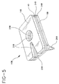
- Figs. 4, 5, 6 and 7 illustrate alternatives to the medical device of the present invention for protecting a catheter penetration site.
- the structure is substantially similar to the preferred device of Figs. 1-3. Accordingly, substantially similar components that perform substantially similar functions are numbered identically to those of Figs. 1-3 except that a suffix "A" is used in Fig. 4, and a suffix "B" is used in Figs. 5-7.
- Fig. 4 illustrates device 10A where void 20A is a single opening shaped like a slot open at one end to allow positioning of the catheter within the slot and inspection of the catheter pentration site by the practitioner analogous to the usage illustrated in Fig. 2.
- Figs. 5-7 illustrate device 10B of the present invention including resilient foam layer 16B and a cut-resistant layer 40 with patient contacting side 18B.
- Adhesive layer 24B is applied to patient contacting side 18B to releasably adhere the device to a patient's skin.
- Cut-resistant layer 40 substantially provides protection to the catheter from inadvertent cutting with bandage scissors and the like when the practitioner is removing a tape cover, when tape is used to attach the catheter.
- Cut-resistant layer 40 may be formed from materials such as a non-woven material 42 as illustrated in Fig. 6a; filamentous material 44, as illustrated in Fig. 6b; and a woven material 46, as illustrated in Fig. 6c.
- Suitable fibers for forming cut-resistant layer 40 include, but are not limited to, polymeric materials such as polyamide and polyaramide. KevlarTM, a polyaramide material available from E. I. duPont, Wilmington, DE is useful for forming cut-resistant layer 40. Metallic strands such as stainless steel and the like are also useful as fibers, filaments, woven and non-woven materials to form cut-resistant layer 40. Cut-resistant layer 40 may also be formed from combinations of metallic strands and polymeric fibers in woven, non-woven and filamentous structures. When metallic fibers, for woven material, non-woven material and as filaments are selected, two or more strands of stainless steel with a diameter greater than about 0.9 microns are preferably twisted together to form a single filament.
- Twisted metallic filaments formed from two or more strands have been shown to have greater resistance to cutting with scissors, commonly used in clinical situations to remove bandages, than an equivalent sized single strand filament.
- a schematic cross-section of woven layer 46 formed with filaments 48 formed from two twisted stainless steel strands 47 and 49 is schematically illustrated in Fig. 6d.
- Cut-resistant layer 40 also may be formed from a continuous film or sheet of polymeric material between about 0.2 mm to about 2.0 mm thick.
- Suitable polymeric materials for this application include, but are not limited to, polypropylene, polyethylene, copolymers of polypropylene and polyethylene and the like.
- Polyethylene LDPE 9551 available from Dow, Midland, Ml and polypropylene Profax PF091B available from Montell, Elkton, MD may be formed into a sheet or film and bonded or laminated to foam layer 16B.
- resilient foam layer 16B is shown forming the outermost layer of device 10B with cut-resistant layer 40 being intermediate adhesive layer 24B and resilient foam layer 16B.
- Alternative structures where cut-resistant layer 40 is outermost or incorporated into foam layer 16B are considered within the scope of the present invention, but not further illustrated in the interest of brevity.
- Fig. 7 illustrates the placement of device 10B with a catheter 22B on the skin of a patient.
- device 10B with the catheter is covered with an adhesive tape 50 following a common practice for securing catheters.
- the tape used is substantially transparent, the practitioner is able to inspect catheter penetration site 12B without removing the tape.
- many practitioners use cloth or paper tapes that are not substantially transparent. In this situation, when the practitioner removes the tape, scissors are often used. This tape removal may result in the catheter being cut by the scissors.
- Device 10B, with cut resistant layer 40 provides a resistance to the passage of the scissors and may alert the practitioner to the proximity of the scissors to the catheter and substantially reduce the possibility of damage to the catheter.
- Suitable adhesives for adhering the catheter penetration device of the invention to a patient's skin include, but are not limited to, acrylic, acrylic copolymer and silicone pressure sensitive adhesives.
- the pressure sensitive adhesive is not water soluble.
- a suitable acrylic pressure sensitive adhesive is 1-780, available from Avery Medical Products, Painesville, OH.
- a suitable silicone pressure sensitive adhesive is 355 Medical Grade Adhesive, available from Dow-Corning, Midland, Ml.
- the adhesive selected is substantially water insoluble.
- the antimicrobial agent may be mixed into the adhesive at between about 0.05 parts to about 5.0 parts per one hundred parts of adhesive.
- the adhesive then preferably is applied to the patient contacting side in a layer between about 0.1 mm to about 0.3 mm thick.
- Alternatives to mixing the antimicrobial agent into the adhesive include spraying or dipping the antimicrobial agent onto an adhesive film or tape and applying the film or tape to the patient adhering side of the device.
- release cover 26 is placed over the adhesive layer to protect the adhesive during packaging, shipping and handling. Suitable release covers may be formed from coated papers or polymer films. Silicone treated papers are widely used as release covers. The particular material selected as a release cover is dependent upon the adhesive selected. The release cover is preferably easily peelable from the adhesive selected.
- Suitable materials for forming resilient layer 16 include, but are not limited to, polymeric materials such as polyurethane foams and felts, polyethylene foam, polyimide foam, cellulose foam, silastic foam and vinyl nitrile foam.
- Representative sources for polyurethane foams and felts include, but are not limited to, Scott Paper Company, Foam Division, Chester, PA and Rogers Corp., East Woodstock, CT.
- Suitable polyethylene foam is available from Voltek, Lawrence, MA.
- Suitable polyimide foam is available from Imi-Tech, Elk Grove Village, IL.
- Suitable vinyl nitrile foam is available from Ensolite, Mishwaka, IN.
- resilient polymeric foams having a porosity between about 4 and 60 pores per cm and preferably between about 15 to about 40 pores per cm are satisfactory for forming the resilient layer in the present invention. Additionally, a foam with a density between about 0.2 to about 0.4 grams per cubic centimeter has been found to work well to form the resilient layer. Suitable resilient layer thickness is between about 2.5mm to about 8.0mm. The resilient layer preferably is between about 3 mm to about 6 mm thick.
- TegadermTM film polyurethane film with pressure sensitive adhesive, available from 3M, St. Paul, MN
- 4 percent (wt./wt.) aqueous chlorhexidine gluconate dipped in 4 percent (wt./wt.) aqueous chlorhexidine gluconate and dried.
- Woven cloth medical adhesive tape (DuraporeTM medical adhesive tape available from 3M, St. Paul, MN) dipped in 4 percent (wt./wt.) aqueous chlorhexidine gluconate and dried.
- Silicone pressure sensitive adhesive mixed with 0.1 percent (wt./wt.) chlorhexidine diacetate and applied to polyurethane foam. (#355 medical grade adhesive, Dow-Corning, Midland, Ml, applied to Scott SIF polyurethane foam, Scott Paper Co., Foam Division, Chester, PA)
- Polyurethane foam (Scott SIF, Scott Paper Co., Foam Division, Chester, PA) dipped in 1 percent (wt./wt.) aqueous chlorhexidine diacetate and dried.
- Acrylic pressure sensitive adhesive mixed with 0.1 percent (wt./wt.) chlorhexidine diacetate and applied to polyurethane foam.
- Table 1 Example # Microorganism 1 2 3 4 5 s. Aureus 2mm 5mm 0mm 1mm 1mm p. Aeruginosa 1mm 2mm 0mm 1mm 1mm c. Albicans 1mm 3mm 0.5mm 0mm 0mm e. Coli 1mm 3mm 0mm 0.5mm 0mm
- Zone of inhibition measurements made at 24, 48 and 72 hours were not materially different from each other, thus only one value is shown.
- the results show that the antimicrobial agents in the test examples are releasable from the samples and available to inhibit growth of microorganism at least under and in many instances beyond the test disc.
- the medical device of the invention is simple to manufacture and to use. It provides a practitioner with a protector for a catheter placement that not only holds the catheter in position at the penetration site, but also inhibits microbial growth around the penetration site.
- the device also allows easy inspection of the penetration site by the practitioner and in some embodiments, substantially protects the catheter from inadvertent damage by scissors and the like during tape removal.
Abstract
A medical device for protecting a catheter penetration site on the skin of a patient includes a resilient pad having a patient contacting side and at least one void therein for receiving and retaining a catheter at a catheter penetration site. The device has an adhesive on the patient contacting side of the pad for releasably adhering the pad to the patient's skin at the catheter penetration site and also includes a sufficient quantity of an antimicrobial agent to create a zone of microbial inhibition around the catheter penetration site.
Description
- This invention generally relates to percutaneous catheters and more particularly to protection of a catheter placement site into the skin of a patient.
- Many medical procedures involve the use of percutaneous catheters inserted into the body of a patient for fluid infusion or removal. Often, the catheter remains in place for 24 to 48 hours. During this period, the catheter needs to be reliably positioned and accessible for attachment and detachment of fluid handling devices. In addition to the need to keep a catheter in position on the patient's skin, the placement site needs to be protected from microorganisms.
- Many workers have developed devices to anchor or position the catheter and allow the needed access. Representative recently patented devices for catheter anchoring and positioning include: A Repositional Catheter Fixation Device (U. S. Patent No. 5,382,239); Catheter Anchoring System (U. S. Patent No. 5,354,282); and Windowed Vein Catheter Dressing (U. S. Patent No. 5,380,294). These representative patents provide several different approaches to fixation of a percutaneous catheter onto a patient's skin. These patented devices address the need to locate the catheter and protect the site, but are somewhat complex and do not address the need to suppress growth of microorganisms at the catheter penetration site.
- U. S. Patent Nos. 5,117,981 and 5,175,977, commonly assigned with the present application, disclose aspects of a kit for preparing and dressing an intravenous catheter site. The kit disclosed in these referenced patents includes an antimicrobial agent useful for disinfecting the skin where the catheter is inserted.
- Practitioners using percutaneous catheters do not rely just on devices such as those referenced above for fixation of catheters. It is a common practice to apply strips of adhesive tape across a catheter at a placement site to secure and protect the placement. Tape can strongly retain the catheter in position, but since most tapes are somewhat inelastic, movement of the skin or a fluid handling device can cause movement of the catheter in the blood vessel or at the penetration site. Additionally, when tape is used for this purpose, there are occasional occurrences of the catheter being dislodged or even cut during tape removal to access the catheter for attachment or detachment of fluid handling devices. Since, in many cases, the catheter remains in position for 24 to 48 or more hours, development of infections by common skin microorganisms at the penetration site are a concern, and practitioners also routinely inspect the penetration site for evidence of infection. Examination of the actual penetration site is sometimes difficult with the devices described above or when adhesive tape is used for securing the catheter. There is a need for a medical device that is compatible with most current practice techniques that would protect the catheter placement site. If the device allows easy access for attachment and detachment of fluid handling devices, allows the practitioner to examine the penetration site and suppresses growth of microorganisms around the penetration site, the practice of catheterization would be advanced. Additionally, if the catheter penetration site protection device additionally provided some protection against cutting the catheter during tape removal, a further benefit would be realized. Such a device is disclosed hereinbelow.
- A medical device of the present invention for protecting a catheter penetration site on the skin of a patient includes a resilient pad having a patient contacting side and at least one void therein to receive and to retain a catheter at the catheter penetration site. The pad has an adhesive on the patient contacting side for releasably adhering the pad to the patient's skin at the catheter penetration site. The device further includes a sufficient quantity of an antimicrobial agent to create a zone of microbial inhibition around the catheter penetration site.
- The device of the present invention is simple to use. The invention fills a need not met by existing retention devices by including an antimicrobial agent releasable at the catheter penetration site that inhibits growth of microorganisms at the penetration site.
-
- Fig. 1 is a perspective view of a preferred embodiment of a medical device of the present invention;
- Fig. 2 is a perspective view of the device of Fig. 1 in use at a catheter placement site;
- Fig. 3 is a side elevation of the device of Fig. 1;
- Fig. 4 is a top plan view of an alternate embodiment of the medical device of the present invention;
- Fig. 5 is a perspective view of another embodiment of the medical device of the present invention;
- Fig. 6a is a schematic illustration of the cut resistant layer in the device of Fig. 5 formed from a non-woven material;
- Fig. 6b is a schematic illustration of the cut-resistant layer in the device of Fig. 5 formed from filamentous material;
- Fig. 6c is a schematic illustration of the cut-resistant layer in the device of Fig. 5 formed from a woven material;
- Fig. 6d is a schematic cross-section of the woven material from Fig. 6c formed from twisted strands; and
- Fig. 7 is a perspective view of the device of Fig. 5 in use at a catheter placement site.
- While this invention is satisfied by embodiments in many different forms, there are shown in the drawings and are herein described in detail preferred embodiments of the invention with the understanding that the present disclosure is to be considered exemplary of the principles of the invention and is not intended to limit the invention to the embodiments illustrated. The scope of the invention is measured by the appended claims and their equivalents.
- Referring to Figs. 1, 2 and 3, a preferred
medical device 10 of the present invention for protecting acatheter penetration site 12 on askin surface 14 of a patient includes aresilient pad 16 having apatient contacting side 18 and at least onevoid 20 to receive and to retain acatheter 22.Resilient pad 16 has anadhesive layer 24 applied topatient contacting side 18 for releasably adhering the pad to the patient's skin atcatheter penetration site 12. Preferably,device 10 has arelease cover 26, releasably attached toadhesive layer 24.Release cover 26 serves to protect the adhesive layer during assembly, shipping and storage of the device and preferably includes atab 28 to facilitate removal of the release cover.Device 10 also includes a sufficient quantity of an antimicrobial agent to create a zone of inhibition of microbial growth on the patient's skin at and around the catheter penetration site when the device is positioned on the patient's skin. - As shown in Figs. 1 and 2,
void 20 may be divided into one ormore openings 23 separated by adivider 21 that serves torestrain catheter 22. Preferably,void 20 is shaped so that whendevice 10 is positioned aroundcatheter penetration site 12, the catheter is restrained and the penetration site is visible so that it can be readily inspected by the practitioner. Other shapes and configurations ofresilient pad 16 andvoid 20 to accomodate particular catheters and applications are considered to be within the scope of the present invention. As schematically illustrated in phantom in Fig. 1,device 10 also is preferably sealed in apackage 30 formed from materials substantially resistant to the passage of microorganisms and exposed to conditions rendering microorganisms non-viable. - Preferably, the antimicrobial agent is incorporated into
adhesive layer 24, but a suitable zone of inhibition may also be created by incorporating the antimicrobial agent intoresilient layer 16 or onto the surface of the adhesive. When the antimicrobial agent is incorporated into the adhesive, a concentration between about 0.1 percent to about 5.0 percent (wt./wt.) of the agent in the adhesive is suitable. Preferably, the concentration of the agent in the adhesive is between about 0.1 percent to about 1.0 percent. The concentration of antimicrobial agent used is dependent upon the particular agent selected and its availablility in the placement. Many antimicrobial agents are satisfactory for use in the present invention including, but not limited to, iodophors, chlorhexidine, salts of chlorhexidine, biguanides, salts of biguanides, phenolics, metallic ions, as well as antibiotic agents produced by microorganisms and their synthetic analogs and the like. Substantially the only restrictions on the selection and incorporation of the antimicrobial agent is the need for the antimicrobial agent to be available to form the zone of inhibition of microbial growth at and around the catheter penetration site and for compatibility between the antimicrobial agent, the resilient material and the adhesive selected. - Figs. 4, 5, 6 and 7 illustrate alternatives to the medical device of the present invention for protecting a catheter penetration site. In these embodiments, the structure is substantially similar to the preferred device of Figs. 1-3. Accordingly, substantially similar components that perform substantially similar functions are numbered identically to those of Figs. 1-3 except that a suffix "A" is used in Fig. 4, and a suffix "B" is used in Figs. 5-7.
- Fig. 4 illustrates
device 10A where void 20A is a single opening shaped like a slot open at one end to allow positioning of the catheter within the slot and inspection of the catheter pentration site by the practitioner analogous to the usage illustrated in Fig. 2. - Figs. 5-7 illustrate
device 10B of the present invention includingresilient foam layer 16B and a cut-resistant layer 40 withpatient contacting side 18B.Adhesive layer 24B is applied topatient contacting side 18B to releasably adhere the device to a patient's skin. Cut-resistant layer 40 substantially provides protection to the catheter from inadvertent cutting with bandage scissors and the like when the practitioner is removing a tape cover, when tape is used to attach the catheter. Cut-resistant layer 40 may be formed from materials such as anon-woven material 42 as illustrated in Fig. 6a;filamentous material 44, as illustrated in Fig. 6b; and awoven material 46, as illustrated in Fig. 6c. - Suitable fibers for forming cut-
resistant layer 40 include, but are not limited to, polymeric materials such as polyamide and polyaramide. Kevlar™, a polyaramide material available from E. I. duPont, Wilmington, DE is useful for forming cut-resistant layer 40. Metallic strands such as stainless steel and the like are also useful as fibers, filaments, woven and non-woven materials to form cut-resistant layer 40. Cut-resistant layer 40 may also be formed from combinations of metallic strands and polymeric fibers in woven, non-woven and filamentous structures. When metallic fibers, for woven material, non-woven material and as filaments are selected, two or more strands of stainless steel with a diameter greater than about 0.9 microns are preferably twisted together to form a single filament. Twisted metallic filaments formed from two or more strands have been shown to have greater resistance to cutting with scissors, commonly used in clinical situations to remove bandages, than an equivalent sized single strand filament. A schematic cross-section of wovenlayer 46 formed withfilaments 48 formed from two twistedstainless steel strands - Cut-
resistant layer 40 also may be formed from a continuous film or sheet of polymeric material between about 0.2 mm to about 2.0 mm thick. Suitable polymeric materials for this application include, but are not limited to, polypropylene, polyethylene, copolymers of polypropylene and polyethylene and the like. Polyethylene LDPE 9551 available from Dow, Midland, Ml and polypropylene Profax PF091B available from Montell, Elkton, MD may be formed into a sheet or film and bonded or laminated tofoam layer 16B. - In Fig. 5,
resilient foam layer 16B is shown forming the outermost layer ofdevice 10B with cut-resistant layer 40 being intermediateadhesive layer 24B andresilient foam layer 16B. Alternative structures where cut-resistant layer 40 is outermost or incorporated intofoam layer 16B are considered within the scope of the present invention, but not further illustrated in the interest of brevity. - Fig. 7 illustrates the placement of
device 10B with acatheter 22B on the skin of a patient. In this illustration,device 10B with the catheter is covered with an adhesive tape 50 following a common practice for securing catheters. If the tape used is substantially transparent, the practitioner is able to inspectcatheter penetration site 12B without removing the tape. However, many practitioners use cloth or paper tapes that are not substantially transparent. In this situation, when the practitioner removes the tape, scissors are often used. This tape removal may result in the catheter being cut by the scissors.Device 10B, with cutresistant layer 40, provides a resistance to the passage of the scissors and may alert the practitioner to the proximity of the scissors to the catheter and substantially reduce the possibility of damage to the catheter. - Suitable adhesives for adhering the catheter penetration device of the invention to a patient's skin include, but are not limited to, acrylic, acrylic copolymer and silicone pressure sensitive adhesives. Preferably, the pressure sensitive adhesive is not water soluble. A suitable acrylic pressure sensitive adhesive is 1-780, available from Avery Medical Products, Painesville, OH. A suitable silicone pressure sensitive adhesive is 355 Medical Grade Adhesive, available from Dow-Corning, Midland, Ml. Preferably, the adhesive selected is substantially water insoluble. In the preferred embodiment, where the antimicrobial agent is incorporated into the adhesive, the antimicrobial agent may be mixed into the adhesive at between about 0.05 parts to about 5.0 parts per one hundred parts of adhesive. The adhesive then preferably is applied to the patient contacting side in a layer between about 0.1 mm to about 0.3 mm thick. Alternatives to mixing the antimicrobial agent into the adhesive include spraying or dipping the antimicrobial agent onto an adhesive film or tape and applying the film or tape to the patient adhering side of the device. Preferably,
release cover 26 is placed over the adhesive layer to protect the adhesive during packaging, shipping and handling. Suitable release covers may be formed from coated papers or polymer films. Silicone treated papers are widely used as release covers. The particular material selected as a release cover is dependent upon the adhesive selected. The release cover is preferably easily peelable from the adhesive selected. - Suitable materials for forming
resilient layer 16 include, but are not limited to, polymeric materials such as polyurethane foams and felts, polyethylene foam, polyimide foam, cellulose foam, silastic foam and vinyl nitrile foam. Representative sources for polyurethane foams and felts include, but are not limited to, Scott Paper Company, Foam Division, Chester, PA and Rogers Corp., East Woodstock, CT. Suitable polyethylene foam is available from Voltek, Lawrence, MA. Suitable polyimide foam is available from Imi-Tech, Elk Grove Village, IL. Suitable vinyl nitrile foam is available from Ensolite, Mishwaka, IN. Generally, resilient polymeric foams having a porosity between about 4 and 60 pores per cm and preferably between about 15 to about 40 pores per cm are satisfactory for forming the resilient layer in the present invention. Additionally, a foam with a density between about 0.2 to about 0.4 grams per cubic centimeter has been found to work well to form the resilient layer. Suitable resilient layer thickness is between about 2.5mm to about 8.0mm. The resilient layer preferably is between about 3 mm to about 6 mm thick. - Several examples of materials for
device 10 were prepared with antimicrobial agent applied variously in the adhesive, adhesive applied to a film for application to the resilient foam and to the resilient foam itself. Samples of these several materials were tested for the ability to form a zone of microbial inhibition under and around a sample of the material placed in growth medium seeded with common microorganisms. For each example, an approximately one centimeter disc of the material to be tested was placed on Petri dishes coated with a suitable growth medium for the organism being tested. The dishes were seeded with the test microorganism and incubated at 37°C. The dishes were examined at 24 hours, 48 hours and 72 hours and the area where growth of the microorganisms was inhibited was measured. The results shown in Table 1 are reported as mm beyond the test sample disc circumference. In all cases tested, the growth of the test microorganisms was inhibited under the test sample disc. - Tegaderm™ film (polyurethane film with pressure sensitive adhesive, available from 3M, St. Paul, MN) dipped in 4 percent (wt./wt.) aqueous chlorhexidine gluconate and dried.
- Woven cloth medical adhesive tape (Durapore™ medical adhesive tape available from 3M, St. Paul, MN) dipped in 4 percent (wt./wt.) aqueous chlorhexidine gluconate and dried.
- Silicone pressure sensitive adhesive mixed with 0.1 percent (wt./wt.) chlorhexidine diacetate and applied to polyurethane foam. (#355 medical grade adhesive, Dow-Corning, Midland, Ml, applied to Scott SIF polyurethane foam, Scott Paper Co., Foam Division, Chester, PA)
- Polyurethane foam (Scott SIF, Scott Paper Co., Foam Division, Chester, PA) dipped in 1 percent (wt./wt.) aqueous chlorhexidine diacetate and dried.
- Acrylic pressure sensitive adhesive mixed with 0.1 percent (wt./wt.) chlorhexidine diacetate and applied to polyurethane foam. (acrylic adhesive I-780, Avery Medical Products, Painesville, OH, Acquell® Foam, Denver, CO).
Table 1 Example # Microorganism 1 2 3 4 5 s. Aureus 2mm 5mm 0mm 1mm 1mm p. Aeruginosa 1mm 2mm 0mm 1mm 1mm c. Albicans 1mm 3mm 0.5mm 0mm 0mm e. Coli 1mm 3mm 0mm 0.5mm 0mm - Zone of inhibition measurements made at 24, 48 and 72 hours were not materially different from each other, thus only one value is shown. The results show that the antimicrobial agents in the test examples are releasable from the samples and available to inhibit growth of microorganism at least under and in many instances beyond the test disc.
- The medical device of the invention is simple to manufacture and to use. It provides a practitioner with a protector for a catheter placement that not only holds the catheter in position at the penetration site, but also inhibits microbial growth around the penetration site. The device also allows easy inspection of the penetration site by the practitioner and in some embodiments, substantially protects the catheter from inadvertent damage by scissors and the like during tape removal.
Claims (10)
- A medical device for protecting a catheter penetration site on the skin of a patient comprising:a resilient pad having a patient contacting side and at least one void therein to receive and to retain a catheter at a catheter penetration site;an adhesive on said patient contacting side of said pad for releasably adhering the pad to the patient's skin at the catheter penetration site; anda sufficient quantity of an antimicrobial agent to create a zone of microbial inhibition around the catheter penetration site.
- The device of claim 1 wherein said resilient pad is formed from a polymeric material.
- The device of claim 1 wherein said adhesive is a pressure sensitive adhesive.
- The device of claim 3 wherein said adhesive further comprises said antimicrobial agent, said agent being releasable from said adhesive when said pad is positioned on the patient's skin.
- The device of claim 1 wherein said antimicrobial agent is impregnated into said resilient pad, said agent being releasable from said pad when said pad is positioned on the patient's skin.
- The device of claim 1 wherein said at least one void comprises an opening through said pad having a shape to accommodate a hub of the catheter and provide visual access to the penetration site of the catheter.
- The device of claim 6 wherein said at least one void further comprises an opening through the pad sized to accommodate said hub and another opening through the pad positioned to allow visual access to the catheter penetration site.
- The device of claim 1 wherein said resilient pad further comprises means for imparting resistance to cutting.
- The device of claim 8 wherein said means for cutting resistance includes a layer formed from materials selected from the group consisting of woven metallic strands, woven polymeric fibers, woven combinations of metallic strands and polymeric fibers, a plurality of non-woven metallic strands, a plurality non-woven polymeric fibers, non-woven combinations of metallic strands and polymeric fibers and a sheet of polymeric material.
- The device of claim 9 wherein each of said metallic strands comprise at least two strands of metallic wire having a diameter greater than about 9 microns being twisted together to form a single filament.
Applications Claiming Priority (2)
| Application Number | Priority Date | Filing Date | Title |
|---|---|---|---|
| US577359 | 1995-12-22 | ||
| US08/577,359 US5686096A (en) | 1995-12-22 | 1995-12-22 | Medical device for the protection of a catheter penetration site |
Publications (1)
| Publication Number | Publication Date |
|---|---|
| EP0780138A1 true EP0780138A1 (en) | 1997-06-25 |
Family
ID=24308374
Family Applications (1)
| Application Number | Title | Priority Date | Filing Date |
|---|---|---|---|
| EP96308526A Ceased EP0780138A1 (en) | 1995-12-22 | 1996-11-26 | Medical device for the protection of a catheter penetration site |
Country Status (4)
| Country | Link |
|---|---|
| US (1) | US5686096A (en) |
| EP (1) | EP0780138A1 (en) |
| JP (1) | JPH09182801A (en) |
| CA (1) | CA2191818A1 (en) |
Cited By (11)
| Publication number | Priority date | Publication date | Assignee | Title |
|---|---|---|---|---|
| WO2001068179A1 (en) * | 2000-03-13 | 2001-09-20 | Nucryst Pharmaceuticals Corp. | Transcutaneous medical device dressings and method of use |
| EP1211935A1 (en) * | 1999-08-31 | 2002-06-12 | STS Biopolymers, Inc. | Anti-infective covering for percutaneous and vascular access devices and coating method |
| US6692773B2 (en) | 2000-07-27 | 2004-02-17 | Nucryst Pharmaceuticals Corp. | Treatment of hyperproliferative skin disorders and diseases |
| WO2004020033A1 (en) * | 2002-08-27 | 2004-03-11 | Jan Liska | A transcutan catheter assembly |
| US6719987B2 (en) | 2000-04-17 | 2004-04-13 | Nucryst Pharmaceuticals Corp. | Antimicrobial bioabsorbable materials |
| US6723350B2 (en) | 2001-04-23 | 2004-04-20 | Nucryst Pharmaceuticals Corp. | Lubricious coatings for substrates |
| US7201925B2 (en) | 2002-04-23 | 2007-04-10 | Nueryst Pharmaceuticals Corp. | Treatment of ungual and subungual diseases |
| WO2007079598A1 (en) * | 2006-01-10 | 2007-07-19 | F. Hoffmann-La Roche Ag | Medical device for placing on a patient’s skin |
| WO2010049734A2 (en) * | 2008-10-28 | 2010-05-06 | Sull Limited | Methods and apparatus for securing a line |
| EP2667922B1 (en) | 2011-01-27 | 2020-04-15 | Prosys International Ltd | Shield apparatus |
| EP2613843B1 (en) | 2010-09-10 | 2021-01-27 | Mölnlycke Health Care AB | Fixation device |
Families Citing this family (63)
| Publication number | Priority date | Publication date | Assignee | Title |
|---|---|---|---|---|
| US6260344B1 (en) | 1998-01-08 | 2001-07-17 | Whizard Protective Wear Corp. | Cut resistant antimicrobial yarn and apparel |
| AU2030599A (en) | 1998-01-09 | 1999-07-26 | Whizard Protective Wear Corp. | Cut resistant yarn and apparel |
| US6572588B1 (en) | 2000-03-10 | 2003-06-03 | Venetec International, Inc. | Medical anchoring system |
| NZ538214A (en) | 2002-08-15 | 2006-05-26 | Venetec Int Inc | Catheter securement device |
| US8100872B2 (en) * | 2002-10-23 | 2012-01-24 | Tyco Healthcare Group Lp | Medical dressing containing antimicrobial agent |
| US7947021B2 (en) * | 2003-05-08 | 2011-05-24 | Boston Scientific Scimed, Inc. | Antimicrobially-charged entry port cuff |
| US8177760B2 (en) | 2004-05-12 | 2012-05-15 | C. R. Bard, Inc. | Valved connector |
| US8197447B2 (en) | 2005-04-19 | 2012-06-12 | Venetec International, Inc. | Flexible IV site protector |
| WO2006124943A2 (en) | 2005-05-18 | 2006-11-23 | Venetec International, Inc. | Insertion site protection device |
| US8057440B2 (en) | 2005-05-26 | 2011-11-15 | Venectec International, Inc. | Anchoring system for use with neonates |
| US7988673B2 (en) | 2005-07-14 | 2011-08-02 | Venetec International, Inc. | Protective dressing and methods of use thereof |
| US8740864B2 (en) | 2005-11-17 | 2014-06-03 | Becton, Dickinson And Company | Patient fluid line access valve antimicrobial cap/cleaner |
| US9138560B2 (en) | 2006-01-12 | 2015-09-22 | Venetec International, Inc. | Universal catheter securement device |
| US8597253B2 (en) | 2007-04-20 | 2013-12-03 | Bard Access Systems | Huber needle with safety sheath |
| US8834425B2 (en) | 2007-07-16 | 2014-09-16 | C.R. Bard, Inc. | Securement system employing polymeric gel |
| US9993619B2 (en) | 2007-07-17 | 2018-06-12 | C. R. Bard, Inc. | Securement system for a medical article |
| US9480821B2 (en) | 2008-06-30 | 2016-11-01 | Venetec International, Inc. | Anchoring system for a medical article |
| WO2010033858A1 (en) | 2008-09-19 | 2010-03-25 | C.R. Bard, Inc. | Medical device securement system |
| WO2010045042A2 (en) | 2008-10-17 | 2010-04-22 | Sterigear LLC | Bodily fluid drainage assembly |
| EP2373270B8 (en) | 2009-01-07 | 2023-04-12 | entrotech life sciences, inc. | Chlorhexidine-containing antimicrobial laminates |
| US20100234815A1 (en) * | 2009-03-11 | 2010-09-16 | Teleflex Medical Incorporated | Stable melt processable chlorhexidine compositions |
| US20120197205A1 (en) | 2009-05-15 | 2012-08-02 | C.R. Bard, Inc. | Universal stabilization device |
| US8689798B2 (en) * | 2009-06-09 | 2014-04-08 | David C Sabin | Protective medical device cushion and method for use thereof |
| US9566417B1 (en) * | 2009-07-02 | 2017-02-14 | Centurion Medical Products Corporation | Integrated antimicrobial dressing |
| US8486004B1 (en) * | 2009-07-02 | 2013-07-16 | Centurion Medical Products Corporation | Dressing having integral antimicrobial |
| US8740852B2 (en) | 2009-08-25 | 2014-06-03 | C. R. Bard, Inc. | Medical article securement device |
| USD754849S1 (en) * | 2009-10-05 | 2016-04-26 | Sterigear, Llc | Bodily fluid drainage bag cover |
| JP2013507186A (en) | 2009-10-06 | 2013-03-04 | ヴェネテック・インターナショナル,インコーポレーテッド | Stabilization device with snap clamp |
| AU2010303477A1 (en) | 2009-10-06 | 2012-04-12 | Venetec International, Inc. | Stabilizing device having a locking collet |
| AU2010319924B2 (en) * | 2009-10-29 | 2014-03-06 | Robert E. Helm | Sealed sterile catheter dressings |
| US10682507B2 (en) | 2009-10-29 | 2020-06-16 | One Iv Solutions, Llc | Catheter extension with integrated circumferentially sealing securement dressing |
| WO2011060197A1 (en) * | 2009-11-11 | 2011-05-19 | Venetec International, Inc. | Stabilizing device for an extension set |
| US9700700B2 (en) | 2010-03-03 | 2017-07-11 | Venetec International, Inc. | Medical article with rotatable wings |
| US9480833B2 (en) | 2010-07-15 | 2016-11-01 | Becton, Dickinson And Company | Antimicrobial IV access cap |
| WO2012015440A1 (en) | 2010-07-30 | 2012-02-02 | Venetec International, Inc. | Securement device |
| US10525234B2 (en) | 2010-09-10 | 2020-01-07 | C. R. Bard, Inc. | Antimicrobial/haemostatic interface pad for placement between percutaneously placed medical device and patient skin |
| US20140066894A1 (en) | 2010-09-10 | 2014-03-06 | C. R. Bard, Inc. | Self-Sealing Pad for a Needle-Based Infusion Set |
| CA2806393A1 (en) | 2010-09-10 | 2012-03-15 | C.R. Bard, Inc. | Systems for isolation of a needle-based infusion set |
| JP2014510038A (en) | 2011-01-21 | 2014-04-24 | エーブリー デニソン コーポレイション | Chlorhexidine gluconate-containing solvent adhesive |
| WO2012106088A2 (en) | 2011-01-31 | 2012-08-09 | Helm Robert E Jr | Snap-seal sterile intravascular catheter-dressing system |
| US9962524B2 (en) | 2011-03-11 | 2018-05-08 | Venetec International, Inc. | Medical article securement device |
| WO2012145683A1 (en) | 2011-04-21 | 2012-10-26 | Venetec International, Inc. | Anchoring system |
| EP2710085B1 (en) | 2011-05-16 | 2018-09-26 | Avery Dennison Corporation | Adhesive containing microparticles |
| WO2013040154A1 (en) | 2011-09-15 | 2013-03-21 | Helm Robert E Jr | Catheter-dressing systems with integrated flushing mechanisms |
| US9132040B2 (en) | 2011-11-17 | 2015-09-15 | Ethicon, Inc. | Dressing device |
| CN102429769B (en) | 2011-12-07 | 2014-04-09 | 唐二虎 | Protecting and fixing device for wound |
| JP6046354B2 (en) * | 2012-02-08 | 2016-12-14 | テルモ株式会社 | Detention connector |
| CN105073934B (en) | 2013-02-07 | 2021-12-28 | 艾利丹尼森公司 | Antimicrobial adhesives with improved properties |
| US9039989B2 (en) | 2013-02-13 | 2015-05-26 | Becton, Dickinson And Company | Disinfection cap for disinfecting a male luer end of an infusion therapy device |
| US9399125B2 (en) | 2013-02-13 | 2016-07-26 | Becton, Dickinson And Company | Needleless connector and access port disinfection cleaner and antimicrobial protection cap |
| US9629983B2 (en) | 2013-03-08 | 2017-04-25 | Ethicon, Inc. | All in one antimicrobial dressing for catheter coverage |
| EP2968014B1 (en) | 2013-03-15 | 2019-04-24 | Avery Dennison Corporation | Transparent cover dressing application system and inclusion of label strip |
| CA3096148C (en) | 2013-03-15 | 2023-01-10 | C.R. Bard, Inc. | Securement device having an integral strap and dressing |
| EP3082917B1 (en) * | 2013-12-20 | 2024-01-24 | Becton, Dickinson and Company | Infusion set adhesive systems |
| US9283369B2 (en) | 2014-02-20 | 2016-03-15 | Becton, Dickinson And Company | IV access port cap for providing antimicrobial protection |
| FR3018453B1 (en) * | 2014-03-17 | 2019-07-19 | P & P Innovation | NECESSARY FOR THE MAINTENANCE AND PROTECTION OF CATHETERS PLACED ON A ZONE OF THE PATIENT'S BODY. |
| EP3131540B1 (en) | 2014-04-18 | 2023-11-22 | Entrotech, Inc. | Methods of processing chlorhexidine-containing polymerizable compositions and antimicrobial articles formed thereby |
| WO2015188031A2 (en) | 2014-06-05 | 2015-12-10 | Avery Dennison Corporation | Articles with active agent concentrated at the substrate contacting surface and related methods |
| WO2016004217A1 (en) * | 2014-07-01 | 2016-01-07 | C.R. Bard, Inc. | Antimicrobial/haemostatic interface pad for placement between percutaneously placed medical device and patient skin |
| KR102469303B1 (en) | 2016-05-13 | 2022-11-18 | 씨. 알. 바드, 인크. | CATHETER SECUREMENT DEVICE INCLUDING A GUIDING NOSE |
| US11730875B2 (en) | 2019-04-01 | 2023-08-22 | Sterigear, Llc | Dual drainage bag, assemblies, and related methods |
| CN113692298B (en) | 2019-04-17 | 2024-02-13 | 巴德阿克塞斯系统股份有限公司 | Catheter securement devices including extended anchor pad and release liner snap-fit features |
| US20230321402A1 (en) * | 2022-04-06 | 2023-10-12 | Becton, Dickinson And Company | Line Draw Optimized Integrated Catheter Stabilization and Securement System |
Citations (11)
| Publication number | Priority date | Publication date | Assignee | Title |
|---|---|---|---|---|
| US3918446A (en) * | 1974-05-03 | 1975-11-11 | E Med Corp | Securement device for intravenous catheter and its tubing |
| US4579120A (en) * | 1982-09-30 | 1986-04-01 | Cordis Corporation | Strain relief for percutaneous lead |
| US4915694A (en) * | 1987-10-02 | 1990-04-10 | Vitaphore Corporation | Antimicrobial wound dressing and skin fixator for percutaneous conduits |
| WO1990004429A2 (en) * | 1988-10-28 | 1990-05-03 | Pfrimmer Kabi Gmbh & Co Kg | Adhesive dressing for intravenous peripheral cannulas |
| US5117981A (en) | 1988-09-27 | 1992-06-02 | Becton, Dickinson And Company | Kit with tourniquet and rolled gloves |
| US5175977A (en) | 1988-09-27 | 1993-01-05 | Becton, Dickinson And Company | Kit and method for packaging gloves |
| WO1993007928A1 (en) * | 1991-10-23 | 1993-04-29 | Bioderm, Inc. | Medical apparatus fixation and infection control device |
| US5354282A (en) | 1990-05-04 | 1994-10-11 | Bierman Steven F | Catheter anchoring system |
| US5364368A (en) * | 1993-07-01 | 1994-11-15 | Kauffman Kathryn M | Stabilization device for intravascular catheters |
| US5380294A (en) | 1991-01-25 | 1995-01-10 | Procter & Gamble Hygien Aktiebolag | Windowed vein catheter dressing |
| US5382239A (en) | 1992-04-24 | 1995-01-17 | Becton, Dickinson And Company | Repositional catheter fixation device |
Family Cites Families (9)
| Publication number | Priority date | Publication date | Assignee | Title |
|---|---|---|---|---|
| NO134790C (en) * | 1968-07-09 | 1984-03-22 | Smith & Nephew | Kleber ,; PRESSURE SENSITIVE, WATERPUME-PERMEABLE PRODUCT FOR SKIN USE BY HUMANS. |
| US4374126A (en) * | 1981-02-23 | 1983-02-15 | Warner-Lambert Company | Film forming antimicrobial material |
| US4397641A (en) * | 1981-04-03 | 1983-08-09 | Jacobs Daimon C | Catheter support device |
| DE3361672D1 (en) * | 1982-04-08 | 1986-02-13 | Smith & Nephew Ass | Surgical adhesive dressing |
| US5195981A (en) * | 1989-12-18 | 1993-03-23 | Johnson Melissa C | Holder for elongated members |
| US5092323A (en) * | 1990-06-29 | 1992-03-03 | Hollister Incorporated | Moisture-absorbing, site-revealing adhesive dressing |
| US5352456A (en) * | 1991-10-10 | 1994-10-04 | Cygnus Therapeutic Systems | Device for administering drug transdermally which provides an initial pulse of drug |
| US5304146A (en) * | 1992-10-23 | 1994-04-19 | Johnson Melissa C | Medical appliance securing device |
| US5536263A (en) * | 1994-03-30 | 1996-07-16 | Lectec Corporation | Non-occulusive adhesive patch for applying medication to the skin |
-
1995
- 1995-12-22 US US08/577,359 patent/US5686096A/en not_active Expired - Lifetime
-
1996
- 1996-11-26 EP EP96308526A patent/EP0780138A1/en not_active Ceased
- 1996-12-02 CA CA002191818A patent/CA2191818A1/en not_active Abandoned
- 1996-12-24 JP JP8343376A patent/JPH09182801A/en active Pending
Patent Citations (11)
| Publication number | Priority date | Publication date | Assignee | Title |
|---|---|---|---|---|
| US3918446A (en) * | 1974-05-03 | 1975-11-11 | E Med Corp | Securement device for intravenous catheter and its tubing |
| US4579120A (en) * | 1982-09-30 | 1986-04-01 | Cordis Corporation | Strain relief for percutaneous lead |
| US4915694A (en) * | 1987-10-02 | 1990-04-10 | Vitaphore Corporation | Antimicrobial wound dressing and skin fixator for percutaneous conduits |
| US5117981A (en) | 1988-09-27 | 1992-06-02 | Becton, Dickinson And Company | Kit with tourniquet and rolled gloves |
| US5175977A (en) | 1988-09-27 | 1993-01-05 | Becton, Dickinson And Company | Kit and method for packaging gloves |
| WO1990004429A2 (en) * | 1988-10-28 | 1990-05-03 | Pfrimmer Kabi Gmbh & Co Kg | Adhesive dressing for intravenous peripheral cannulas |
| US5354282A (en) | 1990-05-04 | 1994-10-11 | Bierman Steven F | Catheter anchoring system |
| US5380294A (en) | 1991-01-25 | 1995-01-10 | Procter & Gamble Hygien Aktiebolag | Windowed vein catheter dressing |
| WO1993007928A1 (en) * | 1991-10-23 | 1993-04-29 | Bioderm, Inc. | Medical apparatus fixation and infection control device |
| US5382239A (en) | 1992-04-24 | 1995-01-17 | Becton, Dickinson And Company | Repositional catheter fixation device |
| US5364368A (en) * | 1993-07-01 | 1994-11-15 | Kauffman Kathryn M | Stabilization device for intravascular catheters |
Cited By (18)
| Publication number | Priority date | Publication date | Assignee | Title |
|---|---|---|---|---|
| EP1211935A1 (en) * | 1999-08-31 | 2002-06-12 | STS Biopolymers, Inc. | Anti-infective covering for percutaneous and vascular access devices and coating method |
| EP1211935A4 (en) * | 1999-08-31 | 2002-09-11 | Sts Biopolymers Inc | Anti-infective covering for percutaneous and vascular access devices and coating method |
| WO2001068179A1 (en) * | 2000-03-13 | 2001-09-20 | Nucryst Pharmaceuticals Corp. | Transcutaneous medical device dressings and method of use |
| AU2001239069B2 (en) * | 2000-03-13 | 2004-11-18 | Smith & Nephew (Overseas) Limited | Transcutaneous medical device dressings and method of use |
| US6719987B2 (en) | 2000-04-17 | 2004-04-13 | Nucryst Pharmaceuticals Corp. | Antimicrobial bioabsorbable materials |
| US6692773B2 (en) | 2000-07-27 | 2004-02-17 | Nucryst Pharmaceuticals Corp. | Treatment of hyperproliferative skin disorders and diseases |
| US6723350B2 (en) | 2001-04-23 | 2004-04-20 | Nucryst Pharmaceuticals Corp. | Lubricious coatings for substrates |
| US6989156B2 (en) | 2001-04-23 | 2006-01-24 | Nucryst Pharmaceuticals Corp. | Therapeutic treatments using the direct application of antimicrobial metal compositions |
| US7201925B2 (en) | 2002-04-23 | 2007-04-10 | Nueryst Pharmaceuticals Corp. | Treatment of ungual and subungual diseases |
| WO2004020033A1 (en) * | 2002-08-27 | 2004-03-11 | Jan Liska | A transcutan catheter assembly |
| WO2007079598A1 (en) * | 2006-01-10 | 2007-07-19 | F. Hoffmann-La Roche Ag | Medical device for placing on a patient’s skin |
| WO2010049734A2 (en) * | 2008-10-28 | 2010-05-06 | Sull Limited | Methods and apparatus for securing a line |
| WO2010049734A3 (en) * | 2008-10-28 | 2010-12-09 | Sull Limited | Methods and apparatus for securing a line |
| US9072871B2 (en) | 2008-10-28 | 2015-07-07 | Braidlock Limited | Methods and apparatus for securing a line |
| US10561824B2 (en) | 2008-10-28 | 2020-02-18 | Braidlock Limited | Methods and apparatus for securing a line |
| US10632288B2 (en) | 2008-10-28 | 2020-04-28 | Braidlock Limited | Methods and apparatus for securing a line |
| EP2613843B1 (en) | 2010-09-10 | 2021-01-27 | Mölnlycke Health Care AB | Fixation device |
| EP2667922B1 (en) | 2011-01-27 | 2020-04-15 | Prosys International Ltd | Shield apparatus |
Also Published As
| Publication number | Publication date |
|---|---|
| CA2191818A1 (en) | 1997-06-23 |
| US5686096A (en) | 1997-11-11 |
| JPH09182801A (en) | 1997-07-15 |
Similar Documents
| Publication | Publication Date | Title |
|---|---|---|
| EP0780138A1 (en) | Medical device for the protection of a catheter penetration site | |
| US4915694A (en) | Antimicrobial wound dressing and skin fixator for percutaneous conduits | |
| US4856504A (en) | Antimicrobial wound dressing and skin fixator for orthopedic pins | |
| US4941882A (en) | Adhesive dressing for retaining a cannula on the skin | |
| US5372589A (en) | Fenestrated transparent catheter securing device and method | |
| US5976117A (en) | Wound dressing | |
| EP3796947B1 (en) | Wound care device having fluid transfer and adhesive properties | |
| US4534762A (en) | Vascular puncture dressing | |
| EP0751757B1 (en) | Dressings | |
| EP3030207B1 (en) | A support device with a contained cushioning element | |
| US20220184269A1 (en) | Wound care device having fluid transfer and adhesive properties | |
| EP0568591B1 (en) | A windowed vein catheter dressing | |
| US4029103A (en) | Anchoring plate for medical tubes | |
| US5755681A (en) | Adhesive material with removable carrier | |
| EP0392640B1 (en) | Vented absorbent dressing | |
| US6841715B2 (en) | Window dressing | |
| CA1316058C (en) | Sterilizing dressing device and method for skin puncture | |
| US5000172A (en) | Dressing system with reference marks | |
| EP1211935B1 (en) | Anti-infective covering for percutaneous and vascular access devices and coating method | |
| JPH09508045A (en) | Medical dressing | |
| JP2004130079A (en) | Adhesive gauze bandage | |
| WO1994012134A1 (en) | Wound dressings | |
| CN114173727A (en) | Antimicrobial device for insertion needle | |
| JP5092105B2 (en) | Catheter fixture and pad for catheter fixture | |
| KR102219436B1 (en) | Adhesive Pad for Fixing Tube |
Legal Events
| Date | Code | Title | Description |
|---|---|---|---|
| PUAI | Public reference made under article 153(3) epc to a published international application that has entered the european phase |
Free format text: ORIGINAL CODE: 0009012 |
|
| AK | Designated contracting states |
Kind code of ref document: A1 Designated state(s): DE ES FR GB IT |
|
| 17P | Request for examination filed |
Effective date: 19971218 |
|
| 17Q | First examination report despatched |
Effective date: 19990312 |
|
| GRAG | Despatch of communication of intention to grant |
Free format text: ORIGINAL CODE: EPIDOS AGRA |
|
| STAA | Information on the status of an ep patent application or granted ep patent |
Free format text: STATUS: THE APPLICATION HAS BEEN REFUSED |
|
| 18R | Application refused |
Effective date: 20001127 |