EP0760007B1 - Hochauflösende genkartierungs verfahren und genkartierungs sonden - Google Patents
Hochauflösende genkartierungs verfahren und genkartierungs sonden Download PDFInfo
- Publication number
- EP0760007B1 EP0760007B1 EP95919156A EP95919156A EP0760007B1 EP 0760007 B1 EP0760007 B1 EP 0760007B1 EP 95919156 A EP95919156 A EP 95919156A EP 95919156 A EP95919156 A EP 95919156A EP 0760007 B1 EP0760007 B1 EP 0760007B1
- Authority
- EP
- European Patent Office
- Prior art keywords
- chromosome
- cdna sequence
- reagents
- human
- mapping
- Prior art date
- Legal status (The legal status is an assumption and is not a legal conclusion. Google has not performed a legal analysis and makes no representation as to the accuracy of the status listed.)
- Expired - Lifetime
Links
- 238000000034 method Methods 0.000 title claims abstract description 62
- 238000013507 mapping Methods 0.000 title claims abstract description 40
- 239000000523 sample Substances 0.000 title description 39
- 210000000349 chromosome Anatomy 0.000 claims abstract description 56
- 239000002299 complementary DNA Substances 0.000 claims abstract description 29
- 239000003153 chemical reaction reagent Substances 0.000 claims abstract description 18
- 230000002759 chromosomal effect Effects 0.000 claims abstract description 15
- 108091028043 Nucleic acid sequence Proteins 0.000 claims abstract description 7
- 210000004027 cell Anatomy 0.000 claims description 25
- 238000010186 staining Methods 0.000 claims description 17
- UPBAOYRENQEPJO-UHFFFAOYSA-N n-[5-[[5-[(3-amino-3-iminopropyl)carbamoyl]-1-methylpyrrol-3-yl]carbamoyl]-1-methylpyrrol-3-yl]-4-formamido-1-methylpyrrole-2-carboxamide Chemical compound CN1C=C(NC=O)C=C1C(=O)NC1=CN(C)C(C(=O)NC2=CN(C)C(C(=O)NCCC(N)=N)=C2)=C1 UPBAOYRENQEPJO-UHFFFAOYSA-N 0.000 claims description 13
- 238000007901 in situ hybridization Methods 0.000 claims description 12
- 230000031864 metaphase Effects 0.000 claims description 12
- 210000003917 human chromosome Anatomy 0.000 claims description 10
- 239000000203 mixture Substances 0.000 claims description 10
- RURLVUZRUFHCJO-UHFFFAOYSA-N Chromomycin A3 Natural products COC(C1Cc2cc3cc(OC4CC(OC(=O)C)C(OC5CC(O)C(OC)C(C)O5)C(C)O4)c(C)c(O)c3c(O)c2C(=O)C1OC6CC(OC7CC(C)(O)C(OC(=O)C)C(C)O7)C(O)C(C)O6)C(=O)C(O)C(C)O RURLVUZRUFHCJO-UHFFFAOYSA-N 0.000 claims description 9
- ZYVSOIYQKUDENJ-WKSBCEQHSA-N chromomycin A3 Chemical compound O([C@@H]1C[C@@H](O[C@H](C)[C@@H]1OC(C)=O)OC=1C=C2C=C3C[C@H]([C@@H](C(=O)C3=C(O)C2=C(O)C=1C)O[C@@H]1O[C@H](C)[C@@H](O)[C@H](O[C@@H]2O[C@H](C)[C@@H](O)[C@H](O[C@@H]3O[C@@H](C)[C@H](OC(C)=O)[C@@](C)(O)C3)C2)C1)[C@H](OC)C(=O)[C@@H](O)[C@@H](C)O)[C@@H]1C[C@@H](O)[C@@H](OC)[C@@H](C)O1 ZYVSOIYQKUDENJ-WKSBCEQHSA-N 0.000 claims description 9
- 108010042747 stallimycin Proteins 0.000 claims description 7
- 229950009902 stallimycin Drugs 0.000 claims description 6
- 150000007523 nucleic acids Chemical group 0.000 claims description 4
- 108091060290 Chromatid Proteins 0.000 claims description 2
- 210000004756 chromatid Anatomy 0.000 claims description 2
- 239000007850 fluorescent dye Substances 0.000 claims description 2
- 108091035539 telomere Proteins 0.000 claims description 2
- 210000003411 telomere Anatomy 0.000 claims description 2
- 102000055501 telomere Human genes 0.000 claims description 2
- 210000002230 centromere Anatomy 0.000 claims 1
- 238000011065 in-situ storage Methods 0.000 claims 1
- 108090000623 proteins and genes Proteins 0.000 abstract description 60
- 108020004414 DNA Proteins 0.000 abstract description 24
- 239000003298 DNA probe Substances 0.000 abstract description 6
- 238000009396 hybridization Methods 0.000 description 27
- 108091008109 Pseudogenes Proteins 0.000 description 16
- 102000057361 Pseudogenes Human genes 0.000 description 16
- 230000004807 localization Effects 0.000 description 15
- 239000012634 fragment Substances 0.000 description 13
- 238000001514 detection method Methods 0.000 description 12
- 108020004635 Complementary DNA Proteins 0.000 description 9
- 108700024394 Exon Proteins 0.000 description 7
- 108700005084 Multigene Family Proteins 0.000 description 7
- 238000002360 preparation method Methods 0.000 description 7
- 108091008077 processed pseudogenes Proteins 0.000 description 7
- 102000053640 Argininosuccinate synthases Human genes 0.000 description 6
- 108700024106 Argininosuccinate synthases Proteins 0.000 description 6
- ZHNUHDYFZUAESO-UHFFFAOYSA-N Formamide Chemical compound NC=O ZHNUHDYFZUAESO-UHFFFAOYSA-N 0.000 description 6
- 239000000872 buffer Substances 0.000 description 6
- 102100036537 von Willebrand factor Human genes 0.000 description 6
- 108010079054 Amyloid beta-Protein Precursor Proteins 0.000 description 5
- 108091003079 Bovine Serum Albumin Proteins 0.000 description 5
- 229920001213 Polysorbate 20 Polymers 0.000 description 5
- 240000004808 Saccharomyces cerevisiae Species 0.000 description 5
- 238000004458 analytical method Methods 0.000 description 5
- 238000010367 cloning Methods 0.000 description 5
- 238000011161 development Methods 0.000 description 5
- 230000018109 developmental process Effects 0.000 description 5
- 239000000256 polyoxyethylene sorbitan monolaurate Substances 0.000 description 5
- 235000010486 polyoxyethylene sorbitan monolaurate Nutrition 0.000 description 5
- 102100040055 Amyloid beta precursor like protein 1 Human genes 0.000 description 4
- 102000014303 Amyloid beta-Protein Precursor Human genes 0.000 description 4
- 101000890407 Homo sapiens Amyloid beta precursor like protein 1 Proteins 0.000 description 4
- 101000909516 Homo sapiens Contactin-2 Proteins 0.000 description 4
- 208000026350 Inborn Genetic disease Diseases 0.000 description 4
- 102000001253 Protein Kinase Human genes 0.000 description 4
- 230000003321 amplification Effects 0.000 description 4
- 229940098773 bovine serum albumin Drugs 0.000 description 4
- 230000007423 decrease Effects 0.000 description 4
- 230000003247 decreasing effect Effects 0.000 description 4
- 201000010099 disease Diseases 0.000 description 4
- 208000037265 diseases, disorders, signs and symptoms Diseases 0.000 description 4
- 208000025688 early-onset autosomal dominant Alzheimer disease Diseases 0.000 description 4
- 208000015756 familial Alzheimer disease Diseases 0.000 description 4
- 208000016361 genetic disease Diseases 0.000 description 4
- 230000002068 genetic effect Effects 0.000 description 4
- 230000035772 mutation Effects 0.000 description 4
- 238000003199 nucleic acid amplification method Methods 0.000 description 4
- 239000002773 nucleotide Substances 0.000 description 4
- 125000003729 nucleotide group Chemical group 0.000 description 4
- 108060006633 protein kinase Proteins 0.000 description 4
- 230000035945 sensitivity Effects 0.000 description 4
- 238000011282 treatment Methods 0.000 description 4
- FWBHETKCLVMNFS-UHFFFAOYSA-N 4',6-Diamino-2-phenylindol Chemical compound C1=CC(C(=N)N)=CC=C1C1=CC2=CC=C(C(N)=N)C=C2N1 FWBHETKCLVMNFS-UHFFFAOYSA-N 0.000 description 3
- WOVKYSAHUYNSMH-RRKCRQDMSA-N 5-bromodeoxyuridine Chemical compound C1[C@H](O)[C@@H](CO)O[C@H]1N1C(=O)NC(=O)C(Br)=C1 WOVKYSAHUYNSMH-RRKCRQDMSA-N 0.000 description 3
- QTBSBXVTEAMEQO-UHFFFAOYSA-N Acetic acid Chemical compound CC(O)=O QTBSBXVTEAMEQO-UHFFFAOYSA-N 0.000 description 3
- 208000024827 Alzheimer disease Diseases 0.000 description 3
- 206010068051 Chimerism Diseases 0.000 description 3
- 102000016918 Complement C3 Human genes 0.000 description 3
- 108010028780 Complement C3 Proteins 0.000 description 3
- 102100024342 Contactin-2 Human genes 0.000 description 3
- 102000001390 Fructose-Bisphosphate Aldolase Human genes 0.000 description 3
- 108010068561 Fructose-Bisphosphate Aldolase Proteins 0.000 description 3
- 108091006027 G proteins Proteins 0.000 description 3
- 102000030782 GTP binding Human genes 0.000 description 3
- 108091000058 GTP-Binding Proteins 0.000 description 3
- 101000782195 Homo sapiens von Willebrand factor Proteins 0.000 description 3
- OKKJLVBELUTLKV-UHFFFAOYSA-N Methanol Chemical compound OC OKKJLVBELUTLKV-UHFFFAOYSA-N 0.000 description 3
- 102000006478 Protein Phosphatase 2 Human genes 0.000 description 3
- 108010058956 Protein Phosphatase 2 Proteins 0.000 description 3
- 102000006382 Ribonucleases Human genes 0.000 description 3
- 108010083644 Ribonucleases Proteins 0.000 description 3
- 108010052164 Sodium Channels Proteins 0.000 description 3
- 102000018674 Sodium Channels Human genes 0.000 description 3
- 238000002105 Southern blotting Methods 0.000 description 3
- 108010062497 VLDL Lipoproteins Proteins 0.000 description 3
- 238000010420 art technique Methods 0.000 description 3
- 230000008901 benefit Effects 0.000 description 3
- 238000010276 construction Methods 0.000 description 3
- 230000002559 cytogenic effect Effects 0.000 description 3
- 238000012217 deletion Methods 0.000 description 3
- 230000037430 deletion Effects 0.000 description 3
- 238000004925 denaturation Methods 0.000 description 3
- 230000036425 denaturation Effects 0.000 description 3
- 210000004698 lymphocyte Anatomy 0.000 description 3
- 230000009061 membrane transport Effects 0.000 description 3
- 239000013612 plasmid Substances 0.000 description 3
- 230000008707 rearrangement Effects 0.000 description 3
- 239000000243 solution Substances 0.000 description 3
- 238000013519 translation Methods 0.000 description 3
- 108010047303 von Willebrand Factor Proteins 0.000 description 3
- 229960001134 von willebrand factor Drugs 0.000 description 3
- 102100029077 3-hydroxy-3-methylglutaryl-coenzyme A reductase Human genes 0.000 description 2
- 102100038910 Alpha-enolase Human genes 0.000 description 2
- WOVKYSAHUYNSMH-UHFFFAOYSA-N BROMODEOXYURIDINE Natural products C1C(O)C(CO)OC1N1C(=O)NC(=O)C(Br)=C1 WOVKYSAHUYNSMH-UHFFFAOYSA-N 0.000 description 2
- 241000283707 Capra Species 0.000 description 2
- 102000014914 Carrier Proteins Human genes 0.000 description 2
- 108010078791 Carrier Proteins Proteins 0.000 description 2
- 208000015374 Central core disease Diseases 0.000 description 2
- 108010087196 Contactin 2 Proteins 0.000 description 2
- 101100135859 Dictyostelium discoideum regA gene Proteins 0.000 description 2
- 241000588724 Escherichia coli Species 0.000 description 2
- 102000009127 Glutaminase Human genes 0.000 description 2
- 108010073324 Glutaminase Proteins 0.000 description 2
- 239000012981 Hank's balanced salt solution Substances 0.000 description 2
- 102000003939 Membrane transport proteins Human genes 0.000 description 2
- 108090000301 Membrane transport proteins Proteins 0.000 description 2
- 102100027179 Microtubule-associated protein 1B Human genes 0.000 description 2
- 108090001040 Microtubule-associated protein 1B Proteins 0.000 description 2
- 101100384865 Neurospora crassa (strain ATCC 24698 / 74-OR23-1A / CBS 708.71 / DSM 1257 / FGSC 987) cot-1 gene Proteins 0.000 description 2
- 108700019535 Phosphoprotein Phosphatases Proteins 0.000 description 2
- 102000045595 Phosphoprotein Phosphatases Human genes 0.000 description 2
- 108010022181 Phosphopyruvate Hydratase Proteins 0.000 description 2
- 102000004861 Phosphoric Diester Hydrolases Human genes 0.000 description 2
- 108090001050 Phosphoric Diester Hydrolases Proteins 0.000 description 2
- 108091000080 Phosphotransferase Proteins 0.000 description 2
- 101100082606 Plasmodium falciparum (isolate 3D7) PDEbeta gene Proteins 0.000 description 2
- 108091081062 Repeated sequence (DNA) Proteins 0.000 description 2
- 241000589180 Rhizobium Species 0.000 description 2
- 101100135860 Saccharomyces cerevisiae (strain ATCC 204508 / S288c) PDE2 gene Proteins 0.000 description 2
- FAPWRFPIFSIZLT-UHFFFAOYSA-M Sodium chloride Chemical compound [Na+].[Cl-] FAPWRFPIFSIZLT-UHFFFAOYSA-M 0.000 description 2
- IQFYYKKMVGJFEH-XLPZGREQSA-N Thymidine Chemical compound O=C1NC(=O)C(C)=CN1[C@@H]1O[C@H](CO)[C@@H](O)C1 IQFYYKKMVGJFEH-XLPZGREQSA-N 0.000 description 2
- 102100039066 Very low-density lipoprotein receptor Human genes 0.000 description 2
- 208000006254 Wolf-Hirschhorn Syndrome Diseases 0.000 description 2
- 101100215634 Yarrowia lipolytica (strain CLIB 122 / E 150) XPR2 gene Proteins 0.000 description 2
- 230000003376 axonal effect Effects 0.000 description 2
- 210000004556 brain Anatomy 0.000 description 2
- 229950004398 broxuridine Drugs 0.000 description 2
- 201000007303 central core myopathy Diseases 0.000 description 2
- 238000011156 evaluation Methods 0.000 description 2
- 238000005562 fading Methods 0.000 description 2
- 108010081400 fluorescein isothiocyante avidin Proteins 0.000 description 2
- MHMNJMPURVTYEJ-UHFFFAOYSA-N fluorescein-5-isothiocyanate Chemical compound O1C(=O)C2=CC(N=C=S)=CC=C2C21C1=CC=C(O)C=C1OC1=CC(O)=CC=C21 MHMNJMPURVTYEJ-UHFFFAOYSA-N 0.000 description 2
- 102000053135 human CNTN2 Human genes 0.000 description 2
- 238000004519 manufacturing process Methods 0.000 description 2
- 230000004048 modification Effects 0.000 description 2
- 238000012986 modification Methods 0.000 description 2
- 230000001537 neural effect Effects 0.000 description 2
- 230000000926 neurological effect Effects 0.000 description 2
- 230000009871 nonspecific binding Effects 0.000 description 2
- 102000020233 phosphotransferase Human genes 0.000 description 2
- XJMOSONTPMZWPB-UHFFFAOYSA-M propidium iodide Chemical compound [I-].[I-].C12=CC(N)=CC=C2C2=CC=C(N)C=C2[N+](CCC[N+](C)(CC)CC)=C1C1=CC=CC=C1 XJMOSONTPMZWPB-UHFFFAOYSA-M 0.000 description 2
- 102000004169 proteins and genes Human genes 0.000 description 2
- 108020003175 receptors Proteins 0.000 description 2
- 102000005962 receptors Human genes 0.000 description 2
- UCSJYZPVAKXKNQ-HZYVHMACSA-N streptomycin Chemical compound CN[C@H]1[C@H](O)[C@@H](O)[C@H](CO)O[C@H]1O[C@@H]1[C@](C=O)(O)[C@H](C)O[C@H]1O[C@@H]1[C@@H](NC(N)=N)[C@H](O)[C@@H](NC(N)=N)[C@H](O)[C@H]1O UCSJYZPVAKXKNQ-HZYVHMACSA-N 0.000 description 2
- 239000013598 vector Substances 0.000 description 2
- XLYOFNOQVPJJNP-UHFFFAOYSA-N water Substances O XLYOFNOQVPJJNP-UHFFFAOYSA-N 0.000 description 2
- NNJPGOLRFBJNIW-HNNXBMFYSA-N (-)-demecolcine Chemical compound C1=C(OC)C(=O)C=C2[C@@H](NC)CCC3=CC(OC)=C(OC)C(OC)=C3C2=C1 NNJPGOLRFBJNIW-HNNXBMFYSA-N 0.000 description 1
- 101150084750 1 gene Proteins 0.000 description 1
- CBCKQZAAMUWICA-UHFFFAOYSA-N 1,4-phenylenediamine Chemical compound NC1=CC=C(N)C=C1 CBCKQZAAMUWICA-UHFFFAOYSA-N 0.000 description 1
- GNXFOGHNGIVQEH-UHFFFAOYSA-N 2-hydroxy-3-(2-methoxyphenoxy)propyl carbamate Chemical compound COC1=CC=CC=C1OCC(O)COC(N)=O GNXFOGHNGIVQEH-UHFFFAOYSA-N 0.000 description 1
- 102000006267 AMP Deaminase Human genes 0.000 description 1
- 108700016228 AMP deaminases Proteins 0.000 description 1
- 102100034526 AP-1 complex subunit mu-1 Human genes 0.000 description 1
- 101800000263 Acidic protein Proteins 0.000 description 1
- 208000036762 Acute promyelocytic leukaemia Diseases 0.000 description 1
- 102100036601 Aggrecan core protein Human genes 0.000 description 1
- 108010067219 Aggrecans Proteins 0.000 description 1
- 101710162350 Alkaline extracellular protease Proteins 0.000 description 1
- 108700028369 Alleles Proteins 0.000 description 1
- 101710168919 Amyloid beta precursor like protein 1 Proteins 0.000 description 1
- 102100040038 Amyloid beta precursor like protein 2 Human genes 0.000 description 1
- 102100029470 Apolipoprotein E Human genes 0.000 description 1
- 101710095339 Apolipoprotein E Proteins 0.000 description 1
- 102100020999 Argininosuccinate synthase Human genes 0.000 description 1
- 241000972773 Aulopiformes Species 0.000 description 1
- 101000950981 Bacillus subtilis (strain 168) Catabolic NAD-specific glutamate dehydrogenase RocG Proteins 0.000 description 1
- 241000894006 Bacteria Species 0.000 description 1
- DWRXFEITVBNRMK-UHFFFAOYSA-N Beta-D-1-Arabinofuranosylthymine Natural products O=C1NC(=O)C(C)=CN1C1C(O)C(O)C(CO)O1 DWRXFEITVBNRMK-UHFFFAOYSA-N 0.000 description 1
- 201000004569 Blindness Diseases 0.000 description 1
- 241000283690 Bos taurus Species 0.000 description 1
- 101100163949 Caenorhabditis elegans asp-3 gene Proteins 0.000 description 1
- 102000019025 Calcium-Calmodulin-Dependent Protein Kinases Human genes 0.000 description 1
- 108010026870 Calcium-Calmodulin-Dependent Protein Kinases Proteins 0.000 description 1
- 102000005367 Carboxypeptidases Human genes 0.000 description 1
- 108010006303 Carboxypeptidases Proteins 0.000 description 1
- 208000005623 Carcinogenesis Diseases 0.000 description 1
- 208000031404 Chromosome Aberrations Diseases 0.000 description 1
- 102100029057 Coagulation factor XIII A chain Human genes 0.000 description 1
- 101710197191 Coagulation factor XIII A chain Proteins 0.000 description 1
- 108091026890 Coding region Proteins 0.000 description 1
- 108091035707 Consensus sequence Proteins 0.000 description 1
- 102000006630 Contactin 2 Human genes 0.000 description 1
- NNJPGOLRFBJNIW-UHFFFAOYSA-N Demecolcine Natural products C1=C(OC)C(=O)C=C2C(NC)CCC3=CC(OC)=C(OC)C(OC)=C3C2=C1 NNJPGOLRFBJNIW-UHFFFAOYSA-N 0.000 description 1
- 101001098814 Dictyostelium discoideum 3',5'-cyclic-nucleotide phosphodiesterase regA Proteins 0.000 description 1
- LFQSCWFLJHTTHZ-UHFFFAOYSA-N Ethanol Chemical class CCO LFQSCWFLJHTTHZ-UHFFFAOYSA-N 0.000 description 1
- 101710089384 Extracellular protease Proteins 0.000 description 1
- 102000017177 Fibromodulin Human genes 0.000 description 1
- 108010013996 Fibromodulin Proteins 0.000 description 1
- 108010093223 Folylpolyglutamate synthetase Proteins 0.000 description 1
- 238000002738 Giemsa staining Methods 0.000 description 1
- 102000053171 Glial Fibrillary Acidic Human genes 0.000 description 1
- 101710193519 Glial fibrillary acidic protein Proteins 0.000 description 1
- 102000016901 Glutamate dehydrogenase Human genes 0.000 description 1
- 101710184005 Glutaminase 2 Proteins 0.000 description 1
- 102100025961 Glutaminase liver isoform, mitochondrial Human genes 0.000 description 1
- 108010043428 Glycine hydroxymethyltransferase Proteins 0.000 description 1
- 102000003886 Glycoproteins Human genes 0.000 description 1
- 108090000288 Glycoproteins Proteins 0.000 description 1
- 206010053759 Growth retardation Diseases 0.000 description 1
- 102100023954 Guanine nucleotide-binding protein subunit alpha-15 Human genes 0.000 description 1
- 241000282412 Homo Species 0.000 description 1
- 101000988577 Homo sapiens 3-hydroxy-3-methylglutaryl-coenzyme A reductase Proteins 0.000 description 1
- 101000797458 Homo sapiens AMP deaminase 2 Proteins 0.000 description 1
- 101000924643 Homo sapiens AP-1 complex subunit mu-1 Proteins 0.000 description 1
- 101100491370 Homo sapiens APLP1 gene Proteins 0.000 description 1
- 101000890401 Homo sapiens Amyloid beta precursor like protein 2 Proteins 0.000 description 1
- 101000904080 Homo sapiens Guanine nucleotide-binding protein subunit alpha-15 Proteins 0.000 description 1
- 101001002634 Homo sapiens Interleukin-1 alpha Proteins 0.000 description 1
- 101000753178 Homo sapiens Sodium/potassium-transporting ATPase subunit alpha-3 Proteins 0.000 description 1
- 101000687911 Homo sapiens Transcription factor SOX-3 Proteins 0.000 description 1
- 101000855256 Homo sapiens Uncharacterized protein C16orf74 Proteins 0.000 description 1
- 101000614791 Homo sapiens cAMP-dependent protein kinase type I-beta regulatory subunit Proteins 0.000 description 1
- 102000006933 Hydroxymethyl and Formyl Transferases Human genes 0.000 description 1
- 108010072462 Hydroxymethyl and Formyl Transferases Proteins 0.000 description 1
- 108090000895 Hydroxymethylglutaryl CoA Reductases Proteins 0.000 description 1
- 206010021067 Hypopituitarism Diseases 0.000 description 1
- 108010048077 Inositol 1,4,5-trisphosphate 3-kinase Proteins 0.000 description 1
- 108091092195 Intron Proteins 0.000 description 1
- ZDXPYRJPNDTMRX-VKHMYHEASA-N L-glutamine Chemical compound OC(=O)[C@@H](N)CCC(N)=O ZDXPYRJPNDTMRX-VKHMYHEASA-N 0.000 description 1
- 229930182816 L-glutamine Natural products 0.000 description 1
- 101710192606 Latent membrane protein 2 Proteins 0.000 description 1
- 102100031955 Lon protease homolog, mitochondrial Human genes 0.000 description 1
- 101710167388 Lon protease homolog, mitochondrial Proteins 0.000 description 1
- 102000012750 Membrane Glycoproteins Human genes 0.000 description 1
- 108010090054 Membrane Glycoproteins Proteins 0.000 description 1
- 102000018697 Membrane Proteins Human genes 0.000 description 1
- 108010052285 Membrane Proteins Proteins 0.000 description 1
- 208000036626 Mental retardation Diseases 0.000 description 1
- 102000006404 Mitochondrial Proteins Human genes 0.000 description 1
- 108010058682 Mitochondrial Proteins Proteins 0.000 description 1
- 102100024193 Mitogen-activated protein kinase 1 Human genes 0.000 description 1
- 241001529936 Murinae Species 0.000 description 1
- 208000012902 Nervous system disease Diseases 0.000 description 1
- 208000025966 Neurological disease Diseases 0.000 description 1
- 102000004132 Ornithine aminotransferases Human genes 0.000 description 1
- 108090000691 Ornithine aminotransferases Proteins 0.000 description 1
- 229930182555 Penicillin Natural products 0.000 description 1
- JGSARLDLIJGVTE-MBNYWOFBSA-N Penicillin G Chemical compound N([C@H]1[C@H]2SC([C@@H](N2C1=O)C(O)=O)(C)C)C(=O)CC1=CC=CC=C1 JGSARLDLIJGVTE-MBNYWOFBSA-N 0.000 description 1
- 108010047620 Phytohemagglutinins Proteins 0.000 description 1
- 108010093965 Polymyxin B Proteins 0.000 description 1
- 102000004257 Potassium Channel Human genes 0.000 description 1
- 239000012980 RPMI-1640 medium Substances 0.000 description 1
- 230000018199 S phase Effects 0.000 description 1
- 229920005654 Sephadex Polymers 0.000 description 1
- 239000012507 Sephadex™ Substances 0.000 description 1
- MTCFGRXMJLQNBG-UHFFFAOYSA-N Serine Natural products OCC(N)C(O)=O MTCFGRXMJLQNBG-UHFFFAOYSA-N 0.000 description 1
- 102000019394 Serine hydroxymethyltransferases Human genes 0.000 description 1
- 102100021952 Sodium/potassium-transporting ATPase subunit alpha-3 Human genes 0.000 description 1
- 108010057722 Synaptosomal-Associated Protein 25 Proteins 0.000 description 1
- 102000004183 Synaptosomal-Associated Protein 25 Human genes 0.000 description 1
- 101710109576 Terminal protein Proteins 0.000 description 1
- 108010022173 Thiosulfate sulfurtransferase Proteins 0.000 description 1
- 102100034707 Thiosulfate sulfurtransferase Human genes 0.000 description 1
- 102100024276 Transcription factor SOX-3 Human genes 0.000 description 1
- 206010044565 Tremor Diseases 0.000 description 1
- 108010040002 Tumor Suppressor Proteins Proteins 0.000 description 1
- 102000001742 Tumor Suppressor Proteins Human genes 0.000 description 1
- 102100026591 Uncharacterized protein C16orf74 Human genes 0.000 description 1
- 108010023795 VLDL receptor Proteins 0.000 description 1
- 201000002919 Van der Woude syndrome Diseases 0.000 description 1
- 101710177612 Very low-density lipoprotein receptor Proteins 0.000 description 1
- 108010053752 Voltage-Gated Sodium Channels Proteins 0.000 description 1
- 102000016913 Voltage-Gated Sodium Channels Human genes 0.000 description 1
- 241000235015 Yarrowia lipolytica Species 0.000 description 1
- MMWCIQZXVOZEGG-HOZKJCLWSA-N [(1S,2R,3S,4S,5R,6S)-2,3,5-trihydroxy-4,6-diphosphonooxycyclohexyl] dihydrogen phosphate Chemical compound O[C@H]1[C@@H](O)[C@H](OP(O)(O)=O)[C@@H](OP(O)(O)=O)[C@H](O)[C@H]1OP(O)(O)=O MMWCIQZXVOZEGG-HOZKJCLWSA-N 0.000 description 1
- 239000011543 agarose gel Substances 0.000 description 1
- 238000000246 agarose gel electrophoresis Methods 0.000 description 1
- 230000032683 aging Effects 0.000 description 1
- 210000004436 artificial bacterial chromosome Anatomy 0.000 description 1
- 210000004507 artificial chromosome Anatomy 0.000 description 1
- 210000001106 artificial yeast chromosome Anatomy 0.000 description 1
- 230000008955 bacterial trafficking Effects 0.000 description 1
- 230000004888 barrier function Effects 0.000 description 1
- IQFYYKKMVGJFEH-UHFFFAOYSA-N beta-L-thymidine Natural products O=C1NC(=O)C(C)=CN1C1OC(CO)C(O)C1 IQFYYKKMVGJFEH-UHFFFAOYSA-N 0.000 description 1
- 230000033228 biological regulation Effects 0.000 description 1
- 230000015572 biosynthetic process Effects 0.000 description 1
- 230000006287 biotinylation Effects 0.000 description 1
- 238000007413 biotinylation Methods 0.000 description 1
- 230000000903 blocking effect Effects 0.000 description 1
- 102100021203 cAMP-dependent protein kinase type I-beta regulatory subunit Human genes 0.000 description 1
- 230000036952 cancer formation Effects 0.000 description 1
- 231100000504 carcinogenesis Toxicity 0.000 description 1
- 230000022131 cell cycle Effects 0.000 description 1
- 230000007910 cell fusion Effects 0.000 description 1
- 230000033077 cellular process Effects 0.000 description 1
- 239000004568 cement Substances 0.000 description 1
- 239000003795 chemical substances by application Substances 0.000 description 1
- 238000004587 chromatography analysis Methods 0.000 description 1
- 230000008711 chromosomal rearrangement Effects 0.000 description 1
- 231100000005 chromosome aberration Toxicity 0.000 description 1
- 208000016653 cleft lip/palate Diseases 0.000 description 1
- 230000000295 complement effect Effects 0.000 description 1
- 238000012790 confirmation Methods 0.000 description 1
- 230000002596 correlated effect Effects 0.000 description 1
- 230000000875 corresponding effect Effects 0.000 description 1
- 208000031513 cyst Diseases 0.000 description 1
- 210000000805 cytoplasm Anatomy 0.000 description 1
- 230000001086 cytosolic effect Effects 0.000 description 1
- 230000018044 dehydration Effects 0.000 description 1
- 238000006297 dehydration reaction Methods 0.000 description 1
- 230000001419 dependent effect Effects 0.000 description 1
- 239000012153 distilled water Substances 0.000 description 1
- 239000000975 dye Substances 0.000 description 1
- 239000000839 emulsion Substances 0.000 description 1
- 230000037149 energy metabolism Effects 0.000 description 1
- 238000005516 engineering process Methods 0.000 description 1
- 239000012894 fetal calf serum Substances 0.000 description 1
- 230000001605 fetal effect Effects 0.000 description 1
- 102000006482 fibulin Human genes 0.000 description 1
- 108010044392 fibulin Proteins 0.000 description 1
- 239000012530 fluid Substances 0.000 description 1
- 102000030722 folylpolyglutamate synthetase Human genes 0.000 description 1
- 230000006870 function Effects 0.000 description 1
- 238000001502 gel electrophoresis Methods 0.000 description 1
- 230000002518 glial effect Effects 0.000 description 1
- 210000005046 glial fibrillary acidic protein Anatomy 0.000 description 1
- 230000012010 growth Effects 0.000 description 1
- 230000036541 health Effects 0.000 description 1
- 102000046715 human AMPD2 Human genes 0.000 description 1
- 239000000815 hypotonic solution Substances 0.000 description 1
- 230000001900 immune effect Effects 0.000 description 1
- 238000010166 immunofluorescence Methods 0.000 description 1
- 238000010348 incorporation Methods 0.000 description 1
- 238000011534 incubation Methods 0.000 description 1
- 238000011835 investigation Methods 0.000 description 1
- 238000002955 isolation Methods 0.000 description 1
- 230000000155 isotopic effect Effects 0.000 description 1
- 238000002372 labelling Methods 0.000 description 1
- 238000012423 maintenance Methods 0.000 description 1
- 239000002609 medium Substances 0.000 description 1
- 239000012528 membrane Substances 0.000 description 1
- 230000037353 metabolic pathway Effects 0.000 description 1
- 238000007431 microscopic evaluation Methods 0.000 description 1
- 238000000386 microscopy Methods 0.000 description 1
- 208000012268 mitochondrial disease Diseases 0.000 description 1
- 230000002438 mitochondrial effect Effects 0.000 description 1
- 239000003226 mitogen Substances 0.000 description 1
- 238000010369 molecular cloning Methods 0.000 description 1
- 210000003205 muscle Anatomy 0.000 description 1
- 210000005036 nerve Anatomy 0.000 description 1
- 210000000653 nervous system Anatomy 0.000 description 1
- 230000004031 neuronal differentiation Effects 0.000 description 1
- 230000024121 nodulation Effects 0.000 description 1
- 201000009958 panhypopituitarism Diseases 0.000 description 1
- 229940049954 penicillin Drugs 0.000 description 1
- 230000002093 peripheral effect Effects 0.000 description 1
- 239000008363 phosphate buffer Substances 0.000 description 1
- 230000000704 physical effect Effects 0.000 description 1
- 230000001885 phytohemagglutinin Effects 0.000 description 1
- 229920000024 polymyxin B Polymers 0.000 description 1
- 229960005266 polymyxin b Drugs 0.000 description 1
- 108020001213 potassium channel Proteins 0.000 description 1
- 239000002243 precursor Substances 0.000 description 1
- 230000008569 process Effects 0.000 description 1
- 108090000765 processed proteins & peptides Proteins 0.000 description 1
- 235000019515 salmon Nutrition 0.000 description 1
- 150000003839 salts Chemical class 0.000 description 1
- 210000002265 sensory receptor cell Anatomy 0.000 description 1
- 238000012163 sequencing technique Methods 0.000 description 1
- 210000002966 serum Anatomy 0.000 description 1
- 238000007493 shaping process Methods 0.000 description 1
- 239000011780 sodium chloride Substances 0.000 description 1
- 239000001509 sodium citrate Substances 0.000 description 1
- NLJMYIDDQXHKNR-UHFFFAOYSA-K sodium citrate Chemical compound O.O.[Na+].[Na+].[Na+].[O-]C(=O)CC(O)(CC([O-])=O)C([O-])=O NLJMYIDDQXHKNR-UHFFFAOYSA-K 0.000 description 1
- 239000007787 solid Substances 0.000 description 1
- 238000001228 spectrum Methods 0.000 description 1
- 238000007619 statistical method Methods 0.000 description 1
- 229960005322 streptomycin Drugs 0.000 description 1
- 239000000725 suspension Substances 0.000 description 1
- 238000012360 testing method Methods 0.000 description 1
- 229940104230 thymidine Drugs 0.000 description 1
- 210000001519 tissue Anatomy 0.000 description 1
- 238000013518 transcription Methods 0.000 description 1
- 230000035897 transcription Effects 0.000 description 1
- 230000007704 transition Effects 0.000 description 1
- 230000014616 translation Effects 0.000 description 1
- 230000005945 translocation Effects 0.000 description 1
- 238000009281 ultraviolet germicidal irradiation Methods 0.000 description 1
- 108700026220 vif Genes Proteins 0.000 description 1
- 238000012800 visualization Methods 0.000 description 1
- 238000005406 washing Methods 0.000 description 1
Images
Classifications
-
- C—CHEMISTRY; METALLURGY
- C12—BIOCHEMISTRY; BEER; SPIRITS; WINE; VINEGAR; MICROBIOLOGY; ENZYMOLOGY; MUTATION OR GENETIC ENGINEERING
- C12Q—MEASURING OR TESTING PROCESSES INVOLVING ENZYMES, NUCLEIC ACIDS OR MICROORGANISMS; COMPOSITIONS OR TEST PAPERS THEREFOR; PROCESSES OF PREPARING SUCH COMPOSITIONS; CONDITION-RESPONSIVE CONTROL IN MICROBIOLOGICAL OR ENZYMOLOGICAL PROCESSES
- C12Q1/00—Measuring or testing processes involving enzymes, nucleic acids or microorganisms; Compositions therefor; Processes of preparing such compositions
- C12Q1/68—Measuring or testing processes involving enzymes, nucleic acids or microorganisms; Compositions therefor; Processes of preparing such compositions involving nucleic acids
- C12Q1/6813—Hybridisation assays
- C12Q1/6841—In situ hybridisation
Definitions
- the present invention relates to methods and reagents used to identify genes responsible for human disease.
- the present invention also relates to construction of high resolution genetic maps spanning the human genome.
- Physical mapping techniques provide a complement to genetic linkage. For example, standard agarose gel electrophoresis gives resolution in the 1 to 10 kb range. Pulsed field techniques extend this range to the megabase level.
- gel electrophoresis methods are not useful for the initial localization of a DNA sequence that has not been mapped previously. Furthermore, such methods do not directly provide the ability to order DNA sequences which are more than a few hundred kilobases apart, nor do such methods provide the ability to assign DNA sequences to specific chromosomal regions.
- Chromosome banding techniques have facilitated the identification of specific human chromosomes and presently provide the major basis upon which chromosomal aberrations are diagnosed.
- several techniques have been used to obtain a high level of precision in localization. These techniques, however, have some disadvantages. For example, techniques for banding that depend in part on DNA loss after UV irradiation, may therefore result in some signal loss ( Cherif et al. in Proc. Natl. Acad. Sci. USA 87:6639-6643 (1990 ); and Takahashi et al. in Hum. Genet 86:14-16 (1990 )).
- Standard DAPI (4',6-diamidino-2-phenylindole), or PI (Propidium Iodide) staining produce inconsistent patterns and are, in general, of lower resolution ( Pinkel et al. in Proc. Natl. Acad. Sci. USA 83:2934-2938 (1986 ); Lemieux et al. in Cytogenet Cell Genet. 59:311-312 (1992 ); Yamada et al. in Genomics 15:449-452 (1993 ); and Heng et al. in Chromosoma 102:325-332 (1993) ).
- Trends in Biotechnology, vol.10, 1992, pp. 27-32 discloses a method for high resolution mapping of (a) nucleic acid sequence (-s) to a single band on a human chromosome, by means of FISH of biotinilated probes and post FISH counterstaining with two counterstains to yield high resolution R bands wherein the counterstains are applied in two staining rounds, first one with chromomycin A3 and then (sequentially) one with distamycin A.
- the present invention relates to a method for mapping cDNA sequences as defined in claim 1 and to the use of reagents as defined in claim 4.
- the present invention provides a high resolution, reproducible reverse (R) banding method that enables rapid determination and accurate assignment of single chromosome band map positions for gene sequences.
- Invention methods expedite correlation of gene sequences having biological significance with specific chromosomal band locations.
- the data generated by invention methods enable construction of complete physical maps of all chromosomes in the human genome and, hence, identification of suitable medical treatments for genetic disorders.
- the improved resolution provided by invention methods allows for accurate establishment and evaluation of mouse homologies relative to human genetic disease.
- the teachings of the present invention enable accurate detection of a transcribed locus within a multigene or processed pseudogene family containing homologies of approximately 93% at the nucleotide level.
- Fluorescence in situ hybridization is one of the most accurate and rapid methods of assigning a single gene to a specific human chromosome band and is an essential element in both human gene mapping and in clinical cytogenetics.
- FISH Fluorescence in situ hybridization
- the present invention provides an improved combination of sensitive fluorescence in situ hybridization with high-resolution chromosome banding to map cDNAs with an average insert size of ⁇ 2 Kb to single human chromosome bands. Also provided are methods for producing high-resolution R-banding compatible with in situ hybridization.
- the present invention provides methods for mapping cDNA. probes directly on R-banded human chromosomes. Also provided are specific methods for chromosome preparation, hybridization, and signal production that can be applied in combination with the R-banding method. Invention methods and reagents enable construction of complete physical maps of human chromosomes.
- the unique reverse (R) banding of the present invention is achieved by post-in situ hybridization staining with chromomycin A3 and distamycin A.
- the distinct banding pattern obtained with invention hybridization method enables simultaneous visualization of both the fluorescent R bands and the in situ hybridization signals using either standard photomicroscopy or an automated image acquisition system.
- the sensitivity of this technique permits mapping of cDNAs in the size range of 1.2-3 Kb to single human chromosome bands using only standard photomicroscopy.
- the invention method is rapid and reproducibly reveals bands at the 350-700 stage.
- mapping results derived from weak signals An important issue in the practice of the present invention is the need to define criteria for confidence in mapping results derived from weak signals. As with any signal present in significantly less than 100% of chromosomes, conservative mapping criteria have been established to distinguish true signals from non-specific chromosomal background. Only signals that are seen on both chromatids of a single chromosome at the same band position were considered positive results. Further, as many metaphase chromosomes may overlap or be otherwise lost from the analysis, the signal on the second chromosome is not used to increase the mapping certainty.
- a probe is considered mapped when at least one chromosome is positive for signals at a single site in greater than or equal to 10% of 200 metaphase cells examined; a probe is considered to be preliminarily mapped when present in 5-10% of metaphase cells examined; and as possibly mapped when present in 2-5% of metaphase cells examined.
- a signal is considered as background when less than 1% of cells are positive.
- Many genes have been and can be accurately mapped with signals present in only 2-5% of cells, for example, as shown with glutaminase ( ⁇ 5% - see below), mapped using invention methodology to 2q32 (with a previous assignment of 2q32-34).
- YACs or yeast artificial chromosomes
- yeast artificial chromosomes are utilized for most mapping of the human genome.
- YACs permit cloning of fragments of ⁇ about 500 Kbp.
- some difficulties have been encountered with the manipulation of YAC libraries.
- a fraction of the clones result from co-cloning events, i.e., they include in a single clone noncontiguous DNA fragments.
- a high percentage of YAC clones, particularly clones having high molecular weight inserts are chimeric. Chimeric clones map to multiple sites on the chromosome and, thus, hamper the progress of mapping and analysis.
- Another problem endemic to YAC cloning is caused by DNA segments that are unclonable or unstable and tend to rearrange and delete.
- BACs Bacteria Artificial Chromosomes
- BACs provide an alternative to the YAC system. BACs mitigate the most problematic aspects of YACs such as, for example the high rate of chimerism and clonal instability.
- BACs are based on the E. coli single-copy plasmid F factor and are capable of faithful propagation of DNA fragments greater than about 300 Kb in size.
- BACs have a number of physical properties that make them amenable to physical mapping, including easy manipulation and an absence of chimerism. The lack of chimerism and the capacity to propagate large exogenous insert DNAs make the BACs excellent candidates for chromosome walking and the generation of contiguous physical maps.
- BACs with inserts exist as supercoiled circular plasmids in E. coli. This configuration permits easy isolation and manipulation of the large DNA in solution with minimal breaking, whereas yeast chromosomes are more difficult to isolate intact since the linear DNA is more susceptible to shear.
- the BAC cloning system provides an intermediate physical mapping system that can be interspersed between YACs and cosmids.
- BACs are much easier to use than YACs and more amenable to fine physical mapping and chromosome walking.
- DNA segments that are unclonable or unstable in the YAC vector i.e., regions where YACs have deletions or gaps, have been found to clone quite well in BACs.
- BACs are as easy to manipulate as cosmids, with an insert size that can be up to 10 times larger. BACs offer high cloning efficiency, easy manipulation of the cloned DNA, and stable maintenance of inserted DNA.
- the present invention significantly increases and improves the potential of FISH. Invention techniques, therefore, enable those of skill in the art to rapidly produce high resolution maps of transcribed sequences.
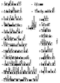
- compositions as defined in claim 4 for unique single band chromosomal sites in the human genome (see Figure 1).
- Reagents are unique molecular cytogenetic markers that identify and mark specific locations on a chromosomal band.
- reagent refers to a short stretch of DNA or cDNA sequence, readily detected, that uniquely identifies a physical genomic location. These reagents can be used as probes suitable for labeling and can be used to identify the precise chromosomal location of disease determining genes.
- the advantages of the present invention over other methods can be illustrated, for example, by the mapping of over 900 DNA sequences including 130 cDNAs, 30 plasmids, 50 cosmids, 500 Bacterial Artificial Chromosomes (BACs) and 150 YACs to single human chromosome bands. Additionally, 39 newly cloned genes expressed in the human brain have been mapped, 26 of which define new loci with significant homology to known genes; 20 of these represent new members of multigene families in higher organisms, and 6 describe new members of genes related to those of lower organisms.
- BACs Bacterial Artificial Chromosomes
- VLDL Very Low Density Lipoprotein
- APP amyloid protein precursor
- results presented herein demonstrate the application of the present invention to large scale cDNA mapping. These results distinguish the present invention over other methods in accuracy (including high resolution chromosome band assignment, multiple signal acquisition and analysis), sensitivity and speed of mapping cDNA sequences.
- sequence homology and exon target size are significant determinants of signal size.
- Other determinants include accessibility, salt concentration, base composition, fragment secondary structure, and the like. It is expected that, due to decreases in homology alone, most processed pseudogenes will generate smaller signals than those expected from the transcribed locus.
- the method of the present invention is, in some manner, a closer correlate of the traditional Cot curve than is solution hybridization to a fixed membrane. This is because each chromosome represents a single molecule, and the proportion of chromosomes with specific hybridization signals therefore sensitively reflects the probability of hybridization, i.e., the proportion of molecules hybridized at a given Cot. Although approximated somewhat by density, this information is largely lost in standard Southern blotting with non-linear signal density and multiple bands. Amplification of FISH signals does contribute to nonlinearity, in that the increase in size and intensity clearly contribute to detection.
- both probe concentration and hybridization conditions viz, Cot
- the proportion of cells with signals largely reflects the degree of homology and target size. Therefore, it may be expected that, given the sequence drift and rearrangements of pseudogenes, the proportion of cells with positive signals will be greater for the transcribed locus than the pseudogene.
- target to probe size is also important for specificity of the hybridization signal. For example, given that the probe and target are both larger than about 50-100 bp, (the size required for single hit stable hybridization under the conditions used), for equal target sizes (pseudogene and transcribed loci), the locus with the higher degree of homology and therefore stability, will generate the large signal. As the probe size increases with respect to the target size, as with small exons ( ⁇ 50-100 bp), a larger proportion of the probe is involved in imperfect fits. For example, for hybridized cDNA fragments containing more than one exon, the second exon will remain unpaired, potentially decreasing the stability of the perfect match.
- probe sizes >200 bp
- small probe size 50-200 bp
- invention methods differ from prior art methods in several ways, e.g., by the use of higher stringency conditions (including increased wash temperatures), multiple amplifications, and the use of smaller fragment probes in the 100-200 bp average range.
- higher stringency conditions including increased wash temperatures
- multiple amplifications including increased wash temperatures
- smaller fragment probes in the 100-200 bp average range.
- the use of smaller fragment size and higher stringency washes tend to increase the specificity of hybrid formation allowing the sensitive definition of the transcribed locus with homology differences of as little as 6% as shown in the argininosuccinate synthase system. (see EXAMPLE 11).
- the cDNA framework produced will both anchor and confirm the detailed YAC and genetic maps and immediately provide large numbers of mapped human genes which can be studied for their role in human genetic diseases.
- Chromosomes were prepared by using a BrdU block, ( Zabel et al. in Proc. Natl. Acad. Sci. USA 80:6932-6936 (1983) ) with some modification. Briefly, human peripheral lymphocytes were grown for 72 hours at 37°C in RPMI 1640 (GIBCO BRL, Gaithersburg, MD) supplemented with L-glutamine (2mM), 15% fetal calf serum, penicillin (100 IU/ml), streptomycin (0.05mg/ml) and 0.02% phytohemagglutinin. The cells were blocked in S-phase by adding 5-bromodeoxyuridine (0.8mg/ml) for 16 hours.
- HBSS Hanks Balanced Salt Solution
- KCl hypotonic solution for 15 minutes at 37°C prior to fixation with a 3:1 mixture of methanol and acetic acid, for 1-5 minutes.
- the metaphase spreads were prepared by letting one drop of suspension fall onto alcohol-cleaned slides, evenly and flat, completely free of cytoplasm.
- the slides were then placed above a container filled with heated water for 20-60 seconds depending on the ambient humidity.
- the optimum conditions were determined by first checking several slides under a phase contrast microscope. For best results, the slides should be aged at room temperature for at least 2-3 weeks prior to in situ hybridization. Slides are best stored at -70°C after aging. The day before denaturation, slides are removed from -70°C and kept overnight at 4°C. Slides are then left at room temperature for 1-2 hours before baking them at 55-60°C for 2 hours, followed by denaturation in 70% formamide (at 66-70°C). For best results, fresh slides are denatured at 66°C.
- the fragment size of a probe labeled by nick translation should be around 100-200 bp and the concentration of probe DNA in the hybridization mixture is in the range of 20-40 ng/ ⁇ l (i.e., 200-400 ng/slide). This can increase non-specific binding, but generally will produce good signal noise ratio.
- a small amount of CotI DNA should be used with cDNA probes (i.e., 1-3 ⁇ g of CotI with 200-400 ng of probe DNA).
- DNA probes were labeled with biotin-14-dATP by nick translation (GIBCO BRL). Unincorporated nucleotides were separated by chromatography (Sephadex G-50). The mean fragment size of all DNA probes was set at around 200 bp, with a range of 100-600 bp, established by nondenaturing agarose gels.
- the buffer was then replaced with FITC-Avidin (0.5 ⁇ g/ml in 4X SSC/1% BSA/0.1% Tween 20), for 30 minutes at 37°C.
- FITC-Avidin 0.5 ⁇ g/ml in 4X SSC/1% BSA/0.1% Tween 20
- the hybridization signal was amplified by the addition of a biotinylated goat anti-avidin antibody layer (5 ⁇ g/ml) for 30 minutes at 37°C.
- the slides were preincubated in 5% goat serum/2X SSC/3% BSA/0.1% Tween 20 for 10-20 minutes prior to the amplification step to reduce immunological non-specific binding.
- a second layer of FITC-avidin was added to the slides as previously described. To increase the intensity of the hybridization signal, a second round of amplification can be applied as necessary.
- the slides were then washed in 2X SSC/0.1% Tween 20 three times at 45°C and briefly left to drain.
- the dyes chromomycin A3 and distamycin A were used as counterstain ( Schweizer in Chromosoma 58:307-324 (1976 ); Schweizer in Hum. Genet 57:1-14 (1981) ; and Magenis et al. in Hum. Genet 69:300-303 (1985 )).
- the slides were rinsed briefly in McIlavane's buffer (pH 8.5-9.0) Schweizer et al., supra. (diluted 1:1 with distilled water) prior to staining.
- chromomycin A3 (0.5mg/ml in 1/2 McIlavane's buffer pH 8.5-9.0) was placed on slides for 10 minutes - 1 hour at room temperature in the dark.
- the staining time required depends on the freshness of the slides; fresher slides need longer staining times, whereas aged slides may need only 10 minutes.
- the slides were rinsed for 1 minute in 1/2 McIlavane's buffer and the excess fluid was shaken off. This was followed by a second round of staining by placing 50 ⁇ l of 0.1mg/ml distamycin A on the slide, incubating for 1-2 minutes at room temperature, followed by rinsing, as above.
- the slides can be mounted with a thin layer of antifade solution containing p-phenylenediamine in phosphate buffer ( Johnson et al. in J. Immunol. Methods 43:349-350 (1981 )).
- the slides were viewed with a Zeiss Axiophot 100 or Axiovert 135 fluorescence microscope (Zeiss, Inc., Thornwood, NY).
- the FITC and chromomycin A3 are both excited by using a 400-490nm band pass exciter, 460nm dichroic, and 470nm barrier (Zeiss filter set #05).
- the hybridized segments appear as bright green-blue or bright yellow-green spots, while the rest of the chromosome bands appear dim green.
- Kodak Technical pan (ASA100) film was used for black and white photographs. Color images were captured by a cooled-CCD camera (Photometrics CH 250, Photometrics Ltd., Tuscon, AZ) using the BDS (Biological Detection Systems, Pittsburgh, PA) imaging software.
- cDNAs were obtained from standard minipreparation cultures ( Sambrook, J. et al., Molecular Cloning, A Laboratory Manual, 2d Edition (Cold Spring Harbor 1989 )). These were isolated and sequenced at random from a fetal brain library cloned in bluescript (Stratagene, La Jolla, CA). For FISH mapping, cDNAs containing inserts in the size range greater than 1 Kb were defined, and included a subset of those with database matches as shown in Tables II and III. TABLE 2. Localization of novel genes by FISH Clone ID % of cells (+) Database Match Match Quality Insert Size Map Location HFBCA82 20 Calmodulin-dep.
- the accuracy of the present invention was tested on the ability to map single genes with known map assignment and genes known to be members of multigene or pseudogene families. First, the map positions of 15 genes with previously known or predicted map positions have been confirmed and, in all cases refined considerably based on FISH analysis of cDNA clones. Further issues concerning single genes and the sensitivity of their detection are considered below.
- a cDNA for the transcribed gene, argininosuccinate synthetase (AS), associated with a family of 14 processed pseudogenes was used to illustrate the ability of the present invention to distinguish between signals.
- Three of the pseudogenes have sequence homologies (versus the expressed sequence) of 93.5% (AS1p1), 92.9% (ASp3) and 89.4% (ASp7) over 2139 bp.
- Amyloid precursor protein-like gene (APLP1) mapping in 19q13.1 to the late onset form of familial Alzheimer's disease (AD2) is of interest. Mutations of the amyloid protein precursor gene (APP) on chromosome 21 are responsible for one form of familial Alzheimer's disease, AD1. Evidence for linkage between AD2 and several DNA markers on proximal 19q has been obtained by Pericak-Vance et al., Cytogenet. Cell Genet., 58:751 (1991 ). In these families, using affected only in the analysis, a maximum multipoint LOD score of 4.4 was obtained near ATP1A3 (19q13.2) and D19S13 (19cenq13.1).
- the second APLP gene maps to 11q26 and is also of interest but, perhaps less so with respect to familial Alzheimer disease, because, despite the higher homology to APP, there is nothing at present to suggest a genetic link. Nonetheless, both of these map positions provide candidate loci for evaluation of genes responsible for Familial Alzheimer Disease in families that are unlinked to either chromosome 21 or chromosome 19 (Swedish families: Lannfelt et al. in Nature Genetics 4:218-219 (1993 ); and Volga German families: Schellenberg et al.).
- Axonal Glycoprotein TAG-1/axonin-1
- TAG-1 cell-surface proteins such as TAG-1 have been identified that are expressed in neural tissue and are thought to play crucial roles in the development of neuronal networks.
- Human TAG-1/axonin has now been independently cloned and its expression found to be restricted to regions of neuronal differentiation in the developing nervous system ( Hasler et al. in Biochem 211:329-339 (1993 )). Therefore, the mapping of human TAG-1/axonin to chromosome band 1q32 provides a gene candidate of interest with respect to the Van der Woude syndrome (cleft-lip/palate with mucous cysts of the lower lip), caused by a deletion of this region.
- the map position of this gene on chromosome 22q13.1 now provides the basis for investigation of its relationship to mitochondrial diseases inherited in an autosomal dominant fashion.
- the mouse mutations mapping in this region are neurological and include gray tremor, waggler and blindness, all of potential interest with respect to this gene.
- Maps in the chromosome 19 region linked to central core disease (CCO) of muscle are of interest ( Mulley et al., Am J of Hum Genet 52:398-405 (1993 )). The current linkage and physical data may immediately be applied to evaluate this relationship.
- this gene is the second or third protein kinase (PRKAR1B), in addition to a calmodulin-like sequence, to be identified in this region.
- PRKAR1B protein kinase
- This gene has high homology (72%) to the Rhizobium gene involved in the nodulation process and strong homology to ATP-dependent bacterial transport proteins. It provides one of the few known genes in the region of 6q21, along with two other neural receptors. Clearly genes with such a high degree of sequence conservation are likely involved in important functions.
- This gene a probable protein kinase with very high homology to the yeast, maps in 12q21, and provides one of the first known genes in this region.
- This gene plays a critical role in energy metabolism, maps in 5q32, a human region with receptors and homology to a mouse region in which neurological mutations are found including vt (vestigial tail with panhypopituitarism), dt (dwarf mutation).
- this new gene mapping in 1p31-32, a gene rich region, is the first mapped of its class.
- Alkaline extracellular protease localized in 15q22, represents the first gene mapped in the entire band. This nucleates the region, provides the first extracellular protease mapped and may be of interest with respect to the 15q22-associated chromosomal rearrangement associated with promyelocytic leukemia.
Landscapes
- Chemical & Material Sciences (AREA)
- Organic Chemistry (AREA)
- Life Sciences & Earth Sciences (AREA)
- Zoology (AREA)
- Wood Science & Technology (AREA)
- Proteomics, Peptides & Aminoacids (AREA)
- Health & Medical Sciences (AREA)
- Engineering & Computer Science (AREA)
- Microbiology (AREA)
- Immunology (AREA)
- Physics & Mathematics (AREA)
- Molecular Biology (AREA)
- Biotechnology (AREA)
- Biophysics (AREA)
- Analytical Chemistry (AREA)
- Biochemistry (AREA)
- Bioinformatics & Cheminformatics (AREA)
- General Engineering & Computer Science (AREA)
- General Health & Medical Sciences (AREA)
- Genetics & Genomics (AREA)
- Measuring Or Testing Involving Enzymes Or Micro-Organisms (AREA)
Claims (10)
- Verfahren zur hochauflösenden Kartierung einer cDNA-Sequenz an einem Einzelbandenlocus auf einem menschlichen Chromosom, wobei das Verfahren Fluoreszenz-In-situ-Hybridisierung und Reverse-(R-)Banding umfasst und das Verfahren die Schritte umfasst:(a) In-situ-Hybridisierung menschlicher Chromosomenausstriche, die vor der In-situ-Hybridisierung mit der cDNA-Sequenz mindestens zwei bis drei Wochen bei Raumtemperatur reifen gelassen werden, wobei die cDNA-Sequenz mit Biotin markiert wird,(b) Kontaktieren hybridisierter Chromosomenausstriche mit mindestens einer Fluoreszenzmarkierung,(c) Durchführen einer Gegenfärbung durch Post-in-situ-Hybridisierungsfärbung mit Chromomycin A3 und Distamycin A, und(d) Nachweisen der Anwesenheit der cDNA-Sequenz und Bestimmen der Chromosomenbandenposition der cDNA-Sequenz, wobei die cDNA-Sequenz als kartiert angesehen wird, wenn in ≥10 % von 200 untersuchten Metaphasenzellen mindestens ein Chromosom an einem Einzelort signalpositiv ist; wobei die cDNA-Sequenz als vorläufig kartiert angesehen wird, wenn sie in 5 - 10 % der untersuchten Metaphasenzellen vorhanden ist, und als möglicherweise kartiert, wenn sie in 2 - 5 % der untersuchten Metaphasenzellen vorhanden ist; und wobei ein positives Signal als Fluoreszenzsignale definiert ist, die auf beiden Chromatiden eines Einzelchromosoms an derselben Bandenposition zu sehen sind.
- Verfahren nach Anspruch 1, wobei die cDNA-Sequenz von ca. 1,2 Kb bis ca. 3 Mb reicht.
- Verfahren nach Anspruch 2, wobei die cDNA-Sequenz eine Länge von <2 Kb aufweist.
- Verwendung einer Zusammensetzung in dem Verfahren gemäß einem der vorhergehenden Ansprüche, wobei die Zusammensetzung eine Vielzahl von Reagenzien umfasst, wobei jedes Reagens eine vom menschlichen Genom hergeleitete Nucleinsäuresequenz enthält und jedes einzelne Reagens einem spezifischen Locus auf einer Chromosomenbande entspricht.
- Verwendung gemäß Anspruch 4, wobei alle Reagenzien in der Zusammensetzung von einer einzelnen Chromosomenbande hergeleitet sind.
- Verwendung gemäß Anspruch 4, wobei alle Reagenzien in der Zusammensetzung von einem Einzelchromosom hergeleitet sind.
- Verwendung gemäß Anspruch 4, wobei die Reagenzien von Centromeren hergeleitete Nucleinsäuresequenzen umfassen.
- Verwendung gemäß Anspruch 4, wobei die Reagenzien von Telomeren hergeleitete Nucleinsäuresequenzen umfassen.
- Verwendung gemäß Anspruch 4, wobei alle Reagenzien in der Zusammensetzung ein cDNA-Contig umfassen.
- Verwendung gemäß Anspruch 4, wobei alle Reagenzien in der Zusammensetzung nicht-chimär sind.
Applications Claiming Priority (3)
| Application Number | Priority Date | Filing Date | Title |
|---|---|---|---|
| US242553 | 1988-09-12 | ||
| US24255394A | 1994-05-13 | 1994-05-13 | |
| PCT/US1995/005918 WO1995031573A1 (en) | 1994-05-13 | 1995-05-09 | High resolution mapping methods and mapping probes |
Publications (2)
| Publication Number | Publication Date |
|---|---|
| EP0760007A1 EP0760007A1 (de) | 1997-03-05 |
| EP0760007B1 true EP0760007B1 (de) | 2007-07-18 |
Family
ID=22915248
Family Applications (1)
| Application Number | Title | Priority Date | Filing Date |
|---|---|---|---|
| EP95919156A Expired - Lifetime EP0760007B1 (de) | 1994-05-13 | 1995-05-09 | Hochauflösende genkartierungs verfahren und genkartierungs sonden |
Country Status (5)
| Country | Link |
|---|---|
| EP (1) | EP0760007B1 (de) |
| JP (1) | JP3776121B2 (de) |
| AT (1) | ATE367451T1 (de) |
| DE (1) | DE69535539D1 (de) |
| WO (1) | WO1995031573A1 (de) |
Families Citing this family (1)
| Publication number | Priority date | Publication date | Assignee | Title |
|---|---|---|---|---|
| CA2786853A1 (en) * | 2010-02-26 | 2011-09-01 | Ventana Medical Systems, Inc. | Cytogenic analysis of metaphase chromosomes |
-
1995
- 1995-05-09 EP EP95919156A patent/EP0760007B1/de not_active Expired - Lifetime
- 1995-05-09 AT AT95919156T patent/ATE367451T1/de not_active IP Right Cessation
- 1995-05-09 JP JP52976895A patent/JP3776121B2/ja not_active Expired - Fee Related
- 1995-05-09 WO PCT/US1995/005918 patent/WO1995031573A1/en not_active Ceased
- 1995-05-09 DE DE69535539T patent/DE69535539D1/de not_active Expired - Lifetime
Non-Patent Citations (1)
| Title |
|---|
| None * |
Also Published As
| Publication number | Publication date |
|---|---|
| DE69535539D1 (de) | 2007-08-30 |
| ATE367451T1 (de) | 2007-08-15 |
| JP3776121B2 (ja) | 2006-05-17 |
| WO1995031573A1 (en) | 1995-11-23 |
| EP0760007A1 (de) | 1997-03-05 |
| JPH10500306A (ja) | 1998-01-13 |
Similar Documents
| Publication | Publication Date | Title |
|---|---|---|
| Fan et al. | Mapping small DNA sequences by fluorescence in situ hybridization directly on banded metaphase chromosomes. | |
| Lohe et al. | Mapping simple repeated DNA sequences in heterochromatin of Drosophila melanogaster. | |
| Lo | Localization of low abundance DNA sequences in tissue sections by in situ hybridization | |
| Wagner et al. | A hybrid cell mapping panel for regional localization of probes to human chromosome 8 | |
| Weler et al. | Quantitative DNA fiber mapping | |
| Weiss et al. | Comparative genomic hybridisation | |
| US5756696A (en) | Compositions for chromosome-specific staining | |
| Trask et al. | Detection of DNA sequences in nuclei in suspension by in situ hybridization and dual beam flow cytometry | |
| US5538869A (en) | In-situ hybridization probes for identification and banding of specific human chromosomes and regions | |
| US6576421B1 (en) | Methods and compositions for the detection of chromosomal aberrations | |
| US6270971B1 (en) | Methods for detecting chromosomal aberrations using chromosome-specific paint probes | |
| JPH03224499A (ja) | 染色体―特異的染色の方法および組成物 | |
| Korenberg et al. | Toward a cDNA map of the human genome | |
| Bell et al. | Chromosomal localization of a gene, GFI1 encoding a novel zinc finger protein reveals a new syntenic region between man and rodents | |
| Gogusev et al. | Genetic abnormalities detected by comparative genomic hybridization in a human endometriosis-derived cell line | |
| Driesen et al. | Generation and fluorescent in situ hybridization mapping of yeast artificial chromosomes of 1p, 17p, 17q, and 19q from a hybrid cell line by high-density screening of an amplified library | |
| Zhu et al. | Discontinuous deletions at 11q23 in B cell chronic lymphocytic leukemia | |
| Fries et al. | Chromosomal assignment of marker loci to monitor marker spacing | |
| Kiechle-Schwarz et al. | Detection of monosomy in interphase nuclei and identification of marker chromosomes using biotinylated alpha-satellite DNA probes | |
| Chowdhary et al. | Cytogenetics and physical gene maps. | |
| Helou et al. | Analysis of genetic changes in rat endometrial carcinomas by means of comparative genomic hybridization | |
| EP0760007B1 (de) | Hochauflösende genkartierungs verfahren und genkartierungs sonden | |
| Carter | Fluorescence in situ hybridization—state of the art | |
| Balajee et al. | Construction of Chinese hamster chromosome specific DNA libraries and their use in the analysis of spontaneous chromosome rearrangements in different cell lines | |
| US5693464A (en) | Method for generating chromosome region-specific probes |
Legal Events
| Date | Code | Title | Description |
|---|---|---|---|
| PUAI | Public reference made under article 153(3) epc to a published international application that has entered the european phase |
Free format text: ORIGINAL CODE: 0009012 |
|
| 17P | Request for examination filed |
Effective date: 19961205 |
|
| AK | Designated contracting states |
Kind code of ref document: A1 Designated state(s): AT BE CH DE DK ES FR GB GR IE IT LI LU MC NL PT SE |
|
| 17Q | First examination report despatched |
Effective date: 19971230 |
|
| GRAP | Despatch of communication of intention to grant a patent |
Free format text: ORIGINAL CODE: EPIDOSNIGR1 |
|
| GRAS | Grant fee paid |
Free format text: ORIGINAL CODE: EPIDOSNIGR3 |
|
| GRAA | (expected) grant |
Free format text: ORIGINAL CODE: 0009210 |
|
| AK | Designated contracting states |
Kind code of ref document: B1 Designated state(s): AT BE CH DE DK ES FR GB GR IE IT LI LU MC NL PT SE |
|
| REG | Reference to a national code |
Ref country code: GB Ref legal event code: FG4D |
|
| REG | Reference to a national code |
Ref country code: CH Ref legal event code: EP |
|
| REF | Corresponds to: |
Ref document number: 69535539 Country of ref document: DE Date of ref document: 20070830 Kind code of ref document: P |
|
| REG | Reference to a national code |
Ref country code: IE Ref legal event code: FG4D |
|
| PG25 | Lapsed in a contracting state [announced via postgrant information from national office to epo] |
Ref country code: PT Free format text: LAPSE BECAUSE OF FAILURE TO SUBMIT A TRANSLATION OF THE DESCRIPTION OR TO PAY THE FEE WITHIN THE PRESCRIBED TIME-LIMIT Effective date: 20071218 Ref country code: NL Free format text: LAPSE BECAUSE OF FAILURE TO SUBMIT A TRANSLATION OF THE DESCRIPTION OR TO PAY THE FEE WITHIN THE PRESCRIBED TIME-LIMIT Effective date: 20070718 Ref country code: ES Free format text: LAPSE BECAUSE OF FAILURE TO SUBMIT A TRANSLATION OF THE DESCRIPTION OR TO PAY THE FEE WITHIN THE PRESCRIBED TIME-LIMIT Effective date: 20071029 |
|
| REG | Reference to a national code |
Ref country code: CH Ref legal event code: PL |
|
| NLV1 | Nl: lapsed or annulled due to failure to fulfill the requirements of art. 29p and 29m of the patents act | ||
| PG25 | Lapsed in a contracting state [announced via postgrant information from national office to epo] |
Ref country code: AT Free format text: LAPSE BECAUSE OF FAILURE TO SUBMIT A TRANSLATION OF THE DESCRIPTION OR TO PAY THE FEE WITHIN THE PRESCRIBED TIME-LIMIT Effective date: 20070718 Ref country code: LI Free format text: LAPSE BECAUSE OF FAILURE TO SUBMIT A TRANSLATION OF THE DESCRIPTION OR TO PAY THE FEE WITHIN THE PRESCRIBED TIME-LIMIT Effective date: 20070718 Ref country code: CH Free format text: LAPSE BECAUSE OF FAILURE TO SUBMIT A TRANSLATION OF THE DESCRIPTION OR TO PAY THE FEE WITHIN THE PRESCRIBED TIME-LIMIT Effective date: 20070718 |
|
| EN | Fr: translation not filed | ||
| PG25 | Lapsed in a contracting state [announced via postgrant information from national office to epo] |
Ref country code: BE Free format text: LAPSE BECAUSE OF FAILURE TO SUBMIT A TRANSLATION OF THE DESCRIPTION OR TO PAY THE FEE WITHIN THE PRESCRIBED TIME-LIMIT Effective date: 20070718 |
|
| PG25 | Lapsed in a contracting state [announced via postgrant information from national office to epo] |
Ref country code: GR Free format text: LAPSE BECAUSE OF FAILURE TO SUBMIT A TRANSLATION OF THE DESCRIPTION OR TO PAY THE FEE WITHIN THE PRESCRIBED TIME-LIMIT Effective date: 20071019 Ref country code: DK Free format text: LAPSE BECAUSE OF FAILURE TO SUBMIT A TRANSLATION OF THE DESCRIPTION OR TO PAY THE FEE WITHIN THE PRESCRIBED TIME-LIMIT Effective date: 20070718 |
|
| PLBE | No opposition filed within time limit |
Free format text: ORIGINAL CODE: 0009261 |
|
| STAA | Information on the status of an ep patent application or granted ep patent |
Free format text: STATUS: NO OPPOSITION FILED WITHIN TIME LIMIT |
|
| 26N | No opposition filed |
Effective date: 20080421 |
|
| PG25 | Lapsed in a contracting state [announced via postgrant information from national office to epo] |
Ref country code: SE Free format text: LAPSE BECAUSE OF FAILURE TO SUBMIT A TRANSLATION OF THE DESCRIPTION OR TO PAY THE FEE WITHIN THE PRESCRIBED TIME-LIMIT Effective date: 20071018 |
|
| PG25 | Lapsed in a contracting state [announced via postgrant information from national office to epo] |
Ref country code: FR Free format text: LAPSE BECAUSE OF FAILURE TO SUBMIT A TRANSLATION OF THE DESCRIPTION OR TO PAY THE FEE WITHIN THE PRESCRIBED TIME-LIMIT Effective date: 20080314 Ref country code: DE Free format text: LAPSE BECAUSE OF FAILURE TO SUBMIT A TRANSLATION OF THE DESCRIPTION OR TO PAY THE FEE WITHIN THE PRESCRIBED TIME-LIMIT Effective date: 20071019 |
|
| PG25 | Lapsed in a contracting state [announced via postgrant information from national office to epo] |
Ref country code: MC Free format text: LAPSE BECAUSE OF NON-PAYMENT OF DUE FEES Effective date: 20080531 |
|
| GBPC | Gb: european patent ceased through non-payment of renewal fee |
Effective date: 20080509 |
|
| PG25 | Lapsed in a contracting state [announced via postgrant information from national office to epo] |
Ref country code: IE Free format text: LAPSE BECAUSE OF NON-PAYMENT OF DUE FEES Effective date: 20080509 |
|
| PG25 | Lapsed in a contracting state [announced via postgrant information from national office to epo] |
Ref country code: GB Free format text: LAPSE BECAUSE OF NON-PAYMENT OF DUE FEES Effective date: 20080509 |
|
| PG25 | Lapsed in a contracting state [announced via postgrant information from national office to epo] |
Ref country code: LU Free format text: LAPSE BECAUSE OF NON-PAYMENT OF DUE FEES Effective date: 20080509 |
|
| PG25 | Lapsed in a contracting state [announced via postgrant information from national office to epo] |
Ref country code: IT Free format text: LAPSE BECAUSE OF NON-PAYMENT OF DUE FEES Effective date: 20080531 |