EP0759074B1 - Stabilizing solutions for troponin - Google Patents
Stabilizing solutions for troponin Download PDFInfo
- Publication number
- EP0759074B1 EP0759074B1 EP96907187A EP96907187A EP0759074B1 EP 0759074 B1 EP0759074 B1 EP 0759074B1 EP 96907187 A EP96907187 A EP 96907187A EP 96907187 A EP96907187 A EP 96907187A EP 0759074 B1 EP0759074 B1 EP 0759074B1
- Authority
- EP
- European Patent Office
- Prior art keywords
- troponin
- composition
- matrix
- analyte
- agent
- Prior art date
- Legal status (The legal status is an assumption and is not a legal conclusion. Google has not performed a legal analysis and makes no representation as to the accuracy of the status listed.)
- Expired - Lifetime
Links
- 230000000087 stabilizing effect Effects 0.000 title claims description 9
- 108010065729 Troponin I Proteins 0.000 claims description 79
- 102000013394 Troponin I Human genes 0.000 claims description 79
- 239000011159 matrix material Substances 0.000 claims description 53
- 239000012491 analyte Substances 0.000 claims description 42
- 210000002966 serum Anatomy 0.000 claims description 31
- 102000004169 proteins and genes Human genes 0.000 claims description 30
- 108090000623 proteins and genes Proteins 0.000 claims description 30
- 235000018102 proteins Nutrition 0.000 claims description 29
- 102000004903 Troponin Human genes 0.000 claims description 24
- 108090001027 Troponin Proteins 0.000 claims description 24
- 239000012634 fragment Substances 0.000 claims description 22
- 239000000203 mixture Substances 0.000 claims description 20
- 108010088751 Albumins Proteins 0.000 claims description 13
- 102000009027 Albumins Human genes 0.000 claims description 13
- 108010010803 Gelatin Proteins 0.000 claims description 13
- 239000008273 gelatin Substances 0.000 claims description 13
- 229920000159 gelatin Polymers 0.000 claims description 13
- 235000019322 gelatine Nutrition 0.000 claims description 13
- 235000011852 gelatine desserts Nutrition 0.000 claims description 13
- 239000000872 buffer Substances 0.000 claims description 12
- KCXVZYZYPLLWCC-UHFFFAOYSA-N EDTA Chemical compound OC(=O)CN(CC(O)=O)CCN(CC(O)=O)CC(O)=O KCXVZYZYPLLWCC-UHFFFAOYSA-N 0.000 claims description 11
- 229960001484 edetic acid Drugs 0.000 claims description 11
- -1 manose Chemical compound 0.000 claims description 11
- 239000002738 chelating agent Substances 0.000 claims description 9
- 239000003638 chemical reducing agent Substances 0.000 claims description 9
- 150000003839 salts Chemical class 0.000 claims description 8
- 239000004067 bulking agent Substances 0.000 claims description 7
- HDTRYLNUVZCQOY-UHFFFAOYSA-N α-D-glucopyranosyl-α-D-glucopyranoside Natural products OC1C(O)C(O)C(CO)OC1OC1C(O)C(O)C(O)C(CO)O1 HDTRYLNUVZCQOY-UHFFFAOYSA-N 0.000 claims description 6
- HDTRYLNUVZCQOY-WSWWMNSNSA-N Trehalose Natural products O[C@@H]1[C@@H](O)[C@@H](O)[C@@H](CO)O[C@@H]1O[C@@H]1[C@H](O)[C@@H](O)[C@@H](O)[C@@H](CO)O1 HDTRYLNUVZCQOY-WSWWMNSNSA-N 0.000 claims description 6
- HDTRYLNUVZCQOY-LIZSDCNHSA-N alpha,alpha-trehalose Chemical compound O[C@@H]1[C@@H](O)[C@H](O)[C@@H](CO)O[C@@H]1O[C@@H]1[C@H](O)[C@@H](O)[C@H](O)[C@@H](CO)O1 HDTRYLNUVZCQOY-LIZSDCNHSA-N 0.000 claims description 6
- 108091005804 Peptidases Proteins 0.000 claims description 5
- 239000004365 Protease Substances 0.000 claims description 5
- 239000002981 blocking agent Substances 0.000 claims description 5
- 108010076119 Caseins Proteins 0.000 claims description 4
- 108010058846 Ovalbumin Proteins 0.000 claims description 4
- 102100037486 Reverse transcriptase/ribonuclease H Human genes 0.000 claims description 4
- 239000005018 casein Substances 0.000 claims description 4
- BECPQYXYKAMYBN-UHFFFAOYSA-N casein, tech. Chemical compound NCCCCC(C(O)=O)N=C(O)C(CC(O)=O)N=C(O)C(CCC(O)=N)N=C(O)C(CC(C)C)N=C(O)C(CCC(O)=O)N=C(O)C(CC(O)=O)N=C(O)C(CCC(O)=O)N=C(O)C(C(C)O)N=C(O)C(CCC(O)=N)N=C(O)C(CCC(O)=N)N=C(O)C(CCC(O)=N)N=C(O)C(CCC(O)=O)N=C(O)C(CCC(O)=O)N=C(O)C(COP(O)(O)=O)N=C(O)C(CCC(O)=N)N=C(O)C(N)CC1=CC=CC=C1 BECPQYXYKAMYBN-UHFFFAOYSA-N 0.000 claims description 4
- 235000021240 caseins Nutrition 0.000 claims description 4
- DEFVIWRASFVYLL-UHFFFAOYSA-N ethylene glycol bis(2-aminoethyl)tetraacetic acid Chemical group OC(=O)CN(CC(O)=O)CCOCCOCCN(CC(O)=O)CC(O)=O DEFVIWRASFVYLL-UHFFFAOYSA-N 0.000 claims description 4
- 229940092253 ovalbumin Drugs 0.000 claims description 4
- DGVVWUTYPXICAM-UHFFFAOYSA-N β‐Mercaptoethanol Chemical compound OCCS DGVVWUTYPXICAM-UHFFFAOYSA-N 0.000 claims description 4
- CZMRCDWAGMRECN-UGDNZRGBSA-N Sucrose Chemical compound O[C@H]1[C@H](O)[C@@H](CO)O[C@@]1(CO)O[C@@H]1[C@H](O)[C@@H](O)[C@H](O)[C@@H](CO)O1 CZMRCDWAGMRECN-UGDNZRGBSA-N 0.000 claims description 3
- 229930006000 Sucrose Natural products 0.000 claims description 3
- 230000004044 response Effects 0.000 claims description 3
- 239000005720 sucrose Substances 0.000 claims description 3
- OWEGMIWEEQEYGQ-UHFFFAOYSA-N 100676-05-9 Natural products OC1C(O)C(O)C(CO)OC1OCC1C(O)C(O)C(O)C(OC2C(OC(O)C(O)C2O)CO)O1 OWEGMIWEEQEYGQ-UHFFFAOYSA-N 0.000 claims description 2
- DBTMGCOVALSLOR-UHFFFAOYSA-N 32-alpha-galactosyl-3-alpha-galactosyl-galactose Natural products OC1C(O)C(O)C(CO)OC1OC1C(O)C(OC2C(C(CO)OC(O)C2O)O)OC(CO)C1O DBTMGCOVALSLOR-UHFFFAOYSA-N 0.000 claims description 2
- GUBGYTABKSRVRQ-PZPXDAEZSA-N 4β-mannobiose Chemical compound O[C@H]1[C@@H](O)[C@H](O)[C@@H](CO)O[C@H]1O[C@@H]1[C@@H](CO)O[C@@H](O)[C@@H](O)[C@H]1O GUBGYTABKSRVRQ-PZPXDAEZSA-N 0.000 claims description 2
- GUBGYTABKSRVRQ-XLOQQCSPSA-N Alpha-Lactose Chemical compound O[C@@H]1[C@@H](O)[C@@H](O)[C@@H](CO)O[C@H]1O[C@@H]1[C@@H](CO)O[C@H](O)[C@H](O)[C@H]1O GUBGYTABKSRVRQ-XLOQQCSPSA-N 0.000 claims description 2
- GUBGYTABKSRVRQ-CUHNMECISA-N D-Cellobiose Chemical compound O[C@@H]1[C@@H](O)[C@H](O)[C@@H](CO)O[C@H]1O[C@@H]1[C@@H](CO)OC(O)[C@H](O)[C@H]1O GUBGYTABKSRVRQ-CUHNMECISA-N 0.000 claims description 2
- RXVWSYJTUUKTEA-UHFFFAOYSA-N D-maltotriose Natural products OC1C(O)C(OC(C(O)CO)C(O)C(O)C=O)OC(CO)C1OC1C(O)C(O)C(O)C(CO)O1 RXVWSYJTUUKTEA-UHFFFAOYSA-N 0.000 claims description 2
- WQZGKKKJIJFFOK-GASJEMHNSA-N Glucose Natural products OC[C@H]1OC(O)[C@H](O)[C@@H](O)[C@@H]1O WQZGKKKJIJFFOK-GASJEMHNSA-N 0.000 claims description 2
- AYRXSINWFIIFAE-SCLMCMATSA-N Isomaltose Natural products OC[C@H]1O[C@H](OC[C@@H](O)[C@@H](O)[C@H](O)[C@@H](O)C=O)[C@@H](O)[C@@H](O)[C@@H]1O AYRXSINWFIIFAE-SCLMCMATSA-N 0.000 claims description 2
- GUBGYTABKSRVRQ-QKKXKWKRSA-N Lactose Natural products OC[C@H]1O[C@@H](O[C@H]2[C@H](O)[C@@H](O)C(O)O[C@@H]2CO)[C@H](O)[C@@H](O)[C@H]1O GUBGYTABKSRVRQ-QKKXKWKRSA-N 0.000 claims description 2
- GUBGYTABKSRVRQ-PICCSMPSSA-N Maltose Natural products O[C@@H]1[C@@H](O)[C@H](O)[C@@H](CO)O[C@@H]1O[C@@H]1[C@@H](CO)OC(O)[C@H](O)[C@H]1O GUBGYTABKSRVRQ-PICCSMPSSA-N 0.000 claims description 2
- FLDFNEBHEXLZRX-DLQNOBSRSA-N Nystose Chemical compound O[C@H]1[C@H](O)[C@@H](CO)O[C@@]1(CO)OC[C@]1(OC[C@]2(O[C@@H]3[C@@H]([C@@H](O)[C@H](O)[C@@H](CO)O3)O)[C@H]([C@H](O)[C@@H](CO)O2)O)[C@@H](O)[C@H](O)[C@@H](CO)O1 FLDFNEBHEXLZRX-DLQNOBSRSA-N 0.000 claims description 2
- DKXNBNKWCZZMJT-UHFFFAOYSA-N O4-alpha-D-Mannopyranosyl-D-mannose Natural products O=CC(O)C(O)C(C(O)CO)OC1OC(CO)C(O)C(O)C1O DKXNBNKWCZZMJT-UHFFFAOYSA-N 0.000 claims description 2
- FTNIPWXXIGNQQF-UHFFFAOYSA-N UNPD130147 Natural products OC1C(O)C(O)C(CO)OC1OC1C(CO)OC(OC2C(OC(OC3C(OC(OC4C(OC(O)C(O)C4O)CO)C(O)C3O)CO)C(O)C2O)CO)C(O)C1O FTNIPWXXIGNQQF-UHFFFAOYSA-N 0.000 claims description 2
- LUEWUZLMQUOBSB-UHFFFAOYSA-N UNPD55895 Natural products OC1C(O)C(O)C(CO)OC1OC1C(CO)OC(OC2C(OC(OC3C(OC(O)C(O)C3O)CO)C(O)C2O)CO)C(O)C1O LUEWUZLMQUOBSB-UHFFFAOYSA-N 0.000 claims description 2
- WQZGKKKJIJFFOK-PHYPRBDBSA-N alpha-D-galactose Chemical compound OC[C@H]1O[C@H](O)[C@H](O)[C@@H](O)[C@H]1O WQZGKKKJIJFFOK-PHYPRBDBSA-N 0.000 claims description 2
- BNABBHGYYMZMOA-AHIHXIOASA-N alpha-maltoheptaose Chemical compound O[C@@H]1[C@@H](O)[C@H](O)[C@@H](CO)O[C@@H]1O[C@@H]1[C@@H](CO)O[C@H](O[C@@H]2[C@H](O[C@H](O[C@@H]3[C@H](O[C@H](O[C@@H]4[C@H](O[C@H](O[C@@H]5[C@H](O[C@H](O[C@@H]6[C@H](O[C@H](O)[C@H](O)[C@H]6O)CO)[C@H](O)[C@H]5O)CO)[C@H](O)[C@H]4O)CO)[C@H](O)[C@H]3O)CO)[C@H](O)[C@H]2O)CO)[C@H](O)[C@H]1O BNABBHGYYMZMOA-AHIHXIOASA-N 0.000 claims description 2
- OCIBBXPLUVYKCH-QXVNYKTNSA-N alpha-maltohexaose Chemical compound O[C@@H]1[C@@H](O)[C@H](O)[C@@H](CO)O[C@@H]1O[C@@H]1[C@@H](CO)O[C@H](O[C@@H]2[C@H](O[C@H](O[C@@H]3[C@H](O[C@H](O[C@@H]4[C@H](O[C@H](O[C@@H]5[C@H](O[C@H](O)[C@H](O)[C@H]5O)CO)[C@H](O)[C@H]4O)CO)[C@H](O)[C@H]3O)CO)[C@H](O)[C@H]2O)CO)[C@H](O)[C@H]1O OCIBBXPLUVYKCH-QXVNYKTNSA-N 0.000 claims description 2
- WQZGKKKJIJFFOK-VFUOTHLCSA-N beta-D-glucose Chemical compound OC[C@H]1O[C@@H](O)[C@H](O)[C@@H](O)[C@@H]1O WQZGKKKJIJFFOK-VFUOTHLCSA-N 0.000 claims description 2
- GUBGYTABKSRVRQ-QUYVBRFLSA-N beta-maltose Chemical compound OC[C@H]1O[C@H](O[C@H]2[C@H](O)[C@@H](O)[C@H](O)O[C@@H]2CO)[C@H](O)[C@@H](O)[C@@H]1O GUBGYTABKSRVRQ-QUYVBRFLSA-N 0.000 claims description 2
- UFULAYFCSOUIOV-UHFFFAOYSA-N cysteamine Chemical compound NCCS UFULAYFCSOUIOV-UHFFFAOYSA-N 0.000 claims description 2
- VHJLVAABSRFDPM-QWWZWVQMSA-N dithiothreitol Chemical compound SC[C@@H](O)[C@H](O)CS VHJLVAABSRFDPM-QWWZWVQMSA-N 0.000 claims description 2
- 229930182830 galactose Natural products 0.000 claims description 2
- 239000008103 glucose Substances 0.000 claims description 2
- DLRVVLDZNNYCBX-RTPHMHGBSA-N isomaltose Chemical compound O[C@@H]1[C@@H](O)[C@H](O)[C@@H](CO)O[C@@H]1OC[C@@H]1[C@@H](O)[C@H](O)[C@@H](O)C(O)O1 DLRVVLDZNNYCBX-RTPHMHGBSA-N 0.000 claims description 2
- 239000008101 lactose Substances 0.000 claims description 2
- DJMVHSOAUQHPSN-UHFFFAOYSA-N malto-hexaose Natural products OC1C(O)C(OC(C(O)CO)C(O)C(O)C=O)OC(CO)C1OC1C(O)C(O)C(OC2C(C(O)C(OC3C(C(O)C(OC4C(C(O)C(O)C(CO)O4)O)C(CO)O3)O)C(CO)O2)O)C(CO)O1 DJMVHSOAUQHPSN-UHFFFAOYSA-N 0.000 claims description 2
- FJCUPROCOFFUSR-UHFFFAOYSA-N malto-pentaose Natural products OC1C(O)C(OC(C(O)CO)C(O)C(O)C=O)OC(CO)C1OC1C(O)C(O)C(OC2C(C(O)C(OC3C(C(O)C(O)C(CO)O3)O)C(CO)O2)O)C(CO)O1 FJCUPROCOFFUSR-UHFFFAOYSA-N 0.000 claims description 2
- UYQJCPNSAVWAFU-UHFFFAOYSA-N malto-tetraose Natural products OC1C(O)C(OC(C(O)CO)C(O)C(O)C=O)OC(CO)C1OC1C(O)C(O)C(OC2C(C(O)C(O)C(CO)O2)O)C(CO)O1 UYQJCPNSAVWAFU-UHFFFAOYSA-N 0.000 claims description 2
- FJCUPROCOFFUSR-GMMZZHHDSA-N maltopentaose Chemical compound O[C@@H]1[C@@H](O)[C@@H](O[C@H]([C@H](O)CO)[C@H](O)[C@@H](O)C=O)O[C@H](CO)[C@H]1O[C@@H]1[C@H](O)[C@@H](O)[C@H](O[C@@H]2[C@@H]([C@@H](O)[C@H](O[C@@H]3[C@@H]([C@@H](O)[C@H](O)[C@@H](CO)O3)O)[C@@H](CO)O2)O)[C@@H](CO)O1 FJCUPROCOFFUSR-GMMZZHHDSA-N 0.000 claims description 2
- LUEWUZLMQUOBSB-OUBHKODOSA-N maltotetraose Chemical compound O[C@H]1[C@H](O)[C@@H](O)[C@H](CO)O[C@H]1O[C@@H]1[C@H](CO)O[C@@H](O[C@@H]2[C@@H](O[C@@H](O[C@@H]3[C@@H](O[C@@H](O)[C@H](O)[C@H]3O)CO)[C@H](O)[C@H]2O)CO)[C@H](O)[C@H]1O LUEWUZLMQUOBSB-OUBHKODOSA-N 0.000 claims description 2
- FYGDTMLNYKFZSV-UHFFFAOYSA-N mannotriose Natural products OC1C(O)C(O)C(CO)OC1OC1C(CO)OC(OC2C(OC(O)C(O)C2O)CO)C(O)C1O FYGDTMLNYKFZSV-UHFFFAOYSA-N 0.000 claims description 2
- 238000004519 manufacturing process Methods 0.000 claims description 2
- FLDFNEBHEXLZRX-UHFFFAOYSA-N nystose Natural products OC1C(O)C(CO)OC1(CO)OCC1(OCC2(OC3C(C(O)C(O)C(CO)O3)O)C(C(O)C(CO)O2)O)C(O)C(O)C(CO)O1 FLDFNEBHEXLZRX-UHFFFAOYSA-N 0.000 claims description 2
- 229940124272 protein stabilizer Drugs 0.000 claims description 2
- FYGDTMLNYKFZSV-BYLHFPJWSA-N β-1,4-galactotrioside Chemical compound O[C@@H]1[C@@H](O)[C@H](O)[C@@H](CO)O[C@H]1O[C@@H]1[C@H](CO)O[C@@H](O[C@@H]2[C@@H](O[C@@H](O)[C@H](O)[C@H]2O)CO)[C@H](O)[C@H]1O FYGDTMLNYKFZSV-BYLHFPJWSA-N 0.000 claims description 2
- 230000003019 stabilising effect Effects 0.000 claims 5
- 150000001860 citric acid derivatives Chemical class 0.000 claims 1
- 239000000243 solution Substances 0.000 description 48
- 108090000765 processed proteins & peptides Proteins 0.000 description 23
- FAPWRFPIFSIZLT-UHFFFAOYSA-M Sodium chloride Chemical group [Na+].[Cl-] FAPWRFPIFSIZLT-UHFFFAOYSA-M 0.000 description 18
- 238000006243 chemical reaction Methods 0.000 description 15
- 206010000891 acute myocardial infarction Diseases 0.000 description 14
- 150000001413 amino acids Chemical group 0.000 description 13
- 238000003018 immunoassay Methods 0.000 description 13
- 239000011550 stock solution Substances 0.000 description 13
- 102000004987 Troponin T Human genes 0.000 description 11
- 108090001108 Troponin T Proteins 0.000 description 11
- 229940024606 amino acid Drugs 0.000 description 10
- 230000008859 change Effects 0.000 description 10
- 239000000137 peptide hydrolase inhibitor Substances 0.000 description 10
- 238000011156 evaluation Methods 0.000 description 9
- 238000002360 preparation method Methods 0.000 description 9
- 239000011780 sodium chloride Substances 0.000 description 9
- 102000036675 Myoglobin Human genes 0.000 description 8
- 108010062374 Myoglobin Proteins 0.000 description 8
- PXIPVTKHYLBLMZ-UHFFFAOYSA-N Sodium azide Chemical compound [Na+].[N-]=[N+]=[N-] PXIPVTKHYLBLMZ-UHFFFAOYSA-N 0.000 description 8
- 239000007864 aqueous solution Substances 0.000 description 8
- PWKSKIMOESPYIA-BYPYZUCNSA-N L-N-acetyl-Cysteine Chemical group CC(=O)N[C@@H](CS)C(O)=O PWKSKIMOESPYIA-BYPYZUCNSA-N 0.000 description 7
- 229960004308 acetylcysteine Drugs 0.000 description 7
- 230000000692 anti-sense effect Effects 0.000 description 7
- 238000011088 calibration curve Methods 0.000 description 7
- 230000000747 cardiac effect Effects 0.000 description 7
- 210000000349 chromosome Anatomy 0.000 description 7
- 230000009871 nonspecific binding Effects 0.000 description 7
- 239000008213 purified water Substances 0.000 description 7
- XLYOFNOQVPJJNP-UHFFFAOYSA-N water Chemical compound O XLYOFNOQVPJJNP-UHFFFAOYSA-N 0.000 description 7
- 239000004743 Polypropylene Substances 0.000 description 6
- 229940124158 Protease/peptidase inhibitor Drugs 0.000 description 6
- 229920001155 polypropylene Polymers 0.000 description 6
- 239000003755 preservative agent Substances 0.000 description 6
- 238000012360 testing method Methods 0.000 description 6
- BCHIXGBGRHLSBE-UHFFFAOYSA-N (4-methyl-2-oxochromen-7-yl) dihydrogen phosphate Chemical compound C1=C(OP(O)(O)=O)C=CC2=C1OC(=O)C=C2C BCHIXGBGRHLSBE-UHFFFAOYSA-N 0.000 description 5
- 101000851334 Homo sapiens Troponin I, cardiac muscle Proteins 0.000 description 5
- 238000003556 assay Methods 0.000 description 5
- 230000027455 binding Effects 0.000 description 5
- 238000002474 experimental method Methods 0.000 description 5
- 239000003365 glass fiber Substances 0.000 description 5
- 238000010369 molecular cloning Methods 0.000 description 5
- 238000003752 polymerase chain reaction Methods 0.000 description 5
- 239000007790 solid phase Substances 0.000 description 5
- 239000000758 substrate Substances 0.000 description 5
- DFVFTMTWCUHJBL-UHFFFAOYSA-N 4-azaniumyl-3-hydroxy-6-methylheptanoate Chemical compound CC(C)CC(N)C(O)CC(O)=O DFVFTMTWCUHJBL-UHFFFAOYSA-N 0.000 description 4
- 102000004190 Enzymes Human genes 0.000 description 4
- 108090000790 Enzymes Proteins 0.000 description 4
- 229960005091 chloramphenicol Drugs 0.000 description 4
- WIIZWVCIJKGZOK-RKDXNWHRSA-N chloramphenicol Chemical compound ClC(Cl)C(=O)N[C@H](CO)[C@H](O)C1=CC=C([N+]([O-])=O)C=C1 WIIZWVCIJKGZOK-RKDXNWHRSA-N 0.000 description 4
- VNFPBHJOKIVQEB-UHFFFAOYSA-N clotrimazole Chemical compound ClC1=CC=CC=C1C(N1C=NC=C1)(C=1C=CC=CC=1)C1=CC=CC=C1 VNFPBHJOKIVQEB-UHFFFAOYSA-N 0.000 description 4
- 239000003599 detergent Substances 0.000 description 4
- 229940042399 direct acting antivirals protease inhibitors Drugs 0.000 description 4
- 229940088598 enzyme Drugs 0.000 description 4
- 239000007788 liquid Substances 0.000 description 4
- 239000003550 marker Substances 0.000 description 4
- YBYRMVIVWMBXKQ-UHFFFAOYSA-N phenylmethanesulfonyl fluoride Chemical compound FS(=O)(=O)CC1=CC=CC=C1 YBYRMVIVWMBXKQ-UHFFFAOYSA-N 0.000 description 4
- 230000002335 preservative effect Effects 0.000 description 4
- 238000011084 recovery Methods 0.000 description 4
- 239000001488 sodium phosphate Substances 0.000 description 4
- 229910000162 sodium phosphate Inorganic materials 0.000 description 4
- RYFMWSXOAZQYPI-UHFFFAOYSA-K trisodium phosphate Chemical compound [Na+].[Na+].[Na+].[O-]P([O-])([O-])=O RYFMWSXOAZQYPI-UHFFFAOYSA-K 0.000 description 4
- 108091003079 Bovine Serum Albumin Proteins 0.000 description 3
- 230000002378 acidificating effect Effects 0.000 description 3
- 229940098773 bovine serum albumin Drugs 0.000 description 3
- 230000006378 damage Effects 0.000 description 3
- 239000011521 glass Substances 0.000 description 3
- 239000004615 ingredient Substances 0.000 description 3
- 230000003278 mimic effect Effects 0.000 description 3
- 229940045641 monobasic sodium phosphate Drugs 0.000 description 3
- 210000004165 myocardium Anatomy 0.000 description 3
- 239000000047 product Substances 0.000 description 3
- 210000002027 skeletal muscle Anatomy 0.000 description 3
- AJPJDKMHJJGVTQ-UHFFFAOYSA-M sodium dihydrogen phosphate Chemical compound [Na+].OP(O)([O-])=O AJPJDKMHJJGVTQ-UHFFFAOYSA-M 0.000 description 3
- 238000003860 storage Methods 0.000 description 3
- 230000001225 therapeutic effect Effects 0.000 description 3
- IQMHGRIOYXVPSE-UHFFFAOYSA-N 2-acetamido-5-[formyl(hydroxy)amino]-n-[1-[3-[5-[3-[formyl(hydroxy)amino]propyl]-3,6-dioxopiperazin-2-yl]propyl-hydroxyamino]-3-hydroxy-1-oxopropan-2-yl]pentanamide Chemical compound O=CN(O)CCCC(NC(=O)C)C(=O)NC(CO)C(=O)N(O)CCCC1NC(=O)C(CCCN(O)C=O)NC1=O IQMHGRIOYXVPSE-UHFFFAOYSA-N 0.000 description 2
- DVLFYONBTKHTER-UHFFFAOYSA-N 3-(N-morpholino)propanesulfonic acid Chemical compound OS(=O)(=O)CCCN1CCOCC1 DVLFYONBTKHTER-UHFFFAOYSA-N 0.000 description 2
- XCBLFURAFHFFJF-UHFFFAOYSA-N 3-[bis(2-hydroxyethyl)azaniumyl]-2-hydroxypropane-1-sulfonate Chemical compound OCCN(CCO)CC(O)CS(O)(=O)=O XCBLFURAFHFFJF-UHFFFAOYSA-N 0.000 description 2
- FWMNVWWHGCHHJJ-SKKKGAJSSA-N 4-amino-1-[(2r)-6-amino-2-[[(2r)-2-[[(2r)-2-[[(2r)-2-amino-3-phenylpropanoyl]amino]-3-phenylpropanoyl]amino]-4-methylpentanoyl]amino]hexanoyl]piperidine-4-carboxylic acid Chemical compound C([C@H](C(=O)N[C@H](CC(C)C)C(=O)N[C@H](CCCCN)C(=O)N1CCC(N)(CC1)C(O)=O)NC(=O)[C@H](N)CC=1C=CC=CC=1)C1=CC=CC=C1 FWMNVWWHGCHHJJ-SKKKGAJSSA-N 0.000 description 2
- 241000283690 Bos taurus Species 0.000 description 2
- 101000714491 Escherichia phage T7 Major capsid protein Proteins 0.000 description 2
- 239000004395 L-leucine Substances 0.000 description 2
- 208000029549 Muscle injury Diseases 0.000 description 2
- 239000007983 Tris buffer Substances 0.000 description 2
- 230000017531 blood circulation Effects 0.000 description 2
- 210000004899 c-terminal region Anatomy 0.000 description 2
- 239000003795 chemical substances by application Substances 0.000 description 2
- 239000000356 contaminant Substances 0.000 description 2
- 208000029078 coronary artery disease Diseases 0.000 description 2
- 238000003745 diagnosis Methods 0.000 description 2
- 239000003085 diluting agent Substances 0.000 description 2
- 230000006870 function Effects 0.000 description 2
- 230000036541 health Effects 0.000 description 2
- 210000005003 heart tissue Anatomy 0.000 description 2
- 208000014674 injury Diseases 0.000 description 2
- 229960003136 leucine Drugs 0.000 description 2
- 230000007246 mechanism Effects 0.000 description 2
- 230000000813 microbial effect Effects 0.000 description 2
- 210000003205 muscle Anatomy 0.000 description 2
- 208000010125 myocardial infarction Diseases 0.000 description 2
- 125000002924 primary amino group Chemical group [H]N([H])* 0.000 description 2
- 238000003756 stirring Methods 0.000 description 2
- FJFWTROADHZFDY-UHFFFAOYSA-N 1-(trihydroxymethylamino)propane-2-sulfonic acid Chemical compound OS(=O)(=O)C(C)CNC(O)(O)O FJFWTROADHZFDY-UHFFFAOYSA-N 0.000 description 1
- IHPYMWDTONKSCO-UHFFFAOYSA-N 2,2'-piperazine-1,4-diylbisethanesulfonic acid Chemical compound OS(=O)(=O)CCN1CCN(CCS(O)(=O)=O)CC1 IHPYMWDTONKSCO-UHFFFAOYSA-N 0.000 description 1
- TZJAEGCLMLTGRJ-UHFFFAOYSA-N 2-(2-aminoethyl)benzenesulfonyl fluoride Chemical compound NCCC1=CC=CC=C1S(F)(=O)=O TZJAEGCLMLTGRJ-UHFFFAOYSA-N 0.000 description 1
- QHQZEEGNGSZBOL-UHFFFAOYSA-N 2-(aminomethyl)-2-(hydroxymethyl)propane-1,3-diol Chemical compound NCC(CO)(CO)CO QHQZEEGNGSZBOL-UHFFFAOYSA-N 0.000 description 1
- JKMHFZQWWAIEOD-UHFFFAOYSA-N 2-[4-(2-hydroxyethyl)piperazin-1-yl]ethanesulfonic acid Chemical compound OCC[NH+]1CCN(CCS([O-])(=O)=O)CC1 JKMHFZQWWAIEOD-UHFFFAOYSA-N 0.000 description 1
- 125000000979 2-amino-2-oxoethyl group Chemical group [H]C([*])([H])C(=O)N([H])[H] 0.000 description 1
- OKPMVGINPLUPFN-UHFFFAOYSA-N 2-hydroxy-3-[methyl(trihydroxymethyl)amino]propane-1-sulfonic acid Chemical compound OC(O)(O)N(C)CC(O)CS(O)(=O)=O OKPMVGINPLUPFN-UHFFFAOYSA-N 0.000 description 1
- RZQXOGQSPBYUKH-UHFFFAOYSA-N 3-[[1,3-dihydroxy-2-(hydroxymethyl)propan-2-yl]azaniumyl]-2-hydroxypropane-1-sulfonate Chemical compound OCC(CO)(CO)NCC(O)CS(O)(=O)=O RZQXOGQSPBYUKH-UHFFFAOYSA-N 0.000 description 1
- 239000007991 ACES buffer Substances 0.000 description 1
- 241000251468 Actinopterygii Species 0.000 description 1
- 206010002388 Angina unstable Diseases 0.000 description 1
- 108010087765 Antipain Proteins 0.000 description 1
- 108010039627 Aprotinin Proteins 0.000 description 1
- 108700016232 Arg(2)-Sar(4)- dermorphin (1-4) Proteins 0.000 description 1
- OYPRJOBELJOOCE-UHFFFAOYSA-N Calcium Chemical compound [Ca] OYPRJOBELJOOCE-UHFFFAOYSA-N 0.000 description 1
- 208000020446 Cardiac disease Diseases 0.000 description 1
- 206010008479 Chest Pain Diseases 0.000 description 1
- 102000002585 Contractile Proteins Human genes 0.000 description 1
- 108010068426 Contractile Proteins Proteins 0.000 description 1
- CEAZRRDELHUEMR-URQXQFDESA-N Gentamicin Chemical compound O1[C@H](C(C)NC)CC[C@@H](N)[C@H]1O[C@H]1[C@H](O)[C@@H](O[C@@H]2[C@@H]([C@@H](NC)[C@@](C)(O)CO2)O)[C@H](N)C[C@@H]1N CEAZRRDELHUEMR-URQXQFDESA-N 0.000 description 1
- 229930182566 Gentamicin Natural products 0.000 description 1
- 239000007995 HEPES buffer Substances 0.000 description 1
- GIZQLVPDAOBAFN-UHFFFAOYSA-N HEPPSO Chemical compound OCCN1CCN(CC(O)CS(O)(=O)=O)CC1 GIZQLVPDAOBAFN-UHFFFAOYSA-N 0.000 description 1
- DGAQECJNVWCQMB-PUAWFVPOSA-M Ilexoside XXIX Chemical compound C[C@@H]1CC[C@@]2(CC[C@@]3(C(=CC[C@H]4[C@]3(CC[C@@H]5[C@@]4(CC[C@@H](C5(C)C)OS(=O)(=O)[O-])C)C)[C@@H]2[C@]1(C)O)C)C(=O)O[C@H]6[C@@H]([C@H]([C@@H]([C@H](O6)CO)O)O)O.[Na+] DGAQECJNVWCQMB-PUAWFVPOSA-M 0.000 description 1
- 206010061216 Infarction Diseases 0.000 description 1
- GDBQQVLCIARPGH-UHFFFAOYSA-N Leupeptin Natural products CC(C)CC(NC(C)=O)C(=O)NC(CC(C)C)C(=O)NC(C=O)CCCN=C(N)N GDBQQVLCIARPGH-UHFFFAOYSA-N 0.000 description 1
- WHXSMMKQMYFTQS-UHFFFAOYSA-N Lithium Chemical compound [Li] WHXSMMKQMYFTQS-UHFFFAOYSA-N 0.000 description 1
- 108090000362 Lymphotoxin-beta Proteins 0.000 description 1
- 239000007993 MOPS buffer Substances 0.000 description 1
- KBAFPSLPKGSANY-UHFFFAOYSA-N Naftidrofuryl Chemical compound C=1C=CC2=CC=CC=C2C=1CC(C(=O)OCCN(CC)CC)CC1CCCO1 KBAFPSLPKGSANY-UHFFFAOYSA-N 0.000 description 1
- 229910019142 PO4 Inorganic materials 0.000 description 1
- 102000035195 Peptidases Human genes 0.000 description 1
- 108010033276 Peptide Fragments Proteins 0.000 description 1
- 102000007079 Peptide Fragments Human genes 0.000 description 1
- 229920001213 Polysorbate 20 Polymers 0.000 description 1
- ZLMJMSJWJFRBEC-UHFFFAOYSA-N Potassium Chemical compound [K] ZLMJMSJWJFRBEC-UHFFFAOYSA-N 0.000 description 1
- 102000007056 Recombinant Fusion Proteins Human genes 0.000 description 1
- 108010008281 Recombinant Fusion Proteins Proteins 0.000 description 1
- 108010023197 Streptokinase Proteins 0.000 description 1
- 108090000373 Tissue Plasminogen Activator Proteins 0.000 description 1
- 102000003978 Tissue Plasminogen Activator Human genes 0.000 description 1
- 239000013504 Triton X-100 Substances 0.000 description 1
- 229920004890 Triton X-100 Polymers 0.000 description 1
- 102000013534 Troponin C Human genes 0.000 description 1
- 208000007814 Unstable Angina Diseases 0.000 description 1
- 208000027418 Wounds and injury Diseases 0.000 description 1
- 230000002159 abnormal effect Effects 0.000 description 1
- 150000003863 ammonium salts Chemical class 0.000 description 1
- 239000000427 antigen Substances 0.000 description 1
- 108091007433 antigens Proteins 0.000 description 1
- 102000036639 antigens Human genes 0.000 description 1
- SDNYTAYICBFYFH-TUFLPTIASA-N antipain Chemical compound NC(N)=NCCC[C@@H](C=O)NC(=O)[C@H](C(C)C)NC(=O)[C@H](CCCN=C(N)N)NC(=O)N[C@H](C(O)=O)CC1=CC=CC=C1 SDNYTAYICBFYFH-TUFLPTIASA-N 0.000 description 1
- 229960004405 aprotinin Drugs 0.000 description 1
- 238000003149 assay kit Methods 0.000 description 1
- 239000010836 blood and blood product Substances 0.000 description 1
- 229940125691 blood product Drugs 0.000 description 1
- 210000001124 body fluid Anatomy 0.000 description 1
- 239000011575 calcium Substances 0.000 description 1
- 229910052791 calcium Inorganic materials 0.000 description 1
- 230000015556 catabolic process Effects 0.000 description 1
- 239000003153 chemical reaction reagent Substances 0.000 description 1
- 230000004087 circulation Effects 0.000 description 1
- 229960004022 clotrimazole Drugs 0.000 description 1
- 239000000470 constituent Substances 0.000 description 1
- 230000008602 contraction Effects 0.000 description 1
- 210000004351 coronary vessel Anatomy 0.000 description 1
- 238000006731 degradation reaction Methods 0.000 description 1
- 238000013461 design Methods 0.000 description 1
- 230000004069 differentiation Effects 0.000 description 1
- 201000010099 disease Diseases 0.000 description 1
- 208000037265 diseases, disorders, signs and symptoms Diseases 0.000 description 1
- CCIVGXIOQKPBKL-UHFFFAOYSA-M ethanesulfonate Chemical compound CCS([O-])(=O)=O CCIVGXIOQKPBKL-UHFFFAOYSA-M 0.000 description 1
- 239000003527 fibrinolytic agent Substances 0.000 description 1
- 238000009472 formulation Methods 0.000 description 1
- 108010090705 foroxymithine Proteins 0.000 description 1
- 230000002538 fungal effect Effects 0.000 description 1
- 229960002518 gentamicin Drugs 0.000 description 1
- 208000019622 heart disease Diseases 0.000 description 1
- 230000004217 heart function Effects 0.000 description 1
- HNDVDQJCIGZPNO-UHFFFAOYSA-N histidine Natural products OC(=O)C(N)CC1=CN=CN1 HNDVDQJCIGZPNO-UHFFFAOYSA-N 0.000 description 1
- 230000007574 infarction Effects 0.000 description 1
- ZPNFWUPYTFPOJU-LPYSRVMUSA-N iniprol Chemical compound C([C@H]1C(=O)NCC(=O)NCC(=O)N[C@H]2CSSC[C@H]3C(=O)N[C@@H](CCCCN)C(=O)N[C@@H](C)C(=O)N[C@@H](CCCNC(N)=N)C(=O)N[C@H](C(N[C@H](C(=O)N[C@@H](CCCNC(N)=N)C(=O)N[C@@H](CC=4C=CC(O)=CC=4)C(=O)N[C@@H](CC=4C=CC=CC=4)C(=O)N[C@@H](CC=4C=CC(O)=CC=4)C(=O)N[C@@H](CC(N)=O)C(=O)N[C@@H](C)C(=O)N[C@@H](CCCCN)C(=O)N[C@@H](C)C(=O)NCC(=O)N[C@@H](CC(C)C)C(=O)N[C@@H](CSSC[C@H](NC(=O)[C@H](CC(O)=O)NC(=O)[C@H](CCC(O)=O)NC(=O)[C@H](C)NC(=O)[C@H](CO)NC(=O)[C@H](CCCCN)NC(=O)[C@H](CC=4C=CC=CC=4)NC(=O)[C@H](CC(N)=O)NC(=O)[C@H](CC(N)=O)NC(=O)[C@H](CCCNC(N)=N)NC(=O)[C@H](CCCCN)NC(=O)[C@H](C)NC(=O)[C@H](CCCNC(N)=N)NC2=O)C(=O)N[C@@H](CCSC)C(=O)N[C@@H](CCCNC(N)=N)C(=O)N[C@@H]([C@@H](C)O)C(=O)N[C@@H](CSSC[C@H](NC(=O)[C@H](CC=2C=CC=CC=2)NC(=O)[C@H](CC(O)=O)NC(=O)[C@H]2N(CCC2)C(=O)[C@@H](N)CCCNC(N)=N)C(=O)N[C@@H](CC(C)C)C(=O)N[C@@H](CCC(O)=O)C(=O)N2[C@@H](CCC2)C(=O)N2[C@@H](CCC2)C(=O)N[C@@H](CC=2C=CC(O)=CC=2)C(=O)N[C@@H]([C@@H](C)O)C(=O)NCC(=O)N2[C@@H](CCC2)C(=O)N3)C(=O)NCC(=O)NCC(=O)N[C@@H](C)C(O)=O)C(=O)N[C@@H](CCC(N)=O)C(=O)N[C@H](C(=O)N[C@@H](CC=2C=CC=CC=2)C(=O)N[C@H](C(=O)N1)C(C)C)[C@@H](C)O)[C@@H](C)CC)=O)[C@@H](C)CC)C1=CC=C(O)C=C1 ZPNFWUPYTFPOJU-LPYSRVMUSA-N 0.000 description 1
- 201000004332 intermediate coronary syndrome Diseases 0.000 description 1
- 239000012263 liquid product Substances 0.000 description 1
- 229910052744 lithium Inorganic materials 0.000 description 1
- 229910003002 lithium salt Inorganic materials 0.000 description 1
- 159000000002 lithium salts Chemical class 0.000 description 1
- 238000005259 measurement Methods 0.000 description 1
- 238000000034 method Methods 0.000 description 1
- 229940049018 mycostatin Drugs 0.000 description 1
- 230000002107 myocardial effect Effects 0.000 description 1
- 208000037891 myocardial injury Diseases 0.000 description 1
- VQOXZBDYSJBXMA-NQTDYLQESA-N nystatin A1 Chemical compound O[C@H]1[C@@H](N)[C@H](O)[C@@H](C)O[C@H]1O[C@H]1/C=C/C=C/C=C/C=C/CC/C=C/C=C/[C@H](C)[C@@H](O)[C@@H](C)[C@H](C)OC(=O)C[C@H](O)C[C@H](O)C[C@H](O)CC[C@@H](O)[C@H](O)C[C@](O)(C[C@H](O)[C@H]2C(O)=O)O[C@H]2C1 VQOXZBDYSJBXMA-NQTDYLQESA-N 0.000 description 1
- JPMIIZHYYWMHDT-UHFFFAOYSA-N octhilinone Chemical compound CCCCCCCCN1SC=CC1=O JPMIIZHYYWMHDT-UHFFFAOYSA-N 0.000 description 1
- 150000003891 oxalate salts Chemical class 0.000 description 1
- 125000000636 p-nitrophenyl group Chemical group [H]C1=C([H])C(=C([H])C([H])=C1*)[N+]([O-])=O 0.000 description 1
- 229950000964 pepstatin Drugs 0.000 description 1
- 108010091212 pepstatin Proteins 0.000 description 1
- FAXGPCHRFPCXOO-LXTPJMTPSA-N pepstatin A Chemical compound OC(=O)C[C@H](O)[C@H](CC(C)C)NC(=O)[C@H](C)NC(=O)C[C@H](O)[C@H](CC(C)C)NC(=O)[C@H](C(C)C)NC(=O)[C@H](C(C)C)NC(=O)CC(C)C FAXGPCHRFPCXOO-LXTPJMTPSA-N 0.000 description 1
- 239000010452 phosphate Substances 0.000 description 1
- 239000008363 phosphate buffer Substances 0.000 description 1
- 125000002467 phosphate group Chemical group [H]OP(=O)(O[H])O[*] 0.000 description 1
- 230000004962 physiological condition Effects 0.000 description 1
- 230000035790 physiological processes and functions Effects 0.000 description 1
- 239000004033 plastic Substances 0.000 description 1
- 229920003023 plastic Polymers 0.000 description 1
- 239000000256 polyoxyethylene sorbitan monolaurate Substances 0.000 description 1
- 235000010486 polyoxyethylene sorbitan monolaurate Nutrition 0.000 description 1
- 229910052700 potassium Inorganic materials 0.000 description 1
- 239000011591 potassium Substances 0.000 description 1
- 159000000001 potassium salts Chemical class 0.000 description 1
- 102000004196 processed proteins & peptides Human genes 0.000 description 1
- 230000001681 protective effect Effects 0.000 description 1
- 238000011002 quantification Methods 0.000 description 1
- 230000009711 regulatory function Effects 0.000 description 1
- 230000035945 sensitivity Effects 0.000 description 1
- 229910052708 sodium Inorganic materials 0.000 description 1
- 239000011734 sodium Substances 0.000 description 1
- 239000001509 sodium citrate Substances 0.000 description 1
- NLJMYIDDQXHKNR-UHFFFAOYSA-K sodium citrate Chemical compound O.O.[Na+].[Na+].[Na+].[O-]C(=O)CC(O)(CC([O-])=O)C([O-])=O NLJMYIDDQXHKNR-UHFFFAOYSA-K 0.000 description 1
- 239000012064 sodium phosphate buffer Substances 0.000 description 1
- 239000007787 solid Substances 0.000 description 1
- 230000003381 solubilizing effect Effects 0.000 description 1
- 238000012421 spiking Methods 0.000 description 1
- 229960005202 streptokinase Drugs 0.000 description 1
- 239000000126 substance Substances 0.000 description 1
- 235000000346 sugar Nutrition 0.000 description 1
- 150000008163 sugars Chemical class 0.000 description 1
- 208000024891 symptom Diseases 0.000 description 1
- 229960003080 taurine Drugs 0.000 description 1
- 238000010257 thawing Methods 0.000 description 1
- 229960000103 thrombolytic agent Drugs 0.000 description 1
- 229960000187 tissue plasminogen activator Drugs 0.000 description 1
- 230000008733 trauma Effects 0.000 description 1
- LENZDBCJOHFCAS-UHFFFAOYSA-N tris Chemical compound OCC(N)(CO)CO LENZDBCJOHFCAS-UHFFFAOYSA-N 0.000 description 1
Images



Classifications
-
- G—PHYSICS
- G01—MEASURING; TESTING
- G01N—INVESTIGATING OR ANALYSING MATERIALS BY DETERMINING THEIR CHEMICAL OR PHYSICAL PROPERTIES
- G01N33/00—Investigating or analysing materials by specific methods not covered by groups G01N1/00 - G01N31/00
- G01N33/48—Biological material, e.g. blood, urine; Haemocytometers
- G01N33/50—Chemical analysis of biological material, e.g. blood, urine; Testing involving biospecific ligand binding methods; Immunological testing
- G01N33/68—Chemical analysis of biological material, e.g. blood, urine; Testing involving biospecific ligand binding methods; Immunological testing involving proteins, peptides or amino acids
- G01N33/6887—Chemical analysis of biological material, e.g. blood, urine; Testing involving biospecific ligand binding methods; Immunological testing involving proteins, peptides or amino acids from muscle, cartilage or connective tissue
-
- C—CHEMISTRY; METALLURGY
- C12—BIOCHEMISTRY; BEER; SPIRITS; WINE; VINEGAR; MICROBIOLOGY; ENZYMOLOGY; MUTATION OR GENETIC ENGINEERING
- C12N—MICROORGANISMS OR ENZYMES; COMPOSITIONS THEREOF; PROPAGATING, PRESERVING, OR MAINTAINING MICROORGANISMS; MUTATION OR GENETIC ENGINEERING; CULTURE MEDIA
- C12N9/00—Enzymes; Proenzymes; Compositions thereof; Processes for preparing, activating, inhibiting, separating or purifying enzymes
- C12N9/96—Stabilising an enzyme by forming an adduct or a composition; Forming enzyme conjugates
-
- G—PHYSICS
- G01—MEASURING; TESTING
- G01N—INVESTIGATING OR ANALYSING MATERIALS BY DETERMINING THEIR CHEMICAL OR PHYSICAL PROPERTIES
- G01N33/00—Investigating or analysing materials by specific methods not covered by groups G01N1/00 - G01N31/00
- G01N33/48—Biological material, e.g. blood, urine; Haemocytometers
- G01N33/50—Chemical analysis of biological material, e.g. blood, urine; Testing involving biospecific ligand binding methods; Immunological testing
- G01N33/53—Immunoassay; Biospecific binding assay; Materials therefor
- G01N33/531—Production of immunochemical test materials
-
- G—PHYSICS
- G01—MEASURING; TESTING
- G01N—INVESTIGATING OR ANALYSING MATERIALS BY DETERMINING THEIR CHEMICAL OR PHYSICAL PROPERTIES
- G01N2333/00—Assays involving biological materials from specific organisms or of a specific nature
- G01N2333/435—Assays involving biological materials from specific organisms or of a specific nature from animals; from humans
- G01N2333/46—Assays involving biological materials from specific organisms or of a specific nature from animals; from humans from vertebrates
- G01N2333/47—Assays involving proteins of known structure or function as defined in the subgroups
- G01N2333/4701—Details
- G01N2333/4712—Muscle proteins, e.g. myosin, actin, protein
Definitions
- the present invention relates to aqueous calibrator solutions for diagnostic assays for troponin and troponin fragments including synthetic and recombinant peptides of troponin.
- Elevated levels generally are associated with myocardial infarction and other conditions which result in myocardial injury.
- Acute myocardial infarction continues to be a major cause of illness and death, particularly in the United States.
- An estimated 1.5 million admissions to hospitals can be attributed to suspected myocardial infarction or related cardiac disease. Of these patients, only roughly 25% are actually suffering an AMI while another 30% are admitted with unstable angina, 20% have stable coronary artery disease (CA), and the remaining 25% have no CAD.
- CA coronary artery disease
- Differentiation of those patients who require immediate care and hospitalization from those who are not in danger is of great value in providing effective medical care, reducing hospitalization costs, and effectively managing hospital facilities.
- Current studies indicate that early intervention is critical for optimum therapeutic measures.
- an ideal cardiac marker or combination of markers should be cardiac tissue specific, it should be diagnostic within four hours after the onset of AMI, it should remain somewhat elevated for at least seven days after AMI but it should detect reinfarction even during the first few days of the first AMI.
- CKMB is often used as the "definitive" serum marker for AMI.
- CK-MB testing has some limitations in contributing to final diagnosis. Skeletal muscle damage and strenuous exercise can artificially elevate CK-MB levels and confuse the clinical picture. In addition, CK-MB does not become diagnostically elevated until 4-6 hours after AMI. In addition, CK-MB levels become non-diagnostic 48-72 hours after AMI.
- CK-MB has been prepared in a control solution to monitor diagnostic measurements of this analyte, however CK-MB is an enzyme which has limited stability in human serum and common buffered aqueous solutions.
- U.S. Patent No. 4,994,375 discloses a stable reconstituted aqueous based control.
- immunoassay kits such as the Dade® Stratus® CK-MB Fluorometric Enzyme Immunoassay Kit available on the market for the determination of CK-MB levels. Many of these kits include calibrators.
- Control solutions containing CK-MB such as the Dade® CK-MB/Myoglobin Immunoassay Control, are also commercially available.
- Myoglobin is a marker present in both skeletal and cardiac muscle. Myoglobin levels are elevated within 2 hours of AMI. The serum level peaks in 6-8 hours but returns to non-diagnostic levels after 24-36 hours. However, serum myoglobin levels are also increased after skeletal muscle injury. There are a few immunoassay kits, such as the Dade® Stratus® Myoglobin Fluorometric Enzyme Immunoassay Kit for the determination of myoglobin levels, that are commercially available. Myoglobin containing control solutions have been prepared and are commercially available from such sources as Dade® CK-MB/Myoglobin Immunoassay Control.
- Troponin is a protein complex having a molecular weight of about 85 kD that performs the regulatory function of the contractile mechanism of the muscle tissue.
- the amino acid sequences of subunits which comprise the troponin complex has been determined. See, for instance, Vallins W.J. et al., Molecular cloning of human cardiac troponin I using polymerase chain reaction , FEBS LETTERS :Vol. 270, number 1, 2 September 1990.
- Troponin is composed of three subunits of similar molecular weight, which, in the presence of calcium, cooperate to control either the contraction or relaxation of the muscle. The three subunits are designated troponin T, C, and I. Both the T and I molecules contained in heart muscle have amino acid sequences which are cardiac specific.
- troponin T and troponin I have potential for superior specificity in testing for damage of myocardial origin. Damage to cardiac tissue causes these contractile proteins to be released into circulation fairly rapidly after injury providing the potential for sensitivity as well. Troponin is diagnostic 4-6 hours after AMI and remains elevated for 4-14 days.
- Proteins of the contractile apparatus such as troponin are part of an insoluble protein complex.
- purified troponin when purified troponin is placed in serum it is difficult to solubilize.
- purified preparations of troponin tend to be very labile and apparent changes in its conformation and/or adhesion to container surfaces tend to complicate quantification of the molecule.
- calibrators and controls used for diagnostic assays for troponin have very limited stability in liquid form.
- aqueous solutions useful for solubilizing and stabilizing troponin can function as a diagnostic control or calibrator matrix for troponin and other cardiac markers or other proteins that are difficult to solubilize and/or stabilize.
- the matrix is useful for storing the protein(s).
- a calibrator or control base for troponin or other proteins that have stability or solubility issues similar to troponin. Stability issues are of primary concern. Liquid products are preferred for reproducibility and ease of use and should be stable. However if the product is lyophilized, it should be stable after reconstitution. Previous troponin calibrators are based on human serum derived products and contribute very little to the stability of the composition. For instance, the published "dating" of human serum based lyophilized troponin T calibrators and controls of the Boehringer Mannheim Elisa-Test® Troponin T after reconstitution is only 6 hours at 2 to 8 C and 3 months when aliquoted and stored at -20 C. Also DE-A-4 405 249 disclosed a stabilized troponin I preparation comprising human serum in addition to a buffer, a stabilizing protein and a salt. A matrix which increases the stability of the product is highly desirable.
- the calibrators in a synthetic matrix must mimic the shape of a response curve using normal human serum. This is important to ensure that results read off a standard curve generated with the matrix are accurate when comparing the results to the actual biological milieu.
- non-specific binding of the analyte to the test surface e.g. solid support such as test tubes, paper, slides etc.
- the test surface e.g. solid support such as test tubes, paper, slides etc.
- the non-specific binding of the analyte in a matrix must be minimized and must be similar to the non-specific binding of samples. It is also important that during storage, the protein or protein fragment does not appreciably bind to the storage container.
- an analyte analogue may be more desirable than the actual analyte. If an analyte analogue is used instead of the analyte, the binding of the analogue must mimic the binding of the analyte. Particular care must be used when selecting analogues for proteins because the immunobinding of the analogue must mimic that of the protein. Thus, any conformational dependence of the protein for the binding site must be maintained in the analogue. In addition, stability of the analogue should be the same or greater than that of the analyte. Again, the stability of the protein analyte may be related to its conformation. Finally, if an analogue is substituted for an analyte, it is desirable that the analogue be more readily available than the analyte.
- This invention provides a diagnostic calibrators, including a matrix, according to Claim 1 and use of such a matrix according to Claim 14.
- the matrix uses a mixture of constituents to impart stability to the analyte that is at least equivalent and preferably better than the stability of troponin in normal human serum.
- an aqueous based mixture of a buffer, albumin, gelatin, chelating agent, reducing agent and salts, all at a slightly acidic to mildly alkaline pH is used to stabilize and solubilize the troponin, or fragment thereof.
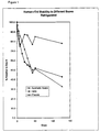
- Figure 1 shows a comparison of the stability at 2-8 C of recombinant full length troponin I in various matrices.
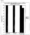
- Figure 2 shows a comparison of the stability at 2-8 C of a recombinantly produced 80 amino acid peptide in various matrices.
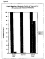
- Figure 3 shows a comparison of the stability at 2-8 C of a 10 ng/mL calibrator of a synthetic peptide of troponin I in various matrices.
- Figure 4 shows a comparison of the stability at 2-8 C of a 50 ng/mL calibrator of a synthetic peptide of troponin I in various matrices.
- Figure 5 shows a comparison of the stability at 37 C of a synthetic peptide of troponin I in various matrices.
- Figure 6 shows a comparison of the stability at 37 C of a synthetic peptide of troponin I in various matrices where all of the matrices include a protease inhibitor.
- Figure 7 shows a calibration curve of different levels of a synthetic peptide of troponin I in a matrix of the present invention.
- the stabilized clinical laboratory non-human serum derived control and/or calibrator matrix comprises an aqueous solution of a buffer, a stabilizing protein such as albumin, ovalbumin, casein and the like, a chelating agent, a reducing agent and a salt, all at a slightly acidic to mildly alkaline pH.
- the matrix can contain a blocking agent such as gelatin, detergents and protease inhibitors.
- a preservative may be added to prevent microbial growth.
- An analyte or analyte analogue such as cardiac markers or other unstable or relatively insoluble proteins or fragments thereof are also added. If the calibrator or control will be lyophilized or frozen, sugars or other bulking agents are added.
- the preferred buffers include buffers such as TRIS buffers and phosphate buffers.
- Other buffers include: 3-(N-Morpholino) propane sulfonic acid (MOPS), N-Tris-hydroxymethyl methyl-2-aminoethane sulfonic acid (TES), 3-[N-bis (hydroxyethyl)-amino] -2-hydroxypropane sulfonic acid (DIPSO), Piperazine-N, N'Bis(2)-hydroxypropane sulfonic acid (HEPPSO), Tris-(hydroxymethyl) aminoethane, N-2-Hydroxyehtylpiperazine-N'-2-aminoethane sulfonic acid (HEPES), 3-[N-(Tris-hydroxymethyl) methylamino] -2-hydroxy-propane sulfonic acid (TAPSO), and (2 p[2-Amino-2-oxoethyl) - amino] ethanesulfonic
- the concentration of the buffer may be from about 10 mM to 200 mM. Preferably the concentration is about 25 mM to 100 mM. Most preferably the concentration is about 50 mM.
- the stabilizing protein may be albumin, ovalbumin, casein or the like.
- the stabilizing protein, as with the other ingredients, should be essentially free of contaminants that interfere with the stability of the analyte in the matrix. Substances that may destabilize or destroy protein structure such as proteases are examples of such contaminants.
- Albumin is a preferred stabilizing protein.
- the source of the albumin is not critical.
- the albumin may be native or recombinant in origin. The most available source of native albumin is of bovine origin. It is most preferred that the albumin be essentially protease-free if the protein analyte or analogue is susceptible to protease degradation. Alternatively, protease inhibitors can be added.
- the preferred concentration of albumin is from 5-20%, 8-12% and most preferably at about 10%.
- the protein stabilizer functions to provide a protective effect to the analyte or analyte analogue.
- a blocking agent i.e. an agent to minimize the non specific binding of the analyte or analyte analogue to surfaces
- gelatin is the preferred blocking agent.
- the gelatin if of bovine origin, is added at a concentration of about 0.01 to .15%, most preferably 0.1%. If the gelatin is of a different origin (e.g. fish) the concentration is adjusted appropriately.
- a chelating agent is also added.
- the preferred chelating agents are ethylenebis (oxyethylene nitrilio)-tetraacetic acid (EGTA) and ethylene diamine tetracetic acid (EDTA), sodium citrate, or oxalate salts such as sodium, potassium, ammonnium or lithium oxalte.
- the most preferred chelating agent is EDTA.
- the concentration of chelating agents may be from 1 mM to 15 mM and most preferably is from 5 to 10 mM.
- the preferred reducing agent is N-acetyl-cysteine (NAC).
- NAC N-acetyl-cysteine
- examples of other reducing agents which may be used include 2-aminoethanethiol, 2-mercaptoethanol, 2-mercaptoethylanine and dithiothreitol.
- the concentration of reducing agent may be from about 0 to 5 mM, preferably the reducing agent is about 2 mM to 3.5 mM, and most preferably about 2.6 mM.
- the pH may be from about 5.0 to 8.0. Slightly acidic pH values lower the non-specific binding of the troponin. The preferred range of pH is about 5 to 7.5 and most preferably about 7.0.
- the preferred salt is sodium chloride. Many other salts may be substituted. Examples of other salts include potassium salts, ammonium salts, and lithium salts.
- protease inhibitor is added, aprotinin and "Protease Inhibitor” (Sigma) are effective and may be used at the manufacturer's recommended concentration.
- protease inhibitors include (2S, 3R)-3-Amino-2-hydroxy-5-methylhexanoyl]-Val-Val-Asp (Amastatin-Sigma), [2S,3R]-3-Amino-2-hydroxy-4-[4-nitrophenyl]-butanoyl-L-leucine, Antipain, [2S,3R]-3-Amino-2-hydroxy-5-methylhexanoyl]-Val-Val-Asp (Epiamascatin-Sigma), ([2R,3R]-3-Amino-2-hydroxy-4-phenylbutanoyl)-L-leucine (Epibestatin-Sigma), Foroxymithine, Acetyl-Leu-Leu-Arg-al (Leupeptin-Sigma)
- the preferred detergents are SDS and Triton X-100.
- Other detergents include Tween-20, Brij, Sorbitin, Tergital and Nonidet.
- the concentration of detergents may be from 0.05% to 0.3%. The preferred range is from about 0.05% and 0.2%. The most preferred concentration is 0.1%.
- the preservative may be added to prevent microbial and fungal growth.
- the preservatives may be clotrimazole of at least 0.03%, chloramphenicol of at least 0.017%, or sodium azide of at least 0.05%.
- Other preservatives include gentamicin, mycostatin, thimerasol and Kathon at an effective concentration.
- plastics such as polypropylene should be used. This minimizes loss of proteins to glass due to non specific binding to glass.
- glass based labware can be utilized but should be siliconized prior to use.
- the preferred bulking agents are trehalose at 3 to 10% and sucrose at about 100 mM. The most preferred concentration of trehalose is about 5-10%.
- Other bulking agents include glucose, sucrose, galactose, manose, maltose, lactose, isomaltose, cellobiose, mannobiose, melbiose, maltotriose, nystose, maltotetraose, maltopentaose, maltohexaose, and maltoheptaose.
- One liter of the matrix may be prepared by dissolving about 30 grams of BSA, about one gram of gelatin, about 15 grams of sodium chloride, 3 grams of EDTA, about 50 grams of trehalose, a preservative and a clinically appropriate level of the analyte or analyte analogue in 700 ml of 0.50 mM sodium phosphate buffer. After all ingredients are dissolved the pH is adjusted to 7.0 and then a sufficient volume of a buffer, such as sodium phosphate, is added to bring the volume to 1.0 L. The solution is sterile filled in suitable containers and lyophilized. The lyophilized analyte containing matrix is reconstituted by adding one liter of a diluent containing 65 grams of BSA, 25 grams of NaCl, protease inhibitor, and about 0.4 grams of NAC.
- one liter of the matrix may be prepared by dissolving about 100 grams of BSA, about one gram of gelatin, about 40 grams of sodium chloride, 3 grams of EDTA, a preservative, and 0.4 grams of NAC and a clinically appropriate level of the analyte or analyte analogue in 700 ml of 0.50m sodium phosphate. After all ingredients are dissolved the pH is adjusted to 7.0 and then a sufficient volume of a buffer, such as sodium phosphate is added to bring the volume to 1.0L. The liquid may be aliquoted and stored refrigerated or frozen. If the liquid is to be frozen, bulking agents are also added.
- troponin I fragments also have increased stability. This is surprising because the stability of the troponin I fragments was found to be less than that of the full length troponin I when the matrix is normal human serum. In addition, certain troponin I fragments had similar binding to the anti-troponin antibody used in the assay when compared with the full length troponin I molecule.
- the matrix is non-human serum derived which prevents the user (and manufacturing personnel) from exposure to many of the diseases which can be spread by contact with human blood products.
- the matrix is also able to keep the analyte stable in liquid form for an extended period of time.
- Current formulations can also be lyophilized or frozen and can be reliably reconstituted or thawed for up to at least nine months shelf storage with very little variation in calibration. After reconstitution or thawing, the analyte is stable for up to three weeks at 2-8C. Spiking of the analyte into the matrix yields a calibration curve which closely parallels a human serum curve.
- Non-specific binding levels in the "synthetic" matrix also closely parallel the levels seen in normal human serum.
- fragments such as recombinantly or synthetically produced peptide fragments of troponin can be utilized instead of the full length protein. Indeed these fragments are preferred because of their stability in the matrix coupled with the availability and reproducibility of the recombinantly or synthetically produced fragments.
- the analyte analogue e.g. the fragment
- a reconstitution diluent of one liter is prepared by combining in aqueous solution about 65 grams of albumin, 25 grams of sodium chloride and 0.4 grams of NAC, protease inhibitor and about 2 milliliters of a 25% solution of sodium azide.
- a stock solution of human recombinant troponin I was prepared in polypropylene labware by adding a sufficient amount of recombinant troponin I so that calibrators or controls can be made that have concentrations ranging from about 0 to 100 ng/mL of troponin I.
- solutions of troponin I were prepared by adding a sufficient amount of an unpurified solution of recombinant troponin I at 100 ug/mL to the following matrices:
- Example 2 Normal human serum, plasma, and a matrix similar to that described in Example 1. For each matrix the final volume was 15 milliliters of a 100 ng/mL solution of recombinant troponin I.
- the recombinant troponin I had the following sequence: and included a 6 His carboxy terminus tail and a seven amino acid sequence from the phage T7 Gene 10 leader sequence at the amino terminus. See, also Vallins W.J. et al., Molecular cloning of human cardiac troponin I using polymerase chain reaction , FEBS LETTERS :Vol. 270, number 1, 2 September 1990.
- troponin I For troponin I, an aliquot of each troponin I solution is applied to the antibody and immunologically binds to the antibody to form a reaction zone. Next, a conjugate of alkaline phosphatase-anti-troponin I antibody is added to the reaction zone. The conjugate binds to the troponin I. A substrate wash solution containing 4 methyl umbelliferyl phosphate is applied to the reaction zone. A front surface fluorometer measures the rate of change of fluorescence in rate units designated as millivolts per minute (mvm).
- the matrix of the present invention provided more stability to the troponin-I analyte than normal human serum or plasma.
- a stock solution of human recombinant troponin I peptide is prepared in polypropylene labware by adding a sufficient amount of a recombinant troponin I peptide so that calibrators or controls can be made that have concentrations ranging from about 0 to 100 ng/mL of troponin I.
- solutions of troponin I were prepared by adding a sufficient amount of a recombinant 80 amino acid peptide of recombinant troponin I having the sequence ADGSSDAAREPRPAPAPIRRRSSNYRAYATEPHAKKKSKISASRKLQLKTLLLQIAKQELER EAEERRGEKGRALSTRCQ (SEQ ID NO: 2) also having a six histidine tail at the carboxy terminus and seven amino acids from the phage T7 Gene 10 leader sequence at the amino terminus (see, also Vallins W.J. et al., Molecular cloning of human cardiac troponin I using polymerase chain reaction , FEBS LETTER S :Vol. 270, number 1, 2 September 1990) at 7500 ng/mL to the following matrices:
- troponin I For troponin I, an aliquot of each troponin I solution is applied to the antibody and immunologically binds to the antibody to form a reaction zone. Next, a conjugate of alkaline phosphatase-anti-troponin I antibody is added to the reaction zone. The conjugate binds to the troponin I. A substrate wash solution containing 4 methyl umbelliferyl phosphate is applied to the reaction zone. A front surface fluorometer measures the rate of change of fluorescence in rate units designated as millivolts per minute (mvm).
- the matrix of the present invention provided more stability to the troponin-I analyte than normal human serum, processed human plasma (PHP) or plasma.
- a stock solution of a synthetic peptide of troponin I is prepared in polypropylene labware by adding a sufficient amount of the peptide so that calibrators or controls can be made that have concentrations ranging from about 0 to 50 ng/mL of troponin I.
- solutions of troponin I were prepared by adding a sufficient amount of a synthetic peptide of troponin I having the sequence at a stock concentration (in purified water) of 1.4 mg/mL to the following matrices:
- troponin I For troponin I, an aliquot of each troponin I solution is applied to the antibody and immunologically binds to the antibody to form a reaction zone. Next, a conjugate of alkaline phosphatase-anti-troponin I antibody is added to the reaction zone. The conjugate binds to the troponin I. A substrate wash solution containing 4 methyl umbelliferyl phosphate is applied to the reaction zone. A front surface fluorometer measures the rate of change of fluorescence.
- the matrix of the present invention provided more stability to the troponin-I analyte analogue than normal human serum, processed human plasma (PHP), the synthetic base DXMC, or plasma.
- a stock solution of a synthetic peptide of troponin I is prepared in polypropylene labware by adding a sufficient amount of the peptide so that calibrators or controls can be made that have concentrations ranging from about 0 to 100 ng/mL of troponin I.
- Example 2 Normal human serum, plasma, and a matrix similar to that described in Example 1 (except that the pH of the synthetic matrix was adjusted to 5.5) to provide 3.0 milliliters of a 100 ng/mL solution of troponin I in each matrix.
- EDTA was added to each matrix at about 3.5 mg/mL.
- a second set of solutions was prepared as above, except that the solutions also contained a protease inhibitor cocktail of PEPSTATIN at 680 ng/mL and aminoethylbenzenesulfonyl fluoride at 208 ug/mL.
- troponin I For troponin I, an aliquot of each troponin I solution is applied to the antibody and immunologically binds to the antibody to form a reaction zone. Next, a conjugate of alkaline phosphatase-anti-troponin I antibody is added to the reaction zone. The conjugate binds to the troponin I. A substrate wash solution containing 4 methyl umbelliferyl phosphate is applied to the reaction zone. A front surface fluorometer measures the rate of change of fluorescence in rate units designated as millivolts per minute (mvm).
- the matrix of the present invention provided more stability to the troponin-I analyte analogue than normal human serum, or plasma with or without the protease inhibitors.
- a stock solution of a synthetic peptide of troponin I is prepared in polypropylene labware by adding a sufficient amount of the peptide so that calibrators or controls can be made that have concentrations ranging from about 0 to 50 ng/mL of troponin I.
- solutions of troponin I were prepared by adding a sufficient amount of a synthetic peptide of troponin I having the sequence at a stock concentration of 1.4 mg/mL to a matrix similar to that described in Example 1 to provide calibrator solutions of troponin I having the following concentrations: 0 ng/mL, 2 ng/mL, 8 ng/mL, 15 ng/mL, 25 ng/mL and 50 ng/mL.
- Controls were prepared in a similar fashion to provide control solutions at about 4 ng/mL , 20 ng/mL and 35 ng/mL.
- the analyzer measures the rate of change of a fluorescent signal.
- An antibody to an analyte such as troponin I
- An aliquot of each troponin I solution is applied to the antibody and immunologically binds to the antibody to form a reaction zone.
- a conjugate of alkaline phosphatase-anti-troponin I antibody is added to the reaction zone.
- the conjugate binds to the troponin I.
- a substrate wash solution containing 4 methyl umbelliferyl phosphate is applied to the reaction zone.
- a front surface fluorometer measures the rate of change of fluorescence in rate units designated as millivolts per minute (mvm).
- Control recoveries are presented in Table 2. Control Rep. 1 (ng/mL) Rep. 2 (ng/mL) Rep. 3 (ng/mL) Rep. 4 (ng/mL) Mean (ng/mL) Syn. Low 3.7 3.6 3.7 Syn. Mid 17.1 16.3 16.7 Syn. Hi 32.6 32.8 32.7 Serum Lo 2.6 2.5 2.5 2.3 2.5 Serum Hi 18.2 18.7 19.7 18.0 18.7
- a stock solution of CK-MB is prepared by adding a sufficient amount of the CK-MB so that calibrators or controls can be made that have concentrations ranging from about 0 to 125 ng/mL of CK-MB.
- Solutions of CK-MB are prepared by adding a sufficient amount of CK-MB to a matrix similar to that described in Example 1 to provide calibrator solutions of CK-MB having the following concentrations: 0, 4, 10, 25, 60 and 125 ng/mL. Controls are prepared in a similar fashion
- a stock solution of Troponin T is prepared by adding a sufficient amount of the Troponin T so that calibrators or controls can be made that have concentrations ranging from about 0 to 12 ng/mL of Troponin T.
- Solutions of Troponin T are prepared by adding a sufficient amount of Troponin T to a matrix similar to that described in Example 1 to provide calibrator solutions of Troponin T having the following concentrations: 0, 1, 2, 4, 8, and 12 ng/mL. Controls are prepared in a similar fashion
Landscapes
- Health & Medical Sciences (AREA)
- Life Sciences & Earth Sciences (AREA)
- Engineering & Computer Science (AREA)
- Chemical & Material Sciences (AREA)
- Immunology (AREA)
- Biomedical Technology (AREA)
- Molecular Biology (AREA)
- Hematology (AREA)
- Urology & Nephrology (AREA)
- Microbiology (AREA)
- General Health & Medical Sciences (AREA)
- Biotechnology (AREA)
- Biochemistry (AREA)
- Medicinal Chemistry (AREA)
- Physics & Mathematics (AREA)
- Zoology (AREA)
- Food Science & Technology (AREA)
- Cell Biology (AREA)
- General Physics & Mathematics (AREA)
- Pathology (AREA)
- Bioinformatics & Cheminformatics (AREA)
- Genetics & Genomics (AREA)
- Wood Science & Technology (AREA)
- Analytical Chemistry (AREA)
- Organic Chemistry (AREA)
- General Engineering & Computer Science (AREA)
- Proteomics, Peptides & Aminoacids (AREA)
- Peptides Or Proteins (AREA)
- Investigating Or Analysing Biological Materials (AREA)
- Enzymes And Modification Thereof (AREA)
- Medicines That Contain Protein Lipid Enzymes And Other Medicines (AREA)
Description




| Calibrator Level | Rate 1 (mvm) | Rate 2 (mvm) | Mean (mvm) |
| 0 | 171.5 | 180.4 | 176.0 |
| 2 | 463.7 | 464.5 | 464.1 |
| 8 | 1287.5 | 1208.5 | 1248.0 |
| 15 | 2487.6 | 2481.9 | 2484.8 |
| 25 | 3926.6 | 3826.6 | 3876.6 |
| 50 | 7668.0 | 6767.0 | 7217.5 |
| Control | Rep. 1 (ng/mL) | Rep. 2 (ng/mL) | Rep. 3 (ng/mL) | Rep. 4 (ng/mL) | Mean (ng/mL) |
| Syn. Low | 3.7 | 3.6 | 3.7 | ||
| Syn. Mid | 17.1 | 16.3 | 16.7 | ||
| Syn. Hi | 32.6 | 32.8 | 32.7 | ||
| Serum Lo | 2.6 | 2.5 | 2.5 | 2.3 | 2.5 |
| Serum Hi | 18.2 | 18.7 | 19.7 | 18.0 | 18.7 |
Sabucedo, Alberto
Chin, Bruce
Bauer, Roger


Claims (15)
- A diagnostic calibrator consisting essentially of a non-human serum derived matrix usable for stabilising troponin or a fragment thereof, comprising a buffer, a reducing agent, a stabilising protein, a chelating agent and a salt, wherein the matrix provides a response which mimics the response using human serum.
- The composition of claim 1 for stabilising troponin I or a fragment thereof.
- The compositions of claim 1 wherein the analyte is

- The composition of claim 1 wherein the analyte is

- The composition of Claims 1, 2, 3 or 4, wherein the reducing agent is selected from N-acetyle-cysteine, 2-aminoethanethiol, 2-mercaptoethanol, 2-mercaptoethylanine and dithiothreitol; the stabilizing protein is selected from albumin, ovalbumin and casein; and the chelating agent is selected from ethylenebis (oxyethylene nitrilio)-tetraacetic acid (EGTA), ethylene diamine tetracetic acid (EDTA), citrates, or exalates.
- The composition of any preceding claim further comprising a blocking agent.
- The composition of any preceding claim wherein the compositions further comprises a bulking agent.
- The composition of claim 9 wherein the composition is lyophilized.
- The composition of any one of claims 1 to 7 wherein the composition is frozen.
- The composition of any preceding claim wherein the protein stabilizer is albumin.
- The composition of claim 10 wherein the albumin is protease free.
- The composition of claim 6 wherein the blocking agent is gelatin.
- The composition of claim 7 wherein the bulking agent is selected from trehalose, glucose, sucrose, galactose, manose, maltose, lactose, isomaltose, cellobiose, mannobiose, melbiose, maltotriose, nystose, maltotetraose, maltopentaose, maltohexaose, and maltoheptaose.
- Use in the manufacture of a diagnostic calibrator which is free of human serum matrix of a buffer, a reducing agent, a stabilising protein, a chelating agent and a salt, for the purpose of stabilising troponin or a fragment thereof.
- Use according to Claim 14, wherein the matrix is according to any one of Claims 1 to 13.
Applications Claiming Priority (3)
| Application Number | Priority Date | Filing Date | Title |
|---|---|---|---|
| US08/400,158 US6165981A (en) | 1995-03-07 | 1995-03-07 | Stabilizing solutions for proteins and peptides |
| US400158 | 1995-03-07 | ||
| PCT/US1996/003034 WO1996027661A1 (en) | 1995-03-07 | 1996-03-06 | Stabilizing solutions for proteins and peptides |
Publications (2)
| Publication Number | Publication Date |
|---|---|
| EP0759074A1 EP0759074A1 (en) | 1997-02-26 |
| EP0759074B1 true EP0759074B1 (en) | 1998-07-08 |
Family
ID=23582452
Family Applications (1)
| Application Number | Title | Priority Date | Filing Date |
|---|---|---|---|
| EP96907187A Expired - Lifetime EP0759074B1 (en) | 1995-03-07 | 1996-03-06 | Stabilizing solutions for troponin |
Country Status (8)
| Country | Link |
|---|---|
| US (1) | US6165981A (en) |
| EP (1) | EP0759074B1 (en) |
| JP (1) | JP3740644B2 (en) |
| AU (1) | AU698790B2 (en) |
| CA (1) | CA2189737A1 (en) |
| DE (1) | DE69600407T2 (en) |
| ES (1) | ES2121479T3 (en) |
| WO (1) | WO1996027661A1 (en) |
Families Citing this family (38)
| Publication number | Priority date | Publication date | Assignee | Title |
|---|---|---|---|---|
| US6627404B1 (en) * | 1995-04-18 | 2003-09-30 | Biosite, Inc. | Methods for improving the recovery of troponin I and T in membranes, filters and vessels |
| US7006874B2 (en) * | 1996-01-05 | 2006-02-28 | Thermage, Inc. | Treatment apparatus with electromagnetic energy delivery device and non-volatile memory |
| WO1998007446A1 (en) * | 1996-08-23 | 1998-02-26 | Eli Lilly And Company | Protein formulations |
| EP0946198B1 (en) | 1996-10-15 | 2002-06-19 | Medical Analysis Systems, Inc. | Method of stabilizing troponin I (cTnI) via conjugation with an active polymer |
| FR2756827B1 (en) * | 1996-12-05 | 1999-01-29 | Pasteur Sanofi Diagnostics | SYNTHETIC BIEPITOPIC COMPOUNDS FOR USE AS STANDARDS IN BIOLOGICAL ASSAYS FOR TROPONIN I |
| US6156521A (en) * | 1997-12-19 | 2000-12-05 | Biosite Diagnostics, Inc. | Methods for the recovery and measurement of troponin complexes |
| WO1998031390A1 (en) * | 1997-01-17 | 1998-07-23 | Eli Lilly And Company | Obesity protein formulations |
| KR20000070338A (en) * | 1997-01-20 | 2000-11-25 | 나가시마 카쭈시게, 노미야마 아키히코 | Method for stabilizing peptides and freeze-dried medicinal compositions containing peptides obtained by using the method |
| WO1998041222A1 (en) * | 1997-03-20 | 1998-09-24 | Eli Lilly And Company | Obesity protein formulations |
| US6248869B1 (en) | 1997-05-29 | 2001-06-19 | Medical Analysis Systems, Inc. | Troponin I forms and use of the same |
| JP2002511932A (en) * | 1997-06-13 | 2002-04-16 | メディカル、アナリシス、システムズ、インコーポレイテッド(エムエイエス) | Heart marker stabilizing composition |
| WO2000023585A1 (en) * | 1998-10-21 | 2000-04-27 | Spectral Diagnostics, Inc. | Cardiac troponin i polypeptide fragments and uses in diagnostics |
| DE19930676B4 (en) | 1999-07-02 | 2006-01-19 | Sanofi-Aventis Deutschland Gmbh | Process for stabilizing insulin, insulin derivatives and / or their precursors in complex mixtures when they are stored in aqueous solvents |
| FR2797689B1 (en) | 1999-08-16 | 2003-06-27 | Pasteur Sanofi Diagnostics | USE OF SYNTHETIC COMPOUNDS FOR IMMUNODAYS |
| US7078486B2 (en) | 1999-12-10 | 2006-07-18 | Spectral Diagnostics, Inc. | Single-chain polypeptides comprising troponin I and troponin C |
| AU2001286996A1 (en) * | 2000-08-31 | 2002-03-13 | Chiron Corporation | Stabilized fgf formulations containing reducing agents |
| US6962700B1 (en) * | 2000-09-13 | 2005-11-08 | Atopix Pharmaceuticals Corporation | Method of manufacturing immune globulin |
| US6538104B2 (en) | 2001-04-27 | 2003-03-25 | Medical Analysis Systems, Inc. | Stabilization of cardiac troponin I subunits and complexes |
| US6927062B2 (en) * | 2002-11-25 | 2005-08-09 | Agdia, Inc. | Controls and standards for assays and method for manufacture thereof |
| US7291501B2 (en) | 2003-07-16 | 2007-11-06 | Abbott Laboratories | Stable compositions for measuring human natriuretic peptides |
| US7445933B2 (en) * | 2003-07-16 | 2008-11-04 | Abbott Laboratories, Inc. | Stable calibrators or controls for measuring human natriuretic peptides |
| US20050136542A1 (en) * | 2003-12-19 | 2005-06-23 | Beckman Coulter, Inc. | Stabilized liquid reference solutions |
| ATE463509T1 (en) * | 2005-04-28 | 2010-04-15 | Abbott Lab | STABILIZATION OF CARDIAC TROPONIN |
| AU2007234612B2 (en) | 2006-12-14 | 2013-06-27 | Johnson & Johnson Regenerative Therapeutics, Llc | Protein stabilization formulations |
| US7678764B2 (en) * | 2007-06-29 | 2010-03-16 | Johnson & Johnson Regenerative Therapeutics, Llc | Protein formulations for use at elevated temperatures |
| WO2009020744A1 (en) * | 2007-08-07 | 2009-02-12 | Johnson & Johnson Regenerative Therapeutics, Llc | Protein formulations comprising gdf-5 in aqueous acidic solution |
| BRPI0911048A2 (en) | 2008-04-14 | 2015-12-29 | Atrm Llc | gdf-5 buffered liquid formulations |
| US20110306148A1 (en) | 2010-06-14 | 2011-12-15 | Siemens Healthcare Diagnostics Inc. | Composition for use as an assay reagent |
| JP6308955B2 (en) | 2012-03-09 | 2018-04-11 | ベスタロン コーポレイション | Production of toxic peptides, expression of peptides in plants, and combinations of cysteine rich peptides |
| CN107167613B (en) * | 2017-06-22 | 2018-10-02 | 深圳清华大学研究院 | Albumen spotting buffer for plasma gold chip |
| CN109254153B (en) * | 2018-08-27 | 2021-10-12 | 广东菲鹏生物有限公司 | Antioxidant of myoglobin and application thereof |
| JP2022501398A (en) * | 2018-09-28 | 2022-01-06 | シーメンス・ヘルスケア・ダイアグノスティックス・インコーポレイテッド | Storage Solution Composition for Stabilization of Purified Muller Tube Suppressant (MIS) and Related Usage and Production Methods |
| CN109725148B (en) * | 2018-12-30 | 2022-05-03 | 广东环凯微生物科技有限公司 | Immunomagnetic bead preservation solution |
| ES3005102T3 (en) | 2020-04-20 | 2025-03-13 | Vestaron Corp | Proteolytically stable u1-agatoxin-ta1b variant polypeptides for pest control |
| MX2022013415A (en) | 2020-05-01 | 2022-11-14 | Vestaron Corp | Insecticidal combinations. |
| CN114324882B (en) * | 2020-10-12 | 2022-12-27 | 广东菲鹏生物有限公司 | Protein stabilizer and application thereof |
| CN117969850A (en) * | 2024-01-11 | 2024-05-03 | 桂林优利特医疗电子有限公司 | A liquid myoglobin calibrator diluent and preparation method thereof |
| WO2025226701A1 (en) | 2024-04-23 | 2025-10-30 | Vestaron Corporation | Proteolytically stable u1-agatoxin-ta1b variant polypeptides for pest control |
Family Cites Families (4)
| Publication number | Priority date | Publication date | Assignee | Title |
|---|---|---|---|---|
| US4931385A (en) * | 1985-06-24 | 1990-06-05 | Hygeia Sciences, Incorporated | Enzyme immunoassays and immunologic reagents |
| US5217890A (en) * | 1991-08-20 | 1993-06-08 | Eastman Kodak Company | Stabilized composition containing creatine kinase and protein having blocked sulfhydryl groups |
| FR2701954B1 (en) * | 1993-02-23 | 1995-07-07 | Pasteur Sanofi Diagnostics | Stabilized troponin composition for immunoassays and method for stabilizing troponin for immunoassays. |
| WO1994027156A1 (en) * | 1993-05-17 | 1994-11-24 | Fortron Bioscience, Inc. | Assay for cardiac troponin i |
-
1995
- 1995-03-07 US US08/400,158 patent/US6165981A/en not_active Expired - Lifetime
-
1996
- 1996-03-06 AU AU50929/96A patent/AU698790B2/en not_active Ceased
- 1996-03-06 CA CA002189737A patent/CA2189737A1/en not_active Abandoned
- 1996-03-06 DE DE69600407T patent/DE69600407T2/en not_active Expired - Lifetime
- 1996-03-06 WO PCT/US1996/003034 patent/WO1996027661A1/en not_active Ceased
- 1996-03-06 EP EP96907187A patent/EP0759074B1/en not_active Expired - Lifetime
- 1996-03-06 ES ES96907187T patent/ES2121479T3/en not_active Expired - Lifetime
- 1996-03-06 JP JP52702396A patent/JP3740644B2/en not_active Expired - Fee Related
Also Published As
| Publication number | Publication date |
|---|---|
| WO1996027661A1 (en) | 1996-09-12 |
| JPH10500704A (en) | 1998-01-20 |
| CA2189737A1 (en) | 1996-09-12 |
| ES2121479T3 (en) | 1998-11-16 |
| EP0759074A1 (en) | 1997-02-26 |
| DE69600407T2 (en) | 1999-04-29 |
| AU5092996A (en) | 1996-09-23 |
| AU698790B2 (en) | 1998-11-05 |
| US6165981A (en) | 2000-12-26 |
| JP3740644B2 (en) | 2006-02-01 |
| DE69600407D1 (en) | 1998-08-13 |
Similar Documents
| Publication | Publication Date | Title |
|---|---|---|
| AU698790B2 (en) | Stabilizing solutions for proteins and peptides | |
| JP3357163B2 (en) | Troponin stabilizing composition and stabilizing method for immunoassay | |
| Katus et al. | Non-invasive assessment of perioperative myocardial cell damage by circulating cardiac troponin T. | |
| US20080248579A1 (en) | Method of stabilizing human natriuretic peptides | |
| Whitelock et al. | ADAMTS‐13 activity in plasma is rapidly measured by a new ELISA method that uses recombinant VWF‐A2 domain as substrate | |
| US20050014287A1 (en) | Stable calibrators or controls for measuring human natriuretic peptides | |
| US6268481B1 (en) | Covalently coupled troponin complexes | |
| US5834210A (en) | Stable troponin subunits and complexes | |
| US20010006683A1 (en) | Stabilized composition of troponin for immunoassays and method of preparation of such a stabilized composition | |
| Gargosky et al. | Urinary insulin-like growth factors (IGF) and IGF-binding proteins in normal subjects, growth hormone deficiency, and renal disease | |
| Affholter et al. | Identification of residues in the insulin molecule important for binding to insulin-degrading enzyme | |
| EP1000140A1 (en) | Stabilized compositions of cardiac markers | |
| CN115639049A (en) | Composite antigen diluent, stable myocardium composite quality control product composition and application | |
| EP0983299A1 (en) | Covalently coupled troponin complexes | |
| JP4593665B2 (en) | Myoglobin-containing aqueous solution | |
| JP4033534B2 (en) | Method for stabilizing human hemoglobin | |
| Wu et al. | Analysis of circulating annexin A5 parameters during pregnancy: absence of differences between women with recurrent spontaneous pregnancy losses and controls | |
| Krizek et al. | Clinical application of a rapid method using agarose gel electrophoresis and Western blotting to evaluate von Willebrand factor protease activity | |
| WO2006116005A2 (en) | Stabilization of cardiac troponin | |
| Burke | Spectrin Johannesburg: α and β Spectrin Defects in a South African Kindred With Hereditary Elliptocytosis | |
| JPH04505664A (en) | How to detect anti-streptokinase antibodies | |
| ISO | Anti-Acetylated Tubulin Antibody, Mouse Monoclonal | |
| Tichenor | Regulation of uterine smooth muscle relaxation | |
| MXPA96004002A (en) | Inhibition of activity of proteasa de lisados decelula de la humana compl |
Legal Events
| Date | Code | Title | Description |
|---|---|---|---|
| PUAI | Public reference made under article 153(3) epc to a published international application that has entered the european phase |
Free format text: ORIGINAL CODE: 0009012 |
|
| 17P | Request for examination filed |
Effective date: 19961121 |
|
| AK | Designated contracting states |
Kind code of ref document: A1 Designated state(s): DE ES FR IT |
|
| 17Q | First examination report despatched |
Effective date: 19970214 |
|
| GRAG | Despatch of communication of intention to grant |
Free format text: ORIGINAL CODE: EPIDOS AGRA |
|
| GRAG | Despatch of communication of intention to grant |
Free format text: ORIGINAL CODE: EPIDOS AGRA |
|
| GRAG | Despatch of communication of intention to grant |
Free format text: ORIGINAL CODE: EPIDOS AGRA |
|
| GRAH | Despatch of communication of intention to grant a patent |
Free format text: ORIGINAL CODE: EPIDOS IGRA |
|
| GRAH | Despatch of communication of intention to grant a patent |
Free format text: ORIGINAL CODE: EPIDOS IGRA |
|
| RAP1 | Party data changed (applicant data changed or rights of an application transferred) |
Owner name: DADE BEHRING INC. |
|
| RAP3 | Party data changed (applicant data changed or rights of an application transferred) |
Owner name: DADE BEHRING INC. |
|
| GRAA | (expected) grant |
Free format text: ORIGINAL CODE: 0009210 |
|
| AK | Designated contracting states |
Kind code of ref document: B1 Designated state(s): DE ES FR IT |
|
| REF | Corresponds to: |
Ref document number: 69600407 Country of ref document: DE Date of ref document: 19980813 |
|
| ET | Fr: translation filed | ||
| REG | Reference to a national code |
Ref country code: ES Ref legal event code: FG2A Ref document number: 2121479 Country of ref document: ES Kind code of ref document: T3 |
|
| PLBE | No opposition filed within time limit |
Free format text: ORIGINAL CODE: 0009261 |
|
| STAA | Information on the status of an ep patent application or granted ep patent |
Free format text: STATUS: NO OPPOSITION FILED WITHIN TIME LIMIT |
|
| 26N | No opposition filed | ||
| PGFP | Annual fee paid to national office [announced via postgrant information from national office to epo] |
Ref country code: ES Payment date: 20000314 Year of fee payment: 5 |
|
| PG25 | Lapsed in a contracting state [announced via postgrant information from national office to epo] |
Ref country code: ES Free format text: LAPSE BECAUSE OF NON-PAYMENT OF DUE FEES Effective date: 20010307 |
|
| REG | Reference to a national code |
Ref country code: ES Ref legal event code: FD2A Effective date: 20030203 |
|
| REG | Reference to a national code |
Ref country code: DE Ref legal event code: R409 Ref document number: 69600407 Country of ref document: DE Ref country code: DE Ref legal event code: R119 Ref document number: 69600407 Country of ref document: DE |
|
| REG | Reference to a national code |
Ref country code: DE Ref legal event code: R409 Ref document number: 69600407 Country of ref document: DE |
|
| REG | Reference to a national code |
Ref country code: FR Ref legal event code: TP Owner name: SIEMENS HEALTHCARE DIAGNOSTICS INC., US Effective date: 20120127 Ref country code: FR Ref legal event code: CD Owner name: SIEMENS HEALTHCARE DIAGNOSTICS INC., US Effective date: 20120127 |
|
| PGFP | Annual fee paid to national office [announced via postgrant information from national office to epo] |
Ref country code: FR Payment date: 20140317 Year of fee payment: 19 Ref country code: IT Payment date: 20140328 Year of fee payment: 19 |
|
| PGFP | Annual fee paid to national office [announced via postgrant information from national office to epo] |
Ref country code: DE Payment date: 20140519 Year of fee payment: 19 |
|
| REG | Reference to a national code |
Ref country code: DE Ref legal event code: R119 Ref document number: 69600407 Country of ref document: DE |
|
| PG25 | Lapsed in a contracting state [announced via postgrant information from national office to epo] |
Ref country code: IT Free format text: LAPSE BECAUSE OF NON-PAYMENT OF DUE FEES Effective date: 20150306 |
|
| REG | Reference to a national code |
Ref country code: FR Ref legal event code: ST Effective date: 20151130 |
|
| PG25 | Lapsed in a contracting state [announced via postgrant information from national office to epo] |
Ref country code: DE Free format text: LAPSE BECAUSE OF NON-PAYMENT OF DUE FEES Effective date: 20151001 |
|
| PG25 | Lapsed in a contracting state [announced via postgrant information from national office to epo] |
Ref country code: FR Free format text: LAPSE BECAUSE OF NON-PAYMENT OF DUE FEES Effective date: 20150331 |