EP0734436B1 - Inhibition of dna methyltransferase - Google Patents
Inhibition of dna methyltransferase Download PDFInfo
- Publication number
- EP0734436B1 EP0734436B1 EP95901306A EP95901306A EP0734436B1 EP 0734436 B1 EP0734436 B1 EP 0734436B1 EP 95901306 A EP95901306 A EP 95901306A EP 95901306 A EP95901306 A EP 95901306A EP 0734436 B1 EP0734436 B1 EP 0734436B1
- Authority
- EP
- European Patent Office
- Prior art keywords
- dna
- cells
- cell
- methylation
- transfectants
- Prior art date
- Legal status (The legal status is an assumption and is not a legal conclusion. Google has not performed a legal analysis and makes no representation as to the accuracy of the status listed.)
- Expired - Lifetime
Links
Images


Classifications
-
- C—CHEMISTRY; METALLURGY
- C12—BIOCHEMISTRY; BEER; SPIRITS; WINE; VINEGAR; MICROBIOLOGY; ENZYMOLOGY; MUTATION OR GENETIC ENGINEERING
- C12N—MICROORGANISMS OR ENZYMES; COMPOSITIONS THEREOF; PROPAGATING, PRESERVING, OR MAINTAINING MICROORGANISMS; MUTATION OR GENETIC ENGINEERING; CULTURE MEDIA
- C12N15/00—Mutation or genetic engineering; DNA or RNA concerning genetic engineering, vectors, e.g. plasmids, or their isolation, preparation or purification; Use of hosts therefor
- C12N15/09—Recombinant DNA-technology
- C12N15/11—DNA or RNA fragments; Modified forms thereof; Non-coding nucleic acids having a biological activity
- C12N15/113—Non-coding nucleic acids modulating the expression of genes, e.g. antisense oligonucleotides; Antisense DNA or RNA; Triplex- forming oligonucleotides; Catalytic nucleic acids, e.g. ribozymes; Nucleic acids used in co-suppression or gene silencing
- C12N15/1137—Non-coding nucleic acids modulating the expression of genes, e.g. antisense oligonucleotides; Antisense DNA or RNA; Triplex- forming oligonucleotides; Catalytic nucleic acids, e.g. ribozymes; Nucleic acids used in co-suppression or gene silencing against enzymes
-
- A—HUMAN NECESSITIES
- A61—MEDICAL OR VETERINARY SCIENCE; HYGIENE
- A61K—PREPARATIONS FOR MEDICAL, DENTAL OR TOILETRY PURPOSES
- A61K31/00—Medicinal preparations containing organic active ingredients
- A61K31/70—Carbohydrates; Sugars; Derivatives thereof
- A61K31/7042—Compounds having saccharide radicals and heterocyclic rings
- A61K31/7052—Compounds having saccharide radicals and heterocyclic rings having nitrogen as a ring hetero atom, e.g. nucleosides, nucleotides
- A61K31/706—Compounds having saccharide radicals and heterocyclic rings having nitrogen as a ring hetero atom, e.g. nucleosides, nucleotides containing six-membered rings with nitrogen as a ring hetero atom
-
- A—HUMAN NECESSITIES
- A61—MEDICAL OR VETERINARY SCIENCE; HYGIENE
- A61P—SPECIFIC THERAPEUTIC ACTIVITY OF CHEMICAL COMPOUNDS OR MEDICINAL PREPARATIONS
- A61P35/00—Antineoplastic agents
-
- A—HUMAN NECESSITIES
- A61—MEDICAL OR VETERINARY SCIENCE; HYGIENE
- A61P—SPECIFIC THERAPEUTIC ACTIVITY OF CHEMICAL COMPOUNDS OR MEDICINAL PREPARATIONS
- A61P7/00—Drugs for disorders of the blood or the extracellular fluid
- A61P7/06—Antianaemics
-
- C—CHEMISTRY; METALLURGY
- C07—ORGANIC CHEMISTRY
- C07K—PEPTIDES
- C07K16/00—Immunoglobulins [IGs], e.g. monoclonal or polyclonal antibodies
- C07K16/40—Immunoglobulins [IGs], e.g. monoclonal or polyclonal antibodies against enzymes
-
- C—CHEMISTRY; METALLURGY
- C12—BIOCHEMISTRY; BEER; SPIRITS; WINE; VINEGAR; MICROBIOLOGY; ENZYMOLOGY; MUTATION OR GENETIC ENGINEERING
- C12N—MICROORGANISMS OR ENZYMES; COMPOSITIONS THEREOF; PROPAGATING, PRESERVING, OR MAINTAINING MICROORGANISMS; MUTATION OR GENETIC ENGINEERING; CULTURE MEDIA
- C12N9/00—Enzymes; Proenzymes; Compositions thereof; Processes for preparing, activating, inhibiting, separating or purifying enzymes
- C12N9/10—Transferases (2.)
- C12N9/1003—Transferases (2.) transferring one-carbon groups (2.1)
- C12N9/1007—Methyltransferases (general) (2.1.1.)
-
- A—HUMAN NECESSITIES
- A61—MEDICAL OR VETERINARY SCIENCE; HYGIENE
- A61K—PREPARATIONS FOR MEDICAL, DENTAL OR TOILETRY PURPOSES
- A61K39/00—Medicinal preparations containing antigens or antibodies
- A61K2039/505—Medicinal preparations containing antigens or antibodies comprising antibodies
-
- C—CHEMISTRY; METALLURGY
- C12—BIOCHEMISTRY; BEER; SPIRITS; WINE; VINEGAR; MICROBIOLOGY; ENZYMOLOGY; MUTATION OR GENETIC ENGINEERING
- C12N—MICROORGANISMS OR ENZYMES; COMPOSITIONS THEREOF; PROPAGATING, PRESERVING, OR MAINTAINING MICROORGANISMS; MUTATION OR GENETIC ENGINEERING; CULTURE MEDIA
- C12N2310/00—Structure or type of the nucleic acid
- C12N2310/10—Type of nucleic acid
- C12N2310/11—Antisense
- C12N2310/111—Antisense spanning the whole gene, or a large part of it
Landscapes
- Health & Medical Sciences (AREA)
- Life Sciences & Earth Sciences (AREA)
- Chemical & Material Sciences (AREA)
- Genetics & Genomics (AREA)
- Organic Chemistry (AREA)
- Engineering & Computer Science (AREA)
- Bioinformatics & Cheminformatics (AREA)
- General Health & Medical Sciences (AREA)
- Molecular Biology (AREA)
- Wood Science & Technology (AREA)
- Zoology (AREA)
- Medicinal Chemistry (AREA)
- Biomedical Technology (AREA)
- Biochemistry (AREA)
- General Engineering & Computer Science (AREA)
- Biotechnology (AREA)
- Microbiology (AREA)
- Animal Behavior & Ethology (AREA)
- Biophysics (AREA)
- Public Health (AREA)
- Pharmacology & Pharmacy (AREA)
- Veterinary Medicine (AREA)
- Immunology (AREA)
- Proteomics, Peptides & Aminoacids (AREA)
- Virology (AREA)
- Physics & Mathematics (AREA)
- General Chemical & Material Sciences (AREA)
- Plant Pathology (AREA)
- Nuclear Medicine, Radiotherapy & Molecular Imaging (AREA)
- Chemical Kinetics & Catalysis (AREA)
- Epidemiology (AREA)
- Hematology (AREA)
- Diabetes (AREA)
- Medicines That Contain Protein Lipid Enzymes And Other Medicines (AREA)
- Pharmaceuticals Containing Other Organic And Inorganic Compounds (AREA)
- Enzymes And Modification Thereof (AREA)
- Micro-Organisms Or Cultivation Processes Thereof (AREA)
- Saccharide Compounds (AREA)
Abstract
Description
- The present invention relates to the interplay between the level of DNA methyltransferase and demethylase activities and to the role of this interplay on the proliferative, differentiated, tumorigenic and homeostatic state of the cell.
- While transcription factors play a critical role in orchestrating the gene expression profiles of all organisms, other, "epigenetic" levels of information that encode the diversification program of an otherwise uniform genetic content exist. Methylation of DNA is thought to be one such critical determinant of the diversification program (Razin et al., 1980, Science 210:604-610).
- DNA methylation is a postreplicative covalent modification of DNA that is catalyzed by the DNA methyltransferase enzyme (MeTase) (Koomar et al., 1994, Nucl. Acids Res. 22:1-10; and Bestor et al., 1988, J. Mol. Biol. 203:971-983). In vertebrates, the cytosine moiety at a fraction of the CpG sequences is methylated (60-80%) in a nonrandom manner generating a pattern of methylation that is gene and tissue specific (Yisraeli and M. Szyf, 1985, In DNA methylation: Biochemistry and Biological significance, pp. 353-378, Razin et al.,(Ed), Springer-Verlag, New York). It is generally believed that methylation in regulatory regions of a gene is correlated with a repressed state of the gene (Yisraeli and Szyf, 1985, In DNA methylation: Biochemistry and Biological significance, pp. 353-378, Razin et al.,(Ed), Springer-Verlag, New York; and Razin et al., 1991, Microbiol. Rev. 55:451-458). Recent data suggest that DNA methylation can repress gene expression directly, by inhibiting binding of transcription factors to regulatory sequences or indirectly, by signaling the binding of methylated-DNA binding factors that direct repression of gene activity (Razin et al., 1991, Microbiol. Rev. 55:451-458). It is well established that regulated changes in the pattern of DNA methylation occur during development and cellular differentiation (Razin et al., 1991, Microbiol. Rev. 55:451-458; and Brandeis et al., 1993, Bioessays 13:709-713). Importantly, the critical role of DNA methylation in differentiation has recently been demonstrated (Li et al., 1992, Cell 69:915-926; and Szyf et al., 1992, J. Biol. Chem. 267:12831-12836). The pattern of methylation is maintained by the DNA MeTase at the time of replication and the level of DNA MeTase activity and gene expression is regulated with the growth state of different primary (Szyf et al., 1985, J. Biol. Chem. 260:8653-8656) and immortal cell lines (Szyf et al., 1991, J. Bol. Chem. 266:10027-10030). This regulated expression of DNA MeTase has been suggested to be critical for preserving the pattern of methylation.
- Many lines of evidence have demonstrated aberrations in the pattern of methylation in transformed cells. For example, the 5' region of the retinoblastoma (Rb) and Wilms Tumor (WT) genes is methylated in a subset of tumors, and it has been suggested that inactivation of these genes in the respective tumors resulted from methylation rather than a mutation. In addition, the short arm of
chromosome 11 in certain neoplastic cells is regionally hypermethylated. Several tumor suppressor genes are thought to be clustered in that area. If the level of DNA MeTase activity is critical for maintaining the pattern of methylation as has been suggested before (Szyf, 1991, Biochem. Cell Biol. 64:764-769), one possible explanation for this observed hypermethylation is the fact that DNA MeTase is dramatically induced in many tumor cells well beyond the change in the rate of DNA synthesis. The fact that the DNA MeTase promoter is activated by the Ras-AP-1 signalling pathway is consistent with the hypothesis that elevation of DNA MeTase activity and resulting hypermethylation in cancer is an effect of activation of the Ras-Jun signalling pathway. - It is clear that the pattern of methylation is established during development by sequential de novo methylation and demethylation events (Razin et al., 1991, Microbiol. Rev. 55:451-458; and Brandeis et al., 1993, Bioessays 13:709-713), the pattern being maintained in somatic cells. It is still unclear however, how methylation patterns are formed and maintained In vivo. Although a simple model has been proposed to explain the clonal inheritance of methylation patterns (Razin et al., 1980, Science 210:604-610), it does not explain how specific sites are de novo methylated or demethylated during the processes of differentiation and cellular transformation. Several lines of evidence suggest that factors, other than the state of methylation of the parental strand, are involved in targeting specific sites for methylation.
- A similar mystery is how specific sizes are demethylated during development and cellular transformation. One possible mechanism could be a passive loss of methylation, although an alternative hypothesis is that demethylation is accomplished by an independent enzymatic machinery.
- Site specific loss of methylation is a well documented fact of vertebrate differentiation (Yisraeli and Szyf, 1985, In DNA methylation: Biochemistry and Biological significance, pp. 353-378, Razin et al., (Ed), Springer-Verlag, New York; Razin et al., 1991, Microbiol. Rev. 55:451-458; and Brandeis et al., 1993, Bioessays 13:709-713). Whereas a loss of methylation could be accomplished by a passive process as described above, a series of observations have demonstrated that an active process of demethylation occurs in mammalian cells (see for example Yisraeli et al., 1986, Cell 46:409-416). Similar to de novo methylation, demethylation is directed by specific signals in the DNA sequence (Yisraeli et al., 1986, Cell 46:409-416; and see Fig. 1 herein for a model) and the probability of a site being methylated or demethylated is determined by the affinity of that site to either one of the DNA MeTase or demethylase. The affinity of each site to either enzyme is determined by the chromatin structure around the site (Szyf, 1991, Biochem. Cell Biol. 64:764-769).
- In normal cells, the DNA methyltransferase is regulated and repressed, possibly by one of the tumor suppressors. An equilibrium between DNA methyltransferase and demethylase activities maintains the methylation pattern. Methylated sites are indicated by (M) in Fig. 1. Inhibition of the repressor results in over-expression of the DNA MeTase (as indicated by the solid arrow) the genome becomes hypermethylated and tumorigenesis is initiated (tumor a). Another mechanism for upregulating the DNA methyltransferase is the activation of the Ras oncogenic pathway resulting in activation of Jun and over-expression of the DNA MeTase. However, it appears that the Ras pathway can activate the demethylase as well. The final pattern of methylation observed in this class of tumors will reflect both activities: hypermethylation (M) of sites that express low or medium affinity to the demethylase (
sites - The lines of evidence that link cancer and hypermethylation are however still circumstantial. The critical question that remains to be answered is whether these changes in DNA methylation play a causal role in carcinogenesis.
- Babiss et al., Science 228 (1985), 1099-1101 study the mechanism for the gradual development ofthe transformed phenotype in
type 5 adenovirus (Ad5)-transformed rat embryo cells. The authors teach that a single exposure of progressed cells to the demethylating agent 5-azacytidine (Aza) resulted in a stable reversion to the unprogressed state of the original parental clone. These observations indicate that progression is a reversible process and suggest that progression may be associated with changes in the state of methylation of one or more specific genes; see page 1099, Abstract,lines 11 to 13. The authors teach that their demonstration that progression can be reversed or accelerated by the appropriate in vitro manipulation will aid in the establishment of a cell culture model system for the molecular analysis of tumor progression. - Jones, Cell 40 (1985), 485-486 summarize in a minireview the altering of gene expression with 5-azacytidine (5-aza-CR). Jones describes the broad spectrum of activity of 5-aza-CR its ability to activate genes in a selective manner rather than causing global increases in gene expression and also the medical applications of 5-aza-CR.
- The demonstration that hypermethylation correlates with carcinogenesis would be immensely useful since it could lead to methods of assessing the carcinogenic potential of cells as well as to therapeutic treatments of cancer patients. Of note, the fact that the level of DNA MeTase is limiting in mammalian cells is supported by the observation that a small elevation of cellular DNA MeTase levels by forced expression of an exogenously introduced DNA methyltransferase into NIH 3T3 cells results in a significant change in the methylation pattern (Wu et al., 1994, Proc. Natl. Acad Sci. USA 90:8891-8895).
- In addition, if DNA methylation provides an important control over the state of differentiation of mammalian cells, then DNA methylation modifiers could serve as important therapeutic agents to alter the genetic program in a predictable manner and/or to restore an authentic program when it is disrupted by deregulation of DNA methylation.
- Furthermore, the identification of the molecule responsible for the demethylase activity would be extremely useful for the same reasons as mentioned above, since the control of gene expression, of differentiation and cellular homeostasis appears dependent on the balance between the level of DNA MeTase and demethylase activities.
- The present invention relates to the interplay between the level of DNA methyltransferase and demethylase activities and to the role of this interplay on the proliferative, differentiated, tumorigenic and homeostatic state of the cell. It relates also to the use of a reduction of a level of methylated cytosine in a CpG dinucleotide, for reversing a transformed state of a cell, for correcting an aberrant methylation pattern in DNA of a cell, or for changing a methylation pattern in DNA of a cell. DNA methyltransferase (DNA MeTase) inhibitors can, according to the present invention, be used to inhibit the excessive activity or hypermethylation of DNA MeTase in cancer cells and induce the original cellular tumor suppressing program. These inhibitors can also be used to turn on alternative gene expression programs. Specific DNA methyltransferase antagonists can also provide therapeutics directed at a nodal point of regulation of genetic information. Moreover, the present invention relates to the pharmacological implications provided by the fact that specific changes in the methylation pattern of a cell can be obtained by modulating the general level of DNA MeTase and demethylase enzymatic activity of that cell. Therefore, silent genes can be activated through a change in the methylation pattern of the DNA. For example, β-thalassemia and sickle cell anemia can be treated by activating the β-globin gene following a change in its methylation pattern.
- Based on the demonstration that over expression of DNA MeTase in NIH 3T3 cells resulted in cellular transformation (Wu et al., 1994, Proc. Natl. Acad Sci. USA 90:8891-8895), the present invention also relates to the DNA MeTase as a candidate target for anticancer therapy.
- The present invention also relates to a recently purified demethylase activity from P19 cells and to the demonstration that this demethylase is induced in P19 cells transformed with RAS. Based on the increased hypomethylation in cancer, and the demonstration that the demethylase from P19 cells is induced by RAS, the present invention further relates to the demethylase as a candidate target for anticancer therapy. Furthermore, the demethylase activity could be extremely useful for the treatment of methylated DNA samples that are to be used in molecular analysis such as restriction mapping or cloning.
- The present invention moreover relates to poly clonal or monoclonal antibodies directed against the DNA MeTase or demethylase, and to the use of such antibodies as therapeutic agents.
- Moreover, the present invention relates to the use of DNA MeTase inhibitors, whether general or specific, as anticancer therapeutic agents.
- In a preferred embodiment, the specific anticancer therapeutic agent is an antisense oligonucleotide, specific to DNA MeTase mRNA sequences. In a case wherein hypermethylation of tumor suppressor loci results in their repression, and demethylation results in the activation of genes encoding tumor stimulators thereby amounting to the induction of tumorigenesis, initiating antisense therapy against the DNA methyltransferase will result in a reduction in DNA MeTase activity, demethylation and reactivation of tumor suppressor genes. The products of these genes will inhibit the tumorigenic effect induced by the tumor stimulating genes and thus, the inhibition of hypermethylation will also inhibit the effects of hypomethylation.
- In an other preferred embodiment, based on the crystal structure of the HhaI methylase (Koomar et al., 1994, Nucl. Acids Res. 22:1-10), presenting a detailed atomic structure of the DNA methyltransferase, it is now possible to rationally design highly specific antagonists. These novel antagonists can be potential candidates for anticancer and gene induction therapy. The potential advantage of anti DNA MeTase therapy over alternative chemotherapy approaches is that it targets a potential regulator of the cancer state rather than a nonspecific proliferative function. DNA MeTase inhibitors can thus provide a novel route of therapy directed at the regulation of the genetic information.
- In yet another preferred embodiment, through the use of antisense therapy, reversal of the tumorigenic phenotype of the cell can be observed.
- In the specification and appended claims the antisense designation should be interpreted as being a DNA or RNA molecule complementary to the mRNA or towards either of the two DNA strands against which it is targeted. This antisense can be a complementary full length version of the target sequence, a fragment thereof, or an oligonucleotide derived therefrom. This antisense can be obtained by biotechnological methods or synthesized chemically.
-
- Fig. 1 illustrates that DNA MeTase and demethylase activities determine the pattern of DNA methylation in tumor cells;
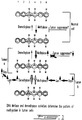
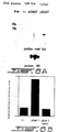
- Fig. 2A illustrates plasmids pZEM and pZαM. The metallothionine (MT) promoter (shaded box), the human growth hormone 3' region (HGH) (open bar), and the MeTase cDNA sequences (hatched) are indicated;
- Fig. 2B shows a Southern blot analysis verifying the presence of the transfected plasmid in the transfectants;
- Fig. 2C shows a Northern blot analysis of the positive clones expressing the expected 1.3 kb chimeric mRNA. Total RNA (5ug) was prepared from the three pZαM lines (7 and 9) and from the pZEM;
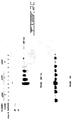
- Fig. 3A,B show the state of methylation of total genomic DNA in Y1pZαM transfectants by nearest neighbour analysis of 2 ug DNA extracted from pZαM transfectant (4) and a pZEM control. Fig 3A shows an autoradiogram of a representative TLC plate. The standard lane is of hemimethylated M13 DNA synthesized In vitro. Fig 3B shows scintillation counts from spots corresponding to C and 5-methyl C. The values represent the means ± SEM;
- Fig. 3C-F. show Southern blot analyses following MspI/HpaII digestion (M/H) of the DNA illustrating, in Y1pZαM transfectants, the state of methylation of specific genes: C: the C21 5'region, D: the C21 gene, E: the retinoblastoma (RB) gene and F: the p53 gene. In Fig. 3C-E, the open arrows indicate the position of demethylated fragments.
- Fig. 4A-C shows the morphological transformation
and reduced anchorage independent growth of Y1 cells
transfected with pZαM. Fig. 4A shows a Phase contrast
microscopy at X200 magnification of living cultures of
Y1 clonal transfectants with pZαM and Y1 controls.
Fig.4B shows pictures of phase contrast microscopy at
X10 of a 21 days soft agar assay of Y1 pZEM cells
(
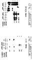
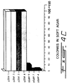
clones 4 and 7) and Y1 pZαM transfectants (clones clones 4 and 7) and Y1 pZαM transfectants (clones - Fig. 5A-B show the In vivo tumorigenicity of Y1pZαM transfectants. Fig.5A illustrates the ability of two control lines (Y1 and pZEM4) and three Y1pZαM transfectants (4, 7 and 9) to form tumors in LAF-1 mice, as well as the level of neovascularization in these tumors. Fig. 5B shows photographs of the homogenized tumors;
- Fig. 6A-B show the loss of antisense expression
in tumors derived from Y1pZαM transfectants by Northern
blot analysis. The 1.3 Kb antisense message is seen
only in the original cell line pZαM 7 (dark arrow), and
is undetectable in tumors arising from
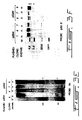
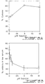
pZαM 7 or Y1 cell lines. A control for the amount of RNA loaded is also shown. FIG. 6B shows the relative expression of the antisense normalized to that of the 18S signal; - Fig. 7A shows a density restricted growth assay of Y1 pZαM relative to control pZEM transfectants;
- Fig. 7B shows the percentage of viable cells as determined using trypan blue staining following serum-deprivation (1% horse serum);
- Fig. 7C shows a Southern analysis of total cellular DNA from the indicated transfectants following growth in 1% serum containing medium and harvested after 1 and 2 days. A 130bp internucleosomal ladder characteristic of cells dying via apoptosis can be seen in the Y1pZdM transfectants only;
- Fig. 7D shows an electron microscopic analysis of various Y1 transfectants cell sections (I-V), following growth of the cells in 1% serum medium for 24 hours.
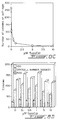
- Fig. 8A-D shows the effect of 5 azaCdR-treatment (0-10µM) of Y1 cells. Fig. 8A shows the content of nonmethylated cytosines in the dinucleotide sequence CpG as determined by a nearest neighbour analysis. Fig. 8B shows the effect of 5azaCdR on the viability of cells grown in low (1%) serum medium. Fig. 8C shows the anchorage independent growth in soft agar (in the absence of 5 azaCdR). Fig. 8D shows the number of colonies upon 5azaCdr treatment.
- Fig. 9 illustrates the Regulation mechanism of the DNA MeTase promoter which determines DNA methylation patterns and cellular transformation.
-
- It has been previously demonstrated that forced expression of an "antisense" mRNA to the most 5' 600 bp of the DNA MeTase message (pZαM) can induce limited DNA demethylation in 10
T 1/2 cells (Szyf et al., 1992, J. Biol. Chem. 267:12831-12836). To directly test the hypothesis that the tumorigenicity of Y1 cells is controlled by the DNA MeTase, Y1 cells were transfected with either pZαM or a pZEM control. - To directly inhibit DNA methylation in Y1 cells, the DNA MeTase antisense expression construct pZαM or a pZEM control vector (Szyf et al., 1992, J. Biol. Chem. 267:12831-12836) were introduced into Y1 cells by DNA mediated gene transfer. Y1 cells were maintained as monolayers in F-10 medium which was supplemented with 7.25% heat inactivated horse serum and 2.5% heat inactivated fetal calf serum (Immunocorp, Montreal). All other media and reagents for cell culture were obtained from GIBCO-BRL. Y1 cells (1X10⁶) were plated on a 150 mm dish (Nunc) 15 hours before transfection. The pZαM expression vector encoding the 5' of the murine DNA MeTase cDNA (10ug) was cointroduced into Y1 cells with 1 ug of pUCSVneo as a selectable marker by DNA mediated gene transfer using the calcium phosphate protocol (Ausubel et al., 1988, In Current Protocols in Molecular Biology. Wiley and Sons, New York). Selection was initiated 48 hours after transfection by adding 0.25 mg/ml G418 (GIBCO-BRL) to the medium. G418 resistant cells were cloned in selective medium. For analysis of growth in soft agar, 1X10³ cells were seeded in triplicate onto 30 mm dishes (Falcon) with 4 ml of F-10 medium containing 7.5% horse serum, 2.5% FCS, 0.25 mg/ml G418 (for transfectants) and 0.33% agar solution at 37°C (Freedman et al.,1974, Cell 3: 355-359). Cells were fed with 2 ml of medium plus G418 every two days. Growth was scored as colonies containing >10 cells, 21 days after plating.
- G418-resistant colonies were isolated and propagated for both constructs. To confirm that the transfectants bear the introduced construct, we prepared DNA from the transfectants and subjected it to digestion by either MspI or HpaII, Southern blot analysis and hybridization with a 32p labelled 0.6 kb DNA MeTase cDNA fragment (Fig. 2A). Preparation of genomic DNA and all other standard molecular biology manipulations, such as Labelling (using the random primer labelling kit from Boehringer Mannheim), were performed according to Ausubel et al., 1988, In Current Protocols in Molecular Biology. Wiley and Sons, New York). MspI and HpaII restriction enzymes (Boehringer Mannheim) were added to DNA at a concentration of 2.5 units/ug for 8 h at 37°C. Radionucleotides (3000mCi/mmol) were purchased from Amersham. The results presented in Fig. 2B demonstrate that the three pZαM transfectants contained significant levels of the DNA MeTase cDNA sequence while the control transfectants were clean. To test whether the pZαM constructs is expressed in the transfectants. and whether the metallothionein promoter is functional in these cells, we cultured the transfectants with 50uM of ZnSO4, prepared RNA at different time points, subjected it to Northern blot analysis and hybridization with the 32p labelled MET 0.6 probe. Preparation of total cellular RNA, blotting RNA on to Hybond-N+(Amersham), were performed according to Ausubel et al., (1988, In Current Protocols in Molecular Biology. Wiley and Sons, New York). As observed in Fig. 2C the
transfectants - To determine whether expression of antisense RNA to the DNA MeTase gene leads to a general reduction in the level of methylation of the genome, we resorted to "nearest neighbour' analysis using [α-32P]-dGTP as previously was performed. This assay enables the determination of the percentage of methylated cytosines residing in the dinucleotide sequence CpG (Razin et al., 1985, IN Biochemistry and Biology of DNA methylation, p. 239, Razin et al., (Ed), Allan R. Liss, Inc. N.Y.). Briefly, Two ug of DNA were incubated at 37°C for 15 minutes with 0.1 unit of DNAase, 2.5 of 32P-α-dGTP (3000 Ci/mmol from Amersham) and 2 units of Kornberg DNA polymerase (Boehringer) were then added and the reaction was incubated for an additional 25 minutes at 30°C. 50ul of water were added and the nonincorporated nucleotides were removed by spinning through a microcon™ column (Amicon) at maximum speed for 30 seconds. The labelled DNA (20ul) was digested with 70ug of micrococal nuclease (Pharmacia) in the manufacturer's recommended buffer for 10 hours at 37°C. Equal amounts of radioactivity were loaded on TLC phosphocellulose plates (Merck) and the 3' mononucleotides were separated by chromatography in one dimension (iso-butyric acid: H2O:NH4OH in the ratio 66:33:1). The chromatograms were exposed to XAR™ film (Eastman-Kodak) and the spots corresponding to cytosine and 5-methylcytosine were scraped and counted in a β-scintillation counter.
- Transfectants and control DNAs were nicked with DNAaseI, nick translated with a single nucleotide [α-32P]-dGTP using DNA polymerase I and the labelled DNA was digested to 3' mononucleotide phosphates with micrococal nuclease which cleaves DNA 3' to the introduced α-32p. The [α-32P] labelled 5' neighbours of dGMP were separated by chromatography on a TLC plate, the resulting spots for dCMP and dCmetMP were scraped and counted by liquid scintillation. The results of a triplicate experiment presented in Fig. 3A (sample autoradiogram) and B (graphic representation) suggest that a limited but significant reduction in the total level of DNA methylation (12 % for
transfectant number 4 and 22% for 7) occurred in transfectants expressing the pZαM construct when compared to the control line pZEM. - To further verify that expression of pZαM results in demethylation and to determine whether specific genes were demethylated, we resorted to a HpaII/MspI restriction enzyme analysis followed by Southern blotting and hybridization with specific gene probes. HpaII cleaves the sequence CCGG, a subset of the CpG dinucleotide sequences, only when the site is unmethylated while MspI will cleave the same sequence irrespective of its state of methylation. By comparing the pattern of HpaII cleavage of specific genes in cells expressing pZαM with that of the parental Y1 or cells harboring only the vector, it can be determined whether the genes are demethylated in the antisense transfectants. The state of methylation of the steroid 21-hydroxylase gene C21 (Szyf et al., 1989, Proc. Natl. Acad. Sci. USA 86: 6853-6857; and Szyf et al., 1990, Mol.Endocrinol. 4:1144-1152) was first analyzed. This gene is specifically expressed and hypomethylated in the adrenal cortex but is inactivated and hypermethylated in Y1 cells. It has been previously suggested that hypermethylation of C21 in Y1 cell is part of the transformation program that includes the shut down of certain differentiated functions. DNA prepared from Y1, pZam and pZEM (Bernards et al., 1989,. Proc. Natl. Acad. Sci. USA. 86:6474-6478) transfectants was subjected to either MspI or HpaII digestion, Southern blot analysis and hybridization with a 0.36 kb Xba-BamHI fragment containing the enhancer and promoter regions of the C21 gene (see references Szyf et al., 1989, Proc. Natl. Acad. Sci. USA 86:6853-6857; and Szyf et al., 1990, Mol. Endocrinol. 4:1144-1152, for a physical map of the probe). This probe should detect 0.36 kb and 0.16 kb HpaII fragments when the promoter region is fully demethylated. The promoter and enhancer region is heavily methylated in Y1 cells and the pZEM transfectants as indicated by the presence of the higher molecular weight partial HpaII fragments at 3.8 and 2 kb and the absence of any lower molecular weight fragments (Fig. 3C). In contrast, the Y1 pZαM transfectants bear a partially demethylated C21 5' region as indicated by the relative diminution of the 3.8 and 2 kb fragments and the appearance of the fully demethylated faint bands at 0.36 kb as well as as the fact that HpaII cleavage yields partial fragments at 0.56 and ∼1 kb indicating partial hypomethylation of sites upstream and downstream to the enhancer region (Fig. 3C). To determine whether hypomethylation was limited to the enhancer region or if it spreads throughout the C21 gene locus, a similar HpaII digestion and Southern blot transfer on different preparations of DNA extracted from Y1 cells was performed. DNA from a control pZEM (Bernards et al., 1989,. Proc. Natl. Acad. Sci. USA. 86: 6474-6478) transfectant and three pZαM antisense transfectants (Fig. 3D) was hybridized to the filter with a 3.8 kb BamHI fragment containing the body of the C21 gene and 3' sequences. Full demethylation of this region should yield a aoublet at ∼1 kb, a 0.8 kb fragment and a 0.4 kb fragment as well as a number of low molecular weight fragments at 0.1-0.2 kb. As observed in Fig. 3D the C21 locus is heavily methylated in Y1 cells as well as the control transfectant as indicated by the high molecular weight fragments above 23 kb. Only a faint band is present in the expected 1 kb molecular weight range as well as a partial at 1.9 kb (Fig. 3D). The DNA extracted from the antisense transfectants exhibits a relative diminution of the high molecular weight fragments and relative intensification of the partial fragment at 1.9 kb as well as the appearance of new partial fragments in the lower molecular weight range between 1 and 0.4 kb indicating partial hypomethylation at large number of HpaII sites contained in the 3' region of the C21 gene. The pattern of demethylation, indicated by the large number of partial HpaII fragments (Fig. 3D), is compatible with a general partial hypomethylation rather than a specific loss of methylation in a distinct region of the C21 gene.
- To determine whether demethylation is limited to genes that are potentially expressible in Y1 cells such as the adrenal cortex-specific C21 gene or if the demethylation is widely spread in the genome, other genes such as the muscle specific MyoD gene as well as the hippocampus specific 5HTlA receptor gene were analysed, and both genes were shown to be hypomethylated. Another class of genes that might have undergone a specific hypomethylation includes the tumor suppressor genes. The state of methylation of two genes from this class, p53 and retinoblastoma (RB) which are both tumor suppressor genes involved in cell cycle regulation was therefore determined. Loss of either one of these gene products has been shown to lead to deregulation of the cell cycle and neoplasia.
- Oligoprimers for the 5'region of the mouse p53 gene were selected from the published genomic sequence (Accession number: X01235) using the Primer selecting program (PC Gene™). The 5' primer corresponding to bases 154-172: 5'TLC GAA TCG GTT TLC ACCC 3' and the 3' primer corresponding to bases 472-492, 5' GGA GGA TGA GGG CCT GAA TGC 3', were added to an amplification reaction mixture containing 100 ng of mouse DNA (from C2C12 cells) using the incubation conditions recommended by the manufacturer (Amersham Hot tub™; 1.5 mM MgCl2) and the DNA was amplified for 40 cycles of 2 minutes at 95°C, 2 minutes at 55°C and 0.5 minutes at 72°C. The reaction products were separated on a low-melt agarose gel (BRL) and the band corresponding to the expected size was excised and extracted according to standard protocols (Ausubel et al., 1988, In Current Protocols in Molecular Biology, Wiley and Sons, New York).
- Since the genomic sequence of the mouse RB gene was unavailable through Genbank, we reverse transcribed the retinoblastoma mRNA from 0.5 ug of total mouse RNA (from C2C12 cells) using random oligonucleotide primers (Boehringer) with Superscript™ reverse transcriptase (BRL) under conditions recommended by the manufacturer. The RB sequence was amplified from the reverse transcribed cDNA using oligonucleotides corresponding to bases 2-628 of the published cDNA (Bernards et al., 1989,. Proc. Natl. Acad. Sci. USA. 86:6474-6478). The oligoprimers used were 5' GGA CTG GGG TGA GGA CGG 3' (1-18) and 5' TTT CAG TAG ATA ACG CAC TGC TGG 3' (620-610). The amplification conditions were as described above.
- Using a probe to a 300 bp sequence from the 5' region of the mouse RB cDNA the level of methylation of this gene in Y1 cells transfected with a control vector as well as the pZαM transfectants was determined (Fig. 3E). Cleavage of this region with HpaII yields 0.6 kb and 0.1 kb fragments (Fig. 3E). The RB locus is heavily methylated in the control cells as indicated by hybridization of the probe to high molecular weight fragments. This locus is partially hypomethylated in the pZαM transfectants as indicated by the relative diminution in the intensity of the high molecular weight fragments, the appearance of numerous partial fragments between 23 and 0.6 kb and the appearance of the demethylated fragments at 0.6 kb and ∼0.1 kb.
- The p53 locus was studied using a 0.3 kb fragment from the 5'
region 300 bp upstream to the initiation site as a probe (Fig. 3F). Cleavage of the p53 loci (two p53 genes are present in the mouse genome) with MspI yields fragments in the 4.4, 2.5, 0,56 and 0.44 kb molecular weight range (Fig. 3F, first lane). Cleavage of the control Y1 pZEM transfectants shows that only the sites flanking the 0.56 kb fragments are demethylated in Y1 cells. The rest of the locus is heavily methylated as indicated by the intensity of the signal at the >4.4 kb range (Fig. 3F, lanes 2-4). In comparison to the control transfectants the p53 gene is partially hypomethylated in Y1 cells expressing an antisense message to the DNA MeTase as implied by the relative reduction in the intensity of the high molecular weight fragments above 4.4 kb and appearance of the 4.4 kb HpaII fragment, the partially cleaved HpaII fragment at 4 kb, the faint partial fragment around 3.5 kb and the faint fragment at 2.5 kb (Fig. 3F last three lanes). These results further substantiate the conclusion that expression of an antisense to the DNA MeTase results in a genome- wide partial hypomethylation. Neither of the genes studied demonstrates a distinct selectivity in demethylation. - To determine whether demethylation induced by the DNA MeTase antisense construct results in a change in the growth properties of cancer cells, the growth and morphological characteristics of the pZαM transfectants versus the controls we compared. To compare the growth curve of pZαM transfectants and controls, 5x10⁴ Y1 pZEM and pZαM transfectants (4 and 7) cells were plated in triplicate. The cells were harvested and counted at the indicated time points (Fig. 7A). The results of this experiment show that the antisense transfectants reach saturation density at lower concentrations than the control cells suggesting that the transfectants have reacquired "contact inhibition" which is one of the traits lost in cancer cells. The morphological properties of the Y1 pZαM transfectants further support this conclusion (Fig.4A). While control Y1 and Y1 pZEM cells exhibit limited contact inhibition and form multilayer foci, Y1 pZαM transfectants exhibit a more rounded and distinct morphology and grow exclusively in monolayers (Fig.4A).
- To determine whether the expression of antisense to the DNA MeTase results in reversal of the tumorigenic potential the ability of the transfectants to grow in an anchorage independent fashion, which is considered an indicator of tumorigenicity, was also determined. The Y1 pZαM transfectants demonstrate an almost complete loss of ability to form colonies in soft agar, moreover the colonies that do form contain only a few cells as demonstrated in Fig. 4B. Growth on soft agar was quantified by visual examination and presented graphically in Fig. 4C.
- These experiments demonstrate that inhibition of DNA methylation by expression of an antisense message to the DNA MeTase leads to loss of tumorigenicity in vitro.
- To determine whether demethylation can result in inhibition of tumorigenesis In vitro, LAF-1 mice (6-8 week old males) were injected subcutaneously (in the flank area) with 106 cells for each of the Y1 pZαM, Y1 and Y1 pZEM transfectants. Mice were monitored for the presence of tumors by daily palpitation. Mice bearing tumors of greater than 1 cm in diameter were sacrificed by asphyxiation with CO2, tumors were removed by dissection and homogenized in guanidium isothiocyanate. Mice that were tumor free were kept for ninety days and then sacrificed. RNA was prepared from the tumors by CsCl2 density gradient centrifugation as described (Ausubel et al., 1988, In Current Protocols in Molecular Biology, Wiley and Sons, New York). While all the animals injected with Y1 cells formed tumors two to three weeks post-injection, the rate of tumor formation in the animals injected with the pZαM transfectants was significantly lower (Fig. 5A; p>0.005).
- Many lines of evidence suggest that angiogenic potential and metastatic potential of cell lines are directly related. The tumors that do arise from the pZαM transfectants exhibit very limited neovascularization (Fig. 5B) while tumors that formed in the animals that were injected with Y1 cells or control transfectants were highly vascularized (Fig. 5B). This difference in neovascularization is indicated by the pale color of the homogenates of tumors removed from animals injected with Y1 pZαM transfectants cells versus the very dark homogenates of tumors arising from control lines (Y1 and Y1pZEM; Fig.5B).
- One possible explanation for the fact that a small number of tumors did form in animals injected with the pZαM transfectants is that they are derived from revertants that lost expression of the antisense to the DNA MeTase under the selective pressure in vivo. This hypothesis was tested with isolated RNA from a tumor arising from the Y1pZαM transfectant, and compared to the level of expression of the 0.6 kb antisense message observed for the transfectant line in vitro. The isolated RNAs were subjected to Northern blot analysis and hybridization with a 32p labelled MET 0.6 fragment. The filter was stripped of its radioactivity and was rehybridized with a 32P labelled oligonucleotide probe for 18S rRNA (Fig. 6A) as previously described (Szyf et al., 1990, Mol. Endocrinol. 4:1144-1152). The autoradiograms were scanned and the level of expression of MET 0.6 was determined relative to the signal obtained with the 18S probe (Fig. 6B). The expression of the antisense message is significantly reduced in the tumors supporting the hypothesis that expression of an antisense message to the DNA MeTase is incompatible with tumorigenesis.
- Tumor cells exhibit limited dependence on serum and are usually capable of serum independent growth. Factors present in the serum are essential for the survival of many nontumorigenic cells. Several lines of evidence have recently suggested that the enhanced survivability of tumorigenic cells is associated with inhibition of programmed cell death. For example, the oncogene bcl-2 is not a stimulator of cell proliferation but rather causes inhibition of apoptosis. The tumor suppressor p53 can induce apoptosis in a human colon tumor derived line and certain chemotherapeutic agents have been shown to induce apoptosis in cancer cells. Since the pZαM transfectants appeared to demonstrate an enhanced dependence on serum and limited survivability under serum deprived conditions, the possibility that demethylation can induce an apoptotic program in Y1 cells was analyzed. It was reasoned that as factors in the serum are known to act as survival factors for cells, an apoptotic program could be activated only when these factors are remove. To test whether pZαM transfectants undergo programmed cell death under serum deprived condition, the effects of serum starvation on these transfectants was studied. pZαM transfectants and control Y1 pZEM transfectants (3x10⁵ per well) were plated in low serum medium (1% horse serum) in six well plates, harvested every 24 hours and tested for viability by trypan blue staining (Fig. 7B). While the control cells exhibited almost 100% viability up to 72 hours after transfer into serum deprived medium, the Y1 pZαM cells showed up to 75 % loss of viability at 48 hours (Fig. 7B).
- The rapid onset of death in Y1 pZαM clones under serum deprived conditions suggests that an active process is involved. Several observable changes distinguish apoptosis from necrosis: apoptosis is an active process requiring de novo protein synthesis; apoptosis is associated with death of isolated cells, unlike necrosis where patches or areas of tissue die; cells dying by apoptosis do not elicit an immune response; and the most diagnostic feature of apoptosis is the pattern of degradation of the DNA from apoptotic cells (Ellis et al., 1991, Annu. Rev. Cell Biol. 7:663-698). DNA from cells dying by apoptosis generally exhibit a characteristic ladder when analyzed by gel electrophoresis because Ca2+/Mg2+ dependent endonucleases cleave the DNA at internucleosomal regions (Ellis et al., 1991, Annu. Rev. Cell Biol. 7:663-698). Although the appearance of the 180 bp internucleosomal ladder is a diagnostic feature of apoptotic death, other morphological changes such as chromatin condensation, cellular fragmentation, and formation of apoptotic bodies are generally considered to be earlier events in the apoptotic process and therefore also serve as useful markers. To test whether the serum deprived Y1 pZαM cells were dying as a result of an activated apoptotic death program, cells were plated in starvation medium (1% horse serum) and harvested at 24 hour intervals. Total cellular DNA was isolated from the cells and was subjected to electrophoresis on a 1.5% agarose gel followed by transfer to nylon membrane and hybridization with random labeled Y1 genomic DNA. After 48 hours in serum starved conditions, pZαM transfectants exhibit the characteristic 180 bp internucleosomal DNA ladder while the control pZEM transfectants show no apoptosis at this time point (Fig. 7C).
- To determine whether cells expressing antisense to the DNA MeTase exhibit early morphological markers of apoptosis, cells were serum starved for 24 hours (2% horse serum), harvested and analyzed by electron microscopy. For electron microscopy, cells were fixed in glutaraldehyde (2.5%) in cacodylate buffer (0.1M) for one hour and further fixed in 1% osmium tetroxide. The samples were dehydrated in ascending alcohol concentrations and propylene oxide followed by embedding in Epon. Semithin sections (1um) were cut from blocks with an ultramicrotome, counterstained with uranil acetate and lead citrate. Samples were analyzed using a Philips 410 electron microscope (Maysinger et al., 1993, Neurochem Intl. 23: 123- 129). Fig.7D shows the electron micrographs of control Y1 pZEM and Y1 pZαM transfectants at various magnifications (I-V). The control cells have a fine uniform nuclear membrane whereas the pZαM cells exhibit the cardinal markers of apoptosis: condensation of chromatin and its margination at the nuclear periphery (panels I and II), chromatin condensation (panel II), nuclear fragmentation (panel III), formation of apoptotic bodies (panel V) and cellular fragmentation (panel IV). This set of experiments suggests that one possible mechanism through which demethylation can inhibit tumorigenesis is by activating programmed cell death. This is supported by data suggesting that cell death is triggered by an endonuclease activity (Ellis et al., 1991, Annu. Rev. Cell Biol. 7: 663-698). Thus, effects on the DNA methylation levels, can affect the pathway leading to apoptosis.
- 1X10⁵ Y1 cells were plated in growth medium. Twenty-four hours after plating, the medium was replaced with fresh medium containing various concentrations (0-10µM) of 5 azaCdR (Sigma). The medium was removed and replaced with fresh medium containing 5 azaCdR every 12 h for a period of 72 h. Following 5 azaCdR treatment the cells were plated onto a six well dish in growth medium (100, 300, 500 cells per well) for cologinecity determinations, in soft agar for determining anchorage independent growth (3
X 103 cells per well) and in low serum (1% horse serum) for five days to determine viability under serum deprived conditions. All of these assays were performed in the absence of 5 azaCdR. - As shown in Fig. 8, treatment of Y1 cells with 5 azaCdR mimics the action of the expression of an antisense for MeTase. Indeed, treatment of Y1 cells with 5 azaCdR is shown to increase the level of nonmethylated cytosine (Fig. 8A), to decrease the viability of serum-starved cells (Fig. 8B), and finally to drastically inhibit the growth of Y1 cells on soft agar (Fig. 8C-D). The effect of 5 azaCdR on Y1 cells was shown not to depend on the cell line per se since performing the same experiment under the same conditions but using Rb- human tumors and human small cell lung carcinoma cells gave similar results.
- This set of experiment therefore suggests that 5 azaCdR can be successfully used as an anticancer agent, to alter the genetic program or to restore an authentic program disrupted by deregulation of DNA methylation.
- The data presented herein, strongly support the hypothesis that hypermethylation plays a critical role in maintenance of the transformed state, and even predict that the increase in methylation is critical for the transformed state. The fact that the RAS signalling pathway has been shown to induce the activity of the DNA MeTase promoter provides us with a mechanism to explain this increase in the DNA methylation capacity of cancer cells (Fig. 9). It stands to reason therefore that the DNA MeTase is an important effector of the RAS signalling pathway.
- Taken together, the data presented above, provide basic principles regarding the therapeutic implications of DNA methylation. First, because the level of DNA MeTase activity is one determinant of the pattern of DNA methylation, partial inhibition of DNA MeTase activity can result in a change in the methylation pattern. If aberrant hypermethylation in cancer cells is caused by over-expression of the DNA methyltransferase, then partial inhibition of methylation is expected to restore the original methylation pattern. Second, since this pattern is not exclusively determined by the DNA MeTase activity but is also defined by cis- and trans-acting signals at the gene site, a partial inhibition of DNA MeTase will result in a programmed change in gene expression rather than a chaotic transformation of the cell. Furthermore, DNA MeTase inhibitors can be used to induce a program that is latent in the cell.
- While the invention has been described with particular reference to the illustrated embodiment, it will be understood that numerous modifications thereto will appear to those skilled in the art. Accordingly, the above description and accompanying drawings should be taken as illustrative of the invention and not in a limiting sense.
Claims (5)
- A DNA methyltransferase inhibitor or antagonist selected from the group consisting of an anti-DNA methyltransferase antibody and an antisense nucleic acid of DNA methyltransferase for use as a therapeutic or diagnostic agent.
- DNA methyltransferase inhibitor or antagonist according to claim 1 for reducing the level of methylated cytosine in a CpG dinucleotide, for reversing a transformed state of a cell, for correcting an aberrant methylation pattern in DNA of a cell, or for changing a methylation pattern in DNA of a cell.
- Use of DNA methyltransferase inhibitor or antagonist according to claim 1 for the manufacture of a medicament for cancer treatment, for restoring an aberrant methylation pattern in a patient DNA, or for changing a methylation pattern in a patient DNA.
- Use according to claim 3, wherein the medicament comprises an anti-DNA methyltransferase antibody and an antisense nucleic acid of DNA methyltransferase.
- Use according to claim 3 or 4 for correction of genetic defect found for β-thalassemia or sickle cell anemia.
Priority Applications (2)
| Application Number | Priority Date | Filing Date | Title |
|---|---|---|---|
| EP03012172A EP1352956A1 (en) | 1993-11-30 | 1994-11-30 | Inhibition of DNA methyltransferase |
| EP98112207A EP0889122A3 (en) | 1993-11-30 | 1994-11-30 | Inhibition of DNA Methyltransferase |
Applications Claiming Priority (5)
| Application Number | Priority Date | Filing Date | Title |
|---|---|---|---|
| CA002110213A CA2110213A1 (en) | 1993-11-30 | 1993-11-30 | Inhibition of dna metase for therapy |
| CA2110213 | 1993-11-30 | ||
| GB9413680 | 1994-07-07 | ||
| GB9413680A GB9413680D0 (en) | 1994-07-07 | 1994-07-07 | Inhibition of tumorgenicity by inhibiting DNA methyltransferase activity |
| PCT/CA1994/000659 WO1995015373A2 (en) | 1993-11-30 | 1994-11-30 | Inhibition of dna methyltransferase |
Related Child Applications (1)
| Application Number | Title | Priority Date | Filing Date |
|---|---|---|---|
| EP98112207A Division EP0889122A3 (en) | 1993-11-30 | 1994-11-30 | Inhibition of DNA Methyltransferase |
Publications (2)
| Publication Number | Publication Date |
|---|---|
| EP0734436A1 EP0734436A1 (en) | 1996-10-02 |
| EP0734436B1 true EP0734436B1 (en) | 1999-04-14 |
Family
ID=25676818
Family Applications (3)
| Application Number | Title | Priority Date | Filing Date |
|---|---|---|---|
| EP98112207A Ceased EP0889122A3 (en) | 1993-11-30 | 1994-11-30 | Inhibition of DNA Methyltransferase |
| EP03012172A Withdrawn EP1352956A1 (en) | 1993-11-30 | 1994-11-30 | Inhibition of DNA methyltransferase |
| EP95901306A Expired - Lifetime EP0734436B1 (en) | 1993-11-30 | 1994-11-30 | Inhibition of dna methyltransferase |
Family Applications Before (2)
| Application Number | Title | Priority Date | Filing Date |
|---|---|---|---|
| EP98112207A Ceased EP0889122A3 (en) | 1993-11-30 | 1994-11-30 | Inhibition of DNA Methyltransferase |
| EP03012172A Withdrawn EP1352956A1 (en) | 1993-11-30 | 1994-11-30 | Inhibition of DNA methyltransferase |
Country Status (9)
| Country | Link |
|---|---|
| US (1) | US6184211B1 (en) |
| EP (3) | EP0889122A3 (en) |
| JP (1) | JPH09506253A (en) |
| KR (1) | KR100392057B1 (en) |
| AT (1) | ATE178939T1 (en) |
| AU (1) | AU1061395A (en) |
| CA (1) | CA2177031A1 (en) |
| DE (1) | DE69417918T2 (en) |
| WO (1) | WO1995015373A2 (en) |
Families Citing this family (123)
| Publication number | Priority date | Publication date | Assignee | Title |
|---|---|---|---|---|
| US5652105A (en) * | 1995-07-28 | 1997-07-29 | Health Research, Inc. | Substrate for detection of mammalian 5-C-DNA methyltransferase |
| WO1997044346A2 (en) * | 1996-05-22 | 1997-11-27 | Mcgill University | Specific inhibitors of dna methyltransferase enzyme |
| US6268137B1 (en) | 1996-05-22 | 2001-07-31 | Methylgene, Inc. | Specific inhibitors of DNA methyl transferase |
| US7138384B1 (en) | 1997-08-29 | 2006-11-21 | The Regents Of The University Of California | Modulators of DNA cytosine-5 methyltransferase and methods for use thereof |
| AU1138699A (en) * | 1997-11-12 | 1999-05-31 | Mcgill University | Dna demethylase, therapeutic and diagnostic uses thereof |
| DE19754482A1 (en) * | 1997-11-27 | 1999-07-01 | Epigenomics Gmbh | Process for making complex DNA methylation fingerprints |
| US7026155B2 (en) * | 1999-02-02 | 2006-04-11 | Regents Of The University Of California | Method of reducing bacterial proliferation |
| CA2291367A1 (en) * | 1999-12-06 | 2001-06-06 | Isabelle Henry | Genetic constructions having a reduced or an increased number of epigenetic control regions and methods of use thereof |
| DE10010282B4 (en) | 2000-02-25 | 2006-11-16 | Epigenomics Ag | Method for the detection of cytosine methylation in DNA samples |
| EP1370685A2 (en) | 2000-04-06 | 2003-12-17 | Epigenomics AG | Diagnosis of diseases associated with dna repair |
| WO2003006987A1 (en) * | 2000-12-11 | 2003-01-23 | The Salk Institute For Biological Studies | Methods and compositions for determining enzymatic activity and specificity of methyltransferases |
| WO2002101353A2 (en) * | 2001-06-08 | 2002-12-19 | U.S. Genomics, Inc. | Methods and products for analyzing nucleic acids based on methylation status |
| AU2002322805B2 (en) * | 2001-07-31 | 2007-11-08 | The Government Of The United States Of America As Represented By The Secretary Of The Department Of Health And Human Services | Inhibitor of DNA methylation |
| DE10154317B4 (en) | 2001-10-26 | 2005-06-09 | Epigenomics Ag | Method for the detection of cytosine methylations in immobilized DNA samples |
| EP1340818A1 (en) | 2002-02-27 | 2003-09-03 | Epigenomics AG | Method and nucleic acids for the analysis of a colon cell proliferative disorder |
| US6890736B1 (en) | 2002-09-20 | 2005-05-10 | Immunex Corporation | Methods for producing proteins in cultured cells |
| WO2005059172A2 (en) | 2003-12-11 | 2005-06-30 | Epigenomics Ag | Method and nucleic acids for the improved treatment of breast cell proliferative disorders |
| CA2500255A1 (en) | 2002-10-01 | 2004-04-29 | Epigenomics Ag | Method and nucleic acids for the treatment of breast cell proliferative disorders |
| AU2003295444A1 (en) * | 2002-11-15 | 2004-06-15 | The Board Of Trustees Of The University Of Illinois | Methods for in vitro expansion of hematopoietic stem cells |
| AU2003902704A0 (en) * | 2003-05-29 | 2003-06-19 | Crc For Waste Management And Pollution Control Limited Of Unsw | Process for producing a nanoscale zero-valent metal |
| EP1636386B1 (en) | 2003-06-23 | 2015-08-05 | Epigenomics AG | Methods and nucleic acids for analyses of colorectal cell proliferative disorders |
| EP2354249A1 (en) | 2003-06-23 | 2011-08-10 | Epigenomics AG | Methods and nucleic acids for analyses of colorectal cell proliferative disorders |
| ATE494390T1 (en) | 2003-06-23 | 2011-01-15 | Epigenomics Ag | METHOD AND NUCLEIC ACIDS FOR ANALYZING DISORDERS IN COLON CELL PROLIFERATION |
| WO2005012575A1 (en) * | 2003-08-01 | 2005-02-10 | U.S. Genomics, Inc. | Methods and compositions related to the use of sequence-specific endonucleases for analyzing nucleic acids under non-cleaving conditions |
| AU2004295712B2 (en) | 2003-12-01 | 2011-05-19 | Epigenomics Ag | Methods and nucleic acids for the analysis of gene expression associated with the development of prostate cell proliferative disorders |
| US8415315B2 (en) | 2004-05-06 | 2013-04-09 | University Of Central Florida Research Foundation, Inc. | Methods and compositions for inhibiting the proliferation of cancer cells |
| EP1778865A1 (en) | 2004-06-23 | 2007-05-02 | Epigenomics AG | Methods and nucleic acids for the detection of metastasis of colon cell proliferative disorders |
| US20090298054A1 (en) | 2004-07-18 | 2009-12-03 | Epigenomics Ag | Epigenetic methods and nucleic acids for the detection of breast cell proliferative disorders |
| EP2311984A1 (en) | 2004-12-02 | 2011-04-20 | Epigenomics AG | Methods and nucleic acids for the analysis of gene expression associated with the prognosis of prostate cell proliferative disorders |
| US7250416B2 (en) | 2005-03-11 | 2007-07-31 | Supergen, Inc. | Azacytosine analogs and derivatives |
| WO2006107751A2 (en) * | 2005-04-01 | 2006-10-12 | Methylgene, Inc. | Combined therapy utilizing reduction of dna methyltansferase expression and/or activity and interferon |
| SG183708A1 (en) | 2005-04-15 | 2012-09-27 | Epigenomics Ag | Methods and nucleic acids for analyses of cellular proliferative disorders |
| EP1885884A2 (en) | 2005-05-02 | 2008-02-13 | University Of Southern California | DNA METHYLATION MARKERS ASSOCIATED WITH THE CpG ISLAND METHYLATOR PHENOTYPE (CIMP) IN HUMAN COLORECTAL CANCER |
| ES2623221T3 (en) * | 2005-05-20 | 2017-07-10 | Methylgene Inc | VEGF receptor and HGF receptor signaling inhibitors |
| US20090191548A1 (en) | 2005-09-29 | 2009-07-30 | Epigenomics Ag | Methods and nucleic acids for the analysis of gene expression associated with tissue classification |
| US7700567B2 (en) | 2005-09-29 | 2010-04-20 | Supergen, Inc. | Oligonucleotide analogues incorporating 5-aza-cytosine therein |
| CN101437933B (en) | 2005-12-28 | 2013-11-06 | 斯克里普斯研究所 | Natural antisense and non-coding RNA transcripts as drug targets |
| AR060061A1 (en) * | 2006-03-22 | 2008-05-21 | Methylgene Inc | INHIBITORS OF THE ACTIVITY OF PROTEIN TIROSINA QUINASA |
| WO2007118704A2 (en) | 2006-04-17 | 2007-10-25 | Epigenomics Ag | Methods and nucleic acids for the detection of colorectal cell proliferative disorders |
| WO2008035209A2 (en) * | 2006-05-30 | 2008-03-27 | Methylgene Inc. | Inhibitors of protein tyrosine kinase activity |
| EP2634265B1 (en) | 2006-07-21 | 2016-09-14 | Epigenomics AG | Methods related to the gene GLI3 for detection of colorectal cancer |
| US20080255155A1 (en) * | 2006-10-18 | 2008-10-16 | Stephane Raeppel | Kinase inhibitors and uses thereof |
| US20100203514A1 (en) | 2006-11-24 | 2010-08-12 | Sledziewski Andrew Z | Methods and nucleic acids for the analysis of gene expression associated with the development of prostate cell proliferative disorders |
| CA2675895C (en) | 2007-01-19 | 2016-03-22 | Epigenomics Ag | Methods and nucleic acids for analyses of cell proliferative disorders |
| EP2302069A1 (en) | 2007-12-11 | 2011-03-30 | Epigenomics AG | Methods and nucleic acids for analyses of cell proliferative disorders |
| US8293897B2 (en) | 2008-10-14 | 2012-10-23 | Ning Xi | Compounds comprising a spiro-ring and methods of use |
| WO2010065671A2 (en) | 2008-12-04 | 2010-06-10 | Curna, Inc. | Treatment of vascular endothelial growth factor (vegf) related diseases by inhibition of natural antisense transcript to vegf |
| US20110294870A1 (en) | 2008-12-04 | 2011-12-01 | Opko Curna, Llc | Treatment of tumor suppressor gene related diseases by inhibition of natural antisense transcript to the gene |
| ES2629630T3 (en) | 2008-12-04 | 2017-08-11 | Curna, Inc. | Treatment of diseases related to erythropoietin (EPO) by inhibiting the natural antisense transcript to EPO |
| ES2762610T3 (en) | 2009-02-12 | 2020-05-25 | Curna Inc | Treatment of diseases related to brain-derived neurotrophic factor (BDNF) by inhibition of natural antisense transcript for BDNF |
| MX2011009751A (en) | 2009-03-16 | 2011-09-29 | Opko Curna Llc | Treatment of nuclear factor (erythroid-derived 2)-like 2 (nrf2) related diseases by inhibition of natural antisense transcript to nrf2. |
| WO2010107740A2 (en) | 2009-03-17 | 2010-09-23 | Curna, Inc. | Treatment of delta-like 1 homolog (dlk1) related diseases by inhibition of natural antisense transcript to dlk1 |
| JP6250930B2 (en) | 2009-05-06 | 2017-12-20 | クルナ・インコーポレーテッド | Treatment of TTP-related diseases by suppression of natural antisense transcripts against tristetraproline (TTP) |
| CN102459596B (en) | 2009-05-06 | 2016-09-07 | 库尔纳公司 | By suppression therapy lipid transfer and the metabolic gene relevant disease of the natural antisense transcript for lipid transfer and metabolic gene |
| KR101742334B1 (en) | 2009-05-08 | 2017-06-01 | 큐알엔에이, 인크. | Treatment of dystrophin family related diseases by inhibition of natural antisense transcript to dmd family |
| CN102575251B (en) | 2009-05-18 | 2018-12-04 | 库尔纳公司 | The relevant disease of the reprogramming factor is treated by inhibiting the natural antisense transcript for the reprogramming factor |
| KR101703695B1 (en) | 2009-05-22 | 2017-02-08 | 큐알엔에이, 인크. | Treatment of transcription factor e3 (tfe3) and insulin receptor substrate 2 (irs2) related diseases by inhibition of natural antisense transcript to tfe3 |
| CN103221541B (en) | 2009-05-28 | 2017-03-01 | 库尔纳公司 | Antiviral gene relevant disease is treated by the natural antisense transcript suppressing antiviral gene |
| KR101702689B1 (en) | 2009-06-16 | 2017-02-06 | 큐알엔에이, 인크. | Treatment of paraoxonase 1 (pon1) related diseases by inhibition of natural antisense transcript to pon1 |
| WO2010148050A2 (en) | 2009-06-16 | 2010-12-23 | Curna, Inc. | Treatment of collagen gene related diseases by inhibition of natural antisense transcript to a collagen gene |
| CA2765889A1 (en) | 2009-06-24 | 2010-12-29 | Opko Curna, Llc | Treatment of tumor necrosis factor receptor 2 (tnfr2) related diseases by inhibition of natural antisense transcript to tnfr2 |
| CA2765815A1 (en) | 2009-06-26 | 2010-12-29 | Opko Curna, Llc | Treatment of down syndrome gene related diseases by inhibition of natural antisense transcript to a down syndrome gene |
| AR077238A1 (en) | 2009-06-26 | 2011-08-10 | Unilever Nv | COMPOSITIONS FOR ORAL CARE. USE OF A POLYMER TO MANUFACTURE THE COMPOSITION |
| CA2768947C (en) | 2009-07-24 | 2018-06-19 | Opko Curna, Llc | Treatment of sirtuin (sirt) related diseases by inhibition of natural antisense transcript to a sirtuin (sirt) |
| US9234199B2 (en) | 2009-08-05 | 2016-01-12 | Curna, Inc. | Treatment of insulin gene (INS) related diseases by inhibition of natural antisense transcript to an insulin gene (INS) |
| EP2464731B1 (en) | 2009-08-11 | 2016-10-05 | CuRNA, Inc. | Treatment of adiponectin (adipoq) related diseases by inhibition of natural antisense transcript to an adiponectin (adipoq) |
| WO2011022606A2 (en) | 2009-08-21 | 2011-02-24 | Curna, Inc. | Treatment of 'c terminus of hsp70-interacting protein' (chip) related diseases by inhibition of natural antisense transcript to chip |
| CN102482671B (en) | 2009-08-25 | 2017-12-01 | 库尔纳公司 | IQGAP relevant diseases are treated by suppressing the natural antisense transcript of ' gtpase activating protein containing IQ die bodys ' (IQGAP) |
| EP2480669B1 (en) | 2009-09-25 | 2017-11-08 | CuRNA, Inc. | Treatment of filaggrin (flg) related diseases by modulation of flg expression and activity |
| US20110104695A1 (en) | 2009-11-05 | 2011-05-05 | Epigenomics Ag | Methods of predicting therapeutic efficacy of cancer therapy |
| ES2661813T3 (en) | 2009-12-16 | 2018-04-04 | Curna, Inc. | Treatment of diseases related to membrane transcription factor peptidase, site 1 (mbtps1) by inhibition of the natural antisense transcript to the mbtps1 gene |
| US9068183B2 (en) | 2009-12-23 | 2015-06-30 | Curna, Inc. | Treatment of uncoupling protein 2 (UCP2) related diseases by inhibition of natural antisense transcript to UCP2 |
| CA2782373C (en) | 2009-12-23 | 2019-03-26 | Opko Curna, Llc | Treatment of hepatocyte growth factor (hgf) related diseases by inhibition of natural antisense transcript to hgf |
| WO2011090740A2 (en) | 2009-12-29 | 2011-07-28 | Opko Curna, Llc | Treatment of nuclear respiratory factor 1 (nrf1) related diseases by inhibition of natural antisense transcript to nrf1 |
| ES2585829T3 (en) | 2009-12-29 | 2016-10-10 | Curna, Inc. | Treatment of diseases related to tumor protein 63 (p63) by inhibition of natural antisense transcription to p63 |
| US20120289583A1 (en) | 2009-12-31 | 2012-11-15 | Curna, Inc. | Treatment of insulin receptor substrate 2 (irs2) related diseases by inhibition of natural antisense transcript to irs2 and transcription factor e3 (tfe3) |
| KR101878501B1 (en) | 2010-01-04 | 2018-08-07 | 큐알엔에이, 인크. | Treatment of interferon regulatory factor 8 (irf8) related diseases by inhibition of natural antisense transcript to irf8 |
| EP2521785B1 (en) | 2010-01-06 | 2022-03-09 | CuRNA, Inc. | Inhibition of natural antisense transcript to a pancreatic developmental gene for use in a treatment of pancreatic developmental gene related diseases |
| DK2524039T3 (en) | 2010-01-11 | 2018-03-12 | Curna Inc | TREATMENT OF GENDER HORMON-BINDING GLOBULIN (SHBG) RELATED DISEASES BY INHIBITION OF NATURAL ANTISENCE TRANSCRIPTS TO SHBG |
| CN102782135A (en) | 2010-01-25 | 2012-11-14 | 库尔纳公司 | Treatment of RNase H1 related diseases by inhibition of natural antisense transcript to RNase H1 |
| CN102844435B (en) | 2010-02-22 | 2017-05-10 | 库尔纳公司 | Treatment of pyrroline-5-carboxylate reductase 1 (pycr1) related diseases by inhibition of natural antisense transcript to pycr1 |
| ES2657969T3 (en) | 2010-04-02 | 2018-03-07 | Curna, Inc. | Treatment of diseases related to Colony Stimulating Factor 3 (CSF3) by inhibition of the natural antisense transcript to CSF3 |
| RU2610661C2 (en) | 2010-04-09 | 2017-02-14 | Курна, Инк. | Treatment of fibroblast growth factor 21 (fgf21) related diseases by inhibition of natural antisense transcript to fgf21 |
| CN107988228B (en) | 2010-05-03 | 2022-01-25 | 库尔纳公司 | Treatment of Sirtuin (SIRT) related diseases by inhibition of natural antisense transcript to Sirtuin (SIRT) |
| TWI531370B (en) | 2010-05-14 | 2016-05-01 | 可娜公司 | Treatment of par4 related diseases by inhibition of natural antisense transcript to par4 |
| US8895528B2 (en) | 2010-05-26 | 2014-11-25 | Curna, Inc. | Treatment of atonal homolog 1 (ATOH1) related diseases by inhibition of natural antisense transcript to ATOH1 |
| CA2799596C (en) | 2010-05-26 | 2020-09-22 | Curna, Inc. | Treatment of methionine sulfoxide reductase a (msra) related diseases by inhibition of natural antisense transcript to msra |
| KR102008708B1 (en) | 2010-06-23 | 2019-08-08 | 큐알엔에이, 인크. | Treatment of sodium channel voltage-gated, alpha subunit (scna) related diseases by inhibition of natural abtisense transcript to scna |
| CN103068982B (en) | 2010-07-14 | 2017-06-09 | 库尔纳公司 | DLG relevant diseases are treated by suppressing the natural antisense transcript of the big homologue of plate-like (DLG) |
| EP2625274B1 (en) | 2010-10-06 | 2017-07-19 | CuRNA, Inc. | Treatment of sialidase 4 (neu4) related diseases by inhibition of natural antisense transcript to neu4 |
| CA2815212A1 (en) | 2010-10-22 | 2012-04-26 | Curna, Inc. | Treatment of alpha-l-iduronidase (idua) related diseases by inhibition of natural antisense transcript to idua |
| WO2012068340A2 (en) | 2010-11-18 | 2012-05-24 | Opko Curna Llc | Antagonat compositions and methods of use |
| CN103459599B (en) | 2010-11-23 | 2017-06-16 | 库尔纳公司 | NANOG relevant diseases are treated by suppressing the natural antisense transcript of NANOG |
| RU2568258C2 (en) | 2011-02-28 | 2015-11-20 | Саншайн Лейк Фарма Ко., Лтд | Substituted quinoline compounds and methods of their application |
| JP6188686B2 (en) | 2011-06-09 | 2017-08-30 | カッパーアールエヌエー,インコーポレイテッド | Treatment of FXN-related diseases by inhibition of natural antisense transcripts to frataxin (FXN) |
| MX358117B (en) | 2011-07-08 | 2018-08-06 | Epigenomics Ag | Methods and nucleic acids for determining the prognosis of a cancer subject. |
| AU2012302051B2 (en) | 2011-08-30 | 2017-04-27 | Astex Pharmaceuticals, Inc. | Decitabine derivative formulations |
| CA2847811C (en) | 2011-09-06 | 2019-10-22 | Curna, Inc. | Treatment of diseases related to alpha subunits of sodium channels, voltage-gated (scnxa) with small molecules |
| US20150031750A1 (en) | 2012-03-15 | 2015-01-29 | The Scripps Research Institute | Treatment of brain derived neurotrophic factor (bdnf) related diseases by inhibition of natural antisense transcript to bdnf |
| AU2013296897B2 (en) | 2012-07-28 | 2015-09-17 | Beijing Findcure Biosciences Ltd. | Substituted pyrazolone compounds and methods of use |
| TWI574962B (en) | 2012-11-14 | 2017-03-21 | 加拓科學公司 | Heteroaromatic compounds as pi3 kinase modulators and methods of use |
| SG11201505493QA (en) | 2013-02-21 | 2015-08-28 | Calitor Sciences Llc | Heteroaromatic compounds as pi3 kinase modulators |
| EP2971169A4 (en) | 2013-03-13 | 2016-10-26 | Abbott Molecular Inc | Systems and methods for isolating nucleic acids |
| EP2971171A4 (en) | 2013-03-14 | 2016-11-02 | Abbott Molecular Inc | Multiplex methylation-specific amplification systems and methods |
| CA2902916C (en) | 2013-03-14 | 2018-08-28 | Mayo Foundation For Medical Education And Research | Detecting neoplasm |
| WO2015124921A1 (en) | 2014-02-19 | 2015-08-27 | The University Court Of The University Of Edinburgh | Methods and uses for determining the presence of inflammatory bowel disease |
| EP3126519A4 (en) | 2014-03-31 | 2018-01-17 | Mayo Foundation for Medical Education and Research | Detecting colorectal neoplasm |
| US10465248B2 (en) | 2014-12-12 | 2019-11-05 | Exact Sciences Development Company, Llc | Method of characterizing ZDHHC1 DNA |
| US10435755B2 (en) | 2015-03-27 | 2019-10-08 | Exact Sciences Development Company, Llc | Detecting esophageal disorders |
| EP3316685A4 (en) | 2015-07-02 | 2019-03-13 | Otsuka Pharmaceutical Co., Ltd. | Lyophilized pharmaceutical compositions |
| KR102627044B1 (en) | 2015-10-19 | 2024-01-18 | 선샤인 레이크 파르마 컴퍼니 리미티드 | Salts of EGFR inhibitors, their crystalline forms and uses |
| US10822638B2 (en) | 2015-10-30 | 2020-11-03 | Exact Sciences Development Company, Llc | Isolation and detection of DNA from plasma |
| KR20170050916A (en) * | 2015-11-02 | 2017-05-11 | 연세대학교 산학협력단 | Method for controlling differentiatin of stem cell by epigenetic modification of intragenic cpg island |
| US10385406B2 (en) | 2016-05-05 | 2019-08-20 | Exact Sciences Development Company, Llc | Detection of lung neoplasia by analysis of methylated DNA |
| EP3488004B1 (en) | 2016-07-19 | 2021-10-06 | Exact Sciences Development Company, LLC | Methylated control dna |
| KR20240038151A (en) | 2016-09-02 | 2024-03-22 | 메이오 파운데이션 포 메디칼 에쥬케이션 앤드 리써치 | Detecting hepatocellular carcinoma |
| US11118228B2 (en) | 2017-01-27 | 2021-09-14 | Exact Sciences Development Company, Llc | Detection of colon neoplasia by analysis of methylated DNA |
| WO2019025863A2 (en) | 2017-08-03 | 2019-02-07 | Otsuka Pharmaceutical Co., Ltd. | Drug compound and purification methods thereof |
| EP3880847A2 (en) | 2018-11-16 | 2021-09-22 | Oslo Universitetssykehus HF | Methods and compositions for characterizing bladder cancer |
| CN111321210B (en) * | 2018-12-13 | 2021-09-14 | 贵阳优乐复生医学检验所有限公司 | Method for non-invasive prenatal detection of whether fetus suffers from genetic disease |
| CA3154354A1 (en) | 2019-10-31 | 2021-05-06 | William R. Taylor | Detecting ovarian cancer |
| WO2022040306A1 (en) | 2020-08-19 | 2022-02-24 | Mayo Foundation For Medical Education And Research | Detecting non-hodgkin lymphoma |
| WO2022165247A1 (en) | 2021-01-29 | 2022-08-04 | Mayo Foundation For Medical Education And Research | Detecting the presence or absence of multiple types of cancer |
Family Cites Families (4)
| Publication number | Priority date | Publication date | Assignee | Title |
|---|---|---|---|---|
| WO1992006985A1 (en) * | 1990-10-23 | 1992-04-30 | City Of Hope | Mechanism based inhibitors of dna methyltransferase |
| CA2108144A1 (en) | 1991-03-06 | 1992-09-07 | Jack A. Roth | Methods and compositions for the selective inhibition of gene expression |
| US5578716A (en) * | 1993-12-01 | 1996-11-26 | Mcgill University | DNA methyltransferase antisense oligonucleotides |
| US6020318A (en) * | 1997-05-30 | 2000-02-01 | Methylgene, Inc. | DNA methyltransferase genomic sequences and antisense oligonucleotides |
-
1994
- 1994-11-30 US US08/652,425 patent/US6184211B1/en not_active Expired - Fee Related
- 1994-11-30 CA CA002177031A patent/CA2177031A1/en not_active Abandoned
- 1994-11-30 AU AU10613/95A patent/AU1061395A/en not_active Abandoned
- 1994-11-30 EP EP98112207A patent/EP0889122A3/en not_active Ceased
- 1994-11-30 EP EP03012172A patent/EP1352956A1/en not_active Withdrawn
- 1994-11-30 AT AT95901306T patent/ATE178939T1/en not_active IP Right Cessation
- 1994-11-30 KR KR1019960702850A patent/KR100392057B1/en not_active IP Right Cessation
- 1994-11-30 JP JP7515312A patent/JPH09506253A/en not_active Withdrawn
- 1994-11-30 DE DE69417918T patent/DE69417918T2/en not_active Expired - Fee Related
- 1994-11-30 EP EP95901306A patent/EP0734436B1/en not_active Expired - Lifetime
- 1994-11-30 WO PCT/CA1994/000659 patent/WO1995015373A2/en active IP Right Grant
Also Published As
| Publication number | Publication date |
|---|---|
| WO1995015373A3 (en) | 1995-08-10 |
| WO1995015373A2 (en) | 1995-06-08 |
| EP1352956A1 (en) | 2003-10-15 |
| US6184211B1 (en) | 2001-02-06 |
| EP0889122A2 (en) | 1999-01-07 |
| KR100392057B1 (en) | 2003-10-30 |
| AU1061395A (en) | 1995-06-19 |
| CA2177031A1 (en) | 1995-06-08 |
| DE69417918D1 (en) | 1999-05-20 |
| DE69417918T2 (en) | 2000-01-05 |
| EP0889122A3 (en) | 1999-03-03 |
| EP0734436A1 (en) | 1996-10-02 |
| ATE178939T1 (en) | 1999-04-15 |
| JPH09506253A (en) | 1997-06-24 |
Similar Documents
| Publication | Publication Date | Title |
|---|---|---|
| EP0734436B1 (en) | Inhibition of dna methyltransferase | |
| US5801154A (en) | Antisense oligonucleotide modulation of multidrug resistance-associated protein | |
| CA2177732C (en) | Antisense oligonucleotides having tumorigenicity-inhibiting activity | |
| Czubayko et al. | Ribozyme-targeting elucidates a direct role of pleiotrophin in tumor growth. | |
| RU2746478C2 (en) | Treatment of tumors of diseases related to the genom-suppressor by therapy of natural transcript inhibition in anti-significant orientation regarding this gene | |
| Salem et al. | PAX6 methylation and ectopic expression in human tumor cells | |
| Koyama et al. | Activated proliferation of B-cell lymphomas/leukemias with the SHP1 gene silencing by aberrant CpG methylation | |
| EP1837405A2 (en) | Method for detecting cancer and method for suppressing cancer | |
| US5556956A (en) | Methods and compositions relating to the androgen receptor gene and uses thereof | |
| Nielsen et al. | Methylation of the p16Ink4a tumor suppressor gene 5′-CpG island in breast cancer | |
| US20040019004A1 (en) | Compositions and methods for treating neoplastic disease using inhibitors of laminin5beta3 | |
| EP1200579B1 (en) | Antisense therapy for hormone-regulated tumors | |
| Shekhar et al. | Transcriptional activation of functional endogenous estrogen receptor gene expression in MCF10AT cells: a model for early breast cancer. | |
| US20030055015A1 (en) | Antisense modulation of expression of tumor necrosis factor receptor-associated factors (TRAFs) | |
| Anderson et al. | Epigenetic predisposition to expression of TIMP1 from the human inactive X chromosome | |
| US20020156032A1 (en) | Inhibition of DNA methyltransferase | |
| Shaffer et al. | Promoter methylation blocks FES protein‐tyrosine kinase gene expression in colorectal cancer | |
| WO2021002805A1 (en) | Inhibitors of rna editing and uses thereof | |
| Szyf | Utilization of antisense oligonucleotides to study the role of 5-cytosine DNA methyltransferase in cellular transformation and oncogenesis | |
| US20020198167A1 (en) | Uses of human UDP-glucuronosyltransferase 2B7 to detect and treat cancer | |
| CA2110213A1 (en) | Inhibition of dna metase for therapy | |
| Ohno et al. | Non-B DNA structures spatially and sequence-specifically associated with individual centromeres in the human interphase nucleus | |
| AU700264C (en) | Antisense oligonucleotides having tumorigenicity-inhibiting activity | |
| Haas et al. | Specific bcr-abl-directed antisense nucleic acids and ribozymes: a tool for the treatment of chronic myelogenous leukemia? | |
| Zhu et al. | Expression of AP-2α in SV40 immortalized human lung fibroblasts is associated with a distinct pattern of cytosine methylation in the AP-2α promoter |
Legal Events
| Date | Code | Title | Description |
|---|---|---|---|
| PUAI | Public reference made under article 153(3) epc to a published international application that has entered the european phase |
Free format text: ORIGINAL CODE: 0009012 |
|
| 17P | Request for examination filed |
Effective date: 19960603 |
|
| AK | Designated contracting states |
Kind code of ref document: A1 Designated state(s): AT BE CH DE DK ES FR GB GR IE IT LI LU MC NL PT SE |
|
| 17Q | First examination report despatched |
Effective date: 19961023 |
|
| GRAG | Despatch of communication of intention to grant |
Free format text: ORIGINAL CODE: EPIDOS AGRA |
|
| GRAG | Despatch of communication of intention to grant |
Free format text: ORIGINAL CODE: EPIDOS AGRA |
|
| GRAG | Despatch of communication of intention to grant |
Free format text: ORIGINAL CODE: EPIDOS AGRA |
|
| GRAH | Despatch of communication of intention to grant a patent |
Free format text: ORIGINAL CODE: EPIDOS IGRA |
|
| GRAH | Despatch of communication of intention to grant a patent |
Free format text: ORIGINAL CODE: EPIDOS IGRA |
|
| GRAA | (expected) grant |
Free format text: ORIGINAL CODE: 0009210 |
|
| AK | Designated contracting states |
Kind code of ref document: B1 Designated state(s): AT BE CH DE DK ES FR GB GR IE IT LI LU MC NL PT SE |
|
| PG25 | Lapsed in a contracting state [announced via postgrant information from national office to epo] |
Ref country code: SE Free format text: THE PATENT HAS BEEN ANNULLED BY A DECISION OF A NATIONAL AUTHORITY Effective date: 19990414 Ref country code: LI Free format text: LAPSE BECAUSE OF FAILURE TO SUBMIT A TRANSLATION OF THE DESCRIPTION OR TO PAY THE FEE WITHIN THE PRESCRIBED TIME-LIMIT Effective date: 19990414 Ref country code: GR Free format text: LAPSE BECAUSE OF NON-PAYMENT OF DUE FEES Effective date: 19990414 Ref country code: ES Free format text: THE PATENT HAS BEEN ANNULLED BY A DECISION OF A NATIONAL AUTHORITY Effective date: 19990414 Ref country code: CH Free format text: LAPSE BECAUSE OF FAILURE TO SUBMIT A TRANSLATION OF THE DESCRIPTION OR TO PAY THE FEE WITHIN THE PRESCRIBED TIME-LIMIT Effective date: 19990414 Ref country code: AT Free format text: LAPSE BECAUSE OF FAILURE TO SUBMIT A TRANSLATION OF THE DESCRIPTION OR TO PAY THE FEE WITHIN THE PRESCRIBED TIME-LIMIT Effective date: 19990414 |
|
| REF | Corresponds to: |
Ref document number: 178939 Country of ref document: AT Date of ref document: 19990415 Kind code of ref document: T |
|
| REG | Reference to a national code |
Ref country code: CH Ref legal event code: EP |
|
| REG | Reference to a national code |
Ref country code: IE Ref legal event code: FG4D |
|
| REF | Corresponds to: |
Ref document number: 69417918 Country of ref document: DE Date of ref document: 19990520 |
|
| PG25 | Lapsed in a contracting state [announced via postgrant information from national office to epo] |
Ref country code: PT Free format text: LAPSE BECAUSE OF FAILURE TO SUBMIT A TRANSLATION OF THE DESCRIPTION OR TO PAY THE FEE WITHIN THE PRESCRIBED TIME-LIMIT Effective date: 19990714 Ref country code: DK Free format text: LAPSE BECAUSE OF FAILURE TO SUBMIT A TRANSLATION OF THE DESCRIPTION OR TO PAY THE FEE WITHIN THE PRESCRIBED TIME-LIMIT Effective date: 19990714 |
|
| ET | Fr: translation filed | ||
| REG | Reference to a national code |
Ref country code: CH Ref legal event code: PL |
|
| PG25 | Lapsed in a contracting state [announced via postgrant information from national office to epo] |
Ref country code: LU Free format text: LAPSE BECAUSE OF NON-PAYMENT OF DUE FEES Effective date: 19991130 Ref country code: IE Free format text: LAPSE BECAUSE OF NON-PAYMENT OF DUE FEES Effective date: 19991130 |
|
| PLBE | No opposition filed within time limit |
Free format text: ORIGINAL CODE: 0009261 |
|
| STAA | Information on the status of an ep patent application or granted ep patent |
Free format text: STATUS: NO OPPOSITION FILED WITHIN TIME LIMIT |
|
| 26N | No opposition filed | ||
| PG25 | Lapsed in a contracting state [announced via postgrant information from national office to epo] |
Ref country code: MC Free format text: LAPSE BECAUSE OF NON-PAYMENT OF DUE FEES Effective date: 20000531 |
|
| REG | Reference to a national code |
Ref country code: IE Ref legal event code: MM4A |
|
| REG | Reference to a national code |
Ref country code: GB Ref legal event code: IF02 |
|
| PGFP | Annual fee paid to national office [announced via postgrant information from national office to epo] |
Ref country code: NL Payment date: 20080131 Year of fee payment: 14 Ref country code: GB Payment date: 20080109 Year of fee payment: 14 |
|
| PGFP | Annual fee paid to national office [announced via postgrant information from national office to epo] |
Ref country code: DE Payment date: 20080425 Year of fee payment: 14 |
|
| PGFP | Annual fee paid to national office [announced via postgrant information from national office to epo] |
Ref country code: IT Payment date: 20080519 Year of fee payment: 14 Ref country code: BE Payment date: 20080513 Year of fee payment: 14 |
|
| PGFP | Annual fee paid to national office [announced via postgrant information from national office to epo] |
Ref country code: FR Payment date: 20080530 Year of fee payment: 14 |
|
| BERE | Be: lapsed |
Owner name: *MCGILL UNIVERSITY Effective date: 20081130 |
|
| GBPC | Gb: european patent ceased through non-payment of renewal fee |
Effective date: 20081130 |
|
| PG25 | Lapsed in a contracting state [announced via postgrant information from national office to epo] |
Ref country code: NL Free format text: LAPSE BECAUSE OF NON-PAYMENT OF DUE FEES Effective date: 20090601 |
|
| NLV4 | Nl: lapsed or anulled due to non-payment of the annual fee |
Effective date: 20090601 |
|
| PG25 | Lapsed in a contracting state [announced via postgrant information from national office to epo] |
Ref country code: IT Free format text: LAPSE BECAUSE OF NON-PAYMENT OF DUE FEES Effective date: 20081130 |
|
| REG | Reference to a national code |
Ref country code: FR Ref legal event code: ST Effective date: 20090731 |
|
| PG25 | Lapsed in a contracting state [announced via postgrant information from national office to epo] |
Ref country code: BE Free format text: LAPSE BECAUSE OF NON-PAYMENT OF DUE FEES Effective date: 20081130 |
|
| PG25 | Lapsed in a contracting state [announced via postgrant information from national office to epo] |
Ref country code: DE Free format text: LAPSE BECAUSE OF NON-PAYMENT OF DUE FEES Effective date: 20090603 |
|
| PG25 | Lapsed in a contracting state [announced via postgrant information from national office to epo] |
Ref country code: GB Free format text: LAPSE BECAUSE OF NON-PAYMENT OF DUE FEES Effective date: 20081130 |
|
| PG25 | Lapsed in a contracting state [announced via postgrant information from national office to epo] |
Ref country code: FR Free format text: LAPSE BECAUSE OF NON-PAYMENT OF DUE FEES Effective date: 20081130 |