EP0732576A1 - Method to determine the sedimentation of blood and relative device - Google Patents
Method to determine the sedimentation of blood and relative device Download PDFInfo
- Publication number
- EP0732576A1 EP0732576A1 EP96103358A EP96103358A EP0732576A1 EP 0732576 A1 EP0732576 A1 EP 0732576A1 EP 96103358 A EP96103358 A EP 96103358A EP 96103358 A EP96103358 A EP 96103358A EP 0732576 A1 EP0732576 A1 EP 0732576A1
- Authority
- EP
- European Patent Office
- Prior art keywords
- sample
- blood
- container
- monitoring
- reading
- Prior art date
- Legal status (The legal status is an assumption and is not a legal conclusion. Google has not performed a legal analysis and makes no representation as to the accuracy of the status listed.)
- Withdrawn
Links
- 210000004369 blood Anatomy 0.000 title claims abstract description 53
- 239000008280 blood Substances 0.000 title claims abstract description 53
- 238000000034 method Methods 0.000 title claims abstract description 48
- 238000004062 sedimentation Methods 0.000 title claims abstract description 31
- 238000012544 monitoring process Methods 0.000 claims abstract description 30
- 230000003287 optical effect Effects 0.000 claims abstract description 25
- 238000012545 processing Methods 0.000 claims abstract description 15
- 230000005670 electromagnetic radiation Effects 0.000 claims abstract description 11
- 238000002835 absorbance Methods 0.000 claims abstract description 8
- 239000003146 anticoagulant agent Substances 0.000 claims abstract description 7
- 229940127219 anticoagulant drug Drugs 0.000 claims abstract description 7
- 238000011161 development Methods 0.000 claims abstract description 7
- 239000000126 substance Substances 0.000 claims abstract description 6
- 238000004458 analytical method Methods 0.000 claims description 40
- 230000001133 acceleration Effects 0.000 claims description 8
- 230000007170 pathology Effects 0.000 claims description 8
- 230000002757 inflammatory effect Effects 0.000 claims description 3
- 238000005406 washing Methods 0.000 claims description 3
- 230000002596 correlated effect Effects 0.000 claims description 2
- 230000004069 differentiation Effects 0.000 claims 1
- 238000007599 discharging Methods 0.000 claims 1
- 238000004659 sterilization and disinfection Methods 0.000 claims 1
- 210000003743 erythrocyte Anatomy 0.000 description 8
- 238000005259 measurement Methods 0.000 description 6
- 238000009472 formulation Methods 0.000 description 4
- 239000000203 mixture Substances 0.000 description 4
- 230000005855 radiation Effects 0.000 description 4
- 230000015572 biosynthetic process Effects 0.000 description 3
- 210000001772 blood platelet Anatomy 0.000 description 3
- 230000005484 gravity Effects 0.000 description 3
- 210000000265 leukocyte Anatomy 0.000 description 3
- KCXVZYZYPLLWCC-UHFFFAOYSA-N EDTA Chemical compound OC(=O)CN(CC(O)=O)CCN(CC(O)=O)CC(O)=O KCXVZYZYPLLWCC-UHFFFAOYSA-N 0.000 description 2
- 230000009471 action Effects 0.000 description 2
- 230000002776 aggregation Effects 0.000 description 2
- 238000004220 aggregation Methods 0.000 description 2
- 238000010586 diagram Methods 0.000 description 2
- 229960001484 edetic acid Drugs 0.000 description 2
- 239000000463 material Substances 0.000 description 2
- 238000012856 packing Methods 0.000 description 2
- 239000002245 particle Substances 0.000 description 2
- 230000001575 pathological effect Effects 0.000 description 2
- 238000012360 testing method Methods 0.000 description 2
- 238000010521 absorption reaction Methods 0.000 description 1
- 208000007502 anemia Diseases 0.000 description 1
- 238000006243 chemical reaction Methods 0.000 description 1
- 238000011109 contamination Methods 0.000 description 1
- 230000000875 corresponding effect Effects 0.000 description 1
- 230000007423 decrease Effects 0.000 description 1
- 230000003247 decreasing effect Effects 0.000 description 1
- 230000001419 dependent effect Effects 0.000 description 1
- 238000003745 diagnosis Methods 0.000 description 1
- 238000011156 evaluation Methods 0.000 description 1
- 238000013213 extrapolation Methods 0.000 description 1
- 239000012530 fluid Substances 0.000 description 1
- 238000011010 flushing procedure Methods 0.000 description 1
- 230000036541 health Effects 0.000 description 1
- 238000005534 hematocrit Methods 0.000 description 1
- 238000002372 labelling Methods 0.000 description 1
- 230000008569 process Effects 0.000 description 1
- 102000004169 proteins and genes Human genes 0.000 description 1
- 108090000623 proteins and genes Proteins 0.000 description 1
- 238000002310 reflectometry Methods 0.000 description 1
- 230000004044 response Effects 0.000 description 1
- 150000003839 salts Chemical class 0.000 description 1
- 238000007789 sealing Methods 0.000 description 1
- 238000000926 separation method Methods 0.000 description 1
- 230000001954 sterilising effect Effects 0.000 description 1
- 238000013518 transcription Methods 0.000 description 1
- 230000035897 transcription Effects 0.000 description 1
Images
Classifications
-
- G—PHYSICS
- G01—MEASURING; TESTING
- G01N—INVESTIGATING OR ANALYSING MATERIALS BY DETERMINING THEIR CHEMICAL OR PHYSICAL PROPERTIES
- G01N15/00—Investigating characteristics of particles; Investigating permeability, pore-volume or surface-area of porous materials
- G01N15/04—Investigating sedimentation of particle suspensions
- G01N15/05—Investigating sedimentation of particle suspensions in blood
-
- G—PHYSICS
- G01—MEASURING; TESTING
- G01N—INVESTIGATING OR ANALYSING MATERIALS BY DETERMINING THEIR CHEMICAL OR PHYSICAL PROPERTIES
- G01N15/00—Investigating characteristics of particles; Investigating permeability, pore-volume or surface-area of porous materials
- G01N15/04—Investigating sedimentation of particle suspensions
- G01N15/042—Investigating sedimentation of particle suspensions by centrifuging and investigating centrifugates
-
- G—PHYSICS
- G01—MEASURING; TESTING
- G01N—INVESTIGATING OR ANALYSING MATERIALS BY DETERMINING THEIR CHEMICAL OR PHYSICAL PROPERTIES
- G01N33/00—Investigating or analysing materials by specific methods not covered by groups G01N1/00 - G01N31/00
- G01N33/48—Biological material, e.g. blood, urine; Haemocytometers
- G01N33/483—Physical analysis of biological material
- G01N33/487—Physical analysis of biological material of liquid biological material
- G01N33/49—Blood
- G01N33/491—Blood by separating the blood components
Definitions
- This invention concerns a method to determine the sedimentation of blood and the relative device, as set forth in the respective main claims.
- the invention is applied to the field of medical analyses and enables the speed of sedimentation of blood to be determined sickly and automatically.
- VES velocity of erythrocyte sedimentation
- ESR erythrocyte sedimentation rate
- the parameter which is usually considered in the measurement of the VES is the second one above, although the first is intimately interconnected with the VES.
- test tubes In a classic system the sample of blood drawn from the patient into suitable containers holding anti-coagulant substances is distributed into suitable tubes, or test tubes, which are generally positioned vertically on appropriate supports.
- the analysis consists in observing at pre-set fixed times the position of the band of separation, or interface, between the fluid plasma part of the blood, which is clear, and the part containing corpuscles, which consists of erythrocytes, leukocytes and blood platelets, which is red.
- the data measured give the speed of sedimentation of the blood.
- CH-A-655.800 discloses a method of monitoring, at pre-set intervals of time, the light transmitted through the sedimentation tube so as to identify the position of the plasma/ corpuscles interface. This system enables the analysis to be automated but does not reduce the performance times.
- GB-A-2,153,072 discloses a method whereby the measurement tubes are inclined to the vertical and are then caused to rotate so as to exploit centrifugal force in accelerating the sedimentation of the erythrocytes and are lastly uprighted to a vertical position to perform the reading of the position of the plasma/corpuscles interface. This system enables the times for carrying out the analysis to be indeed reduced but in a manner not satisfactory enough for users.
- DE-A-2.825.659 discloses the reading of the optical density of the blood along a determined segment of the sedimentation tube by using a plurality of vertically arranged diodes. This measurement provides the variation in time of the position of the plasma/corpuscles interface and therefore the VES.
- US-A-4,848,900 discloses a device to evaluate the optical density at the plasma/corpuscles interface; by moving the movable reading head step-by-step and automatically it is possible to obtain the VES of the analysed sample of blood.
- US-A-5,003,488 discloses the use of a telecamera to identify the height of the interface at determined intervals and to process with a computer the data thus measured so as to obtain the VES.
- US-A-5,316,729 discloses a device to monitor by analysis of the optical density the movement of the plasma/corpuscles interface and to compare the curve thus obtained with reference curves so as to extrapolate the final position of that interface and to determine the VES.
- WO 91/05996 discloses a device whereby the VES is monitored by opto-thermal sensors which measure at determined intervals the heat produced by the absorption, by the sample itself, of radiation which strikes the sample of blood to be analysed.
- the signals monitored at determined intervals of time are a function of the VES of the sample
- US-A-5,328,822 discloses a method and a device whereby the position of the plasma/corpuscles interface is monitored by an analysis of reflectivity of the light on the tube being analysed, this characteristic being a function of the number of corpuscles present corresponding to the sensor.
- This device provides for the use of a step motor associated with the movable reading head, which is lowered along the tube and determines by extrapolation the exact position of the plasma/corpuscles interface. Moreover, this document provides also for the diagnosis of the pathology of the patient by comparing the curve obtained with sample curves and also provides for the analysis of the curve of sedimentation of the part containing corpuscles and of the plasma, including leukocytes and blood platelets after sedimentation of the erythrocytes.
- US-A-3,824,841 and US-A-3,848,796 disclose a method and a device whereby the analysis tubes containing the blood are kept vertically and set in rotation, still in a vertical position, until those tubes are subjected to a centrifugal acceleration equal to 6 to 8.5 times the acceleration of gravity.
- the level reached by the plasma/corpuscles interface is read at determined intervals of time after one or more cycles of centrifuging so as to obtain the measurement of the VES.
- the quantity of blood required for performing the analysis is great and is such as will create problems in some cases, particularly when the analysis has to be carried out on children.
- This type of monitoring requires of necessity a minimum performance time, even if it is greatly reduced by submitting the vertically kept container to centrifuging about a vertical axis so as to accelerate the sedimentation of the particles.
- the monitoring system entails an initial dead time which cannot be eliminated and in which the formation of the plasma/corpuscles interface has to take place.
- the devices of the state of the art require the use of a container intended for this type of analysis and of a specially intended device, this situation being a possible source of errors due to the exchange of samples or to wrong labelling if the various analyses are performed with different instruments and also in different times.
- the times of performance of the analysis of the VES with the devices of the state of the art are always greater than 90 seconds and generally greater than 120 seconds, whereas the haemochromocytometric analysis cited above has performance times less than a minute.
- US-A-3,679,367 and US-A-3,684,450 disclose a method and relative device for determining the haematocrit or the volume of packed red corpuscles by using very high speeds of rotation to ensure a swift packing of the corpuscles.
- This method therefore refers to the third phase of sedimentation and not to the second in which the methods of sedimentation, and therefore the VES, are analysed.
- this method determines that volume of packed corpuscles by measuring continuously the position of the interface found from the different optical density between the limpid plasma and the corpuscles.
- the purpose of this invention is to provide a method for the monitoring of the speed of sedimentation of blood (VES), the method being immediate and secure.
- the invention enables the analysis to be performed by using micro-volumes of blood so that the invention can be used without problems also in the field of paediatrics or on anaemic patients.
- the invention enables the analysis to be performed by taking the sample to be analysed directly from any container containing blood, to which has been added any anti-coagulant substance but advantageously salts of EDTA (ethylene diamine tetra-acetic acid), and used in laboratories of clinical analyses without having to use specially provided containers, thus making possible a resulting saving of disposable material and of blood.
- EDTA ethylene diamine tetra-acetic acid
- the invention obviates the possibility of mistakes due to exchanges of containers between patients inasmuch as the analysis of the VES is carried out at the same time and with the same performance times as other clinical analyses such as the haemochromocytometric examination, as it is possible to use the same sample of blood contained in the test tube for the haemochromocytometric examination too.
- the invention makes also possible the immediate identification of other characteristics of the patient such as, for instance, the inclusion of a low number of erythrocytes, these being characteristics which have to be kept in mind for the analysis of the VES so as not to fall into errors of evaluation in correlating the number of corpuscles present with the VES found.
- the method according to the invention is based on the monitoring of the optical density of the sample of blood with any position of the container employed for the measurement.
- the width of the sample of blood analysed has to be very small so that the law of Lambert-Beer on absorbance or optical density, can be applied.
- the signal monitored is therefore directly in proportion to the number of corpuscles contained in the sample at that point and thus gives an immediate indication without initial dead times since it is not necessary to await the formation of the plasma/corpuscles interface as in the state of the art.
- the study of the absorbance over a period of time is equivalent to the study of the corpuscles present at that point over a period of time and thus can be correlated with the VES.
- the observation point can be located either in the initial part, where the number of corpuscles diminishes over a period of time, or in the final part of the container, where the number of corpuscles increases over a period of time.
- initial part and final part of the container are meant the parts positioned in the vicinity of, and distanced from, the axis of rotation respectively.
- the absorbance decreases over a period of time or increase over a period of time.
- the method according to the invention comprises the following steps:
- the method according to the invention can be employed also to monitor analytically and quantitatively the sedimentation of the plasma part of the blood.
- the method according to the invention can be combined with other methods to carry out other analyses which use containers containing blood to which an anti-coagulant has been added and which have very short performance times such as a haemochromocytometric examination for instance.
- the device according to the invention comprises means to draw the sample to be analysed from the drawing container and also means to centrifuge the sample, these latter means being associated with an assembly which emits/monitors electromagnetic radiation for monitoring the optical density of the sample at a fixed and determined point.
- the sample is delivered into a container which is rotated about an axis perpendicular to the plane of positioning of the container with a determined angular velocity which can be varied as required as a function of the desired duration of the analysis.
- This container is made of a material transparent to the electromagnetic radiation employed by the assembly used to monitor the optical density.
- this container has a through passage and includes an inlet and an outlet to make possible access to the sample to be analysed and the downflow and discharge of the sample when the analysis has been completed.
- the container of the sample to be analysed consists of a capillary tube which extends without a break of continuity from the point of receipt of the sample to the discharge point.
- This formulation is especially advantageous inasmuch as it prevents contact of the sample of blood with the atmosphere, thus obviating possible pollution and loss of the sample.
- this solution makes possible the flushing of the whole container between one examination and another with a suitable washing solution to prevent contagion between various samples of blood.
- the assembly which emits/monitors the electromagnetic radiation is positioned perpendicular, or substantially perpendicular to the plane of positioning of the container and on opposite sides of the container and remains positioned at a fixed determined point during the performance of the analysis.
- the assembly emitting/monitoring the electromagnetic radiation can be positioned radially as desired.
- the container has a reflecting sidewall and the emitting/monitoring assembly is positioned on one and the same side of the plane of positioning of the container.
- the emitting/monitoring assembly can be positioned either at the initial part of the container nearest to the axis of rotation, where the optical density diminishes over a period of time, or at the final part of the container distanced from the axis of rotation, where the optical density increases over a period of time.
- the device according to the invention is advantageously governed by a programming, processing and control assembly, which controls all the steps of the analysis such as the aspiration of the sample, the centrifuging of the sample, the monitoring of the optical density of the sample at determined intervals, the processing of the signal monitored over a period of time and the discharge of the sample.
- a programming, processing and control assembly which controls all the steps of the analysis such as the aspiration of the sample, the centrifuging of the sample, the monitoring of the optical density of the sample at determined intervals, the processing of the signal monitored over a period of time and the discharge of the sample.
- the programming, processing and control assembly is associated with means that represent the signal either on a video or on a printer.
- the programming, processing and control assembly comprises a bank of reference data, for instance in the form of curves or tables with which are compared the monitored signals so as to diagnose possible pathologies.
- the device according to the invention is combined with a supply store holding a plurality of samples of blood to which anti-coagulant substances have been added; these samples of blood are suitably homogenised, for instance by rotation, rocking or other analogous operations.
- This supply store positions in sequence each sample from which a micro-volume of blood to form the sample to be analysed is aspirated in an automatic manner, for instance by means of a needle.
- the container is automatically re-sealed after the aspiration step so as to prevent possible contamination.
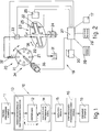
- the reference number 10 in the attached figures denotes generally a method to determine the sedimentation of blood (VES) according to the invention.
- the method 10 according to the invention includes the following steps:
- the method 10 according to the invention is very quick and accurate and makes possible the reading over a period of time of the optical density of the sample so as to describe in a quantitative analytical manner all the steps of the sedimentation which can be used to identify various inflammatory pathologies.
- reading or capillary containers 22 defining a containing chamber of a small inner diameter for the reading of the optical density of a sample of blood enables a linear response to be obtained between the optical density and the quantity of corpuscular mass contained in the blood being examined, this situation enabling results regarding the VES to be provided inasmuch as the optical density at any point of the reading container 22 is a function of the number of corpuscles contained in the sample being examined at every moment during the centrifuging.
- the reading container 22 defines a containing chamber having an inner diameter between 0.1 mm. and 2 mm., but advantageously 1 mm.
- the reference number 19 indicates generally a particular form of embodiment of a device suitable to perform the above method 10.
- the device 19 comprises:
- a means is also included to flush a suitable solution for washing and sterilising the reading container 22 between one analysis and the next one.
- the container 22 of the sample to be analysed consists of a capillary tube which extends without a break of continuity from the point of receipt of the sample to the discharge point.
- This formulation is especially advantageous since it prevents contact of the sample of blood with the atmosphere, thus obviating possible pollution and loss of the sample.
- the programming, processing and control assembly 27 enables all the methods of performing the analysis to be programmed as desired, such as the speed and cycle of the centrifuging of the sample, the momentary scanning of the emitting/monitoring assembly 25 and the representation of the results.
- the aspiration system 20 comprises syringe means 32 and pump means 33, and the sample-drawing container 21 is associated with a sample storage assembly 34 consisting of a rotary drum 35 driven by a second motor 36 and including on its periphery a plurality of sample-drawing containers 21.
- This rotary drum 35 is halted advantageously at a position suitable for the drawing of a micro-volume of sample by means of the syringe means 32.
- the syringe means 32 are associated with a sealing assembly which closes the hole made in the stopper of the container 21 by the needle of the syringe means 32.
- the device 19 according to the invention is associated with other apparatuses for the performance of examinations, such as the haemochromocytometric examination for instance, these apparatuses being fed advantageously by the same aspiration system 20 and being governed by the same programming, processing and control assembly 27.
- the assembly 25 emitting/monitoring electromagnetic radiations can be positioned radially in any usable radial position of the capillary reading container 22.
- the programming, processing and control assembly 27 comprises a bank of reference data 17 containing, for instance in the forms of curves or tables, characteristic signals with which are compared the signals monitored by the device 19 according to the invention so as to diagnose possible pathologies.
Landscapes
- Health & Medical Sciences (AREA)
- Life Sciences & Earth Sciences (AREA)
- Chemical & Material Sciences (AREA)
- Engineering & Computer Science (AREA)
- Physics & Mathematics (AREA)
- Immunology (AREA)
- Hematology (AREA)
- Biochemistry (AREA)
- General Health & Medical Sciences (AREA)
- General Physics & Mathematics (AREA)
- Analytical Chemistry (AREA)
- Pathology (AREA)
- Biomedical Technology (AREA)
- Dispersion Chemistry (AREA)
- Ecology (AREA)
- Biophysics (AREA)
- Molecular Biology (AREA)
- Urology & Nephrology (AREA)
- Food Science & Technology (AREA)
- Medicinal Chemistry (AREA)
- Investigating Or Analysing Biological Materials (AREA)
- Investigating Or Analysing Materials By Optical Means (AREA)
Abstract
Description
- This invention concerns a method to determine the sedimentation of blood and the relative device, as set forth in the respective main claims.
- The invention is applied to the field of medical analyses and enables the speed of sedimentation of blood to be determined sickly and automatically.
- It is well known that the measurement of the speed of sedimentation of the part of the blood containing corpuscles and, to be more exact, of the erythrocytes is linked in some way to the state of health of the patient.
- This speed of sedimentation is called in the technical field VES (velocity of erythrocyte sedimentation) or ESR (erythrocyte sedimentation rate), these terms being employed for the sake of simplicity in the description that follows.
- The sedimentation of the red corpuscles in the blood takes place in three phases as follows:
- a first phase of aggregation of the red corpuscles due to electrostatic forces and to the content of some proteins in the plasma; this aggregation leads to the formation of rouleaux of various dimensions according to the parameters cited; this first phase lasts for about 1 to 10 seconds;
- a second phase of sedimentation of the of rouleaux; this value expresses various characteristics according to the pathologies possibly present; this phase, under the action of the force of gravity alone, may last for some tens of minutes up to hours;
- a third phase of final packing of the rouleaux; these rouleaux tend to arrange themselves by taking up all the available volume on the basis of the difference of density between the rouleaux and the plasma; this phase, under the action of the force of gravity alone, may last for some tens of minutes up to hours.
- The parameter which is usually considered in the measurement of the VES is the second one above, although the first is intimately interconnected with the VES.
- It is known in particular that during the pathological conditions defined as inflammatory the VES increases and therefore this analysis is used to ascertain the presence of such pathological conditions in a quick and easy manner.
- Many different systems have been used hitherto to measure this speed of sedimentation.
- In a classic system the sample of blood drawn from the patient into suitable containers holding anti-coagulant substances is distributed into suitable tubes, or test tubes, which are generally positioned vertically on appropriate supports.
- The analysis consists in observing at pre-set fixed times the position of the band of separation, or interface, between the fluid plasma part of the blood, which is clear, and the part containing corpuscles, which consists of erythrocytes, leukocytes and blood platelets, which is red.
- The data measured give the speed of sedimentation of the blood.
- This method, however, is slow and not automatic and errors may easily be made by the laboratory assistant in the reading and/or transcription of the data giving the height of the interface.
- Various systems to overcome these shortcomings have been disclosed so as to obtain such analysis in an automatic, fast and reproduceable manner.
- CH-A-655.800 discloses a method of monitoring, at pre-set intervals of time, the light transmitted through the sedimentation tube so as to identify the position of the plasma/ corpuscles interface. This system enables the analysis to be automated but does not reduce the performance times.
- GB-A-2,153,072 discloses a method whereby the measurement tubes are inclined to the vertical and are then caused to rotate so as to exploit centrifugal force in accelerating the sedimentation of the erythrocytes and are lastly uprighted to a vertical position to perform the reading of the position of the plasma/corpuscles interface. This system enables the times for carrying out the analysis to be indeed reduced but in a manner not satisfactory enough for users.
- DE-A-2.825.659 discloses the reading of the optical density of the blood along a determined segment of the sedimentation tube by using a plurality of vertically arranged diodes. This measurement provides the variation in time of the position of the plasma/corpuscles interface and therefore the VES.
- US-A-4,848,900 discloses a device to evaluate the optical density at the plasma/corpuscles interface; by moving the movable reading head step-by-step and automatically it is possible to obtain the VES of the analysed sample of blood.
- US-A-5,003,488 discloses the use of a telecamera to identify the height of the interface at determined intervals and to process with a computer the data thus measured so as to obtain the VES.
- US-A-5,316,729 discloses a device to monitor by analysis of the optical density the movement of the plasma/corpuscles interface and to compare the curve thus obtained with reference curves so as to extrapolate the final position of that interface and to determine the VES.
- WO 91/05996 discloses a device whereby the VES is monitored by opto-thermal sensors which measure at determined intervals the heat produced by the absorption, by the sample itself, of radiation which strikes the sample of blood to be analysed. The signals monitored at determined intervals of time are a function of the VES of the sample
- US-A-5,328,822 discloses a method and a device whereby the position of the plasma/corpuscles interface is monitored by an analysis of reflectivity of the light on the tube being analysed, this characteristic being a function of the number of corpuscles present corresponding to the sensor.
- This device provides for the use of a step motor associated with the movable reading head, which is lowered along the tube and determines by extrapolation the exact position of the plasma/corpuscles interface. Moreover, this document provides also for the diagnosis of the pathology of the patient by comparing the curve obtained with sample curves and also provides for the analysis of the curve of sedimentation of the part containing corpuscles and of the plasma, including leukocytes and blood platelets after sedimentation of the erythrocytes.
- US-A-3,824,841 and US-A-3,848,796 disclose a method and a device whereby the analysis tubes containing the blood are kept vertically and set in rotation, still in a vertical position, until those tubes are subjected to a centrifugal acceleration equal to 6 to 8.5 times the acceleration of gravity.
- The level reached by the plasma/corpuscles interface is read at determined intervals of time after one or more cycles of centrifuging so as to obtain the measurement of the VES.
- All the methods disclosed so far, some of which have been briefly set forth, enable the performance of the analysis to be automated but do not enable the performance times to be reduced except very slightly.
- Moreover, all these methods require that the blood to be analysed should be contained in appropriate containers of the disposable type with a resulting increase in the costs of the analysis as regards both the purchase and, above all, the disposal of these containers, which have to be treated as hospital refuse.
- Moreover, the quantity of blood required for performing the analysis is great and is such as will create problems in some cases, particularly when the analysis has to be carried out on children.
- Furthermore, all the methods disclosed hitherto are based on the monitoring in time of the position of the plasma/corpuscles interface, which is determined gradually as the particles of blood settle.
- This type of monitoring requires of necessity a minimum performance time, even if it is greatly reduced by submitting the vertically kept container to centrifuging about a vertical axis so as to accelerate the sedimentation of the particles.
- In fact, the monitoring system entails an initial dead time which cannot be eliminated and in which the formation of the plasma/corpuscles interface has to take place.
- At the present time it is therefore not possible to combine in parallel the device performing this analysis with other existing devices able to carry out other very fast analyses such as the haemochromocytometric analysis, in which is carried out the counting of the number of the erythrocytes, leukocytes and blood platelets.
- Moreover, the devices of the state of the art require the use of a container intended for this type of analysis and of a specially intended device, this situation being a possible source of errors due to the exchange of samples or to wrong labelling if the various analyses are performed with different instruments and also in different times.
- The times of performance of the analysis of the VES with the devices of the state of the art are always greater than 90 seconds and generally greater than 120 seconds, whereas the haemochromocytometric analysis cited above has performance times less than a minute.
- US-A-3,679,367 and US-A-3,684,450 disclose a method and relative device for determining the haematocrit or the volume of packed red corpuscles by using very high speeds of rotation to ensure a swift packing of the corpuscles.
- This method therefore refers to the third phase of sedimentation and not to the second in which the methods of sedimentation, and therefore the VES, are analysed.
- Moreover, this method determines that volume of packed corpuscles by measuring continuously the position of the interface found from the different optical density between the limpid plasma and the corpuscles.
- The present applicants have designed, tested and embodied this invention to overcome the shortcomings of the state of the art and to achieve further advantages.
- This invention is set forth and characterised in the respective main claims, while the dependent claims describe variants of the idea of the main embodiment.
- The purpose of this invention is to provide a method for the monitoring of the speed of sedimentation of blood (VES), the method being immediate and secure.
- The invention enables the analysis to be performed by using micro-volumes of blood so that the invention can be used without problems also in the field of paediatrics or on anaemic patients.
- The invention enables the analysis to be performed by taking the sample to be analysed directly from any container containing blood, to which has been added any anti-coagulant substance but advantageously salts of EDTA (ethylene diamine tetra-acetic acid), and used in laboratories of clinical analyses without having to use specially provided containers, thus making possible a resulting saving of disposable material and of blood.
- The invention obviates the possibility of mistakes due to exchanges of containers between patients inasmuch as the analysis of the VES is carried out at the same time and with the same performance times as other clinical analyses such as the haemochromocytometric examination, as it is possible to use the same sample of blood contained in the test tube for the haemochromocytometric examination too.
- The invention makes also possible the immediate identification of other characteristics of the patient such as, for instance, the inclusion of a low number of erythrocytes, these being characteristics which have to be kept in mind for the analysis of the VES so as not to fall into errors of evaluation in correlating the number of corpuscles present with the VES found.
- The method according to the invention is based on the monitoring of the optical density of the sample of blood with any position of the container employed for the measurement.
- In the method according to the invention the width of the sample of blood analysed has to be very small so that the law of Lambert-Beer on absorbance or optical density, can be applied.
- The law of Lambert-Beer establishes that:

- A =
- absorbance or optical density defined as: log (I0/I) with I0 = intensity of the luminous radiation which passes through the empty container and I = intensity of the luminous radiation which passes through the container filled with the sample to be analysed;
- c =
- concentration of the species which absorbs the radiation;
- d =
- width of the sample thus passed through;
- k =
- constant.
- In this case the signal monitored is therefore directly in proportion to the number of corpuscles contained in the sample at that point and thus gives an immediate indication without initial dead times since it is not necessary to await the formation of the plasma/corpuscles interface as in the state of the art.
- Over a period of time the number of corpuscles present at the point of observation changes owing to the fact that the corpuscles tend to settle, and this variation changes the value of the absorbance at that point.
- Therefore, the study of the absorbance over a period of time is equivalent to the study of the corpuscles present at that point over a period of time and thus can be correlated with the VES.
- The observation point can be located either in the initial part, where the number of corpuscles diminishes over a period of time, or in the final part of the container, where the number of corpuscles increases over a period of time.
- By initial part and final part of the container are meant the parts positioned in the vicinity of, and distanced from, the axis of rotation respectively.
- Depending on whether the observation point is located in the initial part or final part of the container, the absorbance decreases over a period of time or increase over a period of time.
- The method according to the invention comprises the following steps:
- the feeding of the sample of suitably homogenised blood to the analysis device, for instance, by aspiration from the container holding the blood drawn;
- the possible centrifuging of the sample in the container of a small inner diameter, such as a capillary tube; this centrifuging step is advantageously carried out about an axis perpendicular to the plane of positioning of the container; the speed of centrifuging used to optimise the step of sedimentation of the rouleaux is such as to submit the sample of blood to an acceleration between 2 and 20 g, but advantageously between 7 to 8 g;
- the monitoring as a function of time of the optical density of the sample at any fixed and determined point in the container; this monitoring can be carried out advantageously by using electromagnetic radiations having a wave length between 200 and 1000 nm, but advantageously between 700 and 1000 nm;
- processing of the signal obtained over a period of time and conversion of the parameters of the curve which plots the optical density as a function of time so as to determine the VES, with a possibility of making a graph of the total sedimentation; according to whether the point of observation is located in the initial part or final part of the sample, the curve obtained will have a decreasing or increasing development as a function of time;
- comparison with standardised coded signals;
- discharge of the sample.
- The method according to the invention can be employed also to monitor analytically and quantitatively the sedimentation of the plasma part of the blood.
- The method according to the invention can be combined with other methods to carry out other analyses which use containers containing blood to which an anti-coagulant has been added and which have very short performance times such as a haemochromocytometric examination for instance.
- The device according to the invention comprises means to draw the sample to be analysed from the drawing container and also means to centrifuge the sample, these latter means being associated with an assembly which emits/monitors electromagnetic radiation for monitoring the optical density of the sample at a fixed and determined point.
- In the device according to the invention the sample is delivered into a container which is rotated about an axis perpendicular to the plane of positioning of the container with a determined angular velocity which can be varied as required as a function of the desired duration of the analysis.
- This container is made of a material transparent to the electromagnetic radiation employed by the assembly used to monitor the optical density.
- According to a particular form of embodiment of the device according to the invention this container has a through passage and includes an inlet and an outlet to make possible access to the sample to be analysed and the downflow and discharge of the sample when the analysis has been completed.
- According to a particular formulation of the invention the container of the sample to be analysed consists of a capillary tube which extends without a break of continuity from the point of receipt of the sample to the discharge point.
- This formulation is especially advantageous inasmuch as it prevents contact of the sample of blood with the atmosphere, thus obviating possible pollution and loss of the sample.
- Moreover, this solution makes possible the flushing of the whole container between one examination and another with a suitable washing solution to prevent contagion between various samples of blood.
- The assembly which emits/monitors the electromagnetic radiation is positioned perpendicular, or substantially perpendicular to the plane of positioning of the container and on opposite sides of the container and remains positioned at a fixed determined point during the performance of the analysis.
- According to a variant the assembly emitting/monitoring the electromagnetic radiation can be positioned radially as desired.
- According to a further variant the container has a reflecting sidewall and the emitting/monitoring assembly is positioned on one and the same side of the plane of positioning of the container.
- As we said above, the emitting/monitoring assembly can be positioned either at the initial part of the container nearest to the axis of rotation, where the optical density diminishes over a period of time, or at the final part of the container distanced from the axis of rotation, where the optical density increases over a period of time.
- The device according to the invention is advantageously governed by a programming, processing and control assembly, which controls all the steps of the analysis such as the aspiration of the sample, the centrifuging of the sample, the monitoring of the optical density of the sample at determined intervals, the processing of the signal monitored over a period of time and the discharge of the sample.
- Moreover, the programming, processing and control assembly is associated with means that represent the signal either on a video or on a printer.
- According to a variant, the programming, processing and control assembly comprises a bank of reference data, for instance in the form of curves or tables with which are compared the monitored signals so as to diagnose possible pathologies.
- According to a variant the device according to the invention is combined with a supply store holding a plurality of samples of blood to which anti-coagulant substances have been added; these samples of blood are suitably homogenised, for instance by rotation, rocking or other analogous operations.
- This supply store positions in sequence each sample from which a micro-volume of blood to form the sample to be analysed is aspirated in an automatic manner, for instance by means of a needle.
- According to a further variant the container is automatically re-sealed after the aspiration step so as to prevent possible contamination.
- The attached figures are given as a non-restrictive example and show a preferred embodiment of the invention as follows:-
- Fig.1
- is a block diagram of the method according to the invention;
- Fig.2
- is a diagram of a device according to the invention.
- The
reference number 10 in the attached figures denotes generally a method to determine the sedimentation of blood (VES) according to the invention. - The
method 10 according to the invention includes the following steps: - feeding 11 the suitable homogenised sample to be analysed, for instance by aspiration from a
container 21 into a readingcontainer 22 defining a containing chamber having a very small inner diameter, into which is inserted the micro-volume of sample to be analysed; - possible centrifuging 12 of the sample about an axis substantially perpendicular to the plane of positioning of the reading
container 22; the speed of centrifuging used to optimise the step of sedimentation of the rouleaux is such as to subject the sample of blood to an acceleration between 2 and 20 g, but advantageously between 7 to 8 g; - emission/monitoring 13 of an electromagnetic radiation to identify the development over a period of time of the optical density of the sample of blood analysed at any fixed point of the reading
container 22, this emission/monitoring 13 being carried out in a direction perpendicular to the plane of positioning of the readingcontainer 22 containing the sample and with a momentary scanning which can be programmed as desired immediately after the first moment at which the sample is injected into the readingcontainer 22, the minimum usable duration of emission/monitoring 13 being selected as a function of the speed of centrifuging; - the analysis and processing 14 of the monitored signals as a function of the period of time, taking into consideration the initial value of the absorbance which is a function of the number of corpuscles present in the sample analysed;
-
display 15 of the results by video 28 and/or on aprinter 30; - possible comparison 16 with standardised signals of coded pathologies;
-
discharge 18 of the analysed sample. - The
method 10 according to the invention is very quick and accurate and makes possible the reading over a period of time of the optical density of the sample so as to describe in a quantitative analytical manner all the steps of the sedimentation which can be used to identify various inflammatory pathologies. - The use of reading or
capillary containers 22 defining a containing chamber of a small inner diameter for the reading of the optical density of a sample of blood enables a linear response to be obtained between the optical density and the quantity of corpuscular mass contained in the blood being examined, this situation enabling results regarding the VES to be provided inasmuch as the optical density at any point of the readingcontainer 22 is a function of the number of corpuscles contained in the sample being examined at every moment during the centrifuging. - The reading
container 22 defines a containing chamber having an inner diameter between 0.1 mm. and 2 mm., but advantageously 1 mm. - The
reference number 19 indicates generally a particular form of embodiment of a device suitable to perform theabove method 10. - In this case the
device 19 comprises: - an
aspiration system 20 to aspirate a micro-volume of blood from acontainer 21; - a
capillary reading container 22 lying on a substantially horizontal plane and associated with centrifuging means 23, which consist in this case of afirst motor 24 having a vertical axis; - an
assembly 25 to emit/monitor electromagnetic radiations which is positioned perpendicular to, and on opposite sides of, the plane of positioning of thecapillary reading container 22; - a means 26 to acquire the signal monitored by the monitoring
assembly 25; - a programming, processing and
control assembly 27 which governs automatically thedevice 19 performing the method and which comprises display means 28, means 29 to introduce data and control and printing means 30; - a means 31 to discharge the analysed sample.
- According to a variant a means is also included to flush a suitable solution for washing and sterilising the reading
container 22 between one analysis and the next one. - According to a particular formulation of the invention, which is not shown here, the
container 22 of the sample to be analysed consists of a capillary tube which extends without a break of continuity from the point of receipt of the sample to the discharge point. - This formulation is especially advantageous since it prevents contact of the sample of blood with the atmosphere, thus obviating possible pollution and loss of the sample.
- The programming, processing and
control assembly 27 enables all the methods of performing the analysis to be programmed as desired, such as the speed and cycle of the centrifuging of the sample, the momentary scanning of the emitting/monitoring assembly 25 and the representation of the results. - In this case the
aspiration system 20 comprises syringe means 32 and pump means 33, and the sample-drawingcontainer 21 is associated with asample storage assembly 34 consisting of arotary drum 35 driven by asecond motor 36 and including on its periphery a plurality of sample-drawingcontainers 21. - This
rotary drum 35 is halted advantageously at a position suitable for the drawing of a micro-volume of sample by means of the syringe means 32. - According to a particular form of embodiment the syringe means 32 are associated with a sealing assembly which closes the hole made in the stopper of the
container 21 by the needle of the syringe means 32. - According to a particular form of embodiment the
device 19 according to the invention is associated with other apparatuses for the performance of examinations, such as the haemochromocytometric examination for instance, these apparatuses being fed advantageously by thesame aspiration system 20 and being governed by the same programming, processing andcontrol assembly 27. - It is possible in this way to combine the result of the VES examination with other examinations carried out at the same time on a sample of blood drawn from the
same container 21. - In an advantageous form of embodiment of the
device 19 according to the invention theassembly 25 emitting/monitoring electromagnetic radiations can be positioned radially in any usable radial position of thecapillary reading container 22. - According to a variant the programming, processing and
control assembly 27 comprises a bank ofreference data 17 containing, for instance in the forms of curves or tables, characteristic signals with which are compared the signals monitored by thedevice 19 according to the invention so as to diagnose possible pathologies.
Claims (23)
- Method to determine the sedimentation of blood, the method being carried out on a sample consisting of blood to which any anti-coagulant substance has been added and which is suitably homogenised, the method being characterised in that it includes the monitoring of the development of the optical density, or absorbance, of the sample as a function of time and without initial dead times, the monitoring being carried out at any fixed point of a reading container (22) defining a containing chamber of a small inner diameter which contains a micro-volume of blood, the inner diameter of the containing chamber being such as to ensure a linear relationship between the optical density and the number of corpuscles included.
- Method as in Claim 1, which employs reading containers (22), in which the inner diameter of the containing chamber is between 0.1 mm and 2 mm., but advantageously 1 mm.
- Method as in Claim 1 or 2, which enables the value of the corpuscular mass in the sample to be identified at the first moment.
- Method as in any claim hereinbefore, in which the reading container (22) is subjected to centrifuging (12) during the monitoring step (13).
- Method as in Claim 4, in which during the centrifuging the sample undergoes an acceleration between 2 and 20 g. but advantageously 7 to 8 g.
- Method as in Claim 5 or 6, in which the centrifuging (12) takes place about an axis perpendicular to the plane of positioning of the reading container (22).
- Method as in any claim hereinbefore, which is independent of the type of anti-coagulant substance added to the sample of blood.
- Method as in any claim hereinbefore, which comprises the following steps:- homogenising and feeding (11), advantageously by aspiration from a container (21), the sample to be analysed into a reading container (22);- possible centrifuging (12) of the sample about an axis perpendicular to the plane of positioning of the reading container (22);- emitting/monitoring (13) an electromagnetic radiation to identify the development, over a period of time, of the optical density of the sample of blood analysed;- processing (14) the monitored signals as a function of a period of time;- displaying (15) the results;- discharging (18) the analysed sample.
- Method as in any claim hereinbefore, which comprises a step of comparison (16) with previously stored signals of known pathologies, this step (16) being usable in the differentiation of inflammatory pathologies.
- Method as in any claim hereinbefore, which is governed by a programming, processing and control assembly (27) for the automatic performance of the analyses.
- Method as in any claim hereinbefore, which enables the sedimentation of the plasma part of the blood to be monitored in an analytical and quantitative manner.
- Device to determine the sedimentation of blood, which of a type comprising at least one means (20) feeding the sample to be analysed, a reading container (22) set in rotation about an axis perpendicular to its plane of positioning, an assembly (25) to emit/monitor electromagnetic radiations which is positioned perpendicular to the plane of positioning of the reading container (22), a means (26) to acquire data and a programming, processing and control assembly (27), the device being characterised in that the reading container (22) defines a containing chamber having an inner diameter between 0.1 and 2 mm., but advantageously of 1 mm.
- Device as in Claim 12, in which the reading container (22) comprises an inlet and an outlet for the feeding and discharge of the analysed sample.
- Device as in Claim 12 or 13, in which the reading container (22) extends without a break of continuity from the feed to the discharge of the sample.
- Device as in any of Claims 12 to 14 inclusive, in which the emitting/monitoring assembly (25) and the data acquisition means (26) enable the development, over a period of time, of the optical density of the sample of blood to be monitored, this development being able to be correlated with the VES.
- Device as in any of Claims 12 to 15 inclusive, in which the emitting/monitoring assembly (25) can be positioned as desired along the axis of the reading container (22)
- Device as in any of Claims 11 to 14 inclusive, in which the reading container (22) has a reflecting surface, the emitting/monitoring assembly (25) having its emitting means and its monitoring means positioned on the same side of the plane of positioning of the reading container (22).
- Device as in any of Claims 12 to 17 inclusive, in which the method of centrifuging, such as the gradient of acceleration and the final value of the acceleration, can be adjusted as desired as a function of the desired duration of the analysis.
- Device as in any of Claims 12 to 18 inclusive, which subjects the sample to be analysed to a centrifugal acceleration between 2 and 20 g, but advantageously 7 to 8 g.
- Device as in any of Claims 12 to 19 inclusive, in which the times of momentary scanning of the emitting/monitoring means (25) and of the data acquisition means (26) can be adjusted as desired.
- Device as in any of Claims 12 to 20 inclusive, in which the programming, processing and control assembly (27) is associated with a bank (17) of reference data.
- Device as in any of Claims 12 to 21 inclusive, which is coupled hydraulically and electronically to other apparatuses for the performance of other analyses of the blood such as the haemochromocytometric examination, the times of performance of which are compatible with the times of performance of the method according to the invention.
- Device as in any of Claims 12 to 22 inclusive, which comprises a means to flush a suitable solution for the washing and possible sterilisation of the whole reading container (22) between one analysis and the next one.
Applications Claiming Priority (2)
| Application Number | Priority Date | Filing Date | Title |
|---|---|---|---|
| ITUD950042 | 1995-03-15 | ||
| IT95UD000042A IT1280143B1 (en) | 1995-03-15 | 1995-03-15 | PROCEDURE FOR THE DETERMINATION OF BLOOD SEDIMENTATION AND RELATED DEVICE |
Publications (1)
| Publication Number | Publication Date |
|---|---|
| EP0732576A1 true EP0732576A1 (en) | 1996-09-18 |
Family
ID=11421771
Family Applications (1)
| Application Number | Title | Priority Date | Filing Date |
|---|---|---|---|
| EP96103358A Withdrawn EP0732576A1 (en) | 1995-03-15 | 1996-03-05 | Method to determine the sedimentation of blood and relative device |
Country Status (5)
| Country | Link |
|---|---|
| US (1) | US5827746A (en) |
| EP (1) | EP0732576A1 (en) |
| JP (1) | JP3547894B2 (en) |
| CA (1) | CA2171069A1 (en) |
| IT (1) | IT1280143B1 (en) |
Cited By (6)
| Publication number | Priority date | Publication date | Assignee | Title |
|---|---|---|---|---|
| EP0864854A2 (en) * | 1997-03-10 | 1998-09-16 | Stephen Clark Wardlaw | Method and assembly for rapid measurement of cell layers |
| WO1999045363A1 (en) * | 1998-03-05 | 1999-09-10 | Zynocyte Limited | Method of measuring erythrocyte sedimentation rate (esr) or plasma fibrinogen of a blood sample |
| EP1098188A2 (en) * | 1999-11-08 | 2001-05-09 | Sire Analytical Systems S.R.L. | Method to determine the speed of sedimentation of blood and other parameters correlated thereto, and relative apparatus |
| WO2005022125A1 (en) * | 2003-09-03 | 2005-03-10 | Sire Analytical Systems Srl | Integrated apparatus for hematological analyses and related method |
| EP2347709A3 (en) * | 2005-10-06 | 2013-07-31 | Optiscan Biomedical Corporation | Body fluid analyzer |
| US10488396B2 (en) | 2012-01-13 | 2019-11-26 | Alcor Scientific, Inc. | Apparatus, method, system for the determination of the aggregation rate of red blood cells |
Families Citing this family (25)
| Publication number | Priority date | Publication date | Assignee | Title |
|---|---|---|---|---|
| IT1286631B1 (en) * | 1996-05-16 | 1998-07-15 | Diesse Diagnostica | EQUIPMENT FOR THE PREPARATION AND DETERMINATION OF THE TESTS OF THE SPEED OF SEDIMENTATION OF ORGANIC LIQUIDS AND MORE |
| WO2000007012A1 (en) * | 1998-07-31 | 2000-02-10 | Carroll Wallace E | Method and apparatus for determining anticoagulant therapy factors |
| WO1998039643A1 (en) * | 1997-03-05 | 1998-09-11 | Diametrics | Method and apparatus for measurement of whole blood coagulation parameters |
| WO1998043720A1 (en) * | 1997-04-03 | 1998-10-08 | Baxter International Inc. | Interface detection and control systems and methods |
| JP2000329779A (en) * | 1999-05-19 | 2000-11-30 | Sefa Technology Kk | Sedimentation speed measuring method and its device |
| US6204066B1 (en) * | 1999-06-25 | 2001-03-20 | Robert A. Levine | Rapid method for determining the erythrocyte sedimentation rate in a sample of anticoagulated whole blood |
| US6989891B2 (en) | 2001-11-08 | 2006-01-24 | Optiscan Biomedical Corporation | Device and method for in vitro determination of analyte concentrations within body fluids |
| US7207939B2 (en) * | 2002-10-03 | 2007-04-24 | Coulter International Corp. | Apparatus and method for analyzing a liquid in a capillary tube of a hematology instrument |
| US7907985B2 (en) | 2005-02-14 | 2011-03-15 | Optiscan Biomedical Corporation | Fluid handling cassette with a fluid control interface and sample separator |
| US8251907B2 (en) | 2005-02-14 | 2012-08-28 | Optiscan Biomedical Corporation | System and method for determining a treatment dose for a patient |
| US8936755B2 (en) | 2005-03-02 | 2015-01-20 | Optiscan Biomedical Corporation | Bodily fluid composition analyzer with disposable cassette |
| US9561001B2 (en) | 2005-10-06 | 2017-02-07 | Optiscan Biomedical Corporation | Fluid handling cassette system for body fluid analyzer |
| EP2016402A2 (en) | 2006-04-11 | 2009-01-21 | Optiscan Biomedical Corporation | Anti-clotting apparatus and methods for fluid handling system |
| ITUD20060177A1 (en) * | 2006-07-14 | 2008-01-15 | Sire Analytical Systems Srl | INTEGRATED EQUIPMENT AND METHOD FOR DETECTION OF INFLAMMATORY STATES PRESENT IN A WHOLE BLOOD SAMPLE |
| WO2008144575A2 (en) | 2007-05-18 | 2008-11-27 | Optiscan Biomedical Corporation | Fluid injection and safety system |
| SE531510C2 (en) * | 2007-09-04 | 2009-05-05 | Tommy Forsell | blood Analysis |
| US9091676B2 (en) | 2010-06-09 | 2015-07-28 | Optiscan Biomedical Corp. | Systems and methods for measuring multiple analytes in a sample |
| EP2456355B1 (en) | 2009-07-20 | 2016-09-14 | Optiscan Biomedical Corporation | Adjustable connector and dead space reduction |
| US9554742B2 (en) | 2009-07-20 | 2017-01-31 | Optiscan Biomedical Corporation | Fluid analysis system |
| US8928877B2 (en) | 2011-07-06 | 2015-01-06 | Optiscan Biomedical Corporation | Sample cell for fluid analysis system |
| US8647886B1 (en) * | 2012-01-13 | 2014-02-11 | Alcor Scientific, Inc. | Apparatus, method, system for the determination of the aggregation rate of red blood cells |
| ITUD20120137A1 (en) * | 2012-07-31 | 2014-02-01 | Alifax Holding S P A | APPARATUS AND PROCEDURE FOR THE DETERMINATION OF THE SPEED OF SEDIMENTATION OF THE BLOOD AND OTHER PARAMETERS RELATED TO IT |
| CN108020530A (en) * | 2018-01-18 | 2018-05-11 | 中实医疗科技江苏有限公司 | Quick erythrocyte sedimentation rate analytical equipment and its method |
| FR3077639B1 (en) * | 2018-02-02 | 2020-02-14 | Universite De Rennes 1 | METHOD FOR DETERMINING A SEDIMENTATION SPEED |
| IT201800004630A1 (en) * | 2018-04-17 | 2019-10-17 | APPARATUS AND PROCEDURE FOR DETERMINING THE SPEED OF BLOOD SEDIMENTATION AND OTHER PARAMETERS RELATED TO IT |
Citations (6)
| Publication number | Priority date | Publication date | Assignee | Title |
|---|---|---|---|---|
| US3679367A (en) * | 1970-09-14 | 1972-07-25 | Technicon Instr | Apparatus for determining the pack volume of particulates in liquid mixtures |
| US3684450A (en) * | 1970-09-14 | 1972-08-15 | Stanford L Adler | Automatic apparatus and method for determining the packed cell volume of whole blood |
| US4352557A (en) * | 1979-10-20 | 1982-10-05 | Ernst Leitz Wetzlar Gmbh | Determining a test value corresponding to blood subsidence |
| WO1992009879A1 (en) * | 1990-11-29 | 1992-06-11 | Hemocue Ab | A device for rapidly performing a sedimentation-rate test |
| DE9216127U1 (en) * | 1992-11-29 | 1994-04-07 | Orth, Helmut, Prof. Dr., 74321 Bietigheim-Bissingen | Device for measuring blood sedimentation |
| WO1994018557A1 (en) * | 1993-02-11 | 1994-08-18 | Zynocyte Limited | Apparatus for analysing blood and other samples |
Family Cites Families (10)
| Publication number | Priority date | Publication date | Assignee | Title |
|---|---|---|---|---|
| US4135819A (en) * | 1974-03-20 | 1979-01-23 | Ernst Leitz Gmbh | Apparatus for measuring the aggregation rate of particles |
| US4041502A (en) * | 1975-12-22 | 1977-08-09 | Williams Tool, Inc. | Sedimentation recorder |
| JPS5912138B2 (en) * | 1976-02-17 | 1984-03-21 | コニカ株式会社 | Blood sediment measurement method |
| CA1175673A (en) * | 1981-08-18 | 1984-10-09 | Robert N. O'brien | Erythrocyte settling rate meter |
| JPS62226057A (en) * | 1986-03-28 | 1987-10-05 | Minoru Tomita | Method and apparatus for measuring agglutination of red blood cell for whole blood |
| US4848900A (en) * | 1987-06-22 | 1989-07-18 | Kuo Cheng Deng | Computerized automatic monitoring and recording system of erythrocyte sedimentation process |
| US5003488A (en) * | 1989-03-10 | 1991-03-26 | Gespac, Inc. | Automatic fluid sedimentation rate measurement apparatus and method |
| US5328822A (en) * | 1990-04-23 | 1994-07-12 | Solid State Farms, Inc. | Apparatus and method for sedimentation based blood analysis |
| DE4117583A1 (en) * | 1991-05-29 | 1992-12-03 | Helmut Prof Dr Orth | Blood sinking measurement arrangement - compares stored position measurement values with reference curves and derives position of blood serum boundary |
| DK203191D0 (en) * | 1991-12-19 | 1991-12-19 | Novo Nordisk As | METHOD AND APPARATUS FOR DETERMINING RELEVANT BLOOD PARAMETERS |
-
1995
- 1995-03-15 IT IT95UD000042A patent/IT1280143B1/en active IP Right Grant
-
1996
- 1996-03-05 EP EP96103358A patent/EP0732576A1/en not_active Withdrawn
- 1996-03-05 CA CA002171069A patent/CA2171069A1/en not_active Abandoned
- 1996-03-08 US US08/613,233 patent/US5827746A/en not_active Expired - Lifetime
- 1996-03-15 JP JP05814596A patent/JP3547894B2/en not_active Expired - Lifetime
Patent Citations (6)
| Publication number | Priority date | Publication date | Assignee | Title |
|---|---|---|---|---|
| US3679367A (en) * | 1970-09-14 | 1972-07-25 | Technicon Instr | Apparatus for determining the pack volume of particulates in liquid mixtures |
| US3684450A (en) * | 1970-09-14 | 1972-08-15 | Stanford L Adler | Automatic apparatus and method for determining the packed cell volume of whole blood |
| US4352557A (en) * | 1979-10-20 | 1982-10-05 | Ernst Leitz Wetzlar Gmbh | Determining a test value corresponding to blood subsidence |
| WO1992009879A1 (en) * | 1990-11-29 | 1992-06-11 | Hemocue Ab | A device for rapidly performing a sedimentation-rate test |
| DE9216127U1 (en) * | 1992-11-29 | 1994-04-07 | Orth, Helmut, Prof. Dr., 74321 Bietigheim-Bissingen | Device for measuring blood sedimentation |
| WO1994018557A1 (en) * | 1993-02-11 | 1994-08-18 | Zynocyte Limited | Apparatus for analysing blood and other samples |
Cited By (11)
| Publication number | Priority date | Publication date | Assignee | Title |
|---|---|---|---|---|
| EP0864854A2 (en) * | 1997-03-10 | 1998-09-16 | Stephen Clark Wardlaw | Method and assembly for rapid measurement of cell layers |
| EP0864854A3 (en) * | 1997-03-10 | 1999-04-14 | Stephen Clark Wardlaw | Method and assembly for rapid measurement of cell layers |
| WO1999045363A1 (en) * | 1998-03-05 | 1999-09-10 | Zynocyte Limited | Method of measuring erythrocyte sedimentation rate (esr) or plasma fibrinogen of a blood sample |
| EP1098188A2 (en) * | 1999-11-08 | 2001-05-09 | Sire Analytical Systems S.R.L. | Method to determine the speed of sedimentation of blood and other parameters correlated thereto, and relative apparatus |
| EP1098188A3 (en) * | 1999-11-08 | 2002-01-30 | Sire Analytical Systems S.R.L. | Method to determine the speed of sedimentation of blood and other parameters correlated thereto, and relative apparatus |
| WO2005022125A1 (en) * | 2003-09-03 | 2005-03-10 | Sire Analytical Systems Srl | Integrated apparatus for hematological analyses and related method |
| US7541191B2 (en) | 2003-09-03 | 2009-06-02 | Sire Analytical Systems Srl | Integrated apparatus for hematological analysis and related method |
| CN1864060B (en) * | 2003-09-03 | 2011-02-16 | 塞尔分析系统有限公司 | Integrated apparatus for hematological analysis and related method |
| EP2347709A3 (en) * | 2005-10-06 | 2013-07-31 | Optiscan Biomedical Corporation | Body fluid analyzer |
| US10488396B2 (en) | 2012-01-13 | 2019-11-26 | Alcor Scientific, Inc. | Apparatus, method, system for the determination of the aggregation rate of red blood cells |
| US11016081B2 (en) | 2012-01-13 | 2021-05-25 | Alcor Scientific Inc. | Apparatus, method, system for the determination of the aggregation rate of red blood cells |
Also Published As
| Publication number | Publication date |
|---|---|
| ITUD950042A0 (en) | 1995-03-15 |
| US5827746A (en) | 1998-10-27 |
| CA2171069A1 (en) | 1996-09-16 |
| JPH08338840A (en) | 1996-12-24 |
| ITUD950042A1 (en) | 1996-09-15 |
| IT1280143B1 (en) | 1998-01-05 |
| JP3547894B2 (en) | 2004-07-28 |
Similar Documents
| Publication | Publication Date | Title |
|---|---|---|
| US5827746A (en) | Method to determine the sedimentation of blood and relative device | |
| CN109884289B (en) | Low sample volume coagulation assay | |
| JP6005683B2 (en) | Method and apparatus for determining interfering substances and physical dimensions in liquid samples and containers analyzed by a clinical analyzer | |
| US5470534A (en) | Analytical system useful in diagnosis of the condition of a disease | |
| US6043871A (en) | System and method for measuring blood platelet function | |
| US6336358B1 (en) | Method and apparatus for measuring sedimentation rate of sediments in liquid sample | |
| CA2537500C (en) | Integrated apparatus for hematological analyses and related method | |
| US5779983A (en) | Test tube for determining the erythrocyte sedimentation rate and a surfactant for use therein | |
| CA1298181C (en) | Unitized reagent containment system for clinical analyzer | |
| JP2008542753A (en) | A method for identifying interfering factors in small liquid samples on an automated clinical analyzer. | |
| JPH09101303A (en) | Erythrocyte-sedimentation-velocity measuring method | |
| JP2009109196A (en) | Dilution ratio deriving method, quantity determination method and analyzer | |
| JP2016014478A (en) | Diagnostic system and components | |
| CN111712703A (en) | Sample analyzer and sample analyzing method | |
| JP3298332B2 (en) | Biological sample analysis system | |
| CA1198970A (en) | Apparatus to evaluate the "erythrayte sedimentation rate" (esr) in several samples | |
| CN112119293B (en) | Apparatus and method for determining erythrocyte sedimentation rate and other related parameters | |
| JP2020201265A (en) | Apparatus for optically monitoring dosing of liquid to be pipetted | |
| JPH11512829A (en) | Method and apparatus for measuring and controlling the volume of a liquid segment in a tube | |
| EP4187228A1 (en) | System, apparatus, and method for measuring erythrocyte sedimentation rate | |
| EP2077446A1 (en) | Method of determining abnormality and analyzer | |
| JPH05180851A (en) | Micro specimen sampling method for automatic analyzer | |
| JPH0560769A (en) | Method for sampling minute amount of specimen for automatic analyzer | |
| KR20180077822A (en) | Blood specimen container fastening apparatus and method for blood cell analysis system | |
| CN116337817A (en) | Blood analysis device and method |
Legal Events
| Date | Code | Title | Description |
|---|---|---|---|
| PUAI | Public reference made under article 153(3) epc to a published international application that has entered the european phase |
Free format text: ORIGINAL CODE: 0009012 |
|
| AK | Designated contracting states |
Kind code of ref document: A1 Designated state(s): AT BE CH DE DK ES FI FR GB GR IT LI NL PT SE |
|
| 17P | Request for examination filed |
Effective date: 19961029 |
|
| 17Q | First examination report despatched |
Effective date: 20000328 |
|
| GRAG | Despatch of communication of intention to grant |
Free format text: ORIGINAL CODE: EPIDOS AGRA |
|
| GRAG | Despatch of communication of intention to grant |
Free format text: ORIGINAL CODE: EPIDOS AGRA |
|
| GRAH | Despatch of communication of intention to grant a patent |
Free format text: ORIGINAL CODE: EPIDOS IGRA |
|
| STAA | Information on the status of an ep patent application or granted ep patent |
Free format text: STATUS: THE APPLICATION IS DEEMED TO BE WITHDRAWN |
|
| 18D | Application deemed to be withdrawn |
Effective date: 20020301 |