EP0724643B1 - Endosomolytically active particles - Google Patents
Endosomolytically active particles Download PDFInfo
- Publication number
- EP0724643B1 EP0724643B1 EP94928873A EP94928873A EP0724643B1 EP 0724643 B1 EP0724643 B1 EP 0724643B1 EP 94928873 A EP94928873 A EP 94928873A EP 94928873 A EP94928873 A EP 94928873A EP 0724643 B1 EP0724643 B1 EP 0724643B1
- Authority
- EP
- European Patent Office
- Prior art keywords
- particles
- modified
- capsid
- particle
- membrane
- Prior art date
- Legal status (The legal status is an assumption and is not a legal conclusion. Google has not performed a legal analysis and makes no representation as to the accuracy of the status listed.)
- Expired - Lifetime
Links
- 239000002245 particle Substances 0.000 title claims abstract description 129
- 108090000765 processed proteins & peptides Proteins 0.000 claims abstract description 86
- 108090000565 Capsid Proteins Proteins 0.000 claims abstract description 57
- 102100023321 Ceruloplasmin Human genes 0.000 claims abstract description 57
- 108020004707 nucleic acids Proteins 0.000 claims abstract description 30
- 102000039446 nucleic acids Human genes 0.000 claims abstract description 30
- 150000007523 nucleic acids Chemical class 0.000 claims abstract description 30
- 241000700605 Viruses Species 0.000 claims abstract description 23
- 239000000203 mixture Substances 0.000 claims abstract description 12
- 210000003527 eukaryotic cell Anatomy 0.000 claims abstract description 5
- 108090000623 proteins and genes Proteins 0.000 claims description 63
- 229920000656 polylysine Polymers 0.000 claims description 59
- 210000000234 capsid Anatomy 0.000 claims description 51
- 210000004027 cell Anatomy 0.000 claims description 46
- 102000004169 proteins and genes Human genes 0.000 claims description 38
- 108010039918 Polylysine Proteins 0.000 claims description 34
- 230000000021 endosomolytic effect Effects 0.000 claims description 34
- 150000001413 amino acids Chemical class 0.000 claims description 20
- 239000003446 ligand Substances 0.000 claims description 13
- 238000004519 manufacturing process Methods 0.000 claims description 13
- 239000000178 monomer Substances 0.000 claims description 11
- 230000027455 binding Effects 0.000 claims description 10
- CQQGCWPXDHTTNF-GUBZILKMSA-N Leu-Ala-Glu Chemical compound CC(C)C[C@H](N)C(=O)N[C@@H](C)C(=O)N[C@H](C(O)=O)CCC(O)=O CQQGCWPXDHTTNF-GUBZILKMSA-N 0.000 claims description 9
- 210000004899 c-terminal region Anatomy 0.000 claims description 8
- 239000003795 chemical substances by application Substances 0.000 claims description 8
- 239000000126 substance Substances 0.000 claims description 8
- 108091007494 Nucleic acid- binding domains Proteins 0.000 claims description 6
- FUSPCLTUKXQREV-ACZMJKKPSA-N Ala-Glu-Ala Chemical compound [H]N[C@@H](C)C(=O)N[C@@H](CCC(O)=O)C(=O)N[C@@H](C)C(O)=O FUSPCLTUKXQREV-ACZMJKKPSA-N 0.000 claims description 5
- ITYRYNUZHPNCIK-GUBZILKMSA-N Glu-Ala-Leu Chemical compound [H]N[C@@H](CCC(O)=O)C(=O)N[C@@H](C)C(=O)N[C@@H](CC(C)C)C(O)=O ITYRYNUZHPNCIK-GUBZILKMSA-N 0.000 claims description 5
- DZQMXBALGUHGJT-GUBZILKMSA-N Leu-Glu-Ala Chemical compound [H]N[C@@H](CC(C)C)C(=O)N[C@@H](CCC(O)=O)C(=O)N[C@@H](C)C(O)=O DZQMXBALGUHGJT-GUBZILKMSA-N 0.000 claims description 5
- 108010005233 alanylglutamic acid Proteins 0.000 claims description 5
- RUFHOVYUYSNDNY-ACZMJKKPSA-N Glu-Ala-Ala Chemical compound OC(=O)[C@H](C)NC(=O)[C@H](C)NC(=O)[C@@H](N)CCC(O)=O RUFHOVYUYSNDNY-ACZMJKKPSA-N 0.000 claims description 3
- VYUXYMRNGALHEA-DLOVCJGASA-N His-Leu-Ala Chemical compound [H]N[C@@H](CC1=CNC=N1)C(=O)N[C@@H](CC(C)C)C(=O)N[C@@H](C)C(O)=O VYUXYMRNGALHEA-DLOVCJGASA-N 0.000 claims description 3
- RLMISHABBKUNFO-WHFBIAKZSA-N Ala-Ala-Gly Chemical compound C[C@H](N)C(=O)N[C@@H](C)C(=O)NCC(O)=O RLMISHABBKUNFO-WHFBIAKZSA-N 0.000 claims description 2
- RLZBLVSJDFHDBL-KBIXCLLPSA-N Glu-Ala-Ile Chemical compound [H]N[C@@H](CCC(O)=O)C(=O)N[C@@H](C)C(=O)N[C@@H]([C@@H](C)CC)C(O)=O RLZBLVSJDFHDBL-KBIXCLLPSA-N 0.000 claims description 2
- ZWQVYZXPYSYPJD-RYUDHWBXSA-N Glu-Gly-Phe Chemical compound OC(=O)CC[C@H](N)C(=O)NCC(=O)N[C@H](C(O)=O)CC1=CC=CC=C1 ZWQVYZXPYSYPJD-RYUDHWBXSA-N 0.000 claims description 2
- PAWIVEIWWYGBAM-YUMQZZPRSA-N Gly-Leu-Ala Chemical compound NCC(=O)N[C@@H](CC(C)C)C(=O)N[C@@H](C)C(O)=O PAWIVEIWWYGBAM-YUMQZZPRSA-N 0.000 claims description 2
- TVUWMSBGMVAHSJ-KBPBESRZSA-N Gly-Leu-Phe Chemical compound NCC(=O)N[C@@H](CC(C)C)C(=O)N[C@H](C(O)=O)CC1=CC=CC=C1 TVUWMSBGMVAHSJ-KBPBESRZSA-N 0.000 claims description 2
- NIOPEYHPOBWLQO-KBPBESRZSA-N Gly-Trp-Glu Chemical compound NCC(=O)N[C@@H](Cc1c[nH]c2ccccc12)C(=O)N[C@@H](CCC(O)=O)C(O)=O NIOPEYHPOBWLQO-KBPBESRZSA-N 0.000 claims description 2
- BEWFWZRGBDVXRP-PEFMBERDSA-N Ile-Glu-Asn Chemical compound [H]N[C@@H]([C@@H](C)CC)C(=O)N[C@@H](CCC(O)=O)C(=O)N[C@@H](CC(N)=O)C(O)=O BEWFWZRGBDVXRP-PEFMBERDSA-N 0.000 claims description 2
- 108010076324 alanyl-glycyl-glycine Proteins 0.000 claims description 2
- 108010066198 glycyl-leucyl-phenylalanine Proteins 0.000 claims description 2
- 108700008272 transferrin-polylysine conjugate Proteins 0.000 claims 1
- 210000005253 yeast cell Anatomy 0.000 claims 1
- 230000010837 receptor-mediated endocytosis Effects 0.000 abstract description 4
- 235000018102 proteins Nutrition 0.000 description 33
- 108020004414 DNA Proteins 0.000 description 31
- 102000004196 processed proteins & peptides Human genes 0.000 description 31
- 230000014509 gene expression Effects 0.000 description 27
- 239000012528 membrane Substances 0.000 description 24
- 238000012546 transfer Methods 0.000 description 23
- 108091034117 Oligonucleotide Proteins 0.000 description 17
- 235000001014 amino acid Nutrition 0.000 description 16
- 239000013612 plasmid Substances 0.000 description 16
- 238000001890 transfection Methods 0.000 description 16
- 240000004808 Saccharomyces cerevisiae Species 0.000 description 15
- 235000014680 Saccharomyces cerevisiae Nutrition 0.000 description 15
- 230000006870 function Effects 0.000 description 15
- 238000005859 coupling reaction Methods 0.000 description 13
- YBJHBAHKTGYVGT-ZKWXMUAHSA-N (+)-Biotin Chemical compound N1C(=O)N[C@@H]2[C@H](CCCCC(=O)O)SC[C@@H]21 YBJHBAHKTGYVGT-ZKWXMUAHSA-N 0.000 description 12
- 230000000692 anti-sense effect Effects 0.000 description 12
- 239000000872 buffer Substances 0.000 description 12
- 230000008878 coupling Effects 0.000 description 12
- 238000010168 coupling process Methods 0.000 description 12
- 238000000034 method Methods 0.000 description 12
- 229940124276 oligodeoxyribonucleotide Drugs 0.000 description 12
- 238000003780 insertion Methods 0.000 description 11
- 230000037431 insertion Effects 0.000 description 11
- 239000000523 sample Substances 0.000 description 11
- PEDCQBHIVMGVHV-UHFFFAOYSA-N Glycerine Chemical compound OCC(O)CO PEDCQBHIVMGVHV-UHFFFAOYSA-N 0.000 description 10
- 230000004048 modification Effects 0.000 description 10
- 238000012986 modification Methods 0.000 description 10
- 239000000243 solution Substances 0.000 description 10
- 241000701161 unidentified adenovirus Species 0.000 description 10
- JKMHFZQWWAIEOD-UHFFFAOYSA-N 2-[4-(2-hydroxyethyl)piperazin-1-yl]ethanesulfonic acid Chemical compound OCC[NH+]1CCN(CCS([O-])(=O)=O)CC1 JKMHFZQWWAIEOD-UHFFFAOYSA-N 0.000 description 9
- IYMAXBFPHPZYIK-BQBZGAKWSA-N Arg-Gly-Asp Chemical group NC(N)=NCCC[C@H](N)C(=O)NCC(=O)N[C@@H](CC(O)=O)C(O)=O IYMAXBFPHPZYIK-BQBZGAKWSA-N 0.000 description 9
- 230000004568 DNA-binding Effects 0.000 description 9
- 239000007995 HEPES buffer Substances 0.000 description 9
- 102000004338 Transferrin Human genes 0.000 description 9
- 108090000901 Transferrin Proteins 0.000 description 9
- 239000012634 fragment Substances 0.000 description 9
- 239000012581 transferrin Substances 0.000 description 9
- 108010090804 Streptavidin Proteins 0.000 description 8
- 108060008539 Transglutaminase Proteins 0.000 description 8
- XSQUKJJJFZCRTK-UHFFFAOYSA-N Urea Chemical compound NC(N)=O XSQUKJJJFZCRTK-UHFFFAOYSA-N 0.000 description 8
- 102000003601 transglutaminase Human genes 0.000 description 8
- 108091026890 Coding region Proteins 0.000 description 7
- KCXVZYZYPLLWCC-UHFFFAOYSA-N EDTA Chemical compound OC(=O)CN(CC(O)=O)CCN(CC(O)=O)CC(O)=O KCXVZYZYPLLWCC-UHFFFAOYSA-N 0.000 description 7
- 229930006000 Sucrose Natural products 0.000 description 7
- CZMRCDWAGMRECN-UGDNZRGBSA-N Sucrose Chemical compound O[C@H]1[C@H](O)[C@@H](CO)O[C@@]1(CO)O[C@@H]1[C@H](O)[C@@H](O)[C@H](O)[C@@H](CO)O1 CZMRCDWAGMRECN-UGDNZRGBSA-N 0.000 description 7
- 239000000463 material Substances 0.000 description 7
- 229960004793 sucrose Drugs 0.000 description 7
- 230000032258 transport Effects 0.000 description 7
- IJGRMHOSHXDMSA-UHFFFAOYSA-N Atomic nitrogen Chemical compound N#N IJGRMHOSHXDMSA-UHFFFAOYSA-N 0.000 description 6
- LFQSCWFLJHTTHZ-UHFFFAOYSA-N Ethanol Chemical compound CCO LFQSCWFLJHTTHZ-UHFFFAOYSA-N 0.000 description 6
- FAPWRFPIFSIZLT-UHFFFAOYSA-M Sodium chloride Chemical compound [Na+].[Cl-] FAPWRFPIFSIZLT-UHFFFAOYSA-M 0.000 description 6
- ISAKRJDGNUQOIC-UHFFFAOYSA-N Uracil Chemical compound O=C1C=CNC(=O)N1 ISAKRJDGNUQOIC-UHFFFAOYSA-N 0.000 description 6
- 229960002685 biotin Drugs 0.000 description 6
- 235000020958 biotin Nutrition 0.000 description 6
- 239000011616 biotin Substances 0.000 description 6
- 230000000694 effects Effects 0.000 description 6
- 239000005720 sucrose Substances 0.000 description 6
- 241000701447 unidentified baculovirus Species 0.000 description 6
- JLCPHMBAVCMARE-UHFFFAOYSA-N [3-[[3-[[3-[[3-[[3-[[3-[[3-[[3-[[3-[[3-[[3-[[5-(2-amino-6-oxo-1H-purin-9-yl)-3-[[3-[[3-[[3-[[3-[[3-[[5-(2-amino-6-oxo-1H-purin-9-yl)-3-[[5-(2-amino-6-oxo-1H-purin-9-yl)-3-hydroxyoxolan-2-yl]methoxy-hydroxyphosphoryl]oxyoxolan-2-yl]methoxy-hydroxyphosphoryl]oxy-5-(5-methyl-2,4-dioxopyrimidin-1-yl)oxolan-2-yl]methoxy-hydroxyphosphoryl]oxy-5-(6-aminopurin-9-yl)oxolan-2-yl]methoxy-hydroxyphosphoryl]oxy-5-(6-aminopurin-9-yl)oxolan-2-yl]methoxy-hydroxyphosphoryl]oxy-5-(6-aminopurin-9-yl)oxolan-2-yl]methoxy-hydroxyphosphoryl]oxy-5-(6-aminopurin-9-yl)oxolan-2-yl]methoxy-hydroxyphosphoryl]oxyoxolan-2-yl]methoxy-hydroxyphosphoryl]oxy-5-(5-methyl-2,4-dioxopyrimidin-1-yl)oxolan-2-yl]methoxy-hydroxyphosphoryl]oxy-5-(4-amino-2-oxopyrimidin-1-yl)oxolan-2-yl]methoxy-hydroxyphosphoryl]oxy-5-(5-methyl-2,4-dioxopyrimidin-1-yl)oxolan-2-yl]methoxy-hydroxyphosphoryl]oxy-5-(5-methyl-2,4-dioxopyrimidin-1-yl)oxolan-2-yl]methoxy-hydroxyphosphoryl]oxy-5-(6-aminopurin-9-yl)oxolan-2-yl]methoxy-hydroxyphosphoryl]oxy-5-(6-aminopurin-9-yl)oxolan-2-yl]methoxy-hydroxyphosphoryl]oxy-5-(4-amino-2-oxopyrimidin-1-yl)oxolan-2-yl]methoxy-hydroxyphosphoryl]oxy-5-(4-amino-2-oxopyrimidin-1-yl)oxolan-2-yl]methoxy-hydroxyphosphoryl]oxy-5-(4-amino-2-oxopyrimidin-1-yl)oxolan-2-yl]methoxy-hydroxyphosphoryl]oxy-5-(6-aminopurin-9-yl)oxolan-2-yl]methoxy-hydroxyphosphoryl]oxy-5-(4-amino-2-oxopyrimidin-1-yl)oxolan-2-yl]methyl [5-(6-aminopurin-9-yl)-2-(hydroxymethyl)oxolan-3-yl] hydrogen phosphate Polymers Cc1cn(C2CC(OP(O)(=O)OCC3OC(CC3OP(O)(=O)OCC3OC(CC3O)n3cnc4c3nc(N)[nH]c4=O)n3cnc4c3nc(N)[nH]c4=O)C(COP(O)(=O)OC3CC(OC3COP(O)(=O)OC3CC(OC3COP(O)(=O)OC3CC(OC3COP(O)(=O)OC3CC(OC3COP(O)(=O)OC3CC(OC3COP(O)(=O)OC3CC(OC3COP(O)(=O)OC3CC(OC3COP(O)(=O)OC3CC(OC3COP(O)(=O)OC3CC(OC3COP(O)(=O)OC3CC(OC3COP(O)(=O)OC3CC(OC3COP(O)(=O)OC3CC(OC3COP(O)(=O)OC3CC(OC3COP(O)(=O)OC3CC(OC3COP(O)(=O)OC3CC(OC3COP(O)(=O)OC3CC(OC3COP(O)(=O)OC3CC(OC3CO)n3cnc4c(N)ncnc34)n3ccc(N)nc3=O)n3cnc4c(N)ncnc34)n3ccc(N)nc3=O)n3ccc(N)nc3=O)n3ccc(N)nc3=O)n3cnc4c(N)ncnc34)n3cnc4c(N)ncnc34)n3cc(C)c(=O)[nH]c3=O)n3cc(C)c(=O)[nH]c3=O)n3ccc(N)nc3=O)n3cc(C)c(=O)[nH]c3=O)n3cnc4c3nc(N)[nH]c4=O)n3cnc4c(N)ncnc34)n3cnc4c(N)ncnc34)n3cnc4c(N)ncnc34)n3cnc4c(N)ncnc34)O2)c(=O)[nH]c1=O JLCPHMBAVCMARE-UHFFFAOYSA-N 0.000 description 5
- 238000005119 centrifugation Methods 0.000 description 5
- 230000000295 complement effect Effects 0.000 description 5
- 239000013078 crystal Substances 0.000 description 5
- 239000003398 denaturant Substances 0.000 description 5
- 238000002474 experimental method Methods 0.000 description 5
- 238000001415 gene therapy Methods 0.000 description 5
- FWMNVWWHGCHHJJ-SKKKGAJSSA-N 4-amino-1-[(2r)-6-amino-2-[[(2r)-2-[[(2r)-2-[[(2r)-2-amino-3-phenylpropanoyl]amino]-3-phenylpropanoyl]amino]-4-methylpentanoyl]amino]hexanoyl]piperidine-4-carboxylic acid Chemical compound C([C@H](C(=O)N[C@H](CC(C)C)C(=O)N[C@H](CCCCN)C(=O)N1CCC(N)(CC1)C(O)=O)NC(=O)[C@H](N)CC=1C=CC=CC=1)C1=CC=CC=C1 FWMNVWWHGCHHJJ-SKKKGAJSSA-N 0.000 description 4
- 108060001084 Luciferase Proteins 0.000 description 4
- 239000005089 Luciferase Substances 0.000 description 4
- 230000001580 bacterial effect Effects 0.000 description 4
- 239000004202 carbamide Substances 0.000 description 4
- 235000011187 glycerol Nutrition 0.000 description 4
- ZRSNZINYAWTAHE-UHFFFAOYSA-N p-methoxybenzaldehyde Chemical compound COC1=CC=C(C=O)C=C1 ZRSNZINYAWTAHE-UHFFFAOYSA-N 0.000 description 4
- 239000000047 product Substances 0.000 description 4
- 239000013598 vector Substances 0.000 description 4
- QTBSBXVTEAMEQO-UHFFFAOYSA-N Acetic acid Chemical compound CC(O)=O QTBSBXVTEAMEQO-UHFFFAOYSA-N 0.000 description 3
- UBQYURCVBFRUQT-UHFFFAOYSA-N N-benzoyl-Ferrioxamine B Chemical compound CC(=O)N(O)CCCCCNC(=O)CCC(=O)N(O)CCCCCNC(=O)CCC(=O)N(O)CCCCCN UBQYURCVBFRUQT-UHFFFAOYSA-N 0.000 description 3
- 239000007983 Tris buffer Substances 0.000 description 3
- 238000013459 approach Methods 0.000 description 3
- 230000008901 benefit Effects 0.000 description 3
- 230000015572 biosynthetic process Effects 0.000 description 3
- 230000006287 biotinylation Effects 0.000 description 3
- 238000007413 biotinylation Methods 0.000 description 3
- 150000001720 carbohydrates Chemical group 0.000 description 3
- 238000006243 chemical reaction Methods 0.000 description 3
- 230000005494 condensation Effects 0.000 description 3
- 238000010276 construction Methods 0.000 description 3
- 210000001163 endosome Anatomy 0.000 description 3
- 239000000284 extract Substances 0.000 description 3
- 229960000789 guanidine hydrochloride Drugs 0.000 description 3
- PJJJBBJSCAKJQF-UHFFFAOYSA-N guanidinium chloride Chemical compound [Cl-].NC(N)=[NH2+] PJJJBBJSCAKJQF-UHFFFAOYSA-N 0.000 description 3
- 230000006872 improvement Effects 0.000 description 3
- 229910052757 nitrogen Inorganic materials 0.000 description 3
- 239000008188 pellet Substances 0.000 description 3
- 150000003839 salts Chemical class 0.000 description 3
- 239000011780 sodium chloride Substances 0.000 description 3
- 239000001488 sodium phosphate Substances 0.000 description 3
- 229910000162 sodium phosphate Inorganic materials 0.000 description 3
- 239000006228 supernatant Substances 0.000 description 3
- 238000011282 treatment Methods 0.000 description 3
- LENZDBCJOHFCAS-UHFFFAOYSA-N tris Chemical compound OCC(N)(CO)CO LENZDBCJOHFCAS-UHFFFAOYSA-N 0.000 description 3
- RYFMWSXOAZQYPI-UHFFFAOYSA-K trisodium phosphate Chemical compound [Na+].[Na+].[Na+].[O-]P([O-])([O-])=O RYFMWSXOAZQYPI-UHFFFAOYSA-K 0.000 description 3
- 229940035893 uracil Drugs 0.000 description 3
- 210000000605 viral structure Anatomy 0.000 description 3
- XLYOFNOQVPJJNP-UHFFFAOYSA-N water Substances O XLYOFNOQVPJJNP-UHFFFAOYSA-N 0.000 description 3
- UVGHPGOONBRLCX-NJSLBKSFSA-N (2,5-dioxopyrrolidin-1-yl) 6-[5-[(3as,4s,6ar)-2-oxo-1,3,3a,4,6,6a-hexahydrothieno[3,4-d]imidazol-4-yl]pentanoylamino]hexanoate Chemical compound C([C@H]1[C@H]2NC(=O)N[C@H]2CS1)CCCC(=O)NCCCCCC(=O)ON1C(=O)CCC1=O UVGHPGOONBRLCX-NJSLBKSFSA-N 0.000 description 2
- CXURGFRDGROIKG-UHFFFAOYSA-N 3,3-bis(chloromethyl)oxetane Chemical compound ClCC1(CCl)COC1 CXURGFRDGROIKG-UHFFFAOYSA-N 0.000 description 2
- 229920001817 Agar Polymers 0.000 description 2
- 108010039627 Aprotinin Proteins 0.000 description 2
- 241000894006 Bacteria Species 0.000 description 2
- 108090000695 Cytokines Proteins 0.000 description 2
- 102000004127 Cytokines Human genes 0.000 description 2
- 241000702421 Dependoparvovirus Species 0.000 description 2
- 102400001368 Epidermal growth factor Human genes 0.000 description 2
- 101800003838 Epidermal growth factor Proteins 0.000 description 2
- 241000238631 Hexapoda Species 0.000 description 2
- 101000766306 Homo sapiens Serotransferrin Proteins 0.000 description 2
- 241000702617 Human parvovirus B19 Species 0.000 description 2
- XEEYBQQBJWHFJM-UHFFFAOYSA-N Iron Chemical compound [Fe] XEEYBQQBJWHFJM-UHFFFAOYSA-N 0.000 description 2
- PXHVJJICTQNCMI-UHFFFAOYSA-N Nickel Chemical compound [Ni] PXHVJJICTQNCMI-UHFFFAOYSA-N 0.000 description 2
- 108091028043 Nucleic acid sequence Proteins 0.000 description 2
- UIIMBOGNXHQVGW-UHFFFAOYSA-M Sodium bicarbonate Chemical compound [Na+].OC([O-])=O UIIMBOGNXHQVGW-UHFFFAOYSA-M 0.000 description 2
- QAOWNCQODCNURD-UHFFFAOYSA-N Sulfuric acid Chemical compound OS(O)(=O)=O QAOWNCQODCNURD-UHFFFAOYSA-N 0.000 description 2
- 239000008272 agar Substances 0.000 description 2
- 229960004405 aprotinin Drugs 0.000 description 2
- 238000003556 assay Methods 0.000 description 2
- 230000008859 change Effects 0.000 description 2
- 238000010367 cloning Methods 0.000 description 2
- 230000009918 complex formation Effects 0.000 description 2
- 150000001875 compounds Chemical class 0.000 description 2
- 238000009833 condensation Methods 0.000 description 2
- 230000021615 conjugation Effects 0.000 description 2
- 238000004925 denaturation Methods 0.000 description 2
- 230000036425 denaturation Effects 0.000 description 2
- 230000012202 endocytosis Effects 0.000 description 2
- 229940116977 epidermal growth factor Drugs 0.000 description 2
- 239000013613 expression plasmid Substances 0.000 description 2
- 238000002523 gelfiltration Methods 0.000 description 2
- 230000009395 genetic defect Effects 0.000 description 2
- 230000002068 genetic effect Effects 0.000 description 2
- 239000003102 growth factor Substances 0.000 description 2
- 238000003306 harvesting Methods 0.000 description 2
- 229940088597 hormone Drugs 0.000 description 2
- 230000002519 immonomodulatory effect Effects 0.000 description 2
- ZPNFWUPYTFPOJU-LPYSRVMUSA-N iniprol Chemical compound C([C@H]1C(=O)NCC(=O)NCC(=O)N[C@H]2CSSC[C@H]3C(=O)N[C@@H](CCCCN)C(=O)N[C@@H](C)C(=O)N[C@@H](CCCNC(N)=N)C(=O)N[C@H](C(N[C@H](C(=O)N[C@@H](CCCNC(N)=N)C(=O)N[C@@H](CC=4C=CC(O)=CC=4)C(=O)N[C@@H](CC=4C=CC=CC=4)C(=O)N[C@@H](CC=4C=CC(O)=CC=4)C(=O)N[C@@H](CC(N)=O)C(=O)N[C@@H](C)C(=O)N[C@@H](CCCCN)C(=O)N[C@@H](C)C(=O)NCC(=O)N[C@@H](CC(C)C)C(=O)N[C@@H](CSSC[C@H](NC(=O)[C@H](CC(O)=O)NC(=O)[C@H](CCC(O)=O)NC(=O)[C@H](C)NC(=O)[C@H](CO)NC(=O)[C@H](CCCCN)NC(=O)[C@H](CC=4C=CC=CC=4)NC(=O)[C@H](CC(N)=O)NC(=O)[C@H](CC(N)=O)NC(=O)[C@H](CCCNC(N)=N)NC(=O)[C@H](CCCCN)NC(=O)[C@H](C)NC(=O)[C@H](CCCNC(N)=N)NC2=O)C(=O)N[C@@H](CCSC)C(=O)N[C@@H](CCCNC(N)=N)C(=O)N[C@@H]([C@@H](C)O)C(=O)N[C@@H](CSSC[C@H](NC(=O)[C@H](CC=2C=CC=CC=2)NC(=O)[C@H](CC(O)=O)NC(=O)[C@H]2N(CCC2)C(=O)[C@@H](N)CCCNC(N)=N)C(=O)N[C@@H](CC(C)C)C(=O)N[C@@H](CCC(O)=O)C(=O)N2[C@@H](CCC2)C(=O)N2[C@@H](CCC2)C(=O)N[C@@H](CC=2C=CC(O)=CC=2)C(=O)N[C@@H]([C@@H](C)O)C(=O)NCC(=O)N2[C@@H](CCC2)C(=O)N3)C(=O)NCC(=O)NCC(=O)N[C@@H](C)C(O)=O)C(=O)N[C@@H](CCC(N)=O)C(=O)N[C@H](C(=O)N[C@@H](CC=2C=CC=CC=2)C(=O)N[C@H](C(=O)N1)C(C)C)[C@@H](C)O)[C@@H](C)CC)=O)[C@@H](C)CC)C1=CC=C(O)C=C1 ZPNFWUPYTFPOJU-LPYSRVMUSA-N 0.000 description 2
- NOESYZHRGYRDHS-UHFFFAOYSA-N insulin Chemical compound N1C(=O)C(NC(=O)C(CCC(N)=O)NC(=O)C(CCC(O)=O)NC(=O)C(C(C)C)NC(=O)C(NC(=O)CN)C(C)CC)CSSCC(C(NC(CO)C(=O)NC(CC(C)C)C(=O)NC(CC=2C=CC(O)=CC=2)C(=O)NC(CCC(N)=O)C(=O)NC(CC(C)C)C(=O)NC(CCC(O)=O)C(=O)NC(CC(N)=O)C(=O)NC(CC=2C=CC(O)=CC=2)C(=O)NC(CSSCC(NC(=O)C(C(C)C)NC(=O)C(CC(C)C)NC(=O)C(CC=2C=CC(O)=CC=2)NC(=O)C(CC(C)C)NC(=O)C(C)NC(=O)C(CCC(O)=O)NC(=O)C(C(C)C)NC(=O)C(CC(C)C)NC(=O)C(CC=2NC=NC=2)NC(=O)C(CO)NC(=O)CNC2=O)C(=O)NCC(=O)NC(CCC(O)=O)C(=O)NC(CCCNC(N)=N)C(=O)NCC(=O)NC(CC=3C=CC=CC=3)C(=O)NC(CC=3C=CC=CC=3)C(=O)NC(CC=3C=CC(O)=CC=3)C(=O)NC(C(C)O)C(=O)N3C(CCC3)C(=O)NC(CCCCN)C(=O)NC(C)C(O)=O)C(=O)NC(CC(N)=O)C(O)=O)=O)NC(=O)C(C(C)CC)NC(=O)C(CO)NC(=O)C(C(C)O)NC(=O)C1CSSCC2NC(=O)C(CC(C)C)NC(=O)C(NC(=O)C(CCC(N)=O)NC(=O)C(CC(N)=O)NC(=O)C(NC(=O)C(N)CC=1C=CC=CC=1)C(C)C)CC1=CN=CN1 NOESYZHRGYRDHS-UHFFFAOYSA-N 0.000 description 2
- 102000006495 integrins Human genes 0.000 description 2
- 108010044426 integrins Proteins 0.000 description 2
- 230000003993 interaction Effects 0.000 description 2
- 239000007788 liquid Substances 0.000 description 2
- 239000006166 lysate Substances 0.000 description 2
- 235000018977 lysine Nutrition 0.000 description 2
- 125000003588 lysine group Chemical class [H]N([H])C([H])([H])C([H])([H])C([H])([H])C([H])([H])C([H])(N([H])[H])C(*)=O 0.000 description 2
- 230000007246 mechanism Effects 0.000 description 2
- 229920002704 polyhistidine Polymers 0.000 description 2
- 238000012545 processing Methods 0.000 description 2
- XNSAINXGIQZQOO-SRVKXCTJSA-N protirelin Chemical compound NC(=O)[C@@H]1CCCN1C(=O)[C@@H](NC(=O)[C@H]1NC(=O)CC1)CC1=CN=CN1 XNSAINXGIQZQOO-SRVKXCTJSA-N 0.000 description 2
- 238000001338 self-assembly Methods 0.000 description 2
- 208000002491 severe combined immunodeficiency Diseases 0.000 description 2
- 239000007974 sodium acetate buffer Substances 0.000 description 2
- JQWHASGSAFIOCM-UHFFFAOYSA-M sodium periodate Chemical compound [Na+].[O-]I(=O)(=O)=O JQWHASGSAFIOCM-UHFFFAOYSA-M 0.000 description 2
- -1 succinimidyl Chemical group 0.000 description 2
- 210000004881 tumor cell Anatomy 0.000 description 2
- VBEQCZHXXJYVRD-GACYYNSASA-N uroanthelone Chemical compound C([C@@H](C(=O)N[C@H](C(=O)N[C@@H](CS)C(=O)N[C@@H](CC(N)=O)C(=O)N[C@@H](CS)C(=O)N[C@H](C(=O)N[C@@H]([C@@H](C)CC)C(=O)NCC(=O)N[C@@H](CC=1C=CC(O)=CC=1)C(=O)N[C@@H](CO)C(=O)NCC(=O)N[C@@H](CC(O)=O)C(=O)N[C@@H](CCCNC(N)=N)C(=O)N[C@@H](CS)C(=O)N[C@@H](CCC(N)=O)C(=O)N[C@@H]([C@@H](C)O)C(=O)N[C@@H](CCCNC(N)=N)C(=O)N[C@@H](CC(O)=O)C(=O)N[C@@H](CC(C)C)C(=O)N[C@@H](CCCNC(N)=N)C(=O)N[C@@H](CC=1C2=CC=CC=C2NC=1)C(=O)N[C@@H](CC=1C2=CC=CC=C2NC=1)C(=O)N[C@@H](CCC(O)=O)C(=O)N[C@@H](CC(C)C)C(=O)N[C@@H](CCCNC(N)=N)C(O)=O)C(C)C)[C@@H](C)O)NC(=O)[C@H](CO)NC(=O)[C@H](CC(O)=O)NC(=O)[C@H](CC(C)C)NC(=O)[C@H](CO)NC(=O)[C@H](CCC(O)=O)NC(=O)[C@@H](NC(=O)[C@H](CC=1NC=NC=1)NC(=O)[C@H](CCSC)NC(=O)[C@H](CS)NC(=O)[C@@H](NC(=O)CNC(=O)CNC(=O)[C@H](CC(N)=O)NC(=O)[C@H](CC(C)C)NC(=O)[C@H](CS)NC(=O)[C@H](CC=1C=CC(O)=CC=1)NC(=O)CNC(=O)[C@H](CC(O)=O)NC(=O)[C@H](CC=1C=CC(O)=CC=1)NC(=O)[C@H](CO)NC(=O)[C@H](CO)NC(=O)[C@H]1N(CCC1)C(=O)[C@H](CS)NC(=O)CNC(=O)[C@H]1N(CCC1)C(=O)[C@H](CC=1C=CC(O)=CC=1)NC(=O)[C@H](CO)NC(=O)[C@@H](N)CC(N)=O)C(C)C)[C@@H](C)CC)C1=CC=C(O)C=C1 VBEQCZHXXJYVRD-GACYYNSASA-N 0.000 description 2
- 230000029302 virus maturation Effects 0.000 description 2
- MZOFCQQQCNRIBI-VMXHOPILSA-N (3s)-4-[[(2s)-1-[[(2s)-1-[[(1s)-1-carboxy-2-hydroxyethyl]amino]-4-methyl-1-oxopentan-2-yl]amino]-5-(diaminomethylideneamino)-1-oxopentan-2-yl]amino]-3-[[2-[[(2s)-2,6-diaminohexanoyl]amino]acetyl]amino]-4-oxobutanoic acid Chemical compound OC[C@@H](C(O)=O)NC(=O)[C@H](CC(C)C)NC(=O)[C@H](CCCN=C(N)N)NC(=O)[C@H](CC(O)=O)NC(=O)CNC(=O)[C@@H](N)CCCCN MZOFCQQQCNRIBI-VMXHOPILSA-N 0.000 description 1
- 108091032973 (ribonucleotides)n+m Proteins 0.000 description 1
- 102000040650 (ribonucleotides)n+m Human genes 0.000 description 1
- PNXSUXRNBHUCSF-HSYVXBRLSA-N 107658-43-5 Chemical compound C([C@@H](C(=O)N[C@@H](CC(C)C)C(=O)N[C@@H](C)C(=O)OC(=O)CC[C@@H](C(=O)N[C@@H](C)C(=O)OC(=O)[C@H](C)NC(=O)[C@H](C)NC(=O)[C@H](CC(C)C)NC(=O)[C@H](C)NC(=O)[C@H](CCC(O)=O)NC(=O)[C@@H](N)CC(C)C)NC(=O)[C@H](C)NC(=O)[C@H](CC(C)C)NC(=O)[C@H](C)NC(=O)[C@@H](N)CCC(O)=O)NC(=O)[C@H](CCC(=O)OC(=O)[C@H](CCC(=O)OC(=O)[C@H](CC(C)C)NC(=O)[C@H](C)NC(=O)[C@H](C)NC(=O)[C@H](CCC(O)=O)NC(=O)[C@@H](N)CC=1C2=CC=CC=C2NC=1)NC(=O)[C@H](C)NC(=O)[C@H](CC(C)C)NC(=O)[C@H](C)NC(=O)[C@H](CCC(O)=O)NC(=O)[C@H](C)N)NC(=O)[C@H](C)NC(=O)[C@H](CC(C)C)NC(=O)[C@H](C)N)C1C=NC=N1 PNXSUXRNBHUCSF-HSYVXBRLSA-N 0.000 description 1
- 229930024421 Adenine Natural products 0.000 description 1
- GFFGJBXGBJISGV-UHFFFAOYSA-N Adenine Chemical compound NC1=NC=NC2=C1N=CN2 GFFGJBXGBJISGV-UHFFFAOYSA-N 0.000 description 1
- 102000055025 Adenosine deaminases Human genes 0.000 description 1
- 208000010370 Adenoviridae Infections Diseases 0.000 description 1
- 241001135972 Aleutian mink disease virus Species 0.000 description 1
- 108020004491 Antisense DNA Proteins 0.000 description 1
- 108020005544 Antisense RNA Proteins 0.000 description 1
- UXVMQQNJUSDDNG-UHFFFAOYSA-L Calcium chloride Chemical compound [Cl-].[Cl-].[Ca+2] UXVMQQNJUSDDNG-UHFFFAOYSA-L 0.000 description 1
- 101710132601 Capsid protein Proteins 0.000 description 1
- 101710169873 Capsid protein G8P Proteins 0.000 description 1
- CURLTUGMZLYLDI-UHFFFAOYSA-N Carbon dioxide Chemical compound O=C=O CURLTUGMZLYLDI-UHFFFAOYSA-N 0.000 description 1
- 108090000994 Catalytic RNA Proteins 0.000 description 1
- 102000053642 Catalytic RNA Human genes 0.000 description 1
- 101000666165 Cavia cutleri Protein-glutamine gamma-glutamyltransferase 2 Proteins 0.000 description 1
- 102000000844 Cell Surface Receptors Human genes 0.000 description 1
- 108010001857 Cell Surface Receptors Proteins 0.000 description 1
- KRKNYBCHXYNGOX-UHFFFAOYSA-K Citrate Chemical compound [O-]C(=O)CC(O)(CC([O-])=O)C([O-])=O KRKNYBCHXYNGOX-UHFFFAOYSA-K 0.000 description 1
- 108010079245 Cystic Fibrosis Transmembrane Conductance Regulator Proteins 0.000 description 1
- 201000003883 Cystic fibrosis Diseases 0.000 description 1
- 238000001712 DNA sequencing Methods 0.000 description 1
- 102000052510 DNA-Binding Proteins Human genes 0.000 description 1
- 108700020911 DNA-Binding Proteins Proteins 0.000 description 1
- 108010069091 Dystrophin Proteins 0.000 description 1
- 241000709737 Enterobacteria phage GA Species 0.000 description 1
- 241000709743 Enterobacteria phage SP Species 0.000 description 1
- 241000991587 Enterovirus C Species 0.000 description 1
- 102000004190 Enzymes Human genes 0.000 description 1
- 108090000790 Enzymes Proteins 0.000 description 1
- 241001534160 Escherichia virus Qbeta Species 0.000 description 1
- 241000702315 Escherichia virus phiX174 Species 0.000 description 1
- 102000008946 Fibrinogen Human genes 0.000 description 1
- 108010049003 Fibrinogen Proteins 0.000 description 1
- 102100037362 Fibronectin Human genes 0.000 description 1
- 108010067306 Fibronectins Proteins 0.000 description 1
- 241000724791 Filamentous phage Species 0.000 description 1
- 241000723754 Flock house virus Species 0.000 description 1
- 108010087294 GALA peptide Proteins 0.000 description 1
- 108010001515 Galectin 4 Proteins 0.000 description 1
- 102100039556 Galectin-4 Human genes 0.000 description 1
- 241001295925 Gegenes Species 0.000 description 1
- 244000194101 Ginkgo biloba Species 0.000 description 1
- 235000008100 Ginkgo biloba Nutrition 0.000 description 1
- 101710154606 Hemagglutinin Proteins 0.000 description 1
- 208000031220 Hemophilia Diseases 0.000 description 1
- 208000009292 Hemophilia A Diseases 0.000 description 1
- 108010033040 Histones Proteins 0.000 description 1
- 102000006947 Histones Human genes 0.000 description 1
- 208000035150 Hypercholesterolemia Diseases 0.000 description 1
- 208000026350 Inborn Genetic disease Diseases 0.000 description 1
- 108090001061 Insulin Proteins 0.000 description 1
- 102000004877 Insulin Human genes 0.000 description 1
- 102100037850 Interferon gamma Human genes 0.000 description 1
- 108010074328 Interferon-gamma Proteins 0.000 description 1
- 108010002350 Interleukin-2 Proteins 0.000 description 1
- 102000000588 Interleukin-2 Human genes 0.000 description 1
- 108090000978 Interleukin-4 Proteins 0.000 description 1
- 102000004388 Interleukin-4 Human genes 0.000 description 1
- ROHFNLRQFUQHCH-YFKPBYRVSA-N L-leucine Chemical compound CC(C)C[C@H](N)C(O)=O ROHFNLRQFUQHCH-YFKPBYRVSA-N 0.000 description 1
- QIVBCDIJIAJPQS-VIFPVBQESA-N L-tryptophane Chemical compound C1=CC=C2C(C[C@H](N)C(O)=O)=CNC2=C1 QIVBCDIJIAJPQS-VIFPVBQESA-N 0.000 description 1
- OUYCCCASQSFEME-QMMMGPOBSA-N L-tyrosine Chemical compound OC(=O)[C@@H](N)CC1=CC=C(O)C=C1 OUYCCCASQSFEME-QMMMGPOBSA-N 0.000 description 1
- 108010001831 LDL receptors Proteins 0.000 description 1
- ROHFNLRQFUQHCH-UHFFFAOYSA-N Leucine Natural products CC(C)CC(N)C(O)=O ROHFNLRQFUQHCH-UHFFFAOYSA-N 0.000 description 1
- 241000714210 Leviviridae Species 0.000 description 1
- KDXKERNSBIXSRK-UHFFFAOYSA-N Lysine Natural products NCCCCC(N)C(O)=O KDXKERNSBIXSRK-UHFFFAOYSA-N 0.000 description 1
- 239000004472 Lysine Substances 0.000 description 1
- 101710125418 Major capsid protein Proteins 0.000 description 1
- 101710156564 Major tail protein Gp23 Proteins 0.000 description 1
- 241000713333 Mouse mammary tumor virus Species 0.000 description 1
- 206010028980 Neoplasm Diseases 0.000 description 1
- 241000714209 Norwalk virus Species 0.000 description 1
- 101710093908 Outer capsid protein VP4 Proteins 0.000 description 1
- 101710135467 Outer capsid protein sigma-1 Proteins 0.000 description 1
- 241001631646 Papillomaviridae Species 0.000 description 1
- 101710173835 Penton protein Proteins 0.000 description 1
- 241001505332 Polyomavirus sp. Species 0.000 description 1
- 102000007327 Protamines Human genes 0.000 description 1
- 108010007568 Protamines Proteins 0.000 description 1
- 101710176177 Protein A56 Proteins 0.000 description 1
- 101710188314 Protein V Proteins 0.000 description 1
- 241000125945 Protoparvovirus Species 0.000 description 1
- 101000918244 Pseudoalteromonas phage PM2 Peptidoglycan hydrolase P7 Proteins 0.000 description 1
- MUPFEKGTMRGPLJ-RMMQSMQOSA-N Raffinose Natural products O(C[C@H]1[C@@H](O)[C@H](O)[C@@H](O)[C@@H](O[C@@]2(CO)[C@H](O)[C@@H](O)[C@@H](CO)O2)O1)[C@@H]1[C@H](O)[C@@H](O)[C@@H](O)[C@@H](CO)O1 MUPFEKGTMRGPLJ-RMMQSMQOSA-N 0.000 description 1
- 101000884281 Rattus norvegicus Signal transducer CD24 Proteins 0.000 description 1
- 108700008625 Reporter Genes Proteins 0.000 description 1
- 229920005654 Sephadex Polymers 0.000 description 1
- 239000012507 Sephadex™ Substances 0.000 description 1
- 229920002684 Sepharose Polymers 0.000 description 1
- VYPSYNLAJGMNEJ-UHFFFAOYSA-N Silicium dioxide Chemical compound O=[Si]=O VYPSYNLAJGMNEJ-UHFFFAOYSA-N 0.000 description 1
- QIVBCDIJIAJPQS-UHFFFAOYSA-N Tryptophan Natural products C1=CC=C2C(CC(N)C(O)=O)=CNC2=C1 QIVBCDIJIAJPQS-UHFFFAOYSA-N 0.000 description 1
- 108060008682 Tumor Necrosis Factor Proteins 0.000 description 1
- 102000000852 Tumor Necrosis Factor-alpha Human genes 0.000 description 1
- MUPFEKGTMRGPLJ-UHFFFAOYSA-N UNPD196149 Natural products OC1C(O)C(CO)OC1(CO)OC1C(O)C(O)C(O)C(COC2C(C(O)C(O)C(CO)O2)O)O1 MUPFEKGTMRGPLJ-UHFFFAOYSA-N 0.000 description 1
- 108700005077 Viral Genes Proteins 0.000 description 1
- 208000036142 Viral infection Diseases 0.000 description 1
- 108010031318 Vitronectin Proteins 0.000 description 1
- 102100035140 Vitronectin Human genes 0.000 description 1
- 239000013543 active substance Substances 0.000 description 1
- 229960000643 adenine Drugs 0.000 description 1
- 150000001299 aldehydes Chemical class 0.000 description 1
- 239000000427 antigen Substances 0.000 description 1
- 230000030741 antigen processing and presentation Effects 0.000 description 1
- 230000000890 antigenic effect Effects 0.000 description 1
- 108091007433 antigens Proteins 0.000 description 1
- 102000036639 antigens Human genes 0.000 description 1
- 239000011324 bead Substances 0.000 description 1
- 208000005980 beta thalassemia Diseases 0.000 description 1
- 230000001588 bifunctional effect Effects 0.000 description 1
- 239000001110 calcium chloride Substances 0.000 description 1
- 229910001628 calcium chloride Inorganic materials 0.000 description 1
- 201000011510 cancer Diseases 0.000 description 1
- 238000009566 cancer vaccine Methods 0.000 description 1
- 229940022399 cancer vaccine Drugs 0.000 description 1
- 239000002775 capsule Substances 0.000 description 1
- 235000011089 carbon dioxide Nutrition 0.000 description 1
- 230000015556 catabolic process Effects 0.000 description 1
- 238000005341 cation exchange Methods 0.000 description 1
- 125000002091 cationic group Chemical group 0.000 description 1
- 230000021164 cell adhesion Effects 0.000 description 1
- 230000003915 cell function Effects 0.000 description 1
- 210000000170 cell membrane Anatomy 0.000 description 1
- 239000013522 chelant Substances 0.000 description 1
- 239000003638 chemical reducing agent Substances 0.000 description 1
- 108091006116 chimeric peptides Proteins 0.000 description 1
- 239000007979 citrate buffer Substances 0.000 description 1
- 238000010668 complexation reaction Methods 0.000 description 1
- 239000011246 composite particle Substances 0.000 description 1
- 238000001816 cooling Methods 0.000 description 1
- 230000000139 costimulatory effect Effects 0.000 description 1
- 238000005520 cutting process Methods 0.000 description 1
- 231100000433 cytotoxic Toxicity 0.000 description 1
- 230000001472 cytotoxic effect Effects 0.000 description 1
- 230000007812 deficiency Effects 0.000 description 1
- 230000002950 deficient Effects 0.000 description 1
- 238000006731 degradation reaction Methods 0.000 description 1
- 238000012217 deletion Methods 0.000 description 1
- 230000037430 deletion Effects 0.000 description 1
- 230000001419 dependent effect Effects 0.000 description 1
- 239000003599 detergent Substances 0.000 description 1
- 238000000502 dialysis Methods 0.000 description 1
- 235000013681 dietary sucrose Nutrition 0.000 description 1
- 238000007865 diluting Methods 0.000 description 1
- 201000010099 disease Diseases 0.000 description 1
- 208000037265 diseases, disorders, signs and symptoms Diseases 0.000 description 1
- 230000008030 elimination Effects 0.000 description 1
- 238000003379 elimination reaction Methods 0.000 description 1
- 238000010828 elution Methods 0.000 description 1
- 238000005516 engineering process Methods 0.000 description 1
- 230000002255 enzymatic effect Effects 0.000 description 1
- 239000013604 expression vector Substances 0.000 description 1
- 238000000605 extraction Methods 0.000 description 1
- 229940012952 fibrinogen Drugs 0.000 description 1
- 238000011049 filling Methods 0.000 description 1
- 238000007710 freezing Methods 0.000 description 1
- 230000008014 freezing Effects 0.000 description 1
- 230000004927 fusion Effects 0.000 description 1
- 108020001507 fusion proteins Proteins 0.000 description 1
- 102000037865 fusion proteins Human genes 0.000 description 1
- 229930182830 galactose Natural products 0.000 description 1
- 238000001502 gel electrophoresis Methods 0.000 description 1
- 208000016361 genetic disease Diseases 0.000 description 1
- 238000010353 genetic engineering Methods 0.000 description 1
- 239000011521 glass Substances 0.000 description 1
- 235000004554 glutamine Nutrition 0.000 description 1
- 150000002309 glutamines Chemical class 0.000 description 1
- 239000000122 growth hormone Substances 0.000 description 1
- 239000000185 hemagglutinin Substances 0.000 description 1
- 208000002672 hepatitis B Diseases 0.000 description 1
- 239000005556 hormone Substances 0.000 description 1
- 230000003832 immune regulation Effects 0.000 description 1
- 230000028993 immune response Effects 0.000 description 1
- 230000036039 immunity Effects 0.000 description 1
- 230000002163 immunogen Effects 0.000 description 1
- 230000005847 immunogenicity Effects 0.000 description 1
- 238000005470 impregnation Methods 0.000 description 1
- 238000001727 in vivo Methods 0.000 description 1
- 238000011534 incubation Methods 0.000 description 1
- 230000001939 inductive effect Effects 0.000 description 1
- 208000015181 infectious disease Diseases 0.000 description 1
- 230000002458 infectious effect Effects 0.000 description 1
- 206010022000 influenza Diseases 0.000 description 1
- 208000037797 influenza A Diseases 0.000 description 1
- 230000005764 inhibitory process Effects 0.000 description 1
- 238000009434 installation Methods 0.000 description 1
- 229940125396 insulin Drugs 0.000 description 1
- 230000003834 intracellular effect Effects 0.000 description 1
- 229910052742 iron Inorganic materials 0.000 description 1
- NPFOYSMITVOQOS-UHFFFAOYSA-K iron(III) citrate Chemical compound [Fe+3].[O-]C(=O)CC(O)(CC([O-])=O)C([O-])=O NPFOYSMITVOQOS-UHFFFAOYSA-K 0.000 description 1
- BPHPUYQFMNQIOC-NXRLNHOXSA-N isopropyl beta-D-thiogalactopyranoside Chemical compound CC(C)S[C@@H]1O[C@H](CO)[C@H](O)[C@H](O)[C@H]1O BPHPUYQFMNQIOC-NXRLNHOXSA-N 0.000 description 1
- 239000002502 liposome Substances 0.000 description 1
- XIXADJRWDQXREU-UHFFFAOYSA-M lithium acetate Chemical compound [Li+].CC([O-])=O XIXADJRWDQXREU-UHFFFAOYSA-M 0.000 description 1
- 238000011068 loading method Methods 0.000 description 1
- 229920002521 macromolecule Polymers 0.000 description 1
- 239000003550 marker Substances 0.000 description 1
- 238000005259 measurement Methods 0.000 description 1
- 230000001404 mediated effect Effects 0.000 description 1
- 238000002156 mixing Methods 0.000 description 1
- 108091005573 modified proteins Proteins 0.000 description 1
- 102000035118 modified proteins Human genes 0.000 description 1
- 201000006938 muscular dystrophy Diseases 0.000 description 1
- 230000035772 mutation Effects 0.000 description 1
- 229910052759 nickel Inorganic materials 0.000 description 1
- FEMOMIGRRWSMCU-UHFFFAOYSA-N ninhydrin Chemical compound C1=CC=C2C(=O)C(O)(O)C(=O)C2=C1 FEMOMIGRRWSMCU-UHFFFAOYSA-N 0.000 description 1
- 231100000252 nontoxic Toxicity 0.000 description 1
- 230000003000 nontoxic effect Effects 0.000 description 1
- 239000002773 nucleotide Substances 0.000 description 1
- 125000003729 nucleotide group Chemical group 0.000 description 1
- 230000002018 overexpression Effects 0.000 description 1
- 229920000724 poly(L-arginine) polymer Polymers 0.000 description 1
- 229920000729 poly(L-lysine) polymer Polymers 0.000 description 1
- 229920000768 polyamine Polymers 0.000 description 1
- 108010011110 polyarginine Proteins 0.000 description 1
- 229920001184 polypeptide Polymers 0.000 description 1
- GUUBJKMBDULZTE-UHFFFAOYSA-M potassium;2-[4-(2-hydroxyethyl)piperazin-1-yl]ethanesulfonic acid;hydroxide Chemical compound [OH-].[K+].OCCN1CCN(CCS(O)(=O)=O)CC1 GUUBJKMBDULZTE-UHFFFAOYSA-M 0.000 description 1
- 238000002360 preparation method Methods 0.000 description 1
- 229940070353 protamines Drugs 0.000 description 1
- 230000017854 proteolysis Effects 0.000 description 1
- 230000002797 proteolythic effect Effects 0.000 description 1
- 238000000746 purification Methods 0.000 description 1
- MUPFEKGTMRGPLJ-ZQSKZDJDSA-N raffinose Chemical compound O[C@H]1[C@H](O)[C@@H](CO)O[C@@]1(CO)O[C@@H]1[C@H](O)[C@@H](O)[C@H](O)[C@@H](CO[C@@H]2[C@@H]([C@@H](O)[C@@H](O)[C@@H](CO)O2)O)O1 MUPFEKGTMRGPLJ-ZQSKZDJDSA-N 0.000 description 1
- 239000011541 reaction mixture Substances 0.000 description 1
- 102000005962 receptors Human genes 0.000 description 1
- 108020003175 receptors Proteins 0.000 description 1
- 238000010188 recombinant method Methods 0.000 description 1
- 230000003252 repetitive effect Effects 0.000 description 1
- 230000001177 retroviral effect Effects 0.000 description 1
- 108091092562 ribozyme Proteins 0.000 description 1
- 238000012163 sequencing technique Methods 0.000 description 1
- 239000000741 silica gel Substances 0.000 description 1
- 229910002027 silica gel Inorganic materials 0.000 description 1
- 235000017557 sodium bicarbonate Nutrition 0.000 description 1
- 229910000030 sodium bicarbonate Inorganic materials 0.000 description 1
- BEOOHQFXGBMRKU-UHFFFAOYSA-N sodium cyanoborohydride Chemical compound [Na+].[B-]C#N BEOOHQFXGBMRKU-UHFFFAOYSA-N 0.000 description 1
- 238000010186 staining Methods 0.000 description 1
- 238000003756 stirring Methods 0.000 description 1
- 239000000758 substrate Substances 0.000 description 1
- 231100000617 superantigen Toxicity 0.000 description 1
- 239000000725 suspension Substances 0.000 description 1
- 208000011580 syndromic disease Diseases 0.000 description 1
- 230000008685 targeting Effects 0.000 description 1
- 238000010257 thawing Methods 0.000 description 1
- 230000001225 therapeutic effect Effects 0.000 description 1
- 230000009466 transformation Effects 0.000 description 1
- 238000013519 translation Methods 0.000 description 1
- OUYCCCASQSFEME-UHFFFAOYSA-N tyrosine Natural products OC(=O)C(N)CC1=CC=C(O)C=C1 OUYCCCASQSFEME-UHFFFAOYSA-N 0.000 description 1
- 241001515965 unidentified phage Species 0.000 description 1
- 238000002255 vaccination Methods 0.000 description 1
- 230000009385 viral infection Effects 0.000 description 1
- 239000013603 viral vector Substances 0.000 description 1
- 230000003612 virological effect Effects 0.000 description 1
- 239000011782 vitamin Substances 0.000 description 1
- 229940088594 vitamin Drugs 0.000 description 1
- 235000013343 vitamin Nutrition 0.000 description 1
- 229930003231 vitamin Natural products 0.000 description 1
- 238000003260 vortexing Methods 0.000 description 1
- 238000012982 x-ray structure analysis Methods 0.000 description 1
- DGVVWUTYPXICAM-UHFFFAOYSA-N β‐Mercaptoethanol Chemical compound OCCS DGVVWUTYPXICAM-UHFFFAOYSA-N 0.000 description 1
Images

Classifications
-
- C—CHEMISTRY; METALLURGY
- C12—BIOCHEMISTRY; BEER; SPIRITS; WINE; VINEGAR; MICROBIOLOGY; ENZYMOLOGY; MUTATION OR GENETIC ENGINEERING
- C12N—MICROORGANISMS OR ENZYMES; COMPOSITIONS THEREOF; PROPAGATING, PRESERVING, OR MAINTAINING MICROORGANISMS; MUTATION OR GENETIC ENGINEERING; CULTURE MEDIA
- C12N15/00—Mutation or genetic engineering; DNA or RNA concerning genetic engineering, vectors, e.g. plasmids, or their isolation, preparation or purification; Use of hosts therefor
- C12N15/09—Recombinant DNA-technology
- C12N15/87—Introduction of foreign genetic material using processes not otherwise provided for, e.g. co-transformation
-
- A—HUMAN NECESSITIES
- A61—MEDICAL OR VETERINARY SCIENCE; HYGIENE
- A61K—PREPARATIONS FOR MEDICAL, DENTAL OR TOILETRY PURPOSES
- A61K47/00—Medicinal preparations characterised by the non-active ingredients used, e.g. carriers or inert additives; Targeting or modifying agents chemically bound to the active ingredient
- A61K47/50—Medicinal preparations characterised by the non-active ingredients used, e.g. carriers or inert additives; Targeting or modifying agents chemically bound to the active ingredient the non-active ingredient being chemically bound to the active ingredient, e.g. polymer-drug conjugates
- A61K47/69—Medicinal preparations characterised by the non-active ingredients used, e.g. carriers or inert additives; Targeting or modifying agents chemically bound to the active ingredient the non-active ingredient being chemically bound to the active ingredient, e.g. polymer-drug conjugates the conjugate being characterised by physical or galenical forms, e.g. emulsion, particle, inclusion complex, stent or kit
- A61K47/6901—Conjugates being cells, cell fragments, viruses, ghosts, red blood cells or viral vectors
Definitions
- the invention relates to the introduction of Nucleic acids in higher eukaryotic cells.
- Gene therapy also provides one promising approach to the treatment of Cancer, whereby so-called "cancer vaccine” administered become.
- cancer vaccine administered become.
- To improve the immunogenicity of tumor cells increase these are changed to either to make it more antigenic, or to make them too cause certain immunomodulating substances to generate, e.g. Cytokines, which then have an immune response trigger.
- the cells are using DNA transfected for a cytokine, e.g. IL-2, IL-4, IFN-gamma, TNF- ⁇ , encoded.
- cytokine e.g. IL-2, IL-4, IFN-gamma, TNF- ⁇ , encoded.
- the farthest advanced techniques for gene transfer in autologous tumor cells use viral vectors.
- Nucleic acids as therapeutically active substances also apply to certain Inhibit cell functions, e.g. have antisense RNAs and DNAs or ribozymes as effective agents for the selective inhibition of certain gene sequences proved.
- gene transfer systems have been developed that circumvent the limitations of the retroviral and adenoviral vectors and eliminate the security risks that arise from the co-transfer of viable viral gene elements from the original virus.
- These gene transfer systems are based on mechanisms which the cell uses for the transport of macromolecules, for example on the extremely powerful route of receptor-mediated endocytosis (Wu and Wu, 1987; EP-Al 0 388 758; WO 91/17773, WO 92/17210 and WO 92/19281).
- This method which uses bifunctional molecular conjugates, which have a DNA binding domain and a domain with specificity for a cell surface receptor, high gene transfer rates could be achieved.
- the randomly arranged synthetic conjugates Peptides and polylysine used for complexation and Condensation of the DNA molecules used was missing an orderly three-dimensional structuring of the endosomolytic function as it regards its endosomolytic activity extremely efficient Adenovirus particle. From the endosomolytic Activity of the adenovirus particle is thought to be it is located in the penton base (Seth et al., 1984), which in a defined number of copies specific locations on the surface of the Virus particle is present (Stewart et al., 1993). This organized arrangement could be a function with regard on the requirements for assembling one Virus particles, but they could also play a role in controlling the interactions of the membrane-active motif on the penton proteins with the Play endosome membrane.
- the present invention was based on the object provide endosomolytic agents that a Improvement of gene transfer systems via receptor-mediated Allow endocytosis by high Expression rates with the most extensive. Elimination of Ensure security risks.
- membrane-active peptides achieve that they are in an orderly form assembles or arranges by placing them on one Attaches protein that has the ability to self Assemble particles with ordered structures.
- the present invention relates to an endosomolytic effective virus-like particle made up of units of capsid proteins derived from capsid proteins of Viruses or virus-like particles is, the capsid prot units with a membrane-active peptide sequence are modified.
- the skeleton of the particles according to the invention is a empty virus capsid or a capsid-like particle Proteins from viruses or virus-like particles, such as Bacteriophages or yeast transposons.
- the proteins that forming the capsid framework are part of the present invention referred to as "capsid proteins”.
- the membrane active peptide sequence is on the Capsid protein units arranged to ensure is that it is in the place of its effectiveness, ie in the Cell that is functionally available: the membrane-active peptide sequences are either on the Surface of the particles are free or in a way arranged in the surface structure that their Exposure to events in the cell, such as Proteolysis, pH change or change in Redox potential, is exposed.
- the membrane active Function of the peptides which include through her Accessibility is determined, is expressed in their endosomolytic activity. This is reflected in the Increase in gene transfer capacity and can be tested using gene transfer experiments.
- Capsids of simple enveloped viruses are from a number of viruses are available.
- the natural ones Capsids generally consist of one to three Proteins.
- Extra capsids, inherently endosomolytic Have activity that includes recording and Expression of DNA transported into the cell can be improved as such without further modification of the particle can be used, as in WO 93/07283 was proposed.
- the empty ones are an example of this Capsids of parvovirus B19, e.g. by Baculovirus expression can be obtained.
- the particles according to the invention are from Capsid structures derived, which inherently to the Required to increase the efficiency of gene transfer endosomolytic activity not or not in have sufficient or desired extent.
- the initial particles forming the framework can of natural origin, in which case they will especially received from viral infections.
- the particles are preferably recombinant produced by the, optionally modified, Capsid proteins are expressed and purified and, if they are not already in an associated form, then be associated.
- unmodified capsid proteins When expressing unmodified capsid proteins can be obtained after their association Particles that form the scaffold subsequently on their Surface modified with the membrane-active peptides e.g. chemically by coupling with synthetic membrane active peptides.
- the coupling of the membrane-active peptide on the capsid framework can be used for the coupling of peptides in a manner known per se take place, e.g. by chemical means, whereby if required the individual components before the Coupling reaction with linkers.
- the coupling can e.g. take place via disulfide bridges, which split again under reducing conditions can be used (e.g. when connecting using succinimidyl; Jung et al., 1981).
- the capsid has suitable carbohydrate chains, it can with the peptide over these carbohydrate chains get connected. This can be done according to the Preparation of glycoprotein-polycation conjugates in the method described in WO 92/15281 become.
- Another method of making the particles according to the invention is the enzymatic Coupling of the membrane-active peptide to the scaffold capsid by a transglutaminase.
- a transglutaminase According to the in the WO 93/07283 for the coupling of polylysine Adenovirus described method can be followed. The prerequisite for this is that on proteins corresponding glutamines or lysines are present, which can be implemented by the enzyme.
- the particles according to the invention are preferred obtained by using the modified capsid proteins Can be produced using recombinant methods.
- the invention thus relates in a further aspect a method of making endosomolytic effective virus-like particles, one for a capsid protein from viruses or virus-like Particle encoding DNA, which with a for a modified membrane-active peptide coding sequence is expressed and the capsid protein obtained, if required to associate with capsid structures.
- the chimeric DNA one for the capsid protein and a sequence coding for the membrane-active peptide contains, e.g. transformed with baculoviruses Insect cells, expressed in yeast or in bacteria.
- the obtained by a membrane active peptide domain modified capsid protein can e.g. associate be left after the overexpressed Capsid protein monomers denatured, purified and that Denaturants were removed.
- Denaturants come in particular urea or Guanidine hydrochloride, optionally in the presence mild detergents and / or reducing agents, in Consideration.
- a denaturation is not then required if the modified Capsid proteins already in the host organism Arrange capsid structures, as is evident with the Yeast Ty Particle is the case. In this case the host cells are broken up mechanically and the finished capsid particles are harvested.
- bacterial expression system a large one of which Selection available for routine use there are no restrictions. Is preferred used a system that expresses the modified capsid proteins in large quantities permits; because of their efficiency in general bacterial expression systems preferred.
- An example for a suitable in the context of the present invention bacterial expression system is that of Studier et al., 1990.
- An example of a a suitable yeast expression system is that from Emr, 1990, described; also baculovirus systems that are used for the production of capsid proteins already were used differently are suitable (e.g. O'Reilly et al., 1992). It can be constitutive or inducible expression systems can be used.
- modifying existing expression systems can be controlled in what form the capsids are obtained, e.g. can a decrease in the rate of expression denaturation and then resolubilization of the capsid proteins make redundant.
- the recombinant manufacture in which the modification of the capsid proteins in the course of the expression of the appropriately modified DNA has compared the subsequent modification of capsids
- Advantage that the location of the membrane-active domains the protein and the ratio of capsid proteins membrane-active peptides can be precisely defined.
- the Presence of the membrane active peptide be it due to its sequence or arrangement, the ability of expressed capsid proteins to order themselves Put structures together, not affected. This The requirement also applies to the position in the Capsid protein in which the peptide is inserted.
- the proteins can be co-expressed become; e.g. in the case of two capsid proteins by co-transformation the host organism with two plasmids, each carrying a capsid protein sequence, or by Transformation of the host with a double recombinant vector that carries the two sequences, such as. for the expression of the capsid of the parvovirus B19 was shown in insect cells using baculoviruses (Brown et al., 1991).
- the proteins of which in Baculovirus system are expressed and that too Being able to put particles together counts the adeno-associated Virus, a dependovirus (Ruffing et al., 1992); "Aleutian Mink Disease Virus", an autonomous Parvovirus (Christensen et al., 1993); Flock House Virus, a nodovirus (Schneeman et al., 1993), Papilloma virus; Poliovirus (Urakawa et al., 1989); Norwalk Virus (Jiang et al., 1992) and Polyomavirus.
- capsid proteins which by Introduction of foreign domains can be modified are those of the L-A particle made of yeast (Icho and Wickner, 1989; Wickner, 1993), the bacteriophage Q ⁇ , GA, SP and other phages of the Leviviridae family, and des Bacteriophage phi x 174 (Ackerman and DuBow, 1987).
- the yeast Ty particle is particularly good and the MS2 phage with: Kingsman et al., 1991, have shown that ordered yeast Ty particles for the immunogenic, multivalent presentation of peptides and small proteins like HIV gp120 epitopes, hepatitis B antigens, etc. are suitable. This is possible because the main protein of the Ty particle, the product of the TyA gene, has the ability to self at 40 nm large isometric particles (Burns et al., 1992). Kingsman et al. have a fragment of the Proteins identified that for this Self-assembly activity is sufficient; Peptide sequences which are attached to the carboxy terminus of this protein are lying on the surface of the composite Particles free.
- the MS2 virus has a 24 nm quasiicosohedral Capsid consisting of 180 copies of the Major coat protein (13 kd), a copy of the Maturing protein (45 kd), and a 3569 nt RNA genome.
- MS2 was the first organism to have its entire genome was sequenced (Fiers et al., 1976).
- the Crystal structure of this virus has been elucidated and showed an exposed ⁇ -hairpin loop on the Surface of each of the 180 capsomeres (Valegard et al., 1990).
- the unfixed structure of this loop and their surface exposure was exploited to To form particles for the antigen presentation by Sections of 11 to 26 amino acids in this loop were inserted (Mastico et al., 1993).
- the MS2 capsid protein tolerates these insertions under Maintaining his ability to get organized Assemble capsid structures yourself; the Capsid protein can be denatured in urea and builds up to native after its removal virus-like structures together (Mastico et al., 1993). This way the composition of capsids, the numerous peptide epitopes on its surface wear, enables.
- the requirements for the particle in general that it is less than 100 nm in size, preferably have a diameter of less than 50 nm is said to increase uptake by endocytosis allow it to be free of unnecessary, especially infectious nucleic acid that does not Has function for gene transfer, and that it is the Prerequisite is that he has an endosomolytic Activity (in the form of membrane active peptides or small proteins) can be conferred on either chemical way or by means of genetic manipulation.
- the capsules to be produced recombinantly have the following requirements:
- the capsid should have the simplest possible structure, preferably consisting of a maximum of three subunits.
- the subunits are said to either already associate themselves in the host organism in which they are expressed or, if they are in insoluble form after overexpression, to be resolubilizable and to self-assemble into capsid structures.
- capsid proteins are assumed whose genes are available in cloned form, otherwise the cloning must first be carried out.
- capsid structures are preferably used, the 3D structure (crystal structure) of which has been elucidated by means of X-ray structure analysis.
- the sequence region in the capsid protein in which the membrane-active peptide sequence is attached is generally subject to the requirement that it has no function for the formation of the capsid structure or the assembly or that its function is not hindered by the insertion of a peptide structure.
- the crystal structure is available, this will be used when determining the location for the insertion of the peptide domain: If the crystal structure shows that the virus or virus-like particles has loops on its surface, such as the MS2 phage, these loops are preferred are not required for the formation of the capsid structure, used for the modification. If the crystal structure is not known, the insertion site can be determined empirically: the amino acid sequence can be used to determine whether certain areas may form the basis for the formation of structural elements such as ⁇ -helical or ⁇ -sheet structures.
- non-conserved regions that are not required for the structure, such as the loops of the MS2 phage, can be used for the modification ,
- capsid frameworks and membrane-active Peptides When choosing capsid frameworks and membrane-active Peptides and the determination of the insertion site of the Peptide sequence for the construction of the particles according to the invention are e.g. as follows proceeded: The for the capsid protein (s) coding DNA sequence (s) under which Control of suitable expression control sequences, in introduced a vector and in a suitable host expressed.
- vectors for Expression brought in each of which Capsid protein sequence with one for a membrane active Peptide coding sequence is modified. Doing so if necessary, first the insertion point for the Foreign sequence varies to find the optimal location determine. In the course of working up the Expression products can meet the conditions for Purification of the capsid monomers and for their association be optimized.
- membrane active peptides are for the production of the particles according to the invention suitable, provided they meet the condition that Assembly of the capsid structures not too affect and the particle in the cell to confer endosomolytic function which the Brings an increase in gene transfer efficiency.
- suitable membrane active peptides either subsequently linked to the original particles or their coding DNA sequence for the production of chimeric Capsid protein DNA can be used, e.g. that of Subbarao et al., 1987; Parente et al., 1990 and peptides described by Wagner et al., 1992, and the natural and synthetic membrane active endosomolytically active peptides that are in the WO 93/07283 are described.
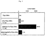
- the invention thus relates to a preferred Aspect of a yeast Ty particle, composed of TyA protein units, those with a membrane active Peptide sequence are modified.
- the Ty particle with the peptide sequence Gly Leu Phe Glu Ala Ile Glu Gly Phe Ile Glu Asn Gly Trp Glu Gly Leu Ala Glu Ala Leu Ala Glu Ala Leu Glu Ala Leu Ala Ala Gly Gly Ser modified at the carboxy terminus of the TyA protein located.
- the yeast Ty particles according to the invention are obtained by making a DNA encoding the TyA protein that a membrane-active peptide sequence at the carboxy terminus , expresses, unlocks the host cells and the capsid is harvesting.
- the expression of the modified TyA sequence e.g. in yeast or bacteria modified Ty particles because of their size and Density can be cleaned.
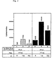
- the cleaned endosomolytic Ty particles were in the Within the scope of the present invention biotinylated and with Streptavidin polylysine and transferrin polylysine as well of the DNA to be transported into the cell Transfection complexes united.
- endosomolytic Ty particles coupled directly to polylysine using transglutaminase and with transferrin-polylysine conjugates and the DNA ternary transfection complexes.
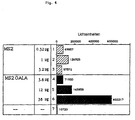
- Another preferred embodiment of the present invention is thus an MS2 particle, composed. from MS2 capsid protein units, the modified with a membrane-active peptide sequence are.
- the membrane active peptide is in the ⁇ -hairpin loop region between amino acid 11 (Asp) and amino acid 17 (Asp), especially between Amino acid 14 (Gly) and 15 (Thr) of the MS2 capsid protein inserted.
- Another possible insertion point for the membrane-active peptide lies in the C-terminal region of the MS2 capsid protein.
- the endosomolytic MS2 particle with the peptide sequence GALA, which lies between amino acid 14 and amino acid 15 of the MS2 capsid protein is inserted, modified.
- the MS2 particles according to the invention are preferred obtained by inserting the for that modified membrane-active peptide coding sequence Capsid protein DNA expressed, and the obtained modified capsid protein denatured and under Associate denaturant removal is left.
- the cleaned endosomolytic MS2 particles were biotinylated in the context of the present invention and with streptavidin polylysine and transferrin polylysine and the DNA to be transported into the cell Complexes united.
- the particles according to the invention in addition to the / the membrane active peptide (s) a peptide sequence with the Function of a ligand for the target cell.
- a Internalization function conferred this Peptide sequence is hereinafter referred to as "ligand peptide" designated.
- the best characterized ligand peptide is the arginine-glycine-aspartic acid sequence (RGD), which was found in various integrin-binding cell adhesion proteins, such as fibronectin, fibrinogen, from Willebrand factor and vitronectin (Pierschbacher and Ruoslahti, 1984; 1987).
- RGD arginine-glycine-aspartic acid sequence
- An RGD motif present in the penton base of type 2 and type 5 adenovirus has been shown to play a role in the internalization of the virus (Wickham et al., 1993).
- Such a short one containing the RGD motif Ligand peptide sequence can be inserted into capsid proteins in order to obtain particles according to the invention which have a cell-binding motif on their surface.
- the ability e.g. of the MS2 capsid protein Urea-denatured monomers themselves assembling can also be used to To produce MS2 particles that have more than one Show foreign domain. The prerequisite for this is also in this case that these insertions have the ability of Do not interfere with self-assembly particles.
- a membrane-active domain e.g. the GALA motif
- a cell binding domain e.g.
- the RGD motif is preferably so proceeded that on the one hand capsid monomers with a membrane-active modification and on the other hand with a ligand modification are produced, and the two differently modified, denatured Monomers in a defined ratio mixed and the denaturant is removed to the association of the modified proteins enable virus-like particles.
- Ligand peptides introduced into the capsid monomers become; Examples are small ones Peptide growth factors and hormones like that EGF (epidermal growth factor) peptide, insulin, the costimulatory Molecule HSA "Heat Stable Antigen” (Kay et al., 1990), also peptides from so-called superantigens, encoded by mouse mammary tumor virus (Torres et al., 1993).
- EGF epidermal growth factor
- HSA Heat Stable Antigen
- the particles according to the invention with a nucleic acid binding Domain especially an organic one polycationic compound such as polylysine.
- a nucleic acid binding Domain especially an organic one polycationic compound such as polylysine.
- virus-like particles to the membrane - active endosomolytic peptides and optionally the cell-binding ligand motifs Domains that have the ability to attach to nucleic acids tie.
- These particles that contain a DNA binding domain can be made by the capsid subsequently with a DNA binding substance such as Polylysine is conjugated.
- the conjugation of the capsid e.g. with polylysine can after for coupling peptides with Polyamine compounds are carried out in a manner known per se, e.g. by chemical means, through coupling via a Biotin-streptavidin bridge or by direct binding of the polylysine to the capsid by means of transglutaminase.
- the same procedure can be used as in the WO 93/07283 for the coupling of polylysine to viruses or virus components.
- capsids As an alternative to the subsequent conjugation of capsids with a DNA binding peptide, the modification of the capsid proteins with a DNA binding domain too directly, i.e. by expression of a chimeric DNA sequence consisting of one for the capsid protein coding DNA sequence, and one for the DNA binding Peptide coding sequence.
- This method of production binds to the DNA binding peptides that also for the other foreign domains current requirement that their presence on the capsid protein's ability to order Assembling structures, not affected.
- DNA binding motifs after expression the chimeric capsid DNA on the invention Particles are cationic polypeptides, e.g. the homologues polylysine, polyarginine, or peptides, the of naturally occurring DNA-binding proteins, such as Histones, core proteins of adenovirus (e.g. protein V, protein VII and the 13 kd protein L211K) or Protamines are derived.
- cationic polypeptides e.g. the homologues polylysine, polyarginine, or peptides
- the of naturally occurring DNA-binding proteins such as Histones, core proteins of adenovirus (e.g. protein V, protein VII and the 13 kd protein L211K) or Protamines are derived.
- the production of particles according to the invention the have more than one foreign domain, e.g. several membrane active domains or a membrane active domain in Connection with a ligand and / or a DNA binding Domain, can be separated into two or more, same or different expression systems getting produced.
- a foreign domain e.g. several membrane active domains or a membrane active domain in Connection with a ligand and / or a DNA binding Domain
- a capsid protein monomer with a membrane active domain e.g. the peptide GALA
- a capsid monomer with a ligand domain e.g. the RGD motif
- the Monomers are mixed in the desired ratio, so that these become orderly structures assemble.
- the optimal mixing ratio will be determined empirically.
- the particles of the invention are as endosomolytic agents in compositions for the Gene transfer used, as described in WO 93/07283 are described.
- the invention thus relates in a further aspect a composition for the transport of nucleic acid into the higher eukaryotic cell, in which the Nucleic acid is complexed with endosomolytic effective virus-like particles consisting of modified units of capsid proteins of viruses or virus-like particles, the Capsid protein units membrane active peptidic Sequences and polycationic sequences for binding of the nucleic acid.
- the Gene transfer complexes in addition to those of the invention endosomolytic particles that have a nucleic acid binding domain have a conjugate in which a Nucleic acid binding domain, generally the same like that of the particle, with one Internalization factor for the transfected Target cell is coupled.
- These ternary complexes or Combination complexes are mainly used if the endosomolytic particle is not by itself can penetrate into the target cell, i.e. if it's not in native form can penetrate into the cell and also not with a ligand domain for the target cell was modified.
- this embodiment can also be used when the ligand function of a particle according to the invention by a additional ligand function should be added.
- Transfection complexes consisting of DNA, the particle according to the invention conjugated with polylysine, and a trarisferrin-polylysine conjugate.
- an additional Nucleic acid binding substance especially polylysine, be contained in non-conjugated form to the Condense nucleic acid.
- Internalizing factor conjugate included Nucleic acid binding domain the function of attachment to the nucleic acid without losing all of the to saturate negative charges.
Landscapes
- Health & Medical Sciences (AREA)
- Life Sciences & Earth Sciences (AREA)
- Genetics & Genomics (AREA)
- Engineering & Computer Science (AREA)
- Bioinformatics & Cheminformatics (AREA)
- Chemical & Material Sciences (AREA)
- Zoology (AREA)
- Wood Science & Technology (AREA)
- Organic Chemistry (AREA)
- General Engineering & Computer Science (AREA)
- Biotechnology (AREA)
- Biomedical Technology (AREA)
- General Health & Medical Sciences (AREA)
- Medicinal Chemistry (AREA)
- Virology (AREA)
- Public Health (AREA)
- Physics & Mathematics (AREA)
- Animal Behavior & Ethology (AREA)
- Biophysics (AREA)
- Epidemiology (AREA)
- Pharmacology & Pharmacy (AREA)
- Molecular Biology (AREA)
- Plant Pathology (AREA)
- Microbiology (AREA)
- Veterinary Medicine (AREA)
- Hematology (AREA)
- Biochemistry (AREA)
- Cell Biology (AREA)
- Peptides Or Proteins (AREA)
- Micro-Organisms Or Cultivation Processes Thereof (AREA)
- Physical Or Chemical Processes And Apparatus (AREA)
- Preparation Of Compounds By Using Micro-Organisms (AREA)
- Manufacturing Of Micro-Capsules (AREA)
- Processes Of Treating Macromolecular Substances (AREA)
Abstract
Description
Die Erfindung bezieht sich auf das Einbringen von Nukleinsäuren in höhere eukaryotische Zellen.The invention relates to the introduction of Nucleic acids in higher eukaryotic cells.
In den letzten Jahren hat der therapeutische Ansatz der Gentherapie für die Behandlung zahlreicher Erkrankungen an Interesse gewonnen. Die Gentherapie wird eingesetzt, um in vivo therapeutisch wirksame Genprodukte zu synthetisieren, wodurch z.B. im Falle eines genetischen Defekts die Funktion des fehlenden Gens ersetzt wird. Beispiele für genetisch bedingte Erkrankungen, bei denen die Gentherapie einen erfolgversprechenden Ansatz darstellt, sind Hämophilie, beta-Thalassämie und "Severe Combined Immune Deficiency" (SCID), ein Syndrom, das durch einen genetisch bedingten Mangel des Enzyms Adenosindeaminase hervorgerufen wird. Anwendungsmöglichkeiten bestehen ferner bei der Immunregulation, mittels einer Impfung eine humorale oder intrazelluläre Immunität erzielt wird. Weitere Beispiele für genetische Defekte, bei denen eine Verabreichung von Nukleinsäure, die für das defekte Gen kodiert, z.B. in individuell auf den Bedarf abgestimmter Form verabreicht werden kann, sind Muskeldystrophie (Dystrophin-Gen), Cystische Fibrose ("Cystic fibrosis transmembrane conductance regulator gene"), Hypercholesterolämie (LDL-Rezeptor-Gen). Gentherapeutische Behandlungsmethoden können auch eingesetzt werden, wenn Hormone, Wachstumsfaktoren oder cytotoxisch oder immunmodulierend wirkende Proteine im Organismus synthetisiert werden sollen.In recent years, the therapeutic approach has Gene therapy for the treatment of numerous diseases gained in interest. Gene therapy is used to produce therapeutically effective gene products in vivo synthesize, e.g. in the case of a genetic Defect the function of the missing gene is replaced. Examples of genetic diseases, at for whom gene therapy is a promising approach represents are hemophilia, beta-thalassemia and Severe Combined Immune Deficiency (SCID) Syndrome caused by a genetic deficiency of the Enzyme adenosine deaminase is produced. Applications also exist in the Immune regulation, by means of vaccination a humoral or intracellular immunity is achieved. Further Examples of genetic defects where one Administration of nucleic acid necessary for the defective gene encoded, e.g. in individual to the need can be administered in a coordinated form Muscular dystrophy (dystrophin gene), cystic fibrosis ("Cystic fibrosis transmembrane conductance regulator gene "), hypercholesterolemia (LDL receptor gene). Gene therapy treatments can also used when hormones, growth factors or cytotoxic or immunomodulating proteins in Organism to be synthesized.
Die Gentherapie stellt ferner einen erfolgversprechenden Ansatz für die Behandlung von Krebs dar, wobei sog. "Krebsvakzine" verabreicht werden. Um die Immunogenizität von Tumorzellen zu erhöhen, werden diese verändert, um sie entweder stärker antigenisch zu machen, oder um sie zu veranlassen, bestimmte immunmodulierende Substanzen zu erzeugen, z.B. Zytokine, die dann eine Immunantwort auslösen. Um dies zu bewirken, werden die Zellen mit DNA transfiziert, die für ein Zytokin, z.B. IL-2, IL-4, IFN-gamma, TNF-α, kodiert. Die am weitesten fortgeschrittenen Techniken für den Gentransfer in autologe Tumorzellen benützen virale Vektoren.Gene therapy also provides one promising approach to the treatment of Cancer, whereby so-called "cancer vaccine" administered become. To improve the immunogenicity of tumor cells increase, these are changed to either to make it more antigenic, or to make them too cause certain immunomodulating substances to generate, e.g. Cytokines, which then have an immune response trigger. To do this, the cells are using DNA transfected for a cytokine, e.g. IL-2, IL-4, IFN-gamma, TNF-α, encoded. The farthest advanced techniques for gene transfer in autologous tumor cells use viral vectors.
Nukleinsäuren als therapeutisch wirksame Substanzen kommen außerdem zur Anwendung, um bestimmte Zellfunktionen zu inhibieren, z.B. haben sich Antisense RNAs und -DNAs oder Ribozyme als wirksame Mittel für die selektive Inhibierung bestimmter Gensequenzen erwiesen.Nucleic acids as therapeutically active substances also apply to certain Inhibit cell functions, e.g. have antisense RNAs and DNAs or ribozymes as effective agents for the selective inhibition of certain gene sequences proved.
In jüngerer Zeit wurden Gentransfersysteme entwickelt,
die die Beschränkungen der retroviralen und
adenoviralen Vektoren umgehen und deren
Sicherheitsrisken, die aufgrund des Co-Transfers von
lebensfähigen viralen Genelementen des Ursprungsvirus
bestehen, ausschalten. Diese Gentransfersysteme beruhen
auf Mechanismen, deren sich die Zelle für den Transport
von Makromolekülen bedient, z.B. auf dem äußerst
leistungsfähigen Weg der Rezeptor-vermittelten
Endozytose (Wu und Wu, 1987; EP-Al 0 388 758;
WO 91/17773, WO 92/17210 und WO 92/19281). Mit Hilfe
dieser Methode, die sich bifunktioneller molekularer
Konjugate bedient, die eine DNA-Bindungsdomäne und eine
Domäne mit Spezifität für einen Zelloberflächenrezeptor
aufweisen, konnten hohe Gentransferraten erzielt
werden.More recently, gene transfer systems have been developed that circumvent the limitations of the retroviral and adenoviral vectors and eliminate the security risks that arise from the co-transfer of viable viral gene elements from the original virus. These gene transfer systems are based on mechanisms which the cell uses for the transport of macromolecules, for example on the extremely powerful route of receptor-mediated endocytosis (Wu and Wu, 1987; EP-Al 0 388 758;
WO 91/17773, WO 92/17210 and WO 92/19281). With the help of this method, which uses bifunctional molecular conjugates, which have a DNA binding domain and a domain with specificity for a cell surface receptor, high gene transfer rates could be achieved.
Da der Gentransfer auf physiologischem Weg, wie ihn die Rezeptor-vermittelte Endozytose mittels Nukleinsäure-Komplexen darstellt, große Vorteile aufweist (nichttoxischer Mechanismus des Durchtritts durch die Zellmembran; Möglichkeit der Verabreichung biologisch aktiver Nukleinsäuren, auf repetitiver oder kontinuierlicher Basis; Möglichkeit des zellspezifischen Targeting; Herstellbarkeit der Konjugate in großen Mengen), besteht das Bedürfnis, dieses System effizienter zu machen.Since the gene transfer in a physiological way, as the Receptor-mediated endocytosis using nucleic acid complexes has great advantages (non-toxic Mechanism of passage through the cell membrane; Possibility of biological administration active nucleic acids, on repetitive or continuous basis; Possibility of cell-specific targeting; Manufacturability of the Conjugates in large quantities), there is a need to make this system more efficient.
Bei der Anwendung der Gentransfertechniken, die auf dem Prinzip der Rezeptor-vermittelten Endozytose beruhen, stellte sich heraus, daß ein limitierender Faktor des Systems der Abbau des Genmaterials in der Zelle nach dessen Freisetzung aus den Endosomen ist. Eine wesentliche Verbesserung des Systems wurde daher durch eine Technik erzielt, die die Fähigkeit von bestimmten Viren und Viruskomponenten ausnützt, Endosomen aufbrechen zu können. Mit Hilfe eines Zusatzes dieser endosomolytischen Mittel konnte eine erhebliche Steigerung der Expressionsraten der in die Zelle importierten Gene erzielt werden (Wagner et al., 1991a und 1991b; Cotten et al., 1992; Wagner et al., 1992a und 1992b; Zatloukal et al., 1992; Cotten et al., 1993a und 1993b; Curiel et al. 1991; WO 93/07283 und WO 93/07282).When applying the gene transfer techniques based on the Principle of receptor-mediated endocytosis based, it turned out that a limiting factor of the Systems of degradation of the genetic material in the cell after whose release is from the endosomes. A significant improvement of the system was therefore made by a technique that achieves the ability of certain Takes advantage of viruses and virus components, endosomes to be able to break up. With the help of an addition this endosomolytic agents could be significant Increase in expression rates in the cell imported genes can be achieved (Wagner et al., 1991a and 1991b; Cotten et al., 1992; Wagner et al., 1992a and 1992b; Zatloukal et al., 1992; Cotten et al., 1993a and 1993b; Curiel et al. 1991; WO 93/07283 and WO 93/07282).
Als endosomolytische Agentien wurden neben Viren oder Viruskomponenten u.a. synthetische Peptide vorgeschlagen, die von viralen, pH-abhängigen, membranaktiven Peptiden, wie z.B. dem Influenza A Hämagglutinin-Fusionspeptid, abgeleitet sind. Synthetische Transfektionskomplexe, enthaltend entweder das Influenzapeptid (WO 93/07283, Wagner et al., 1992) oder verschiedene Peptide auf der Grundlage des GALA-Peptids (Subbarao et al., 1987; Parente et al., 1990; und WO 93/07283) zeigten die Brauchbarkeit dieser Peptide. In addition to viruses or Virus components etc. synthetic peptides suggested that of viral, pH-dependent, membrane active peptides, e.g. the influenza A Hemagglutinin fusion peptide. Synthetic transfection complexes containing either the influenza peptide (WO 93/07283, Wagner et al., 1992) or various peptides based on the GALA peptide (Subbarao et al., 1987; Parente et al., 1990; and WO 93/07283) showed the usability of these Peptides.
Die Anwendung synthetischer membranaktiver Peptide als endosomolytische Mittel ist jedoch begrenzt. Dies könnte daran liegen, daß sie in den wahllos angeordneten chemisch oder ionisch gebundenen Gentransferkomplexen möglicherweise nicht immer in einer Form zugänglich vorliegen, die die Ausübung ihrer Funktion ermöglicht.The application of synthetic membrane active peptides as however, endosomolytic agents are limited. This could be because they are in the indiscriminate arranged chemically or ionically bound Gene transfer complexes may not always be in available in a form that allows the exercise of their Function enables.
Den zufällig angeordneten Konjugaten aus synthetischen Peptiden und Polylysin, die für die Komplexierung und Kondensierung der DNA-Moleküle verwendet wurden, fehlt eine geordnete dreidimensionale Strukturierung der endosomolytischen Funktion, wie sie das hinsichtlich seiner endosomolytischen Aktivität äußerst effiziente Adenoviruspartikels aufweist. Von der endosomolytischen Aktivität des Adenoviruspartikels wird angenommen, daß sie in der Pentonbasis lokalisiert ist (Seth et al., 1984), welche in einer definierten Kopienzahl an spezifischen Stellen auf der Oberfläche des Viruspartikels vorliegt (Stewart et al., 1993). Diese organisierte Anordnung könnte eine Funktion im Hinblick auf die Erfordernisse für den Zusammenbau eines Viruspartikels haben, sie könnte aber ebenso eine Rolle bei der Steuerung der Wechselwirkungen des membranaktiven Motivs auf den Pentonproteinen mit der Endosomenmembran spielen.The randomly arranged synthetic conjugates Peptides and polylysine used for complexation and Condensation of the DNA molecules used was missing an orderly three-dimensional structuring of the endosomolytic function as it regards its endosomolytic activity extremely efficient Adenovirus particle. From the endosomolytic Activity of the adenovirus particle is thought to be it is located in the penton base (Seth et al., 1984), which in a defined number of copies specific locations on the surface of the Virus particle is present (Stewart et al., 1993). This organized arrangement could be a function with regard on the requirements for assembling one Virus particles, but they could also play a role in controlling the interactions of the membrane-active motif on the penton proteins with the Play endosome membrane.
Der vorliegenden Erfindung lag die Aufgabe zugrunde, endosomolytische Mittel bereitzustellen, die eine Verbesserung der Gentransfersysteme über Rezeptor-vermittelte Endozytose ermöglichen, indem sie hohe Expressionsraten bei weitestgehender. Ausschaltung von Sicherheitsrisken gewährleisten.The present invention was based on the object provide endosomolytic agents that a Improvement of gene transfer systems via receptor-mediated Allow endocytosis by high Expression rates with the most extensive. Elimination of Ensure security risks.
Bei der Lösung der gestellten Aufgabe wurde von der Überlegung ausgegangen, Verbesserungen der endosomolytischen Eigenschaften der aus dem Stand der Technik bekannten membranaktiven Peptide dadurch zu erzielen, daß man sie in einer geordneten Form zusammenbaut oder anordnet, indem man sie auf einem Protein anbringt, das die Fähigkeit hat, sich selbst zu Teilchen mit geordneten Strukturen zusammenzubauen.In solving the task, the Considered improvements to the endosomolytic properties of the prior art Technology known membrane-active peptides achieve that they are in an orderly form assembles or arranges by placing them on one Attaches protein that has the ability to self Assemble particles with ordered structures.
Im Zuge der Realisierung der vorliegenden Erfindung wurden zunächst die leeren Kapside getestet, wie sie in normalen Adenovirusinfektionen gefunden werden (Daniell, 1976). Es zeigte sich, daß leere Adenoviruskapside keine endosomolytische Fähigkeit aufweisen; diese dürfte erst durch die proteolytische Prozessierung, die in einer späten Phase der Virusreifung stattfindet, aktiviert werden (Weber, 1976). Diese Annahme ist im Einklang damit, daß ein Adenovirusstamm, der hinsichtlich der Prozessierung temperaturempfindlich ist (Ad2 ts1; Weber, 1976) bei der restriktiven Temperatur unreife Teilchen produziert, die nicht fähig sind, die Freisetzung von co-endozytiertem Material auszulösen (Defer et al., 1990). In Vorversuchen zur vorliegenden Erfindung wurde außerdem festgestellt, daß diese Partikel nicht die Fähigkeit haben, den DNA-Transport zu verbessern oder Liposomen aufzubrechen. Falls dieses bei Adenoviren beobachtete Merkmal ein allgemeines Charakteristikum der Virusreifung ist, ist zu erwarten, daß leere Kapside nicht die endosomolytische Aktivität der ganzen reifen Viruspartikel aufweisen.In the course of realizing the present invention the empty capsids were first tested as they are in normal adenovirus infections can be found (Daniell, 1976). It turned out to be empty Adenovirus capsid has no endosomolytic ability exhibit; this is likely only due to the proteolytic Processing that is in a late phase of Virus maturation takes place, activated (Weber, 1976). This assumption is consistent with the fact that a Adenovirus strain that is related to processing is temperature sensitive (Ad2 ts1; Weber, 1976) at the restrictive temperature of immature particles produces that are unable to release trigger co-endocytosed material (Defer et al., 1990). In preliminary experiments on the present invention also found that these particles are not the Ability to improve DNA transport or Breaking up liposomes. If this is the case with adenoviruses observed characteristic a general characteristic of virus maturation is expected to be empty Capsids not the endosomolytic activity of the whole mature virus particles.
Die vorliegende Erfindung betrifft ein endosomolytisch wirksames virusähnliches Partikel, das aus Einheiten von Kapsidproteinen, abgeleitet von Kapsidproteinen von Viren oder virusähnlichen Teilchen, zusammengesetzt ist, wobei die Kapsidproteinheiten mit einer membranaktiven peptidischen Sequenz modifiziert sind. The present invention relates to an endosomolytic effective virus-like particle made up of units of capsid proteins derived from capsid proteins of Viruses or virus-like particles is, the capsid prot units with a membrane-active peptide sequence are modified.
Das Gerüst der erfindungsgemäßen Partikel ist ein leeres Viruskapsid oder ein kapsidartiges Partikel aus Proteinen von Viren oder virusähnlichen Teilchen, wie Bakteriophagen oder Hefe-Transposons. Die Proteine, die das Kapsidgerüst bilden, werden im Rahmen der vorliegenden Erfindung als "Kapsidproteine" bezeichnet.The skeleton of the particles according to the invention is a empty virus capsid or a capsid-like particle Proteins from viruses or virus-like particles, such as Bacteriophages or yeast transposons. The proteins that forming the capsid framework are part of the present invention referred to as "capsid proteins".
Die membranaktive Peptidsequenz ist auf den Kapsidproteineinheiten so angeordnet, daß gewährleistet ist, daß sie am Ort ihrer Wirksamkeit, also in der Zelle, funktionell verfügbar ist: die membranaktiven peptidischen Sequenzen liegen entweder an der Oberfläche der Partikel frei oder sind in einer Weise in der Oberflächenstruktur angeordnet, daß ihre Freilegung durch Ereignisse in der Zelle, wie Proteolyse, pH-Wertänderung oder Änderung des Redoxpotentials, freigelegt wird. Die membranaktive Funktion der Peptide, die u.a. durch ihre Zugänglichkeit bestimmt wird, äußert sich in ihrer endosomolytischen Aktivität. Diese schlägt sich in der Steigerung der Gentransferkapazität nieder und kann mittels Gentransferexperimenten getestet werden.The membrane active peptide sequence is on the Capsid protein units arranged to ensure is that it is in the place of its effectiveness, ie in the Cell that is functionally available: the membrane-active peptide sequences are either on the Surface of the particles are free or in a way arranged in the surface structure that their Exposure to events in the cell, such as Proteolysis, pH change or change in Redox potential, is exposed. The membrane active Function of the peptides, which include through her Accessibility is determined, is expressed in their endosomolytic activity. This is reflected in the Increase in gene transfer capacity and can be tested using gene transfer experiments.
Leere Kapside von einfachen Viren ohne Hülle sind von einer Reihe von Viren verfügbar. Die natürlichen Kapside bestehen im allgemeinen aus ein bis drei Proteinen. Die Herstellbarkeit der Kapsidproteine in großen Mengen, z.B. im Baculovirussystem, und deren Fähigkeit zum Selbstzusammenbau haben die Gewinnung von virusähnlichen Partikeln mit geordneten Strukturen, ähnlich denen des nativen Virus, ermöglicht. In den meisten Fällen sind die Partikel frei von Nukleinsäure. (Leere Kapside, die von sich aus endosomolytische Aktivität aufweisen, die die Aufnahme und die Expression von in die Zelle transportierter DNA verbessert, können als solche ohne weitere Modifikation des Partikels verwendet werden, wie in der WO 93/07283 vorgeschlagen wurde. Ein Beispiel dafür sind die leeren Kapside des Parvovirus B19, die z.B. durch Baculovirusexpression erhalten werden können.)Empty capsids of simple enveloped viruses are from a number of viruses are available. The natural ones Capsids generally consist of one to three Proteins. The manufacturability of the capsid proteins in large amounts, e.g. in the baculovirus system, and their Have the ability to assemble yourself virus-like particles with ordered structures, similar to that of the native virus. In the in most cases the particles are free of nucleic acid. (Empty capsids, inherently endosomolytic Have activity that includes recording and Expression of DNA transported into the cell can be improved as such without further modification of the particle can be used, as in WO 93/07283 was proposed. The empty ones are an example of this Capsids of parvovirus B19, e.g. by Baculovirus expression can be obtained.)
Die erfindungsgemäßen Partikel sind von Kapsidstrukturen abgeleitet, die von sich aus die zur Steigerung der Effizienz des Gentransfers erforderliche endosomolytische Aktivität nicht oder nicht in ausreichendem oder gewünschtem Ausmaß aufweisen.The particles according to the invention are from Capsid structures derived, which inherently to the Required to increase the efficiency of gene transfer endosomolytic activity not or not in have sufficient or desired extent.
Die das Gerüst bildenden Ausgangspartikel können natürlichen Ursprungs sein, in diesem Fall werden sie insbesondere von Virusinfektionen erhalten.The initial particles forming the framework can of natural origin, in which case they will especially received from viral infections.
Vorzugsweise werden die Partikel auf rekombinantem Weg hergestellt, indem die, gegebenenfalls modifizierten, Kapsidproteine exprimiert und gereinigt und, falls sie nicht bereits in assoziierter Form vorliegen, anschließend assoziieren gelassen werden.The particles are preferably recombinant produced by the, optionally modified, Capsid proteins are expressed and purified and, if they are not already in an associated form, then be associated.
Wenn nicht-modifizierte Kapsidproteine exprimiert werden, können die nach ihrer Assoziation erhaltenen Partikel, die das Gerüst bilden, nachträglich an ihrer Oberfläche mit den membranaktiven Peptiden modifiziert werden, z.B. auf chemischem Weg durch Kopplung mit synthetischen membranaktiven Peptiden. Die Kopplung des membranaktiven Peptids an das Kapsidgerüst kann in für die Kopplung von Peptiden an sich bekannter Weise erfolgen, z.B. auf chemischem Weg, wobei, falls erforderlich, die Einzelkomponenten vor der Kopplungsreaktion mit Linkersubstanzen versehen werden. Die Kopplung kann z.B. erfolgen über Disulfidbrücken, die unter reduzierenden Bedingungen wieder gespalten werden können (z.B. bei Kopplung mittels Succinimidylpyridyldithiopropionat; Jung et al., 1981).When expressing unmodified capsid proteins can be obtained after their association Particles that form the scaffold subsequently on their Surface modified with the membrane-active peptides e.g. chemically by coupling with synthetic membrane active peptides. The coupling of the membrane-active peptide on the capsid framework can be used for the coupling of peptides in a manner known per se take place, e.g. by chemical means, whereby if required the individual components before the Coupling reaction with linkers. The coupling can e.g. take place via disulfide bridges, which split again under reducing conditions can be used (e.g. when connecting using succinimidyl; Jung et al., 1981).
Falls das Kapsid geeignete Kohlenhydratketten aufweist, kann es mit dem Peptid über diese Kohlenhydratketten verbunden werden. Dabei kann nach der für die Herstellung von Glykoprotein-Polykation-Konjugaten in der WO 92/15281 beschriebenen Methode vorgegangen werden.If the capsid has suitable carbohydrate chains, it can with the peptide over these carbohydrate chains get connected. This can be done according to the Preparation of glycoprotein-polycation conjugates in the method described in WO 92/15281 become.
Ein weiteres Verfahren zur Herstellung der erfindungsgemäßen Partikel ist die enzymatische Kopplung des membranaktiven Peptids an das Gerüstkapsid durch eine Transglutaminase. Dabei kann nach der in der WO 93/07283 für die Kopplung von Polylysin an Adenovirus beschriebenen Methode vorgegangen werden. Voraussetzung hierfür ist, daß an Proteinen entsprechende Glutamine oder Lysine vorhanden sind, die von dem Enzym umgesetzt werden können.Another method of making the particles according to the invention is the enzymatic Coupling of the membrane-active peptide to the scaffold capsid by a transglutaminase. According to the in the WO 93/07283 for the coupling of polylysine Adenovirus described method can be followed. The prerequisite for this is that on proteins corresponding glutamines or lysines are present, which can be implemented by the enzyme.
Vorzugsweise werden die erfindungsgemäßen Partikel erhalten, indem die modifizierten Kapsidproteine mit Hilfe rekombinanter Methoden hergestellt werden.The particles according to the invention are preferred obtained by using the modified capsid proteins Can be produced using recombinant methods.
Die Erfindung betrifft somit in einem weiteren Aspekt ein Verfahren zur Herstellung von endosomolytisch wirksamen virusähnlichen Partikeln, wobei man eine für ein Kapsidprotein von Viren oder virusähnlichen Teilchen kodierende DNA, die mit einer für ein membranaktives Peptid kodierenden Sequenz modifiziert ist, exprimiert und das erhaltene Kapsidprotein, falls erforderlich, zu Kapsidstrukturen assoziieren läßt.The invention thus relates in a further aspect a method of making endosomolytic effective virus-like particles, one for a capsid protein from viruses or virus-like Particle encoding DNA, which with a for a modified membrane-active peptide coding sequence is expressed and the capsid protein obtained, if required to associate with capsid structures.
Die chimäre DNA, die eine für das Kapsidprotein und eine für das membranaktive Peptid kodierende Sequenz enthält, wird z.B. in mit Baculoviren transformierten Insektenzellen, in Hefe oder in Bakterien, exprimiert. Das erhaltene, durch eine membranaktive Peptiddomäne modifizierte Kapsidprotein kann z.B. assoziieren gelassen werden, nachdem die überexprimierten Kapsidproteinmonomeren denaturiert, gereinigt und das Denaturierungsmittel entfernt wurden. Als Denaturierungsmittel kommen insbesondere Harnstoff oder Guanidinhydrochlorid, gegebenenfalls in Gegenwart milder Detergentien und/oder Reduktionsmittel, in Betracht. Eine Denaturierung ist dann nicht erforderlich, wenn sich die modifizierten Kapsidproteine bereits im Wirtsorganismus zu Kapsidstrukturen anordnen, wie dies offensichtlich beim Hefe-Ty-Partikel der Fall ist. In diesem Fall werden die Wirtszellen mechanisch aufgebrochen und die fertigen Kapsid-Teilchen geerntet.The chimeric DNA, one for the capsid protein and a sequence coding for the membrane-active peptide contains, e.g. transformed with baculoviruses Insect cells, expressed in yeast or in bacteria. The obtained by a membrane active peptide domain modified capsid protein can e.g. associate be left after the overexpressed Capsid protein monomers denatured, purified and that Denaturants were removed. As Denaturants come in particular urea or Guanidine hydrochloride, optionally in the presence mild detergents and / or reducing agents, in Consideration. A denaturation is not then required if the modified Capsid proteins already in the host organism Arrange capsid structures, as is evident with the Yeast Ty Particle is the case. In this case the host cells are broken up mechanically and the finished capsid particles are harvested.
Bezüglich des Expressionssystems, von dem eine große Auswahl zur routinemäßigen Verwendung zur Verfügung steht, bestehen keine Beschränkungen. Bevorzugt wird ein System eingesetzt, das die Expression der modifizierten Kapsidproteine in großen Mengen ermöglicht; wegen ihrer Effizienz werden im allgemeinen bakterielle Expressionssysteme bevorzugt. Ein Beispiel für ein im Rahmen der vorliegenden Erfindung geeignetes bakterielles Expressionssystem ist das von Studier et al., 1990, beschriebene. Ein Beispiel für ein geeignetes Hefe-Expressionssystem ist das von Emr, 1990, beschriebene; auch Baculovirussysteme, die für die Herstellung von Kapsidproteinen schon verschiedentlich herangezogen wurden, sind geeignet (z.B. O'Reilly et al., 1992). Es können konstitutive oder induzierbare Expressionssysteme verwendet werden. Durch Auswahl und gegebenenfalls Modifikation von vorhandenen Expressionssystemen kann gesteuert werden, in welcher Form die Kapside erhalten werden, z.B. kann eine Verringerung der Expressionsrate die Denaturierung und anschließende Resolubilisierung der Kapsidproteine überflüssig machen.Regarding the expression system, a large one of which Selection available for routine use there are no restrictions. Is preferred used a system that expresses the modified capsid proteins in large quantities permits; because of their efficiency in general bacterial expression systems preferred. An example for a suitable in the context of the present invention bacterial expression system is that of Studier et al., 1990. An example of a a suitable yeast expression system is that from Emr, 1990, described; also baculovirus systems that are used for the production of capsid proteins already were used differently are suitable (e.g. O'Reilly et al., 1992). It can be constitutive or inducible expression systems can be used. By selecting and, if necessary, modifying existing expression systems can be controlled in what form the capsids are obtained, e.g. can a decrease in the rate of expression denaturation and then resolubilization of the capsid proteins make redundant.
Die rekombinante Herstellung, bei der die Modifikation der Kapsidproteine im Zuge der Expression der entsprechend modifizierten DNA erfolgt, hat gegenüber der nachträglichen Modifikation von Kapsiden den Vorteil, daß die Lage der membranaktiven Domänen auf dem Protein und das Verhältnis Kapsidproteine zu membranaktiven Peptiden exakt definierbar ist.The recombinant manufacture, in which the modification of the capsid proteins in the course of the expression of the appropriately modified DNA has compared the subsequent modification of capsids Advantage that the location of the membrane-active domains the protein and the ratio of capsid proteins membrane-active peptides can be precisely defined.
Bei der rekombinanten Herstellung der modifizierten Kapsidproteine ist grundsätzlich zu beachten, daß die Gegenwart des membranaktiven Peptids, sei es aufgrund seiner Sequenz oder Anordnung, die Fähigkeit der exprimierten Kapsidproteine, sich zu geordneten Strukturen zusammenzufügen, nicht beeinträchtigt. Diese Forderung gilt auch hinsichtlich der Stelle im Kapsidprotein, in die das Peptid eingefügt ist.In the recombinant production of the modified It is important to note in principle that the Presence of the membrane active peptide, be it due to its sequence or arrangement, the ability of expressed capsid proteins to order themselves Put structures together, not affected. This The requirement also applies to the position in the Capsid protein in which the peptide is inserted.
Für den Fall, daß die Kapside aus mehr als einem Protein bestehen, können die Proteine co-exprimiert werden; z.B. im Fall von zwei Kapsidproteinen durch Co-Transformation des Wirtsorganismus mit zwei Plasmiden, die je eine Kapsidprotein-Sequenz tragen, oder durch Transformation des Wirts mit einem doppelt rekombinanten Vektor, der die beiden Sequenzen trägt, wie z.B. für die Expression des Kapsids des Parvovirus B19 in Insektenzellen mittels Baculoviren gezeigt wurde (Brown et al., 1991).In the event that the capsids consist of more than one Consist of protein, the proteins can be co-expressed become; e.g. in the case of two capsid proteins by co-transformation the host organism with two plasmids, each carrying a capsid protein sequence, or by Transformation of the host with a double recombinant vector that carries the two sequences, such as. for the expression of the capsid of the parvovirus B19 was shown in insect cells using baculoviruses (Brown et al., 1991).
Zu den derzeit verfügbaren, im Rahmen der vorliegenden Erfindung verwendbaren Kapsiden, deren Proteine im Baculovirus-System exprimiert werden und die sich zu Partikeln zusammensetzen können, zählen das Adenoassoziierte Virus, ein Dependovirus (Ruffing et al., 1992); "Aleutian Mink Disease Virus", ein autonomes Parvovirus (Christensen et al., 1993); Flock House Virus, ein Nodovirus (Schneeman et al., 1993), Papilloma Virus; Poliovirus (Urakawa et al., 1989); Norwalk Virus (Jiang et al., 1992) und Polyomavirus. Beispiele für weitere Kapsidproteine, die durch Einführen von Fremddomänen modifiziert werden können, sind das des L-A-Teilchens aus Hefe (Icho und Wickner, 1989; Wickner, 1993), der Bakteriophagen Qβ, GA, SP und anderer Phagen der Familie der Leviviridae, und des Bakteriophagen phi x 174 (Ackerman und DuBow, 1987).Among the currently available under this Capsids according to the invention, the proteins of which in Baculovirus system are expressed and that too Being able to put particles together counts the adeno-associated Virus, a dependovirus (Ruffing et al., 1992); "Aleutian Mink Disease Virus", an autonomous Parvovirus (Christensen et al., 1993); Flock House Virus, a nodovirus (Schneeman et al., 1993), Papilloma virus; Poliovirus (Urakawa et al., 1989); Norwalk Virus (Jiang et al., 1992) and Polyomavirus. Examples of other capsid proteins, which by Introduction of foreign domains can be modified are those of the L-A particle made of yeast (Icho and Wickner, 1989; Wickner, 1993), the bacteriophage Qβ, GA, SP and other phages of the Leviviridae family, and des Bacteriophage phi x 174 (Ackerman and DuBow, 1987).
Besonders gute Voraussetzungen bringen das Hefe-Ty-Teilchen und der MS2-Phage mit: Kingsman et al., 1991, haben gezeigt, daß geordnete Hefe-Ty-Partikel für die immunogene, multivalente Darbietung von Peptiden und kleinen Proteinen wie HIV gp120-Epitopen, Hepatitis B-Antigenen, etc. geeignet sind. Dies ist möglich, weil das Hauptprctein des Ty-Teilchens, das Produkt des TyA-Gens, die Fähigkeit besitzt, sich selbst zu 40 nm großen isometrischen Partikeln zusammenzusetzen (Burns et al., 1992). Kingsman et al. haben ein Fragment des Proteins identifiziert, das für diese Selbstzusammenbau-Aktivität ausreicht; Peptidsequenzen, die an den Carboxy-Terminus dieses Proteins angehängt werden, liegen an der Oberfläche des zusammengesetzten Partikels frei. Das MS2-Virus besitzt ein 24 nm quasiicosohedrales Kapsid, bestehend aus 180 Kopien des Haupt-Hüllproteins (13 kd), einer Kopie des Reifungsproteins (45 kd), und ein 3569 nt RNA-Genom. MS2 war der erste Organismus, dessen komplettes Genom sequenziert wurde (Fiers et al., 1976). Die Kristallstruktur dieses Virus wurde aufgeklärt und zeigte eine freiliegende β-Haarnadelschleife auf der Oberfläche jedes der 180 Kapsomeren (Valegard et al., 1990). Die nicht-fixierte Struktur dieser Schleife und deren Freiliegen an der Oberfläche wurde ausgenützt, um Partikel für die Antigenpräsentation zu bilden, indem Abschnitte aus 11 bis 26 Aminosäuren in diese Schleife eingefügt wurden (Mastico et al., 1993). Das MS2-Kapsidprotein toleriert diese Insertionen unter Beibehaltung seiner Fähigkeit, sich zu geordneten Kapsidstrukturen selbst zusammenzusetzen; das Kapsidprotein kann in Harnstoff denaturiert werden und baut sich nach dessen Entfernung zu nativen virusähnlichen Strukturen zusammen (Mastico et al., 1993). Dadurch wird die Zusammensetzung von Kapsiden, die an ihrer Oberfläche zahlreiche Peptidepitope tragen, ermöglicht.The yeast Ty particle is particularly good and the MS2 phage with: Kingsman et al., 1991, have shown that ordered yeast Ty particles for the immunogenic, multivalent presentation of peptides and small proteins like HIV gp120 epitopes, hepatitis B antigens, etc. are suitable. This is possible because the main protein of the Ty particle, the product of the TyA gene, has the ability to self at 40 nm large isometric particles (Burns et al., 1992). Kingsman et al. have a fragment of the Proteins identified that for this Self-assembly activity is sufficient; Peptide sequences which are attached to the carboxy terminus of this protein are lying on the surface of the composite Particles free. The MS2 virus has a 24 nm quasiicosohedral Capsid consisting of 180 copies of the Major coat protein (13 kd), a copy of the Maturing protein (45 kd), and a 3569 nt RNA genome. MS2 was the first organism to have its entire genome was sequenced (Fiers et al., 1976). The Crystal structure of this virus has been elucidated and showed an exposed β-hairpin loop on the Surface of each of the 180 capsomeres (Valegard et al., 1990). The unfixed structure of this loop and their surface exposure was exploited to To form particles for the antigen presentation by Sections of 11 to 26 amino acids in this loop were inserted (Mastico et al., 1993). The MS2 capsid protein tolerates these insertions under Maintaining his ability to get organized Assemble capsid structures yourself; the Capsid protein can be denatured in urea and builds up to native after its removal virus-like structures together (Mastico et al., 1993). This way the composition of capsids, the numerous peptide epitopes on its surface wear, enables.
An das Partikel im allgemeinen werden die Anforderungen gestellt, da3 es eine Größe von weniger als 100 nm, vorzugsweise weniger als 50 nm Durchmesser aufweisen soll, um die Aufnahme mittels Endozytose zu ermöglichen, daß es frei ist von überflüssiger, insbesondere infektiöser Nukleinsäure, die keine Funktion für den Gentransfer aufweist, und daß es die Voraussetzung mitbringt, daß ihm eine endosomolytische Aktivität (in Form von membranaktiven Peptiden oder kleinen Proteinen) verliehen werden kann, entweder auf chemischem Weg oder mittels genetischer Manipulation.The requirements for the particle in general that it is less than 100 nm in size, preferably have a diameter of less than 50 nm is said to increase uptake by endocytosis allow it to be free of unnecessary, especially infectious nucleic acid that does not Has function for gene transfer, and that it is the Prerequisite is that he has an endosomolytic Activity (in the form of membrane active peptides or small proteins) can be conferred on either chemical way or by means of genetic manipulation.
An rekombinant herzustellende Kapside im besonderen
werden die folgenden Anforderungen gestellt:
Das Kapsid soll eine möglichst einfache Struktur,
vorzugsweise bestehend aus maximal drei Untereinheiten,
aufweisen. Die Untereinheiten sollen sich entweder
bereits im Wirtsorganismus, in dem sie exprimiert
werden, von selbst assoziieren oder, falls sie nach
Überexpression in unlöslicher Form vorliegen,
resolubilisierbar sein und sich von selbst zu
Kapsidstrukturen zusammensetzen. Der Einfachheit halber
wird von Kapsidproteinen ausgegangen, deren Gene in
klonierter Form verfügbar sind, andernfalls muß die
Klonierung erst vorgenommen werden.
Im Hinblick auf die Insertionsstelle für die Einfügung
der membranaktiven Peptidsequenz wird bevorzugt von
Kapsidstrukturen ausgegangen, deren 3D-Struktur
(Kristallstruktur) mittels Röntgenstrukturanalyse
aufgeklärt wurde. An den Sequenzbereich im
Kapsidprotein, in dem die Anbringung der membranaktiven
Peptidsequenz vorgenommen wird, wird generell die
Anforderung gestellt, daß er für die Ausbildung der
Kapsidstruktur bzw. den Zusammenbau keine Funktion hat
bzw. daß ihre Funktion durch die Einfügung einer
Peptidstruktur nicht behindert wird. Wenn die
Kristallstruktur verfügbar ist, wird diese bei der
Festlegung der Stelle für die Einfügung der Peptiddomäne
herangezogen: Zeigt die Kristallstruktur, daß das Virus
oder virusähnliche Partikel an seiner Oberfläche
Schleifen hat, wie z.B. der MS2-Phage, so werden
bevorzugt diese Schleifen, die für die Ausbildung der
Kapsidstruktur nicht erforderlich sind, für die
Modifikation herangezogen.
Ist die Kristallstruktur nicht bekannt, kann bei der
Bestimmung der Insertionsstelle empirisch vorgegangen
werden: Aus der Aminosäuresequenz kann abgeleitet
werden, ob bestimmte Bereiche gegebenenfalls die
Grundlage für die Ausbildung von Strukturelementen, wie
α-helikalen oder von β-Faltblattstrukturen, darstellen.
Sofern Informationen über Deletionen oder künstliche
oder natürliche Mutationen von Virusproteinen einen
Rückschluß auf die Existenz nicht-konservierter
Regionen zulassen, können die nicht konservierten
Regionen, die für die Struktur nicht erforderlich sind,
wie z.B. die Schleifen des MS2-Phagen, für die
Modifikation benützt werden. The capsules to be produced recombinantly in particular have the following requirements: The capsid should have the simplest possible structure, preferably consisting of a maximum of three subunits. The subunits are said to either already associate themselves in the host organism in which they are expressed or, if they are in insoluble form after overexpression, to be resolubilizable and to self-assemble into capsid structures. For the sake of simplicity, capsid proteins are assumed whose genes are available in cloned form, otherwise the cloning must first be carried out.
With regard to the insertion point for the insertion of the membrane-active peptide sequence, capsid structures are preferably used, the 3D structure (crystal structure) of which has been elucidated by means of X-ray structure analysis. The sequence region in the capsid protein in which the membrane-active peptide sequence is attached is generally subject to the requirement that it has no function for the formation of the capsid structure or the assembly or that its function is not hindered by the insertion of a peptide structure. If the crystal structure is available, this will be used when determining the location for the insertion of the peptide domain: If the crystal structure shows that the virus or virus-like particles has loops on its surface, such as the MS2 phage, these loops are preferred are not required for the formation of the capsid structure, used for the modification.
If the crystal structure is not known, the insertion site can be determined empirically: the amino acid sequence can be used to determine whether certain areas may form the basis for the formation of structural elements such as α-helical or β-sheet structures. Insofar as information about deletions or artificial or natural mutations of virus proteins allows conclusions to be drawn about the existence of non-conserved regions, the non-conserved regions that are not required for the structure, such as the loops of the MS2 phage, can be used for the modification ,
Bei der Auswahl von Kapsidgerüsten und membranaktiven Peptiden sowie der Bestimmung der Insertionsstelle der Peptidsequenz für die Konstruktion der erfindungsgemäßen Partikel wird z.B. wie folgt vorgegangen: Die für das bzw. die Kapsidprotein(e) kodierende(n) DNA-Sequenz(en) werden, unter der Kontrolle geeigneter Expressionskontrollsequenzen, in einen Vektor eingebracht und in einem geeigneten Wirt exprimiert. Parallel dazu werden Vektoren zur Expression gebracht, in denen jeweils die Kapsidproteinsequenz mit einer für ein membranaktives Peptid kodierenden Sequenz modifiziert ist. Dabei wird gegebenenfalls zunächst die Insertionsstelle für die Fremdsequenz variiert, um die optimale Stelle zu ermitteln. Im Zuge der Aufarbeitung der Expressionsprodukte können die Bedingungen für die Reinigung der Kapsidmonomeren und für deren Assoziation optimiert werden.When choosing capsid frameworks and membrane-active Peptides and the determination of the insertion site of the Peptide sequence for the construction of the particles according to the invention are e.g. as follows proceeded: The for the capsid protein (s) coding DNA sequence (s) under which Control of suitable expression control sequences, in introduced a vector and in a suitable host expressed. In parallel, vectors for Expression brought in each of which Capsid protein sequence with one for a membrane active Peptide coding sequence is modified. Doing so if necessary, first the insertion point for the Foreign sequence varies to find the optimal location determine. In the course of working up the Expression products can meet the conditions for Purification of the capsid monomers and for their association be optimized.
Im Anschluß daran wird in einer Serie von Transfektionsversuchen, in denen als DNA zweckmäßigerweise ein Reportergen eingesetzt wird, unter ansonsten identischen Transfektionsbedingungen die Fähigkeit der nicht-modifizierten und der mit verschiedenen membranaktiven Peptiden, die gegebenenfalls an verschiedenen Stellen des Kapsidproteins eingefügt wurden, modifizierten Kapside getestet, den Transport der Reporter-DNA in die Zelle zu verbessern.Following this, a series of Transfection experiments in which as DNA a reporter gene is expediently used, under otherwise identical transfection conditions the ability of the unmodified and the with different membrane active peptides that where appropriate at various points in the Capsid protein were inserted, modified capsids tested the transport of the reporter DNA into the cell to improve.
Grundsätzlich sind sämtliche membranaktive Peptide für die Herstellung der erfindungsgemäßen Partikel geeignet, sofern sie die Bedingung erfüllen, den Zusammenbau der Kapsidstrukturen nicht zu beeinträchtigen und dem Partikel in der Zelle die endosomolytische Funktion zu verleihen, die die Steigerung der Gentransfereffizienz mit sich bringt. Zu geeigneten membranaktiven Peptiden, die entweder nachträglich an die Ausgangspartikel gekoppelt oder deren kodierende DNA-Sequenz zur Herstellung chimärer Kapsidprotein-DNA verwendet werden kann, zählen z.B. die von Subbarao et al., 1987; Parente et al., 1990 und von Wagner et al., 1992, beschriebenen Peptide sowie die natürlichen und synthetischen membranaktiven endosomolytisch wirksamen Peptide, die in der WO 93/07283 beschrieben sind.Basically all membrane active peptides are for the production of the particles according to the invention suitable, provided they meet the condition that Assembly of the capsid structures not too affect and the particle in the cell to confer endosomolytic function which the Brings an increase in gene transfer efficiency. To suitable membrane active peptides, either subsequently linked to the original particles or their coding DNA sequence for the production of chimeric Capsid protein DNA can be used, e.g. that of Subbarao et al., 1987; Parente et al., 1990 and peptides described by Wagner et al., 1992, and the natural and synthetic membrane active endosomolytically active peptides that are in the WO 93/07283 are described.
Die Eignung der Hefe-Ty-Partikel, Fremdpeptide an der Oberfläche zu präsentieren, wurde im Rahmen der vorliegenden Erfindung ausgenutzt, um, ausgehend von Hefe-Ty-Elementen (Boeke et al., 1988), genetisch modifizierte Partikel zu erhalten, die auf ihrer Oberfläche membranaktive Peptide aufweisen.The suitability of the yeast Ty particles, foreign peptides on the Presenting the surface was part of the exploited the present invention, based on Yeast Ty elements (Boeke et al., 1988), genetic to get modified particles on their Have surface-active peptides.
Die Erfindung betrifft somit in einem bevorzugten Aspekt ein Hefe-Ty-Partikel, zusammengesetzt aus TyA-Protein-Einheiten, die mit einer membranaktiven Peptidsequenz modifiziert sind.The invention thus relates to a preferred Aspect of a yeast Ty particle, composed of TyA protein units, those with a membrane active Peptide sequence are modified.
In einer bevorzugten Ausgestaltung ist das Ty-Partikel mit der Peptidsequenz Glu Ala Ala Leu Ala Glu Ala Leu Ala Glu Ala Leu Ala Glu His Leu Ala Glu Ala Leu Ala Glu Ala Leu Glu Ala Leu Ala Ala (GALA) modifiziert, die sich am Carboxy-Terminus des TyA-Proteins befindet.In a preferred embodiment, the Ty particle with the peptide sequence Glu Ala Ala Leu Ala Glu Ala Leu Ala Glu Ala Leu Ala Glu His Leu Ala Glu Ala Leu Ala Glu Ala Leu Glu Ala Leu Ala Ala (GALA) modified the is at the carboxy terminus of the TyA protein.
In einer weiteren bevorzugten Ausgestaltung ist das Ty-Partikel mit der Peptidsequenz Gly Leu Phe Glu Ala Ile Glu Gly Phe Ile Glu Asn Gly Trp Glu Gly Leu Ala Glu Ala Leu Ala Glu Ala Leu Glu Ala Leu Ala Ala Gly Gly Ser modifiziert, die sich am Carboxy-Terminus des TyA-Proteins befindet. In a further preferred embodiment, the Ty particle with the peptide sequence Gly Leu Phe Glu Ala Ile Glu Gly Phe Ile Glu Asn Gly Trp Glu Gly Leu Ala Glu Ala Leu Ala Glu Ala Leu Glu Ala Leu Ala Ala Gly Gly Ser modified at the carboxy terminus of the TyA protein located.
Die erfindungsgemäßen Hefe-Ty-Partikel werden erhalten, indem man eine DNA, kodierend für das TyA-Protein, das am Carboxy-Terminus eine membranaktive Peptid-Sequenz aufweist, exprimiert, die Wirtszellen aufschließt und die Kapside erntet. Die Expression der modifizierten TyA-Sequenz, z.B. in Hefe oder Bakterien, liefert modifizierte Ty-Partikel, die aufgrund ihrer Größe und Dichte gereinigt werden können.The yeast Ty particles according to the invention are obtained by making a DNA encoding the TyA protein that a membrane-active peptide sequence at the carboxy terminus , expresses, unlocks the host cells and the capsid is harvesting. The expression of the modified TyA sequence, e.g. in yeast or bacteria modified Ty particles because of their size and Density can be cleaned.
Die gereinigten endosomolytischen Ty-Partikel wurden im Rahmen der vorliegenden Erfindung biotinyliert und mit Streptavidin-Polylysin und Transferrin-Polylysin sowie der in die Zelle zu transportierenden DNA zu ternären Transfektionskomplexen vereinigt.The cleaned endosomolytic Ty particles were in the Within the scope of the present invention biotinylated and with Streptavidin polylysine and transferrin polylysine as well of the DNA to be transported into the cell Transfection complexes united.
Alternativ wurden die endosomolytischen Ty-Partikel mittels Transglutaminase direkt mit Polylysin gekoppelt und mit Transferrin-Polylysin-Konjugaten und der DNA zu ternären Transfektionskomplexen vereinigt.Alternatively, the endosomolytic Ty particles coupled directly to polylysine using transglutaminase and with transferrin-polylysine conjugates and the DNA ternary transfection complexes.
Ferner wurde ihm Rahmen der vorliegenden Erfindung die Toleranz des MS2-Phagen für die Insertion von Fremdsequenzen in die zwischen den β-Faltblättern gelegene Schleife in der Nähe des N-Terminus des MS2-Kapsidproteins ausgenützt, indem seine Kapsidproteine modifiziert wurden, um membranaktive Peptidsequenzen auf seiner Oberfläche zu exprimieren.Furthermore, he was within the scope of the present invention Tolerance of the MS2 phage for the insertion of Foreign sequences in between the β-sheets loop located near the N-terminus of the MS2 capsid protein exploited by its capsid proteins were modified to make membrane active peptide sequences to express on its surface.
Eine weitere bevorzugte Ausführungsform der vorliegenden Erfindung ist somit ein MS2-Partikel, zusammengesetzt. aus MS2-Kapsidprotein-Einheiten, die mit einer membranaktiven Peptidsequenz modifiziert sind. Another preferred embodiment of the present invention is thus an MS2 particle, composed. from MS2 capsid protein units, the modified with a membrane-active peptide sequence are.
Vorzugsweise ist das membranaktive Peptid in der β-Haarnadelschleifenregion zwischen Aminosäure 11 (Asp) und Aminosäure 17 (Asp), insbesondere zwischen Aminosäure 14 (Gly) und 15 (Thr) des MS2-Kapsidproteins eingefügt.Preferably the membrane active peptide is in the β-hairpin loop region between amino acid 11 (Asp) and amino acid 17 (Asp), especially between Amino acid 14 (Gly) and 15 (Thr) of the MS2 capsid protein inserted.
Eine weitere mögliche Insertionsstelle für das membranaktive Peptid liegt in der C-terminalen Region des MS2-Kapsidproteins.Another possible insertion point for the membrane-active peptide lies in the C-terminal region of the MS2 capsid protein.
In einer bevorzugten Ausgestaltung der Erfindung ist das endosomolytische MS2-Partikel mit der Peptidsequenz GALA, die zwischen Aminosäure 14 und Aminosäure 15 des MS2-Kapsidproteins eingefügt ist, modifiziert.In a preferred embodiment of the invention the endosomolytic MS2 particle with the peptide sequence GALA, which lies between amino acid 14 and amino acid 15 of the MS2 capsid protein is inserted, modified.
Die erfindungsgemäßen MS2-Partikel werden bevorzugt erhalten, indem die durch Einfügung der für das membranaktive Peptid kodierende Sequenz modifizierte Kapsidprotein-DNA exprimiert, und das erhaltene modifizierte Kapsidprotein denaturiert und unter Entfernung des Denaturierungsmittels assoziieren gelassen wird.The MS2 particles according to the invention are preferred obtained by inserting the for that modified membrane-active peptide coding sequence Capsid protein DNA expressed, and the obtained modified capsid protein denatured and under Associate denaturant removal is left.
Die gereinigten endosomolytischen MS2-Partikel wurden im Rahmen der vorliegenden Erfindung biotinyliert und mit Streptavidin-Polylysin und Transferrin-Polylysin sowie der in die Zelle zu transportierenden DNA zu Komplexen vereinigt.The cleaned endosomolytic MS2 particles were biotinylated in the context of the present invention and with streptavidin polylysine and transferrin polylysine and the DNA to be transported into the cell Complexes united.
In einer bevorzugten Ausführungsform weisen die erfindungsgemäßen Partikel zusätzlich zu dem/den membranaktiven Peptid(en) eine Peptidsequenz mit der Funktion eines Liganden für die Zielzelle auf. Damit wird dem erfindungsgemäßen Partikel neben seiner endosomolytischen Funktion eine Internalisierungsfunktion verliehen; diese Peptidsequenz wird im folgenden "Ligandenpeptid" bezeichnet.In a preferred embodiment, the particles according to the invention in addition to the / the membrane active peptide (s) a peptide sequence with the Function of a ligand for the target cell. In order to is the particle of the invention in addition to its endosomolytic function a Internalization function conferred; this Peptide sequence is hereinafter referred to as "ligand peptide" designated.
Das am besten charakterisierte Ligandenpeptid ist die
Arginin-Glycin-Asparaginsäure-Sequenz (RGD), die in
verschiedenen Integrin-bindenden
Zelladhäsionsproteinen, wie Fibronektin, Fibrinogen,
von Willebrand Faktor und Vitronektin (Pierschbacher
und Ruoslahti, 1984; 1987), gefunden wurde. Von einem
RGD-Motiv, das in der Pentonbasis von Adenovirus Typ 2
und Typ 5 vorhanden ist, wurde gezeigt, daß es bei der
Internalisierung des Virus eine Rolle spielt (Wickham
et al., 1993). Von einer synthetischen, eine DisulfidBrücke
enthaltenden, in ihrer Struktur fixierten
Version der RGD-Sequenz, die das Muster Cys-(Xaa)6-Cys
benützt, wobei die sechs Aminosäuren neben der RGD-Sequenz
drei andere Aminosäuren entsprechend den von
Pierschbacher und Ruoslahti, 1987, und O'Neil et al.,
1992, definierten Regeln enthalten, wurde festgestellt,
daß sie eine um drei Größenordnungen höhere Affinität
für ein Integrinsubstrat aufweist als eine nicht-fixierte
Version der Sequenz (O'Neil et al., 1992).
Eine für dieses Motiv kodierende Sequenz wurde auch in
das M13-Gen III eingeführt, um es an der Oberfläche des
filamentösen Phagen zu präsentieren.The best characterized ligand peptide is the arginine-glycine-aspartic acid sequence (RGD), which was found in various integrin-binding cell adhesion proteins, such as fibronectin, fibrinogen, from Willebrand factor and vitronectin (Pierschbacher and Ruoslahti, 1984; 1987). An RGD motif present in the penton base of
A sequence coding for this motif was also introduced into the M13 gene III in order to present it on the surface of the filamentous phage.
Eine derartige kurze, das RGD-Motiv enthaltende Ligandenpeptidsequenz kann in Kapsidproteine eingefügt werden, um erfindungsgemäße Partikel zu erhalten, die auf ihrer Oberfläche ein zellbindendes Motiv aufweisen.Such a short one containing the RGD motif Ligand peptide sequence can be inserted into capsid proteins in order to obtain particles according to the invention which have a cell-binding motif on their surface.
Die Fähigkeit z.B. des MS2-Kapsidproteins, sich aus Harnstoff-denaturierten Monomeren selbst zusammenzusetzen, kann auch dazu ausgenutzt werden, MS2-Partikel herzustellen, die mehr als eine Fremddomäne aufweisen. Voraussetzung dafür ist auch in diesem Fall, daß diese Insertionen die Fähigkeit der Partikel zum Selbstzusammenbau nicht beeinträchtigen. Um ein erfindungsgemäßes Partikel zu erhalten, das einerseits eine membranaktive Domäne (z.B. das GALA-Motiv) und andererseits eine zellbindende Domäne (z.B. das RGD-Motiv) aufweist, wird vorzugsweise so vorgegangen, daß einerseits Kapsidmonomere mit einer membranaktiven Modifikation und andererseits solche mit einer Ligandenmodifikation hergestellt werden, und die beiden unterschiedlich modifizierten, denaturierten Monomeren in einem definierten Mengenverhältnis gemischt und das Denaturierungsmittel entfernt wird, um die Assoziation der modifizierten Proteine zu virusähnlichen Partikeln zu ermöglichen.The ability e.g. of the MS2 capsid protein Urea-denatured monomers themselves assembling can also be used to To produce MS2 particles that have more than one Show foreign domain. The prerequisite for this is also in this case that these insertions have the ability of Do not interfere with self-assembly particles. In order to obtain a particle according to the invention, the on the one hand a membrane-active domain (e.g. the GALA motif) and on the other hand a cell binding domain (e.g. the RGD motif) is preferably so proceeded that on the one hand capsid monomers with a membrane-active modification and on the other hand with a ligand modification are produced, and the two differently modified, denatured Monomers in a defined ratio mixed and the denaturant is removed to the association of the modified proteins enable virus-like particles.
Alternativ zu dem RGD-Motiv können andere Ligandenpeptide in die Kapsidmonomeren eingeführt werden; Beispiele dafür sind kleine Peptidwachstumsfaktoren und Hormone, wie das EGF(Epidermal Growth Factor)-Peptid , Insulin, das costimulatorische Molekül HSA "Heat Stable Antigen" (Kay et al., 1990), ferner Peptide von sog. Superantigenen, kodiert vom Maus-Mammatumor-Virus (Torres et al., 1993).As an alternative to the RGD motif, others can Ligand peptides introduced into the capsid monomers become; Examples are small ones Peptide growth factors and hormones like that EGF (epidermal growth factor) peptide, insulin, the costimulatory Molecule HSA "Heat Stable Antigen" (Kay et al., 1990), also peptides from so-called superantigens, encoded by mouse mammary tumor virus (Torres et al., 1993).
In einer bevorzugten Ausführungsform sind die erfindungsgemäßen Partikel mit einer Nukleinsäure-bindenden Domäne, insbesondere einer organischen polykationischen Verbindung wie Polylysin, versehen. Als Nukleinsäure-affine Substanzen kommen auch andere organische Polykationen, wie sie z.B. in der WO 93/07283 vorgeschlagen sind, in Betracht.In a preferred embodiment, the particles according to the invention with a nucleic acid binding Domain, especially an organic one polycationic compound such as polylysine. Others come as nucleic acid-affine substances organic polycations, e.g. in the WO 93/07283 are proposed.
In dieser Ausführungsform der vorliegenden Erfindung enthalten somit die virusähnlichen Partikel zusätzlich zu den membranaktiven endosomolytischen Peptiden und gegebenenfalls den zellbindenden Liganden-Motiven Domänen, die die Fähigkeit haben, an Nukleinsäuren zu binden.In this embodiment of the present invention thus also contain the virus-like particles to the membrane - active endosomolytic peptides and optionally the cell-binding ligand motifs Domains that have the ability to attach to nucleic acids tie.
Diese Partikel, die eine DNA-Bindungsdomäne enthalten, können hergestellt werden, indem das Kapsid nachträglich mit einer DNA bindenden Substanz wie Polylysin konjugiert wird.These particles that contain a DNA binding domain can be made by the capsid subsequently with a DNA binding substance such as Polylysine is conjugated.
Die Konjugation des Kapsids z.B. mit Polylysin kann nach für die Kopplung von Peptiden mit Polyaminverbindungen an sich bekannter Weise erfolgen, z.B. auf chemischem Weg, durch Kopplung über eine Biotin-Streptavidin-Brücke oder durch direkte Bindung des Polylysins an das Kapsid mittels Transglutaminase. Dabei kann analog vorgegangen werden, wie in der WO 93/07283 für die Kopplung von Polylysin an Viren oder Viruskomponenten beschrieben.The conjugation of the capsid e.g. with polylysine can after for coupling peptides with Polyamine compounds are carried out in a manner known per se, e.g. by chemical means, through coupling via a Biotin-streptavidin bridge or by direct binding of the polylysine to the capsid by means of transglutaminase. The same procedure can be used as in the WO 93/07283 for the coupling of polylysine to viruses or virus components.
Alternativ zur nachträglichen Konjugation von Kapsiden mit einem DNA-bindenden Peptid kann die Modifikation der Kapsidproteine mit einer DNA-Bindungsdomäne auch direkt erfolgen, d.h. durch Expression einer chimären DNA-Sequenz, bestehend aus einer für das Kapsidprotein kodierenden DNA-Sequenz, und einer für das DNA-bindende Peptid kodierenden Sequenz.As an alternative to the subsequent conjugation of capsids with a DNA binding peptide, the modification of the capsid proteins with a DNA binding domain too directly, i.e. by expression of a chimeric DNA sequence consisting of one for the capsid protein coding DNA sequence, and one for the DNA binding Peptide coding sequence.
Bei dieser Herstellungsmethode wird an die DNA-Bindungspeptide die auch für die anderen Fremddomänen geltende Anforderung gestellt, daß ihre Gegenwart auf dem Kapsidprotein dessen Fähigkeit, sich zu geordneten Strukturen zusammenzusetzen, nicht beeinträchtigt.This method of production binds to the DNA binding peptides that also for the other foreign domains current requirement that their presence on the capsid protein's ability to order Assembling structures, not affected.
Beispiele für DNA-bindende Motive, die nach Expression der chimären Kapsid-DNA auf den erfindungsgemäßen Partikeln vorliegen, sind kationische Polypeptide, z.B. die Homologen Polylysin, Polyarginin, oder Peptide, die von natürlich vorkommenden DNA-bindenden Proteinen, wie Histonen, Core-Proteinen von Adenovirus (z.B. Protein V, Protein VII und das 13 kd Protein L211K) oder Protaminen, abgeleitet sind.Examples of DNA binding motifs after expression the chimeric capsid DNA on the invention Particles are cationic polypeptides, e.g. the homologues polylysine, polyarginine, or peptides, the of naturally occurring DNA-binding proteins, such as Histones, core proteins of adenovirus (e.g. protein V, protein VII and the 13 kd protein L211K) or Protamines are derived.
Das Vorliegen einer polykationischen Domäne in Form von Polylysin ermöglicht die Komplexbildung der erfindungsgemäßen Kapsid-Konjugate mit der in die Zelle zu transportierenden Nukleinsäure.The presence of a polycationic domain in the form of Polylysine enables complex formation of the Capsid conjugates according to the invention with the in the cell nucleic acid to be transported.
Die Herstellung von erfindungsgemäßen Partikeln, die mehr als eine Fremddomäne aufweisen, z.B. mehrere membranaktive Domänen oder eine membranaktive Domäne in Verbindung mit einer Liganden- und/oder einer DNA-bindenden Domäne, können in zwei oder mehr getrennten, gleichen oder verschiedenen Expressionssystemen hergestellt werden.The production of particles according to the invention, the have more than one foreign domain, e.g. several membrane active domains or a membrane active domain in Connection with a ligand and / or a DNA binding Domain, can be separated into two or more, same or different expression systems getting produced.
So kann z.B. ein Kapsidprotein-Monomeres mit einer membranaktiven Domäne, z.B. dem Peptid GALA, einerseits und ein Kapsid-Monomeres mit einer Ligandendomäne, z.B. dem RGD-Motiv, andererseits hergestellt und die Monomeren im gewünschten Verhältnis gemischt werden, damit diese sich zu geordneten Strukturen zusammenzubauen. Das optimale Mischungsverhältnis wird empirisch bestimmt.For example, a capsid protein monomer with a membrane active domain, e.g. the peptide GALA, on the one hand and a capsid monomer with a ligand domain, e.g. the RGD motif, on the other hand manufactured and the Monomers are mixed in the desired ratio, so that these become orderly structures assemble. The optimal mixing ratio will be determined empirically.
Die erfindungsgemäßen Partikel werden als endosomolytische Mittel in Zusammensetzungen für den Gentransfer eingesetzt, wie sie in der WO 93/07283 beschrieben sind.The particles of the invention are as endosomolytic agents in compositions for the Gene transfer used, as described in WO 93/07283 are described.
Die Erfindung betrifft somit in einem weiteren Aspekt eine Zusammensetzung für den Transport von Nukleinsäure in die höhere eukaryotische Zelle, in denen die Nukleinsäure komplexiert ist mit endosomolytisch wirksamen virusähnlichen Partikeln, bestehend aus modifizierten Einheiten von Kapsidproteinen, abgeleitet von Viren oder virusähnlichen Teilchen, wobei die Kapsidproteineinheiten membranaktive peptidische Sequenzen sowie polykationische Sequenzen zur Bindung der Nukleinsäure aufweisen.The invention thus relates in a further aspect a composition for the transport of nucleic acid into the higher eukaryotic cell, in which the Nucleic acid is complexed with endosomolytic effective virus-like particles consisting of modified units of capsid proteins of viruses or virus-like particles, the Capsid protein units membrane active peptidic Sequences and polycationic sequences for binding of the nucleic acid.
In einer bevorzugten Ausführungsform enthalten die Gentransferkomplexe zusätzlich zu den erfindungsgemäßen endosomolytischen Partikeln, die eine Nukleinsäure-Bindungsdomäne aufweisen, ein Konjugat, in dem eine Nukleinsäure-bindende Domäne, im allgemeinen dieselbe wie die des Partikels, mit einem Internalisierungsfaktor für die zu transfizierende Zielzelle gekoppelt ist. Diese ternären Komplexe oder Kombinationskomplexe werden vor allem dann angewendet, wenn das endosomolytische Partikel nicht von sich aus in die Zielzelle eindringen kann, d.h. wenn es nicht in nativer Form in die Zelle eindringen kann und auch nicht mit einer Ligandendomäne für die Zielzelle modifiziert wurde. Diese Ausführungsform kann jedoch auch zum Einsatz kommen, wenn die Ligandenfunktion eines erfindungsgemäßen Partikels durch eine zusätzliche Ligandenfunktion ergänzt werden soll.In a preferred embodiment, the Gene transfer complexes in addition to those of the invention endosomolytic particles that have a nucleic acid binding domain have a conjugate in which a Nucleic acid binding domain, generally the same like that of the particle, with one Internalization factor for the transfected Target cell is coupled. These ternary complexes or Combination complexes are mainly used if the endosomolytic particle is not by itself can penetrate into the target cell, i.e. if it's not in native form can penetrate into the cell and also not with a ligand domain for the target cell was modified. However, this embodiment can also be used when the ligand function of a particle according to the invention by a additional ligand function should be added.
Bevorzugt im Rahmen der vorliegenden Erfindung sind Transfektionskomplexe, bestehend aus DNA, dem erfindungsgemäßen, mit Polylysin konjugiertem Partikel, und einem Trarisferrin-Polylysin-Konjugat.Are preferred in the context of the present invention Transfection complexes consisting of DNA, the particle according to the invention conjugated with polylysine, and a trarisferrin-polylysine conjugate.
In den Transfektionskomplexen kann zusätzlich eine Nukleinsäure-bindende Substanz, insbesondere Polylysin, in nicht-konjugierter Form enthalten sein, um die Nukleinsäure zu kondensieren. In diesem Fall hat die in dem erfindungsgemäßen Partikel bzw. In the transfection complexes an additional Nucleic acid binding substance, especially polylysine, be contained in non-conjugated form to the Condense nucleic acid. In this case, the in the particle according to the invention or
Internalisierungsfaktor-Konjugat enthaltene Nukleinsäure-Bindungsdomäne die Funktion des Anhaftens an die Nukleinsäure, ohne dabei die Gesamtheit der negativen Ladungen abzusättigen.Internalizing factor conjugate included Nucleic acid binding domain the function of attachment to the nucleic acid without losing all of the to saturate negative charges.
Bezüglich der Definition des Begriffes "Internalisierungsfaktor" sowie der Anwendbarkeit von Internalisierungsfaktor-Konjugaten zusammen mit den erfindungsgemäßen endosomolytischen Partikeln in ternären Transfektionskomplexen wird auf die Offenbarung der WO 93/b7283 Bezug genommen.Regarding the definition of the term "Internalization factor" and the applicability of Internalizing factor conjugates along with the endosomolytic particles according to the invention in ternary transfection complexes is based on the Disclosure of WO 93 / b7283.
- Fig. 1:Fig. 1:
- Transfer von DNA in K562-Zellen mittels Transfektionskomplexen, enthaltend endosomolytische Ty-Partikel, die über Biotin-Streptavidin an Polylysin gekoppelt sindTransfer of DNA into K562 cells using Containing transfection complexes endosomolytic Ty particles that over Biotin streptavidin coupled to polylysine are
- Fig. 2:Fig. 2:
- Transfer von DNA in K562-Zellen mittels Transfektionskomplexen, enthaltend endosomolytische Ty-Partikel, die direkt an Polylysin gekoppelt sindTransfer of DNA into K562 cells using Containing transfection complexes endosomolytic Ty particles directly attached Polylysine are coupled
- Fig. 3 und 4:3 and 4:
- Transfer von DNA in K562-Zellen mittels Transfektionskomplexen, enthaltend endosomolytische MS2-Partikel, die über Biotin-Streptavidin an Polylysin gekoppelt sindTransfer of DNA into K562 cells using Containing transfection complexes endosomolytic MS2 particles that over Biotin streptavidin coupled to polylysine are
Die Erfindung wird anhand der folgenden Beispiele illustriert:The invention is illustrated by the following examples illustrated:
Dadurch wurde das Einführen von DNA-Sequenzen,
kodierend für membranaktive Peptide, die auf der Oberfläche des resultierenden TyA-Fusionsproteins exprimiert werden sollten, in die nun nur einmal vorkommende BamHI-Stelle ermöglicht. In diese Stelle wurden zwei verschiedene, für membranaktive Peptide kodierende Sequenzen eingeführt: die komplementären Oligonukleotide der Bezeichnung GALA.1 (SEQ ID NO:7) und GALA.2 (SEQ ID NO:9), kodierend für die Original-GALA-Sequenz minus Trp am N-Terminus (Subbarao et al., 1987; Parente et al., 1990) (SEQ ID NO:8); die komplementären Oligonukleotide GALAP50.1 (SEQ ID NO:10) und GALAP50.2 (SEQ ID NO:12), die für das chimäre Peptid der Bezeichnung GALAP50 (SEQ ID NO.11) kodieren. Die erhaltenen Plasmide wurden pJefGALA und pJefGALAP50 bezeichnet; die Plasmide wurden über die eingefügte Region sequenziert, um die Korrektheit der Modifikation zu bestätigen.a) Construction of the Ty expression plasmid The plasmid pJef1668 was used as the starting plasmid. This plasmid is derived from the plasmid pGTyH3 described by Boeke et al., 1988, from which the two internal Bgl II fragments had been removed. First the BamHI site at position 2695 was removed by cutting with BamHI, filling up with Klenow and religating; the clone obtained was named pJefnoBam. The PCR primers named TyBstX.1 (SEQ ID NO: 1) and Tya.2 (SEQ ID NO: 2) were used with pJef1668 as a template to form an 870 bp fragment (fragment 1), which was at position 1952 contains a new BamHI site. Then pJefnoBam was used as a template in order to obtain
This made the introduction of DNA sequences
coding for membrane-active peptides, which should be expressed on the surface of the resulting TyA fusion protein, in the now unique BamHI site. Two different sequences coding for membrane-active peptides were introduced at this point: the complementary oligonucleotides with the designation GALA.1 (SEQ ID NO: 7) and GALA.2 (SEQ ID NO: 9), coding for the original GALA sequence minus Trp at the N-terminus (Subbarao et al., 1987; Parente et al., 1990) (SEQ ID NO: 8); the complementary oligonucleotides GALAP50.1 (SEQ ID NO: 10) and GALAP50.2 (SEQ ID NO: 12) which code for the chimeric peptide of the designation GALAP50 (SEQ ID NO.11). The plasmids obtained were named pJefGALA and pJefGALAP50; the plasmids were sequenced across the inserted region to confirm the correctness of the modification.


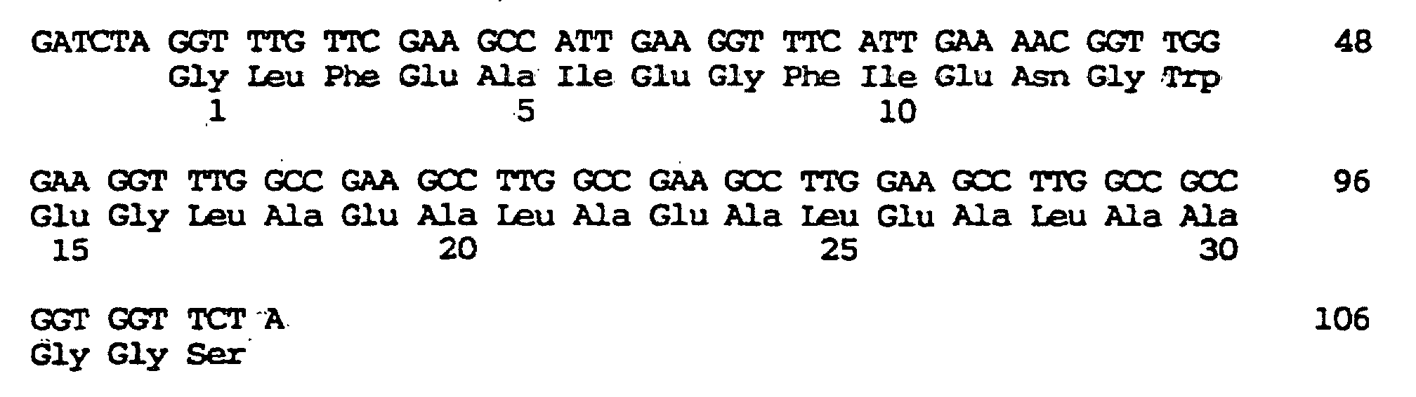
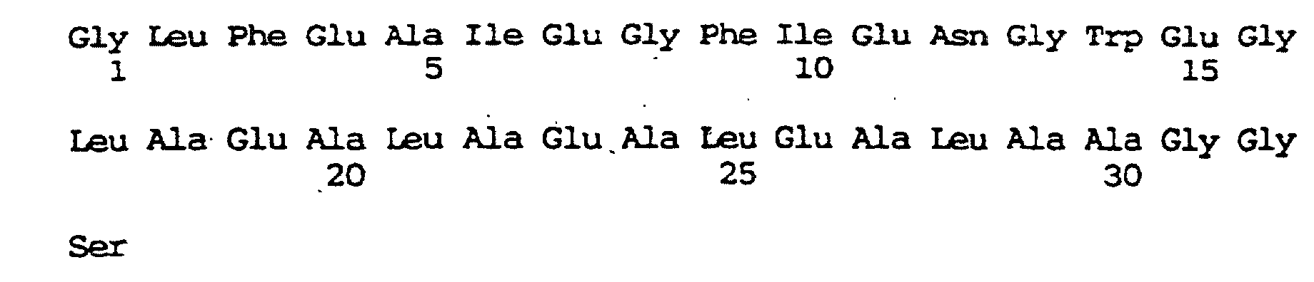
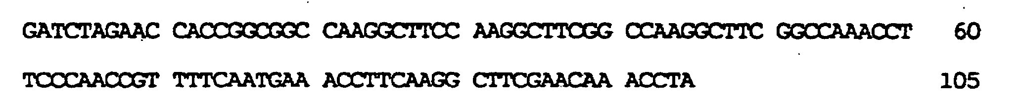


Claims (17)
- Endosomolytically active virus-like particle, characterised in that it is made up of units of capsid proteins derived from viruses or virus-like particles which are modified with a membrane-active peptide sequence.
- Particle according to claim 1, characterised in that it is a modified yeast-Ty particle which is made up of units of TyA protein modified with a membrane-active peptide sequence.
- Particle according to claim 2, characterised in that the Ty particle is modified with the peptide sequence Glu Ala Ala Leu Ala Glu Ala Leu Ala Glu Ala Leu Ala Glu His Leu Ala Glu Ala Leu Ala Glu Ala Leu Glu Ala Leu Ala Ala which is located at the carboxy terminus of the TyA protein.
- Particle according to claim 2, characterised in that the Ty particle is modified with the peptide sequence Gly Leu Phe Glu Ala Ile Glu Gly Phe Ile Glu Asn Gly Trp Glu Gly Leu Ala Glu Ala Leu Ala Glu Ala Leu Glu Ala Leu Ala Ala Gly Gly Ser, which is located at the carboxy terminus of the TyA protein.
- Particle according to claim 1, characterised in that it is a modified MS2 particle which made up of units of the MS2 capsid protein modified with a membrane-active peptide sequence.
- Particle according to claim 5, characterised in that the membrane-active peptide is inserted in the β-hairpin loop region between amino acid 11 (Asp) and amino acid 17 (Asp), particularly between amino acid 14 (Gly) and 15 (Thr) of the MS2 capsid protein.
- Particle according to claim 5 or 6, characterised in that it is modified with the peptide sequence Glu Ala Ala Leu Ala Glu Ala Leu Ala Glu Ala Leu Ala Glu His Leu Ala Glu Ala Leu Ala Glu Ala Leu Glu Ala Leu Ala Ala.
- Particle according to one of claims 1 to 7, characterised in that it also has a nucleic acid-binding domain.
- Particle according to claim 8, characterised in that the nucleic acid-binding domain is polylysine.
- Particle according to claim 9, characterised in that it is bound directly to polylysine.
- Particle according to one of claims 1 to 8, characterised in that it also has a peptide sequence which has the function of a ligand for a higher eukaryotic cell.
- Process for preparing particles according to claim 1, characterised in that a DNA coding for a capsid protein of viruses or virus-like particles, which is modified with a sequence coding for a membrane-active peptide, is expressed and the resulting capsid is harvested or, if necessary, the capsid protein monomers are allowed to associate into capsid structures.
- Process for preparing yeast-Ty particles according to claim 2, characterised in that a DNA coding for the TyA protein which contains a membrane-active peptide at the carboxy terminus is expressed in yeast cells, the cells are opened up and the particles are harvested.
- Process for preparing MS2 particles according to claim 5, characterised in that a DNA coding for the MS2 capsid protein which contains a sequence inserted therein coding for a membrane-active peptide is expressed, the resulting modified capsid protein is denatured and after removal of the denaturing agent it is allowed to associate.
- Composition for transporting nucleic acid into the higher eukaryotic cell, containing an endosomolytic agent, characterised in that the endosomolytic agent is an endosomolytically active virus-like particle according to one of claims 7 to 11.
- Composition according to claim 15 characterised in that it also contains a conjugate of a nucleic acid-binding substance and an internalising factor for the cell.
- Composition according to claim 16, characterised in that it contains a transferrin-polylysine conjugate.
Applications Claiming Priority (3)
| Application Number | Priority Date | Filing Date | Title |
|---|---|---|---|
| DE4335025 | 1993-10-14 | ||
| DE4335025A DE4335025A1 (en) | 1993-10-14 | 1993-10-14 | Endosomolytically active particles |
| PCT/EP1994/003313 WO1995010624A1 (en) | 1993-10-14 | 1994-10-07 | Endosomolytically active particles |
Publications (2)
| Publication Number | Publication Date |
|---|---|
| EP0724643A1 EP0724643A1 (en) | 1996-08-07 |
| EP0724643B1 true EP0724643B1 (en) | 2004-06-30 |
Family
ID=6500137
Family Applications (1)
| Application Number | Title | Priority Date | Filing Date |
|---|---|---|---|
| EP94928873A Expired - Lifetime EP0724643B1 (en) | 1993-10-14 | 1994-10-07 | Endosomolytically active particles |
Country Status (8)
| Country | Link |
|---|---|
| US (1) | US5789230A (en) |
| EP (1) | EP0724643B1 (en) |
| JP (1) | JPH09503665A (en) |
| AT (1) | ATE270342T1 (en) |
| AU (1) | AU681705B2 (en) |
| CA (1) | CA2172139A1 (en) |
| DE (2) | DE4335025A1 (en) |
| WO (1) | WO1995010624A1 (en) |
Families Citing this family (33)
| Publication number | Priority date | Publication date | Assignee | Title |
|---|---|---|---|---|
| NZ244306A (en) | 1991-09-30 | 1995-07-26 | Boehringer Ingelheim Int | Composition for introducing nucleic acid complexes into eucaryotic cells, complex containing nucleic acid and endosomolytic agent, peptide with endosomolytic domain and nucleic acid binding domain and preparation |
| DE4426429A1 (en) * | 1994-07-26 | 1996-02-01 | Boehringer Ingelheim Int | Method for introducing DNA into higher eukaryotic cells |
| US6010871A (en) * | 1994-09-29 | 2000-01-04 | Ajinomoto Co., Inc. | Modification of peptide and protein |
| WO1996012027A1 (en) | 1994-10-18 | 1996-04-25 | Scottish Crop Research Institute | Method of producing a chimeric protein |
| GB9607899D0 (en) * | 1996-04-17 | 1996-06-19 | Scottish Crop Research Inst | Virus-like particle |
| DE19618797C2 (en) * | 1996-05-10 | 2000-03-23 | Bertling Wolf | Vehicle for the transport of molecular substances |
| FR2749323B1 (en) * | 1996-06-04 | 1998-07-10 | Pasteur Merieux Serums Vacc | PSEUDO-PARTICLES VIRALES USEFUL AS A VECTOR OF NUCLEIC ACID DELIVERY |
| US20060002949A1 (en) | 1996-11-14 | 2006-01-05 | Army Govt. Of The Usa, As Rep. By Secretary Of The Office Of The Command Judge Advocate, Hq Usamrmc. | Transcutaneous immunization without heterologous adjuvant |
| US6797276B1 (en) | 1996-11-14 | 2004-09-28 | The United States Of America As Represented By The Secretary Of The Army | Use of penetration enhancers and barrier disruption agents to enhance the transcutaneous immune response |
| DE19829005C2 (en) * | 1998-06-29 | 2000-08-31 | November Ag Molekulare Medizin | Process for the transport of molecular substance into predetermined areas of eukaryotic cells |
| US6500276B1 (en) | 1998-12-15 | 2002-12-31 | Lynntech Coatings, Ltd. | Polymetalate and heteropolymetalate conversion coatings for metal substrates |
| DE19956568A1 (en) * | 1999-01-30 | 2000-08-17 | Roland Kreutzer | Method and medicament for inhibiting the expression of a given gene |
| GB9908195D0 (en) | 1999-04-09 | 1999-06-02 | Microbiological Res Authority | Treatment of intracellular infection |
| DE19916224C1 (en) | 1999-04-10 | 2000-06-21 | November Ag Molekulare Medizin | Synthetic molecule for delivering active ingredient to cell, used e.g. to treat human immune deficiency virus infection, comprises the ingredient linked to VP2 or 3 peptide from polyoma virus |
| DE19952957B4 (en) * | 1999-11-03 | 2006-08-31 | Acgt Progenomics Ag | Modular transport systems for molecular substances and their preparation and use |
| DE10131145B4 (en) * | 2001-06-28 | 2005-07-14 | Innovent E.V. | Composition for cell-specific transfer of active ingredients |
| WO2003078576A2 (en) * | 2002-03-12 | 2003-09-25 | Nitto Denko Corporation | Vector for transfection of eukaryotic cells |
| IL165486A0 (en) | 2002-05-31 | 2006-01-15 | Pendu Le Jacques | Method composition and kit for antigenic binding of norwalk-like viruses |
| US7977098B2 (en) | 2002-05-31 | 2011-07-12 | Children's Hospital Medical Center | Antigenic binding patterns of norovirus to human histo-blood group antigens |
| US8062891B2 (en) | 2003-10-24 | 2011-11-22 | Gencia Corporation | Nonviral vectors for delivering polynucleotides to plants |
| US8507277B2 (en) | 2003-10-24 | 2013-08-13 | Gencia Corporation | Nonviral vectors for delivering polynucleotides |
| CA2543257C (en) | 2003-10-24 | 2013-12-31 | Gencia Corporation | Methods and compositions for delivering polynucleotides |
| US20090123468A1 (en) | 2003-10-24 | 2009-05-14 | Gencia Corporation | Transducible polypeptides for modifying metabolism |
| US8133733B2 (en) | 2003-10-24 | 2012-03-13 | Gencia Corporation | Nonviral vectors for delivering polynucleotides to target tissues |
| US20050232919A1 (en) * | 2004-02-12 | 2005-10-20 | Morphotek, Inc. | Monoclonal antibodies that specifically block biological activity of a tumor antigen |
| CA2607455A1 (en) * | 2005-04-22 | 2006-11-02 | Morphotek, Inc. | Antibodies with immune effector activity and that internalize in endosialin-positive cells |
| US20110152263A1 (en) * | 2006-11-16 | 2011-06-23 | Xi Jiang | Composition and method for inhibiting norovirus infection |
| WO2008128251A1 (en) | 2007-04-17 | 2008-10-23 | The Children's Hospital Of Philadelphia | Humanized viral vectors and methods of use thereof |
| EP2440582B1 (en) | 2009-06-09 | 2018-11-07 | Children's Hospital Medical Center | Antigen-norovirus p-domain monomers and dimers, antigen-norovirus p-particle molecules, and methods for their making and use |
| US9321803B2 (en) | 2013-07-12 | 2016-04-26 | Children's Hospital Medical Center | Compositions and methods for inhibiting norovirus infection |
| WO2017120504A1 (en) | 2016-01-08 | 2017-07-13 | Durfee Paul N | Osteotropic nanoparticles for prevention or treatment of bone metastases |
| US11344629B2 (en) | 2017-03-01 | 2022-05-31 | Charles Jeffrey Brinker | Active targeting of cells by monosized protocells |
| JP2020515531A (en) | 2017-03-28 | 2020-05-28 | チルドレンズ ホスピタル メディカル センター | Norovirus S particle based vaccine and methods of making and using same |
Family Cites Families (6)
| Publication number | Priority date | Publication date | Assignee | Title |
|---|---|---|---|---|
| DE3788806T2 (en) * | 1986-11-01 | 1994-07-07 | British Bio Technology | SPHERICAL HYBRID HIV ANTIGENS. |
| US5166320A (en) * | 1987-04-22 | 1992-11-24 | University Of Connecticut | Carrier system and method for the introduction of genes into mammalian cells |
| IL103059A0 (en) * | 1991-09-30 | 1993-02-21 | Boehringer Ingelheim Int | Conjugates for introducing nucleic acid into higher eucaryotic cells |
| NZ244306A (en) * | 1991-09-30 | 1995-07-26 | Boehringer Ingelheim Int | Composition for introducing nucleic acid complexes into eucaryotic cells, complex containing nucleic acid and endosomolytic agent, peptide with endosomolytic domain and nucleic acid binding domain and preparation |
| GB9213601D0 (en) * | 1992-06-26 | 1992-08-12 | Mastico Robert A | Protein based delivery system |
| WO1994006923A1 (en) * | 1992-09-24 | 1994-03-31 | The University Of Connecticut | Modification of a virus to redirect infectivity and enhance targeted delivery of polynucleotides to cells |
-
1993
- 1993-10-14 DE DE4335025A patent/DE4335025A1/en not_active Withdrawn
-
1994
- 1994-10-07 WO PCT/EP1994/003313 patent/WO1995010624A1/en active IP Right Grant
- 1994-10-07 AT AT94928873T patent/ATE270342T1/en not_active IP Right Cessation
- 1994-10-07 AU AU78120/94A patent/AU681705B2/en not_active Ceased
- 1994-10-07 EP EP94928873A patent/EP0724643B1/en not_active Expired - Lifetime
- 1994-10-07 DE DE59410380T patent/DE59410380D1/en not_active Expired - Fee Related
- 1994-10-07 JP JP7511255A patent/JPH09503665A/en active Pending
- 1994-10-07 US US08/628,665 patent/US5789230A/en not_active Expired - Lifetime
- 1994-10-07 CA CA002172139A patent/CA2172139A1/en not_active Abandoned
Also Published As
| Publication number | Publication date |
|---|---|
| US5789230A (en) | 1998-08-04 |
| JPH09503665A (en) | 1997-04-15 |
| DE4335025A1 (en) | 1995-04-20 |
| AU681705B2 (en) | 1997-09-04 |
| WO1995010624A1 (en) | 1995-04-20 |
| EP0724643A1 (en) | 1996-08-07 |
| ATE270342T1 (en) | 2004-07-15 |
| DE59410380D1 (en) | 2004-08-05 |
| AU7812094A (en) | 1995-05-04 |
| CA2172139A1 (en) | 1995-04-20 |
Similar Documents
| Publication | Publication Date | Title |
|---|---|---|
| EP0724643B1 (en) | Endosomolytically active particles | |
| DE69831741T2 (en) | MODIFIED ADENOVIRAL FIBERS AND ADENOVERS CONTAINING THEM | |
| DE69010206T2 (en) | STABILIZED PROTEIN OR PEPTIDE CONJUGATES. | |
| DE69725878T2 (en) | COMPOSITIONS FOR POLYNUCLEOTIDE DELIVERY | |
| DE69521331T2 (en) | NUCLEIC ACID ENTRY SYSTEM | |
| JP4697982B2 (en) | Modular transfection system | |
| EP0571414B1 (en) | Novel complexes containing nucleic acid which can be absorbed into higher eucaryotic cells via endocytosis | |
| DD294969A5 (en) | THERMALLY STABLE CYTOSINE DEAMINEASE | |
| JPS6058077A (en) | Use of gal yeast promotor | |
| CN113811611A (en) | Nontoxic CAS9 enzyme and application thereof | |
| EP0689604A1 (en) | Process for preparing cancer vaccines | |
| DE19618797C2 (en) | Vehicle for the transport of molecular substances | |
| WO1999066061A1 (en) | Nucleic acid transfer phage | |
| CA2392202A1 (en) | Constructs for delivery of therapeutic agents to neuronal cells | |
| DE4339922C1 (en) | Vector for gene therapy of the liver | |
| JP2834111B2 (en) | Microbial production of human parathyroid hormone | |
| DE4421079C1 (en) | New chimaeric peptide nucleic acid construct | |
| DE69634489T2 (en) | ADENOVIRUS DODEKAHEDRIC PROTEIN COMPLEX, THIS COMPOSITION AND USES THEREOF | |
| DE10131145B4 (en) | Composition for cell-specific transfer of active ingredients | |
| EP1228235B1 (en) | Method for transfer of molecular substances with prokaryontic nucleic acid-binding proteins | |
| DE69933875T2 (en) | PROTEIN ADMINISTRATION SYSTEM USING SIMILAR PARTICLES TO HUMAN PAPILLOMA VIRUS. | |
| WO2005059129A2 (en) | Adapter for coupling a substance which is to be coupled to a cell surface | |
| DE102004032888B4 (en) | Recombinant ScV particles, their coding nucleic acids and their uses | |
| EP1228199A2 (en) | Method for directed packaging of molecular substances in protein shells | |
| KR100626157B1 (en) | - GST-ALS Fusion Protein and Method for Preparation Thereof |
Legal Events
| Date | Code | Title | Description |
|---|---|---|---|
| PUAI | Public reference made under article 153(3) epc to a published international application that has entered the european phase |
Free format text: ORIGINAL CODE: 0009012 |
|
| 17P | Request for examination filed |
Effective date: 19960514 |
|
| AK | Designated contracting states |
Kind code of ref document: A1 Designated state(s): AT BE CH DE DK ES FR GB GR IE IT LI LU MC NL PT SE |
|
| RAP1 | Party data changed (applicant data changed or rights of an application transferred) |
Owner name: BOEHRINGER INGELHEIM INTERNATIONAL GMBH |
|
| GRAP | Despatch of communication of intention to grant a patent |
Free format text: ORIGINAL CODE: EPIDOSNIGR1 |
|
| GRAS | Grant fee paid |
Free format text: ORIGINAL CODE: EPIDOSNIGR3 |
|
| GRAA | (expected) grant |
Free format text: ORIGINAL CODE: 0009210 |
|
| AK | Designated contracting states |
Kind code of ref document: B1 Designated state(s): AT BE CH DE DK ES FR GB GR IE IT LI LU MC NL PT SE |
|
| PG25 | Lapsed in a contracting state [announced via postgrant information from national office to epo] |
Ref country code: NL Free format text: LAPSE BECAUSE OF FAILURE TO SUBMIT A TRANSLATION OF THE DESCRIPTION OR TO PAY THE FEE WITHIN THE PRESCRIBED TIME-LIMIT Effective date: 20040630 Ref country code: IT Free format text: LAPSE BECAUSE OF FAILURE TO SUBMIT A TRANSLATION OF THE DESCRIPTION OR TO PAY THE FEE WITHIN THE PRE;WARNING: LAPSES OF ITALIAN PATENTS WITH EFFECTIVE DATE BEFORE 2007 MAY HAVE OCCURRED AT ANY TIME BEFORE 2007. THE CORRECT EFFECTIVE DATE MAY BE DIFFERENT FROM THE ONE RECORDED.SCRIBED TIME-LIMIT Effective date: 20040630 Ref country code: IE Free format text: LAPSE BECAUSE OF FAILURE TO SUBMIT A TRANSLATION OF THE DESCRIPTION OR TO PAY THE FEE WITHIN THE PRESCRIBED TIME-LIMIT Effective date: 20040630 Ref country code: GB Free format text: LAPSE BECAUSE OF FAILURE TO SUBMIT A TRANSLATION OF THE DESCRIPTION OR TO PAY THE FEE WITHIN THE PRESCRIBED TIME-LIMIT Effective date: 20040630 Ref country code: FR Free format text: LAPSE BECAUSE OF FAILURE TO SUBMIT A TRANSLATION OF THE DESCRIPTION OR TO PAY THE FEE WITHIN THE PRESCRIBED TIME-LIMIT Effective date: 20040630 |
|
| REG | Reference to a national code |
Ref country code: GB Ref legal event code: FG4D Free format text: NOT ENGLISH Ref country code: CH Ref legal event code: EP |
|
| REG | Reference to a national code |
Ref country code: IE Ref legal event code: FG4D Free format text: GERMAN |
|
| REF | Corresponds to: |
Ref document number: 59410380 Country of ref document: DE Date of ref document: 20040805 Kind code of ref document: P |
|
| PG25 | Lapsed in a contracting state [announced via postgrant information from national office to epo] |
Ref country code: SE Free format text: LAPSE BECAUSE OF FAILURE TO SUBMIT A TRANSLATION OF THE DESCRIPTION OR TO PAY THE FEE WITHIN THE PRESCRIBED TIME-LIMIT Effective date: 20040930 Ref country code: GR Free format text: LAPSE BECAUSE OF FAILURE TO SUBMIT A TRANSLATION OF THE DESCRIPTION OR TO PAY THE FEE WITHIN THE PRESCRIBED TIME-LIMIT Effective date: 20040930 Ref country code: DK Free format text: LAPSE BECAUSE OF FAILURE TO SUBMIT A TRANSLATION OF THE DESCRIPTION OR TO PAY THE FEE WITHIN THE PRESCRIBED TIME-LIMIT Effective date: 20040930 |
|
| PG25 | Lapsed in a contracting state [announced via postgrant information from national office to epo] |
Ref country code: LU Free format text: LAPSE BECAUSE OF NON-PAYMENT OF DUE FEES Effective date: 20041007 Ref country code: AT Free format text: LAPSE BECAUSE OF NON-PAYMENT OF DUE FEES Effective date: 20041007 |
|
| PG25 | Lapsed in a contracting state [announced via postgrant information from national office to epo] |
Ref country code: ES Free format text: LAPSE BECAUSE OF FAILURE TO SUBMIT A TRANSLATION OF THE DESCRIPTION OR TO PAY THE FEE WITHIN THE PRESCRIBED TIME-LIMIT Effective date: 20041011 |
|
| PG25 | Lapsed in a contracting state [announced via postgrant information from national office to epo] |
Ref country code: MC Free format text: LAPSE BECAUSE OF NON-PAYMENT OF DUE FEES Effective date: 20041031 Ref country code: LI Free format text: LAPSE BECAUSE OF NON-PAYMENT OF DUE FEES Effective date: 20041031 Ref country code: CH Free format text: LAPSE BECAUSE OF NON-PAYMENT OF DUE FEES Effective date: 20041031 Ref country code: BE Free format text: LAPSE BECAUSE OF NON-PAYMENT OF DUE FEES Effective date: 20041031 |
|
| GBV | Gb: ep patent (uk) treated as always having been void in accordance with gb section 77(7)/1977 [no translation filed] |
Effective date: 20040630 |
|
| NLV1 | Nl: lapsed or annulled due to failure to fulfill the requirements of art. 29p and 29m of the patents act | ||
| BERE | Be: lapsed |
Owner name: BOEHRINGER INGELHEIM INTERNATIONAL G.M.B.H. Effective date: 20041031 |
|
| PG25 | Lapsed in a contracting state [announced via postgrant information from national office to epo] |
Ref country code: DE Free format text: LAPSE BECAUSE OF NON-PAYMENT OF DUE FEES Effective date: 20050503 |
|
| PLBE | No opposition filed within time limit |
Free format text: ORIGINAL CODE: 0009261 |
|
| STAA | Information on the status of an ep patent application or granted ep patent |
Free format text: STATUS: NO OPPOSITION FILED WITHIN TIME LIMIT |
|
| REG | Reference to a national code |
Ref country code: CH Ref legal event code: PL |
|
| 26N | No opposition filed |
Effective date: 20050331 |
|
| EN | Fr: translation not filed | ||
| BERE | Be: lapsed |
Owner name: *BOEHRINGER INGELHEIM INTERNATIONAL G.M.B.H. Effective date: 20041031 |
|
| PG25 | Lapsed in a contracting state [announced via postgrant information from national office to epo] |
Ref country code: PT Free format text: LAPSE BECAUSE OF NON-PAYMENT OF DUE FEES Effective date: 20041130 |