EP0693085B1 - Procede de couplage de ligands au sein de supports poreux (p.e. azlactone) et ses utilisations - Google Patents
Procede de couplage de ligands au sein de supports poreux (p.e. azlactone) et ses utilisations Download PDFInfo
- Publication number
- EP0693085B1 EP0693085B1 EP94914106A EP94914106A EP0693085B1 EP 0693085 B1 EP0693085 B1 EP 0693085B1 EP 94914106 A EP94914106 A EP 94914106A EP 94914106 A EP94914106 A EP 94914106A EP 0693085 B1 EP0693085 B1 EP 0693085B1
- Authority
- EP
- European Patent Office
- Prior art keywords
- ligand
- coupling
- porous support
- coupled
- support
- Prior art date
- Legal status (The legal status is an assumption and is not a legal conclusion. Google has not performed a legal analysis and makes no representation as to the accuracy of the status listed.)
- Expired - Lifetime
Links
Images

Classifications
-
- C—CHEMISTRY; METALLURGY
- C12—BIOCHEMISTRY; BEER; SPIRITS; WINE; VINEGAR; MICROBIOLOGY; ENZYMOLOGY; MUTATION OR GENETIC ENGINEERING
- C12N—MICROORGANISMS OR ENZYMES; COMPOSITIONS THEREOF; PROPAGATING, PRESERVING, OR MAINTAINING MICROORGANISMS; MUTATION OR GENETIC ENGINEERING; CULTURE MEDIA
- C12N9/00—Enzymes; Proenzymes; Compositions thereof; Processes for preparing, activating, inhibiting, separating or purifying enzymes
- C12N9/14—Hydrolases (3)
- C12N9/48—Hydrolases (3) acting on peptide bonds (3.4)
- C12N9/50—Proteinases, e.g. Endopeptidases (3.4.21-3.4.25)
- C12N9/64—Proteinases, e.g. Endopeptidases (3.4.21-3.4.25) derived from animal tissue
- C12N9/6421—Proteinases, e.g. Endopeptidases (3.4.21-3.4.25) derived from animal tissue from mammals
- C12N9/6424—Serine endopeptidases (3.4.21)
- C12N9/6464—Protein C (3.4.21.69)
-
- C—CHEMISTRY; METALLURGY
- C07—ORGANIC CHEMISTRY
- C07K—PEPTIDES
- C07K1/00—General methods for the preparation of peptides, i.e. processes for the organic chemical preparation of peptides or proteins of any length
- C07K1/04—General methods for the preparation of peptides, i.e. processes for the organic chemical preparation of peptides or proteins of any length on carriers
-
- C—CHEMISTRY; METALLURGY
- C07—ORGANIC CHEMISTRY
- C07K—PEPTIDES
- C07K16/00—Immunoglobulins [IGs], e.g. monoclonal or polyclonal antibodies
- C07K16/06—Immunoglobulins [IGs], e.g. monoclonal or polyclonal antibodies from serum
- C07K16/065—Purification, fragmentation
-
- C—CHEMISTRY; METALLURGY
- C07—ORGANIC CHEMISTRY
- C07K—PEPTIDES
- C07K17/00—Carrier-bound or immobilised peptides; Preparation thereof
-
- C—CHEMISTRY; METALLURGY
- C12—BIOCHEMISTRY; BEER; SPIRITS; WINE; VINEGAR; MICROBIOLOGY; ENZYMOLOGY; MUTATION OR GENETIC ENGINEERING
- C12N—MICROORGANISMS OR ENZYMES; COMPOSITIONS THEREOF; PROPAGATING, PRESERVING, OR MAINTAINING MICROORGANISMS; MUTATION OR GENETIC ENGINEERING; CULTURE MEDIA
- C12N11/00—Carrier-bound or immobilised enzymes; Carrier-bound or immobilised microbial cells; Preparation thereof
-
- C—CHEMISTRY; METALLURGY
- C12—BIOCHEMISTRY; BEER; SPIRITS; WINE; VINEGAR; MICROBIOLOGY; ENZYMOLOGY; MUTATION OR GENETIC ENGINEERING
- C12Y—ENZYMES
- C12Y304/00—Hydrolases acting on peptide bonds, i.e. peptidases (3.4)
- C12Y304/21—Serine endopeptidases (3.4.21)
- C12Y304/21069—Protein C activated (3.4.21.69)
Definitions
- This invention relates to an improved method of covalently immobilizing ligands to supports and products produced from the method.
- biologically active substances such as proteins
- separation techniques such as affinity chromatography are based on the ability of the coupled ligand to bind specific, targeted biologically active substances from a mixture of other materials.
- affinity chromatography techniques include the binding of immunoglobulins using coupled proteins and the binding of antigens using coupled antibodies.
- Quantity immobilized expressed as density per unit volume of support, is an indicator of the amount of ligand coupled regardless of the quality of that immobilization. In fact, most protein coupled in highly dense regions of a support is biologically inactive. That is a waste.
- Quality of immobilization expressed as bound specific biological activity, is an indicator of the amount of ligand coupled onto a support in a manner that causes the ligand to retain its biological activity. Maximizing bound specific biological activity is desirable. However, there must be enough ligand density to achieve practical utility.
- the optimal condition in ligand coupling would be the maximum amount of ligand that is coupled with maximum bound specific biological activity. That results in optimal ligate binding or functional efficiency of the coupled ligand on the support.
- “functional efficiency” means the combination of acceptable quantity of ligand coupling with acceptable bound specific biological activity.
- U.S. Pat. No. 4,968,742 (Lewis et al.) uses an elaborate, stepwise method to couple ligands involving derivatizing a polymer with an activating agent to introduce a couplable functional group, with the derivatization performed in the presence of a blocking agent which is reactive with the same functionality on the polymer as the activating agent, in order to control the number of couplable functional groups for covalent immobilization of ligand.
- U.S. Pat. No. 4,775,714 discloses a two-step process of immobilization of biologically efficient compounds on a carrier involving the steps of hydrophobic interaction and covalent immobilization.
- Examples 5-7 therein disclose the stepwise addition of a solution of inorganic salt, in a concentration of between 0.5 M to 3.0 M, to a reaction vessel containing the biologically efficient compound and the carrier, followed by a slow reaction (40 hours at 40°C under moderate shaking) in order to produce an immobilized, biologically active compound.
- Publication WO-A-8 907 618 discloses a method of preparing an affinity matrix of modified polysaccharide supports where a "one-pot" system is used to polymerize monomers in the presence of polysaccharide followed by linking of hydroxy groups of the polysaccharide to the formed polymer.
- the temperature used in the linking is higher than the temperature used in polymerization of the monomers.
- the present invention provides a rapid method of covalent immobilization that surprisingly enhances functional efficiency of a biologically active substance as a ligand coupled to surfaces within a porous support.
- the method effectivcly distributes the ligand within a support prior to coupling of the ligand onto the support.
- the method employs a two-step coupling process where reaction conditions are altered between steps, and preferably where no immobilization agent is added between steps.
- the support derivatized with the ligand optimizes functional efficiency with a targeted biologically active substance.
- the first step of the method suppresses conditions or other reaction between ligand and a porous support, which enhances the relative rate of diffusion, to the rate of reaction, of the ligand into and within the support.
- the second step of the method enhances coupling conditions, such that the ligand couples to the support rapidly, i.e., within about four hours so that the ligand couples to the support before the ligand has an opportunity to vacate the desired location for coupling.
- the method achieves a ligand coupling that avoids surface crowding that would otherwise inhibit or lower the biological activity of the ligand.
- Coupled ligand on a derivatizcd support prepared using the method of the present invention is remarkably more uniformly spatially distributed than on a derivatized support using previously known methods.
- the resulting derivatized support achieves about a 1.25-fold to 10-fold increase in functional efficiency than seen when previously known coupling methods have been employed.
- a feature of the present invention is the ease of ligand coupling in a manner that optimizes functional efficiency.
- Another feature of the present invention is the increased efficiency of use of precious or expensive biologically active substances such as ligands for coupling onto a support.
- An advantage of the present invention is a dramatic reduction of the amount of derivatized support needed to achieve a given biochemical processing capacity provided by the immobilized biologically active substance for any given amount of ligand coupled per volume of support. Conversely expressed, the advantage is that the use of a given amount of derivatized support prepared according to the present invention will dramatically increase the biochemical processing capacity. Either way, dramatic improvement to biochemical processing capacity is realized by the present invention.
- Another advantage of the present invention is the savings in related costs (e.g., the expense of processing fluids such as aqueous buffers) of biochemical processing capacity due to the unexpectedly enhanced functional efficiency of the derivatized support.
- Another advantage of the present invention is that the method provides a more uniform spatial distribution of ligand coupled to support. This advantage results in a decrease in spatial density of ligand coupled to the support, making greater use of electrophilic functionality residing at the surfaces of the support. This advantage also permits a larger average density of ligand coupled to the support with increased amounts of ligand, without overcrowding of ligand at or near the exterior surfaces of the support.
- the invention provides a method for coupling a ligand within a porous support that comprises the steps of mixing ligand and porous support under conditions sufficient to suppress coupling of the ligand to the porous support while enhancing the relative rate of diffusion, to the rate of reaction, of the ligand into and within the porous support, and altering conditions to enhance rapid coupling of the ligand within the porous support.
- the method improves functional efficiency of binding a ligate otherwise deleteriously affected by restricted diffusion into a porous support or by steric effects of binding to a ligand coupled to the porous support.
- the steps of the method comprise coupling a ligand to the porous support according to the two-step diffusion/coupling method indicated above, whereby spatial distribution of ligand coupled to surfaces of the supports enhances functional efficiency of ligates otherwise affected by restricted diffusion or steric effects, and binding the ligate to the spatially distributed ligand, such that functional efficiency of the ligand is greater than the functional efficiency of the ligand coupled in circumstances where either restricted diffusion into the porous support, or steric effects of the ligate binding, or both, are present.
- the invention also provides a derivatized porous support produced according to the method of the present invention.
- the invention also provides a derivatized porous support comprising Protein A coupled to the porous support in a coupling efficiency of at least about 80% and having a functional efficiency relative to binding immunoglobulins of greater than about 3.0 bound IgG/coupled Protein A. It is contemplated that an increase of greater than 15% functional efficiency can be achieved at coupling densities of from about 6 mg to about 15 mg of Protein A coupled per ml of swollen or hydrated support.
- the invention also provides a derivatized porous support comprising an antibody against ligate protein (e.g., Protein C) coupled to the porous support in a coupling efficiency of greater than about 70% and having a functional efficiency relative to binding ligate protein (e.g., Protein C) to the coupled anti-ligate protein (e.g., Protein C antibody) of greater than about 20% at a molar ratio of 2:1 of ligate protein to antibody. It is contemplated that greater than 15% functional efficiency can be achieved at coupling densities of from about 3 mg to about 10 mg of antibody coupled per ml of swollen or hydrated support.
- ligate protein e.g., Protein C
- coupled anti-ligate protein e.g., Protein C antibody
- the invention also provides a derivatized porous support comprising ligand coupled to the porous support in a manner that at least 30% of the amount of coupled ligand is coupled to internal surfaces, i.e., those surfaces that are within 70% of the geometric center of the support.
- internal surfaces are those surfaces within a sphere having a radius of 70% of the total radius of the particle.
- the invention also provides a derivatized porous support comprising ligand coupled to the porous support in a manner that has a percentage of permeation of coupled ligand of about 30% to the internal surfaces of a porous support.
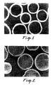
- Fig. 1 is a comparison fluorescence micrograph of a derivatized porous support, shown in cross-section, produced according to prior art methods, that shows an uneven distribution of Protein A ligand coupled principally at outer surfaces of the support, demonstrating an undesired "halo effect".
- Fig. 2 is a fluorescence micrograph of a derivatized porous support, shown in cross-section, produced according to the present invention, that shows a more even distribution of Protein A ligand coupled throughout all surfaces of the support, enhancing functional efficiency.
- Fig. 3 is a comparison fluorescence micrograph of the same derivatized porous support as in Fig. 1, shown in cross-section, that shows an uneven distribution of immunoglobulin bound to the Protein A principally at outer surfaces of the support, demonstrating an undesired "halo effect" for functional efficiency.
- Fig. 4 is a fluorescence micrograph of the same derivatized porous support as in Fig. 2, shown in cross-section, that shows a more even binding of immunoglobulin to Protein A ligand coupled throughout all surfaces of the support, proving enhanced functional efficiency.
- Acceptable porous supports for use in the present invention include those commercially available for affinity chromatography techniques.
- the porous support can be any porous solid, whether natural or synthetic, organic or inorganic, having a porous structure and which is insoluble in water or aqueous solutions.
- Suitable solids with a porous structure have pores of an average diameter of at least 30 nanometers and a pore volume of over 0.1 cm 3 /g.
- the average pore diameter is at least 50 nm because larger pores will be less restrictive to diffusion.
- the pore volume is at least 0.5 cm 3 /g for greater potential capacity due to greater surface area surrounding the pores.
- porous supports are naturally or synthetically-modified natural compositions such as polysaccharides, celluloses, and agaroses.
- An example of a commercially available agarose is Sepharose branded beads from Pharmacia AB of Uppsala, Sweden.
- Such porous supports require activation with a composition, such as cyanogen bromide or cyanotransfer agents, in order to covalently immobilize ligands. Such activation is required prior to using the method of the present invention.
- porous supports are synthetic homopolymers and copolymers of acrylates, methacrylates, acrylamides, vinyl aromatics, and vinyl alcohols.
- such homopolymers or copolymers have a functionality (e.g., azlactone, aldehyde, or the like) or are modified to provide a functionality to permit rapid, direct covalent reaction with ligands to form derivatized supports.
- Oxirane or epoxide functionality is not suitable for methods of the present invention because such groups are not rapid in coupling reaction or require adversely high pH for rapid coupling. See Yarmush et al., Biotech Adv. 10, 413-446 (1992).
- the coupling reaction time was 40 hours at 40°C under moderate shaking. That is not a rapid coupling condition, i.e., within about four hours.
- porous supports are porous inorganic particles such as porous glass, silica, alumina, zirconium oxides, and other metal oxides.
- Porous supports can be membranes, porous fibers, webs, or particles, such as beads.
- porous supports useful in the present invention are reactive porous particles so that the ligand can be covalently coupled to the support.
- Directly reactive porous particles useful in the present invention are generally of two broad types: chemically modified inorganic particles and organic, polymeric particles.
- the inorganic particles may be, for example, metal oxides such as alumina, silica, and zirconia; glass beads, glass bubbles, and controlled pore glass; and the like. These particles are chemically modified by methods such as coating with a polymer (usually organic) which contains a reactive functional group or by reaction with a suitable reagent (e.g. an alkoxy silane coupling agent) containing the reactive functional group.
- a polymer usually organic
- a suitable reagent e.g. an alkoxy silane coupling agent
- the organic particles may be crosslinked or noncrosslinked polymers which have been prepared, for example, by polymerization or copolymerization of a monomer containing the appropriate reactive functional group, by coating a particle support as described above, or by chemical modification of another polymer to introduce the reactive functional group.
- Directly reactive particles useful in the present invention can have a spherical shape, a regular shape, or an irregular shape. Size of reactive particles can vary widely within the scope of the invention and will depend to some extent upon the intended use of the particles.
- Generally size of reactive particles ranges from 0.1 micrometers to 5 millimeters in average diameter.
- the covalently reactive functional groups which are useful for the purposes of the invention can be classified in general as electrophiles.
- Reaction with a nucleophile e.g. amine, alcohol, or mercaptan
- a covalent chemical bond either by an addition reaction or by a displacement or substitution type reaction (in which a byproduct molecule is released).
- Addition type reactions are preferred.
- reactive particles useful in the present invention are particles having azlactone-functional groups on internal and/or external surfaces of such particles.
- such reactive particles have an azlactone-functional group of Formula I: wherein:
- Azlactone-functional reactive particles are particularly preferred in the present invention because such particles rapidly and directly covalently couple ligands better than commercially available reactive functional groups shown in Table 1. Further, such azlactone-functional groups are quite stable prior to covalent coupling with a ligand. Further, covalent coupling of a ligand with an azlactone-functional group causes no displacement of a byproduct molecule, which avoids undesired purification of the composite article after covalent coupling of the ligand.
- azlactone-functional groups are known to possess high covalent coupling capacities with biologically active materials such as Protein A. Further, such high covalent coupling capacities with Protein A also yield high specific bound biological activity of Protein A as the coupled ligand. Thus, an azlactone-functional reactive particle is particularly preferred for use in the present invention.
- Azlactone-functional polymeric particles can be made, for example, by copolymerization of a (meth)acryloylamino acid with a variety of other free radically polymerizable comonomers followed by reaction with a cyclizing agent, as described in U.S. Patent Nos. 4,737,560 and 4,871,824 or by copolymerization of an alkenyl azlactone with other comonomers as described in European Patent Publication 0 392 735, which are both incorporated herein by reference.
- Azlactone-functional particles can also be prepared by solution coating an azlactone-functional polymer onto an organic or inorganic particle, also as described in above mentioned European Patent Publication 0 392
- Azlactone-functional reactive particles can also be made from azlactone graft copolymers which are disclosed in U.S. Patent 5,013,795 and European Patent Publication 0 392 783.
- Size of particles of azlactone-functional particles can be from about 0.1 to 1,000 micrometers and preferably from 0.5 to 250 micrometers.
- Dry azlactone-functional particles can have an average pore size ranging from about 1 to about 300 nanometers and preferably from 5 to about 200 nanometers.
- Azlactone-functional particles can have an average pore volume of at least 1.0 cm 3 /g of particle. In a particle having a size of 50-80 micrometers, a pore volume of at least 1.2 cm 3 /g provides a pore volume of about 60% of the particle volume. In the same particle, the surface area is at least 50 m 2 /g. Thus, there is substantial surface area within an azlactone-functional particle available for covalent immobilization according to the present invention.
- porous supports useful for the present invention are EmphazeTM brand porous azlactone-functional activated affinity chromatography beads commercially available from Minnesota Mining and Manufacturing Company of St. Paul, MN.
- reactive functional groups on porous supports are desirably electrophiles.
- ligands useful in the present invention contain nucleophiles.
- Nonlimiting examples of ligand functional groups include primary and secondary amines, alcohols, and mercaptans. Of these, amine-functional ligands are especially preferred.
- Ligands useful for the preparation of adduct composite articles can also vary widely within the scope of the present invention.
- a ligand is chosen based upon the contemplated end use of the derivatized porous support.
- ligands are coupled according to methods of the present invention, such ligands are available for biological or chemical interaction with an enhanced functional efficiency, such as adsorbing, complexing, catalysis, or reagent end use.
- Derivatized porous supports are useful as adsorbants, complexing agents, catalysts, reagents, as enzyme and other protein-bearing supports, and as chromatographic articles.
- the ligand desired for covalent immobilization is a biologically active substance or compound having nucleophilic-functional groups.
- biologically active materials are substances which are biologically, immunochemically, physiologically, or pharmaceutically active.
- biologically active materials include proteins, peptides, polypeptides, antibodies (monoclonal or polyclonal), antigenic substances, enzymes, cofactors, inhibitors, lectins, hormones, receptors, coagulation factors, amino acids, histones, vitamins, drugs, cell surface markers, and substances which interact with them.
- proteins, enzymes and antigenic substances are desired for covalent immobilization.
- proteins, enzymes, and antigenic substances include natural and recombinant Protein A (ProtA), Immunoglobulins such as rat (rIg), human (hIg), bovine (bIg), rabbit (rbIg), and mouse (mIg), Concanavalin A (ConA), Bovine Serum Albumin (BSA), Thyroglobulin (TG), Apoferritin (Af), Lysozyme (Ly), Carbonic Anhydrase (CA), and Bacterial Antigen (BA).
- Protein A Protein A
- Immunoglobulins such as rat (rIg), human (hIg), bovine (bIg), rabbit (rbIg), and mouse (mIg)
- Concanavalin A (ConA) Bovine Serum Albumin (BSA), Thyroglobulin (TG), Apoferritin (Af), Lysozyme (Ly), Carbonic An
- Uses for coupled proteins, enzymes and antigenic substances are disclosed in European Patent Publication 0 392 735.
- Uses for coagulation factors include activation of zymogens to active proteins, such as Protein C to Active Protein C by coupled thrombin.
- the presently preferred biologically active substances are antibodies and ProtA.
- a derivatized porous support of the present invention can comprise a coupled enzyme to catalyze a chemical transformation of substances recognized by the enzyme.
- a derivatized porous support comprising a coupled antigenic substance can be utilized for affinity purification of a corresponding antibody from a complex biological fluid.
- porous particles having Protein A coupled to internal and external surfaces according to the method of the present invention can adsorb biologically active materials such as Immunoglobulin G for affinity separations processes.
- a derivatized porous support can be used for immobilization of antibodies or be used for immunodiagnostics or for Western blotting.
- azlactone-functional groups will undergo nucleophilic attack by amines, thiols, and alcohols.
- ligands having at least one amine, thiol, or alcohol group thereon are candidates for covalent immobilization in an azlactone-functional porous support.
- the method of the present invention is particularly useful for binding large ligates that are otherwise affected by restricted diffusion into a porous support or by steric effects of binding to a densely coupled ligand, or both.
- ligates can be characterized as those ligates which would be unable to traverse a pore already having ligate bound to coupled ligand at opening surfaces of the pore.
- coupled ligand within the derivatized support are under-utilized or possibly un-utilized during binding if opening surfaces of the pore coupled with ligand bind ligates and block passage to derivatized surfaces within the porous support.
- large ligates can be characterized as those ligates which adversely alter the binding of additional ligates due to steric effects.
- binding of the ligate can occur but inefficiently (e.g., in a manner that causes a waste of coupled ligand). If that ligand were more uniformly spatially distributed, the support could accommodate both a greater binding capacity and a greater functional efficiency.
- the "largeness" of the ligate is a relative consequence of the pore size of the porous support, the reactivity and density of coupled ligand, and other factors.
- large ligates are those biologically active materials identified above as ligand candidates, except for small molecules such as peptides, polypeptides, amino acids, and drugs.
- the method of the present invention involves two steps.
- the first step brings the ligand in close proximity to surfaces of the porous support.
- the second step causes rapid covalent immobilization, i.e., coupling within about four hours, of the ligand to surfaces of the porous support.
- An advantage of the present invention is the superior functional efficiency achieved by coupling ligands according to the method of the present invention.
- the first step employs, in a single reaction vessel, conditions sufficient to suppress coupling of ligand to the support while enhancing the relative rate of diffusion, to the rate of reaction, of the ligand into and within the porous support.
- the first step suppresses the rate of ligand coupling relative to the rate of ligand diffusion.
- Nonlimiting examples of suppressing coupling conditions in the reaction vessel include use of specific ranges of conditions in one or more of a combination of pH, ionic strength, temperature, and coupling competitors.
- Presently preferred as conditions to suppress coupling are a control of pH of a reaction solution and/or control of ionic strength, in which the ligand and the porous support are mixed and otherwise in condition for coupling.
- the pH can be controlled to be in a range from about 3 to about 7.
- a pH of that range provides diffusion conditions that minimize the reaction of a nucleophilic group on a ligand with an electrophilic group on the surface of a porous support.
- the pH during diffusion should be from about 4 to about 6 because of the stability of the electrophilic functional groups to hydrolysis is greater as pH increases.
- the pH during diffusion is about 5, because electrophilic functional groups are more stable to possible hydrolysis.
- pH of the reaction solution ranges from about 3 to about 7
- other reaction conditions can be as conventionally employed in the art of immobilization.
- adjustment of the pH alone can be sufficient to suppress coupling conditions while also enhancing the relative rate of diffusion, to the rate of reaction, of the ligand into and within the porous support.
- Ionic strength due to the presence of polyanionic salts e.g., sulfates, phosphates, citrates, tartrates, and the like
- the molarity of the polyanionic salts in the reaction solution during diffusion is from about 0.01 to about 0.4 M.
- Coupling competitors described in greater detail below, which would otherwise compete for nucleophilic reaction with the functional groups can be 0 to 2 Molarity.
- Temperature of the reaction solution during diffusion needs to be controlled to slow the reaction rate of ligand coupling.
- the temperature can be from about the freezing point of the aqueous solution to about 25°C.
- the reaction solution usually includes buffering agents.
- Buffering agents for aqueous media include acetate, phosphate, pyrophosphate, borate, and other salts known to those skilled in the art, such as those buffering agents disclosed in Good et al., Biochemistry , 5, (1966) p. 467 et seq.
- the concentration of buffering agents in aqueous media can range from about 10 mM to about 750 mM and desirably from about 50 mM to about 200 mM, inclusive, depending on the concentration of biologically active substance chosen for coupling and the concentrations of other optional ingredients that can affect the ionic strength of the reaction solution.
- the duration of the first step should be sufficient in length to assure diffusion of the ligand into proximity with all surfaces of the porous support.
- the amount of time can vary according to the type of porous support employed, the pore size and pore volume of the porous support, the size and conformation of the ligand to diffuse through the pore volume of the porous support and other physical considerations.
- a diffusion duration of at least 5 minutes is sufficient to accomplish acceptable diffusion.
- diffusion lasts at least 10 minutes to improve diffusion.
- diffusion lasts at least 15 minutes to assure diffusion in most porous supports in the case of smaller support geometries.
- the diffusion lasts to at least the characteristic diffusion time, which is the mean support thickness divided by the diffusivity of the ligand of interest in aqueous solution.
- the second step causes coupling of ligand to surfaces of the porous support to occur rapidly and assuredly.
- reaction conditions in the second step are abruptly altered from that of the first step and preferably occur in the absence of adding any coupling agent to the reaction solution.
- a "coupling agent” means a reagent that reacts with either the ligand or the support to improve the coupling of the ligand to the support but does not mean a coupling competitor, i.e., a reagent that competes for reaction sites on the porous support.
- the rate of coupling is a function of the rate constant of coupling, the concentration of ligand, reactivity of the functional groups per unit area of the support, the rate of diffusion of ligand into the support, and the temperature.
- a change in pH comprises the second step.
- Coupling conditions are enhanced when pH of a reaction solution is changed to a pH within one pKa of the nucleophilic ligand, usually within a range from about 7 to about 10. This range causes rapid and assured coupling of ligand to the porous support.
- a pH of this range provides coupling conditions that maximize the reaction of a nucleophilic group on a ligand with an electrophilic group on the surface of a porous support.
- the pH of the reaction solution for coupling should be from about 7.5 to about 9.5 so that hydrolysis or other reaction with solvent is lessened.
- the pH of the reaction solution for coupling is about 8.5 to maintain the biological activity of ligands, especially proteinaceous ligands.
- the alteration in reaction conditions from the diffusion step to the coupling step can be limited to a change in pH or can also include other changes.
- a change in ionic strength of the reaction solution can also be used to enhance functional efficiency of the ligand coupled to the porous support.
- the amount of change in ionic strength can be in an amount sufficient to enhance coupling of ligand and to enhance functional efficiency of the coupled ligand.
- the amount of change in ionic strength from the diffusion step to the coupling step can range from about 0.5M to about 1.5M.
- the amount of change in ionic strength can range from about 0.6M to about 1.2M to maintain the solubility of ligands, especially proteinaceous ligands.
- the change in ionic strength of the solution in the coupling step can be about 1.0-1.2M.
- polyanionic salts both inorganic and organic, are identified in PCT WO 92/07879 and U.S. Pat. No. 5,200,471 (Coleman et al.).
- inorganic polyanionic salts are desired because of increased bound specific biological activity relative to the molar concentration of inorganic polyanion in the aqueous media.
- inorganic polyanionic salts use of Na 2 SO 4 is presently preferred when coupling proteins (that have activity unaffected by metallic cations) in an aqueous medium buffered at a pH from about pH 4 to about pH 9.
- Sulfates are also preferred to phosphates because a lower molar concentration of sulfates than phosphates is necessary to achieve the same density of coupled ligand on the azlactone-functional polymeric support. Evidence of this advantage may be found in Coleman et al. J. Chromatogr. 512 (1990) 345-363.
- organic polyacids and their salts can provide even more productive and efficient coupling of ligands on preferred azlactone-functional polymeric supports than inorganic polyanionic salts.
- Organic polyanionic salts are more consistently ionic than inorganic polyanionic salts in a pH range of pH 7 to pH 9 where most covalent immobilizations are conducted and in the preferred range of pH alteration from the diffusion step to the coupling step of the present invention.
- organic polyanionic salts have a higher ionic strength per mole of polyanion. Consequently, fewer moles of organic salt are frequently required for covalent immobilization.
- organic polyanionic salts are presently preferred to inorganic polyanionic salts.
- organic polyacid candidates di-acids, tri-acids, and tetra-acids, or their salts are desired.
- salts of such acids include malonate, malate, and tartrate di-acid salts of alkali metals, citrate tri-acid and nitrilo-tri-acetic acid (NTA) salts of alkali metals, and ethylenediaminetetraacetic acid (EDTA) tetra-acid salts of alkali metals.
- NTA nitrilo-tri-acetic acid
- EDTA ethylenediaminetetraacetic acid
- the presently preferred organic polyanionic salt is sodium citrate.
- an addition of a coupling competitor to the reaction solution can also be used to enhance functional efficiency of the ligand coupled to the porous support.
- the coupling competitor can be added in an amount sufficient to enhance bound specific biological activity of ligand (but not in an amount which substantially reduces the amount of ligand coupled) in a manner resulting in enhanced functional efficiency of the coupled ligand.
- the type of coupling competitor can vary according to the nature of the ligand to be coupled to the porous support.
- the kinetics of reaction (as influenced by pH, ligand concentration, temperature, ionic strength of the reaction solution, among other factors) between the porous support and the ligand determine the amount and type of coupling competitor to be used.
- a coupling competitor competes for the reactive sites on a porous support where ligand would otherwise couple.
- the reduction in the number of reactive sites can limit the possibility that ligand couples in a manner that alters its conformation and reduces or eliminates its biological activity.
- a coupling competitor enhances functional efficiency of the coupled ligand by providing a sparsity of reactive sites without eliminating too many reactive sites for coupling. This also tends to result in a more uniform or effective distribution of coupled ligand.
- azlactone quenchers When preferred azlactone-functional porous supports are used, coupling competitors are azlactone quenchers. Suitable azlactone quenchers are also identified in PCT WO 92/07879 and U.S. Pat. No. 5,200,471 (Coleman et al.).
- Nonlimiting examples of azlactone quenchers for use include ethanolamine, bovine serum albumin, casein lysate, hydroxylamine, ethylamine, ammonium hydroxide, glycine, ammonium sulfate, butylamine, glycinamide, TRIS, gelatin, lysozyme, non-fat dry milk, beta-mercaptoethanol, mercaptoethylether, dithiothreitol, glutathione, arginine, guanidine, lysine, diamines, and combinations thereof.
- Some of these nonlimiting examples include proteins "irrelevant" to the immobilization desired.
- the concentration of azlactone quencher to be added with a change in pH and/or an increase of ionic strength for the second step of the method can range from about 0.1 M to about 10 M. Desirably, the range may be between about 0.5 M to about 2 M. When ethanolamine serves as azlactone quencher, the concentration may range from about 0.1 M to about 1 M. The presently preferred concentration of ethanolamine as azlactone quencher is about 0.5 M to about 1 M.
- an increase in the temperature of the reaction solution can also be used to enhance functional efficiency of the ligand coupled to the porous support.
- the amount of temperature change can be an amount sufficient to enhance coupling of ligand, so long as the speed of the reaction is rapid, and to enhance functional efficiency of the coupled ligand.
- the temperature increase can be about 10°C to about 35°C, and preferably about 20°C to about 30°C because an increase in temperature of that amount increases reactivity of the ligand to the support without deleteriously affecting the biological activity of the ligand.
- one or more of an increase of pH, an increase in ionic strength, an increase of temperature, or an addition of a coupling competitor to the reaction solution causes ligand to couple in a manner which retains bound specific biological activity of the ligand due to a resulting more uniform or effective distribution of ligand which minimizes restricted diffusion or steric effects of ligates attempting to bind to the coupled ligand. This improves functional efficiency of the resulting coupled ligand by minimizing the number of coupled ligands that are not biologically active and maximizing the amount of ligand coupled.
- the duration of the step can range from 0.5 to 4 hours.
- Coupling is completed by quenching any remaining reactive sites with an addition, in excess, of a quencher that couples to effectively all of the remaining reactive sites on the porous support.
- the quencher employed can be any of the azlactone quenchers identified above in concentrations in excess.
- the method of the present invention utilizes the advantages of controlling the Thiele modulus during the diffusion and the coupling steps of the method.
- the first step of the present invention is to suppress coupling conditions to enhance the relative rate of diffusion to the rate of coupling.
- the first step utilizes a lower Thiele modulus (where rate of diffusion is appreciably greater than rate of reaction) relative to the second step.
- the second step utilizes a higher Thiele modulus (where rate of reaction is appreciably greater than rate of diffusion) than found in the first step.
- the method of the present invention provides a permeation step for more uniform spatial distribution having conditions using relatively low Thiele modulus followed by a coupling step for rapid coupling which uses conditions having a relatively high Thiele modulus.
- Relatively low Thiele modulus conditions are achievable with a low pH, low ionic strength reaction medium, lower temperature, coupling competitor, and combinations of them.
- one of the results in controlling the difference in Thiele modulus between the first and second step is a change in the activation energy necessary for a coupling of ligand to a functional group on a surface within a porous support.
- the conditions of the lower Thiele modulus raise the activation energy required for the coupling reaction, while the conditions of the higher Thiele modulus lower the activation energy required for the coupling reaction.
- the abruptness of the step change in Thiele modulus conditions after the diffusion step and to initiate the coupling step of the method results in minimal back-diffusion of ligand from the support. This maintains much of the dispersed spatial distribution achieved during the diffusion step. Further, the reaction kinetics of the functional group on the surface of the porous support enhances the rapidity of the coupling reaction before the desired more uniform spatial distribution of the ligand within the support is lost. Rapidity of the coupling reaction should be less than about 4 hours and preferably less than about 1 hour.
- the method of the present invention provides a process of immobilizing biologically active substances on porous supports in such a fashion as to avoid surface crowding which inactivates a significant portion of the coupled ligand.
- the derivatized support has coupled ligand with a molecular sparcity which has a functional efficiency that is significantly higher than ligand coupled with a higher surface density.
- Figs. 1 and 2 provide a direct comparison of the advantages of the ) present invention.
- Fig. 1 is a comparison fluorescence micrograph of cross-sections of a derivatized porous support produced according to a method used in the prior art and identified in Comparison Example 6 below, where there is no attempt to provide a two step reaction of diffusion and then coupling.
- the cross-section shows an uneven distribution of ligand coupled at outer surfaces of the support. This is evidence of overcrowding that the method of the present invention avoids.
- Fig. 2 is a fluorescence micrograph of cross-sections of a derivatized porous support produced according to the method of present invention and specifically according to Example 7 below where there was a change in pH between the diffusion step and the coupling step.
- a significantly more even distribution of ligand coupled throughout all surfaces of the support is found.
- an optimum of coupling is achieved.
- the method of the present invention provides a controllable, more uniform spatial distribution of coupling of the ligand to the porous support.
- Derivatized porous supports of the present invention have a sparsity of coupling of the ligand to outer surfaces of the porous support and have an enhanced coupling of the ligand to inner surfaces of the porous support.
- Functional efficiency of coupled ligand can increase as much as 1.1 to 10 fold using the method of the present invention over functional efficiency achieved using prior methods. As such an unexpectedly superior derivatized support is achieved.
- Fig. 3 is a comparison fluorescence micrograph of cross-sections of a derivatized porous support shown in Fig. 1 with binding of immunoglobulin, produced according to a method used in the prior art and identified in Comparison Example 8 below.
- the cross-section shows an uneven distribution of ligate binding to ligand coupled at outer surfaces of the support. This is evidence of restricted diffusion and steric effects that demonstrates the "halo effect" of inadequate functional efficiency.
- Fig. 4 is a fluorescence micrograph of cross-sections of a derivatized porous support shown in Fig. 2, produced according to the method of present invention and specifically according to Example 9 below where there was a ligate binding more uniformly throughout the bead and an avoidance of restricted diffusion and steric effect.
- a significantly more even distribution of ligate bound to coupled ligand throughout all surfaces of the support is found.
- an optimum of functional efficiency is achieved.
- This example describes the coupling of Protein A to EmphazeTM Biosupport Medium AB1 using a one step method of the prior art.
- the beads from the combined triplicates were then sequentially washed with phosphate buffered saline (PBS: 0.025 M NaH 2 PO 4 , 0.15 M NaCl pH 7.4), 0.2 M sodium acetate pH 5.0, 0.5 M sodium bicarbonate pH 8.5, and PBS on a sintered glass fritted funnel (porosity D) using 20-30 volumes of each solution.
- PBS phosphate buffered saline
- the washed beads were then tested for their immunoglobulin binding capacity by packing them into a 3 x 50 mm Omni glass column and running a chromatogram with purified human IgG (Sigma Chemical Company, St. Louis, MO) as the test solution.
- a total of 48 milligrams (16 milliliters of 3 milligrams per milliliter in 0.01 M NaH 2 PO 4 pH 7.5) was loaded at a flow rate of 0.57 milliliters per minute followed by a wash of 6.8 milliliters of loading buffer, 6.8 milliliters of 2 M NaCl, 0.01 M NaH 2 PO 4 pH 7.5, 4.6 milliliters of loading buffer, and the bound IgG was eluted with 4.6 milliliters of 0.1 M glycine, 2% acetic acid pH 2.2. Elution fractions were collected and the amount of IgG present determined by absorbance at 280 nm. The results for these beads are shown in Table A.
- This example describes the coupling of Protein A to Emphaze beads in a manner similar to that of Comparison Example 1 according to the prior art with the exception that the coupling solution contains a coupling competitor, 0.5 M Tris(hydroxymethyl)aminomethane (TRIS) in addition to sodium sulfate and phosphate buffer at pH 7.5.
- TMS Tris(hydroxymethyl)aminomethane
- This example describes a two step method of the present invention of coupling Protein A to Emphaze beads as a direct comparison to Comparison Example 1.
- the final solution conditions are the same as those of Comparison Example 1 but there is a different ionic strength of the reaction mixture during the first step.
- the ionic strength of the reaction mixture is increased.
- This example describes the coupling of Protein A to Emphaze beads using a two step method of the present invention as a direct comparison to Comparison Example 2.
- the second step the ionic strength of the reaction mixture is increased, a coupling competitor that quenches azlactone is added, and the pH is increased.
- the method is similar to that of Example 3 with the exceptions that the Protein A is in 0.05 M sodium acetate pH 5.0 and the solution added in the second step contains 0.5 M TRIS in addition to the sodium sulfate and phosphate buffer at pH 7.5.
- the Protein A coupling and IgG binding capacities were determined as in Comparison Example 1 and those results are shown in Table A.
- This example describes the coupling of Protein A to Emphaze beads using a two step method of the present invention where only the pH changes between the first and second step.
- the method is similar to that of Example 3 with the exception that the Protein A was in 0.05 M sodium acetate pH 5.0 and the second step uses 0.125 M boric acid solution pH 9.5. Protein A coupling was determined as in Comparison Example 1 and the results are shown in Table A.
- FIG. 1 shows significantly different spatial distributions of the Protein A coupled to Emphaze beads. Since both preparations have the same total Protein A content, the "halo" distribution of Protein A in Fig. 1 (Comparison Example 6 beads) indicates that the ligand is concentrated into significantly less volume than the more uniform distribution of Fig. 2 (Example 7 beads).
- the Protein A is coupled to the bead in a manner that at least 70% of the amount of coupled Protein A is coupled to internal surfaces of the bead that are within 35% of the geometric center of the bead.
- the ligate is bound to ligand coupled to internal surfaces within a radius of 70% of the total radius of the particle.
- the ligand is coupled to the porous bead in a manner that has a percentage of permeation of coupled ligand of about 70% to the internal surfaces of a porous support.
- This example describes the coupling of Protein A to cyanogen bromide activated Sepharose branded agarose using a one step method of the prior art.
- CNBr-Activated Sepharose 4B (Pharmacia LKB, Biotechnology AB, Uppsala, Sweden) was prepared for reaction according to the manufacturer's instructions and aliquots of slurry equivalent to 0.6 milliliters of gel were placed into 15 milliliter screw capped polypropylene tubes. After removing the supernatant buffer solution, 3.75 milliliters of 1.0 milligrams of Protein A per milliliter in 0.5 M NaHCO 3 pH 8.5 was added and the mixture agitated by end-over-end rotation for a total of 75 minutes at room temperature.
- This example describes the coupling of Protein A to cyanogen bromide activated Sepharose using a two step method where pH alone was adjusted from the first step to the second step.
- Slurry aliquots (0.6 milliliters) of CNBr-Activated Sepharose 4B were prepared as in Comparison Example 10 and reacted with 0.75 milliliters of 5.0 milligram Protein A per milliliter, and 0.05 M sodium acetate pH 5.0 at 4 degrees C for 15 minutes.
- 3.0 milliliters of 0.625 M sodium bicarbonate pH 8.5 was added and the reaction mixture agitated by end-over-end rotation at room temperature for an additional 60 minutes. The mixture was then treated as in Comparison Example 10 to determine the amount of Protein A coupled and its binding capacity. Results are shown in Table B.
- This example describes the coupling of Protein A to cyanogen bromide activated Sepharose using a one step method of the prior art similar to that of Comparison Example 10 with the exception that the coupling solution contained 1.0 M Na 2 SO 4 , 0.5 M NaHCO 3 pH 8.5. Protein A coupling results and binding capacities were determined as in Comparison Example 1 and are shown in Table B.
- This example describes the coupling of Protein A to cyanogen bromide activated Sepharose using a two step method of the present invention similar to that of Example 11 with the exception that the solution used in the second step contained 1.25 M Na 2 SO 4 , 0.625 M NaHCO 3 pH 8.5. Protein A coupling results and binding capacities were determined as in Comparison Example 1 and are shown in Table B.
- This example describes the coupling of Protein A to cyanogen bromide activated Sepharose using a one step method similar to that of Comparison Example 10 with the exception that the coupling solution contained 1.0 M Na 2 SO 4 , 0.5 M NaHCO 3 , 0.4 M TRIS pH 8.5. Protein A coupling results and binding capacities were determined as in Comparison Example 1 and are shown in Table B.
- This example describes the coupling of Protein A to cyanogen bromide activated Sepharose using a two step method of the present invention similar to that of Comparison Example 11 with the exception that the solution used in the second step contained 1.25 M Na 2 SO 4 , 0.625 M NaHCO 3 , 0.5 M TRIS pH 8.5. Protein A coupling results and binding capacities were determined as in Comparison Example 1 and are shown in Table B.
- 1 mg of 7D7B10-Mab (obtained from American National Red Cross) is incubated with 125 mg EmphazeTM beads at pH 4.0 and the solution is allowed to permeate the beads for 10 mins at 4°C in the presence of 0.5M Tris. After the first 10 minute incubation with 0.5 M Tris, the salt concentration is raised to 0.8 M Na 2 SO 4 at pH 4.0 and allowed to permeate the beads for 10 mins at 4°C. The pH is then increased to pH 9.0 with several drops of 1N NaOH. The reaction at pH 9.0 is allowed to proceed for 40 mins at 4°C. The total permeation/diffusion and reaction time is 60 mins at 4 °C. The supernatant is pipetted off.
- Residual reactive sites are blocked with 4 ml of 1.0 M ethanolamine in 0.05 M sodium pyrophosphate, pH 9.3 for 30 mins at RT. Beads are allowed to settle and the supernatant is pipetted off. An additional 4 mls of blocking solution is combined with the beads and incubated for 60 mins at RT. Upon completion of the second blocking step, the beads are washed with four column volumes of 0.5 M NaCl and equilibrated with loading buffer for protein immunosorption. The 7D7B10-Mab coupling efficency to the azlactone is greater than 70%.
- Residual reactive sites are blocked with 4 ml of 1.0 M ethanolamine in 0.05 M sodium pyrophosphate, pH 9.3 for 30 mins at RT. Beads are allowed to settle and the supernatant is pipetted off. An additional 4 mls of blocking solution is combined with the beads and incubated for 60 mins at RT. Upon completion of the second blocking step, the beads are washed with four column volumes of 0.5 M NaCl and equilibrated with loading buffer for protein immunosorption. The 7D7B10-Mab coupling efficiency to the azlactone is greater than 70%.
- 0.07 or 0.7 mgs of recombinant human Protein C in 3 ml of 0.125 M Tris, 0.1 M NaCl, 25 mMEDTA at pH 6.5 is loaded batchwise and eluted columnwise at a linear velocity of 1 cm/min into a 1.0 cm by 10.0 cm length glass chromatography column containing the 7D7B10-Mab:azlactone beads at 4°C.
- the protein C is eluted from the immunosorbent at 1 cm/min linear velocity with 4.0 ml of 0.125 M Tris, 0.1 M NaCl, 25 mM CaCl 2 at pH 6.5.
- the immunosorbent functional efficiency is about 16%.
- 1 mg of 7D7B10-Mab is incubated with 125 mg EmphazeTM beads at pH 4.0 for 10 mins at 4°C in the presence of 0.5M Tris. After the first 10 minute incubation with 0.5 M Tris, the pH is then increased to pH 9.0 with several drops of IN NaOH. The reaction at pH 9.0 is allowed to proceed for 50 mins at 4°C. The total permeation/diffusion and reaction time is 60 mins at 4 °C. The supernatant is pipetted off. Residual reactive sites are blocked with 4 ml of 1.0 M ethanolamine in 0.05 M sodium pyrophosphate, pH 9.3 for 30 mins at RT.
- Beads are allowed to settle and the supernatant is pipetted off.
- the 7D7B10-Mab coupling efficiency is greater than 50%.
- An additional 4 mls of blocking solution is combined with the beads and incubated for 60 mins at RT.
- the beads are washed with four column volumes of 0.5 M NaCl and equilibrated with loading buffer for protein immunosorption.
- 0.07 or 0.7 mg of recombinant human Protein C in 2.0 ml of 0.125 M Tris, 0.1 M NaCl, 25 mM EDTA at pH 6.5 is loaded batchwise and eluted columnwise at 1 cm/min linear velocity into a 1.0 cm by 10.0 cm length glass chromatography column containing the 7D7B10-Mab:azlactone beads at 4°C.
- the protein C is eluted from the immunosorbent at 1 cm/min linear velocity with 4.0 ml of 0.125 M Tris, 0.1 M NaCl, 25 mM CaCl 2 at pH 6.5.
- the immunosorbent functional efficiency is about 14%.
- Beads are allowed to settle and the supernatant is pipetted off.
- the 7D7B10-Mab coupling efficiency is greater than 50%.
- An additional 4 mls of blocking solution is combined with the beads and incubated for 60 mins at RT.
- the beads are washed with four column volumes of 0.5 M NaCl and equilibrated with loading buffer for protein immunosorption.
- 0.07 or 0.7 mg of recombinant human Protein C in 2.0 ml of 0.125 M Tris, 0.1 M NaCl, 25 mM EDTA at pH 6.5 is loaded batchwise and eluted columnwise at 1 cm/min linear velocity into a 1.0 cm by 10.0 cm length glass chromatography column containing the 7D7B10-Mab:azlactone beads at 4°C.
- the protein C is eluted from the immunosorbent at 1 cm/min linear velocity with 4.0 ml of 0.125 M Tris, 0.1 M NaCl, 25 mM CaCl 2 at pH 6.5.
- the immunosorbent functional efficiency is 14%.
- Residual reactive sites are blocked with 10 ml of 1.0 M ethanolamine in 0.05 M sodium pyrophosphate, pH 9.3 for 30 mins at RT. Beads are allowed to settle and the supernatant is pipetted off. The 12A8-Mab coupling efficiency is greater than 70%. An additional 10 mls of blocking solution is combined with the beads and incubated for 60 mins at RT. Upon completion of the second blocking step, the beads are washed with four column volumes of 0.5 M NaCl and equilibrated with loading buffer for protein immunosorption.
- Residual reactive sites are blocked with 240 ml of 1.0 M ethanolamine in 0.05 M sodium pyrophosphate, pH 9.3 for 30 mins at RT. Beads are allowed to settle and the supernatant is pipetted off. The 12A8-Mab coupling efficiency is greater than 70%. An additional 240 mls of blocking solution is combined with the beads and incubated for 60 mins at RT. Upon completion of the second blocking step, the beads are washed with four column volumes of 0.5 M NaCl and equilibrated with loading buffer for protein immunosorption.
- 125 mg of recombinant human Protein C in 600 ml of 0.125 M Tris, 0.1 M NaCl, 15 mM MgCl 2 at pH 8.0 is loaded columnwise at 1 cm/min linear velocity into a 5.0 cm by 50.0 cm length glass chromatography column containing the 12A8-Mab:azlactone beads at 4°C.
- the protein C is eluted from the immunosorbent at 1 cm/min linear velocity with 210 ml of 0.1 M NaHCO 3 , 0.15 M NaCl at pH 10.0.
- the immunosorbent functional efficiency is 25 %.
Claims (9)
- Procédé de couplage d'un ligand à l'intérieur d'un support poreux, comprenant les étapes de :(a) diffusion d'un ligand biologiquement actif à l'intérieur d'un support poreux, dans des conditions qui suppriment le couplage du ligand, dans lequel la durée de l'étape de diffusion doit être suffisamment longue pour assurer la diffusion du ligand à proximité de toutes les surfaces du support poreux ; suivi par(b) des conditions de vieillissement pour obtenir un couplage covalent rapide du ligand au support poreux en 4 heures,dans lequel la vitesse de couplage du ligand pendant l'étape (a) est inférieure à la vitesse de couplage du ligand pendant l'étape (b), etdans lequel les conditions modifiées de l'étape (a) à l'étape (b) comprennent (i) une augmentation du pH, le pH de l'étape (a) étant dans la gamme comprise entre 3 et 7, et le pH de l'étape (b) étant dans la gamme comprise entre 7 et 10, (ii) une augmentation de la concentration ionique, le niveau de changement de la concentration ionique de l'étape (a) à l'étape (b) étant dans la gamme comprise entre 0,5 M et 1,5 M, ou (iii) une augmentation de la température, le niveau de changement de la température de l'étape (a) à l'étape (b) étant dans la gamme comprise entre 10°C et 35°C, ou une combinaison de (i), (ii) et (iii),et dans lequel l'activité biologique du ligand couplé est conservée.
- Procédé selon la revendication 1, dans lequel le procédé apporte un moyen de maítriser la répartition spatiale de couplage du ligand au support poreux et dans lequel le procédé fournit un couplage covalent du ligand aux surfaces internes et aux surfaces externes du support poreux.
- Procédé selon les revendications 1 et 2, dans lequel les conditions de couplage de l'étape (b) emploient un concurrent de couplage en une concentration située dans la gamme comprise entre 0,1 M et 10 M.
- Procédé selon la revendication 1, dans lequel le support poreux est à fonctionnalité azlactone.
- Procédé de liaison d'un ligat autrement influencé défavorablement par une diffusion limitée dans un support poreux ou par des effets stériques de liaison au ligand couplé au support poreux, comprenant les étapes consistant à :(1) coupler le ligand au support poreux selon les étapes (a) et (b) du procédé des revendications 1 à 4 pour obtenir une répartition spatiale du ligand couplé aux surfaces du support ; et(2) lier le ligat au ligand réparti spatialement.
- Support poreux dérivé comprenant un anticorps monoclonal couplé à un support poreux de manière à ce qu'au moins 30% de la quantité d'anticorps monoclonal couplé soit couplé aux surfaces internes du support poreux qui sont dans les 70% proches du centre géométrique du support.
- Support poreux dérivé selon la revendication 6, comprenant l'anticorps monoclonal couplé au support poreux directement réactif avec une efficacité de couplage d'au moins 70% et avec une efficacité fonctionnelle pour la liaison à la protéine de ligat supérieure à environ 20% à un rapport molaire de 2 : 1 de la protéine de ligat par rapport à l'anticorps.
- Support poreux dérivé selon les revendications 6 et 7, dans lequel le support est une particule.
- Support selon les revendications 6 à 8, dans lequel le support est un support poreux dérivé à fonctionnalité aziactone.
Applications Claiming Priority (3)
| Application Number | Priority Date | Filing Date | Title |
|---|---|---|---|
| US3864593A | 1993-03-29 | 1993-03-29 | |
| US38645 | 1993-03-29 | ||
| PCT/US1994/003927 WO1994022918A1 (fr) | 1993-03-29 | 1994-02-18 | Procede de couplage de ligands au sein de supports poreux (p.e. azlactone) et ses utilisations |
Publications (2)
| Publication Number | Publication Date |
|---|---|
| EP0693085A1 EP0693085A1 (fr) | 1996-01-24 |
| EP0693085B1 true EP0693085B1 (fr) | 2001-10-31 |
Family
ID=21901089
Family Applications (1)
| Application Number | Title | Priority Date | Filing Date |
|---|---|---|---|
| EP94914106A Expired - Lifetime EP0693085B1 (fr) | 1993-03-29 | 1994-02-18 | Procede de couplage de ligands au sein de supports poreux (p.e. azlactone) et ses utilisations |
Country Status (8)
| Country | Link |
|---|---|
| US (1) | US5907016A (fr) |
| EP (1) | EP0693085B1 (fr) |
| JP (1) | JP3607289B2 (fr) |
| KR (1) | KR960701099A (fr) |
| CA (1) | CA2158056A1 (fr) |
| DE (1) | DE69428891T2 (fr) |
| NO (1) | NO953836L (fr) |
| WO (1) | WO1994022918A1 (fr) |
Families Citing this family (12)
| Publication number | Priority date | Publication date | Assignee | Title |
|---|---|---|---|---|
| GB9425138D0 (en) | 1994-12-12 | 1995-02-08 | Dynal As | Isolation of nucleic acid |
| US6379952B1 (en) | 1999-02-01 | 2002-04-30 | 3M Innovative Properties Company | Method for cell selection utilizing azlactone-functional supports |
| US6787635B2 (en) * | 2001-04-05 | 2004-09-07 | 3M Innovative Properties Company | Solid phase synthesis supports and methods |
| EP1468116A2 (fr) | 2002-01-16 | 2004-10-20 | Dynal Biotech ASA | Methode permettant d'isoler des acides nucleiques et des proteines contenus dans un echantillon unique |
| US20030203504A1 (en) * | 2002-04-26 | 2003-10-30 | John Hefti | Diffusion-based system and method for detecting and monitoring activity of biologic and chemical species |
| GB0229287D0 (en) * | 2002-12-16 | 2003-01-22 | Dna Res Innovations Ltd | Polyfunctional reagents |
| US20060070950A1 (en) * | 2004-10-01 | 2006-04-06 | 3M Innovative Properties Company | Composite filtration article |
| CN101039734B (zh) * | 2004-10-15 | 2010-09-08 | 3M创新有限公司 | 褶式多层过滤介质及滤筒 |
| CA2586803C (fr) * | 2004-12-14 | 2012-12-11 | Ge Healthcare Bio-Sciences Ab | Purification d'immunoglobulines |
| US8728828B2 (en) | 2004-12-22 | 2014-05-20 | Ge Healthcare Bio-Sciences Ab | Purification of immunoglobulins |
| US20100012588A1 (en) * | 2006-08-28 | 2010-01-21 | Maciej Siewinski | system and method for the extra-corporeal purification of blood of pathogenic enzymes |
| GB201703116D0 (en) * | 2017-02-27 | 2017-04-12 | Ge Healthcare Bioprocess R&D Ab | A seperation matrix and a method of seperating antibodies |
Family Cites Families (15)
| Publication number | Priority date | Publication date | Assignee | Title |
|---|---|---|---|---|
| JPS635133B2 (fr) * | 1978-01-24 | 1988-02-02 | Paburitsuku Herusu Raboratarii Saauisu Boodo | |
| US4704366A (en) * | 1984-06-22 | 1987-11-03 | Bio-Rad Laboratories, Inc. | Process for binding IgG to protein A |
| US4791069A (en) * | 1984-09-21 | 1988-12-13 | Ortho Diagnostic Systems Inc. | Methods for attaching ligands or anti-ligands to a solid phase |
| CS253971B1 (en) * | 1984-12-11 | 1987-12-17 | Peter Hermann | Production method of high active biological effective compounds immobilized on carrier |
| DE3515252C2 (de) * | 1985-04-27 | 1994-02-24 | Roehm Gmbh | Verfahren zur Immobilisierung gelöster Eiweißstoffe |
| US4822747A (en) * | 1986-12-09 | 1989-04-18 | Miles Inc. | Polyacrylamide gel particles having hapten moieties bound thereto as immunoassay reagent |
| US4968742A (en) * | 1987-11-09 | 1990-11-06 | Miles Inc. | Preparation of ligand-polymer conjugate having a controlled number of introduced ligands |
| AU3047989A (en) * | 1988-02-11 | 1989-09-06 | Cuno Incorporated | Affinity matrices of modified polysaccharide supports |
| US5013795A (en) * | 1989-04-10 | 1991-05-07 | Minnesota Mining And Manufacturing Company | Azlactone graft copolymers |
| US5039813A (en) * | 1990-06-29 | 1991-08-13 | Polaroid Corporation | 2-(4-alkenylphenyl)-5-oxazolones and polymers thereof |
| US5200471A (en) * | 1990-11-05 | 1993-04-06 | Minnesota Mining And Manufacturing Company | Biomolecules covalently immobilized with a high bound specific biological activity and method of preparing same |
| US5120833A (en) * | 1991-03-15 | 1992-06-09 | Alexander Kaplan | Method of producing grafts |
| US5219749A (en) * | 1991-10-09 | 1993-06-15 | Institute For Molecular Biology & Biotechnology/Forth | Process for isolating and preparing purified chitin deacetylase |
| US5993935A (en) * | 1991-10-11 | 1999-11-30 | 3M Innovative Properties Company | Covalently reactive particles incorporated in a continous porous matrix |
| ATE154526T1 (de) * | 1992-04-16 | 1997-07-15 | Merck Patent Gmbh | Aktivierte trägermaterialien, ihre herstellung und verwendung |
-
1994
- 1994-02-18 WO PCT/US1994/003927 patent/WO1994022918A1/fr active IP Right Grant
- 1994-02-18 EP EP94914106A patent/EP0693085B1/fr not_active Expired - Lifetime
- 1994-02-18 KR KR1019950704186A patent/KR960701099A/ko not_active Application Discontinuation
- 1994-02-18 CA CA002158056A patent/CA2158056A1/fr not_active Abandoned
- 1994-02-18 DE DE69428891T patent/DE69428891T2/de not_active Expired - Fee Related
- 1994-02-18 JP JP52250394A patent/JP3607289B2/ja not_active Expired - Fee Related
- 1994-08-29 US US08/296,588 patent/US5907016A/en not_active Expired - Lifetime
-
1995
- 1995-09-28 NO NO953836A patent/NO953836L/no not_active Application Discontinuation
Also Published As
| Publication number | Publication date |
|---|---|
| JPH09506067A (ja) | 1997-06-17 |
| JP3607289B2 (ja) | 2005-01-05 |
| WO1994022918A1 (fr) | 1994-10-13 |
| US5907016A (en) | 1999-05-25 |
| KR960701099A (ko) | 1996-02-24 |
| EP0693085A1 (fr) | 1996-01-24 |
| DE69428891T2 (de) | 2002-06-27 |
| NO953836D0 (no) | 1995-09-28 |
| CA2158056A1 (fr) | 1994-10-13 |
| NO953836L (no) | 1995-11-29 |
| DE69428891D1 (de) | 2001-12-06 |
Similar Documents
| Publication | Publication Date | Title |
|---|---|---|
| CZ252095A3 (en) | Method of attaching a ligand to a porous carrier | |
| US5561097A (en) | Method of controlling density of ligand coupled onto supports and products produced therefrom | |
| Mallik et al. | Affinity monolith chromatography | |
| US5200471A (en) | Biomolecules covalently immobilized with a high bound specific biological activity and method of preparing same | |
| Zou et al. | Affinity membrane chromatography for the analysis and purification of proteins | |
| EP0693085B1 (fr) | Procede de couplage de ligands au sein de supports poreux (p.e. azlactone) et ses utilisations | |
| US4639513A (en) | Intravenously injectable immunoglobulin G (IGG) and method for producing same | |
| US4352884A (en) | Carrier having acrylate copolymer coating for immobilization of bioactive materials | |
| US5092992A (en) | Polyethyleneimine matrixes for affinity chromatography | |
| US4610962A (en) | Carriers for immobilization of physiologically active substances | |
| Josić et al. | Application of monoliths as supports for affinity chromatography and fast enzymatic conversion | |
| EP1974215B1 (fr) | Matrices de chromatographie d'affinite et procedes de fabrication et d'utilisation correspondants | |
| CA1320718C (fr) | Materiel chromatographique | |
| US5085779A (en) | Polyethyleneimine matrixes for affinity chromatography | |
| Kim et al. | Immobilization methods for affinity chromatography | |
| US5147537A (en) | Carrier for affinity chromatography immobilized with antibodies | |
| WO1989007618A1 (fr) | Matrices d'affinite de supports de polysaccharides modifies | |
| Turková | Bioaffinity chromatography | |
| WO1988002776A1 (fr) | Nouveau systeme de purification par immunoaffinite | |
| CA1332598C (fr) | Matrices de polyethylene pour la chromatographie d'affinite | |
| AU597568B2 (en) | Method for producing high-active biologically efficient compounds immobilized on a carrier | |
| Margel | Affinity separation with polyaldehyde microsphere beads | |
| Velander et al. | Method of coupling ligands onto supports and products produced therefrom | |
| Olson | Affinity chromatography | |
| Stubbings et al. | Two methods for the introduction of amino groups into agarose-based matrices: their use in immunoaffinity chromatography |
Legal Events
| Date | Code | Title | Description |
|---|---|---|---|
| PUAI | Public reference made under article 153(3) epc to a published international application that has entered the european phase |
Free format text: ORIGINAL CODE: 0009012 |
|
| 17P | Request for examination filed |
Effective date: 19951017 |
|
| AK | Designated contracting states |
Kind code of ref document: A1 Designated state(s): BE CH DE DK FR GB IT LI NL SE |
|
| 17Q | First examination report despatched |
Effective date: 19960424 |
|
| GRAG | Despatch of communication of intention to grant |
Free format text: ORIGINAL CODE: EPIDOS AGRA |
|
| GRAG | Despatch of communication of intention to grant |
Free format text: ORIGINAL CODE: EPIDOS AGRA |
|
| RIC1 | Information provided on ipc code assigned before grant |
Free format text: 7C 07K 17/04 A, 7B 01D 15/08 B |
|
| GRAG | Despatch of communication of intention to grant |
Free format text: ORIGINAL CODE: EPIDOS AGRA |
|
| GRAG | Despatch of communication of intention to grant |
Free format text: ORIGINAL CODE: EPIDOS AGRA |
|
| GRAH | Despatch of communication of intention to grant a patent |
Free format text: ORIGINAL CODE: EPIDOS IGRA |
|
| GRAH | Despatch of communication of intention to grant a patent |
Free format text: ORIGINAL CODE: EPIDOS IGRA |
|
| GRAA | (expected) grant |
Free format text: ORIGINAL CODE: 0009210 |
|
| AK | Designated contracting states |
Kind code of ref document: B1 Designated state(s): BE CH DE DK FR GB IT LI NL SE |
|
| PG25 | Lapsed in a contracting state [announced via postgrant information from national office to epo] |
Ref country code: NL Free format text: LAPSE BECAUSE OF FAILURE TO SUBMIT A TRANSLATION OF THE DESCRIPTION OR TO PAY THE FEE WITHIN THE PRESCRIBED TIME-LIMIT Effective date: 20011031 Ref country code: LI Free format text: LAPSE BECAUSE OF FAILURE TO SUBMIT A TRANSLATION OF THE DESCRIPTION OR TO PAY THE FEE WITHIN THE PRESCRIBED TIME-LIMIT Effective date: 20011031 Ref country code: IT Free format text: LAPSE BECAUSE OF FAILURE TO SUBMIT A TRANSLATION OF THE DESCRIPTION OR TO PAY THE FEE WITHIN THE PRESCRIBED TIME-LIMIT;WARNING: LAPSES OF ITALIAN PATENTS WITH EFFECTIVE DATE BEFORE 2007 MAY HAVE OCCURRED AT ANY TIME BEFORE 2007. THE CORRECT EFFECTIVE DATE MAY BE DIFFERENT FROM THE ONE RECORDED. Effective date: 20011031 Ref country code: CH Free format text: LAPSE BECAUSE OF FAILURE TO SUBMIT A TRANSLATION OF THE DESCRIPTION OR TO PAY THE FEE WITHIN THE PRESCRIBED TIME-LIMIT Effective date: 20011031 Ref country code: BE Free format text: LAPSE BECAUSE OF FAILURE TO SUBMIT A TRANSLATION OF THE DESCRIPTION OR TO PAY THE FEE WITHIN THE PRESCRIBED TIME-LIMIT Effective date: 20011031 |
|
| REG | Reference to a national code |
Ref country code: CH Ref legal event code: EP |
|
| REF | Corresponds to: |
Ref document number: 69428891 Country of ref document: DE Date of ref document: 20011206 |
|
| REG | Reference to a national code |
Ref country code: GB Ref legal event code: IF02 |
|
| PG25 | Lapsed in a contracting state [announced via postgrant information from national office to epo] |
Ref country code: DK Free format text: LAPSE BECAUSE OF FAILURE TO SUBMIT A TRANSLATION OF THE DESCRIPTION OR TO PAY THE FEE WITHIN THE PRESCRIBED TIME-LIMIT Effective date: 20020131 |
|
| PG25 | Lapsed in a contracting state [announced via postgrant information from national office to epo] |
Ref country code: SE Free format text: LAPSE BECAUSE OF NON-PAYMENT OF DUE FEES Effective date: 20020219 |
|
| NLV1 | Nl: lapsed or annulled due to failure to fulfill the requirements of art. 29p and 29m of the patents act | ||
| ET | Fr: translation filed | ||
| REG | Reference to a national code |
Ref country code: CH Ref legal event code: PL |
|
| PLBE | No opposition filed within time limit |
Free format text: ORIGINAL CODE: 0009261 |
|
| STAA | Information on the status of an ep patent application or granted ep patent |
Free format text: STATUS: NO OPPOSITION FILED WITHIN TIME LIMIT |
|
| EUG | Se: european patent has lapsed |
Ref document number: 94914106.3 |
|
| 26N | No opposition filed | ||
| PGFP | Annual fee paid to national office [announced via postgrant information from national office to epo] |
Ref country code: GB Payment date: 20090227 Year of fee payment: 16 |
|
| PGFP | Annual fee paid to national office [announced via postgrant information from national office to epo] |
Ref country code: DE Payment date: 20090331 Year of fee payment: 16 |
|
| PGFP | Annual fee paid to national office [announced via postgrant information from national office to epo] |
Ref country code: FR Payment date: 20090217 Year of fee payment: 16 |
|
| GBPC | Gb: european patent ceased through non-payment of renewal fee |
Effective date: 20100218 |
|
| REG | Reference to a national code |
Ref country code: FR Ref legal event code: ST Effective date: 20101029 |
|
| PG25 | Lapsed in a contracting state [announced via postgrant information from national office to epo] |
Ref country code: FR Free format text: LAPSE BECAUSE OF NON-PAYMENT OF DUE FEES Effective date: 20100301 |
|
| PG25 | Lapsed in a contracting state [announced via postgrant information from national office to epo] |
Ref country code: DE Free format text: LAPSE BECAUSE OF NON-PAYMENT OF DUE FEES Effective date: 20100901 |
|
| PG25 | Lapsed in a contracting state [announced via postgrant information from national office to epo] |
Ref country code: GB Free format text: LAPSE BECAUSE OF NON-PAYMENT OF DUE FEES Effective date: 20100218 |