EP0677114B1 - Recombinant swinepox virus - Google Patents
Recombinant swinepox virus Download PDFInfo
- Publication number
- EP0677114B1 EP0677114B1 EP93904494A EP93904494A EP0677114B1 EP 0677114 B1 EP0677114 B1 EP 0677114B1 EP 93904494 A EP93904494 A EP 93904494A EP 93904494 A EP93904494 A EP 93904494A EP 0677114 B1 EP0677114 B1 EP 0677114B1
- Authority
- EP
- European Patent Office
- Prior art keywords
- virus
- dna
- spv
- recombinant
- swinepox virus
- Prior art date
- Legal status (The legal status is an assumption and is not a legal conclusion. Google has not performed a legal analysis and makes no representation as to the accuracy of the status listed.)
- Expired - Lifetime
Links
- 241000700565 Swinepox virus Species 0.000 title claims abstract description 179
- 108020004414 DNA Proteins 0.000 claims abstract description 144
- 239000013598 vector Substances 0.000 claims abstract description 55
- 108020005202 Viral DNA Proteins 0.000 claims abstract description 18
- 230000010076 replication Effects 0.000 claims abstract description 15
- 239000012634 fragment Substances 0.000 claims description 96
- 210000004027 cell Anatomy 0.000 claims description 57
- 108091028043 Nucleic acid sequence Proteins 0.000 claims description 39
- 108090000765 processed proteins & peptides Proteins 0.000 claims description 38
- 241000282898 Sus scrofa Species 0.000 claims description 31
- 102000005936 beta-Galactosidase Human genes 0.000 claims description 30
- 108010005774 beta-Galactosidase Proteins 0.000 claims description 30
- 108700026244 Open Reading Frames Proteins 0.000 claims description 28
- 239000003550 marker Substances 0.000 claims description 28
- 229920001184 polypeptide Polymers 0.000 claims description 28
- 102000004196 processed proteins & peptides Human genes 0.000 claims description 28
- 108090000288 Glycoproteins Proteins 0.000 claims description 26
- 102000003886 Glycoproteins Human genes 0.000 claims description 25
- 101710133291 Hemagglutinin-neuraminidase Proteins 0.000 claims description 25
- 230000000890 antigenic effect Effects 0.000 claims description 16
- 229960005486 vaccine Drugs 0.000 claims description 16
- 241000588724 Escherichia coli Species 0.000 claims description 15
- 230000003612 virological effect Effects 0.000 claims description 12
- 241000589893 Brachyspira hyodysenteriae Species 0.000 claims description 7
- 108091008146 restriction endonucleases Proteins 0.000 claims description 7
- 201000010740 swine influenza Diseases 0.000 claims description 7
- 241000702670 Rotavirus Species 0.000 claims description 6
- 230000003053 immunization Effects 0.000 claims description 6
- 210000004962 mammalian cell Anatomy 0.000 claims description 6
- 241000710777 Classical swine fever virus Species 0.000 claims description 5
- 241000710198 Foot-and-mouth disease virus Species 0.000 claims description 5
- 241000125945 Protoparvovirus Species 0.000 claims description 5
- 101800000385 Transmembrane protein Proteins 0.000 claims description 5
- 210000003501 vero cell Anatomy 0.000 claims description 5
- 241000701386 African swine fever virus Species 0.000 claims description 4
- 108090000565 Capsid Proteins Proteins 0.000 claims description 4
- 102100023321 Ceruloplasmin Human genes 0.000 claims description 4
- 101710154606 Hemagglutinin Proteins 0.000 claims description 4
- 241000204045 Mycoplasma hyopneumoniae Species 0.000 claims description 4
- 101710093908 Outer capsid protein VP4 Proteins 0.000 claims description 4
- 101710135467 Outer capsid protein sigma-1 Proteins 0.000 claims description 4
- 101710194807 Protective antigen Proteins 0.000 claims description 4
- 101710176177 Protein A56 Proteins 0.000 claims description 4
- 241000725681 Swine influenza virus Species 0.000 claims description 4
- 239000000185 hemagglutinin Substances 0.000 claims description 4
- 101000942694 Bos taurus Clusterin Proteins 0.000 claims description 3
- 241001148534 Brachyspira Species 0.000 claims description 3
- 101710170453 Glycoprotein 55 Proteins 0.000 claims description 3
- 108060003393 Granulin Proteins 0.000 claims description 3
- 102000005348 Neuraminidase Human genes 0.000 claims description 3
- 108010006232 Neuraminidase Proteins 0.000 claims description 3
- 108090000623 proteins and genes Proteins 0.000 description 91
- 238000000034 method Methods 0.000 description 80
- 241000701093 Suid alphaherpesvirus 1 Species 0.000 description 65
- 235000001014 amino acid Nutrition 0.000 description 60
- 150000001413 amino acids Chemical class 0.000 description 57
- 241000700605 Viruses Species 0.000 description 51
- 239000013612 plasmid Substances 0.000 description 49
- 102000004169 proteins and genes Human genes 0.000 description 37
- 235000018102 proteins Nutrition 0.000 description 36
- 241000711404 Avian avulavirus 1 Species 0.000 description 24
- 230000000692 anti-sense effect Effects 0.000 description 23
- 239000000427 antigen Substances 0.000 description 23
- 102000036639 antigens Human genes 0.000 description 23
- 108091007433 antigens Proteins 0.000 description 23
- 101150066555 lacZ gene Proteins 0.000 description 22
- 239000000047 product Substances 0.000 description 21
- 241000700618 Vaccinia virus Species 0.000 description 19
- 238000003780 insertion Methods 0.000 description 19
- 230000037431 insertion Effects 0.000 description 19
- 108020004707 nucleic acids Proteins 0.000 description 19
- 102000039446 nucleic acids Human genes 0.000 description 19
- 150000007523 nucleic acids Chemical class 0.000 description 19
- 101000686824 Enterobacteria phage N4 Virion DNA-directed RNA polymerase Proteins 0.000 description 18
- 101000644628 Escherichia phage Mu Tail fiber assembly protein U Proteins 0.000 description 18
- 230000014509 gene expression Effects 0.000 description 18
- 230000006801 homologous recombination Effects 0.000 description 15
- 238000002744 homologous recombination Methods 0.000 description 15
- 239000002609 medium Substances 0.000 description 14
- 241001465754 Metazoa Species 0.000 description 13
- 238000003556 assay Methods 0.000 description 13
- 108091032973 (ribonucleotides)n+m Proteins 0.000 description 12
- 239000013592 cell lysate Substances 0.000 description 12
- 239000000463 material Substances 0.000 description 11
- 238000013519 translation Methods 0.000 description 11
- 208000015181 infectious disease Diseases 0.000 description 10
- 238000001890 transfection Methods 0.000 description 8
- 102000004190 Enzymes Human genes 0.000 description 7
- 108090000790 Enzymes Proteins 0.000 description 7
- 239000007983 Tris buffer Substances 0.000 description 7
- 238000010276 construction Methods 0.000 description 7
- 201000010099 disease Diseases 0.000 description 7
- 208000037265 diseases, disorders, signs and symptoms Diseases 0.000 description 7
- 239000003814 drug Substances 0.000 description 7
- 238000000746 purification Methods 0.000 description 7
- 210000002966 serum Anatomy 0.000 description 7
- 238000013518 transcription Methods 0.000 description 7
- 230000035897 transcription Effects 0.000 description 7
- UYYRDZGZGNYVBA-VPXCCNNISA-N (2s,3r,4s,5r,6r)-2-[2-chloro-4-[3-(3-chloro-4-hydroxyphenyl)-1,1-dioxo-2,1$l^{6}-benzoxathiol-3-yl]phenoxy]-6-(hydroxymethyl)oxane-3,4,5-triol Chemical compound O[C@@H]1[C@@H](O)[C@@H](O)[C@@H](CO)O[C@H]1OC1=CC=C(C2(C3=CC=CC=C3S(=O)(=O)O2)C=2C=C(Cl)C(O)=CC=2)C=C1Cl UYYRDZGZGNYVBA-VPXCCNNISA-N 0.000 description 6
- 108020004511 Recombinant DNA Proteins 0.000 description 6
- 238000004458 analytical method Methods 0.000 description 6
- 239000000499 gel Substances 0.000 description 6
- 238000002360 preparation method Methods 0.000 description 6
- 229940124597 therapeutic agent Drugs 0.000 description 6
- 238000011144 upstream manufacturing Methods 0.000 description 6
- LINMATFDVHBYOS-MBJXGIAVSA-N (2s,3r,4s,5r,6r)-2-[(5-bromo-1h-indol-3-yl)oxy]-6-(hydroxymethyl)oxane-3,4,5-triol Chemical compound O[C@@H]1[C@@H](O)[C@@H](O)[C@@H](CO)O[C@H]1OC1=CNC2=CC=C(Br)C=C12 LINMATFDVHBYOS-MBJXGIAVSA-N 0.000 description 5
- 241000283690 Bos taurus Species 0.000 description 5
- 208000005577 Gastroenteritis Diseases 0.000 description 5
- 239000013553 cell monolayer Substances 0.000 description 5
- 239000002299 complementary DNA Substances 0.000 description 5
- 238000005304 joining Methods 0.000 description 5
- 239000006166 lysate Substances 0.000 description 5
- 244000005700 microbiome Species 0.000 description 5
- 239000008188 pellet Substances 0.000 description 5
- 238000003752 polymerase chain reaction Methods 0.000 description 5
- LENZDBCJOHFCAS-UHFFFAOYSA-N tris Chemical compound OCC(N)(CO)CO LENZDBCJOHFCAS-UHFFFAOYSA-N 0.000 description 5
- GOJUJUVQIVIZAV-UHFFFAOYSA-N 2-amino-4,6-dichloropyrimidine-5-carbaldehyde Chemical class NC1=NC(Cl)=C(C=O)C(Cl)=N1 GOJUJUVQIVIZAV-UHFFFAOYSA-N 0.000 description 4
- 229920000936 Agarose Polymers 0.000 description 4
- 108091026890 Coding region Proteins 0.000 description 4
- 101150008820 HN gene Proteins 0.000 description 4
- GUBGYTABKSRVRQ-QKKXKWKRSA-N Lactose Natural products OC[C@H]1O[C@@H](O[C@H]2[C@H](O)[C@@H](O)C(O)O[C@@H]2CO)[C@H](O)[C@@H](O)[C@H]1O GUBGYTABKSRVRQ-QKKXKWKRSA-N 0.000 description 4
- 241000283973 Oryctolagus cuniculus Species 0.000 description 4
- PXIPVTKHYLBLMZ-UHFFFAOYSA-N Sodium azide Chemical compound [Na+].[N-]=[N+]=[N-] PXIPVTKHYLBLMZ-UHFFFAOYSA-N 0.000 description 4
- FAPWRFPIFSIZLT-UHFFFAOYSA-M Sodium chloride Chemical compound [Na+].[Cl-] FAPWRFPIFSIZLT-UHFFFAOYSA-M 0.000 description 4
- 229930006000 Sucrose Natural products 0.000 description 4
- CZMRCDWAGMRECN-UGDNZRGBSA-N Sucrose Chemical compound O[C@H]1[C@H](O)[C@@H](CO)O[C@@]1(CO)O[C@@H]1[C@H](O)[C@@H](O)[C@H](O)[C@@H](CO)O1 CZMRCDWAGMRECN-UGDNZRGBSA-N 0.000 description 4
- 206010046865 Vaccinia virus infection Diseases 0.000 description 4
- 206010051511 Viral diarrhoea Diseases 0.000 description 4
- 230000000120 cytopathologic effect Effects 0.000 description 4
- 238000010586 diagram Methods 0.000 description 4
- 239000008101 lactose Substances 0.000 description 4
- 230000003472 neutralizing effect Effects 0.000 description 4
- 230000001105 regulatory effect Effects 0.000 description 4
- 239000005720 sucrose Substances 0.000 description 4
- 208000007089 vaccinia Diseases 0.000 description 4
- 210000002845 virion Anatomy 0.000 description 4
- 238000001262 western blot Methods 0.000 description 4
- 241000283707 Capra Species 0.000 description 3
- 101000709520 Chlamydia trachomatis serovar L2 (strain 434/Bu / ATCC VR-902B) Atypical response regulator protein ChxR Proteins 0.000 description 3
- 102000053602 DNA Human genes 0.000 description 3
- 101001095863 Enterobacteria phage T4 RNA ligase 1 Proteins 0.000 description 3
- 241000700588 Human alphaherpesvirus 1 Species 0.000 description 3
- OKKJLVBELUTLKV-UHFFFAOYSA-N Methanol Chemical compound OC OKKJLVBELUTLKV-UHFFFAOYSA-N 0.000 description 3
- 238000012300 Sequence Analysis Methods 0.000 description 3
- HMNZFMSWFCAGGW-XPWSMXQVSA-N [3-[hydroxy(2-hydroxyethoxy)phosphoryl]oxy-2-[(e)-octadec-9-enoyl]oxypropyl] (e)-octadec-9-enoate Chemical compound CCCCCCCC\C=C\CCCCCCCC(=O)OCC(COP(O)(=O)OCCO)OC(=O)CCCCCCC\C=C\CCCCCCCC HMNZFMSWFCAGGW-XPWSMXQVSA-N 0.000 description 3
- 230000008901 benefit Effects 0.000 description 3
- 239000003153 chemical reaction reagent Substances 0.000 description 3
- 238000011161 development Methods 0.000 description 3
- 239000002054 inoculum Substances 0.000 description 3
- 239000000203 mixture Substances 0.000 description 3
- 239000013600 plasmid vector Substances 0.000 description 3
- 230000029812 viral genome replication Effects 0.000 description 3
- 239000013603 viral vector Substances 0.000 description 3
- 101150069452 z gene Proteins 0.000 description 3
- 241000894006 Bacteria Species 0.000 description 2
- 108091003079 Bovine Serum Albumin Proteins 0.000 description 2
- 101800001415 Bri23 peptide Proteins 0.000 description 2
- 101800000655 C-terminal peptide Proteins 0.000 description 2
- 102400000107 C-terminal peptide Human genes 0.000 description 2
- HEDRZPFGACZZDS-UHFFFAOYSA-N Chloroform Chemical compound ClC(Cl)Cl HEDRZPFGACZZDS-UHFFFAOYSA-N 0.000 description 2
- 108020004705 Codon Proteins 0.000 description 2
- 238000001712 DNA sequencing Methods 0.000 description 2
- KCXVZYZYPLLWCC-UHFFFAOYSA-N EDTA Chemical compound OC(=O)CN(CC(O)=O)CCN(CC(O)=O)CC(O)=O KCXVZYZYPLLWCC-UHFFFAOYSA-N 0.000 description 2
- ZHNUHDYFZUAESO-UHFFFAOYSA-N Formamide Chemical compound NC=O ZHNUHDYFZUAESO-UHFFFAOYSA-N 0.000 description 2
- 208000000666 Fowlpox Diseases 0.000 description 2
- 102000014150 Interferons Human genes 0.000 description 2
- 108010050904 Interferons Proteins 0.000 description 2
- 102000000588 Interleukin-2 Human genes 0.000 description 2
- 108010002350 Interleukin-2 Proteins 0.000 description 2
- 239000007760 Iscove's Modified Dulbecco's Medium Substances 0.000 description 2
- 208000010359 Newcastle Disease Diseases 0.000 description 2
- 108091034117 Oligonucleotide Proteins 0.000 description 2
- 241000700638 Raccoonpox virus Species 0.000 description 2
- DBMJMQXJHONAFJ-UHFFFAOYSA-M Sodium laurylsulphate Chemical compound [Na+].CCCCCCCCCCCCOS([O-])(=O)=O DBMJMQXJHONAFJ-UHFFFAOYSA-M 0.000 description 2
- 241000700568 Suipoxvirus Species 0.000 description 2
- 241000711484 Transmissible gastroenteritis virus Species 0.000 description 2
- 230000001580 bacterial effect Effects 0.000 description 2
- 210000001124 body fluid Anatomy 0.000 description 2
- 239000010839 body fluid Substances 0.000 description 2
- 210000004899 c-terminal region Anatomy 0.000 description 2
- 238000005119 centrifugation Methods 0.000 description 2
- 238000010367 cloning Methods 0.000 description 2
- 230000029087 digestion Effects 0.000 description 2
- -1 e.g. Substances 0.000 description 2
- 238000000605 extraction Methods 0.000 description 2
- 239000012091 fetal bovine serum Substances 0.000 description 2
- 230000004927 fusion Effects 0.000 description 2
- 238000010353 genetic engineering Methods 0.000 description 2
- 230000028993 immune response Effects 0.000 description 2
- NOESYZHRGYRDHS-UHFFFAOYSA-N insulin Chemical compound N1C(=O)C(NC(=O)C(CCC(N)=O)NC(=O)C(CCC(O)=O)NC(=O)C(C(C)C)NC(=O)C(NC(=O)CN)C(C)CC)CSSCC(C(NC(CO)C(=O)NC(CC(C)C)C(=O)NC(CC=2C=CC(O)=CC=2)C(=O)NC(CCC(N)=O)C(=O)NC(CC(C)C)C(=O)NC(CCC(O)=O)C(=O)NC(CC(N)=O)C(=O)NC(CC=2C=CC(O)=CC=2)C(=O)NC(CSSCC(NC(=O)C(C(C)C)NC(=O)C(CC(C)C)NC(=O)C(CC=2C=CC(O)=CC=2)NC(=O)C(CC(C)C)NC(=O)C(C)NC(=O)C(CCC(O)=O)NC(=O)C(C(C)C)NC(=O)C(CC(C)C)NC(=O)C(CC=2NC=NC=2)NC(=O)C(CO)NC(=O)CNC2=O)C(=O)NCC(=O)NC(CCC(O)=O)C(=O)NC(CCCNC(N)=N)C(=O)NCC(=O)NC(CC=3C=CC=CC=3)C(=O)NC(CC=3C=CC=CC=3)C(=O)NC(CC=3C=CC(O)=CC=3)C(=O)NC(C(C)O)C(=O)N3C(CCC3)C(=O)NC(CCCCN)C(=O)NC(C)C(O)=O)C(=O)NC(CC(N)=O)C(O)=O)=O)NC(=O)C(C(C)CC)NC(=O)C(CO)NC(=O)C(C(C)O)NC(=O)C1CSSCC2NC(=O)C(CC(C)C)NC(=O)C(NC(=O)C(CCC(N)=O)NC(=O)C(CC(N)=O)NC(=O)C(NC(=O)C(N)CC=1C=CC=CC=1)C(C)C)CC1=CN=CN1 NOESYZHRGYRDHS-UHFFFAOYSA-N 0.000 description 2
- 238000002955 isolation Methods 0.000 description 2
- 125000001360 methionine group Chemical group N[C@@H](CCSC)C(=O)* 0.000 description 2
- 239000013642 negative control Substances 0.000 description 2
- 230000001717 pathogenic effect Effects 0.000 description 2
- 230000037452 priming Effects 0.000 description 2
- 230000001681 protective effect Effects 0.000 description 2
- 239000002356 single layer Substances 0.000 description 2
- 239000011780 sodium chloride Substances 0.000 description 2
- 238000002415 sodium dodecyl sulfate polyacrylamide gel electrophoresis Methods 0.000 description 2
- 241000894007 species Species 0.000 description 2
- UCSJYZPVAKXKNQ-HZYVHMACSA-N streptomycin Chemical compound CN[C@H]1[C@H](O)[C@@H](O)[C@H](CO)O[C@H]1O[C@@H]1[C@](C=O)(O)[C@H](C)O[C@H]1O[C@@H]1[C@@H](NC(N)=N)[C@H](O)[C@@H](NC(N)=N)[C@H](O)[C@H]1O UCSJYZPVAKXKNQ-HZYVHMACSA-N 0.000 description 2
- 239000000758 substrate Substances 0.000 description 2
- 239000006228 supernatant Substances 0.000 description 2
- 230000001225 therapeutic effect Effects 0.000 description 2
- 210000001519 tissue Anatomy 0.000 description 2
- 238000005406 washing Methods 0.000 description 2
- 102000040650 (ribonucleotides)n+m Human genes 0.000 description 1
- QKNYBSVHEMOAJP-UHFFFAOYSA-N 2-amino-2-(hydroxymethyl)propane-1,3-diol;hydron;chloride Chemical compound Cl.OCC(N)(CO)CO QKNYBSVHEMOAJP-UHFFFAOYSA-N 0.000 description 1
- LVSPDZAGCBEQAV-UHFFFAOYSA-N 4-chloronaphthalen-1-ol Chemical compound C1=CC=C2C(O)=CC=C(Cl)C2=C1 LVSPDZAGCBEQAV-UHFFFAOYSA-N 0.000 description 1
- 208000007407 African swine fever Diseases 0.000 description 1
- 102000002260 Alkaline Phosphatase Human genes 0.000 description 1
- 108020004774 Alkaline Phosphatase Proteins 0.000 description 1
- 108020004491 Antisense DNA Proteins 0.000 description 1
- 108020005544 Antisense RNA Proteins 0.000 description 1
- 241000700663 Avipoxvirus Species 0.000 description 1
- 235000014653 Carica parviflora Nutrition 0.000 description 1
- 108090000994 Catalytic RNA Proteins 0.000 description 1
- 102000053642 Catalytic RNA Human genes 0.000 description 1
- 206010008631 Cholera Diseases 0.000 description 1
- 241000272201 Columbiformes Species 0.000 description 1
- 238000007399 DNA isolation Methods 0.000 description 1
- 230000006820 DNA synthesis Effects 0.000 description 1
- 241000450599 DNA viruses Species 0.000 description 1
- 102000004163 DNA-directed RNA polymerases Human genes 0.000 description 1
- 108090000626 DNA-directed RNA polymerases Proteins 0.000 description 1
- 102100031780 Endonuclease Human genes 0.000 description 1
- 108010067770 Endopeptidase K Proteins 0.000 description 1
- 241000283073 Equus caballus Species 0.000 description 1
- 241000588722 Escherichia Species 0.000 description 1
- LFQSCWFLJHTTHZ-UHFFFAOYSA-N Ethanol Chemical compound CCO LFQSCWFLJHTTHZ-UHFFFAOYSA-N 0.000 description 1
- 208000007212 Foot-and-Mouth Disease Diseases 0.000 description 1
- 101710178376 Heat shock 70 kDa protein Proteins 0.000 description 1
- 101710121996 Hexon protein p72 Proteins 0.000 description 1
- 108010001336 Horseradish Peroxidase Proteins 0.000 description 1
- 241000701085 Human alphaherpesvirus 3 Species 0.000 description 1
- 102000004877 Insulin Human genes 0.000 description 1
- 108090001061 Insulin Proteins 0.000 description 1
- 102000015696 Interleukins Human genes 0.000 description 1
- 108010063738 Interleukins Proteins 0.000 description 1
- 239000007836 KH2PO4 Substances 0.000 description 1
- ROHFNLRQFUQHCH-YFKPBYRVSA-N L-leucine Chemical compound CC(C)C[C@H](N)C(O)=O ROHFNLRQFUQHCH-YFKPBYRVSA-N 0.000 description 1
- 241000700563 Leporipoxvirus Species 0.000 description 1
- ROHFNLRQFUQHCH-UHFFFAOYSA-N Leucine Natural products CC(C)CC(N)C(O)=O ROHFNLRQFUQHCH-UHFFFAOYSA-N 0.000 description 1
- 102000008072 Lymphokines Human genes 0.000 description 1
- 108010074338 Lymphokines Proteins 0.000 description 1
- 101710125418 Major capsid protein Proteins 0.000 description 1
- 241000699670 Mus sp. Species 0.000 description 1
- 241000204031 Mycoplasma Species 0.000 description 1
- 241001606091 Neophasia menapia Species 0.000 description 1
- 108091060545 Nonsense suppressor Proteins 0.000 description 1
- 241000700629 Orthopoxvirus Species 0.000 description 1
- 229930182555 Penicillin Natural products 0.000 description 1
- JGSARLDLIJGVTE-MBNYWOFBSA-N Penicillin G Chemical compound N([C@H]1[C@H]2SC([C@@H](N2C1=O)C(O)=O)(C)C)C(=O)CC1=CC=CC=C1 JGSARLDLIJGVTE-MBNYWOFBSA-N 0.000 description 1
- ISWSIDIOOBJBQZ-UHFFFAOYSA-N Phenol Chemical compound OC1=CC=CC=C1 ISWSIDIOOBJBQZ-UHFFFAOYSA-N 0.000 description 1
- 102000045595 Phosphoprotein Phosphatases Human genes 0.000 description 1
- 108700019535 Phosphoprotein Phosphatases Proteins 0.000 description 1
- 239000004793 Polystyrene Substances 0.000 description 1
- 241000702619 Porcine parvovirus Species 0.000 description 1
- 241000700625 Poxviridae Species 0.000 description 1
- 102000001253 Protein Kinase Human genes 0.000 description 1
- 108010092799 RNA-directed DNA polymerase Proteins 0.000 description 1
- 239000012980 RPMI-1640 medium Substances 0.000 description 1
- 241000711798 Rabies lyssavirus Species 0.000 description 1
- 241000700584 Simplexvirus Species 0.000 description 1
- 241000282887 Suidae Species 0.000 description 1
- 108010008038 Synthetic Vaccines Proteins 0.000 description 1
- 108020005038 Terminator Codon Proteins 0.000 description 1
- 230000003466 anti-cipated effect Effects 0.000 description 1
- 239000003816 antisense DNA Substances 0.000 description 1
- 108010058966 bacteriophage T7 induced DNA polymerase Proteins 0.000 description 1
- 238000010170 biological method Methods 0.000 description 1
- 239000000872 buffer Substances 0.000 description 1
- 239000006227 byproduct Substances 0.000 description 1
- 238000012410 cDNA cloning technique Methods 0.000 description 1
- 239000000969 carrier Substances 0.000 description 1
- 230000001413 cellular effect Effects 0.000 description 1
- 238000006243 chemical reaction Methods 0.000 description 1
- 239000003795 chemical substances by application Substances 0.000 description 1
- 239000003184 complementary RNA Substances 0.000 description 1
- 150000001875 compounds Chemical class 0.000 description 1
- 230000006835 compression Effects 0.000 description 1
- 238000007906 compression Methods 0.000 description 1
- 238000001816 cooling Methods 0.000 description 1
- 230000009260 cross reactivity Effects 0.000 description 1
- 230000009089 cytolysis Effects 0.000 description 1
- 210000000805 cytoplasm Anatomy 0.000 description 1
- HAAZLUGHYHWQIW-KVQBGUIXSA-N dGTP Chemical compound C1=NC=2C(=O)NC(N)=NC=2N1[C@H]1C[C@H](O)[C@@H](COP(O)(=O)OP(O)(=O)OP(O)(O)=O)O1 HAAZLUGHYHWQIW-KVQBGUIXSA-N 0.000 description 1
- UFJPAQSLHAGEBL-RRKCRQDMSA-N dITP Chemical compound O1[C@H](COP(O)(=O)OP(O)(=O)OP(O)(O)=O)[C@@H](O)C[C@@H]1N1C(N=CNC2=O)=C2N=C1 UFJPAQSLHAGEBL-RRKCRQDMSA-N 0.000 description 1
- 230000003247 decreasing effect Effects 0.000 description 1
- 238000013461 design Methods 0.000 description 1
- 238000001514 detection method Methods 0.000 description 1
- 238000002405 diagnostic procedure Methods 0.000 description 1
- 238000010790 dilution Methods 0.000 description 1
- 239000012895 dilution Substances 0.000 description 1
- BNIILDVGGAEEIG-UHFFFAOYSA-L disodium hydrogen phosphate Chemical compound [Na+].[Na+].OP([O-])([O-])=O BNIILDVGGAEEIG-UHFFFAOYSA-L 0.000 description 1
- 229910000397 disodium phosphate Inorganic materials 0.000 description 1
- 230000000694 effects Effects 0.000 description 1
- 235000013861 fat-free Nutrition 0.000 description 1
- 238000001502 gel electrophoresis Methods 0.000 description 1
- ZDXPYRJPNDTMRX-UHFFFAOYSA-N glutamine Natural products OC(=O)C(N)CCC(N)=O ZDXPYRJPNDTMRX-UHFFFAOYSA-N 0.000 description 1
- 239000001963 growth medium Substances 0.000 description 1
- 239000005556 hormone Substances 0.000 description 1
- 229940088597 hormone Drugs 0.000 description 1
- 230000036039 immunity Effects 0.000 description 1
- 238000011534 incubation Methods 0.000 description 1
- 230000001939 inductive effect Effects 0.000 description 1
- 239000003999 initiator Substances 0.000 description 1
- 229940125396 insulin Drugs 0.000 description 1
- 229940079322 interferon Drugs 0.000 description 1
- 229940047124 interferons Drugs 0.000 description 1
- 229940047122 interleukins Drugs 0.000 description 1
- 238000007918 intramuscular administration Methods 0.000 description 1
- 238000007912 intraperitoneal administration Methods 0.000 description 1
- 238000010253 intravenous injection Methods 0.000 description 1
- 210000003734 kidney Anatomy 0.000 description 1
- 230000003902 lesion Effects 0.000 description 1
- 210000001165 lymph node Anatomy 0.000 description 1
- 238000004519 manufacturing process Methods 0.000 description 1
- 238000013507 mapping Methods 0.000 description 1
- 108020004999 messenger RNA Proteins 0.000 description 1
- 235000006109 methionine Nutrition 0.000 description 1
- 235000013336 milk Nutrition 0.000 description 1
- 239000008267 milk Substances 0.000 description 1
- 210000004080 milk Anatomy 0.000 description 1
- 239000000178 monomer Substances 0.000 description 1
- 229910000402 monopotassium phosphate Inorganic materials 0.000 description 1
- 235000015097 nutrients Nutrition 0.000 description 1
- 229940127240 opiate Drugs 0.000 description 1
- 238000004806 packaging method and process Methods 0.000 description 1
- 230000007918 pathogenicity Effects 0.000 description 1
- 229940049954 penicillin Drugs 0.000 description 1
- 239000008363 phosphate buffer Substances 0.000 description 1
- 230000026731 phosphorylation Effects 0.000 description 1
- 238000006366 phosphorylation reaction Methods 0.000 description 1
- 229920002401 polyacrylamide Polymers 0.000 description 1
- 229920000642 polymer Polymers 0.000 description 1
- 229920002223 polystyrene Polymers 0.000 description 1
- GNSKLFRGEWLPPA-UHFFFAOYSA-M potassium dihydrogen phosphate Chemical compound [K+].OP(O)([O-])=O GNSKLFRGEWLPPA-UHFFFAOYSA-M 0.000 description 1
- 108060006633 protein kinase Proteins 0.000 description 1
- 208000009305 pseudorabies Diseases 0.000 description 1
- 108010062697 pseudorabies virus glycoproteins Proteins 0.000 description 1
- 230000009257 reactivity Effects 0.000 description 1
- 238000010188 recombinant method Methods 0.000 description 1
- 229940124551 recombinant vaccine Drugs 0.000 description 1
- 230000003362 replicative effect Effects 0.000 description 1
- 238000011160 research Methods 0.000 description 1
- 108091092562 ribozyme Proteins 0.000 description 1
- 238000007790 scraping Methods 0.000 description 1
- 238000012163 sequencing technique Methods 0.000 description 1
- 239000012679 serum free medium Substances 0.000 description 1
- 239000000243 solution Substances 0.000 description 1
- 239000003381 stabilizer Substances 0.000 description 1
- 238000010561 standard procedure Methods 0.000 description 1
- 229960005322 streptomycin Drugs 0.000 description 1
- 238000007920 subcutaneous administration Methods 0.000 description 1
- 239000000126 substance Substances 0.000 description 1
- 235000000346 sugar Nutrition 0.000 description 1
- 150000008163 sugars Chemical class 0.000 description 1
- 238000012360 testing method Methods 0.000 description 1
- 238000010257 thawing Methods 0.000 description 1
- 230000002103 transcriptional effect Effects 0.000 description 1
- 230000009466 transformation Effects 0.000 description 1
- 239000013638 trimer Substances 0.000 description 1
- NQSJDHNNJIQPNW-UHFFFAOYSA-K trisodium;trichloride Chemical compound [Na+].[Na+].[Na+].[Cl-].[Cl-].[Cl-] NQSJDHNNJIQPNW-UHFFFAOYSA-K 0.000 description 1
- 241000700570 unidentified entomopoxvirus Species 0.000 description 1
- 229940126580 vector vaccine Drugs 0.000 description 1
- DGVVWUTYPXICAM-UHFFFAOYSA-N β‐Mercaptoethanol Chemical compound OCCS DGVVWUTYPXICAM-UHFFFAOYSA-N 0.000 description 1
Images







Classifications
-
- C—CHEMISTRY; METALLURGY
- C12—BIOCHEMISTRY; BEER; SPIRITS; WINE; VINEGAR; MICROBIOLOGY; ENZYMOLOGY; MUTATION OR GENETIC ENGINEERING
- C12N—MICROORGANISMS OR ENZYMES; COMPOSITIONS THEREOF; PROPAGATING, PRESERVING, OR MAINTAINING MICROORGANISMS; MUTATION OR GENETIC ENGINEERING; CULTURE MEDIA
- C12N15/00—Mutation or genetic engineering; DNA or RNA concerning genetic engineering, vectors, e.g. plasmids, or their isolation, preparation or purification; Use of hosts therefor
- C12N15/09—Recombinant DNA-technology
- C12N15/63—Introduction of foreign genetic material using vectors; Vectors; Use of hosts therefor; Regulation of expression
- C12N15/79—Vectors or expression systems specially adapted for eukaryotic hosts
- C12N15/85—Vectors or expression systems specially adapted for eukaryotic hosts for animal cells
- C12N15/86—Viral vectors
-
- A—HUMAN NECESSITIES
- A61—MEDICAL OR VETERINARY SCIENCE; HYGIENE
- A61P—SPECIFIC THERAPEUTIC ACTIVITY OF CHEMICAL COMPOUNDS OR MEDICINAL PREPARATIONS
- A61P31/00—Antiinfectives, i.e. antibiotics, antiseptics, chemotherapeutics
- A61P31/12—Antivirals
-
- C—CHEMISTRY; METALLURGY
- C07—ORGANIC CHEMISTRY
- C07K—PEPTIDES
- C07K14/00—Peptides having more than 20 amino acids; Gastrins; Somatostatins; Melanotropins; Derivatives thereof
- C07K14/005—Peptides having more than 20 amino acids; Gastrins; Somatostatins; Melanotropins; Derivatives thereof from viruses
-
- C—CHEMISTRY; METALLURGY
- C07—ORGANIC CHEMISTRY
- C07K—PEPTIDES
- C07K14/00—Peptides having more than 20 amino acids; Gastrins; Somatostatins; Melanotropins; Derivatives thereof
- C07K14/195—Peptides having more than 20 amino acids; Gastrins; Somatostatins; Melanotropins; Derivatives thereof from bacteria
-
- C—CHEMISTRY; METALLURGY
- C12—BIOCHEMISTRY; BEER; SPIRITS; WINE; VINEGAR; MICROBIOLOGY; ENZYMOLOGY; MUTATION OR GENETIC ENGINEERING
- C12N—MICROORGANISMS OR ENZYMES; COMPOSITIONS THEREOF; PROPAGATING, PRESERVING, OR MAINTAINING MICROORGANISMS; MUTATION OR GENETIC ENGINEERING; CULTURE MEDIA
- C12N2710/00—MICROORGANISMS OR ENZYMES; COMPOSITIONS THEREOF; PROPAGATING, PRESERVING, OR MAINTAINING MICROORGANISMS; MUTATION OR GENETIC ENGINEERING; CULTURE MEDIA dsDNA viruses
- C12N2710/00011—Details
- C12N2710/16011—Herpesviridae
- C12N2710/16611—Simplexvirus, e.g. human herpesvirus 1, 2
- C12N2710/16622—New viral proteins or individual genes, new structural or functional aspects of known viral proteins or genes
-
- C—CHEMISTRY; METALLURGY
- C12—BIOCHEMISTRY; BEER; SPIRITS; WINE; VINEGAR; MICROBIOLOGY; ENZYMOLOGY; MUTATION OR GENETIC ENGINEERING
- C12N—MICROORGANISMS OR ENZYMES; COMPOSITIONS THEREOF; PROPAGATING, PRESERVING, OR MAINTAINING MICROORGANISMS; MUTATION OR GENETIC ENGINEERING; CULTURE MEDIA
- C12N2710/00—MICROORGANISMS OR ENZYMES; COMPOSITIONS THEREOF; PROPAGATING, PRESERVING, OR MAINTAINING MICROORGANISMS; MUTATION OR GENETIC ENGINEERING; CULTURE MEDIA dsDNA viruses
- C12N2710/00011—Details
- C12N2710/16011—Herpesviridae
- C12N2710/16711—Varicellovirus, e.g. human herpesvirus 3, Varicella Zoster, pseudorabies
- C12N2710/16722—New viral proteins or individual genes, new structural or functional aspects of known viral proteins or genes
-
- C—CHEMISTRY; METALLURGY
- C12—BIOCHEMISTRY; BEER; SPIRITS; WINE; VINEGAR; MICROBIOLOGY; ENZYMOLOGY; MUTATION OR GENETIC ENGINEERING
- C12N—MICROORGANISMS OR ENZYMES; COMPOSITIONS THEREOF; PROPAGATING, PRESERVING, OR MAINTAINING MICROORGANISMS; MUTATION OR GENETIC ENGINEERING; CULTURE MEDIA
- C12N2710/00—MICROORGANISMS OR ENZYMES; COMPOSITIONS THEREOF; PROPAGATING, PRESERVING, OR MAINTAINING MICROORGANISMS; MUTATION OR GENETIC ENGINEERING; CULTURE MEDIA dsDNA viruses
- C12N2710/00011—Details
- C12N2710/24011—Poxviridae
- C12N2710/24111—Orthopoxvirus, e.g. vaccinia virus, variola
- C12N2710/24141—Use of virus, viral particle or viral elements as a vector
- C12N2710/24143—Use of virus, viral particle or viral elements as a vector viral genome or elements thereof as genetic vector
-
- C—CHEMISTRY; METALLURGY
- C12—BIOCHEMISTRY; BEER; SPIRITS; WINE; VINEGAR; MICROBIOLOGY; ENZYMOLOGY; MUTATION OR GENETIC ENGINEERING
- C12N—MICROORGANISMS OR ENZYMES; COMPOSITIONS THEREOF; PROPAGATING, PRESERVING, OR MAINTAINING MICROORGANISMS; MUTATION OR GENETIC ENGINEERING; CULTURE MEDIA
- C12N2720/00—MICROORGANISMS OR ENZYMES; COMPOSITIONS THEREOF; PROPAGATING, PRESERVING, OR MAINTAINING MICROORGANISMS; MUTATION OR GENETIC ENGINEERING; CULTURE MEDIA dsRNA viruses
- C12N2720/00011—Details
- C12N2720/12011—Reoviridae
- C12N2720/12311—Rotavirus, e.g. rotavirus A
- C12N2720/12322—New viral proteins or individual genes, new structural or functional aspects of known viral proteins or genes
-
- C—CHEMISTRY; METALLURGY
- C12—BIOCHEMISTRY; BEER; SPIRITS; WINE; VINEGAR; MICROBIOLOGY; ENZYMOLOGY; MUTATION OR GENETIC ENGINEERING
- C12N—MICROORGANISMS OR ENZYMES; COMPOSITIONS THEREOF; PROPAGATING, PRESERVING, OR MAINTAINING MICROORGANISMS; MUTATION OR GENETIC ENGINEERING; CULTURE MEDIA
- C12N2750/00—MICROORGANISMS OR ENZYMES; COMPOSITIONS THEREOF; PROPAGATING, PRESERVING, OR MAINTAINING MICROORGANISMS; MUTATION OR GENETIC ENGINEERING; CULTURE MEDIA ssDNA viruses
- C12N2750/00011—Details
- C12N2750/14011—Parvoviridae
- C12N2750/14311—Parvovirus, e.g. minute virus of mice
- C12N2750/14322—New viral proteins or individual genes, new structural or functional aspects of known viral proteins or genes
-
- C—CHEMISTRY; METALLURGY
- C12—BIOCHEMISTRY; BEER; SPIRITS; WINE; VINEGAR; MICROBIOLOGY; ENZYMOLOGY; MUTATION OR GENETIC ENGINEERING
- C12N—MICROORGANISMS OR ENZYMES; COMPOSITIONS THEREOF; PROPAGATING, PRESERVING, OR MAINTAINING MICROORGANISMS; MUTATION OR GENETIC ENGINEERING; CULTURE MEDIA
- C12N2760/00—MICROORGANISMS OR ENZYMES; COMPOSITIONS THEREOF; PROPAGATING, PRESERVING, OR MAINTAINING MICROORGANISMS; MUTATION OR GENETIC ENGINEERING; CULTURE MEDIA ssRNA viruses negative-sense
- C12N2760/00011—Details
- C12N2760/18011—Paramyxoviridae
- C12N2760/18111—Avulavirus, e.g. Newcastle disease virus
- C12N2760/18122—New viral proteins or individual genes, new structural or functional aspects of known viral proteins or genes
-
- C—CHEMISTRY; METALLURGY
- C12—BIOCHEMISTRY; BEER; SPIRITS; WINE; VINEGAR; MICROBIOLOGY; ENZYMOLOGY; MUTATION OR GENETIC ENGINEERING
- C12N—MICROORGANISMS OR ENZYMES; COMPOSITIONS THEREOF; PROPAGATING, PRESERVING, OR MAINTAINING MICROORGANISMS; MUTATION OR GENETIC ENGINEERING; CULTURE MEDIA
- C12N2770/00—MICROORGANISMS OR ENZYMES; COMPOSITIONS THEREOF; PROPAGATING, PRESERVING, OR MAINTAINING MICROORGANISMS; MUTATION OR GENETIC ENGINEERING; CULTURE MEDIA ssRNA viruses positive-sense
- C12N2770/00011—Details
- C12N2770/20011—Coronaviridae
- C12N2770/20022—New viral proteins or individual genes, new structural or functional aspects of known viral proteins or genes
-
- C—CHEMISTRY; METALLURGY
- C12—BIOCHEMISTRY; BEER; SPIRITS; WINE; VINEGAR; MICROBIOLOGY; ENZYMOLOGY; MUTATION OR GENETIC ENGINEERING
- C12N—MICROORGANISMS OR ENZYMES; COMPOSITIONS THEREOF; PROPAGATING, PRESERVING, OR MAINTAINING MICROORGANISMS; MUTATION OR GENETIC ENGINEERING; CULTURE MEDIA
- C12N2770/00—MICROORGANISMS OR ENZYMES; COMPOSITIONS THEREOF; PROPAGATING, PRESERVING, OR MAINTAINING MICROORGANISMS; MUTATION OR GENETIC ENGINEERING; CULTURE MEDIA ssRNA viruses positive-sense
- C12N2770/00011—Details
- C12N2770/24011—Flaviviridae
- C12N2770/24022—New viral proteins or individual genes, new structural or functional aspects of known viral proteins or genes
Definitions
- Swinepox virus belongs to the family Poxviridae. Viruses belonging to this group are large, double-stranded DNA viruses that characteristically develop in the cytoplasm of the host cell. SPV is the only member of the genus Suipoxvirus. Several features distinguish SPV from other poxviruses. SPV exhibits species specificity (18) compared to other poxviruses such as vaccinia which exhibit a broad host range. SPV infection of tissue culture cell lines also differs dramatically from other poxviruses (24).
- SPV does not exhibit antigenic cross-reactivity with vaccinia virus and shows no gross detectable homology at the DNA level with the ortho, lepori, avi or entomopox virus groups (24). Accordingly, what is known and described in the prior art regarding other poxviruses does not pertain a priori to swinepox virus.
- SPV is only mildly pathogenic, being characterized by a self-limiting infection with lesions detected only in the skin and regional lymph nodes. Although the SPV infection is quite limited, pigs which have recovered from SPV are refractory to challenge with SPV, indicating development of active immunity (18).
- the present invention concerns the use of SPV as a vector for the delivery of vaccine antigens and therapeutic agents to swine.
- the following properties of SPV support this rationale: SPV is only mildly pathogenic in swine, SPV is species specific, and SPV elicits a protective immune response. Accordingly, SPV is an excellent candidate for a viral vector delivery system, having little intrinsic risk which must be balanced against the benefit contributed by the vector's vaccine and therapeutic properties.
- poxviruses Five (vaccinia, fowlpox, canarypox, pigeon, and raccoon pox) have been engineered, previous to this disclosure, to contain foreign DNA sequences.
- Vaccinia virus has been used extensively to vector foreign genes (25) and is the subject of U.S. Patents 4,603,112 and 4,722,848.
- fowlpox has been used to vector foreign genes and is the subject of several patent applications EPA 0 284 416, PCT WO 89/03429, and PCT WO 89/12684.
- Raccoon pox (10) and Canarypox (31) have been utilized to express antigens from the rabies virus.
- the idea of using live viruses as delivery systems for antigens has a very long history going back to the first live virus vaccines.
- the antigens delivered were not foreign but were naturally expressed by the live virus in the vaccines.
- the use of viruses to deliver foreign antigens in the modern sense became obvious with the recombinant vaccinia virus studies.
- the vaccinia virus was the vector and various antigens from other disease causing viruses were the foreign antigens, and the vaccine was created by genetic engineering. While the concept became obvious with these disclosures, what was not obvious was the answer to a more practical question of what makes the best candidate virus vector.
- the prior art relating to the use of poxviruses to deliver therapeutic agents relates to the use of a vaccinia virus to deliver interleukin-2 (12).
- interleukin-2 had an attenuating effect on the vaccinia vector, the host did not demonstrate any therapeutic benefit.
- the therapeutic agent that is delivered by a viral vector of the present invention must be a biological molecule that is a by-product of swinepox virus replication. This limits the therapeutic agent in the first analysis to either DNA, RNA or protein.
- therapeutic agents from each of these classes of compounds in the form of anti-sense DNA, anti-sense RNA (16), ribozymes (34), suppressor tRNAs (2), interferon-inducing double stranded RNA and numerous examples of protein therapeutics, from hormones, e.g., insulin, to lymphokines, e.g., interferons and interleukins, to natural opiates.
- hormones e.g., insulin
- lymphokines e.g., interferons and interleukins
- the present invention provides a recombinant swinepox virus which comprises a swinepox viral genome containing a foreign DNA sequence inserted within the larger Hind III to Bgl II subfragment of the Hind III M fragment of the genomic DNA, and wherein the foreign DNA sequence is expressed in a host cell infected with the recombinant swinepox virus.
- the present invention provides a homology vector for producing a recombinant swinepox virus by inserting a foreign DNA into the genomic DNA of the swinepox virus, which comprises
- the present invention provides a recoabinant swinepox virus (SPV) capable of replication in an animal into which the recombinant swinepox virus is introduced which comprises swinepox viral DNA and foreign DNA encoding RNA which does not naturally occur in the animal into which the recombinant swinepox virus is introduced, the foreign DNA being inserted into the swinepox viral DNA at an insertion site which is not essential for replication of the swinepox virus and being under the control of a promoter.
- SPV recoabinant swinepox virus
- the invention provides a recombinant swinepox virus which comprises a swinepox viral genome containing a foreign DNA sequence inserted within the larger Hind III to Bgl II subfragment of the Hind III M fragment of the genomic DNA, and wherein the foreign DNA sequence is expressed in a host cell infected with the recombinant swinepox virus.
- a recombinant swinepox virus capable of replication is a live swinepox virus which has been generated by the recombinant methods well known to those of skill in the art, e.g., the methods set forth in HOMOLOGOUS RECOMBINATION PROCEDURE FOR GENERATING RECOMBINANT SPV in Materials and Methods and has not had genetic material essential for the replication of the recombinant swinepox virus deleted.
- an insertion site which is not essential for replication of the swinepox virus is a location in the genome where a sequence of DNA is not necessary for viral replication, for example, complex protein binding sequences, sequences which code for reverse transcriptase or an essential glycoprotein, DNA sequences necessary for packaging, etc.
- a “promoter” is a specific DNA sequence on the DNA molecule to which the foreign RNA polymerase attaches and at which transcription of the foreign RNA is initiated.
- the invention further provides foreign RNA which encodes a polypeptide.
- the polypeptide is antigenic in the animal.
- this antigenic polypeptide is a linear polymer of more than 10 amino acids linked by peptide bonds which stimulates the animal to produce antibodies.
- the invention provides an insertion site present within the larger Hind III to Bgl II subfragment of the Bind III M fragment of swinepox viral DNA.
- the insertion site is within an open reading frame contained in the Hind III to Bgl II subfragment.
- the insertion site is the Acc I restriction endonuclease site located in the Hind III to Bgl II subfragment.
- an "open reading frame” is a segment of DNA which contains codons that can be transcribed into RNA which can be translated into an amino acid sequence and which does not contain a termination codon.
- the invention further provides a recombinant swinepox virus comprising foreign DNA encoding RNA encoding an antigenic polypeptide which is or is from pseudorabies virus (PRV) glycoprotein 50, pseudorabies virus (PRV) glycoprotein II, Pseudorabies virus (PRV) glycoprotein III, Pseudorabies virus (PRV) glycoprotein H, Transmissible gastroenteritis (TGE) glycoprotein 195, Transmissible gastroenteritis (TGE) matrix protein, swine rotavirus glycoprotein 38, swine parvovirus capsid protein, Serpulina hydodysenteriae protective antigen, Bovine Viral Diarrhea (BVD) glycoprotein 55, Newcastle Disease Virus (NDV) hemagglutinin-neuraminidase, swine flu hemagglutinin or swine flu neuraminidase.
- the antigenic polypeptide is Pseudor
- the invention further provides a recombinant swinepox virus comprising foreign DNA encoding RNA encoding an antigenic polypeptide which is or is from Serpulina hyodysenteriae, Foot and Mouth Disease Virus, Hog Cholera Virus, Swine Influenza Virus, African Swine Fever Virus or Mycoplasma hyopneumoniae .
- the invention further provides a recombinant swinepox virus where the foreign DNA encodes RNA which encodes a polypeptide which is a detectable marker.
- the detectable marker is the polypeptide E. coil ⁇ -galactosidase.
- a "polypeptide which is a detectable marker" includes the bimer, trimer and tetramer form of the polypeptide.
- E. coli ⁇ -galactosidase is a tetramer composed of four polypeptides or monomer sub-units.
- this recombinant swinepox virus is designated S-SPV-003 (ATCC Accession No. VR 2335).
- the S-SPV-003 swinepox virus has been deposited pursuant to the Budapest Treaty on the International Deposit of Microorganisms for the Purposes of Patent Procedure with the Patent Culture Depository of the American Type Culture Collection, 12301 Parklawn Drive, Rockville, Maryland 20852 U.S.A. under ATCC Accession No. VR 2335.
- the invention further provides for a recombinant swinepox virus capable of replication which contains foreign DNA encoding RNA encoding the antigenic polypeptide pseudorabies virus (PRV) glycoprotein 50 further comprising foreign DNA encoding a polypeptide which is a detectable marker.
- this recombinant swinepox virus is designated S-SPV-008 (ATCC Accession No. VR 2339).
- S-SPV-008 swinepox virus has been deposited pursuant to the Budapest Treaty on the International Deposit of Microorganisms for the Purposes of Patent Procedure with the Patent Culture Depository of the American Type Culture Collection, 12301 Parklawn Drive, Rockville, Maryland 20852 U.S.A. under ATCC Accession No. VR 2339.
- the invention further provides for a recombinant swinepox virus capable of replication which contains foreign DNA encoding RNA encoding the antigenic polypeptide Newcastle Disease Virus (NDV) hemagglutinin-neuraminidase further comprising foreign DNA encoding a polypeptide which is a detectable marker.
- NDV Newcastle Disease Virus
- this recombinant swinepox virus is designated S-SPV-009 (ATCC Accession No. VR 2344).
- the S-SPV-009 swinepox virus has been deposited pursuant to the Budapest Treaty on the International Deposit of Microorganisms for the Purposes of Patent Procedure with the Patent Culture Depository of the American Type Culture Collection, 12301 Parklawn Drive, Rockville, Maryland 20852 U.S.A. under ATCC Accession No. VR 2344.
- the invention further provides that the inserted foreign DNA is under the control of a promoter.
- the promoter is a swinepox viral promoter.
- the promoter is a synthetic pox viral promoter.
- the promoters were generated by methods well known to those of skill in the art, for example, as set forth in the STRATEGY FOR THE CONSTRUCTION OF SYNTHETIC POX VIRAL PROMOTERS in Materials and Methods.
- a synthetic pox promoter includes a synthetic late pox promoter, a synthetic early pox promoter or a synthetic early/late pox promoter.
- the invention provides for a homology vector for producing a recombinant swinepox virus by inserting foreign DNA into the genomic DNA of a swinepox virus.
- the homology vector comprises a double-stranded DNA molecule consisting essentially of a double-stranded foreign DNA encoding RNA which does not naturally occur in an animal into which the recombinant swinepox virus is introduced, with at one end of the foreign DNA, double-stranded swinepox viral DNA homologous to genomic DNA located at one side of a site on the genomic DNA which is not essential for replication of the swinepox virus, and at the other end of the foreign DNA, double-stranded swinepox viral DNA homologous to genomic DNA located at the other side of the same site on the genomic DNA.
- the invention provides for a homology vector for producing a recombinant swinepox virus by inserting a foreign DNA into the genomic DNA of the swinepox virus, which comprises
- the foreign DVA encodes a polypeptide.
- the polypeptide is antigenic in the animal.
- the antigenic polypeptide is or is from pseudorabies virus (PRV) glycoprotein 50, pseudorabies virus (PRV) glycoprotein II, Pseudorabies virus (PRV) glycoprotein III, Pseudorabies virus (PRV) glycoprotein H, Transmissible gastroenteritis (TGE) glycoprotein 195, Transmissible gastroenteritis (TGE) matrix protein, swine rotavirus glycoprotein 38, swine parvovirus capsid protein, Serpulina hydodysenteriae protective antigen, Bovine Viral Diarrhea (BVD) glycoprotein 55, Newcastle Disease Virus (NDV) hemagglutinin-neuraminidase, swine flu hemagglutinin or swine flu neuraminidase.
- PRV pseudorabies virus
- PRV pseudorabies virus
- PRV pseudorabies virus
- PRV pseudorabies virus
- the antigenic polypeptide is or is from Serpulina hyodysenteriae, Foot and Mouth Disease Virus, Hog Cholera Virus, Swine Influenza Virus, African Swine Fever Virus or Mycoplasma hyopneumoniae.
- the polypeptide is a detectable marker.
- the polypeptide which is a detectable marker is E. coli ⁇ -galactosidase.
- the double-stranded swinepox viral DNA is homologous to genomic DNA present within the larger Hind III to Bgl II subfragment of the HindIII M fragment of swinepox virus.
- the double-stranded swinepox viral DNA is homologous to genomic DNA present within the open reading frame contained in this Hind III to Bgl II subfragment.
- the double-stranded swinepox viral DNA is homologous to genomic DNA present within the Acc I restriction endonuclease site located in this Hind III to Bgl II subfragment.
- a "homology vector” is a plasmid constructed to insert foreign DNA in a specific site on the genome of a swinepox virus.
- the invention further provides a homology vector where foreign DNA further comprises a synthetic pox viral promoter.
- the invention further provides a vaccine which comprises an effective immunizing amount of a recoobinant swinepox virus of the present invention and a suitable carrier.
- Suitable carriers for the pseudorabies virus are well known in the art and include proteins, sugars, etc.
- One example of such a suitable carrier is a physiologically balanced culture medium containing one or more stabilizing agents such as stabilized, hydrolyzed proteins, lactose, etc.
- an "effective immunizing amount" of the recombinant swinepox virus of the present invention is within the range of 10 3 to 10 9 PFU/dose.
- the present invention discloses a method of immunizing an animal, wherein the animal is a swine, bovine, equine, caprine or ovine.
- this includes immunizing the animal against the virus or viruses which cause the disease or diseases pseudorabies, transmissible gastroenteritis, swine rotavirus, swine parvovirus, Serpulina hyodysenteriae, bovine viral diarrhea, Newcastle disease, swine flu, foot and mouth disease, hog cholera, African swine fever or Mycoplasma hyopneumoniae.
- the method comprises administering to the animal an effective immunizing dose of the vaccine of the present invention.
- the vaccine may be administered by any of the methods well known to those skilled in the art, for example, by intramuscular, subcutaneous, intraperitoneal or intravenous injection. Alternatively, the vaccine may be administered intranasally or orally.
- the present invention also discloses a method for testing a swine to determine whether the swine has been vaccinated with the vaccine of the present invention, particularly the embodiment which contains the recombinant swinepox virus S-SPV-008 (ATCC Accession No. VR 2339), or is infected with a naturally-occurring, wild-type pseudorabies virus.
- This method comprises obtaining from the swine to be tested a sample of a suitable body fluid, detecting in the sample the presence of antibodies to pseudorabies virus, the absence of such antibodies indicating that the swine has been neither vaccinated nor infected, and for the swine in which antibodies to pseudorabies virus are present, detecting in the sample the absence of antibodies to pseudorabies virus antigens which are normally present in the body fluid of a swine infected by the naturally-occurring pseudorabies virus but which are not present in a vaccinated swine indicating that the swine was vaccinated and is not infected.
- the present invention also discloses a host cell infected with a recombinant swinepox virus capable of replication.
- the host cell is a mammalian cell.
- the mammalian cell is a Vero cell.
- the mammalian cell is an EMSK cell.
- a "host cell” is a cell used to propagate a vector and its insert. Infecting the cells was accomplished by methods well known to those of skill in the art, for example, as set forth in INFECTION - TRANSFECTION PROCEDURE in Material and Methods .
- Swinepox virus (SPV) samples were prepared by infecting embryonic swine kidney (EMSK) cells at a multiplicity of infection of 0.01 PFU/cell in a 1:1 mixture of Iscove's Modified Dulbecco's Medium (IMDM) and RPMI 1640 medium containing 2 mM glutamine, 100 units/ml penicillin, 100 units/ml streptomycin (these components were obtained from Sigma or equivalent supplier, and hereafter are referred to as EMSK negative medium). Prior to infection, the cell monolayers were washed once with EMSK negative medium to remove traces of fetal bovine serum.
- IMDM Iscove's Modified Dulbecco's Medium
- RPMI 1640 medium containing 2 mM glutamine, 100 units/ml penicillin, 100 units/ml streptomycin
- the SPV contained in the initial inoculum (0.5 ml for 10 cm plate; 10 ml for T175 cm flask) was then allowed to absorb onto the cell monolayer for two hours, being redistributed every half hour. After this period, the original inoculum was brought up to the recommended volume with the addition of complete EMSK medium (EMSK negative medium plus 5% fetal bovine serum).
- EMSK medium EMSK negative medium plus 5% fetal bovine serum
- the plates were incubated at 37°C in 5% CO 2 until cytopathic effect was complete.
- the medium and cells were harvested and frozen in a 50 ml conical screw cap tube at -70°C. Upon thawing at 37°C, the virus stock was aliquoted into 1.0 ml vials and refrozen at -70°C.
- the titers were usually about 10 6 PFU/ml.
- the medium was decanted, and the cell pellet was gently resuspended in 1.0 ml Phosphate Buffer Saline (PBS: 1.5g Na 2 HPO 4 , 0.2g KH 2 PO 4 , 0.8g NaCL and 0.2g KCl per liter H 2 O) (per T175) and subjected to two successive freeze-thaws (-70° C to 37° C). Upon the last thaw, the cells (on ice) were sonicated two times for 30 seconds each with 45 seconds cooling time in between. Cellular debris was then removed by centrifuging (Sorvall RC-5B superspeed centrifuge) at 3000 rpm for 5 minutes in a HB4 rotor at 4° C.
- PBS Phosphate Buffer Saline
- SPV virions present in the supernatant, were then pelleted by centrifugation at 15,000 rpm for 20 minutes at 4° C in a SS34 rotor (Sorvall) and resuspended in 10 mM Tris (pH 7.5). This fraction was then layered onto a 36% sucrose gradient (w/v in 10 mM tris pH 7.5) and centrifuged (Beckman L8-70M Ultracentrifuge) at 18,000 rpm for 60 minutes in a SW41 rotor (Beckman) at 4° C. The virion pellet was resuspended in 1.0 ml of 10 mM tris pH 7.5 and sonicated on ice for 30 seconds.
- This fraction was layered onto a 20% to 50% continuous sucrose gradient and centrifuged 16,000 rpm for 60 minutes in a SW41 rotor at 4° C.
- the SPV virion band located about three quarters down the gradient was harvested, diluted with 20% sucrose and pelleted by centrifugation at 18,000 rpm for 60 minutes in a SW41 rotor at 4° C.
- the resultant pellet was then washed once with 10 mM Tris pH 7.5 to remove traces of sucrose and finally resuspended in 10 mM Tris pH 7.5.
- SPV DNA was then extracted from the purified virions by lysis (4 hours at 60° C) induced by the addition of EDTA, SDS, and proteinase K to final concentrations of 20 mM, 0.5% and 0.5 mg/ml, respectively. After digestion, three phenol:chloroform (1:1) extractions were conducted and the sample precipitated by the addition of two volumes of absolute ethanol and incubation at -20° C for 30 minutes. The sample was then centrifuged in an Eppendorf minifuge for 5 minutes at full speed. The supernatant was decanted, and the pellet air dried and rehydrated in 0.01 M Tris pH 7.5, 1 mM EDTA at 4° C.
- PREPARATION OF INFECTED CELL LYSATES For cell lysate preparation, serum free medium was used. A confluent monolayer of cells (EMSK for SPV or VERO for PRV) in a 25 cm 2 flask or a 60 mm petri dish was infected with 100 ⁇ l of virus sample. After cytopathic effect was complete, the medium and cells were harvested and the cells were pelleted at 3000 rpm for 5 minutes in a clinical centrifuge. The cell pellet was resuspended in 250 ⁇ l of disruption buffer (2% sodium dodecyl sulfate, 2% ⁇ -mercapto-ethanol). The samples were sonicated for 30 seconds on ice and stored at -20°C.
- disruption buffer 2% sodium dodecyl sulfate, 2% ⁇ -mercapto-ethanol
- WESTERN BLOTTING PROCEDURE Samples of lysates and protein standards were run on a polyacrylamide gel according to the procedure of Laemnli (1970). After gel electrophoresis the proteins were transferred and processed according to Sambrook et al. (1982).
- the primary antibody was a swine anti-PRV serum (Shope strain; lot370, PDV8201, NVSL, Ames, IA) diluted 1:100 with 5% non-fat dry milk in Tris-sodium chloride, and sodium Azide (TSA: 6.61g Tris-HCl, 0.97g Tris-base, 9.0g NaCl and 2.0g Sodium Azide per liter H 2 O).
- the secondary antibody was a goat anti-swine alkaline phosphatase conjugate diluted 1:1000 with TSA.
- MOLECULAR BIOLOGICAL TECHNIQUES Techniques for the manipulation of bacteria and DNA, including such procedures as digestion with restriction endonucleases, gel eiectrophoresis, extraction of DNA from gels, ligation, phosphorylation with kinase, treatment with phosphatase, growth of bacterial cultures, transformation of bacteria with DNA, and other molecular biological methods are described by Maniatis et al. (1982) and Sambrook et al . (1989). Except as noted, these were used with minor variation.
- DNA SEQUENCING Sequencing was performed using the USB Sequenase Kit and 35 S-dATP (NEN). Reactions using both the dGTP mixes and the dITP mixes were performed to clarify areas of compression. Alternatively, compressed areas were resolved on formamide gels. Templates were double-stranded plasmid subclones or single stranded M13 subclones, and primers were either made to the vector just outside the insert to be sequenced, or to previously obtained sequence. Sequence obtained was assembled and compared using Dnastar software. Manipulation and comparison of sequences obtained was performed with SupercloneTM and SuperseeTM programs from Coral Software.
- HOMOLOGOUS RECOMBINATION PROCEDURE FOR GENERATING RECOMBINANT SPV This method relies upon the homologous recombination between the swinepox virus DNA and the plasmid homology vector DNA which occurs in the tissue culture cells containing both swinepox virus DNA and transfected plasmid homology vector.
- the monolayers of EMSK cells are infected with S-SPV-001 (Kasza SPV strain, 17) at a multiplicity of infection of 0.01 PFU/cell to introduce replicating SPV (i.e. DNA synthesis) into the cells.
- the plasmid homology vector DNA is then transfected into these cells according to the INFECTION - TRANSFECTION PROCEDURE.
- the construction of homology vectors used in this procedure is described below
- INFECTION - TRANSFECTION PROCEDURE 6 cm plates of EMSK cells (about 80% confluent) were infected with S-SPV-001 at a multiplicity of infection of 0.01 PFU/cell in EMSK negative medium and incubated at 37°C in a humidified 5% CO 2 environment for 5 hours.
- the transfection procedure used is essentially that recommended for LipofectinTM Reagent (BRL). Briefly, for each 6 cm plate, 15 ⁇ g of plasmid DNA was diluted up to 100 ⁇ l with H 2 O. Separately, 50 micrograms of Lipofectin Reagent was diluted to 100 ⁇ l with H 2 O.
- the 100 ⁇ l of diluted Lipofectin Reagent was then added dropwise to the diluted plasmid DNA contained in a polystyrene 5 ml snap cap tube and mixed gently. The mixture was then incubated for 15-20 minutes at room temperature. During this time, the virus inoculum was removed from the 6 cm plates and the cell monolayers washed once with EMSK negative medium. Three ml of EMSK negative medium was then added to the plasmid DNA/lipofectin mixture and the contents pipetted onto the cell monolayer. The cells were incubated overnight (about 16 hours) at 37°C in a humidified 5% CO 2 environment.
- EMSK negative medium was removed and replaced with 5 ml EMSK complete medium.
- the cells were incubated at 37°C in 5% CO 2 for 3-7 days until cytopathic effect from the virus was 80-100%.
- Virus was harvested as described above for the preparation of virus stocks. This stock was referred to as a transfection stock and was subsequently screened for recombinant virus by the BLUOGAL SCREEN FOR RECOMBINANT SWINEPOX VIRUS OR CPRG SCREEN FOR RECOMBINANT SWINEPOX VIRUS.
- CPRG Boehringer Mannheim
- the primary antibody was then diluted to the appropriate dilution with PBS and incubated on the cell monolayer for 2 hours at room temperature.
- PRV gp50 expression from S-SPV-008
- swine anti-PRV serum Shope strain; lot370, PDV8201, NVSL, Ames, IA
- NDV HN expression from S-SPV-009
- a rabbit antiserum specific for the HN protein (rabbit anti-NDV#2) was used (diluted 1:1000). Unbound antibody was then removed by washing the cells three times with PBS at room temperature.
- the secondary antibody either a goat anti-swine (PRVgp50; S-SPV-008) or goat anti-rabbit (NDV HN; S-SPV-009), horseradish peroxidase conjugate was diluted 1:250 with PBS and incubated with the cells for 2 hours at room temperature. Unbound secondary antibody was then removed by washing the cells three times with PBS at room temperature. The cells were then incubated 15-30 minutes at room temperature with freshly prepared substrate solution (100 ⁇ g/ml 4-chloro-1-naphthol, 0.003% H 2 O 2 in PBS). Plaques expressing the correct antigen stain black.
- PRVgp50 goat anti-swine
- S-SPV-008 goat anti-rabbit
- NDV HN goat anti-rabbit
- a set of translational stop codons in all three reading frames and an early transcriptional termination signal (9) were also engineered downstream of the inframe fusion site.
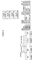
- DNA encoding each cassette was synthesized according to standard techniques and cloned into the appropriate homology vectors (see Figures 4, 5 and 8).
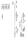
- the plasmid 515-85.1 was constructed for the purpose of inserting foreign DNA into SPV. It contains a unique AccI restriction enzyme site into which foreign DNA may be inserted. When a plasmid, containing a foreign DNA insert at the AccI site, is used according to the HOMOLOGOUS RECOMBINATION PROCEDURE FOR GENERATING RECOMBINANT SPV a virus containing the foreign DNA will result.
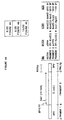
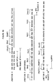
- a restriction map of the DNA insert in homology vector 515-85.1 is given in figure 4. It may be constructed utilizing standard recombinant DNA techniques (22 and 29), by joining two restriction fragments from the following sources.
- the first fragment is an approximately 2972 base pair Hind III to Bam HI restriction fragment of pSP64 (Promega).
- the second fragment is an approximately 3300 base pair Hind III to Bgl II restriction sub-fragment of the SPV Hind III restriction fragment M (23).
- the plasmid 520-17.5 was constructed for the purpose of inserting foreign DNA into SPV. It incorporates an E. coli ⁇ -galactosidase (lacZ) marker gene flanked by SPV DNA. Upstream of the marker gene is an approximately 2015 base pair fragment of SPV DNA. Downstream of the marker gene is an approximately 1103 base pair fragment of SPV DNA.
- lacZ E. coli ⁇ -galactosidase
- Upstream of the marker gene is an approximately 2015 base pair fragment of SPV DNA.
- Downstream of the marker gene is an approximately 1103 base pair fragment of SPV DNA.
- the plasmid vector is derived from an approximately 2972 base pair Hind III to Bam HI restriction fragment of pSP64 (Promega).
- Fragment 1 is an approximately 2015 base pair Hind III to Acc I restriction sub-fragment of the SPV Hind III restriction fragment M (23).
- Fragment 2 is an approximately 3010 base pair Bam HI to Pvu II restriction fragment of plasmid pJF751 (11).
- Fragment 3 is an approximately 1103 base pair Acc I to Bgl II restriction sub-fragment of the SPV Hind III fragment M (23).
- the plasmid 538-46.16 was constructed for the purpose of inserting foreign DNA into SPV. It incorporates an E. coli ⁇ -galactosidase (lacZ) marker gene and the PRV gp50 gene flanked by SPV DNA. Upstream of the foreign genes is an approximately 2015 base pair fragment of SPV DNA. Downstream of the foreign genes is an approximately 1103 base pair fragment of SPV DNA.
- lacZ E. coli ⁇ -galactosidase
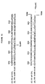
- ⁇ -galactosidase (lacZ) marker gene is under the control of a synthetic late pox promoter and the gp50 gene is under the control of a synthetic early/late pox promoter.
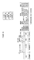
- a detailed description of the plasmid is given in figure 5. It may be constructed utilizing standard recombinant DNA techniques (22 and 30), by joining restriction fragments from the following sources with the synthetic DNA sequences indicated in figure 5.
- the plasmid vector is derived from an approximately 2972 base pair Hind III to BamHI restriction fragment of pSP64 (Promega). Fragment 1 is an approximately 2015 base pair Hind III to Acc I restriction sub-fragment of the SPV Hind III restriction fragment M (23).
- Fragment 2 is an approximately 3010 base pair Bam HI to Pvu II restriction fragment of plasmid pJF751 (11).
- Fragment 3 is an approximately 1571 base pair Eco RI to Stu I restriction sub-fragment of the PRV Bam HI fragment 7 (21). Note that the Eco RI site was introduced in to this fragment by PCR cloning. In this procedure the primers described below were used along with a template consisting of a PRV BamHI #7 fragment subcloned into pSP64.
- the first primer 87.03 (5'-CGCGAATTCGCTCGCAGCGCTATTGGC-3') (SEQ ID NO:41) sits down on the PRV gp50 sequence (26) at approximately amino acid 3 priming toward the 3' end of the gene.
- the second primer 87.06 (5'-GTAGGAGTGGCTGCTGAAG-3') (SEQ ID NO:42) sits down on the opposite strand at approximately amino acid 174 priming toward the 5' end of the gene.
- the PCR product may be digested with EcoRI and Sal I to produce an approximately 509 base pair fragment.
- the approximately 1049 base pair Sail to Stu I sub-fragment of PRV Bam HI #7 may then be ligated to the approximately 509 base pair Eco RI to Sal I fragment to generate the approximately 1558 base pair Eco RI to Stu I fragment 3.
- Fragment 4 is an approximately 1103 base pair Acc I to Bgl II restriction sub-fragment of the SPV Hind III fragment M (23).
- the plasmid 538-46.26 was constructed for the purpose of inserting foreign DNA into SPV. It incorporates an E.coli ⁇ -galactosidase (lacZ) marker gene and the Newcastle Disease Virus (NDV) hemagglutinin-Neuraminidase (HN) gene flanked by SPV DNA. Upstream of the foreign genes is an approximately 2015 base pair fragment of SPV DNA. Downstream of the foreign genes is an approximately 1103 base pair fragment of SPV DNA.
- lacZ E.coli ⁇ -galactosidase
- NDV Newcastle Disease Virus
- HN hemagglutinin-Neuraminidase
- ⁇ -galactosidase (lacZ) marker gene is under the control of a synthetic late pox promoter and the HN gene is under the control of a synthetic early/late pox promoter.
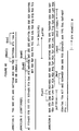
- a detailed description of the plasmid is given in figure 8. It may be constructed utilizing standard recombinant DNA techniques (22 and 30), by joining restriction fragments from the following sources with the synthetic DNA sequences indicated in figure 8.
- the plasmid vector is derived from an approximately 2972 base pair Hind III to BamHI restriction fragment of pSP64 (Promega). Fragment 1 is an approximately 2015 base pair Hind III to Acc I restriction sub-fragment of the SPV Hind III restriction fragment M (23).
- Fragment 2 is an approximately 1810 base pair Ava II to Nae I restriction fragment of a NDV HN cDNA clone. The sequence of the HN cDNA clone is given in figure 7. The cDNA clone was generated from the B1 strain of NDV using standard cDNA cloning techniques (14).
- Fragment 3 is an approximately 3010 base pair Bam HI to PvuII restriction fragment of plasmid pJF751 (11).
- Fragment 4 is an approximately 1103 base pair AccI to Bgl II restriction sub-fragment of the SPV Hind III fragment M (23).
- Plasmid 520-90.15 was constructed for the purpose of inserting foreign DNA into SPV. It contains a unique Nde I restriction enzyme site into which foreign DNA may be inserted. When a plasmid, containing a foreign DNA insert at the Nde I site, is used according to the HOMOLOGOUS RECOMBINATION PROCEDURE FOR GENERATING RECOMBINANT SPV a virus containing the foreign DNA will result. Plasmid 520-90.15 was constructed utilizing standard recombinant DNA techniques (22 and 30), by joining two restriction fragments from the following sources. The first fragment is an approximately 2972 base pair Hind III to Bam HI restriction fragment of pSP64 (Promega). The second fragment is an approximately 1700 base pair HindIII to Bam HI restriction subfragment of the SPV Hind III restriction fragment G (23).
- the homology vector 515-85.1 is a plasmid useful for the insertion of foreign DNA into SPV.
- Plasmid 515-85.1 contains a unique Acc I restriction site into which foreign DNA may be cloned.
- a plasmid containing such a foreign DNA insert may be used according to the HOMOLOGOUS RECOMBINATION PROCEDURE FOR GENERATING RECOMBINANT SPV to generate a SPV containing the foreign DNA.
- the insertion site Acc I
- the site be flanked with swinepox virus DNA appropriate for mediating homologous recombination between virus and plasmid DNAs.
- the Acc I site in homology vector 515-85.1 may be used to insert foreign DNA into at least three recombinant SPV (see examples 2-4).
- recombinant virus was determined by the SCREEN FOR RECOMBINANT SPV EXPRESSING ⁇ -GALACTOSIDASE ASSAYS.
- Four of the six sites were found to generate recombinant virus, however the ability of each of these viruses to be purified away from the parental SPV varied greatly. In one case virus could not be purified above the level of 1%, in another case virus could not be purified above the level of 50%, and in a third case virus could not be purified above the level of 90%.
- the inability to purify these viruses indicates instability at the insertion site. This makes the corresponding sites inappropriate for insertion of foreign DNA. However the insertion at one site, the AccI site of Homology vector 515-85.1, resulted in a virus which was easily purified to 100% (see example 2), clearly defining an appropriate site for the insertion of foreign DNA.
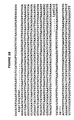
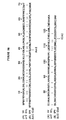
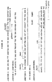
- the homology vector 515-85.1 was further characterized by DNA sequence analysis. Two regions of the homology vector were sequenced. The first region covers a 599 base pair sequence which flanks the unique Acc I site (see figure 2). The second region covers the 899 base pairs upstream of the unique Hind III site (see figure 2). The sequence of the first region codes for an open reading frame (ORF) which shows homology to amino acids 1 to 115 of the vaccinia virus (VV) OIL open reading frame identified by Goebel et al , 1990 (see figure 3). The sequence of the second region codes for an open reading frame which shows homology to amino acids 568 to 666 of the same vaccinia virus OIL open reading frame (see figure 3).
- ORF open reading frame
- VV vaccinia virus
- the sequence of the second region codes for an open reading frame which shows homology to amino acids 568 to 666 of the same vaccinia virus OIL open reading frame (see figure 3).
- VV O1L ORF contains a leucine zipper motif characteristic of certain eukaryotic transcriptional regulatory proteins, however they indicate that it is not known whether this gene is essential for virus replication.
- the DNA sequence located upstream of the VV O1L-like ORF would be expected to contain a swinepox viral promoter.
- This swinepox viral promoter will be useful as the control element of foreign DNA introduced into the swinepox genome.
- S-SPV-003 is a swinepox virus that expresses a foreign gene.
- the gene for E.coli ⁇ -galactosidase (lacZ gene) was inserted into the SPV 515-85.1 ORF.
- the foreign gene (lacZ) is under the control of a synthetic early/late promoter (EP1LP2).
- S-SPV-003 was derived from S-SPV-001 (Kasza strain). This was accomplished utilizing the homology vector 520-17.5 (see Materials and Methods) and virus S-SPV-001 in the HOMOLOGOUS RECOMBINATION PROCEDURE FOR GENERATING RECOMBINANT SPV. The transfection stock was screened by the SCREEN FOR RECOMBINANT SPV EXPRESSING ⁇ -GALACTOSIDASE (BLUOGAL AND CPRG ASSAYS). The final result of red plaque purification was the recombinant virus designated S-SPV-003.
- This virus was assayed for ⁇ -galactosidase expression, purity, and insert stability by multiple passages monitored by the blue plaque assay as described in Materials and Methods. After the initial three rounds of purification, all plaques observed were blue indicating that the virus was pure, stable and expressing the foreign gene.
- the assays described here were carried out in VERO cells as well as EMSK cells, indicating that VERO cells would be a suitable substrate for the production of SPV recombinant vaccines.
- S-SPV-003 has been deposited with the ATCC under Accession No. VR 2335.
- S-SPV-008 is a swinepox virus that expresses at least two foreign genes.
- the gene for E. coli ⁇ -galactosidase (lacZ gene) and the gene for pseudorabies virus (PRV) gp50 (26) were inserted into the SPV 515-85.1 ORF.
- the lacZ gene is under the control of a synthetic late promoter (LP1) and the gp50 gene is under the control of a synthetic early/late promoter (EP1LP2).
- S-SPV-008 was derived from S-SPV-001 (Kasza strain). This was accomplished utilizing the homology vector 538-46.16 (see Materials and Methods) and virus S-SPV-001 in the HOMOLOGOUS RECOMBINATION PROCEDURE FOR GENERATING RECOMBINANT SPV.
- the transfection stock was screened by the SCREEN FOR RECOMBINANT SPV EXPRESSING ⁇ -GALACTOSIDASE (BLUOGAL AND CPRG ASSAYS). The final result of red plaque purification was the recombinant virus designated S-SPV-008.
- This virus was assayed for ⁇ -galactosidase expression, purity, and insert stability by multiple passages monitored by the blue plaque assay as described in Materials and Methods. After the initial three rounds of purification, all plaques observed were blue indicating that the virus was pure, stable and expressing the marker gene.
- S-SPV-008. was assayed for expression of PRV specific antigens using the BLACK PLAQUE SCREEN FOR FOREIGN GENE EXPRESSION IN RECOMBINANT SPV. Swine anti-PRV serum was shown to react specifically with S-SPV-008 plaques and not with S-SPV-009 negative control plaques. All S-SPV-008 observed plaques reacted with the swine antiserum indicating that the virus was stably expressing the PRV foreign gene. The black plaque assay was also performed on unfixed monolayers. The SPV plaques on the unfixed monolayers also exhibited specific reactivity with swine anti-PRV serum indicating that the PRV antigen is expressed on the infected cell surface.
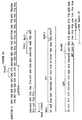
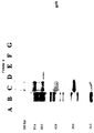
- PRV gp50 To confirm the expression of the PRV gp50 gene product, cells were infected with SPV and samples of infected cell lysates were subjected to SDS-polyacrylamide gel electrophoresis. The gel was blotted and analyzed using the WESTERN BLOTTING PROCEDURE. The swine anti-PRV serum was used to detect expression of PRV specific proteins. As shown in figure 6, the lysate from S-SPV-008 infected cells exhibits a specific band of approximately 48 kd, the reported size of PRV gp50 (35).
- PRV gp50 is the gD homologue of HSV-1 (26).
- VV expressing HSV-1 gD will protect mice against challenge with HSV-1 (6 and 34). Therefore the S-SPV-008 should be valuable as a vaccine to protect swine against PRV disease.
- PRV glycoproteins will be useful in the creation of recombinant swinepox vaccines to protect against PRV disease.
- PRV glycoproteins include gpII (28), gpIII (27), and gpH (19).
- the PRV gpIII coding region has been engineered behind several synthetic pox promoters.
- the techniques utilized for the creation of S-SPV-008 will be used to create recombinant swinepox viruses expressing all four of these PRV glycoprotein genes. Such recombinant swinepox viruses will be useful as vaccines against PRV disease.
- PRV vaccines described here do not express PRV gpX or gpI, they would be compatible with current PRV diagnostic tests (gX HerdChek®, gI HerdChek® and ClinEase®) which are utilized to distinguish vaccinated animals from infected animals.
- S-SPV-008 has been deposited with the ATCC under Accession No. VR 2339.
- S-SPV-009 is a swinepox virus that expresses at least two foreign genes.
- the gene for E. coli ⁇ -galactosidase (lacZ gene) and the gene for Newcastle's Disease virus hemagglutinin (HN) gene were inserted into the SPV 515-85.1 ORF.
- the lacZ gene is under the control of a synthetic late promoter (LP1) and the HN gene is under the control of an synthetic early/late promoter (EP1LP2).
- S-SPV-009 was derived from S-SPV-001 (Kasza strain). This was accomplished utilizing the homology vector 538-46.26 (see Materials and Methods) and virus S-SPV-001 in the HOMOLOGOUS RECOMBINATION PROCEDURE FOR GENERATING RECOMBINANT SPV.
- the transfection stock was screened by the SCREEN FOR RECOMBINANT SPV EXPRESSING ⁇ -GALACTOSIDASE (BLUOGAL AND CPRG ASSAYS). The final result of red plaque purification was the recombinant virus designated S-SPV-009.
- This virus was assayed for ⁇ -galactosidase expression, purity, and insert stability by multiple passages monitored by the blue plaque assay as described in Materials and Methods. After the initial three rounds of purification, all plaques observed were blue indicating that the virus was pure, stable and expressing the marker gene.
- S-SPV-009 was assayed for expression of PRV specific antigens using the BLACK PLAQUE SCREEN FOR FOREIGN GENE EXPRESSION IN RECOMBINANT SPV. Rabbit anti-NDV HN serum was shown to react specifically with S-SPV-009 plaques and not with S-SPV-008 negative control plaques. All S-SPV-009 observed plaques reacted with the swine antiserum indicating that the virus was stably expressing the NDV foreign gene. S-SPV-009 has been deposited with the ATCC under Acession No. VR 2344).
- NDV HN NDV HN gene product
- cells were infected with SPV and samples of infected cell lysates were subjected to SDS-polyacrylamide gel electrophoresis. The gel was blotted and analyzed using the WESTERN BLOTTING PROCEDURE. The rabbit anti-NDV HN serum was used to detect expression of the HN protein.
- the lysate from S-SPV-009 infected cells exhibited a specific band of approximately 74 kd, the reported size of NDV HN (29).
- TGE transmissible gastroenteritis virus
- glycoprotein 195 for use in the swinepox virus vector
- the clone of the neutralizing antigen is disclosed in U.S. Serial No. 078,519, filed July 27, 1987. It is contemplated that the procedures that have been used to express PRV gp50 in SPV and are disclosed herein are applicable to TGE.
- glycoprotein 38 we have cloned the major neutralizing antigen of the swine rotavirus, glycoprotein 38, for use in the swinepox virus vector.
- the clone of glycoprotein 38 is disclosed in U.S. Patent No. 5,068,192 issued November 26, 1991. It is contemplated that the procedures that have been used to express PRV gp50 in SPV and are disclosed herein are applicable to SRV.
- the major neutralizing antigen of the bovine viral diarrhea (BVD) virus was cloned as disclosed in U.S. Serial No. 225,032, filed July 27, 1988. Since the BVD and hog cholera viruses are cross protective (31), the BVD virus antigen has been targeted for use in the swinepox virus vector. It is contemplated that the procedures that have been used to express PRV gp50 in SPV and are disclosed herein are applicable to BVD virus.
- a protective antigen of Serpulina hyodysenteriae (3), for use in the swinepox virus vector has been cloned. It is contemplated that the procedures that have been used to express PRV gp50 in SPV and are disclosed herein are also applicable to Serpulina hyodysenteriae.
- Antigens from the following microorganisms may also be utilized to develop animal vaccines: Swine influenza virus, foot and mouth disease virus, African swine fever virus, hog cholera virus and Mycoplasma hyodysenteriae.
Landscapes
- Health & Medical Sciences (AREA)
- Chemical & Material Sciences (AREA)
- Life Sciences & Earth Sciences (AREA)
- Organic Chemistry (AREA)
- Genetics & Genomics (AREA)
- General Health & Medical Sciences (AREA)
- Biophysics (AREA)
- Biochemistry (AREA)
- Molecular Biology (AREA)
- Engineering & Computer Science (AREA)
- Virology (AREA)
- Medicinal Chemistry (AREA)
- Biomedical Technology (AREA)
- Wood Science & Technology (AREA)
- Biotechnology (AREA)
- Proteomics, Peptides & Aminoacids (AREA)
- Gastroenterology & Hepatology (AREA)
- General Engineering & Computer Science (AREA)
- Bioinformatics & Cheminformatics (AREA)
- Zoology (AREA)
- Plant Pathology (AREA)
- Microbiology (AREA)
- Physics & Mathematics (AREA)
- Pharmacology & Pharmacy (AREA)
- Oncology (AREA)
- Veterinary Medicine (AREA)
- Nuclear Medicine, Radiotherapy & Molecular Imaging (AREA)
- General Chemical & Material Sciences (AREA)
- Chemical Kinetics & Catalysis (AREA)
- Public Health (AREA)
- Animal Behavior & Ethology (AREA)
- Communicable Diseases (AREA)
- Micro-Organisms Or Cultivation Processes Thereof (AREA)
- Medicines Containing Antibodies Or Antigens For Use As Internal Diagnostic Agents (AREA)
- Medicines Containing Material From Animals Or Micro-Organisms (AREA)
- Measuring Or Testing Involving Enzymes Or Micro-Organisms (AREA)
- Preparation Of Compounds By Using Micro-Organisms (AREA)
Abstract
Description
- Within this application several publications are referenced by arabic numerals within parentheses. Full citations for these publications may be found at the end of the specification immediately preceding the claims.
- Swinepox virus (SPV) belongs to the family Poxviridae. Viruses belonging to this group are large, double-stranded DNA viruses that characteristically develop in the cytoplasm of the host cell. SPV is the only member of the genus Suipoxvirus. Several features distinguish SPV from other poxviruses. SPV exhibits species specificity (18) compared to other poxviruses such as vaccinia which exhibit a broad host range. SPV infection of tissue culture cell lines also differs dramatically from other poxviruses (24). It has also been demonstrated that SPV does not exhibit antigenic cross-reactivity with vaccinia virus and shows no gross detectable homology at the DNA level with the ortho, lepori, avi or entomopox virus groups (24). Accordingly, what is known and described in the prior art regarding other poxviruses does not pertain a priori to swinepox virus.
- SPV is only mildly pathogenic, being characterized by a self-limiting infection with lesions detected only in the skin and regional lymph nodes. Although the SPV infection is quite limited, pigs which have recovered from SPV are refractory to challenge with SPV, indicating development of active immunity (18).
- The present invention concerns the use of SPV as a vector for the delivery of vaccine antigens and therapeutic agents to swine. The following properties of SPV support this rationale: SPV is only mildly pathogenic in swine, SPV is species specific, and SPV elicits a protective immune response. Accordingly, SPV is an excellent candidate for a viral vector delivery system, having little intrinsic risk which must be balanced against the benefit contributed by the vector's vaccine and therapeutic properties.
- The prior art for this invention stems first from the ability to clone and analyze DNA while in bacterial plasmids. The techniques that are available are detailed for the most part in Maniatis et al., 1983 and Sambrook et al., 1989. These publications teach state of the art general recombinant DNA techniques.
- Among the poxviruses, five (vaccinia, fowlpox, canarypox, pigeon, and raccoon pox) have been engineered, previous to this disclosure, to contain foreign DNA sequences. Vaccinia virus has been used extensively to vector foreign genes (25) and is the subject of U.S. Patents 4,603,112 and 4,722,848. Similarly, fowlpox has been used to vector foreign genes and is the subject of several patent applications EPA 0 284 416, PCT WO 89/03429, and PCT WO 89/12684. Raccoon pox (10) and Canarypox (31) have been utilized to express antigens from the rabies virus. These examples of insertions of foreign genes into poxviruses do not include an example from the genus Suipoxvirus. Thus, they do not teach methods to genetically engineer swinepox viruses, that is, where to make insertions and how to get expression in swinepox virus.
- The idea of using live viruses as delivery systems for antigens has a very long history going back to the first live virus vaccines. The antigens delivered were not foreign but were naturally expressed by the live virus in the vaccines. The use of viruses to deliver foreign antigens in the modern sense became obvious with the recombinant vaccinia virus studies. The vaccinia virus was the vector and various antigens from other disease causing viruses were the foreign antigens, and the vaccine was created by genetic engineering. While the concept became obvious with these disclosures, what was not obvious was the answer to a more practical question of what makes the best candidate virus vector. In answering this question, details of the pathogenicity of the virus, its site of replication, the kind of immune response it elicits, the potential it has to express foreign antigens, its suitability for genetic engineering, its probability of being licensed by regulatory agencies, etc, are all factors in the selection. The prior art does not teach these questions of utility.
- The prior art relating to the use of poxviruses to deliver therapeutic agents relates to the use of a vaccinia virus to deliver interleukin-2 (12). In this case, although the interleukin-2 had an attenuating effect on the vaccinia vector, the host did not demonstrate any therapeutic benefit.
- The therapeutic agent that is delivered by a viral vector of the present invention must be a biological molecule that is a by-product of swinepox virus replication. This limits the therapeutic agent in the first analysis to either DNA, RNA or protein. There are examples of therapeutic agents from each of these classes of compounds in the form of anti-sense DNA, anti-sense RNA (16), ribozymes (34), suppressor tRNAs (2), interferon-inducing double stranded RNA and numerous examples of protein therapeutics, from hormones, e.g., insulin, to lymphokines, e.g., interferons and interleukins, to natural opiates. The discovery of these therapeutic agents and the elucidation of their structure and function does not make obvious the ability to use them in a viral vector delivery system.
- The present invention provides a recombinant swinepox virus which comprises a swinepox viral genome containing a foreign DNA sequence inserted within the larger HindIII to BglII subfragment of the HindIII M fragment of the genomic DNA, and wherein the foreign DNA sequence is expressed in a host cell infected with the recombinant swinepox virus.
- The present invention provides a homology vector for producing a recombinant swinepox virus by inserting a foreign DNA into the genomic DNA of the swinepox virus, which comprises
- a) a double-stranded foreign DNA sequence,
- b) flanked at one end of the foreign DNA by double-stranded swinepox viral DNA homologous to the genomic DNA located at one side of a site on the genomic DNA, wherein, said site is located in the larger HindIII to BglII subfragment of the HindIII M fragment of the swinepox genomic DNA,
- c) flanked at the other end of the foreign DNA by double-stranded swinepox viral DNA homologous to genomic DNA located at the other side of the same site on the genomic DNA.
-
- Figure 1
- Details of the SPV Kasza Strain. Diagram of SPV genomic DNA showing the unique long and Terminal repeat (TR) regions. A restriction map for the enzyme HindIII is indicated (23). Fragments are lettered in order of decreasing size. Note that the terminal repeats are greater than 2.1 Kb but less than 9.7 kb in size.
- Figure 2
- DNA sequence from homology vector 515-85.1. The sequence of two regions of the homology vector 515-85.1 are shown. The first region (Figure 2A) (SEQ ID NO:1) covers a 599 base pair sequence which flanks the unique AccI site as indicated in Figure 3. The beginning (Met) and end (Val) of a 115 amino acid ORF is indicated by the translation of amino acids below the DNA sequence. The second region (Figure 2B) (SEQ ID NO:3) covers the 899 base pairs upstream of the unique HindIII site as indicated in Figure 3. The beginning (Asp) and end (Ile) of a 220 amino acid ORF is indicated by the translation of amino acids below the DNA sequence.
- Figure 3
- Homology between the 515.85.1 ORF and the
Vaccinia virus 01L ORF. The first line shows a restriction map of the SPV HindIII M fragment. The second map shows a restriction map of the DNA insertion in plasmid 515-85.1. The location of the 515-85.1 [VV 01L-like] ORF is indicated on the map. The locations of the DNA sequences shown in Figure 2 are indicated below the map by heavy bars. The third line shows the homology between the VV 01L ORF (SEQ ID NO:5) and the 515-85.1 ORF (SEQ ID NO:6) at their respective N-termini. The fourth line shows the homology between the VV O1L ORF (SEQ ID NO:7) and the 515-85.1 ORF (SEQ ID NO:8) at their respective C-termini. - Figure 4
- Detailed description of the DNA insertion in Homology Vector 520-17.5. Diagram showing the orientation of DNA fragments assembled in plasmid 520-17.5. The origin of each fragment is indicated in the table. The sequences located at each of the junctions between fragments is also shown (SEQ ID NO's: 9, 10, 13, and 16). The restriction sites used to generate each fragment as well as the synthetic linker sequences which were used to join the fragments are described for each junction. The synthetic linker sequences are underlined by a heavy bar. The location of several gene coding regions and regulatory elements are also given. The following two conventions are used: numbers in parenthesis ( ) refer to amino acids, and restriction sites in brackets [ ] indicate the remnants of sites which were destroyed during construction. The following abbreviations are used, swinepox virus (SPV), early promoter 1 (EP1), late promoter 2 (LP2), lactose operon Z gene (lacZ), and Escherichia coli (E. coli).
- Figure 5
- Detailed description of the DNA insertion in Homology Vector 538-46.16. Diagram showing the orientation of DNA fragments assembled in plasmid 538-46.16. The origin of each fragment is indicated in the table. The sequences located at each of the junctions between fragments are also shown (SEQ ID NO's: 17, 18, 21, 26, and 28). The restriction sites used to generate each fragment as well as the synthetic linker sequences which were used to join the fragments are described for each junction. The synthetic linker sequences are underlined by a heavy bar. The location of several gene coding regions and regulatory elements is also given. The following two conventions are used: numbers in parenthesis ( ) refer to amino acids, and restriction sites in brackets [ ] indicate the remnants of sites which were destroyed during construction. The following abbreviations are used, swinepox virus (SPV), pseudorabies virus (PRV), glycoprotein 50 (gp50), glycoprotein 63 (gp63), early promoter 1 (EP1), late promoter 1 (LP1), late promoter 2 (LP2), lactose operon Z gene (lacZ), and Escherichia coli (E. coli).
- Figure 6
- Western blot of lysates from recombinant SPV infected cells with anti-serum to PRV. Lanes (A) uninfected Vero cell lysate, (B) S-PRV-000 (pseudorabies virus Cooper strain) infected cell lysate, (C) pre-stained molecular weight markers, (D) uninfected EMSK cell lysate, (E) S-SPV-000 infected cell lysate, (F) S-SPV-003 infected cell lysate, (G) S-SPV-008 infected cell lysate. Cell lysates were prepared as described in the PREPARATION OF INFECTED CELL LYSATES. Approximately 1/5 of the total lysate sample was loaded in each lane.
- Figure 7
- DNA sequence of NDV Hemagglutinin-Neuraminidase gene (HN) (SEQ ID NO: 29). The sequence of 1907 base pairs of the NDV HN cDNA clone are shown. The translational start and stop of the HN gene is indicated by the amino acid translation below the DNA sequence.
- Figure 8
- Detailed description of the DNA insertion in Homology Vector 538-46.26. Diagram showing the orientation of DNA fragments assembled in plasmid 538-46.26. The origin of each fragment is indicated in the table. The sequences located at each of the junctions between fragments is also shown (SEQ ID NO's: 31, 32, 34, 37, and 40). The restriction sites used to generate each fragment as well as the synthetic linker sequences which were used to join the fragments are described for each junction. The synthetic linker sequences are underlined by a heavy bar. The location of several gene coding regions and regulatory elements is also given. The following two conventions are used: numbers in parenthesis ( ) refer to amino acids, and restriction sites in brackets [ ] indicate the remnants of sites which were destroyed during construction. The following abbreviations are used, swinepox virus (SPV), Newcastle Disease virus (NDV), hemagglutinin-neuraminidase (HN), early promoter 1 (EP1), late promoter 1 (LP1), late promoter 2 (LP2), lactose operon Z gene (lacZ), and Escherichia coil (E. coli).
- The present invention provides a recoabinant swinepox virus (SPV) capable of replication in an animal into which the recombinant swinepox virus is introduced which comprises swinepox viral DNA and foreign DNA encoding RNA which does not naturally occur in the animal into which the recombinant swinepox virus is introduced, the foreign DNA being inserted into the swinepox viral DNA at an insertion site which is not essential for replication of the swinepox virus and being under the control of a promoter. More specifically, the invention provides a recombinant swinepox virus which comprises a swinepox viral genome containing a foreign DNA sequence inserted within the larger HindIII to BglII subfragment of the HindIII M fragment of the genomic DNA, and wherein the foreign DNA sequence is expressed in a host cell infected with the recombinant swinepox virus.
- For purposes of this invention, "a recombinant swinepox virus capable of replication" is a live swinepox virus which has been generated by the recombinant methods well known to those of skill in the art, e.g., the methods set forth in HOMOLOGOUS RECOMBINATION PROCEDURE FOR GENERATING RECOMBINANT SPV in Materials and Methods and has not had genetic material essential for the replication of the recombinant swinepox virus deleted.
- For purposes of this invention, "an insertion site which is not essential for replication of the swinepox virus" is a location in the genome where a sequence of DNA is not necessary for viral replication, for example, complex protein binding sequences, sequences which code for reverse transcriptase or an essential glycoprotein, DNA sequences necessary for packaging, etc.
- For purposes of this invention, a "promoter" is a specific DNA sequence on the DNA molecule to which the foreign RNA polymerase attaches and at which transcription of the foreign RNA is initiated.
- The invention further provides foreign RNA which encodes a polypeptide. Preferably, the polypeptide is antigenic in the animal.
- Preferably, this antigenic polypeptide is a linear polymer of more than 10 amino acids linked by peptide bonds which stimulates the animal to produce antibodies.
- The invention provides an insertion site present within the larger HindIII to BglII subfragment of the BindIII M fragment of swinepox viral DNA. Preferably, the insertion site is within an open reading frame contained in the HindIII to BglII subfragment. Preferably, the insertion site is the AccI restriction endonuclease site located in the HindIII to BglII subfragment.
- For purposes of this invention, an "open reading frame" is a segment of DNA which contains codons that can be transcribed into RNA which can be translated into an amino acid sequence and which does not contain a termination codon.
- The invention further provides a recombinant swinepox virus comprising foreign DNA encoding RNA encoding an antigenic polypeptide which is or is from pseudorabies virus (PRV)
glycoprotein 50, pseudorabies virus (PRV) glycoprotein II, Pseudorabies virus (PRV) glycoprotein III, Pseudorabies virus (PRV) glycoprotein H, Transmissible gastroenteritis (TGE) glycoprotein 195, Transmissible gastroenteritis (TGE) matrix protein, swine rotavirus glycoprotein 38, swine parvovirus capsid protein, Serpulina hydodysenteriae protective antigen, Bovine Viral Diarrhea (BVD) glycoprotein 55, Newcastle Disease Virus (NDV) hemagglutinin-neuraminidase, swine flu hemagglutinin or swine flu neuraminidase. Preferably, the antigenic polypeptide is Pseudorabies Virus (PRV)glycoprotein 50. Preferably, the antigenic protein is Newcastle Disease Virus (NDV) hemagglutinin-neuraminidase. - The invention further provides a recombinant swinepox virus comprising foreign DNA encoding RNA encoding an antigenic polypeptide which is or is from Serpulina hyodysenteriae, Foot and Mouth Disease Virus, Hog Cholera Virus, Swine Influenza Virus, African Swine Fever Virus or Mycoplasma hyopneumoniae.
- The invention further provides a recombinant swinepox virus where the foreign DNA encodes RNA which encodes a polypeptide which is a detectable marker. Preferably the detectable marker is the polypeptide E. coil β-galactosidase. For purposes of this invention, a "polypeptide which is a detectable marker" includes the bimer, trimer and tetramer form of the polypeptide. E. coli β-galactosidase is a tetramer composed of four polypeptides or monomer sub-units. Preferably, this recombinant swinepox virus is designated S-SPV-003 (ATCC Accession No. VR 2335). The S-SPV-003 swinepox virus has been deposited pursuant to the Budapest Treaty on the International Deposit of Microorganisms for the Purposes of Patent Procedure with the Patent Culture Depository of the American Type Culture Collection, 12301 Parklawn Drive, Rockville, Maryland 20852 U.S.A. under ATCC Accession No. VR 2335.
- The invention further provides for a recombinant swinepox virus capable of replication which contains foreign DNA encoding RNA encoding the antigenic polypeptide pseudorabies virus (PRV)
glycoprotein 50 further comprising foreign DNA encoding a polypeptide which is a detectable marker. Preferably, this recombinant swinepox virus is designated S-SPV-008 (ATCC Accession No. VR 2339). The S-SPV-008 swinepox virus has been deposited pursuant to the Budapest Treaty on the International Deposit of Microorganisms for the Purposes of Patent Procedure with the Patent Culture Depository of the American Type Culture Collection, 12301 Parklawn Drive, Rockville, Maryland 20852 U.S.A. under ATCC Accession No. VR 2339. - The invention further provides for a recombinant swinepox virus capable of replication which contains foreign DNA encoding RNA encoding the antigenic polypeptide Newcastle Disease Virus (NDV) hemagglutinin-neuraminidase further comprising foreign DNA encoding a polypeptide which is a detectable marker. Preferably, this recombinant swinepox virus is designated S-SPV-009 (ATCC Accession No. VR 2344). The S-SPV-009 swinepox virus has been deposited pursuant to the Budapest Treaty on the International Deposit of Microorganisms for the Purposes of Patent Procedure with the Patent Culture Depository of the American Type Culture Collection, 12301 Parklawn Drive, Rockville, Maryland 20852 U.S.A. under ATCC Accession No. VR 2344.
- The invention further provides that the inserted foreign DNA is under the control of a promoter. Preferably, the promoter is a swinepox viral promoter. Preferably, the promoter is a synthetic pox viral promoter. For purposes of this invention, the promoters were generated by methods well known to those of skill in the art, for example, as set forth in the STRATEGY FOR THE CONSTRUCTION OF SYNTHETIC POX VIRAL PROMOTERS in Materials and Methods. For purposes of this invention, a synthetic pox promoter includes a synthetic late pox promoter, a synthetic early pox promoter or a synthetic early/late pox promoter.
- The invention provides for a homology vector for producing a recombinant swinepox virus by inserting foreign DNA into the genomic DNA of a swinepox virus. The homology vector comprises a double-stranded DNA molecule consisting essentially of a double-stranded foreign DNA encoding RNA which does not naturally occur in an animal into which the recombinant swinepox virus is introduced, with at one end of the foreign DNA, double-stranded swinepox viral DNA homologous to genomic DNA located at one side of a site on the genomic DNA which is not essential for replication of the swinepox virus, and at the other end of the foreign DNA, double-stranded swinepox viral DNA homologous to genomic DNA located at the other side of the same site on the genomic DNA.
- More specifically, the invention provides for a homology vector for producing a recombinant swinepox virus by inserting a foreign DNA into the genomic DNA of the swinepox virus, which comprises
- a) a double-stranded foreign DNA sequence,
- b) flanked at one end of the foreign DNA by double-stranded swinepox viral DNA homologous to the genomic DNA located at one side of a site on the genomic DNA, wherein said site is located in the larger HindIII to BglII subfragment of the HindIII M fragment of the swinepox genomic DNA,
- c) flanked at the other end of the foreign DNA by double-stranded swinepox viral DNA homologous to genomic DNA located at the other side of the same site on the genomic DNA.
-
- Preferably, the foreign DVA encodes a polypeptide.
- In one embodiment, the polypeptide is antigenic in the animal. Preferably, the antigenic polypeptide is or is from pseudorabies virus (PRV)
glycoprotein 50, pseudorabies virus (PRV) glycoprotein II, Pseudorabies virus (PRV) glycoprotein III, Pseudorabies virus (PRV) glycoprotein H, Transmissible gastroenteritis (TGE) glycoprotein 195, Transmissible gastroenteritis (TGE) matrix protein, swine rotavirus glycoprotein 38, swine parvovirus capsid protein, Serpulina hydodysenteriae protective antigen, Bovine Viral Diarrhea (BVD) glycoprotein 55, Newcastle Disease Virus (NDV) hemagglutinin-neuraminidase, swine flu hemagglutinin or swine flu neuraminidase. Preferably, the antigenic polypeptide is or is from Serpulina hyodysenteriae, Foot and Mouth Disease Virus, Hog Cholera Virus, Swine Influenza Virus, African Swine Fever Virus or Mycoplasma hyopneumoniae. - In one embodiment, the polypeptide is a detectable marker. Preferably, the polypeptide which is a detectable marker is E. coli β-galactosidase.
- The double-stranded swinepox viral DNA is homologous to genomic DNA present within the larger HindIII to BglII subfragment of the HindIII M fragment of swinepox virus. Preferably, the double-stranded swinepox viral DNA is homologous to genomic DNA present within the open reading frame contained in this HindIII to BglII subfragment. Preferably, the double-stranded swinepox viral DNA is homologous to genomic DNA present within the AccI restriction endonuclease site located in this HindIII to BglII subfragment.
- For purposes of this invention, a "homology vector" is a plasmid constructed to insert foreign DNA in a specific site on the genome of a swinepox virus.
- The invention further provides a homology vector where foreign DNA further comprises a synthetic pox viral promoter.
- The invention further provides a vaccine which comprises an effective immunizing amount of a recoobinant swinepox virus of the present invention and a suitable carrier.
- Suitable carriers for the pseudorabies virus are well known in the art and include proteins, sugars, etc. One example of such a suitable carrier is a physiologically balanced culture medium containing one or more stabilizing agents such as stabilized, hydrolyzed proteins, lactose, etc.
- For purposes of this invention, an "effective immunizing amount" of the recombinant swinepox virus of the present invention is within the range of 103 to 109 PFU/dose.
- The present invention discloses a method of immunizing an animal, wherein the animal is a swine, bovine, equine, caprine or ovine. For purposes of this invention, this includes immunizing the animal against the virus or viruses which cause the disease or diseases pseudorabies, transmissible gastroenteritis, swine rotavirus, swine parvovirus, Serpulina hyodysenteriae, bovine viral diarrhea, Newcastle disease, swine flu, foot and mouth disease, hog cholera, African swine fever or Mycoplasma hyopneumoniae. The method comprises administering to the animal an effective immunizing dose of the vaccine of the present invention. The vaccine may be administered by any of the methods well known to those skilled in the art, for example, by intramuscular, subcutaneous, intraperitoneal or intravenous injection. Alternatively, the vaccine may be administered intranasally or orally.
- The present invention also discloses a method for testing a swine to determine whether the swine has been vaccinated with the vaccine of the present invention, particularly the embodiment which contains the recombinant swinepox virus S-SPV-008 (ATCC Accession No. VR 2339), or is infected with a naturally-occurring, wild-type pseudorabies virus. This method comprises obtaining from the swine to be tested a sample of a suitable body fluid, detecting in the sample the presence of antibodies to pseudorabies virus, the absence of such antibodies indicating that the swine has been neither vaccinated nor infected, and for the swine in which antibodies to pseudorabies virus are present, detecting in the sample the absence of antibodies to pseudorabies virus antigens which are normally present in the body fluid of a swine infected by the naturally-occurring pseudorabies virus but which are not present in a vaccinated swine indicating that the swine was vaccinated and is not infected.
- The present invention also discloses a host cell infected with a recombinant swinepox virus capable of replication.
- In one embodiment, the host cell is a mammalian cell. Preferably, the mammalian cell is a Vero cell. Preferably, the mammalian cell is an EMSK cell.
- For purposes of this invention a "host cell" is a cell used to propagate a vector and its insert. Infecting the cells was accomplished by methods well known to those of skill in the art, for example, as set forth in INFECTION - TRANSFECTION PROCEDURE in Material and Methods.
- Methods for constructing, selecting and purifying recombinant swinepox virus, including S-SPV-003, S-SPV-008 and S-SPV-009, are detailed below in Materials and Methods.
- PREPARATION OF SWINEPOX VIRUS STOCK SAMPLES. Swinepox virus (SPV) samples were prepared by infecting embryonic swine kidney (EMSK) cells at a multiplicity of infection of 0.01 PFU/cell in a 1:1 mixture of Iscove's Modified Dulbecco's Medium (IMDM) and RPMI 1640 medium containing 2 mM glutamine, 100 units/ml penicillin, 100 units/ml streptomycin (these components were obtained from Sigma or equivalent supplier, and hereafter are referred to as EMSK negative medium). Prior to infection, the cell monolayers were washed once with EMSK negative medium to remove traces of fetal bovine serum. The SPV contained in the initial inoculum (0.5 ml for 10 cm plate; 10 ml for T175 cm flask) was then allowed to absorb onto the cell monolayer for two hours, being redistributed every half hour. After this period, the original inoculum was brought up to the recommended volume with the addition of complete EMSK medium (EMSK negative medium plus 5% fetal bovine serum). The plates were incubated at 37°C in 5% CO2 until cytopathic effect was complete. The medium and cells were harvested and frozen in a 50 ml conical screw cap tube at -70°C. Upon thawing at 37°C, the virus stock was aliquoted into 1.0 ml vials and refrozen at -70°C. The titers were usually about 106 PFU/ml.
- PREPARATION OF SPV DNA. For swinepox virus DNA isolation, a confluent monolayer of EMSK cells in a T175 cm2 flask was infected at a multiplicity of 0.1 and incubated 4-6 days until the cells were showing 100% cytopathic effect. The infected cells were then harvested by scraping the cells into the medium and centrifuging at 3000 rpm for 5 minutes in a clinical centrifuge. The medium was decanted, and the cell pellet was gently resuspended in 1.0 ml Phosphate Buffer Saline (PBS: 1.5g Na2HPO4, 0.2g KH2PO4, 0.8g NaCL and 0.2g KCl per liter H2O) (per T175) and subjected to two successive freeze-thaws (-70° C to 37° C). Upon the last thaw, the cells (on ice) were sonicated two times for 30 seconds each with 45 seconds cooling time in between. Cellular debris was then removed by centrifuging (Sorvall RC-5B superspeed centrifuge) at 3000 rpm for 5 minutes in a HB4 rotor at 4° C. SPV virions, present in the supernatant, were then pelleted by centrifugation at 15,000 rpm for 20 minutes at 4° C in a SS34 rotor (Sorvall) and resuspended in 10 mM Tris (pH 7.5). This fraction was then layered onto a 36% sucrose gradient (w/v in 10 mM tris pH 7.5) and centrifuged (Beckman L8-70M Ultracentrifuge) at 18,000 rpm for 60 minutes in a SW41 rotor (Beckman) at 4° C. The virion pellet was resuspended in 1.0 ml of 10 mM tris pH 7.5 and sonicated on ice for 30 seconds. This fraction was layered onto a 20% to 50% continuous sucrose gradient and centrifuged 16,000 rpm for 60 minutes in a SW41 rotor at 4° C. The SPV virion band located about three quarters down the gradient was harvested, diluted with 20% sucrose and pelleted by centrifugation at 18,000 rpm for 60 minutes in a SW41 rotor at 4° C. The resultant pellet was then washed once with 10 mM Tris pH 7.5 to remove traces of sucrose and finally resuspended in 10 mM Tris pH 7.5. SPV DNA was then extracted from the purified virions by lysis (4 hours at 60° C) induced by the addition of EDTA, SDS, and proteinase K to final concentrations of 20 mM, 0.5% and 0.5 mg/ml, respectively. After digestion, three phenol:chloroform (1:1) extractions were conducted and the sample precipitated by the addition of two volumes of absolute ethanol and incubation at -20° C for 30 minutes. The sample was then centrifuged in an Eppendorf minifuge for 5 minutes at full speed. The supernatant was decanted, and the pellet air dried and rehydrated in 0.01 M Tris pH 7.5, 1 mM EDTA at 4° C.
- PREPARATION OF INFECTED CELL LYSATES. For cell lysate preparation, serum free medium was used. A confluent monolayer of cells (EMSK for SPV or VERO for PRV) in a 25 cm2 flask or a 60 mm petri dish was infected with 100 µl of virus sample. After cytopathic effect was complete, the medium and cells were harvested and the cells were pelleted at 3000 rpm for 5 minutes in a clinical centrifuge. The cell pellet was resuspended in 250 µl of disruption buffer (2% sodium dodecyl sulfate, 2% β-mercapto-ethanol). The samples were sonicated for 30 seconds on ice and stored at -20°C.
- WESTERN BLOTTING PROCEDURE. Samples of lysates and protein standards were run on a polyacrylamide gel according to the procedure of Laemnli (1970). After gel electrophoresis the proteins were transferred and processed according to Sambrook et al. (1982). The primary antibody was a swine anti-PRV serum (Shope strain; lot370, PDV8201, NVSL, Ames, IA) diluted 1:100 with 5% non-fat dry milk in Tris-sodium chloride, and sodium Azide (TSA: 6.61g Tris-HCl, 0.97g Tris-base, 9.0g NaCl and 2.0g Sodium Azide per liter H2O). The secondary antibody was a goat anti-swine alkaline phosphatase conjugate diluted 1:1000 with TSA.
- MOLECULAR BIOLOGICAL TECHNIQUES. Techniques for the manipulation of bacteria and DNA, including such procedures as digestion with restriction endonucleases, gel eiectrophoresis, extraction of DNA from gels, ligation, phosphorylation with kinase, treatment with phosphatase, growth of bacterial cultures, transformation of bacteria with DNA, and other molecular biological methods are described by Maniatis et al. (1982) and Sambrook et al. (1989). Except as noted, these were used with minor variation.
- DNA SEQUENCING. Sequencing was performed using the USB Sequenase Kit and 35S-dATP (NEN). Reactions using both the dGTP mixes and the dITP mixes were performed to clarify areas of compression. Alternatively, compressed areas were resolved on formamide gels. Templates were double-stranded plasmid subclones or single stranded M13 subclones, and primers were either made to the vector just outside the insert to be sequenced, or to previously obtained sequence. Sequence obtained was assembled and compared using Dnastar software. Manipulation and comparison of sequences obtained was performed with Superclone™ and Supersee™ programs from Coral Software.
- CLONING WITH THE POLYMERASE CHAIN REACTION. The polymerase chain reaction (PCR) was used to introduce restriction sites convenient for the manipulation of various DNAs. The procedures used are described by Innis, et al. (1990). In general, amplified fragments were less than 500 base pairs in size and critical regions of amplified fragments were confirmed by DNA sequencing. The primers used in each case are detailed in the descriptions of the construction of homology vectors below.
- HOMOLOGOUS RECOMBINATION PROCEDURE FOR GENERATING RECOMBINANT SPV. This method relies upon the homologous recombination between the swinepox virus DNA and the plasmid homology vector DNA which occurs in the tissue culture cells containing both swinepox virus DNA and transfected plasmid homology vector. For homologous recombination to occur, the monolayers of EMSK cells are infected with S-SPV-001 (Kasza SPV strain, 17) at a multiplicity of infection of 0.01 PFU/cell to introduce replicating SPV (i.e. DNA synthesis) into the cells. The plasmid homology vector DNA is then transfected into these cells according to the INFECTION - TRANSFECTION PROCEDURE. The construction of homology vectors used in this procedure is described below
- INFECTION - TRANSFECTION PROCEDURE. 6 cm plates of EMSK cells (about 80% confluent) were infected with S-SPV-001 at a multiplicity of infection of 0.01 PFU/cell in EMSK negative medium and incubated at 37°C in a humidified 5% CO2 environment for 5 hours. The transfection procedure used is essentially that recommended for Lipofectin™ Reagent (BRL). Briefly, for each 6 cm plate, 15 µg of plasmid DNA was diluted up to 100 µl with H2O. Separately, 50 micrograms of Lipofectin Reagent was diluted to 100 µl with H2O. The 100 µl of diluted Lipofectin Reagent was then added dropwise to the diluted plasmid DNA contained in a polystyrene 5 ml snap cap tube and mixed gently. The mixture was then incubated for 15-20 minutes at room temperature. During this time, the virus inoculum was removed from the 6 cm plates and the cell monolayers washed once with EMSK negative medium. Three ml of EMSK negative medium was then added to the plasmid DNA/lipofectin mixture and the contents pipetted onto the cell monolayer. The cells were incubated overnight (about 16 hours) at 37°C in a humidified 5% CO2 environment. The next day the 3 ml of EMSK negative medium was removed and replaced with 5 ml EMSK complete medium. The cells were incubated at 37°C in 5% CO2 for 3-7 days until cytopathic effect from the virus was 80-100%. Virus was harvested as described above for the preparation of virus stocks. This stock was referred to as a transfection stock and was subsequently screened for recombinant virus by the BLUOGAL SCREEN FOR RECOMBINANT SWINEPOX VIRUS OR CPRG SCREEN FOR RECOMBINANT SWINEPOX VIRUS.
- SCREEN FOR RECOMBINANT SPV EXPRESSING B-GALACTOSIDASE (BLUOGAL AND CPRG ASSAYS). When the E. coli β-galactosidase (lacZ) marker gene was incorporated into a recombinant virus the plaques containing the recombinants were visualized by one of two simple methods. In the first method, the chemical Bluogal™ (Bethesda Research Labs) was incorporated (200 µg/ml) into the agarose overlay during the plaque assay, and plaques expressing active β-galactosidase turned blue. The blue plaques were then picked onto fresh cells (EMSK) and purified by further blue plaque isolation. In the second method, CPRG (Boehringer Mannheim) was incorporated (400 µg/ml) into the agarose overlay during the plaque assay, and plaques expressing active β-galactosidase turned red. The red plaques were then picked onto fresh cells (EMSK) and purified by further red plaque isolation. In both cases viruses were typically purified with three rounds of plaque purification.
- SCREEN FOR FOREIGN GENE EXPRESSION IN RECOMBINANT SPV USING BLACK PLAQUE ASSAYS. To analyze expression of foreign antigens expressed by recombinant swinepox viruses, monolayers of EMSK cells were infected with recombinant SPV, overlayed with nutrient agarose media and incubated for 6-7 days at 37°C for plaque development to occur. The agarose overlay was then removed from the dish, the cells fixed with 100% methanol for 10 minutes at room temperature and the cells air dried. Fixation of the cells results in cytoplasmic antigen as well as surface antigen detection whereas specific surface antigen expression can be detected using non-fixed cells. The primary antibody was then diluted to the appropriate dilution with PBS and incubated on the cell monolayer for 2 hours at room temperature. To detect PRV gp50 expression from S-SPV-008, swine anti-PRV serum (Shope strain; lot370, PDV8201, NVSL, Ames, IA) was used (diluted 1:100). To detect NDV HN expression from S-SPV-009, a rabbit antiserum specific for the HN protein (rabbit anti-NDV#2) was used (diluted 1:1000). Unbound antibody was then removed by washing the cells three times with PBS at room temperature. The secondary antibody, either a goat anti-swine (PRVgp50; S-SPV-008) or goat anti-rabbit (NDV HN; S-SPV-009), horseradish peroxidase conjugate was diluted 1:250 with PBS and incubated with the cells for 2 hours at room temperature. Unbound secondary antibody was then removed by washing the cells three times with PBS at room temperature. The cells were then incubated 15-30 minutes at room temperature with freshly prepared substrate solution (100 µg/ml 4-chloro-1-naphthol, 0.003% H2O2 in PBS). Plaques expressing the correct antigen stain black.
- STRATEGY FOR THE CONSTRUCTION OF SYNTHETIC POX VIRAL PROMOTERS. For recombinant swinepox vectors synthetic pox promoters offer several advantages including the ability to control the strength and timing of foreign gene expression. We chose to design three promoter cassettes LP1, EP1 and LP2 based on promoters that have been defined in the vaccinia virus (1, 7 and 8). Each cassette was designed to contain the DNA sequences defined in vaccinia flanked by restriction sites which could be used to combine the cassettes in any order or combination. Initiator methionines were also designed into each cassette such that inframe fusions could be made at either EcoRI or BamHI sites. A set of translational stop codons in all three reading frames and an early transcriptional termination signal (9) were also engineered downstream of the inframe fusion site. DNA encoding each cassette was synthesized according to standard techniques and cloned into the appropriate homology vectors (see Figures 4, 5 and 8).
- HOMOLOGY VECTOR 515-85.1. The plasmid 515-85.1 was constructed for the purpose of inserting foreign DNA into SPV. It contains a unique AccI restriction enzyme site into which foreign DNA may be inserted. When a plasmid, containing a foreign DNA insert at the AccI site, is used according to the HOMOLOGOUS RECOMBINATION PROCEDURE FOR GENERATING RECOMBINANT SPV a virus containing the foreign DNA will result. A restriction map of the DNA insert in homology vector 515-85.1 is given in figure 4. It may be constructed utilizing standard recombinant DNA techniques (22 and 29), by joining two restriction fragments from the following sources. The first fragment is an approximately 2972 base pair HindIII to BamHI restriction fragment of pSP64 (Promega). The second fragment is an approximately 3300 base pair HindIII to BglII restriction sub-fragment of the SPV HindIII restriction fragment M (23).
- HOMOLOGY VECTOR 520-17.5. The plasmid 520-17.5 was constructed for the purpose of inserting foreign DNA into SPV. It incorporates an E. coli β-galactosidase (lacZ) marker gene flanked by SPV DNA. Upstream of the marker gene is an approximately 2015 base pair fragment of SPV DNA. Downstream of the marker gene is an approximately 1103 base pair fragment of SPV DNA. When this plasmid is used according to the HOMOLOGOUS RECOMBINATION PROCEDURE FOR GENERATING RECOMBINANT SPV a virus containing DNA coding for the marker gene will result. Note that the β-galactosidase (lacZ) marker gene is under the control of a synthetic early/late pox promoter. A detailed description of the plasmid is given in figure 4. It may be constructed utilizing standard recombinant DNA techniques (22 and 30), by joining restriction fragments from the following sources with the synthetic DNA sequences indicated in figure 4. The plasmid vector is derived from an approximately 2972 base pair HindIII to BamHI restriction fragment of pSP64 (Promega).
Fragment 1 is an approximately 2015 base pair HindIII to AccI restriction sub-fragment of the SPV HindIII restriction fragment M (23).Fragment 2 is an approximately 3010 base pair BamHI to PvuII restriction fragment of plasmid pJF751 (11).Fragment 3 is an approximately 1103 base pair AccI to BglII restriction sub-fragment of the SPV HindIII fragment M (23). - HOMOLOGY VECTOR 538-46.16. The plasmid 538-46.16 was constructed for the purpose of inserting foreign DNA into SPV. It incorporates an E. coli β-galactosidase (lacZ) marker gene and the PRV gp50 gene flanked by SPV DNA. Upstream of the foreign genes is an approximately 2015 base pair fragment of SPV DNA. Downstream of the foreign genes is an approximately 1103 base pair fragment of SPV DNA. When this plasmid is used according to the HOMOLOGOUS RECOMBINATION PROCEDURE FOR GENERATING RECOMBINANT SPV a virus containing DNA coding for the foreign genes will result. Note that the β-galactosidase (lacZ) marker gene is under the control of a synthetic late pox promoter and the gp50 gene is under the control of a synthetic early/late pox promoter. A detailed description of the plasmid is given in figure 5. It may be constructed utilizing standard recombinant DNA techniques (22 and 30), by joining restriction fragments from the following sources with the synthetic DNA sequences indicated in figure 5. The plasmid vector is derived from an approximately 2972 base pair HindIII to BamHI restriction fragment of pSP64 (Promega).
Fragment 1 is an approximately 2015 base pair HindIII to AccI restriction sub-fragment of the SPV HindIII restriction fragment M (23).Fragment 2 is an approximately 3010 base pair BamHI to PvuII restriction fragment of plasmid pJF751 (11).Fragment 3 is an approximately 1571 base pair EcoRI to StuI restriction sub-fragment of the PRV BamHI fragment 7 (21). Note that the EcoRI site was introduced in to this fragment by PCR cloning. In this procedure the primers described below were used along with a template consisting of aPRV BamHI # 7 fragment subcloned into pSP64. The first primer 87.03 (5'-CGCGAATTCGCTCGCAGCGCTATTGGC-3') (SEQ ID NO:41) sits down on the PRV gp50 sequence (26) at approximatelyamino acid 3 priming toward the 3' end of the gene. The second primer 87.06 (5'-GTAGGAGTGGCTGCTGAAG-3') (SEQ ID NO:42) sits down on the opposite strand at approximately amino acid 174 priming toward the 5' end of the gene. The PCR product may be digested with EcoRI and SalI to produce an approximately 509 base pair fragment. The approximately 1049 base pair Sail to StuI sub-fragment ofPRV BamHI # 7 may then be ligated to the approximately 509 base pair EcoRI to SalI fragment to generate the approximately 1558 base pair EcoRI toStu I fragment 3.Fragment 4 is an approximately 1103 base pair AccI to BglII restriction sub-fragment of the SPV HindIII fragment M (23). - HOMOLOGY VECTOR 538-46.26. The plasmid 538-46.26 was constructed for the purpose of inserting foreign DNA into SPV. It incorporates an E.coli β-galactosidase (lacZ) marker gene and the Newcastle Disease Virus (NDV) hemagglutinin-Neuraminidase (HN) gene flanked by SPV DNA. Upstream of the foreign genes is an approximately 2015 base pair fragment of SPV DNA. Downstream of the foreign genes is an approximately 1103 base pair fragment of SPV DNA. When this plasmid is used according to the HOMOLOGOUS RECOMBINATION PROCEDURE FOR GENERATING RECOMBINANT SPV a virus containing DNA coding for the foreign genes will result. Note that the β-galactosidase (lacZ) marker gene is under the control of a synthetic late pox promoter and the HN gene is under the control of a synthetic early/late pox promoter. A detailed description of the plasmid is given in figure 8. It may be constructed utilizing standard recombinant DNA techniques (22 and 30), by joining restriction fragments from the following sources with the synthetic DNA sequences indicated in figure 8. The plasmid vector is derived from an approximately 2972 base pair HindIII to BamHI restriction fragment of pSP64 (Promega).
Fragment 1 is an approximately 2015 base pair HindIII to AccI restriction sub-fragment of the SPV HindIII restriction fragment M (23).Fragment 2 is an approximately 1810 base pair AvaII to NaeI restriction fragment of a NDV HN cDNA clone. The sequence of the HN cDNA clone is given in figure 7. The cDNA clone was generated from the B1 strain of NDV using standard cDNA cloning techniques (14).Fragment 3 is an approximately 3010 base pair BamHI to PvuII restriction fragment of plasmid pJF751 (11).Fragment 4 is an approximately 1103 base pair AccI to BglII restriction sub-fragment of the SPV HindIII fragment M (23). - HOMOLOGY VECTOR 520-90.15. The plasmid 520-90.15 was constructed for the purpose of inserting foreign DNA into SPV. It contains a unique NdeI restriction enzyme site into which foreign DNA may be inserted. When a plasmid, containing a foreign DNA insert at the NdeI site, is used according to the HOMOLOGOUS RECOMBINATION PROCEDURE FOR GENERATING RECOMBINANT SPV a virus containing the foreign DNA will result. Plasmid 520-90.15 was constructed utilizing standard recombinant DNA techniques (22 and 30), by joining two restriction fragments from the following sources. The first fragment is an approximately 2972 base pair HindIII to BamHI restriction fragment of pSP64 (Promega). The second fragment is an approximately 1700 base pair HindIII to BamHI restriction subfragment of the SPV HindIII restriction fragment G (23).
- The homology vector 515-85.1 is a plasmid useful for the insertion of foreign DNA into SPV. Plasmid 515-85.1 contains a unique AccI restriction site into which foreign DNA may be cloned. A plasmid containing such a foreign DNA insert may be used according to the HOMOLOGOUS RECOMBINATION PROCEDURE FOR GENERATING RECOMBINANT SPV to generate a SPV containing the foreign DNA. For this procedure to be successful it is important that the insertion site (AccI) be in a region non-essential to the replication of the SPV and that the site be flanked with swinepox virus DNA appropriate for mediating homologous recombination between virus and plasmid DNAs. We have demonstrated that the AccI site in homology vector 515-85.1 may be used to insert foreign DNA into at least three recombinant SPV (see examples 2-4).
- In order to define an appropriate insertion site, a library of SPV HindIII restriction fragments was generated. Several of these restriction fragments (HindIII fragments G, J, and M see figure 1) were subjected to restriction mapping analysis. Two restriction sites were identified in each fragment as potential insertion sites. These sites included HpaI and NruI in fragment G, BalI and XbaI in fragment J, and AccI and PstI in fragment M. A β-galactosidase (lacZ) marker gene was inserted in each of the potential sites. The resulting plasmids were utilized in the HOMOLOGOUS RECOMBINATION PROCEDURE FOR GENERATING RECOMBINANT SPV. The generation of recombinant virus was determined by the SCREEN FOR RECOMBINANT SPV EXPRESSING β-GALACTOSIDASE ASSAYS. Four of the six sites were found to generate recombinant virus, however the ability of each of these viruses to be purified away from the parental SPV varied greatly. In one case virus could not be purified above the level of 1%, in another case virus could not be purified above the level of 50%, and in a third case virus could not be purified above the level of 90%. The inability to purify these viruses indicates instability at the insertion site. This makes the corresponding sites inappropriate for insertion of foreign DNA. However the insertion at one site, the AccI site of Homology vector 515-85.1, resulted in a virus which was easily purified to 100% (see example 2), clearly defining an appropriate site for the insertion of foreign DNA.
- The homology vector 515-85.1 was further characterized by DNA sequence analysis. Two regions of the homology vector were sequenced. The first region covers a 599 base pair sequence which flanks the unique AccI site (see figure 2). The second region covers the 899 base pairs upstream of the unique HindIII site (see figure 2). The sequence of the first region codes for an open reading frame (ORF) which shows homology to
amino acids 1 to 115 of the vaccinia virus (VV) OIL open reading frame identified by Goebel et al, 1990 (see figure 3). The sequence of the second region codes for an open reading frame which shows homology to amino acids 568 to 666 of the same vaccinia virus OIL open reading frame (see figure 3). These data suggest that the AccI site interrupts the presumptive VV O1L-like ORF at approximately amino acid 41, suggesting that this ORF codes for a gene non-essential for SPV replication. Goebel et al. suggest that the VV O1L ORF contains a leucine zipper motif characteristic of certain eukaryotic transcriptional regulatory proteins, however they indicate that it is not known whether this gene is essential for virus replication. - The DNA sequence located upstream of the VV O1L-like ORF (see Figure 2A) would be expected to contain a swinepox viral promoter. This swinepox viral promoter will be useful as the control element of foreign DNA introduced into the swinepox genome.
- S-SPV-003 is a swinepox virus that expresses a foreign gene. The gene for E.coli β-galactosidase (lacZ gene) was inserted into the SPV 515-85.1 ORF. The foreign gene (lacZ) is under the control of a synthetic early/late promoter (EP1LP2).
- S-SPV-003 was derived from S-SPV-001 (Kasza strain). This was accomplished utilizing the homology vector 520-17.5 (see Materials and Methods) and virus S-SPV-001 in the HOMOLOGOUS RECOMBINATION PROCEDURE FOR GENERATING RECOMBINANT SPV. The transfection stock was screened by the SCREEN FOR RECOMBINANT SPV EXPRESSING β-GALACTOSIDASE (BLUOGAL AND CPRG ASSAYS). The final result of red plaque purification was the recombinant virus designated S-SPV-003. This virus was assayed for β-galactosidase expression, purity, and insert stability by multiple passages monitored by the blue plaque assay as described in Materials and Methods. After the initial three rounds of purification, all plaques observed were blue indicating that the virus was pure, stable and expressing the foreign gene. The assays described here were carried out in VERO cells as well as EMSK cells, indicating that VERO cells would be a suitable substrate for the production of SPV recombinant vaccines. S-SPV-003 has been deposited with the ATCC under Accession No. VR 2335.
- S-SPV-008 is a swinepox virus that expresses at least two foreign genes. The gene for E. coli β-galactosidase (lacZ gene) and the gene for pseudorabies virus (PRV) gp50 (26) were inserted into the SPV 515-85.1 ORF. The lacZ gene is under the control of a synthetic late promoter (LP1) and the gp50 gene is under the control of a synthetic early/late promoter (EP1LP2).
- S-SPV-008 was derived from S-SPV-001 (Kasza strain). This was accomplished utilizing the homology vector 538-46.16 (see Materials and Methods) and virus S-SPV-001 in the HOMOLOGOUS RECOMBINATION PROCEDURE FOR GENERATING RECOMBINANT SPV. The transfection stock was screened by the SCREEN FOR RECOMBINANT SPV EXPRESSING β-GALACTOSIDASE (BLUOGAL AND CPRG ASSAYS). The final result of red plaque purification was the recombinant virus designated S-SPV-008. This virus was assayed for β-galactosidase expression, purity, and insert stability by multiple passages monitored by the blue plaque assay as described in Materials and Methods. After the initial three rounds of purification, all plaques observed were blue indicating that the virus was pure, stable and expressing the marker gene.
- S-SPV-008. was assayed for expression of PRV specific antigens using the BLACK PLAQUE SCREEN FOR FOREIGN GENE EXPRESSION IN RECOMBINANT SPV. Swine anti-PRV serum was shown to react specifically with S-SPV-008 plaques and not with S-SPV-009 negative control plaques. All S-SPV-008 observed plaques reacted with the swine antiserum indicating that the virus was stably expressing the PRV foreign gene. The black plaque assay was also performed on unfixed monolayers. The SPV plaques on the unfixed monolayers also exhibited specific reactivity with swine anti-PRV serum indicating that the PRV antigen is expressed on the infected cell surface.
- To confirm the expression of the PRV gp50 gene product, cells were infected with SPV and samples of infected cell lysates were subjected to SDS-polyacrylamide gel electrophoresis. The gel was blotted and analyzed using the WESTERN BLOTTING PROCEDURE. The swine anti-PRV serum was used to detect expression of PRV specific proteins. As shown in figure 6, the lysate from S-SPV-008 infected cells exhibits a specific band of approximately 48 kd, the reported size of PRV gp50 (35).
- PRV gp50 is the gD homologue of HSV-1 (26). Several investigators have shown that VV expressing HSV-1 gD will protect mice against challenge with HSV-1 (6 and 34). Therefore the S-SPV-008 should be valuable as a vaccine to protect swine against PRV disease.
- It is anticipated that several other PRV glycoproteins will be useful in the creation of recombinant swinepox vaccines to protect against PRV disease. These PRV glycoproteins include gpII (28), gpIII (27), and gpH (19). The PRV gpIII coding region has been engineered behind several synthetic pox promoters. The techniques utilized for the creation of S-SPV-008 will be used to create recombinant swinepox viruses expressing all four of these PRV glycoprotein genes. Such recombinant swinepox viruses will be useful as vaccines against PRV disease. Since the PRV vaccines described here do not express PRV gpX or gpI, they would be compatible with current PRV diagnostic tests (gX HerdChek®, gI HerdChek® and ClinEase®) which are utilized to distinguish vaccinated animals from infected animals. S-SPV-008 has been deposited with the ATCC under Accession No. VR 2339.
- S-SPV-009 is a swinepox virus that expresses at least two foreign genes. The gene for E. coli β-galactosidase (lacZ gene) and the gene for Newcastle's Disease virus hemagglutinin (HN) gene were inserted into the SPV 515-85.1 ORF. The lacZ gene is under the control of a synthetic late promoter (LP1) and the HN gene is under the control of an synthetic early/late promoter (EP1LP2).
- S-SPV-009 was derived from S-SPV-001 (Kasza strain). This was accomplished utilizing the homology vector 538-46.26 (see Materials and Methods) and virus S-SPV-001 in the HOMOLOGOUS RECOMBINATION PROCEDURE FOR GENERATING RECOMBINANT SPV. The transfection stock was screened by the SCREEN FOR RECOMBINANT SPV EXPRESSING β-GALACTOSIDASE (BLUOGAL AND CPRG ASSAYS). The final result of red plaque purification was the recombinant virus designated S-SPV-009. This virus was assayed for β-galactosidase expression, purity, and insert stability by multiple passages monitored by the blue plaque assay as described in Materials and Methods. After the initial three rounds of purification, all plaques observed were blue indicating that the virus was pure, stable and expressing the marker gene.
- S-SPV-009 was assayed for expression of PRV specific antigens using the BLACK PLAQUE SCREEN FOR FOREIGN GENE EXPRESSION IN RECOMBINANT SPV. Rabbit anti-NDV HN serum was shown to react specifically with S-SPV-009 plaques and not with S-SPV-008 negative control plaques. All S-SPV-009 observed plaques reacted with the swine antiserum indicating that the virus was stably expressing the NDV foreign gene. S-SPV-009 has been deposited with the ATCC under Acession No. VR 2344).
- To confirm the expression of the NDV HN gene product, cells were infected with SPV and samples of infected cell lysates were subjected to SDS-polyacrylamide gel electrophoresis. The gel was blotted and analyzed using the WESTERN BLOTTING PROCEDURE. The rabbit anti-NDV HN serum was used to detect expression of the HN protein. The lysate from S-SPV-009 infected cells exhibited a specific band of approximately 74 kd, the reported size of NDV HN (29).
- The development of vaccines utilizing the swinepox virus to express antigens from various disease causing microorganisms can be engineered.
- The major neutralizing antigen of the transmissible gastroenteritis virus (TGE), glycoprotein 195, for use in the swinepox virus vector has been cloned. The clone of the neutralizing antigen is disclosed in U.S. Serial No. 078,519, filed July 27, 1987. It is contemplated that the procedures that have been used to express PRV gp50 in SPV and are disclosed herein are applicable to TGE.
- We have cloned the major capsid protein of the porcine (swine) parvovirus (PPV) for use in the swinepox virus vector. The clone of the capsid protein is disclosed in U.S. Patent No. 5,068,192 issued November 26, 1991. It is contemplated that the procedures that have been used to express PRV gp50 in SPV and are disclosed herein are applicable to PPV.
- We have cloned the major neutralizing antigen of the swine rotavirus, glycoprotein 38, for use in the swinepox virus vector. The clone of glycoprotein 38 is disclosed in U.S. Patent No. 5,068,192 issued November 26, 1991. It is contemplated that the procedures that have been used to express PRV gp50 in SPV and are disclosed herein are applicable to SRV.
- The major neutralizing antigen of the bovine viral diarrhea (BVD) virus was cloned as disclosed in U.S. Serial No. 225,032, filed July 27, 1988. Since the BVD and hog cholera viruses are cross protective (31), the BVD virus antigen has been targeted for use in the swinepox virus vector. It is contemplated that the procedures that have been used to express PRV gp50 in SPV and are disclosed herein are applicable to BVD virus.
- A protective antigen of Serpulina hyodysenteriae (3), for use in the swinepox virus vectorhas been cloned. It is contemplated that the procedures that have been used to express PRV gp50 in SPV and are disclosed herein are also applicable to Serpulina hyodysenteriae.
- Antigens from the following microorganisms may also be utilized to develop animal vaccines: Swine influenza virus, foot and mouth disease virus, African swine fever virus, hog cholera virus and Mycoplasma hyodysenteriae.
-
- 1.
- C. Bertholet, et al., EMBO Journal 5, 1951-1957 (1986).
- 2.
- R. A. Bhat, et al., Nucleic Acids Research 17, 1159-1176 (1989).
- 3.
- D. A. Boyden, et al., Infection and Immunity 57, 3808-3815 (1989).
- 4.
- D. B. Boyle and B. E. H. Coupar,
Virus Research 10, 343-356 (1988). - 5.
- R. M. Buller, et al., Nature 317, 813-815 (1985).
- 6.
- K. J. Cremer, et al., Science 228, 737-739 (1985).
- 7.
- A. J. Davidson and B. Moss, J. Mol. Biol. 210, 749-769 (1989).
- 8.
- A. J. Davidson and B. Moss, J. Mol. Biol. 210, 771-784 (1989).
- 9.
- P. L. Earl, et al., Journal of Virology 64, 2448-2451 (1990).
- 10.
- J. J. Esposito, et al., Virology 165, 313 (1988).
- 11.
- F. A. Ferrari, et al., J. of Bacteriology 161, 556-562 (1985).
- 12. C.
- Flexner, et al., Vaccine 8, 17-21 (1990).
- 13.
- S. J. Goebel, et al., Virology 179, 247-266 (1990).
- 14.
- U. Gubler and B. J. Hoffman, Gene 25, 263-269 (1983).
- 15.
- M. A. Innis, et al., PCR Protocols A Guide to Methods and Applications, 84-91, Academic Press, Inc., San Diego (1990).
- 16.
- S. Joshi, et al., Journal of Virology. 65, 5524-5530 (1991).
- 17.
- L. Kasza, et al., Am. J. Vet. Res. 21, 269-273 (1960).
- 18.
- L. Kasza, Diseases of Swine, 254-260, Ed. A.D. Leman, et al., The Iowa State University Press, Ames, Iowa (1981).
- 19.
- B. G. Klupp and T. C. Mettenleiter, Virology 182, 732-741 (1991).
- 20.
- U. K. Laemnli, Nature 227, 680-685 (1970).
- 21.
- B. Lominiczi, et al., Journal of Virology 49, 970-979 (1984).
- 22.
- T. Maniatis, et al., Molecular Cloning: A Laboratory Manual, Cold Spring Harbor Press, Cold Spring Harbor, New York (1982).
- 23.
- R. F. Massung, and R. W. Moyer, Virology 180, 347-354 (1991).
- 24.
- R. F. Massung, and R. W. Moyer, Virology 180, 355-364 (1991).
- 25.
- B. Moss, Science 252, 1662-1667 (1991).
- 26.
- E. A. Petrovskis, et al., Journal of virology 59, 216-223 (1986).
- 27.
- A. K. Robbins et al., Journal of Virology 58, 339-347 (1986).
- 28.
- A. K. Robbins et al., Journal of Virology 61, 2691-2701 (1987).
- 29.
- A. C. R. Samson, Journal of Virology 67, 1199-1203 (1986).
- 30.
- J. Sambrook, et al., Molecular Cloning A Laboratory Manual Second Edition, Cold Spring Harbor Press, Cold Spring Harbor, New York (1989).
- 31.
- Sheffy, et al., Proceedings 65th Annual Meeting of the United States Livestock Association 65, 347-353 (1961).
- 32.
- W. M. Schnitzlein and D. N. Tripathy, Virology 181, 727-732, (1991).
- 33.
- J. Taylor, et al., Vaccine 9, 190-193, (1991).
- 34.
- M. Wachsman, et al., Journal of
General Virology 70, 2513-2520 (1989). - 35.
- M. W. Wathen, et al., Journal of Virology 51, 57-62 (1984).
- 36.
- M. Weerasinge, Journal of Virology 65, 5531-5534 (1991).
-
- (1) GENERAL INFORMATION:
- (i) APPLICANT: Cochran Ph.D., Mark D Junker M.S., David E
- (ii) TITLE OF INVENTION: Recombinant Swinepox Virus
- (iii) NUMBER OF SEQUENCES: 42
- (iv) CORRESPONDENCE ADDRESS:
- (A) ADDRESSEE: John P. White
- (B) STREET: 30 Rockefeller Plaza
- (C) CITY: New York
- (D) STATE: New York
- (E) COUNTRY: USA
- (F) ZIP: 10112
- (v) COMPUTER READABLE FORM:
- (A) MEDIUM TYPE: Floppy disk
- (B) COMPUTER: IBM PC compatible
- (C) OPERATING SYSTEM: PC-DOS/MS-DOS
- (D) SOFTWARE: PatentIn Release #1.0, Version #1.25
- (vi) CURRENT APPLICATION DATA:
- (A) APPLICATION NUMBER:
- (B) FILING DATE:
- (C) CLASSIFICATION:
- (viii) ATTORNEY/AGENT INFORMATION:
- (A) NAME: White, John P
- (ix) TELECOMMUNICATION INFORMATION:
- (A) TELEPHONE: (212)977-9550
- (B) TELEFAX: (212)664-0525
- (C) TELEX: 422523
- (2) INFORMATION FOR SEQ ID NO:1:
- (i) SEQUENCE CHARACTERISTICS:
- (A) LENGTH: 599 base pairs
- (B) TYPE: nucleic acid
- (C) STRANDEDNESS: double
- (D) TOPOLOGY: linear
- (ii) MOLECULE TYPE: DNA (genomic)
- (iii) HYPOTHETICAL: NO
- (iv) ANTI-SENSE: NO
- (vi) ORIGINAL SOURCE:
- (A) ORGANISM: Swinepox virus
- (B) STRAIN: Kasza
- (C) INDIVIDUAL ISOLATE: S-SPV-001
- (vii) IMMEDIATE SOURCE:
- (B) CLONE: 515-85.1
- (viii) POSITION IN GENOME:
- (B) MAP POSITION: -23.2
- (C) UNITS: %G
- (ix) FEATURE:
- (A) NAME/KEY: CDS
- (B) LOCATION: 202..597
- (D) OTHER INFORMATION: /partial
/codon_start= 202
/function= "Potential eukaryotic transcriptional regulatory protein"
/standard name= "515-85.1 ORF" - (xi) SEQUENCE DESCRIPTION: SEQ ID NO:1:

- (2) INFORMATION FOR SEQ ID NO:2:
- (i) SEQUENCE CHARACTERISTICS:
- (A) LENGTH: 132 amino acids
- (B) TYPE: amino acid
- (D) TOPOLOGY: linear
- (ii) MOLECULE TYPE: protein
- (xi) SEQUENCE DESCRIPTION: SEQ ID NO:2:

- (2) INFORMATION FOR SEQ ID NO:3:
- (i) SEQUENCE CHARACTERISTICS:
- (A) LENGTH: 899 base pairs
- (B) TYPE: nucleic acid
- (C) STRANDEDNESS: double
- (D) TOPOLOGY: linear
- (ii) MOLECULE TYPE: DNA (genomic)
- (iii) HYPOTHETICAL: NO
- (iv) ANTI-SENSE: NO
- (vi) ORIGINAL SOURCE:
- (A) ORGANISM: Swinepox virus
- (B) STRAIN: Kasza
- (C) INDIVIDUAL ISOLATE: S-SPV-001
- (vii) IMMEDIATE SOURCE:
- (B) CLONE: 515-85.1
- (viii) POSITION IN GENOME:
- (B) MAP POSITION: -23.2
- (C) UNITS: %G
- (ix) FEATURE:
- (A) NAME/KEY: CDS
- (B) LOCATION: 3..662
- (D) OTHER INFORMATION: /partial
/codon_start= 3
/function= "Potential eukaryotic transcriptional regulatory protein"
/standard_name= "515-85.1 ORF" - (xi) SEQUENCE DESCRIPTION: SEQ ID NO:3:
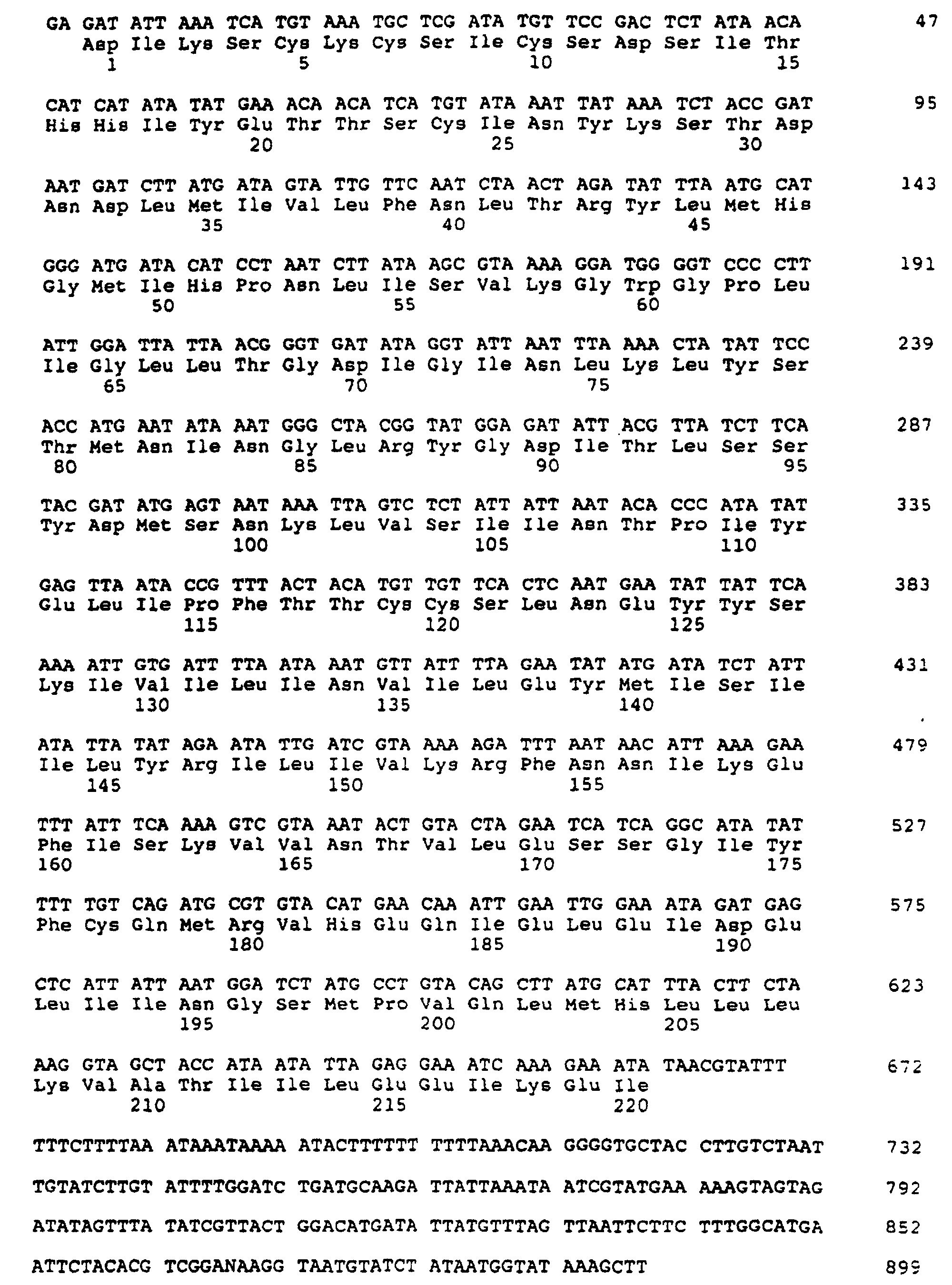
- (2) INFORMATION FOR SEQ ID NO:4:
- (i) SEQUENCE CHARACTERISTICS:
- (A) LENGTH: 220 amino acids
- (B) TYPE: amino acid
- (D) TOPOLOGY: linear
- (ii) MOLECULE TYPE: protein
- (xi) SEQUENCE DESCRIPTION: SEQ ID NO:4:
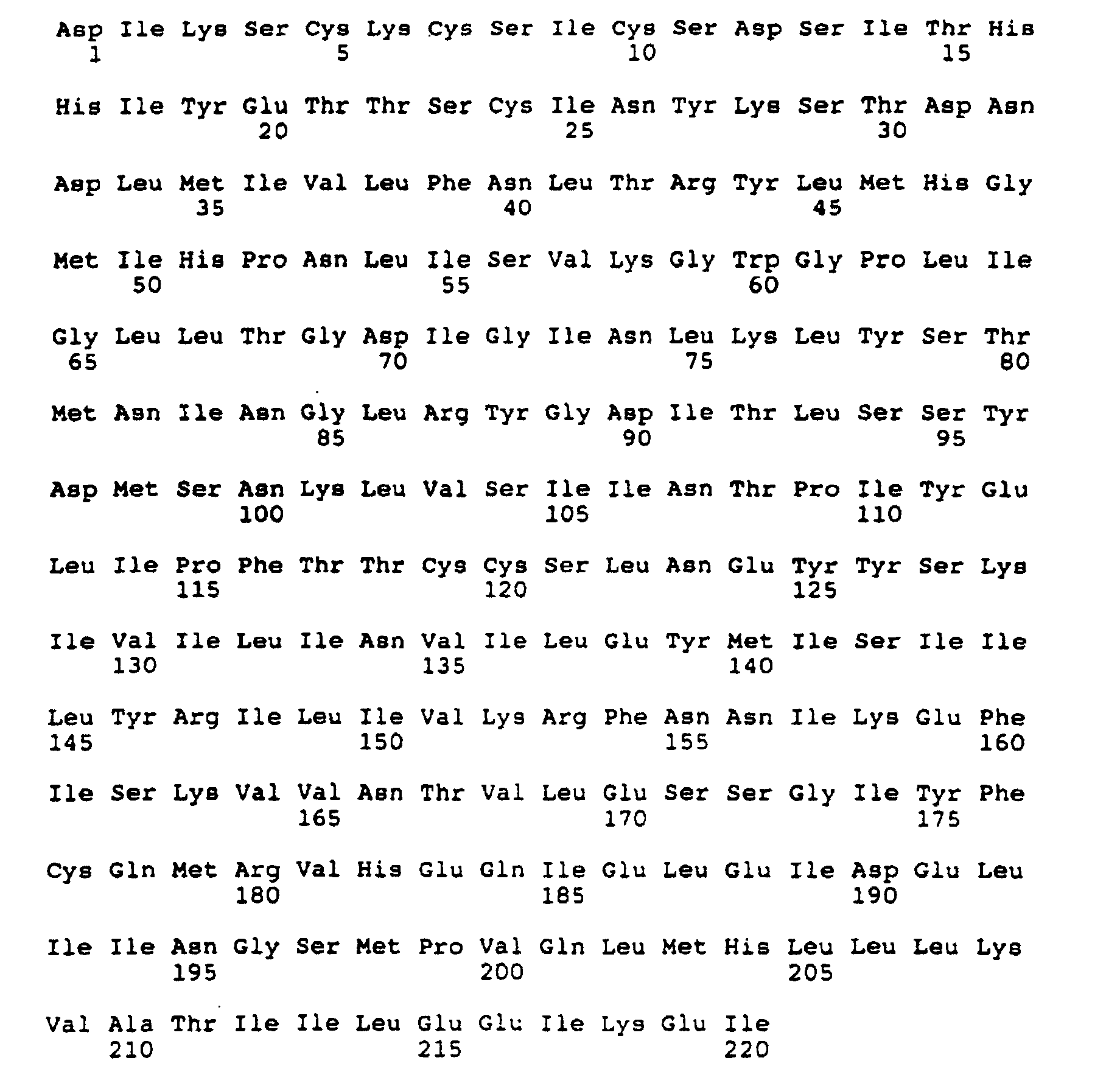
- (2) INFORMATION FOR SEQ ID NO:5:
- (i) SEQUENCE CHARACTERISTICS:
- (A) LENGTH: 129 amino acids
- (B) TYPE: amino acid
- (C) STRANDEDNESS: double
- (D) TOPOLOGY: linear
- (ii) MOLECULE TYPE: peptide
- (iii) HYPOTHETICAL: YES
- (iv) ANTI-SENSE: NO
- (v) FRAGMENT TYPE: N-terminal
- (vi) ORIGINAL SOURCE:
- (A) ORGANISM: Vaccinia virus
- (B) STRAIN: Copenhagen
- (viii) POSITION IN GENOME:
- (B) MAP POSITION: -23.2
- (C) UNITS: %G

- (2) INFORMATION FOR SEQ ID NO:6:
- (i) SEQUENCE CHARACTERISTICS:
- (A) LENGTH: 132 amino acids
- (B) TYPE: amino acid
- (C) STRANDEDNESS: double
- (D) TOPOLOGY: linear
- (ii) MOLECULE TYPE: peptide
- (iii) HYPOTHETICAL: YES
- (iv) ANTI-SENSE: NO
- (v) FRAGMENT TYPE: N-terminal
- (vi) ORIGINAL SOURCE:
- (A) ORGANISM: Swinepox virus
- (B) STRAIN: Kasza
- (viii) POSITION IN GENOME:
- (B) MAP POSITION: -23.2
- (C) UNITS: %G
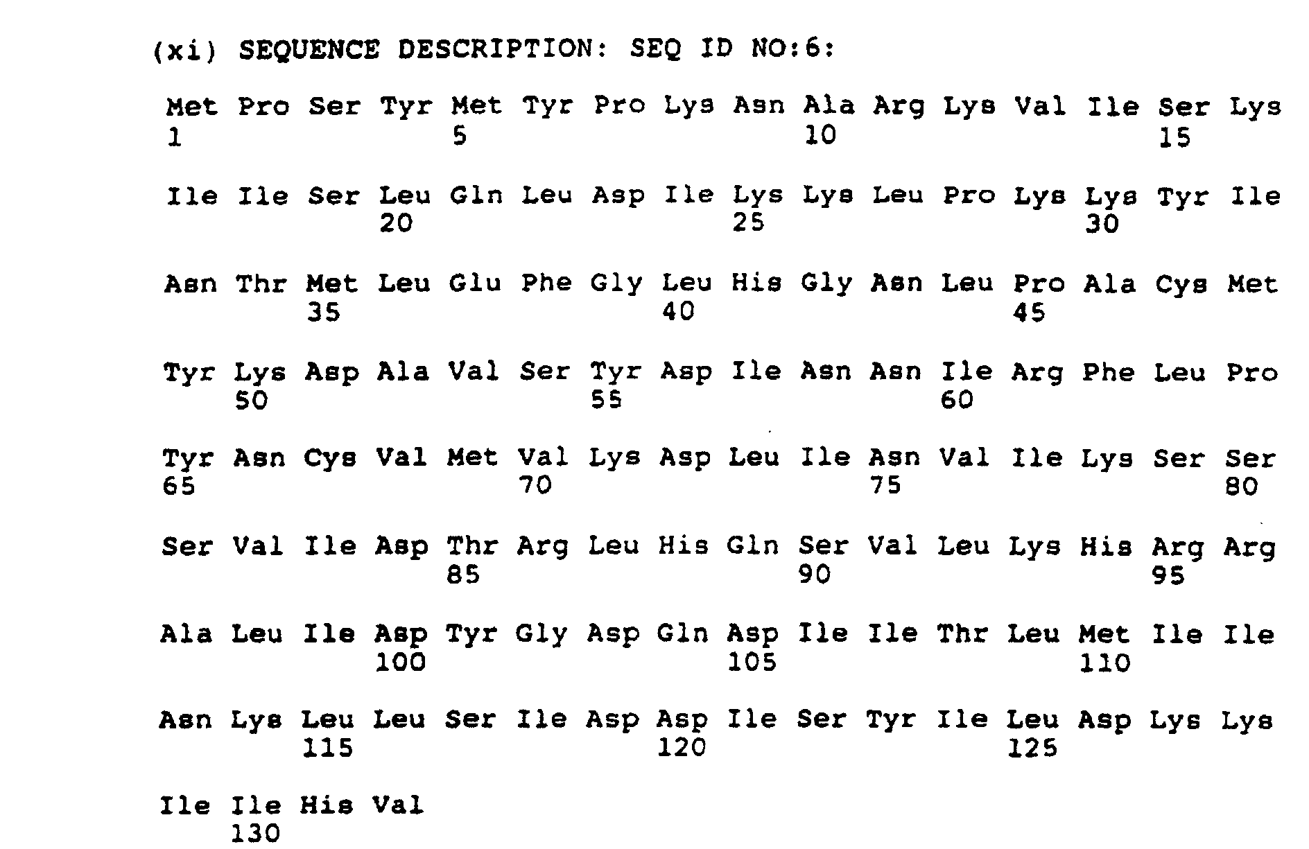
- (2) INFORMATION FOR SEQ ID NO:7:
- (i) SEQUENCE CHARACTERISTICS:
- (A) LENGTH: 101 amino acids
- (B) TYPE: amino acid
- (C) STRANDEDNESS: double
- (D) TOPOLOGY: linear
- (ii) MOLECULE TYPE: peptide
- (iii) HYPOTHETICAL: YES
- (iv) ANTI-SENSE: NO
- (v) FRAGMENT TYPE: C-terminal
- (vi) ORIGINAL SOURCE:
- (A) ORGANISM: Vaccinia virus
- (B) STRAIN: Copenhagen
- (viii) POSITION IN GENOME:
- (B) MAP POSITION: -23.2
- (C) UNITS: %G
- (xi) SEQUENCE DESCRIPTION: SEQ ID NO:7:

- (2) INFORMATION FOR SEQ ID NO:8:
- (i) SEQUENCE CHARACTERISTICS:
- (A) LENGTH: 100 amino acids
- (B) TYPE: amino acid
- (C) STRANDEDNESS: double
- (D) TOPOLOGY: linear
- (ii) MOLECULE TYPE: peptide
- (iii) HYPOTHETICAL: YES
- (iv) ANTI-SENSE: NO
- (v) FRAGMENT TYPE: C-terminal
- (vi) ORIGINAL SOURCE:
- (A) ORGANISM: Swinepox virus
- (B) STRAIN: Kasza
- (viii) POSITION IN GENOME:
- (B) MAP POSITION: -23.2
- (C) UNITS: %G
- (xi) SEQUENCE DESCRIPTION: SEQ ID NO:8:
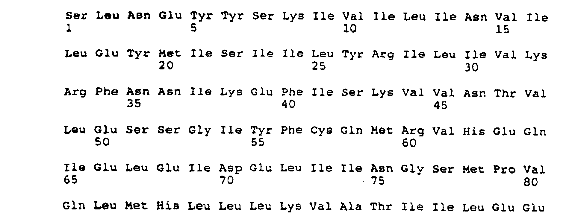

- (2) INFORMATION FOR SEQ ID NO:9:
- (i) SEQUENCE CHARACTERISTICS:
- (A) LENGTH: 102 base pairs
- (B) TYPE: nucleic acid
- (C) STRANDEDNESS: double
- (D) TOPOLOGY: circular
- (ii) MOLECULE TYPE: DNA (genomic)
- (iii) HYPOTHETICAL: NO
- (iv) ANTI-SENSE: NO
- (vi) ORIGINAL SOURCE:
- (A) ORGANISM: Plasmid
- (vii) IMMEDIATE SOURCE:
- (B) CLONE: 520-17.5 (Junction A)
- (x) PUBLICATION INFORMATION:
- (A) AUTHORS: Ferrari, Franco A
Trach, Kathleen
Hoch, James A - (B) TITLE: Sequence Analysis of the spoOB Locus Revels a Polycistronic Transcription Unit
- (C) JOURNAL: J. Bacteriol.
- (D) VOLUME: 161
- (E) ISSUE: 2
- (F) PAGES: 556-562
- (G) DATE: Feb.-1985
- (xi) SEQUENCE DESCRIPTION: SEQ ID NO:9:

- (2) INFORMATION FOR SEQ ID NO:10:
- (i) SEQUENCE CHARACTERISTICS:
- (A) LENGTH: 102 base pairs
- (B) TYPE: nucleic acid
- (C) STRANDEDNESS: double
- (D) TOPOLOGY: circular
- (ii) MOLECULE TYPE: DNA (genomic)
- (iii) HYPOTHETICAL: NO
- (iv) ANTI-SENSE: NO
- (vi) ORIGINAL SOURCE:
- (A) ORGANISM: Plasmid
- (vii) IMMEDIATE SOURCE:
- (B) CLONE: 520-17.5 (Junction B)
- (ix) FEATURE:
- (A) NAME/KEY: CDS
- (B) LOCATION: 85..99
- (D) OTHER INFORMATION: /codon_start= 85
/function= "Translational start of hybrid protein" /product= "N-terminal peptide"
/number= 1
/etandard_name= "Translation of synthetic DNA sequence" - (ix) FEATURE:
- (A) NAME/KEY: CDS
- (B) LOCATION: 100..102
- (C) IDENTIFICATION METHOD: experimental
- (D) OTHER INFORMATION: /partial
/codon_start= 100
/function= "marker enzyme"
/product= "Beta-Galactosidase"
/evidence= EXPERIMENTAL
/gene= "lacZ"
/number= 2
/citation= ([1]) - (x) PUBLICATION INFORMATION:
- (A) AUTHORS: Ferrari, Franco A
Trach, Kathleen
Hoch, James A - (B) TITLE: Seqquence Analysis of the spoOB Locus Reveals a Polycistronic Transcription Unit
- (C) JOURNAL: J. Bacteriol.
- (D) VOLUME: 161
- (E) ISSUE: 2
- (F) PAGES: 556-562
- (G) DATE: Feb.-1985
- (xi) SEQUENCE DESCRIPTION: SEQ ID NO:10:

- (2) INFORMATION FOR SEQ ID NO:11:
- (i) SEQUENCE CHARACTERISTICS:
- (A) LENGTH: 5 amino acids
- (B) TYPE: amino acid
- (D) TOPOLOGY: linear
- (ii) MOLECULE TYPE: protein
- (xi) SEQUENCE DESCRIPTION: SEQ ID NO:11:

- (2) INFORMATION FOR SEQ ID NO:12:
- (i) SEQUENCE CHARACTERISTICS:
- (A) LENGTH: 1 amino acids
- (B) TYPE: amino acid
- (D) TOPOLOGY: linear
- (ii) MOLECULE TYPE: protein
- (xi) SEQUENCE DESCRIPTION: SEQ ID NO:12:

- (2) INFORMATION FOR SEQ ID NO:13:
- (i) SEQUENCE CHARACTERISTICS:
- (A) LENGTH: 103 base pairs
- (B) TYPE: nucleic acid
- (C) STRANDEDNESS: double
- (D) TOPOLOGY: circular
- (ii) MOLECULE TYPE: DNA (genomic)
- (iii) HYPOTHETICAL: NO
- (iv) ANTI-SENSE: NO
- (vi) ORIGINAL SOURCE:
- (A) ORGANISM: Plasmid
- (vii) IMMEDIATE SOURCE:
- (B) CLONE: 520-17.5 (Junction C)
- (ix) FEATURE:
- (A) NAME/KEY: CDS
- (B) LOCATION: 1..72
- (C) IDENTIFICATION METHOD: experimental
- (D) OTHER INFORMATION: /partial
/codon_start= 1
/function= "marker enzyme"
/product= "Beta-galactosidase"
/evidence= EXPERIMENTAL
/gene= "lacZ"
/number= 1
/citation= ([1]) - (ix) FEATURE:
- (A) NAME/KEY: CDS
- (B) LOCATION: 73..78
- (C) IDENTIFICATION METHOD: experimental
- (D) OTHER INFORMATION: /codon_start= 73
/function= "Translational finish of hybrid
protein"
/product= "c-terminal peptide"
/evidence= EXPERIMENTAL
/number= 2
/standard name= "Translation of synthetic DNA
sequence" - (x) PUBLICATION INFORMATION:
- (A) AUTHORS: Ferrari, Franco A
Trach, Kathleen
Hoch, James A - (B) TITLE: Seqquence Analysis of the spoOB Locus Reveals a Polycistronic Transcription Unit
- (C) JOURNAL: J. Bacteriol.
- (D) VOLUME: 161
- (E) ISSUE: 2
- (F) PAGES: 556-562
- (G) DATE: Feb.-1985
- (xi) SEQUENCE DESCRIPTION: SEQ ID NO:13:

- (2) INFORMATION FOR SEQ ID NO:14:
- (i) SEQUENCE CHARACTERISTICS:
- (A) LENGTH: 24 amino acids
- (B) TYPE: amino acid
- (D) TOPOLOGY: linear
- (ii) MOLECULE TYPE: protein
- (xi) SEQUENCE DESCRIPTION: SEQ ID NO:14:

- (2) INFORMATION FOR SEQ ID NO:15:
- (i) SEQUENCE CHARACTERISTICS:
- (A) LENGTH: 2 amino acids
- (B) TYPE: amino acid
- (D) TOPOLOGY: linear
- (ii) MOLECULE TYPE: protein
- (xi) SEQUENCE DESCRIPTION: SEQ ID NO:15:

- (2) INFORMATION FOR SEQ ID NO:16:
- (i) SEQUENCE CHARACTERISTICS:
- (A) LENGTH: 48 base pairs
- (B) TYPE: nucleic acid
- (C) STRANDEDNESS: double
- (D) TOPOLOGY: circular
- (ii) MOLECULE TYPE: DNA (genomic)
- (iii) HYPOTHETICAL: NO
- (iv) ANTI-SENSE: NO
- (vi) ORIGINAL SOURCE:
- (A) ORGANISM: Plasmid
- (vii) IMMEDIATE SOURCE:
- (B) CLONE: 520-17.5 (Junction D)
- (xi) SEQUENCE DESCRIPTION: SEQ ID NO:16:

- (2) INFORMATION FOR SEQ ID NO:17:
- (i) SEQUENCE CHARACTERISTICS:
- (A) LENGTH: 57 base pairs
- (B) TYPE: nucleic acid
- (C) STRANDEDNESS: double
- (D) TOPOLOGY: circular
- (ii) MOLECULE TYPE: DNA (genomic)
- (iii) HYPOTHETICAL: NO
- (iv) ANTI-SENSE: NO
- (vi) ORIGINAL SOURCE:
- (A) ORGANISM: Plasmid
- (vii) IMMEDIATE SOURCE:
- (B) CLONE: 538-46.26 (Junction A)
- (xi) SEQUENCE DESCRIPTION: SEQ ID NO:17:

- (2) INFORMATION FOR SEQ ID NO:18:
- (i) SEQUENCE CHARACTERISTICS:
- (A) LENGTH: 102 base pairs
- (B) TYPE: nucleic acid
- (C) STRANDEDNESS: double
- (D) TOPOLOGY: circular
- (ii) MOLECULE TYPE: DNA (genomic)
- (iii) HYPOTHETICAL: NO
- (iv) ANTI-SENSE: NO
- (vi) ORIGINAL SOURCE:
- (A) ORGANISM: Plasmid
- (vii) IMMEDIATE SOURCE:
- (B) CLONE: 538-46.16 (Junction B)
- (ix) FEATURE:
- (A) NAME/KEY: CDS
- (B) LOCATION: 91..102
- (C) IDENTIFICATION METHOD: experimental
- (D) OTHER INFORMATION: /partial
/codon_start= 91
/function= "marker enzyme"
/product= "Beta-Galactosidase"
/evidence= EXPERIMENTAL
/gene= "lacZ"
/number= 2
/citation= ([1]) - (ix) FEATURE:
- (A) NAME/KEY: CDS
- (B) LOCATION: 76..90
- (D) OTHER INFORMATION: /partial
/codon_start= 76
/function= "Translational start of hybrid protein" /product= "N-terminal peptide"
/number= 1
/standard_name= "Translation of synthetic DNA
sequence" - (x) PUBLICATION INFORMATION:
- (A) AUTHORS: Ferrari, Franco A
Trach, Kathleen
Hoch, James A - (B) TITLE: Seqquence Analysis of the spoOB Locus Reveals a Polycistronic Transcription Unit
- (C) JOURNAL: J. Bacteriol.
- (D) VOLUME: 161
- (E) ISSUE: 2
- (F) PAGES: 556-562
- (G) DATE: Feb.-1985
- (xi) SEQUENCE DESCRIPTION: SEQ ID NO:18:

- (2) INFORMATION FOR SEQ ID NO:19:
- (i) SEQUENCE CHARACTERISTICS:
- (A) LENGTH: 5 amino acids
- (B) TYPE: amino acid
- (D) TOPOLOGY: linear
- (ii) MOLECULE TYPE: protein
- (xi) SEQUENCE DESCRIPTION: SEQ ID NO:19:

- (2) INFORMATION FOR SEQ ID NO:20:
- (i) SEQUENCE CHARACTERISTICS:
- (A) LENGTH: 4 amino acids
- (B) TYPE: amino acid
- (D) TOPOLOGY: linear
- (ii) MOLECULE TYPE: protein
- (Xi) SEQUENCE DESCRIPTION: SEQ ID NO:20:

- (2) INFORMATION FOR SEQ ID NO:21:
- (i) SEQUENCE CHARACTERISTICS:
- (A) LENGTH: 206 base pairs
- (B) TYPE: nucleic acid
- (C) STRANDEDNESS: double
- (D) TOPOLOGY: circular
- (ii) MOLECULE TYPE: DNA (genomic)
- (iii) HYPOTHETICAL: NO
- (iv) ANTI-SENSE: NO
- (vi) ORIGINAL SOURCE:
- (A) ORGANISM: Plasmid
- (vii) IMMEDIATE SOURCE:
- (B) CLONE: 538-46.16 (Junction C)
- (ix) FEATURE:
- (A) NAME/KEY: CDS
- (B) LOCATION: 1..63
- (C) IDENTIFICATION METHOD: experimental
- (D) OTHER INFORMATION: /partial
/codon_start= 1
/function= "marker enzyme"
/product= "Beta-galactosidase"
/evidence= EXPERIMENTAL
/number= 1
/citation= ([1]) - (ix) FEATURE:
- (A) NAME/KEY: CDS
- (B) LOCATION: 64..69
- (C) IDENTIFICATION METHOD: experimental
- (D) OTHER INFORMATION: /codon_start= 64
/function= "Translational finish of hybrid
protein"
/product= "C-terminal peptide"
/evidence= EXPERIMENTAL
/standard_name= "Translation of synthetic DNA
sequence" - (ix) FEATURE:
- (A) NAME/KEY: CDS
- (B) LOCATION: 177..185
- (C) IDENTIFICATION METHOD: experimental
- (D) OTHER INFORMATION: /codon_start= 177
/function= "Translational start of hybrid protein" /product= "N-terminal peptide"
/evidence= EXPERIMENTAL
/standard_name= "Translation of synthetic DNA
sequence" - (ix) FEATURE:
- (A) NAME/KEY: CDS
- (B) LOCATION: 186..206
- (C) IDENTIFICATION METHOD: experimental
- (D) OTHER INFORMATION: /partial
/codon_start= 186
/function= "glycoprotein"
/product= "PRV gp50"
/evidence= EXPERIMENTAL
/gene= "gp50"
/number= 3
/citation= ([2]) - (x) PUBLICATION INFORMATION:
- (A) AUTHORS: Ferrari, Franco A
Trach, Kathleen
Hoch, James A - (B) TITLE: Seqquence Analysis of the spoOB Locus Reveals a Polycistronic Transcription Unit
- (C) JOURNAL: J. Bacteriol.
- (D) VOLUME: 161
- (E) ISSUE: 2
- (F) PAGES: 556-562
- (G) DATE: Feb.-1985
- (x) PUBLICATION INFORMATION:
- (A) AUTHORS: Petrovakis, Erik A
Timmins, James G
Armentrout, Marty A
Marchioli, Carmine C
Jr. Yancy, Robert J
Post, Leonard E - (B) TITLE: DNA Sequence of the Gene for Pseudorabies Virus gp50, a Glycoprotein without N-Linked Glycoaylation
- (C) JOURNAL: J. Virol.
- (D) VOLUME: 59
- (E) ISSUE: 2
- (F) PAGES: 216-223
- (G) DATE: Aug.-1986
- (xi) SEQUENCE DESCRIPTION: SEQ ID NO:21:

- (2) INFORMATION FOR SEQ ID NO:22:
- (i) SEQUENCE CHARACTERISTICS:
- (A) LENGTH: 21 amino acids
- (B) TYPE: amino acid
- (D) TOPOLOGY: linear
- (ii) MOLECULE TYPE: protein
- (xi) SEQUENCE DESCRIPTION: SEQ ID NO:22:

- (2) INFORMATION FOR SEQ ID NO:23:
- (i) SEQUENCE CHARACTERISTICS:
- (A) LENGTH: 2 amino acids
- (B) TYPE: amino acid
- (D) TOPOLOGY: linear
- (ii) MOLECULE TYPE: protein
- (xi) SEQUENCE DESCRIPTION: SEQ ID NO:23:

- (2) INFORMATION FOR SEQ ID NO:24:
- (i) SEQUENCE CHARACTERISTICS:
- (A) LENGTH: 3 amino acids
- (B) TYPE: amino acid
- (D) TOPOLOGY: linear
- (ii) MOLECULE TYPE: protein
- (xi) SEQUENCE DESCRIPTION: SEQ ID NO:24:

- (2) INFORMATION FOR SEQ ID NO:25:
- (i) SEQUENCE CHARACTERISTICS:
- (A) LENGTH: 7 amino acids
- (B) TYPE: amino acid
- (D) TOPOLOGY: linear
- (ii) MOLECULE TYPE: protein
- (xi) SEQUENCE DESCRIPTION: SEQ ID NO:25:

- (2) INFORMATION FOR SEQ ID NO:26:
- (i) SEQUENCE CHARACTERISTICS:
- (A) LENGTH: 101 base pairs
- (B) TYPE: nucleic acid
- (C) STRANDEDNESS: double
- (D) TOPOLOGY: circular
- (ii) MOLECULE TYPE: DNA (genomic)
- (iii) HYPOTHETICAL: NO
- (iv) ANTI-SENSE: NO
- (vi) ORIGINAL SOURCE:
- (A) ORGANISM: Plasmid
- (vii) IMMEDIATE SOURCE:
- (B) CLONE: 538-46.16 (Junction D)
- (ix) FEATURE:
- (A) NAME/KEY: CDS
- (B) LOCATION: 1..15
- (D) OTHER INFORMATION: /partial
/codon_start= 1
/function= "glycoprotein"
/product= "PRV gp63"
/gene= "gp63"
/number= 1
/citation= ([1]) - (x) PUBLICATION INFORMATION:
- (A) AUTHORS: Petrovskis, Erik A
Timmins, James G
Post, Lenoard E - (B) TITLE: Use of Lambda-gtll To Isolate Genes for two Pseudorabies Virus Glycoproteins with homology to Herpes Simplex Virus and Varicella-Zoster Virus Glycoproteins
- (C) JOURNAL: J. Virol.
- (D) VOLUME: 60
- (E) ISSUE: 1
- (F) PAGES: 185-193
- (G) DATE: Oct.-1986
- (xi) SEQUENCE DESCRIPTION: SEQ ID NO:26:

- (2) INFORMATION FOR SEQ ID NO:27:
- (i) SEQUENCE CHARACTERISTICS:
- (A) LENGTH: 5 amino acids
- (B) TYPE: amino acid
- (D) TOPOLOGY: linear
- (ii) MOLECULE TYPE: protein
- (xi) SEQUENCE DESCRIPTION: SEQ ID NO:27:

- (2) INFORMATION FOR SEQ ID NO:28:
- (i) SEQUENCE CHARACTERISTICS:
- (A) LENGTH: 57 base pairs
- (B) TYPE: nucleic acid
- (C) STRANDEDNESS: double
- (D) TOPOLOGY: circular
- (ii) MOLECULE TYPE: DNA (genomic)
- (iii) HYPOTHETICAL: NO
- (iv) ANTI-SENSE: NO
- (vi) ORIGINAL SOURCE:
- (A) ORGANISM: Plasmid
- (vii) IMMEDIATE SOURCE:
- (B) CLONE: 538-46.16 (Junction E)
- (xi) SEQUENCE DESCRIPTION: SEQ ID No:28:

- (2) INFORMATION FOR SEQ ID NO:29:
- (i) SEQUENCE CHARACTERISTICS:
- (A) LENGTH: 1907 base pairs
- (B) TYPE: nucleic acid
- (C) STRANDEDNESS: double
- (D) TOPOLOGY: linear
- (ii) MOLECULE TYPE: cDNA to mRNA
- (iii) HYPOTHETICAL: NO
- (iv) ANTI-SENSE: NO
- (vi) ORIGINAL SOURCE:
- (A) ORGANISM: Newcastle disease virus
- (B) STRAIN: Bl
- (vii) IMMEDIATE SOURCE:
- (B) CLONE: 137-23.803 (PSY1142)
- (viii) POSITION IN GENOME:
- (B) MAP POSITION: -50%
- (C) UNITS: %G
- (ix) FEATURE:
- (A) NAME/KEY: CDS
- (B) LOCATION: 92..1822
- (D) OTHER INFORMATION: /codon_start= 92
/product= "NDV heamagglutinin-Neuraminidase"
/gene= "HN"
/number= 1 - (xi) SEQUENCE DESCRIPTION: SEQ ID NO:29:
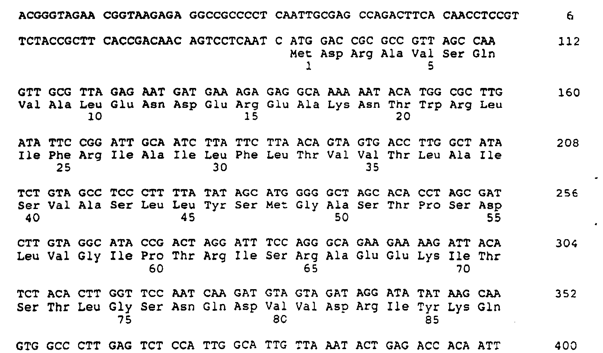
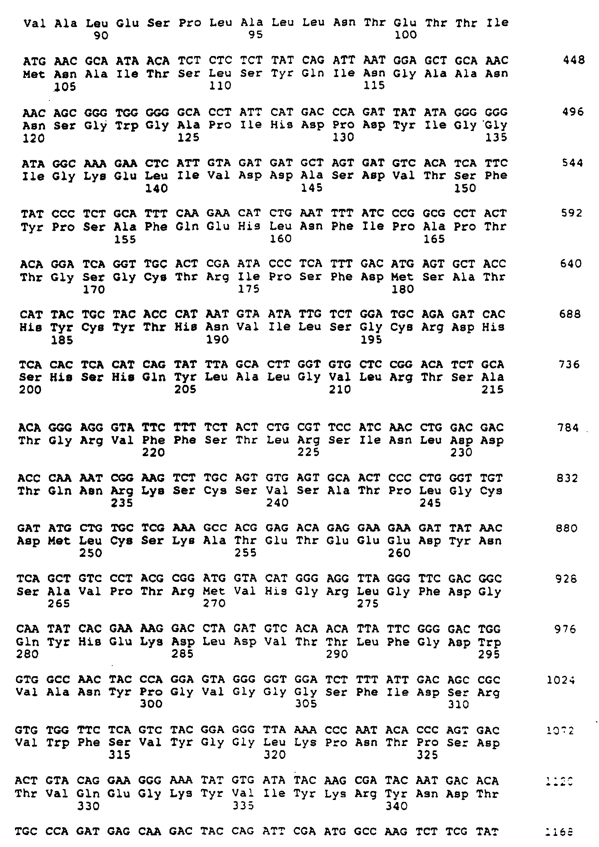
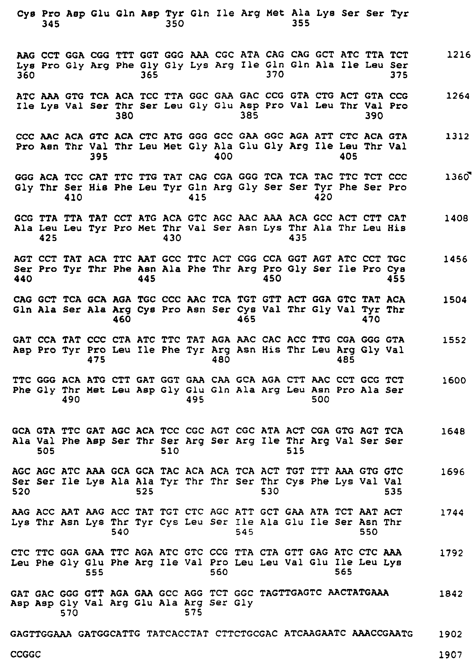
- (2) INFORMATION FOR SEQ ID NO:30:
- (i) SEQUENCE CHARACTERISTICS:
- (A) LENGTH: 577 amino acids
- (B) TYPE: amino acid
- (D) TOPOLOGY: linear
- (ii) MOLECULE TYPE: protein
- (xi) SEQUENCE DESCRIPTION: SEQ ID NO:30:


- (2) INFORMATION FOR SEQ ID NO:31:
- (i) SEQUENCE CHARACTERISTICS:
- (A) LENGTH: 57 base pairs
- (B) TYPE: nucleic acid
- (C) STRANDEDNESS: double
- (D) TOPOLOGY: circular
- (ii) MOLECULE TYPE: DNA (genomic)
- (iii) HYPOTHETICAL: NO
- (iv) ANTI-SENSE: NO
- (vi) ORIGINAL SOURCE:
- (A) ORGANISM: Plasmid
- (vii) IMMEDIATE SOURCE:
- (B) CLONE: 538-46.26 (Junction A)
- (xi) SEQUENCE DESCRIPTION: SEQ ID NO:31:

- (2) INFORMATION FOR SEQ ID NO:32:
- (i) SEQUENCE CHARACTERISTICS:
- (A) LENGTH: 108 base pairs
- (B) TYPE: nucleic acid
- (C) STRANDEDNESS: double
- (D) TOPOLOGY: circular
- (ii) MOLECULE TYPE: DNA (genomic)
- (iii) HYPOTHETICAL: NO
- (iv) ANTI-SENSE: NO
- (vi) ORIGINAL SOURCE:
- (A) ORGANISM: Plasmid
- (vii) IMMEDIATE SOURCE:
- (B) CLONE: 538-46.26 (Junction B)
- (ix) FEATURE:
- (A) NAME/KEY: exon
- (B) LOCATION: 88..102
- (D) OTHER INFORMATION: /codon_start= 88
/function= "Translational start of hybrid protein" /product= "N-terminal peptide"
/number= 1
/standard_name= "Translation of synthetic DNA
sequence" - (ix) FEATURE:
- (A) NAME/KEY: CDS
- (B) LOCATION: 103..108
- (C) IDENTIFICATION METHOD: experimental
- (D) OTHER INFORMATION: /partial
/codon_start= 103
/product= "NDV Heamagglutinin-Neuraminidase"
/evidence= EXPERIMENTAL
/gene= "HN"
/number= 2 - (xi) SEQUENCE DESCRIPTION: SEQ ID NO:32:

- (2) INFORMATION FOR SEQ ID NO:33:
- (i) SEQUENCE CHARACTERISTICS:
- (A) LENGTH: 2 amino acids
- (B) TYPE: amino acid
- (D) TOPOLOGY: linear
- (ii) MOLECULE TYPE: protein
- (xi) SEQUENCE DESCRIPTION: SEQ ID NO:33:

- (2) INFORMATION FOR SEQ ID NO:34:
- (i) SEQUENCE CHARACTERISTICS:
- (A) LENGTH: 108 base pairs
- (B) TYPE: nucleic acid
- (C) STRANDEDNESS: double
- (D) TOPOLOGY: circular
- (ii) MOLECULE TYPE: DNA (genomic)
- (iii) HYPOTHETICAL: NO
- (iv) ANTI-SENSE: NO
- (vi) ORIGINAL SOURCE:
- (A) ORGANISM: Plasmid
- (vii) IMMEDIATE SOURCE:
- (B) CLONE: 538-46.26 (Junction C)
- (ix) FEATURE:
- (A) NAME/KEY: CDS
- (B) LOCATION: 70..84
- (D) OTHER INFORMATION: /codon_start= 70
/function= "Transiational start of hybrid protein" /product= "N-terminal peptide"
/number= 1
/standard_name= "Translation of synthetic DNA sequence" - (ix) FEATURE:
- (A) NAME/KEY: CDS
- (B) LOCATION: 85..108
- (C) IDENTIFICATION METHOD: experimental
- (D) OTHER INFORMATION: /partial
/codon_start= 85
/function= "marker enzyme"
/product= "Beta-galactosidase"
/evidence= EXPERIMENTAL
/gene= "lacZ"
/number= 2
/ciiation= ([1]) - (x) PUBLICATION INFORMATION:
- (A) AUTHORS: Ferrari, Franco A
Trach, Kathleen
Hoch, James A - (B) TITLE: Sequence Analysis of the spoOB Locus Reveals a Polycistronic Transcription Unit
- (C) JOURNAL: J. Bacteriol.
- (D) VOLUME: 161
- (E) ISSUE: 2
- (F) PAGES: 556-562
- (G) DATE: Feb.-1985
- (xi) SEQUENCE DESCRIPTION: SEQ ID NO:34:

- (2) INFORMATION FOR SEQ ID NO:35:
- (i) SEQUENCE CHARACTERISTICS:
- (A) LENGTH: 5 amino acids
- (B) TYPE: amino acid
- (D) TOPOLOGY: linear
- (ii) MOLECULE TYPE: protein
- (xi) SEQUENCE DESCRIPTION: SEQ ID NO:35:

- (2) INFORMATION FOR SEQ ID NO:36:
- (i) SEQUENCE CHARACTERISTICS:
- (A) LENGTH: 8 amino acids
- (B) TYPE: amino acid
- (D) TOPOLOGY: linear
- (ii) MOLECULE TYPE: protein
- (xi) SEQUENCE DESCRIPTION: SEQ ID NO:36:

- (2) INFORMATION FOR SEQ ID NO:37:
- (i) SEQUENCE CHARACTERISTICS:
- (A) LENGTH: 108 base pairs
- (B) TYPE: nucleic acid
- (C) STRANDEDNESS: double
- (D) TOPOLOGY: circular
- (ii) MOLECULE TYPE: DNA (genomic)
- (iii) HYPOTHETICAL: NO
- (iv) ANTI-SENSE: NO
- (vi) ORIGINAL SOURCE:
- (A) ORGANISM: Plasmid
- (vii) IMMEDIATE SOURCE:
- (B) CLONE: 538-46.26
- (ix) FEATURE:
- (A) NAME/KEY: CDS
- (B) LOCATION: 1..54
- (C) IDENTIFICATION METHOD: experimental
- (D) OTHER INFORMATION: /partial
/codon_start= 1
/function= "marker enzyme"
/product= "Beta-galactosidase"
/evidence= EXPERIMENTAL
/gene= "lacZ"
/number= 1
/citation= ([1]) - (ix) FEATURE:
- (A) NAME/KEY: CDS
- (B) LOCATION: 55..63
- (C) IDENTIFICATION METHOD: experimental
- (D) OTHER INFORMATION: /codon_start= 55
/function= "Translational finish of hybrid
protein"
/product= "C-terminai peptide"
/evidence= EXPERIMENTAL
/number= 2
/standard_name= "Translation of synthetic DNA sequence" - (xi) SEQUENCE DESCRIPTION: SEQ ID NO:37:

- (2) INFORMATION FOR SEQ ID NO:38:
- (i) SEQUENCE CHARACTERISTICS:
- (A) LENGTH: 18 amino acids
- (B) TYPE: amino acid
- (D) TOPOLOGY: linear
- (ii) MOLECULE TYPE: protein
- (xi) SEQUENCE DESCRIPTION: SEQ ID NO:38:

- (2) INFORMATION FOR SEQ ID NO:39:
- (i) SEQUENCE CHARACTERISTICS:
- (A) LENGTH: 2 amino acids
- (B) TYPE: amino acid
- (D) TOPOLOGY: linear
- (ii) MOLECULE TYPE: protein
- (xi) SEQUENCE DESCRIPTION: SEQ ID NO:39:

- (2) INFORMATION FOR SEQ ID NO:40:
- (i) SEQUENCE CHARACTERISTICS:
- (A) LENGTH: 57 base pairs
- (B) TYPE: nucleic acid
- (C) STRANDEDNESS: double
- (D) TOPOLOGY: circular
- (ii) MOLECULE TYPE: DNA (genomic)
- (iii) HYPOTHETICAL: NO
- (iv) ANTI-SENSE: NO
- (vi) ORIGINAL SOURCE:
- (A) ORGANISM: Plasmid
- (vii) IMMEDIATE SOURCE:
- (B) CLONE: 538-46.26 (Junction E)
- (xi) SEQUENCE DESCRIPTION: SEQ ID NO:40:

- (2) INFORMATION FOR SEQ ID NO:41:
- (i) SEQUENCE CHARACTERISTICS:
- (A) LENGTH: 27 base pairs
- (B) TYPE: nucleic acid
- (C) STRANDEDNESS: single
- (D) TOPOLOGY: linear
- (ii) MOLECULE TYPE: DNA (genomic)
- (iii) HYPOTHETICAL: N
- (iv) ANTI-SENSE: N
- (vi) ORIGINAL SOURCE:
- (A) ORGANISM: Pseudorabies virus \ synthetic oligonucleotide primer
- (xi) SEQUENCE DESCRIPTION: SEQ ID NO:41:

- (2) INFORMATION FOR SEQ ID NO:42:
- (i) SEQUENCE CHARACTERISTICS:
- (A) LENGTH: 19 base pairs
- (B) TYPE: nucleic acid
- (C) STRANDEDNESS: single
- (D) TOPOLOGY: linear
- (ii) MOLECULE TYPE: DNA (genomic)
- (iii) HYPOTHETICAL: N
- (iv) ANTI-SENSE: N
- (vi) ORIGINAL SOURCE:
- (A) ORGANISM: Pseudorabies virus \ Synthetic oligonucleotide prime
- (xi) SEQUENCE DESCRIPTION: SEQ ID NO:42:

-
Claims (22)
- A recombinant swinepox virus which comprises a swinepox viral genome containing a foreign DNA sequence inserted within the larger HindIII to BglII subfragment of the HindIII M fragment of the genomic DNA, and wherein the foreign DNA sequence is expressed in a host cell infected with the recombinant swinepox virus.
- The recombinant swinepox virus of claim 1, wherein the foreign DNA sequence is inserted within an open reading frame contained in the HindIII to BglII subfragment.
- The recombinant swinepox virus of claim 2, wherein the foreign DNA is inserted into a unique AccI restriction endonuclease site located in the HindIII to BglII subfragment.
- The recombinant swinepox virus of claim 1, 2 or 3, wherein the foreign DNA sequence is a first foreign DNA sequence, further comprising a second foreign DNA sequence inserted into an invention site that is not essential for the replication of the swinepox virus.
- A homology vector for producing a recombinant swinepox virus by inserting a foreign DNA into the genomic DNA of the swinepox virus, which comprisesa) a double-stranded foreign DNA sequence,b) flanked at one end of the foreign DNA by double-stranded swinepox viral DNA homologous to the genomic DNA located at one side of a site on the genomic DNA, wherein said site is located in the larger HindIII to BglII subfragment of the HindIII M fragment of the swinepox genomic DNA,c) flanked at the other end of the foreign DNA by double-stranded swinepox viral DNA homologous to genomic DNA located at the other side of the same site on the genomic DNA.
- The homology vector of claim 5, wherein the swinepox viral DNA flanking the double-stranded foreign DNA sequence is homologous to the genomic DNA present within the open reading frame contained in the HindIII to BglII subfragment.
- The homology vector of claim 6, wherein the double-stranded swinepox viral DNA flanking the double-stranded foreign DNA sequence is homologous to the genomic DNA located on either side of the unique AccI restriction endonuclease site located in the HindIII to BglII subfragment.
- The recombinant swinepox virus of any one of claims 1 to 3 or the homology vector of any one of claims 5 to 7, wherein the foreign DNA encodes a polypeptide.
- The recombinant swinepox virus or the homology vector of claim 8, wherein the polypeptide is a detectable marker.
- The recombinant swinepox virus or the homology vector of claim 9. wherein the detectable marker is E. coli betagalactosidase.
- The recombinant swinepox virus of claim 10, designated S-SPV-003 (ATTC Accession No. VR 2335).
- The recombinant swinepox virus or homology vector of claim 8, wherein the polypeptide is an antigenic polypeptide.
- The recombinant swinepox virus or homology vector of claim 12, wherein the antigenic polypeptide is or is from PRV glycoprotein 50, PRV glycoprotein II, PRV glycoprotein III, PRV glycoprotein H, TGE glycoprotein 195, TGE matrix protein, swine rotavirus glycoprotein 38, swine parvovirus capsid protein, Serpulina hyodysenteriae protective antigen, BVD glycoprotein 55, NDV hemagglutinin-neuraminidase, swine flu hemagglutinin, swine flu neuraminidase, Serpulina hyodysentariae, Foot and Mouth Disease Virus, Hog Cholera Virus, Swine Influenza Virus, African Swine Fever Virus or Mycoplasma hyopneumoniae.
- The recombinant swinepox virus of claim 13, wherein the antigenic polypeptide is PRV glycoprotein 50, designated S-SPV-008 (ATCC Accession No. VR 2339).
- The recombinant swinepox virus of claim 13, wherein the antigenic polypeptide is NDV hemagglutinin-neuraminidase, designated S-SPV-009 (ATCC Accession No. VR 2344).
- The recombinant swinepox virus of claim 8, wherein the foreign DNA further comprises a swinepox viral promoter.
- The recombinant swinepox virus or the homology vector of claim 8, wherein the foreign DNA further comprises a synthetic pox viral promoter.
- A vaccine which comprises an effective immunizing amount of the recombinant swinepox virus of any one of claims 12 to 15 and a suitable carrier.
- A host cell infected with the recombinant swinepox virus of any one of claims 1 to 4 or 8 to 17.
- The host cell of claim 19, wherein the host cell is a mammalian cell.
- The host cell of claim 20, wherein the mammalian cell is a Vero cell.
- The host cell of claim 20, wherein the mammalian cell is an EMSK cell.
Applications Claiming Priority (3)
| Application Number | Priority Date | Filing Date | Title |
|---|---|---|---|
| US820154 | 1992-01-13 | ||
| US07/820,154 US5382425A (en) | 1992-01-13 | 1992-01-13 | Recombinant swinepox virus |
| PCT/US1993/000324 WO1993014194A1 (en) | 1992-01-13 | 1993-01-13 | Recombinant swinepox virus |
Publications (3)
| Publication Number | Publication Date |
|---|---|
| EP0677114A4 EP0677114A4 (en) | 1995-07-13 |
| EP0677114A1 EP0677114A1 (en) | 1995-10-18 |
| EP0677114B1 true EP0677114B1 (en) | 2003-04-02 |
Family
ID=25230024
Family Applications (1)
| Application Number | Title | Priority Date | Filing Date |
|---|---|---|---|
| EP93904494A Expired - Lifetime EP0677114B1 (en) | 1992-01-13 | 1993-01-13 | Recombinant swinepox virus |
Country Status (13)
| Country | Link |
|---|---|
| US (1) | US5382425A (en) |
| EP (1) | EP0677114B1 (en) |
| JP (1) | JP3527240B2 (en) |
| AT (1) | ATE236262T1 (en) |
| AU (1) | AU675216B2 (en) |
| CA (1) | CA2127541C (en) |
| DE (1) | DE69332831T2 (en) |
| DK (1) | DK0677114T3 (en) |
| ES (1) | ES2191663T3 (en) |
| NZ (1) | NZ249278A (en) |
| PT (1) | PT677114E (en) |
| RU (1) | RU94038050A (en) |
| WO (1) | WO1993014194A1 (en) |
Families Citing this family (34)
| Publication number | Priority date | Publication date | Assignee | Title |
|---|---|---|---|---|
| US5651972A (en) * | 1989-04-21 | 1997-07-29 | University Of Florida Research Foundation, Inc. | Use of recombinant swine poxvirus as a live vaccine vector |
| US6127163A (en) * | 1992-01-13 | 2000-10-03 | Syntro Corporation | Recombinant swinepox virus |
| US6497882B1 (en) * | 1992-01-13 | 2002-12-24 | Syntro Corporation | Recombinant swinepox virus |
| US5869312A (en) * | 1992-01-13 | 1999-02-09 | Syntro Corporation | Recombinant swinepox virus |
| EP0617478B1 (en) * | 1993-03-25 | 1998-07-29 | Matsushita Electric Industrial Co., Ltd. | Laminated dielectric resonator and dielectric filter |
| US6875856B2 (en) | 1993-09-24 | 2005-04-05 | Syntro Corporation | Recombinant infectious laryngotracheitis virus and uses thereof |
| US5698394A (en) * | 1994-06-01 | 1997-12-16 | Board Of Regents Of The University Of Nebraska | Nucleotide sequences and methods for detection of Serpulina hyodysenteriae |
| US6221361B1 (en) * | 1995-01-19 | 2001-04-24 | Syntro Corporation | Recombinant swinepox virus |
| CA2210732C (en) * | 1995-01-19 | 2011-06-14 | Syntro Corporation | Recombinant swinepox virus |
| AU755763B2 (en) * | 1995-01-19 | 2002-12-19 | Syntro Corporation | Recombinant swinepox virus |
| EP0954593A1 (en) * | 1996-07-25 | 1999-11-10 | Therion Biologics Corporation | Recombinant pox virus for immunization against tumor-associated antigens |
| WO1998020899A1 (en) * | 1996-11-12 | 1998-05-22 | Board Of Regents University Of Nebraska Lincoln | Attenuated serpulina hyodysenteriae mutant for prevention of swine dysentery |
| US6517843B1 (en) | 1999-08-31 | 2003-02-11 | Merial | Reduction of porcine circovirus-2 viral load with inactivated PCV-2 |
| FR2772047B1 (en) * | 1997-12-05 | 2004-04-09 | Ct Nat D Etudes Veterinaires E | GENOMIC SEQUENCE AND POLYPEPTIDES OF CIRCOVIRUS ASSOCIATED WITH PIGLET LOSS DISEASE (MAP), APPLICATIONS TO DIAGNOSIS AND TO PREVENTION AND / OR TREATMENT OF INFECTION |
| US6497883B1 (en) | 1999-06-10 | 2002-12-24 | Merial | Porcine circovirus recombinant poxvirus vaccine |
| ES2170622B1 (en) * | 1999-12-03 | 2004-05-16 | Consejo Superior De Investigaciones Cientificas | CLONES AND INFECTIVE VECTORS DERIVED FROM CORONAVIRUS AND ITS APPLICATIONS. |
| FR2823222B1 (en) | 2001-04-06 | 2004-02-06 | Merial Sas | VACCINE AGAINST NILE FEVER VIRUS |
| AU2003239589A1 (en) | 2002-05-24 | 2003-12-12 | Schering-Plough Ltd. | Eta-1 gene and methods for use |
| US7468273B2 (en) | 2003-05-01 | 2008-12-23 | Meial Limited | Canine GHRH gene, polypeptides and methods of use |
| ATE486966T1 (en) * | 2003-12-08 | 2010-11-15 | Univ Loma Linda | METHOD FOR DETECTING SMALLPOX VIRUSES |
| ES2343270T3 (en) | 2005-04-25 | 2010-07-27 | Merial Ltd. | VACCINES AGAINST VIRUS NIPAH. |
| US20080241184A1 (en) | 2005-08-25 | 2008-10-02 | Jules Maarten Minke | Canine influenza vaccines |
| US7771995B2 (en) | 2005-11-14 | 2010-08-10 | Merial Limited | Plasmid encoding human BMP-7 |
| EP1951751B1 (en) | 2005-11-14 | 2017-11-01 | Merial, Inc. | Gene therapy for renal failure |
| US7862821B2 (en) | 2006-06-01 | 2011-01-04 | Merial Limited | Recombinant vaccine against bluetongue virus |
| US7794714B2 (en) * | 2008-03-14 | 2010-09-14 | The United States Of America As Represented By The Secretary Of Agriculture | Newcastle disease virus monoclonal antibodies |
| TWI627281B (en) | 2009-09-02 | 2018-06-21 | 百靈佳殷格翰家畜藥品公司 | Methods of reducing viricidal activity in pcv-2 compositions and pcv-2 compositions with an improved immunogenicity |
| AR083533A1 (en) | 2010-10-22 | 2013-03-06 | Boehringer Ingelheim Vetmed | PROTEINS OF HEMAGLUTININ 5 (H5) FOR THE TREATMENT AND PREVENTION OF INFLECTIONS OF FLU |
| US9216213B2 (en) | 2011-04-20 | 2015-12-22 | Merial, Inc. | Adjuvanted rabies vaccine with improved viscosity profile |
| ES2674439T3 (en) | 2011-06-01 | 2018-06-29 | Merial, Inc. | Needle-free administration of VSRRP vaccines |
| CA2844927C (en) | 2011-08-12 | 2017-10-31 | Merial Limited | Vacuum-assisted preservation of biological products, in particular of vaccines |
| CA2925281C (en) | 2013-09-25 | 2022-05-03 | Zoetis Services Llc | Pcv2b divergent vaccine composition and methods of use |
| CA2943816A1 (en) | 2014-04-03 | 2015-10-08 | Boehringer Ingelheim Vetmedica, Inc. | Porcine epidemic diarrhea virus vaccine |
| AU2015343369B2 (en) | 2014-11-03 | 2018-11-22 | Georgia Tech Research Corporation | Methods of using microneedle vaccine formulations to elicit in animals protective immunity against rabies virus |
Family Cites Families (4)
| Publication number | Priority date | Publication date | Assignee | Title |
|---|---|---|---|---|
| AU7966987A (en) * | 1986-09-08 | 1988-04-07 | Applied Biotechnology, Inc. | Empty viral capsid vaccines |
| EP0261940A3 (en) * | 1986-09-23 | 1989-07-05 | Applied Biotechnology, Inc. | Pseudorabies vaccines and dna vectors for recombination with pox viruses |
| WO1989010965A2 (en) * | 1988-05-03 | 1989-11-16 | The Upjohn Company | Glycoprotein h of herpes viruses |
| US5225336A (en) * | 1989-03-08 | 1993-07-06 | Health Research Incorporated | Recombinant poxvirus host range selection system |
-
1992
- 1992-01-13 US US07/820,154 patent/US5382425A/en not_active Expired - Lifetime
-
1993
- 1993-01-13 WO PCT/US1993/000324 patent/WO1993014194A1/en not_active Ceased
- 1993-01-13 NZ NZ249278A patent/NZ249278A/en not_active IP Right Cessation
- 1993-01-13 AU AU35830/93A patent/AU675216B2/en not_active Expired
- 1993-01-13 ES ES93904494T patent/ES2191663T3/en not_active Expired - Lifetime
- 1993-01-13 AT AT93904494T patent/ATE236262T1/en not_active IP Right Cessation
- 1993-01-13 PT PT93904494T patent/PT677114E/en unknown
- 1993-01-13 EP EP93904494A patent/EP0677114B1/en not_active Expired - Lifetime
- 1993-01-13 DE DE69332831T patent/DE69332831T2/en not_active Expired - Lifetime
- 1993-01-13 DK DK93904494T patent/DK0677114T3/en active
- 1993-01-13 JP JP51265693A patent/JP3527240B2/en not_active Expired - Lifetime
- 1993-01-13 RU RU94038050/13A patent/RU94038050A/en unknown
- 1993-01-13 CA CA002127541A patent/CA2127541C/en not_active Expired - Lifetime
Also Published As
| Publication number | Publication date |
|---|---|
| JPH07506481A (en) | 1995-07-20 |
| DE69332831T2 (en) | 2004-01-29 |
| EP0677114A4 (en) | 1995-07-13 |
| DK0677114T3 (en) | 2003-04-22 |
| RU94038050A (en) | 1996-05-10 |
| ATE236262T1 (en) | 2003-04-15 |
| CA2127541C (en) | 2005-03-22 |
| EP0677114A1 (en) | 1995-10-18 |
| US5382425A (en) | 1995-01-17 |
| CA2127541A1 (en) | 1993-07-22 |
| NZ249278A (en) | 1996-05-28 |
| WO1993014194A1 (en) | 1993-07-22 |
| PT677114E (en) | 2003-07-31 |
| JP3527240B2 (en) | 2004-05-17 |
| AU675216B2 (en) | 1997-01-30 |
| DE69332831D1 (en) | 2003-05-08 |
| AU3583093A (en) | 1993-08-03 |
| ES2191663T3 (en) | 2003-09-16 |
Similar Documents
| Publication | Publication Date | Title |
|---|---|---|
| US5382425A (en) | Recombinant swinepox virus | |
| US6221361B1 (en) | Recombinant swinepox virus | |
| US5869312A (en) | Recombinant swinepox virus | |
| WO1994019014A9 (en) | Recombinant fowlpox virus s-fpv-043 and uses thereof | |
| EP0696204A1 (en) | Recombinant fowlpox viruses and uses thereof | |
| AU729518B2 (en) | Recombinant fowlpox viruses and uses thereof | |
| US6033904A (en) | Recombinant swinepox virus | |
| US6136318A (en) | Recombinant fowlpox viruses and uses thereof | |
| US5925358A (en) | Recombinant fowlpox viruses and uses thereof | |
| US6497882B1 (en) | Recombinant swinepox virus | |
| US6127163A (en) | Recombinant swinepox virus | |
| AU755763B2 (en) | Recombinant swinepox virus | |
| US6328975B1 (en) | Recombinant swinepox virus | |
| US6251403B1 (en) | Recombinant swinepox virus | |
| AU727278B2 (en) | Recombinant fowlpox viruses and uses thereof II | |
| JP4505511B2 (en) | Recombinant swinepox virus | |
| JP2007050001A (en) | Recombinant swinepox virus | |
| AU7215900A (en) | Recombinant fowlpox viruses and uses thereof | |
| AU4863396A (en) | Recombinant swinepox virus | |
| HK1024931B (en) | Recombinant swinepox virus |
Legal Events
| Date | Code | Title | Description |
|---|---|---|---|
| A4 | Supplementary search report drawn up and despatched | ||
| AK | Designated contracting states |
Kind code of ref document: A4 Designated state(s): AT BE CH DE DK ES FR GB GR IE IT LI LU MC NL PT SE |
|
| PUAI | Public reference made under article 153(3) epc to a published international application that has entered the european phase |
Free format text: ORIGINAL CODE: 0009012 |
|
| 17P | Request for examination filed |
Effective date: 19940816 |
|
| AK | Designated contracting states |
Kind code of ref document: A1 Designated state(s): AT BE CH DE DK ES FR GB GR IE IT LI LU MC NL PT SE |
|
| 17Q | First examination report despatched |
Effective date: 20000120 |
|
| GRAG | Despatch of communication of intention to grant |
Free format text: ORIGINAL CODE: EPIDOS AGRA |
|
| GRAG | Despatch of communication of intention to grant |
Free format text: ORIGINAL CODE: EPIDOS AGRA |
|
| GRAG | Despatch of communication of intention to grant |
Free format text: ORIGINAL CODE: EPIDOS AGRA |
|
| GRAH | Despatch of communication of intention to grant a patent |
Free format text: ORIGINAL CODE: EPIDOS IGRA |
|
| GRAH | Despatch of communication of intention to grant a patent |
Free format text: ORIGINAL CODE: EPIDOS IGRA |
|
| GRAA | (expected) grant |
Free format text: ORIGINAL CODE: 0009210 |
|
| AK | Designated contracting states |
Designated state(s): AT BE CH DE DK ES FR GB GR IE IT LI LU MC NL PT SE |
|
| PG25 | Lapsed in a contracting state [announced via postgrant information from national office to epo] |
Ref country code: AT Free format text: LAPSE BECAUSE OF FAILURE TO SUBMIT A TRANSLATION OF THE DESCRIPTION OR TO PAY THE FEE WITHIN THE PRESCRIBED TIME-LIMIT Effective date: 20030402 |
|
| REG | Reference to a national code |
Ref country code: GB Ref legal event code: FG4D |
|
| REG | Reference to a national code |
Ref country code: CH Ref legal event code: EP |
|
| REG | Reference to a national code |
Ref country code: DK Ref legal event code: T3 |
|
| REG | Reference to a national code |
Ref country code: IE Ref legal event code: FG4D |
|
| REF | Corresponds to: |
Ref document number: 69332831 Country of ref document: DE Date of ref document: 20030508 Kind code of ref document: P |
|
| PG25 | Lapsed in a contracting state [announced via postgrant information from national office to epo] |
Ref country code: SE Free format text: LAPSE BECAUSE OF FAILURE TO SUBMIT A TRANSLATION OF THE DESCRIPTION OR TO PAY THE FEE WITHIN THE PRESCRIBED TIME-LIMIT Effective date: 20030702 Ref country code: GR Free format text: LAPSE BECAUSE OF FAILURE TO SUBMIT A TRANSLATION OF THE DESCRIPTION OR TO PAY THE FEE WITHIN THE PRESCRIBED TIME-LIMIT Effective date: 20030702 |
|
| REG | Reference to a national code |
Ref country code: CH Ref legal event code: NV Representative=s name: DR. R.C. SALGO EUROPEAN PATENT ATTORNEY |
|
| RAP2 | Party data changed (patent owner data changed or rights of a patent transferred) |
Owner name: SYNTRO CORPORATION |
|
| REG | Reference to a national code |
Ref country code: ES Ref legal event code: FG2A Ref document number: 2191663 Country of ref document: ES Kind code of ref document: T3 |
|
| REG | Reference to a national code |
Ref country code: CH Ref legal event code: PFA Owner name: SYNTRO CORPORATION Free format text: SYNTRO CORPORATION#9669 LACKMAN ROAD#LENEXA KANSAS 66219 (US) -TRANSFER TO- SYNTRO CORPORATION#JOHN HOPKINS COURT#SAN DIEGO, CA 92121 (US) |
|
| NLT2 | Nl: modifications (of names), taken from the european patent patent bulletin |
Owner name: SYNTRO CORPORATION |
|
| ET | Fr: translation filed | ||
| PG25 | Lapsed in a contracting state [announced via postgrant information from national office to epo] |
Ref country code: MC Free format text: LAPSE BECAUSE OF NON-PAYMENT OF DUE FEES Effective date: 20040131 |
|
| PLBE | No opposition filed within time limit |
Free format text: ORIGINAL CODE: 0009261 |
|
| STAA | Information on the status of an ep patent application or granted ep patent |
Free format text: STATUS: NO OPPOSITION FILED WITHIN TIME LIMIT |
|
| REG | Reference to a national code |
Ref country code: FR Ref legal event code: CA |
|
| 26N | No opposition filed |
Effective date: 20040105 |
|
| PG25 | Lapsed in a contracting state [announced via postgrant information from national office to epo] |
Ref country code: IT Free format text: LAPSE BECAUSE OF NON-PAYMENT OF DUE FEES Effective date: 20050113 |
|
| REG | Reference to a national code |
Ref country code: CH Ref legal event code: PFA Owner name: SYNTRO CORPORATION Free format text: SYNTRO CORPORATION#JOHN HOPKINS COURT#SAN DIEGO, CA 92121 (US) -TRANSFER TO- SYNTRO CORPORATION#JOHN HOPKINS COURT#SAN DIEGO, CA 92121 (US) |
|
| PGRI | Patent reinstated in contracting state [announced from national office to epo] |
Ref country code: IT Effective date: 20080301 |
|
| PGFP | Annual fee paid to national office [announced via postgrant information from national office to epo] |
Ref country code: LU Payment date: 20110124 Year of fee payment: 19 |
|
| PGFP | Annual fee paid to national office [announced via postgrant information from national office to epo] |
Ref country code: PT Payment date: 20111222 Year of fee payment: 20 |
|
| PGFP | Annual fee paid to national office [announced via postgrant information from national office to epo] |
Ref country code: CH Payment date: 20120125 Year of fee payment: 20 Ref country code: IE Payment date: 20120126 Year of fee payment: 20 Ref country code: FR Payment date: 20120130 Year of fee payment: 20 |
|
| PGFP | Annual fee paid to national office [announced via postgrant information from national office to epo] |
Ref country code: DE Payment date: 20120127 Year of fee payment: 20 |
|
| PGFP | Annual fee paid to national office [announced via postgrant information from national office to epo] |
Ref country code: DK Payment date: 20120125 Year of fee payment: 20 Ref country code: IT Payment date: 20120124 Year of fee payment: 20 Ref country code: GB Payment date: 20120126 Year of fee payment: 20 Ref country code: BE Payment date: 20120125 Year of fee payment: 20 |
|
| PGFP | Annual fee paid to national office [announced via postgrant information from national office to epo] |
Ref country code: NL Payment date: 20120130 Year of fee payment: 20 |
|
| REG | Reference to a national code |
Ref country code: DE Ref legal event code: R071 Ref document number: 69332831 Country of ref document: DE |
|
| REG | Reference to a national code |
Ref country code: DE Ref legal event code: R071 Ref document number: 69332831 Country of ref document: DE Ref country code: CH Ref legal event code: PL |
|
| REG | Reference to a national code |
Ref country code: DK Ref legal event code: EUP |
|
| REG | Reference to a national code |
Ref country code: PT Ref legal event code: MM4A Free format text: MAXIMUM VALIDITY LIMIT REACHED Effective date: 20130113 |
|
| REG | Reference to a national code |
Ref country code: NL Ref legal event code: V4 Effective date: 20130113 |
|
| BE20 | Be: patent expired |
Owner name: *SYNTRO CORP. Effective date: 20130113 |
|
| REG | Reference to a national code |
Ref country code: GB Ref legal event code: PE20 Expiry date: 20130112 |
|
| PG25 | Lapsed in a contracting state [announced via postgrant information from national office to epo] |
Ref country code: DE Free format text: LAPSE BECAUSE OF EXPIRATION OF PROTECTION Effective date: 20130115 Ref country code: GB Free format text: LAPSE BECAUSE OF EXPIRATION OF PROTECTION Effective date: 20130112 |
|
| PG25 | Lapsed in a contracting state [announced via postgrant information from national office to epo] |
Ref country code: PT Free format text: LAPSE BECAUSE OF EXPIRATION OF PROTECTION Effective date: 20130122 |
|
| PGFP | Annual fee paid to national office [announced via postgrant information from national office to epo] |
Ref country code: ES Payment date: 20120126 Year of fee payment: 20 |
|
| REG | Reference to a national code |
Ref country code: ES Ref legal event code: FD2A Effective date: 20130717 |
|
| PG25 | Lapsed in a contracting state [announced via postgrant information from national office to epo] |
Ref country code: ES Free format text: LAPSE BECAUSE OF EXPIRATION OF PROTECTION Effective date: 20130114 |
|
| REG | Reference to a national code |
Ref country code: IE Ref legal event code: MK9A |
|
| PG25 | Lapsed in a contracting state [announced via postgrant information from national office to epo] |
Ref country code: IE Free format text: LAPSE BECAUSE OF EXPIRATION OF PROTECTION Effective date: 20130113 |