EP0674766B1 - Method and device for identifying a mycobacterium species responsible for a mycobacterial infection - Google Patents
Method and device for identifying a mycobacterium species responsible for a mycobacterial infection Download PDFInfo
- Publication number
- EP0674766B1 EP0674766B1 EP94903148A EP94903148A EP0674766B1 EP 0674766 B1 EP0674766 B1 EP 0674766B1 EP 94903148 A EP94903148 A EP 94903148A EP 94903148 A EP94903148 A EP 94903148A EP 0674766 B1 EP0674766 B1 EP 0674766B1
- Authority
- EP
- European Patent Office
- Prior art keywords
- antigen
- mycobacterial
- serum
- antibody
- species
- Prior art date
- Legal status (The legal status is an assumption and is not a legal conclusion. Google has not performed a legal analysis and makes no representation as to the accuracy of the status listed.)
- Expired - Lifetime
Links
- 238000000034 method Methods 0.000 title claims abstract description 22
- 241000186359 Mycobacterium Species 0.000 title claims abstract description 21
- 208000027531 mycobacterial infectious disease Diseases 0.000 title claims abstract description 12
- 206010062207 Mycobacterial infection Diseases 0.000 title claims abstract description 8
- 102000036639 antigens Human genes 0.000 claims abstract description 91
- 108091007433 antigens Proteins 0.000 claims abstract description 91
- 239000000427 antigen Substances 0.000 claims abstract description 89
- 210000002966 serum Anatomy 0.000 claims abstract description 37
- 238000012360 testing method Methods 0.000 claims abstract description 34
- 238000006243 chemical reaction Methods 0.000 claims abstract description 27
- 238000002360 preparation method Methods 0.000 claims abstract description 21
- 241000894007 species Species 0.000 claims abstract description 16
- 238000003119 immunoblot Methods 0.000 claims abstract description 13
- 238000001962 electrophoresis Methods 0.000 claims abstract description 10
- 241001465754 Metazoa Species 0.000 claims abstract description 4
- 102000004190 Enzymes Human genes 0.000 claims description 19
- 108090000790 Enzymes Proteins 0.000 claims description 19
- 201000008827 tuberculosis Diseases 0.000 claims description 17
- 239000012528 membrane Substances 0.000 claims description 11
- 230000028996 humoral immune response Effects 0.000 claims description 7
- 102000004169 proteins and genes Human genes 0.000 claims description 5
- 108090000623 proteins and genes Proteins 0.000 claims description 5
- 238000007598 dipping method Methods 0.000 claims description 4
- 239000000758 substrate Substances 0.000 claims description 4
- 108040007629 peroxidase activity proteins Proteins 0.000 claims description 3
- 239000000020 Nitrocellulose Substances 0.000 claims description 2
- 229920001220 nitrocellulos Polymers 0.000 claims description 2
- 102000003992 Peroxidases Human genes 0.000 claims 1
- 238000009007 Diagnostic Kit Methods 0.000 abstract description 4
- 239000003153 chemical reaction reagent Substances 0.000 abstract 3
- 239000000499 gel Substances 0.000 description 21
- 239000000287 crude extract Substances 0.000 description 14
- 238000011534 incubation Methods 0.000 description 12
- 201000010099 disease Diseases 0.000 description 9
- 208000037265 diseases, disorders, signs and symptoms Diseases 0.000 description 9
- 208000011231 Crohn disease Diseases 0.000 description 7
- 230000001580 bacterial effect Effects 0.000 description 7
- 239000007858 starting material Substances 0.000 description 7
- 238000001262 western blot Methods 0.000 description 7
- 241000894006 Bacteria Species 0.000 description 6
- 108091003079 Bovine Serum Albumin Proteins 0.000 description 6
- 229940098773 bovine serum albumin Drugs 0.000 description 6
- 206010024229 Leprosy Diseases 0.000 description 5
- 238000003556 assay Methods 0.000 description 5
- 210000004027 cell Anatomy 0.000 description 5
- 238000001514 detection method Methods 0.000 description 5
- 238000003018 immunoassay Methods 0.000 description 5
- 239000000243 solution Substances 0.000 description 5
- 238000010790 dilution Methods 0.000 description 4
- 239000012895 dilution Substances 0.000 description 4
- 239000000203 mixture Substances 0.000 description 4
- 239000008188 pellet Substances 0.000 description 4
- QKNYBSVHEMOAJP-UHFFFAOYSA-N 2-amino-2-(hydroxymethyl)propane-1,3-diol;hydron;chloride Chemical compound Cl.OCC(N)(CO)CO QKNYBSVHEMOAJP-UHFFFAOYSA-N 0.000 description 3
- 241000605059 Bacteroidetes Species 0.000 description 3
- 206010006049 Bovine Tuberculosis Diseases 0.000 description 3
- PEDCQBHIVMGVHV-UHFFFAOYSA-N Glycerine Chemical compound OCC(O)CO PEDCQBHIVMGVHV-UHFFFAOYSA-N 0.000 description 3
- 108060003951 Immunoglobulin Proteins 0.000 description 3
- 206010024227 Lepromatous leprosy Diseases 0.000 description 3
- 230000005875 antibody response Effects 0.000 description 3
- 210000002421 cell wall Anatomy 0.000 description 3
- KRKNYBCHXYNGOX-UHFFFAOYSA-N citric acid Chemical compound OC(=O)CC(O)(C(O)=O)CC(O)=O KRKNYBCHXYNGOX-UHFFFAOYSA-N 0.000 description 3
- 238000004040 coloring Methods 0.000 description 3
- 238000003745 diagnosis Methods 0.000 description 3
- 230000028993 immune response Effects 0.000 description 3
- 210000000987 immune system Anatomy 0.000 description 3
- 102000018358 immunoglobulin Human genes 0.000 description 3
- 150000002632 lipids Chemical class 0.000 description 3
- 238000011068 loading method Methods 0.000 description 3
- 239000003147 molecular marker Substances 0.000 description 3
- 229920002401 polyacrylamide Polymers 0.000 description 3
- 235000010482 polyoxyethylene sorbitan monooleate Nutrition 0.000 description 3
- 229920000053 polysorbate 80 Polymers 0.000 description 3
- 235000018102 proteins Nutrition 0.000 description 3
- 239000007790 solid phase Substances 0.000 description 3
- 238000005406 washing Methods 0.000 description 3
- 208000030507 AIDS Diseases 0.000 description 2
- 241001659321 ANME-2 cluster Species 0.000 description 2
- KCXVZYZYPLLWCC-UHFFFAOYSA-N EDTA Chemical compound OC(=O)CN(CC(O)=O)CCN(CC(O)=O)CC(O)=O KCXVZYZYPLLWCC-UHFFFAOYSA-N 0.000 description 2
- LFQSCWFLJHTTHZ-UHFFFAOYSA-N Ethanol Chemical compound CCO LFQSCWFLJHTTHZ-UHFFFAOYSA-N 0.000 description 2
- CSNNHWWHGAXBCP-UHFFFAOYSA-L Magnesium sulfate Chemical compound [Mg+2].[O-][S+2]([O-])([O-])[O-] CSNNHWWHGAXBCP-UHFFFAOYSA-L 0.000 description 2
- 241000186367 Mycobacterium avium Species 0.000 description 2
- 230000008348 humoral response Effects 0.000 description 2
- 229940072221 immunoglobulins Drugs 0.000 description 2
- 230000001506 immunosuppresive effect Effects 0.000 description 2
- 208000015181 infectious disease Diseases 0.000 description 2
- 238000004519 manufacturing process Methods 0.000 description 2
- 102000013415 peroxidase activity proteins Human genes 0.000 description 2
- 230000000405 serological effect Effects 0.000 description 2
- 238000010561 standard procedure Methods 0.000 description 2
- UCSJYZPVAKXKNQ-HZYVHMACSA-N streptomycin Chemical compound CN[C@H]1[C@H](O)[C@@H](O)[C@H](CO)O[C@H]1O[C@@H]1[C@](C=O)(O)[C@H](C)O[C@H]1O[C@@H]1[C@@H](NC(N)=N)[C@H](O)[C@@H](NC(N)=N)[C@H](O)[C@H]1O UCSJYZPVAKXKNQ-HZYVHMACSA-N 0.000 description 2
- 239000006228 supernatant Substances 0.000 description 2
- 239000000725 suspension Substances 0.000 description 2
- HVCOBJNICQPDBP-UHFFFAOYSA-N 3-[3-[3,5-dihydroxy-6-methyl-4-(3,4,5-trihydroxy-6-methyloxan-2-yl)oxyoxan-2-yl]oxydecanoyloxy]decanoic acid;hydrate Chemical compound O.OC1C(OC(CC(=O)OC(CCCCCCC)CC(O)=O)CCCCCCC)OC(C)C(O)C1OC1C(O)C(O)C(O)C(C)O1 HVCOBJNICQPDBP-UHFFFAOYSA-N 0.000 description 1
- YRNWIFYIFSBPAU-UHFFFAOYSA-N 4-[4-(dimethylamino)phenyl]-n,n-dimethylaniline Chemical compound C1=CC(N(C)C)=CC=C1C1=CC=C(N(C)C)C=C1 YRNWIFYIFSBPAU-UHFFFAOYSA-N 0.000 description 1
- HKJKONMZMPUGHJ-UHFFFAOYSA-N 4-amino-5-hydroxy-3-[(4-nitrophenyl)diazenyl]-6-phenyldiazenylnaphthalene-2,7-disulfonic acid Chemical compound OS(=O)(=O)C1=CC2=CC(S(O)(=O)=O)=C(N=NC=3C=CC=CC=3)C(O)=C2C(N)=C1N=NC1=CC=C([N+]([O-])=O)C=C1 HKJKONMZMPUGHJ-UHFFFAOYSA-N 0.000 description 1
- 208000031295 Animal disease Diseases 0.000 description 1
- DCXYFEDJOCDNAF-UHFFFAOYSA-N Asparagine Natural products OC(=O)C(N)CC(N)=O DCXYFEDJOCDNAF-UHFFFAOYSA-N 0.000 description 1
- 208000023275 Autoimmune disease Diseases 0.000 description 1
- YGBWWZGQGIQINA-UHFFFAOYSA-N C1=CC=C2NC(OP(=O)(OCl)O)=CC2=C1 Chemical compound C1=CC=C2NC(OP(=O)(OCl)O)=CC2=C1 YGBWWZGQGIQINA-UHFFFAOYSA-N 0.000 description 1
- 241000283707 Capra Species 0.000 description 1
- 239000004215 Carbon black (E152) Substances 0.000 description 1
- CURLTUGMZLYLDI-UHFFFAOYSA-N Carbon dioxide Chemical compound O=C=O CURLTUGMZLYLDI-UHFFFAOYSA-N 0.000 description 1
- 102000016911 Deoxyribonucleases Human genes 0.000 description 1
- 108010053770 Deoxyribonucleases Proteins 0.000 description 1
- 201000004624 Dermatitis Diseases 0.000 description 1
- 238000002965 ELISA Methods 0.000 description 1
- 229930186217 Glycolipid Natural products 0.000 description 1
- 241000282412 Homo Species 0.000 description 1
- 206010062016 Immunosuppression Diseases 0.000 description 1
- DCXYFEDJOCDNAF-REOHCLBHSA-N L-asparagine Chemical compound OC(=O)[C@@H](N)CC(N)=O DCXYFEDJOCDNAF-REOHCLBHSA-N 0.000 description 1
- 241000186366 Mycobacterium bovis Species 0.000 description 1
- 241000187478 Mycobacterium chelonae Species 0.000 description 1
- 241000186365 Mycobacterium fortuitum Species 0.000 description 1
- 241000186364 Mycobacterium intracellulare Species 0.000 description 1
- 241000186362 Mycobacterium leprae Species 0.000 description 1
- 241000187492 Mycobacterium marinum Species 0.000 description 1
- 241000187490 Mycobacterium scrofulaceum Species 0.000 description 1
- 241000187917 Mycobacterium ulcerans Species 0.000 description 1
- 229910019142 PO4 Inorganic materials 0.000 description 1
- 229930182555 Penicillin Natural products 0.000 description 1
- JGSARLDLIJGVTE-MBNYWOFBSA-N Penicillin G Chemical compound N([C@H]1[C@H]2SC([C@@H](N2C1=O)C(O)=O)(C)C)C(=O)CC1=CC=CC=C1 JGSARLDLIJGVTE-MBNYWOFBSA-N 0.000 description 1
- 102000004160 Phosphoric Monoester Hydrolases Human genes 0.000 description 1
- 108090000608 Phosphoric Monoester Hydrolases Proteins 0.000 description 1
- 241000588733 Pseudescherichia vulneris Species 0.000 description 1
- 102000006382 Ribonucleases Human genes 0.000 description 1
- 108010083644 Ribonucleases Proteins 0.000 description 1
- 240000008042 Zea mays Species 0.000 description 1
- 238000010521 absorption reaction Methods 0.000 description 1
- 239000002253 acid Substances 0.000 description 1
- 150000007513 acids Chemical class 0.000 description 1
- 239000002671 adjuvant Substances 0.000 description 1
- 238000004458 analytical method Methods 0.000 description 1
- 230000001355 anti-mycobacterial effect Effects 0.000 description 1
- 239000003926 antimycobacterial agent Substances 0.000 description 1
- 238000013459 approach Methods 0.000 description 1
- 229960001230 asparagine Drugs 0.000 description 1
- 235000009582 asparagine Nutrition 0.000 description 1
- 230000001363 autoimmune Effects 0.000 description 1
- 230000015572 biosynthetic process Effects 0.000 description 1
- 239000000038 blue colorant Substances 0.000 description 1
- 125000001246 bromo group Chemical group Br* 0.000 description 1
- UDSAIICHUKSCKT-UHFFFAOYSA-N bromophenol blue Chemical compound C1=C(Br)C(O)=C(Br)C=C1C1(C=2C=C(Br)C(O)=C(Br)C=2)C2=CC=CC=C2S(=O)(=O)O1 UDSAIICHUKSCKT-UHFFFAOYSA-N 0.000 description 1
- 235000011089 carbon dioxide Nutrition 0.000 description 1
- 210000000170 cell membrane Anatomy 0.000 description 1
- 230000008859 change Effects 0.000 description 1
- 239000011248 coating agent Substances 0.000 description 1
- 238000000576 coating method Methods 0.000 description 1
- 230000009918 complex formation Effects 0.000 description 1
- 150000001875 compounds Chemical class 0.000 description 1
- 238000012790 confirmation Methods 0.000 description 1
- 238000011109 contamination Methods 0.000 description 1
- 238000001816 cooling Methods 0.000 description 1
- 230000037029 cross reaction Effects 0.000 description 1
- 210000000805 cytoplasm Anatomy 0.000 description 1
- 230000001086 cytosolic effect Effects 0.000 description 1
- 206010012601 diabetes mellitus Diseases 0.000 description 1
- 238000002405 diagnostic procedure Methods 0.000 description 1
- ZPWVASYFFYYZEW-UHFFFAOYSA-L dipotassium hydrogen phosphate Chemical compound [K+].[K+].OP([O-])([O-])=O ZPWVASYFFYYZEW-UHFFFAOYSA-L 0.000 description 1
- 229910000396 dipotassium phosphate Inorganic materials 0.000 description 1
- 239000012153 distilled water Substances 0.000 description 1
- 238000002651 drug therapy Methods 0.000 description 1
- 239000012636 effector Substances 0.000 description 1
- 230000000694 effects Effects 0.000 description 1
- 238000010828 elution Methods 0.000 description 1
- 230000007613 environmental effect Effects 0.000 description 1
- 150000002148 esters Chemical class 0.000 description 1
- 239000000284 extract Substances 0.000 description 1
- 238000001914 filtration Methods 0.000 description 1
- 239000001963 growth medium Substances 0.000 description 1
- 238000004128 high performance liquid chromatography Methods 0.000 description 1
- 229930195733 hydrocarbon Natural products 0.000 description 1
- 150000002430 hydrocarbons Chemical class 0.000 description 1
- 230000002519 immonomodulatory effect Effects 0.000 description 1
- 230000008105 immune reaction Effects 0.000 description 1
- 230000008073 immune recognition Effects 0.000 description 1
- 208000026278 immune system disease Diseases 0.000 description 1
- 230000036039 immunity Effects 0.000 description 1
- 229940127121 immunoconjugate Drugs 0.000 description 1
- 238000010324 immunological assay Methods 0.000 description 1
- 230000002134 immunopathologic effect Effects 0.000 description 1
- 230000003993 interaction Effects 0.000 description 1
- 239000006166 lysate Substances 0.000 description 1
- 210000002540 macrophage Anatomy 0.000 description 1
- 229910052943 magnesium sulfate Inorganic materials 0.000 description 1
- WRUGWIBCXHJTDG-UHFFFAOYSA-L magnesium sulfate heptahydrate Chemical compound O.O.O.O.O.O.O.[Mg+2].[O-]S([O-])(=O)=O WRUGWIBCXHJTDG-UHFFFAOYSA-L 0.000 description 1
- 239000000463 material Substances 0.000 description 1
- 239000011159 matrix material Substances 0.000 description 1
- 230000007246 mechanism Effects 0.000 description 1
- 230000001404 mediated effect Effects 0.000 description 1
- 239000002609 medium Substances 0.000 description 1
- 238000002156 mixing Methods 0.000 description 1
- 239000013642 negative control Substances 0.000 description 1
- JPXMTWWFLBLUCD-UHFFFAOYSA-N nitro blue tetrazolium(2+) Chemical compound COC1=CC(C=2C=C(OC)C(=CC=2)[N+]=2N(N=C(N=2)C=2C=CC=CC=2)C=2C=CC(=CC=2)[N+]([O-])=O)=CC=C1[N+]1=NC(C=2C=CC=CC=2)=NN1C1=CC=C([N+]([O-])=O)C=C1 JPXMTWWFLBLUCD-UHFFFAOYSA-N 0.000 description 1
- 230000003287 optical effect Effects 0.000 description 1
- 244000052769 pathogen Species 0.000 description 1
- 230000001717 pathogenic effect Effects 0.000 description 1
- 229940049954 penicillin Drugs 0.000 description 1
- NBIIXXVUZAFLBC-UHFFFAOYSA-K phosphate Chemical compound [O-]P([O-])([O-])=O NBIIXXVUZAFLBC-UHFFFAOYSA-K 0.000 description 1
- 239000010452 phosphate Substances 0.000 description 1
- 230000008569 process Effects 0.000 description 1
- 230000009257 reactivity Effects 0.000 description 1
- 230000004044 response Effects 0.000 description 1
- 206010039073 rheumatoid arthritis Diseases 0.000 description 1
- 210000003705 ribosome Anatomy 0.000 description 1
- 239000012266 salt solution Substances 0.000 description 1
- 201000000306 sarcoidosis Diseases 0.000 description 1
- 239000013049 sediment Substances 0.000 description 1
- 239000007787 solid Substances 0.000 description 1
- 238000003756 stirring Methods 0.000 description 1
- 239000012089 stop solution Substances 0.000 description 1
- 229960005322 streptomycin Drugs 0.000 description 1
- 238000012546 transfer Methods 0.000 description 1
- YWYZEGXAUVWDED-UHFFFAOYSA-N triammonium citrate Chemical compound [NH4+].[NH4+].[NH4+].[O-]C(=O)CC(O)(CC([O-])=O)C([O-])=O YWYZEGXAUVWDED-UHFFFAOYSA-N 0.000 description 1
- XLYOFNOQVPJJNP-UHFFFAOYSA-N water Chemical compound O XLYOFNOQVPJJNP-UHFFFAOYSA-N 0.000 description 1
Images

Classifications
-
- G—PHYSICS
- G01—MEASURING; TESTING
- G01N—INVESTIGATING OR ANALYSING MATERIALS BY DETERMINING THEIR CHEMICAL OR PHYSICAL PROPERTIES
- G01N33/00—Investigating or analysing materials by specific methods not covered by groups G01N1/00 - G01N31/00
- G01N33/48—Biological material, e.g. blood, urine; Haemocytometers
- G01N33/50—Chemical analysis of biological material, e.g. blood, urine; Testing involving biospecific ligand binding methods; Immunological testing
- G01N33/53—Immunoassay; Biospecific binding assay; Materials therefor
- G01N33/543—Immunoassay; Biospecific binding assay; Materials therefor with an insoluble carrier for immobilising immunochemicals
- G01N33/54366—Apparatus specially adapted for solid-phase testing
-
- G—PHYSICS
- G01—MEASURING; TESTING
- G01N—INVESTIGATING OR ANALYSING MATERIALS BY DETERMINING THEIR CHEMICAL OR PHYSICAL PROPERTIES
- G01N33/00—Investigating or analysing materials by specific methods not covered by groups G01N1/00 - G01N31/00
- G01N33/48—Biological material, e.g. blood, urine; Haemocytometers
- G01N33/50—Chemical analysis of biological material, e.g. blood, urine; Testing involving biospecific ligand binding methods; Immunological testing
- G01N33/53—Immunoassay; Biospecific binding assay; Materials therefor
- G01N33/569—Immunoassay; Biospecific binding assay; Materials therefor for microorganisms, e.g. protozoa, bacteria, viruses
- G01N33/56911—Bacteria
- G01N33/5695—Mycobacteria
Definitions
- the invention relates to a method for identifying a Mycobacterium species responsible for a mycobacterial infection in human or animal.
- the invention further relates to diagnostic kits for use in the method.
- the genus Mycobacterium which contains about 50 species is responsible for a number of human and animal diseases which are known collectively as the mycobacterioses.
- the best known of these in humans is leprosy, caused by M. leprae , which affects more than ten million people worldwide, and tuberculosis, usually caused by M. tuberculosis , at least ten million new cases of which occur each year.
- Most other mycobacteria normally occur only as environmental saprophytes but can also cause opportunist diseases. This happens usually, but not only, in the case of people who have problems with their immune system, such as AIDS patients and people undergoing immunosuppression.
- These opportunist types comprise the slow-growing species M.
- M. intracellulare and M. scrofulaceum (often referred to together as MAIS complex), M. kansasi , M. marinum and M. ulcerans , and the fast-growing species M. chelonae and M. fortuitum .
- MAIS complex M. intracellulare and M. scrofulaceum
- M. kansasi M. marinum and M. ulcerans
- M. chelonae and M. fortuitum the fast-growing species.
- mycobacteria or antigens thereof play a direct or indirect part in the etiology of a plurality of other diseases such as sarcoidosis and Crohn's disease and different auto-immune diseases such as auto-immune dermatitis, rheumatoid arthritis and diabetes. This could be attributed to a structural mimicry between epitopes of mycobacteria and those of the host.
- the cell walls of mycobacteria are very complex and contain many lipids, some with structures unique to the genus. These structures comprise mycolinic acids and esters, peptido-glycolipide, arabino-galactane and lipo-arabino-manane.
- the lipid-rich mycobacterial cell walls are responsible for the characterizing colouring properties of the mycobacteria. They also enable mycobacteria to counter an attack by the immune system of the host. A number of species, once taken up into macrophages, are capable of surrounding themselves with a thick layer of secreted lipids.
- mycobacteria Many different components of the mycobacteria begin an interaction with the immune system. These components comprise protein and hydrocarbon antigens, which can either be actively secreted by the mycobacteria or can form part of the cell wall or cell membrane. In addition they may be present in the cytoplasm, for instance in the cytoplasmic matrix, ribosomes and enzymes. Mycobacteria also possess immuno-modulating components such as immunosuppressing compounds and adjuvants. Consequently, a single mycobacterial species can induce a large variety of immune responses in different forms and with diverse specificities. It is therefore difficult to distinguish immune responses against species-specific components from cross reactions.
- immuno-modulating components such as immunosuppressing compounds and adjuvants. Consequently, a single mycobacterial species can induce a large variety of immune responses in different forms and with diverse specificities. It is therefore difficult to distinguish immune responses against species-specific components from cross reactions.
- Im-CRAC immuno-dominant-mycobacterial cross-reactive antigen components
- Im-CRAC mycobacterial immuno-cross-reactive antigen components
- the antigen preparation is separated by electrophoresis prior to step c) and the carrier is a membrane to which the antigen is bound by means of electroblotting.
- This process is called Western blotting.
- the Im-CRAC comprise namely a number of antigens with specific molecular weights which, as has now been found, after immunoblotting exhibit a binding pattern which correlates to the disease, respectively infection.
- the specific band pattern is characterized by the presence or absence of four individual components, for instance:
- the mycobacterioses are all characterized by a specific banding pattern which is formed when a blot having thereon an antigen preparation of mycobacteria separated to size is incubated with serum of an infected individual.
- Bovine tuberculosis Regions (MW in kD) Pattern and/or 10-16 and/or 20-28 and/or 31-40 and 45-48 1.
- 14 kD band 22 kD band 20-28 kD region 31 kD band 45/48 kD doublet Johne's Disease Regions (MW in kD) Pattern and/or > 8 and 10-16 and/or 20-28 and/or 31-40 and 45-48 and 66 1.
- the invention further relates to a heterogeneous enzyme immunoassay.
- the antigen for a heterogeneous enzyme immunoassay is preferably chosen from the group which consists of mycobacterial immuno-cross-reactive antigen components with a molecular weight of 29-33 kD, 45-48 kD, 64-65 kD and a fraction designated with the term KP-100.
- ImCRAC can be used separately or in combination with each other for serological diagnosis of the correlating diseases in a heterogeneous enzyme immunoasssay (EIA).
- EIA enzyme immunoasssay
- Rambukkana et al. in Scand. J. Immunol. 33, 763-755 (1991) disclose an ELISA using a microtiter plate coated with 2g - and 33-KDa antigens from M.tuberculosis to demonstrate binding of certain monoclonal antibodies.
- microtiter plates Solid Phase
- binding antibodies are detected using an anti-antibody antibody labelled with an enzyme.
- Binding of the enzyme is only possible when specific antibodies have adhered to the immobilized antigens. Substrate conversion by the binding enzyme to a visually or photometrically observable signal is thereby directly related to the presence of specific antibodies in the tested serum.
- the choice of specificity of the enzyme-bound anti-antibody antibody determines the type of reaction that takes place. For instance, it may be desirable in some cases to demonstrate the specifically binding immunoglobulins of the IgG isotype, while in other cases immunoglobulins of the IgA and/or IgM isotype are just demonstrated.
- the combination of antigen and immunoglobulin type defines the specificity of the test.
- the said methods that is, the immunoblot and the EIA, can be used as mutual confirmation.
- test stick for the serological diagnosis based on the said antigens, use can be made of a test stick as solid phase.
- a particularly advantageous embodiment of the invention relates to a test stick, the so-called “dip-stick", which is used as solid phase in the heterogeneous enzyme immunoassay.
- the said mycobacterial antigens can be irreversibly bound to such a dip-stick.
- the antigen is brought into reaction with antibody from serum for testing by dipping the dip-stick in a serum sample for testing.
- the formed antigen-antibody complex can be made visible by subsequently dipping the dip-stick in an anti-antibody antibody-enzyme conjugate solution.
- a substrate With the binding enzyme a substrate can then be converted to a visually or photometrically observable signal.
- the invention provides a diagnostic kit for:
- the mycobacteria were cultured in commercially available Sauton medium supplemented with 2 g MgSO 4 , 8 g citric acid, 2g K 2 HPO 4 , 16 g asparagine, 2 g (Fe + ) ammonium citrate, 240 ml glycerol.
- the bacteria were cultured under standard conditions.
- the cells were harvested by filtration of the culture medium with a 12 ⁇ m filter.
- the cells were subsequently resuspended in 20 ml PBS (phosphate-buffered salt solution) (pH 7.4) and the harvested cells were autoclaved under a pressure of 15 Psi for 20 minutes in order to deactivate and sterilize the bacteria.
- the thus obtained bacterial mass can be stored at -80°C.
- the bacterial mass was subsequently broken open using an automatic French-X-press or RIBI press (American Instruments Company, Trevenollab. Inc. Maryland).
- the buckets were pre-cooled overnight at -20°C. Before use the buckets were held in a mixture of ethanol and dry ice (-20°C). After the buckets were filled with 1 g bacterial mass per bucket of 5 ml and cooled at -80°C for 20 minutes, the buckets were placed in the French-X press and 12 tons of pressure were applied by pushing in the plunger of the press. The buckets were then removed and cooled again at -80°C for 20 minutes. The buckets were inverted and treated for the second time. 10 tons of pressure were applied the second time.
- Cooling and breaking were then repeated a number of times, normally about 5 times.
- the disrupted cells were eluted with a suitable volume of PBS and subsequently centrifuged at 4°C at 300 x g for 10 minutes in order to remove the unbroken bacteria with the sediment.
- the collected supernatant was then centrifuged at 4°C and 145,000 x g for 2 hours.
- the pellet was suspended in 0.1 M Tris-HCl (pH 7.2), 0.01 M EDTA which contained 20 mM MgSO 4 .7H2O in a concentration of about 1 g per 10 ml. 1 mg RNase and 1 mg DNase were added per 10 ml volume. Incubation then took place overnight at 4°C with careful stirring.
- a 12% polyacrylamide analytical gel of 1.5 mm thickness was manufactured according to normal standard procedures. No comb was used in the stacking gel. 5 mg of the starting material obtained under 1. (100 mg per ml), respectively KP-100 or SP-100 (see example 2) were used for each gel. 40 microlitres of this material was diluted with 1200 microlitres PBS. 300 microlitres 5x loading mixture (0.3 g 250 mM Tris-HCl, 1.0 g 10% SDS, 1.0 g 10% dithioerytreitol, 5 mg 0.05% bromophenol blue) was then added.
- Incubation was carried out for 20 minutes at 65°C. 1500 microlitres were subsequently applied to the gel and electrophoresis performed under the following conditions: 150 V for the run through the stacking gel for 30 minutes and 100 V through the running gel for 6 hours.
- the proteins present in the gel were transferred at 50 V for 3 hours to a nitrocellulose membrane. After completion of the transfer the membrane was coloured with 1.5% amido black for 2 minutes to check the membrane for irregularities and air bubbles. The membrane was then decolourized in 0.05% Tween 80 in PBS with 1% BSA (bovine serum albumin). The membrane was then cut into strips and was ready for use.
- BSA bovine serum albumin
- the strips were incubated with 1:200 diluted human serum.
- the serum was diluted in PBS with 3% BSA.
- the incubation took place for 1 hour at room temperature.
- the strips were subsequently washed three times (for 3 minutes at a time) in PBS.
- the strips were then incubated with a goat anti-total-human immunoglobulin-alkaline phosphatase conjugate in a dilution of 1 to 1000 in PBS with 3% BSA and 0.05% Tween 80. Washing in PBS then took place again three times.
- the colour was developed with an NBT/BCIP (nitroblue tetrazolium/Bromo, Chloro Indolyl phosphate) colour solution (1 mg per 10 ml) to which 10 microlitres H 2 O 2 were added. The strips were incubated for a maximum of 2 hours in 1 ml of this solution per strip. The colour reaction was stopped by transferring the strips to 0.1 M Tris-HCl (pH 8.3), 0.01 M EDTA. The obtained patterns are interpreted by comparison with a reference pattern.
- NBT/BCIP nitrogen blue tetrazolium/Bromo, Chloro Indolyl phosphate
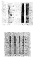
- Figure 1 shows in the blots A and B an example of Western blotting patterns which are developed after incubation respectively with representative negative and positive sera (positive for bovine tuberculosis).
- Blots C and D are an example of Western blotting patterns which are developed after incubation with a representative negative and positive serum sample, respectively (positive for Cattle Jones Disease).
- Blots A and B Lane 1: BCG crude extract, Lane 2: crude extract of an M. tuberculosis strain, Lane 3: M. bovis crude extract.
- Blots C and D Lane 1: BCG derived KP-100, Lane 2: RIVM 7114 derived KP-100.
- Figure 2a is an example of different Western blotting patterns developed after incubation with representative variable sera of tuberculosis patients. Noticeable is the combination of different patterns demonstrating the presence of different dominant bands, as shown in table 1. These band patterns function as "hallmarks" for TB patients as diagnosed serologically.
- Lane 1 BCG crude extract
- Lane 2 crude extract of an M. tuberculosis strain.
- Figure 2b is an example of different Western blotting patterns developed after incubation with representative sera of patients with Lepromatous Leprosy (LL), Blot A and C, and Tuberculous Leprosy (TT), Blot B and D.
- LL Lepromatous Leprosy
- TT Tuberculous Leprosy
- the "hallmark" patterns are shown in table 1 and are for LL: distinctive 29/33 kD doublet, and for TT: distinctive 64/65 kD doublet (often observed as single band) or a very pronounced smear in the 30-64 kD region.
- Lane 1 BCG crude extract
- Lane 2 crude extract of a M. tuberculosis strain
- Blot C Lane 1: Molecular marker
- Lane 2 not relevant
- Lane 3 BCG crude extract
- Lane 4 crude extract of an M. tuberculosis strain
- Blot D Lane 1: BCG crude extract
- Lane 2 crude extract of an M. tuberculosis strain
- Lane 3 Molecular marker.
- figure 2c is an example of different Western blotting patterns developed after incubation with representative sera of patients with Crohn's Disease.
- the "hallmark” patterns are shown in table 3 and are for Crohn's Disease a pronounced 45/48 kD doublet.
- blot A BCG crude extract
- blot B crude extract of an M. tuberculosis strain
- blot C Mycobacterium avium crude extract
- blot D molecular marker
- the starting material (see EXAMPLE 1 under 1.) was, depending on the chosen M. tuberculosis strain, centrifuged at 70,000 x g to 120,000 x g at 4°C for 2 hours. The pellet was washed three tines with PBS. Between the washing steps centrifuging took place at 70,000 x g to 120,000 x g at 4°C for 2 hours. The pellet was collected and resuspended in 10 ml PBS. The supernatant, SP-100, can also be used for immunoblots (example 1) and enzyme immunoassays (this example). The suspension was subsequently sonified for 2 minutes at 80 watts at 4°C. After the protein concentration was determined, quantities of 100 ⁇ l were frozen at a concentration of 1 mg/ml and stored at -80°C until time of use (this preparation is designated with the term KP-100).
- the elution was checked using a 12% polyacrylamide analytical gel of 1.5 mm thickness.
- the gel was cast with a comb. After 20 minutes incubation at 65°C 40 ⁇ l of each tube with gel pieces was placed in the slots in the presence of 10 ⁇ l 5x loading mixture.
- the electrophoresis was carried out for 30 minutes at 150 V in the "stacking gel” and for 6 hours at 100 V in the "running gel”. The electrophoresis was stopped and the gel made ready for preparation of a Western blot.
- the blotting procedure is described in EXAMPLE 1 under 2. Similar results were obtained using HPLC, FPLC and other routine separating procedures.
- Microtiter plates are coated (via standard techniques) with either KP-100, SP-100, starting material, whole bacteria, 29/33 kD, 64/65 kD or 45/48 kD antigens.
- the plates After coating the plates are blocked, in order to prevent an aspecific binding of serum components, with a 3% bovine serum albumin (BSA) solution. Plates are then dried and stored at 4°C.
- BSA bovine serum albumin
- Tuberculosis EIA test (microtiter plates coated with KP-100)
- Test sera are pipetted in a 1:100 dilution into the coated wells of a microtiter plate. The reaction takes place for 1 hour at 37°C. Aspecific serum components and non-binding serum components are washed away in a washing step. A second incubation with a suitable dilution of an antihuman IgA peroxidase conjugate is carried out again for 1 hour at 37°C, and excess conjugate is then washed away.
- TMB tetramethylbenzidine
- Binding enzyme results in the occurrence of a blue colour which, after addition of a colouring stop solution, changes to yellow. This yellow colour has an absorption maximum of 450 nm.
- the intensity of the resulting colour is proportional to the amount of bound KP-100-specific IgA.
- test patient and control sera are used from two different populations.
- Each population is sub-divided into 4 sub-groups, namely:
- the test is performed with two kits having different lot numbers and production dates.
- test serum can be considered positive when the result found in the test scores higher than 2.1 units.
- test serum can be considered negative when the result found scores lower than 1.2 units.
- Non-Endemic range Group 1: SERUM NO. # UNITS TEST SCORE REMARKS 1 2.24 positive culture positive 2 2.53 positive culture positive 3 4.40 positive culture positive 4 14.95 positive culture positive 5 16.82 positive culture positive 6 10.54 positive culture positive 7 5.70 positive culture positive 8 6.72 positive culture positive 9 5.06 positive culture positive Population B.
Landscapes
- Health & Medical Sciences (AREA)
- Life Sciences & Earth Sciences (AREA)
- Immunology (AREA)
- Engineering & Computer Science (AREA)
- Molecular Biology (AREA)
- Biomedical Technology (AREA)
- Chemical & Material Sciences (AREA)
- Hematology (AREA)
- Urology & Nephrology (AREA)
- Food Science & Technology (AREA)
- General Physics & Mathematics (AREA)
- Cell Biology (AREA)
- Biotechnology (AREA)
- Medicinal Chemistry (AREA)
- Physics & Mathematics (AREA)
- Analytical Chemistry (AREA)
- Biochemistry (AREA)
- General Health & Medical Sciences (AREA)
- Microbiology (AREA)
- Pathology (AREA)
- Tropical Medicine & Parasitology (AREA)
- Virology (AREA)
- Measuring Or Testing Involving Enzymes Or Micro-Organisms (AREA)
- Peptides Or Proteins (AREA)
- Medicines Containing Antibodies Or Antigens For Use As Internal Diagnostic Agents (AREA)
- Micro-Organisms Or Cultivation Processes Thereof (AREA)
- Preparation Of Compounds By Using Micro-Organisms (AREA)
Abstract
Description
- The invention relates to a method for identifying a Mycobacterium species responsible for a mycobacterial infection in human or animal. The invention further relates to diagnostic kits for use in the method.
- The genus Mycobacterium which contains about 50 species is responsible for a number of human and animal diseases which are known collectively as the mycobacterioses. The best known of these in humans is leprosy, caused by M. leprae, which affects more than ten million people worldwide, and tuberculosis, usually caused by M. tuberculosis, at least ten million new cases of which occur each year. Most other mycobacteria normally occur only as environmental saprophytes but can also cause opportunist diseases. This happens usually, but not only, in the case of people who have problems with their immune system, such as AIDS patients and people undergoing immunosuppression. These opportunist types comprise the slow-growing species M. avium, and the closely related M. intracellulare and M. scrofulaceum (often referred to together as MAIS complex), M. kansasi, M. marinum and M. ulcerans, and the fast-growing species M. chelonae and M. fortuitum. Although once rare, the incidence of opportunist mycobacterial diseases and tuberculosis shows a parallel increase in the western world with the incidence of AIDS. In addition there is limited but increasing evidence that mycobacteria or antigens thereof play a direct or indirect part in the etiology of a plurality of other diseases such as sarcoidosis and Crohn's disease and different auto-immune diseases such as auto-immune dermatitis, rheumatoid arthritis and diabetes. This could be attributed to a structural mimicry between epitopes of mycobacteria and those of the host.
- The cell walls of mycobacteria are very complex and contain many lipids, some with structures unique to the genus. These structures comprise mycolinic acids and esters, peptido-glycolipide, arabino-galactane and lipo-arabino-manane. The lipid-rich mycobacterial cell walls are responsible for the characterizing colouring properties of the mycobacteria. They also enable mycobacteria to counter an attack by the immune system of the host. A number of species, once taken up into macrophages, are capable of surrounding themselves with a thick layer of secreted lipids.
- Many different components of the mycobacteria begin an interaction with the immune system. These components comprise protein and hydrocarbon antigens, which can either be actively secreted by the mycobacteria or can form part of the cell wall or cell membrane. In addition they may be present in the cytoplasm, for instance in the cytoplasmic matrix, ribosomes and enzymes. Mycobacteria also possess immuno-modulating components such as immunosuppressing compounds and adjuvants. Consequently, a single mycobacterial species can induce a large variety of immune responses in different forms and with diverse specificities. It is therefore difficult to distinguish immune responses against species-specific components from cross reactions. For this reason it has therefore been found difficult to derive protein antigens suitable for the detection of species-specific humoral responses as a basis for a very sensitive and specific sero-diagnostic test for tuberculosis. Because the mycobacteria occur a great deal in the environment, human serum nearly always contains anti-mycobacterial anti-bodies.
- In view of the problems with the specificity of protein antigens, a number of researchers, including the present inventors, have focused their attention on species-specific glycolipid antigens for the detection of specific humoral immune responses. This approach is for example illustrated in Vega-Lopez et al., J. Clin. Microbiol. 26(12), 2474-2479 (1988) and Roche et al., Int. J. Mycobact. Dis. 60(2), 201-207 (1992) who both stated that in serodiagnosis species-specific antigens and antibodies are required. Although the immune reactivity against mycobacteria is of the cell-mediated type and the humoral immune responses probably play a minor part in the total effector mechanism of mycobacterial immunity and immunopathology, studies into the antibody response to immuno-dominant-mycobacterial cross-reactive antigen components (further referred to as Im-CRAC) could shed light on the varying capability of the host to recognize different mycobacterial antigens. They could therefore provide indirect information relating to the nature of the immune recognition of, and response to, a specific mycobacterial pathogen.
- It has now been found that the clinical manifestation of mycobacterial diseases appears to be related to the varying capability of an individual host to produce a humoral response to different mycobacterial immuno-cross-reactive antigen components (Im-CRAC). Each mycobacterial infection generates its own specific antibody response to a number of specified antigens. Analysis of the antibody-response by means of immunoblotting has demonstrated that the immune-dominant Im-CRAC vary in accordance with the immunopathological manifestation of the mycobacterial diseases. It has been found that the sera of individuals which are infected with different Mycobacterium species cause different and distinguishing band patterns on immunoblots of mycobacterial antigens.
- This discovery forms the basis of the present invention, whereby a method is provided for identifying a Mycobacterium species responsible for a mycobacterial infection in human or animal, comprising the steps of:
- a) selecting a suitable mycobacterial species and strain;
- b) preparing from said strain at least one mycobacterial antigen, respectively antigen preparation comprising at least one selected immuno-cross-reactive antigen component, which antigen component may induce a non-specific humoral immune response cross-reactive among Mycobacterial species;
- c) binding the antigen, respectively the antigen preparation to a suitable carrier;
- d) causing the binding antigen to react with antibodies from serum of an individual infected with a Mycobacterium species;
- e) making visible antigen-antibody reactions for a suitable antibody (sub-)class which reactions when taken together are unique to certain Mycobacterium species; and
- f) identifying the responsible Mycobacterium species on the basis of the unique set of the reactions which are made visible.
-
- In preference the antigen preparation is separated by electrophoresis prior to step c) and the carrier is a membrane to which the antigen is bound by means of electroblotting. This process is called Western blotting. The Im-CRAC comprise namely a number of antigens with specific molecular weights which, as has now been found, after immunoblotting exhibit a binding pattern which correlates to the disease, respectively infection. The specific band pattern is characterized by the presence or absence of four individual components, for instance:
- a region comprising different pronounced bands and/ or overlapping bands, which can be observed as a smear ("region");
- sharp single bands which are strongly positive ("band");
- sharp double bands which are strongly positive ("doublet"); and
- other positive bands ("extra bands").
- For a survey of the different antigens, their molecular weights and binding characteristics, see table 1.
Survey of characteristic binding patterns of mycobacterial Immuno-Cross-reactive Antigen Components. Antigen Diagnostic for MW range (in kD) Binding characteristic A J < 8 band B T, B, J, 10-16 band C B, J, T, 20-28 band or region D L 29-33 doublet E B, J, T, 31-40 band or region F T 38-40 band or region G C, B, J, 45-48 doublet H T 58-60 band or region I L 64-65 doublet J J 66 band K T 68 band L L 30-64 region T: Human Tuberculosis
L: Leprosy
C: Crohn's Disease
B: Bovine Tuberculosis
J: Johne's Disease - The mycobacterioses are all characterized by a specific banding pattern which is formed when a blot having thereon an antigen preparation of mycobacteria separated to size is incubated with serum of an infected individual.
- The tables 2a-2e below show a survey of the banding patterns of a number of mycobacterial, respectively immunological diseases.
Bovine tuberculosis
Regions (MW in kD)Pattern and/or 10-16 and/or 20-28 and/or 31-40 and 45-48 1. 14 kD band 22 kD band 20-28 kD region 31 kD band 45/48 kD doublet Johne's Disease
Regions (MW in kD)Pattern and/or > 8 and 10-16 and/or 20-28 and/or 31-40 and 45-48 and 66 1. region 14 kD band and 22 (25)-kD band, and/or 27 KDa band 31 kD band 45/48 kD doublet 66 kD band Human Tuberculosis
Regions (MW in kD)Pattern 10-16 20-28 31-38 38-40 58-60 68 other 1. 10 and 16 kD bands / 33 kD band region bands / +/- 2. 16 kD single band region 33 kD band bands region / +/- 3. 10 or 16 kD band / / bands bands / +/- 4. 16 kD band / 33 kD band region bands / +/- 5. 10 and/or 16 kD band region 33 kD band bands bands and/or region 68 kD band +/- Leprosy
Regions (MW in kD)Pattern 29-33 30-65 64-65 other LL pattern 1 29/33 kD doublet / / +/- TT pattern 1 / regions and/or bands / +/- TT pattern 2 / regions and/or bands 64/65 kD doublet +/- TT pattern 3 / / 64/65 kD doublet +/- Crohn's Disease
Regions (MW in kD)Pattern 45-48 other 1. 45/48 kD doublet +/- Regions (MW in kD) Pattern and 42 kD and/or 80-90 kD and/or 58-60 kD and/or 14-18 kD 1. band region region region - When an immunoblot is used, two questions can be answered. Firstly, the presence of any positive band pattern will answer the question of whether a mycobacterial infection is present. Secondly, the presence of specific banding patterns indicates which mycobacterial species has caused the infection, and therefore what the nature and etiology of the disease will be.
- The invention further relates to a heterogeneous enzyme immunoassay.
- From the patterns in the immunoblotting it follows which mycobacterial antigens respectively antigen preparations are suitable for diagnosis of any particular disease.
- The antigen for a heterogeneous enzyme immunoassay is preferably chosen from the group which consists of mycobacterial immuno-cross-reactive antigen components with a molecular weight of 29-33 kD, 45-48 kD, 64-65 kD and a fraction designated with the term KP-100. These ImCRAC can be used separately or in combination with each other for serological diagnosis of the correlating diseases in a heterogeneous enzyme immunoasssay (EIA). Rambukkana et al. in Scand. J. Immunol. 33, 763-755 (1991) disclose an ELISA using a microtiter plate coated with 2g - and 33-KDa antigens from M.tuberculosis to demonstrate binding of certain monoclonal antibodies.
- In this form of assay antibody-conjugates labelled with a standard enzyme are used. An important detail is that the enzyme activity does not change during the immunological reaction.
- In order to test the immune response in patients to the selected antigens, use is made for instance of microtiter plates ("Solid Phase"). By means of standard published techniques the antigens are irreversibly immobilized on the surface of the wells in such a microtiter plate.
- This binding takes place while retaining specific antigen determinants on the used antigens. After incubation with serum, in the wells of the microtiter plate, antibodies present therein can specifically form a complex with the irreversibly bound antigens.
- After removal of non-binding serum components, binding antibodies are detected using an anti-antibody antibody labelled with an enzyme.
- Binding of the enzyme is only possible when specific antibodies have adhered to the immobilized antigens. Substrate conversion by the binding enzyme to a visually or photometrically observable signal is thereby directly related to the presence of specific antibodies in the tested serum.
- The choice of specificity of the enzyme-bound anti-antibody antibody determines the type of reaction that takes place. For instance, it may be desirable in some cases to demonstrate the specifically binding immunoglobulins of the IgG isotype, while in other cases immunoglobulins of the IgA and/or IgM isotype are just demonstrated.
- The combination of antigen and immunoglobulin type defines the specificity of the test.
- The said methods, that is, the immunoblot and the EIA, can be used as mutual confirmation.
- In addition, for the serological diagnosis based on the said antigens, use can be made of a test stick as solid phase.
- A particularly advantageous embodiment of the invention relates to a test stick, the so-called "dip-stick", which is used as solid phase in the heterogeneous enzyme immunoassay.
- The said mycobacterial antigens can be irreversibly bound to such a dip-stick.
- The antigen is brought into reaction with antibody from serum for testing by dipping the dip-stick in a serum sample for testing. The formed antigen-antibody complex can be made visible by subsequently dipping the dip-stick in an anti-antibody antibody-enzyme conjugate solution.
- With the binding enzyme a substrate can then be converted to a visually or photometrically observable signal.
- The invention provides a diagnostic kit for:
- an immunoblot assay; comprising ImCRAC antigens separated by electrophoresis as described above, immobilized on a solid carrier, in addition to an associated suitable detection system.
- a heterogeneous enzyme immunological assay; comprising a microtiter plate, the wells of which are coated with above mentioned antigens or antigen preparations, in addition to an associated suitable detection system.
- a dip-stick assay; comprising test sticks coated with antigen or antigen preparations, in addition to an associated suitable detection system.
- The present invention will be further elucidated with reference to a number of examples which are given herein by way of illustration and are not intended to limit the invention.
- The mycobacteria were cultured in commercially available Sauton medium supplemented with 2 g MgSO4, 8 g citric acid, 2g K2HPO4, 16 g asparagine, 2 g (Fe+) ammonium citrate, 240 ml glycerol. The bacteria were cultured under standard conditions. The cells were harvested by filtration of the culture medium with a 12 µm filter. The cells were subsequently resuspended in 20 ml PBS (phosphate-buffered salt solution) (pH 7.4) and the harvested cells were autoclaved under a pressure of 15 Psi for 20 minutes in order to deactivate and sterilize the bacteria. The thus obtained bacterial mass can be stored at -80°C.
- In order to determine the quantity of starting material a 1/100 dilution of the harvested autoclaved suspension in PBS was made. The optical density thereof, measured at 420 nm (O.D.420) must be 0.1. If necessary the concentrated bacterial mass is supplemented with PBS (pH 7.4) until the correct O.D. is obtained. An O.D.420 of 0.1 indicates the presence of 7x10¹¹ bacteria per 30 ml, which is equivalent to 12 g wet weight of the bacterial mass.
- For preparation of a crude mycobacterial extract 5 g wet weight of the bacterial mass was washed three times with PBS (pH 7.4). Centrifuging was then carried out at 3000 x g until the mass precipitated. The pellet was suspended in 50 ml PBS and stirred carefully to reduce formation of lumps to a minimum. To prevent lump forming 0.05% Tween 80 was optionally added. In order to avoid bacterial contamination 3 mg penicillin/streptomycin was added to this solution. The concentration was then brought with PBS to 2 g wet weight/ml.
- The bacterial mass was subsequently broken open using an automatic French-X-press or RIBI press (American Instruments Company, Trevenollab. Inc. Maryland). The buckets were pre-cooled overnight at -20°C. Before use the buckets were held in a mixture of ethanol and dry ice (-20°C). After the buckets were filled with 1 g bacterial mass per bucket of 5 ml and cooled at -80°C for 20 minutes, the buckets were placed in the French-X press and 12 tons of pressure were applied by pushing in the plunger of the press. The buckets were then removed and cooled again at -80°C for 20 minutes. The buckets were inverted and treated for the second time. 10 tons of pressure were applied the second time. Cooling and breaking were then repeated a number of times, normally about 5 times. The disrupted cells were eluted with a suitable volume of PBS and subsequently centrifuged at 4°C at 300 x g for 10 minutes in order to remove the unbroken bacteria with the sediment. The collected supernatant was then centrifuged at 4°C and 145,000 x g for 2 hours. The pellet was suspended in 0.1 M Tris-HCl (pH 7.2), 0.01 M EDTA which contained 20 mM MgSO4.7H2O in a concentration of about 1 g per 10 ml. 1 mg RNase and 1 mg DNase were added per 10 ml volume. Incubation then took place overnight at 4°C with careful stirring. Incubation thereafter took place for 1 hour at 37°C and the lysate was centrifuged at 300 x g and 4°C for 10 minutes in order to remove the last-remaining unbroken bacteria (this is further referred to as "starting material").
- A 12% polyacrylamide analytical gel of 1.5 mm thickness was manufactured according to normal standard procedures. No comb was used in the stacking gel. 5 mg of the starting material obtained under 1. (100 mg per ml), respectively KP-100 or SP-100 (see example 2) were used for each gel. 40 microlitres of this material was diluted with 1200 microlitres PBS. 300 microlitres 5x loading mixture (0.3 g 250 mM Tris-HCl, 1.0 g 10% SDS, 1.0 g 10% dithioerytreitol, 5 mg 0.05% bromophenol blue) was then added.
- Incubation was carried out for 20 minutes at 65°C. 1500 microlitres were subsequently applied to the gel and electrophoresis performed under the following conditions: 150 V for the run through the stacking gel for 30 minutes and 100 V through the running gel for 6 hours.
- In order to form a Western blot the proteins present in the gel were transferred at 50 V for 3 hours to a nitrocellulose membrane. After completion of the transfer the membrane was coloured with 1.5% amido black for 2 minutes to check the membrane for irregularities and air bubbles. The membrane was then decolourized in 0.05% Tween 80 in PBS with 1% BSA (bovine serum albumin). The membrane was then cut into strips and was ready for use.
- The strips were incubated with 1:200 diluted human serum. The serum was diluted in PBS with 3% BSA. The incubation took place for 1 hour at room temperature. The strips were subsequently washed three times (for 3 minutes at a time) in PBS. The strips were then incubated with a goat anti-total-human immunoglobulin-alkaline phosphatase conjugate in a dilution of 1 to 1000 in PBS with 3% BSA and 0.05% Tween 80. Washing in PBS then took place again three times. The colour was developed with an NBT/BCIP (nitroblue tetrazolium/Bromo, Chloro Indolyl phosphate) colour solution (1 mg per 10 ml) to which 10 microlitres H2O2 were added. The strips were incubated for a maximum of 2 hours in 1 ml of this solution per strip. The colour reaction was stopped by transferring the strips to 0.1 M Tris-HCl (pH 8.3), 0.01 M EDTA. The obtained patterns are interpreted by comparison with a reference pattern.
- The results are shown in figures 1 and 2a-2c.
- Figure 1 shows in the blots A and B an example of Western blotting patterns which are developed after incubation respectively with representative negative and positive sera (positive for bovine tuberculosis).
- Blots C and D are an example of Western blotting patterns which are developed after incubation with a representative negative and positive serum sample, respectively (positive for Cattle Jones Disease).
- Blots A and B: Lane 1: BCG crude extract, Lane 2: crude extract of an M. tuberculosis strain, Lane 3: M. bovis crude extract.
- Blots C and D: Lane 1: BCG derived KP-100, Lane 2: RIVM 7114 derived KP-100.
- Only the specific characteristics are stated herein.
- Blot A: only the background bands can be observed in blots incubated with negative serum.
- Blot B: region in the 10-16 kD region in
lane 3, 22 kD band inlane 2, 31 KDa bands inlane 1 and 2, 14 KDa band in lane 2. - Blot C: only background bands can be observed in blots incubated with negative serum.
- Blot D: 45-48 kD doublet in
lane 1 and 2, 22 and 25 kD band in lane 1 and 2, 66 kD band in lane 1 and 2, 27 kD band in lane 1. -
- Figure 2a is an example of different Western blotting patterns developed after incubation with representative variable sera of tuberculosis patients. Noticeable is the combination of different patterns demonstrating the presence of different dominant bands, as shown in table 1. These band patterns function as "hallmarks" for TB patients as diagnosed serologically.
- Applicable to all blots (from left to right): Lane 1 = BCG crude extract, Lane 2 = crude extract of an M. tuberculosis strain.
-
- Blot A: Mycobacterium avium infected patient.
- Blot B-F: Tuberculosis patients.
- Blot G: non-endemic negative serum.
- Blot H: endemic negative serum (known recent contact, blot
developed 2 weeks after patient returned to Netherlands from
endemic range).
Only "hallmarks" are mentioned. - Blot A: Mycobacterium avium infected patients, sera, band at 68 kD in lane 1 and 2, range in 10-16 kD in lane 1, band in the 58-60 kD region in lane 2. Patient shows low IgA titer in P-90 ELISA).
- Blot B: 38-40 kD band in lane 1 and 2, 10-16 kD band in lane 1 and 2, band in 58-60 kD region in lane 2, smear in 22-28 kD region in lane 1.
- Blot C: 16 kD band in lane 1 and 2, bands in 58-60* kD region in lane 1 and 2, bands in 38-40 kD region in lane 1 and 2, smear in 22-28 kD region in lane 1, 33 kD band in lane 1 and 2.
- Blot D: 10 kD band in 10-16 kD region in lane 1, 16 kD band in 10-16 kD region in lane 2, 68 kD band in lane 1 and 2, bands in 58-60* kD region in lane 1 and 2.
- Blot E: smear in 33-38 kD region in lane 1 and 2, 16 kD bands in lane 1 and 2, bands in 58-60* kD region in lane 2.
- Blot F: bands in 10-16, 22-28, 38-40, 58-60 regions and 68 KDa band in both lanes 1 and 2.
- Blot G: non-endemic negative serum.
- Blot H: endemic negative serum (known contact).
-
- Figure 2b is an example of different Western blotting patterns developed after incubation with representative sera of patients with Lepromatous Leprosy (LL), Blot A and C, and Tuberculous Leprosy (TT), Blot B and D.
- The "hallmark" patterns are shown in table 1 and are for LL: distinctive 29/33 kD doublet, and for TT: distinctive 64/65 kD doublet (often observed as single band) or a very pronounced smear in the 30-64 kD region.
- To Blot A and B are applied: Lane 1: BCG crude extract, Lane 2: crude extract of a M. tuberculosis strain, Blot C: Lane 1: Molecular marker, Lane 2: not relevant, Lane 3: BCG crude extract, Lane 4: crude extract of an M. tuberculosis strain, Blot D: Lane 1: BCG crude extract, Lane 2: crude extract of an M. tuberculosis strain, Lane 3: Molecular marker.
-
- Blot A/C: 29-33 kD doublet in lane 1 and 2.
- Blot B/D: 64/65 kD doublet in lane 1 and 2.
-
- Noticeable is the very intensive smear in the 30-64 kD range on blot D.
- Finally, figure 2c is an example of different Western blotting patterns developed after incubation with representative sera of patients with Crohn's Disease. The "hallmark" patterns are shown in table 3 and are for Crohn's Disease a pronounced 45/48 kD doublet.
- Applied to blot A is: BCG crude extract,
blot B: crude extract of an M. tuberculosis strain,
blot C: Mycobacterium avium crude extract,
blot D: molecular marker. - All lanes show a distinctive colouring of the 45/48 KDa doublet positivity, which indicates Crohn's Disease. The 45/48 KDa doublet reacts positively in 65% of all Crohn patients.
- The starting material (see EXAMPLE 1 under 1.) was, depending on the chosen M. tuberculosis strain, centrifuged at 70,000 x g to 120,000 x g at 4°C for 2 hours. The pellet was washed three tines with PBS. Between the washing steps centrifuging took place at 70,000 x g to 120,000 x g at 4°C for 2 hours. The pellet was collected and resuspended in 10 ml PBS. The supernatant, SP-100, can also be used for immunoblots (example 1) and enzyme immunoassays (this example). The suspension was subsequently sonified for 2 minutes at 80 watts at 4°C. After the protein concentration was determined, quantities of 100 µl were frozen at a concentration of 1 mg/ml and stored at -80°C until time of use (this preparation is designated with the term KP-100).
- 30 mg of the starting material was then applied in the presence of loading mixture onto a preparatory 12% polyacrylamide gel of 0.5 cm thickness after 20 minutes of incubation at 65°C. Electrophoresis was carried out for 30 minutes at 150 V (stacking gel) and for 6 hours at 100 V (running gel). The electrophoresis was stopped after the blue colorant band had disappeared from the gel. The gel was then cut into horizontal strips of 2 mm thickness which in turn were divided into pieces of 1 cm length. The gel pieces were each eluted overnight at 4°C in a tube with 5 ml sterile distilled water. Thorough mixing thereafter took place and the remaining gel pieces were centrifuged to the bottom.
- The elution was checked using a 12% polyacrylamide analytical gel of 1.5 mm thickness. The gel was cast with a comb. After 20 minutes incubation at 65°C 40 µl of each tube with gel pieces was placed in the slots in the presence of 10 µl 5x loading mixture. The electrophoresis was carried out for 30 minutes at 150 V in the "stacking gel" and for 6 hours at 100 V in the "running gel". The electrophoresis was stopped and the gel made ready for preparation of a Western blot. The blotting procedure is described in EXAMPLE 1 under 2. Similar results were obtained using HPLC, FPLC and other routine separating procedures.
- In order to establish which fractions contain the relevant antigens, strips of the blot were incubated with sera of patients with lepromatous leprosy, tuberculous leprosy and Crohn's Disease. Shown herewith are respectively the 29/33 kD antigens, the 64/65 kD antigen and the 45/48 kD antigens. The complex formation was visualized using anti-human IgG peroxidase conjugate and DAB. The desired fractions were collected, combined and used to coat a microtiter plate (see below).
- Microtiter plates are coated (via standard techniques) with either KP-100, SP-100, starting material, whole bacteria, 29/33 kD, 64/65 kD or 45/48 kD antigens.
- After coating the plates are blocked, in order to prevent an aspecific binding of serum components, with a 3% bovine serum albumin (BSA) solution. Plates are then dried and stored at 4°C.
- Test sera are pipetted in a 1:100 dilution into the coated wells of a microtiter plate. The reaction takes place for 1 hour at 37°C. Aspecific serum components and non-binding serum components are washed away in a washing step. A second incubation with a suitable dilution of an antihuman IgA peroxidase conjugate is carried out again for 1 hour at 37°C, and excess conjugate is then washed away.
- Indication of human antibodies of the sub-type IgA binding specifically to KP-100 takes place by adding TMB (tetramethylbenzidine) to the wells.
- Binding enzyme results in the occurrence of a blue colour which, after addition of a colouring stop solution, changes to yellow. This yellow colour has an absorption maximum of 450 nm.
- The intensity of the resulting colour is proportional to the amount of bound KP-100-specific IgA.
- The results are shown in the tables below.
- In the described test patient and control sera are used from two different populations.
- A = Endemic area (Africa, Ghana)
- B = Non-endemic area (Europe, the Netherlands).
-
- Each population is sub-divided into 4 sub-groups, namely:
- Group 1 =
- culture confirmed TB patients
- Group 2 =
- negative control group (normal healthy individuals
- Group 3 =
- suspected positives (TB contacts)
- Group 4 =
- suspected negatives (no known data, but certainly no TB, possibly leprosy or other aspecific Mycobacteriosis).
- The test is performed with two kits having different lot numbers and production dates.
- Interpretation of the test results takes place on the basis of the so-called calibration line which is made up of control sera with a determined arbitrary unit definition which corresponds to a known OD value (1 unit, 4 units and 8 units).
- Each time a test is carried out the units are included in the assay. Found sample values can then be related to the unit definition.
- A test serum can be considered positive when the result found in the test scores higher than 2.1 units.
- A test serum can be considered negative when the result found scores lower than 1.2 units.
- Test sera with unit values between 2.1 and 1.2 units fall into the set so-called reconfirmation zone. This means that in the first instance positivity or negativity for tuberculosis cannot be determined with this test.
- Reconfirmation of these sera takes place using the described Western blot strips with which, after serum incubation on the basis of banding patterns and specific "hallmarks", an answer can be given to the question of whether the test serum is positive (bands present) or negative (bands absent).
Population A. Endemic range:
Group 1:SERUM NO. # UNITS TEST SCORE REMARKS 1 2.44 positive culture positive 2 3.43 positive culture positive 3 1.78 reconfirmation culture positive 4 1.37 reconfirmation culture positive 5 5.74 positive culture positive 6 3.21 positive culture positive 7 1.66 reconfirmation culture positive 8 2.00 reconfirmation culture positive 9 2.00 reconfirmation culture positive Population A. Group 2: SERUM NO. # UNITS TEST SCORE REMARKS 10 1.16 negative healthy individual 11 0,86 negative healthy individual 12 0.77 negative healthy individual 13 0.64 negative healthy individual 14 0.74 negative healthy individual 15 0.79 negative healthy individual Population A. Group 3: SERUM NO. # UNITS TEST SCORE REMARKS 16 3.13 positive sick individual 17 1.57 reconfirmation sick individual 18 1.59 reconfirmation sick individual 19 5.39 positive sick individual 20 1.97 reconfirmation sick individual 21 2.29 positive sick individual 22 0.48 negative sick individual Population A. Group 4: SERUM NO. # UNITS TEST SCORE REMARKS 23 1.54 reconfirmation normal control 24 1.76 reconfirmation normal control 25 0.58 negative culture negative 26 1.03 negative culture negative 27 0.79 negative culture negative 28 0.89 negative culture negative Population B. Non-Endemic range:
Group 1:SERUM NO. # UNITS TEST SCORE REMARKS 1 2.24 positive culture positive 2 2.53 positive culture positive 3 4.40 positive culture positive 4 14.95 positive culture positive 5 16.82 positive culture positive 6 10.54 positive culture positive 7 5.70 positive culture positive 8 6.72 positive culture positive 9 5.06 positive culture positive Population B.
Group 2:SERUM NO. # UNITS TEST SCORE REMARKS 10 1.04 negative healthy individual 11 0.92 negative healthy individual 12 0.85 negative healthy individual 13 0.20 negative healthy individual 14 0.42 negative healthy individual 15 0.90 negative healthy individual 16 0.35 negative healthy individual 17 0.73 negative healthy individual Population B.
Group 3:SERUM NO. # UNITS TEST SCORE REMARKS 18 2.23 positive sick individual 19 4.70 positive sick individual 20 1.22 reconfirmation sick individual 21 1.43 reconfirmation sick individual 22 2.21 positive sick individual with TB history 23 6.38 positive sick individual 24 1.59 reconfirmation sick individual Population B.
Group 4:SERUM NO. # UNITS TEST SCORE REMARKS 25 0.20 negative patient resistant to drug therapy
Claims (15)
- Serodiagnostic method for identifying a Mycobacterium species responsible for a mycobacterial infection in human or animal, comprising the steps of:a) selecting a suitable mycobacterial species and strain;b) preparing from said strain at least one mycobacterial antigen, respectively antigen preparation, comprising at least one selected immuno-cross-reactive antigen component, which antigen component may induce a non-specific humoral immune response cross-reactive among Mycobacterial species;c) binding the antigen, respectively the antigen preparation to a suitable carrier;d) causing the bound immuno-cross-reactive antigen component(s) to react with antibodies from serum of an individual infected with a Mycobacterium species;e) making visible a set of antigen-antibody reactions for a suitable antibody (sub-)class, which reactions when taken together are unique to a certain Mycobacterium species; andf) identifying the responsible Mycobacterium species on the basis of the unique set of the reactions which are made visible.
- Method as claimed in claim 1, characterized in that the antigen preparation is separated by electrophoresis prior to step c) and the carrier is a membrane to which the antigen is bound by means of electroblotting.
- Method as claimed in claim 2, characterized in that the membrane is a nitrocellulose membrane.
- Method as claimed in claim 1, characterized in that the Mycobacterium species responsible for the mycobacterial infection is identified on the basis of a reference pattern.
- Method as claimed in any of the claims 1-4, characterized in that the antigen preparation is a total protein preparation of Mycobacterium.
- Method as claimed in any of the claims 1-4, characterized in that the antigen preparation is a KP-100 or SP-100 fraction of the total protein preparation.
- Method as claimed in claim 1, characterized in that the antigen is specific for one Mycobacterium species and the carrier is a microtiter plate.
- Method as claimed in claim 7, characterized in that the specific antigen is chosen from the group consisting of mycobacterial immuno-cross-reactive antigen components with a molecular weight of respectively 29/33 kD, 45/48 kD, 64 kD and the antigen fractions KP-100 or SP-100 or combinations thereof.
- Method as claimed in claim 7 or 8, characterized in that the antigen-antibody reactions are made visible using antibody-enzyme conjugates directed against antibodies of an isotype chosen from IgG, IgM, IgA.
- Method as claimed in claim 9, characterized in that the enzyme is peroxidase.
- Method as claimed in claim 1, characterized in that the carrier is a dip-stick with mycobacterial antigens bound thereto, and that the antigens are reacted with antibodies from a test serum sample by dipping the stick in said sample and subsequently visualizing the antigen-antibody reactions by dipping the stick into a solution with antibody-enzyme conjugate against bound antibodies followed by a solution with substrate for the enzyme to produce a colour reaction.
- Method as claimed in claim 1, characterized in that the antigen is bound to the wells of a microtiter plate, that the antigen is reacted with antibodies from the serum to be tested by placing in the wells a sample of the serum to be tested, that the non-binding antibody is removed, and antigen-antibody reactions are made visible by subsequently placing in the wells an antibody-enzyme conjugate against bound antibodies followed by a substrate for the enzyme to produce a colour reaction.
- Serodiagnostic kit comprising a dip-stick on which is arranged a carrier strip with mycobacterial antigens bound thereto, comprising at least one selected immuno-cross-reactive antigen component, which antigen component may induce a non-specific humoral immune response cross-reactive among mycobacterial species, and means for visualizing antigen-antibody reactions, which reactions when taken together are unique to a certain Mycobacterium species, occurring on the carrier after contact with a serum for testing.
- Serodiagnostic kit, comprising a microtiter plate in the wells of which are arranged mycobacterial antigens, comprising at least one selected immuno-cross-reactive antigen component, which antigen component may induce a non-specific humoral immune response cross-reactive among mycobacterial species, and means for visualizing antigen-antibody reactions occurring in the wells after contact with a serum for testing, which reactions when taken together are unique to a certain Mycobacterium species, with the proviso that when mycobacterial immuno-cross-reactive antigen components with a molecular weight of 29 or 33 KD of M. Tuberculosis are used as the mycobacterial antigen they are always used in combination with each other or other mycobacterial antigens.
- Serodiagnostic kit comprising an immunoblot membrane with mycobacterial antigens bound thereto, the antigens comprising at least one selected immuno-cross-reactive antigen component, which antigen component may induce a non-specific humoral immune response cross-reactive among mycobacterial species, and which antigens were separated by electrophoresis, and means for visualizing antigen-antibody reactions, which reactions when taken together are unique to a certain Mycobacterium species, occurring on the immunoblot after contact with a serum for testing.
Applications Claiming Priority (3)
| Application Number | Priority Date | Filing Date | Title |
|---|---|---|---|
| NL9202197A NL9202197A (en) | 1992-12-17 | 1992-12-17 | Method and device for identifying a mycobacterium species responsible for mycobacterial infection. |
| NL9202197 | 1992-12-17 | ||
| PCT/NL1993/000270 WO1994014069A1 (en) | 1992-12-17 | 1993-12-17 | Method and device for identifying a mycobacterium species responsible for a mycobacterial infection |
Publications (2)
| Publication Number | Publication Date |
|---|---|
| EP0674766A1 EP0674766A1 (en) | 1995-10-04 |
| EP0674766B1 true EP0674766B1 (en) | 2000-04-26 |
Family
ID=19861648
Family Applications (1)
| Application Number | Title | Priority Date | Filing Date |
|---|---|---|---|
| EP94903148A Expired - Lifetime EP0674766B1 (en) | 1992-12-17 | 1993-12-17 | Method and device for identifying a mycobacterium species responsible for a mycobacterial infection |
Country Status (11)
| Country | Link |
|---|---|
| US (1) | US5817473A (en) |
| EP (1) | EP0674766B1 (en) |
| AT (1) | ATE192238T1 (en) |
| AU (1) | AU5720394A (en) |
| DE (1) | DE69328493T2 (en) |
| DK (1) | DK0674766T3 (en) |
| ES (1) | ES2148314T3 (en) |
| GR (1) | GR3034022T3 (en) |
| NL (1) | NL9202197A (en) |
| PT (1) | PT674766E (en) |
| WO (1) | WO1994014069A1 (en) |
Families Citing this family (25)
| Publication number | Priority date | Publication date | Assignee | Title |
|---|---|---|---|---|
| US5658749A (en) * | 1994-04-05 | 1997-08-19 | Corning Clinical Laboratories, Inc. | Method for processing mycobacteria |
| RU2145977C1 (en) * | 1994-04-05 | 2000-02-27 | Интигрейтид Рисеч Текнолэджи эЛэЛСи | Method of preparing sample for determination of microorganisms (variants) and set for sample treatment for determination of microorganisms |
| US6458366B1 (en) * | 1995-09-01 | 2002-10-01 | Corixa Corporation | Compounds and methods for diagnosis of tuberculosis |
| US6592877B1 (en) | 1995-09-01 | 2003-07-15 | Corixa Corporation | Compounds and methods for immunotherapy and diagnosis of tuberculosis |
| US6290969B1 (en) * | 1995-09-01 | 2001-09-18 | Corixa Corporation | Compounds and methods for immunotherapy and diagnosis of tuberculosis |
| US6416962B1 (en) * | 1995-11-20 | 2002-07-09 | Kreatech Biotechnology B.V. | Method and device for identifying a mycobacterium species responsible for a mycobacterial infection |
| DE19621488C2 (en) * | 1996-05-29 | 1998-05-14 | Gerlach Gerald F Prof Dr | Diagnostic procedure for the detection of subclinical mammals suffering from paratuberculosis |
| RU2133965C1 (en) * | 1997-07-08 | 1999-07-27 | Московский областной научно-исследовательский клинический институт | Serological method for diagnosing tuberculosis in patients suffering from immunodeficiency |
| EP0921397A1 (en) * | 1997-12-08 | 1999-06-09 | Kreatech Biotechnology B.V. | A method for identifying a mycobacterium species |
| RU2141665C1 (en) * | 1998-04-14 | 1999-11-20 | Научно-исследовательский ветеринарный институт Нечерноземной зоны РФ | Method of detection of tuberculosis mycobacterium |
| GB2342443A (en) * | 1998-10-02 | 2000-04-12 | Abp Diagnostics Limited | Immunoassay device |
| US8143386B2 (en) * | 1999-04-07 | 2012-03-27 | Corixa Corporation | Fusion proteins of mycobacterium tuberculosis antigens and their uses |
| AU779978B2 (en) * | 1999-10-07 | 2005-02-24 | Corixa Corporation | Fusion proteins of mycobacterium tuberculosis |
| AU2001241738A1 (en) * | 2000-02-25 | 2001-09-03 | Corixa Corporation | Compounds and methods for diagnosis and immunotherapy of tuberculosis |
| DE60139963D1 (en) * | 2000-06-20 | 2009-10-29 | Corixa Corp | FUSION PROTEINS FROM MYCOBACTERIUM TUBERCULOSIS |
| RU2219547C2 (en) * | 2001-07-23 | 2003-12-20 | Нижегородская государственная медицинская академия | Method for predicting tuberculosis activity in respiratory organs |
| WO2003040722A1 (en) * | 2001-11-09 | 2003-05-15 | Kreatech Biotechnology B.V. | Means and methods for the detection of immunoglobulin capable of binding to mycobacterium antigen |
| US6841159B2 (en) * | 2002-01-30 | 2005-01-11 | The United States Of America As Represented By The Secretary Of The Navy | Rapid lateral flow assay for determining exposure to Mycobacterium tuberculosis and other mycobacteria |
| US7026465B2 (en) * | 2002-02-15 | 2006-04-11 | Corixa Corporation | Fusion proteins of Mycobacterium tuberculosis |
| US7276350B2 (en) * | 2004-04-27 | 2007-10-02 | University Of Tennessee Research Foundation | Method for diagnosing infectious diseases |
| US8956859B1 (en) | 2010-08-13 | 2015-02-17 | Aviex Technologies Llc | Compositions and methods for determining successful immunization by one or more vaccines |
| NL2017204B1 (en) * | 2016-06-08 | 2017-12-18 | Kei International Ltd | Solid substrate comprising antigens immobilised thereto, biosensor comprising said solid substrate and method for detecting the presence of mycobacterial material in a sample |
| WO2017211314A1 (en) | 2016-06-08 | 2017-12-14 | Kei International Limited | Method for detecting presence of mycobacterial material in sample using immobilised mannosyl phosphoketide antigen |
| NL2016913B1 (en) * | 2016-06-08 | 2017-12-18 | Kei International Ltd | Solid substrate comprising antigens immobilised thereto and use thereof in a method for detecting the presence of mycobacterial material in a sample |
| NL2021443B1 (en) | 2018-08-08 | 2020-02-20 | Kei International Ltd | Synthetic antigens for tuberculosis detection |
Family Cites Families (1)
| Publication number | Priority date | Publication date | Assignee | Title |
|---|---|---|---|---|
| US4391904A (en) * | 1979-12-26 | 1983-07-05 | Syva Company | Test strip kits in immunoassays and compositions therein |
-
1992
- 1992-12-17 NL NL9202197A patent/NL9202197A/en not_active Application Discontinuation
-
1993
- 1993-12-17 AU AU57203/94A patent/AU5720394A/en not_active Abandoned
- 1993-12-17 AT AT94903148T patent/ATE192238T1/en not_active IP Right Cessation
- 1993-12-17 DK DK94903148T patent/DK0674766T3/en active
- 1993-12-17 WO PCT/NL1993/000270 patent/WO1994014069A1/en not_active Ceased
- 1993-12-17 PT PT94903148T patent/PT674766E/en unknown
- 1993-12-17 DE DE69328493T patent/DE69328493T2/en not_active Expired - Fee Related
- 1993-12-17 US US08/454,122 patent/US5817473A/en not_active Expired - Fee Related
- 1993-12-17 EP EP94903148A patent/EP0674766B1/en not_active Expired - Lifetime
- 1993-12-17 ES ES94903148T patent/ES2148314T3/en not_active Expired - Lifetime
-
2000
- 2000-07-26 GR GR20000401710T patent/GR3034022T3/en not_active IP Right Cessation
Also Published As
| Publication number | Publication date |
|---|---|
| GR3034022T3 (en) | 2000-11-30 |
| ATE192238T1 (en) | 2000-05-15 |
| DE69328493D1 (en) | 2000-05-31 |
| ES2148314T3 (en) | 2000-10-16 |
| US5817473A (en) | 1998-10-06 |
| DK0674766T3 (en) | 2000-09-25 |
| NL9202197A (en) | 1994-07-18 |
| PT674766E (en) | 2000-10-31 |
| WO1994014069A1 (en) | 1994-06-23 |
| EP0674766A1 (en) | 1995-10-04 |
| DE69328493T2 (en) | 2000-12-21 |
| AU5720394A (en) | 1994-07-04 |
Similar Documents
| Publication | Publication Date | Title |
|---|---|---|
| US5817473A (en) | Method and device for identifying a mycobacterium species responsible for a mycobacterial infection | |
| Amadori et al. | Use of recombinant proteins in antibody tests for bovine tuberculosis | |
| Brusasca et al. | Antigen recognition by serum antibodies in non-human primates experimentally infected with Mycobacterium tuberculosis | |
| Chenthamarakshan et al. | Detection of immunoglobulins M and G using culture filtrate antigen of Burkholderia pseudomallei | |
| Uhaa et al. | Evaluation of specificity of indirect enzyme-linked immunosorbent assay for diagnosis of human Q fever | |
| US6416962B1 (en) | Method and device for identifying a mycobacterium species responsible for a mycobacterial infection | |
| Kashyap et al. | Diagnostic value of early secreted antigenic target-6 for the diagnosis of tuberculous meningitis patients | |
| He et al. | Rapid serodiagnosis of human mycobacteriosis by ELISA using cord factor (trehalose-6, 6′-dimyclate) purified from Mycobacterium tuberculosis as antigen | |
| Harrington et al. | Antibody responses of cervids (Cervus elaphus) following experimental Mycobacterium bovis infection and the implications for immunodiagnosis | |
| US6733983B1 (en) | Method for identifying a mycobacterium species | |
| WO2000072877A1 (en) | Rapid immunoassay to detect antibodies in saliva to disease-related antigens | |
| AU2001271554A1 (en) | Mycobacterial proteins as early antigens for serodiagnosis and vaccines | |
| US6146839A (en) | Chlamydia pneumoniae antigens, method for production of the antigens, method and reagents for measurement of anti-Chlamydia pneumoniae antibodies using the antigens | |
| Magnarelli | Laboratory diagnosis of Lyme disease | |
| Ng et al. | Serodiagnosis of pericardial tuberculosis | |
| Diagbouga et al. | Immunoblot analysis for serodiagnosis of tuberculosis using a 45/47-kilodalton antigen complex of Mycobacterium tuberculosis | |
| LUH et al. | Tuberculosis antigen A60 serodiagnosis in tuberculous infection: application in extrapulmonary and smear‐negative pulmonary tuberculosis | |
| Nagao-Dias et al. | Salivary anti-PGL IgM and IgA titers and serum antibody IgG titers and avidities in leprosy patients and their correlation with time of infection and antigen exposure | |
| US20090011442A1 (en) | TB diagnostics based on mycobacterium tuberculosis excretory secretory antigens and their specific immunoglobulins | |
| US5532134A (en) | Mycoplasma diagnostic assay | |
| Gandhi et al. | Enzyme linked protein-A: An ELISA for detection of IgG antibodies against Mycobacterium tuberculosis in intestinal tuberculosis | |
| Cázares-Sosa et al. | Comparative analysis of antibodies and lymphocytes in pulmonary tuberculosis patients with negative and positive cultures | |
| Mehta et al. | Serodiagnostic potentialities of enzyme-linked immunosorbent assay (ELISA) using mannophosphoinositides of Mycobacterium tuberculosis H37Rv | |
| FOFANA | Human tuberculosis sera show prominent antibody responses to particulate fractions ofMycobacterium tuberculosis | |
| BR102024002645A2 (en) | Kit, Method for Immunodiagnosis of Leprosy and Uses |
Legal Events
| Date | Code | Title | Description |
|---|---|---|---|
| PUAI | Public reference made under article 153(3) epc to a published international application that has entered the european phase |
Free format text: ORIGINAL CODE: 0009012 |
|
| 17P | Request for examination filed |
Effective date: 19950616 |
|
| AK | Designated contracting states |
Kind code of ref document: A1 Designated state(s): AT BE CH DE DK ES FR GB GR IE IT LI LU MC NL PT SE |
|
| 17Q | First examination report despatched |
Effective date: 19951214 |
|
| GRAG | Despatch of communication of intention to grant |
Free format text: ORIGINAL CODE: EPIDOS AGRA |
|
| GRAG | Despatch of communication of intention to grant |
Free format text: ORIGINAL CODE: EPIDOS AGRA |
|
| GRAH | Despatch of communication of intention to grant a patent |
Free format text: ORIGINAL CODE: EPIDOS IGRA |
|
| GRAH | Despatch of communication of intention to grant a patent |
Free format text: ORIGINAL CODE: EPIDOS IGRA |
|
| GRAA | (expected) grant |
Free format text: ORIGINAL CODE: 0009210 |
|
| AK | Designated contracting states |
Kind code of ref document: B1 Designated state(s): AT BE CH DE DK ES FR GB GR IE IT LI LU MC NL PT SE |
|
| REF | Corresponds to: |
Ref document number: 192238 Country of ref document: AT Date of ref document: 20000515 Kind code of ref document: T |
|
| REG | Reference to a national code |
Ref country code: CH Ref legal event code: EP |
|
| REF | Corresponds to: |
Ref document number: 69328493 Country of ref document: DE Date of ref document: 20000531 |
|
| REG | Reference to a national code |
Ref country code: IE Ref legal event code: FG4D |
|
| ITF | It: translation for a ep patent filed | ||
| ET | Fr: translation filed | ||
| REG | Reference to a national code |
Ref country code: DK Ref legal event code: T3 |
|
| REG | Reference to a national code |
Ref country code: CH Ref legal event code: NV Representative=s name: TROESCH SCHEIDEGGER WERNER AG |
|
| REG | Reference to a national code |
Ref country code: ES Ref legal event code: FG2A Ref document number: 2148314 Country of ref document: ES Kind code of ref document: T3 |
|
| REG | Reference to a national code |
Ref country code: PT Ref legal event code: SC4A Free format text: AVAILABILITY OF NATIONAL TRANSLATION Effective date: 20000720 |
|
| PLBE | No opposition filed within time limit |
Free format text: ORIGINAL CODE: 0009261 |
|
| STAA | Information on the status of an ep patent application or granted ep patent |
Free format text: STATUS: NO OPPOSITION FILED WITHIN TIME LIMIT |
|
| 26N | No opposition filed | ||
| REG | Reference to a national code |
Ref country code: GB Ref legal event code: IF02 |
|
| PGFP | Annual fee paid to national office [announced via postgrant information from national office to epo] |
Ref country code: BE Payment date: 20040908 Year of fee payment: 12 |
|
| PGFP | Annual fee paid to national office [announced via postgrant information from national office to epo] |
Ref country code: MC Payment date: 20041021 Year of fee payment: 12 |
|
| PGFP | Annual fee paid to national office [announced via postgrant information from national office to epo] |
Ref country code: LU Payment date: 20041116 Year of fee payment: 12 |
|
| PGFP | Annual fee paid to national office [announced via postgrant information from national office to epo] |
Ref country code: AT Payment date: 20041217 Year of fee payment: 12 |
|
| PG25 | Lapsed in a contracting state [announced via postgrant information from national office to epo] |
Ref country code: AT Free format text: LAPSE BECAUSE OF NON-PAYMENT OF DUE FEES Effective date: 20051217 |
|
| PG25 | Lapsed in a contracting state [announced via postgrant information from national office to epo] |
Ref country code: MC Free format text: LAPSE BECAUSE OF NON-PAYMENT OF DUE FEES Effective date: 20051231 Ref country code: LU Free format text: LAPSE BECAUSE OF NON-PAYMENT OF DUE FEES Effective date: 20051231 Ref country code: BE Free format text: LAPSE BECAUSE OF NON-PAYMENT OF DUE FEES Effective date: 20051231 |
|
| PGFP | Annual fee paid to national office [announced via postgrant information from national office to epo] |
Ref country code: SE Payment date: 20070612 Year of fee payment: 14 Ref country code: CH Payment date: 20070612 Year of fee payment: 14 |
|
| PGFP | Annual fee paid to national office [announced via postgrant information from national office to epo] |
Ref country code: IE Payment date: 20070613 Year of fee payment: 14 Ref country code: DK Payment date: 20070613 Year of fee payment: 14 |
|
| PGFP | Annual fee paid to national office [announced via postgrant information from national office to epo] |
Ref country code: GB Payment date: 20070608 Year of fee payment: 14 |
|
| BERE | Be: lapsed |
Owner name: *KREATECH BIOTECHNOLOGY B.V. Effective date: 20051231 |
|
| PGFP | Annual fee paid to national office [announced via postgrant information from national office to epo] |
Ref country code: GR Payment date: 20070613 Year of fee payment: 14 |
|
| PGFP | Annual fee paid to national office [announced via postgrant information from national office to epo] |
Ref country code: ES Payment date: 20080623 Year of fee payment: 15 |
|
| REG | Reference to a national code |
Ref country code: CH Ref legal event code: PL |
|
| REG | Reference to a national code |
Ref country code: DK Ref legal event code: EBP |
|
| EUG | Se: european patent has lapsed | ||
| GBPC | Gb: european patent ceased through non-payment of renewal fee |
Effective date: 20071217 |
|
| REG | Reference to a national code |
Ref country code: IE Ref legal event code: MM4A |
|
| PGFP | Annual fee paid to national office [announced via postgrant information from national office to epo] |
Ref country code: PT Payment date: 20080616 Year of fee payment: 15 |
|
| PG25 | Lapsed in a contracting state [announced via postgrant information from national office to epo] |
Ref country code: SE Free format text: LAPSE BECAUSE OF NON-PAYMENT OF DUE FEES Effective date: 20071218 Ref country code: LI Free format text: LAPSE BECAUSE OF NON-PAYMENT OF DUE FEES Effective date: 20071231 Ref country code: IE Free format text: LAPSE BECAUSE OF NON-PAYMENT OF DUE FEES Effective date: 20071217 Ref country code: CH Free format text: LAPSE BECAUSE OF NON-PAYMENT OF DUE FEES Effective date: 20071231 |
|
| PGFP | Annual fee paid to national office [announced via postgrant information from national office to epo] |
Ref country code: DE Payment date: 20080625 Year of fee payment: 15 Ref country code: NL Payment date: 20080624 Year of fee payment: 15 |
|
| PGFP | Annual fee paid to national office [announced via postgrant information from national office to epo] |
Ref country code: IT Payment date: 20080628 Year of fee payment: 15 Ref country code: FR Payment date: 20080624 Year of fee payment: 15 |
|
| PG25 | Lapsed in a contracting state [announced via postgrant information from national office to epo] |
Ref country code: GB Free format text: LAPSE BECAUSE OF NON-PAYMENT OF DUE FEES Effective date: 20071217 |
|
| PG25 | Lapsed in a contracting state [announced via postgrant information from national office to epo] |
Ref country code: DK Free format text: LAPSE BECAUSE OF NON-PAYMENT OF DUE FEES Effective date: 20080102 |
|
| REG | Reference to a national code |
Ref country code: PT Ref legal event code: MM4A Free format text: LAPSE DUE TO NON-PAYMENT OF FEES Effective date: 20090617 |
|
| PG25 | Lapsed in a contracting state [announced via postgrant information from national office to epo] |
Ref country code: GR Free format text: LAPSE BECAUSE OF NON-PAYMENT OF DUE FEES Effective date: 20080702 |
|
| PG25 | Lapsed in a contracting state [announced via postgrant information from national office to epo] |
Ref country code: PT Free format text: LAPSE BECAUSE OF NON-PAYMENT OF DUE FEES Effective date: 20090617 |
|
| NLV4 | Nl: lapsed or anulled due to non-payment of the annual fee |
Effective date: 20090701 |
|
| REG | Reference to a national code |
Ref country code: FR Ref legal event code: ST Effective date: 20090831 |
|
| PG25 | Lapsed in a contracting state [announced via postgrant information from national office to epo] |
Ref country code: DE Free format text: LAPSE BECAUSE OF NON-PAYMENT OF DUE FEES Effective date: 20090701 |
|
| PG25 | Lapsed in a contracting state [announced via postgrant information from national office to epo] |
Ref country code: NL Free format text: LAPSE BECAUSE OF NON-PAYMENT OF DUE FEES Effective date: 20090701 |
|
| REG | Reference to a national code |
Ref country code: ES Ref legal event code: FD2A Effective date: 20081218 |
|
| PG25 | Lapsed in a contracting state [announced via postgrant information from national office to epo] |
Ref country code: FR Free format text: LAPSE BECAUSE OF NON-PAYMENT OF DUE FEES Effective date: 20081231 Ref country code: ES Free format text: LAPSE BECAUSE OF NON-PAYMENT OF DUE FEES Effective date: 20081218 |
|
| PG25 | Lapsed in a contracting state [announced via postgrant information from national office to epo] |
Ref country code: IT Free format text: LAPSE BECAUSE OF NON-PAYMENT OF DUE FEES Effective date: 20081217 |