EP0672179B1 - Nucleic acid analysis - Google Patents
Nucleic acid analysis Download PDFInfo
- Publication number
- EP0672179B1 EP0672179B1 EP93906753A EP93906753A EP0672179B1 EP 0672179 B1 EP0672179 B1 EP 0672179B1 EP 93906753 A EP93906753 A EP 93906753A EP 93906753 A EP93906753 A EP 93906753A EP 0672179 B1 EP0672179 B1 EP 0672179B1
- Authority
- EP
- European Patent Office
- Prior art keywords
- nucleotide
- sequence
- construct
- dna
- synthetic
- Prior art date
- Legal status (The legal status is an assumption and is not a legal conclusion. Google has not performed a legal analysis and makes no representation as to the accuracy of the status listed.)
- Expired - Lifetime
Links
Images


Classifications
-
- C—CHEMISTRY; METALLURGY
- C12—BIOCHEMISTRY; BEER; SPIRITS; WINE; VINEGAR; MICROBIOLOGY; ENZYMOLOGY; MUTATION OR GENETIC ENGINEERING
- C12Q—MEASURING OR TESTING PROCESSES INVOLVING ENZYMES, NUCLEIC ACIDS OR MICROORGANISMS; COMPOSITIONS OR TEST PAPERS THEREFOR; PROCESSES OF PREPARING SUCH COMPOSITIONS; CONDITION-RESPONSIVE CONTROL IN MICROBIOLOGICAL OR ENZYMOLOGICAL PROCESSES
- C12Q1/00—Measuring or testing processes involving enzymes, nucleic acids or microorganisms; Compositions therefor; Processes of preparing such compositions
- C12Q1/68—Measuring or testing processes involving enzymes, nucleic acids or microorganisms; Compositions therefor; Processes of preparing such compositions involving nucleic acids
- C12Q1/6844—Nucleic acid amplification reactions
- C12Q1/6858—Allele-specific amplification
-
- C—CHEMISTRY; METALLURGY
- C12—BIOCHEMISTRY; BEER; SPIRITS; WINE; VINEGAR; MICROBIOLOGY; ENZYMOLOGY; MUTATION OR GENETIC ENGINEERING
- C12Q—MEASURING OR TESTING PROCESSES INVOLVING ENZYMES, NUCLEIC ACIDS OR MICROORGANISMS; COMPOSITIONS OR TEST PAPERS THEREFOR; PROCESSES OF PREPARING SUCH COMPOSITIONS; CONDITION-RESPONSIVE CONTROL IN MICROBIOLOGICAL OR ENZYMOLOGICAL PROCESSES
- C12Q1/00—Measuring or testing processes involving enzymes, nucleic acids or microorganisms; Compositions therefor; Processes of preparing such compositions
- C12Q1/68—Measuring or testing processes involving enzymes, nucleic acids or microorganisms; Compositions therefor; Processes of preparing such compositions involving nucleic acids
- C12Q1/6813—Hybridisation assays
- C12Q1/6827—Hybridisation assays for detection of mutation or polymorphism
-
- C—CHEMISTRY; METALLURGY
- C12—BIOCHEMISTRY; BEER; SPIRITS; WINE; VINEGAR; MICROBIOLOGY; ENZYMOLOGY; MUTATION OR GENETIC ENGINEERING
- C12Q—MEASURING OR TESTING PROCESSES INVOLVING ENZYMES, NUCLEIC ACIDS OR MICROORGANISMS; COMPOSITIONS OR TEST PAPERS THEREFOR; PROCESSES OF PREPARING SUCH COMPOSITIONS; CONDITION-RESPONSIVE CONTROL IN MICROBIOLOGICAL OR ENZYMOLOGICAL PROCESSES
- C12Q1/00—Measuring or testing processes involving enzymes, nucleic acids or microorganisms; Compositions therefor; Processes of preparing such compositions
- C12Q1/68—Measuring or testing processes involving enzymes, nucleic acids or microorganisms; Compositions therefor; Processes of preparing such compositions involving nucleic acids
- C12Q1/6876—Nucleic acid products used in the analysis of nucleic acids, e.g. primers or probes
- C12Q1/6881—Nucleic acid products used in the analysis of nucleic acids, e.g. primers or probes for tissue or cell typing, e.g. human leukocyte antigen [HLA] probes
-
- C—CHEMISTRY; METALLURGY
- C12—BIOCHEMISTRY; BEER; SPIRITS; WINE; VINEGAR; MICROBIOLOGY; ENZYMOLOGY; MUTATION OR GENETIC ENGINEERING
- C12Q—MEASURING OR TESTING PROCESSES INVOLVING ENZYMES, NUCLEIC ACIDS OR MICROORGANISMS; COMPOSITIONS OR TEST PAPERS THEREFOR; PROCESSES OF PREPARING SUCH COMPOSITIONS; CONDITION-RESPONSIVE CONTROL IN MICROBIOLOGICAL OR ENZYMOLOGICAL PROCESSES
- C12Q2600/00—Oligonucleotides characterized by their use
- C12Q2600/156—Polymorphic or mutational markers
Definitions
- This invention relates to a method for examining single or multiple nucleotide polymorphisms (or mutations) in a nucleotide sequence in particular, but not exclusively, a nucleotide sequence contained within a sample of mammalian genomic DNA sequence.
- the technique exploits the formation of mismatched DNA hybrids (heteroduplexes) which are formed at the end of each PCR cycle between coamplified products of the second exons of expressed DRB genes (DRB1, DRB3, DRB4, and DRB5) and DRB pseudogenes (DRB2, DRB6, DRB7) (Also see Clay TM, Bidwell JL, Howard MR and Bradley BA 1991 and Wood NAP, Clay TM and Bidwell JL 1991).
- DRB1 second exons of expressed DRB genes
- DRB pseudogenes DRB pseudogenes
- a proportion of coding strands of each DRB locus may hybridise to noncoding strands of a different DRB locus and vice-versa.
- Resulting molecules contain double stranded regions where the nucleotide sequence is complementary, but regions of nucleotide sequence mismatch form single stranded loops along the length of the molecule.
- the number, size and position of the single stranded loops vary for each DR haplotype and each combination of haplotypes, in both DR-Dw homozygous and in heterozygous individuals. Because the molecular conformations of heteroduplexes vary, they may be separated from each other, and from the corresponding homoduplexes, by, for example, gel retardation analysis in nondenaturing polyacrylamide gels. In practice, haplotype-specific gel banding patterns (termed PCR fingerprints) are observed.
- PCR fingerprints observed in DR-Dw heterozygous individuals may be exactly reproduced by mixing DNAs from two corresponding DR-Dw homozygous typing cells (HTCs) prior to PCR (Bidwell and Hui 1990).
- HTCs DR-Dw homozygous typing cells
- additional bands may be observed which are not present in either of the patterns from the HTCs. These additional bands arise from heteroduplex formation in trans, that is, between PCR products from the two different haplotypes. This phenomenon is exploited in the DNA crossmatch test (Wood et al 1991, Clay et al 1991).
- genomic DNAs from two different individuals are coamplified in the PCR.
- the resulting PCR fingerprint is compared with those obtained by separate PCRs for each individual.
- the number of conformationally distinct heteroduplexes generated from a DR-Dw homozygous or heterozygous individual with a total of n different alleles at coamplifiable loci is n 2 - n (Bidwell and Hui, 1990).
- n 2 - n the number of conformationally distinct heteroduplexes generated from a DR-Dw homozygous or heterozygous individual with a total of n different alleles at coamplifiable loci.
- PCR spiking is the inclusion or “spiking" of PCRs with DNA from a DR-Dw disparate HTC, for example a DR8 HTC. Spiking PCRs in this manner with third party DNA generates new heteroduplexes which enhance discrimination between otherwise similar PCR fingerprints (Clay et al 1991 and Bidwell JL 1992). DNA from a homozygous DR8 cell line has been successfully used as a "spiking" reagent since it generates a defined but limited number of new heteroduplexes.
- PCR fingerprinting works well for HLA-DR/Dw matching, extension of the principle to matching HLA-DP and DQ (other loci in the human HLA region coding for alloantigenic functional proteins) is not directly achievable because there are insufficient amplifiable loci. Moreover, the PCR fingerprinting technique is unsuitable as a generalised method for examining single or low numbers of dispersed nucleotide polymorphisms, such as those which result in genetic diseases.
- a method for examining polymorphisms in a nucleotide sequence of a first sample of nucleic acid comprising the steps of:
- the synthetic construct contains one or more deliberate nucleotide substitution(s) and/or deletion(s) and/or insertion(s) which is/are (i) opposite to a known variable nucleotide or sequence of nucleotides within the sequence under examination and/or (ii) contiguous with a nucleotide which is opposite to a known variable nucleotide or sequence of nucleotides within the sequence under examination.
- the term “opposite”, we mean that, in the resultant duplex between the nucleic acid fragment strand and the synthetic construct strand, the deliberate nucleotide substitution(s) and/or deletion(s) and/or insertion(s) is/are in the position(s) in the synthetic construct strand corresponding to the variable nucleotide(s) under examination.
- the "contiguous" portion of the synthetic construct strand may be a single nucleotide in contact with the nucleotide opposite the variable nucleotide(s) or may be a nucleotide sequence, normally no more than 20 bases in length, in which one of the end nucleotides is in contact with the nucleotide opposite the variable nucleotide(s).
- nucleotide substitution(s) and/or deletion(s) and/or insertion(s) contained in the sequence of the construct are made either (i) relative to the wild type of the nucleotide sequence under examination so that, in the duplexes formed between the nucleic acid fragments and the construct, there is created a deliberate or controlled mismatch when the nucleotide sequence contains a mutation from the wild type or (ii) relative to a mutant of the nucleotide sequence under examination so that, in the duplexes formed between the nucleic acid fragments and the construct, there is created a deliberate or controlled mismatch when the nucleotide sequence is either wild type or another (different) mutant.
- the nucleotide sequence of the synthetic construct at least contains deliberate nucleotide substitution(s) and/or deletion(s) and/or insertion(s) contiguous with the nucleotide which is opposite the known variable nucleotide or sequence of nucleotides. It may also contain deliberate nucleotide substitution(s) and/or deletion(s) and/or insertion(s) opposite the known variable nucleotide or sequence of nucleotides.
- the synthetic construct includes nucleotide substitution(s) and deletion(s) both opposite and contiguous with the mutation.
- nucleotide substitution(s) and deletion(s) both opposite and contiguous with the mutation.
- the sequence of the construct is such that duplexes of different molecular conformation are formed between the construct and the nucleic acid fragments dependent upon the presence or absence of polymorphisms at two or more known variable nucleotides and/or sequences of nucleotides within the sequence of the sample under examination; it is therefore preferred that the synthetic construct includes deliberate nucleotide substitution(s) and/or deletion(s) and/or insertion(s) corresponding to at least two known polymorphisms in the nucleotide sequence, and preferably corresponding to all of the known polymorphisms in the nucleotide sequence.
- the extent of deliberate nucleotide substitution(s) and/or deletion(s) and/or insertion(s) should not be so great that duplex formation between the nucleic acid fragments and the construct is not possible.
- the construct may also include a deliberate deletion or a series of deletions, at a position away from polymorphic sites in the nucleotide sequence under investigation (this is in addition to any deletion adjacent to a mutation site) ; this has the effect of permitting better electrophoretic separation of heteroduplexes from homoduplexes.
- the results of the separation conducted in step (d) of the method of this invention may be compared with the results of one or more similar separations conducted on duplexes formed using steps (b) to (d) above with the same synthetic nucleotide construct and a population of nucleic acid fragments bearing a nucleotide sequence corresponding to the said nucleotide sequence under examination, but taken from one or more different samples of DNA or RNA. This enables polymorphisms in any of the nucleic acid samples to be detected and compared.
- the nucleic acid under examination will be DNA, normally mammalian (such as human) genomic DNA.
- the nucleic acid may be DNA or RNA from any source, genomic or otherwise, for example, a virus, bacterium, plant or animal source.
- the synthetic nucleotide construct will normally be a synthetic DNA, because this will form the most stable duplexes with DNA fragments containing the nucleotide sequence under investigation.
- DNA can be extracted from all nucleated cells. Typically, the DNA is obtained from peripheral blood cells for convenience. Foetal DNA can be obtained from placental cells or amniotic fluid. Other sources of DNA include hair follicles, mummified bodies, etc.
- the DNA may be isolated by any appropriate method, for example by the rapid salting out method described by Miller et al (Miller, S., Dykes, D. and Polesky, H. (1988) "A simple salting out procedure for extracting DNA from human nucleated cells”; Nucl. Acids Res. 16:1215).
- the DNA may be isolated as cDNA from mRNA by reverse transcription.
- the nucleotide sequence contains the polymorphism under examination; thus, the sequence will include at least that part of the relevant gene containing the polymorphism. This will normally be a part of the gene ultimately coding for a region of the functional protein (i.e. exon), but may alternatively or additionally be an intervening part of the gene which is not responsible for coding the functional protein (e.g. an intron) but which nevertheless affects transcription and translation or is known to be in genetic linkage with specific polymorphisms in coding sequences of the gene.
- This will normally be a part of the gene ultimately coding for a region of the functional protein (i.e. exon), but may alternatively or additionally be an intervening part of the gene which is not responsible for coding the functional protein (e.g. an intron) but which nevertheless affects transcription and translation or is known to be in genetic linkage with specific polymorphisms in coding sequences of the gene.
- step (c) Since two or more loci coding for different forms of the same protein - or representing pseudogenes - may be present (as for the DRB genes of the HLA-DR loci) so there may be other forms of the nucleotide sequence under investigation and these other forms of the nucleotide sequence may be formed into a population of nucleic acid fragments which can participate in the duplex formation reaction of step (c) - for example see Bidwell, JL and Hui, KM (1990).
- the population of nucleic acid fragments bearing the nucleotide sequence under examination is formed from the first sample of nucleic acid by a method which preserves the relevant sequence information. It is presently preferred that the method used is one which directly amplifies the nucleotide sequence in the sample of DNA or RNA (which may for example be isolated directly from the organism under investigation or may be cDNA derived from mRNA by reverse transcription) to result in a population of nucleic acid fragments bearing the nucleotide sequence. Depending upon the method of amplification employed, the resulting nucleic acid fragments may be RNA or DNA. However, they are preferably DNA, formed from a DNA sample using an enzymatic amplification technique such as the polymerase chain reaction (PCR).
- PCR polymerase chain reaction
- the preferred method for forming from a DNA source a population of DNA fragments bearing the nucleotide sequence is the polymerase chain reaction (PCR).
- PCR polymerase chain reaction
- two oligonucleotide primers are added to the DNA source for annealing to complementary sequences at either end of the nucleotide sequence, a heat-stable DNA polymerase, such as Taq polymerase, dATP, dCTP, dGTP and dTTP and appropriate biological buffers and cofactors.
- the DNA is denatured, the oligonucleotide primers anneal to their complementary sequences with the 3' ends pointing towards each other and the DNA polymerase results in extension of the annealed primers and amplification of the segment of DNA defined by the 5' ends of the primers.
- the population of the synthetic DNA construct may be formed using the PCR, preferably employing the same primers as used for amplification of the nucleotide sequence under consideration although different primers could be used so long as the resulting amplified construct is capable of forming the necessary duplexes with the nucleic acid fragments.
- the cycle of DNA denaturation, primer annealing and synthesis of the DNA segment defined by the 5' ends of the primers is repeated as many times as is necessary to amplify the nucleotide sequence under consideration (or the construct) until sufficient is available for step (c) of the present method.
- Amplification may proceed for from 20 to 40 cycles, for example from 25 to 35 cycles.
- Amplification of the nucleotide sequence under investigation and the construct may be conducted simultaneously in a mixed PCR or in separate PCRs.
- oligonucleotide primers may be employed.
- the primers should be suitable for amplification of specific locus under investigation.
- the primers may be labelled for facilitating the comparison between the results of the separation in step (iii) of the present method and the separation conducted on the duplexes formed in the second reaction on the second sample of DNA.
- the primers can be labelled with a directly detectable tag, for example a radionuclide such as 32 P or 35 S, a fluorescent compound such as fluorescein, an enzyme such as a horseradish peroxidase or alkaline phosphatase, with biotin or digoxigenin.
- the two primers may have the same or different labels.
- the construct Before formation of a population of the construct (for example by using the PCR), the construct must first be synthesised. Typically, the construct is synthesised from multiple synthetic oligonucleotides, for example. Once synthesised, it may be cloned in a suitable host, for instance an M13 phage.
- the amplified nucleic acid fragments and the amplified construct obtained from mixed PCR or separate PCRs are heated (mixed PCR) or mixed and heated (separate PCRs) to permit separation of the amplified product into single stranded nucleic acid which is then allowed to re-anneal under suitable annealing conditions to form the duplexes.
- Duplexes will be formed between the coding and non-coding strands present in the reaction vessel and, in particular, a proportion of the duplexes so formed will be mismatched hybrids between sense and anti-sense strands of partially homologous sequences ("heteroduplexes”), formed either in cis or trans or in both cis and trans.
- the heteroduplexes formed are separated according to their molecular conformation which affects their apparent, but not actual, molecular weight. This may be achieved by, for example, electrophoresis.
- the separation is typically effected on a gel which does not fully denature the nucleic acid, such as a nondenaturing polyacrylamide gel.
- Electrophoresis is conducted under conditions which effect a desired degree of resolution of the duplexes. A degree of resolution that separates duplexes that differ in "apparent size" - resulting from their different molecular conformations - by as little as about 10 bp is usually sufficient.
- heteroduplexes are "shifted" away from the homoduplexes in the separation by introducing one or more deliberate deletions in the construct as described above; this has the effect of retarding the mobilities of all heteroduplexes within the gel. Size markers may also be run on the gel to permit estimation of the apparent size of duplexes.
- the distribution, i.e. the resolution pattern, of the heteroduplexes will be allele-specific.
- This resolution pattern or PCR fingerprint can next be visualised. Where a PCR primer has been labelled, this label may be revealed. A substrate carrying the separated labelled duplexes is contacted with a reagent which detects the presence of the label. Where the PCR primers were not labelled, the substrate bearing the PCR fingerprint may be contacted with, for example, ethidium bromide and the nucleic acid fragments visualised under ultraviolet light; alternatively, the heteroduplexes may be visualised with silver staining.
- the relative molecular conformation of the duplexes is thus determined, in this embodiment, as a pattern of bands representing the duplexes which migrate different distances from the origin on the gel, dependent on their relative molecular conformation. This is compared with the relative molecular conformation of duplexes resulting from a similar exercise (using the same construct) conducted on a nucleotide sequence of a second sample of nucleic acid. In this way, correspondence between the nucleic acid samples may be obtained. It may be ascertained whether the relative molecular conformation of the duplexes obtained, and therefore whether the nucleotide sequences of the two samples are the same or not.
- the relative molecular conformation in respect of the second sample of the nucleic acid may be obtained using the same conditions as are employed to obtain the relative molecular conformation in respect of the first nucleic acid sample. Typically the same primers are used. PCR amplification need not necessarily be for the same number of cycles or under identical reaction conditions, though. Similarly, separation of the resulting duplexes need not be carried out in an identical fashion provided it is possible to assess the relative correspondence of the molecular conformation of the duplexes resulting from amplification of each sample.
- the second sample of nucleic acid may be analysed according to the present method simultaneously with or at a different time to analysis of the first nucleic cid sample. Indeed, a multiplicity of samples may be analysed. Typically the or each sample is a selected sample. Samples from selected individuals can be analysed. The relative molecular conformation determined for each sample may be held in a computer.
- a computer database may therefore be generated containing the relative molecular conformation distribution patterns for different samples.
- the PCR fingerprint using the method of the invention may be compared with another PCR fingerprint to determine whether the individuals, whose DNA has been tested to obtain the two fingerprints, match in respect of the particular nucleotide sequence under investigation.
- the present method can therefore be applied to DNA samples from two or more individuals.
- a PCR fingerprint may be compared with a standard or reference fingerprint previously obtained. Correspondence between fingerprints can therefore be determined.
- the present method can be used for example to determine whether a donor of a transplant or transfusion and a recipient or proposed recipient of the transplant or transfusion have matching allotypes. PCR fingerprints from the donor and the recipient or proposed recipient can be compared.
- the transplant may be a tissue transplant such as a heart, lung, liver or kidney transplant or a bone marrow transplant.
- the transfusion may be a blood transfusion. Matching of living related or unrelated donors for allogeneic transplantation may therefore be achieved.
- the method can be used in matching individuals in respect of one or more of the functional proteins expressed by the HLA (human leucocyte antigen) class I and II genes, for example the Class II DPB, DQB, DQA and DRB genes which all exhibit polymorphism.
- HLA human leucocyte antigen
- the DPB gene is under investigation, it has been found effective to analyse the second exon of the gene which is the known polymorphic sequence of the gene.
- a DNA construct for use in the method of the invention for analysis of the DPB1 second exon should preferably have a nucleotide sequence substantially corresponding to the invariable regions of the gene and nucleotide substitutions at the known variable nucleotides, for instance at one or more of positions 56, 58, 59, 60, 65, 66, 138, 141, 198, 199, 201, 205, 227, 239, 260, 262, 285, 288, 290, 293 starting from the 3'- end of primer UG19 (a known primer used to prime DNA polymerisation of the second exon of DPB1).
- the construct contains at least one deletion when compared with the gene sequence under consideration, for instance a 5 base pair deletion at position 235 starting from the 3'- end of primer UG19 (SEQ ID NO: 6). This is in order more effectively to separate the heteroduplexes from the homoduplexes.
- constructs may be used following PCR reactions in order to produce heteroduplexes specific for individual DPB1 alleles, and thus to allow rapid matching.
- a construct see Figure 1 specific for the DPB1 locus which discriminates between the 21 specificities DPB1*0101 to DPB1*2001 (Fig 2).
- DPB1 second exon sequences are amplified from genomic DNA in the normal manner.
- the synthetic "DPB construct" or universal heteroduplex generator (UHG) is selectively amplified from an M13mp18 phage clone.
- aliquots from the two separate reactions are mixed together and subjected to 2-3 further cycles of PCR, in order to allow heteroduplex formation.
- PCR fingerprints are examined in the normal way following polyacrylamide gel electrophoresis.
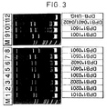
- Fig 3 shows the results obtained for a selection of DPB1 HTCs.
- DPB1 heterozygotes some naturally occurring heteroduplexes formed in trans might be expected, but in practice not all combinations of alleles produce heteroduplexes without use of the method of the present invention employing a DPB construct (Fig 4a).
- conducting the method with an amplified DPB construct yields highly discriminatory PCR fingerprints of heteroduplexes (Fig 4b), analogous to the DR-Dw system.
- Newly described alleles for example, DPB1*2101-3601
- the present method can also be used in determining whether an individual is susceptible to or has a disease associated with a particular nucleotide sequence which codes for a functional protein. Such diseases are reviewed in Immunol. Rev. 70 , 1-218, 1983.
- a suitable synthetic construct (usually a DNA construct) which is capable of forming duplexes with a nucleotide sequence of (a) a translated or non-translated exon region, or (b) an intron region which affects expression or mRNA slicing, the nucleotide sequence containing a mutation responsible for a genetic disease, the construct containing one or more deliberate nucleotide substitution(s) and/or deletion(s) and/or insertion(s) which is/are (i) opposite to a known variable nucleotide or sequence of nucleotides within the sequence under examination and/or (ii) contiguous with a nucleotide which is opposite to a known variable nucleotide or sequence of nucleotides within the sequence under examination.
- DNA is then isolated from the individual under diagnosis, for example from whole blood obtained according to standard protocols or from dried blood spots or from any other suitable source of DNA.
- Populations of the chosen synthetic construct and the nucleotide sequence are formed, for example using the polymerase chain reaction and these populations are then combined under suitable conditions to permit duplex formation within the combined populations.
- the duplexes formed are then separated, for example using gel electrophoresis, and the duplexes visualised, for example by staining.
- the resulting profile is then compared with a standard profile representing the results obtained using the same synthetic construct either with a DNA sample known to contain the mutation under examination or a DNA sample known not to contain the mutation under examination. This enables the user of the method readily to identify whether the individual under diagnosis carries the mutation responsible for the genetic disease.
- DNA heteroduplexes can be generated by hybridising PCR products with synthetic DNA molecules. These are PCR-amplifiable DNA sequences that mimic a DNA sequence but differ from it as a result of controlled nucleotide substitution(s), deletion(s) or insertion(s) at sites continuous to known mutation sites within the DNA.
- the construct and DNA sequences are amplified with the same PCR primers, and hybridised together post-PCR by heating and slow cooling, resulting in DNA heteroduplexes having different conformational forms, and thus different electrophoretic mobilities, unique for individual alleles of a gene (Bidwell et al, 1992).
- Phenylketonuria is probably the most common disorder of amino acid metabolism and results from mutations at the phenylalanine hydroxylase (PAH) gene.
- PAH phenylalanine hydroxylase
- the gene consists of 13 exons and encodes a MRNA of 2.8 kilobases (Kwok et al, 1985; DiLella et al, 1986). To date, more than 50 mutations have been described at the PAH gene but there are wide variations in the frequency distribution of individual mutations between different populations (Scriver et al, 1992).
- the DNA construct should preferably have a nucleotide sequence substantially corresponding to an invariable region or regions of the nucleotide sequence of exon 12 of the gene, with at least one nucleotide substitution, deletion or insertion at or adjacent the known variable nucleotides.
- the DNA construct may have such a deletion, substitution or insertion at or adjacent to one or more of the following four positions, defined by reference to the specific codons of the PAH exon 12 wild type gene:
- test kit which kit comprises:
- the synthetic nucleotide construct contains one or more deliberate nucleotide substitution(s) and/or deletion(s) and/or insertion(s) which is/are (i) opposite to a known variable nucleotide or sequence of nucleotides within the sequence under examination and/or (ii) contiguous with a nucleotide which is opposite to a known variable nucleotide or sequence of nucleotides within the sequence under examination;
- the primers may be labelled as above.
- the control PCR amplification product may also be labelled.
- the kit may further comprise one or more of the following:
- a DNA construct specific for the DPB1 locus was synthesised from multiple synthetic oligonucleotide "longmers", each of about 100 nucleotides in length, using techniques known in the art. In this example, four longmers were employed having the sequences identified SEQ ID NO: 2, SEQ ID NO: 3, SEQ ID NO: 4 and SEQ ID NO: 5. The DNA sequence of the construct was chosen to overlap the start of the second exon of the DPB1 gene, the nucleotide sequence of which is known from Marsh, SGE and Bodmer, JG.
- the construct was 338 base pairs in length.
- the construct sequence included 29 mismatches and 5 unmatched bases between the construct and a "consensus" sequence for the second exon and 3' flanking intron of the DPB1 gene, the consensus sequence being determined by using a computer alignment program, MicroGenie (Version 7.01), Beckman Instruments.
- the construct was cloned in an M13mp18 phage.
- the DPB1 "consensus" sequence is the top line in Figure 1. This "consensus” sequence was determined using a computer alignment program and variable bases are identified by the letter N. This means that these particular bases in the DPB1 second exon vary between individuals and give rise to the various allelic forms of the gene. For simplicity, the 5'-3' coding strand of the consensus sequence only is shown.
- the bottom line of Figure 1 (also SEQ ID NO: 1) is a DNA construct designed to contain deliberate nucleotide substitutions and deletions contiguous with the known variable nucleotides within the nucleotide sequence (here the second exon of the DPB1 gene) under examination. Again, the 5'-3' coding stand is shown for simplicity.
- the construct is designed to have an identical sequence with the great majority of the non-variable nucleotides of the DPB1 consensus sequence, in order that duplexes will form between the DPB1 DNA fragments from the sample of genomic DNA under consideration and the construct.
- deliberate nucleotide substitutions (sometimes hereinbelow referred to as "identifiers” as they "identify” any mutation at this position) are created. Each of these deliberate substitutions is marked by an asterisk. The substitutions are chosen such that, in different allelic forms of the gene, a different extent of mismatch will exist.
- variable nucleotides adjacent the known variable nucleotides, it may be desirable to create a deliberate mismatching with a conserved nucleotide; these are underlined in the construct sequence and effectively amplify the adjacent mismatches. As a result, hereinbelow, these are occasionally referred to as "amplifiers".
- the DNA construct shown has a number of different mismatches at different variable nucleotides of the consensus sequence and, by careful choice of the mismatches, different numbers and extent of single stranded loops will be formed in the duplex between the nucleotide sequence under investigation and the construct, for different alleles.
- a deliberate deletion for example the 5 base pair deletion in the construct shown by the " ⁇ " in Figure 1 ensures that all duplexes formed between the genomic DNA derived from the sample and the construct will migrate at a significantly different rate to the duplexes formed within the population of genomic DNA. This "shifts" the bands derived from duplexes including the construct away from bands derived from duplexes not including the construct in the PCR fingerprint produced in accordance with the present invention.
- the DBP nucleotide sequences under investigation were amplified in the polymerase chain reaction from genomic DNA.
- the PCR primers employed were those described by Begovich et al (Begovich AB, Bugawan TL, Nepom BS, Klitz W, Nepom GT and Erlich HA (1989), "A specific HLA-DP ⁇ allele is associated with pauciarticular juvenile rheumatoid arthritis but not adult rheumatoid arthritis", Proc Natl Acad Sci USA 86:9489-9493) and which are identified as UG19 (SEQ ID NO: 6) and UG21 (SEQ ID NO: 7), available from suppliers of custom oligonucleotides.
- the reaction mixture contained: Genomic DNA @ 0.1 ⁇ g/ ⁇ l 5.0 ⁇ l UG19 primer (5 ⁇ M stock) 10.0 ⁇ l UG21 primer (5 ⁇ M stock) 10.0 ⁇ l 10X PCR buffer (supplied) 10.0 ⁇ l dNTP mix (10mM each DNTP) 2.0 ⁇ l ddH 2 O 62.5 ⁇ l
- the resulting mixtures were combined, vortexed and then centrifuged briefly.
- the genomic DNA was denatured by heating the tube at 94°C for 5 min and then snap cooled on ice for 3 min and overlaid with 75 ⁇ l of paraffin oil.
- PCR was initiated using a "hot-start" procedure, whereby tubes containing the reaction mixture without Taq polymerase were equilibrated at 72°C in a heat block.
- Taq polymerase 2.5 units, 0.5 ⁇ l
- the genomic DNA samples were then subjected to 35 PCR cycles, as follows. primer extension 72°C, 30 sec denaturation 94°C, 30 sec primer annealing 65°C, 60 sec
- the 72°C primer extension time at the end of the final cycle was increased to 10 min.
- the DNA construct was amplified from the M13mp18 clone using the same primers as for the genomic DNA under study, the reaction mixture being as follows: DPB-UHG M13mp18 DNA: 1/100 dilution from supplied stock 1.0 ⁇ l UG19 primer (5 ⁇ M stock) 10.0 ⁇ l UG21 primer (5 ⁇ M stock) 10.0 ⁇ l 10X PCR buffer (supplied) 10.0 ⁇ l dNTP mix (10mM each dNTP) 2.0 ⁇ l ddH 2 O 66.5 ⁇ l The same PCR and thermal cycling conditions were used as above for the genomic DNA, but with only 25 cycles.
- the homoduplex band should be allowed to migrate to within 0.5cm of the end of the gel. Stain gels for 20 min in 1X TBE containing 0.5 ⁇ g/ml ethidium bromide and examine using a UV transilluminator.
- Black triangles represent deliberate deletions in the DPB-construct designed to increase gel retardation of the heteroduplexes and thereby to produce a clear spread of visible bands (see Figs. 3 and 4b).
- the single DPB-construct employed theoretically generates an unique heteroduplex with each DPB1 allele.
- HTCs HLA-DPB1 homozygous typing cells
- Lane 11 shows an HLA-DP heterozygote: the pattern observed is in part a combination of the relevant HTC patterns (lanes 5 and 6).
- M molecular weight marker (pUCBM21 Hpa II plus Dra I and Hind III digests: fragment sizes 1114, 900, 692, 501, 489, 404, 320, 242, 190, 147, 124, 110, 67, 37, 34, 34, 26 and 19 base pairs).
- PCR fingerprints of HLA-DPB1 heterozygotes were obtained.
- PCRs not conducted in the presence of the DPB-construct show (see Figure 4a) some rudimentary heteroduplexes formed in trans between alleles in some, but not all, combinations of alleles (lanes 3, 4, 6, and 7).
- the effect of conducting the PCRs as shown in ( Figure 4a) in the presence of the construct is shown in Figure 4b.
- Unique PCR fingerprints are now revealed for all heterozygotes, with excellent resolution of heteroduplexes.
- Lanes 7 and 8 show a leukaemic patient and potential donor, respectively, matched for HLA-A, B and DR-Dw but mismatched for DP as revealed by the PCR fingerprints.
- the patient (lane 7) is DPB1*0301,0401 (compare with lane 3) and the potential donor (lane 8) is DPB1*0201,0401 (compare with lane 1).
- the mixed lymphocyte reaction (MLR) was positive in this case.
- M molecular weight marker (see above).
- This example illustrates the application of the present invention to the classification of phenylketonuria genotypes.
- a DNA construct (SEQ ID NO: 8) was synthesised to identify five mutations (R408W, R413P, Y414C, T418P and IVS12nt1) within exon 12 of the phenylalanine hydroxylase (PAH) gene. Identifiers for four of the five mutations were incorporated within the construct and consisted of nucleotide substitutions or substitutions plus deletions. A specific identifier for the T418P mutation was not necessary.
- Figure 5 shows the nucleotide sequence of a portion of the normal human PAH gene, including exon 12 and part of the 5' and 3' flanking introns. Sequences of the PKU point mutations and the DNA construct are shown aligned to the normal sequence: dashes indicate identity.
- reaction mixtures were set up containing 2.5 ⁇ l of a 5 ⁇ M aqueous solution of each longmer, 5 ⁇ l dNTP mix (dATP, dCTP, dGTP and dTTP each at a concentration of 10mM), 1 ⁇ l of 10mg/ml bovine serum albumin, 10 ⁇ l Vent TM DNA polymerase reaction buffer (New England Biolabs), 58 ⁇ l deionized double distilled water, 2 units Vent TM DNA polymerase (New England Biolabs), added at 72°C to hot-start the reaction (D'Aquila et al, 1991), and 0.5 ⁇ l E.
- 5 ⁇ l dNTP mix dATP, dCTP, dGTP and dTTP each at a concentration of 10mM
- 10mg/ml bovine serum albumin 10 ⁇ l
- Vent TM DNA polymerase reaction buffer New England Biolabs
- 58 ⁇ l deionized double distilled water 2 units Vent TM DNA polymerase (New England Bio
- Thermal cycling conditions were as for genomic DNA amplification (see below).
- the products of six replicate reactions were pooled and electrophoresed on two 8% w/v nondenaturing polyacrylamide gels (Protogel, National Diagnostics) as previously described (Bidwell and Hui, 1990).
- Gel slices containing the double stranded construct band were excised and the DNA extracted by isotachophoresis (Ofverstedt et al, 1984), precipitated with ethanol, washed in 70% ethanol, and redissolved in 50 ⁇ l sterile double distilled water.
- Ten microlitres of a 1:10,000 serial dilution was amplified for use in the heteroduplex analyses.
- Genomic DNA was isolated from whole blood according to standard protocols, or alternatively from dried blood spots used in neonatal screening tests, as follows: a 2mm 2 area of blood spot was excised from the paper, added to a 0.5ml microcentrifuge tube containing 25 ⁇ l of a 1:10 dilution of AmpliTaq TM reaction buffer (Perkin Elmer Cetus). Samples were heated at 96°C for 15 min, cooled, diluted with a further 25 ⁇ l of 1X AmpliTaq TM reaction buffer, mixed and centrifuged at 13000g for 15 min. Supernatants were used directly in the PCR.
- the enzyme was added at 72°C to hot-start the reaction (D'Aquila et al, 1991).
- 30 ⁇ l of supernatant was used in the reaction mixtures, and the volumes of AmpliTaq TM reaction buffer and water were reduced to 7 ⁇ l and 40.6p1 respectively.
- the reaction mixtures were subjected to 35 rounds of thermal cycling at 94°C for 1 min, 48°C for 1 min and 72°C for 1 min.
- Figure 6a, 6b, 7 and 8 show the stained gels for a series of experiments on the of PAH gene exon 12 mutations.
- Lanes 1 and 12 show molecular weight markers (M, marker VIII, Boehringer Mannheim).
- the leading double bands in all other lanes are the homoduplexes of the synthetic DNA construct (leading band, 176bp) and PAH gene exon 12 (trailing band, 185bp) PCR products.
- Bands of apparent molecular weight 400-500bp are different conformational forms of DNA heteroduplexes (Figs 6b and 8).
- Lanes 2, 11, 13 and 16 show the heteroduplexes observed in normal individuals (controls).
- Lanes 6, 8 and 14 show individuals with different PAH gene exon 12 mutations on each chromosome. In all cases, characteristic heteroduplexes (also see Figure 6b) are observed for each mutation, and in addition the homozygosity or heterozygosity status is readily determined.
- Figure 6(b) is a schematic representation of DNA heteroduplex banding patterns obtained for each PAH gene exon 12 mutation. For each mutation, banding patterns are shown for homozygous (hom) and heterozygous (het) states.
- the mobilities of the bands are: a, 446bp; b, 433bp; c, 416bp; d, 395bp; e, 374bp; f, 359bp; g, 354bp.
- Bands c plus g are observed in normal PAH genes and in genes with non-exon 12 mutations.
- Figures 6a and 6b and 8 show that the synthetic construct was able to generate unique and discriminatory DNA heteroduplexes for all five known exon 12 mutations within DNA from PKU individuals, both in the homozygous and heterozygous states. Moreover, the visual quality of these results compared favourably with those obtained with DNA isolated from fresh blood samples, and permitted unambiguous assignment of exon 12 genotypes.
- the reliability and reproducibility of the test was examined by screening two sets of genomic DNA samples collected from PKU individuals in whom exon 12 mutations were established by other methods, either prospectively or retrospectively.
- Figures 7a and 7b show the results of a blind trial of concordance between the DNA analysis method of the present invention and the results of other DNA typing analyses.
- Figure 7a represents the results using the first set of genomic DNA whilst
- Figure 7b represents the results using the second set of genomic DNA constituting untreated, PKU individuals with reduced intelligence from a Scottish population.
- lane M are molecular weight markers (for details see Figure 6 legend) and lane C is a normal control.
- the lanes were as follows: lane 1, R408W heterozygous individual; lanes 2, 8, 9 and 11, IVS12nt1 heterozygous individuals; plane 3, individual with Y414C and IVS12nt1 mutations on opposite alleles; lane 4, Y414C heterozygous individual; lane 5, IVS12nt1 homozygote; lane 7, individual with R408W and IVS12nt1 mutations on opposite alleles.
- Lanes 6, 10 and 12 are PKU individuals with mutations outside exon 12.
- the lanes are as follows: lanes 1-7 and 9, IVS12nt1 heterozygous individuals; lane 8, PKU individual with mutation outside exon 12.
- This Example shows the technique of the present invention to be a highly sensitive, rapid and non-radioactive screening and classification system for PKU in which five mutations in exon 12 of the PAH gene can be identified using a single DNA construct and one PCR-based test.
- the tests are technically simple to perform and suitable for the routine screening of large numbers of individuals. Results were obtained within 24 hours of receipt of blood samples, and were of equal visual quality when performed on whole blood ( Figures 6 and 7) or dried blood spots ( Figure 8).
- This Example illustrates the wide potential of synthetic DNA construct-based DNA heteroduplex technology.
- the technique appears ideally suited to the genotyping of multi-allelic genes where mutations are clustered within a reasonable distance and are flanked by conserved sequences suitable for PCR priming. Genotyping mutations within separate exons in genomic DNA may be facilitated using exon-specific constructs, or potentially by a single construct for genotyping cDNA derived from mRNA by reverse transcription PCR (Myers and Gelfand, 1991)
Abstract
Description
- a heat-stable DNA polymerase;
- DATP, DCTP, DGTP and DTTP;
- appropriate biological buffers and cofactors;
- a database comprising the relative molecular conformation and gel mobilities of DNA fragments generated by PCR amplification and heteroduplex formation between selected DNA samples and the construct.
| Genomic DNA @ 0.1µg/µl | 5.0µl |
| UG19 primer (5µM stock) | 10.0µl |
| UG21 primer (5µM stock) | 10.0µl |
| 10X PCR buffer (supplied) | 10.0µl |
| dNTP mix (10mM each DNTP) | 2.0µl |
| ddH2O | 62.5µl |
| primer extension | 72°C, 30 sec |
| denaturation | 94°C, 30 sec |
| primer annealing | 65°C, 60 sec |
| DPB-UHG M13mp18 DNA: 1/100 dilution from supplied stock | 1.0µl |
| UG19 primer (5µM stock) | 10.0µl |
| UG21 primer (5µM stock) | 10.0µl |
| 10X PCR buffer (supplied) | 10.0µl |
| dNTP mix (10mM each dNTP) | 2.0µl |
| ddH2O | 66.5µl |


Claims (16)
- A method for examining polymorphisms in a nucleotide sequence of a first sample of nucleic acid comprising the steps of:wherein the synthetic construct contains one or more deliberate nucleotide substitution(s) and/or deletion(s) and/or insertion(s) which is/are (i) opposite to a known variable nucleotide or sequence of nucleotides within the sequence under examination and/or (ii) contiguous with a nucleotide which is opposite to a known variable nucleotide or sequence of nucleotides within the sequence under examination.(a) forming a population of nucleic acid fragments bearing the said nucleotide sequence;(b) combining the population of nucleic acid fragments with a population of a synthetic nucleotide construct which construct is capable of forming duplexes with the nucleic acid fragments, the sequence of the construct being such that duplexes of different molecular conformation are formed between the construct and the nucleic acid fragments dependent upon the presence of a polymorphism at a known variable nucleotide or sequence of nucleotides within the sequence of the sample under examination;(c) permitting duplex formation within the combined populations; and(d) separating the duplexes formed in step (c);
- A method according to claim 1, wherein the nucleotide sequence of the synthetic construct at least contains deliberate nucleotide substitution(s) and/or deletion(s) and/or insertion(s) contiguous with the nucleotide which is opposite the known variable nucleotide or sequence of nucleotides within the sequence under examination.
- A method according to claim 1 or 2, wherein the nucleotide sequence of the synthetic construct contains deliberate nucleotide substitution(s) and/or deletion(s) and/or insertion(s) opposite the known variable nucleotide or sequence of nucleotides.
- A method according to claim 3, wherein the synthetic construct includes nucleotide substitution(s) and deletion(s) both opposite and contiguous with the mutation.
- A method according to any one of claims 1 to 4, wherein the nucleotide sequence of the synthetic construct contains between one and ten contiguous substitutions/deletions adjacent the position of the polymorphism in the nucleotide sequence under investigation
- A method according to any one of claims 1 to 5, wherein the synthetic construct includes deliberate nucleotide substitution(s) and/or deletion(s) and/or insertion(s) corresponding to at least two known polymorphisms in the nucleotide sequence
- A method according to any preceding claim, wherein the construct includes a deliberate deletion or a series of deletions, at a position away from polymorphic sites in the nucleotide sequence under investigation.
- A method according to any preceding claim, wherein the nucleic acid under examination is DNA.
- A method according to any preceding claim, wherein the synthetic nucleotide construct is a synthetic DNA.
- A method according to any preceding claim, wherein the population of DNA fragments bearing the nucleotide sequence is formed from a DNA source by a polymerase chain reaction.
- A method according to any preceding claim in which the synthetic construct is a DNA construct, wherein the population of synthetic DNA construct is formed by a polymerase chain reaction.
- A method according to any preceding claim for matching individuals in respect of one or more of the functional proteins expressed by the HLA (human leucocyte antigen) class I and II genes, wherein the nucleotide sequence under examination is a sequence, or part of a sequence, of a HLA Class I or II gene containing a known polymorphic site or sites.
- A method according to any one of claims 1 to 11, for determining an individual's susceptibility to a genetic disease, wherein the nucleotide sequence under examination is a sequence, or part of a sequence, of a gene containing a known polymorphic site or sites associated with a genetic defect.
- A method according to claim 13, wherein the genetic disease is phenylketonuria genotyping and the gene containing a known polymorphic site or sites is the phenylalanine hydroxylase (PAH) gene.
- A test kit, which kit comprises:wherein the synthetic construct contains one or more deliberate nucleotide substitution(s) and/or deletion(s) and/or insertion(s) which is/are (i) opposite to a known variable nucleotide or sequence of nucleotides within the sequence under examination and/or (ii) contiguous with a nucleotide which is opposite to a known variable nucleotide or sequence of nucleotides within the sequence under examination.(a) two oligonucleotide primers suitable for use in PCR and capable of annealing to complementary sequences at respective ends of a nucleotide sequence to be examined;(b) a synthetic nucleotide construct which construct is capable of forming duplexes with the nucleotide sequence under consideration, the sequence of the construct being such that duplexes of different molecular conformation are formed between the construct and the nucleotide sequence under examination dependent upon the presence of a polymorphism at a known variable nucleotide or sequence of nucleotides within the sequence under examination; and(c) a control DNA and/or control PCR amplification product;
- A test kit according to claim 15, wherein the synthetic nucleotide construct is a synthetic DNA.
Applications Claiming Priority (3)
| Application Number | Priority Date | Filing Date | Title |
|---|---|---|---|
| GB9206567 | 1992-03-25 | ||
| GB929206567A GB9206567D0 (en) | 1992-03-25 | 1992-03-25 | Dna analysis |
| PCT/GB1993/000616 WO1993019201A1 (en) | 1992-03-25 | 1993-03-25 | Nucleic acid analysis |
Publications (2)
| Publication Number | Publication Date |
|---|---|
| EP0672179A1 EP0672179A1 (en) | 1995-09-20 |
| EP0672179B1 true EP0672179B1 (en) | 1998-07-08 |
Family
ID=10712866
Family Applications (1)
| Application Number | Title | Priority Date | Filing Date |
|---|---|---|---|
| EP93906753A Expired - Lifetime EP0672179B1 (en) | 1992-03-25 | 1993-03-25 | Nucleic acid analysis |
Country Status (5)
| Country | Link |
|---|---|
| EP (1) | EP0672179B1 (en) |
| AU (1) | AU3764793A (en) |
| DE (1) | DE69319615D1 (en) |
| GB (2) | GB9206567D0 (en) |
| WO (1) | WO1993019201A1 (en) |
Families Citing this family (8)
| Publication number | Priority date | Publication date | Assignee | Title |
|---|---|---|---|---|
| US5633129A (en) * | 1989-07-13 | 1997-05-27 | Massachusetts Institute Of Technology | Electrophoretic detection and separation of mutant DNA using replaceable polymer matrices |
| DE69636725T2 (en) | 1995-11-29 | 2007-09-20 | The Anthony Nolan Bone Marrow Trust, Hampstead | METHOD FOR SEPARATING AND / OR DETECTING DNA MOLECULES |
| GB9809063D0 (en) * | 1998-04-29 | 1998-06-24 | Univ Wales Medicine | Heteroduplex analysis |
| NZ555009A (en) | 2004-10-08 | 2010-03-26 | Ihg Pharmaco Ltd | Improvements in induced heteroduplex generators for genotyping a target gene, polypeptide includes at least one known polymorphism and at least one identifier that is spaced by at least one base from the polymorphism |
| CN100510105C (en) * | 2006-02-08 | 2009-07-08 | 博奥生物有限公司 | Sequence specific oligonucleotide probe and application thereof |
| GB2435326A (en) * | 2006-02-15 | 2007-08-22 | Cell Analysis Ltd | Heteroduplex analysis of non-human analytes |
| US9828637B2 (en) * | 2010-04-18 | 2017-11-28 | Wake Forest University Health Sciences | Methods of predicting predisposition to or risk of kidney disease |
| PL3129033T3 (en) | 2014-04-07 | 2021-02-08 | Ihg Pharmaco Limited | Carrier status of annexin a5 m2 haplotype and obstetric risks |
Family Cites Families (3)
| Publication number | Priority date | Publication date | Assignee | Title |
|---|---|---|---|---|
| US4965190A (en) * | 1986-07-31 | 1990-10-23 | Howard Hughes Medical Institute | Methods for the identification of mutations in the human phenylalanine hydroxylase gene using DNA probes |
| AU5645690A (en) * | 1989-05-05 | 1990-11-29 | Lifecodes Corporation | Method for genetic analysis of a nucleic acid sample |
| GB9002625D0 (en) * | 1990-02-06 | 1990-04-04 | Univ Singapore | Human leukocyte antigen typing |
-
1992
- 1992-03-25 GB GB929206567A patent/GB9206567D0/en active Pending
-
1993
- 1993-03-25 DE DE69319615T patent/DE69319615D1/en not_active Expired - Lifetime
- 1993-03-25 GB GB9419292A patent/GB2280266B/en not_active Expired - Fee Related
- 1993-03-25 AU AU37647/93A patent/AU3764793A/en not_active Abandoned
- 1993-03-25 WO PCT/GB1993/000616 patent/WO1993019201A1/en active IP Right Grant
- 1993-03-25 EP EP93906753A patent/EP0672179B1/en not_active Expired - Lifetime
Also Published As
| Publication number | Publication date |
|---|---|
| EP0672179A1 (en) | 1995-09-20 |
| AU3764793A (en) | 1993-10-21 |
| GB9419292D0 (en) | 1994-11-16 |
| GB2280266A (en) | 1995-01-25 |
| WO1993019201A1 (en) | 1993-09-30 |
| DE69319615D1 (en) | 1998-08-13 |
| GB2280266B (en) | 1996-01-24 |
| GB9206567D0 (en) | 1992-05-06 |
Similar Documents
| Publication | Publication Date | Title |
|---|---|---|
| AU672519B2 (en) | Intron sequence analysis method for detection of adjacent and remote locus alleles as haplotypes | |
| US5759771A (en) | Method of determining a genotype by comparing the nucleotide sequence of members of a gene family and kit therefor | |
| CA2239896C (en) | Method for evaluation of polymorphic genetic sequences, and the use thereof in identification of hla types | |
| EP0663923B1 (en) | Three highly informative microsatellite repeat polymorphic dna markers | |
| EP0577708A4 (en) | Dna sequence-based hla typing method | |
| US8343720B2 (en) | Methods and probes for identifying a nucleotide sequence | |
| WO2005123951A2 (en) | Methods of human leukocyte antigen typing by neighboring single nucleotide polymorphism haplotypes | |
| US6500614B1 (en) | Method for identifying an unknown allele | |
| Zimmerman et al. | Exploiting structural differences among heteroduplex molecules to simplify genotyping the DQA1 and DQB1 alleles in human lymphocyte typing | |
| EP0672179B1 (en) | Nucleic acid analysis | |
| Wood et al. | Rapid classification of phenylketonuria genotypes by analysis of heteroduplexes generated by PCR‐amplifiable synthetic DNA | |
| US6994958B2 (en) | Methods for separating and/or identifying DNA molecules | |
| Paul et al. | Resolution of cis‐trans ambiguities between HLA‐DRB1 alleles using single‐strand conformation polymorphisms and sequencing | |
| Gibbs et al. | The application of recombinant DNA technology for genetic probing in epidemiology | |
| Mitsunaga et al. | Multiplex ARMS‐PCR‐RFLP method for high‐resolution typing of HLA‐DRB1 | |
| Feolo et al. | A strategy for high throughput HLA-DQ typing | |
| Wood et al. | DNA typing in hereditary disease | |
| CN115323048A (en) | Primer combination and method for detecting human embryo alpha-thalassemia gene mutation | |
| CN116837114A (en) | Integrated detection system for X chromosome Multi-InDel for personal identification and kindred relation identification | |
| El-Borai et al. | A practical approach to HLA-DR genomic typing by heteroduplex analysis and a selective cleavage at position 86 | |
| Dolf | DNA Fingerprinting: Approaches and Applications | |
| Pfäffle | Diagnosis of endocrine disorders with molecular genetic methods | |
| El-Borai | Composite HLA-DRB genomic typing procedure: a simple and cost effective substitute for serological typing | |
| Bein et al. | HLA DNA pre-organ retrieval donor typing | |
| Bouwens et al. | DR4 high resolution typing by direct genomic sequencing |
Legal Events
| Date | Code | Title | Description |
|---|---|---|---|
| PUAI | Public reference made under article 153(3) epc to a published international application that has entered the european phase |
Free format text: ORIGINAL CODE: 0009012 |
|
| 17P | Request for examination filed |
Effective date: 19941018 |
|
| AK | Designated contracting states |
Kind code of ref document: A1 Designated state(s): BE CH DE DK ES FR IT LI NL SE |
|
| 17Q | First examination report despatched |
Effective date: 19970304 |
|
| GRAG | Despatch of communication of intention to grant |
Free format text: ORIGINAL CODE: EPIDOS AGRA |
|
| GRAG | Despatch of communication of intention to grant |
Free format text: ORIGINAL CODE: EPIDOS AGRA |
|
| GRAH | Despatch of communication of intention to grant a patent |
Free format text: ORIGINAL CODE: EPIDOS IGRA |
|
| RBV | Designated contracting states (corrected) |
Designated state(s): BE CH DE DK ES FR IT LI NL SE |
|
| GRAH | Despatch of communication of intention to grant a patent |
Free format text: ORIGINAL CODE: EPIDOS IGRA |
|
| GRAA | (expected) grant |
Free format text: ORIGINAL CODE: 0009210 |
|
| AK | Designated contracting states |
Kind code of ref document: B1 Designated state(s): BE CH DE DK ES FR IT LI NL SE |
|
| PG25 | Lapsed in a contracting state [announced via postgrant information from national office to epo] |
Ref country code: NL Free format text: LAPSE BECAUSE OF FAILURE TO SUBMIT A TRANSLATION OF THE DESCRIPTION OR TO PAY THE FEE WITHIN THE PRESCRIBED TIME-LIMIT Effective date: 19980708 Ref country code: LI Free format text: LAPSE BECAUSE OF FAILURE TO SUBMIT A TRANSLATION OF THE DESCRIPTION OR TO PAY THE FEE WITHIN THE PRESCRIBED TIME-LIMIT Effective date: 19980708 Ref country code: IT Free format text: LAPSE BECAUSE OF FAILURE TO SUBMIT A TRANSLATION OF THE DESCRIPTION OR TO PAY THE FEE WITHIN THE PRE;WARNING: LAPSES OF ITALIAN PATENTS WITH EFFECTIVE DATE BEFORE 2007 MAY HAVE OCCURRED AT ANY TIME BEFORE 2007. THE CORRECT EFFECTIVE DATE MAY BE DIFFERENT FROM THE ONE RECORDED.SCRIBED TIME-LIMIT Effective date: 19980708 Ref country code: FR Free format text: LAPSE BECAUSE OF FAILURE TO SUBMIT A TRANSLATION OF THE DESCRIPTION OR TO PAY THE FEE WITHIN THE PRESCRIBED TIME-LIMIT Effective date: 19980708 Ref country code: ES Free format text: THE PATENT HAS BEEN ANNULLED BY A DECISION OF A NATIONAL AUTHORITY Effective date: 19980708 Ref country code: CH Free format text: LAPSE BECAUSE OF FAILURE TO SUBMIT A TRANSLATION OF THE DESCRIPTION OR TO PAY THE FEE WITHIN THE PRESCRIBED TIME-LIMIT Effective date: 19980708 Ref country code: BE Free format text: LAPSE BECAUSE OF FAILURE TO SUBMIT A TRANSLATION OF THE DESCRIPTION OR TO PAY THE FEE WITHIN THE PRESCRIBED TIME-LIMIT Effective date: 19980708 |
|
| REG | Reference to a national code |
Ref country code: CH Ref legal event code: EP |
|
| REF | Corresponds to: |
Ref document number: 69319615 Country of ref document: DE Date of ref document: 19980813 |
|
| PG25 | Lapsed in a contracting state [announced via postgrant information from national office to epo] |
Ref country code: SE Free format text: LAPSE BECAUSE OF FAILURE TO SUBMIT A TRANSLATION OF THE DESCRIPTION OR TO PAY THE FEE WITHIN THE PRESCRIBED TIME-LIMIT Effective date: 19981008 Ref country code: DK Free format text: LAPSE BECAUSE OF FAILURE TO SUBMIT A TRANSLATION OF THE DESCRIPTION OR TO PAY THE FEE WITHIN THE PRESCRIBED TIME-LIMIT Effective date: 19981008 |
|
| PG25 | Lapsed in a contracting state [announced via postgrant information from national office to epo] |
Ref country code: DE Free format text: LAPSE BECAUSE OF FAILURE TO SUBMIT A TRANSLATION OF THE DESCRIPTION OR TO PAY THE FEE WITHIN THE PRESCRIBED TIME-LIMIT Effective date: 19981009 |
|
| NLV1 | Nl: lapsed or annulled due to failure to fulfill the requirements of art. 29p and 29m of the patents act | ||
| EN | Fr: translation not filed | ||
| REG | Reference to a national code |
Ref country code: CH Ref legal event code: PL |
|
| PLBE | No opposition filed within time limit |
Free format text: ORIGINAL CODE: 0009261 |
|
| STAA | Information on the status of an ep patent application or granted ep patent |
Free format text: STATUS: NO OPPOSITION FILED WITHIN TIME LIMIT |
|
| 26N | No opposition filed |