EP0669833B1 - Type ii tgf-beta-binding receptor fragment as therapeutic agent - Google Patents
Type ii tgf-beta-binding receptor fragment as therapeutic agent Download PDFInfo
- Publication number
- EP0669833B1 EP0669833B1 EP94902198A EP94902198A EP0669833B1 EP 0669833 B1 EP0669833 B1 EP 0669833B1 EP 94902198 A EP94902198 A EP 94902198A EP 94902198 A EP94902198 A EP 94902198A EP 0669833 B1 EP0669833 B1 EP 0669833B1
- Authority
- EP
- European Patent Office
- Prior art keywords
- tgf
- rii
- use according
- type
- excess
- Prior art date
- Legal status (The legal status is an assumption and is not a legal conclusion. Google has not performed a legal analysis and makes no representation as to the accuracy of the status listed.)
- Expired - Lifetime
Links
- ZRKFYGHZFMAOKI-QMGMOQQFSA-N tgfbeta Chemical compound C([C@H](NC(=O)[C@H](C(C)C)NC(=O)CNC(=O)[C@H](CCC(O)=O)NC(=O)[C@H](CCCNC(N)=N)NC(=O)[C@H](CC(N)=O)NC(=O)[C@H](CC(C)C)NC(=O)[C@H]([C@@H](C)O)NC(=O)[C@H](CCC(O)=O)NC(=O)[C@H]([C@@H](C)O)NC(=O)[C@H](CC(C)C)NC(=O)CNC(=O)[C@H](C)NC(=O)[C@H](CO)NC(=O)[C@H](CCC(N)=O)NC(=O)[C@@H](NC(=O)[C@H](C)NC(=O)[C@H](C)NC(=O)[C@@H](NC(=O)[C@H](CC(C)C)NC(=O)[C@@H](N)CCSC)C(C)C)[C@@H](C)CC)C(=O)N[C@@H]([C@@H](C)O)C(=O)N[C@@H](C(C)C)C(=O)N[C@@H](CC=1C=CC=CC=1)C(=O)N[C@@H](C)C(=O)N1[C@@H](CCC1)C(=O)N[C@@H]([C@@H](C)O)C(=O)N[C@@H](CC(N)=O)C(=O)N[C@@H](CCC(O)=O)C(=O)N[C@@H](C)C(=O)N[C@@H](CC=1C=CC=CC=1)C(=O)N[C@@H](CCCNC(N)=N)C(=O)N[C@@H](C)C(=O)N[C@@H](CC(C)C)C(=O)N1[C@@H](CCC1)C(=O)N1[C@@H](CCC1)C(=O)N[C@@H](CCCNC(N)=N)C(=O)N[C@@H](CCC(O)=O)C(=O)N[C@@H](CCCNC(N)=N)C(=O)N[C@@H](CO)C(=O)N[C@@H](CCCNC(N)=N)C(=O)N[C@@H](CC(C)C)C(=O)N[C@@H](CC(C)C)C(O)=O)C1=CC=C(O)C=C1 ZRKFYGHZFMAOKI-QMGMOQQFSA-N 0.000 title claims abstract description 166
- 239000012634 fragment Substances 0.000 title claims abstract description 68
- 238000009739 binding Methods 0.000 title claims description 32
- 239000003814 drug Substances 0.000 title claims description 9
- 229940124597 therapeutic agent Drugs 0.000 title description 2
- 102000005962 receptors Human genes 0.000 claims abstract description 33
- 108020003175 receptors Proteins 0.000 claims abstract description 33
- 102000016715 Transforming Growth Factor beta Receptors Human genes 0.000 claims abstract description 14
- 108091005735 TGF-beta receptors Proteins 0.000 claims abstract description 13
- 102000004887 Transforming Growth Factor beta Human genes 0.000 claims description 163
- 108090001012 Transforming Growth Factor beta Proteins 0.000 claims description 163
- 206010028980 Neoplasm Diseases 0.000 claims description 20
- 229960005486 vaccine Drugs 0.000 claims description 19
- 230000001506 immunosuppresive effect Effects 0.000 claims description 17
- 206010016654 Fibrosis Diseases 0.000 claims description 15
- 206010062016 Immunosuppression Diseases 0.000 claims description 15
- 208000027418 Wounds and injury Diseases 0.000 claims description 15
- 108010082684 Transforming Growth Factor-beta Type II Receptor Proteins 0.000 claims description 14
- 102000004060 Transforming Growth Factor-beta Type II Receptor Human genes 0.000 claims description 14
- 230000000694 effects Effects 0.000 claims description 14
- 206010052428 Wound Diseases 0.000 claims description 13
- 150000001413 amino acids Chemical class 0.000 claims description 13
- 208000019425 cirrhosis of liver Diseases 0.000 claims description 13
- 208000037265 diseases, disorders, signs and symptoms Diseases 0.000 claims description 13
- 206010018364 Glomerulonephritis Diseases 0.000 claims description 11
- 201000011510 cancer Diseases 0.000 claims description 10
- 230000004761 fibrosis Effects 0.000 claims description 10
- 210000003734 kidney Anatomy 0.000 claims description 10
- 239000000203 mixture Substances 0.000 claims description 10
- 231100000241 scar Toxicity 0.000 claims description 10
- 210000002808 connective tissue Anatomy 0.000 claims description 9
- 230000000893 fibroproliferative effect Effects 0.000 claims description 9
- 238000001959 radiotherapy Methods 0.000 claims description 9
- 206010039073 rheumatoid arthritis Diseases 0.000 claims description 9
- 230000009772 tissue formation Effects 0.000 claims description 9
- 208000035475 disorder Diseases 0.000 claims description 8
- 238000004519 manufacturing process Methods 0.000 claims description 8
- 208000029147 Collagen-vascular disease Diseases 0.000 claims description 7
- 206010060862 Prostate cancer Diseases 0.000 claims description 7
- 206010039710 Scleroderma Diseases 0.000 claims description 6
- 201000009594 Systemic Scleroderma Diseases 0.000 claims description 6
- 206010042953 Systemic sclerosis Diseases 0.000 claims description 6
- 238000001990 intravenous administration Methods 0.000 claims description 6
- 208000032544 Cicatrix Diseases 0.000 claims description 5
- 208000002158 Proliferative Vitreoretinopathy Diseases 0.000 claims description 5
- 206010038934 Retinopathy proliferative Diseases 0.000 claims description 5
- 208000021971 neovascular inflammatory vitreoretinopathy Diseases 0.000 claims description 5
- 230000006785 proliferative vitreoretinopathy Effects 0.000 claims description 5
- 208000037803 restenosis Diseases 0.000 claims description 5
- 230000037387 scars Effects 0.000 claims description 5
- 201000000596 systemic lupus erythematosus Diseases 0.000 claims description 5
- 208000007342 Diabetic Nephropathies Diseases 0.000 claims description 4
- 208000002260 Keloid Diseases 0.000 claims description 4
- 208000036142 Viral infection Diseases 0.000 claims description 4
- 210000004204 blood vessel Anatomy 0.000 claims description 4
- 208000033679 diabetic kidney disease Diseases 0.000 claims description 4
- 208000006454 hepatitis Diseases 0.000 claims description 4
- 230000028993 immune response Effects 0.000 claims description 4
- 230000001965 increasing effect Effects 0.000 claims description 4
- 208000015181 infectious disease Diseases 0.000 claims description 4
- 208000014674 injury Diseases 0.000 claims description 4
- 210000001117 keloid Anatomy 0.000 claims description 4
- 208000005069 pulmonary fibrosis Diseases 0.000 claims description 4
- 230000009385 viral infection Effects 0.000 claims description 4
- 208000023275 Autoimmune disease Diseases 0.000 claims description 3
- 208000035473 Communicable disease Diseases 0.000 claims description 3
- 206010061598 Immunodeficiency Diseases 0.000 claims description 3
- 208000034693 Laceration Diseases 0.000 claims description 3
- 208000007452 Plasmacytoma Diseases 0.000 claims description 3
- 208000002847 Surgical Wound Diseases 0.000 claims description 3
- 206010044708 Trypanosomal infections Diseases 0.000 claims description 3
- 241000700605 Viruses Species 0.000 claims description 3
- 201000001981 dermatomyositis Diseases 0.000 claims description 3
- 208000005017 glioblastoma Diseases 0.000 claims description 3
- 230000002440 hepatic effect Effects 0.000 claims description 3
- 231100000283 hepatitis Toxicity 0.000 claims description 3
- 230000001969 hypertrophic effect Effects 0.000 claims description 3
- 208000005987 polymyositis Diseases 0.000 claims description 3
- 206010000050 Abdominal adhesions Diseases 0.000 claims description 2
- 206010004664 Biliary fibrosis Diseases 0.000 claims description 2
- 241000712899 Lymphocytic choriomeningitis mammarenavirus Species 0.000 claims description 2
- 206010033128 Ovarian cancer Diseases 0.000 claims description 2
- 206010028537 myelofibrosis Diseases 0.000 claims description 2
- 210000004303 peritoneum Anatomy 0.000 claims description 2
- 230000008733 trauma Effects 0.000 claims description 2
- 206010003571 Astrocytoma Diseases 0.000 claims 1
- 241000714260 Human T-lymphotropic virus 1 Species 0.000 claims 1
- 206010023330 Keloid scar Diseases 0.000 claims 1
- 238000000034 method Methods 0.000 abstract description 14
- 108091008324 binding proteins Proteins 0.000 abstract description 5
- 102000014914 Carrier Proteins Human genes 0.000 abstract 1
- 238000011200 topical administration Methods 0.000 abstract 1
- 210000004027 cell Anatomy 0.000 description 48
- 108090000623 proteins and genes Proteins 0.000 description 33
- 102000004169 proteins and genes Human genes 0.000 description 30
- 102000046299 Transforming Growth Factor beta1 Human genes 0.000 description 25
- 101800002279 Transforming growth factor beta-1 Proteins 0.000 description 25
- 210000001519 tissue Anatomy 0.000 description 19
- 238000002347 injection Methods 0.000 description 16
- 239000007924 injection Substances 0.000 description 16
- 239000013598 vector Substances 0.000 description 15
- 239000003656 tris buffered saline Substances 0.000 description 14
- 229940024606 amino acid Drugs 0.000 description 13
- 239000013612 plasmid Substances 0.000 description 13
- 241000588724 Escherichia coli Species 0.000 description 12
- 241000700159 Rattus Species 0.000 description 12
- 210000003000 inclusion body Anatomy 0.000 description 11
- 230000015572 biosynthetic process Effects 0.000 description 10
- 230000023750 transforming growth factor beta production Effects 0.000 description 9
- 102000004237 Decorin Human genes 0.000 description 8
- 108090000738 Decorin Proteins 0.000 description 8
- FAPWRFPIFSIZLT-UHFFFAOYSA-M Sodium chloride Chemical compound [Na+].[Cl-] FAPWRFPIFSIZLT-UHFFFAOYSA-M 0.000 description 8
- 102000011117 Transforming Growth Factor beta2 Human genes 0.000 description 8
- 101800000304 Transforming growth factor beta-2 Proteins 0.000 description 8
- 230000035876 healing Effects 0.000 description 8
- 210000001503 joint Anatomy 0.000 description 8
- 239000000243 solution Substances 0.000 description 8
- 238000001356 surgical procedure Methods 0.000 description 8
- 241001465754 Metazoa Species 0.000 description 7
- 241000283973 Oryctolagus cuniculus Species 0.000 description 7
- 239000011159 matrix material Substances 0.000 description 7
- 108020004999 messenger RNA Proteins 0.000 description 7
- 238000012261 overproduction Methods 0.000 description 7
- 210000002966 serum Anatomy 0.000 description 7
- PEDCQBHIVMGVHV-UHFFFAOYSA-N Glycerine Chemical compound OCC(O)CO PEDCQBHIVMGVHV-UHFFFAOYSA-N 0.000 description 6
- 206010003246 arthritis Diseases 0.000 description 6
- 230000004054 inflammatory process Effects 0.000 description 6
- 210000004379 membrane Anatomy 0.000 description 6
- 239000012528 membrane Substances 0.000 description 6
- 239000013642 negative control Substances 0.000 description 6
- 108090000765 processed proteins & peptides Proteins 0.000 description 6
- 230000035755 proliferation Effects 0.000 description 6
- 230000037390 scarring Effects 0.000 description 6
- 238000002415 sodium dodecyl sulfate polyacrylamide gel electrophoresis Methods 0.000 description 6
- 108020004705 Codon Proteins 0.000 description 5
- 108010035532 Collagen Proteins 0.000 description 5
- 102000008186 Collagen Human genes 0.000 description 5
- 206010061218 Inflammation Diseases 0.000 description 5
- 229920001213 Polysorbate 20 Polymers 0.000 description 5
- 208000000236 Prostatic Neoplasms Diseases 0.000 description 5
- 210000002421 cell wall Anatomy 0.000 description 5
- 238000006243 chemical reaction Methods 0.000 description 5
- 230000007882 cirrhosis Effects 0.000 description 5
- 229920001436 collagen Polymers 0.000 description 5
- 201000010099 disease Diseases 0.000 description 5
- 238000002474 experimental method Methods 0.000 description 5
- 238000005755 formation reaction Methods 0.000 description 5
- 238000009472 formulation Methods 0.000 description 5
- 230000014509 gene expression Effects 0.000 description 5
- 239000003112 inhibitor Substances 0.000 description 5
- 108020004707 nucleic acids Proteins 0.000 description 5
- 102000039446 nucleic acids Human genes 0.000 description 5
- 150000007523 nucleic acids Chemical class 0.000 description 5
- 239000000256 polyoxyethylene sorbitan monolaurate Substances 0.000 description 5
- 235000010486 polyoxyethylene sorbitan monolaurate Nutrition 0.000 description 5
- 238000003786 synthesis reaction Methods 0.000 description 5
- 238000007910 systemic administration Methods 0.000 description 5
- MSWZFWKMSRAUBD-GASJEMHNSA-N 2-amino-2-deoxy-D-galactopyranose Chemical compound N[C@H]1C(O)O[C@H](CO)[C@H](O)[C@@H]1O MSWZFWKMSRAUBD-GASJEMHNSA-N 0.000 description 4
- 108020004414 DNA Proteins 0.000 description 4
- 102000012085 Endoglin Human genes 0.000 description 4
- 108010036395 Endoglin Proteins 0.000 description 4
- WQZGKKKJIJFFOK-GASJEMHNSA-N Glucose Natural products OC[C@H]1OC(O)[C@H](O)[C@@H](O)[C@@H]1O WQZGKKKJIJFFOK-GASJEMHNSA-N 0.000 description 4
- 241000598436 Human T-cell lymphotropic virus Species 0.000 description 4
- 108091034117 Oligonucleotide Proteins 0.000 description 4
- 208000037062 Polyps Diseases 0.000 description 4
- 239000007983 Tris buffer Substances 0.000 description 4
- 230000001154 acute effect Effects 0.000 description 4
- 230000001580 bacterial effect Effects 0.000 description 4
- MSWZFWKMSRAUBD-UHFFFAOYSA-N beta-D-galactosamine Natural products NC1C(O)OC(CO)C(O)C1O MSWZFWKMSRAUBD-UHFFFAOYSA-N 0.000 description 4
- 102000023732 binding proteins Human genes 0.000 description 4
- 239000000969 carrier Substances 0.000 description 4
- ZWIBGKZDAWNIFC-UHFFFAOYSA-N disuccinimidyl suberate Chemical compound O=C1CCC(=O)N1OC(=O)CCCCCCC(=O)ON1C(=O)CCC1=O ZWIBGKZDAWNIFC-UHFFFAOYSA-N 0.000 description 4
- 230000006872 improvement Effects 0.000 description 4
- 238000010253 intravenous injection Methods 0.000 description 4
- 210000004185 liver Anatomy 0.000 description 4
- 239000008267 milk Substances 0.000 description 4
- 210000005036 nerve Anatomy 0.000 description 4
- 239000002953 phosphate buffered saline Substances 0.000 description 4
- 239000011780 sodium chloride Substances 0.000 description 4
- 239000006228 supernatant Substances 0.000 description 4
- 238000012360 testing method Methods 0.000 description 4
- LENZDBCJOHFCAS-UHFFFAOYSA-N tris Chemical compound OCC(N)(CO)CO LENZDBCJOHFCAS-UHFFFAOYSA-N 0.000 description 4
- 210000004881 tumor cell Anatomy 0.000 description 4
- 208000030507 AIDS Diseases 0.000 description 3
- 201000003066 Diffuse Scleroderma Diseases 0.000 description 3
- 102000004190 Enzymes Human genes 0.000 description 3
- 108090000790 Enzymes Proteins 0.000 description 3
- 241000124008 Mammalia Species 0.000 description 3
- OKKJLVBELUTLKV-UHFFFAOYSA-N Methanol Chemical compound OC OKKJLVBELUTLKV-UHFFFAOYSA-N 0.000 description 3
- HEMHJVSKTPXQMS-UHFFFAOYSA-M Sodium hydroxide Chemical compound [OH-].[Na+] HEMHJVSKTPXQMS-UHFFFAOYSA-M 0.000 description 3
- ISAKRJDGNUQOIC-UHFFFAOYSA-N Uracil Chemical group O=C1C=CNC(=O)N1 ISAKRJDGNUQOIC-UHFFFAOYSA-N 0.000 description 3
- 238000009825 accumulation Methods 0.000 description 3
- 230000003321 amplification Effects 0.000 description 3
- 230000001494 anti-thymocyte effect Effects 0.000 description 3
- 238000000376 autoradiography Methods 0.000 description 3
- 230000001684 chronic effect Effects 0.000 description 3
- 239000002299 complementary DNA Substances 0.000 description 3
- 238000007887 coronary angioplasty Methods 0.000 description 3
- 230000002596 correlated effect Effects 0.000 description 3
- 230000006378 damage Effects 0.000 description 3
- 238000011161 development Methods 0.000 description 3
- 230000018109 developmental process Effects 0.000 description 3
- 229940088598 enzyme Drugs 0.000 description 3
- 239000000499 gel Substances 0.000 description 3
- 230000036210 malignancy Effects 0.000 description 3
- 239000000463 material Substances 0.000 description 3
- 201000008265 mesangial proliferative glomerulonephritis Diseases 0.000 description 3
- 235000013336 milk Nutrition 0.000 description 3
- 210000004080 milk Anatomy 0.000 description 3
- 238000003199 nucleic acid amplification method Methods 0.000 description 3
- 239000002773 nucleotide Substances 0.000 description 3
- 125000003729 nucleotide group Chemical group 0.000 description 3
- 230000001575 pathological effect Effects 0.000 description 3
- 208000015768 polyposis Diseases 0.000 description 3
- 238000002360 preparation method Methods 0.000 description 3
- 238000000746 purification Methods 0.000 description 3
- 230000008929 regeneration Effects 0.000 description 3
- 238000011069 regeneration method Methods 0.000 description 3
- 230000004044 response Effects 0.000 description 3
- 230000036573 scar formation Effects 0.000 description 3
- 230000001225 therapeutic effect Effects 0.000 description 3
- 102000002260 Alkaline Phosphatase Human genes 0.000 description 2
- 108020004774 Alkaline Phosphatase Proteins 0.000 description 2
- 208000006820 Arthralgia Diseases 0.000 description 2
- IJGRMHOSHXDMSA-UHFFFAOYSA-N Atomic nitrogen Chemical compound N#N IJGRMHOSHXDMSA-UHFFFAOYSA-N 0.000 description 2
- 102000004954 Biglycan Human genes 0.000 description 2
- 108090001138 Biglycan Proteins 0.000 description 2
- 201000009030 Carcinoma Diseases 0.000 description 2
- 102000000844 Cell Surface Receptors Human genes 0.000 description 2
- 108010001857 Cell Surface Receptors Proteins 0.000 description 2
- 108010069514 Cyclic Peptides Proteins 0.000 description 2
- 102000001189 Cyclic Peptides Human genes 0.000 description 2
- 229920002307 Dextran Polymers 0.000 description 2
- 206010016228 Fasciitis Diseases 0.000 description 2
- 208000005176 Hepatitis C Diseases 0.000 description 2
- 101000712669 Homo sapiens TGF-beta receptor type-2 Proteins 0.000 description 2
- 108010001336 Horseradish Peroxidase Proteins 0.000 description 2
- 241000725303 Human immunodeficiency virus Species 0.000 description 2
- VEXZGXHMUGYJMC-UHFFFAOYSA-N Hydrochloric acid Chemical compound Cl VEXZGXHMUGYJMC-UHFFFAOYSA-N 0.000 description 2
- 206010020751 Hypersensitivity Diseases 0.000 description 2
- 108060003951 Immunoglobulin Proteins 0.000 description 2
- WHUUTDBJXJRKMK-VKHMYHEASA-N L-glutamic acid Chemical compound OC(=O)[C@@H](N)CCC(O)=O WHUUTDBJXJRKMK-VKHMYHEASA-N 0.000 description 2
- 241000222722 Leishmania <genus> Species 0.000 description 2
- 206010027476 Metastases Diseases 0.000 description 2
- 206010027982 Morphoea Diseases 0.000 description 2
- 208000013901 Nephropathies and tubular disease Diseases 0.000 description 2
- 206010034650 Peritoneal adhesions Diseases 0.000 description 2
- 239000002202 Polyethylene glycol Substances 0.000 description 2
- 108010050808 Procollagen Proteins 0.000 description 2
- 102000016611 Proteoglycans Human genes 0.000 description 2
- 108010067787 Proteoglycans Proteins 0.000 description 2
- 239000012614 Q-Sepharose Substances 0.000 description 2
- 241000219061 Rheum Species 0.000 description 2
- 108010071390 Serum Albumin Proteins 0.000 description 2
- 102000007562 Serum Albumin Human genes 0.000 description 2
- 101710084188 TGF-beta receptor type-2 Proteins 0.000 description 2
- 102100033455 TGF-beta receptor type-2 Human genes 0.000 description 2
- 108090000097 Transforming growth factor beta-3 Proteins 0.000 description 2
- 102000056172 Transforming growth factor beta-3 Human genes 0.000 description 2
- QPMSXSBEVQLBIL-CZRHPSIPSA-N ac1mix0p Chemical compound C1=CC=C2N(C[C@H](C)CN(C)C)C3=CC(OC)=CC=C3SC2=C1.O([C@H]1[C@]2(OC)C=CC34C[C@@H]2[C@](C)(O)CCC)C2=C5[C@]41CCN(C)[C@@H]3CC5=CC=C2O QPMSXSBEVQLBIL-CZRHPSIPSA-N 0.000 description 2
- 239000002671 adjuvant Substances 0.000 description 2
- 238000010171 animal model Methods 0.000 description 2
- 210000001367 artery Anatomy 0.000 description 2
- 230000004071 biological effect Effects 0.000 description 2
- 230000000903 blocking effect Effects 0.000 description 2
- 150000001720 carbohydrates Chemical class 0.000 description 2
- 230000006037 cell lysis Effects 0.000 description 2
- 238000005119 centrifugation Methods 0.000 description 2
- 230000003247 decreasing effect Effects 0.000 description 2
- 230000006866 deterioration Effects 0.000 description 2
- 239000008121 dextrose Substances 0.000 description 2
- 238000011156 evaluation Methods 0.000 description 2
- 235000013861 fat-free Nutrition 0.000 description 2
- 210000002950 fibroblast Anatomy 0.000 description 2
- 239000008103 glucose Substances 0.000 description 2
- 231100000784 hepatotoxin Toxicity 0.000 description 2
- 230000002209 hydrophobic effect Effects 0.000 description 2
- 102000018358 immunoglobulin Human genes 0.000 description 2
- 230000001976 improved effect Effects 0.000 description 2
- 230000003834 intracellular effect Effects 0.000 description 2
- 238000007918 intramuscular administration Methods 0.000 description 2
- 238000009533 lab test Methods 0.000 description 2
- 239000003446 ligand Substances 0.000 description 2
- 239000007788 liquid Substances 0.000 description 2
- 210000004962 mammalian cell Anatomy 0.000 description 2
- 238000005259 measurement Methods 0.000 description 2
- 230000001404 mediated effect Effects 0.000 description 2
- 210000003584 mesangial cell Anatomy 0.000 description 2
- 230000009401 metastasis Effects 0.000 description 2
- 230000035772 mutation Effects 0.000 description 2
- 239000007923 nasal drop Substances 0.000 description 2
- 229940100662 nasal drops Drugs 0.000 description 2
- 238000007911 parenteral administration Methods 0.000 description 2
- YBYRMVIVWMBXKQ-UHFFFAOYSA-N phenylmethanesulfonyl fluoride Chemical compound FS(=O)(=O)CC1=CC=CC=C1 YBYRMVIVWMBXKQ-UHFFFAOYSA-N 0.000 description 2
- -1 poly(2-hydroxyethylmethacrylate) Polymers 0.000 description 2
- 229920001223 polyethylene glycol Polymers 0.000 description 2
- 239000013641 positive control Substances 0.000 description 2
- 230000002265 prevention Effects 0.000 description 2
- 230000008569 process Effects 0.000 description 2
- 239000000047 product Substances 0.000 description 2
- 238000003127 radioimmunoassay Methods 0.000 description 2
- 230000002829 reductive effect Effects 0.000 description 2
- 239000004017 serum-free culture medium Substances 0.000 description 2
- 238000002741 site-directed mutagenesis Methods 0.000 description 2
- 241000894007 species Species 0.000 description 2
- 238000001228 spectrum Methods 0.000 description 2
- 238000012453 sprague-dawley rat model Methods 0.000 description 2
- 230000004936 stimulating effect Effects 0.000 description 2
- 238000007920 subcutaneous administration Methods 0.000 description 2
- 239000000126 substance Substances 0.000 description 2
- 239000000725 suspension Substances 0.000 description 2
- 238000013268 sustained release Methods 0.000 description 2
- 239000012730 sustained-release form Substances 0.000 description 2
- 230000008961 swelling Effects 0.000 description 2
- XMQUEQJCYRFIQS-YFKPBYRVSA-N (2s)-2-amino-5-ethoxy-5-oxopentanoic acid Chemical compound CCOC(=O)CC[C@H](N)C(O)=O XMQUEQJCYRFIQS-YFKPBYRVSA-N 0.000 description 1
- 108091032973 (ribonucleotides)n+m Proteins 0.000 description 1
- NKDFYOWSKOHCCO-YPVLXUMRSA-N 20-hydroxyecdysone Chemical compound C1[C@@H](O)[C@@H](O)C[C@]2(C)[C@@H](CC[C@@]3([C@@H]([C@@](C)(O)[C@H](O)CCC(C)(O)C)CC[C@]33O)C)C3=CC(=O)[C@@H]21 NKDFYOWSKOHCCO-YPVLXUMRSA-N 0.000 description 1
- QFVHZQCOUORWEI-UHFFFAOYSA-N 4-[(4-anilino-5-sulfonaphthalen-1-yl)diazenyl]-5-hydroxynaphthalene-2,7-disulfonic acid Chemical compound C=12C(O)=CC(S(O)(=O)=O)=CC2=CC(S(O)(=O)=O)=CC=1N=NC(C1=CC=CC(=C11)S(O)(=O)=O)=CC=C1NC1=CC=CC=C1 QFVHZQCOUORWEI-UHFFFAOYSA-N 0.000 description 1
- 208000007122 AIDS-Associated Nephropathy Diseases 0.000 description 1
- 208000007848 Alcoholism Diseases 0.000 description 1
- 208000035285 Allergic Seasonal Rhinitis Diseases 0.000 description 1
- 206010004446 Benign prostatic hyperplasia Diseases 0.000 description 1
- 208000008439 Biliary Liver Cirrhosis Diseases 0.000 description 1
- 206010004659 Biliary cirrhosis Diseases 0.000 description 1
- 108010017384 Blood Proteins Proteins 0.000 description 1
- 102000004506 Blood Proteins Human genes 0.000 description 1
- 206010070918 Bone deformity Diseases 0.000 description 1
- 206010051728 Bone erosion Diseases 0.000 description 1
- 108091003079 Bovine Serum Albumin Proteins 0.000 description 1
- 101000583086 Bunodosoma granuliferum Delta-actitoxin-Bgr2b Proteins 0.000 description 1
- 241000283707 Capra Species 0.000 description 1
- 206010008761 Choriomeningitis lymphocytic Diseases 0.000 description 1
- 206010008909 Chronic Hepatitis Diseases 0.000 description 1
- PMATZTZNYRCHOR-CGLBZJNRSA-N Cyclosporin A Chemical compound CC[C@@H]1NC(=O)[C@H]([C@H](O)[C@H](C)C\C=C\C)N(C)C(=O)[C@H](C(C)C)N(C)C(=O)[C@H](CC(C)C)N(C)C(=O)[C@H](CC(C)C)N(C)C(=O)[C@@H](C)NC(=O)[C@H](C)NC(=O)[C@H](CC(C)C)N(C)C(=O)[C@H](C(C)C)NC(=O)[C@H](CC(C)C)N(C)C(=O)CN(C)C1=O PMATZTZNYRCHOR-CGLBZJNRSA-N 0.000 description 1
- 229930105110 Cyclosporin A Natural products 0.000 description 1
- 108010036949 Cyclosporine Proteins 0.000 description 1
- 102000004127 Cytokines Human genes 0.000 description 1
- 108090000695 Cytokines Proteins 0.000 description 1
- SHZGCJCMOBCMKK-UHFFFAOYSA-N D-mannomethylose Natural products CC1OC(O)C(O)C(O)C1O SHZGCJCMOBCMKK-UHFFFAOYSA-N 0.000 description 1
- 102000053602 DNA Human genes 0.000 description 1
- 238000001712 DNA sequencing Methods 0.000 description 1
- 239000006144 Dulbecco’s modified Eagle's medium Substances 0.000 description 1
- KCXVZYZYPLLWCC-UHFFFAOYSA-N EDTA Chemical compound OC(=O)CN(CC(O)=O)CCN(CC(O)=O)CC(O)=O KCXVZYZYPLLWCC-UHFFFAOYSA-N 0.000 description 1
- 102000016942 Elastin Human genes 0.000 description 1
- 108010014258 Elastin Proteins 0.000 description 1
- 206010015150 Erythema Diseases 0.000 description 1
- 241001302584 Escherichia coli str. K-12 substr. W3110 Species 0.000 description 1
- 102000010834 Extracellular Matrix Proteins Human genes 0.000 description 1
- 108010037362 Extracellular Matrix Proteins Proteins 0.000 description 1
- 208000004930 Fatty Liver Diseases 0.000 description 1
- 102000016359 Fibronectins Human genes 0.000 description 1
- 108010067306 Fibronectins Proteins 0.000 description 1
- 206010018378 Glomerulonephritis rapidly progressive Diseases 0.000 description 1
- 206010070737 HIV associated nephropathy Diseases 0.000 description 1
- 206010019708 Hepatic steatosis Diseases 0.000 description 1
- 241000282412 Homo Species 0.000 description 1
- 101500025614 Homo sapiens Transforming growth factor beta-1 Proteins 0.000 description 1
- 108090000144 Human Proteins Proteins 0.000 description 1
- 102000003839 Human Proteins Human genes 0.000 description 1
- 108091006905 Human Serum Albumin Proteins 0.000 description 1
- 102000008100 Human Serum Albumin Human genes 0.000 description 1
- 206010020772 Hypertension Diseases 0.000 description 1
- 201000009794 Idiopathic Pulmonary Fibrosis Diseases 0.000 description 1
- 206010061216 Infarction Diseases 0.000 description 1
- 208000029523 Interstitial Lung disease Diseases 0.000 description 1
- 206010023421 Kidney fibrosis Diseases 0.000 description 1
- SHZGCJCMOBCMKK-JFNONXLTSA-N L-rhamnopyranose Chemical compound C[C@@H]1OC(O)[C@H](O)[C@H](O)[C@H]1O SHZGCJCMOBCMKK-JFNONXLTSA-N 0.000 description 1
- PNNNRSAQSRJVSB-UHFFFAOYSA-N L-rhamnose Natural products CC(O)C(O)C(O)C(O)C=O PNNNRSAQSRJVSB-UHFFFAOYSA-N 0.000 description 1
- 206010067125 Liver injury Diseases 0.000 description 1
- 208000000185 Localized scleroderma Diseases 0.000 description 1
- 208000019693 Lung disease Diseases 0.000 description 1
- 102000018697 Membrane Proteins Human genes 0.000 description 1
- 108010052285 Membrane Proteins Proteins 0.000 description 1
- 102000016943 Muramidase Human genes 0.000 description 1
- 108010014251 Muramidase Proteins 0.000 description 1
- 108010062010 N-Acetylmuramoyl-L-alanine Amidase Proteins 0.000 description 1
- 238000000636 Northern blotting Methods 0.000 description 1
- 108091028043 Nucleic acid sequence Proteins 0.000 description 1
- 208000012868 Overgrowth Diseases 0.000 description 1
- 238000012408 PCR amplification Methods 0.000 description 1
- 206010035226 Plasma cell myeloma Diseases 0.000 description 1
- 208000004403 Prostatic Hyperplasia Diseases 0.000 description 1
- 208000003782 Raynaud disease Diseases 0.000 description 1
- 208000012322 Raynaud phenomenon Diseases 0.000 description 1
- 206010039491 Sarcoma Diseases 0.000 description 1
- 108050007496 Shikimate kinase 2 Proteins 0.000 description 1
- 108020004682 Single-Stranded DNA Proteins 0.000 description 1
- 206010072170 Skin wound Diseases 0.000 description 1
- 241001505901 Streptococcus sp. 'group A' Species 0.000 description 1
- 230000005867 T cell response Effects 0.000 description 1
- 210000001744 T-lymphocyte Anatomy 0.000 description 1
- 239000004098 Tetracycline Substances 0.000 description 1
- 108010092867 Transforming Growth Factor beta Receptors Proteins 0.000 description 1
- 241000223109 Trypanosoma cruzi Species 0.000 description 1
- JLCPHMBAVCMARE-UHFFFAOYSA-N [3-[[3-[[3-[[3-[[3-[[3-[[3-[[3-[[3-[[3-[[3-[[5-(2-amino-6-oxo-1H-purin-9-yl)-3-[[3-[[3-[[3-[[3-[[3-[[5-(2-amino-6-oxo-1H-purin-9-yl)-3-[[5-(2-amino-6-oxo-1H-purin-9-yl)-3-hydroxyoxolan-2-yl]methoxy-hydroxyphosphoryl]oxyoxolan-2-yl]methoxy-hydroxyphosphoryl]oxy-5-(5-methyl-2,4-dioxopyrimidin-1-yl)oxolan-2-yl]methoxy-hydroxyphosphoryl]oxy-5-(6-aminopurin-9-yl)oxolan-2-yl]methoxy-hydroxyphosphoryl]oxy-5-(6-aminopurin-9-yl)oxolan-2-yl]methoxy-hydroxyphosphoryl]oxy-5-(6-aminopurin-9-yl)oxolan-2-yl]methoxy-hydroxyphosphoryl]oxy-5-(6-aminopurin-9-yl)oxolan-2-yl]methoxy-hydroxyphosphoryl]oxyoxolan-2-yl]methoxy-hydroxyphosphoryl]oxy-5-(5-methyl-2,4-dioxopyrimidin-1-yl)oxolan-2-yl]methoxy-hydroxyphosphoryl]oxy-5-(4-amino-2-oxopyrimidin-1-yl)oxolan-2-yl]methoxy-hydroxyphosphoryl]oxy-5-(5-methyl-2,4-dioxopyrimidin-1-yl)oxolan-2-yl]methoxy-hydroxyphosphoryl]oxy-5-(5-methyl-2,4-dioxopyrimidin-1-yl)oxolan-2-yl]methoxy-hydroxyphosphoryl]oxy-5-(6-aminopurin-9-yl)oxolan-2-yl]methoxy-hydroxyphosphoryl]oxy-5-(6-aminopurin-9-yl)oxolan-2-yl]methoxy-hydroxyphosphoryl]oxy-5-(4-amino-2-oxopyrimidin-1-yl)oxolan-2-yl]methoxy-hydroxyphosphoryl]oxy-5-(4-amino-2-oxopyrimidin-1-yl)oxolan-2-yl]methoxy-hydroxyphosphoryl]oxy-5-(4-amino-2-oxopyrimidin-1-yl)oxolan-2-yl]methoxy-hydroxyphosphoryl]oxy-5-(6-aminopurin-9-yl)oxolan-2-yl]methoxy-hydroxyphosphoryl]oxy-5-(4-amino-2-oxopyrimidin-1-yl)oxolan-2-yl]methyl [5-(6-aminopurin-9-yl)-2-(hydroxymethyl)oxolan-3-yl] hydrogen phosphate Polymers Cc1cn(C2CC(OP(O)(=O)OCC3OC(CC3OP(O)(=O)OCC3OC(CC3O)n3cnc4c3nc(N)[nH]c4=O)n3cnc4c3nc(N)[nH]c4=O)C(COP(O)(=O)OC3CC(OC3COP(O)(=O)OC3CC(OC3COP(O)(=O)OC3CC(OC3COP(O)(=O)OC3CC(OC3COP(O)(=O)OC3CC(OC3COP(O)(=O)OC3CC(OC3COP(O)(=O)OC3CC(OC3COP(O)(=O)OC3CC(OC3COP(O)(=O)OC3CC(OC3COP(O)(=O)OC3CC(OC3COP(O)(=O)OC3CC(OC3COP(O)(=O)OC3CC(OC3COP(O)(=O)OC3CC(OC3COP(O)(=O)OC3CC(OC3COP(O)(=O)OC3CC(OC3COP(O)(=O)OC3CC(OC3COP(O)(=O)OC3CC(OC3CO)n3cnc4c(N)ncnc34)n3ccc(N)nc3=O)n3cnc4c(N)ncnc34)n3ccc(N)nc3=O)n3ccc(N)nc3=O)n3ccc(N)nc3=O)n3cnc4c(N)ncnc34)n3cnc4c(N)ncnc34)n3cc(C)c(=O)[nH]c3=O)n3cc(C)c(=O)[nH]c3=O)n3ccc(N)nc3=O)n3cc(C)c(=O)[nH]c3=O)n3cnc4c3nc(N)[nH]c4=O)n3cnc4c(N)ncnc34)n3cnc4c(N)ncnc34)n3cnc4c(N)ncnc34)n3cnc4c(N)ncnc34)O2)c(=O)[nH]c1=O JLCPHMBAVCMARE-UHFFFAOYSA-N 0.000 description 1
- 230000003187 abdominal effect Effects 0.000 description 1
- 230000009471 action Effects 0.000 description 1
- 230000010398 acute inflammatory response Effects 0.000 description 1
- 231100000439 acute liver injury Toxicity 0.000 description 1
- 206010069351 acute lung injury Diseases 0.000 description 1
- 208000009956 adenocarcinoma Diseases 0.000 description 1
- 206010001584 alcohol abuse Diseases 0.000 description 1
- 208000025746 alcohol use disease Diseases 0.000 description 1
- 230000007815 allergy Effects 0.000 description 1
- 230000001668 ameliorated effect Effects 0.000 description 1
- 238000004458 analytical method Methods 0.000 description 1
- 210000003423 ankle Anatomy 0.000 description 1
- 238000000137 annealing Methods 0.000 description 1
- 230000003042 antagnostic effect Effects 0.000 description 1
- 239000003242 anti bacterial agent Substances 0.000 description 1
- 230000003110 anti-inflammatory effect Effects 0.000 description 1
- 230000000692 anti-sense effect Effects 0.000 description 1
- 229940088710 antibiotic agent Drugs 0.000 description 1
- 238000013459 approach Methods 0.000 description 1
- 239000007864 aqueous solution Substances 0.000 description 1
- 230000002917 arthritic effect Effects 0.000 description 1
- 238000003556 assay Methods 0.000 description 1
- 210000001130 astrocyte Anatomy 0.000 description 1
- 210000003719 b-lymphocyte Anatomy 0.000 description 1
- 230000009286 beneficial effect Effects 0.000 description 1
- 230000008901 benefit Effects 0.000 description 1
- 210000000013 bile duct Anatomy 0.000 description 1
- 238000004166 bioassay Methods 0.000 description 1
- 229920001222 biopolymer Polymers 0.000 description 1
- 238000001574 biopsy Methods 0.000 description 1
- 210000004369 blood Anatomy 0.000 description 1
- 239000008280 blood Substances 0.000 description 1
- 230000037396 body weight Effects 0.000 description 1
- 210000000988 bone and bone Anatomy 0.000 description 1
- 244000309466 calf Species 0.000 description 1
- 230000009787 cardiac fibrosis Effects 0.000 description 1
- 230000001364 causal effect Effects 0.000 description 1
- 230000024245 cell differentiation Effects 0.000 description 1
- 230000010261 cell growth Effects 0.000 description 1
- 238000001516 cell proliferation assay Methods 0.000 description 1
- 210000003169 central nervous system Anatomy 0.000 description 1
- 239000007795 chemical reaction product Substances 0.000 description 1
- 239000003153 chemical reaction reagent Substances 0.000 description 1
- 239000003795 chemical substances by application Substances 0.000 description 1
- 229960001265 ciclosporin Drugs 0.000 description 1
- 238000010367 cloning Methods 0.000 description 1
- 238000004891 communication Methods 0.000 description 1
- 230000000536 complexating effect Effects 0.000 description 1
- 239000003636 conditioned culture medium Substances 0.000 description 1
- 230000033035 connective tissue development Effects 0.000 description 1
- 208000018631 connective tissue disease Diseases 0.000 description 1
- 229920001577 copolymer Polymers 0.000 description 1
- 201000005637 crescentic glomerulonephritis Diseases 0.000 description 1
- 229930182912 cyclosporin Natural products 0.000 description 1
- OOTFVKOQINZBBF-UHFFFAOYSA-N cystamine Chemical compound CCSSCCN OOTFVKOQINZBBF-UHFFFAOYSA-N 0.000 description 1
- 229940099500 cystamine Drugs 0.000 description 1
- 210000000805 cytoplasm Anatomy 0.000 description 1
- 230000007423 decrease Effects 0.000 description 1
- 230000002950 deficient Effects 0.000 description 1
- 230000008021 deposition Effects 0.000 description 1
- 238000003745 diagnosis Methods 0.000 description 1
- 230000029087 digestion Effects 0.000 description 1
- 230000001079 digestive effect Effects 0.000 description 1
- 239000003085 diluting agent Substances 0.000 description 1
- 238000010790 dilution Methods 0.000 description 1
- 239000012895 dilution Substances 0.000 description 1
- LOKCTEFSRHRXRJ-UHFFFAOYSA-I dipotassium trisodium dihydrogen phosphate hydrogen phosphate dichloride Chemical compound P(=O)(O)(O)[O-].[K+].P(=O)(O)([O-])[O-].[Na+].[Na+].[Cl-].[K+].[Cl-].[Na+] LOKCTEFSRHRXRJ-UHFFFAOYSA-I 0.000 description 1
- VHJLVAABSRFDPM-QWWZWVQMSA-N dithiothreitol Chemical compound SC[C@@H](O)[C@H](O)CS VHJLVAABSRFDPM-QWWZWVQMSA-N 0.000 description 1
- 230000003828 downregulation Effects 0.000 description 1
- 238000002651 drug therapy Methods 0.000 description 1
- 239000000428 dust Substances 0.000 description 1
- 230000002500 effect on skin Effects 0.000 description 1
- 229920002549 elastin Polymers 0.000 description 1
- 238000001962 electrophoresis Methods 0.000 description 1
- 210000002889 endothelial cell Anatomy 0.000 description 1
- 230000002708 enhancing effect Effects 0.000 description 1
- 230000002327 eosinophilic effect Effects 0.000 description 1
- 231100000321 erythema Toxicity 0.000 description 1
- 210000003527 eukaryotic cell Anatomy 0.000 description 1
- 239000013613 expression plasmid Substances 0.000 description 1
- 239000013604 expression vector Substances 0.000 description 1
- 210000002744 extracellular matrix Anatomy 0.000 description 1
- 239000003925 fat Substances 0.000 description 1
- 208000010706 fatty liver disease Diseases 0.000 description 1
- 239000012091 fetal bovine serum Substances 0.000 description 1
- 230000001605 fetal effect Effects 0.000 description 1
- 239000002657 fibrous material Substances 0.000 description 1
- 102000034240 fibrous proteins Human genes 0.000 description 1
- 108091005899 fibrous proteins Proteins 0.000 description 1
- 238000011049 filling Methods 0.000 description 1
- 238000013467 fragmentation Methods 0.000 description 1
- 238000006062 fragmentation reaction Methods 0.000 description 1
- 235000011389 fruit/vegetable juice Nutrition 0.000 description 1
- 230000004927 fusion Effects 0.000 description 1
- 210000000232 gallbladder Anatomy 0.000 description 1
- 230000001434 glomerular Effects 0.000 description 1
- 210000003904 glomerular cell Anatomy 0.000 description 1
- 229960002989 glutamic acid Drugs 0.000 description 1
- 230000036433 growing body Effects 0.000 description 1
- 230000012010 growth Effects 0.000 description 1
- 239000003102 growth factor Substances 0.000 description 1
- 229960004198 guanidine Drugs 0.000 description 1
- PJJJBBJSCAKJQF-UHFFFAOYSA-N guanidinium chloride Chemical compound [Cl-].NC(N)=[NH2+] PJJJBBJSCAKJQF-UHFFFAOYSA-N 0.000 description 1
- 210000003494 hepatocyte Anatomy 0.000 description 1
- 238000010562 histological examination Methods 0.000 description 1
- 238000012766 histopathologic analysis Methods 0.000 description 1
- 102000054783 human TGFBR2 Human genes 0.000 description 1
- 230000001631 hypertensive effect Effects 0.000 description 1
- 210000002865 immune cell Anatomy 0.000 description 1
- 210000000987 immune system Anatomy 0.000 description 1
- 230000003053 immunization Effects 0.000 description 1
- 238000003018 immunoassay Methods 0.000 description 1
- 239000007943 implant Substances 0.000 description 1
- 238000000338 in vitro Methods 0.000 description 1
- 238000001727 in vivo Methods 0.000 description 1
- 238000011534 incubation Methods 0.000 description 1
- 230000001939 inductive effect Effects 0.000 description 1
- 230000007574 infarction Effects 0.000 description 1
- 238000001802 infusion Methods 0.000 description 1
- 239000004615 ingredient Substances 0.000 description 1
- 230000002401 inhibitory effect Effects 0.000 description 1
- 230000005764 inhibitory process Effects 0.000 description 1
- 229940090044 injection Drugs 0.000 description 1
- 229910052500 inorganic mineral Inorganic materials 0.000 description 1
- 230000002452 interceptive effect Effects 0.000 description 1
- 208000036971 interstitial lung disease 2 Diseases 0.000 description 1
- 230000000968 intestinal effect Effects 0.000 description 1
- 210000000936 intestine Anatomy 0.000 description 1
- 238000007912 intraperitoneal administration Methods 0.000 description 1
- 239000007928 intraperitoneal injection Substances 0.000 description 1
- 238000011835 investigation Methods 0.000 description 1
- 208000018937 joint inflammation Diseases 0.000 description 1
- 229960000318 kanamycin Drugs 0.000 description 1
- 229930027917 kanamycin Natural products 0.000 description 1
- SBUJHOSQTJFQJX-NOAMYHISSA-N kanamycin Chemical compound O[C@@H]1[C@@H](O)[C@H](O)[C@@H](CN)O[C@@H]1O[C@H]1[C@H](O)[C@@H](O[C@@H]2[C@@H]([C@@H](N)[C@H](O)[C@@H](CO)O2)O)[C@H](N)C[C@@H]1N SBUJHOSQTJFQJX-NOAMYHISSA-N 0.000 description 1
- 229930182823 kanamycin A Natural products 0.000 description 1
- 208000017169 kidney disease Diseases 0.000 description 1
- 238000002372 labelling Methods 0.000 description 1
- 230000003902 lesion Effects 0.000 description 1
- 231100000518 lethal Toxicity 0.000 description 1
- 230000001665 lethal effect Effects 0.000 description 1
- 230000000670 limiting effect Effects 0.000 description 1
- 238000012317 liver biopsy Methods 0.000 description 1
- 235000020905 low-protein-diet Nutrition 0.000 description 1
- 208000001419 lymphocytic choriomeningitis Diseases 0.000 description 1
- 210000003810 lymphokine-activated killer cell Anatomy 0.000 description 1
- 229960000274 lysozyme Drugs 0.000 description 1
- 235000010335 lysozyme Nutrition 0.000 description 1
- 239000004325 lysozyme Substances 0.000 description 1
- 210000002540 macrophage Anatomy 0.000 description 1
- 230000007246 mechanism Effects 0.000 description 1
- 239000002609 medium Substances 0.000 description 1
- 208000037819 metastatic cancer Diseases 0.000 description 1
- 208000011575 metastatic malignant neoplasm Diseases 0.000 description 1
- 206010061289 metastatic neoplasm Diseases 0.000 description 1
- 239000003094 microcapsule Substances 0.000 description 1
- 239000011707 mineral Substances 0.000 description 1
- 230000004048 modification Effects 0.000 description 1
- 238000012986 modification Methods 0.000 description 1
- 239000000178 monomer Substances 0.000 description 1
- 238000002703 mutagenesis Methods 0.000 description 1
- 231100000350 mutagenesis Toxicity 0.000 description 1
- 201000000050 myeloid neoplasm Diseases 0.000 description 1
- 229940097496 nasal spray Drugs 0.000 description 1
- 239000007922 nasal spray Substances 0.000 description 1
- 210000000822 natural killer cell Anatomy 0.000 description 1
- 230000003472 neutralizing effect Effects 0.000 description 1
- JPXMTWWFLBLUCD-UHFFFAOYSA-N nitro blue tetrazolium(2+) Chemical compound COC1=CC(C=2C=C(OC)C(=CC=2)[N+]=2N(N=C(N=2)C=2C=CC=CC=2)C=2C=CC(=CC=2)[N+]([O-])=O)=CC=C1[N+]1=NC(C=2C=CC=CC=2)=NN1C1=CC=C([N+]([O-])=O)C=C1 JPXMTWWFLBLUCD-UHFFFAOYSA-N 0.000 description 1
- 229910052757 nitrogen Inorganic materials 0.000 description 1
- 108091027963 non-coding RNA Proteins 0.000 description 1
- 102000042567 non-coding RNA Human genes 0.000 description 1
- 238000010606 normalization Methods 0.000 description 1
- 230000003204 osmotic effect Effects 0.000 description 1
- 230000001151 other effect Effects 0.000 description 1
- 231100000915 pathological change Toxicity 0.000 description 1
- 230000036285 pathological change Effects 0.000 description 1
- 230000007170 pathology Effects 0.000 description 1
- 239000008188 pellet Substances 0.000 description 1
- 230000010412 perfusion Effects 0.000 description 1
- 230000000737 periodic effect Effects 0.000 description 1
- 235000020030 perry Nutrition 0.000 description 1
- 239000000546 pharmaceutical excipient Substances 0.000 description 1
- 230000000144 pharmacologic effect Effects 0.000 description 1
- 239000012071 phase Substances 0.000 description 1
- 239000004033 plastic Substances 0.000 description 1
- 229920003023 plastic Polymers 0.000 description 1
- 229920000747 poly(lactic acid) Polymers 0.000 description 1
- 230000008488 polyadenylation Effects 0.000 description 1
- 229920002338 polyhydroxyethylmethacrylate Polymers 0.000 description 1
- 229920000642 polymer Polymers 0.000 description 1
- 238000003752 polymerase chain reaction Methods 0.000 description 1
- 229920001184 polypeptide Polymers 0.000 description 1
- 230000008092 positive effect Effects 0.000 description 1
- 230000002980 postoperative effect Effects 0.000 description 1
- 235000008476 powdered milk Nutrition 0.000 description 1
- 208000003476 primary myelofibrosis Diseases 0.000 description 1
- 102000004196 processed proteins & peptides Human genes 0.000 description 1
- 230000000750 progressive effect Effects 0.000 description 1
- 210000001236 prokaryotic cell Anatomy 0.000 description 1
- 230000002062 proliferating effect Effects 0.000 description 1
- 230000002035 prolonged effect Effects 0.000 description 1
- 230000001737 promoting effect Effects 0.000 description 1
- 210000002307 prostate Anatomy 0.000 description 1
- 201000004240 prostatic hypertrophy Diseases 0.000 description 1
- 238000001243 protein synthesis Methods 0.000 description 1
- 238000011552 rat model Methods 0.000 description 1
- 239000011541 reaction mixture Substances 0.000 description 1
- 238000003259 recombinant expression Methods 0.000 description 1
- 238000010188 recombinant method Methods 0.000 description 1
- 230000009467 reduction Effects 0.000 description 1
- 201000002793 renal fibrosis Diseases 0.000 description 1
- 238000004153 renaturation Methods 0.000 description 1
- 238000002271 resection Methods 0.000 description 1
- 230000002207 retinal effect Effects 0.000 description 1
- 150000003839 salts Chemical class 0.000 description 1
- 230000028327 secretion Effects 0.000 description 1
- 238000012163 sequencing technique Methods 0.000 description 1
- 238000007390 skin biopsy Methods 0.000 description 1
- 208000017520 skin disease Diseases 0.000 description 1
- 210000000813 small intestine Anatomy 0.000 description 1
- 239000011734 sodium Substances 0.000 description 1
- 210000004872 soft tissue Anatomy 0.000 description 1
- 239000007787 solid Substances 0.000 description 1
- 239000007790 solid phase Substances 0.000 description 1
- 238000010186 staining Methods 0.000 description 1
- 231100000240 steatosis hepatitis Toxicity 0.000 description 1
- 238000006467 substitution reaction Methods 0.000 description 1
- 239000012134 supernatant fraction Substances 0.000 description 1
- 230000008093 supporting effect Effects 0.000 description 1
- 239000000829 suppository Substances 0.000 description 1
- 230000002459 sustained effect Effects 0.000 description 1
- 208000024891 symptom Diseases 0.000 description 1
- 208000011580 syndromic disease Diseases 0.000 description 1
- 210000005222 synovial tissue Anatomy 0.000 description 1
- 210000002437 synoviocyte Anatomy 0.000 description 1
- 230000002194 synthesizing effect Effects 0.000 description 1
- 238000010189 synthetic method Methods 0.000 description 1
- 230000009885 systemic effect Effects 0.000 description 1
- 229960002180 tetracycline Drugs 0.000 description 1
- 229930101283 tetracycline Natural products 0.000 description 1
- 235000019364 tetracycline Nutrition 0.000 description 1
- 150000003522 tetracyclines Chemical class 0.000 description 1
- 238000002560 therapeutic procedure Methods 0.000 description 1
- 231100000331 toxic Toxicity 0.000 description 1
- 230000002588 toxic effect Effects 0.000 description 1
- 231100000765 toxin Toxicity 0.000 description 1
- 239000003053 toxin Substances 0.000 description 1
- 108700012359 toxins Proteins 0.000 description 1
- 230000009466 transformation Effects 0.000 description 1
- 230000014616 translation Effects 0.000 description 1
- 230000000472 traumatic effect Effects 0.000 description 1
- 230000004614 tumor growth Effects 0.000 description 1
- 229940035893 uracil Drugs 0.000 description 1
- 238000002255 vaccination Methods 0.000 description 1
- 238000012800 visualization Methods 0.000 description 1
- XLYOFNOQVPJJNP-UHFFFAOYSA-N water Substances O XLYOFNOQVPJJNP-UHFFFAOYSA-N 0.000 description 1
- 230000003442 weekly effect Effects 0.000 description 1
- 238000005303 weighing Methods 0.000 description 1
- 238000001262 western blot Methods 0.000 description 1
- 230000029663 wound healing Effects 0.000 description 1
Images

Classifications
-
- C—CHEMISTRY; METALLURGY
- C07—ORGANIC CHEMISTRY
- C07K—PEPTIDES
- C07K14/00—Peptides having more than 20 amino acids; Gastrins; Somatostatins; Melanotropins; Derivatives thereof
- C07K14/435—Peptides having more than 20 amino acids; Gastrins; Somatostatins; Melanotropins; Derivatives thereof from animals; from humans
- C07K14/705—Receptors; Cell surface antigens; Cell surface determinants
- C07K14/71—Receptors; Cell surface antigens; Cell surface determinants for growth factors; for growth regulators
-
- A—HUMAN NECESSITIES
- A61—MEDICAL OR VETERINARY SCIENCE; HYGIENE
- A61P—SPECIFIC THERAPEUTIC ACTIVITY OF CHEMICAL COMPOUNDS OR MEDICINAL PREPARATIONS
- A61P1/00—Drugs for disorders of the alimentary tract or the digestive system
- A61P1/16—Drugs for disorders of the alimentary tract or the digestive system for liver or gallbladder disorders, e.g. hepatoprotective agents, cholagogues, litholytics
-
- A—HUMAN NECESSITIES
- A61—MEDICAL OR VETERINARY SCIENCE; HYGIENE
- A61P—SPECIFIC THERAPEUTIC ACTIVITY OF CHEMICAL COMPOUNDS OR MEDICINAL PREPARATIONS
- A61P11/00—Drugs for disorders of the respiratory system
-
- A—HUMAN NECESSITIES
- A61—MEDICAL OR VETERINARY SCIENCE; HYGIENE
- A61P—SPECIFIC THERAPEUTIC ACTIVITY OF CHEMICAL COMPOUNDS OR MEDICINAL PREPARATIONS
- A61P13/00—Drugs for disorders of the urinary system
- A61P13/02—Drugs for disorders of the urinary system of urine or of the urinary tract, e.g. urine acidifiers
-
- A—HUMAN NECESSITIES
- A61—MEDICAL OR VETERINARY SCIENCE; HYGIENE
- A61P—SPECIFIC THERAPEUTIC ACTIVITY OF CHEMICAL COMPOUNDS OR MEDICINAL PREPARATIONS
- A61P15/00—Drugs for genital or sexual disorders; Contraceptives
-
- A—HUMAN NECESSITIES
- A61—MEDICAL OR VETERINARY SCIENCE; HYGIENE
- A61P—SPECIFIC THERAPEUTIC ACTIVITY OF CHEMICAL COMPOUNDS OR MEDICINAL PREPARATIONS
- A61P17/00—Drugs for dermatological disorders
-
- A—HUMAN NECESSITIES
- A61—MEDICAL OR VETERINARY SCIENCE; HYGIENE
- A61P—SPECIFIC THERAPEUTIC ACTIVITY OF CHEMICAL COMPOUNDS OR MEDICINAL PREPARATIONS
- A61P27/00—Drugs for disorders of the senses
- A61P27/02—Ophthalmic agents
-
- A—HUMAN NECESSITIES
- A61—MEDICAL OR VETERINARY SCIENCE; HYGIENE
- A61P—SPECIFIC THERAPEUTIC ACTIVITY OF CHEMICAL COMPOUNDS OR MEDICINAL PREPARATIONS
- A61P29/00—Non-central analgesic, antipyretic or antiinflammatory agents, e.g. antirheumatic agents; Non-steroidal antiinflammatory drugs [NSAID]
-
- A—HUMAN NECESSITIES
- A61—MEDICAL OR VETERINARY SCIENCE; HYGIENE
- A61P—SPECIFIC THERAPEUTIC ACTIVITY OF CHEMICAL COMPOUNDS OR MEDICINAL PREPARATIONS
- A61P3/00—Drugs for disorders of the metabolism
- A61P3/08—Drugs for disorders of the metabolism for glucose homeostasis
-
- A—HUMAN NECESSITIES
- A61—MEDICAL OR VETERINARY SCIENCE; HYGIENE
- A61P—SPECIFIC THERAPEUTIC ACTIVITY OF CHEMICAL COMPOUNDS OR MEDICINAL PREPARATIONS
- A61P31/00—Antiinfectives, i.e. antibiotics, antiseptics, chemotherapeutics
-
- A—HUMAN NECESSITIES
- A61—MEDICAL OR VETERINARY SCIENCE; HYGIENE
- A61P—SPECIFIC THERAPEUTIC ACTIVITY OF CHEMICAL COMPOUNDS OR MEDICINAL PREPARATIONS
- A61P31/00—Antiinfectives, i.e. antibiotics, antiseptics, chemotherapeutics
- A61P31/12—Antivirals
-
- A—HUMAN NECESSITIES
- A61—MEDICAL OR VETERINARY SCIENCE; HYGIENE
- A61P—SPECIFIC THERAPEUTIC ACTIVITY OF CHEMICAL COMPOUNDS OR MEDICINAL PREPARATIONS
- A61P35/00—Antineoplastic agents
-
- A—HUMAN NECESSITIES
- A61—MEDICAL OR VETERINARY SCIENCE; HYGIENE
- A61P—SPECIFIC THERAPEUTIC ACTIVITY OF CHEMICAL COMPOUNDS OR MEDICINAL PREPARATIONS
- A61P37/00—Drugs for immunological or allergic disorders
-
- A—HUMAN NECESSITIES
- A61—MEDICAL OR VETERINARY SCIENCE; HYGIENE
- A61P—SPECIFIC THERAPEUTIC ACTIVITY OF CHEMICAL COMPOUNDS OR MEDICINAL PREPARATIONS
- A61P37/00—Drugs for immunological or allergic disorders
- A61P37/02—Immunomodulators
-
- A—HUMAN NECESSITIES
- A61—MEDICAL OR VETERINARY SCIENCE; HYGIENE
- A61K—PREPARATIONS FOR MEDICAL, DENTAL OR TOILETRY PURPOSES
- A61K38/00—Medicinal preparations containing peptides
-
- C—CHEMISTRY; METALLURGY
- C07—ORGANIC CHEMISTRY
- C07K—PEPTIDES
- C07K2319/00—Fusion polypeptide
Definitions
- This invention relates to the fields of drug therapy and protein synthesis.
- a soluble TGF- ⁇ binding protein fragment can be used to treat conditions characterized by an excess of TGF- ⁇ , including fibroproliferation and immunosuppression.
- the present invention also relates to recombinant expression of the binding protein fragment in prokaryotic and eukaryotic cells.
- TGF- ⁇ Transforming growth factor- ⁇
- TGF- ⁇ represents a family of polypeptides, of which three are present in mammals, TGF- ⁇ 1, TGF- ⁇ 2 and TGF- ⁇ 3. These factors have global effects on cell growth and differentiation (Roberts and Sporn (1990) Handbk. Exp. Pharm. 95 :419-58).
- TGF- ⁇ also modulates the immune process (Wahl et al. (1989) Immunol. Today 10 :258-61).
- TGF- ⁇ In addition to stimulating the congregation of immune cells at the site of injury, TGF- ⁇ also provides strong positive feedback for its own continued synthesis (Kim et al. (1990) Mol. Cell. Biol. 10 :1492-1497). These factors have led to the investigation of the role of TGF- ⁇ in immune and fibroproliferative disorders.
- TGF- ⁇ In fibroproliferative diseases, TGF- ⁇ is becoming considered as a prominent factor. TGF- ⁇ is known 1) to stimulate cells to produce more proteins, including collagen, biglycan, decorin and fibronectin; and 2) to inhibit enzymes which degrade these proteins. Thus, TGF- ⁇ can cause fibrous tissue to accumulate. For example, in diabetic nephropathy and human mesangial proliferative glomerulonephritis, both fibroproliferative diseases, a prominent and important pathological feature is the accumulation of mesangial matrix (Mauer et al. (1984) J. Clin. Invest. 74 : 1143-55). Likewise, postradiation fibrosis is characterized by excessive TGF- ⁇ , proliferation of fibroblasts and overproduction of connective tissue (Canney and Dean (199) Brit. J. Radiol. 63 :620-23).
- Anti-TGF- ⁇ antibodies have been administered to animals given an intraarticular injection of bacterial cell walls in an amount sufficient to cause the development of arthritis.
- One intraarticular injection of anti-TGF- ⁇ was sufficient to prevent arthritis.
- Wahl et al. reported that antibody injected into a joint before systemic administration of streptococcal cell wall (SCW) resulted in a 75% decrease in joint inflammation. Even if the antibody were injected about two weeks after the SCW injection, at which time the inflammation had become chronic, there was still a significant benefit.
- Goddard et al. ((1990) Cytokine 2 : 149-55) found that TGF- ⁇ inhibited the growth of cultured synovial cells, which was reversed by administration of neutralizing antibodies.
- TGF- ⁇ -specific antibodies also were injected into the margins of healing dermal wounds in adult rats. Control wounds (those with irrelevant antibody or TGF- ⁇ ) all had scarring, but the antibody-treated wounds healed completely with normal strength but no scar formation. Shah et al., (1992) The Lancet 339 : 213-14.
- TGF- ⁇ -specific antibody has also been found to partially restore, at least in laboratory tests, the defective T-cell responses due to excess TGF- ⁇ production such as in patients with acquired immune deficiency syndrome (AIDS).
- AIDS acquired immune deficiency syndrome
- TGF- ⁇ 1 In rat prostate cancer, TGF- ⁇ 1 is overexpressed, compared to normal prostate tissue. It appears that TGF- ⁇ 1 enhances tumor growth by stimulating tumor cells (Steiner and Barrack (1992) Mol. Endocrinol. 6 :15-25). Steiner and Barrack tested the effect of anti-TGF- ⁇ antibody on over-producing prostate cancer cells which had stopped growing. The antibody caused the prostate cancer cells to begin proliferating again. In mouse prostate cancer, both TGF- ⁇ 1 and 3 were elevated and were correlated with progression to malignancy and may even promote carcinoma (Merz et al. (1991) Mol. Endocrinol. 5 :503-13).
- TGF- ⁇ has natural inhibitors, including decorin and endoglin.
- Yamaguchi et al. ((1990) Nature 346 :281-84) have proposed that decorin binds TGF- ⁇ and provides negative regulation of the TGF- ⁇ by interfering with TGF- ⁇ binding to a receptor.
- Decorin is highly glycosylated and has a molecular weight of about 46 kd and an affinity for TGF- ⁇ of about 10 -8 to 10 -9 M.
- Endoglin also appears to bind TGF- ⁇ with an affinity constant of about 5 X 10 -11 M. Endoglin also is highly glycosylated and has a molecular weight of about 90 kd.
- anti-TGF- ⁇ , decorin and endoglin have been suggested as inhibitors of TGF- ⁇ .
- these three proteins share the undesirable feature of high molecular weight (20-180 kd).
- Affinity constants for the antibody and decorin are relatively low, ranging from about 10 -8 to 10 -9 M.
- administering antibodies from other species can cause cross-species reactions.
- An improved inhibitor would have a much lower molecular weight and higher affinity. This combination of features would permit much lower doses and increase ease of administration. Moreover, a native protein would not cause cross-species reactions.
- TGF- ⁇ receptors Nine classes of TGF- ⁇ receptors have been found. The main receptors on cells are Type I, Type II and Type III. A soluble form of the Type III receptor has been detected, and it binds TGF- ⁇ similarly to the membrane-bound Type III receptor (Andres et al. (1989) J. Cell. Biol. 109 :3137-45).
- the human Type II receptor has been cloned and codes for a protein of 563 amino acids (Lin et al. (1992) Cell , 68 : 775-85). This protein contains three discreet domains: a 136-amino acid extracellular domain, a 30-amino acid hydrophobic transmembrane domain, and a 376-amino acid intracellular domain. The extracellular region binds TGF- ⁇ .
- the Type II receptor has a very high affinity for TGF- ⁇ , on the order of about 10 -11 to 10 -12 M.
- the Type II receptor is a native human protein, which should help avoid cross-species reactions.
- the region which binds TGF- ⁇ is only about 100 amino acids long. Therefore, its molecular weight is only a fraction of previously suggested inhibitors.
- Proteins produced in bacterial host cells has also proven to be problematic. In many cases, the proteins of interest, particularly when incorporated into inclusion bodies, co-purify with bacterial cell wall components which can be toxic.
- Mammalian cells are sometimes preferred for recombinant production because they can appropriately glycosylate and properly fold proteins.
- the invention relates to the treatment of an individual for a medical condition associated with TGF- ⁇ excess.
- a Type II TGF- ⁇ -binding fragment wherein the fragment consists of amino acids 1 to 105 of the extracellular portion of the binding receptor, and is capable of reducing excess TGF- ⁇ activity in an individual in the manufacture of a medicament for treating an individual for a medical condition associated with TGF- ⁇ excess.
- Use according to the invention may involve parenteral, oral or local administration of a sufficient amount of the Type II TGF- ⁇ -binding receptor fragment to the individual to reduce excess TGF- ⁇ activity in the individual.
- the Type II TGF- ⁇ receptor fragment may be a fragment of human recombinant TGF- ⁇ receptor.
- Use according to the invention may involve administration of the Type II TGF- ⁇ receptor fragment administration by intravenous, intraocular, intraarticular, transdermal and enteral methods.
- Use according to the invention may involve administration of the Type II TGF- ⁇ receptor to patients with cancer.
- the type of cancer may be plasmacytoma, glioblastoma, or prostatic or ovarian carcinoma.
- Use according to the invention may involve administration of the Type II TGF- ⁇ receptor fragment to patients with collagen vascular diseases, such as systemic sclerosis, polymyositis, scleroderma, dermatomyositis, or systemic lupus erythematosus.
- collagen vascular diseases such as systemic sclerosis, polymyositis, scleroderma, dermatomyositis, or systemic lupus erythematosus.
- use according to the invention may involve administration of the Type II TGF- ⁇ receptor fragment to patients with fibroproliferative disorders.
- Use according to the invention may also involve administration of the TGF- ⁇ receptor fragment to patients with hepatic, intraocular and pulmonary fibrosis.
- Use according to the invention may also involve administration of the TGF- ⁇ receptor fragment to patients with diabetic nephropathy, glomerulonephritis, proliferative vitreoretinopathy, rheumatoid arthritis, liver cirrhosis, or biliary fibrosis.
- the present invention relates to treatment of a wound in an individual to avoid excessive connective tissue formation which is associated with TGF- ⁇ excess.
- Use according to the invention may thus involve administration of a sufficient amount of the Type II TGF- ⁇ -binding receptor fragment to the individual to reduce the excess of TGF- ⁇ in the individual.
- the types of wounds include surgical incisions, trauma-induced lacerations and wounds involving the peritoneum for which the excessive connective tissue formation is abdominal adhesions.
- the types of excessive connective tissue formations which are avoided include scars, including those where the scar involves restenosis of blood vessels, and hypertrophic scars, and keloids.
- Use according to the present invention may involve administration of the Type II TGF- ⁇ receptor fragment where the condition of TGF- ⁇ excess is characterised by immunosuppression associated with an infectious disease.
- the immunosuppression may be associated with trypanosomal infection or viral infections such as human immunosuppression virus, human T cell lymphotropic virus (HTLV-1), lymphocytic choriomeningitis virus or hepatitis.
- the invention also relates to increasing the effectiveness of a vaccine.
- the Type II TGF- ⁇ -binding receptor fragment is administered to an individual about to receive a vaccine or receiving a vaccine.
- the amount of Type II TGF- ⁇ -binding receptor fragment is sufficient to increase the individual's immune response to the vaccine.
- the vaccinated individual is immunocompromised.
- the invention also relates to the prevention of postradiation fibrosis in an individual undergoing or about to undergo radiation therapy.
- Use according to the invention thus may involve administration of the Type II TGF- ⁇ -binding receptor fragment in an amount sufficient to prevent excessive fibrous tissue formation.
- “Individual” means a living organism, including humans, other mammals and any other animals which produce TGF- ⁇ .
- TGF- ⁇ is a family of peptide growth factors, including five members, numbered 1 through 5.
- TGF- ⁇ excess is an amount of TGF- ⁇ present in serum or tissue which is significantly above the normal level. More preferably, TGF- ⁇ excess is a level between about 2 and about 20 times normal. Even more preferably, TGF- ⁇ excess is a level between about 2 and about 15 times normal. For example, Deguchi measured 24-hour TGF- ⁇ production of bronchoalveolar cells and reported normal levels of 410 ⁇ 225 pg/10 7 cells against excess TGF- ⁇ production of 1288 ⁇ 453 pg/10 7 cells in systemic lupus erythematosus and 1417 ⁇ 471 pg/10 7 cells in scleroderma ((1992) Ann. Rheum. Dis. 51 :362-65). TGF- ⁇ excess can be determined, in combination with normal levels, by measurement of the TGF- ⁇ protein, of TGF- ⁇ mRNA, or of products whose synthesis is stimulated by TGF- ⁇ , such as collagen.
- TGF- ⁇ receptors are cell surface proteins, of which three (Type I, Type II and Type III) are known in mammals.
- TGF- ⁇ receptor Type II is a membrane-bound protein with an intracellular domain, transmembrane domain and extracellular portion which binds to TGF- ⁇ .
- Human TGF- ⁇ receptor Type II has been determined to have the amino acid sequence shown in Lin et al., 1992, and corrected by personal communication as shown in SEQ ID NO:1.
- a TGF- ⁇ receptor fragment is a portion or all of a TGF- ⁇ receptor molecule which is capable of binding TGF- ⁇ .
- this fragment has a high affinity for TGF- ⁇ .
- the TGF- ⁇ receptor fragment has a greater affinity for TGF- ⁇ than does anti-TGF- ⁇ antibody or decorin.
- s ⁇ -RII refers to protein fragments of the extracellular portion of the TGF- ⁇ receptor Type II which are soluble and bind with high affinity to TGF- ⁇ .
- the affinity is in the range of about 10 -11 to 10 -12 M, although the affinity may vary considerably with fragments of different sizes ranging from 10 -7 to 10 -13 M.
- the fragment used in the invention is 105 amino acid in length.
- the s ⁇ -RII consists of amino acids 1 to 105 of the extracellular portion of the Type II TGF- ⁇ receptor and comprises the TGF- ⁇ binding site.
- the s ⁇ -RII fragment employed in the invention resembles a portion of the extracellular portion of the Type II TGF- ⁇ receptor and binds TGF- ⁇ with high affinity.
- s ⁇ -RII Although the sequence of the s ⁇ -RII used in the invention is based on the native TGF- ⁇ receptor II extracellular fragment, the definition of s ⁇ -RII also comprises analogs of s ⁇ -RII which have high affinity for TGF- ⁇ . Such analogs include those made by conservative substitutions of amino acids, as well as those made by mutated cells synthesising s ⁇ -RII. Only analogs with high affinity for TGF- ⁇ are included in this definition.
- Connective tissue is fibrous tissue characterised by the presence of fibroblasts and fibrous proteins such as collagen and elastin.
- a “therapeutic composition” as used herein is defined as comprising s ⁇ -RII and other physiologically compatible ingredients.
- the therapeutic composition may contain excipients such as water, minerals and carriers such as protein.
- a sufficient amount of TGF- ⁇ -binding receptor fragment refers to the amount of TGF- ⁇ receptor fragment that neutralises the biologic activity of excess TGF- ⁇ . It may be determined by (1) suitable clinical variables of improvement, (2) pathologic evaluation of the effects on fibrosis and/or immunosuppression or prevention of fibrosis, or (3) a direct inhibition of TGF- ⁇ .
- This invention provides use of the s ⁇ -RII in the manufacture of a medicament for treating an individual with a medical condition associated with TGF- ⁇ excess so as to reduce excess TGF- ⁇ activity in the individual.
- the TGF- ⁇ -binding receptor fragment is all or only a portion of a receptor which is capable of binding TGF- ⁇ .
- s ⁇ -RII is made by synthesizing the extracellular domain of the TGF- ⁇ Type II receptor ( ⁇ -RII) and developing a fragment of this ⁇ -RII domain as a high affinity, soluble binding protein (s ⁇ -RII) for TGF- ⁇ .
- This invention further provides for delivering s ⁇ -RII to a site where TGF- ⁇ is in excess, such as in disease states characterized by fibroproliferation and immunosuppression such as is associated with infectious disease.
- the s ⁇ -RII of the present invention may be synthesized by means known in the art. Preferably, it is synthesized by recombinant techniques which are known to those in the art. Alternatively, the s ⁇ -RII can be synthesized by solid-phase synthetic methods known to those in the art.
- the s ⁇ -RII regulates TGF- ⁇ activity by competing for TGF- ⁇ with cell-surface receptors. It is further believed that the s ⁇ -RII inactivates TGF- ⁇ by removing it from the free pool of TGF- ⁇ available to interact with cell surface receptors. Depending on the pharmacologic properties of clearance, the s ⁇ -RII/TGF- ⁇ complex is removed from the site of TGF- ⁇ excess. This complexing with excess TGF- ⁇ reduces the amount of free TGF- ⁇ . With less TGF- ⁇ available to complex with cell receptors, TGF- ⁇ induced fibroproliferation slows down, resulting in stasis of the disease state.
- the s ⁇ -RII fragment of the present invention may be used to treat viral infections in which there is an overproduction of TGF- ⁇ and immunosuppression.
- viruses with which TGF- ⁇ excess is associated include, but are not limited to, hepatitis C, lymphocytic choriomeningitis, human immunodeficiency virus (HIV), and human T cell lymphotropic virus (HTLV-1), the latter being discussed in Kim et al. ((1991) Mol. Cell. Biol. 11 :5222-28).
- the s ⁇ -RII fragment of the present invention may be used to treat the trypanosome-mediated immunosuppression. This may be caused by Trypanosoma cruzi or Leishmania cruzi, among others. Immunosuppression in Leishmania cruzi has been studied by Barral-Netto et al., (1992) Science 257 :545-48.
- the s ⁇ -RII of the present invention also may be used to increase the efficacy of vaccines. Because TGF- ⁇ may cause immunosuppression, the administration of s ⁇ -RII can counteract immunosuppression caused by TGF- ⁇ and increase the vaccine recipient's immune response to the vaccine. s ⁇ -RII should be particularly effective in immunosuppressed patients. s ⁇ -RII may be administered before or concomitantly with the vaccine.
- the s ⁇ -RII fragment of the present invention also may be used to treat forms of cancer which are associated with excess TGF- ⁇ .
- TGF- ⁇ is produced by different tumor cells (sarcomas and carcinomas).
- Specific examples of tumors in which TGF- ⁇ production is excessive include glioblastoma (Wrann et al. (1987) EMBO. J. 6 : 1633-36; and Bodner et al. (1989) J. Immunol. 143: 3222-29) and plasmacytoma (Berg and Lynch, (1991) J. Immunol. 146: 2865-72).
- This TGF- ⁇ production may protect the tumor cells from recognition by the host's immune system, Wrann et al.
- TGF- ⁇ suppresses the proliferation of T and B cells, NK cells, LAK cells, and macrophages that are normally involved in tumor destruction.
- the s ⁇ -RII fragments of the present invention also may be used to treat prostatic cancer.
- Merz et al. ((1991) Mol. Endocrin. 5 :503-13) reported that elevated TGF- ⁇ is correlated with progression of prostatic hypertrophy to malignancy and to metastasis. Steiner and Barrack have also reported that overproduction of TGF- ⁇ 1 prostatic tumors produced more extensive metastatic disease ((1992) Mol. Endocrin. 6 :15-25)). Therefore, early treatment with s ⁇ -RII may help abort progression to malignancy; and later treatment may prevent metastasis.
- the administration of the s ⁇ -RII fragment of the present invention may be used in fibroproliferative disorders.
- animal models of glomerulonephritis have shown good results with anti-TGF- ⁇ antibodies blocking excess TGF- ⁇ .
- These antibodies will be difficult to deliver because they have a high molecular weight and they may result in severe allergic reactions when they are derived from other species.
- Kidney diseases associated with TGF- ⁇ excess include, but are not limited to, mesangial proliferative glomerulonephritis, crescentic glomerulonephritis, diabetic nephropathy, renal interstitial fibrosis, renal fibrosis in transplant patients receiving cyclosporin, and HIV-associated nephropathy. These conditions are associated with excessive fibrous tissue formation which administration of s ⁇ -RII should suppress.
- s ⁇ -RII by itself is not known to have an effect aside from capturing TGF- ⁇ , s ⁇ -RII may safely be administered during or at the end of retinal reattachment surgery, which is the most common cause of proliferative vitreoretinopathy (PVR) (Connor et al. (1989) J. Clin. Invest. 83 :1661-1666).
- PVR proliferative vitreoretinopathy
- RA rheumatoid arthritis
- TGF- ⁇ rheumatoid arthritis
- RA rheumatoid arthritis
- data indicate that blocking TGF- ⁇ at any time in the development or chronic stages of RA may help stop the progressive deterioration of the joint and bone.
- the s ⁇ -RII fragment of the present invention may be administered to patients with early joint pain and to patients with prolonged joint pain and deteriorated joints.
- the current theory is that joint deterioration in RA is due to an overproduction of TGF- ⁇ .
- Excess TGF- ⁇ has been measured in joints after test animals are injected with streptococcal cell walls, whose presence is believed to cause RA. Because anti-TGF- ⁇ antibody blocks arthritic changes in this model, it is believed that the s ⁇ -RII may also have a positive effect.
- Cirrhosis of the liver is a widespread condition which is associated with an abnormally high degree of fibrous tissue in the liver and frequently with high levels of TGF- ⁇ .
- Cirrhosis is the end product of the liver's reaction to many types of injury, including alcohol abuse, exposure to other chemicals, infections (such as hepatitis), intestinal bypass operations and others.
- normal hepatocytes which produce fibrous blood proteins and clear toxins from the blood, have been replaced by fibrous tissue.
- TGF- ⁇ is in excess.
- the s ⁇ -RII fragment of the present invention may be used to treat cirrhosis.
- the s ⁇ -RII fragment also may be used to treat biliary cirrhosis, a condition in which the bile ducts become scarred and interfere with the gall bladder emptying its enzymes and digestive juices into the small intestine and hence with the digestion of fats. s ⁇ -RII fragments also may be of assistance in treating this condition when it is associated with excess TGF- ⁇ .
- TGF- ⁇ levels include idiopathic pulmonary fibrosis and myelofibrosis.
- s ⁇ -RII is intended for administration in these conditions.
- the s ⁇ -RII fragment of the present invention may be used to treat collagen vascular diseases that are associated with overproduction of TGF- ⁇ . It is currently believed that there is an overproduction of TGF- ⁇ in-collagen vascular diseases, such as progressive systemic sclerosis (PSS), polymyositis, scleroderma, dermatomyositis, eosinophilic fascitis, and morphea. Collagen vascular diseases may also be associated with the occurrence of Raynaud's syndrome.
- PSS progressive systemic sclerosis
- polymyositis polymyositis
- scleroderma scleroderma
- dermatomyositis eosinophilic fascitis
- morphea morphea.
- Collagen vascular diseases may also be associated with the occurrence of Raynaud's syndrome.
- excess TGF- ⁇ production may also be involved in interstitial pulmonary fibrosis, an end-stage lung disease which is associated with autoimmune diseases such as systemic lupus erythematosus (SLE) and scleroderma (Deguchi, (1992). Ann. Rheum. Dis. 51 :362-65); or it may be caused by chemical contact, allergies to dust and hay fever.
- a therapeutically effective amount of the s ⁇ -RII of this invention may be administered to neutralize the biologic activity of excess TGF- ⁇ , which in turn would prevent unwanted fibrosis.
- the s ⁇ -RII fragment of the present invention also may be used in preventing excessive scarring in patients who are known to form keloids or hypertrophic scars.
- s ⁇ -RII may be administered to prevent scarring or excessive scarring during healing of various types of wounds including surgical incisions and traumatic lacerations.
- s ⁇ -RII may be applied to skin wounds before they are closed to help in healing without scar formation.
- s ⁇ -RII also may be placed in surgical abdominal wounds to help prevent adhesion formation which occurs all too commonly after that type of surgery. Williams et al. ((1992) J. Surg. Res. 52 :65-70) recently reported that TGF- ⁇ was more effective in.
- the present invention provides for the local administration of sufficient S ⁇ -RII to complex with local TGF- ⁇ overproduction and prevent-excessive healing processes.
- TGF- ⁇ 1 is a strong inhibitor of astrocyte proliferation and may thus interfere with nerve regeneration. Because s ⁇ -RII complexes with TGF- ⁇ , s ⁇ -RII may encourage nerve regeneration. In deeper wounds where nerves are cut, the application of s ⁇ -RII also can help nerve regeneration.
- TGF- ⁇ excess also has been reported in nasal polyposis, a condition characterized by multiple polyps (Ohno et al. (1992) J. Clin. Invest. 89 : 1662-68).
- s ⁇ -RII can help lower the TGF- ⁇ excess and slow the hyperproliferation that results in polyps.
- s ⁇ -RII can be administered after polyp surgery to prevent excessive scarring and recurrence of polyps.
- s ⁇ -RII can also be administered to inhibit polyp formation in the intestine.
- s ⁇ -RII may also be administered following coronary angioplasty, preferably placed along the inside of the affected arteries.
- coronary angioplasty preferably placed along the inside of the affected arteries.
- restenosis or scarring and reclosing of arteries following coronary angioplasty is seen in approximately one-third of patients operated on. Because the fibrous network which ultimately develops into a scar normally accumulates rapidly, early administration of s ⁇ -RII would reduce excess TGF- ⁇ in this area and slow excessive proliferation of connective tissue and restenosis.
- TGF- ⁇ excess has also been observed in cardiac fibrosis after infarction and in hypertensive vasculopathy.
- s ⁇ -RII can be administered in these conditions.
- TGF- ⁇ excess also has been observed in the tissues of patients receiving radiation therapy. Such tissue is characterized by excess connective tissue development, epithelial thinning and blood vessel occlusion associated with overgrowth of endothelial cells. Administration of s ⁇ -RII will complex with the excess TGF- ⁇ and will contribute to healing without excessive fibrosis.
- TGF- ⁇ The formulation, method of administration and dosage s ⁇ -RII will depend upon the disorder to be treated, and the medical history of the patient. These factors are readily determinable in the course of therapy. Suitable patients with conditions caused by an excess of TGF- ⁇ can be identified by laboratory tests, medical history and physical findings. TGF- ⁇ excess can be determined directly by immunoassay (Example 6 below) of the patient's serum or of the affected tissue. Excess TGF- ⁇ can also be determined by bioassays such as the cell proliferation assay described in Kekow et al., (1990) Proc. Natl. Acad. Sci. U.S.A. 87 : 8321-25. Excess TGF- ⁇ also can be determined indirectly by measuring the level of TGF- ⁇ mRNA (for example, in the polymerase chain reaction of Kekow et al.).
- the medical history may reveal facts which support a diagnosis of fibroproliferative disorder, collagen vascular disease, immunosuppression, or of potential for problematic wound healing, as in peritoneal adhesions following surgery, or restenosis of blood vessels after coronary angioplasty. Conditions which are identified as being associated with high levels of TGF- ⁇ and/or proliferation of fibrous tissue are considered to cause TGF- ⁇ excess.
- the formulation comprises s ⁇ -RII in an administrable form.
- the method of the present invention provides for formulating s ⁇ -RII in modes which are readily apparent to those skilled in the art.
- the s ⁇ -RII is dissolved in physiologically compatible carriers.
- Physiologically compatible carriers for s ⁇ -RII include intravenous solutions, such as normal saline, serum albumin, 5% dextrose, plasma preparations, other protein-containing solutions and TPN solutions.
- the preferred carrier for parenteral administration of s ⁇ -RII is a sterile, isotonic aqueous solution, such as saline or 5% dextrose. Even more preferred is normal saline with human serum albumin.
- s ⁇ -RII may be mixed with the vaccine formulation.
- the s ⁇ -RII composition may be in the form of liquid or semi-solid dosage preparations, such as for example, liquids, suspensions or the like.
- a solution of s ⁇ -RII may be placed into an implant, such as an osmotic pump, for the slow release of s ⁇ -RII over an extended period of time.
- s ⁇ -RII may be provided in sustained release carrier formulations such as semi-permeable polymer carriers in the form of suppositories or microcapsules. See, for instance, U.S. Patent No.
- s ⁇ -RII The mode of administration delivers s ⁇ -RII to the individual in a safe, physiologically effective manner.
- s ⁇ -RII may be given by intraocular, intranasal, subcutaneous, intravenous, intramuscular, intradermal, intraperitoneal, intraarticular, enteral or other conventional routes of administration.
- the s ⁇ -RII of the invention is administered locally to the affected tissue sites by bolus injection or perfusion.
- the preferred mode of administration is a single intraocular injection.
- Local administration is also preferred in peritoneal wounds to avoid adhesion formation and in other wounds to encourage healing with no keloids or visible scars.
- nasal drops are preferred.
- RA Local and systemic administration are equally preferred in lung fibrosis (parenteral injection or nasal spray or drops) and cancer. In early, localized tumors, localized administration may be preferred. In later tumor stages, where cancer cells may have metastasized, parenteral administration may be preferred, alone or in combination with local injection. RA can be treated by intraarticular or systemic administration.
- Systemic administration is the preferred mode of administration in glomerulonephritis, liver cirrhosis, immunosuppressive conditions (such as viral infections, AIDS and trypanosomal infections), and in widespread skin diseases (such as progressive systemic sclerosis, diffuse fascitis, and generalized morphea).
- Systemic administration also is preferred when s ⁇ -RII is used to enhance vaccine response.
- s ⁇ -RII can be administered with the vaccine by subcutaneous, intramuscular or intradermal injection.
- the dose of s ⁇ -RII to be administered can be readily determined by those skilled in the art, based on the usual patient symptoms discussed above.
- the dosage of s ⁇ -RII to be given in a bolus injection is preferred to be between 20 ng and 300 mg.
- the bolus injection may be repeated over several days, or the s ⁇ -RII can be continuously infused. If given as an intravenous infusion, the amount of s ⁇ -RII to be infused over a 24-hour period is about 1 mg to about 100 mg.
- the amount of s ⁇ -RII to administer may also be determined by maintaining the local tissue concentration of TGF- ⁇ at a subnormal level, or about 1-1,000 ⁇ g/ml.
- the amount administered is preferably about 20 ng to 300 mg s ⁇ -RII per gram tumor tissue.
- s ⁇ -RII is applied topically, injected at the site of the problem or injected intravenously. Most preferably, s ⁇ -RII is administered by bolus injection at the site where TGF- ⁇ is to be controlled. By intravenous injection, s ⁇ -RII should be administered at a rate to maintain a circulating serum concentration sufficient to reduce the TGF- ⁇ excess.
- the patient is started with a . relatively low dose of s ⁇ -RII.
- the low dose preferably should be continued until the patient's acute phase is ameliorated or adequately improved, as indicated appropriate physical findings and laboratory results. Such improvement may be evident in two to three weeks. In the absence of significant improvement, the dose of s ⁇ -RII should be increased.
- the dose of s ⁇ -RII is preferably between 20 ng and 300 mg. Preferably, more s ⁇ -RII is given to immunocompromised vaccinated patients.
- s ⁇ -RII can be administered a short time before the vaccine, to permit s ⁇ -RII to complex with TGF- ⁇ prior to vaccination. Or s ⁇ -RII can be administered simultaneously with the vaccine.
- the invention has been disclosed by direct description.
- the following examples show that the s ⁇ -RII binding protein fragment can treat conditions characterized by an excess of TGF- ⁇ ; however, these examples should not be taken in any way as limiting the scope of the method.
- s ⁇ -RII was expressed in E . coli as a 15 kd protein of 136 amino acids with no carbohydrate.
- Lin et al. ((1992) Cell 68 : 775-785) Lin et al. also disclose the complete amino acid and nucleotide sequences.
- the expression vectors used in this work were prepared from pET3b supplied by W. Studier.
- a new vector, pETX was prepared and differs from pET3b in that it contains a modified oligonucleotide linker downstream of the unique BamHI site in the vector, having the sequence 5' ... GGATCCCGTGGAGGATTAAACCATGGATGGATGCATAAGCTT CGAATTC...3' (SEQ ID NO:2).
- the pDJ12833 vector backbone was derived from pETX by reconstituting the tetracycline resistance gene and inserting a 385 bp fragment carrying the par locus of pSC101 (according to the method of Meacock and Cohen, (1980), Cell 20 : 529-42) into the unique PvuII site of pBR322 backbone present in pET3b, the parent vector.
- pER10088 is similar to pDJ12833 but does not carry the par locus. Both vectors contain, in addition, the translational coupler described in Squires, et al. (1988) J. Biol. Chem. 263 :16297-302.
- the DNA encoding the extracellular domain of the type II receptor was subcloned after PCR amplification of the 136 codons of that domain from a pre-existing cDNA clone, pH2-3FF (obtained from MIT).
- the oligonucleotides used in the amplification were 5'...GGGGATCCGATAGTGGAGGATGATTAAATGATCCCACCGCACGTTCAGAAGT ...3' (5' oligo) (SEQ ID NO:3); and 5'GGGGAATTCAAGCTTAGTCAGGATTGCTGGTGT TATATTCTTCTGA...3' (3' oligo) (SEQ ID NO:4).
- Amplification was for 40 cycles with annealing at 55°C.
- the single major product of this amplification was purified using the Mermaid Kit from Bio 101 (La Jolla, CA) and digested with BamHI and EcoRI, gel purified and ligated to pETX vector digested with the same enzymes + calf alkaline phosphatase. After transformation of JM109, the structure of a single recombinant plasmid (pDJ16902) was confirmed by DNA sequencing.
- pDJ16919 was constructed by transferring the expression cassette from pDJ16902 into the backbone of expression plasmid pDJ12833.
- the s ⁇ -RII protein was obtained by growing E . coli strain W3110 DE3 containing pDJ16919 at 37°C in a Biostat E fermentor (manufactured by Braun) and inducing s ⁇ -RII synthesis.
- the cell paste was collected and stored at -80° C until ready for use.
- the inclusion body fraction was solubilized in 6 M guanidine HCl and dialyzed against 10 mM HCl.
- the dialyzate was neutralized with 1M NaOH and chromatographed on a Q-Sepharose column (Pharmacia, Piscataway, NJ).
- the bound material was eluted with a salt gradient (0-0.5 M sodium chloride) in 0.1 M Tris pH 7.5 and the fractions were analyzed on 18% SDS-PAGE with Coomassie blue stain.
- the bulk of the s ⁇ -RII appeared in fractions 5-15 and was pooled for analysis or further purification.
- the s ⁇ -RII protein prepared in this example was found to bind TGF- ⁇ when s ⁇ -RII was bound to a hydrophobic support, as discussed in Example 2, and in solution as discussed in Example 3.
- s ⁇ -RII had been previously solubilized from inclusion bodies in 6M guanidium hydrochloride with 25mM dithiothreitol. The sample was diluted five fold and split into two fractions. The "control" sample was left untreated. The "s ⁇ -RII” sample was brought to a final concentration of 5mM cystamine and incubated overnight at 4°C. Both samples were dialyzed against 0.1M Tris, concentrated, and applied to a membrane for a ligand blotting assay as follows.
- Immobilon P membrane (Millipore Corp., Bedford MA) was soaked in methanol for five seconds and then in tris-buffered saline (50 mM Tris, pH 7.5, 0.2 NaCl) (TBS) for one to ten minutes. Meanwhile, the dot-blot apparatus (Gibco-BRL, Gaithersburg, MD) was set up and the membrane inserted on top of a sheet of 3MM paper (Whatman International, Ltd., Maidstone, UK) wetted with TBS. This assembly was tightened.
- the assembly was disassembled, the filter was marked and blocked in TBS with 5% dried milk overnight at 4° C. It is important to start with a fresh TBS-milk suspension.. Alternately, the filter could also be blocked for one hour at room temperature.
- the filter was moved to a new dish. It was overlaid with about 6 ml of a 1:1 mixture of ECL reagents (Amersham Corp., Arlington Heights, IL) and incubated for one minute.
- the filter paper was blotted on a paper towel and was placed in a plastic bag and sealed.
- the filter was exposed to film (XAR-5, Eastman Kodak, Rochester, NY) for one to 45 minutes.
- Plasmid H2-3FF containing a 4.5 kb cDNA fragment of the human TGF- ⁇ type II receptor (Lin et al., (1992) Cell, 68 :775-85) was cloned into the EcoRI site of TGF- ⁇ plasmid pcDNAI was digested with EcoRI. The 4.5 kb cDNA fragment was isolated and subcloned into the EcoRI site of plasmid BlueScript SKII + . The plasmid obtained from this subcloning experiment was named BS/ ⁇ RII RI. This plasmid was used to transform E . coli strain CJ236 to obtain single-stranded DNA containing uracil residues (UssDNA).
- Single-stranded uracil-containing DNA was isolated by infecting CJ236 cells containing plasmid BS/ ⁇ RII RI with helper phage VCS-M13 and subsequent kanamycin selection.
- the UssDNA obtained from these cultures was to be used as a template for site-directed mutagenesis experiments.
- oligonucleotide was synthesized with the antisense sequence of the TGF- ⁇ -type II receptor from nucleotides 553-583 (nucleotides are numbered according to Lin et al., 1992) with the exception of the codon for Asn 106 which was changed such that a stop codon would be inserted.
- the sequence of the oligonucleotide used to create this mutation is 5'-TAGCAACAAGTCAGGTTAGCTGGTGTTATATTC-3' (SEQ ID NO:5).
- This primer in combination with the UssDNA described above was used to carry out an in vitro mutagenesis experiment (Kunkel et al., (1985) Proc. Natl. Acad. Sci. USA 82 :488-492). Clones containing the desired mutation were identified by nucleotide sequencing. A clone containing a stop codon in place of Asn 106 was named BS/ ⁇ RIIs.
- BS/ ⁇ RIIs plasmid DNA was purified and digested with EcoRI and BgIII to isolate a 1177 base pair fragment comprising the 5' untranslated sequence and sequences for the extracellular domain (ECD) of the receptor and a portion of the transmembrane domain containing the stop codon created by site-directed mutagenesis.
- This 1177 base pair EcoRI-BgIII fragment was subcloned into the EcoRI-BgIII site of plasmid pSG5 (Stratagene).
- This plasmid allows expression of heterologous genes in mammalian cells utilizing a SV 40 early promoter and SV 40 splice and polyadenylation signals.
- the plasmid created by this subcloning experiment was called pSG/ ⁇ RIIs.
- COS-M6 cells were maintained in DMEM high glucose media supplemented with 10% fetal bovine serum and antibiotics. COS-M6 cells were transiently transfected using the DEAE-dextran method as described by Seed et al. (1987) Proc. Natl. Acad. Sci. USA 84 :3365-3369. Briefly, plasmids pSG/ ⁇ RIIs or pSG5 (negative control) were complexed with DEAE-dextran and added to the cultures for 2 hours. Following this incubation, the cells were glycerol shocked, washed and then allowed to recover for 32 hours. The cultures were then washed three times with serum-free media and allowed to grow for an additional 72 hours in serum-free media. The media were collected, the cell debris was removed by centrifugation and then analyzed for the presence of soluble TGF- ⁇ type II receptor expression.
- soluble type II receptor protein s ⁇ -RII
- Western blot Supernatants were electrophoresed under reducing conditions on SDS-PAGE and blotted (Towbin et al., (1979) Proc. Natl. Acad. Sci. USA 76 :4350-4354).
- Polyclonal rabbit antisera prepared against a peptide (residues 68-89) were incubated with the blot and the immunoreactive proteins were visualized after developing with an alkaline phosphatase conjugated anti-rabbit IgG and Nitro blue tetrazolium ( Figures 3A and 3B). See Example 10 for more detail on the antisera preparation.
- the peptide antisera detect a single protein from E . coli (Fig. 3A) and a heterogeneous set of proteins ranging in size from 24-32 kDa from COS cells (Fig. 3B). Binding of radiolabeled TGF- ⁇ to proteins expressed by transfected COS-M6 cells was performed essentially as described previously (Segarini et al. (1989) Mol. Endocrinol. 3 :261-272).
- TGF- ⁇ was radiolabeled with Na 125 I and incubated with aliquots of the conditioned media from cells transfected with either plasmid pSG/ ⁇ RIIs or pSG5 as a negative control.
- TGF- ⁇ /soluble receptor complexes were covalently crosslinked with disuccinimidyl suberate, separated by reducing SDS-PAGE and visualized by autoradiography. Included in some of the binding reactions was a 1000-fold molar excess of unlabeled TGF- ⁇ to compete for binding with radiolabeled material.
- FIG. 2B is the result of such a binding experiment demonstrating the presence of an affinity labeled protein of approximately 40 kda that was not present in vector-only transfected cells. Binding of radiolabeled TGF- ⁇ 1 could be effectively blocked by including a 1000-fold molar excess of unlabeled TGF- ⁇ 1 (Fig. 2B), lane labeled pSG/ ⁇ RIIs + ) but not TGF- ⁇ 2 (data not shown) in the binding reaction mixture.
- s ⁇ -RII The effect of s ⁇ -RII is compared with that of anti-TGF antibody in a glomerulonephritis model.
- Experimental glomerulonephritis can be induced in rats with a single injection of antithymocyte serum because the glomerular mesangial cells express a thy-1.1 epitope on their surfaces.
- the experimental lesion is acute mesangial proliferative glomerulonephritis and is characterized by expansion of the mesangial matrix and hypercellularity.
- the injured cells also express more TGF- ⁇ 1 mRNA and TGF- ⁇ 1, which in turn stimulates the synthesis of two proteoglycans, biglycan and decorin.
- the antiserum is prepared by immunizing a rabbit with a cyclized, synthetic peptide containing residues 78-109 of human TGF- ⁇ 1.
- the anti-TGF- ⁇ 1 antiserum is capable of inhibiting binding of TGF- ⁇ to cells.
- glomerulonephritis is induced in rats by an intravenous injection of antithymocyte serum.
- three groups of rats are treated with daily intravenous injections of saline (the negative control group), anti-TGF- ⁇ 1 antiserum (the positive control group) or s ⁇ -RII.
- the negative control kidneys have full-blown glomerulonephritis with reddish-pink amorphous fibrous material filling most of the glomerulus.
- the positive control kidneys have a staining pattern which is similar to a normal glomerulus.
- the kidney which is treated with s ⁇ -RII also has a normal appearance, indicating that the s ⁇ -RII blocks the response due to the secretion of excessive TGF- ⁇ .
- the extent of glomerular injury can be quantitated by performing glomerular cell counts from 30 randomly selected glomeruli from normal animals and nephritic animals in each group. On day 4, there are fewer cells in glomeruli from antithymocyte-treated rats, presumably because the treatment causes cell lysis. By day 7, there are more cells than normal. The changes in cell counts in the anti-TGF- ⁇ 1 and s ⁇ -RII group are expected to be the same.
- Another measure of the effect of anti-TGF- ⁇ 1 and s ⁇ -RII on the disease process is to quantitate the amount of extracellular matrix accumulation in the glomeruli.
- the degree of glomerular matrix expansion is determined as the percentage of each glomerulus occupied by the mesangial matrix according to the method of Raij et al. (1984) Kidney Int. 26 : 137-43.
- the anti-TGF- ⁇ 1 and s ⁇ -RII kidneys are expected to have similar percentages of mesangial matrix to that in normal kidney, and significantly less mesangial matrix than in the negative control kidneys.
- TGF- ⁇ antibody is prepared as disclosed in U.S. Application Serial No. 759,109, which is incorporated by reference in its entirety. This application discloses the formation of monoclonal antibodies 3C7.14 specific for TGF- ⁇ 2 and TGF- ⁇ 3 and 1D11.16 specific for TGF- ⁇ 1, - ⁇ 2 and - ⁇ 3.
- arthritis is induced in pathogen-free female LEW rats (Harlan Sprague Dawley, Indianapolis, IN) weighing about 100 grams. Each receives a dose of cell wall fragments from Group A streptococci (SCW) (30 ⁇ g rhamnose/gm body weight), injected intraperitoneally (ip) according to the technique described in Brandes et al. (1991) J. Clin. Invest. 87 :1108.
- SCW Group A streptococci
- Joints are clinically monitored by determining the amount of joint erythema, swelling and distortion on a scale of 0 (normal) to 4 (severe inflammation). Radiographs are taken and are evaluated for soft tissue swelling, joint space narrowing, bone erosions and deformity. Tissue specimens are obtained and prepared for histopathologic analysis as described in Brandes et al., ibid. Total RNA is isolated from excised synovial tissues according to the method of Allen et al. (1990) J. Exp. Med. 171 :231.
- Injection of SCW produces an acute inflammatory response which is clinically detectable within hours and maximal in 3-5 days.
- anti-TGF- ⁇ When anti-TGF- ⁇ is injected directly into a joint before ip administration of the SCW, inflammation at 24 hours is significantly below that observed in joints with the irrelevant antibody. At the peak of the acute response, inflammation of anti-TGF- ⁇ joints remains far below that of joints with the irrelevant antibody. Even if joints are injected with anti-TGF- ⁇ when inflammation is well developed (day 13), anti-TGF- ⁇ still has a significant anti-inflammatory effect, when compared to irrelevant antibody. Because s ⁇ -RII also binds TGF- ⁇ , s ⁇ -RII has a similarly beneficial effect when given early or late in the inflammatory process.
- the hepatotoxin D-galactosamine
- liver fibrosis a rat model utilizing this hepatotoxin is used to evaluate the therapeutic effect of s ⁇ -RII on acute liver fibrosis and serves as a model for liver cirrhosis.
- Sprague-Dawley rats are administered 1.6 g/kg D-galactosamine intraperitoneally. Half of the rats are also to be given s ⁇ -RII two hours prior to D-galactosamine administration, and at 24, 48, and 72 hours after D-galactosamine administration. Two rats from each test group are sacrificed at 48 hours to evaluate the efficacy of s ⁇ -RII at peak TGF- ⁇ gene expression.
- Polyclonal antibodies to the carboxy-terminal region of s ⁇ -RII have been prepared and tested.
- Linear or cyclic peptides of amino acids 68-89 were injected once per month (in one case a month was skipped) at a concentration of 4 mg/ml in phosphate buffered saline with 200 ⁇ l per injection in complete Freund's adjuvant (first injection) or incomplete Freund's adjuvant (succeeding boosts). Bleeds were collected during the second, fourth, fifth, sixth, and seventh months after the initial boost.
- Soluble and inclusion body fractions from E . coli, and supernatant fractions from s ⁇ -RII and vector-transfected COS cells were electrophoresed under reducing conditions on SDS-PAGE.
- the E . coli fractions were prepared in duplicate.
- the proteins were electrotransferred to Immobilon P filters (Millipore, Bedford MA).
- the E . coli filters were blocked with super-Blotto (2.5% nonfat dry milk in Tris buffered saline (TBS), 10% (v,v) glycerol, 1M glucose, 0.5% Tween 20).
- the COS filters were blocked with standard Blotto (5% nonfat dry milk in Tris buffered saline).
- Figure 3A shows E . coli soluble and inclusion body fractions reacted with peptide antiserum (left lane) or with control rabbit antisera (right lane).
- Figure 3B shows COS supernatants from cells transfected with control vector (right lane) or s ⁇ -RII vector (left lane). s ⁇ -RII produced in COS cells is glycosylated and appears as heterogeneous bands.
- a soluble receptor was produced and included an amino acid sequence that is recognized by antisera induced by linear and/or cyclic peptides of amino acids 68-89 of the extracellular domain of the type II TGF- ⁇ receptor. Control nonimmune rabbit antiserum did not recognize the type II receptor. Vector-transfected COS cells did not produce a protein that is reactive with the antisera.
- COS cells were transfected with the pSG/ ⁇ RII vector or with a control vector (pSG5). After 72 hours, the two supernatants were collected, incubated with 50 pM 125 I-TGF- ⁇ 1 without (-) or with (+) 40 nM unlabeled TGF- ⁇ 1. The samples were crosslinked with 0.3mM disuccinimidyl suberate (DSS) (Pierce Chemical Co., Rockford IL) and electrophoresed on SDS-PAGE under reducing conditions. The gels were dried and exposed for autoradiography, with the result shown in Figure 2B.
- DSS disuccinimidyl suberate
- a single asterisk (*) indicates monomeric TGF- ⁇ 1
- a double asterisk indicates dimeric TGF- ⁇ 1
- a single arrowhead indicates the TGF- ⁇ 1/s ⁇ -RII complex.
- the size and heterogeneous appearance of the complex suggests that the s ⁇ -RII from COS cells is glycosylated with 7-10 kD of carbohydrate.
- TGF- ⁇ The efficacy of TGF- ⁇ in preventing postradiation fibrosis can be assessed in patients who require radiation therapy for a tumor, such as adenocarcinoma, prior to bowel resection. At surgery, biopsies of various tissues can be obtained. There are two negative control groups: patients who do not receive radiotherapy prior to surgery and patients who receive radiotherapy but no TGF- ⁇ . The study group is administered s ⁇ -RII concomitantly with the radiotherapy.
- the patient receives an intravenous injection of s ⁇ -RII. After radiation therapy is stopped, the patient receives weekly intravenous doses of s ⁇ -RII until surgery.
- the tumor and associated tissues are removed. Slides are made from the tumor and tissue samples. Under microscopic examination, the tissue samples show signs of healing without excessive fibrosis.
Landscapes
- Health & Medical Sciences (AREA)
- Life Sciences & Earth Sciences (AREA)
- Chemical & Material Sciences (AREA)
- Organic Chemistry (AREA)
- Medicinal Chemistry (AREA)
- General Health & Medical Sciences (AREA)
- Veterinary Medicine (AREA)
- General Chemical & Material Sciences (AREA)
- Pharmacology & Pharmacy (AREA)
- Chemical Kinetics & Catalysis (AREA)
- Animal Behavior & Ethology (AREA)
- Nuclear Medicine, Radiotherapy & Molecular Imaging (AREA)
- Public Health (AREA)
- Bioinformatics & Cheminformatics (AREA)
- Engineering & Computer Science (AREA)
- Immunology (AREA)
- Gastroenterology & Hepatology (AREA)
- Oncology (AREA)
- Proteomics, Peptides & Aminoacids (AREA)
- Diabetes (AREA)
- Communicable Diseases (AREA)
- Endocrinology (AREA)
- Molecular Biology (AREA)
- Genetics & Genomics (AREA)
- Biophysics (AREA)
- Biochemistry (AREA)
- Zoology (AREA)
- Toxicology (AREA)
- Cell Biology (AREA)
- Emergency Medicine (AREA)
- Pulmonology (AREA)
- Urology & Nephrology (AREA)
- Dermatology (AREA)
- Reproductive Health (AREA)
- Rheumatology (AREA)
- Pain & Pain Management (AREA)
- Ophthalmology & Optometry (AREA)
- Obesity (AREA)
- Hematology (AREA)
- Virology (AREA)
Abstract
Description
- This invention relates to the fields of drug therapy and protein synthesis. A soluble TGF-β binding protein fragment can be used to treat conditions characterized by an excess of TGF-β, including fibroproliferation and immunosuppression. The present invention also relates to recombinant expression of the binding protein fragment in prokaryotic and eukaryotic cells.
- Transforming growth factor-β (TGF-β) represents a family of polypeptides, of which three are present in mammals, TGF-β1, TGF-β2 and TGF-β3. These factors have global effects on cell growth and differentiation (Roberts and Sporn (1990) Handbk. Exp. Pharm. 95:419-58). There is a growing body of evidence that TGF-β also modulates the immune process (Wahl et al. (1989) Immunol. Today 10:258-61). In addition to stimulating the congregation of immune cells at the site of injury, TGF-β also provides strong positive feedback for its own continued synthesis (Kim et al. (1990) Mol. Cell. Biol. 10:1492-1497). These factors have led to the investigation of the role of TGF-β in immune and fibroproliferative disorders.
- In fibroproliferative diseases, TGF-β is becoming considered as a prominent factor. TGF-β is known 1) to stimulate cells to produce more proteins, including collagen, biglycan, decorin and fibronectin; and 2) to inhibit enzymes which degrade these proteins. Thus, TGF-β can cause fibrous tissue to accumulate. For example, in diabetic nephropathy and human mesangial proliferative glomerulonephritis, both fibroproliferative diseases, a prominent and important pathological feature is the accumulation of mesangial matrix (Mauer et al. (1984) J. Clin. Invest. 74: 1143-55). Likewise, postradiation fibrosis is characterized by excessive TGF-β, proliferation of fibroblasts and overproduction of connective tissue (Canney and Dean (199) Brit. J. Radiol. 63:620-23).
- There have been several attempts to suppress the effects of TGF-β excess by administering antibody which is specific for TGF-β. In a pending patent application Serial No. 759,109, filed September 6, 1991, also assigned to Celtrix Pharmaceuticals, Inc., monoclonal antibodies to TGF-β were shown to have affinity constants ranging from 1.6 X 10⁷ L/mol to 3.4 X 10⁸ L/mol in a competitiv radioimmunoassay test. These monoclonal antibodies were suggested for use in treating tumor cells that produce TGF-β to counteract the immunosuppressive effects of TGF-β. Another proposed use was treating metastatic cancers.
- Border et al. (1990) Nature 346:371-74, found that antiserum against TGF-β suppressed experimentally induced glomerulonephritis, which was characterized by mesangial proliferation. Border et al. reported that the antibodies to TGF-β which were raised in rabbits had 50% binding to TGF-β at a ratio of 1:6000 in a radioimmunoassay. Antibodies typically have a molecular weight of at least 150 kilodaltons (kd).
- More recently, Border et al. ((1992) Kidney Int. 41:566-570) mentioned that "[o]ther investigators have used our strategy of antagonizing TGF-β by administering anti-TGF-β in vivo. This approach has confirmed a causal role for TGF-β in pathological matrix accumulation by reducing scar formation in fetal skin, in the central nervous system following wounding and reduction of interstitial fibrosis in a model of acute lung injury." (p. 567) In a paper accepted for publication in J. Exp. Med., Wahl also cites TGF-β excess in "a spectrum of connective tissue disorders including rheumatoid arthritis, scleroderma, myelofibrosis, and hepatic, intraocular, and pulmonary fibrosis."
- Anti-TGF-β antibodies have been administered to animals given an intraarticular injection of bacterial cell walls in an amount sufficient to cause the development of arthritis. One intraarticular injection of anti-TGF-β was sufficient to prevent arthritis. (Wahl (1992) J. Clin. Immunol. 12:1-14). In the J. Exp. Med. paper, Wahl et al. reported that antibody injected into a joint before systemic administration of streptococcal cell wall (SCW) resulted in a 75% decrease in joint inflammation. Even if the antibody were injected about two weeks after the SCW injection, at which time the inflammation had become chronic, there was still a significant benefit. Likewise, Goddard et al. ((1990) Cytokine 2: 149-55) found that TGF-β inhibited the growth of cultured synovial cells, which was reversed by administration of neutralizing antibodies.
- TGF-β-specific antibodies also were injected into the margins of healing dermal wounds in adult rats. Control wounds (those with irrelevant antibody or TGF-β) all had scarring, but the antibody-treated wounds healed completely with normal strength but no scar formation. Shah et al., (1992) The Lancet 339: 213-14.
- TGF-β-specific antibody has also been found to partially restore, at least in laboratory tests, the defective T-cell responses due to excess TGF-β production such as in patients with acquired immune deficiency syndrome (AIDS). (Kekow et al. (1990) Proc. Natl. Acad. Sci. 87:8321).
- In rat prostate cancer, TGF-β1 is overexpressed, compared to normal prostate tissue. It appears that TGF-β1 enhances tumor growth by stimulating tumor cells (Steiner and Barrack (1992) Mol. Endocrinol. 6:15-25). Steiner and Barrack tested the effect of anti-TGF-β antibody on over-producing prostate cancer cells which had stopped growing. The antibody caused the prostate cancer cells to begin proliferating again. In mouse prostate cancer, both TGF-β1 and 3 were elevated and were correlated with progression to malignancy and may even promote carcinoma (Merz et al. (1991) Mol. Endocrinol. 5:503-13).
- Another way of suppressing TGF-β in experimental glomerulonephritis in rats, which is associated with TGF-β1 excess, was a low-protein diet. Both the excreted nitrogen and the expressed TGF-β1 decreased. (Okuda et al. (1991) Proc. Natl. Acad. Sci. U.S.A., 88:9765-69.)
- In addition, TGF-β has natural inhibitors, including decorin and endoglin. Yamaguchi et al. ((1990) Nature 346:281-84) have proposed that decorin binds TGF-β and provides negative regulation of the TGF-β by interfering with TGF-β binding to a receptor. Decorin is highly glycosylated and has a molecular weight of about 46 kd and an affinity for TGF-β of about 10-8 to 10-9 M.
- Endoglin also appears to bind TGF-β with an affinity constant of about 5 X 10-11 M. Endoglin also is highly glycosylated and has a molecular weight of about 90 kd.
- Previously, anti-TGF-β, decorin and endoglin have been suggested as inhibitors of TGF-β. However, these three proteins share the undesirable feature of high molecular weight (20-180 kd). Affinity constants for the antibody and decorin are relatively low, ranging from about 10-8 to 10-9 M. Moreover, administering antibodies from other species can cause cross-species reactions.
- An improved inhibitor would have a much lower molecular weight and higher affinity. This combination of features would permit much lower doses and increase ease of administration. Moreover, a native protein would not cause cross-species reactions.
- Nine classes of TGF-β receptors have been found. The main receptors on cells are Type I, Type II and Type III. A soluble form of the Type III receptor has been detected, and it binds TGF-β similarly to the membrane-bound Type III receptor (Andres et al. (1989) J. Cell. Biol. 109:3137-45).
- The human Type II receptor has been cloned and codes for a protein of 563 amino acids (Lin et al. (1992) Cell, 68: 775-85). This protein contains three discreet domains: a 136-amino acid extracellular domain, a 30-amino acid hydrophobic transmembrane domain, and a 376-amino acid intracellular domain. The extracellular region binds TGF-β. The Type II receptor has a very high affinity for TGF-β, on the order of about 10-11 to 10-12 M. Furthermore, the Type II receptor is a native human protein, which should help avoid cross-species reactions. And finally, the region which binds TGF-β is only about 100 amino acids long. Therefore, its molecular weight is only a fraction of previously suggested inhibitors.
- Production of recombinant heterologous proteins in prokaryotic host cells is essential to produce commercially feasible amounts of protein. Unfortunately, bacterial host cells such as E. coli often are not ideal or simply cannot be used to produce such proteins. This is because the proteins, when over-expressed, form refractile, insoluble "inclusion bodies" and/or prove lethal to the cells. Inclusion bodies are found in the cytoplasm of the cell. Although inclusion bodies can be isolated from the cell by cell lysis and centrifugation, subsequent purification of the proteins involves dissolving the inclusion bodies and renaturing the proteins. Renaturation is not always effective or efficient. A variety of mechanisms have been sought to overcome these problems. However, none of the methods are ideal.
- Purification of proteins produced in bacterial host cells has also proven to be problematic. In many cases, the proteins of interest, particularly when incorporated into inclusion bodies, co-purify with bacterial cell wall components which can be toxic.
- Mammalian cells are sometimes preferred for recombinant production because they can appropriately glycosylate and properly fold proteins.
- The invention relates to the treatment of an individual for a medical condition associated with TGF-β excess. According to the invention there is thus provided the use of a Type II TGF-β-binding fragment, wherein the fragment consists of
amino acids 1 to 105 of the extracellular portion of the binding receptor, and is capable of reducing excess TGF-β activity in an individual in the manufacture of a medicament for treating an individual for a medical condition associated with TGF-β excess. - Use according to the invention may involve parenteral, oral or local administration of a sufficient amount of the Type II TGF-β-binding receptor fragment to the individual to reduce excess TGF-β activity in the individual.
- The Type II TGF-β receptor fragment may be a fragment of human recombinant TGF-β receptor.
- Use according to the invention may involve administration of the Type II TGF-β receptor fragment administration by intravenous, intraocular, intraarticular, transdermal and enteral methods.
- Use according to the invention may involve administration of the Type II TGF-β receptor to patients with cancer. The type of cancer may be plasmacytoma, glioblastoma, or prostatic or ovarian carcinoma.
- Use according to the invention may involve administration of the Type II TGF-β receptor fragment to patients with collagen vascular diseases, such as systemic sclerosis, polymyositis, scleroderma, dermatomyositis, or systemic lupus erythematosus.
- Also, use according to the invention may involve administration of the Type II TGF-β receptor fragment to patients with fibroproliferative disorders.
- Use according to the invention may also involve administration of the TGF-β receptor fragment to patients with hepatic, intraocular and pulmonary fibrosis.
- Use according to the invention may also involve administration of the TGF-β receptor fragment to patients with diabetic nephropathy, glomerulonephritis, proliferative vitreoretinopathy, rheumatoid arthritis, liver cirrhosis, or biliary fibrosis.
- In addition, the present invention relates to treatment of a wound in an individual to avoid excessive connective tissue formation which is associated with TGF-β excess. Use according to the invention may thus involve administration of a sufficient amount of the Type II TGF-β-binding receptor fragment to the individual to reduce the excess of TGF-β in the individual. The types of wounds include surgical incisions, trauma-induced lacerations and wounds involving the peritoneum for which the excessive connective tissue formation is abdominal adhesions. The types of excessive connective tissue formations which are avoided include scars, including those where the scar involves restenosis of blood vessels, and hypertrophic scars, and keloids.
- Use according to the present invention, may involve administration of the Type II TGF-β receptor fragment where the condition of TGF-β excess is characterised by immunosuppression associated with an infectious disease. The immunosuppression may be associated with trypanosomal infection or viral infections such as human immunosuppression virus, human T cell lymphotropic virus (HTLV-1), lymphocytic choriomeningitis virus or hepatitis.
- The invention also relates to increasing the effectiveness of a vaccine. In this aspect, the Type II TGF-β-binding receptor fragment is administered to an individual about to receive a vaccine or receiving a vaccine. The amount of Type II TGF-β-binding receptor fragment is sufficient to increase the individual's immune response to the vaccine. Preferably, the vaccinated individual is immunocompromised.
- The invention also relates to the prevention of postradiation fibrosis in an individual undergoing or about to undergo radiation therapy. Use according to the invention thus may involve administration of the Type II TGF-β-binding receptor fragment in an amount sufficient to prevent excessive fibrous tissue formation.
-
- Figure 1 is a photograph which illustrates binding of biotinylated TGF-β2 to filter paper embedded with Type II soluble TGF-β receptor fragment (sβ-RII).
- Figures 2A and 2B are photographs of electrophoresis results showing that sβ-RII from E. coli and COS cells, respectively, binds with specificity to TGF-β in solution.
- Figures 3A and 3B show the reaction of antibody specific for a fragment of the sβ-RII with E. coli soluble and inclusion body fractions (Figure 3a) and with COS supernatants (Figure 3b) transfected with control vector (right lane) or sβ-RII vector (left lane).
-
- The following terms are used herein:
- "Individual" means a living organism, including humans, other mammals and any other animals which produce TGF-β.
- "TGF-β" is a family of peptide growth factors, including five members, numbered 1 through 5.
- "TGF-β excess" as used herein is an amount of TGF-β present in serum or tissue which is significantly above the normal level. More preferably, TGF-β excess is a level between about 2 and about 20 times normal. Even more preferably, TGF-β excess is a level between about 2 and about 15 times normal. For example, Deguchi measured 24-hour TGF-β production of bronchoalveolar cells and reported normal levels of 410 ± 225 pg/107 cells against excess TGF-β production of 1288 ± 453 pg/107 cells in systemic lupus erythematosus and 1417 ± 471 pg/107 cells in scleroderma ((1992) Ann. Rheum. Dis. 51:362-65). TGF-β excess can be determined, in combination with normal levels, by measurement of the TGF-β protein, of TGF-β mRNA, or of products whose synthesis is stimulated by TGF-β, such as collagen.
- TGF-β receptors are cell surface proteins, of which three (Type I, Type II and Type III) are known in mammals.
- TGF-β receptor Type II is a membrane-bound protein with an intracellular domain, transmembrane domain and extracellular portion which binds to TGF-β. Human TGF-β receptor Type II has been determined to have the amino acid sequence shown in Lin et al., 1992, and corrected by personal communication as shown in SEQ ID NO:1.
- A TGF-β receptor fragment is a portion or all of a TGF-β receptor molecule which is capable of binding TGF-β. Preferably, this fragment has a high affinity for TGF-β. Even more preferably, the TGF-β receptor fragment has a greater affinity for TGF-β than does anti-TGF-β antibody or decorin.
- "sβ-RII" refers to protein fragments of the extracellular portion of the TGF-β receptor Type II which are soluble and bind with high affinity to TGF-β. Preferably, the affinity is in the range of about 10-11 to 10-12 M, although the affinity may vary considerably with fragments of different sizes ranging from 10-7 to 10-13 M. The fragment used in the invention is 105 amino acid in length.
- The sβ-RII consists of
amino acids 1 to 105 of the extracellular portion of the Type II TGF-β receptor and comprises the TGF-β binding site. - The sβ-RII fragment employed in the invention resembles a portion of the extracellular portion of the Type II TGF-β receptor and binds TGF-β with high affinity.
- Although the sequence of the sβ-RII used in the invention is based on the native TGF-β receptor II extracellular fragment, the definition of sβ-RII also comprises analogs of sβ-RII which have high affinity for TGF-β. Such analogs include those made by conservative substitutions of amino acids, as well as those made by mutated cells synthesising sβ-RII. Only analogs with high affinity for TGF-β are included in this definition.
- "Connective tissue" is fibrous tissue characterised by the presence of fibroblasts and fibrous proteins such as collagen and elastin.
- A "therapeutic composition" as used herein is defined as comprising sβ-RII and other physiologically compatible ingredients. The therapeutic composition may contain excipients such as water, minerals and carriers such as protein.
- "A sufficient amount of TGF-β-binding receptor fragment" as used herein refers to the amount of TGF-β receptor fragment that neutralises the biologic activity of excess TGF-β. It may be determined by (1) suitable clinical variables of improvement, (2) pathologic evaluation of the effects on fibrosis and/or immunosuppression or prevention of fibrosis, or (3) a direct inhibition of TGF-β.
- This invention provides use of the sβ-RII in the manufacture of a medicament for treating an individual with a medical condition associated with TGF-β excess so as to reduce excess TGF-β activity in the individual. The TGF-β-binding receptor fragment is all or only a portion of a receptor which is capable of binding TGF-β. sβ-RII is made by synthesizing the extracellular domain of the TGF-β Type II receptor (β-RII) and developing a fragment of this β-RII domain as a high affinity, soluble binding protein (sβ-RII) for TGF-β. This invention further provides for delivering sβ-RII to a site where TGF-β is in excess, such as in disease states characterized by fibroproliferation and immunosuppression such as is associated with infectious disease.
- The sβ-RII of the present invention may be synthesized by means known in the art. Preferably, it is synthesized by recombinant techniques which are known to those in the art. Alternatively, the sβ-RII can be synthesized by solid-phase synthetic methods known to those in the art.
- While not wishing to be bound by any particular theory, it appears that the sβ-RII regulates TGF-β activity by competing for TGF-β with cell-surface receptors. It is further believed that the sβ-RII inactivates TGF-β by removing it from the free pool of TGF-β available to interact with cell surface receptors. Depending on the pharmacologic properties of clearance, the sβ-RII/TGF-β complex is removed from the site of TGF-β excess. This complexing with excess TGF-β reduces the amount of free TGF-β. With less TGF-β available to complex with cell receptors, TGF-β induced fibroproliferation slows down, resulting in stasis of the disease state.
- The sβ-RII fragment of the present invention may be used to treat viral infections in which there is an overproduction of TGF-β and immunosuppression. Examples of viruses with which TGF-β excess is associated include, but are not limited to, hepatitis C, lymphocytic choriomeningitis, human immunodeficiency virus (HIV), and human T cell lymphotropic virus (HTLV-1), the latter being discussed in Kim et al. ((1991) Mol. Cell. Biol. 11:5222-28).
- The sβ-RII fragment of the present invention may be used to treat the trypanosome-mediated immunosuppression. This may be caused by Trypanosoma cruzi or Leishmania cruzi, among others. Immunosuppression in Leishmania cruzi has been studied by Barral-Netto et al., (1992) Science 257:545-48.
- The sβ-RII of the present invention also may be used to increase the efficacy of vaccines. Because TGF-β may cause immunosuppression, the administration of sβ-RII can counteract immunosuppression caused by TGF-β and increase the vaccine recipient's immune response to the vaccine. sβ-RII should be particularly effective in immunosuppressed patients. sβ-RII may be administered before or concomitantly with the vaccine.
- The sβ-RII fragment of the present invention also may be used to treat forms of cancer which are associated with excess TGF-β. It is also generally known that TGF-β is produced by different tumor cells (sarcomas and carcinomas). Specific examples of tumors in which TGF-β production is excessive include glioblastoma (Wrann et al. (1987) EMBO. J. 6: 1633-36; and Bodner et al. (1989) J. Immunol. 143: 3222-29) and plasmacytoma (Berg and Lynch, (1991) J. Immunol. 146: 2865-72). This TGF-β production may protect the tumor cells from recognition by the host's immune system, Wrann et al. (1987) EMBO. J. 6:1633. In these situations, TGF-β suppresses the proliferation of T and B cells, NK cells, LAK cells, and macrophages that are normally involved in tumor destruction. The sβ-RII fragments of the present invention also may be used to treat prostatic cancer. Merz et al. ((1991) Mol. Endocrin. 5:503-13) reported that elevated TGF-β is correlated with progression of prostatic hypertrophy to malignancy and to metastasis. Steiner and Barrack have also reported that overproduction of TGF-β1 prostatic tumors produced more extensive metastatic disease ((1992) Mol. Endocrin. 6:15-25)). Therefore, early treatment with sβ-RII may help abort progression to malignancy; and later treatment may prevent metastasis.
- The administration of the sβ-RII fragment of the present invention may be used in fibroproliferative disorders. As mentioned above, animal models of glomerulonephritis have shown good results with anti-TGF-β antibodies blocking excess TGF-β. These antibodies will be difficult to deliver because they have a high molecular weight and they may result in severe allergic reactions when they are derived from other species. Thus, it would be preferable to administer a lower molecular weight, native protein or close analog, such as sβ-RII, in glomerulonephritis. Kidney diseases associated with TGF-β excess include, but are not limited to, mesangial proliferative glomerulonephritis, crescentic glomerulonephritis, diabetic nephropathy, renal interstitial fibrosis, renal fibrosis in transplant patients receiving cyclosporin, and HIV-associated nephropathy. These conditions are associated with excessive fibrous tissue formation which administration of sβ-RII should suppress.
- As sβ-RII by itself is not known to have an effect aside from capturing TGF-β, sβ-RII may safely be administered during or at the end of retinal reattachment surgery, which is the most common cause of proliferative vitreoretinopathy (PVR) (Connor et al. (1989) J. Clin. Invest. 83:1661-1666).
- Another important fibroproliferative condition is rheumatoid arthritis (RA), which is also associated with excess TGF-β production. Data indicate that blocking TGF-β at any time in the development or chronic stages of RA may help stop the progressive deterioration of the joint and bone. Hence, the sβ-RII fragment of the present invention may be administered to patients with early joint pain and to patients with prolonged joint pain and deteriorated joints.. The current theory is that joint deterioration in RA is due to an overproduction of TGF-β. Excess TGF-β has been measured in joints after test animals are injected with streptococcal cell walls, whose presence is believed to cause RA. Because anti-TGF-β antibody blocks arthritic changes in this model, it is believed that the sβ-RII may also have a positive effect.
- Work in an animal model suggests that chronic liver cirrhosis which is characterized by excess collagen deposition, could be mediated by TGF-β. (Czaja et al. (1989) J. Cell. Biol., 108: 2477-82; and Hoyt et al. (1988) J. Pharm. Exp. Ther. 246: 765). In patients with chronic hepatitis and cirrhosis, the levels of TGF-β1 MRNA were 2-14 times higher and correlated with higher measurements of serum procollagen than were observed in patients with normal or fatty livers. Six of eight patients with hepatitis C were treated with alpha-interferon for one year and had sustained clinical improvement and normalization of serum procollagen activity. These treated patients also had normal levels of TGF-β1 mRNA in liver biopsy specimens taken at the end of one year, further supporting the role of TGF-β in liver fibrosis (Castilla et al. (1991) N. Engl. J. Med. 324:933-40).
- Cirrhosis of the liver is a widespread condition which is associated with an abnormally high degree of fibrous tissue in the liver and frequently with high levels of TGF-β. Cirrhosis is the end product of the liver's reaction to many types of injury, including alcohol abuse, exposure to other chemicals, infections (such as hepatitis), intestinal bypass operations and others. In cirrhosis, normal hepatocytes, which produce fibrous blood proteins and clear toxins from the blood, have been replaced by fibrous tissue. In many instances, TGF-β is in excess. In such instandes, the sβ-RII fragment of the present invention may be used to treat cirrhosis.
- The sβ-RII fragment also may be used to treat biliary cirrhosis, a condition in which the bile ducts become scarred and interfere with the gall bladder emptying its enzymes and digestive juices into the small intestine and hence with the digestion of fats. sβ-RII fragments also may be of assistance in treating this condition when it is associated with excess TGF-β.
- Other conditions associated with excess TGF-β levels include idiopathic pulmonary fibrosis and myelofibrosis. To complex with excess TGF-β and to slow the development of excess fibrous tissue, the sβ-RII is intended for administration in these conditions.
- The sβ-RII fragment of the present invention may be used to treat collagen vascular diseases that are associated with overproduction of TGF-β. It is currently believed that there is an overproduction of TGF-β in-collagen vascular diseases, such as progressive systemic sclerosis (PSS), polymyositis, scleroderma, dermatomyositis, eosinophilic fascitis, and morphea. Collagen vascular diseases may also be associated with the occurrence of Raynaud's syndrome. Among other effects, excess TGF-β production may also be involved in interstitial pulmonary fibrosis, an end-stage lung disease which is associated with autoimmune diseases such as systemic lupus erythematosus (SLE) and scleroderma (Deguchi, (1992). Ann. Rheum. Dis. 51:362-65); or it may be caused by chemical contact, allergies to dust and hay fever. A therapeutically effective amount of the sβ-RII of this invention may be administered to neutralize the biologic activity of excess TGF-β, which in turn would prevent unwanted fibrosis.
- The sβ-RII fragment of the present invention also may be used in preventing excessive scarring in patients who are known to form keloids or hypertrophic scars. sβ-RII may be administered to prevent scarring or excessive scarring during healing of various types of wounds including surgical incisions and traumatic lacerations. sβ-RII may be applied to skin wounds before they are closed to help in healing without scar formation. sβ-RII also may be placed in surgical abdominal wounds to help prevent adhesion formation which occurs all too commonly after that type of surgery. Williams et al. ((1992) J. Surg. Res. 52:65-70) recently reported that TGF-β was more effective in. promoting postoperative peritoneal adhesions than a control of diluent. Intraperitoneal injections of TGF-β for five days did not induce adhesions in unoperated rats. Williams et al. proposed that preventing TGF-β production postoperatively might help prevent adhesion formation. Rather than preventing TGF-β production, which could have systemic side effects, the present invention provides for the local administration of sufficient Sβ-RII to complex with local TGF-β overproduction and prevent-excessive healing processes.
- According to Lindholm et al., ((1992) J. Cell. Biol. 117:395-400), TGF-β1 is a strong inhibitor of astrocyte proliferation and may thus interfere with nerve regeneration. Because sβ-RII complexes with TGF-β, sβ-RII may encourage nerve regeneration. In deeper wounds where nerves are cut, the application of sβ-RII also can help nerve regeneration.
- TGF-β excess also has been reported in nasal polyposis, a condition characterized by multiple polyps (Ohno et al. (1992) J. Clin. Invest. 89: 1662-68). sβ-RII can help lower the TGF-β excess and slow the hyperproliferation that results in polyps. sβ-RII can be administered after polyp surgery to prevent excessive scarring and recurrence of polyps. sβ-RII can also be administered to inhibit polyp formation in the intestine.
- sβ-RII may also be administered following coronary angioplasty, preferably placed along the inside of the affected arteries. According to Karas et al., ((1991) Clin. Cardiol. 14:791-801) restenosis or scarring and reclosing of arteries following coronary angioplasty is seen in approximately one-third of patients operated on. Because the fibrous network which ultimately develops into a scar normally accumulates rapidly, early administration of sβ-RII would reduce excess TGF-β in this area and slow excessive proliferation of connective tissue and restenosis.
- TGF-β excess has also been observed in cardiac fibrosis after infarction and in hypertensive vasculopathy. To aid in proper healing without excess scar or fibrous tissue formation, sβ-RII can be administered in these conditions.
- TGF-β excess also has been observed in the tissues of patients receiving radiation therapy. Such tissue is characterized by excess connective tissue development, epithelial thinning and blood vessel occlusion associated with overgrowth of endothelial cells. Administration of sβ-RII will complex with the excess TGF-β and will contribute to healing without excessive fibrosis.
- The formulation, method of administration and dosage sβ-RII will depend upon the disorder to be treated, and the medical history of the patient. These factors are readily determinable in the course of therapy. Suitable patients with conditions caused by an excess of TGF-β can be identified by laboratory tests, medical history and physical findings. TGF-β excess can be determined directly by immunoassay (Example 6 below) of the patient's serum or of the affected tissue. Excess TGF-β can also be determined by bioassays such as the cell proliferation assay described in Kekow et al., (1990) Proc. Natl. Acad. Sci. U.S.A. 87: 8321-25. Excess TGF-β also can be determined indirectly by measuring the level of TGF-β mRNA (for example, in the polymerase chain reaction of Kekow et al.).
- The medical history may reveal facts which support a diagnosis of fibroproliferative disorder, collagen vascular disease, immunosuppression, or of potential for problematic wound healing, as in peritoneal adhesions following surgery, or restenosis of blood vessels after coronary angioplasty. Conditions which are identified as being associated with high levels of TGF-β and/or proliferation of fibrous tissue are considered to cause TGF-β excess.
- Patients may have a wide spectrum of physical findings which are indicative of such disorders. Skin biopsies have been used to test TGF-β in patients with systemic sclerosis. Swollen, hot joints are seen in arthritis.
- In accordance with the method of the present invention, the formulation comprises sβ-RII in an administrable form. The method of the present invention provides for formulating sβ-RII in modes which are readily apparent to those skilled in the art. Preferably, the sβ-RII is dissolved in physiologically compatible carriers.
- Physiologically compatible carriers for sβ-RII include intravenous solutions, such as normal saline, serum albumin, 5% dextrose, plasma preparations, other protein-containing solutions and TPN solutions. The preferred carrier for parenteral administration of sβ-RII is a sterile, isotonic aqueous solution, such as saline or 5% dextrose. Even more preferred is normal saline with human serum albumin. For use in enhancing the immune response to vaccines, sβ-RII may be mixed with the vaccine formulation.
- Depending on the mode of administration, the sβ-RII composition may be in the form of liquid or semi-solid dosage preparations, such as for example, liquids, suspensions or the like. Alternatively, a solution of sβ-RII may be placed into an implant, such as an osmotic pump, for the slow release of sβ-RII over an extended period of time. Alternatively, sβ-RII may be provided in sustained release carrier formulations such as semi-permeable polymer carriers in the form of suppositories or microcapsules. See, for instance, U.S. Patent No. 3,773,919 for Microcapsular Sustained Release Matrices Including Polylactides; Sidmon et al., Biopolymers 22 (1), 547-556 (1983) for copolymers of L-glutamic acid and γ-ethyl-L-glutamate; Langer et al., J. Biomed. Res. 15, 167-277 (1981) for poly(2-hydroxyethylmethacrylate) or the like. Finally, receptor fragmentation and modifications, such as fusion of the sβ-RII fragment with human immunoglobulin (IgG) or with polyethylene glycol (PEG) so as to extend the half life of the sβ-RII fragment, are other alternative forms of administration.
- The mode of administration delivers sβ-RII to the individual in a safe, physiologically effective manner. sβ-RII may be given by intraocular, intranasal, subcutaneous, intravenous, intramuscular, intradermal, intraperitoneal, intraarticular, enteral or other conventional routes of administration.
- In a preferred embodiment, the sβ-RII of the invention is administered locally to the affected tissue sites by bolus injection or perfusion. For example, for PVR, the preferred mode of administration is a single intraocular injection. Local administration is also preferred in peritoneal wounds to avoid adhesion formation and in other wounds to encourage healing with no keloids or visible scars. For nasal polyposis, nasal drops are preferred.
- Local and systemic administration are equally preferred in lung fibrosis (parenteral injection or nasal spray or drops) and cancer. In early, localized tumors, localized administration may be preferred. In later tumor stages, where cancer cells may have metastasized, parenteral administration may be preferred, alone or in combination with local injection. RA can be treated by intraarticular or systemic administration.
- Systemic administration is the preferred mode of administration in glomerulonephritis, liver cirrhosis, immunosuppressive conditions (such as viral infections, AIDS and trypanosomal infections), and in widespread skin diseases (such as progressive systemic sclerosis, diffuse fascitis, and generalized morphea). Systemic administration also is preferred when sβ-RII is used to enhance vaccine response. sβ-RII can be administered with the vaccine by subcutaneous, intramuscular or intradermal injection.
- The dose of sβ-RII to be administered can be readily determined by those skilled in the art, based on the usual patient symptoms discussed above. The dosage of sβ-RII to be given in a bolus injection is preferred to be between 20 ng and 300 mg. The bolus injection may be repeated over several days, or the sβ-RII can be continuously infused. If given as an intravenous infusion, the amount of sβ-RII to be infused over a 24-hour period is about 1 mg to about 100 mg.
- The amount of sβ-RII to administer may also be determined by maintaining the local tissue concentration of TGF-β at a subnormal level, or about 1-1,000 µg/ml. For tumors, the amount administered is preferably about 20 ng to 300 mg sβ-RII per gram tumor tissue.
- Preferably, sβ-RII is applied topically, injected at the site of the problem or injected intravenously. Most preferably, sβ-RII is administered by bolus injection at the site where TGF-β is to be controlled. By intravenous injection, sβ-RII should be administered at a rate to maintain a circulating serum concentration sufficient to reduce the TGF-β excess.
- Preferably, the patient is started with a . relatively low dose of sβ-RII. The low dose preferably should be continued until the patient's acute phase is ameliorated or adequately improved, as indicated appropriate physical findings and laboratory results. Such improvement may be evident in two to three weeks. In the absence of significant improvement, the dose of sβ-RII should be increased.
- For patients to be vaccinated, the dose of sβ-RII is preferably between 20 ng and 300 mg. Preferably, more sβ-RII is given to immunocompromised vaccinated patients. sβ-RII can be administered a short time before the vaccine, to permit sβ-RII to complex with TGF-β prior to vaccination. Or sβ-RII can be administered simultaneously with the vaccine.
- The invention has been disclosed by direct description. The following examples show that the sβ-RII binding protein fragment can treat conditions characterized by an excess of TGF-β; however, these examples should not be taken in any way as limiting the scope of the method.
- sβ-RII was expressed in E. coli as a 15 kd protein of 136 amino acids with no carbohydrate. Those skilled in the art are familiar with cloning genes in the fashion detailed in Lin et al. ((1992) Cell 68: 775-785) Lin et al. also disclose the complete amino acid and nucleotide sequences.
- The expression vectors used in this work were prepared from pET3b supplied by W. Studier. A new vector, pETX, was prepared and differs from pET3b in that it contains a modified oligonucleotide linker downstream of the unique BamHI site in the vector, having the sequence 5' ... GGATCCCGTGGAGGATTAAACCATGGATGGATGCATAAGCTT CGAATTC...3' (SEQ ID NO:2).
- In addition, the restriction fragment between the unique EcoRI site and the EcoRV site downstream of the terminator was deleted so that both restriction sites were destroyed.
- The pDJ12833 vector backbone was derived from pETX by reconstituting the tetracycline resistance gene and inserting a 385 bp fragment carrying the par locus of pSC101 (according to the method of Meacock and Cohen, (1980), Cell 20: 529-42) into the unique PvuII site of pBR322 backbone present in pET3b, the parent vector. pER10088 is similar to pDJ12833 but does not carry the par locus. Both vectors contain, in addition, the translational coupler described in Squires, et al. (1988) J. Biol. Chem. 263:16297-302.
- The DNA encoding the extracellular domain of the type II receptor was subcloned after PCR amplification of the 136 codons of that domain from a pre-existing cDNA clone, pH2-3FF (obtained from MIT). The oligonucleotides used in the amplification were 5'...GGGGATCCGATAGTGGAGGATGATTAAATGATCCCACCGCACGTTCAGAAGT ...3' (5' oligo) (SEQ ID NO:3); and 5'GGGGAATTCAAGCTTAGTCAGGATTGCTGGTGT TATATTCTTCTGA...3' (3' oligo) (SEQ ID NO:4). Amplification was for 40 cycles with annealing at 55°C. The single major product of this amplification was purified using the Mermaid Kit from Bio 101 (La Jolla, CA) and digested with BamHI and EcoRI, gel purified and ligated to pETX vector digested with the same enzymes + calf alkaline phosphatase. After transformation of JM109, the structure of a single recombinant plasmid (pDJ16902) was confirmed by DNA sequencing.
- pDJ16919 was constructed by transferring the expression cassette from pDJ16902 into the backbone of expression plasmid pDJ12833.
- The sβ-RII protein was obtained by growing E. coli strain W3110 DE3 containing pDJ16919 at 37°C in a Biostat E fermentor (manufactured by Braun) and inducing sβ-RII synthesis. The cell paste was collected and stored at -80° C until ready for use.
- An aliquot of paste was suspended in 0.1 M Tris pH 8.0, 5 mM EDTA, 1 mM PMSF. Lysozyme was added to a final concentration of 0.2 mg/ml and the sample incubated at 4°C for approximately 30 min. The sample was sonicated with 3-60 sec pulses on ice on a Branson 250 Sonifier and centrifuged at 10,000 rpm on a Sorvall (Wilmington, DE) centrifuge. The pellet was collected, and this fraction is known as the "inclusion body fraction".
- The inclusion body fraction was solubilized in 6 M guanidine HCl and dialyzed against 10 mM HCl. The dialyzate was neutralized with 1M NaOH and chromatographed on a Q-Sepharose column (Pharmacia, Piscataway, NJ). The bound material was eluted with a salt gradient (0-0.5 M sodium chloride) in 0.1 M Tris pH 7.5 and the fractions were analyzed on 18% SDS-PAGE with Coomassie blue stain. The bulk of the sβ-RII appeared in fractions 5-15 and was pooled for analysis or further purification.
- The sβ-RII protein prepared in this example was found to bind TGF-β when sβ-RII was bound to a hydrophobic support, as discussed in Example 2, and in solution as discussed in Example 3.
- sβ-RII had been previously solubilized from inclusion bodies in 6M guanidium hydrochloride with 25mM dithiothreitol. The sample was diluted five fold and split into two fractions. The "control" sample was left untreated. The "sβ-RII" sample was brought to a final concentration of 5mM cystamine and incubated overnight at 4°C. Both samples were dialyzed against 0.1M Tris, concentrated, and applied to a membrane for a ligand blotting assay as follows. First, Immobilon P membrane (Millipore Corp., Bedford MA) was soaked in methanol for five seconds and then in tris-buffered saline (50 mM Tris, pH 7.5, 0.2 NaCl) (TBS) for one to ten minutes. Meanwhile, the dot-blot apparatus (Gibco-BRL, Gaithersburg, MD) was set up and the membrane inserted on top of a sheet of 3MM paper (Whatman International, Ltd., Maidstone, UK) wetted with TBS. This assembly was tightened.
- Fifty µl TBS was placed in each well. Next, samples of SβRII and control were added in any volume up to 200 µl and vacuum filtered very slowly for about 5-15 minutes.
- The assembly was disassembled, the filter was marked and blocked in TBS with 5% dried milk overnight at 4° C. It is important to start with a fresh TBS-milk suspension.. Alternately, the filter could also be blocked for one hour at room temperature.
- One blot was incubated with 50 pM biotinylated TGF-β2 (1.25 ng/ml) alone while the other blot was incubated with 50 pM biotinylated TGF-β2 and 50 nM TGF-β2(+) as a competing ligand for 1.5 hours at 37°C. Next the filter was washed three times, 10 minutes each with TBS/0.05
% Tween 20. Next streptavidin-HRP (Zymed Laboratories, Inc., South San Francisco CA) was added at a dilution of 1:1000 in TBS/5% milk/0.05% Tween 20. This was incubated at room temperature for 30 minutes. The filter was washed three times, 10 minutes each with TBS/0.05% Tween 20. - The filter was moved to a new dish. It was overlaid with about 6 ml of a 1:1 mixture of ECL reagents (Amersham Corp., Arlington Heights, IL) and incubated for one minute.
- The filter paper was blotted on a paper towel and was placed in a plastic bag and sealed. The filter was exposed to film (XAR-5, Eastman Kodak, Rochester, NY) for one to 45 minutes.
- The results of the sβ-RII fragment binding to biotinylated TGF-β2 bound to filter paper are shown in Figure 1.
- This example demonstrates that sβ-RII binds with TGF-β in solution. Q-Sepharose bound material (804 µg/ml) was incubated overnight at 4°C with 50 pM 125I-TGF-β1 without (-) or with (+) 40 nM unlabeled TGF-β1. The complexes were cross-linked at 4°C for 15 min with 0.3 mM disuccinimidyl suberate and electrophoresed under reduced conditions on 18% SDS-PAGE. The gel was dried and complexes were visualized by autoradiography. Figure 2A is a photograph of these results. Molecular size standards are indicated at the left in kilodaltons. The "*" indicates monomeric TGF-β1; the "**" indicates dimeric TGF-β1; the "→ " indicates the appropriate size complex at approximately 31 kD (monomer of TGF-β-cross-linked to sβ-RII).
- Plasmid H2-3FF containing a 4.5 kb cDNA fragment of the human TGF-β type II receptor (Lin et al., (1992) Cell, 68:775-85) was cloned into the EcoRI site of TGF-β plasmid pcDNAI was digested with EcoRI. The 4.5 kb cDNA fragment was isolated and subcloned into the EcoRI site of plasmid BlueScript SKII+. The plasmid obtained from this subcloning experiment was named BS/βRII RI. This plasmid was used to transform E. coli strain CJ236 to obtain single-stranded DNA containing uracil residues (UssDNA). Single-stranded uracil-containing DNA was isolated by infecting CJ236 cells containing plasmid BS/βRII RI with helper phage VCS-M13 and subsequent kanamycin selection. The UssDNA obtained from these cultures was to be used as a template for site-directed mutagenesis experiments.
- An oligonucleotide was synthesized with the antisense sequence of the TGF-β-type II receptor from nucleotides 553-583 (nucleotides are numbered according to Lin et al., 1992) with the exception of the codon for Asn106 which was changed such that a stop codon would be inserted. The sequence of the oligonucleotide used to create this mutation is 5'-TAGCAACAAGTCAGGTTAGCTGGTGTTATATTC-3' (SEQ ID NO:5). This primer in combination with the UssDNA described above was used to carry out an in vitro mutagenesis experiment (Kunkel et al., (1985) Proc. Natl. Acad. Sci. USA 82:488-492). Clones containing the desired mutation were identified by nucleotide sequencing. A clone containing a stop codon in place of Asn106 was named BS/βRIIs.
- BS/βRIIs plasmid DNA was purified and digested with EcoRI and BgIII to isolate a 1177 base pair fragment comprising the 5' untranslated sequence and sequences for the extracellular domain (ECD) of the receptor and a portion of the transmembrane domain containing the stop codon created by site-directed mutagenesis. This 1177 base pair EcoRI-BgIII fragment was subcloned into the EcoRI-BgIII site of plasmid pSG5 (Stratagene). This plasmid allows expression of heterologous genes in mammalian cells utilizing a SV40 early promoter and SV40 splice and polyadenylation signals. The plasmid created by this subcloning experiment was called pSG/βRIIs.
- COS-M6 cells were maintained in DMEM high glucose media supplemented with 10% fetal bovine serum and antibiotics. COS-M6 cells were transiently transfected using the DEAE-dextran method as described by Seed et al. (1987) Proc. Natl. Acad. Sci. USA 84:3365-3369. Briefly, plasmids pSG/βRIIs or pSG5 (negative control) were complexed with DEAE-dextran and added to the cultures for 2 hours. Following this incubation, the cells were glycerol shocked, washed and then allowed to recover for 32 hours. The cultures were then washed three times with serum-free media and allowed to grow for an additional 72 hours in serum-free media. The media were collected, the cell debris was removed by centrifugation and then analyzed for the presence of soluble TGF-β type II receptor expression.
- Initially the soluble type II receptor protein (sβ-RII) was detected by visualization on a Western blot. Supernatants were electrophoresed under reducing conditions on SDS-PAGE and blotted (Towbin et al., (1979) Proc. Natl. Acad. Sci. USA 76:4350-4354). Polyclonal rabbit antisera prepared against a peptide (residues 68-89) were incubated with the blot and the immunoreactive proteins were visualized after developing with an alkaline phosphatase conjugated anti-rabbit IgG and Nitro blue tetrazolium (Figures 3A and 3B). See Example 10 for more detail on the antisera preparation. The peptide antisera detect a single protein from E. coli (Fig. 3A) and a heterogeneous set of proteins ranging in size from 24-32 kDa from COS cells (Fig. 3B). Binding of radiolabeled TGF-β to proteins expressed by transfected COS-M6 cells was performed essentially as described previously (Segarini et al. (1989) Mol. Endocrinol. 3:261-272).
- Briefly, purified TGF-β was radiolabeled with Na125I and incubated with aliquots of the conditioned media from cells transfected with either plasmid pSG/βRIIs or pSG5 as a negative control. Following affinity labeling, TGF-β/soluble receptor complexes were covalently crosslinked with disuccinimidyl suberate, separated by reducing SDS-PAGE and visualized by autoradiography. Included in some of the binding reactions was a 1000-fold molar excess of unlabeled TGF-β to compete for binding with radiolabeled material. Fig. 2B is the result of such a binding experiment demonstrating the presence of an affinity labeled protein of approximately 40 kda that was not present in vector-only transfected cells. Binding of radiolabeled TGF-β1 could be effectively blocked by including a 1000-fold molar excess of unlabeled TGF-β1 (Fig. 2B), lane labeled pSG/βRIIs+) but not TGF-β2 (data not shown) in the binding reaction mixture.
- The effect of sβ-RII is compared with that of anti-TGF antibody in a glomerulonephritis model. Experimental glomerulonephritis can be induced in rats with a single injection of antithymocyte serum because the glomerular mesangial cells express a thy-1.1 epitope on their surfaces. The experimental lesion is acute mesangial proliferative glomerulonephritis and is characterized by expansion of the mesangial matrix and hypercellularity. The injured cells also express more TGF-β1 mRNA and TGF-β1, which in turn stimulates the synthesis of two proteoglycans, biglycan and decorin.
- The antiserum is prepared by immunizing a rabbit with a cyclized, synthetic peptide containing residues 78-109 of human TGF-β1. The anti-TGF-β1 antiserum is capable of inhibiting binding of TGF-β to cells. (Flanders et al. (1988) Biochemistry 27:739 -46)
- First, glomerulonephritis is induced in rats by an intravenous injection of antithymocyte serum. Next, for six days, three groups of rats are treated with daily intravenous injections of saline (the negative control group), anti-TGF-β1 antiserum (the positive control group) or sβ-RII.
- On the seventh day, the animals are sacrificed and slides are made of the kidneys, which are stained with periodic acid-Schiff solution to emphasize the pathological changes. The negative control kidneys have full-blown glomerulonephritis with reddish-pink amorphous fibrous material filling most of the glomerulus. The positive control kidneys have a staining pattern which is similar to a normal glomerulus. The kidney which is treated with sβ-RII also has a normal appearance, indicating that the sβ-RII blocks the response due to the secretion of excessive TGF-β.
- The extent of glomerular injury can be quantitated by performing glomerular cell counts from 30 randomly selected glomeruli from normal animals and nephritic animals in each group. On day 4, there are fewer cells in glomeruli from antithymocyte-treated rats, presumably because the treatment causes cell lysis. By day 7, there are more cells than normal. The changes in cell counts in the anti-TGF-β1 and sβ-RII group are expected to be the same.
- Another measure of the effect of anti-TGF-β1 and sβ-RII on the disease process is to quantitate the amount of extracellular matrix accumulation in the glomeruli. The degree of glomerular matrix expansion is determined as the percentage of each glomerulus occupied by the mesangial matrix according to the method of Raij et al. (1984) Kidney Int. 26: 137-43. The anti-TGF-β1 and sβ-RII kidneys are expected to have similar percentages of mesangial matrix to that in normal kidney, and significantly less mesangial matrix than in the negative control kidneys.
- After glomerular injury and simultaneous treatment with anti-TGF-β, the mesangial cells expressed more TGF-β1 mRNA; however, proteoglycan synthesis is. nearly normal with anti-TGF-β1 and sβ-RII.
- The following compares the action of TGF-β antibody with sβ-RII in an arthritis model. TGF-β antibody is prepared as disclosed in U.S. Application Serial No. 759,109, which is incorporated by reference in its entirety. This application discloses the formation of monoclonal antibodies 3C7.14 specific for TGF-β2 and TGF-β3 and 1D11.16 specific for TGF-β1, -β2 and -β3.
- First, arthritis is induced in pathogen-free female LEW rats (Harlan Sprague Dawley, Indianapolis, IN) weighing about 100 grams. Each receives a dose of cell wall fragments from Group A streptococci (SCW) (30 µg rhamnose/gm body weight), injected intraperitoneally (ip) according to the technique described in Brandes et al. (1991) J. Clin. Invest. 87:1108.
- SCW-injected and control LEW rats are given an intraarticular (IA) injection in one of the hind ankles of one of the following:
- 1. anti-TGF-β (1D11.16) which is specific for TGF-β1 and TGF-β1 in 25 µl PBS,
- 2. sβ-RII in 25 µl PBS,
- 3. PBS only, or
- 4. an irrelevant isotype control mouse myeloma immunoglobulin (MOPC21, IgG1)
-
- Joints are clinically monitored by determining the amount of joint erythema, swelling and distortion on a scale of 0 (normal) to 4 (severe inflammation). Radiographs are taken and are evaluated for soft tissue swelling, joint space narrowing, bone erosions and deformity. Tissue specimens are obtained and prepared for histopathologic analysis as described in Brandes et al., ibid. Total RNA is isolated from excised synovial tissues according to the method of Allen et al. (1990) J. Exp. Med. 171:231.
- Injection of SCW produces an acute inflammatory response which is clinically detectable within hours and maximal in 3-5 days. When anti-TGF-β is injected directly into a joint before ip administration of the SCW, inflammation at 24 hours is significantly below that observed in joints with the irrelevant antibody. At the peak of the acute response, inflammation of anti-TGF-β joints remains far below that of joints with the irrelevant antibody. Even if joints are injected with anti-TGF-β when inflammation is well developed (day 13), anti-TGF-β still has a significant anti-inflammatory effect, when compared to irrelevant antibody. Because sβ-RII also binds TGF-β, sβ-RII has a similarly beneficial effect when given early or late in the inflammatory process.
- To simulate acute liver injury, the hepatotoxin, D-galactosamine, is administered to cause liver fibrosis, mortality, and maximal TGF-β gene expression approximately 48 hours after administration. A rat model utilizing this hepatotoxin is used to evaluate the therapeutic effect of sβ-RII on acute liver fibrosis and serves as a model for liver cirrhosis.
- Sprague-Dawley rats are administered 1.6 g/kg D-galactosamine intraperitoneally. Half of the rats are also to be given sβ-RII two hours prior to D-galactosamine administration, and at 24, 48, and 72 hours after D-galactosamine administration. Two rats from each test group are sacrificed at 48 hours to evaluate the efficacy of sβ-RII at peak TGF-β gene expression.
- Histological examination reveals that sβ-RII-treated animals exhibit reduced liver pathology. Northern blot evaluation of tissues from specimens treated with sβ-RII show significantly decreased levels of collagen mRNA and almost normal levels of serum albumin, in contrast to non-treated controls.
- Polyclonal antibodies to the carboxy-terminal region of sβ-RII have been prepared and tested. Linear or cyclic peptides of amino acids 68-89 were injected once per month (in one case a month was skipped) at a concentration of 4 mg/ml in phosphate buffered saline with 200 µl per injection in complete Freund's adjuvant (first injection) or incomplete Freund's adjuvant (succeeding boosts). Bleeds were collected during the second, fourth, fifth, sixth, and seventh months after the initial boost.
- Soluble and inclusion body fractions from E. coli, and supernatant fractions from sβ-RII and vector-transfected COS cells were electrophoresed under reducing conditions on SDS-PAGE. The E. coli fractions were prepared in duplicate. The proteins were electrotransferred to Immobilon P filters (Millipore, Bedford MA). Next, the E. coli filters were blocked with super-Blotto (2.5% nonfat dry milk in Tris buffered saline (TBS), 10% (v,v) glycerol, 1M glucose, 0.5% Tween 20). The COS filters were blocked with standard Blotto (5% nonfat dry milk in Tris buffered saline). Then all filters were reacted with 10 µg/ml of rabbit antiserum. One half of the duplicate E. coli filters was incubated with nonimmune rabbit antiserum as a control. The blots were washed with TBS, containing 0.05% Tween 20 (T-TBS), and reacted with a secondary antibody, goat anti-rabbit IgG horseradish peroxidase (HRP) diluted 1:50,000, and incubated for one hour. The blot was washed with T-TBS and reacted with a membrane TMB kit for Kirkegaard and Perry according to package directions.
- Figure 3A shows E. coli soluble and inclusion body fractions reacted with peptide antiserum (left lane) or with control rabbit antisera (right lane). Figure 3B shows COS supernatants from cells transfected with control vector (right lane) or sβ-RII vector (left lane). sβ-RII produced in COS cells is glycosylated and appears as heterogeneous bands.
- In both E. coli and COS systems, a soluble receptor was produced and included an amino acid sequence that is recognized by antisera induced by linear and/or cyclic peptides of amino acids 68-89 of the extracellular domain of the type II TGF-β receptor. Control nonimmune rabbit antiserum did not recognize the type II receptor. Vector-transfected COS cells did not produce a protein that is reactive with the antisera.
- COS cells were transfected with the pSG/βRII vector or with a control vector (pSG5). After 72 hours, the two supernatants were collected, incubated with 50 pM 125I-TGF-β1 without (-) or with (+) 40 nM unlabeled TGF-β1. The samples were crosslinked with 0.3mM disuccinimidyl suberate (DSS) (Pierce Chemical Co., Rockford IL) and electrophoresed on SDS-PAGE under reducing conditions. The gels were dried and exposed for autoradiography, with the result shown in Figure 2B. A single asterisk (*) indicates monomeric TGF-β1, a double asterisk indicates dimeric TGF-β1 and a single arrowhead indicates the TGF-β1/sβ-RII complex. The size and heterogeneous appearance of the complex suggests that the sβ-RII from COS cells is glycosylated with 7-10 kD of carbohydrate.
- The efficacy of TGF-β in preventing postradiation fibrosis can be assessed in patients who require radiation therapy for a tumor, such as adenocarcinoma, prior to bowel resection. At surgery, biopsies of various tissues can be obtained. There are two negative control groups: patients who do not receive radiotherapy prior to surgery and patients who receive radiotherapy but no TGF-β. The study group is administered sβ-RII concomitantly with the radiotherapy.
- Each day, when the patient reports for radiotherapy, the patient receives an intravenous injection of sβ-RII. After radiation therapy is stopped, the patient receives weekly intravenous doses of sβ-RII until surgery.
- At surgery, the tumor and associated tissues are removed. Slides are made from the tumor and tissue samples. Under microscopic examination, the tissue samples show signs of healing without excessive fibrosis.
- This invention has been detailed both by example and by direct description. It should be apparent that one having ordinary skill in this art would be able to surmise equivalents to the invention as described in the claims which follow but which would be within the spirit of the description above. Those equivalents are to be included within the scope of this invention.
-
- (1) GENERAL INFORMATION:
- (i) APPLICANT:
- SEGARINI, PATRICIA R.
- DASCH, JAMES R.
- OLSEN, DAVID R.
- CARRILLO, PEDRO A.
- (ii) TITLE OF INVENTION: USES OF TCP-BETA RECEPTOR FRAGMENT AS A THERAPEUTIC AGENT
- (iii) NUMBER OF SEQUENCES: 5
- (iv) CORRESPONDENCE ADDRESS:
- (A) ADDRESSEE: Morrison & Foerster
- (B) STREET: 755 Page Mill Road
- (C) CITY: Palo Alto
- (D) STATE: California
- (E) COUNTRY: USA
- (F) ZIP: 94304-1018
- (v) COMPUTER READABLE FORM:
- (A) MEDIUM TYPE: Floppy disk
- (B) COMPUTER: IBM PC compatible
- (C) OPERATING SYSTEM: PC-DOS/MS-DOS
- (D) SOFTWARE: PatentIn Release #1.0, Version #1.25
- (vi) CURRENT APPLICATION DATA:
- (A) APPLICATION NUMBER: US UNASSIGNED
- (B) FILING DATE:
- (C) CLASSIFICATION:
- (viii) ATTORNEY/AGENT INFORMATION:
- (A) NAME: LUTHER, BARBARA J.
- (B) REGISTRATION NUMBER: 33,954
- (C) REFERENCE/DOCKET NUMBER: 22095-20261.20
- (ix) TELECOMMUNICATION INFORMATION:
- (A) TELEPHONE: (415) 813-5600
- (B) TELEFAX: (415) 494-0792
- (C). TELEX: 706141
- (2) INFORMATION FOR SEQ ID NO:1:
- (i) SEQUENCE CHARACTERISTICS:
- (A) LENGTH: 2095 base pairs
- (B) TYPE: nucleic acid
- (C) STRANDEDNESS: single
- (D) TOPOLOGY: linear
- (xi) SEQUENCE DESCRIPTION: SEQ ID NO:1:


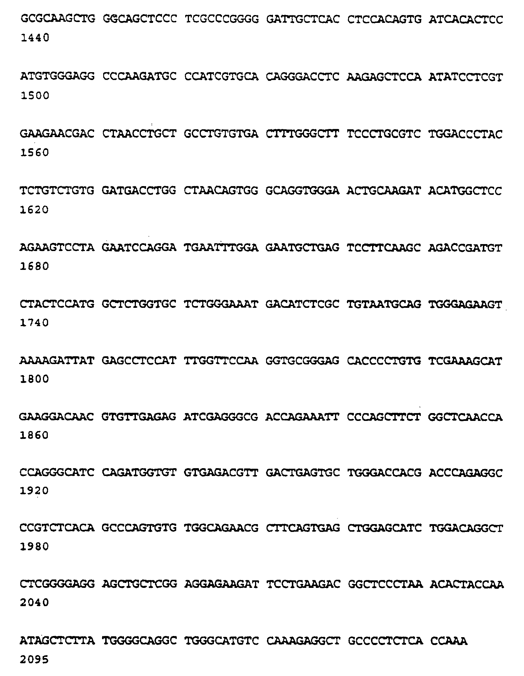
- (2) INFORMATION FOR SEQ ID NO:2:
- (i) SEQUENCE CHARACTERISTICS:
- (A) LENGTH: 49 base pairs
- (B) TYPE: nucleic acid
- (C) STRANDEDNESS: single
- (D). TOPOLOGY: linear
- (xi) SEQUENCE DESCRIPTION: SEQ ID NO:2:

- (2) INFORMATION FOR SEQ ID NO:3:
- (i) SEQUENCE CHARACTERISTICS:
- (A) LENGTH: 52 base pairs
- (B) TYPE: nucleic acid
- (C) STRANDEDNESS: single
- (D) TOPOLOGY: linear
- (xi) SEQUENCE DESCRIPTION: SEQ ID NO:3:

- (2) INFORMATION FOR SEQ ID NO:4:
- (i) SEQUENCE CHARACTERISTICS:
- (A) LENGTH: 46 base pairs
- (B) TYPE: nucleic acid
- (C) STRANDEDNESS: single
- (D) TOPOLOGY: linear
- (xi) SEQUENCE DESCRIPTION: SEQ ID NO:4:

- (2) INFORMATION FOR SEQ ID NO:5:
- (i) SEQUENCE CHARACTERISTICS:
- (A) LENGTH: 33 base pairs
- (B) TYPE: nucleic acid
- (C) STRANDEDNESS: single
- (D) TOPOLOGY: linear
- (xi) SEQUENCE DESCRIPTION: SEQ ID NO:5:

-
Claims (22)
- The use of a Type II TGF-β-binding receptor fragment, wherein the fragment consists of amino acids 1 to 105 of the extracellular portion of the binding receptor, and is capable of reducing excess TGF-β activity in an individual in the manufacture of a medicament for treating an individual for a medical condition associated with TGF-β excess.
- The use according to claim 1 wherein the Type II TGF-β receptor fragment is a fragment of recombinant human TGF-β receptor.
- The use according to claim 1 or 2 wherein the TGF-β-binding receptor fragment is administered parenterally, orally or locally.
- The use according to any preceding claim wherein the Type II TGF-β receptor fragment is administered by intravenous, intraocular, intraarticular, transdermal, or enteral administration.
- The use according to any preceding claim wherein the medical condition is cancer.
- The use according to claim 5 wherein the cancer is plasmacytoma, glioblastoma, astrocytoma, or prostatic or ovarian carcinoma.
- The use according to any one of claims 1 to 4 wherein the medical condition is a collagen vascular disease.
- The use according to claim 7 wherein the collagen vascular disease is systemic sclerosis, polymyositis, scleroderma, or dermatomyositis.
- The use according to any of claims 1 to 4 wherein the medical condition is an autoimmune disease.
- The use according to claim 9 wherein the autoimmune disease is rheumatoid arthritis or systemic lupus erythematosus.
- The use according to any of claims 1 to 4 wherein the medical condition is a fibroproliferative disorder.
- The use according to claim 11 wherein the fibroproliferative disorder is hepatic, kidney, intraocular, or pulmonary fibrosis, diabetic nephropathy, glomerulonephritis, proliferative vitreoretinopathy, liver cirrhosis, biliary fibrosis, or myelofibrosis.
- The use according to any of claims 1 to 4 wherein the condition of TGF-β excess is characterized by immunosuppression associated with an infectious disease.
- The use according to claim 13 wherein the immunosuppression occurs with a trypanosomal infection or with a viral infection from a human immunosuppression virus, HTLV-1, lymphocytic choriomeningitis virus or hepatitis.
- The use of a Type II TGF-β-binding receptor fragment as defined in claim 1 capable of reducing an excess of TGF-β in an individual in the manufacture of a medicament for treating a wound to avoid excessive connective tissue formation associated with TGF-β excess.
- The use according to claim 15 wherein the wound is a surgical incision or trauma-induced laceration or involves the peritoneum and the excessive connective tissue formation comprises abdominal adhesions.
- The use according to claim 15, wherein the excessive connective tissue is a scar, including scars which involve restenosis of blood vessels, a hypertrophic scar or a keloid.
- The use of a Type II TFG-β-binding receptor fragment as defined in claim 1 capable of preventing excessive fibrous tissue formation in the manufacture of a medicament for preventing postradiation fibrosis in an individual undergoing, or about to undergo, radiation therapy.
- A Type II TGF-β-binding receptor fragment as defined in claim 1 for use in vaccine.
- The use of a Type II TGF-β-binding receptor fragment as defined in claim 1 capable of increasing the immune response to a vaccine in an individual in the manufacture of a medicament for increasing the effectiveness of a vaccine in an individual about to receive or receiving a vaccine.
- The use according to claim 20 wherein the individual is an immunocompromised individual.
- A vaccine composition which contains a Type II TGF-β-binding receptor fragment as defined in claim 1.
Applications Claiming Priority (5)
| Application Number | Priority Date | Filing Date | Title |
|---|---|---|---|
| US96837592A | 1992-10-29 | 1992-10-29 | |
| US968375 | 1992-10-29 | ||
| US3759793A | 1993-03-26 | 1993-03-26 | |
| US37597 | 1993-03-26 | ||
| PCT/US1993/010455 WO1994009815A1 (en) | 1992-10-29 | 1993-10-29 | USES OF TGF-β RECEPTOR FRAGMENT AS A THERAPEUTIC AGENT |
Publications (3)
| Publication Number | Publication Date |
|---|---|
| EP0669833A1 EP0669833A1 (en) | 1995-09-06 |
| EP0669833A4 EP0669833A4 (en) | 1996-07-31 |
| EP0669833B1 true EP0669833B1 (en) | 2002-06-12 |
Family
ID=26714287
Family Applications (1)
| Application Number | Title | Priority Date | Filing Date |
|---|---|---|---|
| EP94902198A Expired - Lifetime EP0669833B1 (en) | 1992-10-29 | 1993-10-29 | Type ii tgf-beta-binding receptor fragment as therapeutic agent |
Country Status (8)
| Country | Link |
|---|---|
| US (1) | US5693607A (en) |
| EP (1) | EP0669833B1 (en) |
| JP (1) | JPH08504763A (en) |
| AU (1) | AU669256B2 (en) |
| CA (1) | CA2146973C (en) |
| DE (1) | DE69332026T2 (en) |
| ES (1) | ES2174868T3 (en) |
| WO (1) | WO1994009815A1 (en) |
Cited By (1)
| Publication number | Priority date | Publication date | Assignee | Title |
|---|---|---|---|---|
| US7527791B2 (en) | 2004-03-31 | 2009-05-05 | Genentech, Inc. | Humanized anti-TGF-beta antibodies |
Families Citing this family (53)
| Publication number | Priority date | Publication date | Assignee | Title |
|---|---|---|---|---|
| US5571714A (en) * | 1988-12-22 | 1996-11-05 | Celtrix Pharmaceuticals, Inc. | Monoclonal antibodies which bind both transforming growth factors β1 and β2 and methods of use |
| US7384634B2 (en) | 1991-08-30 | 2008-06-10 | University Of South Florida | Connective tissue growth factor |
| US6515009B1 (en) | 1991-09-27 | 2003-02-04 | Neorx Corporation | Therapeutic inhibitor of vascular smooth muscle cells |
| US5811447A (en) | 1993-01-28 | 1998-09-22 | Neorx Corporation | Therapeutic inhibitor of vascular smooth muscle cells |
| CA2122491A1 (en) * | 1991-10-31 | 1993-05-13 | Herbert Y. Lin | Tgf-.beta. type receptor cdnas and uses therefor |
| US6395494B1 (en) | 1993-05-13 | 2002-05-28 | Neorx Corporation | Method to determine TGF-β |
| US6491938B2 (en) | 1993-05-13 | 2002-12-10 | Neorx Corporation | Therapeutic inhibitor of vascular smooth muscle cells |
| ATE315939T1 (en) * | 1993-11-19 | 2006-02-15 | Univ Sydney | METHOD FOR PROPHYLAXIS OR CONTROL OF CATARACT |
| AU688409B2 (en) * | 1993-11-19 | 1998-03-12 | University Of Sydney, The | A method for preventing or controlling cataract |
| US5866323A (en) | 1995-04-07 | 1999-02-02 | Case Western Reserve University | Cancer diagnosis prognosis and therapy based on mutation of receptors for transforming growth factor β and homologous growth controlling factors |
| GB2304045A (en) * | 1995-08-04 | 1997-03-12 | Univ Manchester | Betaglycan compositions for promoting the healing of wounds and fibrotic diseases |
| WO1998046588A2 (en) * | 1997-04-11 | 1998-10-22 | Neorx Corporation | Compounds and therapies for the prevention of vascular and non-vascular pathologies |
| ES2289778T3 (en) * | 1997-04-18 | 2008-02-01 | Biogen Idec Ma Inc. | FUSION PROTEINS OF THE CONSTANT REGION OF THE IMMUNOGLOBULIN / RECEIVER TGF-BETA TYPE II. |
| ZA988461B (en) | 1997-09-18 | 1999-03-30 | Idec Pharma Corp | Synergistic composition and methods for treating neoplastic or cancerous growths and for restoring or boosting hematopoiesis |
| US6444657B1 (en) | 1998-12-31 | 2002-09-03 | Guilford Pharmaceuticals Inc. | Methods for treating certain diseases using naaladase inhibitors |
| JP2001206899A (en) * | 1999-11-18 | 2001-07-31 | Japan Tobacco Inc | HUMAN MONOCLONAL ANTIBODY AGAINST TGF-beta II TYPE RECEPTOR AND MEDICINAL USE THEREOF |
| US20040018228A1 (en) * | 2000-11-06 | 2004-01-29 | Afmedica, Inc. | Compositions and methods for reducing scar tissue formation |
| US6534693B2 (en) * | 2000-11-06 | 2003-03-18 | Afmedica, Inc. | Surgically implanted devices having reduced scar tissue formation |
| US20040241211A9 (en) * | 2000-11-06 | 2004-12-02 | Fischell Robert E. | Devices and methods for reducing scar tissue formation |
| US6613083B2 (en) | 2001-05-02 | 2003-09-02 | Eckhard Alt | Stent device and method |
| EP1478354A4 (en) * | 2002-01-22 | 2008-09-24 | Genzyme Corp | USE OF TGF-BETA ANTAGONISTS FOR TREATING OR PREVENTING CHRONIC TRANSPLANT DELIVERY |
| US7795389B2 (en) * | 2004-09-28 | 2010-09-14 | The Board Of Regents Of The University Of Texas System | Antagonizing TGF-beta activity with various ectodomains TGF-beta receptors used in combination or as fusion proteins |
| JP5481028B2 (en) * | 2004-10-13 | 2014-04-23 | ザ オハイオ ステート ユニバーシティー リサーチ ファウンデーション | Methods for treating or preventing virus-related lymphoproliferative disorders |
| WO2007050793A2 (en) | 2005-10-25 | 2007-05-03 | The Johns Hopkins University | Methods and compositions for the treatment of marfan syndrome and associated disorders |
| EP2230252A1 (en) | 2006-03-13 | 2010-09-22 | The Johns Hopkins University | Augmentation of endothelial thromboresistance |
| EP2918288B1 (en) | 2006-10-03 | 2017-08-16 | Genzyme Corporation | Use of TGF beta antagonists to treat infants at risk of developing bronchopulmonary dysplasia |
| EP2171062A1 (en) * | 2007-06-15 | 2010-04-07 | Genzyme Corporation | Fusion proteins containing two tgf-beta binding domains of tgf-beta type ii receptor |
| CA2699454C (en) * | 2007-08-27 | 2012-07-24 | Auxagen, Inc. | Methods for inhibiting tgf-.beta. |
| WO2010033507A1 (en) * | 2008-09-16 | 2010-03-25 | St. Louis University | Method of enhancing tgf-beta signalling |
| TR201816180T4 (en) | 2010-03-12 | 2018-11-21 | Genzyme Corp | Combination therapy to treat breast cancer. |
| CA2823154A1 (en) | 2010-12-27 | 2012-07-05 | Lsip, Llc | Ips cells and method for generating same |
| US20140308275A1 (en) | 2011-07-27 | 2014-10-16 | Inserm (Institut National De La Sante Et De La Recherche Medicale | Methods for diagnosing and treating myhre syndrome |
| BR112014009789A2 (en) | 2011-10-26 | 2017-04-25 | Seattle Children's Res Inst | cysteamine in the treatment of fibrotic disease |
| EP2799537B1 (en) | 2011-12-28 | 2021-09-22 | Kyoto Prefectural Public University Corporation | Normalization of culture of corneal endothelial cells |
| AU2014308751B2 (en) * | 2013-08-22 | 2020-03-05 | Acceleron Pharma, Inc. | TGF-beta receptor type II variants and uses thereof |
| WO2015064768A1 (en) | 2013-10-31 | 2015-05-07 | 京都府公立大学法人 | Therapeutic drug for diseases related to endoplasmic reticulum cell death in corneal endothelium |
| AR098827A1 (en) | 2013-12-19 | 2016-06-15 | Consejo Nac De Investig Científicas Y Técnicas (Conicet) | ISOFORM OF TGF-b RECEPTOR II, POLYCUCLEOTID CODING VECTORS, TRANSFORMED CELLS, PEPTIDES AND FUSION, METHODS AND USES |
| US10882903B2 (en) | 2015-05-18 | 2021-01-05 | Arizona Board Of Regents On Behalf Of The University Of Arizona | Methods and compositions for treating an alphavirus infection |
| CN116327952A (en) | 2015-08-04 | 2023-06-27 | 阿塞勒隆制药公司 | Methods for treating myeloproliferative disorders |
| CA2996996A1 (en) | 2015-08-31 | 2017-03-09 | National Research Council Of Canada | Tgf-.beta.-receptor ectodomain fusion molecules and uses thereof |
| JP7231553B2 (en) | 2017-03-02 | 2023-03-01 | ナショナル リサーチ カウンシル オブ カナダ | TGF-B-receptor ectodomain fusion molecule and uses thereof |
| BR112019023071A2 (en) | 2017-05-04 | 2020-06-09 | Acceleron Pharma Inc | type ii tgf-beta receptor fusion proteins and use thereof |
| AU2018264455B2 (en) | 2017-05-12 | 2024-12-12 | Jiangsu Hengrui Medicine Co., Ltd. | Fusion protein containing TGF-beta receptor and medicinal uses thereof |
| WO2019173324A1 (en) | 2018-03-06 | 2019-09-12 | The Trustees Of The University Of Pennsylvania | Prostate-specific membrane antigen cars and methods of use thereof |
| TWI823906B (en) | 2018-03-09 | 2023-12-01 | 美商艾吉納斯公司 | Anti-cd73 antibodies and methods of use thereof |
| TWI809130B (en) | 2018-06-22 | 2023-07-21 | 美商凱特製藥公司 | Chimeric transmembrane proteins and uses thereof |
| TWI829799B (en) | 2018-11-09 | 2024-01-21 | 大陸商江蘇恆瑞醫藥股份有限公司 | A PHARMACEUTICAL COMPOSITION COMPRISING TGF-β RECEPTOR FUSION PROTEIN AND THE USE THEREOF |
| US20220177978A1 (en) | 2019-04-02 | 2022-06-09 | INSERM (Institut National de la Santé et de la Recherche Médicale) | Methods of predicting and preventing cancer in patients having premalignant lesions |
| US12109235B2 (en) | 2019-06-21 | 2024-10-08 | Kite Pharma, Inc. | TGF-beta receptors and methods of use |
| JP7766672B2 (en) | 2020-07-24 | 2025-11-10 | ▲邁▼威(上海)生物科技股▲フン▼有限公司 | TGF-β RII mutants and fusion proteins thereof |
| CN117730100A (en) | 2021-06-08 | 2024-03-19 | 凯德药业股份有限公司 | GPC3 binding molecule |
| WO2023141724A1 (en) | 2022-01-28 | 2023-08-03 | 35Pharma Inc. | Activin receptor type iib variants and uses thereof |
| US20260092096A1 (en) | 2024-05-14 | 2026-04-02 | 35Pharma Inc. | Activin receptor type iib variants and uses thereof |
Family Cites Families (15)
| Publication number | Priority date | Publication date | Assignee | Title |
|---|---|---|---|---|
| US4886747A (en) * | 1985-03-22 | 1989-12-12 | Genentech, Inc. | Nucleic acid encoding TGF-β and its uses |
| ATE247708T1 (en) * | 1986-10-02 | 2003-09-15 | Massachusetts Inst Technology | METHOD FOR REGULATING THE METABOLIC STABILITY OF PROTEINS |
| US5084384A (en) * | 1987-04-23 | 1992-01-28 | Monsanto Company | Secretion of insulin-like growth factor-I |
| US5108919A (en) * | 1988-06-24 | 1992-04-28 | Genentech, Inc. | Dna sequences encoding yeast ubiquitin hydrolase |
| CA2002011A1 (en) * | 1988-11-14 | 1990-05-14 | Anthony F. Purchio | Normal human growth regulatory receptor for tgf-beta |
| EP0413440B1 (en) * | 1989-07-19 | 1995-08-09 | Tonen Corporation | Peptidyl prolyl-cis.trans-isomerase |
| ZA914750B (en) * | 1990-06-20 | 1992-03-25 | Bristol Myers Squibb Co | Methods of modulating blood pressure using tgf-beta and antagonists thereof |
| US5187151A (en) * | 1991-02-12 | 1993-02-16 | Genentech, Inc. | Use of binding protein with igf-i as an anabolic growth promoting agent |
| GB9106678D0 (en) * | 1991-03-28 | 1991-05-15 | Ferguson Mark W J | Wound healing |
| CA2122491A1 (en) * | 1991-10-31 | 1993-05-13 | Herbert Y. Lin | Tgf-.beta. type receptor cdnas and uses therefor |
| AU3178893A (en) * | 1991-11-15 | 1993-06-15 | Memorial Sloan-Kettering Cancer Center | Purified proteoglycan betaglycan, compositions, and methods |
| MX9301135A (en) * | 1992-03-06 | 1993-09-01 | Steven G Reed | METHOD FOR TREATING MACROPHAGO PATHOGEN INFECTIONS. |
| CA2155515A1 (en) * | 1993-02-19 | 1994-09-01 | Wendy Waegell | Expansion of stem cells in long term bone marrow cultures by neutralization of tgf-.beta. |
| EP0637450A3 (en) * | 1993-08-04 | 1995-04-05 | Collagen Corp | Composition for revitalizing scar tissue. |
| AU8016894A (en) * | 1993-10-15 | 1995-05-04 | La Jolla Cancer Research Foundation | Betaglycan polypeptides having tgf-beta binding activity |
-
1993
- 1993-10-29 CA CA002146973A patent/CA2146973C/en not_active Expired - Fee Related
- 1993-10-29 ES ES94902198T patent/ES2174868T3/en not_active Expired - Lifetime
- 1993-10-29 EP EP94902198A patent/EP0669833B1/en not_active Expired - Lifetime
- 1993-10-29 JP JP6511364A patent/JPH08504763A/en not_active Ceased
- 1993-10-29 WO PCT/US1993/010455 patent/WO1994009815A1/en not_active Ceased
- 1993-10-29 AU AU56650/94A patent/AU669256B2/en not_active Ceased
- 1993-10-29 DE DE69332026T patent/DE69332026T2/en not_active Expired - Lifetime
-
1994
- 1994-12-22 US US08/361,873 patent/US5693607A/en not_active Expired - Lifetime
Cited By (2)
| Publication number | Priority date | Publication date | Assignee | Title |
|---|---|---|---|---|
| US7527791B2 (en) | 2004-03-31 | 2009-05-05 | Genentech, Inc. | Humanized anti-TGF-beta antibodies |
| US8012482B2 (en) | 2004-03-31 | 2011-09-06 | Genentech, Inc. | Humanized anti-TGF-beta antibodies |
Also Published As
| Publication number | Publication date |
|---|---|
| US5693607A (en) | 1997-12-02 |
| AU669256B2 (en) | 1996-05-30 |
| CA2146973A1 (en) | 1994-05-11 |
| AU5665094A (en) | 1994-05-24 |
| WO1994009815A1 (en) | 1994-05-11 |
| DE69332026T2 (en) | 2002-10-31 |
| ES2174868T3 (en) | 2002-11-16 |
| EP0669833A1 (en) | 1995-09-06 |
| DE69332026D1 (en) | 2002-07-18 |
| JPH08504763A (en) | 1996-05-21 |
| CA2146973C (en) | 2008-09-02 |
| EP0669833A4 (en) | 1996-07-31 |
Similar Documents
| Publication | Publication Date | Title |
|---|---|---|
| AU669256B2 (en) | Uses of TGF-beta receptor fragment as a therapeutic agent | |
| AU696764B2 (en) | Vascular endothelial growth factor 2 | |
| CN107286248B (en) | High-glycosylation human growth hormone (HGH) fusion protein and preparation method thereof and purposes | |
| EP0975771B1 (en) | Type ii tgf-beta receptor/immunoglobulin constant region fusion proteins | |
| US6638912B2 (en) | Peptide compositions mimicking TGF-β activity | |
| CA2404945C (en) | Taci as an anti-tumor agent | |
| EP0559769B1 (en) | Heparin binding mitogen with homology to epidermal growth factor (egf) | |
| KR20160003848A (en) | Therapeutic peptides | |
| EP1320541A2 (en) | Tgf-beta inhibitors and methods | |
| US11883464B2 (en) | Nerve growth factor fusion protein, preparation method and use thereof | |
| JP2010529859A (en) | Fusion protein containing two TGF-β binding domains of TGF-β type II receptor | |
| JP2003525864A (en) | Insulin dimers cross-linked by covalent bonds | |
| PT94754B (en) | METHOD OF OBTAINING A TUMOR NECROSIS FACTOR INHIBITOR AND INSULATING GENES THAT ENCODER THE ABOVE INHIBITOR | |
| AU4485200A (en) | Tribonectins | |
| USRE49280E1 (en) | TGFbeta type II-type III receptor fusions | |
| AU2001253920A1 (en) | Use of taci as an anti-tumor agent | |
| JP2002526073A (en) | A coding sequence for a novel human growth differentiation factor, a polypeptide encoded by the DNA sequence thereof, and a method for producing them. | |
| JP2018529729A (en) | Treatment of bile acid disorders | |
| CN118638210A (en) | Preparation method and application of long-acting PTH analogs | |
| CN111454371A (en) | PD L1-pH L IP, preparation method and application thereof in treatment of autoimmune disease | |
| CZ389397A3 (en) | Stimulation factor of bones | |
| JP2013253079A (en) | BIOLOGICALLY ACTIVE Fc FUSION PROTEIN OF HUMAN URINARY TRYPSIN INHIBITOR, AND PREPARATION METHOD AND APPLICATION THEREOF | |
| HK1103299A (en) | Use of taci as an anti-tumor agent | |
| MXPA01003764A (en) | Site-directed dual pegylation of proteins for improved bioactivity and biocompatibility | |
| HK1023513A1 (en) | Compositions comprising conjugates of stable, active, human ob protein with antibody fc chain and methods |
Legal Events
| Date | Code | Title | Description |
|---|---|---|---|
| PUAI | Public reference made under article 153(3) epc to a published international application that has entered the european phase |
Free format text: ORIGINAL CODE: 0009012 |
|
| 17P | Request for examination filed |
Effective date: 19950426 |
|
| AK | Designated contracting states |
Kind code of ref document: A1 Designated state(s): DE ES FR GB IT |
|
| RIN1 | Information on inventor provided before grant (corrected) |
Inventor name: CARRILLO, PEDRO A. Inventor name: OLSEN, DAVID R. Inventor name: DASCH, JAMES R. Inventor name: SEGARINI, PATRICIA R. |
|
| RHK1 | Main classification (correction) |
Ipc: A61K 38/04 |
|
| RIN1 | Information on inventor provided before grant (corrected) |
Inventor name: CARRILLO, PEDRO A. Inventor name: OLSEN, DAVID R. Inventor name: DASCH, JAMES R. Inventor name: SEGARINI, PATRICIA R. |
|
| A4 | Supplementary search report drawn up and despatched |
Effective date: 19960618 |
|
| AK | Designated contracting states |
Kind code of ref document: A4 Designated state(s): DE ES FR GB IT |
|
| RHK1 | Main classification (correction) |
Ipc: A61K 37/43 |
|
| RA4 | Supplementary search report drawn up and despatched (corrected) |
Effective date: 19960618 |
|
| 17Q | First examination report despatched |
Effective date: 19990521 |
|
| GRAG | Despatch of communication of intention to grant |
Free format text: ORIGINAL CODE: EPIDOS AGRA |
|
| RTI1 | Title (correction) |
Free format text: TYPE II TGF-BETA-BINDING RECEPTOR FRAGMENT AS THERAPEUTIC AGENT |
|
| GRAG | Despatch of communication of intention to grant |
Free format text: ORIGINAL CODE: EPIDOS AGRA |
|
| GRAH | Despatch of communication of intention to grant a patent |
Free format text: ORIGINAL CODE: EPIDOS IGRA |
|
| GRAH | Despatch of communication of intention to grant a patent |
Free format text: ORIGINAL CODE: EPIDOS IGRA |
|
| GRAA | (expected) grant |
Free format text: ORIGINAL CODE: 0009210 |
|
| AK | Designated contracting states |
Kind code of ref document: B1 Designated state(s): DE ES FR GB IT |
|
| REG | Reference to a national code |
Ref country code: GB Ref legal event code: FG4D |
|
| RIC1 | Information provided on ipc code assigned before grant |
Free format text: 7A 61K 38/04 A, 7A 61K 38/00 B |
|
| REF | Corresponds to: |
Ref document number: 69332026 Country of ref document: DE Date of ref document: 20020718 |
|
| ET | Fr: translation filed | ||
| REG | Reference to a national code |
Ref country code: ES Ref legal event code: FG2A Ref document number: 2174868 Country of ref document: ES Kind code of ref document: T3 |
|
| PLBE | No opposition filed within time limit |
Free format text: ORIGINAL CODE: 0009261 |
|
| STAA | Information on the status of an ep patent application or granted ep patent |
Free format text: STATUS: NO OPPOSITION FILED WITHIN TIME LIMIT |
|
| 26N | No opposition filed |
Effective date: 20030313 |
|
| PGFP | Annual fee paid to national office [announced via postgrant information from national office to epo] |
Ref country code: ES Payment date: 20091023 Year of fee payment: 17 Ref country code: DE Payment date: 20091026 Year of fee payment: 17 |
|
| PGFP | Annual fee paid to national office [announced via postgrant information from national office to epo] |
Ref country code: IT Payment date: 20091029 Year of fee payment: 17 Ref country code: GB Payment date: 20091022 Year of fee payment: 17 Ref country code: FR Payment date: 20091110 Year of fee payment: 17 |
|
| GBPC | Gb: european patent ceased through non-payment of renewal fee |
Effective date: 20101029 |
|
| PG25 | Lapsed in a contracting state [announced via postgrant information from national office to epo] |
Ref country code: FR Free format text: LAPSE BECAUSE OF NON-PAYMENT OF DUE FEES Effective date: 20101102 |
|
| REG | Reference to a national code |
Ref country code: FR Ref legal event code: ST Effective date: 20110630 |
|
| PG25 | Lapsed in a contracting state [announced via postgrant information from national office to epo] |
Ref country code: GB Free format text: LAPSE BECAUSE OF NON-PAYMENT OF DUE FEES Effective date: 20101029 |
|
| REG | Reference to a national code |
Ref country code: DE Ref legal event code: R119 Ref document number: 69332026 Country of ref document: DE Effective date: 20110502 |
|
| REG | Reference to a national code |
Ref country code: ES Ref legal event code: FD2A Effective date: 20111118 |
|
| PG25 | Lapsed in a contracting state [announced via postgrant information from national office to epo] |
Ref country code: IT Free format text: LAPSE BECAUSE OF NON-PAYMENT OF DUE FEES Effective date: 20101029 |
|
| PG25 | Lapsed in a contracting state [announced via postgrant information from national office to epo] |
Ref country code: ES Free format text: LAPSE BECAUSE OF NON-PAYMENT OF DUE FEES Effective date: 20101030 |
|
| PG25 | Lapsed in a contracting state [announced via postgrant information from national office to epo] |
Ref country code: DE Free format text: LAPSE BECAUSE OF NON-PAYMENT OF DUE FEES Effective date: 20110502 |