EP0652954B1 - A novel gene vector coding human epidermal growth factor and process for preparing the same - Google Patents
A novel gene vector coding human epidermal growth factor and process for preparing the same Download PDFInfo
- Publication number
- EP0652954B1 EP0652954B1 EP94913826A EP94913826A EP0652954B1 EP 0652954 B1 EP0652954 B1 EP 0652954B1 EP 94913826 A EP94913826 A EP 94913826A EP 94913826 A EP94913826 A EP 94913826A EP 0652954 B1 EP0652954 B1 EP 0652954B1
- Authority
- EP
- European Patent Office
- Prior art keywords
- hegf
- sequence
- coli
- gene
- seq
- Prior art date
- Legal status (The legal status is an assumption and is not a legal conclusion. Google has not performed a legal analysis and makes no representation as to the accuracy of the status listed.)
- Expired - Lifetime
Links
- 238000004519 manufacturing process Methods 0.000 title claims abstract description 12
- 239000013598 vector Substances 0.000 title claims description 9
- 108090000623 proteins and genes Proteins 0.000 title abstract description 56
- GVUGOAYIVIDWIO-UFWWTJHBSA-N nepidermin Chemical compound C([C@@H](C(=O)N[C@@H]([C@@H](C)CC)C(=O)NCC(=O)N[C@@H](CCC(O)=O)C(=O)N[C@@H](CCCNC(N)=N)C(=O)N[C@@H](CS)C(=O)N[C@@H](CCC(N)=O)C(=O)N[C@@H](CC=1C=CC(O)=CC=1)C(=O)N[C@@H](CCCNC(N)=N)C(=O)N[C@@H](CC(O)=O)C(=O)N[C@@H](CC(C)C)C(=O)N[C@@H](CCCCN)C(=O)N[C@@H](CC=1C2=CC=CC=C2NC=1)C(=O)N[C@@H](CC=1C2=CC=CC=C2NC=1)C(=O)N[C@@H](CCC(O)=O)C(=O)N[C@@H](CC(C)C)C(=O)N[C@@H](CCCNC(N)=N)C(O)=O)NC(=O)CNC(=O)[C@@H](NC(=O)[C@@H](NC(=O)[C@H](CS)NC(=O)[C@H](CC(N)=O)NC(=O)[C@H](CS)NC(=O)[C@H](C)NC(=O)[C@H](CC=1C=CC(O)=CC=1)NC(=O)[C@H](CCCCN)NC(=O)[C@H](CC(O)=O)NC(=O)[C@H](CC(C)C)NC(=O)[C@H](C)NC(=O)[C@H](CCC(O)=O)NC(=O)[C@@H](NC(=O)[C@H](CC=1C=CC(O)=CC=1)NC(=O)[C@H](CCSC)NC(=O)[C@H](CS)NC(=O)[C@@H](NC(=O)CNC(=O)[C@H](CC(O)=O)NC(=O)[C@H](CC=1NC=NC=1)NC(=O)[C@H](CC(C)C)NC(=O)[C@H](CS)NC(=O)[C@H](CC=1C=CC(O)=CC=1)NC(=O)CNC(=O)[C@H](CC(O)=O)NC(=O)[C@H](CC=1NC=NC=1)NC(=O)[C@H](CO)NC(=O)[C@H](CC(C)C)NC(=O)[C@H]1N(CCC1)C(=O)[C@H](CS)NC(=O)[C@H](CCC(O)=O)NC(=O)[C@H](CO)NC(=O)[C@H](CC(O)=O)NC(=O)[C@H](CO)NC(=O)[C@@H](N)CC(N)=O)C(C)C)[C@@H](C)CC)C(C)C)C(C)C)C1=CC=C(O)C=C1 GVUGOAYIVIDWIO-UFWWTJHBSA-N 0.000 title abstract description 6
- 101500025419 Homo sapiens Epidermal growth factor Proteins 0.000 title abstract description 5
- 229940116978 human epidermal growth factor Drugs 0.000 title abstract description 5
- 241000588724 Escherichia coli Species 0.000 claims abstract description 36
- 239000013604 expression vector Substances 0.000 claims abstract description 18
- 238000013519 translation Methods 0.000 claims abstract description 18
- 239000003550 marker Substances 0.000 claims abstract description 17
- 230000014509 gene expression Effects 0.000 claims abstract description 16
- 230000005030 transcription termination Effects 0.000 claims abstract description 12
- 239000004098 Tetracycline Substances 0.000 claims abstract description 10
- 229960002180 tetracycline Drugs 0.000 claims abstract description 10
- 229930101283 tetracycline Natural products 0.000 claims abstract description 10
- 235000019364 tetracycline Nutrition 0.000 claims abstract description 10
- 150000003522 tetracyclines Chemical class 0.000 claims abstract description 10
- 108010030074 endodeoxyribonuclease MluI Proteins 0.000 claims abstract description 6
- 230000006641 stabilisation Effects 0.000 claims abstract description 3
- 238000011105 stabilization Methods 0.000 claims abstract description 3
- 108091008146 restriction endonucleases Proteins 0.000 claims description 8
- 239000002773 nucleotide Substances 0.000 claims description 7
- 125000003729 nucleotide group Chemical group 0.000 claims description 7
- 108091026890 Coding region Proteins 0.000 claims description 6
- 238000003776 cleavage reaction Methods 0.000 claims description 3
- 230000007017 scission Effects 0.000 claims description 3
- 238000012258 culturing Methods 0.000 claims description 2
- 230000022532 regulation of transcription, DNA-dependent Effects 0.000 claims description 2
- 108020005091 Replication Origin Proteins 0.000 abstract description 4
- 108020004705 Codon Proteins 0.000 abstract description 3
- 238000003259 recombinant expression Methods 0.000 abstract description 2
- 108020004414 DNA Proteins 0.000 description 42
- 239000012634 fragment Substances 0.000 description 35
- 108091034117 Oligonucleotide Proteins 0.000 description 30
- 239000013612 plasmid Substances 0.000 description 30
- 108020004707 nucleic acids Proteins 0.000 description 22
- 102000039446 nucleic acids Human genes 0.000 description 22
- 150000007523 nucleic acids Chemical class 0.000 description 22
- JLCPHMBAVCMARE-UHFFFAOYSA-N [3-[[3-[[3-[[3-[[3-[[3-[[3-[[3-[[3-[[3-[[3-[[5-(2-amino-6-oxo-1H-purin-9-yl)-3-[[3-[[3-[[3-[[3-[[3-[[5-(2-amino-6-oxo-1H-purin-9-yl)-3-[[5-(2-amino-6-oxo-1H-purin-9-yl)-3-hydroxyoxolan-2-yl]methoxy-hydroxyphosphoryl]oxyoxolan-2-yl]methoxy-hydroxyphosphoryl]oxy-5-(5-methyl-2,4-dioxopyrimidin-1-yl)oxolan-2-yl]methoxy-hydroxyphosphoryl]oxy-5-(6-aminopurin-9-yl)oxolan-2-yl]methoxy-hydroxyphosphoryl]oxy-5-(6-aminopurin-9-yl)oxolan-2-yl]methoxy-hydroxyphosphoryl]oxy-5-(6-aminopurin-9-yl)oxolan-2-yl]methoxy-hydroxyphosphoryl]oxy-5-(6-aminopurin-9-yl)oxolan-2-yl]methoxy-hydroxyphosphoryl]oxyoxolan-2-yl]methoxy-hydroxyphosphoryl]oxy-5-(5-methyl-2,4-dioxopyrimidin-1-yl)oxolan-2-yl]methoxy-hydroxyphosphoryl]oxy-5-(4-amino-2-oxopyrimidin-1-yl)oxolan-2-yl]methoxy-hydroxyphosphoryl]oxy-5-(5-methyl-2,4-dioxopyrimidin-1-yl)oxolan-2-yl]methoxy-hydroxyphosphoryl]oxy-5-(5-methyl-2,4-dioxopyrimidin-1-yl)oxolan-2-yl]methoxy-hydroxyphosphoryl]oxy-5-(6-aminopurin-9-yl)oxolan-2-yl]methoxy-hydroxyphosphoryl]oxy-5-(6-aminopurin-9-yl)oxolan-2-yl]methoxy-hydroxyphosphoryl]oxy-5-(4-amino-2-oxopyrimidin-1-yl)oxolan-2-yl]methoxy-hydroxyphosphoryl]oxy-5-(4-amino-2-oxopyrimidin-1-yl)oxolan-2-yl]methoxy-hydroxyphosphoryl]oxy-5-(4-amino-2-oxopyrimidin-1-yl)oxolan-2-yl]methoxy-hydroxyphosphoryl]oxy-5-(6-aminopurin-9-yl)oxolan-2-yl]methoxy-hydroxyphosphoryl]oxy-5-(4-amino-2-oxopyrimidin-1-yl)oxolan-2-yl]methyl [5-(6-aminopurin-9-yl)-2-(hydroxymethyl)oxolan-3-yl] hydrogen phosphate Polymers Cc1cn(C2CC(OP(O)(=O)OCC3OC(CC3OP(O)(=O)OCC3OC(CC3O)n3cnc4c3nc(N)[nH]c4=O)n3cnc4c3nc(N)[nH]c4=O)C(COP(O)(=O)OC3CC(OC3COP(O)(=O)OC3CC(OC3COP(O)(=O)OC3CC(OC3COP(O)(=O)OC3CC(OC3COP(O)(=O)OC3CC(OC3COP(O)(=O)OC3CC(OC3COP(O)(=O)OC3CC(OC3COP(O)(=O)OC3CC(OC3COP(O)(=O)OC3CC(OC3COP(O)(=O)OC3CC(OC3COP(O)(=O)OC3CC(OC3COP(O)(=O)OC3CC(OC3COP(O)(=O)OC3CC(OC3COP(O)(=O)OC3CC(OC3COP(O)(=O)OC3CC(OC3COP(O)(=O)OC3CC(OC3COP(O)(=O)OC3CC(OC3CO)n3cnc4c(N)ncnc34)n3ccc(N)nc3=O)n3cnc4c(N)ncnc34)n3ccc(N)nc3=O)n3ccc(N)nc3=O)n3ccc(N)nc3=O)n3cnc4c(N)ncnc34)n3cnc4c(N)ncnc34)n3cc(C)c(=O)[nH]c3=O)n3cc(C)c(=O)[nH]c3=O)n3ccc(N)nc3=O)n3cc(C)c(=O)[nH]c3=O)n3cnc4c3nc(N)[nH]c4=O)n3cnc4c(N)ncnc34)n3cnc4c(N)ncnc34)n3cnc4c(N)ncnc34)n3cnc4c(N)ncnc34)O2)c(=O)[nH]c1=O JLCPHMBAVCMARE-UHFFFAOYSA-N 0.000 description 20
- 230000014616 translation Effects 0.000 description 18
- 238000000034 method Methods 0.000 description 16
- 102000004169 proteins and genes Human genes 0.000 description 16
- WEVYAHXRMPXWCK-UHFFFAOYSA-N Acetonitrile Chemical compound CC#N WEVYAHXRMPXWCK-UHFFFAOYSA-N 0.000 description 12
- 238000010276 construction Methods 0.000 description 12
- 238000010828 elution Methods 0.000 description 12
- 210000004027 cell Anatomy 0.000 description 9
- 239000000499 gel Substances 0.000 description 9
- 239000000243 solution Substances 0.000 description 9
- 102000012410 DNA Ligases Human genes 0.000 description 8
- 108010061982 DNA Ligases Proteins 0.000 description 8
- ZMANZCXQSJIPKH-UHFFFAOYSA-N Triethylamine Chemical compound CCN(CC)CC ZMANZCXQSJIPKH-UHFFFAOYSA-N 0.000 description 7
- BPHPUYQFMNQIOC-NXRLNHOXSA-N isopropyl beta-D-thiogalactopyranoside Chemical compound CC(C)S[C@@H]1O[C@H](CO)[C@H](O)[C@H](O)[C@H]1O BPHPUYQFMNQIOC-NXRLNHOXSA-N 0.000 description 7
- 238000000746 purification Methods 0.000 description 7
- 238000002415 sodium dodecyl sulfate polyacrylamide gel electrophoresis Methods 0.000 description 7
- WSFSSNUMVMOOMR-UHFFFAOYSA-N Formaldehyde Chemical compound O=C WSFSSNUMVMOOMR-UHFFFAOYSA-N 0.000 description 6
- 239000011543 agarose gel Substances 0.000 description 6
- 150000001413 amino acids Chemical class 0.000 description 6
- 230000015572 biosynthetic process Effects 0.000 description 6
- 238000010367 cloning Methods 0.000 description 6
- 239000008363 phosphate buffer Substances 0.000 description 6
- 238000005406 washing Methods 0.000 description 6
- FAPWRFPIFSIZLT-UHFFFAOYSA-M Sodium chloride Chemical compound [Na+].[Cl-] FAPWRFPIFSIZLT-UHFFFAOYSA-M 0.000 description 5
- 238000004458 analytical method Methods 0.000 description 5
- 239000000872 buffer Substances 0.000 description 5
- 238000011534 incubation Methods 0.000 description 5
- 244000005700 microbiome Species 0.000 description 5
- 230000028327 secretion Effects 0.000 description 5
- 102000004594 DNA Polymerase I Human genes 0.000 description 4
- 108010017826 DNA Polymerase I Proteins 0.000 description 4
- 239000006137 Luria-Bertani broth Substances 0.000 description 4
- VYPSYNLAJGMNEJ-UHFFFAOYSA-N Silicium dioxide Chemical compound O=[Si]=O VYPSYNLAJGMNEJ-UHFFFAOYSA-N 0.000 description 4
- 239000007983 Tris buffer Substances 0.000 description 4
- 230000009089 cytolysis Effects 0.000 description 4
- 230000029087 digestion Effects 0.000 description 4
- 230000000694 effects Effects 0.000 description 4
- 238000001962 electrophoresis Methods 0.000 description 4
- 238000004128 high performance liquid chromatography Methods 0.000 description 4
- 239000002609 medium Substances 0.000 description 4
- 108020004999 messenger RNA Proteins 0.000 description 4
- 239000012071 phase Substances 0.000 description 4
- 239000011780 sodium chloride Substances 0.000 description 4
- 238000003786 synthesis reaction Methods 0.000 description 4
- LENZDBCJOHFCAS-UHFFFAOYSA-N tris Chemical compound OCC(N)(CO)CO LENZDBCJOHFCAS-UHFFFAOYSA-N 0.000 description 4
- 238000001262 western blot Methods 0.000 description 4
- LFQSCWFLJHTTHZ-UHFFFAOYSA-N Ethanol Chemical compound CCO LFQSCWFLJHTTHZ-UHFFFAOYSA-N 0.000 description 3
- OKKJLVBELUTLKV-UHFFFAOYSA-N Methanol Chemical compound OC OKKJLVBELUTLKV-UHFFFAOYSA-N 0.000 description 3
- 229910019142 PO4 Inorganic materials 0.000 description 3
- 239000012564 Q sepharose fast flow resin Substances 0.000 description 3
- HEMHJVSKTPXQMS-UHFFFAOYSA-M Sodium hydroxide Chemical compound [OH-].[Na+] HEMHJVSKTPXQMS-UHFFFAOYSA-M 0.000 description 3
- 229960000723 ampicillin Drugs 0.000 description 3
- AVKUERGKIZMTKX-NJBDSQKTSA-N ampicillin Chemical compound C1([C@@H](N)C(=O)N[C@H]2[C@H]3SC([C@@H](N3C2=O)C(O)=O)(C)C)=CC=CC=C1 AVKUERGKIZMTKX-NJBDSQKTSA-N 0.000 description 3
- 210000000172 cytosol Anatomy 0.000 description 3
- 239000002158 endotoxin Substances 0.000 description 3
- 238000003780 insertion Methods 0.000 description 3
- 230000037431 insertion Effects 0.000 description 3
- 238000001155 isoelectric focusing Methods 0.000 description 3
- 238000002955 isolation Methods 0.000 description 3
- 239000010452 phosphate Substances 0.000 description 3
- 108090000765 processed proteins & peptides Proteins 0.000 description 3
- 238000001525 receptor binding assay Methods 0.000 description 3
- 102000005962 receptors Human genes 0.000 description 3
- 108020003175 receptors Proteins 0.000 description 3
- 239000006228 supernatant Substances 0.000 description 3
- RMVRSNDYEFQCLF-UHFFFAOYSA-N thiophenol Chemical compound SC1=CC=CC=C1 RMVRSNDYEFQCLF-UHFFFAOYSA-N 0.000 description 3
- 108091032973 (ribonucleotides)n+m Proteins 0.000 description 2
- FWMNVWWHGCHHJJ-SKKKGAJSSA-N 4-amino-1-[(2r)-6-amino-2-[[(2r)-2-[[(2r)-2-[[(2r)-2-amino-3-phenylpropanoyl]amino]-3-phenylpropanoyl]amino]-4-methylpentanoyl]amino]hexanoyl]piperidine-4-carboxylic acid Chemical compound C([C@H](C(=O)N[C@H](CC(C)C)C(=O)N[C@H](CCCCN)C(=O)N1CCC(N)(CC1)C(O)=O)NC(=O)[C@H](N)CC=1C=CC=CC=1)C1=CC=CC=C1 FWMNVWWHGCHHJJ-SKKKGAJSSA-N 0.000 description 2
- VHUUQVKOLVNVRT-UHFFFAOYSA-N Ammonium hydroxide Chemical compound [NH4+].[OH-] VHUUQVKOLVNVRT-UHFFFAOYSA-N 0.000 description 2
- 108090000204 Dipeptidase 1 Proteins 0.000 description 2
- WQZGKKKJIJFFOK-GASJEMHNSA-N Glucose Natural products OC[C@H]1OC(O)[C@H](O)[C@@H](O)[C@@H]1O WQZGKKKJIJFFOK-GASJEMHNSA-N 0.000 description 2
- PXIPVTKHYLBLMZ-UHFFFAOYSA-N Sodium azide Chemical compound [Na+].[N-]=[N+]=[N-] PXIPVTKHYLBLMZ-UHFFFAOYSA-N 0.000 description 2
- XSQUKJJJFZCRTK-UHFFFAOYSA-N Urea Chemical compound NC(N)=O XSQUKJJJFZCRTK-UHFFFAOYSA-N 0.000 description 2
- 235000011114 ammonium hydroxide Nutrition 0.000 description 2
- 238000013459 approach Methods 0.000 description 2
- 102000006635 beta-lactamase Human genes 0.000 description 2
- 239000012148 binding buffer Substances 0.000 description 2
- 230000032823 cell division Effects 0.000 description 2
- 238000004587 chromatography analysis Methods 0.000 description 2
- 230000000295 complement effect Effects 0.000 description 2
- 238000011109 contamination Methods 0.000 description 2
- 230000008021 deposition Effects 0.000 description 2
- 239000008103 glucose Substances 0.000 description 2
- 230000006698 induction Effects 0.000 description 2
- 238000012454 limulus amebocyte lysate test Methods 0.000 description 2
- 238000011068 loading method Methods 0.000 description 2
- 230000014759 maintenance of location Effects 0.000 description 2
- 239000011159 matrix material Substances 0.000 description 2
- 238000010369 molecular cloning Methods 0.000 description 2
- -1 phosphate triester Chemical class 0.000 description 2
- 229920002401 polyacrylamide Polymers 0.000 description 2
- 102000004196 processed proteins & peptides Human genes 0.000 description 2
- 239000000047 product Substances 0.000 description 2
- 125000006239 protecting group Chemical group 0.000 description 2
- 238000004153 renaturation Methods 0.000 description 2
- 238000004007 reversed phase HPLC Methods 0.000 description 2
- 238000011218 seed culture Methods 0.000 description 2
- 238000000926 separation method Methods 0.000 description 2
- 239000000377 silicon dioxide Substances 0.000 description 2
- 239000007790 solid phase Substances 0.000 description 2
- 230000002103 transcriptional effect Effects 0.000 description 2
- 239000003643 water by type Substances 0.000 description 2
- RYHBNJHYFVUHQT-UHFFFAOYSA-N 1,4-Dioxane Chemical compound C1COCCO1 RYHBNJHYFVUHQT-UHFFFAOYSA-N 0.000 description 1
- QKNYBSVHEMOAJP-UHFFFAOYSA-N 2-amino-2-(hydroxymethyl)propane-1,3-diol;hydron;chloride Chemical compound Cl.OCC(N)(CO)CO QKNYBSVHEMOAJP-UHFFFAOYSA-N 0.000 description 1
- NKDFYOWSKOHCCO-YPVLXUMRSA-N 20-hydroxyecdysone Chemical compound C1[C@@H](O)[C@@H](O)C[C@]2(C)[C@@H](CC[C@@]3([C@@H]([C@@](C)(O)[C@H](O)CCC(C)(O)C)CC[C@]33O)C)C3=CC(=O)[C@@H]21 NKDFYOWSKOHCCO-YPVLXUMRSA-N 0.000 description 1
- 238000009010 Bradford assay Methods 0.000 description 1
- 108010092265 CCWGG-specific type II deoxyribonucleases Proteins 0.000 description 1
- 108010072454 CTGCAG-specific type II deoxyribonucleases Proteins 0.000 description 1
- UXVMQQNJUSDDNG-UHFFFAOYSA-L Calcium chloride Chemical compound [Cl-].[Cl-].[Ca+2] UXVMQQNJUSDDNG-UHFFFAOYSA-L 0.000 description 1
- 108700010070 Codon Usage Proteins 0.000 description 1
- 239000006144 Dulbecco’s modified Eagle's medium Substances 0.000 description 1
- 102000004190 Enzymes Human genes 0.000 description 1
- 108090000790 Enzymes Proteins 0.000 description 1
- 101000851176 Homo sapiens Pro-epidermal growth factor Proteins 0.000 description 1
- 239000007836 KH2PO4 Substances 0.000 description 1
- FFEARJCKVFRZRR-BYPYZUCNSA-N L-methionine Chemical compound CSCC[C@H](N)C(O)=O FFEARJCKVFRZRR-BYPYZUCNSA-N 0.000 description 1
- 125000001429 N-terminal alpha-amino-acid group Chemical group 0.000 description 1
- 108010079246 OMPA outer membrane proteins Proteins 0.000 description 1
- 102000015731 Peptide Hormones Human genes 0.000 description 1
- 108010038988 Peptide Hormones Proteins 0.000 description 1
- 108010021757 Polynucleotide 5'-Hydroxyl-Kinase Proteins 0.000 description 1
- 102000008422 Polynucleotide 5'-hydroxyl-kinase Human genes 0.000 description 1
- 239000012614 Q-Sepharose Substances 0.000 description 1
- 108020004511 Recombinant DNA Proteins 0.000 description 1
- 102000040739 Secretory proteins Human genes 0.000 description 1
- 108091058545 Secretory proteins Proteins 0.000 description 1
- 208000007107 Stomach Ulcer Diseases 0.000 description 1
- 108020005038 Terminator Codon Proteins 0.000 description 1
- 208000027418 Wounds and injury Diseases 0.000 description 1
- 238000000246 agarose gel electrophoresis Methods 0.000 description 1
- 238000005349 anion exchange Methods 0.000 description 1
- 238000005571 anion exchange chromatography Methods 0.000 description 1
- 239000002518 antifoaming agent Substances 0.000 description 1
- 238000000376 autoradiography Methods 0.000 description 1
- 239000004327 boric acid Substances 0.000 description 1
- 239000007853 buffer solution Substances 0.000 description 1
- 210000004899 c-terminal region Anatomy 0.000 description 1
- 239000001110 calcium chloride Substances 0.000 description 1
- 229910001628 calcium chloride Inorganic materials 0.000 description 1
- 244000309466 calf Species 0.000 description 1
- 229940041514 candida albicans extract Drugs 0.000 description 1
- 239000004202 carbamide Substances 0.000 description 1
- 230000015556 catabolic process Effects 0.000 description 1
- 238000004113 cell culture Methods 0.000 description 1
- 230000006037 cell lysis Effects 0.000 description 1
- 238000005119 centrifugation Methods 0.000 description 1
- 238000011097 chromatography purification Methods 0.000 description 1
- 230000006378 damage Effects 0.000 description 1
- 230000003247 decreasing effect Effects 0.000 description 1
- 238000006731 degradation reaction Methods 0.000 description 1
- 238000004925 denaturation Methods 0.000 description 1
- 230000036425 denaturation Effects 0.000 description 1
- 238000013461 design Methods 0.000 description 1
- BNIILDVGGAEEIG-UHFFFAOYSA-L disodium hydrogen phosphate Chemical compound [Na+].[Na+].OP([O-])([O-])=O BNIILDVGGAEEIG-UHFFFAOYSA-L 0.000 description 1
- 229910000397 disodium phosphate Inorganic materials 0.000 description 1
- 238000005516 engineering process Methods 0.000 description 1
- 210000001339 epidermal cell Anatomy 0.000 description 1
- 210000002919 epithelial cell Anatomy 0.000 description 1
- 108020001507 fusion proteins Proteins 0.000 description 1
- 102000037865 fusion proteins Human genes 0.000 description 1
- 210000004211 gastric acid Anatomy 0.000 description 1
- 201000005917 gastric ulcer Diseases 0.000 description 1
- 230000012010 growth Effects 0.000 description 1
- 230000007773 growth pattern Effects 0.000 description 1
- XLYOFNOQVPJJNP-ZSJDYOACSA-N heavy water Substances [2H]O[2H] XLYOFNOQVPJJNP-ZSJDYOACSA-N 0.000 description 1
- 208000014674 injury Diseases 0.000 description 1
- 230000003834 intracellular effect Effects 0.000 description 1
- 238000002372 labelling Methods 0.000 description 1
- 210000004962 mammalian cell Anatomy 0.000 description 1
- 229930182817 methionine Natural products 0.000 description 1
- 229910000402 monopotassium phosphate Inorganic materials 0.000 description 1
- 238000002703 mutagenesis Methods 0.000 description 1
- 231100000350 mutagenesis Toxicity 0.000 description 1
- 230000003647 oxidation Effects 0.000 description 1
- 238000007254 oxidation reaction Methods 0.000 description 1
- 239000000813 peptide hormone Substances 0.000 description 1
- NBIIXXVUZAFLBC-UHFFFAOYSA-K phosphate Chemical compound [O-]P([O-])([O-])=O NBIIXXVUZAFLBC-UHFFFAOYSA-K 0.000 description 1
- 239000013600 plasmid vector Substances 0.000 description 1
- 238000002264 polyacrylamide gel electrophoresis Methods 0.000 description 1
- GNSKLFRGEWLPPA-UHFFFAOYSA-M potassium dihydrogen phosphate Chemical compound [K+].OP(O)([O-])=O GNSKLFRGEWLPPA-UHFFFAOYSA-M 0.000 description 1
- 238000002360 preparation method Methods 0.000 description 1
- 238000002953 preparative HPLC Methods 0.000 description 1
- 238000012207 quantitative assay Methods 0.000 description 1
- 230000014493 regulation of gene expression Effects 0.000 description 1
- 230000009711 regulatory function Effects 0.000 description 1
- 230000010076 replication Effects 0.000 description 1
- 150000003839 salts Chemical class 0.000 description 1
- 238000012163 sequencing technique Methods 0.000 description 1
- 210000002966 serum Anatomy 0.000 description 1
- 210000002784 stomach Anatomy 0.000 description 1
- 210000002700 urine Anatomy 0.000 description 1
- 239000012138 yeast extract Substances 0.000 description 1
Images








Classifications
-
- C—CHEMISTRY; METALLURGY
- C07—ORGANIC CHEMISTRY
- C07K—PEPTIDES
- C07K14/00—Peptides having more than 20 amino acids; Gastrins; Somatostatins; Melanotropins; Derivatives thereof
- C07K14/435—Peptides having more than 20 amino acids; Gastrins; Somatostatins; Melanotropins; Derivatives thereof from animals; from humans
- C07K14/475—Growth factors; Growth regulators
- C07K14/485—Epidermal growth factor [EGF], i.e. urogastrone
-
- C—CHEMISTRY; METALLURGY
- C12—BIOCHEMISTRY; BEER; SPIRITS; WINE; VINEGAR; MICROBIOLOGY; ENZYMOLOGY; MUTATION OR GENETIC ENGINEERING
- C12N—MICROORGANISMS OR ENZYMES; COMPOSITIONS THEREOF; PROPAGATING, PRESERVING, OR MAINTAINING MICROORGANISMS; MUTATION OR GENETIC ENGINEERING; CULTURE MEDIA
- C12N15/00—Mutation or genetic engineering; DNA or RNA concerning genetic engineering, vectors, e.g. plasmids, or their isolation, preparation or purification; Use of hosts therefor
- C12N15/09—Recombinant DNA-technology
- C12N15/63—Introduction of foreign genetic material using vectors; Vectors; Use of hosts therefor; Regulation of expression
- C12N15/70—Vectors or expression systems specially adapted for E. coli
Definitions
- the present invention relates to a process for preparing human epidermal growth factor employing a recombinant expression vector therefor.
- 'hEGF' human epidermal growth factor
- hEGF plays an important role in growth control in mammalian cells, inter alia epidermal and epithelial cells, on a molecular level [ see : Sporn, M.B. et al , Nature (London), 313: 745-747 (1985); Sporn, M.B. et al , N. Engl. J. Med., 303: 878-880 (1980)] and the treatment of injury [ see : Buckley, A. et al , Proc. Natl. Acad. Sci., USA, 82: 7340-7344 (1985)].
- hEGF can be applied in the treatment of a stomach ulcer, owing to the fact that it represses secretion of gastric acid into the stomach [ see : Gregory, H., J. Cell Sci. Suppl., 3: 11-17 (1985)].
- the inventors synthesized a novel hEGF gene and a novel expression vector therefor which expresses hEFG in a massive amount and developed a process for preparing hEGF therefrom.
- the invention provides an expression vector for hEGF comprising a par site for stabilization in E.coli; a tetracycline resistance marker; and an expression cassette consisting of an Omp A leader sequence, an hEGF coding sequence, a translation termination sequence, and a trp A transcription termination sequence; wherein the expression cassette is under transcriptional regulation by the tac promoter.
- the hEGF sequence preferably comprises a restriction enzyme cleavage site for HpaI at its 5'-terminus, for PstI at its 3'-terminus, and for Bpu1102I, NsiI, MluI, Eco47III and AflII, wherein the nucleotide sequence of the coding sequence is:
- hEGF expressed therefrom shall be the same as natural human EGF in light of amino acid sequence and protein structure; secondly, the nucleotide sequence shall be designed in consideration of the codons ubiquitous in E.coli; thirdly, the portion of secondary structure of mRNA transcribed therefrom shall be minimized; fourthly, many unique restriction sites shall be positioned as far as possible.
- the hEGF gene was designed, based on the previously known hEGF sequence in the art [ see : Gregory, H., Nature, 257: 325 (1975)].
- the inventors synthesized 10 complementary oligonucleotides by using an automatic nucleotide synthesizer in accordance with the solid-phase phosphate triester method [ see : Narang, S.A., Synthesis and Applications of DNA and RNA, Academic Press, 1987].
- the shot-gun ligation method was employed.
- the hEGF coding sequence used in the invention contains the following restriction sites: HpaI at the 5'terminus, PstI at the 3' terminus and Bpu1102I, NsiI, MluI, Eco47III and AflII within its internal sequence.
- Introduction of the hEGF gene into pUC18, a starting vector used in the invention produces the above 7 specific sites which exist as unique restriction sites in the recombinant vector.
- the fact that said unique sites are cleaved by their specific restriction enzymes and positioned at a regular interval, allows practical availability of the gene for mutagenesis induction, which assures high activity and stability of hEGF.
- the HpaI restriction site at the 5' terminus allows production of intact hEGF free of fusion protein from the expression vector.
- E.coli JM109 is transformed with pUE118 according to Hanahan's method [ see : DNA Cloning Vol. I: A Practical Approach, IRL Press, 1985, pp 109-135], and this transformant is deposited with the Korean Culture Collection of Microorganisms (KCCM) located in the Department of Food Engineering, College of Eng., Yonsei University, Sodaemun-gu, Seoul 120-749, Korea on April 9, 1993 under the name of E. coli DW/BT-2040 (KCCM 10026).
- KCCM Korean Culture Collection of Microorganisms
- a tac promoter [ see : de Boer et al , DNA, 2: 231-235 (1983); Amann et al , Gene, 25: 167-178 (1983)] is introduced to the expression vector of the invention. Since the tac promoter is continuously linked to two downstream ribosome binding sites [ see : Shine and Dalgarno, Proc. Natl. Acad. Sci., USA, 71: 1342 (1974)], it initiates the translation of the hEGF gene efficiently.
- the expression vector of the invention employs the following sequences: (1) an Omp A leader sequence [ see : von Gabain, A. et al , Proc. Natl. Acad. Sci., USA, 80: 653-657 (1983)]; (2) a universal translation termination sequence [ see : P. Singleton and D. Sainsbury, Dictionary of Microbiology and Molecular Biology, 2nd. Ed., Wiley, 383, 1987]; and, (3) a trp A transcription termination sequence [ see : Christie, G. E. et al , Proc. Natl. Acad. Sci., USA, 78: 418 (1981)]. Further, the expression vector contains a par site for stability in E.coli [ see : Austin and Abeles, J. Mol. Biol., 169: 373-387 (1983)].
- the "Omp A leader-universal translation termination-trp A transcription termination sequence” is designed and synthesized for insertion of an hEGF gene comprising a BamHI restriction site at the 5' terminus and an XbaI site at the 3'terminus, and Nael and PstI sites between the Omp A leader and the universal translation termination sequence.
- a commercially available plasmid pDR540 is digested with PvuII and then ligated with XbaI linker; and, double digestion with XbaI and BamHI is carried out.
- 2.4kb of DNA fragment thus obtained is ligated with the "Omp A leader-universal translation termination-Trp A transcription termination sequence"; and named pDT420.
- Double digestion of pDT420 with NaeI and PstI produces a cleavage between the Omp A leader and the universal translation termination sequence; and, pUE118 the carrying hEGF gene is digested with HpaI and PstI to obtain the hEGF gene. Then, the hEGF gene is ligated into pDT420 digested with NaeI and PstI, and the resultant vector is named pDE135.
- Plasmid pUC19 known in the art is digested with Oral and EcoRI to obtain a 1.2kb DNA fragment whose two ends are blunt-ended and cohesive for EcoRI, respectively; and, pBR322 is digested with AvaI and blunt-ended with Klenow's fragment, and digesteion with EcoRI is followed to obtain a 1.4kb DNA fragment.
- the above 1.4kb and 1.2kb DNA fragments are ligated with T 4 DNA ligase, and the product is named pTC108.
- pTC108 comprises a tetracycline-resistant marker, a multiple cloning site and the replication origin of pUC19.
- Plasmid pTC108 is digested with EcoRI and SmaI.
- pSC101 is digested with AvaI to obtain a 3.3kb DNA fragment, which is blunt-ended with Klenow's fragment and then ligated with EcoRI linker, followed by digestion with EcoRI.
- the 3.3kb DNA fragment containing EcoRI sticky ends is digested with HincII to obtain a 0.37kb DNA fragment containing a par site. This 0.37kb DNA fragment is ligated to pTC108 digested with EcoRI and SmaI by T 4 DNA ligase, and the ligated plasmid is named pTC226.
- Plasmid pTC226 is digested with AflIII, blunt-ended with Klenow fragment and digested with XbaI in a serial manner to obtain a 2.5kb DNA fragment.
- pDE135 is digested with HindIII, blunt-ended with Klenow fragment and digested with XbaI in a serial manner to obtain a 0.45kb DNA fragment.
- the 2.5kb and 0.45kb DNA fragments are ligated with T 4 DNA ligase, and the ligated plasmid is named pTE105.
- E. coli JM101 is transformed with pTE105, and the transformant is deposited with the Korean Culture Collection of Microorganisms(KCCM) located in the Department of Food Engineering, College of Eng., Yonsei University, Sodaemun-gu, Seoul 120-749, Korea on April 9, 1993 under the name of E. coli DW/BT-2042(KCCM 10027).
- KCCM Korean Culture Collection of Microorganisms
- Transformants E. coli DW/BT-2042 are grown in LB media and expression of hEGF therefrom is determined by 15% SDS-PAGE and Western blot analysis by employing commercially available hEGF(Amersham, ARN 5100, UK) as standard.
- the amount of expressed hEGF is determined by hEGF receptor binding analysis employing A431 cell line(ATCC CRL 1555). Culturing said transformant for 30hrs gave 343.5mg/L of hEGF, where most of the expressed hEGF was secreted out of cytosol.
- the present inventors isolated hEGF from the culture, while reducing contamination of other celluar proteins and endotoxins, by employing a series of chromatographic purification methods, i.e., Amberchrom CG71 chromatography, Q-Sepharose anion exchange chromagraphy and reverse phase C 18 preparative HPLC.
- the purity of purified hEGF is determined by analytical HPLC in accordance with modified method of Hayashi et al's[ see : Hayashi, T. et al., Anal. Sci., 3: 445-449(1987)].
- the hEGF gene was firstly designed, based on the hEGF amino acid sequence and Grantham et al's study on the frequency of codon usage in E. coli[ see : Grantham et al., Nucleic Acid Res., 9:243-274(1981)]. Then, sequence which does not cause formation of secondary structure of mRNA transcribed therefrom was selected, while examining whether the gene contains codons ubiquitous in E. coli and causes formation of secondary structure of mRNA or not, by employing PC-FOLD(Version 2.0) program[ see : Turner, D. et al, Cold Spring Harbor Symp. Quant. Biol., 52:123(1987)].
- the hEGF sequence thus designed carries the following two restriction sites to guarantee accurate insertion, isolation and manipulation of hEGF gene: HpaI at the 5' terminal; PstI at the down stream of translational termination codon.
- the hEGF sequence futher comprises many unique restriction sites at a regular basis as follows:
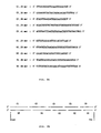
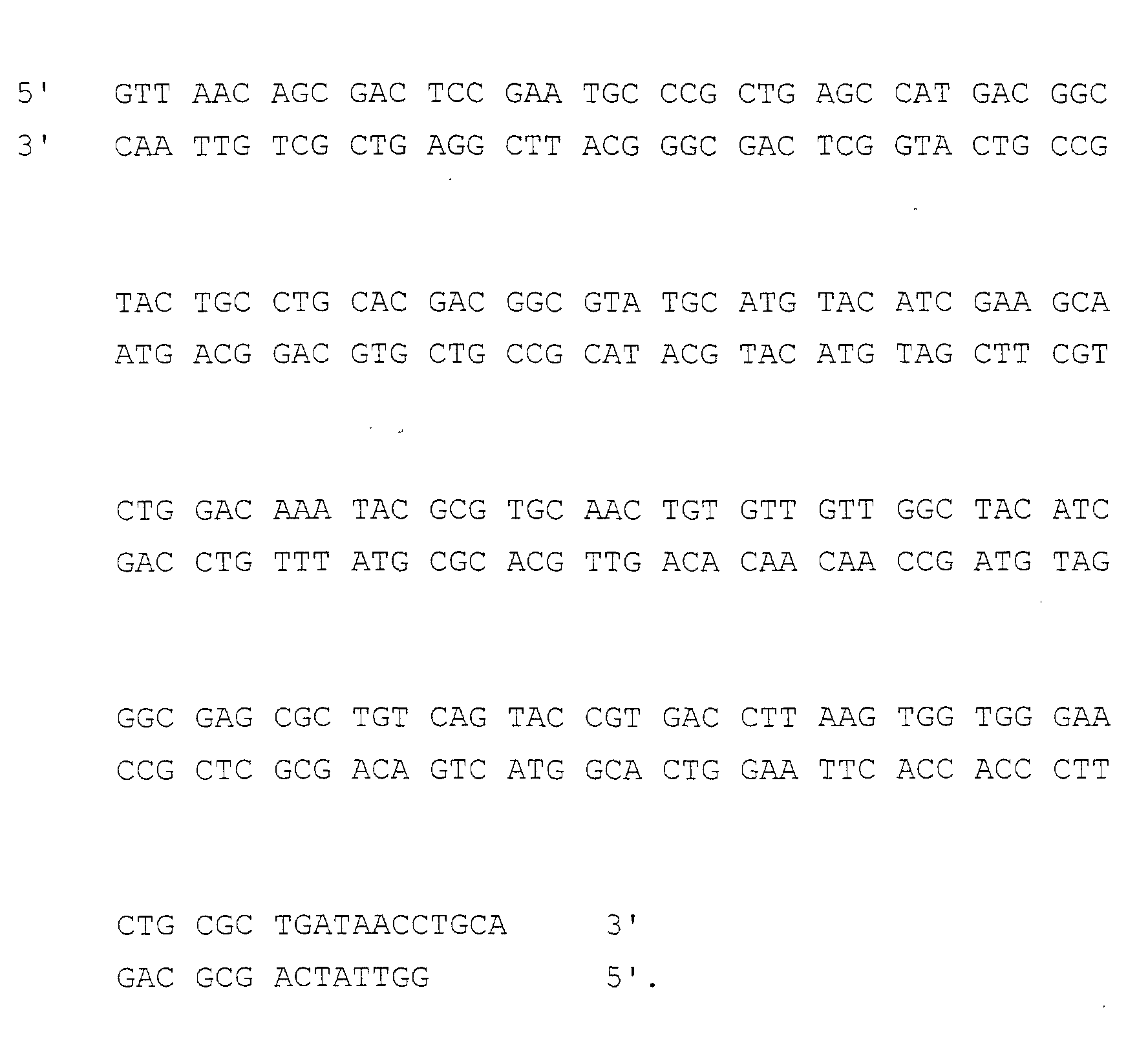
- Fig. 1 shows positions of restriction sites: HpaI at the 5' terminal, PstI at the 3' terminal, and Bpu1102I, NsiI, HluI, Eco47III and AflII.
- oligonucleotides consisted of 29mer to 41mer were synthesized in a separate manner. They are disclosed in Fig. 2A: in this connection, C1(30mer), C2 (35mer), C3(29mer), C4(39mer) and C5(41mer) oligonucleotides have the same sequence as that of mRNA transcribed from the corresponding hEGF sequence; and, N1(29mer), N2(38mer), N3(29mer), N4(36mer) and N5 (38mer) oligonucleotides were complementary to the C5, C4, C3, C2 and C1 oligonucleotides, respectively[ see : Fig. 2B]. Each oligonucleotide was synthesized by automatic Nucleotide Synthesizer(Pharmacia LKB Biotechnology, Uppsala, Sweden).
- the solution containing oligonucleotide thus separated was subjected in strong ammonia water at 50°C for 12hrs to remove protecting group; and concentration under vacuum condition was followed to the volume of 0.5ml, together with removal of gas.
- oligonucleotide solution thus concentrated was applied on SEP-PAK cartridge(Waters Inc., MA, USA), and elution was made with acetonitrile/ triethylamine solution to obtain partially purified oligonucleotide. Then, electrophoresis on 15% denatured polyacrylamide gel(TE-boric acid (pH 8.3) with 8M urea) was carried out and location of oligonucleotide in the gel was determined by the irradiation of ultraviolet rays.
- oligonucleotide was eletroeluted and salts were removed on SEP-PAK cartridge connected with injector by eluting with acetonitrile/triethlamine solution.
- the oligonucleotides thus isolated were labeled with ⁇ - 32 P-ATP by employing T 4 polynucleotide kinase(New England Biolabs., #201S, USA) and sequenced in accordance with Maxam & Gilbert's method[see: Maxam, A. M. & Gilbert, W., Proc. Natl. Acad. Sci., USA, 74:560-564 (1977)].
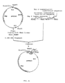
- pUC18 known in the art(see: Norrander, J. et al, Gene, 26:101(1985)) was digested with SmaI and PstI(New England Biolabs., #141S and #140S, MA, USA; all the restriction enzymes and linkers described hereinafter were purchased from New England Biolabs., MA, USA), and ligated with the hEGF gene. Ligation was easily performed, based on the blunt 5' terminus and the sticky 3' terminus in the hEGF gene as designed. The recombinant vector thus obtained was named pUE118[ see : Fig. 3].
- pUE1181 was digested with HpaI, PstI, Bpu1102I, NsiI, MluI, Eco47III and AflII, respectively, and electrophoresis of the gene fragments produced was carried out on 1% agarose gel. As disclosed in Fig. 4, the insertion of hEGF gene was carried out in an accurate manner and it was confirmed that single restriction site exists for each restriction enzyme. In Fig.
- Lane 1 is 1kb ladder DNA (BRL, USA); Lanes 2 and 3 are pUE118 digested with HpaI and PstI, respectively; Lane 4 is pUE118 digested doubly with HpaI and PstI; and, Lanes 5-9 are pUE118 digested with Bpu1102I, NsiI, MluI, Eco47III and AflII, respectively.
- Competent E. coli JM109 was transformed with the pUE118; and E. coli JM109 thus transformed was deposited with the Korean Culture Center of Microorganisms(KCCM) in the name of Escherichia coli(DW/BT-2040) on April 9, 1993 as deposition No. KCCM 10026.
- KCCM Korean Culture Center of Microorganisms
- Oligonucleotide comprising Omp A leader, universal translation termination and trp A transcription termination sequence designed to have several restriction sites as disclosed in Fig. 5.
- the oligonucleotide comprises restriction sites of BamHI at 5' terminal and XbaI at 3' terminal, and NaeI and PstI between Omp A leader and universal translation termination sequence to assure that N-terminal amino acid sequence is exactly the same as original protein free of additional amino acid sequence.
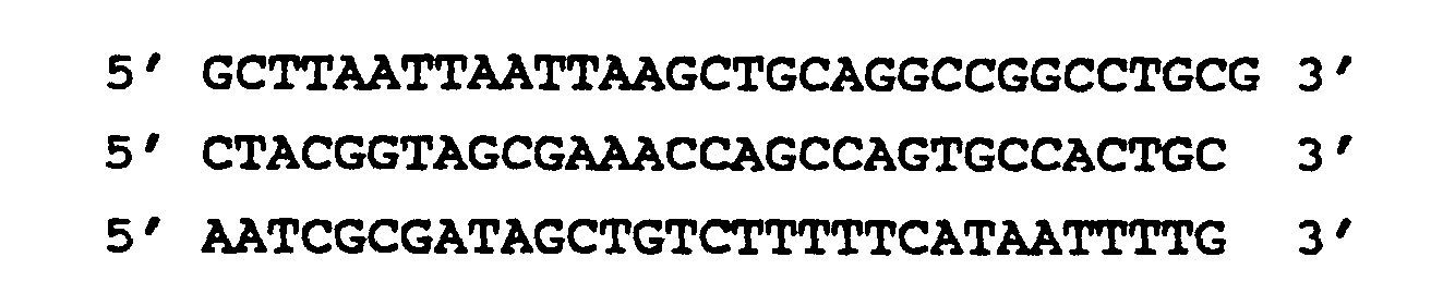
- Omp A leader-universal translation termination-Trp A transcription termination sequence was synthesized as following 8 oligonucleotides of 31mer or 32mer in a separate manner:
- oligonucleotides were synthesized with automatic Nucleotide Synthesizer(Pharmacia LKB Biotechnology, Uppsala, Sweden) in accordance with solid-phase phosphate triester method[ see : Narang, S. A., Synthesis and Applications of DNA and RNA, Academic Press, 1987].
- the synthesized oligonucleotides were separated from the silica matrix; removal of protecting group and concentration were followed; isolation of oligonucleotides, labeling with ⁇ - 32 P-ATP, denaturation and renaturation, and ligation of oligonucleotides were performed.
- Plasmid pDR540 (Pharmacia LKB Biotechnology, #27-4928-01, Upssala, Sweden) was digested with PvuII(#1032), ligated with XbaI linker and digested with XbaI(#145S) and BamHI (#136S) to obtain 2.4kb DNA fragment. Said fragment was isolated from the gel after electrophoresis of digested fragments by employing Geneclean II DNA elution kit(BIO 101 Inc., CA, USA), and ligated with "Omp A leader-universal translation termination-Trp A transcription termination sequence" prepared in Example 6 by T 4 DNA ligase(#202S). E.
- coli JM101 was transformed with the ligated DNA according to Hanahan's method[see: DNA Cloning Vol.I; A Practical Approach, IRL Press, 109-135(1985)]. From the transformants, recombinant plasmid was isolated and sequenced in accordance with Maxam and Gilbert's method to screen recombinant plasmid containing "OmpA leader-universal translation termination-Trp A transcription termination sequence". The recombinant plasmid thus selected was named pDT420 [ see : Fig. 6].
- Example 8 Construction of expression vector pDE135 and determination of hEGF expression
- Double digestion on plasmid pDT420 was made with NaeI (#190S) and PstI(# 140S) to cleave the sequence between Omp A leader and universal translation termination sequence.
- plasmid pUE118 prepared in Example 5 was digested with HpaI(#105S) and PstI to obtain 0.17kb hEGF gene.
- hEGF gene thus obtained was ligated with pDT420 cleaved with NaeI and PstI, and the ligated DNA was transformed into E. coli JM101.
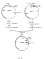
- recombinant plasmid was isolated by alkaline lysis method[Sambrook et al., Molecular Cloning, a laboratory manual, 2nd Ed., Cold Spring Harbor, 1989]. Referring to PstI and HindIII restriction enzyme map, recombinant plasmid containing hEGF gene between Omp A leader and universal translation termination sequence was selected and named pDE135[ see : Fig. 7].
- E. coli JM101 was transformed with pDE135, and the transformant was cultured in LB media(Luria-Bertani; Molecular Cloning/a laboratory manual, 2nd Ed., CSH, 1989). Then, expression of hEGF was investigated by SDS-PAGE and Western blot analysis. Expression of hEGF was determined by receptor binding assay(see: M. W. Rieman, Peptides, 8:877-885(1987)) employing A431 cell line(ATCC CRL 1555) and commercially available hEGF(Amersham, ARN 5100, UK) as a standard. hEGF was expressed with a yield of 10mg/L after 30hrs cultivation.
- the inventors assumed that the grounds of low efficiency of expression were: the biased transcriptional direction between ampicillin resistant marker and hEGF gene; competition of protein production; and, instability of the expression vector in transformant. Accordingly, the inventors determined to develop an improved hEGF expression cassette to solve the above confronted problems by employing tetracycline resistant marker whose product was not secreted out of cytosol, instead of ampicillin resistant marker.
- Plasmid pBR322 (Bolivar, F. et al., Gene, 2:95-113 (1977)) was digested with AvaI(#152S), and manipulated with Klenow's fragment(#210S) to have blunt-end. Then, the resultant was digested with EcoRI and followed by electrophoresis on 0.8% agarose gel, and the 1.4kb DNA fragment was obtained from gel by employing Geneclean II DNA elution kit. On the other hand, pUC19(Yanish-Perron, C., et al., Gene, 33:103-119(1985)) was digested with DraI(#129S) and EcoRI to give 1.2kb DNA fragment.
- the 1.2kb DNA fragment has replication origin of pUC19 to maintain high efficiency of replication and multiple cloning site to ease gene manipulation.
- 1.4kb and 1.2kb DNA fragments thus obtained were ligated with T 4 DNA ligase, and E. coli JM101 was transformed with the ligated DNA fragment in accordance with Hanahan's method. From the transformants, recombinant plasmid was isolated by alkaline lysis. Referring to EcoRI and AflIII restriction enzyme map, recombinant plasmid containing replication origin of pUC19, multiple cloning site and tetracycline resistant marker was selected and named pTC108[ see : Fig. 8].
- Plasmid pSC101 (Cohen and Chang, Proc. Natl. Acad. Sci., USA, 70:1293-1297(1973); KCTC 11251) was digested with AvaI(#152S) and electrophoresed on 0.8% agarose gel; and, 3.3kb DNA fragment was obtained therefrom by employing Geneclean II DNA elution kit. DNA fragment thus obtained was manipulated with Klenow's fragment to produce blunt-end, ligated with EcoRI linker (#1020) and digested with EcoRI.
- the 3.3kb DNA fragment containing sticky end of EcoRI site was digested with HincII(#103S) and electrophoresed on 0.8% agarose gel. 0.37kb DNA fragment containing par site was obtained from electrophoresed gel by employing Geneclean II DNA elution kit.
- the obtained DNA fragment was ligated to pTC108 digested with EcoRI and SmaI(#141S) by the aid of T, DNA ligase, and E. coli JM101 was transformed with the ligated DNA fragment in accordance with Hanahan's method. From the transformants, recombinant plasmid was isolated by alkaline lysis. Referring to EcoRI and PstI restriction enzyme map, recombinant plasmid containing par site was selected and named pTC226[ see : Fig 9].
- the inventors constructed plasmid vector which is stable in E. coli and is of high efficiency of hEGF expression, by inserting hEGF gene expression cassette of the pDE135 into pTC226 to have same transcriptional direction. Plasmid pTC226 was digested with AflIII, manipulated with Klenow fragment to produce blunt-end, digested with XbaI(#145S) and electrophoresed on 0.8% agarose gel; and, 2.5kb DNA fragment was obtained from electrophoresed gel by employing Geneclean II DNA elution kit.
- plasmid pDE135 was digested with HindIII(#104S), blunt-ended with Klenow fragment, digested with XbaI and electrophoresed on 1% agarose gel; and, 0.45kb DNA fragment was obtained from electrophoresed gel by employing Geneclean II DNA elution kit.
- Each isolated DNA fragment i.e., 2.5kb and 0.45kb DNA fragments, was ligated with T 4 DNA ligase, and E. coli JM101 was transformed with the ligated DNA fragment in accordance with Hanahan's method. From the transformant, recombinant plasmid was isolated by alkaline lysis.
- pTE105 a recombinant plasmid containing tetracycline resistant marker, par site, and Omp A leader and hEGF gene was selected and named pTE105[ see : Fig. 10].
- E. coli JM101 transformed with pTE105 was named DW/BT-2042, and deposited with the Korean Culture Collection of Microorganisms (KCCM) on April 9, 1993 as deposition No. KCCM 10027.
- Transformant E. coli DW/BT-2042(KCCM 10027) was cultured in LB media for 5hrs with the addition of isopropyl B-D-thio-galactoside(IPTG, Sigma 1-6758) to the final concentration of 1mM; and, cultures for 19hrs and 25hrs were collected and centrifuged respectively.
- the supernatants were electrophoresed on 15% polyacrylamide gel( see ; H. Schagger et al., Anal. Biochem., 166:368-379(1987)) employing commercially available hEGF(Amersham, ARN 5100, UK) as a standard.
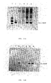
- Fig. 11A showed that molecular size of hEGF expressed is determined as 6,000 dalton.
- Figs. 11A and 11B Lane 1 is molecular weight marker(Sigma, #MW-SDS-17S); Lane 2 is the standard hEGF; Lane 3 is the culture for 24hrs without addition of IPTG; Lane 4 is the culture for 30hrs without addition of IPTG; Lane 5 is the culture for 24hrs with addition of IPTG ; and, Lane 6 is the culture for 30hrs with addition of IPTG.
- the amount of the hEGF in cultures for 24hrs and 30hrs were analyzed quantitatively by hEGF receptor binding assay; and disclosed in Table 1.
- Transformant E. coli DW/BT-2042(KCCM 10027) was cultured in 4ml of LB media containing 0.5% glucose and 12.5 ⁇ g/ml of tetracycline at 37°C for llhrs; and, 400 ⁇ l of the culture was inoculated on 100ml of the same media and incubated for llhrs to obtain seed culture.
- 80ml of seed culture was inoculated to 2L of media containing bactotrypton 10g, yeast extract 20g, KH 2 PO 4 3g, Na 2 HPO 4 ⁇ 8H 2 O 4g, (MH 4 ) 2 HPO 4 2.5g, CaCl 2 ⁇ 2H 2 O 0.01g, Sigma antifoam A 1ml, glucose 5g and tetracycline 5mg per 1L, and incubated at 30°C. After 4hrs incubation, IPTG was added to the final concentration of 1mM for induction of hEGF and further incubation was carried out for 26hrs.
- the amount of expressed hEGF was determined by receptor binding assay employing A431 cell line(ATCC CRL 1555), which is modified method of Rieman's[ see : Rieman, M.W., Peptides, 8:877-885(1987)] and DiAugustine's[ see : DiAugustine, R. P., J. Biol. Chem., 260:2807-2811(1985)].
- the A431 cells were mixed with DMEM medium containing calf serum, inoculated on Costa 24well cell culture plate to 4 x 10 5 cells/well and incubated under 5% CO 2 -atmosphere at 37°C for 6 or 7 days with the change of new medium every other day.
- each well was rinsed with receptor binding buffer and bound cells thereon were separated from the well by the incubation with 250 ⁇ l of cell lysis solution consisting of 0.1N NaOH and 1% SDS. Radioactivity of 125 I-EGF bound cells were determined with the aid of ⁇ -scintillation counter (Packard Cobra II, USA).
- Example 13 2L of culture prepared in Example 13 was centrifuged (SorvallTM RC 28S, USA) at 8,000rpm for 30min. The supernatant was loaded on Amberchrom CG71(Tosohass Corp., USA) column(2.5 x 40cm) preequilibrated with 20mM Tris buffer(pH 8.0). Washing was made with 1L of the same buffer; and step elution was carried out with 500ml of 20mM Tris buffer(pH 8.0) containing 40% acetonitrile. Flow rate was controlled at 60cm/hr in the course of loading, washing and elution. The eluted hEGF fraction was stored at 4°C.
- hEGF fraction obtained as above was loaded on Q-Sepharose FF(Pharmacia, USA) column(2.5 x 40cm) preequilibrated with 20mM Tris buffer(pH 8.0) and washed with 500ml of the same buffer. Elution was made with linear gradient of OM to 0.5M NaCl in 20mM Tris buffer(pH 8.0). In this connection, flow rate was controlled at 55cm/hr during loading, washing and elution. 316.4mg of hEGF was obtained with 80 to 85% of purity.
- the hEGF fraction obtained as above was adjusted to pH 6.5 with 20% phosphate solution, and loaded on C 18 reverse phase column(Waters Delta Prep 4000, USA, 8 x 100mm) and followed by second C 18 reverse phase column(8 x 100mm).
- the C 18 reverse phase columns employed were preequilibrated with 10mM phosphate buffer(pH 6.5); and, flow rate was controlled at 4ml/min.
- the elution of hEGF was carried out with 10mM phosphate buffer(pH 6.5, buffer A) and 10mM phosphate buffer containing 70% acetonitrile(buffer B). 98% of purity was obtained at the retention time of 29min.
- Table 2 illustrates HPLC chromatographic conditions for hEGF purification.
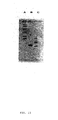
- Fig. 12 The homogeneity of purified hEGF in process of purification was determined by SDS-PAGE(Schagger. H. et al., Anal. Biochem., 166:368-379(1987))[ see : Fig. 12].
- Lane A is supernatant obtained by centrifugation of culture;
- Lane B is proteins isolated by Amberchrom CG71 chromatography;
- Lane C is proteins isolated by Q-Sepharose FF anion exchange chromatography; and, Lane D is purified hEGF by C 18 reverse phase HPLC.
- the purification step of hEGF is summarized in Table 3. Purification step Total volume (ml) Total hEGF (mg) Total protein (mg) Yield (%) Specific activity (mg hEGF/ mg protein) Culture 2,000 582.1 5,490 100 0.106 Amberchrom CG 71 350 436.5 997 74.9 0.438 Q-Sepharose FF 300 316.4 414 54.3 0.764 C 18 HPLC 240 282.3 285 48.4 0.990
- Fig. 14 The purity of purified hEGF was analysed in HPLC and represented in Fig. 14. As clearly disclosed in Fig. 14, it was determined that: the purity of hEGF was over 98% and degradation of C-terminal and oxidation of methionine had not occured.
- hEGF purification process of the invention was determined to be very efficient, while reducing contamination of other celluar protein and endotoxin.
Landscapes
- Health & Medical Sciences (AREA)
- Genetics & Genomics (AREA)
- Life Sciences & Earth Sciences (AREA)
- Chemical & Material Sciences (AREA)
- Organic Chemistry (AREA)
- Engineering & Computer Science (AREA)
- Zoology (AREA)
- General Engineering & Computer Science (AREA)
- Bioinformatics & Cheminformatics (AREA)
- Biophysics (AREA)
- Wood Science & Technology (AREA)
- Molecular Biology (AREA)
- General Health & Medical Sciences (AREA)
- Biochemistry (AREA)
- Gastroenterology & Hepatology (AREA)
- Biotechnology (AREA)
- Biomedical Technology (AREA)
- Toxicology (AREA)
- Physics & Mathematics (AREA)
- Plant Pathology (AREA)
- Proteomics, Peptides & Aminoacids (AREA)
- Microbiology (AREA)
- Medicinal Chemistry (AREA)
- Preparation Of Compounds By Using Micro-Organisms (AREA)
- Micro-Organisms Or Cultivation Processes Thereof (AREA)
Abstract
Description
- The present invention relates to a process for preparing human epidermal growth factor employing a recombinant expression vector therefor.
- It is known that human epidermal growth factor (hereinafter referred to as 'hEGF') is a polypeptide hormone consisting of 53 amino acids and 3 disulfide bridges [see: Cohen, S., J. Biol. Chem., 237:1555-1562 (1962); Savage, C.R., Jr. et al, J. Biol. Chem., 248: 7669-7672 (1973); Savage, C.R., Jr. et al, J. Biol. Chem., 247: 7612-7621 (1972)]. hEGF plays an important role in growth control in mammalian cells, inter alia epidermal and epithelial cells, on a molecular level [see: Sporn, M.B. et al, Nature (London), 313: 745-747 (1985); Sporn, M.B. et al, N. Engl. J. Med., 303: 878-880 (1980)] and the treatment of injury [see: Buckley, A. et al, Proc. Natl. Acad. Sci., USA, 82: 7340-7344 (1985)]. Further, it has been reported that hEGF can be applied in the treatment of a stomach ulcer, owing to the fact that it represses secretion of gastric acid into the stomach [see: Gregory, H., J. Cell Sci. Suppl., 3: 11-17 (1985)].
- Under the circumstances, studies on the mass production of hEGF has been actively carried out since Starkey et al reported the biochemical properties of hEGF purified from human urine [see: Starkey, R.H. et al, Science, 189: 800 (1975); Cohen, S. et al, Proc. Natl. Acad. Sci., USA, 72: 1317 (1975)]. Several researchers have successfully accomplished cloning of the hEGF gene by recombinant DNA technology [see: Smith, J. et al, Nucleic Acids Res., 10: 4467-4482 (1982); Urdea, M.S. et al, Proc. Natl. Acad. Sci., USA, 80: 7461-7465 (1983); Oka, T. et al, Proc. Natl. Acad. Sci., USA, 82: 7212-7216 (1985)]. The prior art, however, had not provided hEGF at an industrially useful level owing to its low activity and productivity.
- Accordingly, studies on elevation of activity and productivity in hEGF manufacture have been carried out, which were primarily concentrated on the preparation of a nucleotide sequence encoding hEGF efficient for its mass production and of an expression vector whose regulatory function is strengthened.
- In accordance with the present invention, the inventors synthesized a novel hEGF gene and a novel expression vector therefor which expresses hEFG in a massive amount and developed a process for preparing hEGF therefrom.
- The invention provides an expression vector for hEGF comprising a par site for stabilization in E.coli; a tetracycline resistance marker; and an expression cassette consisting of an Omp A leader sequence, an hEGF coding sequence, a translation termination sequence, and a trp A transcription termination sequence; wherein the expression cassette is under transcriptional regulation by the tac promoter. In the vector the hEGF sequence preferably comprises a restriction enzyme cleavage site for HpaI at its 5'-terminus, for PstI at its 3'-terminus, and for Bpu1102I, NsiI, MluI, Eco47III and AflII, wherein the nucleotide sequence of the coding sequence is:
-
- Figure 1
- depicts the sequence of the hEGF gene used in the invention;
- Figure 2A
- depicts 10 oligonucleotides synthesized;
- Figure 2B
- depicts assembly pattern of synthesized 10 oligonucleotides of Figure 2A;
- Figure 3
- depicts construction strategy of pUE118;
- Figure 4
- is a photograph showing agarose gel electrophoresis pattern of pUE118 carrying hEGF gene digested with restriction enzymes;
- Figure 5
- depicts the Omp A leader-universal translation termination-Trp A transcription termination sequence;
- Figure 6
- depicts construction strategy of pDT420;
- Figure 7
- depicts construction strategy of pDE135;
- Figure 8
- depicts construction strategy of pTC108;
- Figure 9
- depicts construction strategy of pTC226;
- Figure 10
- depicts construction strategy of pTE105;
- Figure 11A
- is a photograph showing SDS-PAGE pattern of hEGF expressed in E. coli JM101 harboring pTE105;
- Figure 11B
- is a photograph showing the Western blot analysis of hEGF expressed in E. coli JM101 harboring pTE105;
- Figure 12
- is a photograph showing SDS-PAGE pattern of hEGF expressed from E. coli JM101 harboring pTE105 in each purification step;
- Figure 13
- is a photograph showing SDS-PAGE pattern of purified hEGF from E. coli JM101 harboring pTE105;
- Figure 14
- is a chromatogram of purified hEGF by reverse phase HPLC; and,
- Figure 15
- is a photograph showing isoelectric focusing analysis of purified hEGF from E. coli JM101 harboring pTE105.
- The inventors synthesized a hEGF gene designed to maximise expression of the gene which satisfies the following requirements: first, hEGF expressed therefrom shall be the same as natural human EGF in light of amino acid sequence and protein structure; secondly, the nucleotide sequence shall be designed in consideration of the codons ubiquitous in E.coli; thirdly, the portion of secondary structure of mRNA transcribed therefrom shall be minimized; fourthly, many unique restriction sites shall be positioned as far as possible.
- In this connection, the hEGF gene was designed, based on the previously known hEGF sequence in the art [see: Gregory, H., Nature, 257: 325 (1975)]. First of all, the inventors synthesized 10 complementary oligonucleotides by using an automatic nucleotide synthesizer in accordance with the solid-phase phosphate triester method [see: Narang, S.A., Synthesis and Applications of DNA and RNA, Academic Press, 1987]. To obtain the full hEGF gene, the shot-gun ligation method was employed.
- The hEGF coding sequence used in the invention contains the following restriction sites: HpaI at the 5'terminus, PstI at the 3' terminus and Bpu1102I, NsiI, MluI, Eco47III and AflII within its internal sequence. Introduction of the hEGF gene into pUC18, a starting vector used in the invention, produces the above 7 specific sites which exist as unique restriction sites in the recombinant vector. The fact that said unique sites are cleaved by their specific restriction enzymes and positioned at a regular interval, allows practical availability of the gene for mutagenesis induction, which assures high activity and stability of hEGF. Furthermore, the HpaI restriction site at the 5' terminus allows production of intact hEGF free of fusion protein from the expression vector.
- The hEGF gene is inserted into pUC18 digested with SmaI and PstI, and the plasmid thus obtained is named pUE118. E.coli JM109 is transformed with pUE118 according to Hanahan's method [see: DNA Cloning Vol. I: A Practical Approach, IRL Press, 1985, pp 109-135], and this transformant is deposited with the Korean Culture Collection of Microorganisms (KCCM) located in the Department of Food Engineering, College of Eng., Yonsei University, Sodaemun-gu, Seoul 120-749, Korea on April 9, 1993 under the name of E. coli DW/BT-2040 (KCCM 10026).
- It is known that the regulation of protein expression corresponds to the growth patterns of microorganisms, and this is very important for mass protein production. In this connection, a tac promoter [see: de Boer et al, DNA, 2: 231-235 (1983); Amann et al, Gene, 25: 167-178 (1983)] is introduced to the expression vector of the invention. Since the tac promoter is continuously linked to two downstream ribosome binding sites [see: Shine and Dalgarno, Proc. Natl. Acad. Sci., USA, 71: 1342 (1974)], it initiates the translation of the hEGF gene efficiently.
- For the accurate and efficient expression and secretion of hEGF, the expression vector of the invention employs the following sequences: (1) an Omp A leader sequence [see: von Gabain, A. et al, Proc. Natl. Acad. Sci., USA, 80: 653-657 (1983)]; (2) a universal translation termination sequence [see: P. Singleton and D. Sainsbury, Dictionary of Microbiology and Molecular Biology, 2nd. Ed., Wiley, 383, 1987]; and, (3) a trp A transcription termination sequence [see: Christie, G. E. et al, Proc. Natl. Acad. Sci., USA, 78: 418 (1981)]. Further, the expression vector contains a par site for stability in E.coli [see: Austin and Abeles, J. Mol. Biol., 169: 373-387 (1983)].
- The "Omp A leader-universal translation termination-trp A transcription termination sequence" is designed and synthesized for insertion of an hEGF gene comprising a BamHI restriction site at the 5' terminus and an XbaI site at the 3'terminus, and Nael and PstI sites between the Omp A leader and the universal translation termination sequence.
- It is known that secretion of the expressed protein is decreased when an ampicillin-resistance marker which codes β-lactamase, a secretory protein, is used to screen for E. coli containing a vector comprising said marker, owing to the fact that the two proteins, i.e., the protein of interest and β-lactamase, compete in the course of secretion [see: A. Oka, et al, J. Mol. Biol., 147: 217 (1981)]. Therefore a tetracycline-resistant marker, which codes an intracellular protein, brings about high secretion of the expressed protein of interest by avoiding said competition, and is employed in the invention instead of the ampicillin-resistant marker. As a result, it is clearly guaranteed that the expression vector of the invention expresses and secretes hEGF in high yield, and is stable in E. coli.
- A commercially available plasmid pDR540 is digested with PvuII and then ligated with XbaI linker; and, double digestion with XbaI and BamHI is carried out. 2.4kb of DNA fragment thus obtained is ligated with the "Omp A leader-universal translation termination-Trp A transcription termination sequence"; and named pDT420.
- Double digestion of pDT420 with NaeI and PstI produces a cleavage between the Omp A leader and the universal translation termination sequence; and, pUE118 the carrying hEGF gene is digested with HpaI and PstI to obtain the hEGF gene. Then, the hEGF gene is ligated into pDT420 digested with NaeI and PstI, and the resultant vector is named pDE135.
- Plasmid pUC19 known in the art is digested with Oral and EcoRI to obtain a 1.2kb DNA fragment whose two ends are blunt-ended and cohesive for EcoRI, respectively; and, pBR322 is digested with AvaI and blunt-ended with Klenow's fragment, and digesteion with EcoRI is followed to obtain a 1.4kb DNA fragment. The above 1.4kb and 1.2kb DNA fragments are ligated with T4 DNA ligase, and the product is named pTC108. As a result, pTC108 comprises a tetracycline-resistant marker, a multiple cloning site and the replication origin of pUC19.
- A par site is introduced for stability of the expression vector in transformants and exact separation of plasmids after cell division. Plasmid pTC108 is digested with EcoRI and SmaI. On the other hand, pSC101 is digested with AvaI to obtain a 3.3kb DNA fragment, which is blunt-ended with Klenow's fragment and then ligated with EcoRI linker, followed by digestion with EcoRI. The 3.3kb DNA fragment containing EcoRI sticky ends is digested with HincII to obtain a 0.37kb DNA fragment containing a par site. This 0.37kb DNA fragment is ligated to pTC108 digested with EcoRI and SmaI by T4 DNA ligase, and the ligated plasmid is named pTC226.
- Plasmid pTC226 is digested with AflIII, blunt-ended with Klenow fragment and digested with XbaI in a serial manner to obtain a 2.5kb DNA fragment. On the other hand, pDE135 is digested with HindIII, blunt-ended with Klenow fragment and digested with XbaI in a serial manner to obtain a 0.45kb DNA fragment. The 2.5kb and 0.45kb DNA fragments are ligated with T4 DNA ligase, and the ligated plasmid is named pTE105.
- E. coli JM101 is transformed with pTE105, and the transformant is deposited with the Korean Culture Collection of Microorganisms(KCCM) located in the Department of Food Engineering, College of Eng., Yonsei University, Sodaemun-gu, Seoul 120-749, Korea on April 9, 1993 under the name of E. coli DW/BT-2042(KCCM 10027).
- Transformants E. coli DW/BT-2042 are grown in LB media and expression of hEGF therefrom is determined by 15% SDS-PAGE and Western blot analysis by employing commercially available hEGF(Amersham, ARN 5100, UK) as standard. The amount of expressed hEGF is determined by hEGF receptor binding analysis employing A431 cell line(ATCC CRL 1555). Culturing said transformant for 30hrs gave 343.5mg/L of hEGF, where most of the expressed hEGF was secreted out of cytosol.
- The present inventors isolated hEGF from the culture, while reducing contamination of other celluar proteins and endotoxins, by employing a series of chromatographic purification methods, i.e., Amberchrom CG71 chromatography, Q-Sepharose anion exchange chromagraphy and reverse phase C18 preparative HPLC. The purity of purified hEGF is determined by analytical HPLC in accordance with modified method of Hayashi et al's[see: Hayashi, T. et al., Anal. Sci., 3: 445-449(1987)].
- The present invention is further illustrated in the following examples, which should not be taken to limit the scope of the invention.
- The hEGF gene was firstly designed, based on the hEGF amino acid sequence and Grantham et al's study on the frequency of codon usage in E. coli[see: Grantham et al., Nucleic Acid Res., 9:243-274(1981)]. Then, sequence which does not cause formation of secondary structure of mRNA transcribed therefrom was selected, while examining whether the gene contains codons ubiquitous in E. coli and causes formation of secondary structure of mRNA or not, by employing PC-FOLD(Version 2.0) program[see: Turner, D. et al, Cold Spring Harbor Symp. Quant. Biol., 52:123(1987)]. The hEGF sequence thus designed carries the following two restriction sites to guarantee accurate insertion, isolation and manipulation of hEGF gene: HpaI at the 5' terminal; PstI at the down stream of translational termination codon. The hEGF sequence futher comprises many unique restriction sites at a regular basis as follows:

- The hEGF sequence designed for use in the invention is disclosed in Fig. 1, which shows positions of restriction sites: HpaI at the 5' terminal, PstI at the 3' terminal, and Bpu1102I, NsiI, HluI, Eco47III and AflII.
- The hEGF gene designed in Example 1 was chemically synthesized. First of all, 10 oligonucleotides consisted of 29mer to 41mer were synthesized in a separate manner. They are disclosed in Fig. 2A: in this connection, C1(30mer), C2 (35mer), C3(29mer), C4(39mer) and C5(41mer) oligonucleotides have the same sequence as that of mRNA transcribed from the corresponding hEGF sequence; and, N1(29mer), N2(38mer), N3(29mer), N4(36mer) and N5 (38mer) oligonucleotides were complementary to the C5, C4, C3, C2 and C1 oligonucleotides, respectively[see: Fig. 2B]. Each oligonucleotide was synthesized by automatic Nucleotide Synthesizer(Pharmacia LKB Biotechnology, Uppsala, Sweden).
- Oligonucleotides thus synthesized were separated on the silica matrix, through treatment with TTD solution (thiophenol/triethylamine/dioxane = 1/2/2, v/v) and washing with methanol, ethanol and strong ammonia water in a serial manner. The solution containing oligonucleotide thus separated was subjected in strong ammonia water at 50°C for 12hrs to remove protecting group; and concentration under vacuum condition was followed to the volume of 0.5ml, together with removal of gas. The oligonucleotide solution thus concentrated was applied on SEP-PAK cartridge(Waters Inc., MA, USA), and elution was made with acetonitrile/ triethylamine solution to obtain partially purified oligonucleotide. Then, electrophoresis on 15% denatured polyacrylamide gel(TE-boric acid (pH 8.3) with 8M urea) was carried out and location of oligonucleotide in the gel was determined by the irradiation of ultraviolet rays. The gel corresponding to oligonucleotide band was cut out, oligonucleotide was eletroeluted and salts were removed on SEP-PAK cartridge connected with injector by eluting with acetonitrile/triethlamine solution. The oligonucleotides thus isolated were labeled with γ-32P-ATP by employing T4 polynucleotide kinase(New England Biolabs., #201S, USA) and sequenced in accordance with Maxam & Gilbert's method[see: Maxam, A. M. & Gilbert, W., Proc. Natl. Acad. Sci., USA, 74:560-564 (1977)].
- 100pmole of each oligonucleotide prepared in Example 3, where all the oligonucleotides except for two oligonucleotides (C1 and N1) were phosphorylated at 5' terminal, were placed in Eppendorf tube with the addition of 40µl 0.1M Tris-HCl(pH 7.5). Then, incubation was carried out at 100°C for 3min to denature oligonucleotides, and renaturation was followed by lowering temperature slowly. To the resultant were added 10units of T4 DNA ligase(New England Biolabs., #202S, USA) and ligation buffer solution, and incubated at 4°C for 12hr and at room temperature for 3hrs, respectively. Then, 7% polyacrylamide gel electrophoresis and autoradiography were followed to determine the ligated hEGF sequence of the invention.
- pUC18 known in the art(see: Norrander, J. et al, Gene, 26:101(1985)) was digested with SmaI and PstI(New England Biolabs., #141S and #140S, MA, USA; all the restriction enzymes and linkers described hereinafter were purchased from New England Biolabs., MA, USA), and ligated with the hEGF gene. Ligation was easily performed, based on the blunt 5' terminus and the sticky 3' terminus in the hEGF gene as designed. The recombinant vector thus obtained was named pUE118[see: Fig. 3].
- Then, pUE1181 was digested with HpaI, PstI, Bpu1102I, NsiI, MluI, Eco47III and AflII, respectively, and electrophoresis of the gene fragments produced was carried out on 1% agarose gel. As disclosed in Fig. 4, the insertion of hEGF gene was carried out in an accurate manner and it was confirmed that single restriction site exists for each restriction enzyme. In Fig. 4,
Lane 1 is 1kb ladder DNA (BRL, USA);Lanes 2 and 3 are pUE118 digested with HpaI and PstI, respectively; Lane 4 is pUE118 digested doubly with HpaI and PstI; and, Lanes 5-9 are pUE118 digested with Bpu1102I, NsiI, MluI, Eco47III and AflII, respectively. - Competent E. coli JM109 was transformed with the pUE118; and E. coli JM109 thus transformed was deposited with the Korean Culture Center of Microorganisms(KCCM) in the name of Escherichia coli(DW/BT-2040) on April 9, 1993 as deposition No. KCCM 10026.
- Oligonucleotide comprising Omp A leader, universal translation termination and trp A transcription termination sequence designed to have several restriction sites as disclosed in Fig. 5. The oligonucleotide comprises restriction sites of BamHI at 5' terminal and XbaI at 3' terminal, and NaeI and PstI between Omp A leader and universal translation termination sequence to assure that N-terminal amino acid sequence is exactly the same as original protein free of additional amino acid sequence.
- First of all, Omp A leader-universal translation termination-Trp A transcription termination sequence was synthesized as following 8 oligonucleotides of 31mer or 32mer in a separate manner:

- The oligonucleotides were synthesized with automatic Nucleotide Synthesizer(Pharmacia LKB Biotechnology, Uppsala, Sweden) in accordance with solid-phase phosphate triester method[see: Narang, S. A., Synthesis and Applications of DNA and RNA, Academic Press, 1987].
- In an analogy to Example 3, the synthesized oligonucleotides were separated from the silica matrix; removal of protecting group and concentration were followed; isolation of oligonucleotides, labeling with γ-32P-ATP, denaturation and renaturation, and ligation of oligonucleotides were performed.
- Plasmid pDR540(Pharmacia LKB Biotechnology, #27-4928-01, Upssala, Sweden) was digested with PvuII(#1032), ligated with XbaI linker and digested with XbaI(#145S) and BamHI (#136S) to obtain 2.4kb DNA fragment. Said fragment was isolated from the gel after electrophoresis of digested fragments by employing Geneclean II DNA elution kit(BIO 101 Inc., CA, USA), and ligated with "Omp A leader-universal translation termination-Trp A transcription termination sequence" prepared in Example 6 by T4 DNA ligase(#202S). E. coli JM101 was transformed with the ligated DNA according to Hanahan's method[see: DNA Cloning Vol.I; A Practical Approach, IRL Press, 109-135(1985)]. From the transformants, recombinant plasmid was isolated and sequenced in accordance with Maxam and Gilbert's method to screen recombinant plasmid containing "OmpA leader-universal translation termination-Trp A transcription termination sequence". The recombinant plasmid thus selected was named pDT420 [see: Fig. 6].
- Double digestion on plasmid pDT420 was made with NaeI (#190S) and PstI(# 140S) to cleave the sequence between Omp A leader and universal translation termination sequence. On the other hand, plasmid pUE118 prepared in Example 5 was digested with HpaI(#105S) and PstI to obtain 0.17kb hEGF gene. hEGF gene thus obtained was ligated with pDT420 cleaved with NaeI and PstI, and the ligated DNA was transformed into E. coli JM101. From the transformants, recombinant plasmid was isolated by alkaline lysis method[Sambrook et al., Molecular Cloning, a laboratory manual, 2nd Ed., Cold Spring Harbor, 1989]. Referring to PstI and HindIII restriction enzyme map, recombinant plasmid containing hEGF gene between Omp A leader and universal translation termination sequence was selected and named pDE135[see: Fig. 7].
- E. coli JM101 was transformed with pDE135, and the transformant was cultured in LB media(Luria-Bertani; Molecular Cloning/a laboratory manual, 2nd Ed., CSH, 1989). Then, expression of hEGF was investigated by SDS-PAGE and Western blot analysis. Expression of hEGF was determined by receptor binding assay(see: M. W. Rieman, Peptides, 8:877-885(1987)) employing A431 cell line(ATCC CRL 1555) and commercially available hEGF(Amersham, ARN 5100, UK) as a standard. hEGF was expressed with a yield of 10mg/L after 30hrs cultivation. Under the circumstance, the inventors assumed that the grounds of low efficiency of expression were: the biased transcriptional direction between ampicillin resistant marker and hEGF gene; competition of protein production; and, instability of the expression vector in transformant. Accordingly, the inventors determined to develop an improved hEGF expression cassette to solve the above confronted problems by employing tetracycline resistant marker whose product was not secreted out of cytosol, instead of ampicillin resistant marker.
- Plasmid pBR322(Bolivar, F. et al., Gene, 2:95-113 (1977)) was digested with AvaI(#152S), and manipulated with Klenow's fragment(#210S) to have blunt-end. Then, the resultant was digested with EcoRI and followed by electrophoresis on 0.8% agarose gel, and the 1.4kb DNA fragment was obtained from gel by employing Geneclean II DNA elution kit. On the other hand, pUC19(Yanish-Perron, C., et al., Gene, 33:103-119(1985)) was digested with DraI(#129S) and EcoRI to give 1.2kb DNA fragment. The 1.2kb DNA fragment has replication origin of pUC19 to maintain high efficiency of replication and multiple cloning site to ease gene manipulation. 1.4kb and 1.2kb DNA fragments thus obtained were ligated with T4 DNA ligase, and E. coli JM101 was transformed with the ligated DNA fragment in accordance with Hanahan's method. From the transformants, recombinant plasmid was isolated by alkaline lysis. Referring to EcoRI and AflIII restriction enzyme map, recombinant plasmid containing replication origin of pUC19, multiple cloning site and tetracycline resistant marker was selected and named pTC108[see: Fig. 8].
- The inventors introduced par site for the stability of expression vector in transformant and easy separation of plasmids after cell division. Plasmid pSC101(Cohen and Chang, Proc. Natl. Acad. Sci., USA, 70:1293-1297(1973); KCTC 11251) was digested with AvaI(#152S) and electrophoresed on 0.8% agarose gel; and, 3.3kb DNA fragment was obtained therefrom by employing Geneclean II DNA elution kit. DNA fragment thus obtained was manipulated with Klenow's fragment to produce blunt-end, ligated with EcoRI linker (#1020) and digested with EcoRI. The 3.3kb DNA fragment containing sticky end of EcoRI site was digested with HincII(#103S) and electrophoresed on 0.8% agarose gel. 0.37kb DNA fragment containing par site was obtained from electrophoresed gel by employing Geneclean II DNA elution kit. The obtained DNA fragment was ligated to pTC108 digested with EcoRI and SmaI(#141S) by the aid of T, DNA ligase, and E. coli JM101 was transformed with the ligated DNA fragment in accordance with Hanahan's method. From the transformants, recombinant plasmid was isolated by alkaline lysis. Referring to EcoRI and PstI restriction enzyme map, recombinant plasmid containing par site was selected and named pTC226[see: Fig 9].
- The inventors constructed plasmid vector which is stable in E. coli and is of high efficiency of hEGF expression, by inserting hEGF gene expression cassette of the pDE135 into pTC226 to have same transcriptional direction. Plasmid pTC226 was digested with AflIII, manipulated with Klenow fragment to produce blunt-end, digested with XbaI(#145S) and electrophoresed on 0.8% agarose gel; and, 2.5kb DNA fragment was obtained from electrophoresed gel by employing Geneclean II DNA elution kit. In the same manner, plasmid pDE135 was digested with HindIII(#104S), blunt-ended with Klenow fragment, digested with XbaI and electrophoresed on 1% agarose gel; and, 0.45kb DNA fragment was obtained from electrophoresed gel by employing Geneclean II DNA elution kit. Each isolated DNA fragment, i.e., 2.5kb and 0.45kb DNA fragments, was ligated with T4 DNA ligase, and E. coli JM101 was transformed with the ligated DNA fragment in accordance with Hanahan's method. From the transformant, recombinant plasmid was isolated by alkaline lysis. Referring to restriction EcoRI and BamHI enzyme map, a recombinant plasmid containing tetracycline resistant marker, par site, and Omp A leader and hEGF gene was selected and named pTE105[see: Fig. 10]. E. coli JM101 transformed with pTE105 was named DW/BT-2042, and deposited with the Korean Culture Collection of Microorganisms (KCCM) on April 9, 1993 as deposition No. KCCM 10027.
- Transformant E. coli DW/BT-2042(KCCM 10027) was cultured in LB media for 5hrs with the addition of isopropyl B-D-thio-galactoside(IPTG, Sigma 1-6758) to the final concentration of 1mM; and, cultures for 19hrs and 25hrs were collected and centrifuged respectively. The supernatants were electrophoresed on 15% polyacrylamide gel(see; H. Schagger et al., Anal. Biochem., 166:368-379(1987)) employing commercially available hEGF(Amersham, ARN 5100, UK) as a standard. Fig. 11A showed that molecular size of hEGF expressed is determined as 6,000 dalton. Western blot analysis(W. N. Burnett, Anal. Biochem., 112:195-203(1981)) was also carried out to confirm that the produced was hEGF[see: Fig. 11B]. In Figs. 11A and 11B,
Lane 1 is molecular weight marker(Sigma, #MW-SDS-17S); Lane 2 is the standard hEGF;Lane 3 is the culture for 24hrs without addition of IPTG; Lane 4 is the culture for 30hrs without addition of IPTG;Lane 5 is the culture for 24hrs with addition of IPTG ; and, Lane 6 is the culture for 30hrs with addition of IPTG. The amount of the hEGF in cultures for 24hrs and 30hrs were analyzed quantitatively by hEGF receptor binding assay; and disclosed in Table 1. - As clearly illustrated in Table 1, it was determined that: total hEGF amount of cultivation for 30hrs was 343.5mg/L and most of expressed hEGF was secreted out of cytosol.

- Transformant E. coli DW/BT-2042(KCCM 10027) was cultured in 4ml of LB media containing 0.5% glucose and 12.5µg/ml of tetracycline at 37°C for llhrs; and, 400µl of the culture was inoculated on 100ml of the same media and incubated for llhrs to obtain seed culture. 80ml of seed culture was inoculated to 2L of media containing bactotrypton 10g, yeast extract 20g, KH2PO4 3g, Na2HPO4·8H2O 4g, (MH4)2HPO4 2.5g, CaCl2·2H2O 0.01g, Sigma antifoam A 1ml, glucose 5g and tetracycline 5mg per 1L, and incubated at 30°C. After 4hrs incubation, IPTG was added to the final concentration of 1mM for induction of hEGF and further incubation was carried out for 26hrs.
- The amount of expressed hEGF was determined by receptor binding assay employing A431 cell line(ATCC CRL 1555), which is modified method of Rieman's[see: Rieman, M.W., Peptides, 8:877-885(1987)] and DiAugustine's[see: DiAugustine, R. P., J. Biol. Chem., 260:2807-2811(1985)]. The A431 cells were mixed with DMEM medium containing calf serum, inoculated on Costa 24well cell culture plate to 4 x 10⁵ cells/well and incubated under 5% CO2-atmosphere at 37°C for 6 or 7 days with the change of new medium every other day. Then, removal of medium and washing with saline phosphate buffer were followed, and cells were immobilized by treatment of 10% formaldehyde for 10min. After the removal of formaldehyde was carried out by washing with saline phosphate buffer, 250µl of receptor binding buffer consisting of 1% BSA, 0.2% sodium azide and saline phosphate buffer was added to every well. To the above, were added standard hEGF solution diluted to 0.01-20ng/20µl or hEGF solution to be determined and 125I-EGF(Amersham, IM196, UK) diluted to 30,000cpm/100µl in a serial manner, and incubation with shaking at 100rpm was followed for 2hrs. Then, each well was rinsed with receptor binding buffer and bound cells thereon were separated from the well by the incubation with 250µl of cell lysis solution consisting of 0.1N NaOH and 1% SDS. Radioactivity of 125I-EGF bound cells were determined with the aid of γ-scintillation counter (Packard Cobra II, USA).
- 2L of culture prepared in Example 13 was centrifuged (Sorvall™ RC 28S, USA) at 8,000rpm for 30min. The supernatant was loaded on Amberchrom CG71(Tosohass Corp., USA) column(2.5 x 40cm) preequilibrated with 20mM Tris buffer(pH 8.0). Washing was made with 1L of the same buffer; and step elution was carried out with 500ml of 20mM Tris buffer(pH 8.0) containing 40% acetonitrile. Flow rate was controlled at 60cm/hr in the course of loading, washing and elution. The eluted hEGF fraction was stored at 4°C.
- 350ml of hEGF fraction obtained as above was loaded on Q-Sepharose FF(Pharmacia, USA) column(2.5 x 40cm) preequilibrated with 20mM Tris buffer(pH 8.0) and washed with 500ml of the same buffer. Elution was made with linear gradient of OM to 0.5M NaCl in 20mM Tris buffer(pH 8.0). In this connection, flow rate was controlled at 55cm/hr during loading, washing and elution. 316.4mg of hEGF was obtained with 80 to 85% of purity.
- The hEGF fraction obtained as above was adjusted to pH 6.5 with 20% phosphate solution, and loaded on C18 reverse phase column(Waters Delta Prep 4000, USA, 8 x 100mm) and followed by second C18 reverse phase column(8 x 100mm). The C18 reverse phase columns employed were preequilibrated with 10mM phosphate buffer(pH 6.5); and, flow rate was controlled at 4ml/min. The elution of hEGF was carried out with 10mM phosphate buffer(pH 6.5, buffer A) and 10mM phosphate buffer containing 70% acetonitrile(buffer B). 98% of purity was obtained at the retention time of 29min. Table 2 illustrates HPLC chromatographic conditions for hEGF purification.
Retention time(min) Flow rate (ml/min) Buffer ratio (A/B, %) Gradient type 0 4 0 / 100 - 2 4 74 / 26 linear 20 4 72 / 28 linear 45 4 70 / 30 linear 70 4 66 / 44 linear 75 4 0 / 100 linear 85 4 0 / 100 linear - The homogeneity of purified hEGF in process of purification was determined by SDS-PAGE(Schagger. H. et al., Anal. Biochem., 166:368-379(1987))[see: Fig. 12]. In Fig. 12, Lane A is supernatant obtained by centrifugation of culture; Lane B is proteins isolated by Amberchrom CG71 chromatography; Lane C is proteins isolated by Q-Sepharose FF anion exchange chromatography; and, Lane D is purified hEGF by C18 reverse phase HPLC.
- The purification step of hEGF is summarized in Table 3.
Purification step Total volume (ml) Total hEGF (mg) Total protein (mg) Yield (%) Specific activity (mg hEGF/ mg protein) Culture 2,000 582.1 5,490 100 0.106 Amberchrom CG 71 350 436.5 997 74.9 0.438 Q-Sepharose FF 300 316.4 414 54.3 0.764 C18 HPLC 240 282.3 285 48.4 0.990 - 282.3mg of purified hEGF was obtained with the yield of 48.4%. Protein concentration was determined by Bradford method(Bradford, M., Anal. Biochem., 72:248(1976)) employing protein quantitative kit(Biorad, #500-0006, USA). The molecular weight of purified hEGF was determined as 6,000 dalton by 15% SDS-PAGE analysis[see: Fig. 13]. In Fig. 13, Lane A is low molecular size weight marker(Biorad, #161-0304); Lane B is purified hEGF; and, Lane C is peptide marker(Sigma, #MW-SDS-17S).
- The purity of purified hEGF was analysed in HPLC and represented in Fig. 14. As clearly disclosed in Fig. 14, it was determined that: the purity of hEGF was over 98% and degradation of C-terminal and oxidation of methionine had not occured.
- Finally, isoelectric focusing analysis was carried out for pI value determination in the pH range of 4 to 6 ampholite(see: Carfin, D.E., Methods Enzymol., 183:459-475 (1990)) and its result was shown in Fig. 15. In Fig. 15, Lanes A and D are low calibration isoelectric focusing analysis marker(Pharmacia, #17-0472-01, USA); Lanes B and C are 2µg of purified hEGF; and, Lane E is 4µg of purified hEGF. As well be seen in Fig. 15, it was determined that pI of the purified hEGF is 4.55, which is the same pI value of prior art hEGF.
- The purified hEGF was lyophilized and followed by Limulus Amebocyte Lysate test(Associates of Cape Code, USA). The result showed that the amount of endotoxin in hEGF was below 0.36EU per lmg, which was extremely low. Accordingly, hEGF purification process of the invention was determined to be very efficient, while reducing contamination of other celluar protein and endotoxin.
-
- (1) GENERAL INFORMATION:
- (i) APPLICANT:
- (A) NAME: DAEWOONG PHARMACEUTICAL CO., LTD
- (B) STREET: 223-23, Sangdaewon-Dong, Joongwon-Ku
- (C) CITY: Sungnam, Kyunggi-Do
- (D) STATE: not applicable
- (E) COUNTRY: Korea
- (F) POSTAL CODE (ZIP): 462-120
- (ii) TITLE OF INVENTION: A NOVEL GENE CODING HUMAN EPIDERMAL GROWTH FACTOR AND PROCESS FOR PREPARING THE SAME
- (iii) NUMBER OF SEQUENCES: 21
- (iv) COMPUTER READABLE FORM:
- (A) MEDIUM TYPE: Floppy disk
- (B) COMPUTER: IBM PC compatible
- (C) OPERATING SYSTEM: PC-DOS/MS-DOS
- (D) SOFTWARE: PatentIn Release #1.0, Version #1.30 (EPO)
- (v) CURRENT APPLICATION DATA:
APPLICATION NUMBER: EP 94913826.7 - (2) INFORMATION FOR SEQ ID NO: 1:
- (i) SEQUENCE CHARACTERISTICS:
- (A) LENGTH: 174 base pairs
- (B) TYPE: nucleic acid
- (C) STRANDEDNESS: double
- (D) TOPOLOGY: linear
- (ix) FEATURE:
- (A) NAME/KEY: CDS
- (B) LOCATION:4..162
- (xi) SEQUENCE DESCRIPTION: SEQ ID NO: 1:


- (2) INFORMATION FOR SEQ ID NO: 2:
- (i) SEQUENCE CHARACTERISTICS:
- (A) LENGTH: 53 amino acids
- (B) TYPE: amino acid
- (D) TOPOLOGY: linear
- (ii) MOLECULE TYPE: protein
- (xi) SEQUENCE DESCRIPTION: SEQ ID NO: 2:
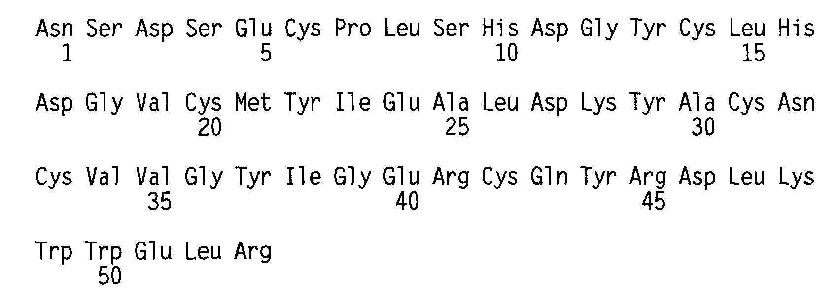
- (2) INFORMATION FOR SEQ ID NO: 3:
- (i) SEQUENCE CHARACTERISTICS:
- (A) LENGTH: 30 base pairs
- (B) TYPE: nucleic acid
- (C) STRANDEDNESS: single
- (D) TOPOLOGY: linear
- (xi) SEQUENCE DESCRIPTION: SEQ ID NO: 3:

- (2) INFORMATION FOR SEQ ID NO: 4:
- (i) SEQUENCE CHARACTERISTICS:
- (A) LENGTH: 35 base pairs
- (B) TYPE: nucleic acid
- (C) STRANDEDNESS: single
- (D) TOPOLOGY: linear
- (xi) SEQUENCE DESCRIPTION: SEQ ID NO: 4:

- (2) INFORMATION FOR SEQ ID NO: 5:
- (i) SEQUENCE CHARACTERISTICS:
- (A) LENGTH: 29 base pairs
- (B) TYPE: nucleic acid
- (C) STRANDEDNESS: single
- (D) TOPOLOGY: linear
- (xi) SEQUENCE DESCRIPTION: SEQ ID NO: 5:

- (2) INFORMATION FOR SEQ ID NO: 6:
- (i) SEQUENCE CHARACTERISTICS:
- (A) LENGTH: 39 base pairs
- (B) TYPE: nucleic acid
- (C) STRANDEDNESS: single
- (D) TOPOLOGY: linear
- (xi) SEQUENCE DESCRIPTION: SEQ ID NO: 6:

- (2) INFORMATION FOR SEQ ID NO: 7:
- (i) SEQUENCE CHARACTERISTICS:
- (A) LENGTH: 41 base pairs
- (B) TYPE: nucleic acid
- (C) STRANDEDNESS: single
- (D) TOPOLOGY: linear
- (xi) SEQUENCE DESCRIPTION: SEQ ID NO: 7:

- (2) INFORMATION FOR SEQ ID NO: 8:
- (i) SEQUENCE CHARACTERISTICS:
- (A) LENGTH: 29 base pairs
- (B) TYPE: nucleic acid
- (C) STRANDEDNESS: single
- (D) TOPOLOGY: linear
- (xi) SEQUENCE DESCRIPTION: SEQ ID NO: 8:

- (2) INFORMATION FOR SEQ ID NO: 9:
- (i) SEQUENCE CHARACTERISTICS:
- (A) LENGTH: 38 base pairs
- (B) TYPE: nucleic acid
- (C) STRANDEDNESS: single
- (D) TOPOLOGY: linear
- (xi) SEQUENCE DESCRIPTION: SEQ ID NO: 9:

- (2) INFORMATION FOR SEQ ID NO: 10:
- (i) SEQUENCE CHARACTERISTICS:
- (A) LENGTH: 29 base pairs
- (B) TYPE: nucleic acid
- (C) STRANDEDNESS: single
- (D) TOPOLOGY: linear
- (xi) SEQUENCE DESCRIPTION: SEQ ID NO: 10:

- (2) INFORMATION FOR SEQ ID NO: 11:
- (i) SEQUENCE CHARACTERISTICS:
- (A) LENGTH: 36 base pairs
- (B) TYPE: nucleic acid
- (C) STRANDEDNESS: single
- (D) TOPOLOGY: linear
- (xi) SEQUENCE DESCRIPTION: SEQ ID NO: 11:

- (2) INFORMATION FOR SEQ ID NO: 12:
- (i) SEQUENCE CHARACTERISTICS:
- (A) LENGTH: 38 base pairs
- (B) TYPE: nucleic acid
- (C) STRANDEDNESS: single
- (D) TOPOLOGY: linear
- (xi) SEQUENCE DESCRIPTION: SEQ ID NO: 12:

- (2) INFORMATION FOR SEQ ID NO: 13:
- (i) SEQUENCE CHARACTERISTICS:
- (A) LENGTH: 32 base pairs
- (B) TYPE: nucleic acid
- (C) STRANDEDNESS: single
- (D) TOPOLOGY: linear
- (xi) SEQUENCE DESCRIPTION: SEQ ID NO: 13:

- (2) INFORMATION FOR SEQ ID NO: 14:
- (i) SEQUENCE CHARACTERISTICS:
- (A) LENGTH: 30 base pairs
- (B) TYPE: nucleic acid
- (C) STRANDEDNESS: single
- (D) TOPOLOGY: linear
- (xi) SEQUENCE DESCRIPTION: SEQ ID NO: 14:

- (2) INFORMATION FOR SEQ ID NO: 15:
- (i) SEQUENCE CHARACTERISTICS:
- (A) LENGTH: 32 base pairs
- (B) TYPE: nucleic acid
- (C) STRANDEDNESS: single
- (D) TOPOLOGY: linear
- (xi) SEQUENCE DESCRIPTION: SEQ ID NO: 15:

- (2) INFORMATION FOR SEQ ID NO: 16:
- (i) SEQUENCE CHARACTERISTICS:
- (A) LENGTH: 32 base pairs
- (B) TYPE: nucleic acid
- (C) STRANDEDNESS: single
- (D) TOPOLOGY: linear
- (xi) SEQUENCE DESCRIPTION: SEQ ID NO: 16:

- (2) INFORMATION FOR SEQ ID NO: 17:
- (i) SEQUENCE CHARACTERISTICS:
- (A) LENGTH: 32 base pairs
- (B) TYPE: nucleic acid
- (C) STRANDEDNESS: single
- (D) TOPOLOGY: linear
- (xi) SEQUENCE DESCRIPTION: SEO ID NO: 17:

- (2) INFORMATION FOR SEQ ID NO: 18:
- (i) SEQUENCE CHARACTERISTICS:
- (A) LENGTH: 32 base pairs
- (B) TYPE: nucleic acid
- (C) STRANDEDNESS: single
- (D) TOPOLOGY: linear
- (xi) SEQUENCE DESCRIPTION: SEQ ID NO: 18:

- (2) INFORMATION FOR SEQ ID NO: 19:
- (i) SEQUENCE CHARACTERISTICS:
- (A) LENGTH: 31 base pairs
- (B) TYPE: nucleic acid
- (C) STRANDEDNESS: single
- (D) TOPOLOGY: linear
- (xi) SEQUENCE DESCRIPTION: SEQ ID NO: 19:

- (2) INFORMATION FOR SEQ ID NO: 20:
- (i) SEQUENCE CHARACTERISTICS:
- (A) LENGTH: 31 base pairs
- (B) TYPE: nucleic acid
- (C) STRANDEDNESS: single
- (D) TOPOLOGY: linear
- (xi) SEQUENCE DESCRIPTION: SEQ ID NO: 20:

- (2) INFORMATION FOR SEQ ID NO: 21:
- (i) SEQUENCE CHARACTERISTICS:
- (A) LENGTH: 126 base pairs
- (B) TYPE: nucleic acid
- (C) STRANDEDNESS: double
- (D) TOPOLOGY: linear
- (xi) SEQUENCE DESCRIPTION: SEQ ID NO: 21:

-
Claims (5)
- An expression vector for hEGF comprising a par site for stabilization in E.coli; a tetracycline resistance marker; and an expression cassette consisting of an Omp A leader sequence, an hEGF coding sequence, a translation termination sequence, and a trp A transcription termination sequence; wherein the expression cassette is under transcriptional regulation by the tac promoter.
- A vector according to claim 1 wherein the hEGF coding sequence comprises a restriction enzyme cleavage site for HpaI at its 5'-terminus, for PstI at its 3'-terminus, and for Bpul 1021, NsiI, MluI, Eco47III and AflII, and wherein the nucleotide sequence of the coding sequence is:
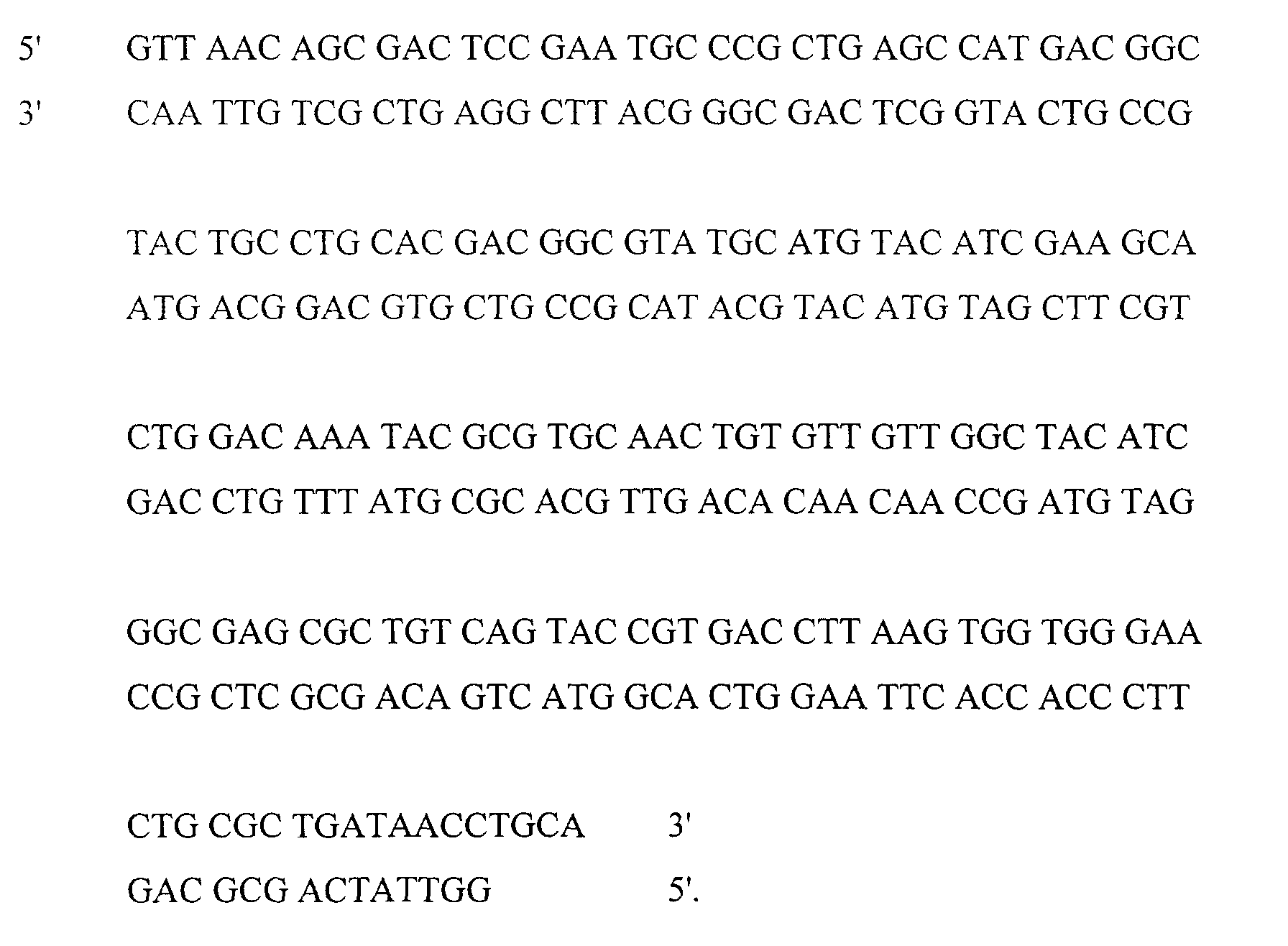
- A vector according to claim 2 which is pTE105 as shown in Figure 10.
- E.coli JM101 (KCCM 10027) transformed with an expression vector of claim 3.
- A process for preparing hEGF, which comprises culturing E.coli JM101 (KCCM 10027) according to claim 4.
Applications Claiming Priority (7)
| Application Number | Priority Date | Filing Date | Title |
|---|---|---|---|
| KR9306978 | 1993-04-26 | ||
| KR1019930006978A KR960010951B1 (en) | 1993-04-26 | 1993-04-26 | New human epidermal growth factor gene |
| KR9306980 | 1993-04-26 | ||
| KR1019930006979A KR960006121B1 (en) | 1993-04-26 | 1993-04-26 | Human epidermal growth factor (hEGF) expression vector and method for producing hEGF using the same |
| KR1019930006980A KR970001508B1 (en) | 1993-04-26 | 1993-04-26 | Purification method of recombinant human epithelial cell growth factor |
| KR9306979 | 1993-04-26 | ||
| PCT/KR1994/000036 WO1994025592A1 (en) | 1993-04-26 | 1994-04-25 | A novel gene coding human epidermal growth factor and process for preparing the same |
Publications (2)
| Publication Number | Publication Date |
|---|---|
| EP0652954A1 EP0652954A1 (en) | 1995-05-17 |
| EP0652954B1 true EP0652954B1 (en) | 2001-06-20 |
Family
ID=27348946
Family Applications (1)
| Application Number | Title | Priority Date | Filing Date |
|---|---|---|---|
| EP94913826A Expired - Lifetime EP0652954B1 (en) | 1993-04-26 | 1994-04-25 | A novel gene vector coding human epidermal growth factor and process for preparing the same |
Country Status (6)
| Country | Link |
|---|---|
| US (1) | US5652120A (en) |
| EP (1) | EP0652954B1 (en) |
| JP (1) | JP2609515B2 (en) |
| AU (1) | AU6582594A (en) |
| DE (1) | DE69427515T2 (en) |
| WO (1) | WO1994025592A1 (en) |
Families Citing this family (7)
| Publication number | Priority date | Publication date | Assignee | Title |
|---|---|---|---|---|
| RU2150501C1 (en) * | 1999-02-17 | 2000-06-10 | Эльдаров Михаил Анатольевич | Strain of yeast saccharomyces cerevisiae vkm cr-349d as producer of human epidermal growth factor |
| KR100447530B1 (en) * | 2001-08-14 | 2004-09-08 | 한국과학기술원 | A Method for Extracellular Production of Target Proteins Employing OmpF in E. coli |
| CA2427190A1 (en) * | 2002-04-30 | 2003-10-30 | Alberta Research Council Inc. | Production of recombinant epidermal growth factor in plants |
| TW200927161A (en) * | 2007-11-15 | 2009-07-01 | Posh Cosmeceuticals Inc | Composition containing granulocyte colony stimulating factor (GCSF) for hair growth |
| CN103484471B (en) * | 2013-09-30 | 2015-07-08 | 王悦 | HEGF (human epidermal growth factor) nucleotide sequence and colibacillus expression vector |
| KR102013788B1 (en) * | 2016-11-04 | 2019-08-23 | 한국과학기술연구원 | A vector for expressing hEGF or PTD-hEGF and microalgae transformed with the same |
| KR102091170B1 (en) * | 2018-04-20 | 2020-03-19 | 티앤에이치바이오(주) | Manufacture method of epidermal growth factor |
Family Cites Families (19)
| Publication number | Priority date | Publication date | Assignee | Title |
|---|---|---|---|---|
| DK339781A (en) * | 1980-08-05 | 1982-02-06 | Searle & Co | SYNTHETIC GEN |
| US5096825A (en) * | 1983-01-12 | 1992-03-17 | Chiron Corporation | Gene for human epidermal growth factor and synthesis and expression thereof |
| DE3382547D1 (en) * | 1983-01-12 | 1992-05-27 | Chiron Corp | SECRETORIC EXPRESSION IN EUKARYOTS. |
| JPS6028994A (en) * | 1983-07-08 | 1985-02-14 | Wakunaga Seiyaku Kk | (21-leucine) human urogastrone, corresponding gene, corresponding recombinant plasmid, transformed cell and their preparation |
| JPS60130393A (en) * | 1983-12-19 | 1985-07-11 | Sumitomo Chem Co Ltd | Human urogastrone gene, corresponding recombined plasmid, and recombinant |
| JP2554459B2 (en) * | 1984-07-02 | 1996-11-13 | アース製薬 株式会社 | β-urogastron gene, corresponding plasmid recombinant and corresponding transformant |
| JPS61291598A (en) * | 1985-06-18 | 1986-12-22 | Sagami Chem Res Center | Fish calcitonin derivative and production thereof |
| CA1263619A (en) * | 1984-10-09 | 1989-12-05 | Ryuji Marumoto | Dna, production and use thereof |
| JPH0642833B2 (en) * | 1984-10-09 | 1994-06-08 | 武田薬品工業株式会社 | Human epidermal growth factor gene |
| JPH0644866B2 (en) * | 1985-08-13 | 1994-06-15 | 武田薬品工業株式会社 | Transformant for producing human epidermal growth factor |
| US4935350A (en) * | 1985-11-18 | 1990-06-19 | Amgen | Materials and methods for controlling plasmid copy number and stability |
| US4743679A (en) * | 1986-02-24 | 1988-05-10 | Creative Biomolecules, Inc. | Process for producing human epidermal growth factor and analogs thereof |
| US5447862A (en) * | 1987-02-04 | 1995-09-05 | Ciba-Geigy Corporation | Pectin lyase genes of aspergillus niger |
| EP0335400B1 (en) * | 1988-03-30 | 1994-06-08 | Hitachi, Ltd. | Processes for production of human epidermal growth factor by genetic engineering |
| JPH01247098A (en) * | 1988-03-30 | 1989-10-02 | Hitachi Ltd | Biotechnological production of human epitheliocyte growth factor |
| JPH01257482A (en) * | 1988-04-08 | 1989-10-13 | Hitachi Ltd | DNA having a synthetic gene for human epidermal growth factor production and its plasmid recombinant |
| JPH0227993A (en) * | 1988-07-15 | 1990-01-30 | Nippon Shinyaku Co Ltd | Production of hegf |
| US5223407A (en) * | 1988-08-31 | 1993-06-29 | Allelix Inc. | Excretion of heterologous proteins from e. coli |
| AU7779191A (en) * | 1990-04-11 | 1991-10-30 | Indu Parikh | Method for the purification of epidermal growth factor |
-
1994
- 1994-04-25 JP JP6524111A patent/JP2609515B2/en not_active Expired - Lifetime
- 1994-04-25 DE DE69427515T patent/DE69427515T2/en not_active Expired - Lifetime
- 1994-04-25 US US08/360,841 patent/US5652120A/en not_active Expired - Lifetime
- 1994-04-25 EP EP94913826A patent/EP0652954B1/en not_active Expired - Lifetime
- 1994-04-25 AU AU65825/94A patent/AU6582594A/en not_active Abandoned
- 1994-04-25 WO PCT/KR1994/000036 patent/WO1994025592A1/en not_active Ceased
Also Published As
| Publication number | Publication date |
|---|---|
| JP2609515B2 (en) | 1997-05-14 |
| AU6582594A (en) | 1994-11-21 |
| US5652120A (en) | 1997-07-29 |
| DE69427515D1 (en) | 2001-07-26 |
| DE69427515T2 (en) | 2001-10-04 |
| JPH07509140A (en) | 1995-10-12 |
| WO1994025592A1 (en) | 1994-11-10 |
| EP0652954A1 (en) | 1995-05-17 |
Similar Documents
| Publication | Publication Date | Title |
|---|---|---|
| Hopp et al. | A short polypeptide marker sequence useful for recombinant protein identification and purification | |
| AU605291B2 (en) | Process for the purification of recombinant polypeptides | |
| Hochuli et al. | Genetic approach to facilitate purification of recombinant proteins with a novel metal chelate adsorbent | |
| US7105638B1 (en) | Product and process for the production, isolation, and purification of recombinant polypeptide | |
| Sassenfeld et al. | A polypeptide fusion designed for the purification of recombinant proteins | |
| JP2665359B2 (en) | Purification method by immunoaffinity | |
| US6072039A (en) | Hybrid polypeptide comparing a biotinylated avidin binding polypeptide fused to a polypeptide of interest | |
| JP2686090B2 (en) | Novel fusion protein and purification method thereof | |
| EP0131363B1 (en) | Polypeptide and protein products, and processes for their production and use | |
| CA1314507C (en) | Secretion of insulin-like growth factor-i in e. coli | |
| EP0155655B1 (en) | Process for production of insulin-like growth factor i | |
| JPH074255B2 (en) | DNA expression vector for expressing hybrid polypeptide in transformed host cell | |
| US5489517A (en) | Secretion of insulin-like growth factor-I in E. coli | |
| EP1246845A1 (en) | Method for making insulin precursors and insulin precursor analogs | |
| Kempe et al. | Multiple-copy genes: production and modification of monomeric peptides from large multimeric fusion proteins | |
| JPH0759193B2 (en) | Cloning vector for foreign protein expression | |
| US5691168A (en) | DNA sequences encoding AlaGlu-IGF-1 and vectors and microorganisms comprising said sequences | |
| WO2002022779A2 (en) | High level expression of heterologous proteins | |
| US5652120A (en) | Gene coding human epidermal growth factor and process for preparing the same | |
| CA2076320C (en) | Process for producing peptide | |
| EP0147178B1 (en) | Expression plasmids for improved production of heterologous protein in bacteria | |
| US7595172B2 (en) | Method for making acylated polypeptides | |
| EP0207165A1 (en) | Polypeptide secretion-causing vector, microorganisms transformed by said vector, and process for preparing polypeptide using said microorganisms | |
| NO175872B (en) | ||
| HU202281B (en) | Process for producing selectable and independently replicable recombinant dna expression vectors |
Legal Events
| Date | Code | Title | Description |
|---|---|---|---|
| PUAI | Public reference made under article 153(3) epc to a published international application that has entered the european phase |
Free format text: ORIGINAL CODE: 0009012 |
|
| 17P | Request for examination filed |
Effective date: 19941227 |
|
| AK | Designated contracting states |
Kind code of ref document: A1 Designated state(s): DE FR GB |
|
| 17Q | First examination report despatched |
Effective date: 19981125 |
|
| GRAG | Despatch of communication of intention to grant |
Free format text: ORIGINAL CODE: EPIDOS AGRA |
|
| RTI1 | Title (correction) |
Free format text: A NOVEL GENE VECTOR CODING HUMAN EPIDERMAL GROWTH FACTOR AND PROCESS FOR PREPARING THE SAME |
|
| RTI1 | Title (correction) |
Free format text: A NOVEL GENE VECTOR CODING HUMAN EPIDERMAL GROWTH FACTOR AND PROCESS FOR PREPARING THE SAME |
|
| GRAG | Despatch of communication of intention to grant |
Free format text: ORIGINAL CODE: EPIDOS AGRA |
|
| GRAH | Despatch of communication of intention to grant a patent |
Free format text: ORIGINAL CODE: EPIDOS IGRA |
|
| GRAH | Despatch of communication of intention to grant a patent |
Free format text: ORIGINAL CODE: EPIDOS IGRA |
|
| GRAA | (expected) grant |
Free format text: ORIGINAL CODE: 0009210 |
|
| AK | Designated contracting states |
Kind code of ref document: B1 Designated state(s): DE FR GB |
|
| REF | Corresponds to: |
Ref document number: 69427515 Country of ref document: DE Date of ref document: 20010726 |
|
| ET | Fr: translation filed | ||
| REG | Reference to a national code |
Ref country code: GB Ref legal event code: IF02 |
|
| PLBE | No opposition filed within time limit |
Free format text: ORIGINAL CODE: 0009261 |
|
| STAA | Information on the status of an ep patent application or granted ep patent |
Free format text: STATUS: NO OPPOSITION FILED WITHIN TIME LIMIT |
|
| 26N | No opposition filed | ||
| PGFP | Annual fee paid to national office [announced via postgrant information from national office to epo] |
Ref country code: DE Payment date: 20130423 Year of fee payment: 20 Ref country code: GB Payment date: 20130426 Year of fee payment: 20 |
|
| PGFP | Annual fee paid to national office [announced via postgrant information from national office to epo] |
Ref country code: FR Payment date: 20130515 Year of fee payment: 20 |
|
| REG | Reference to a national code |
Ref country code: DE Ref legal event code: R071 Ref document number: 69427515 Country of ref document: DE |
|
| REG | Reference to a national code |
Ref country code: DE Ref legal event code: R071 Ref document number: 69427515 Country of ref document: DE |
|
| REG | Reference to a national code |
Ref country code: GB Ref legal event code: PE20 Expiry date: 20140424 |
|
| PG25 | Lapsed in a contracting state [announced via postgrant information from national office to epo] |
Ref country code: GB Free format text: LAPSE BECAUSE OF EXPIRATION OF PROTECTION Effective date: 20140424 |
|
| PG25 | Lapsed in a contracting state [announced via postgrant information from national office to epo] |
Ref country code: DE Free format text: LAPSE BECAUSE OF EXPIRATION OF PROTECTION Effective date: 20140426 |