EP0637336B1 - Mn gene and protein - Google Patents
Mn gene and protein Download PDFInfo
- Publication number
- EP0637336B1 EP0637336B1 EP93907263A EP93907263A EP0637336B1 EP 0637336 B1 EP0637336 B1 EP 0637336B1 EP 93907263 A EP93907263 A EP 93907263A EP 93907263 A EP93907263 A EP 93907263A EP 0637336 B1 EP0637336 B1 EP 0637336B1
- Authority
- EP
- European Patent Office
- Prior art keywords
- protein
- cells
- polypeptide
- proteins
- composition
- Prior art date
- Legal status (The legal status is an assumption and is not a legal conclusion. Google has not performed a legal analysis and makes no representation as to the accuracy of the status listed.)
- Expired - Lifetime
Links
- 108090000623 proteins and genes Proteins 0.000 title claims abstract description 323
- 102000004169 proteins and genes Human genes 0.000 title claims abstract description 251
- 108090000765 processed proteins & peptides Proteins 0.000 claims abstract description 119
- 102000004196 processed proteins & peptides Human genes 0.000 claims abstract description 115
- 229920001184 polypeptide Polymers 0.000 claims abstract description 114
- 239000000427 antigen Substances 0.000 claims abstract description 108
- 108091007433 antigens Proteins 0.000 claims abstract description 107
- 102000036639 antigens Human genes 0.000 claims abstract description 107
- 238000000034 method Methods 0.000 claims abstract description 64
- 230000014509 gene expression Effects 0.000 claims abstract description 38
- 150000007523 nucleic acids Chemical class 0.000 claims abstract description 33
- 201000010099 disease Diseases 0.000 claims abstract description 32
- 208000037265 diseases, disorders, signs and symptoms Diseases 0.000 claims abstract description 32
- 239000000203 mixture Substances 0.000 claims abstract description 25
- 230000000692 anti-sense effect Effects 0.000 claims abstract description 23
- 230000001613 neoplastic effect Effects 0.000 claims abstract description 22
- 241000251539 Vertebrata <Metazoa> Species 0.000 claims abstract description 19
- 108020001507 fusion proteins Proteins 0.000 claims abstract description 14
- 108020004707 nucleic acids Proteins 0.000 claims abstract description 14
- 102000039446 nucleic acids Human genes 0.000 claims abstract description 14
- 229960005486 vaccine Drugs 0.000 claims abstract description 14
- 102000037865 fusion proteins Human genes 0.000 claims abstract description 13
- 108091028043 Nucleic acid sequence Proteins 0.000 claims description 24
- 108020004999 messenger RNA Proteins 0.000 claims description 23
- 230000000295 complement effect Effects 0.000 claims description 19
- 230000027455 binding Effects 0.000 claims description 18
- 210000004408 hybridoma Anatomy 0.000 claims description 17
- 125000003729 nucleotide group Chemical group 0.000 claims description 17
- 239000002773 nucleotide Substances 0.000 claims description 16
- 108020004705 Codon Proteins 0.000 claims description 11
- 230000002163 immunogen Effects 0.000 claims description 11
- 230000000890 antigenic effect Effects 0.000 claims description 8
- 230000002068 genetic effect Effects 0.000 claims description 8
- 239000013598 vector Substances 0.000 claims description 6
- 239000012216 imaging agent Substances 0.000 claims description 4
- 231100000252 nontoxic Toxicity 0.000 claims description 3
- 230000003000 nontoxic effect Effects 0.000 claims description 3
- 239000003440 toxic substance Substances 0.000 claims description 3
- 239000002246 antineoplastic agent Substances 0.000 claims description 2
- 231100000167 toxic agent Toxicity 0.000 claims description 2
- 210000003527 eukaryotic cell Anatomy 0.000 claims 3
- 210000001236 prokaryotic cell Anatomy 0.000 claims 3
- 238000012258 culturing Methods 0.000 claims 1
- 229940127089 cytotoxic agent Drugs 0.000 claims 1
- 230000001131 transforming effect Effects 0.000 claims 1
- 239000000523 sample Substances 0.000 abstract description 30
- 238000003018 immunoassay Methods 0.000 abstract description 19
- 238000003149 assay kit Methods 0.000 abstract description 13
- 230000001855 preneoplastic effect Effects 0.000 abstract description 9
- 108020004711 Nucleic Acid Probes Proteins 0.000 abstract description 7
- 239000002853 nucleic acid probe Substances 0.000 abstract description 7
- 238000003384 imaging method Methods 0.000 abstract description 6
- 238000001261 affinity purification Methods 0.000 abstract 1
- 210000004027 cell Anatomy 0.000 description 240
- 235000018102 proteins Nutrition 0.000 description 221
- 239000000284 extract Substances 0.000 description 51
- 206010028980 Neoplasm Diseases 0.000 description 40
- 150000001413 amino acids Chemical group 0.000 description 39
- 238000003556 assay Methods 0.000 description 33
- 239000012634 fragment Substances 0.000 description 32
- 238000003119 immunoblot Methods 0.000 description 28
- 239000002299 complementary DNA Substances 0.000 description 27
- 210000002950 fibroblast Anatomy 0.000 description 26
- 210000001519 tissue Anatomy 0.000 description 26
- 241000711975 Vesicular stomatitis virus Species 0.000 description 25
- 210000002966 serum Anatomy 0.000 description 25
- 235000001014 amino acid Nutrition 0.000 description 21
- 238000003127 radioimmunoassay Methods 0.000 description 21
- 229940024606 amino acid Drugs 0.000 description 20
- 229940046166 oligodeoxynucleotide Drugs 0.000 description 20
- 108020004414 DNA Proteins 0.000 description 19
- 201000011510 cancer Diseases 0.000 description 16
- OKKJLVBELUTLKV-UHFFFAOYSA-N Methanol Chemical compound OC OKKJLVBELUTLKV-UHFFFAOYSA-N 0.000 description 15
- 210000004754 hybrid cell Anatomy 0.000 description 15
- 241000283973 Oryctolagus cuniculus Species 0.000 description 14
- 241000700605 Viruses Species 0.000 description 14
- 230000015572 biosynthetic process Effects 0.000 description 14
- 210000001124 body fluid Anatomy 0.000 description 13
- 239000010839 body fluid Substances 0.000 description 13
- 238000010367 cloning Methods 0.000 description 12
- 208000015181 infectious disease Diseases 0.000 description 12
- 239000000499 gel Substances 0.000 description 11
- 238000012360 testing method Methods 0.000 description 11
- 230000000381 tumorigenic effect Effects 0.000 description 11
- 238000002474 experimental method Methods 0.000 description 10
- 238000004519 manufacturing process Methods 0.000 description 10
- 230000002611 ovarian Effects 0.000 description 10
- 239000000047 product Substances 0.000 description 10
- 231100000588 tumorigenic Toxicity 0.000 description 10
- 210000002845 virion Anatomy 0.000 description 10
- 102000004190 Enzymes Human genes 0.000 description 9
- 108090000790 Enzymes Proteins 0.000 description 9
- 238000004113 cell culture Methods 0.000 description 9
- 239000003795 chemical substances by application Substances 0.000 description 9
- 230000000875 corresponding effect Effects 0.000 description 9
- 230000005764 inhibitory process Effects 0.000 description 9
- 201000001441 melanoma Diseases 0.000 description 9
- 239000013612 plasmid Substances 0.000 description 9
- 210000004881 tumor cell Anatomy 0.000 description 9
- 206010006187 Breast cancer Diseases 0.000 description 8
- 108010021625 Immunoglobulin Fragments Proteins 0.000 description 8
- 102000008394 Immunoglobulin Fragments Human genes 0.000 description 8
- 241000699666 Mus <mouse, genus> Species 0.000 description 8
- NWIBSHFKIJFRCO-WUDYKRTCSA-N Mytomycin Chemical compound C1N2C(C(C(C)=C(N)C3=O)=O)=C3[C@@H](COC(N)=O)[C@@]2(OC)[C@@H]2[C@H]1N2 NWIBSHFKIJFRCO-WUDYKRTCSA-N 0.000 description 8
- 238000010790 dilution Methods 0.000 description 8
- 239000012895 dilution Substances 0.000 description 8
- 238000009396 hybridization Methods 0.000 description 8
- 238000002264 polyacrylamide gel electrophoresis Methods 0.000 description 8
- 238000002415 sodium dodecyl sulfate polyacrylamide gel electrophoresis Methods 0.000 description 8
- 239000003981 vehicle Substances 0.000 description 8
- 108091032973 (ribonucleotides)n+m Proteins 0.000 description 7
- 108090000209 Carbonic anhydrases Proteins 0.000 description 7
- 102000003846 Carbonic anhydrases Human genes 0.000 description 7
- 201000009030 Carcinoma Diseases 0.000 description 7
- 241001465754 Metazoa Species 0.000 description 7
- 108090000829 Ribosome Inactivating Proteins Proteins 0.000 description 7
- 238000004458 analytical method Methods 0.000 description 7
- 230000004927 fusion Effects 0.000 description 7
- 230000001939 inductive effect Effects 0.000 description 7
- DGVVWUTYPXICAM-UHFFFAOYSA-N β‐Mercaptoethanol Chemical compound OCCS DGVVWUTYPXICAM-UHFFFAOYSA-N 0.000 description 7
- 208000026310 Breast neoplasm Diseases 0.000 description 6
- 238000002965 ELISA Methods 0.000 description 6
- 206010014759 Endometrial neoplasm Diseases 0.000 description 6
- 238000000636 Northern blotting Methods 0.000 description 6
- 108091034117 Oligonucleotide Proteins 0.000 description 6
- 108700020796 Oncogene Proteins 0.000 description 6
- 230000003321 amplification Effects 0.000 description 6
- 239000013611 chromosomal DNA Substances 0.000 description 6
- 230000037430 deletion Effects 0.000 description 6
- 238000012217 deletion Methods 0.000 description 6
- 230000002357 endometrial effect Effects 0.000 description 6
- DNJIEGIFACGWOD-UHFFFAOYSA-N ethyl mercaptane Natural products CCS DNJIEGIFACGWOD-UHFFFAOYSA-N 0.000 description 6
- 238000001727 in vivo Methods 0.000 description 6
- 231100001221 nontumorigenic Toxicity 0.000 description 6
- 238000003199 nucleic acid amplification method Methods 0.000 description 6
- 238000003752 polymerase chain reaction Methods 0.000 description 6
- 238000001556 precipitation Methods 0.000 description 6
- 206010005003 Bladder cancer Diseases 0.000 description 5
- 241000588724 Escherichia coli Species 0.000 description 5
- WSFSSNUMVMOOMR-UHFFFAOYSA-N Formaldehyde Chemical compound O=C WSFSSNUMVMOOMR-UHFFFAOYSA-N 0.000 description 5
- 241000282412 Homo Species 0.000 description 5
- 206010027476 Metastases Diseases 0.000 description 5
- 241000699670 Mus sp. Species 0.000 description 5
- 102000043276 Oncogene Human genes 0.000 description 5
- 239000012083 RIPA buffer Substances 0.000 description 5
- 238000002105 Southern blotting Methods 0.000 description 5
- 230000001580 bacterial effect Effects 0.000 description 5
- 201000008275 breast carcinoma Diseases 0.000 description 5
- 208000019065 cervical carcinoma Diseases 0.000 description 5
- 238000003776 cleavage reaction Methods 0.000 description 5
- 201000010255 female reproductive organ cancer Diseases 0.000 description 5
- 230000012010 growth Effects 0.000 description 5
- 230000036039 immunity Effects 0.000 description 5
- 238000010166 immunofluorescence Methods 0.000 description 5
- 239000012528 membrane Substances 0.000 description 5
- 210000003470 mitochondria Anatomy 0.000 description 5
- 210000004940 nucleus Anatomy 0.000 description 5
- 230000001681 protective effect Effects 0.000 description 5
- 238000010814 radioimmunoprecipitation assay Methods 0.000 description 5
- 230000007017 scission Effects 0.000 description 5
- 238000012163 sequencing technique Methods 0.000 description 5
- 230000001225 therapeutic effect Effects 0.000 description 5
- 238000013519 translation Methods 0.000 description 5
- 108020000948 Antisense Oligonucleotides Proteins 0.000 description 4
- 206010003445 Ascites Diseases 0.000 description 4
- KCXVZYZYPLLWCC-UHFFFAOYSA-N EDTA Chemical compound OC(=O)CN(CC(O)=O)CCN(CC(O)=O)CC(O)=O KCXVZYZYPLLWCC-UHFFFAOYSA-N 0.000 description 4
- 108010070675 Glutathione transferase Proteins 0.000 description 4
- 102000005720 Glutathione transferase Human genes 0.000 description 4
- 101710088172 HTH-type transcriptional regulator RipA Proteins 0.000 description 4
- 108010001336 Horseradish Peroxidase Proteins 0.000 description 4
- ROHFNLRQFUQHCH-YFKPBYRVSA-N L-leucine Chemical compound CC(C)C[C@H](N)C(O)=O ROHFNLRQFUQHCH-YFKPBYRVSA-N 0.000 description 4
- FFEARJCKVFRZRR-BYPYZUCNSA-N L-methionine Chemical compound CSCC[C@H](N)C(O)=O FFEARJCKVFRZRR-BYPYZUCNSA-N 0.000 description 4
- 206010033128 Ovarian cancer Diseases 0.000 description 4
- 206010061535 Ovarian neoplasm Diseases 0.000 description 4
- 206010035226 Plasma cell myeloma Diseases 0.000 description 4
- 102000007056 Recombinant Fusion Proteins Human genes 0.000 description 4
- 108010008281 Recombinant Fusion Proteins Proteins 0.000 description 4
- 206010039491 Sarcoma Diseases 0.000 description 4
- 102220497176 Small vasohibin-binding protein_T47D_mutation Human genes 0.000 description 4
- DBMJMQXJHONAFJ-UHFFFAOYSA-M Sodium laurylsulphate Chemical compound [Na+].CCCCCCCCCCCCOS([O-])(=O)=O DBMJMQXJHONAFJ-UHFFFAOYSA-M 0.000 description 4
- 208000006105 Uterine Cervical Neoplasms Diseases 0.000 description 4
- JLCPHMBAVCMARE-UHFFFAOYSA-N [3-[[3-[[3-[[3-[[3-[[3-[[3-[[3-[[3-[[3-[[3-[[5-(2-amino-6-oxo-1H-purin-9-yl)-3-[[3-[[3-[[3-[[3-[[3-[[5-(2-amino-6-oxo-1H-purin-9-yl)-3-[[5-(2-amino-6-oxo-1H-purin-9-yl)-3-hydroxyoxolan-2-yl]methoxy-hydroxyphosphoryl]oxyoxolan-2-yl]methoxy-hydroxyphosphoryl]oxy-5-(5-methyl-2,4-dioxopyrimidin-1-yl)oxolan-2-yl]methoxy-hydroxyphosphoryl]oxy-5-(6-aminopurin-9-yl)oxolan-2-yl]methoxy-hydroxyphosphoryl]oxy-5-(6-aminopurin-9-yl)oxolan-2-yl]methoxy-hydroxyphosphoryl]oxy-5-(6-aminopurin-9-yl)oxolan-2-yl]methoxy-hydroxyphosphoryl]oxy-5-(6-aminopurin-9-yl)oxolan-2-yl]methoxy-hydroxyphosphoryl]oxyoxolan-2-yl]methoxy-hydroxyphosphoryl]oxy-5-(5-methyl-2,4-dioxopyrimidin-1-yl)oxolan-2-yl]methoxy-hydroxyphosphoryl]oxy-5-(4-amino-2-oxopyrimidin-1-yl)oxolan-2-yl]methoxy-hydroxyphosphoryl]oxy-5-(5-methyl-2,4-dioxopyrimidin-1-yl)oxolan-2-yl]methoxy-hydroxyphosphoryl]oxy-5-(5-methyl-2,4-dioxopyrimidin-1-yl)oxolan-2-yl]methoxy-hydroxyphosphoryl]oxy-5-(6-aminopurin-9-yl)oxolan-2-yl]methoxy-hydroxyphosphoryl]oxy-5-(6-aminopurin-9-yl)oxolan-2-yl]methoxy-hydroxyphosphoryl]oxy-5-(4-amino-2-oxopyrimidin-1-yl)oxolan-2-yl]methoxy-hydroxyphosphoryl]oxy-5-(4-amino-2-oxopyrimidin-1-yl)oxolan-2-yl]methoxy-hydroxyphosphoryl]oxy-5-(4-amino-2-oxopyrimidin-1-yl)oxolan-2-yl]methoxy-hydroxyphosphoryl]oxy-5-(6-aminopurin-9-yl)oxolan-2-yl]methoxy-hydroxyphosphoryl]oxy-5-(4-amino-2-oxopyrimidin-1-yl)oxolan-2-yl]methyl [5-(6-aminopurin-9-yl)-2-(hydroxymethyl)oxolan-3-yl] hydrogen phosphate Polymers Cc1cn(C2CC(OP(O)(=O)OCC3OC(CC3OP(O)(=O)OCC3OC(CC3O)n3cnc4c3nc(N)[nH]c4=O)n3cnc4c3nc(N)[nH]c4=O)C(COP(O)(=O)OC3CC(OC3COP(O)(=O)OC3CC(OC3COP(O)(=O)OC3CC(OC3COP(O)(=O)OC3CC(OC3COP(O)(=O)OC3CC(OC3COP(O)(=O)OC3CC(OC3COP(O)(=O)OC3CC(OC3COP(O)(=O)OC3CC(OC3COP(O)(=O)OC3CC(OC3COP(O)(=O)OC3CC(OC3COP(O)(=O)OC3CC(OC3COP(O)(=O)OC3CC(OC3COP(O)(=O)OC3CC(OC3COP(O)(=O)OC3CC(OC3COP(O)(=O)OC3CC(OC3COP(O)(=O)OC3CC(OC3COP(O)(=O)OC3CC(OC3CO)n3cnc4c(N)ncnc34)n3ccc(N)nc3=O)n3cnc4c(N)ncnc34)n3ccc(N)nc3=O)n3ccc(N)nc3=O)n3ccc(N)nc3=O)n3cnc4c(N)ncnc34)n3cnc4c(N)ncnc34)n3cc(C)c(=O)[nH]c3=O)n3cc(C)c(=O)[nH]c3=O)n3ccc(N)nc3=O)n3cc(C)c(=O)[nH]c3=O)n3cnc4c3nc(N)[nH]c4=O)n3cnc4c(N)ncnc34)n3cnc4c(N)ncnc34)n3cnc4c(N)ncnc34)n3cnc4c(N)ncnc34)O2)c(=O)[nH]c1=O JLCPHMBAVCMARE-UHFFFAOYSA-N 0.000 description 4
- 239000000074 antisense oligonucleotide Substances 0.000 description 4
- 238000012230 antisense oligonucleotides Methods 0.000 description 4
- 210000003567 ascitic fluid Anatomy 0.000 description 4
- 210000000481 breast Anatomy 0.000 description 4
- 230000001413 cellular effect Effects 0.000 description 4
- 238000005516 engineering process Methods 0.000 description 4
- 239000013604 expression vector Substances 0.000 description 4
- 230000006870 function Effects 0.000 description 4
- PCHJSUWPFVWCPO-UHFFFAOYSA-N gold Chemical compound [Au] PCHJSUWPFVWCPO-UHFFFAOYSA-N 0.000 description 4
- 238000002372 labelling Methods 0.000 description 4
- 210000001161 mammalian embryo Anatomy 0.000 description 4
- 239000000463 material Substances 0.000 description 4
- 239000002609 medium Substances 0.000 description 4
- 229960004857 mitomycin Drugs 0.000 description 4
- 201000000050 myeloid neoplasm Diseases 0.000 description 4
- 210000001672 ovary Anatomy 0.000 description 4
- 208000003154 papilloma Diseases 0.000 description 4
- 239000013610 patient sample Substances 0.000 description 4
- YBYRMVIVWMBXKQ-UHFFFAOYSA-N phenylmethanesulfonyl fluoride Chemical compound FS(=O)(=O)CC1=CC=CC=C1 YBYRMVIVWMBXKQ-UHFFFAOYSA-N 0.000 description 4
- 239000002953 phosphate buffered saline Substances 0.000 description 4
- 238000002360 preparation method Methods 0.000 description 4
- 230000002285 radioactive effect Effects 0.000 description 4
- 239000000725 suspension Substances 0.000 description 4
- 238000003786 synthesis reaction Methods 0.000 description 4
- 210000003932 urinary bladder Anatomy 0.000 description 4
- 208000010570 urinary bladder carcinoma Diseases 0.000 description 4
- MTCFGRXMJLQNBG-REOHCLBHSA-N (2S)-2-Amino-3-hydroxypropansäure Chemical compound OC[C@H](N)C(O)=O MTCFGRXMJLQNBG-REOHCLBHSA-N 0.000 description 3
- CSCPPACGZOOCGX-UHFFFAOYSA-N Acetone Chemical compound CC(C)=O CSCPPACGZOOCGX-UHFFFAOYSA-N 0.000 description 3
- 206010001197 Adenocarcinoma of the cervix Diseases 0.000 description 3
- 208000034246 Adenocarcinoma of the cervix uteri Diseases 0.000 description 3
- 108010039627 Aprotinin Proteins 0.000 description 3
- 102100032230 Caveolae-associated protein 1 Human genes 0.000 description 3
- 108091026890 Coding region Proteins 0.000 description 3
- 239000006144 Dulbecco’s modified Eagle's medium Substances 0.000 description 3
- 241000701959 Escherichia virus Lambda Species 0.000 description 3
- WHUUTDBJXJRKMK-UHFFFAOYSA-N Glutamic acid Natural products OC(=O)C(N)CCC(O)=O WHUUTDBJXJRKMK-UHFFFAOYSA-N 0.000 description 3
- DHMQDGOQFOQNFH-UHFFFAOYSA-N Glycine Chemical compound NCC(O)=O DHMQDGOQFOQNFH-UHFFFAOYSA-N 0.000 description 3
- 102100030385 Granzyme B Human genes 0.000 description 3
- 101000869049 Homo sapiens Caveolae-associated protein 1 Proteins 0.000 description 3
- 101001009603 Homo sapiens Granzyme B Proteins 0.000 description 3
- WHUUTDBJXJRKMK-VKHMYHEASA-N L-glutamic acid Chemical compound OC(=O)[C@@H](N)CCC(O)=O WHUUTDBJXJRKMK-VKHMYHEASA-N 0.000 description 3
- AYFVYJQAPQTCCC-GBXIJSLDSA-N L-threonine Chemical compound C[C@@H](O)[C@H](N)C(O)=O AYFVYJQAPQTCCC-GBXIJSLDSA-N 0.000 description 3
- 241000124008 Mammalia Species 0.000 description 3
- 108010052285 Membrane Proteins Proteins 0.000 description 3
- 102000018697 Membrane Proteins Human genes 0.000 description 3
- 241001529936 Murinae Species 0.000 description 3
- 108020004511 Recombinant DNA Proteins 0.000 description 3
- HEMHJVSKTPXQMS-UHFFFAOYSA-M Sodium hydroxide Chemical compound [OH-].[Na+] HEMHJVSKTPXQMS-UHFFFAOYSA-M 0.000 description 3
- 108091081024 Start codon Proteins 0.000 description 3
- 239000002671 adjuvant Substances 0.000 description 3
- 238000001042 affinity chromatography Methods 0.000 description 3
- 239000002585 base Substances 0.000 description 3
- 239000011324 bead Substances 0.000 description 3
- 238000010364 biochemical engineering Methods 0.000 description 3
- 201000001531 bladder carcinoma Diseases 0.000 description 3
- 210000004369 blood Anatomy 0.000 description 3
- 239000008280 blood Substances 0.000 description 3
- 210000000170 cell membrane Anatomy 0.000 description 3
- 201000006662 cervical adenocarcinoma Diseases 0.000 description 3
- 238000006243 chemical reaction Methods 0.000 description 3
- 238000002512 chemotherapy Methods 0.000 description 3
- 210000000349 chromosome Anatomy 0.000 description 3
- 210000001072 colon Anatomy 0.000 description 3
- 201000001130 congenital generalized lipodystrophy type 1 Diseases 0.000 description 3
- 201000001113 congenital generalized lipodystrophy type 3 Diseases 0.000 description 3
- 201000001116 congenital generalized lipodystrophy type 4 Diseases 0.000 description 3
- 230000002950 deficient Effects 0.000 description 3
- 238000001514 detection method Methods 0.000 description 3
- 239000003814 drug Substances 0.000 description 3
- 210000004696 endometrium Anatomy 0.000 description 3
- 239000012530 fluid Substances 0.000 description 3
- MHMNJMPURVTYEJ-UHFFFAOYSA-N fluorescein-5-isothiocyanate Chemical compound O1C(=O)C2=CC(N=C=S)=CC=C2C21C1=CC=C(O)C=C1OC1=CC(O)=CC=C21 MHMNJMPURVTYEJ-UHFFFAOYSA-N 0.000 description 3
- 238000010353 genetic engineering Methods 0.000 description 3
- 201000010536 head and neck cancer Diseases 0.000 description 3
- 208000014829 head and neck neoplasm Diseases 0.000 description 3
- 210000005260 human cell Anatomy 0.000 description 3
- 230000003053 immunization Effects 0.000 description 3
- 238000000338 in vitro Methods 0.000 description 3
- 238000011534 incubation Methods 0.000 description 3
- 210000005075 mammary gland Anatomy 0.000 description 3
- 230000009401 metastasis Effects 0.000 description 3
- 230000004048 modification Effects 0.000 description 3
- 238000012986 modification Methods 0.000 description 3
- 238000012544 monitoring process Methods 0.000 description 3
- 230000035772 mutation Effects 0.000 description 3
- 108091008819 oncoproteins Proteins 0.000 description 3
- 238000010647 peptide synthesis reaction Methods 0.000 description 3
- 210000002381 plasma Anatomy 0.000 description 3
- 239000002244 precipitate Substances 0.000 description 3
- 230000008569 process Effects 0.000 description 3
- 238000003156 radioimmunoprecipitation Methods 0.000 description 3
- 230000009467 reduction Effects 0.000 description 3
- 238000011160 research Methods 0.000 description 3
- FAPWRFPIFSIZLT-UHFFFAOYSA-M sodium chloride Inorganic materials [Na+].[Cl-] FAPWRFPIFSIZLT-UHFFFAOYSA-M 0.000 description 3
- 210000002784 stomach Anatomy 0.000 description 3
- 239000000758 substrate Substances 0.000 description 3
- 238000013518 transcription Methods 0.000 description 3
- 230000035897 transcription Effects 0.000 description 3
- 230000009466 transformation Effects 0.000 description 3
- 238000011282 treatment Methods 0.000 description 3
- 210000003501 vero cell Anatomy 0.000 description 3
- YBJHBAHKTGYVGT-ZKWXMUAHSA-N (+)-Biotin Chemical compound N1C(=O)N[C@@H]2[C@H](CCCCC(=O)O)SC[C@@H]21 YBJHBAHKTGYVGT-ZKWXMUAHSA-N 0.000 description 2
- 208000030507 AIDS Diseases 0.000 description 2
- 241000283690 Bos taurus Species 0.000 description 2
- 108091003079 Bovine Serum Albumin Proteins 0.000 description 2
- 208000005623 Carcinogenesis Diseases 0.000 description 2
- 206010008342 Cervix carcinoma Diseases 0.000 description 2
- 101150020392 Cgl3 gene Proteins 0.000 description 2
- 241000282552 Chlorocebus aethiops Species 0.000 description 2
- 108010047041 Complementarity Determining Regions Proteins 0.000 description 2
- 241000196324 Embryophyta Species 0.000 description 2
- 241000702374 Enterobacteria phage fd Species 0.000 description 2
- 208000000461 Esophageal Neoplasms Diseases 0.000 description 2
- 208000006168 Ewing Sarcoma Diseases 0.000 description 2
- ZHNUHDYFZUAESO-UHFFFAOYSA-N Formamide Chemical compound NC=O ZHNUHDYFZUAESO-UHFFFAOYSA-N 0.000 description 2
- 108091006027 G proteins Proteins 0.000 description 2
- 102000030782 GTP binding Human genes 0.000 description 2
- 108091000058 GTP-Binding Proteins 0.000 description 2
- 206010017993 Gastrointestinal neoplasms Diseases 0.000 description 2
- 230000010558 Gene Alterations Effects 0.000 description 2
- 102000018251 Hypoxanthine Phosphoribosyltransferase Human genes 0.000 description 2
- 108010091358 Hypoxanthine Phosphoribosyltransferase Proteins 0.000 description 2
- 108091092195 Intron Proteins 0.000 description 2
- DCXYFEDJOCDNAF-REOHCLBHSA-N L-asparagine Chemical compound OC(=O)[C@@H](N)CC(N)=O DCXYFEDJOCDNAF-REOHCLBHSA-N 0.000 description 2
- CKLJMWTZIZZHCS-REOHCLBHSA-N L-aspartic acid Chemical compound OC(=O)[C@@H](N)CC(O)=O CKLJMWTZIZZHCS-REOHCLBHSA-N 0.000 description 2
- AGPKZVBTJJNPAG-WHFBIAKZSA-N L-isoleucine Chemical compound CC[C@H](C)[C@H](N)C(O)=O AGPKZVBTJJNPAG-WHFBIAKZSA-N 0.000 description 2
- COLNVLDHVKWLRT-QMMMGPOBSA-N L-phenylalanine Chemical compound OC(=O)[C@@H](N)CC1=CC=CC=C1 COLNVLDHVKWLRT-QMMMGPOBSA-N 0.000 description 2
- OUYCCCASQSFEME-QMMMGPOBSA-N L-tyrosine Chemical compound OC(=O)[C@@H](N)CC1=CC=C(O)C=C1 OUYCCCASQSFEME-QMMMGPOBSA-N 0.000 description 2
- ROHFNLRQFUQHCH-UHFFFAOYSA-N Leucine Natural products CC(C)CC(N)C(O)=O ROHFNLRQFUQHCH-UHFFFAOYSA-N 0.000 description 2
- 206010058467 Lung neoplasm malignant Diseases 0.000 description 2
- KDXKERNSBIXSRK-UHFFFAOYSA-N Lysine Natural products NCCCCC(N)C(O)=O KDXKERNSBIXSRK-UHFFFAOYSA-N 0.000 description 2
- 241000699660 Mus musculus Species 0.000 description 2
- 206010029260 Neuroblastoma Diseases 0.000 description 2
- 239000000020 Nitrocellulose Substances 0.000 description 2
- 108700026244 Open Reading Frames Proteins 0.000 description 2
- 241001494479 Pecora Species 0.000 description 2
- 208000002151 Pleural effusion Diseases 0.000 description 2
- 208000006994 Precancerous Conditions Diseases 0.000 description 2
- ONIBWKKTOPOVIA-UHFFFAOYSA-N Proline Natural products OC(=O)C1CCCN1 ONIBWKKTOPOVIA-UHFFFAOYSA-N 0.000 description 2
- 201000000582 Retinoblastoma Diseases 0.000 description 2
- 241000714474 Rous sarcoma virus Species 0.000 description 2
- 229920002684 Sepharose Polymers 0.000 description 2
- MTCFGRXMJLQNBG-UHFFFAOYSA-N Serine Natural products OCC(N)C(O)=O MTCFGRXMJLQNBG-UHFFFAOYSA-N 0.000 description 2
- 208000005718 Stomach Neoplasms Diseases 0.000 description 2
- 208000037065 Subacute sclerosing leukoencephalitis Diseases 0.000 description 2
- 206010042297 Subacute sclerosing panencephalitis Diseases 0.000 description 2
- AYFVYJQAPQTCCC-UHFFFAOYSA-N Threonine Natural products CC(O)C(N)C(O)=O AYFVYJQAPQTCCC-UHFFFAOYSA-N 0.000 description 2
- 239000004473 Threonine Substances 0.000 description 2
- ISAKRJDGNUQOIC-UHFFFAOYSA-N Uracil Chemical compound O=C1C=CNC(=O)N1 ISAKRJDGNUQOIC-UHFFFAOYSA-N 0.000 description 2
- 208000007097 Urinary Bladder Neoplasms Diseases 0.000 description 2
- 208000006593 Urologic Neoplasms Diseases 0.000 description 2
- 206010047741 Vulval cancer Diseases 0.000 description 2
- 241000607479 Yersinia pestis Species 0.000 description 2
- 201000008395 adenosquamous carcinoma Diseases 0.000 description 2
- 210000004102 animal cell Anatomy 0.000 description 2
- 230000002788 anti-peptide Effects 0.000 description 2
- 230000002622 anti-tumorigenesis Effects 0.000 description 2
- 238000000376 autoradiography Methods 0.000 description 2
- AIYUHDOJVYHVIT-UHFFFAOYSA-M caesium chloride Inorganic materials [Cl-].[Cs+] AIYUHDOJVYHVIT-UHFFFAOYSA-M 0.000 description 2
- 244000309466 calf Species 0.000 description 2
- 230000036952 cancer formation Effects 0.000 description 2
- 231100000504 carcinogenesis Toxicity 0.000 description 2
- 201000010881 cervical cancer Diseases 0.000 description 2
- 230000008859 change Effects 0.000 description 2
- 239000003153 chemical reaction reagent Substances 0.000 description 2
- VDQQXEISLMTGAB-UHFFFAOYSA-N chloramine T Chemical compound [Na+].CC1=CC=C(S(=O)(=O)[N-]Cl)C=C1 VDQQXEISLMTGAB-UHFFFAOYSA-N 0.000 description 2
- 208000029742 colonic neoplasm Diseases 0.000 description 2
- 230000002860 competitive effect Effects 0.000 description 2
- 238000007796 conventional method Methods 0.000 description 2
- 210000000805 cytoplasm Anatomy 0.000 description 2
- OPTASPLRGRRNAP-UHFFFAOYSA-N cytosine Chemical compound NC=1C=CNC(=O)N=1 OPTASPLRGRRNAP-UHFFFAOYSA-N 0.000 description 2
- 229960003964 deoxycholic acid Drugs 0.000 description 2
- KXGVEGMKQFWNSR-LLQZFEROSA-N deoxycholic acid Chemical compound C([C@H]1CC2)[C@H](O)CC[C@]1(C)[C@@H]1[C@@H]2[C@@H]2CC[C@H]([C@@H](CCC(O)=O)C)[C@@]2(C)[C@@H](O)C1 KXGVEGMKQFWNSR-LLQZFEROSA-N 0.000 description 2
- 230000001419 dependent effect Effects 0.000 description 2
- 238000002405 diagnostic procedure Methods 0.000 description 2
- 229940079593 drug Drugs 0.000 description 2
- 238000001962 electrophoresis Methods 0.000 description 2
- 210000000416 exudates and transudate Anatomy 0.000 description 2
- 230000037433 frameshift Effects 0.000 description 2
- 235000013922 glutamic acid Nutrition 0.000 description 2
- 239000004220 glutamic acid Substances 0.000 description 2
- RWSXRVCMGQZWBV-WDSKDSINSA-N glutathione Chemical compound OC(=O)[C@@H](N)CCC(=O)N[C@@H](CS)C(=O)NCC(O)=O RWSXRVCMGQZWBV-WDSKDSINSA-N 0.000 description 2
- 239000010931 gold Substances 0.000 description 2
- 229910052737 gold Inorganic materials 0.000 description 2
- UYTPUPDQBNUYGX-UHFFFAOYSA-N guanine Chemical compound O=C1NC(N)=NC2=C1N=CN2 UYTPUPDQBNUYGX-UHFFFAOYSA-N 0.000 description 2
- 238000010438 heat treatment Methods 0.000 description 2
- 206010020718 hyperplasia Diseases 0.000 description 2
- 230000002390 hyperplastic effect Effects 0.000 description 2
- 238000011532 immunohistochemical staining Methods 0.000 description 2
- 238000010348 incorporation Methods 0.000 description 2
- 239000000411 inducer Substances 0.000 description 2
- 230000006698 induction Effects 0.000 description 2
- 230000002458 infectious effect Effects 0.000 description 2
- 230000002401 inhibitory effect Effects 0.000 description 2
- 238000003780 insertion Methods 0.000 description 2
- 230000037431 insertion Effects 0.000 description 2
- BPHPUYQFMNQIOC-NXRLNHOXSA-N isopropyl beta-D-thiogalactopyranoside Chemical compound CC(C)S[C@@H]1O[C@H](CO)[C@H](O)[C@H](O)[C@H]1O BPHPUYQFMNQIOC-NXRLNHOXSA-N 0.000 description 2
- 108010045069 keyhole-limpet hemocyanin Proteins 0.000 description 2
- 239000007791 liquid phase Substances 0.000 description 2
- 201000007270 liver cancer Diseases 0.000 description 2
- 208000014018 liver neoplasm Diseases 0.000 description 2
- 201000005202 lung cancer Diseases 0.000 description 2
- 208000020816 lung neoplasm Diseases 0.000 description 2
- 210000002751 lymph Anatomy 0.000 description 2
- 230000003211 malignant effect Effects 0.000 description 2
- 229930182817 methionine Natural products 0.000 description 2
- 244000005700 microbiome Species 0.000 description 2
- 238000001000 micrograph Methods 0.000 description 2
- 238000000386 microscopy Methods 0.000 description 2
- 230000009826 neoplastic cell growth Effects 0.000 description 2
- 229920001220 nitrocellulos Polymers 0.000 description 2
- 238000011580 nude mouse model Methods 0.000 description 2
- 210000000056 organ Anatomy 0.000 description 2
- 201000008968 osteosarcoma Diseases 0.000 description 2
- 239000002245 particle Substances 0.000 description 2
- 210000002826 placenta Anatomy 0.000 description 2
- 230000003389 potentiating effect Effects 0.000 description 2
- 238000004393 prognosis Methods 0.000 description 2
- 238000010188 recombinant method Methods 0.000 description 2
- 238000011084 recovery Methods 0.000 description 2
- 238000007894 restriction fragment length polymorphism technique Methods 0.000 description 2
- 201000009410 rhabdomyosarcoma Diseases 0.000 description 2
- 210000003705 ribosome Anatomy 0.000 description 2
- 210000003296 saliva Anatomy 0.000 description 2
- 239000012723 sample buffer Substances 0.000 description 2
- 238000004621 scanning probe microscopy Methods 0.000 description 2
- 238000012216 screening Methods 0.000 description 2
- 230000035945 sensitivity Effects 0.000 description 2
- 230000000405 serological effect Effects 0.000 description 2
- 239000011780 sodium chloride Substances 0.000 description 2
- 210000001082 somatic cell Anatomy 0.000 description 2
- 210000004989 spleen cell Anatomy 0.000 description 2
- 206010041823 squamous cell carcinoma Diseases 0.000 description 2
- 239000000126 substance Substances 0.000 description 2
- 238000006467 substitution reaction Methods 0.000 description 2
- 230000002195 synergetic effect Effects 0.000 description 2
- 230000002194 synthesizing effect Effects 0.000 description 2
- RWQNBRDOKXIBIV-UHFFFAOYSA-N thymine Chemical compound CC1=CNC(=O)NC1=O RWQNBRDOKXIBIV-UHFFFAOYSA-N 0.000 description 2
- 239000003104 tissue culture media Substances 0.000 description 2
- 229940108519 trasylol Drugs 0.000 description 2
- 210000002993 trophoblast Anatomy 0.000 description 2
- 210000001635 urinary tract Anatomy 0.000 description 2
- 210000002700 urine Anatomy 0.000 description 2
- 206010046766 uterine cancer Diseases 0.000 description 2
- 208000012991 uterine carcinoma Diseases 0.000 description 2
- 208000013139 vaginal neoplasm Diseases 0.000 description 2
- 108700026220 vif Genes Proteins 0.000 description 2
- 201000005102 vulva cancer Diseases 0.000 description 2
- 238000001262 western blot Methods 0.000 description 2
- -1 without limitation Chemical group 0.000 description 2
- RYCNUMLMNKHWPZ-SNVBAGLBSA-N 1-acetyl-sn-glycero-3-phosphocholine Chemical compound CC(=O)OC[C@@H](O)COP([O-])(=O)OCC[N+](C)(C)C RYCNUMLMNKHWPZ-SNVBAGLBSA-N 0.000 description 1
- NHBKXEKEPDILRR-UHFFFAOYSA-N 2,3-bis(butanoylsulfanyl)propyl butanoate Chemical compound CCCC(=O)OCC(SC(=O)CCC)CSC(=O)CCC NHBKXEKEPDILRR-UHFFFAOYSA-N 0.000 description 1
- MSWZFWKMSRAUBD-IVMDWMLBSA-N 2-amino-2-deoxy-D-glucopyranose Chemical compound N[C@H]1C(O)O[C@H](CO)[C@@H](O)[C@@H]1O MSWZFWKMSRAUBD-IVMDWMLBSA-N 0.000 description 1
- 208000036762 Acute promyelocytic leukaemia Diseases 0.000 description 1
- 229930024421 Adenine Natural products 0.000 description 1
- GFFGJBXGBJISGV-UHFFFAOYSA-N Adenine Chemical compound NC1=NC=NC2=C1N=CN2 GFFGJBXGBJISGV-UHFFFAOYSA-N 0.000 description 1
- 208000005641 Adenomyosis Diseases 0.000 description 1
- 206010001244 Adenosquamous carcinoma of the cervix Diseases 0.000 description 1
- 229920001817 Agar Polymers 0.000 description 1
- 102000002260 Alkaline Phosphatase Human genes 0.000 description 1
- 108020004774 Alkaline Phosphatase Proteins 0.000 description 1
- 206010059313 Anogenital warts Diseases 0.000 description 1
- 108020004491 Antisense DNA Proteins 0.000 description 1
- 108020005544 Antisense RNA Proteins 0.000 description 1
- 239000004475 Arginine Substances 0.000 description 1
- DCXYFEDJOCDNAF-UHFFFAOYSA-N Asparagine Natural products OC(=O)C(N)CC(N)=O DCXYFEDJOCDNAF-UHFFFAOYSA-N 0.000 description 1
- 241000271566 Aves Species 0.000 description 1
- 108090001008 Avidin Proteins 0.000 description 1
- 108700003860 Bacterial Genes Proteins 0.000 description 1
- 102100026189 Beta-galactosidase Human genes 0.000 description 1
- OYPRJOBELJOOCE-UHFFFAOYSA-N Calcium Chemical compound [Ca] OYPRJOBELJOOCE-UHFFFAOYSA-N 0.000 description 1
- UXVMQQNJUSDDNG-UHFFFAOYSA-L Calcium chloride Chemical compound [Cl-].[Cl-].[Ca+2] UXVMQQNJUSDDNG-UHFFFAOYSA-L 0.000 description 1
- 241000283707 Capra Species 0.000 description 1
- 101710132601 Capsid protein Proteins 0.000 description 1
- 108010078791 Carrier Proteins Proteins 0.000 description 1
- 102000014914 Carrier Proteins Human genes 0.000 description 1
- 241000282693 Cercopithecidae Species 0.000 description 1
- 108010077544 Chromatin Proteins 0.000 description 1
- 208000031404 Chromosome Aberrations Diseases 0.000 description 1
- 101710094648 Coat protein Proteins 0.000 description 1
- WQZGKKKJIJFFOK-QTVWNMPRSA-N D-mannopyranose Chemical compound OC[C@H]1OC(O)[C@@H](O)[C@@H](O)[C@@H]1O WQZGKKKJIJFFOK-QTVWNMPRSA-N 0.000 description 1
- 102000053602 DNA Human genes 0.000 description 1
- 102000004163 DNA-directed RNA polymerases Human genes 0.000 description 1
- 108090000626 DNA-directed RNA polymerases Proteins 0.000 description 1
- BWGNESOTFCXPMA-UHFFFAOYSA-N Dihydrogen disulfide Chemical compound SS BWGNESOTFCXPMA-UHFFFAOYSA-N 0.000 description 1
- 241000206602 Eukaryota Species 0.000 description 1
- 241000282324 Felis Species 0.000 description 1
- 241000233866 Fungi Species 0.000 description 1
- 241000287828 Gallus gallus Species 0.000 description 1
- 108700039691 Genetic Promoter Regions Proteins 0.000 description 1
- 108010024636 Glutathione Proteins 0.000 description 1
- 239000004471 Glycine Substances 0.000 description 1
- 108090000288 Glycoproteins Proteins 0.000 description 1
- 102000003886 Glycoproteins Human genes 0.000 description 1
- 102100021181 Golgi phosphoprotein 3 Human genes 0.000 description 1
- 101710178376 Heat shock 70 kDa protein Proteins 0.000 description 1
- 241000238631 Hexapoda Species 0.000 description 1
- 108060003951 Immunoglobulin Proteins 0.000 description 1
- 208000008839 Kidney Neoplasms Diseases 0.000 description 1
- ONIBWKKTOPOVIA-BYPYZUCNSA-N L-Proline Chemical compound OC(=O)[C@@H]1CCCN1 ONIBWKKTOPOVIA-BYPYZUCNSA-N 0.000 description 1
- GUBGYTABKSRVRQ-QKKXKWKRSA-N Lactose Natural products OC[C@H]1O[C@@H](O[C@H]2[C@H](O)[C@@H](O)C(O)O[C@@H]2CO)[C@H](O)[C@@H](O)[C@H]1O GUBGYTABKSRVRQ-QKKXKWKRSA-N 0.000 description 1
- 239000012741 Laemmli sample buffer Substances 0.000 description 1
- 102000008072 Lymphokines Human genes 0.000 description 1
- 108010074338 Lymphokines Proteins 0.000 description 1
- 239000004472 Lysine Substances 0.000 description 1
- 101710125418 Major capsid protein Proteins 0.000 description 1
- 108010090054 Membrane Glycoproteins Proteins 0.000 description 1
- 102000012750 Membrane Glycoproteins Human genes 0.000 description 1
- 230000004988 N-glycosylation Effects 0.000 description 1
- 206010029719 Nonspecific reaction Diseases 0.000 description 1
- 101710163270 Nuclease Proteins 0.000 description 1
- 101710141454 Nucleoprotein Proteins 0.000 description 1
- 208000010191 Osteitis Deformans Diseases 0.000 description 1
- 101710160107 Outer membrane protein A Proteins 0.000 description 1
- 208000027868 Paget disease Diseases 0.000 description 1
- 241000276498 Pollachius virens Species 0.000 description 1
- 102000029797 Prion Human genes 0.000 description 1
- 108091000054 Prion Proteins 0.000 description 1
- 101710083689 Probable capsid protein Proteins 0.000 description 1
- 101710093543 Probable non-specific lipid-transfer protein Proteins 0.000 description 1
- 206010036790 Productive cough Diseases 0.000 description 1
- 206010060862 Prostate cancer Diseases 0.000 description 1
- 208000000236 Prostatic Neoplasms Diseases 0.000 description 1
- 108010039491 Ricin Proteins 0.000 description 1
- 241000283984 Rodentia Species 0.000 description 1
- 240000004808 Saccharomyces cerevisiae Species 0.000 description 1
- 208000000453 Skin Neoplasms Diseases 0.000 description 1
- 208000034254 Squamous cell carcinoma of the cervix uteri Diseases 0.000 description 1
- 108700025695 Suppressor Genes Proteins 0.000 description 1
- 230000005867 T cell response Effects 0.000 description 1
- 241000473945 Theria <moth genus> Species 0.000 description 1
- 108091036066 Three prime untranslated region Proteins 0.000 description 1
- 102000006601 Thymidine Kinase Human genes 0.000 description 1
- 108020004440 Thymidine kinase Proteins 0.000 description 1
- 108020004566 Transfer RNA Proteins 0.000 description 1
- 229920004890 Triton X-100 Polymers 0.000 description 1
- 239000013504 Triton X-100 Substances 0.000 description 1
- 102000004142 Trypsin Human genes 0.000 description 1
- 108090000631 Trypsin Proteins 0.000 description 1
- QIVBCDIJIAJPQS-UHFFFAOYSA-N Tryptophan Natural products C1=CC=C2C(CC(N)C(O)=O)=CNC2=C1 QIVBCDIJIAJPQS-UHFFFAOYSA-N 0.000 description 1
- 108010075344 Tryptophan synthase Proteins 0.000 description 1
- 102000044209 Tumor Suppressor Genes Human genes 0.000 description 1
- 108700025716 Tumor Suppressor Genes Proteins 0.000 description 1
- 101710100170 Unknown protein Proteins 0.000 description 1
- 208000002495 Uterine Neoplasms Diseases 0.000 description 1
- 206010046798 Uterine leiomyoma Diseases 0.000 description 1
- 241000713893 Xenotropic murine leukemia virus Species 0.000 description 1
- 238000002835 absorbance Methods 0.000 description 1
- 239000002253 acid Substances 0.000 description 1
- 239000000654 additive Substances 0.000 description 1
- 230000000996 additive effect Effects 0.000 description 1
- 229960000643 adenine Drugs 0.000 description 1
- 208000009956 adenocarcinoma Diseases 0.000 description 1
- 239000008272 agar Substances 0.000 description 1
- 239000011543 agarose gel Substances 0.000 description 1
- 230000004520 agglutination Effects 0.000 description 1
- 235000004279 alanine Nutrition 0.000 description 1
- 239000003513 alkali Substances 0.000 description 1
- 230000004075 alteration Effects 0.000 description 1
- 229940037003 alum Drugs 0.000 description 1
- 210000004381 amniotic fluid Anatomy 0.000 description 1
- 230000003698 anagen phase Effects 0.000 description 1
- 230000003322 aneuploid effect Effects 0.000 description 1
- 208000036878 aneuploidy Diseases 0.000 description 1
- 230000003302 anti-idiotype Effects 0.000 description 1
- 239000003816 antisense DNA Substances 0.000 description 1
- 238000013459 approach Methods 0.000 description 1
- 229960004405 aprotinin Drugs 0.000 description 1
- 101150010487 are gene Proteins 0.000 description 1
- ODKSFYDXXFIFQN-UHFFFAOYSA-N arginine Natural products OC(=O)C(N)CCCNC(N)=N ODKSFYDXXFIFQN-UHFFFAOYSA-N 0.000 description 1
- 235000009582 asparagine Nutrition 0.000 description 1
- 229960001230 asparagine Drugs 0.000 description 1
- 235000003704 aspartic acid Nutrition 0.000 description 1
- 238000002820 assay format Methods 0.000 description 1
- 230000008901 benefit Effects 0.000 description 1
- MSWZFWKMSRAUBD-UHFFFAOYSA-N beta-D-galactosamine Natural products NC1C(O)OC(CO)C(O)C1O MSWZFWKMSRAUBD-UHFFFAOYSA-N 0.000 description 1
- 108010005774 beta-Galactosidase Proteins 0.000 description 1
- OQFSQFPPLPISGP-UHFFFAOYSA-N beta-carboxyaspartic acid Natural products OC(=O)C(N)C(C(O)=O)C(O)=O OQFSQFPPLPISGP-UHFFFAOYSA-N 0.000 description 1
- 230000004071 biological effect Effects 0.000 description 1
- 230000033228 biological regulation Effects 0.000 description 1
- 229960002685 biotin Drugs 0.000 description 1
- 235000020958 biotin Nutrition 0.000 description 1
- 239000011616 biotin Substances 0.000 description 1
- 208000002352 blister Diseases 0.000 description 1
- 230000037396 body weight Effects 0.000 description 1
- 229940098773 bovine serum albumin Drugs 0.000 description 1
- 239000000872 buffer Substances 0.000 description 1
- 210000004899 c-terminal region Anatomy 0.000 description 1
- 239000011575 calcium Substances 0.000 description 1
- 229910052791 calcium Inorganic materials 0.000 description 1
- 239000001110 calcium chloride Substances 0.000 description 1
- 229910001628 calcium chloride Inorganic materials 0.000 description 1
- 238000002619 cancer immunotherapy Methods 0.000 description 1
- 125000003178 carboxy group Chemical group [H]OC(*)=O 0.000 description 1
- 239000000969 carrier Substances 0.000 description 1
- 230000015556 catabolic process Effects 0.000 description 1
- 230000011712 cell development Effects 0.000 description 1
- 230000032823 cell division Effects 0.000 description 1
- 239000013553 cell monolayer Substances 0.000 description 1
- 230000009134 cell regulation Effects 0.000 description 1
- 210000003850 cellular structure Anatomy 0.000 description 1
- 239000001913 cellulose Substances 0.000 description 1
- 229920002678 cellulose Polymers 0.000 description 1
- 238000005119 centrifugation Methods 0.000 description 1
- 210000001175 cerebrospinal fluid Anatomy 0.000 description 1
- 201000011146 cervical adenosquamous carcinoma Diseases 0.000 description 1
- 201000006612 cervical squamous cell carcinoma Diseases 0.000 description 1
- 210000003679 cervix uteri Anatomy 0.000 description 1
- 229940044683 chemotherapy drug Drugs 0.000 description 1
- 210000003483 chromatin Anatomy 0.000 description 1
- 230000002759 chromosomal effect Effects 0.000 description 1
- 231100000005 chromosome aberration Toxicity 0.000 description 1
- 239000013599 cloning vector Substances 0.000 description 1
- 239000005515 coenzyme Substances 0.000 description 1
- 239000000084 colloidal system Substances 0.000 description 1
- 239000003184 complementary RNA Substances 0.000 description 1
- 239000002131 composite material Substances 0.000 description 1
- 150000001875 compounds Chemical class 0.000 description 1
- 239000003636 conditioned culture medium Substances 0.000 description 1
- 230000001268 conjugating effect Effects 0.000 description 1
- 238000011109 contamination Methods 0.000 description 1
- 230000001276 controlling effect Effects 0.000 description 1
- NKLPQNGYXWVELD-UHFFFAOYSA-M coomassie brilliant blue Chemical compound [Na+].C1=CC(OCC)=CC=C1NC1=CC=C(C(=C2C=CC(C=C2)=[N+](CC)CC=2C=C(C=CC=2)S([O-])(=O)=O)C=2C=CC(=CC=2)N(CC)CC=2C=C(C=CC=2)S([O-])(=O)=O)C=C1 NKLPQNGYXWVELD-UHFFFAOYSA-M 0.000 description 1
- 210000004087 cornea Anatomy 0.000 description 1
- 230000002596 correlated effect Effects 0.000 description 1
- 235000018417 cysteine Nutrition 0.000 description 1
- XUJNEKJLAYXESH-UHFFFAOYSA-N cysteine Natural products SCC(N)C(O)=O XUJNEKJLAYXESH-UHFFFAOYSA-N 0.000 description 1
- 229940104302 cytosine Drugs 0.000 description 1
- 210000000172 cytosol Anatomy 0.000 description 1
- 230000001086 cytosolic effect Effects 0.000 description 1
- 239000000824 cytostatic agent Substances 0.000 description 1
- 238000005034 decoration Methods 0.000 description 1
- 238000006731 degradation reaction Methods 0.000 description 1
- 230000003413 degradative effect Effects 0.000 description 1
- 238000011161 development Methods 0.000 description 1
- 230000018109 developmental process Effects 0.000 description 1
- 238000003745 diagnosis Methods 0.000 description 1
- 230000004069 differentiation Effects 0.000 description 1
- 239000003085 diluting agent Substances 0.000 description 1
- 239000012153 distilled water Substances 0.000 description 1
- 230000009977 dual effect Effects 0.000 description 1
- 239000000975 dye Substances 0.000 description 1
- 230000000694 effects Effects 0.000 description 1
- 238000004520 electroporation Methods 0.000 description 1
- 239000003623 enhancer Substances 0.000 description 1
- 230000002708 enhancing effect Effects 0.000 description 1
- 239000002532 enzyme inhibitor Substances 0.000 description 1
- 210000003743 erythrocyte Anatomy 0.000 description 1
- 210000003238 esophagus Anatomy 0.000 description 1
- 239000012894 fetal calf serum Substances 0.000 description 1
- 239000000706 filtrate Substances 0.000 description 1
- 238000007421 fluorometric assay Methods 0.000 description 1
- 238000012215 gene cloning Methods 0.000 description 1
- 229960002442 glucosamine Drugs 0.000 description 1
- ZDXPYRJPNDTMRX-UHFFFAOYSA-N glutamine Natural products OC(=O)C(N)CCC(N)=O ZDXPYRJPNDTMRX-UHFFFAOYSA-N 0.000 description 1
- 229960003180 glutathione Drugs 0.000 description 1
- 230000013595 glycosylation Effects 0.000 description 1
- 238000006206 glycosylation reaction Methods 0.000 description 1
- 238000000227 grinding Methods 0.000 description 1
- 239000001963 growth medium Substances 0.000 description 1
- ZRALSGWEFCBTJO-UHFFFAOYSA-O guanidinium Chemical compound NC(N)=[NH2+] ZRALSGWEFCBTJO-UHFFFAOYSA-O 0.000 description 1
- 210000003128 head Anatomy 0.000 description 1
- HNDVDQJCIGZPNO-UHFFFAOYSA-N histidine Natural products OC(=O)C(N)CC1=CN=CN1 HNDVDQJCIGZPNO-UHFFFAOYSA-N 0.000 description 1
- 230000002209 hydrophobic effect Effects 0.000 description 1
- 230000028993 immune response Effects 0.000 description 1
- 210000004201 immune sera Anatomy 0.000 description 1
- 229940042743 immune sera Drugs 0.000 description 1
- 238000002649 immunization Methods 0.000 description 1
- 230000000984 immunochemical effect Effects 0.000 description 1
- 238000010820 immunofluorescence microscopy Methods 0.000 description 1
- 102000018358 immunoglobulin Human genes 0.000 description 1
- 229940072221 immunoglobulins Drugs 0.000 description 1
- 238000010324 immunological assay Methods 0.000 description 1
- 238000013198 immunometric assay Methods 0.000 description 1
- 238000007901 in situ hybridization Methods 0.000 description 1
- 238000011065 in-situ storage Methods 0.000 description 1
- 239000003112 inhibitor Substances 0.000 description 1
- ZPNFWUPYTFPOJU-LPYSRVMUSA-N iniprol Chemical compound C([C@H]1C(=O)NCC(=O)NCC(=O)N[C@H]2CSSC[C@H]3C(=O)N[C@@H](CCCCN)C(=O)N[C@@H](C)C(=O)N[C@@H](CCCNC(N)=N)C(=O)N[C@H](C(N[C@H](C(=O)N[C@@H](CCCNC(N)=N)C(=O)N[C@@H](CC=4C=CC(O)=CC=4)C(=O)N[C@@H](CC=4C=CC=CC=4)C(=O)N[C@@H](CC=4C=CC(O)=CC=4)C(=O)N[C@@H](CC(N)=O)C(=O)N[C@@H](C)C(=O)N[C@@H](CCCCN)C(=O)N[C@@H](C)C(=O)NCC(=O)N[C@@H](CC(C)C)C(=O)N[C@@H](CSSC[C@H](NC(=O)[C@H](CC(O)=O)NC(=O)[C@H](CCC(O)=O)NC(=O)[C@H](C)NC(=O)[C@H](CO)NC(=O)[C@H](CCCCN)NC(=O)[C@H](CC=4C=CC=CC=4)NC(=O)[C@H](CC(N)=O)NC(=O)[C@H](CC(N)=O)NC(=O)[C@H](CCCNC(N)=N)NC(=O)[C@H](CCCCN)NC(=O)[C@H](C)NC(=O)[C@H](CCCNC(N)=N)NC2=O)C(=O)N[C@@H](CCSC)C(=O)N[C@@H](CCCNC(N)=N)C(=O)N[C@@H]([C@@H](C)O)C(=O)N[C@@H](CSSC[C@H](NC(=O)[C@H](CC=2C=CC=CC=2)NC(=O)[C@H](CC(O)=O)NC(=O)[C@H]2N(CCC2)C(=O)[C@@H](N)CCCNC(N)=N)C(=O)N[C@@H](CC(C)C)C(=O)N[C@@H](CCC(O)=O)C(=O)N2[C@@H](CCC2)C(=O)N2[C@@H](CCC2)C(=O)N[C@@H](CC=2C=CC(O)=CC=2)C(=O)N[C@@H]([C@@H](C)O)C(=O)NCC(=O)N2[C@@H](CCC2)C(=O)N3)C(=O)NCC(=O)NCC(=O)N[C@@H](C)C(O)=O)C(=O)N[C@@H](CCC(N)=O)C(=O)N[C@H](C(=O)N[C@@H](CC=2C=CC=CC=2)C(=O)N[C@H](C(=O)N1)C(C)C)[C@@H](C)O)[C@@H](C)CC)=O)[C@@H](C)CC)C1=CC=C(O)C=C1 ZPNFWUPYTFPOJU-LPYSRVMUSA-N 0.000 description 1
- 238000002347 injection Methods 0.000 description 1
- 239000007924 injection Substances 0.000 description 1
- 230000003993 interaction Effects 0.000 description 1
- 230000002452 interceptive effect Effects 0.000 description 1
- 230000016507 interphase Effects 0.000 description 1
- 230000003834 intracellular effect Effects 0.000 description 1
- 238000002955 isolation Methods 0.000 description 1
- AGPKZVBTJJNPAG-UHFFFAOYSA-N isoleucine Natural products CCC(C)C(N)C(O)=O AGPKZVBTJJNPAG-UHFFFAOYSA-N 0.000 description 1
- 229960000310 isoleucine Drugs 0.000 description 1
- 210000003734 kidney Anatomy 0.000 description 1
- 239000008101 lactose Substances 0.000 description 1
- 208000032839 leukemia Diseases 0.000 description 1
- 239000002502 liposome Substances 0.000 description 1
- 239000007788 liquid Substances 0.000 description 1
- 210000004185 liver Anatomy 0.000 description 1
- 230000004807 localization Effects 0.000 description 1
- 210000004072 lung Anatomy 0.000 description 1
- 210000004698 lymphocyte Anatomy 0.000 description 1
- 230000000527 lymphocytic effect Effects 0.000 description 1
- 238000012423 maintenance Methods 0.000 description 1
- 208000027202 mammary Paget disease Diseases 0.000 description 1
- 239000003550 marker Substances 0.000 description 1
- 230000007246 mechanism Effects 0.000 description 1
- 238000001466 metabolic labeling Methods 0.000 description 1
- 230000031864 metaphase Effects 0.000 description 1
- 238000000520 microinjection Methods 0.000 description 1
- 230000009456 molecular mechanism Effects 0.000 description 1
- 239000004570 mortar (masonry) Substances 0.000 description 1
- 108700024542 myc Genes Proteins 0.000 description 1
- 210000005170 neoplastic cell Anatomy 0.000 description 1
- 238000006386 neutralization reaction Methods 0.000 description 1
- 230000003472 neutralizing effect Effects 0.000 description 1
- 210000004882 non-tumor cell Anatomy 0.000 description 1
- 230000036963 noncompetitive effect Effects 0.000 description 1
- 229940124276 oligodeoxyribonucleotide Drugs 0.000 description 1
- 238000006384 oligomerization reaction Methods 0.000 description 1
- 238000011275 oncology therapy Methods 0.000 description 1
- 238000005457 optimization Methods 0.000 description 1
- 230000002018 overexpression Effects 0.000 description 1
- 230000000803 paradoxical effect Effects 0.000 description 1
- 244000045947 parasite Species 0.000 description 1
- 230000036961 partial effect Effects 0.000 description 1
- 230000035515 penetration Effects 0.000 description 1
- 238000002823 phage display Methods 0.000 description 1
- 239000012071 phase Substances 0.000 description 1
- COLNVLDHVKWLRT-UHFFFAOYSA-N phenylalanine Natural products OC(=O)C(N)CC1=CC=CC=C1 COLNVLDHVKWLRT-UHFFFAOYSA-N 0.000 description 1
- 239000008363 phosphate buffer Substances 0.000 description 1
- 230000026731 phosphorylation Effects 0.000 description 1
- 238000006366 phosphorylation reaction Methods 0.000 description 1
- 230000003169 placental effect Effects 0.000 description 1
- 210000005059 placental tissue Anatomy 0.000 description 1
- 230000008488 polyadenylation Effects 0.000 description 1
- 230000001376 precipitating effect Effects 0.000 description 1
- 230000037452 priming Effects 0.000 description 1
- 210000002307 prostate Anatomy 0.000 description 1
- 125000006239 protecting group Chemical group 0.000 description 1
- 238000000159 protein binding assay Methods 0.000 description 1
- 230000017854 proteolysis Effects 0.000 description 1
- 230000002797 proteolythic effect Effects 0.000 description 1
- 210000001938 protoplast Anatomy 0.000 description 1
- 238000000746 purification Methods 0.000 description 1
- 239000001397 quillaja saponaria molina bark Substances 0.000 description 1
- 150000003254 radicals Chemical class 0.000 description 1
- 238000001959 radiotherapy Methods 0.000 description 1
- 230000009257 reactivity Effects 0.000 description 1
- 230000008707 rearrangement Effects 0.000 description 1
- 230000022532 regulation of transcription, DNA-dependent Effects 0.000 description 1
- 230000010076 replication Effects 0.000 description 1
- 238000002271 resection Methods 0.000 description 1
- 108091008146 restriction endonucleases Proteins 0.000 description 1
- 230000000717 retained effect Effects 0.000 description 1
- YGSDEFSMJLZEOE-UHFFFAOYSA-M salicylate Chemical compound OC1=CC=CC=C1C([O-])=O YGSDEFSMJLZEOE-UHFFFAOYSA-M 0.000 description 1
- 229960001860 salicylate Drugs 0.000 description 1
- 239000004576 sand Substances 0.000 description 1
- 229930182490 saponin Natural products 0.000 description 1
- 150000007949 saponins Chemical class 0.000 description 1
- 210000000582 semen Anatomy 0.000 description 1
- 238000002741 site-directed mutagenesis Methods 0.000 description 1
- 201000000849 skin cancer Diseases 0.000 description 1
- 239000001488 sodium phosphate Substances 0.000 description 1
- 229910000162 sodium phosphate Inorganic materials 0.000 description 1
- 239000007790 solid phase Substances 0.000 description 1
- 238000010532 solid phase synthesis reaction Methods 0.000 description 1
- 238000001179 sorption measurement Methods 0.000 description 1
- 241000894007 species Species 0.000 description 1
- 210000003802 sputum Anatomy 0.000 description 1
- 208000024794 sputum Diseases 0.000 description 1
- 238000010186 staining Methods 0.000 description 1
- 210000000130 stem cell Anatomy 0.000 description 1
- 238000001356 surgical procedure Methods 0.000 description 1
- 208000024891 symptom Diseases 0.000 description 1
- 229940126577 synthetic vaccine Drugs 0.000 description 1
- 230000008685 targeting Effects 0.000 description 1
- 210000001138 tear Anatomy 0.000 description 1
- 238000002560 therapeutic procedure Methods 0.000 description 1
- 238000011285 therapeutic regimen Methods 0.000 description 1
- 229940113082 thymine Drugs 0.000 description 1
- 238000004448 titration Methods 0.000 description 1
- 229960001479 tosylchloramide sodium Drugs 0.000 description 1
- 231100000331 toxic Toxicity 0.000 description 1
- 230000002588 toxic effect Effects 0.000 description 1
- 231100000419 toxicity Toxicity 0.000 description 1
- 230000001988 toxicity Effects 0.000 description 1
- 230000002463 transducing effect Effects 0.000 description 1
- 238000001890 transfection Methods 0.000 description 1
- 238000003151 transfection method Methods 0.000 description 1
- 238000012546 transfer Methods 0.000 description 1
- 230000014621 translational initiation Effects 0.000 description 1
- 230000005945 translocation Effects 0.000 description 1
- 239000013638 trimer Substances 0.000 description 1
- RYFMWSXOAZQYPI-UHFFFAOYSA-K trisodium phosphate Chemical compound [Na+].[Na+].[Na+].[O-]P([O-])([O-])=O RYFMWSXOAZQYPI-UHFFFAOYSA-K 0.000 description 1
- GPRLSGONYQIRFK-MNYXATJNSA-N triton Chemical compound [3H+] GPRLSGONYQIRFK-MNYXATJNSA-N 0.000 description 1
- 239000012588 trypsin Substances 0.000 description 1
- OUYCCCASQSFEME-UHFFFAOYSA-N tyrosine Natural products OC(=O)C(N)CC1=CC=C(O)C=C1 OUYCCCASQSFEME-UHFFFAOYSA-N 0.000 description 1
- 241001515965 unidentified phage Species 0.000 description 1
- 229940035893 uracil Drugs 0.000 description 1
- 201000005112 urinary bladder cancer Diseases 0.000 description 1
- 210000004291 uterus Anatomy 0.000 description 1
- 230000003612 virological effect Effects 0.000 description 1
- XLYOFNOQVPJJNP-UHFFFAOYSA-N water Chemical compound O XLYOFNOQVPJJNP-UHFFFAOYSA-N 0.000 description 1
- 230000003442 weekly effect Effects 0.000 description 1
Images


Classifications
-
- C—CHEMISTRY; METALLURGY
- C07—ORGANIC CHEMISTRY
- C07K—PEPTIDES
- C07K14/00—Peptides having more than 20 amino acids; Gastrins; Somatostatins; Melanotropins; Derivatives thereof
- C07K14/82—Translation products from oncogenes
-
- A—HUMAN NECESSITIES
- A61—MEDICAL OR VETERINARY SCIENCE; HYGIENE
- A61P—SPECIFIC THERAPEUTIC ACTIVITY OF CHEMICAL COMPOUNDS OR MEDICINAL PREPARATIONS
- A61P35/00—Antineoplastic agents
-
- A—HUMAN NECESSITIES
- A61—MEDICAL OR VETERINARY SCIENCE; HYGIENE
- A61P—SPECIFIC THERAPEUTIC ACTIVITY OF CHEMICAL COMPOUNDS OR MEDICINAL PREPARATIONS
- A61P43/00—Drugs for specific purposes, not provided for in groups A61P1/00-A61P41/00
-
- C—CHEMISTRY; METALLURGY
- C07—ORGANIC CHEMISTRY
- C07K—PEPTIDES
- C07K16/00—Immunoglobulins [IGs], e.g. monoclonal or polyclonal antibodies
- C07K16/08—Immunoglobulins [IGs], e.g. monoclonal or polyclonal antibodies against material from viruses
-
- A—HUMAN NECESSITIES
- A61—MEDICAL OR VETERINARY SCIENCE; HYGIENE
- A61K—PREPARATIONS FOR MEDICAL, DENTAL OR TOILETRY PURPOSES
- A61K39/00—Medicinal preparations containing antigens or antibodies
-
- C—CHEMISTRY; METALLURGY
- C07—ORGANIC CHEMISTRY
- C07K—PEPTIDES
- C07K2319/00—Fusion polypeptide
Definitions
- the present invention is in the general area of medical genetics and in the fields of biochemical engineering and immunochemistry. More specifically, it relates to the identification of a new gene--the MN gene--a cellular gene coding for the MN protein.
- the inventors hereof found MN proteins to be associated with tumorigenicity. Identification of MN antigen as well as antibodies specific therefor in patient samples provides the basis for diagnostic/prognostic assays for cancer.
- MaTu is a novel quasi-viral agent with rather unusual properties [Zavada, J., Arch. Virol, 50: 1-10 (1976)]. It is presumably derived from a human mammary tumor. In some respects, it resembles classical viruses whereas in other respects, it resembles "slow" viruses (prions), and in still other respects it is different from both classes of viruses.
- MaTu was first detected by its capacity to complement mutants of vesicular stomatitis virus (VSV) with heat-labile surface G protein in HeLa cells (cell line derived from human cervical adenocarcinoma), which had been cocultivated with human breast carcinoma cells.
- VSV vesicular stomatitis virus
- HeLa cells cell line derived from human cervical adenocarcinoma
- the complementation resulted in the formation of phenotypically mixed virions--the VSV(MaTu) pseudotypes [Zavada et al., Nature New Biol. 240: 124-125 (1972)].
- the virions contain the VSV genome, which is responsible for their ability to produce plaques (as well as internal VSV proteins), but the surface protein, corresponding to MaTu, determines their host range and neutralization specificities.
- VSV(MaTu) is infectious only for human fibroblasts, but not for HeLa; however, the MaTu agent, detected by its capacity to donate surface protein for the VSV(MaTu) pseudotypes, is transmissible only to HeLa, but not to fibroblasts [Zavada et al., J. Gen. Virol.. 24: 327-337 (1974)].
- MaTu By its complementation of VSV mutants and by its formation of pseudotypes, MaTu resembles known enveloped viruses. However, MaTu is transmissible only by direct cell-to-cell contact, and not by cell-free filtrates, thus differing from both classical and "slow” viruses. Its only permissive host appears to be HeLa cells. In those cells, MaTu spreads extremely slowly, and does not form morphologically distinct virions, thus resembling the "slow” viruses. [Zavada et al., (1974); Zavada and Zavadova, Arch. Virol, 118 : 189-197 (1991)]. No known virus has HeLa cells as an exclusive host.
- MaTu might be an entirely new type of molecular parasite of living cells, and since it possibly originated from a human tumor, there was a significant medical research interest to characterize it in more detail.
- MX exogenous transmissible component
- MN endogenous cellular component
- the MN gene was further found to be present in the chromosomal DNA of all vertebrates tested, and its expression was found to be strongly correlated with tumorigenicity.
- Described herein is the cloning and sequencing of the MN gene and the production of a MN-encoded protein in a bacterial vector. That genetically engineered MN protein as well as other MN proteins/polypeptides, can be used in serological assays according to this invention to detect MN-specific antibodies. Further, such MN proteins/polypeptides and antibodies reactive with MN antigen can be used in immunoassays according to this invention to detect and/or quantitate MN antigen. Such assays may be diagnostic and/or prognostic for neoplastic and/or pre-neoplastic disease.
- MN gene a cellular gene which is the endogenous component of the MaTu agent.
- Figures 1A-1B Substantially the entire cDNA sequence for the apparently intronless gene is shown in Figures 1A-1B [SEQ ID NO.: 1].
- This invention is directed to said MN gene, fragments thereof and the related cDNA which are useful, for example, as follows: 1) to produce MN proteins/ polypeptides by biochemical engineering; 2) to prepare nucleic acid probes to test for the presence of the MN gene in cells of a subject: 3) to prepare appropriate polymerase chain reaction (PCR) primers for use, for example, in PCR-based assays or to produce nucleic acid probes; 4) to identify MN proteins and polypeptides as well as homologs or near homologs thereto; 5) to identify various mRNAs transcribed from MN genes in various tissues and cell lines, preferably human; and 6) to identify mutations in MN genes.
- the invention further concerns purified and isolated DNA molecules comprising the MN gene or fragments, thereof, or the related cDNA or fragments thereof.
- the invention further concerns the discovery of a hitherto unknown protein -- MN, encoded by the MN gene.
- MN hitherto unknown protein
- MN proteins were found to be produced by some human tumor cell lines in vitro , for example, by HeLa (cervical carcinoma), T24 (bladder carcinoma) and T47D (mammary carcinoma) and SK-Mel 1477 (melanoma) cell lines, by tumorigenic hybrid cells and by cells of some human cancers in vivo , for example, by cells of uterine cervical, ovarian and endometrial carcinomas as well as cells of some benign neoplasias such as mammary papillomas. MN proteins were not found in non-tumorigenic hybrid cells or in the cells of normal tissues. Thus, MN proteins are considered to be tumor-specific.
- MN protein In HeLa and in tumorigenic HeLa x fibroblast hybrid (H/F/T) cells, MN protein is manifested as a "twin" protein p54/58N; it is glycosylated and forms disulfide-linked oligomers. As determined by electrophoresis upon reducing gels, MN proteins have molecular weights in the range of from about 40 kd to about 70 kd, preferably from about 45 kd to about 65 kd, more preferably from about 48 kd to about 58 kd.
- MN proteins in the form of oligomers Upon non-reducing gels, MN proteins in the form of oligomers have a molecular weights in the range of from about 145 kd to about 160 kd, preferably from about 150 to about 155 kd, still more preferably from about 152 to about 154 kd.
- the predicted amino acid sequence for a preferred MN protein of this invention is shown in Figure 1A-1B.
- MN gene and protein and thus, of substantially complementary MN genes and proteins encoded thereby, led to the finding that the expression of MN proteins was associated with tumorigenicity. That finding resulted in the creation of methods that are diagnostic/ prognostic for cancer and precancerous conditions.
- Methods and compositions are provided for identifying the onset and presence of neoplastic disease by detecting and/or quantitating MN antigen in patient samples, including cell and tissue extracts from vertebrates, preferably mammals and more preferably humans. Such MN antigen may also be found in body fluids.
- MN proteins and genes are of use in research concerning the molecular mechanisms of oncogenesis, in cancer diagnostics/prognostics, and may be of use in cancer immunotherapy.
- neoplastic diseases include carcinomas, such as mammary, bladder, ovarian, uterine, cervical, endometrial, squamous cell and adenosquamous carcinomas; and head and neck cancers; mesodermal tumors, such as neuroblastomas and retinoblastomas; sarcomas, such as osteosarcomas and Ewing's sarcoma; and melanomas.
- gynecologic cancers including ovarian, cervical, vaginal, endometrial and vulval cancers; gastrointestinal cancer, such as, stomach, colon and esophageal cancers; urinary tract cancer, such as, bladder and kidney cancers; skin cancer; liver cancer; prostate cancer; lung cancer; and breast cancer.
- gynecologic cancers especially bladder cancer; lung cancer; gastrointestinal cancer, such as, stomach, colon and esophageal cancers; and liver cancer.
- gynecologic cancers and breast cancer are examples of particular interest.
- Gynecologic cancers of particular interest are carcinomas of the uterine cervix, endometrium and ovaries; more particularly such gynecologic cancers include cervical squamous cell carcinomas, adenosquamous carcinomas, adenocarcinomas as well as gynecologic precancerous conditions, such as metaplastic cervical tissues and condylomas.
- the invention further relates to the biochemical engineering of the MN gene, fragments thereof or related cDNA.
- said gene or a fragment thereof or related cDNA can be inserted into a suitable expression vector; host cells can be transformed with such an expression vector; and an MN protein/polypeptide, preferably an MN protein, is expressed therein.
- an MN protein/polypeptide preferably an MN protein
- Such a recombinant protein or polypeptide can be glycosylated or nonglycosylated, preferably glycosylated, and can be purified to substantial purity.
- the invention further concerns MN proteins/polypeptides which are synthetically or otherwise biologically prepared.
- MN proteins/polypeptides can be used in assays to detect MN antigen in patient samples and in serological assays to test for MN-specific antibodies.
- MN proteins/polypeptides of this invention are serologically active, immunogenic and/or antigenic. They can further be used as immunogens to produce MN-specific antibodies, polyclonal and/or monoclonal, as well as an immune T-cell response.
- MN-specific antibodies which can be used diagnostically/prognostically and may be used therapeutically.
- MN-specific antibodies can be used, for example, in laboratory diagnostics, using immunofluorescence microscopy or immunohistochemical staining; as a component in immunoassays for detecting and/or quantitating MN antigen in, for example, clinical samples; as probes for immunoblotting to detect MN antigen; in immunoelectron microscopy with colloid gold beads for localization of MN proteins and/or polypeptides in cells; and in genetic engineering for cloning the MN gene or fragments thereof, or related cDNA.
- Such MN-specific antibodies can be used as components of diagnostic/ prognostic kits, for example, for in vitro use on histological sections; such antibodies can also and used for in vivo diagnostics/ prognostics, for example, such antibodies can be labeled appropriately, as with a suitable radioactive isotope, and used in vivo to locate metastases by scintigraphy. Further such antibodies may be used in vivo therapeutically to treat cancer patients with or without toxic and/or cytostatic agents attached thereto. Further, such antibodies can be used in vivo to detect the presence of neoplastic and/or pre-neoplastic disease. Still further, such antibodies can be used to affinity purify MN proteins and polypeptides.
- a hybridoma that produces a representative MN-specific antibody was deposited at the American Type Culture Collection [ATCC; Rockville, MD (USA)] on September 17, 1992, under ATCC Number HB 11128.
- the M75 antibody was used to discover and identify the MN protein and can be used to readily identify MN antigen in Western blots, in radioimmunoassays and immunohistochemically, for example, in tissue samples that have been formalin fixed.
- This invention also concerns recombinant DNA molecules comprising a DNA sequence that encodes for an MN protein or polypeptide, and also recombinant DNA molecules that encode not only for an MN protein or polypeptide but also for an amino acid sequence of a non-MN protein/polypeptide, preferably which is not immunogenic to humans and which is not typically reactive to antibodies in human body fluids.
- a DNA sequence is the alpha-peptide coding region of beta-galactosidase and a sequence coding for glutathione S-transferase or a fragment thereof.
- claimed herein are such recombinant fusion proteins/ polypeptides which are substantially pure and non-naturally occurring.
- An exemplary fusion protein of this invention is pGEX-3X-MN.
- This invention also concerns methods of treating neoplastic disease and/or pre-neoplastic disease comprising inhibiting the expression of MN genes by administering antisense nucleic acid sequences that are substantially complementary to mRNA transcribed from MN genes.
- antisense nucleic acid sequences that are substantially complementary to sequences at the 5' end of the MN cDNA sequence shown in Figure 1A-1B.
- said antisense nucleic acid sequences are oligonucleotides.
- This invention also concerns vaccines comprising an immunogenic amount of one or more substantially pure MN proteins and/or polypeptides dispersed in a physiologically acceptable, nontoxic vehicle, which amount is effective to immunize a vertebrate, preferably a mammal, more preferably a human, against a neoplastic disease associated with the expression of MN proteins.
- Said proteins can be recombinantly, synthetically or otherwise biologically produced.
- Recombinant MN proteins includes fusion proteins, as exemplified by pGEX-3X-MN.
- a particular use of said vaccine would be to prevent recidivism and/or metastasis. For example, it could be administered to a patient who has had an MN-carrying tumor surgically removed, to prevent recurrence of the tumor.
- the invention still further concerns nucleic acid probes that are substantially complementary to nucleic acid sequences of the MN gene.
- Preferred nucleic acid probes of this invention are those with sequences substantially complementary to sequences from the MN cDNA shown in Figure 1A-1B.
- Test kits of this invention can comprise such probes which are useful diagnostically/prognostically for neoplastic and/or pre-neoplastic disease.
- Preferred test kits comprise means for detecting or measuring the hybridization of said probes to the MN gene or to the mRNA product of the MN gene, such as a visualizing means.
- test kits which comprise MN proteins/polypeptides and/or MN-specific antibodies.
- test kits can be in solid phase formats, but are not limited thereto, and can also be in liquid phase format, and can be based on ELISAS, particle assays, radiometric or fluorometric assays either unamplified or amplified, using, for example, avidin/biotin technology.
- Figure 1A-1B provides the nucleotide sequence for the MN cDNA clone isolated as described herein and the predicted amino acid sequence encoded by the cDNA [SEQ ID NOS.: 1 and 2, respectively]. That sequence data has been sent to the EMBL Data Library in Heidelberg, Germany and is available under Accession No. X66839.
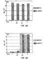
- Figure 2A-2B graphically illustrates the expression of MN- and MX-specific proteins in human fibroblasts (F), in HeLa cells (H) and in H/F-N and H/F-T hybrid cells and contrasts the expression in MX-infected and MX-uninfected cells.
- F human fibroblasts
- H HeLa cells
- H/F-N and H/F-T hybrid cells contrasts the expression in MX-infected and MX-uninfected cells.
- Example 5 details the procedures and results.
- Figure 3A-3B (discussed in Example 8) graphically illustrates the results from radioimmunoprecipitation experiments with 125 I-pGEX-3X-MN protein and different antibodies.
- the radioactive protein (15 x 10 3 cpm/tube) was precipitated with ascitic fluid or sera and SAC as follows: (A) ascites with MAb M75; (B) rabbit anti-MaTu serum; (C) normal rabbit serum; (D) human serum L8; (E) human serum KH; and (F) human serum M7.
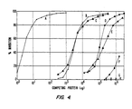
- Figure 4 shows the results from radioimmunoassays for MN antigen. Ascitic fluid (dilution precipitating 50% radioactivity) was allowed to react for 2 hours with (A) "cold" (unlabeled) protein pGEX-3X-MN, or with extracts from cells as follows: (B) HeLa + MX; (C) Rat-2Tk - ; (D) HeLa; (E) rat XC; (F) T24; and (G) HEF. Subsequently 125 I-labeled pGEX-3X-MN protein (25 x 10 3 cpm/tube) was added and incubated for an additional 2 hours. Finally, the radioactivity to MAb M75 was adsorbed to SAC and measured.
- MaTu is a two-component system.
- One part of the complex, exogenous MX is transmissible, and is manifested by a protein, p58X, which is a cytoplasmic antigen which reacts with some natural sera, of humans and of various animals.
- the other component, MN is endogenous to human cells.
- MN is a cellular gene, showing only very little homology with known DNA sequences. It is rather conservative and is present as a single copy gene in the chromosomal DNA of various vertebrates. Described herein is the cloning and sequencing of the MN cDNA, and the genetic engineering of a fusion protein, namely MN plus the carboxyl terminus of glutathione S-transferase, that can be conveniently purified by affinity chromatography.
- MN is manifested in HeLa cells by a twin protein(s), p54/58N, that is localized on the cell surface and in the nucleus.
- Immunoblots using a monoclonal antibody reactive with p54/58N (MAb M75) revealed two bands at 54 kd and 58 kd. Those two bands may correspond to one type of protein that differs by glycosylation pattern or by how it is processed.
- Both p54N and p58N are glycosylated with oligosaccharidic residues containing mannose, but only p58N also contains glucosamine.
- the phrase "twin protein” indicates p54/58N.
- MN is absent in rapidly growing, sparse cultures of HeLa, but is inducible either by keeping the cells in dense cultures or, more efficiently, by infecting them with MX. Those inducing factors are synergistic. Only p54/58N is associated with virions of vesicular stomatitis virus (VSV), reproduced in MaTu-infected HeLa. Whereas the twin protein p54/58N is glycosylated and forms oligomers linked by disulfidic bonds, p58X is not glycosylated and does not form S-S-linked oligomers.
- VSV vesicular stomatitis virus
- VSV assembles p54/58N into virions in HeLa cells, indicating that the twin protein is responsible for complementation of VSV G-protein mutants and for formation of VSV(MaTu) pseudotypes.
- enveloped viruses provide surface glycoproteins for the formation of infectious, functioning pseudotypes, which can perform such specific functions as adsorption and penetration of virions into cells [Zavada, J., J. Gen. Virol., 63: 15-24 (1982)], that observation implies that the MN gene behaves as a quasi-viral sequence.
- the surface proteins of enveloped viruses which participate in the formation of VSV pseudotypes, are glycosylated as is the MN twin protein, p54/58N. MN proteins also resemble viral glycoproteins in the formation of oligomers (preferably tri- or tetramers); such oligomerization, although not necessarily involving S-S bonds (disulfidic bonds), is essential for the assembly of virions [Kreis and Lodish, Cell, 46 : 929-937 (1986)]. The disulfidic bonds can be disrupted by reduction with 2-mercaptoethanol.
- p54/58N As reported in Pastorekova et al., Virology, 187: 620-626 (1992), after reduction with mercaptoethanol, p54/58N from cell extracts or from VSV looks very similar on immunoblot. Without reduction, in cell extracts, it gives several bands around 150 kd, suggesting that the cells may contain several different oligomers (probably with a different p54:p58 ratio), but VSV selectively assembles only one of them, with a molecular weight of about 153 kd. That oligomer might be a trimer, or rather a tetramer, consisting of 54 kd and 58 kd proteins. The equimolar ratio of p54:p58 in VSV virions is indicated by approximately the same strength of 54 kd and 58 kd bands in a VSV sample analyzed under reducing conditions.
- MN proteins appear to be diagnostic/prognostic for neoplastic disease.
- the MN twin protein, p54/58N was found to be expressed in HeLa cells and in Stanbridge's tumorigenic (H/F-T) hybrid cells [Stanbridge et al., Somatic Cell Genet, 7 : 699-712 (1981); and Stanbridge et al., Science, 215: 252-259 (1982)] but not in fibroblasts or in non-tumorigenic (H/F-N) hybrid cells [Stanbridge et al., id .] MN proteins were found in immunoblots prepared from human ovarian, endometrial and uterine cervical carcinomas, and in some benign neoplasias (as mammary papilloma) but not from normal ovarian, endometrial, uterine or placental tissues.
- MN proteins appear to be tumor-specific as they do not appear to be produced by normal non-tumor cells.
- MX and MN are two different entities, that can exist independently of each other.
- MX as an exogenous, transmissible agent can multiply in fibroblasts and in H/F-N hybrid cells which are not expressing MN-related proteins ( Figure 2A-2B). In such cells, MX does not induce the production of MN protein.
- MN protein can be produced in HeLa and other tumor cells even in the absence of MX as shown in Figure 2A-2B and in Examples 5 and 6.
- MX is a potent inducer of MN-related protein in HeLa cells; it increases its production thirty times over the concentration observed in uninfected cells (Examples 5 and 8, and Table 1 in Example 8, below).
- Figure 1A-1B provides the nucleotide sequence for the MN cDNA clone isolated as described within this section. It is understood that because of the degeneracy of the genetic code, that is, that more than one codon will code for one amino acid [for example, the codons TTA, TTG, CTT, CTC, CTA and CTG each code for the amino acid leucine (leu)], that variations of the nucleotide sequence in, for example, Figure 1A-1B, wherein one codon is substituted for another, would produce a substantially equivalent protein or polypeptide according to this invention. All such variations in the nucleotide sequence of the MN cDNA and complementary nucleic acid sequences are included within the scope of this invention.
- nucleotide sequence herein described and shown in Figure 1A-1B represents only the precise structure of the cDNA nucleotide sequence isolated and described herein. It is expected that slightly modified nucleotide sequences will be found or can be modified by techniques known in the art to code for substantially similar MN proteins and polypeptides, for example, those having similar epitopes, and such nucleotide sequences and proteins/polypeptides are considered to be equivalents for the purpose of this invention.
- DNA or RNA having equivalent codons is considered within the scope of the invention, as are synthetic nucleic acid sequences that encode proteins/polypeptides homologous or substantially homologous to MN proteins/polypeptides, as well as those nucleic acid sequences but for the degeneracy of the genetic code would hybridize to said cDNA nucleotide sequence. Modifications and variations of nucleic acid sequences as indicated herein are considered to result in sequences that are substantially the same as the MN sequence and fragments thereof.
- a lambda gtll cDNA library from MX-infected HeLa cells was prepared.
- Total RNA from MX-infected HeLa cells was isolated by a guanidinium-thiocyanate-CsCl method, and the mRNA was affinity separated on oligo dT-cellulose.
- the synthesis of the cDNA and its cloning into lambda gtll was carried out using kits from Amersham, except that the ECORI - NotI adaptor was from Stratagene [La Jolla, CA (USA)].
- the library was subjected to immunoscreening with monoclonal antibody M75 in combination with goat anti-mouse antibodies conjugated with alkaline phosphatase. That immunoscreening method is described in Young and Davis, PNAS (USA), 80 : 1194-1198 (1983).
- One positive clone was picked from 350,000 screened plaques (representing about one-half of the whole library).
- the positive clone was subcloned into the NotI site of pBluescript KS [Stratagene] thereby creating pBluescript-MN.
- Two oppositely oriented nested deletions were made using Erase-a-Base TM kit [Promega; Madison, WI (USA)] and sequenced by dideoxy method with a T7 sequencing kit [Pharmacia; Piscataway, NJ (USA)].
- the sequencing showed a partial cDNA clone, the insert being 1397 bp long. That sequence is shown in Figure 1A-1B [SEQ ID NO.: 1].
- the sequence comprises a large 1290 bp open reading frame and 107 bp 3' untranslated region containing a polyadenylation signal (AATAAA).
- the open reading frame of MN cDNA clone encodes a putative protein of about 48 kd ( Figure 1A-1B; SEQ ID NO.: 2).
- Analysis of the deduced translated amino acid (AA) sequence failed to show any significant homology to published protein sequences.
- the closest homology found was that of the C-terminal part of the MN protein and different types of carbonic anhydrase (about 30-35% in 170-200 AA overlap).
- the active site as well as the Zn 2+ binding domain of carbonic anhydrase are well-conserved in the MN protein.
- the MN gene is clearly a novel sequence derived from the human genome.
- the MN gene shows some homology with known carbonic anhydrases, it differs from them in several repects. Seven carbonic anhydrases are known [Dodgson et al. (eds.), The Carbonic Anhydrases , (Plenum Press; New York/London (1991)]. Each of their genes contains seven introns whereas the MN gene is apparently intronless. Also, all the known carbonic anhydrases are proteins of about 30 kd, smaller than the p54/58N-related products of the MN gene. Further, the carbonic anhydrases do not form oligomers as do the MN-related proteins.
- the cDNA insert from the above-described pBluescript-MN was released by digesting the plasmid DNA by NotI . It was then treated with S1 nuclease to obtain blunt ends and then cloned into a dephosphorylated SmaI site of pGEX-3X (Pharmacia). After transformation of XL1-Blue cells and induction with IPTG, a fusion protein was obtained.
- the fusion protein--MN glutathione S-transferase was purified by affinity chromatography on Glutathione-S-Sepharose 4B (Pharmacia). Twenty micrograms of the purified recombinant protein in each of two parallel samples were separated by SDS-PAGE on a 10% gel. One of the samples (A) was stained with Coomassie brilliant blue, whereas the other (B) was blotted onto a Hybond C membrane (Amersham; Aylesbury, Bucks, England). The blot was developed by autoradiography with 125 I-labeled MAb M75.
- the fusion protein pGEX-3X-MN was used in radioimmunoassays for MN-specific antibodies and for MN antigen.
- MN proteins and/or polypeptides are herein defined to mean proteins and/or polypeptides encoded by an MN gene or fragments thereof.
- An exemplary and preferred MN protein is that for which the predicted amino acid sequence is shown in Figure 1A-1B (SEQ ID NO.: 2).
- Preferred MN proteins/polypeptides are those proteins and/or polypeptides that have substantial homology with that MN protein shown in Figure 1A-1B.
- a “polypeptide” is a chain of amino acids covalently bound by peptide linkages and is herein considered to be composed of 50 or less amino acids.
- a “protein” is herein defined to be a polypeptide composed of more than 50 amino acids.
- MN proteins and/or polypeptides which have varying amino acid sequences including without limitation, amino acid substitutions, extensions, deletions, truncations and combinations thereof, fall within the scope of this invention.
- a protein extant within body fluids is subject to degradative processes, such as, proteolytic processes; thus, MN proteins that are significantly truncated and MN polypeptides may be found in body fluids, such as, sera.
- the phrase "MN antigen" is used herein to encompass MN proteins and/or polypeptides.
- amino acid sequence of MN proteins and polypeptides can be modified by genetic techniques. One or more amino acids can be deleted or substituted. Such amino acid changes may not cause any measurable change in the biological activity of the protein or polypeptide and result in proteins or polypeptides which are within the scope of this invention.
- MN proteins and polypeptides of this invention can be prepared in a variety of ways according to this invention, for example, recombinantly, synthetically or otherwise biologically, that is, by cleaving longer proteins and polypeptides enzymatically and/or chemically.
- a preferred method to prepare MN proteins is by a recombinant means.
- a particularly preferred method of recombinantly producing a MN protein is described above for the fusion protein pGEX-3X-MN.
- a representative method to prepare the MN protein shown in Figure 1A-1B or fragments thereof would be to insert the appropriate fragment of the MN cDNA into an appropriate expression vector as exemplified above.
- a wide variety of host-cloning vector combinations may be usefully employed in cloning the MN DNA isolated as described herein.
- useful cloning vehicles may include chromosomal, nonchromosomal and synthetic DNA sequences such as various known bacterial plasmids such as pBR322, other E.
- coli plasmids and their derivatives and wider host range plasmids such as RP4, phage DNA, such as, the numerous derivatives of phage lambda, e.g., NB989 and vectors derived from combinations of plasmids and phage DNAs such as plasmids which have been modified to employ phage DNA expression control sequences.
- the plasmid pGEX-3X is a preferred cloning vehicle.
- Useful hosts may be eukaryotic or prokaryotic and include bacterial hosts such as E. coli and other bacterial strains, yeasts and other fungi, animal or plant hosts such as animal or plant cells in culture, insect cells and other hosts. Of course, not all hosts may be equally efficient.
- bacterial hosts such as E. coli and other bacterial strains, yeasts and other fungi
- animal or plant hosts such as animal or plant cells in culture, insect cells and other hosts.
- host-cloning vehicle combination may be made by those of skill in the art after due consideration of the principles set forth herein without departing from the scope of this invention.
- the particular site chosen for insertion of the selected DNA fragment into the cloning vehicle to form a recombinant DNA molecule is determined by a variety of factors. These include size and structure of the protein or polypeptide to be expressed, susceptibility of the desired protein or polypeptide to endoenzymatic degradation by the host cell components and contamination by its proteins, expression characteristics such as the location of start and stop codons, and other factors recognized by those of skill in the art.
- the recombinant nucleic acid molecule containing the MN gene, fragment thereof, or cDNA therefrom may be employed to transform a host so as to permit that host (transformant) to express the structural gene or fragment thereof and to produce the protein or polypeptide for which the hybrid DNA encodes.
- the recombinant nucleic acid molecule may also be employed to transform a host so as to permit that host on replication to produce additional recombinant nucleic acid molecules as a source of MN nucleic acid and fragments thereof.
- the selection of an appropriate host for either of those uses is controlled by a number of factors recognized in the art. These include, for example, compatibility with the chosen vector, toxicity of the co-products, ease of recovery of the desired protein or polypeptide, expression characteristics, biosafety and costs.
- competent cells which are capable of DNA uptake are prepared from cells harvested after exponential growth phase and subsequently treated by the CaCl 2 method by well known procedures. Transformation can also be performed after forming a protoplast of the host cell.
- transfection methods such as the use of a calcium phosphate-precipitate, electroporation, conventional mechanical procedures such as microinjection, insertion of a plasmid encapsulated in red blood cell hosts or in liposomes, treatment of cells with agents such as lysophosphatidyl-choline or use of virus vectors, or the like may be used.
- the level of production of a protein or polypeptide is governed by three major factors: (1) the number of copies of the gene or DNA sequence encoding for it within the cell; (2) the efficiency with which those gene and sequence copies are transcribed and translated; and (3) the stability of the mRNA. Efficiencies of transcription and translation (which together comprise expression) are in turn dependent upon nucleotide sequences, normally situated ahead of the desired coding sequence. Those nucleotide sequences or expression control sequences define, inter alia, the location at which an RNA polymerase interacts to initiate transcription (the promoter sequence) and at which ribosomes bind and interact with the mRNA (the product of transcription) to initiate translation. Not all such expression control sequences function with equal efficiency.
- the newly engineered DNA fragment may be inserted into a multicopy plasmid or a bacteriophage derivative in order to increase the number of gene or sequence copies within the cell and thereby further improve the yield of expressed protein.
- expression control sequences may be employed. These include the operator, promoter and ribosome binding and interaction sequences (including sequences such as the Shine-Dalgarno sequences) of the lactose operon of E. coli ("the lac system"), the corresponding sequences of the tryptophan synthetase system of E. coli ("the trp system”), a fusion of the trp and lac promoter ("the tac system”), the major operator and promoter regions of phage lambda (O L P L and O R P R ,), and the control region of the phage fd coat protein.
- the lac system the lactose operon of E. coli
- the trp system the corresponding sequences of the tryptophan synthetase system of E. coli
- the tac system a fusion of the trp and lac promoter
- the tac system the major operator and promoter regions of phage lambda (O L P L and O R P R
- DNA fragments containing these sequences are excised by cleavage with restriction enzymes from the DNA isolated from transducing phages that carry the lac or trp operons, or from the DNA of phage lambda or fd. Those fragments are then manipulated in order to obtain a limited population of molecules such that the essential controlling sequences can be joined very close to, or in juxtaposition with, the initiation codon of the coding sequence.
- the fusion product is then inserted into a cloning vehicle for transformation or transfection of the appropriate hosts and the level of antigen production is measured. Cells giving the most efficient expression may be thus selected.
- cloning vehicles carrying the lac, trp or lambda P L control system attached to an initiation codon may be employed and fused to a fragment containing a sequence coding for a MN protein or polypeptide such that the gene or sequence is correctly translated from the initiation codon of the cloning vehicle.
- nucleic acid molecule is herein defined to mean a hybrid nucleotide sequence comprising at least two nucleotide sequences, the first sequence not normally being found together in nature with the second.
- expression control sequence is herein defined to mean a sequence of nucleotides that controls and regulates expression of structural genes when operatively linked to those genes.
- MN proteins and polypeptides of this invention may be prepared not only by recombinant means but also by synthetic and by other biologic means. Synthetic formation of the polypeptide or protein requires chemically synthesizing the desired chain of amino acids by methods well known in the art. Exemplary of other biologic means to prepare the desired polypeptide or protein is to subject to selective proteolysis a longer MN polypeptide or protein containing the desired amino acid sequence; for example, the longer polypeptide or protein can be split with chemical reagents or with enzymes.
- Techniques of chemical peptide synthesis include using automatic peptide synthesizers employing commercially available protected amino acids, for example, Biosearch [San Rafael, CA (USA)] Models 9500 and 9600; Applied Biosystems, Inc. [Foster City, CA (USA)] Model 430; Milligen [a division of Millipore Corp.; Bedford, MA (USA)] Model 9050; and Du Pont's RAMP (Rapid Automated Multiple Peptide Synthesis) [Du Pont Compass, Wilmington, DE (USA)].
- Biosearch San Rafael, CA (USA)
- Models 9500 and 9600 Applied Biosystems, Inc.
- Applied Biosystems, Inc. [Foster City, CA (USA)] Model 430
- Milligen a division of Millipore Corp.; Bedford, MA (USA)] Model 9050
- Du Pont's RAMP Rapid Automated Multiple Peptide Synthesis
- Nucleic acid probes of this invention are those comprising sequences that are substantially complementary to the MN cDNA sequence shown in Figure 1A-1B or to MN gene sequences.
- the phrase "substantially complementary” is defined herein to have the meaning as it is well understood in the art and, thus, used in the context of standard hybridization conditions. The stringency of hybridization conditions can be adjusted to control the precision of complementarity.
- Said probes can be used to detect MN DNA and/or RNA, and thus can be used to test for the presence or absence of MN genes, and amplification(s), mutation(s) or genetic rearrangements of MN genes in the cells of a patient.
- overexpression of an MN gene may be detected by Northern blotting using probes of this invention.
- Gene alterations, as amplifications, translocations, inversions, and deletions among others, can be detected by using probes of this invention for in situ hybridization to chromosomes from a patient's cells, whether in metaphase spreads or interphase nuclei. Southern blotting could also be used with the probes of this invention to detect amplifications or deletions of MN genes.
- Restriction Fragment Length Polymorphism (RFLP) analysis using said probes is a preferred method of detecting gene alterations, mutations and deletions.
- Said probes can also be used to identify MN proteins and/or polypeptides as well as homologs or near homologs thereto by their hybridization to various mRNAs transcribed from MN genes in different tissues.
- Said probes thus can be useful diagnostically/ prognostically.
- Said probes can be embodied in test kits, preferably with appropriate means to enable said probes when hybridized to an appropriate MN gene or MN mRNA target to be visualized.
- samples include tissue specimens, body fluids and tissue and cell extracts.
- Assays according to this invention are provided to detect and/or quantitate MN antigen or MN-specific antibodies in vertebrate samples, preferably mammalian samples, more preferably human samples.
- Such samples include tissue specimens, body fluids, tissue extracts and cell extracts.
- MN antigen may be detected by immunoassay, immunohistochemical staining, immunoelectron and scanning microscopy using immunogold among other techniques.
- MN antigen may be detected in body fluids, which can include among other fluids: blood, serum, plasma, semen, breast exudate, saliva, tears, sputum, mucous, urine, lymph, cytosols, ascites, pleural effusions, amniotic fluid, bladder washes, bronchioalveolar lavages and cerebrospinal fluid. It is preferred that the MN antigen be concentrated from a larger volume of body fluid before testing. Preferred body fluids to assay would depend on the type of cancer for which one was testing, but in general preferred body fluids would be breast exudate, pleural effusions and ascites.
- MN-specific antibodies can be bound by serologically active MN proteins/polypeptides in samples of such body fluids as blood, plasma, serum, lymph, mucous, tears, urine, spinal fluid and saliva; however, such antibodies are found most usually in blood, plasma and serum, preferably in serum.
- a representative assay to detect MN-specific antibodies is shown in Example 8 below wherein the fusion protein pGEX-3X-MN is used. Correlation of the results from the assays to detect and/or quantitate MN antigen and MN-specific antibodies reactive therewith, provides a preferred profile of the disease condition of a patient.
- the assays of this invention are both diagnostic and/or prognostic, i.e., diagnostic/prognostic.
- diagnostic/ prognostic is herein defined to encompass the following processes either individually or cumulatively depending upon the clinical context: determining the presence of disease, determining the nature of a disease, distinguishing one disease from another, forecasting as to the probable outcome of a disease state, determining the prospect as to recovery from a disease as indicated by the nature and symptoms of a case, monitoring the disease status of a patient, monitoring a patient for recurrence of disease, and/or determining the preferred therapeutic regimen for a patient.
- the diagnostic/prognostic methods of this invention are useful, for example, for screening populations for the presence of neoplastic or pre-neoplastic disease, determining the risk of developing neoplastic disease, diagnosing the presence of neoplastic and/or pre-neoplastic disease, monitoring the disease status of patients with neoplastic disease, and/or determining the prognosis for the course of neoplastic disease.
- the present invention is useful for screening for the presence of a wide variety of neoplastic diseases including carcinomas, such as, mammary, urinary tract, ovarian, uterine, cervical, endometrial, squamous cell and adenosquamous carcinomas; head and neck cancers; mesodermal tumors, such as, neuroblastomas and retinoblastomas; sarcomas, such as osteosarcomas and Ewing's sarcoma; and melanomas.
- carcinomas such as, mammary, urinary tract, ovarian, uterine, cervical, endometrial, squamous cell and adenosquamous carcinomas
- head and neck cancers mesodermal tumors, such as, neuroblastomas and retinoblastomas
- sarcomas such as osteosarcomas and Ewing's sarcoma
- melanomas include ovarian, uterine, cervical, vaginal,
- the invention provides methods and compositions for evaluating the probability of the presence of malignant or pre-malignant cells, for example, in a group of cells freshly removed from a host.
- Such an assay can be used to detect tumors, quantitate their growth, and help in the diagnosis and prognosis of disease.
- the assays can also be used to detect the presence of cancer metastasis, as well as confirm the absence or removal of all tumor tissue following surgery, cancer chemotherapy and/or radiation therapy. It can further be used to monitor cancer chemotherapy and tumor reappearance.
- MN antigen or antibodies can be detected and/or quantitated using a number of well-defined diagnostic assays.
- Those in the art can adapt any of the conventional immunoassay formats to detect and/or quantitate MN antigen and/or antibodies.
- Example 8 details the format of a preferred diagnostic method of this invention--a radioimmunoassay.
- Many other formats for detection of MN antigen and MN-specific antibodies are, of course available. Those can be Western blots, ELISAs (enzyme-linked immunosorbent assays), RIAs (radioimmunoassay), competitive EIA or dual antibody sandwich assays, among other assays all commonly used in the diagnostic industry.
- the interpretation of the results is based on the assumption that the antibody or antibody combination will not cross-react with other proteins and protein fragments present in the sample that are unrelated to MN.
- Representative of one type of ELISA test for MN antigen is a format wherein a microtiter plate is coated with antibodies made to MN proteins/polypeptides or antibodies made to whole cells expressing MN proteins, and to this is added a patient sample, for example, a tissue or cell extract. After a period of incubation permitting any antigen to bind to the antibodies, the plate is washed and another set of anti-MN antibodies which are linked to an enzyme is added, incubated to allow reaction to take place, and the plate is then rewashed. Thereafter, enzyme substrate is added to the microtiter plate and incubated for a period of time to allow the enzyme to work on the substrate, and the adsorbance of the final preparation is measured. A large change in absorbance indicates a positive result.
- MN proteins and/or polypeptides can be used to detect and/or quantitate the presence of MN antigen in the body fluids, tissues and/or cells of patients.
- a competition immunoassay is used, wherein the MN protein/polypeptide is labeled and a body fluid is added to compete the binding of the labeled MN protein/polypeptide to antibodies specific to MN protein/polypeptide.
- Such an assay can be used to detect and/or quantitate MN antigen as described in Example 8.
- an immunometric assay may be used wherein a labeled antibody made to a MN protein or polypeptide is used.
- the amount of labeled antibody which complexes with the antigen-bound antibody is directly proportional to the amount of MN antigen in the sample.
- a representative assay to detect MN-specific antibodies is a competition assay in which labeled MN protein/polypeptide is precipitated by antibodies in a sample, for example, in combination with monoclonal antibodies recognizing MN proteins/polypeptides.
- One skilled in the art could adapt any of the conventional immunoassay formats to detect and/or quantitate MN-specific antibodies. Detection of the binding of said antibodies to said MN protein/polypeptide could be by many ways known to those in the art, e.g., in humans with the use of anti-human labeled IgG.
- An exemplary immunoassay method of this invention to detect and/or quantitate MN antigen in a vertebrate sample comprises the steps of:
- Another exemplary immunoassay method according to this invention is that wherein a competition immunoassay is used to detect and/or quantitate MN antigen in a vertebrate sample and wherein said method comprises the steps of:
- antibodies including biologically active antibody fragments having suitable specificity
- immunological assay methods are available for determining the formation of specific antibody-antigen complexes.
- Numerous competitive and non-competitive protein binding assays have been described in the scientific and patent literature, and a large number of such assays are commercially available.
- Exemplary immunoassays which are suitable for detecting a serum antigen include those described in U.S. Patent Nos.
- Antibodies employed in assays may be labeled or unlabeled. Unlabeled antibodies may be employed in agglutination; labeled antibodies may be employed in a wide variety of assays, employing a wide variety of labels.
- Suitable detection means include the use of labels such as radionuclides, enzymes, coenzymes, fluorescers, chemiluminescers, chromogens, enzyme substrates or co-factors, enzyme inhibitors, free radicals, particles, dyes and the like.
- labels such as radionuclides, enzymes, coenzymes, fluorescers, chemiluminescers, chromogens, enzyme substrates or co-factors, enzyme inhibitors, free radicals, particles, dyes and the like.
- labeled reagents may be used in a variety of well known assays, such as radioimmunoassays, enzyme immunoassays, e.g., ELISA, fluorescent immunoassays, and the like. See for example, U.S. Patent Nos. 3,766,162; 3,791,932; 3,817,837; and 4,233,402.
- kits to detect and/or quantitate MN antigen and/or MN-specific antibodies can comprise MN protein(s)/polypeptides(s) and/or MN-specific antibodies, polyclonal and/or monoclonal.
- Kits to detect and/or quantitate MN antigen can comprise MN protein(s)/polypeptides(s) and/or MN-specific antibodies, polyclonal and/or monoclonal.
- diagnostic/prognostic test kits can comprise one or more sets of antibodies, polyclonal and/or monoclonal, for a sandwich format wherein antibodies recognize epitopes on the MN antigen, and one set is appropriately labeled or is otherwise detectable.
- Test kits for an assay format wherein there is competition between a labeled (or otherwise detectable) MN protein/polypeptide and MN antigen in the sample, for binding to an antibody can comprise the combination of the labeled protein/polypeptide and the antibody in amounts which provide for optimum sensitivity and accuracy.
- Test kits for MN-specific antibodies preferably comprise labeled/detectable MN proteins(s) and/or polypeptides(s), and may comprise other components as necessary, for example, to perform a preferred assay as outlined in Example 8 below.
- Such test kits can have other appropriate formats for conventional assays.
- antibodies is defined herein to include not only whole antibodies but also biologically active fragments of antibodies, preferably fragments containing the antigen binding regions. Such antibodies may be prepared by conventional methodology and/or by genetic engineering. Antibody fragments may be genetically engineered, preferably from the variable regions of the light and/or heavy chains (V H and V L ), including the hypervariable regions, and still more preferably from both the V H and V L regions.
- V H and V L variable regions of the light and/or heavy chains
- antibodies as used herein comprehends polyclonal and monoclonal antibodies and biologically active fragments thereof including among other possibilities "univalent” antibodies [Glennie et al., Nature, 295 : 712 (1982)]; Fab proteins including Fab' and F(ab') 2 fragments whether covalently or non-covalently aggregated; light or heavy chains alone, preferably variable heavy and light chain regions (V H and V L regions), and more preferably including the hypervariable regions [otherwise known as the complementarity determining regions (CDRs) of said V H and V L regions]; F c proteins; "hybrid” antibodies capable of binding more than one antigen; constant-variable region chimeras; "composite” immunoglobulins with heavy and light chains of different origins; "altered” antibodies with improved specificity and other characteristics as prepared by standard recombinant techniques and also by oligonucleotide-directed mutagenesis techniques [Dalbadie-Mc
- the antibodies be biologically active antibody fragments, preferably genetically engineered fragments, more preferably genetically engineered fragments from the V H and/or V L regions, and still more preferably comprising the hypervariable regions thereof.
- Immunogens to prepare MN-specific antibodies include MN proteins and/or polypeptides, preferably purified, and MX-infected tumor line cells, for example, MX-infected HeLa cells, among other immunogens.
- Anti-peptide antibodies are also made by conventional methods in the art as described in European Patent Publication No. 44,710 (published Jan. 27, 1982). Briefly, such anti-peptide antibodies are prepared by selecting a peptide from an MN amino acid sequence as from Figure 1A-1B, chemically synthesizing it, conjugating it to an appropriate immunogenic protein and injecting it into an appropriate animal, usually a rabbit or a mouse; then, either polyclonal or monoclonal antibodies are made, the latter by the Kohler-Milstein procedure.
- Monoclonal antibodies for use in the assays of this invention may be obtained by methods well known in the art for example, Galfre and Milstein, "Preparation of Monoclonal Antibodies: Strategies and Procedures," in Methods in Enzynology: Immunochemical Techniques, 73: 1-46 [Langone and Vanatis (eds); Academic Press (1981)]; and in the classic reference, Milstein and Kohler, Nature, 256: 495-497 (1975).
- human/human hybridomas [Olsson et al., PNAS (USA), 77 : 5429 (1980)] and human/murine hybridomas [Schlom et al., PNAS (USA), 77 : 6841 (1980); Shearman et al. J. Immunol., 146: 928-935 (1991); and Gorman et al., PNAS (USA), 88: 4181-4185 (1991)] can also be prepared among other possiblities.
- Such humanized monoclonal antibodies would be preferred monoclonal antibodies for therapeutic and imaging uses.
- Monoclonal antibodies specific for this invention can be prepared by immunizing appropriate mammals, preferably rodents, more preferably rabbits or mice, with an appropriate immunogen, for example, MaTu-infected HeLa cells or MN proteins/polypeptides attached to a carrier protein if necessary.
- an appropriate immunogen for example, MaTu-infected HeLa cells or MN proteins/polypeptides attached to a carrier protein if necessary.
- the production of hybridoma VU-M75 which secretes MAb M75 is exemplary and described below.
- MAb M75 serves to identify MN proteins/polypeptides in various laboratory diagnostic tests, for example, in tumor cell cultures or in clinical samples.
- MAbs M16 isotype IgG2A
- M67 isotype IgG1
- MAb M75 Monoclonal antibody M75
- VU-M75 mouse lymphocytic hybridoma VU-M75, which was initially deposited in the Collection of Hybridomas at the Institute of Virology, Slovak Academy of Sciences (Bratislava, Czechoslovakia) and was deposited under ATCC Designation HB 11128 on September 17, 1992 at the American Type Culture Collection (ATCC) in Rockville, MD (USA).
- Hybridoma VU-M75 was produced according to the procedure described in Gerhard, W., "Fusion of cells in suspension and outgrowth of hybrids in conditioned medium," In: Monoclonal Antibodies. Hybridomas: A New Dimension in Biological Analysis, page 370 [Kennet et al. (eds.); Plenum NY (USA)]. BALB/C mice were immunized with MaTu-infected HeLa cells, and their spleen cells were fused with myeloma cell line NS-0.
- Tissue culture media from the hybridomas were screened for monoclonal antibodies, using as antigen the p58 immunoprecipitated from cell extracts of MaTu-infected HeLa with rabbit anti-MaTu serum and protein A- Staphylococcus aureus cells (SAC) [Zavada and Zavadova, Arch, Virol., 118 189-197 (1991)], and eluted from SDS-PAGE gels.
- Monoclonal antibodies were purified from TC media by affinity chromatography on protein A-Sepharose [Harlow and Lane, "Antibodies: A Laboratory Manual,” Cold Spring Harbor, Cold Spring Harbor, NY (USA); 1988].
- the monoclonal antibodies useful according to this invention to identify MN proteins/polypeptides can be labeled in any conventional manner, for example, with enzymes such as horseradish peroxidase (HRP), fluorescent compounds, or with radioactive isotopes such as, 125 I, among other labels.
- HRP horseradish peroxidase
- a preferred label, according to this invention is 125 I, and a preferred method of labeling the antibodies is by using chloramine-T [Hunter, W.M., "Radioimmunoassay," In: Handbook of Experimental Immunology , pp. 14.1-14.40 (D.W. Weir ed.; Blackwell, Oxford/London/Edinburgh/Melbourne; 1978)].
- MAb H460 Monoclonal antibody H460
- MAb H460 Monoclonal antibody H460
- the MN-specific antibodies of this invention may be used therapeutically in the treatment of neoplastic and/or pre-neoplastic disease, either alone or in combination with chemotherapeutic drugs or toxic agents, such as ricin A. Further preferred for therapeutic use would be biologically active antibody fragments as described herein. Also preferred MN-specific antibodies for such therapeutic uses would be humanized monoclonal antibodies.
- the MN-specific antibodies can be administered in a therapeutically effective amount, preferably dispersed in a physiologically acceptable, nontoxic liquid vehicle.
- MN-specific antibodies of this invention when linked to an imaging agent, such as a radionuclide, can be used for imaging.
- an imaging agent such as a radionuclide
- Biologically active antibody fragments or humanized monoclonal antibodies may be preferred for imaging use.
- a patient's neoplastic tissue can be identified as, for example, sites of tumors and locations of any metastases.
- Antibodies appropriately labeled or linked to an imaging agent, can be injected in a physiologically acceptable carrier into a patient, and the binding of the antibodies can be detected by a method appropriate to the label or imaging agent, for example, by scintigraphy.
- MN genes are herein considered putative oncogenes and the proteins encoded thereby are considered to be putative oncoproteins.
- Antisense nucleic acid sequences substantially complementary to mRNA transcribed from MN genes, as represented by the antisense oligodeoxynucleotides (ODNs) of Example 11, infra, can be used to reduce or prevent expression of the MN gene.
- ODNs antisense oligodeoxynucleotides
- antisense nucleic acid sequences preferably oligonucleotides, by hybridizing to the MN mRNA, particularly in the vicinity of the ribosome binding site and translation initiation point, inhibits translation of the mRNA.
- the use of such antisense nucleic acid sequences may be considered to be a form of cancer therapy.
- Preferred antisense oligonucleotides according to this invention are gene-specific ODNs or oligonucleotides complementary to the 5' end of MN mRNA. Particularly preferred are the 29-mer ODN1 and 19-mer ODN2 for which the sequences are provided in Example 11, infra . Those antisense ODNs are representative of the many antisense nucleic acid sequences that can function to inhibit MN gene expression. Ones of ordinary skill in the art could determine appropriate antisense nucleic acid sequences, preferably antisense oligonucleotides, from the nucleic acid sequence of Figure 1A-1B.
- MN proteins and polypeptides of this invention can be incorporated into vaccines capable of inducing protective immunity against neoplastic disease and a dampening effect upon tumorigenic activity.
- MN proteins and/or polypeptides may be synthesized or prepared recombinantly or otherwise biologically, to comprise one or more amino acid sequences corresponding to one or more epitopes of the MN proteins either in monomeric or multimeric form. Those proteins and/or polypeptides may then be incorporated into vaccines capable of inducing protective immunity.
- Techniques for enhancing the antigenicity of such polypeptides include incorporation into a multimeric structure, binding to a highly immunogenic protein carrier, for example, keyhole limpet hemocyanin (KLH), or diptheria toxoid, and administration in combination with adjuvants or any other enhancers of immune response.
- KLH keyhole limpet hemocyanin
- diptheria toxoid diptheria toxoid
- MN proteins/polypeptides to be used in a vaccine according to this invention would be genetically engineered MN proteins.
- a preferred recombinant MN protein is the fusion protein pGEX-3X-MN, produced according to this invention.
- a preferred exemplary use of such a vaccine of this invention would be its administration to patients whose MN-carrying primary cancer had been surgically removed.
- the vaccine may induce active immunity in the patients and prevent recidivism or metastasis.
- anti-idiotype antibodies to antibodies to MN proteins/polypeptides are also useful as vaccines and can be similarly formulated.
- amino acid sequence corresponding to an epitope of an MN protein/polypeptide either in monomeric or multimeric form may also be obtained by chemical synthetic means or by purification from biological sources including genetically modified microorganisms or their culture media [See Lerner, "Synthetic Vaccines", Sci. Am. 248 (2): 66-74 (1983)].
- the protein/polypeptide may be combined in an amino acid sequence with other proteins/polypeptides including fragments of other proteins, as for example, when synthesized as a fusion protein, or linked to other antigenic or non-antigeneic polypeptides of synthetic or biological origin.
- amino acid sequence variations of a naturally occurring protein or polypeptide may be antigenic and confer protective immunity against neoplastic disease and/or anti-tumorigenic effects.
- Possible sequence variations include, without limitation, amino acid substitutions, extensions, deletions, truncations, interpolations and combinations thereof.
- Such variations fall within the contemplated scope of the invention provided the protein or polypeptide containing them is immunogenic and antibodies elicited by such a polypeptide or protein cross-react with naturally occurring MN proteins and polypeptides to a sufficient extent to provide protective immunity and/or anti-tumorigenic activity when administered as a vaccine.
- Such vaccine compositions will be combined with a physiologically acceptable medium, including immunologically acceptable diluents and carriers as well as commonly employed adjuvants such as Freund's Complete Adjuvant, saponin, alum, and the like.
- Administration would be in immunologically effective amounts of the MN proteins or polypeptides, preferably in quantities providing unit doses of from 0.01 to 10.0 micrograms of immunologically active MN protein and/or polypeptide per kilogram of the recipient's body weight.
- Total protective doses may range from 0.1 to about 100 micrograms of antigen.
- MaTu agent [Zavada et al., Nature New Biol., 240: 124-125 (1972); Zavada et al., J. Gen. Virol, 24 : 327-337 (1974)] was from original "MaTu" cells [Widmaier et al., Arch. Geschwulstforsch, 44: 1-10 (1974)] transferred into our stock of HeLa by cocultivation with MaTu cells treated with mitomycin C, to ensure that control and MaTu-infected cells were comparable.
- MaTu cells were incubated for 3 hours at 37°C in media with 5 ⁇ g/ml of mitomycin C [Calbiochem, LaJolla, CA (USA)].
- Mixed cultures were set to 2 x 10 5 of mitomycin C-treated cells and 4 x 10 5 of fresh recipient cells in 5 ml of medium. After 3 days they were first subcultured and further passaged 1-2 times weekly.
- Control HeLa cells were the same as those described in Zavada et al., Nature New Biol.. 240: 124-125 (1972).
- Serum L8 was from a patient with Paget's disease. Serum M7 was from a healthy donor.
- Rabbit anti-MaTu serum was prepared by immunizing a rabbit three times at intervals of 30 days with 10-5 x 10 7 viable MaTu infected HeLa cells.
- RIP and PAGE were performed essentially as described in Zavada and Zavadova, Arch. Virol., 118 : 189-197 (1991), except that in the experiments described herein [ 35 S]methionine (NEN), 10 ⁇ Ci/ml of methionine-free MEM medium, supplemented with 2% FCS and 3% complete MEM were used. Confluent petri dish cultures of cells were incubated overnight in that media.
- Immunoblotting used as described herein follows the method of Towbin et al., PNAS (USA), 76 : 4350-4354 (1979).
- the proteins were transferred from the gels onto nitrocellulose [Schleicher and Schuell; Dassel Germany; 0.45 ⁇ m porosity] in Laemmli electrode buffer diluted 1:10 with distilled water, with no methanol or SDS. The transfer was for 2 1/2 hours at 1.75 mA/cm 2 .
- the blots were developed with 125 I-labeled MAbs and autoradiography was performed using intensifying screens, with X-ray films exposed at -70°C.
- MAbs which in preliminary tests proved to be specific for MaTu-related antigens, showed two different reactivities in immunofluorescence.
- MAb M16 gave the same type of fluorescence. With either M67 or M16, only extremely weak "background" fluorescence was seen in control HeLa cells.
- MAb M75 Another MAb, M75, showed a granular membrane fluorescence on living MaTu-infected cells and a granular nuclear fluorescence in acetone-fixed cells. However, M75 sometimes showed a similar, although much weaker, fluorescence on uninfected HeLa cells. A relationship was observed based upon the conditions of growth: in HeLa cells uninfected with MaTu, both types of fluorescence with MAb M75 were observed only if the cells were grown for several passages in dense cultures, but not in sparse ones.
- the amount of M75-reactive cell surface antigen was analyzed cytofluorometrically and was dependent on the density of the cell cultures and on infection with MaTu.
- Control and MaTu infected HeLa cells were grown for 12 days in dense or sparse cultures. The cells were released with Versene (EDTA), and incubated with MAb M75 or with no MAb, and subsequently incubated with FITC-conjugated anti-mouse IgG. The intensity of fluorescence was measured.
- the antigen binding MAb M75 is inducible: it was found to be absent in control HeLa grown in sparse culture, and to be induced either by the growth of HeLa in dense culture or by infection with MaTu. Those two factors were found to have an additive or synergistic effect. Those observations indicated along with other results described herein that there were two different agents involved: exogenous, transmissible MX, reactive with M67, and endogenous, inducible MN, detected by MAb M75.
- MAb M75 reacted with two MN-specific protein bands of 54 kd and 58 kd, which were the same in uninfected HeLa grown at high density and in MaTu-infected HeLa, evidencing that M75 recognizes the same protein(s) in both uninfected and MaTu-infected HeLa cells. Consistent with the cytofluorometric results, the amount of the antigen depended both on cell density and on infection with MaTu, the latter being a much more potent inducer of p54/58N.
- the simple radioimmunoassay procedure of this example was performed directly in petri dish cultures.
- Sixteen variants of the radioimmunoassay enabled us to determine whether the MX and MN antigens are located on the surface or in the interior of tr.e cells and how the expression of those two antigens depends on infection with MaTu and on the density, in which the cells had been grown before the petri dishes were seeded.
- live cells only cell surface antigens can bind the MAbs.
- M67 showed no reaction with any variant of the cultures, whereas M75 reacted in accord with the results of Examples 1 and 2 above.
- MAb M75 in methanol-fixed cells confirmed the absence of corresponding antigen in uninfected HeLa from sparse cultures and its induction both by growth in dense cultures and by infection with MaTu.
- MX antigen was found to be present in MaTu-infected fibroblasts.
- Zavada and Zavadova (1991) it was reported that a p58 band from MX-infected fibroblasts could not be detected by RIP with rabbit anti-MaTu serum. That serum contains more antibodies to MX than to MN antigen. The discrepancy can be explained by the extremely slow spread of MX in infected cultures.
- the results reported in Zavada and Zavadova (1991) were from fibroblasts tested 6 weeks after infection, whereas the later testing was 4 months after infection.
- Figure 2A-2B graphically illustrates the expression of MN- and MX- specific proteins in human fibroblasts, in HeLa cells and in H/F-N and H/F-T hybrid cells, and contrasts the expression in MX-infected and uninfected cells.
- Cells were infected with MX by co-cultivation with mitomycin C-treated MX-infected HeLa. The infected and uninfected cells were grown for three passages in dense cultures. About four months after infection, the infected cells concurrently with uninfected cells were grown in petri dishes to produce dense monolayers.
- a radimmunoassay was performed directly in confluent petri dish (5 cm) culture of cells, fixed with methanol essentially as described in Example 3, supra .
- the monolayers were fixed with methanol and treated with 125 I-labeled MAbs M67 (specific for exogenous MX antigen) or M75 (specific for endogenous MN antigen) at 6 x 10 4 cpm/dish.
- the bound radioactivity was measured; the results are shown in Figure 2A-2B.
- Figure 2A shows that MX was transmitted to all four cell lines tested, that is, to human embryo fibroblasts, to HeLa and to both H/F-N and H/F-T hybrids; at the same time, all four uninfected counterpart cell lines were MX-negative.
- Figure 2B shows that MN antigens are present in both MX-infected and uninfected HeLa and H/F-T cells, but not in the fibroblasts. No MN antigen was found in the control H/F-N, and only a minimum increase over background of MN antigen was found in MaTu infected H/F-N. Thus, it was found that in the hybrids, expression of MN antigen very strongly correlates with tumorigenicity.
- the MN-specific twin protein p54/58N was detected by immunoblotting in HeLa cell lines (both our standard type, that is, HeLa K, and in the Stanbridge mutant HeLa, that is, D98/AH.2 or HeLa S) and in tumorigenic H/F-T; however, p54/58N was not detected in the fibroblasts nor in the non-tumorigenic H/F-N even upon deliberately long exposure of the film used to detect radioactivity. Infection of the HeLa cells with MX resulted in a strong increase in the concentration of the p54/58N protein(s).
- the hybrid cells H/F-N and H/F-T were constructed by Eric J. Stanbridge [Stanbridge et al., Somatic Cell Genetics. 7: 699-712 (1981); and Stanbridge et al., Science, 215: 252-259 (1982)].
- His original hybrid, produced by the fusion of a HeLa cell and a human fibroblast was not tumorigenic in nude mice, although it retained some properties of transformed cells, for example, its growth on soft agar.
- Rare segregants from the hybrid which have lost chromosome 11 are tumorigenic. The most likely explanation for the tumorigenicity of those segregants is that chromosome 11 contains a suppressor gene (an antioncogene), which blocks the expression of a as yet unknown oncogene.
- the oncoprotein encoded by that oncogene is critical for the capacity of the H/F hybrids to produce tumors in nude mice. Since the p54/58N protein shows a correlation with the tumorigenicity of H/F hybrids, it is a candidate for that putative oncoprotein.
- Immunoblots of MN proteins in cell culture extracts were prepared from the following: (A) MX-infected HeLa cells; (B) human fibroblasts; (C) T24; (D) T47D; (E) SK-Mel 1477; and (F) HeLa cells uninfected with MX.
- the proteins were separated by PAGE after heating in a sample buffer, either with or without 3% mercaptoethanol (+ ME or O ME, repectively). Thus, one run was done for each extract + ME and then O ME.
- lane (A) the cell culture extract from MX-infected HeLa cells was analysed directly (10 ⁇ l per lane), whereas the antigens from the other extracts (lanes B-E) were each concentrated from a 500 ⁇ l extract by precipitation with MAb M75 and SAC.
- the immunoblots indicated that two other human carcinoma cell lines contain MN-related proteins -- T24 (bladder carcinoma; lane C) and T47D (mammary carcinoma; lane D).
- Those cells contain proteins which react with MAb M75 that under reducing conditions have molecular weights of 54 kd and 56 kd, and under non-reducing conditions have a molecular weight of about 153 kd.
- the intensity of those bands was at least ten times lower than that for the p54/58N twin protein from HeLa cells.
- tissue extracts in the other lanes were prepared from the following: (B) full-term placenta; (C) corpus uteri; (D, M) adenocarcinoma endometrii; (E, N) carcinoma ovari; (F, G) trophoblasts; (H) normal ovary; (I) myoma uteri; (J) mammary papilloma; (K) normal mammary gland; (L) hyperplastic endometrium; (O) cervical carcinoma; and (P) melanoma.
- MN-related antigen from all the extracts but for lane A was first concentrated from a 1 ml extract as explained above.
- MN proteins were found in endometrial (lanes D and M), ovarian (lanes E and N) and in uterine cervical (lane 0) carcinomas. In those extracts MN-related proteins were found in three Dands having molecular weights between about 48 kd and about 58 kd.
- Another MN-related protein was present in the tissue extract from a mammary papilloma; that protein was seen as a single band at about 48 kd (lane J).
- MN-related antigen was searched for also in cell lines derived from normal tissues and from tumors of several animal species.
- MN-related protein was found in two rat cell lines: one of them was the XC cell line derived from rat rhabdomyosarcoma induced with Rous sarcoma virus; the other was the Rat2-Tk - cell line.
- a single protein band was found on the blots : its molecular weight on blots produced from a reducing gel and from a non-reducing gel was respectively 53.5 kd and 153 kd.
- Immunoblots of MN proteins were prepared from (A) MX-infected HeLa cells and from (B) Rat-TK - cells [wherein, as indicated above, the proteins were separated by PAGE after heating in a sample buffer with 3% mercaptoethanol (+ ME) or without 3% mercaptoethanol (O ME)]. The concentration of MN antigen in those two cell lines was found to be similar both in the + ME and O ME runs. The extracts were analysed directly (40 ⁇ l per lane).
- MN-related protein from XC cells showed the same pattern as for Rat2-Tk - cells both under reducing and non-reducing conditions, except that its concentration was about 30x lower.
- the finding of a MN-related protein--p53.5N--in two rat cell lines provides the basis for a model system.
- the MN-negative cells were: Vero cells (African green monkey); mouse L cells; mouse NIH-3T3 cells normal, infected with Moloney leukemia virus, or transformed with Harvey sarcoma virus; GR cells (mouse mammary tumor cells induced with MTV), and NMG cells (normal mouse mammary gland).
- the genetically engineered MN protein fused with glutathione S-transferase--pGEX-3X-MN--prepared and purified as described above was labeled with 125 I by the chloramine T method [Hunter (1978)].
- the purified protein enabled the development of a quantitative RIA for MN-specific antibodies as well as for MN antigens. All dilutions of antibodies and of antigens were prepared in RIPA buffer (1% TRITON X-100 and 0.1% sodium deoxycholate in PBS--phosphate buffered saline, pH 7.2), to which was added 1% of foetal calf serum (FCS).
- Tissue and cell extracts were prepared in RIPA buffer containing 1 mM phenylmethylsulfonylfluoride and 200 trypsin inhibiting units of Trasylol (aprotinin) per ml, with no FCS.
- aprotinin trypsin inhibiting units of Trasylol
- 125 I-labeled pGEX-3X-MN protein (2.27 ⁇ Ci/ ⁇ g of TCA-precipitable protein) was before use diluted with RIPA + 1% FCS, and non-specifically binding radioactivity was adsorbed with a suspension of fixed protein A- Staphylococcus aureus cells (SAC).
- SAC fixed protein A- Staphylococcus aureus cells
- MAb-containing ascites fluids or test sera were mixed with 125 I-labeled protein and allowed to react in a total volume of 1 ml for 2 hours at room temperature. Subsequently, 50 ⁇ l of a 10% suspension of SAC [Kessler, supra ] was added and the mixture was incubated for 30 minutes. Finally, the SAC was pelleted, 3x washed with RIPA, and the bound radioactivity was determined on a gamma counter.
- FIG. 3A-3B Titration of antibodies to MN antigen is shown in Figure 3A-3B.
- Ascitic fluid from a mouse carrying M75 hybridoma cells (A) is shown to have a 50% end-point at dilution 1:1.4 x 10 -6 .
- ascitic fluids with MAbs specific for MX protein (M16 and M67) showed no precipitation of 125 I-labeled pGEX-3X-MN even at dilution 1:200 (result not shown).
- An approximate concentration of p58X protein can be obtained by RIP from extracts of MaTu-infected HeLa cells that have been metabolically labeled with [ 35 S]-methionine or with a mixture of [ 14 C]-amino acids. The results are shown in Table 2. Radioimmunoprecipitation of metabolically labeled p58X protein Radioactivity Precipit.
- MN antigen detected by indirect immunofluorescence with MAb M75, is located on the surface membranes and in the nuclei of MX-infected HeLa cells or in HeLa cells grown in dense cultures.
- immunoelectron microscopy was used wherein MAb M75 bound to MN antigen was visualized with immunogold beads.
- Immunoelectron and scanning micrographs of the stained cells demonstrated the location of MN antigen in the cells, and in addition, the striking ultrastructural differences between control and MX-infected HeLa.
- a control HeLa cell was shown to have on its surface very little MN antigen, as visualized with gold beads. The cell surface was rather smooth, with only two little protrusions. No mitochondria could be seen in the cytoplasm.
- MX-infected HeLa cells were shown to have abundant, dense filamentous protrusions from their surfaces. Most of the MN antigen is located on those filaments, which were decorated with immunogold when immunogold was used to stain them.
- the cytoplasm of MX-infected HeLa was shown to contain numerous mitochondria.
- MN antigen was found to be in the nucleus of MX-infected cells: some of the MN antigen was in the nucleoplasm (possibly linked to chromatin), but a higher concentration of the MN antigen was in the nucleoli. Again, the surface of normal HeLa cells was rather smooth whereas MX-infected HeLa cells have on their surface, numerous filaments and "blebs". Some of the filaments appear to form bridges connecting them to adjacent cells.
- RNA ladder (0.24-9.5 kb) [Bethesda Research Laboratories (BRL); Bethesda, MD (USA)] was used as a size standard.
- MN genes in the genomic DNAs of various vertebrates were detected by Southern blotting.
- Chromosomal DNA digested by SstI was as follows: (A) chicken; (B) bovine; (C) feline; (D) MX-infected HeLa cells; (E) mouse NIH 3T3 cells; (F) human placental cells; (G) HeLa cells; (H) sheep; (I) human melanoma cells; and (J) monkey Vero cells. Restriction fragments were separated on a 0.7% agarose gel and alkali blotted onto a Hybond N membrane (Amersham). The MN cDNA probe labelling and hybridization procedures were the same as for the Northern blotting analyses described in Example 12.
- the material listed below was deposited with the American Type Culture Collection (ATCC) at 12301 Parklawn Drive, Rockville, Maryland 20852 (USA). The deposits were made under the provisions of the Budapest Treaty on the International Recognition of Deposited Microorganisms for the Purposes of Patent Procedure and Regulations thereunder (Budapest Treaty). Maintenance of a viable culture is assured for thirty years from the date of deposit. The organism will be made available by the ATCC under the terms of the Budapest Treaty, and subject to an agreement between the Applicants and the ATCC which assures unrestricted availability upon issuance of the pertinent U.S. Patent.
Abstract
Description
- The present invention is in the general area of medical genetics and in the fields of biochemical engineering and immunochemistry. More specifically, it relates to the identification of a new gene--the MN gene--a cellular gene coding for the MN protein. The inventors hereof found MN proteins to be associated with tumorigenicity. Identification of MN antigen as well as antibodies specific therefor in patient samples provides the basis for diagnostic/prognostic assays for cancer.
- MaTu is a novel quasi-viral agent with rather unusual properties [Zavada, J., Arch. Virol, 50: 1-10 (1976)]. It is presumably derived from a human mammary tumor. In some respects, it resembles classical viruses whereas in other respects, it resembles "slow" viruses (prions), and in still other respects it is different from both classes of viruses.
- MaTu was first detected by its capacity to complement mutants of vesicular stomatitis virus (VSV) with heat-labile surface G protein in HeLa cells (cell line derived from human cervical adenocarcinoma), which had been cocultivated with human breast carcinoma cells. The complementation resulted in the formation of phenotypically mixed virions--the VSV(MaTu) pseudotypes [Zavada et al., Nature New Biol. 240: 124-125 (1972)]. The virions contain the VSV genome, which is responsible for their ability to produce plaques (as well as internal VSV proteins), but the surface protein, corresponding to MaTu, determines their host range and neutralization specificities.
- One of the paradoxical features of the MaTu agent is its host range. VSV(MaTu) is infectious only for human fibroblasts, but not for HeLa; however, the MaTu agent, detected by its capacity to donate surface protein for the VSV(MaTu) pseudotypes, is transmissible only to HeLa, but not to fibroblasts [Zavada et al., J. Gen. Virol.. 24: 327-337 (1974)].
- By its complementation of VSV mutants and by its formation of pseudotypes, MaTu resembles known enveloped viruses. However, MaTu is transmissible only by direct cell-to-cell contact, and not by cell-free filtrates, thus differing from both classical and "slow" viruses. Its only permissive host appears to be HeLa cells. In those cells, MaTu spreads extremely slowly, and does not form morphologically distinct virions, thus resembling the "slow" viruses. [Zavada et al., (1974); Zavada and Zavadova, Arch. Virol, 118: 189-197 (1991)]. No known virus has HeLa cells as an exclusive host.
- Since the above-described properties suggest that MaTu might be an entirely new type of molecular parasite of living cells, and since it possibly originated from a human tumor, there was a significant medical research interest to characterize it in more detail. Herein elucidated is the biological and molecular nature of MaTu. MaTu was found to be a two-component system, having an exogenous transmissible component, MX, and an endogenous cellular component, MN. The MN gene was further found to be present in the chromosomal DNA of all vertebrates tested, and its expression was found to be strongly correlated with tumorigenicity.
- Described herein is the cloning and sequencing of the MN gene and the production of a MN-encoded protein in a bacterial vector. That genetically engineered MN protein as well as other MN proteins/polypeptides, can be used in serological assays according to this invention to detect MN-specific antibodies. Further, such MN proteins/polypeptides and antibodies reactive with MN antigen can be used in immunoassays according to this invention to detect and/or quantitate MN antigen. Such assays may be diagnostic and/or prognostic for neoplastic and/or pre-neoplastic disease.
- Herein disclosed is the MN gene, a cellular gene which is the endogenous component of the MaTu agent.
Substantially the entire cDNA sequence for the apparently intronless gene is shown in Figures 1A-1B [SEQ ID NO.: 1]. - This invention is directed to said MN gene, fragments thereof and the related cDNA which are useful, for example, as follows: 1) to produce MN proteins/ polypeptides by biochemical engineering; 2) to prepare nucleic acid probes to test for the presence of the MN gene in cells of a subject: 3) to prepare appropriate polymerase chain reaction (PCR) primers for use, for example, in PCR-based assays or to produce nucleic acid probes; 4) to identify MN proteins and polypeptides as well as homologs or near homologs thereto; 5) to identify various mRNAs transcribed from MN genes in various tissues and cell lines, preferably human; and 6) to identify mutations in MN genes. The invention further concerns purified and isolated DNA molecules comprising the MN gene or fragments, thereof, or the related cDNA or fragments thereof.
- The invention further concerns the discovery of a hitherto unknown protein -- MN, encoded by the MN gene. The expresssion of MN proteins is inducible by growing cells in dense cultures, and such expression was discovered to be associated with tumorigenic cells.
- MN proteins were found to be produced by some human tumor cell lines in vitro, for example, by HeLa (cervical carcinoma), T24 (bladder carcinoma) and T47D (mammary carcinoma) and SK-Mel 1477 (melanoma) cell lines, by tumorigenic hybrid cells and by cells of some human cancers in vivo, for example, by cells of uterine cervical, ovarian and endometrial carcinomas as well as cells of some benign neoplasias such as mammary papillomas. MN proteins were not found in non-tumorigenic hybrid cells or in the cells of normal tissues. Thus, MN proteins are considered to be tumor-specific.
- In HeLa and in tumorigenic HeLa x fibroblast hybrid (H/F/T) cells, MN protein is manifested as a "twin" protein p54/58N; it is glycosylated and forms disulfide-linked oligomers. As determined by electrophoresis upon reducing gels, MN proteins have molecular weights in the range of from about 40 kd to about 70 kd, preferably from about 45 kd to about 65 kd, more preferably from about 48 kd to about 58 kd. Upon non-reducing gels, MN proteins in the form of oligomers have a molecular weights in the range of from about 145 kd to about 160 kd, preferably from about 150 to about 155 kd, still more preferably from about 152 to about 154 kd. The predicted amino acid sequence for a preferred MN protein of this invention is shown in Figure 1A-1B.
- The discovery of the MN gene and protein and thus, of substantially complementary MN genes and proteins encoded thereby, led to the finding that the expression of MN proteins was associated with tumorigenicity. That finding resulted in the creation of methods that are diagnostic/ prognostic for cancer and precancerous conditions. Methods and compositions are provided for identifying the onset and presence of neoplastic disease by detecting and/or quantitating MN antigen in patient samples, including cell and tissue extracts from vertebrates, preferably mammals and more preferably humans. Such MN antigen may also be found in body fluids.
- MN proteins and genes are of use in research concerning the molecular mechanisms of oncogenesis, in cancer diagnostics/prognostics, and may be of use in cancer immunotherapy.
- The present invention is useful for detecting a wide variety of neoplastic and/or pre-neoplastic diseases. Exemplary neoplastic diseases include carcinomas, such as mammary, bladder, ovarian, uterine, cervical, endometrial, squamous cell and adenosquamous carcinomas; and head and neck cancers; mesodermal tumors, such as neuroblastomas and retinoblastomas; sarcomas, such as osteosarcomas and Ewing's sarcoma; and melanomas. Of particular interest are head and neck cancers, gynecologic cancers including ovarian, cervical, vaginal, endometrial and vulval cancers; gastrointestinal cancer, such as, stomach, colon and esophageal cancers; urinary tract cancer, such as, bladder and kidney cancers; skin cancer; liver cancer; prostate cancer; lung cancer; and breast cancer. Of still further particular interest are gynecologic cancers; breast cancer; urinary tract cancers, especially bladder cancer; lung cancer; gastrointestinal cancer, such as, stomach, colon and esophageal cancers; and liver cancer. Even further of particular interest are gynecologic cancers and breast cancer. Gynecologic cancers of particular interest are carcinomas of the uterine cervix, endometrium and ovaries; more particularly such gynecologic cancers include cervical squamous cell carcinomas, adenosquamous carcinomas, adenocarcinomas as well as gynecologic precancerous conditions, such as metaplastic cervical tissues and condylomas.
- The invention further relates to the biochemical engineering of the MN gene, fragments thereof or related cDNA. For example, said gene or a fragment thereof or related cDNA can be inserted into a suitable expression vector; host cells can be transformed with such an expression vector; and an MN protein/polypeptide, preferably an MN protein, is expressed therein. Such a recombinant protein or polypeptide can be glycosylated or nonglycosylated, preferably glycosylated, and can be purified to substantial purity. The invention further concerns MN proteins/polypeptides which are synthetically or otherwise biologically prepared.
- Said MN proteins/polypeptides can be used in assays to detect MN antigen in patient samples and in serological assays to test for MN-specific antibodies. MN proteins/polypeptides of this invention are serologically active, immunogenic and/or antigenic. They can further be used as immunogens to produce MN-specific antibodies, polyclonal and/or monoclonal, as well as an immune T-cell response.
- The invention further is directed to MN-specific antibodies, which can be used diagnostically/prognostically and may be used therapeutically. MN-specific antibodies can be used, for example, in laboratory diagnostics, using immunofluorescence microscopy or immunohistochemical staining; as a component in immunoassays for detecting and/or quantitating MN antigen in, for example, clinical samples; as probes for immunoblotting to detect MN antigen; in immunoelectron microscopy with colloid gold beads for localization of MN proteins and/or polypeptides in cells; and in genetic engineering for cloning the MN gene or fragments thereof, or related cDNA. Such MN-specific antibodies can be used as components of diagnostic/ prognostic kits, for example, for in vitro use on histological sections; such antibodies can also and used for in vivo diagnostics/ prognostics, for example, such antibodies can be labeled appropriately, as with a suitable radioactive isotope, and used in vivo to locate metastases by scintigraphy. Further such antibodies may be used in vivo therapeutically to treat cancer patients with or without toxic and/or cytostatic agents attached thereto. Further, such antibodies can be used in vivo to detect the presence of neoplastic and/or pre-neoplastic disease. Still further, such antibodies can be used to affinity purify MN proteins and polypeptides.
- A hybridoma that produces a representative MN-specific antibody, the monoclonal antibody M75, was deposited at the American Type Culture Collection [ATCC; Rockville, MD (USA)] on September 17, 1992, under ATCC Number HB 11128. The M75 antibody was used to discover and identify the MN protein and can be used to readily identify MN antigen in Western blots, in radioimmunoassays and immunohistochemically, for example, in tissue samples that have been formalin fixed.
- This invention also concerns recombinant DNA molecules comprising a DNA sequence that encodes for an MN protein or polypeptide, and also recombinant DNA molecules that encode not only for an MN protein or polypeptide but also for an amino acid sequence of a non-MN protein/polypeptide, preferably which is not immunogenic to humans and which is not typically reactive to antibodies in human body fluids. Examples of such a DNA sequence is the alpha-peptide coding region of beta-galactosidase and a sequence coding for glutathione S-transferase or a fragment thereof. Further, claimed herein are such recombinant fusion proteins/ polypeptides which are substantially pure and non-naturally occurring. An exemplary fusion protein of this invention is pGEX-3X-MN.
- This invention also concerns methods of treating neoplastic disease and/or pre-neoplastic disease comprising inhibiting the expression of MN genes by administering antisense nucleic acid sequences that are substantially complementary to mRNA transcribed from MN genes. Preferred are antisense nucleic acid sequences that are substantially complementary to sequences at the 5' end of the MN cDNA sequence shown in Figure 1A-1B. Preferably said antisense nucleic acid sequences are oligonucleotides.
- This invention also concerns vaccines comprising an immunogenic amount of one or more substantially pure MN proteins and/or polypeptides dispersed in a physiologically acceptable, nontoxic vehicle, which amount is effective to immunize a vertebrate, preferably a mammal, more preferably a human, against a neoplastic disease associated with the expression of MN proteins. Said proteins can be recombinantly, synthetically or otherwise biologically produced. Recombinant MN proteins includes fusion proteins, as exemplified by pGEX-3X-MN. A particular use of said vaccine would be to prevent recidivism and/or metastasis. For example, it could be administered to a patient who has had an MN-carrying tumor surgically removed, to prevent recurrence of the tumor.
- The invention still further concerns nucleic acid probes that are substantially complementary to nucleic acid sequences of the MN gene. Preferred nucleic acid probes of this invention are those with sequences substantially complementary to sequences from the MN cDNA shown in Figure 1A-1B. Test kits of this invention can comprise such probes which are useful diagnostically/prognostically for neoplastic and/or pre-neoplastic disease. Preferred test kits comprise means for detecting or measuring the hybridization of said probes to the MN gene or to the mRNA product of the MN gene, such as a visualizing means.
- The immunoassays of this invention can be embodied in test kits which comprise MN proteins/polypeptides and/or MN-specific antibodies. Such test kits can be in solid phase formats, but are not limited thereto, and can also be in liquid phase format, and can be based on ELISAS, particle assays, radiometric or fluorometric assays either unamplified or amplified, using, for example, avidin/biotin technology.
- The following abbreviations are used herein:
- AA -
- amino acid
- ATCC -
- American Type Culture Collection
- bp -
- base pairs
- BSA -
- bovine serum albumin
- Ci -
- curie
- cm -
- centimeter
- cpm -
- counts per minute
- C-terminus -
- carboxyl-terminus
- °C -
- degrees centigrade
- DMEM -
- Dulbecco modified Eagle medium
- EDTA -
- ethylenediaminetetracetate
- EIA -
- enzyme immunoassay
- ELISA -
- enzyme-linked immunosorbent assay
- F -
- fibroblasts
- FCS -
- fetal calf serum
- FIBR -
- fibroblasts
- FITC -
- fluorescein isothiocyanate
- H -
- HeLa cells
- HEF -
- human embryo fibroblasts
- HeLa K -
- standard type of HeLa cells
- HeLa S -
- Stanbridge's mutant HeLa D98/AH.2
- H/F-T -
- hybrid HeLa fibroblast cells that are tumorigenic; derived from HeLa D98/AH.2
- H/F-N -
- hybrid HeLa fibroblast cells that are nontumorigenic; derived from HeLa D98/AH.2
- HGPRT- -
- hypoxanthine guanine phosphoribosyl transferase-deficient
- HRP -
- horseradish peroxidase
- IPTG -
- isopropyl-Beta-D-thiogalacto-pyranoside
- kb -
- kilobase
- kd -
- kilodaltons
- M -
- molar
- mA -
- milliampere
- MAb -
- monoclonal antibody
- ME -
- mercaptoethanol
- MEM -
- minimal essential medium
- mg -
- milligram
- ml -
- milliliter
- mM -
- millimolar
- MTV -
- mammary tumor virus
- N -
- normal concentration
- ng -
- nanogram
- N-terminus -
- amino-terminus
- ODN -
- oligodeoxynucleotide
- PAGE -
- polyacrylamide gel electrophoresis
- PBS -
- phosphate buffered saline
- PEST -
- combination of one-letter abbreviations for proline, glutamic acid, serine, threonine
- pI -
- isoelectric point
- RIP -
- radioimmunoprecipitation
- RIPA -
- radioimmunoprecipitation assay
- SAC -
- protein A-Staphylococcus aureus cells
- SDS -
- sodium dodecyl sulfate
- SDS-PAGE -
- sodium dodecyl sulfate-polyacrylamide gel electrophoresis
- SSPE -
- NaCl (0.18 M), sodium phosphate (0.01 M), EDTA (0.001 M)
- TCA -
- trichloroacetic acid
- TC media -
- tissue culture media
- µCi -
- microcurie
- µg -
- microgram
- µl -
- microliter
- µM -
- micromolar
- VSV -
- vesicular stomatitis virus
- X-MLV -
- xenotropic murine leukemia virus
- The following cell lines were used in the experiments herein described:
- HeLa K --
- standard type of HeLa cells; aneuploid, epithelial-like cell line isolated from a human cervical adenocarcinoma [Gey et al., Cancer Res., 12: 264 (1952); Jones et al., Obstet. Gynecol., 38: 945-949 (1971)] obtained from Professor B. Korych, [Institute of Medical Microbiology and Immunology, Charles University; Prague, Czechoslovakia]
- HeLa D98/AH.2 (also HeLa S) --
- Mutant HeLa clone that is hypoxanthine guanine phosphoribosyl transferase-deficient (HGPRT-) kindly provided by Eric J. Stanbridge [Department of Microbiology, College of Medicine, University of California, Irvine, CA (USA)] and reported in Stanbridge et al., Science, 215: 252-259 (15 Jan. 1982); parent of hybrid cells H/F-N and H/F-T, also obtained from E.J. Stanbridge.
- NIH-373 --
- murine fibroblast cell line reported in Aaronson, Science, 237: 178 (1987).
- T47D --
- cell line derived from a human mammary carcinoma [Keydar et al., Eur. J. Cancer, 15: 659-670 (1979)]; kindly provided by J. Keydar [Haddasah Medical School; Jerusalem, Israel]
- T24 --
- cell line from urinary bladder carcinoma [Bubenik et al., Int. J. Cancer, 11: 765-773 (1973)] kindly provided by J. Bubenik [Institute of Molecular Genetics, Czechoslovak Academy of Sciences; Prague, Czechoslovakia]
- HMB2 --
- cell line from melanoma [Svec et al., Neoplasma, 35: 665-681 (1988)]
- HEF --
- human embryo fibroblasts [Zavada et al., Nature New Biology, 240: 124-125 (1972)]
- SIRC --
- cell line from rabbit cornea (control and X-MLV-infected) [Zavada et al., Virology, 82: 221-231 (1977)]
- Vero cells --
- African green monkey cell line [Zavada et al. (1977)]
- myeloma cell line NS-0 --
- myeloma cell line used as a fusion parent in production of monoclonal antibodies [Galfre and Milstein, Methods Enzymol., 73: 3-46 (1981)]
- SK-Mel 1477 --
- human melanoma cell line kindly provided by K.E. Hellstrom [Division of Tumor Immunology, Fred Hutchins Cancer Research Center; Seattle, Washington (USA)]
- XC --
- cells derived from a a rat rhabdomyosarcoma induced with Rous sarcoma virus-induced rat sarcoma [Svoboda, J., Natl. Cancer Center Institute Monograph No. 17, IN: "International Conference on Avian Tumor Viruses" (J.W. Beard ed.), pp. 277-298 (1964)], kindly provided by Jan Svoboda [Institute of Molecular Genetics, Czechoslovak Academy of Sciences; Prague, Czechoslovakia]; and
- Rat 2-Tk- --
- a thymidine kinase deficient cell line, kindly provided by L. Kutinova [Institute of Sera and Vaccines; Prague, Czechoslovakia]
- CGL1 --
- H/F-N hybrid cells (HeLa D98/AH.2 derivative)
- CGL2 --
- H/F-T hybrid cells (HeLa D98/AH.2 derivative)
- CGL3 --
- H/F-T hybrid cells (HeLa D98/AH.2 derivative)
- CGL4 --
- H/F-T hybrid cells (HeLa D98/Ah.2 derivative)
- The following symbols are used to represent nucleotides herein:
Base Symbol adenine A cytosine C guanine G thymine T uracil U - There are twenty main amino acids, each of which is specified by a different arrangement of three adjacent nucleotides (triplet code or codon), and which are linked together in a specific order to form a characteristic protein. A three-letter convention is used herein to identify said amino acids, as, for example, in Figure 1A-1B, as follows:
Amino acid name Symbol Alanine Ala Arginine Arg Asparagine Asn Aspartic Acid Asp Cysteine Cys Glutamic Acid Glu Glutamine Gln Glycine Gly Histidine His Isoleucine Ile Leucine Leu Lysine Lys Methionine Met Phenylalanine Phe Proline Pro Serine Ser Threonine Thr Tryptophan Trp Tyrosine Tyr Valine Val - Figure 1A-1B provides the nucleotide sequence for the MN cDNA clone isolated as described herein and the predicted amino acid sequence encoded by the cDNA [SEQ ID NOS.: 1 and 2, respectively]. That sequence data has been sent to the EMBL Data Library in Heidelberg, Germany and is available under Accession No. X66839.
- Figure 2A-2B graphically illustrates the expression of MN- and MX-specific proteins in human fibroblasts (F), in HeLa cells (H) and in H/F-N and H/F-T hybrid cells and contrasts the expression in MX-infected and MX-uninfected cells. Example 5 details the procedures and results.
- Figure 3A-3B (discussed in Example 8) graphically illustrates the results from radioimmunoprecipitation experiments with 125I-pGEX-3X-MN protein and different antibodies. The radioactive protein (15 x 103 cpm/tube) was precipitated with ascitic fluid or sera and SAC as follows: (A) ascites with MAb M75; (B) rabbit anti-MaTu serum; (C) normal rabbit serum; (D) human serum L8; (E) human serum KH; and (F) human serum M7.
- Figure 4 (discussed in Example 8) shows the results from radioimmunoassays for MN antigen. Ascitic fluid (dilution precipitating 50% radioactivity) was allowed to react for 2 hours with (A) "cold" (unlabeled) protein pGEX-3X-MN, or with extracts from cells as follows: (B) HeLa + MX; (C) Rat-2Tk-; (D) HeLa; (E) rat XC; (F) T24; and (G) HEF. Subsequently 125I-labeled pGEX-3X-MN protein (25 x 103 cpm/tube) was added and incubated for an additional 2 hours. Finally, the radioactivity to MAb M75 was adsorbed to SAC and measured.
- As demonstrated herein MaTu is a two-component system. One part of the complex, exogenous MX, is transmissible, and is manifested by a protein, p58X, which is a cytoplasmic antigen which reacts with some natural sera, of humans and of various animals. The other component, MN, is endogenous to human cells.
- MN is a cellular gene, showing only very little homology with known DNA sequences. It is rather conservative and is present as a single copy gene in the chromosomal DNA of various vertebrates. Described herein is the cloning and sequencing of the MN cDNA, and the genetic engineering of a fusion protein, namely MN plus the carboxyl terminus of glutathione S-transferase, that can be conveniently purified by affinity chromatography.
- MN is manifested in HeLa cells by a twin protein(s), p54/58N, that is localized on the cell surface and in the nucleus. Immunoblots using a monoclonal antibody reactive with p54/58N (MAb M75) revealed two bands at 54 kd and 58 kd. Those two bands may correspond to one type of protein that differs by glycosylation pattern or by how it is processed. (Both p54N and p58N are glycosylated with oligosaccharidic residues containing mannose, but only p58N also contains glucosamine.) Herein, the phrase "twin protein" indicates p54/58N.
- MN is absent in rapidly growing, sparse cultures of HeLa, but is inducible either by keeping the cells in dense cultures or, more efficiently, by infecting them with MX. Those inducing factors are synergistic. Only p54/58N is associated with virions of vesicular stomatitis virus (VSV), reproduced in MaTu-infected HeLa. Whereas the twin protein p54/58N is glycosylated and forms oligomers linked by disulfidic bonds, p58X is not glycosylated and does not form S-S-linked oligomers.
- VSV assembles p54/58N into virions in HeLa cells, indicating that the twin protein is responsible for complementation of VSV G-protein mutants and for formation of VSV(MaTu) pseudotypes. As only enveloped viruses provide surface glycoproteins for the formation of infectious, functioning pseudotypes, which can perform such specific functions as adsorption and penetration of virions into cells [Zavada, J., J. Gen. Virol., 63: 15-24 (1982)], that observation implies that the MN gene behaves as a quasi-viral sequence.
- The surface proteins of enveloped viruses, which participate in the formation of VSV pseudotypes, are glycosylated as is the MN twin protein, p54/58N. MN proteins also resemble viral glycoproteins in the formation of oligomers (preferably tri- or tetramers); such oligomerization, although not necessarily involving S-S bonds (disulfidic bonds), is essential for the assembly of virions [Kreis and Lodish, Cell, 46: 929-937 (1986)]. The disulfidic bonds can be disrupted by reduction with 2-mercaptoethanol.
- As reported in Pastorekova et al., Virology, 187: 620-626 (1992), after reduction with mercaptoethanol, p54/58N from cell extracts or from VSV looks very similar on immunoblot. Without reduction, in cell extracts, it gives several bands around 150 kd, suggesting that the cells may contain several different oligomers (probably with a different p54:p58 ratio), but VSV selectively assembles only one of them, with a molecular weight of about 153 kd. That oligomer might be a trimer, or rather a tetramer, consisting of 54 kd and 58 kd proteins. The equimolar ratio of p54:p58 in VSV virions is indicated by approximately the same strength of 54 kd and 58 kd bands in a VSV sample analyzed under reducing conditions.
- The expression of MN proteins appears to be diagnostic/prognostic for neoplastic disease. The MN twin protein, p54/58N, was found to be expressed in HeLa cells and in Stanbridge's tumorigenic (H/F-T) hybrid cells [Stanbridge et al., Somatic Cell Genet, 7: 699-712 (1981); and Stanbridge et al., Science, 215: 252-259 (1982)] but not in fibroblasts or in non-tumorigenic (H/F-N) hybrid cells [Stanbridge et al., id.] MN proteins were found in immunoblots prepared from human ovarian, endometrial and uterine cervical carcinomas, and in some benign neoplasias (as mammary papilloma) but not from normal ovarian, endometrial, uterine or placental tissues. In HeLa cells infected with MX, observed were conspicuous ultrastructural alterations, that is, the formation of abundant filaments on cell surfaces and the amplification of mitochondria. Using an immunogold technique, p54/58N was visualized on the surface filaments and in the nucleus, particularly in the nucleoli. Thus MN proteins appear to be tumor-specific as they do not appear to be produced by normal non-tumor cells.
- The examples herein show that MX and MN are two different entities, that can exist independently of each other. MX as an exogenous, transmissible agent can multiply in fibroblasts and in H/F-N hybrid cells which are not expressing MN-related proteins (Figure 2A-2B). In such cells, MX does not induce the production of MN protein. MN protein can be produced in HeLa and other tumor cells even in the absence of MX as shown in Figure 2A-2B and in Examples 5 and 6. However, MX is a potent inducer of MN-related protein in HeLa cells; it increases its production thirty times over the concentration observed in uninfected cells (Examples 5 and 8, and Table 1 in Example 8, below).
- Figure 1A-1B provides the nucleotide sequence for the MN cDNA clone isolated as described within this section. It is understood that because of the degeneracy of the genetic code, that is, that more than one codon will code for one amino acid [for example, the codons TTA, TTG, CTT, CTC, CTA and CTG each code for the amino acid leucine (leu)], that variations of the nucleotide sequence in, for example, Figure 1A-1B, wherein one codon is substituted for another, would produce a substantially equivalent protein or polypeptide according to this invention. All such variations in the nucleotide sequence of the MN cDNA and complementary nucleic acid sequences are included within the scope of this invention.
- It is further understood that the nucleotide sequence herein described and shown in Figure 1A-1B represents only the precise structure of the cDNA nucleotide sequence isolated and described herein. It is expected that slightly modified nucleotide sequences will be found or can be modified by techniques known in the art to code for substantially similar MN proteins and polypeptides, for example, those having similar epitopes, and such nucleotide sequences and proteins/polypeptides are considered to be equivalents for the purpose of this invention. DNA or RNA having equivalent codons is considered within the scope of the invention, as are synthetic nucleic acid sequences that encode proteins/polypeptides homologous or substantially homologous to MN proteins/polypeptides, as well as those nucleic acid sequences but for the degeneracy of the genetic code would hybridize to said cDNA nucleotide sequence. Modifications and variations of nucleic acid sequences as indicated herein are considered to result in sequences that are substantially the same as the MN sequence and fragments thereof.
- To find the MN gene, a lambda gtll cDNA library from MX-infected HeLa cells was prepared. Total RNA from MX-infected HeLa cells was isolated by a guanidinium-thiocyanate-CsCl method, and the mRNA was affinity separated on oligo dT-cellulose. The synthesis of the cDNA and its cloning into lambda gtll was carried out using kits from Amersham, except that the ECORI-NotI adaptor was from Stratagene [La Jolla, CA (USA)]. The library was subjected to immunoscreening with monoclonal antibody M75 in combination with goat anti-mouse antibodies conjugated with alkaline phosphatase. That immunoscreening method is described in Young and Davis, PNAS (USA), 80: 1194-1198 (1983). One positive clone was picked from 350,000 screened plaques (representing about one-half of the whole library).
- The positive clone was subcloned into the NotI site of pBluescript KS [Stratagene] thereby creating pBluescript-MN. Two oppositely oriented nested deletions were made using Erase-a-BaseTM kit [Promega; Madison, WI (USA)] and sequenced by dideoxy method with a T7 sequencing kit [Pharmacia; Piscataway, NJ (USA)]. The sequencing showed a partial cDNA clone, the insert being 1397 bp long. That sequence is shown in Figure 1A-1B [SEQ ID NO.: 1]. The sequence comprises a large 1290 bp open reading frame and 107 bp 3' untranslated region containing a polyadenylation signal (AATAAA). Another interesting feature of the sequence is the presence of a region contributing to instability of the mRNA (AUUUA at position 1389) which is characteristic for mRNAs of some oncogenes and lymphokines [Shaw and Kamen, Cell, 46: 659-667 (1986)]. As follows from a comparison of the size of the MN clone with that of the corresponding mRNA in a Northern blot (Example 12), the cDNA is missing about 100 bp from the 5' end of its sequence.
- The open reading frame of MN cDNA clone encodes a putative protein of about 48 kd (Figure 1A-1B; SEQ ID NO.: 2). Analysis of the deduced translated amino acid (AA) sequence failed to show any significant homology to published protein sequences. The closest homology found was that of the C-terminal part of the MN protein and different types of carbonic anhydrase (about 30-35% in 170-200 AA overlap). The active site as well as the Zn2+ binding domain of carbonic anhydrase are well-conserved in the MN protein. However, the MN gene is clearly a novel sequence derived from the human genome.
- Although as indicated, the MN gene shows some homology with known carbonic anhydrases, it differs from them in several repects. Seven carbonic anhydrases are known [Dodgson et al. (eds.), The Carbonic Anhydrases, (Plenum Press; New York/London (1991)]. Each of their genes contains seven introns whereas the MN gene is apparently intronless. Also, all the known carbonic anhydrases are proteins of about 30 kd, smaller than the p54/58N-related products of the MN gene. Further, the carbonic anhydrases do not form oligomers as do the MN-related proteins.
- From the predicted amino acid sequence, it is evident that the product of the MN gene is a basic protein (pI 9.08), with one potential N-glycosylation site located at the amino acid positions 303-313. Those observations correspond to the finding that p54/58N proteins from HeLa cells are sensitive to Endo H and Endo F cleavage, which causes a loss of about 3 kd each. The hydrophilicity profile reveals a hydrophobic sequence of amino acids (at positions 371-395) probably representing the region spanning the plasma membrane and containing also a potential cleavage signal. The profile fits well with the observation that p54/58N proteins are localized on the cell membrane. There are no PEST regions in the MN amino acid sequence, suggesting that the product of the MN gene is a stable long-lived protein [Rogers et al., Science, 234: 364-368 (1986)]. Such a feature explains our experience with inefficient metabolic labeling of p54/58N. The deduced amino acid sequence displays also other features namely, 10 potential phosphorylation a. 7 myristylation sites, and 3 antigenic determinants.
- To determine whether both p54/58N proteins were encoded by one gene, antisense ODNs were used to inhibit specifically MN gene expression. [Such use of antisense ODNs is reviewed in Stein and Cohen, Cancer Res., 48: 2659-2668 (1988).] Those experiments are detailed in Example 11. The findings indicated that cultivation of HeLa cells with ODNs resulted in a considerable inhibition of p54/58N synthesis, whereas the amount of different HeLa cell proteins produced remained approximately the same. Further, and importantly on immunoblotting, the specific inhibition by ODNs affected both of the p54/58N proteins (Example 11). Thus, it was concluded that the MN gene that was cloned codes for both of the p54/58N proteins in HeLa cells.
- To confirm whether the gene that was cloned codes for the p54/58N-specific protein, it was subcloned into the bacterial expression vector pGEX-3X [Pharmacia; Upsala, Sweden], constructed to express a fusion protein containing the C-terminus of glutathione S-transferase. That subcloning is representative of one method to genetically engineer an MN-related protein of this invention. The following description is exemplary and not meant to limit the invention in any way.
- The cDNA insert from the above-described pBluescript-MN was released by digesting the plasmid DNA by NotI. It was then treated with S1 nuclease to obtain blunt ends and then cloned into a dephosphorylated SmaI site of pGEX-3X (Pharmacia). After transformation of XL1-Blue cells and induction with IPTG, a fusion protein was obtained.
- The fusion protein--MN glutathione S-transferase was purified by affinity chromatography on Glutathione-S-Sepharose 4B (Pharmacia). Twenty micrograms of the purified recombinant protein in each of two parallel samples were separated by SDS-PAGE on a 10% gel. One of the samples (A) was stained with Coomassie brilliant blue, whereas the other (B) was blotted onto a Hybond C membrane (Amersham; Aylesbury, Bucks, England). The blot was developed by autoradiography with 125I-labeled MAb M75.
- SDS-PAGE analysis provided an interesting result: a number of protein bands with different molecular weights. A similar SDS-PAGE pattern was obtained with another representative fusion protein produced according to this invention, beta-galactosidase-MN that was expressed from lambda gtll lysogens. It appears that those patterns are due to translation errors caused by the presence of 9 AGGAGG codon tandems in the MN sequence. The use of those codons is strongly avoided in bacterial genes because of the shortage of corresponding tRNAs. Thus, during the translation of AGGAGG tandems from foreign mRNA, +1 ribosomal frameshifts arise with a high frequency (about 50%) [Spanjaard et al., Nuc. Acid Res., 18: 5031-5036 (1990)].
- By immunoblotting, a similar pattern was obtained: all the bands seen on stained SDS-PAGE gel reacted with the MN-specific MAb M75, indicating that all the protein bands are MN-specific. Also, that result indicates that the binding site for MAb M75 is on the N-terminal part of the MN protein, which is not affected by frameshifts.
- As shown in Example 8 below, the fusion protein pGEX-3X-MN was used in radioimmunoassays for MN-specific antibodies and for MN antigen.
- The phrase "MN proteins and/or polypeptides" (MN proteins/polypeptides) is herein defined to mean proteins and/or polypeptides encoded by an MN gene or fragments thereof. An exemplary and preferred MN protein is that for which the predicted amino acid sequence is shown in Figure 1A-1B (SEQ ID NO.: 2). Preferred MN proteins/polypeptides are those proteins and/or polypeptides that have substantial homology with that MN protein shown in Figure 1A-1B.
- A "polypeptide" is a chain of amino acids covalently bound by peptide linkages and is herein considered to be composed of 50 or less amino acids. A "protein" is herein defined to be a polypeptide composed of more than 50 amino acids.
- It can be appreciated that a protein or polypeptide produced by a neoplastic cell in vivo could be altered in sequence from that produced by a tumor cell in cell culture. Thus, MN proteins and/or polypeptides which have varying amino acid sequences including without limitation, amino acid substitutions, extensions, deletions, truncations and combinations thereof, fall within the scope of this invention. It can also be appreciated that a protein extant within body fluids is subject to degradative processes, such as, proteolytic processes; thus, MN proteins that are significantly truncated and MN polypeptides may be found in body fluids, such as, sera. The phrase "MN antigen" is used herein to encompass MN proteins and/or polypeptides.
- It will further be appreciated that the amino acid sequence of MN proteins and polypeptides can be modified by genetic techniques. One or more amino acids can be deleted or substituted. Such amino acid changes may not cause any measurable change in the biological activity of the protein or polypeptide and result in proteins or polypeptides which are within the scope of this invention.
- The MN proteins and polypeptides of this invention can be prepared in a variety of ways according to this invention, for example, recombinantly, synthetically or otherwise biologically, that is, by cleaving longer proteins and polypeptides enzymatically and/or chemically. A preferred method to prepare MN proteins is by a recombinant means. A particularly preferred method of recombinantly producing a MN protein is described above for the fusion protein pGEX-3X-MN.
- A representative method to prepare the MN protein shown in Figure 1A-1B or fragments thereof would be to insert the appropriate fragment of the MN cDNA into an appropriate expression vector as exemplified above. A wide variety of host-cloning vector combinations may be usefully employed in cloning the MN DNA isolated as described herein. For example, useful cloning vehicles may include chromosomal, nonchromosomal and synthetic DNA sequences such as various known bacterial plasmids such as pBR322, other E. coli plasmids and their derivatives and wider host range plasmids such as RP4, phage DNA, such as, the numerous derivatives of phage lambda, e.g., NB989 and vectors derived from combinations of plasmids and phage DNAs such as plasmids which have been modified to employ phage DNA expression control sequences. The plasmid pGEX-3X is a preferred cloning vehicle.
- Useful hosts may be eukaryotic or prokaryotic and include bacterial hosts such as E. coli and other bacterial strains, yeasts and other fungi, animal or plant hosts such as animal or plant cells in culture, insect cells and other hosts. Of course, not all hosts may be equally efficient. The particular selection of host-cloning vehicle combination may be made by those of skill in the art after due consideration of the principles set forth herein without departing from the scope of this invention.
- The particular site chosen for insertion of the selected DNA fragment into the cloning vehicle to form a recombinant DNA molecule is determined by a variety of factors. These include size and structure of the protein or polypeptide to be expressed, susceptibility of the desired protein or polypeptide to endoenzymatic degradation by the host cell components and contamination by its proteins, expression characteristics such as the location of start and stop codons, and other factors recognized by those of skill in the art.
- The recombinant nucleic acid molecule containing the MN gene, fragment thereof, or cDNA therefrom, may be employed to transform a host so as to permit that host (transformant) to express the structural gene or fragment thereof and to produce the protein or polypeptide for which the hybrid DNA encodes. The recombinant nucleic acid molecule may also be employed to transform a host so as to permit that host on replication to produce additional recombinant nucleic acid molecules as a source of MN nucleic acid and fragments thereof. The selection of an appropriate host for either of those uses is controlled by a number of factors recognized in the art. These include, for example, compatibility with the chosen vector, toxicity of the co-products, ease of recovery of the desired protein or polypeptide, expression characteristics, biosafety and costs.
- Where the host cell is a procaryote such as E. coli, competent cells which are capable of DNA uptake are prepared from cells harvested after exponential growth phase and subsequently treated by the CaCl2 method by well known procedures. Transformation can also be performed after forming a protoplast of the host cell.
- Where the host used is an eucaryote, transfection methods such as the use of a calcium phosphate-precipitate, electroporation, conventional mechanical procedures such as microinjection, insertion of a plasmid encapsulated in red blood cell hosts or in liposomes, treatment of cells with agents such as lysophosphatidyl-choline or use of virus vectors, or the like may be used.
- The level of production of a protein or polypeptide is governed by three major factors: (1) the number of copies of the gene or DNA sequence encoding for it within the cell; (2) the efficiency with which those gene and sequence copies are transcribed and translated; and (3) the stability of the mRNA. Efficiencies of transcription and translation (which together comprise expression) are in turn dependent upon nucleotide sequences, normally situated ahead of the desired coding sequence. Those nucleotide sequences or expression control sequences define, inter alia, the location at which an RNA polymerase interacts to initiate transcription (the promoter sequence) and at which ribosomes bind and interact with the mRNA (the product of transcription) to initiate translation. Not all such expression control sequences function with equal efficiency. It is thus of advantage to separate the specific coding sequences for the desired protein from their adjacent nucleotide sequences and fuse them instead to known expression control sequences so as to favor higher levels of expression. This having been achieved, the newly engineered DNA fragment may be inserted into a multicopy plasmid or a bacteriophage derivative in order to increase the number of gene or sequence copies within the cell and thereby further improve the yield of expressed protein.
- Several expression control sequences may be employed. These include the operator, promoter and ribosome binding and interaction sequences (including sequences such as the Shine-Dalgarno sequences) of the lactose operon of E. coli ("the lac system"), the corresponding sequences of the tryptophan synthetase system of E. coli ("the trp system"), a fusion of the trp and lac promoter ("the tac system"), the major operator and promoter regions of phage lambda (OLPL and ORPR,), and the control region of the phage fd coat protein. DNA fragments containing these sequences are excised by cleavage with restriction enzymes from the DNA isolated from transducing phages that carry the lac or trp operons, or from the DNA of phage lambda or fd. Those fragments are then manipulated in order to obtain a limited population of molecules such that the essential controlling sequences can be joined very close to, or in juxtaposition with, the initiation codon of the coding sequence.
- The fusion product is then inserted into a cloning vehicle for transformation or transfection of the appropriate hosts and the level of antigen production is measured. Cells giving the most efficient expression may be thus selected. Alternatively, cloning vechicles carrying the lac, trp or lambda PL control system attached to an initiation codon may be employed and fused to a fragment containing a sequence coding for a MN protein or polypeptide such that the gene or sequence is correctly translated from the initiation codon of the cloning vehicle.
- The phrase "recombinant nucleic acid molecule" is herein defined to mean a hybrid nucleotide sequence comprising at least two nucleotide sequences, the first sequence not normally being found together in nature with the second.
- The phrase "expression control sequence" is herein defined to mean a sequence of nucleotides that controls and regulates expression of structural genes when operatively linked to those genes.
- MN proteins and polypeptides of this invention may be prepared not only by recombinant means but also by synthetic and by other biologic means. Synthetic formation of the polypeptide or protein requires chemically synthesizing the desired chain of amino acids by methods well known in the art. Exemplary of other biologic means to prepare the desired polypeptide or protein is to subject to selective proteolysis a longer MN polypeptide or protein containing the desired amino acid sequence; for example, the longer polypeptide or protein can be split with chemical reagents or with enzymes.
- Chemical synthesis of a peptide is conventional in the art and can be accomplished, for example, by the Merrifield solid phase synthesis technique [Merrifield, J., Am. Chem. Soc., 85: 2149-2154 (1963); Kent et al., Synthetic Peptides in Biology and Medicine, 29 f.f. eds. Alitalo et al., (Elsevier Science Publishers 1985); and Haug, J.D., "Peptide Synthesis and Protecting Group Strategy", American Biotechnology Laboratory, 5(1): 40-47 (Jan/Feb. 1987)].
- Techniques of chemical peptide synthesis include using automatic peptide synthesizers employing commercially available protected amino acids, for example, Biosearch [San Rafael, CA (USA)] Models 9500 and 9600; Applied Biosystems, Inc. [Foster City, CA (USA)] Model 430; Milligen [a division of Millipore Corp.; Bedford, MA (USA)] Model 9050; and Du Pont's RAMP (Rapid Automated Multiple Peptide Synthesis) [Du Pont Compass, Wilmington, DE (USA)].
- Nucleic acid probes of this invention are those comprising sequences that are substantially complementary to the MN cDNA sequence shown in Figure 1A-1B or to MN gene sequences. The phrase "substantially complementary" is defined herein to have the meaning as it is well understood in the art and, thus, used in the context of standard hybridization conditions. The stringency of hybridization conditions can be adjusted to control the precision of complementarity.
- Said probes can be used to detect MN DNA and/or RNA, and thus can be used to test for the presence or absence of MN genes, and amplification(s), mutation(s) or genetic rearrangements of MN genes in the cells of a patient. For example, overexpression of an MN gene may be detected by Northern blotting using probes of this invention. Gene alterations, as amplifications, translocations, inversions, and deletions among others, can be detected by using probes of this invention for in situ hybridization to chromosomes from a patient's cells, whether in metaphase spreads or interphase nuclei. Southern blotting could also be used with the probes of this invention to detect amplifications or deletions of MN genes. Restriction Fragment Length Polymorphism (RFLP) analysis using said probes is a preferred method of detecting gene alterations, mutations and deletions. Said probes can also be used to identify MN proteins and/or polypeptides as well as homologs or near homologs thereto by their hybridization to various mRNAs transcribed from MN genes in different tissues.
- Said probes thus can be useful diagnostically/ prognostically. Said probes can be embodied in test kits, preferably with appropriate means to enable said probes when hybridized to an appropriate MN gene or MN mRNA target to be visualized. Such samples include tissue specimens, body fluids and tissue and cell extracts.
- Assays according to this invention are provided to detect and/or quantitate MN antigen or MN-specific antibodies in vertebrate samples, preferably mammalian samples, more preferably human samples. Such samples include tissue specimens, body fluids, tissue extracts and cell extracts. MN antigen may be detected by immunoassay, immunohistochemical staining, immunoelectron and scanning microscopy using immunogold among other techniques.
- Preferred samples in which to assay MN antigen are tissue and/or cell extracts. (Examples 7 and 8 below are representative.) However, MN antigen may be detected in body fluids, which can include among other fluids: blood, serum, plasma, semen, breast exudate, saliva, tears, sputum, mucous, urine, lymph, cytosols, ascites, pleural effusions, amniotic fluid, bladder washes, bronchioalveolar lavages and cerebrospinal fluid. It is preferred that the MN antigen be concentrated from a larger volume of body fluid before testing. Preferred body fluids to assay would depend on the type of cancer for which one was testing, but in general preferred body fluids would be breast exudate, pleural effusions and ascites.
- MN-specific antibodies can be bound by serologically active MN proteins/polypeptides in samples of such body fluids as blood, plasma, serum, lymph, mucous, tears, urine, spinal fluid and saliva; however, such antibodies are found most usually in blood, plasma and serum, preferably in serum. A representative assay to detect MN-specific antibodies is shown in Example 8 below wherein the fusion protein pGEX-3X-MN is used. Correlation of the results from the assays to detect and/or quantitate MN antigen and MN-specific antibodies reactive therewith, provides a preferred profile of the disease condition of a patient.
- The assays of this invention are both diagnostic and/or prognostic, i.e., diagnostic/prognostic. The term "diagnostic/ prognostic" is herein defined to encompass the following processes either individually or cumulatively depending upon the clinical context: determining the presence of disease, determining the nature of a disease, distinguishing one disease from another, forecasting as to the probable outcome of a disease state, determining the prospect as to recovery from a disease as indicated by the nature and symptoms of a case, monitoring the disease status of a patient, monitoring a patient for recurrence of disease, and/or determining the preferred therapeutic regimen for a patient. The diagnostic/prognostic methods of this invention are useful, for example, for screening populations for the presence of neoplastic or pre-neoplastic disease, determining the risk of developing neoplastic disease, diagnosing the presence of neoplastic and/or pre-neoplastic disease, monitoring the disease status of patients with neoplastic disease, and/or determining the prognosis for the course of neoplastic disease.
- The present invention is useful for screening for the presence of a wide variety of neoplastic diseases including carcinomas, such as, mammary, urinary tract, ovarian, uterine, cervical, endometrial, squamous cell and adenosquamous carcinomas; head and neck cancers; mesodermal tumors, such as, neuroblastomas and retinoblastomas; sarcomas, such as osteosarcomas and Ewing's sarcoma; and melanomas. Of particular interest are gynecological cancers including ovarian, uterine, cervical, vaginal, vulval and endometrial cancers, particularly ovarian, uterine cervical and endometrial cancers. Also of particular interest are cancers of the breast, of the stomach including esophagus, of the colon, of the kidney, of the prostate, of the liver, of the urinary tract including bladder, of the lung, and of the head and neck.
- The invention provides methods and compositions for evaluating the probability of the presence of malignant or pre-malignant cells, for example, in a group of cells freshly removed from a host. Such an assay can be used to detect tumors, quantitate their growth, and help in the diagnosis and prognosis of disease. The assays can also be used to detect the presence of cancer metastasis, as well as confirm the absence or removal of all tumor tissue following surgery, cancer chemotherapy and/or radiation therapy. It can further be used to monitor cancer chemotherapy and tumor reappearance.
- The presence of MN antigen or antibodies can be detected and/or quantitated using a number of well-defined diagnostic assays. Those in the art can adapt any of the conventional immunoassay formats to detect and/or quantitate MN antigen and/or antibodies. Example 8 details the format of a preferred diagnostic method of this invention--a radioimmunoassay. Many other formats for detection of MN antigen and MN-specific antibodies are, of course available. Those can be Western blots, ELISAs (enzyme-linked immunosorbent assays), RIAs (radioimmunoassay), competitive EIA or dual antibody sandwich assays, among other assays all commonly used in the diagnostic industry. In such immunoassays, the interpretation of the results is based on the assumption that the antibody or antibody combination will not cross-react with other proteins and protein fragments present in the sample that are unrelated to MN.
- Representative of one type of ELISA test for MN antigen is a format wherein a microtiter plate is coated with antibodies made to MN proteins/polypeptides or antibodies made to whole cells expressing MN proteins, and to this is added a patient sample, for example, a tissue or cell extract. After a period of incubation permitting any antigen to bind to the antibodies, the plate is washed and another set of anti-MN antibodies which are linked to an enzyme is added, incubated to allow reaction to take place, and the plate is then rewashed. Thereafter, enzyme substrate is added to the microtiter plate and incubated for a period of time to allow the enzyme to work on the substrate, and the adsorbance of the final preparation is measured. A large change in absorbance indicates a positive result.
- It is also apparent to one skilled in the art of immunoassays that MN proteins and/or polypeptides can be used to detect and/or quantitate the presence of MN antigen in the body fluids, tissues and/or cells of patients. In one such embodiment, a competition immunoassay is used, wherein the MN protein/polypeptide is labeled and a body fluid is added to compete the binding of the labeled MN protein/polypeptide to antibodies specific to MN protein/polypeptide. Such an assay can be used to detect and/or quantitate MN antigen as described in Example 8.
- In another embodiment, an immunometric assay may be used wherein a labeled antibody made to a MN protein or polypeptide is used. In such an assay, the amount of labeled antibody which complexes with the antigen-bound antibody is directly proportional to the amount of MN antigen in the sample.
- A representative assay to detect MN-specific antibodies is a competition assay in which labeled MN protein/polypeptide is precipitated by antibodies in a sample, for example, in combination with monoclonal antibodies recognizing MN proteins/polypeptides. One skilled in the art could adapt any of the conventional immunoassay formats to detect and/or quantitate MN-specific antibodies. Detection of the binding of said antibodies to said MN protein/polypeptide could be by many ways known to those in the art, e.g., in humans with the use of anti-human labeled IgG.
- An exemplary immunoassay method of this invention to detect and/or quantitate MN antigen in a vertebrate sample comprises the steps of:
- a) incubating said vertebrate sample with one or more sets of antibodies (an antibody or antibodies) that bind to MN antigen wherein one set is labeled or otherwise detectable;
- b) examining the incubated sample for the presence of immune complexes comprising MN antigen and said antibodies.
-
- Another exemplary immunoassay method according to this invention is that wherein a competition immunoassay is used to detect and/or quantitate MN antigen in a vertebrate sample and wherein said method comprises the steps of:
- a) incubating a vertebrate sample with one or more sets of MN-specific antibodies and a certain amount of a labeled or otherwise detectable MN protein/polypeptide wherein said MN protein/ polypeptide competes for binding to said antibodies with MN antigen present in the sample;
- b) examining the incubated sample to determine the amount of labeled/detectable MN protein/polypeptide bound to said antibodies; and
- c) determining from the results of the examination in step b) whether MN antigen is present in said sample and/or the amount of MN antigen present in said sample.
-
- Once antibodies (including biologically active antibody fragments) having suitable specificity have been prepared, a wide variety of immunological assay methods are available for determining the formation of specific antibody-antigen complexes. Numerous competitive and non-competitive protein binding assays have been described in the scientific and patent literature, and a large number of such assays are commercially available. Exemplary immunoassays which are suitable for detecting a serum antigen include those described in U.S. Patent Nos. 3,791,932; 3,817,837; 3,839,153; 3,850,752; 3,850,578; 3,853,987; 3,867,517; 3,879,262; 3,901,654; 3,935,074; 3,984,533; 3,996,345; 4,034,074; and 4,098,876.
- Antibodies employed in assays may be labeled or unlabeled. Unlabeled antibodies may be employed in agglutination; labeled antibodies may be employed in a wide variety of assays, employing a wide variety of labels.
- Suitable detection means include the use of labels such as radionuclides, enzymes, coenzymes, fluorescers, chemiluminescers, chromogens, enzyme substrates or co-factors, enzyme inhibitors, free radicals, particles, dyes and the like. Such labeled reagents may be used in a variety of well known assays, such as radioimmunoassays, enzyme immunoassays, e.g., ELISA, fluorescent immunoassays, and the like. See for example, U.S. Patent Nos. 3,766,162; 3,791,932; 3,817,837; and 4,233,402.
- Methods to prepare antibodies useful in the assays of the invention are described below. The examples below detail representative assays according to this invention.
- The above outlined assays can be embodied in test kits to detect and/or quantitate MN antigen and/or MN-specific antibodies (including biologically active antibody fragments). Kits to detect and/or quantitate MN antigen can comprise MN protein(s)/polypeptides(s) and/or MN-specific antibodies, polyclonal and/or monoclonal. Such diagnostic/prognostic test kits can comprise one or more sets of antibodies, polyclonal and/or monoclonal, for a sandwich format wherein antibodies recognize epitopes on the MN antigen, and one set is appropriately labeled or is otherwise detectable.
- Test kits for an assay format wherein there is competition between a labeled (or otherwise detectable) MN protein/polypeptide and MN antigen in the sample, for binding to an antibody, can comprise the combination of the labeled protein/polypeptide and the antibody in amounts which provide for optimum sensitivity and accuracy.
- Test kits for MN-specific antibodies preferably comprise labeled/detectable MN proteins(s) and/or polypeptides(s), and may comprise other components as necessary, for example, to perform a preferred assay as outlined in Example 8 below. Such test kits can have other appropriate formats for conventional assays.
- The term "antibodies" is defined herein to include not only whole antibodies but also biologically active fragments of antibodies, preferably fragments containing the antigen binding regions. Such antibodies may be prepared by conventional methodology and/or by genetic engineering. Antibody fragments may be genetically engineered, preferably from the variable regions of the light and/or heavy chains (VH and VL), including the hypervariable regions, and still more preferably from both the VH and VL regions. For example, the term "antibodies" as used herein comprehends polyclonal and monoclonal antibodies and biologically active fragments thereof including among other possibilities "univalent" antibodies [Glennie et al., Nature, 295: 712 (1982)]; Fab proteins including Fab' and F(ab')2 fragments whether covalently or non-covalently aggregated; light or heavy chains alone, preferably variable heavy and light chain regions (VH and VL regions), and more preferably including the hypervariable regions [otherwise known as the complementarity determining regions (CDRs) of said VH and VL regions]; Fc proteins; "hybrid" antibodies capable of binding more than one antigen; constant-variable region chimeras; "composite" immunoglobulins with heavy and light chains of different origins; "altered" antibodies with improved specificity and other characteristics as prepared by standard recombinant techniques and also by oligonucleotide-directed mutagenesis techniques [Dalbadie-McFarland et al., PNAS (USA), 79: 6409 (1982)].
- It may be preferred for therapeutic and/or imaging uses that the antibodies be biologically active antibody fragments, preferably genetically engineered fragments, more preferably genetically engineered fragments from the VH and/or VL regions, and still more preferably comprising the hypervariable regions thereof.
- There are conventional techniques for making polyclonal and monoclonal antibodies well-known in the immunoassay art. Immunogens to prepare MN-specific antibodies include MN proteins and/or polypeptides, preferably purified, and MX-infected tumor line cells, for example, MX-infected HeLa cells, among other immunogens.
- Anti-peptide antibodies are also made by conventional methods in the art as described in European Patent Publication No. 44,710 (published Jan. 27, 1982). Briefly, such anti-peptide antibodies are prepared by selecting a peptide from an MN amino acid sequence as from Figure 1A-1B, chemically synthesizing it, conjugating it to an appropriate immunogenic protein and injecting it into an appropriate animal, usually a rabbit or a mouse; then, either polyclonal or monoclonal antibodies are made, the latter by the Kohler-Milstein procedure.
- Besides conventional hybridoma technology, newer technologies can be used to produce antibodies according to this invention. For example, the use of the polymerase chain reaction (PCR) to clone and express antibody V-genes and phage display technology to select antibody genes encoding fragments with binding activities has resulted in the isolation of antibody fragments from repertoires of PCR amplified V-genes using immunized mice or humans. [Marks et al., BioTechnology, 10: 779 (July 1992) for references; Chiang et al., BioTechniques, 7(4): 360 (1989); Ward et al., Nature. 341: 544 (Oct. 12, 1989); Marks et al., J. Mol. Biol., 222: 581 (1991); Clackson et al., Nature, 352: 624-628 (15 August 1991); and Mullinax et al., PNAS (USA), 87: 8095 (Oct. 1990).
- Descriptions of preparing antibodies, which term is herein defined to include biologically active antibody fragments, by recombinant techniques can be found in U.S. Patent No. 4,816,567 (issued March 28, 1989); European Patent Application Publication Number (EP) 338,745 (published Oct. 25, 1989); EP 368,684 (published June 16, 1990); EP 239,400 (published September 30, 1987); WO 90/14424 (published Nov. 29, 1990); WO 90/14430 (published May 16, 1990); Huse et al., Science, 246: 1275 (Dec. 8, 1989); Marks et al., BioTechnology, 10: 779 (July 1992); La Sastry et al., PNAS (USA), 86: 5728 (August 1989); Chiang et al., BioTechniques, 7(40): 360 (1989); Orlandi et al., PNAS (USA), 86: 3833 (May 1989); Ward et al. Nature. 341: 544 (October 12, 1989); Marks et al., J. Mol. Biol., 222: 581 (1991); and Hoogenboom et al., Nucleic Acids Res., 19(15): 4133 (1991).
- Monoclonal antibodies for use in the assays of this invention may be obtained by methods well known in the art for example, Galfre and Milstein, "Preparation of Monoclonal Antibodies: Strategies and Procedures," in Methods in Enzynology: Immunochemical Techniques, 73: 1-46 [Langone and Vanatis (eds); Academic Press (1981)]; and in the classic reference, Milstein and Kohler, Nature, 256: 495-497 (1975).
- Although the representative hybridoma of this invention is formed by the fusion of murine cell lines, human/human hybridomas [Olsson et al., PNAS (USA), 77: 5429 (1980)] and human/murine hybridomas [Schlom et al., PNAS (USA), 77: 6841 (1980); Shearman et al. J. Immunol., 146: 928-935 (1991); and Gorman et al., PNAS (USA), 88: 4181-4185 (1991)] can also be prepared among other possiblities. Such humanized monoclonal antibodies would be preferred monoclonal antibodies for therapeutic and imaging uses.
- Monoclonal antibodies specific for this invention can be prepared by immunizing appropriate mammals, preferably rodents, more preferably rabbits or mice, with an appropriate immunogen, for example, MaTu-infected HeLa cells or MN proteins/polypeptides attached to a carrier protein if necessary. The production of hybridoma VU-M75 which secretes MAb M75 is exemplary and described below. MAb M75 serves to identify MN proteins/polypeptides in various laboratory diagnostic tests, for example, in tumor cell cultures or in clinical samples. Also produced by the method described for producing MAb M75 (isotype IgG2B) were MAbs M16 (isotype IgG2A) and M67 (isotype IgG1).
- Monoclonal antibody M75 (MAb M75) is produced by mouse lymphocytic hybridoma VU-M75, which was initially deposited in the Collection of Hybridomas at the Institute of Virology, Slovak Academy of Sciences (Bratislava, Czechoslovakia) and was deposited under ATCC Designation HB 11128 on September 17, 1992 at the American Type Culture Collection (ATCC) in Rockville, MD (USA).
- Hybridoma VU-M75 was produced according to the procedure described in Gerhard, W., "Fusion of cells in suspension and outgrowth of hybrids in conditioned medium," In: Monoclonal Antibodies. Hybridomas: A New Dimension in Biological Analysis, page 370 [Kennet et al. (eds.); Plenum NY (USA)]. BALB/C mice were immunized with MaTu-infected HeLa cells, and their spleen cells were fused with myeloma cell line NS-0. Tissue culture media from the hybridomas were screened for monoclonal antibodies, using as antigen the p58 immunoprecipitated from cell extracts of MaTu-infected HeLa with rabbit anti-MaTu serum and protein A-Staphylococcus aureus cells (SAC) [Zavada and Zavadova, Arch, Virol., 118 189-197 (1991)], and eluted from SDS-PAGE gels. Monoclonal antibodies were purified from TC media by affinity chromatography on protein A-Sepharose [Harlow and Lane, "Antibodies: A Laboratory Manual," Cold Spring Harbor, Cold Spring Harbor, NY (USA); 1988].
- The monoclonal antibodies useful according to this invention to identify MN proteins/polypeptides can be labeled in any conventional manner, for example, with enzymes such as horseradish peroxidase (HRP), fluorescent compounds, or with radioactive isotopes such as, 125I, among other labels. A preferred label, according to this invention is 125I, and a preferred method of labeling the antibodies is by using chloramine-T [Hunter, W.M., "Radioimmunoassay," In: Handbook of Experimental Immunology, pp. 14.1-14.40 (D.W. Weir ed.; Blackwell, Oxford/London/Edinburgh/Melbourne; 1978)].
- Monoclonal antibody H460 (MAb H460) was prepared in a manner similar to that for MAb M75 except that the mice were immunized with HeLa cells uninfected with MaTu, and lymphocytes of the mice rather than spleen cells were fused with cells from myeloma cell line NS-0. MAb H460 reacts about equally with any human cells.
- The MN-specific antibodies of this invention, monoclonal and/or polyclonal, preferably monoclonal, more preferably MAb M75, may be used therapeutically in the treatment of neoplastic and/or pre-neoplastic disease, either alone or in combination with chemotherapeutic drugs or toxic agents, such as ricin A. Further preferred for therapeutic use would be biologically active antibody fragments as described herein. Also preferred MN-specific antibodies for such therapeutic uses would be humanized monoclonal antibodies.
- The MN-specific antibodies can be administered in a therapeutically effective amount, preferably dispersed in a physiologically acceptable, nontoxic liquid vehicle.
- Further, the MN-specific antibodies of this invention when linked to an imaging agent, such as a radionuclide, can be used for imaging. Biologically active antibody fragments or humanized monoclonal antibodies, may be preferred for imaging use.
- A patient's neoplastic tissue can be identified as, for example, sites of tumors and locations of any metastases. Antibodies, appropriately labeled or linked to an imaging agent, can be injected in a physiologically acceptable carrier into a patient, and the binding of the antibodies can be detected by a method appropriate to the label or imaging agent, for example, by scintigraphy.
- MN genes are herein considered putative oncogenes and the proteins encoded thereby are considered to be putative oncoproteins. Antisense nucleic acid sequences substantially complementary to mRNA transcribed from MN genes, as represented by the antisense oligodeoxynucleotides (ODNs) of Example 11, infra, can be used to reduce or prevent expression of the MN gene. [Zamecnick, P.C., "Introduction: Oligonucleotide Base Hybridization as a Modulator of Genetic Message Readout," pp. 1-6, Prospects for Antisense Nucleic Acid Therapy of Cancer and AIDS, (Wiley-Liss, Inc., New York, NY, USA; 1991); Wickstrom, E., "Antisense DNA Treatment of HL-60 Promyelocytic Leukemia Cells: Terminal Differentiation and Dependence on Target Sequence," pp. 7-24, id.; Leserman et al., "Targeting and Intracellular Delivery of Antisense Oligonucleotides Interfering with Oncogene Expression," pp. 25-34, id.; Yokoyama, K., "Transcriptional Regulation of c-myc Proto-oncogene by Antisense RNA," pp. 35-52, id.; van den Berg et al., "Antisense fos Oligodeoxyribonucleotides Suppress the Generation of Chromosomal Aberrations," pp. 63-70, id.; Mercola, D., "Antisense fos and fun RNA," pp. 83-114, id.; Inouye, Gene. 72: 25-34 (1988); Miller and Ts'o, Ann. Reports Med. Chem.. 23: 295-304 (1988); Stein and Cohen, Cancer Res., 48: 2659-2668 (1988); Stevenson and Inversen, J. Gen. Virol., 70: 2673-2682 (1989); Goodchild, "Inhibition of Gene Expression by Oligonucleotides," pp. 53-77, Oligodeoxynucleotides: Antisense Inhibitors of Gene Expression (Cohen, J.S., ed; CRC Press, Boca Raton, Florida, USA; 1989); Dervan et al., "Oligonucleotide Recognition of Double-helical DNA by Triple-helix Formation," pp. 197-210, id.; Neckers, L.M., "Antisense Oligodeoxynucleotides as a Tool for Studying Cell Regulation: Mechanisms of Uptake and Application to the Study of Oncogene Function," pp. 211-232, id.; Leitner et al., PNAS (USA), 87: 3430-3434 (1990); Bevilacqua et al., PNAS (USA), 85: 831-835 (1988); Loke et al. Curr. Top. Microbiol. Immunol., 141: 282-288 (1988); Sarin et al., PNAS (USA), 85: 7448-7451 (1988); Agrawal et al., "Antisense Oligonucleotides: A Possible Approach for Chemotherapy and AIDS," International Union of Biochemistry Conference on Nucleic Acid Therapeutics (Jan. 13-17, 1991; Clearwater Beach, Florida, USA); Armstrong, L., Ber. Week, pp. 88-89 (March 5, 1990); and Weintraub et al., Trends. 1: 22-25 (1985).] Such antisense nucleic acid sequences, preferably oligonucleotides, by hybridizing to the MN mRNA, particularly in the vicinity of the ribosome binding site and translation initiation point, inhibits translation of the mRNA. Thus, the use of such antisense nucleic acid sequences may be considered to be a form of cancer therapy.
- Preferred antisense oligonucleotides according to this invention are gene-specific ODNs or oligonucleotides complementary to the 5' end of MN mRNA. Particularly preferred are the 29-mer ODN1 and 19-mer ODN2 for which the sequences are provided in Example 11, infra. Those antisense ODNs are representative of the many antisense nucleic acid sequences that can function to inhibit MN gene expression. Ones of ordinary skill in the art could determine appropriate antisense nucleic acid sequences, preferably antisense oligonucleotides, from the nucleic acid sequence of Figure 1A-1B.
- It will be readily appreciated that MN proteins and polypeptides of this invention can be incorporated into vaccines capable of inducing protective immunity against neoplastic disease and a dampening effect upon tumorigenic activity. MN proteins and/or polypeptides may be synthesized or prepared recombinantly or otherwise biologically, to comprise one or more amino acid sequences corresponding to one or more epitopes of the MN proteins either in monomeric or multimeric form. Those proteins and/or polypeptides may then be incorporated into vaccines capable of inducing protective immunity. Techniques for enhancing the antigenicity of such polypeptides include incorporation into a multimeric structure, binding to a highly immunogenic protein carrier, for example, keyhole limpet hemocyanin (KLH), or diptheria toxoid, and administration in combination with adjuvants or any other enhancers of immune response.
- Preferred MN proteins/polypeptides to be used in a vaccine according to this invention would be genetically engineered MN proteins. A preferred recombinant MN protein is the fusion protein pGEX-3X-MN, produced according to this invention.
- A preferred exemplary use of such a vaccine of this invention would be its administration to patients whose MN-carrying primary cancer had been surgically removed. The vaccine may induce active immunity in the patients and prevent recidivism or metastasis.
- It will further be appreciated that anti-idiotype antibodies to antibodies to MN proteins/polypeptides are also useful as vaccines and can be similarly formulated.
- An amino acid sequence corresponding to an epitope of an MN protein/polypeptide either in monomeric or multimeric form may also be obtained by chemical synthetic means or by purification from biological sources including genetically modified microorganisms or their culture media [See Lerner, "Synthetic Vaccines", Sci. Am. 248(2): 66-74 (1983)]. The protein/polypeptide may be combined in an amino acid sequence with other proteins/polypeptides including fragments of other proteins, as for example, when synthesized as a fusion protein, or linked to other antigenic or non-antigeneic polypeptides of synthetic or biological origin.
- The term "corresponding to an epitope of an MN protein/polypeptide" will be understood to include the practical possibility that, in some instances, amino acid sequence variations of a naturally occurring protein or polypeptide may be antigenic and confer protective immunity against neoplastic disease and/or anti-tumorigenic effects. Possible sequence variations include, without limitation, amino acid substitutions, extensions, deletions, truncations, interpolations and combinations thereof. Such variations fall within the contemplated scope of the invention provided the protein or polypeptide containing them is immunogenic and antibodies elicited by such a polypeptide or protein cross-react with naturally occurring MN proteins and polypeptides to a sufficient extent to provide protective immunity and/or anti-tumorigenic activity when administered as a vaccine.
- Such vaccine compositions will be combined with a physiologically acceptable medium, including immunologically acceptable diluents and carriers as well as commonly employed adjuvants such as Freund's Complete Adjuvant, saponin, alum, and the like. Administration would be in immunologically effective amounts of the MN proteins or polypeptides, preferably in quantities providing unit doses of from 0.01 to 10.0 micrograms of immunologically active MN protein and/or polypeptide per kilogram of the recipient's body weight. Total protective doses may range from 0.1 to about 100 micrograms of antigen.
- Routes of administration, antigen dose, number and frequency of injections are all matters of optimization within the scope of the ordinary skill in the art.
- The following examples are for purposes of illustration only and not meant to limit the invention in any way.
- The following materials and methods were used in the Examples below.
- MaTu agent [Zavada et al., Nature New Biol., 240: 124-125 (1972); Zavada et al., J. Gen. Virol, 24: 327-337 (1974)] was from original "MaTu" cells [Widmaier et al., Arch. Geschwulstforsch, 44: 1-10 (1974)] transferred into our stock of HeLa by cocultivation with MaTu cells treated with mitomycin C, to ensure that control and MaTu-infected cells were comparable. MaTu cells were incubated for 3 hours at 37°C in media with 5 µg/ml of mitomycin C [Calbiochem, LaJolla, CA (USA)]. Mixed cultures were set to 2 x 10⁵ of mitomycin C-treated cells and 4 x 10⁵ of fresh recipient cells in 5 ml of medium. After 3 days they were first subcultured and further passaged 1-2 times weekly.
- Control HeLa cells were the same as those described in Zavada et al., Nature New Biol.. 240: 124-125 (1972).
- Human sera from cancer patients, from patients suffering with various non-tumor complaints and from healthy women were obtained from the Clinics of Obstetrics and Gynaecology at the Postgraduate Medical School, Bratislava, Czechoslovakia.
- Human sera KH was from a fifty year old mammary carcinoma patient, fourteen months after resection. That serum was one of two sera out of 401 serum samples that contained neutralizing antibodies to the VSV(MaTU) pseudotype as described in Zavada et al., Nature New Biology, 240: 124-125 (1972). Serum L8 was from a patient with Paget's disease. Serum M7 was from a healthy donor.
- Rabbit anti-MaTu serum was prepared by immunizing a rabbit three times at intervals of 30 days with 10-5
x 107 viable MaTu infected HeLa cells. - RIP and PAGE were performed essentially as described in Zavada and Zavadova, Arch. Virol., 118: 189-197 (1991), except that in the experiments described herein [35S]methionine (NEN), 10 µCi/ml of methionine-free MEM medium, supplemented with 2% FCS and 3% complete MEM were used. Confluent petri dish cultures of cells were incubated overnight in that media.
- For RIP, the SAC procedure [Kessler, J. Immunol., 115: 1617-1624 (1975)] was used. All incubations and centrifugations were performed at 0-4°C. Cell monolayers were extracted with RIPA buffer (0.14 M NaCl, 7.5 mM phosphate buffer, pH 7.2, 1% Triton X-100, 0.1% sodium deoxycholate, 1 mM phenylmethylsulfonyl fluoride and Trasylol). To reduce non-specific reactions, antisera were preabsorbed with foetal calf serum [Barbacid et al., PNAS (USA), 77: 1617-1621 (1980)] and antigenic extracts with SAC.
- For PAGE (under reducing conditions) we used 10% gels with SDS [Laemmli, Nature, 227: 680-685 (1970)]. As reference marker proteins served the Sigma kit (product MW-SDS-200). For fluorography we used salicylate [Heegaard et al., Electrophoresis, 5: 263-269 (1984)].
- Immunoblotting used as described herein follows the method of Towbin et al., PNAS (USA), 76: 4350-4354 (1979). The proteins were transferred from the gels onto nitrocellulose [Schleicher and Schuell; Dassel Germany; 0.45 µm porosity] in Laemmli electrode buffer diluted 1:10 with distilled water, with no methanol or SDS. The transfer was for 2 1/2 hours at 1.75 mA/cm2. The blots were developed with 125I-labeled MAbs and autoradiography was performed using intensifying screens, with X-ray films exposed at -70°C.
- In extracts from cell cultures containing only small amounts of MN antigen, we concentrated the antigen from 0.5 or 1 ml of an extract by adding 50 µl of a 10% SAC suspension, pre-loaded with MAb M75. This method allowed the concentration of MN antigen even from clinical specimens, containing human IgG; preliminary control experiments showed that such a method did not interfere with the binding of the MN antigen to SAC-adsorbed M75. Tissue extracts were made by grinding the tissue with a mortar and pestle and sand (analytical grade). To the homogenates was added RIPA buffer, 10:1 (volume to weight) of original tissue. The extracts were clarified for 3 minutes on an Eppendorf centrifuge.
- Immunofluorescence experiments were performed on control and MaTu-infected HeLa cells with monoclonal antibodies, prepared as described above, which are specific for MaTu-related antigens. FITC-conjugated anti-mouse IgG was used to detect the presence of the monoclonal antibodies. Staining of the cells with Giemsa revealed no clear differences between control and MaTu-infected HeLa cells.
- MAbs, which in preliminary tests proved to be specific for MaTu-related antigens, showed two different reactivities in immunofluorescence. A representative of the first group, MAb M67, gave a granular cytoplasmic fluorescence in MaTu-infected HeLa, which was only seen in cells fixed with acetone; living cells showed no fluorescence. MAb M16 gave the same type of fluorescence. With either M67 or M16, only extremely weak "background" fluorescence was seen in control HeLa cells.
- Another MAb, M75, showed a granular membrane fluorescence on living MaTu-infected cells and a granular nuclear fluorescence in acetone-fixed cells. However, M75 sometimes showed a similar, although much weaker, fluorescence on uninfected HeLa cells. A relationship was observed based upon the conditions of growth: in HeLa cells uninfected with MaTu, both types of fluorescence with MAb M75 were observed only if the cells were grown for several passages in dense cultures, but not in sparse ones.
- The amount of M75-reactive cell surface antigen was analyzed cytofluorometrically and was dependent on the density of the cell cultures and on infection with MaTu. Control and MaTu infected HeLa cells were grown for 12 days in dense or sparse cultures. The cells were released with Versene (EDTA), and incubated with MAb M75 or with no MAb, and subsequently incubated with FITC-conjugated anti-mouse IgG. The intensity of fluorescence was measured.
- It appeared that the antigen binding MAb M75 is inducible: it was found to be absent in control HeLa grown in sparse culture, and to be induced either by the growth of HeLa in dense culture or by infection with MaTu. Those two factors were found to have an additive or synergistic effect. Those observations indicated along with other results described herein that there were two different agents involved: exogenous, transmissible MX, reactive with M67, and endogenous, inducible MN, detected by MAb M75.
- To determine whether MAb M75 reacts with the same protein in both uninfected and MaTu-infected HeLa, and to determine the molecular weight of the protein, extracts of those cells were analyzed by PAGE and immunoblotting (as described above). HeLa cells uninfected or MaTu-infected, that had been grown for 12 days in dense or sparse cultures, were seeded in 5-cm petri dishes, all variants at 5 x 10⁵ cells per dish. Two days later, the cells were extracted with RIPA buffer (above described), 200 µl/dish. The extracts were mixed with 2x concentrated Laemmli sample buffer containing 6% mercaptoethanol and boiled for five minutes. Proteins were separated by SDS-PAGE and blotted on nitrocellulose. The blots were developed with 125I-labeled MAb M75 and autoradiography.
- MAb M75 reacted with two MN-specific protein bands of 54 kd and 58 kd, which were the same in uninfected HeLa grown at high density and in MaTu-infected HeLa, evidencing that M75 recognizes the same protein(s) in both uninfected and MaTu-infected HeLa cells. Consistent with the cytofluorometric results, the amount of the antigen depended both on cell density and on infection with MaTu, the latter being a much more potent inducer of p54/58N.
- In contrast to the results with M75, the other MAb, M67, appeared to be specific for the exogenous, transmissible agent MX. With M67 we observed no immunofluorescence in control HeLa, regardless of whether the cells were grown in dense or in sparse culture. That difference was clearly evidenced in radioimmunoassay experiments wherein 125I-labeled MAbs M67 and M75 were used.
- For such experiments, parallel cultures of uninfected and MaTu-infected cells were grown in dense or sparse cultures. The cultures were either live (without fixation), or fixed (with methanol for five minutes and air-dried). The cultures were incubated for two hours in petri dishes with the 125I-labeled MAbs, 6 x 10⁴ cpm/dish. Afterward, the cultures were rinsed four times with PBS and solubilized with 1 ml/dish of 2 N NaOH, and the radioactivity was determined on a gamma counter.
- The simple radioimmunoassay procedure of this example was performed directly in petri dish cultures. Sixteen variants of the radioimmunoassay enabled us to determine whether the MX and MN antigens are located on the surface or in the interior of tr.e cells and how the expression of those two antigens depends on infection with MaTu and on the density, in which the cells had been grown before the petri dishes were seeded. In live, unfixed cells only cell surface antigens can bind the MAbs. In those cells, M67 showed no reaction with any variant of the cultures, whereas M75 reacted in accord with the results of Examples 1 and 2 above.
- Fixation of the cells with methanol made the cell membrane permeable to the MAbs: M67 reacted with HeLa infected with MaTu, independently of previous cell density, and it did not bind to control HeLa. MAb M75 in methanol-fixed cells confirmed the absence of corresponding antigen in uninfected HeLa from sparse cultures and its induction both by growth in dense cultures and by infection with MaTu.
- Immunoblot analyses of MaTu-specific proteins from RIPA extracts from uninfected or MaTu-infected HeLa and from purified VSV reproduced in control or in MaTu-infected HeLa, identified which of the antigens, p58X or p54/58N, were radioimmunoprecipitated with animal sera, and which of them was responsible for complementation of VSV mutants and for the formation of pseudotype virions. Details concerning the procedures can be found in Pastorekova et al., Virology, 187: 620-626 (1992).
- The serum of a rabbit immunized with MaTu-infected HeLa immunoprecipitated both MAb M67- and MAb M75-reactive proteins (both p58X and p54/58N), whereas the "spontaneously" immune sera of normal rabbit, sheep or leukemic cow immunoprecipitated only the M67-reactive protein (p58X). On the other hand, in VSV reproduced in MaTu-infected HeLa cells and subsequently purified, only the M75-reactive bands of p54/58N were present. Thus, it was concluded that MX and MN are independent components of MaTu, and that it was p54/58N that complemented VSV mutants and was assembled into pseudotype virions.
- As shown in Figure 2A discussed below in Example 5, MX antigen was found to be present in MaTu-infected fibroblasts. In Zavada and Zavadova (1991), it was reported that a p58 band from MX-infected fibroblasts could not be detected by RIP with rabbit anti-MaTu serum. That serum contains more antibodies to MX than to MN antigen. The discrepancy can be explained by the extremely slow spread of MX in infected cultures. The results reported in Zavada and Zavadova (1991) were from fibroblasts tested 6 weeks after infection, whereas the later testing was 4 months after infection. We have found by immunoblots that MX can be first detected in both H/F-N and H/F-T hybrids after 4 weeks, in HeLa cells after six weeks and in fibroblasts only 10 weeks after infection.
- Figure 2A-2B graphically illustrates the expression of MN- and MX- specific proteins in human fibroblasts, in HeLa cells and in H/F-N and H/F-T hybrid cells, and contrasts the expression in MX-infected and uninfected cells. Cells were infected with MX by co-cultivation with mitomycin C-treated MX-infected HeLa. The infected and uninfected cells were grown for three passages in dense cultures. About four months after infection, the infected cells concurrently with uninfected cells were grown in petri dishes to produce dense monolayers.
- A radimmunoassay was performed directly in confluent petri dish (5 cm) culture of cells, fixed with methanol essentially as described in Example 3, supra. The monolayers were fixed with methanol and treated with 125I-labeled MAbs M67 (specific for exogenous MX antigen) or M75 (specific for endogenous MN antigen) at 6 x 10⁴ cpm/dish. The bound radioactivity was measured; the results are shown in Figure 2A-2B.
- Figure 2A shows that MX was transmitted to all four cell lines tested, that is, to human embryo fibroblasts, to HeLa and to both H/F-N and H/F-T hybrids; at the same time, all four uninfected counterpart cell lines were MX-negative. Figure 2B shows that MN antigens are present in both MX-infected and uninfected HeLa and H/F-T cells, but not in the fibroblasts. No MN antigen was found in the control H/F-N, and only a minimum increase over background of MN antigen was found in MaTu infected H/F-N. Thus, it was found that in the hybrids, expression of MN antigen very strongly correlates with tumorigenicity.
- Those results were consistent with the results obtained by immunoblotting. The MN-specific twin protein p54/58N was detected by immunoblotting in HeLa cell lines (both our standard type, that is, HeLa K, and in the Stanbridge mutant HeLa, that is, D98/AH.2 or HeLa S) and in tumorigenic H/F-T; however, p54/58N was not detected in the fibroblasts nor in the non-tumorigenic H/F-N even upon deliberately long exposure of the film used to detect radioactivity. Infection of the HeLa cells with MX resulted in a strong increase in the concentration of the p54/58N protein(s).
- The hybrid cells H/F-N and H/F-T were constructed by Eric J. Stanbridge [Stanbridge et al., Somatic Cell Genetics. 7: 699-712 (1981); and Stanbridge et al., Science, 215: 252-259 (1982)]. His original hybrid, produced by the fusion of a HeLa cell and a human fibroblast was not tumorigenic in nude mice, although it retained some properties of transformed cells, for example, its growth on soft agar. Rare segregants from the hybrid which have lost chromosome 11 are tumorigenic. The most likely explanation for the tumorigenicity of those segregants is that chromosome 11 contains a suppressor gene (an antioncogene), which blocks the expression of a as yet unknown oncogene. The oncoprotein encoded by that oncogene is critical for the capacity of the H/F hybrids to produce tumors in nude mice. Since the p54/58N protein shows a correlation with the tumorigenicity of H/F hybrids, it is a candidate for that putative oncoprotein.
- The association of MN antigen with tumorigenicity in the H/F hybrid cells as illustrated by Example 5 prompted testing for the presence of MN antigen in other human tumor cell cultures and in clinical specimens. Preliminary experiments indicated that the concentration of MN antigen in the extracts from other human tumor cell cultures was lower than in HeLa; thus, it was realized that long exposure of the autoradiographs would be required. Therefore, the sensitivity of the method was increased by the method indicated under Materials and Methods: Immunoblotting, supra, wherein the MN antigen was concentrated by precipitation with MAb M75-loaded SAC.
- Immunoblots of MN proteins in cell culture extracts were prepared from the following: (A) MX-infected HeLa cells; (B) human fibroblasts; (C) T24; (D) T47D; (E) SK-Mel 1477; and (F) HeLa cells uninfected with MX. The proteins were separated by PAGE after heating in a sample buffer, either with or without 3% mercaptoethanol (+ ME or O ME, repectively). Thus, one run was done for each extract + ME and then O ME. In lane (A) (the cell culture extract from MX-infected HeLa cells) was analysed directly (10 µl per lane), whereas the antigens from the other extracts (lanes B-E) were each concentrated from a 500 µl extract by precipitation with MAb M75 and SAC. The immunoblots indicated that two other human carcinoma cell lines contain MN-related proteins -- T24 (bladder carcinoma; lane C) and T47D (mammary carcinoma; lane D). Those cells contain proteins which react with MAb M75 that under reducing conditions have molecular weights of 54 kd and 56 kd, and under non-reducing conditions have a molecular weight of about 153 kd. The intensity of those bands was at least ten times lower than that for the p54/58N twin protein from HeLa cells.
- An extremely weak band at approximately 52 kd could be seen under reducing conditions from extracts from human melanoma cells (SK-Mel 1477; lane E), but no bands for human fibroblast extracts (lane B) could be seen either on the reducing or non-reducing gels.
- Immunoblots of human tissue extracts including those of surgical specimens were compared to that of a cell extract from MX-infected HeLa (lane A). The tissue extracts in the other lanes were prepared from the following: (B) full-term placenta; (C) corpus uteri; (D, M) adenocarcinoma endometrii; (E, N) carcinoma ovari; (F, G) trophoblasts; (H) normal ovary; (I) myoma uteri; (J) mammary papilloma; (K) normal mammary gland; (L) hyperplastic endometrium; (O) cervical carcinoma; and (P) melanoma. The MN-related antigen from all the extracts but for lane A (analysed directly at 10 µl per lane) was first concentrated from a 1 ml extract as explained above. MN proteins were found in endometrial (lanes D and M), ovarian (lanes E and N) and in uterine cervical (lane 0) carcinomas. In those extracts MN-related proteins were found in three Dands having molecular weights between about 48 kd and about 58 kd. Another MN-related protein was present in the tissue extract from a mammary papilloma; that protein was seen as a single band at about 48 kd (lane J).
- Clearly negative were the extracts from full-term placenta (lane B), normal mammary gland (lane K), hyperplastic endometrium (lane L), normal ovaries (lane H), and from uterine myoma (lane I). Only extremely slightly MN-related bands were seen in extracts from trophoblasts (lanes F and G) and from melanoma (lane P).
- The observations that antigen related to p54/58N was expressed in clinical specimens of several types of human carcinomas but not in normal tissues of corresponding organs further strengthens the association of MN antigen with tumorigenesis. However, it should be noted that for human tumors, a normal tissue is never really an adequate control in that tumors are believed not to arise from mature, differentiated cells, but rather from some stem cells, capable of division and of differentiation. In body organs, such cells may be quite rare.
- Since the MN gene is present in the chromosomal DNA of all vertebrate species that were tested, MN-related antigen was searched for also in cell lines derived from normal tissues and from tumors of several animal species. MN-related protein was found in two rat cell lines: one of them was the XC cell line derived from rat rhabdomyosarcoma induced with Rous sarcoma virus; the other was the Rat2-Tk- cell line. In extracts from both of those rat cell lines, a single protein band was found on the blots : its molecular weight on blots produced from a reducing gel and from a non-reducing gel was respectively 53.5 kd and 153 kd.
- Immunoblots of MN proteins were prepared from (A) MX-infected HeLa cells and from (B) Rat-TK-cells [wherein, as indicated above, the proteins were separated by PAGE after heating in a sample buffer with 3% mercaptoethanol (+ ME) or without 3% mercaptoethanol (O ME)]. The concentration of MN antigen in those two cell lines was found to be similar both in the + ME and O ME runs. The extracts were analysed directly (40 µl per lane).
- MN-related protein from XC cells showed the same pattern as for Rat2-Tk- cells both under reducing and non-reducing conditions, except that its concentration was about 30x lower. The finding of a MN-related protein--p53.5N--in two rat cell lines (the just discussed immunoblots and Figure 4) provides the basis for a model system.
- None of the other animal cell lines tested contained detectable amounts of MN antigen, even when the highly sensitive immunoblot technique in which the MN antigens are concentrated was used. The MN-negative cells were: Vero cells (African green monkey); mouse L cells; mouse NIH-3T3 cells normal, infected with Moloney leukemia virus, or transformed with Harvey sarcoma virus; GR cells (mouse mammary tumor cells induced with MTV), and NMG cells (normal mouse mammary gland).
- The genetically engineered MN protein fused with glutathione S-transferase--pGEX-3X-MN--prepared and purified as described above was labeled with 125I by the chloramine T method [Hunter (1978)]. The purified protein enabled the development of a quantitative RIA for MN-specific antibodies as well as for MN antigens. All dilutions of antibodies and of antigens were prepared in RIPA buffer (1% TRITON X-100 and 0.1% sodium deoxycholate in PBS--phosphate buffered saline, pH 7.2), to which was added 1% of foetal calf serum (FCS). Tissue and cell extracts were prepared in RIPA buffer containing 1 mM phenylmethylsulfonylfluoride and 200 trypsin inhibiting units of Trasylol (aprotinin) per ml, with no FCS. 125I-labeled pGEX-3X-MN protein (2.27 µCi/µg of TCA-precipitable protein) was before use diluted with RIPA + 1% FCS, and non-specifically binding radioactivity was adsorbed with a suspension of fixed protein A-Staphylococcus aureus cells (SAC).
- In an RIA for MN-specific antibodies, MAb-containing ascites fluids or test sera were mixed with 125I-labeled protein and allowed to react in a total volume of 1 ml for 2 hours at room temperature. Subsequently, 50 µl of a 10% suspension of SAC [Kessler, supra] was added and the mixture was incubated for 30 minutes. Finally, the SAC was pelleted, 3x washed with RIPA, and the bound radioactivity was determined on a gamma counter.
- Titration of antibodies to MN antigen is shown in Figure 3A-3B. Ascitic fluid from a mouse carrying M75 hybridoma cells (A) is shown to have a 50% end-point at dilution 1:1.4 x 10⁻⁶. At the same time, ascitic fluids with MAbs specific for MX protein (M16 and M67) showed no precipitation of 125I-labeled pGEX-3X-MN even at dilution 1:200 (result not shown). Normal rabbit serum (C) did not significantly precipitate the MN antigen; rabbit anti-MaTu serum (B), obtained after immunization with live MX-infected HeLa cells, precipitated 7% of radioactive MN protein, when diluted 1:200. The rabbit anti-MaTu serum was shown by immunoblot as indicated in Example 4 (above) to precipitate both MX and MN proteins.
- Only one out of 180 human sera tested (90 control and 90 sera of patients with breast, ovarian or uterine cervical cancer) showed a significant precipitation of the radioactively labeled MN recombinant protein. That serum--L8--(D) was retested on immunoblot (as in Example 4), but it did not precipitate any p54/58N from MX-infected HeLa cells. Also, six other human sera, including KH (E), were negative on immunoblot. Thus, the only positive human serum in the RIA, L8, was reactive only with the genetically engineered product, but not with native p54/58N expressed by HeLa cells.
- In an RIA for MN antigen, the dilution of MAb M75, which in the previous test precipitated 50% of maximum precipitable radioactivity (= dilution 1:1.4 x 10⁻⁶) was mixed with dilutions of cell extracts and allowed to react for 2 hours. Then, 125I-labeled pGEX-3X-MN (25 x 103 cpm/tube) was added for another 2 hours. Finally, the radioactivity bound to MAb M75 was precipitated with SAC and washed as above. One hundred percent precipitation (= 0 inhibition) was considered the maximum radioactivity bound by the dilution of MAb used. The concentration of the MN antigen in the tested cell extracts was calculated from an inhibition curve obtained with "cold" pGEX-3X-MN, used as the standard (A in Figure 4).
- The reaction of radioactively labeled pGEX-3X-MN protein with MAb M75 enabled us to quantitate MN antigen directly in cell extracts. Figure 4 shows that 3 ng of "cold" pGEX-3X-MN (A) caused a 50% inhibition of precipitation of "hot" pGEX-3X-MN; an equivalent amount of MN antigen is present in 3 x 10³ ng of proteins extracted from MaTu-infected HeLa (B) or from Rat2-Tk- cells (C). Concentrations of MN protein in cell extracts, determined by this RIA, are presented in Table 1 below. It must be understood that the calculated values are not absolute, since MN antigens in cell extracts are of somewhat different sizes, and also since the genetically engineered MN protein is a product containing molecules of varying size.
Concentration of MN Protein in Cell Extracts Cells ng MN/mg total protein HeLa + MX 939.00 Rat2-Tk- 1065.00 HeLa 27.50 XC 16.40 T24 1.18 HEF 0.00 - An approximate concentration of p58X protein can be obtained by RIP from extracts of MaTu-infected HeLa cells that have been metabolically labeled with [35S]-methionine or with a mixture of [14C]-amino acids. The results are shown in Table 2.
Radioimmunoprecipitation of metabolically labeled p58X protein Radioactivity Precipit. with MAb M16 + SAC cpm x 10-3 Label Interval Cells Total cpm x 10-6 Prec.1 Prec.2 % cpm in p58X (prec.1+2) 35S methionine A HeLa 7.850 11.455 7.631 0 HeLa+MX 9.337 93.797 12.117 0.891 B HeLa 6.270 7.299 5.947 0 HeLa+MX 6.469 67.099 7.346 0.935 14C amino acids A HeLa 4.223 6.423 4.168 0 HeLa+MX 3.577 29.280 4.936 0.705 B HeLa 3.266 4.915 3.805 0 HeLa+MX 2.627 24.323 4.346 0.824
From the results shown in Table 2 it follows that p58X represents approximately 0.8% of the proteins in the cell extracts. - Very similar values were obtained in cultures after overnight incubation with labeled amino acids and in parallel cultures, which in addition were incubated for another 24 hours in "cold" media with a full complement of amino acids. Those results indicate that the values of radioactivity obtained reflect already an equilibrium state, rather than the velocity of incorporation; therefore, the values cannot be very different from the actual contents of p58X in the extracts. Extracts from cells labeled with [35S]-methionine gave values of p58X similar to those of cells from extracts of cultures labeled with a mixture of [14C]-amino acids.
- As indicated above in Example 1, MN antigen, detected by indirect immunofluorescence with MAb M75, is located on the surface membranes and in the nuclei of MX-infected HeLa cells or in HeLa cells grown in dense cultures. To elucidate more clearly the location of the MN antigen, immunoelectron microscopy was used wherein MAb M75 bound to MN antigen was visualized with immunogold beads. [Herzog et al., "Colloidal gold labeling for determining cell surface area," IN: Colloidal Gold, Vol. 3 (Hayat, M.A., ed.), pp. 139-149 (Academic Press Inc.; San Diego, CA).]
- Ultrathin sections of MX-uninfected (control) and of MX-infected HeLa cells were stained with MAb M75 with and without immunogold. [Some cells were fixed and treated with M75 and immunogold before they were embedded and sectioned. That procedure allows for immunogold decoration only of cell surface antigens. Some of the cells were treated with M75 and immunogold only once they had been embedded and sectioned, and thus antigens inside the cells could also be decorated. Some of the cells that were not treated with immunogold were in a terminal phase of cell division.]
- Immunoelectron and scanning micrographs of the stained cells demonstrated the location of MN antigen in the cells, and in addition, the striking ultrastructural differences between control and MX-infected HeLa. A control HeLa cell was shown to have on its surface very little MN antigen, as visualized with gold beads. The cell surface was rather smooth, with only two little protrusions. No mitochondria could be seen in the cytoplasm. In contrast, MX-infected HeLa cells were shown to have abundant, dense filamentous protrusions from their surfaces. Most of the MN antigen is located on those filaments, which were decorated with immunogold when immunogold was used to stain them. The cytoplasm of MX-infected HeLa was shown to contain numerous mitochondria. MN antigen was found to be in the nucleus of MX-infected cells: some of the MN antigen was in the nucleoplasm (possibly linked to chromatin), but a higher concentration of the MN antigen was in the nucleoli. Again, the surface of normal HeLa cells was rather smooth whereas MX-infected HeLa cells have on their surface, numerous filaments and "blebs". Some of the filaments appear to form bridges connecting them to adjacent cells.
- It has been noted that in some instances of in vitro transformed cells compared to their normal parent cells that one of the differences is that the surface of normal cells was smooth whereas on the transformed cells were numerous hair-like protrusions [Darnell et al. "Molecular Cell Biology," (2nd edition) Sci. Am. Books; W.H. Freeman and Co., New York (1990)]. Under that criteria MX-infected HeLa cells, as seen in the micrographs discussed herein, have a supertransformed appearance.
- Further in some tumors, amplification of mitochondria has been described [Bernhard, W., "Handbook of Molecular Cytology," pp. 687-715, Lima de Faria (ed.), North Holland Publishing Co.; Amsterdam-London (1972)]. Such amplification was noted for MX-infected HeLa cells which stained very intensely with Janus' green, specific for mitochondria whereas control HeLa were only weakly stained.
- It should be noted that electron microscopists were unable to find any structural characteristics specific for tumor cells.
- To determine whether both of the p54/58N proteins were encoded by one gene, the following experiments with antisense ODNs were performed. Previously sparse-growing HeLa cells were seeded to obtain an overcrowded culture and incubated for 130 hours either in the absence or in the presence of two gene-specific ODNs complementary to the 5' end of MN mRNA. HeLa cells were subcultured at 8 x 10⁵ cells per ml of DMEM with 10% FCS. Simultaneously, ODNs were added to the media as follows: (A) 29-mer ODN1 [5' CGCCCAGTGGGTCATCTTCCCCAGAAGAG 3' (SEQ. ID. NO.: 3), complementary to positions 44-72] in 4 µM final concentration, (B) 19-mer ODN2 [5' GGAATCCTCCTGCATCCGG 3' (SEQ. ID. NO.: 4), complementary to positions 12-30] in 4 µM final concentration and (C) both ODN1 and ODN2 in 2 µM final concentration each. (D) Cells treated in the same way, but incubated without ODNs, served as a control. After 130 hours, extracts from the cells were prepared and analyzed by immunoblotting using 125I-labeled MAb M75. Protein extracts from the cells were analyzed by immunoblotting and RIA using MAb M75.
- It was found that cultivation of HeLa cells with the ODNs resulted in considerable inhibition of p54/58N synthesis. The 19-mer ODN2 in 4 µM final concentration was very effective; as determined by RIA, it caused 40% inhibition, whereas the 29-mer ODN1 (4 µM) and a combination of the two ODNs, each in 2 µM final concentration, were less effective in RIA showing a 25-35% increase of the MN-related proteins. At the same time, the amount of different HeLa cell protein determined by RIA using specific MAb H460 was in all cell variants approximately the same. Most importantly was that on immunoblot it could be seen that specific inhibition by the ODNs affected both of the p54/58N proteins. Thus, we concluded that the MN gene we cloned coded for both p54/58N proteins in HeLa cells.
- Northern blotting of MN mRNA in human cell lines was performed. Total RNA was prepared from the following cell lines by the guanidinium thiocyanate-CsCl method: HeLa cells growing in a dense (A) and sparse (B) culture; CGL1 (H/F-N) hybrid cells (C); CGL3 (D) and CGL4 (E) segregants (both H/F-T); and human embryo fibroblasts (F). Fifteen µg of RNA were separated on a 1.2% formaldehyde gel and blotted onto a Hybond C Super membrane (Amersham). MN cDNA NotI probe was labeled by random priming (Multiprime DNA labelling system; Amersham). Hybridization was carried out in the presence of 50% formamide at 42°C, and the final wash was in 0.1% SSPE and 0.1% SDS at 65°C. An RNA ladder (0.24-9.5 kb) [Bethesda Research Laboratories (BRL); Bethesda, MD (USA)] was used as a size standard.
- Detected was a 1.5 kb MN-specific mRNA only in two tumorigenic segregant clones--CGL3 and CGL4 (H/F-T), but not in the non-tumorigenic hybrid clone CGL1 (H/F-N) or in normal human fibroblasts. Further, the 1.5 kb mRNA was found in the HeLa cells growing in dense but not in sparse culture.
- Thus, the results of the Northern blotting were consistent with those of the above example in regard to MN-related proteins being associated with tumorigenicity.
- MN genes in the genomic DNAs of various vertebrates were detected by Southern blotting. Chromosomal DNA digested by SstI was as follows: (A) chicken; (B) bovine; (C) feline; (D) MX-infected HeLa cells; (E) mouse NIH 3T3 cells; (F) human placental cells; (G) HeLa cells; (H) sheep; (I) human melanoma cells; and (J) monkey Vero cells. Restriction fragments were separated on a 0.7% agarose gel and alkali blotted onto a Hybond N membrane (Amersham). The MN cDNA probe labelling and hybridization procedures were the same as for the Northern blotting analyses described in Example 12.
- The Southern blots made with SstI restriction fragments of chromosomal DNA showed in every species only one discrete band of about 1.5 kb. Further, such hybridizations with restriction fragments resulting from cleavage with XhoI and SalI gave in each chromosomal DNA sample only one band of 4.5 kb and 4.7 kb, respectively. Those results indicate that the MN gene is present as a single copy in vertebrate genomes. The results further indicate that the MN gene together with its flanking sequences is rather conservative.
- Since the cleavage sites of SstI create natural boundaries in the MN gene, and since the size of MN mRNA is the same as the size of the MN gene on Southern blot (compare results of Example 12), it was inferred that there are no introns in the MN gene. That conclusion was also supported by the fact that restriction patterns of MN cDNA and of MN-specific genomic SstI fragments are the same.
- The material listed below was deposited with the American Type Culture Collection (ATCC) at 12301 Parklawn Drive, Rockville, Maryland 20852 (USA). The deposits were made under the provisions of the Budapest Treaty on the International Recognition of Deposited Microorganisms for the Purposes of Patent Procedure and Regulations thereunder (Budapest Treaty). Maintenance of a viable culture is assured for thirty years from the date of deposit. The organism will be made available by the ATCC under the terms of the Budapest Treaty, and subject to an agreement between the Applicants and the ATCC which assures unrestricted availability upon issuance of the pertinent U.S. Patent. Availability of the deposited strain is not to be construed as a license to practice the invention in contravention of the rights granted under the authority of any Government in accordance with its patent laws.
Hybridoma Deposit Date ATCC # VU-M75 September 17, 1992 HB 1128 - The description of the foregoing embodiments of the invention have been presented for purposes of illustration and description. They are not intented to be exhaustive or to limit the invention to the precise form disclosed, and obviously many modifications and variations are possible in light of the above teachings. The embodiments were chosen and described in order to explain the principles of the invention and its practical application to enable thereby others skilled in the art to utilize the invention in various embodiments and with various modifications as are suited to the particular use contemplated. It is intended that the scope of the invention be defined by the claims appended hereto.
- All references cited herein are hereby incorporated by reference.
Claims (21)
- A composition comprising an MN protein or MN polypeptide, that is serologically active, immunogenic and/or antigenic, wherein said MN protein or MN polypeptide is encoded by a nucleotide sequence that is selected from the group consisting of:(a) SEQ. ID. NO.: 1;(b) nucleotide sequences that hybridise under stringent conditions to the complement of SEQ. ID. NO.: 1;(c) nucleotide sequences that differ from SEQ. ID. NO.: 1 or from the nucleotide sequences of (b) in codon sequence due to the degeneracy of the genetic code.
- A composition comprising nucleic acid sequence encoding an MN protein or an MN polypeptide, that is serologically active, immunogenic and/or antigenic, wherein the nucleotide sequence for said nucleic acid is selected from the group consisting of:(a) SEQ. ID. NO.: 1;(b) nucleotide sequences that hybridise under stringent conditions to the complement of SEQ. ID. NO.: 1;(c) nucleotide sequences that differ from SEQ. ID. NO.: 1 or from the nucleotide sequences of (b) in codon sequence due to the degeneracy of the genetic code.
- A composition according to Claim 2 wherein said nucleic acid sequence is contained in a vector.
- A composition according to Claim 3 wherein said nucleic acid sequence is operatively linked to an expression control sequence in said vector.
- A prokaryotic or eukaryotic cell that is transformed or transfected with the composition of Claim 4.
- A composition comprising an MN protein or an MN polypeptide to which a monoclonal antibody designated M75 specifically binds, wherein said M75 monoclonal antibody is secreted from a hybridoma that is deposited at the American Type Culture Collection (ATCC) in Rockville, Maryland in the United States of America under ATCC No. 11128.
- A composition comprising an antibody which specifically binds to an MN protein or MN polypeptide according to Claim 6.
- A composition comprising an MN fusion protein or polypeptide, wherein an MN protein or MN polypeptide according to Claim 1 is fused to a non-MN protein or non-MN polypeptide, and wherein said non-MN protein or non-MN polypeptide is not an MN protein or MN polypeptide according to Claim 1.
- A composition comprising an antibody which specifically binds to an MN protein or MN polypeptide according to Claim 1.
- A composition according to Claim 9 wherein said antibody is linked to a chemotherapeutic agent, to a toxic agent or to an imaging agent.
- A composition according to Claim 9 wherein said antibody is monoclonal.
- A composition according to Claim 9 wherein said antibody is polyclonal.
- A composition comprising a monoclonal antibody that is designated M75 and is produced by the hybridoma VU-M75 which is deposited at the American Type Culture Collection (ATCC) under ATCC No. HB 11128.
- A composition comprising a hybridoma which produces a monoclonal antibody which binds specifically to an MN protein or MN polypeptide according to Claim 1.
- A composition comprising a hybridoma designated VU-M75 which is deposited at the American Type Culture Collection (ATCC) under ATCC No. HB 11128.
- A method of recombinantly producing an MN protein or MN polypeptide comprising the steps of:(a) transforming a prokaryotic or eukaryotic cell with a composition according to Claim 4;(b) culturing said prokaryotic or eukaryotic cell so that said MN protein or MN polypeptide is expressed; and(c) extracting and isolating said MN protein or polypeptide.
- A method of detecting and/or quantitating in a vertebrate sample MN antigen comprising the steps of:(a) contacting said sample with an antibody according to Claim 9; and(b) detecting and/or quantitating binding of said antibody to antigen in said sample.
- A method of detecting and/or quantitating MN-specific antibodies in a vertebrate sample comprising the steps of:(a) contacting and incubating the vertebrate sample with an MN protein or MN polypeptide according to Claim 1; and(b) detecting and/or quantitating binding of said MN protein or MN polypeptide to antibody in said sample.
- A vaccine comprising an immunogenic amount of one or more substantially pure MN proteins and/or MN polypeptides according to Claim 1 dispersed in a physiologically acceptable, nontoxic vehicle, which amount is effective to immunize a vertebrate against a neoplastic disease associated with expression of MN antigen.
- A composition comprising an antisense nucleic acid that hybridizes under stringent conditions to mRNA transcribed from a nucleic acid that encodes an MN protein or an MN polypeptide that is serologically active, immunogenic and/or antigenic, wherein the nucleotide sequence for said antisense nucleic acid is selected from the group consisting of:(a) the nucleotide sequence that is the complement of SEQ. ID. NO.: 1;(b) nucleotide sequences that hybridise under stringent conditions to SEQ. ID. NO.: 1; and(c) nucleotide sequences that differ from the complement of SEQ. ID. NO.: 1 or nucleotide sequences of (b) in codon sequence due to the degeneracy of the genetic code.
- A composition comprising an MN gene wherein said MN gene contains the nucleic acid of Claim 2.
Applications Claiming Priority (6)
| Application Number | Priority Date | Filing Date | Title |
|---|---|---|---|
| CS70992 | 1992-03-11 | ||
| CS70992 | 1992-03-11 | ||
| CS709/92 | 1992-03-11 | ||
| US07/964,589 US5387676A (en) | 1992-03-11 | 1992-10-21 | MN gene and protein |
| US964589 | 1992-10-21 | ||
| PCT/US1993/002024 WO1993018152A1 (en) | 1992-03-11 | 1993-03-08 | Mn gene and protein |
Publications (2)
| Publication Number | Publication Date |
|---|---|
| EP0637336A1 EP0637336A1 (en) | 1995-02-08 |
| EP0637336B1 true EP0637336B1 (en) | 1999-07-07 |
Family
ID=25745325
Family Applications (1)
| Application Number | Title | Priority Date | Filing Date |
|---|---|---|---|
| EP93907263A Expired - Lifetime EP0637336B1 (en) | 1992-03-11 | 1993-03-08 | Mn gene and protein |
Country Status (8)
| Country | Link |
|---|---|
| US (5) | US5387676A (en) |
| EP (1) | EP0637336B1 (en) |
| JP (3) | JP3755536B2 (en) |
| AU (1) | AU669694B2 (en) |
| CA (1) | CA2131826C (en) |
| DE (1) | DE69325577T2 (en) |
| NO (1) | NO314191B1 (en) |
| WO (1) | WO1993018152A1 (en) |
Cited By (5)
| Publication number | Priority date | Publication date | Assignee | Title |
|---|---|---|---|---|
| WO2004002526A1 (en) | 2002-07-01 | 2004-01-08 | Wilex Ag | Co-administration of cg250 and il-2 or ifn-alpha for treating cancer such as renal cell carcinomas |
| WO2008069864A2 (en) | 2006-10-12 | 2008-06-12 | Bayer Pharmaceuticals Corporation | Mn/ca9 splice variants |
| EP2497497A1 (en) | 2004-07-02 | 2012-09-12 | Wilex AG | Improved adjuvant therapy of G250-expressing tumors |
| EP2508596A2 (en) | 2002-02-21 | 2012-10-10 | Bayer Healthcare | MN/CA IX-specific monoclonal antibodies generated from MN/CA IX-deficient mice and methods of use |
| WO2014128258A1 (en) | 2013-02-22 | 2014-08-28 | Wilex Ag | Caix stratification based cancer treatment |
Families Citing this family (52)
| Publication number | Priority date | Publication date | Assignee | Title |
|---|---|---|---|---|
| US20030206899A1 (en) * | 1991-03-29 | 2003-11-06 | Genentech, Inc. | Vascular endothelial cell growth factor antagonists |
| US6204370B1 (en) | 1992-03-11 | 2001-03-20 | Institute Of Virology, Slovak Academy Of Sciences | MN gene and protein |
| US5387676A (en) * | 1992-03-11 | 1995-02-07 | Ciba Corning Diagnostics Corp. | MN gene and protein |
| US5972353A (en) * | 1992-03-11 | 1999-10-26 | Institute Of Virology, Slovak Academy Of Sciences | MN proteins, polypeptides, fusion proteins and fusion polypeptides |
| US6027887A (en) * | 1992-03-11 | 2000-02-22 | Institute Of Virology, Solvak Academy Of Sciences | MN gene and protein |
| US6069242A (en) * | 1992-03-11 | 2000-05-30 | Institute Of Virology, Slovak Academy Of Sciences | MN gene and protein |
| US5989838A (en) * | 1992-03-11 | 1999-11-23 | Institute Of Virology, Slovak Academy Of Sciences | Immunological methods of detecting MN proteins and MN polypeptides |
| US6093548A (en) * | 1992-03-11 | 2000-07-25 | Institute Of Virology, Slovak Academy Of Sciences | Detection and quantitation of MN-specific antibodies. |
| US5955075A (en) * | 1992-03-11 | 1999-09-21 | Institute Of Virology, Slovak Academy Of Sciences | Method of inhibiting tumor growth using antibodies to MN protein |
| US6297041B1 (en) * | 1992-03-11 | 2001-10-02 | Institute Of Virology, Slovak Academy Of Sciences | MN gene and protein |
| US5981711A (en) * | 1992-03-11 | 1999-11-09 | Institute Of Virology, Slovak Academy Of Sciences | MN-specific antibodies and hybridomas |
| US6297051B1 (en) | 1997-01-24 | 2001-10-02 | Institute Of Virology, Slovak Academy Of Sciences | MN gene and protein |
| US6774117B1 (en) | 1992-03-11 | 2004-08-10 | Institute Of Virology, Slovak Academy Of Sciences | MN gene and protein |
| WO1995034650A2 (en) * | 1994-06-15 | 1995-12-21 | Ciba Corning Diagnostics Corp. | Mn gene and protein |
| ES2339461T3 (en) * | 1994-06-15 | 2010-05-20 | Institute Of Virology | GEN AND PROTEIN MN. |
| US5744460A (en) * | 1996-03-07 | 1998-04-28 | Novartis Corporation | Combination for treatment of proliferative diseases |
| US5858673A (en) * | 1996-06-24 | 1999-01-12 | Charlotte-Mecklenburg Hospital Authority | Method for detecting prostate cells |
| US6087098A (en) | 1997-04-15 | 2000-07-11 | The Trustees Of Columbia University In The City Of New York | Enhanced reverse transcriptase polymerase chain assay to detect MN in patients with renal cell carcinoma |
| US7713704B1 (en) | 1998-10-23 | 2010-05-11 | Institute Of Virology Of The Slovak Academy Of Science | MN gene and protein |
| US6379907B1 (en) | 1999-08-05 | 2002-04-30 | The Regents Of The University Of California | Diagnostic method using expression of MN/CA9 protein in AGUS Pap smears |
| US6403327B1 (en) | 1999-08-05 | 2002-06-11 | The Regents Of The University Of California | Diagnostic method using expression of MN/CA9 protein in ASCUS Pap smears |
| US20080146780A1 (en) * | 1999-10-22 | 2008-06-19 | Jan Zavada | MN Gene and Protein |
| US20100135980A1 (en) * | 2000-10-16 | 2010-06-03 | Rodriguez Victorio C | Therapeutic and prophylactic uses of cell specific carbonic anhydrase enzymes in treating aging, disorders of aging, cancer, as growth factors, and as an alternative to stem cell therapy |
| EP1733736B1 (en) | 2001-02-07 | 2015-02-25 | Wilex AG | Method of producing recombinant antibodies against tumours |
| US20040077081A1 (en) * | 2001-02-07 | 2004-04-22 | Egbert Oosterwijk | Hybridoma cell line g250 and its use for producing monoclonal antibodies |
| US20030157111A1 (en) * | 2001-04-23 | 2003-08-21 | Ayelet Chajut | 76A11 polypeptide and uses thereof |
| AU2002351208A1 (en) * | 2001-12-03 | 2003-06-17 | Abgenix, Inc. | Antibodies against carbonic anhydrase IX (CA IX) tumor antigen |
| US7910549B2 (en) | 2001-12-13 | 2011-03-22 | Institute Of Virology Of The Slovak Academy Of Sciences | MN gene and protein |
| ATE509032T1 (en) | 2002-02-13 | 2011-05-15 | Ludwig Inst For Cancer Res Ltd | CHIMERIZED GM-CSF ANTIBODIES |
| HUE036891T2 (en) * | 2002-02-21 | 2018-08-28 | Inst Virology | Mn/ca ix-specific monoclonal antibodies generated from mn/ca ix-deficient mice and methods of use |
| US20090162382A1 (en) * | 2002-03-01 | 2009-06-25 | Bernett Matthew J | Optimized ca9 antibodies and methods of using the same |
| ATE437235T1 (en) * | 2002-04-16 | 2009-08-15 | Univ California | METHOD FOR PROGNOSIS OF RENAL CELL CARCINOMA AND TREATMENT SELECTION WITH CARBOANHYDRASE IX |
| US20030232030A1 (en) * | 2002-06-12 | 2003-12-18 | L'oreal | Compositions containing at least one oil structured with at least one silicone-polyamide polymer, and at least one gelling agent and methods of using the same |
| US20090252738A1 (en) * | 2002-08-23 | 2009-10-08 | Novartis Vaccines And Diagnostics, Inc. | Compositions and methods of therapy for cancers characterized by expression of the tumor-associated antigen mn/ca ix |
| SI2594265T1 (en) | 2002-11-26 | 2016-03-31 | Institute Of Virology Slovak Academy Of Sciences | Ca ix-specific inhibitors |
| WO2005037083A2 (en) * | 2003-10-16 | 2005-04-28 | Bayer Healthcare | Mn/ca ix and cancer prognosis |
| WO2005108623A2 (en) * | 2004-05-04 | 2005-11-17 | Bayer Healthcare | Mn/ca ix/ ca9 and renal cancer prognosis |
| JP5855326B2 (en) | 2005-01-06 | 2016-02-09 | ノヴォ ノルディスク アー/エス | Anti-KIR combination therapy and method |
| DOP2006000277A (en) | 2005-12-12 | 2007-08-31 | Bayer Pharmaceuticals Corp | ANTI MN ANTIBODIES AND METHODS FOR USE |
| US20080038251A1 (en) * | 2006-03-20 | 2008-02-14 | Silvia Pastorekova | MN/CA IX and MAPK inhibition |
| US7820159B2 (en) * | 2006-10-31 | 2010-10-26 | Instiute of Virology of the Slovak Academy of Sciences | MN/CA IX and EGFR pathway inhibition |
| US8097423B2 (en) * | 2007-07-30 | 2012-01-17 | Institute Of Virology | MN/CA IX and breast cancer therapy |
| US7919006B2 (en) * | 2007-10-31 | 2011-04-05 | Freescale Semiconductor, Inc. | Method of anti-stiction dimple formation under MEMS |
| WO2009056342A1 (en) | 2007-11-02 | 2009-05-07 | Wilex Ag | Binding epitopes for g250 antibody |
| WO2009059281A1 (en) * | 2007-11-02 | 2009-05-07 | Health Research Inc. | Compositions and methods for using ca9 protein to stimulate an immune response |
| ES2632353T3 (en) | 2009-09-15 | 2017-09-12 | Wilex Ag | Selective detection of metastases in clear cell renal carcinoma |
| US8993347B2 (en) | 2009-12-17 | 2015-03-31 | Cornell University | Methods for detecting antibodies in mucosal samples and device for sampling mucosal material |
| US9616114B1 (en) | 2014-09-18 | 2017-04-11 | David Gordon Bermudes | Modified bacteria having improved pharmacokinetics and tumor colonization enhancing antitumor activity |
| WO2016070016A1 (en) | 2014-10-31 | 2016-05-06 | Massachusetts Institute Of Technology | Compositions and methods for forming emulsions |
| US11129906B1 (en) | 2016-12-07 | 2021-09-28 | David Gordon Bermudes | Chimeric protein toxins for expression by therapeutic bacteria |
| US11180535B1 (en) | 2016-12-07 | 2021-11-23 | David Gordon Bermudes | Saccharide binding, tumor penetration, and cytotoxic antitumor chimeric peptides from therapeutic bacteria |
| MX2022005332A (en) | 2019-11-05 | 2022-05-26 | Mabpro A S | Humanized anti-ca ix antibodies and methods of their use. |
Family Cites Families (6)
| Publication number | Priority date | Publication date | Assignee | Title |
|---|---|---|---|---|
| US5225539A (en) * | 1986-03-27 | 1993-07-06 | Medical Research Council | Recombinant altered antibodies and methods of making altered antibodies |
| EP0366707A1 (en) * | 1987-05-06 | 1990-05-09 | OOSTERWIJK, Egbert | Monoclonal antibodies to renal cell carcinoma |
| IL162181A (en) * | 1988-12-28 | 2006-04-10 | Pdl Biopharma Inc | A method of producing humanized immunoglubulin, and polynucleotides encoding the same |
| US5981711A (en) * | 1992-03-11 | 1999-11-09 | Institute Of Virology, Slovak Academy Of Sciences | MN-specific antibodies and hybridomas |
| US5387676A (en) * | 1992-03-11 | 1995-02-07 | Ciba Corning Diagnostics Corp. | MN gene and protein |
| WO1995034650A2 (en) * | 1994-06-15 | 1995-12-21 | Ciba Corning Diagnostics Corp. | Mn gene and protein |
-
1992
- 1992-10-21 US US07/964,589 patent/US5387676A/en not_active Expired - Lifetime
-
1993
- 1993-03-08 JP JP51592893A patent/JP3755536B2/en not_active Expired - Lifetime
- 1993-03-08 WO PCT/US1993/002024 patent/WO1993018152A1/en active IP Right Grant
- 1993-03-08 CA CA002131826A patent/CA2131826C/en not_active Expired - Lifetime
- 1993-03-08 EP EP93907263A patent/EP0637336B1/en not_active Expired - Lifetime
- 1993-03-08 DE DE69325577T patent/DE69325577T2/en not_active Expired - Lifetime
- 1993-03-08 AU AU37927/93A patent/AU669694B2/en not_active Expired
- 1993-12-30 US US08/177,093 patent/US6051226A/en not_active Expired - Lifetime
-
1994
- 1994-09-09 NO NO19943344A patent/NO314191B1/en not_active IP Right Cessation
- 1994-11-07 US US08/335,469 patent/US6004535A/en not_active Expired - Lifetime
-
2005
- 2005-10-12 JP JP2005297594A patent/JP4624900B2/en not_active Expired - Lifetime
-
2007
- 2007-02-13 US US11/706,101 patent/US7816496B2/en not_active Expired - Fee Related
- 2007-10-31 US US11/932,697 patent/US20080221309A1/en not_active Abandoned
-
2010
- 2010-09-08 JP JP2010200720A patent/JP4865896B2/en not_active Expired - Lifetime
Cited By (8)
| Publication number | Priority date | Publication date | Assignee | Title |
|---|---|---|---|---|
| EP2508596A2 (en) | 2002-02-21 | 2012-10-10 | Bayer Healthcare | MN/CA IX-specific monoclonal antibodies generated from MN/CA IX-deficient mice and methods of use |
| EP3031910A1 (en) | 2002-02-21 | 2016-06-15 | Institute Of Virology | Mn/ca ix-specific monoclonal antibodies generated from mn/ca ix-deficient mice and methods of use |
| WO2004002526A1 (en) | 2002-07-01 | 2004-01-08 | Wilex Ag | Co-administration of cg250 and il-2 or ifn-alpha for treating cancer such as renal cell carcinomas |
| EP2357005A1 (en) | 2002-07-01 | 2011-08-17 | Wilex AG | Co-administration of CG250 and IL-2 or IFN-alpha for treating cancer such as renal cell carcinomas |
| EP2497497A1 (en) | 2004-07-02 | 2012-09-12 | Wilex AG | Improved adjuvant therapy of G250-expressing tumors |
| WO2008069864A2 (en) | 2006-10-12 | 2008-06-12 | Bayer Pharmaceuticals Corporation | Mn/ca9 splice variants |
| WO2014128258A1 (en) | 2013-02-22 | 2014-08-28 | Wilex Ag | Caix stratification based cancer treatment |
| US10620208B2 (en) | 2013-02-22 | 2020-04-14 | Wilex Ag | Classifying a cancer disease using CAIX expression |
Also Published As
| Publication number | Publication date |
|---|---|
| JP4865896B2 (en) | 2012-02-01 |
| US6004535A (en) | 1999-12-21 |
| US6051226A (en) | 2000-04-18 |
| US7816496B2 (en) | 2010-10-19 |
| US20070255043A1 (en) | 2007-11-01 |
| JP4624900B2 (en) | 2011-02-02 |
| DE69325577D1 (en) | 1999-08-12 |
| DE69325577T2 (en) | 2000-01-13 |
| JP2006083179A (en) | 2006-03-30 |
| NO943344D0 (en) | 1994-09-09 |
| NO943344L (en) | 1994-09-09 |
| WO1993018152A1 (en) | 1993-09-16 |
| CA2131826A1 (en) | 1993-09-16 |
| JP3755536B2 (en) | 2006-03-15 |
| NO314191B1 (en) | 2003-02-10 |
| JPH07508160A (en) | 1995-09-14 |
| JP2011036254A (en) | 2011-02-24 |
| US20080221309A1 (en) | 2008-09-11 |
| US5387676A (en) | 1995-02-07 |
| CA2131826C (en) | 2000-05-16 |
| EP0637336A1 (en) | 1995-02-08 |
| AU3792793A (en) | 1993-10-05 |
| AU669694B2 (en) | 1996-06-20 |
Similar Documents
| Publication | Publication Date | Title |
|---|---|---|
| EP0637336B1 (en) | Mn gene and protein | |
| US7186514B2 (en) | MN gene and protein | |
| US8759493B2 (en) | MN gene and protein | |
| US5955075A (en) | Method of inhibiting tumor growth using antibodies to MN protein | |
| US7115715B2 (en) | Anti-idiotype antibodies to MN proteins and MN polypeptides | |
| CA2192678C (en) | Mn gene and protein | |
| US5989838A (en) | Immunological methods of detecting MN proteins and MN polypeptides | |
| WO1995034650A9 (en) | Mn gene and protein | |
| US5981711A (en) | MN-specific antibodies and hybridomas | |
| US6069242A (en) | MN gene and protein | |
| US6093548A (en) | Detection and quantitation of MN-specific antibodies. | |
| US5972353A (en) | MN proteins, polypeptides, fusion proteins and fusion polypeptides | |
| KR100282284B1 (en) | MN genes and proteins | |
| US20080214787A1 (en) | MN Gene and Protein | |
| EP0763110B9 (en) | Mn gene and protein | |
| JP3977416B6 (en) | MN gene and MN protein |
Legal Events
| Date | Code | Title | Description |
|---|---|---|---|
| PUAI | Public reference made under article 153(3) epc to a published international application that has entered the european phase |
Free format text: ORIGINAL CODE: 0009012 |
|
| 17P | Request for examination filed |
Effective date: 19940930 |
|
| AK | Designated contracting states |
Kind code of ref document: A1 Designated state(s): CH DE FR GB IT LI |
|
| 17Q | First examination report despatched |
Effective date: 19951201 |
|
| RAP1 | Party data changed (applicant data changed or rights of an application transferred) |
Owner name: INSTITUTE OF VIROLOGY Owner name: CHIRON DIAGNOSTICS CORPORATION |
|
| GRAG | Despatch of communication of intention to grant |
Free format text: ORIGINAL CODE: EPIDOS AGRA |
|
| RHK1 | Main classification (correction) |
Ipc: C12N 15/12 |
|
| GRAG | Despatch of communication of intention to grant |
Free format text: ORIGINAL CODE: EPIDOS AGRA |
|
| GRAH | Despatch of communication of intention to grant a patent |
Free format text: ORIGINAL CODE: EPIDOS IGRA |
|
| GRAH | Despatch of communication of intention to grant a patent |
Free format text: ORIGINAL CODE: EPIDOS IGRA |
|
| GRAA | (expected) grant |
Free format text: ORIGINAL CODE: 0009210 |
|
| RBV | Designated contracting states (corrected) |
Designated state(s): CH DE FR GB IT LI |
|
| AK | Designated contracting states |
Kind code of ref document: B1 Designated state(s): CH DE FR GB IT LI |
|
| REG | Reference to a national code |
Ref country code: CH Ref legal event code: EP |
|
| REF | Corresponds to: |
Ref document number: 69325577 Country of ref document: DE Date of ref document: 19990812 |
|
| ITF | It: translation for a ep patent filed |
Owner name: MODIANO & ASSOCIATI S.R.L. |
|
| ET | Fr: translation filed | ||
| PLBQ | Unpublished change to opponent data |
Free format text: ORIGINAL CODE: EPIDOS OPPO |
|
| PLBI | Opposition filed |
Free format text: ORIGINAL CODE: 0009260 |
|
| 26 | Opposition filed |
Opponent name: WILEX BIOTECHNOLOGY GMBH Effective date: 20000405 |
|
| PLBF | Reply of patent proprietor to notice(s) of opposition |
Free format text: ORIGINAL CODE: EPIDOS OBSO |
|
| PLBF | Reply of patent proprietor to notice(s) of opposition |
Free format text: ORIGINAL CODE: EPIDOS OBSO |
|
| PLBF | Reply of patent proprietor to notice(s) of opposition |
Free format text: ORIGINAL CODE: EPIDOS OBSO |
|
| REG | Reference to a national code |
Ref country code: GB Ref legal event code: IF02 |
|
| PLBL | Opposition procedure terminated |
Free format text: ORIGINAL CODE: EPIDOS OPPC |
|
| PLBM | Termination of opposition procedure: date of legal effect published |
Free format text: ORIGINAL CODE: 0009276 |
|
| STAA | Information on the status of an ep patent application or granted ep patent |
Free format text: STATUS: OPPOSITION PROCEDURE CLOSED |
|
| 27C | Opposition proceedings terminated |
Effective date: 20020719 |
|
| PG25 | Lapsed in a contracting state [announced via postgrant information from national office to epo] |
Ref country code: IT Free format text: LAPSE BECAUSE OF NON-PAYMENT OF DUE FEES Effective date: 20110308 |
|
| PGFP | Annual fee paid to national office [announced via postgrant information from national office to epo] |
Ref country code: FR Payment date: 20120406 Year of fee payment: 20 Ref country code: CH Payment date: 20120326 Year of fee payment: 20 |
|
| PGFP | Annual fee paid to national office [announced via postgrant information from national office to epo] |
Ref country code: GB Payment date: 20120326 Year of fee payment: 20 |
|
| PGFP | Annual fee paid to national office [announced via postgrant information from national office to epo] |
Ref country code: DE Payment date: 20120328 Year of fee payment: 20 |
|
| PGRI | Patent reinstated in contracting state [announced from national office to epo] |
Ref country code: IT Effective date: 20120920 |
|
| PGFP | Annual fee paid to national office [announced via postgrant information from national office to epo] |
Ref country code: IT Payment date: 20120327 Year of fee payment: 20 |
|
| REG | Reference to a national code |
Ref country code: DE Ref legal event code: R071 Ref document number: 69325577 Country of ref document: DE |
|
| REG | Reference to a national code |
Ref country code: DE Ref legal event code: R071 Ref document number: 69325577 Country of ref document: DE |
|
| REG | Reference to a national code |
Ref country code: CH Ref legal event code: PL |
|
| REG | Reference to a national code |
Ref country code: GB Ref legal event code: PE20 Expiry date: 20130307 |
|
| PG25 | Lapsed in a contracting state [announced via postgrant information from national office to epo] |
Ref country code: GB Free format text: LAPSE BECAUSE OF EXPIRATION OF PROTECTION Effective date: 20130307 Ref country code: DE Free format text: LAPSE BECAUSE OF EXPIRATION OF PROTECTION Effective date: 20130309 |