JP3755536B2 - MN gene and MN protein - Google Patents
MN gene and MN protein Download PDFInfo
- Publication number
- JP3755536B2 JP3755536B2 JP51592893A JP51592893A JP3755536B2 JP 3755536 B2 JP3755536 B2 JP 3755536B2 JP 51592893 A JP51592893 A JP 51592893A JP 51592893 A JP51592893 A JP 51592893A JP 3755536 B2 JP3755536 B2 JP 3755536B2
- Authority
- JP
- Japan
- Prior art keywords
- protein
- polypeptide
- sequence
- cells
- seq
- Prior art date
- Legal status (The legal status is an assumption and is not a legal conclusion. Google has not performed a legal analysis and makes no representation as to the accuracy of the status listed.)
- Expired - Lifetime
Links
- 108090000623 proteins and genes Proteins 0.000 title claims description 323
- 102000004169 proteins and genes Human genes 0.000 title claims description 241
- 210000004027 cell Anatomy 0.000 claims description 269
- 108090000765 processed proteins & peptides Proteins 0.000 claims description 118
- 102000004196 processed proteins & peptides Human genes 0.000 claims description 114
- 239000000427 antigen Substances 0.000 claims description 111
- 229920001184 polypeptide Polymers 0.000 claims description 111
- 108091007433 antigens Proteins 0.000 claims description 110
- 102000036639 antigens Human genes 0.000 claims description 110
- 238000000034 method Methods 0.000 claims description 78
- 230000014509 gene expression Effects 0.000 claims description 38
- 150000007523 nucleic acids Chemical group 0.000 claims description 32
- 125000003275 alpha amino acid group Chemical group 0.000 claims description 27
- 239000002773 nucleotide Substances 0.000 claims description 23
- 125000003729 nucleotide group Chemical group 0.000 claims description 23
- 108091028043 Nucleic acid sequence Proteins 0.000 claims description 22
- 210000004408 hybridoma Anatomy 0.000 claims description 20
- 150000001413 amino acids Chemical class 0.000 claims description 19
- 241000251539 Vertebrata <Metazoa> Species 0.000 claims description 17
- 230000027455 binding Effects 0.000 claims description 17
- 230000000295 complement effect Effects 0.000 claims description 17
- 108020001507 fusion proteins Proteins 0.000 claims description 14
- 102000037865 fusion proteins Human genes 0.000 claims description 14
- 238000004519 manufacturing process Methods 0.000 claims description 11
- 108020004705 Codon Proteins 0.000 claims description 10
- 239000013598 vector Substances 0.000 claims description 9
- 230000004927 fusion Effects 0.000 claims description 7
- 230000002068 genetic effect Effects 0.000 claims description 6
- 238000012258 culturing Methods 0.000 claims description 4
- 239000012216 imaging agent Substances 0.000 claims description 4
- 230000005847 immunogenicity Effects 0.000 claims description 3
- 210000001236 prokaryotic cell Anatomy 0.000 claims description 3
- 239000002246 antineoplastic agent Substances 0.000 claims description 2
- 229940127089 cytotoxic agent Drugs 0.000 claims description 2
- 231100000167 toxic agent Toxicity 0.000 claims description 2
- 239000003440 toxic substance Substances 0.000 claims description 2
- FWMNVWWHGCHHJJ-SKKKGAJSSA-N 4-amino-1-[(2r)-6-amino-2-[[(2r)-2-[[(2r)-2-[[(2r)-2-amino-3-phenylpropanoyl]amino]-3-phenylpropanoyl]amino]-4-methylpentanoyl]amino]hexanoyl]piperidine-4-carboxylic acid Chemical compound C([C@H](C(=O)N[C@H](CC(C)C)C(=O)N[C@H](CCCCN)C(=O)N1CCC(N)(CC1)C(O)=O)NC(=O)[C@H](N)CC=1C=CC=CC=1)C1=CC=CC=C1 FWMNVWWHGCHHJJ-SKKKGAJSSA-N 0.000 claims 4
- 210000003527 eukaryotic cell Anatomy 0.000 claims 1
- 230000001131 transforming effect Effects 0.000 claims 1
- 235000018102 proteins Nutrition 0.000 description 216
- 206010028980 Neoplasm Diseases 0.000 description 50
- 239000000284 extract Substances 0.000 description 50
- 238000004458 analytical method Methods 0.000 description 37
- 239000012634 fragment Substances 0.000 description 36
- 239000000523 sample Substances 0.000 description 31
- 210000002966 serum Anatomy 0.000 description 29
- 239000002299 complementary DNA Substances 0.000 description 28
- 238000003119 immunoblot Methods 0.000 description 27
- 210000001519 tissue Anatomy 0.000 description 27
- 201000010099 disease Diseases 0.000 description 24
- 208000037265 diseases, disorders, signs and symptoms Diseases 0.000 description 24
- 210000002950 fibroblast Anatomy 0.000 description 23
- 108020004999 messenger RNA Proteins 0.000 description 22
- 238000003127 radioimmunoassay Methods 0.000 description 22
- 230000000692 anti-sense effect Effects 0.000 description 21
- 108020004414 DNA Proteins 0.000 description 20
- 235000001014 amino acid Nutrition 0.000 description 20
- 201000011510 cancer Diseases 0.000 description 20
- 229940046166 oligodeoxynucleotide Drugs 0.000 description 20
- 230000001613 neoplastic effect Effects 0.000 description 19
- 238000012360 testing method Methods 0.000 description 19
- 210000004754 hybrid cell Anatomy 0.000 description 17
- 238000003018 immunoassay Methods 0.000 description 17
- 230000015572 biosynthetic process Effects 0.000 description 16
- OKKJLVBELUTLKV-UHFFFAOYSA-N Methanol Chemical compound OC OKKJLVBELUTLKV-UHFFFAOYSA-N 0.000 description 15
- 241000283973 Oryctolagus cuniculus Species 0.000 description 14
- 238000001514 detection method Methods 0.000 description 14
- 208000015181 infectious disease Diseases 0.000 description 13
- 239000002609 medium Substances 0.000 description 13
- 229960005486 vaccine Drugs 0.000 description 13
- 210000001124 body fluid Anatomy 0.000 description 12
- 238000003745 diagnosis Methods 0.000 description 12
- 238000001727 in vivo Methods 0.000 description 12
- 102000004190 Enzymes Human genes 0.000 description 11
- 108090000790 Enzymes Proteins 0.000 description 11
- 239000010839 body fluid Substances 0.000 description 11
- 238000010367 cloning Methods 0.000 description 11
- 238000004393 prognosis Methods 0.000 description 11
- 210000004881 tumor cell Anatomy 0.000 description 11
- DGVVWUTYPXICAM-UHFFFAOYSA-N β‐Mercaptoethanol Chemical compound OCCS DGVVWUTYPXICAM-UHFFFAOYSA-N 0.000 description 11
- 206010006187 Breast cancer Diseases 0.000 description 10
- 241000588724 Escherichia coli Species 0.000 description 10
- 238000002474 experimental method Methods 0.000 description 10
- PCHJSUWPFVWCPO-UHFFFAOYSA-N gold Chemical compound [Au] PCHJSUWPFVWCPO-UHFFFAOYSA-N 0.000 description 10
- 108020004707 nucleic acids Proteins 0.000 description 10
- 102000039446 nucleic acids Human genes 0.000 description 10
- 241000699666 Mus <mouse, genus> Species 0.000 description 9
- 241000700605 Viruses Species 0.000 description 9
- 238000004113 cell culture Methods 0.000 description 9
- 210000004748 cultured cell Anatomy 0.000 description 9
- DNJIEGIFACGWOD-UHFFFAOYSA-N ethyl mercaptane Natural products CCS DNJIEGIFACGWOD-UHFFFAOYSA-N 0.000 description 9
- 239000000499 gel Substances 0.000 description 9
- 201000001441 melanoma Diseases 0.000 description 9
- 210000001672 ovary Anatomy 0.000 description 9
- 230000002829 reductive effect Effects 0.000 description 9
- 238000003786 synthesis reaction Methods 0.000 description 9
- 206010003445 Ascites Diseases 0.000 description 8
- 208000026310 Breast neoplasm Diseases 0.000 description 8
- 239000013611 chromosomal DNA Substances 0.000 description 8
- 238000012217 deletion Methods 0.000 description 8
- 230000037430 deletion Effects 0.000 description 8
- 230000002163 immunogen Effects 0.000 description 8
- 238000002264 polyacrylamide gel electrophoresis Methods 0.000 description 8
- 230000001855 preneoplastic effect Effects 0.000 description 8
- 238000002560 therapeutic procedure Methods 0.000 description 8
- 230000000381 tumorigenic effect Effects 0.000 description 8
- 210000002845 virion Anatomy 0.000 description 8
- 108091032973 (ribonucleotides)n+m Proteins 0.000 description 7
- 108091003079 Bovine Serum Albumin Proteins 0.000 description 7
- 206010014759 Endometrial neoplasm Diseases 0.000 description 7
- 241001465754 Metazoa Species 0.000 description 7
- 238000000636 Northern blotting Methods 0.000 description 7
- 108091034117 Oligonucleotide Proteins 0.000 description 7
- 108700020796 Oncogene Proteins 0.000 description 7
- 238000009396 hybridization Methods 0.000 description 7
- 230000036039 immunity Effects 0.000 description 7
- 230000005764 inhibitory process Effects 0.000 description 7
- 239000000463 material Substances 0.000 description 7
- 231100001221 nontumorigenic Toxicity 0.000 description 7
- 239000013612 plasmid Substances 0.000 description 7
- 238000001556 precipitation Methods 0.000 description 7
- 239000000047 product Substances 0.000 description 7
- NWIBSHFKIJFRCO-WUDYKRTCSA-N Mytomycin Chemical compound C1N2C(C(C(C)=C(N)C3=O)=O)=C3[C@@H](COC(N)=O)[C@@]2(OC)[C@@H]2[C@H]1N2 NWIBSHFKIJFRCO-WUDYKRTCSA-N 0.000 description 6
- 108020004711 Nucleic Acid Probes Proteins 0.000 description 6
- 102100022501 Receptor-interacting serine/threonine-protein kinase 1 Human genes 0.000 description 6
- 108090000829 Ribosome Inactivating Proteins Proteins 0.000 description 6
- 230000001413 cellular effect Effects 0.000 description 6
- 238000010790 dilution Methods 0.000 description 6
- 239000012895 dilution Substances 0.000 description 6
- 210000004696 endometrium Anatomy 0.000 description 6
- 239000012894 fetal calf serum Substances 0.000 description 6
- 239000012530 fluid Substances 0.000 description 6
- 238000010353 genetic engineering Methods 0.000 description 6
- 239000010931 gold Substances 0.000 description 6
- 229910052737 gold Inorganic materials 0.000 description 6
- 238000000338 in vitro Methods 0.000 description 6
- 239000000203 mixture Substances 0.000 description 6
- 239000002853 nucleic acid probe Substances 0.000 description 6
- 210000004940 nucleus Anatomy 0.000 description 6
- 230000002285 radioactive effect Effects 0.000 description 6
- 108091008146 restriction endonucleases Proteins 0.000 description 6
- 238000002415 sodium dodecyl sulfate polyacrylamide gel electrophoresis Methods 0.000 description 6
- 230000009466 transformation Effects 0.000 description 6
- 230000014616 translation Effects 0.000 description 6
- 238000011282 treatment Methods 0.000 description 6
- 231100000588 tumorigenic Toxicity 0.000 description 6
- 206010008342 Cervix carcinoma Diseases 0.000 description 5
- 238000002965 ELISA Methods 0.000 description 5
- 241000282412 Homo Species 0.000 description 5
- 108010021625 Immunoglobulin Fragments Proteins 0.000 description 5
- 102000008394 Immunoglobulin Fragments Human genes 0.000 description 5
- 239000012083 RIPA buffer Substances 0.000 description 5
- 208000006105 Uterine Cervical Neoplasms Diseases 0.000 description 5
- 230000003321 amplification Effects 0.000 description 5
- 238000003556 assay Methods 0.000 description 5
- 230000001580 bacterial effect Effects 0.000 description 5
- 201000010881 cervical cancer Diseases 0.000 description 5
- 210000000349 chromosome Anatomy 0.000 description 5
- 238000007796 conventional method Methods 0.000 description 5
- 239000003814 drug Substances 0.000 description 5
- 238000003384 imaging method Methods 0.000 description 5
- 238000010166 immunofluorescence Methods 0.000 description 5
- 230000001965 increasing effect Effects 0.000 description 5
- 238000002372 labelling Methods 0.000 description 5
- 230000004807 localization Effects 0.000 description 5
- 239000012528 membrane Substances 0.000 description 5
- 238000003199 nucleic acid amplification method Methods 0.000 description 5
- 238000003752 polymerase chain reaction Methods 0.000 description 5
- 238000002360 preparation method Methods 0.000 description 5
- 239000000126 substance Substances 0.000 description 5
- 238000013519 translation Methods 0.000 description 5
- 206010005003 Bladder cancer Diseases 0.000 description 4
- 108091026890 Coding region Proteins 0.000 description 4
- 108020005199 Dehydrogenases Proteins 0.000 description 4
- 206010014733 Endometrial cancer Diseases 0.000 description 4
- 108010070675 Glutathione transferase Proteins 0.000 description 4
- 102100029100 Hematopoietic prostaglandin D synthase Human genes 0.000 description 4
- 241000124008 Mammalia Species 0.000 description 4
- 206010027476 Metastases Diseases 0.000 description 4
- 241000699670 Mus sp. Species 0.000 description 4
- 102000043276 Oncogene Human genes 0.000 description 4
- 206010035226 Plasma cell myeloma Diseases 0.000 description 4
- 206010039491 Sarcoma Diseases 0.000 description 4
- FAPWRFPIFSIZLT-UHFFFAOYSA-M Sodium chloride Chemical compound [Na+].[Cl-] FAPWRFPIFSIZLT-UHFFFAOYSA-M 0.000 description 4
- 238000002105 Southern blotting Methods 0.000 description 4
- 208000007097 Urinary Bladder Neoplasms Diseases 0.000 description 4
- 230000000890 antigenic effect Effects 0.000 description 4
- 239000000074 antisense oligonucleotide Substances 0.000 description 4
- 238000012230 antisense oligonucleotides Methods 0.000 description 4
- 210000004369 blood Anatomy 0.000 description 4
- 239000008280 blood Substances 0.000 description 4
- AIYUHDOJVYHVIT-UHFFFAOYSA-M caesium chloride Chemical compound [Cl-].[Cs+] AIYUHDOJVYHVIT-UHFFFAOYSA-M 0.000 description 4
- 238000006243 chemical reaction Methods 0.000 description 4
- 230000000694 effects Effects 0.000 description 4
- 239000013604 expression vector Substances 0.000 description 4
- RWSXRVCMGQZWBV-WDSKDSINSA-N glutathione Chemical compound OC(=O)[C@@H](N)CCC(=O)N[C@@H](CS)C(=O)NCC(O)=O RWSXRVCMGQZWBV-WDSKDSINSA-N 0.000 description 4
- 201000010536 head and neck cancer Diseases 0.000 description 4
- 208000014829 head and neck neoplasm Diseases 0.000 description 4
- 210000005260 human cell Anatomy 0.000 description 4
- 210000005075 mammary gland Anatomy 0.000 description 4
- 201000000050 myeloid neoplasm Diseases 0.000 description 4
- 239000013610 patient sample Substances 0.000 description 4
- YBYRMVIVWMBXKQ-UHFFFAOYSA-N phenylmethanesulfonyl fluoride Chemical compound FS(=O)(=O)CC1=CC=CC=C1 YBYRMVIVWMBXKQ-UHFFFAOYSA-N 0.000 description 4
- 238000010814 radioimmunoprecipitation assay Methods 0.000 description 4
- 238000010188 recombinant method Methods 0.000 description 4
- 230000009467 reduction Effects 0.000 description 4
- 238000004062 sedimentation Methods 0.000 description 4
- 238000012163 sequencing technique Methods 0.000 description 4
- 239000000725 suspension Substances 0.000 description 4
- 210000003932 urinary bladder Anatomy 0.000 description 4
- 201000005112 urinary bladder cancer Diseases 0.000 description 4
- 210000004291 uterus Anatomy 0.000 description 4
- 208000030507 AIDS Diseases 0.000 description 3
- CSCPPACGZOOCGX-UHFFFAOYSA-N Acetone Chemical compound CC(C)=O CSCPPACGZOOCGX-UHFFFAOYSA-N 0.000 description 3
- 108020000948 Antisense Oligonucleotides Proteins 0.000 description 3
- 108010039627 Aprotinin Proteins 0.000 description 3
- 101710088194 Dehydrogenase Proteins 0.000 description 3
- KCXVZYZYPLLWCC-UHFFFAOYSA-N EDTA Chemical compound OC(=O)CN(CC(O)=O)CCN(CC(O)=O)CC(O)=O KCXVZYZYPLLWCC-UHFFFAOYSA-N 0.000 description 3
- WSFSSNUMVMOOMR-UHFFFAOYSA-N Formaldehyde Chemical compound O=C WSFSSNUMVMOOMR-UHFFFAOYSA-N 0.000 description 3
- 101710088172 HTH-type transcriptional regulator RipA Proteins 0.000 description 3
- FFEARJCKVFRZRR-BYPYZUCNSA-N L-methionine Chemical compound CSCC[C@H](N)C(O)=O FFEARJCKVFRZRR-BYPYZUCNSA-N 0.000 description 3
- 206010058467 Lung neoplasm malignant Diseases 0.000 description 3
- 206010061535 Ovarian neoplasm Diseases 0.000 description 3
- 206010036790 Productive cough Diseases 0.000 description 3
- 108020004511 Recombinant DNA Proteins 0.000 description 3
- 102000007056 Recombinant Fusion Proteins Human genes 0.000 description 3
- 108010008281 Recombinant Fusion Proteins Proteins 0.000 description 3
- 102220497176 Small vasohibin-binding protein_T47D_mutation Human genes 0.000 description 3
- HEMHJVSKTPXQMS-UHFFFAOYSA-M Sodium hydroxide Chemical compound [OH-].[Na+] HEMHJVSKTPXQMS-UHFFFAOYSA-M 0.000 description 3
- 241000191967 Staphylococcus aureus Species 0.000 description 3
- 238000001042 affinity chromatography Methods 0.000 description 3
- 238000000376 autoradiography Methods 0.000 description 3
- 239000011324 bead Substances 0.000 description 3
- 210000000481 breast Anatomy 0.000 description 3
- 230000015556 catabolic process Effects 0.000 description 3
- 210000000170 cell membrane Anatomy 0.000 description 3
- 210000003679 cervix uteri Anatomy 0.000 description 3
- 238000002512 chemotherapy Methods 0.000 description 3
- 238000003776 cleavage reaction Methods 0.000 description 3
- 210000001072 colon Anatomy 0.000 description 3
- 201000001130 congenital generalized lipodystrophy type 1 Diseases 0.000 description 3
- 201000001113 congenital generalized lipodystrophy type 3 Diseases 0.000 description 3
- 201000001116 congenital generalized lipodystrophy type 4 Diseases 0.000 description 3
- 238000006731 degradation reaction Methods 0.000 description 3
- 229940079593 drug Drugs 0.000 description 3
- 238000001962 electrophoresis Methods 0.000 description 3
- 230000012010 growth Effects 0.000 description 3
- 238000011065 in-situ storage Methods 0.000 description 3
- 230000001939 inductive effect Effects 0.000 description 3
- 201000005202 lung cancer Diseases 0.000 description 3
- 208000020816 lung neoplasm Diseases 0.000 description 3
- 210000001161 mammalian embryo Anatomy 0.000 description 3
- 230000009401 metastasis Effects 0.000 description 3
- 244000005700 microbiome Species 0.000 description 3
- 210000003470 mitochondria Anatomy 0.000 description 3
- 229960004857 mitomycin Drugs 0.000 description 3
- 238000012544 monitoring process Methods 0.000 description 3
- 230000035772 mutation Effects 0.000 description 3
- 230000002611 ovarian Effects 0.000 description 3
- 208000003154 papilloma Diseases 0.000 description 3
- 239000012071 phase Substances 0.000 description 3
- 239000002953 phosphate buffered saline Substances 0.000 description 3
- 210000002826 placenta Anatomy 0.000 description 3
- 210000002381 plasma Anatomy 0.000 description 3
- 239000002244 precipitate Substances 0.000 description 3
- 238000012545 processing Methods 0.000 description 3
- 230000035755 proliferation Effects 0.000 description 3
- 230000001681 protective effect Effects 0.000 description 3
- 238000011002 quantification Methods 0.000 description 3
- 238000011160 research Methods 0.000 description 3
- 230000007017 scission Effects 0.000 description 3
- 238000012216 screening Methods 0.000 description 3
- 230000035945 sensitivity Effects 0.000 description 3
- 239000011780 sodium chloride Substances 0.000 description 3
- 210000003802 sputum Anatomy 0.000 description 3
- 208000024794 sputum Diseases 0.000 description 3
- 238000010186 staining Methods 0.000 description 3
- 210000002784 stomach Anatomy 0.000 description 3
- 239000000758 substrate Substances 0.000 description 3
- 230000001225 therapeutic effect Effects 0.000 description 3
- YBJHBAHKTGYVGT-ZKWXMUAHSA-N (+)-Biotin Chemical compound N1C(=O)N[C@@H]2[C@H](CCCCC(=O)O)SC[C@@H]21 YBJHBAHKTGYVGT-ZKWXMUAHSA-N 0.000 description 2
- 208000003200 Adenoma Diseases 0.000 description 2
- 229920001817 Agar Polymers 0.000 description 2
- 102100026189 Beta-galactosidase Human genes 0.000 description 2
- 241000283690 Bos taurus Species 0.000 description 2
- 208000005623 Carcinogenesis Diseases 0.000 description 2
- 102100032230 Caveolae-associated protein 1 Human genes 0.000 description 2
- 101150020392 Cgl3 gene Proteins 0.000 description 2
- 241000282552 Chlorocebus aethiops Species 0.000 description 2
- 108010047041 Complementarity Determining Regions Proteins 0.000 description 2
- 102000053602 DNA Human genes 0.000 description 2
- 239000006144 Dulbecco’s modified Eagle's medium Substances 0.000 description 2
- 241000196324 Embryophyta Species 0.000 description 2
- 241000701959 Escherichia virus Lambda Species 0.000 description 2
- 208000000461 Esophageal Neoplasms Diseases 0.000 description 2
- 208000006168 Ewing Sarcoma Diseases 0.000 description 2
- ZHNUHDYFZUAESO-UHFFFAOYSA-N Formamide Chemical compound NC=O ZHNUHDYFZUAESO-UHFFFAOYSA-N 0.000 description 2
- 108091006027 G proteins Proteins 0.000 description 2
- 102000030782 GTP binding Human genes 0.000 description 2
- 108091000058 GTP-Binding Proteins 0.000 description 2
- 206010017993 Gastrointestinal neoplasms Diseases 0.000 description 2
- 108010024636 Glutathione Proteins 0.000 description 2
- 102100030385 Granzyme B Human genes 0.000 description 2
- 101000869049 Homo sapiens Caveolae-associated protein 1 Proteins 0.000 description 2
- 101001009603 Homo sapiens Granzyme B Proteins 0.000 description 2
- 108010001336 Horseradish Peroxidase Proteins 0.000 description 2
- 102000018251 Hypoxanthine Phosphoribosyltransferase Human genes 0.000 description 2
- 108010091358 Hypoxanthine Phosphoribosyltransferase Proteins 0.000 description 2
- 108091092195 Intron Proteins 0.000 description 2
- 241000699660 Mus musculus Species 0.000 description 2
- 206010029260 Neuroblastoma Diseases 0.000 description 2
- 239000000020 Nitrocellulose Substances 0.000 description 2
- 108700026244 Open Reading Frames Proteins 0.000 description 2
- 206010033128 Ovarian cancer Diseases 0.000 description 2
- 240000002390 Pandanus odoratissimus Species 0.000 description 2
- 235000005311 Pandanus odoratissimus Nutrition 0.000 description 2
- 241001494479 Pecora Species 0.000 description 2
- 208000002151 Pleural effusion Diseases 0.000 description 2
- 208000006994 Precancerous Conditions Diseases 0.000 description 2
- 201000000582 Retinoblastoma Diseases 0.000 description 2
- 241000714474 Rous sarcoma virus Species 0.000 description 2
- 229920002684 Sepharose Polymers 0.000 description 2
- DBMJMQXJHONAFJ-UHFFFAOYSA-M Sodium laurylsulphate Chemical compound [Na+].CCCCCCCCCCCCOS([O-])(=O)=O DBMJMQXJHONAFJ-UHFFFAOYSA-M 0.000 description 2
- 108091081024 Start codon Proteins 0.000 description 2
- 208000005718 Stomach Neoplasms Diseases 0.000 description 2
- 208000037065 Subacute sclerosing leukoencephalitis Diseases 0.000 description 2
- 206010042297 Subacute sclerosing panencephalitis Diseases 0.000 description 2
- 208000006593 Urologic Neoplasms Diseases 0.000 description 2
- 206010046798 Uterine leiomyoma Diseases 0.000 description 2
- JLCPHMBAVCMARE-UHFFFAOYSA-N [3-[[3-[[3-[[3-[[3-[[3-[[3-[[3-[[3-[[3-[[3-[[5-(2-amino-6-oxo-1H-purin-9-yl)-3-[[3-[[3-[[3-[[3-[[3-[[5-(2-amino-6-oxo-1H-purin-9-yl)-3-[[5-(2-amino-6-oxo-1H-purin-9-yl)-3-hydroxyoxolan-2-yl]methoxy-hydroxyphosphoryl]oxyoxolan-2-yl]methoxy-hydroxyphosphoryl]oxy-5-(5-methyl-2,4-dioxopyrimidin-1-yl)oxolan-2-yl]methoxy-hydroxyphosphoryl]oxy-5-(6-aminopurin-9-yl)oxolan-2-yl]methoxy-hydroxyphosphoryl]oxy-5-(6-aminopurin-9-yl)oxolan-2-yl]methoxy-hydroxyphosphoryl]oxy-5-(6-aminopurin-9-yl)oxolan-2-yl]methoxy-hydroxyphosphoryl]oxy-5-(6-aminopurin-9-yl)oxolan-2-yl]methoxy-hydroxyphosphoryl]oxyoxolan-2-yl]methoxy-hydroxyphosphoryl]oxy-5-(5-methyl-2,4-dioxopyrimidin-1-yl)oxolan-2-yl]methoxy-hydroxyphosphoryl]oxy-5-(4-amino-2-oxopyrimidin-1-yl)oxolan-2-yl]methoxy-hydroxyphosphoryl]oxy-5-(5-methyl-2,4-dioxopyrimidin-1-yl)oxolan-2-yl]methoxy-hydroxyphosphoryl]oxy-5-(5-methyl-2,4-dioxopyrimidin-1-yl)oxolan-2-yl]methoxy-hydroxyphosphoryl]oxy-5-(6-aminopurin-9-yl)oxolan-2-yl]methoxy-hydroxyphosphoryl]oxy-5-(6-aminopurin-9-yl)oxolan-2-yl]methoxy-hydroxyphosphoryl]oxy-5-(4-amino-2-oxopyrimidin-1-yl)oxolan-2-yl]methoxy-hydroxyphosphoryl]oxy-5-(4-amino-2-oxopyrimidin-1-yl)oxolan-2-yl]methoxy-hydroxyphosphoryl]oxy-5-(4-amino-2-oxopyrimidin-1-yl)oxolan-2-yl]methoxy-hydroxyphosphoryl]oxy-5-(6-aminopurin-9-yl)oxolan-2-yl]methoxy-hydroxyphosphoryl]oxy-5-(4-amino-2-oxopyrimidin-1-yl)oxolan-2-yl]methyl [5-(6-aminopurin-9-yl)-2-(hydroxymethyl)oxolan-3-yl] hydrogen phosphate Polymers Cc1cn(C2CC(OP(O)(=O)OCC3OC(CC3OP(O)(=O)OCC3OC(CC3O)n3cnc4c3nc(N)[nH]c4=O)n3cnc4c3nc(N)[nH]c4=O)C(COP(O)(=O)OC3CC(OC3COP(O)(=O)OC3CC(OC3COP(O)(=O)OC3CC(OC3COP(O)(=O)OC3CC(OC3COP(O)(=O)OC3CC(OC3COP(O)(=O)OC3CC(OC3COP(O)(=O)OC3CC(OC3COP(O)(=O)OC3CC(OC3COP(O)(=O)OC3CC(OC3COP(O)(=O)OC3CC(OC3COP(O)(=O)OC3CC(OC3COP(O)(=O)OC3CC(OC3COP(O)(=O)OC3CC(OC3COP(O)(=O)OC3CC(OC3COP(O)(=O)OC3CC(OC3COP(O)(=O)OC3CC(OC3COP(O)(=O)OC3CC(OC3CO)n3cnc4c(N)ncnc34)n3ccc(N)nc3=O)n3cnc4c(N)ncnc34)n3ccc(N)nc3=O)n3ccc(N)nc3=O)n3ccc(N)nc3=O)n3cnc4c(N)ncnc34)n3cnc4c(N)ncnc34)n3cc(C)c(=O)[nH]c3=O)n3cc(C)c(=O)[nH]c3=O)n3ccc(N)nc3=O)n3cc(C)c(=O)[nH]c3=O)n3cnc4c3nc(N)[nH]c4=O)n3cnc4c(N)ncnc34)n3cnc4c(N)ncnc34)n3cnc4c(N)ncnc34)n3cnc4c(N)ncnc34)O2)c(=O)[nH]c1=O JLCPHMBAVCMARE-UHFFFAOYSA-N 0.000 description 2
- 238000002835 absorbance Methods 0.000 description 2
- 239000002671 adjuvant Substances 0.000 description 2
- 238000004220 aggregation Methods 0.000 description 2
- 210000004102 animal cell Anatomy 0.000 description 2
- 230000002788 anti-peptide Effects 0.000 description 2
- 230000000259 anti-tumor effect Effects 0.000 description 2
- 238000003149 assay kit Methods 0.000 description 2
- 108010005774 beta-Galactosidase Proteins 0.000 description 2
- 238000010364 biochemical engineering Methods 0.000 description 2
- 238000002306 biochemical method Methods 0.000 description 2
- 230000033228 biological regulation Effects 0.000 description 2
- 230000036952 cancer formation Effects 0.000 description 2
- BVKZGUZCCUSVTD-UHFFFAOYSA-N carbonic acid Chemical compound OC(O)=O BVKZGUZCCUSVTD-UHFFFAOYSA-N 0.000 description 2
- 231100000504 carcinogenesis Toxicity 0.000 description 2
- 239000000969 carrier Substances 0.000 description 2
- 230000032823 cell division Effects 0.000 description 2
- 230000008859 change Effects 0.000 description 2
- 239000003153 chemical reaction reagent Substances 0.000 description 2
- VDQQXEISLMTGAB-UHFFFAOYSA-N chloramine T Chemical compound [Na+].CC1=CC=C(S(=O)(=O)[N-]Cl)C=C1 VDQQXEISLMTGAB-UHFFFAOYSA-N 0.000 description 2
- 208000029742 colonic neoplasm Diseases 0.000 description 2
- 230000002860 competitive effect Effects 0.000 description 2
- 238000002967 competitive immunoassay Methods 0.000 description 2
- 239000003184 complementary RNA Substances 0.000 description 2
- 210000000805 cytoplasm Anatomy 0.000 description 2
- 230000002950 deficient Effects 0.000 description 2
- 229960003964 deoxycholic acid Drugs 0.000 description 2
- KXGVEGMKQFWNSR-LLQZFEROSA-N deoxycholic acid Chemical compound C([C@H]1CC2)[C@H](O)CC[C@]1(C)[C@@H]1[C@@H]2[C@@H]2CC[C@H]([C@@H](CCC(O)=O)C)[C@@]2(C)[C@@H](O)C1 KXGVEGMKQFWNSR-LLQZFEROSA-N 0.000 description 2
- 230000001419 dependent effect Effects 0.000 description 2
- 238000011161 development Methods 0.000 description 2
- 238000002405 diagnostic procedure Methods 0.000 description 2
- 230000029087 digestion Effects 0.000 description 2
- 230000002357 endometrial effect Effects 0.000 description 2
- 239000012091 fetal bovine serum Substances 0.000 description 2
- MHMNJMPURVTYEJ-UHFFFAOYSA-N fluorescein-5-isothiocyanate Chemical compound O1C(=O)C2=CC(N=C=S)=CC=C2C21C1=CC=C(O)C=C1OC1=CC(O)=CC=C21 MHMNJMPURVTYEJ-UHFFFAOYSA-N 0.000 description 2
- 230000037433 frameshift Effects 0.000 description 2
- 229960003180 glutathione Drugs 0.000 description 2
- ZJYYHGLJYGJLLN-UHFFFAOYSA-N guanidinium thiocyanate Chemical compound SC#N.NC(N)=N ZJYYHGLJYGJLLN-UHFFFAOYSA-N 0.000 description 2
- 230000028993 immune response Effects 0.000 description 2
- 230000003053 immunization Effects 0.000 description 2
- 230000000984 immunochemical effect Effects 0.000 description 2
- 239000000411 inducer Substances 0.000 description 2
- 239000003112 inhibitor Substances 0.000 description 2
- 238000003780 insertion Methods 0.000 description 2
- 230000037431 insertion Effects 0.000 description 2
- 230000003834 intracellular effect Effects 0.000 description 2
- 108010045069 keyhole-limpet hemocyanin Proteins 0.000 description 2
- 201000010260 leiomyoma Diseases 0.000 description 2
- 201000007270 liver cancer Diseases 0.000 description 2
- 208000014018 liver neoplasm Diseases 0.000 description 2
- 210000002751 lymph Anatomy 0.000 description 2
- 210000004698 lymphocyte Anatomy 0.000 description 2
- 230000003211 malignant effect Effects 0.000 description 2
- 239000003550 marker Substances 0.000 description 2
- 229930182817 methionine Natural products 0.000 description 2
- 238000000386 microscopy Methods 0.000 description 2
- 235000013336 milk Nutrition 0.000 description 2
- 210000004080 milk Anatomy 0.000 description 2
- 239000008267 milk Substances 0.000 description 2
- 230000002438 mitochondrial effect Effects 0.000 description 2
- 230000004048 modification Effects 0.000 description 2
- 238000012986 modification Methods 0.000 description 2
- 210000003097 mucus Anatomy 0.000 description 2
- 229920001220 nitrocellulos Polymers 0.000 description 2
- 231100000252 nontoxic Toxicity 0.000 description 2
- 230000003000 nontoxic effect Effects 0.000 description 2
- 238000011580 nude mouse model Methods 0.000 description 2
- 231100000590 oncogenic Toxicity 0.000 description 2
- 230000002246 oncogenic effect Effects 0.000 description 2
- 201000008968 osteosarcoma Diseases 0.000 description 2
- 239000002245 particle Substances 0.000 description 2
- 230000007170 pathology Effects 0.000 description 2
- 238000010647 peptide synthesis reaction Methods 0.000 description 2
- 230000003389 potentiating effect Effects 0.000 description 2
- 230000008569 process Effects 0.000 description 2
- 230000000644 propagated effect Effects 0.000 description 2
- 230000017854 proteolysis Effects 0.000 description 2
- 238000003156 radioimmunoprecipitation Methods 0.000 description 2
- 230000009257 reactivity Effects 0.000 description 2
- 238000005215 recombination Methods 0.000 description 2
- 230000006798 recombination Effects 0.000 description 2
- 238000011084 recovery Methods 0.000 description 2
- 201000009410 rhabdomyosarcoma Diseases 0.000 description 2
- 210000003296 saliva Anatomy 0.000 description 2
- 239000012723 sample buffer Substances 0.000 description 2
- 238000004621 scanning probe microscopy Methods 0.000 description 2
- 230000000405 serological effect Effects 0.000 description 2
- 210000001082 somatic cell Anatomy 0.000 description 2
- 238000006467 substitution reaction Methods 0.000 description 2
- 208000024891 symptom Diseases 0.000 description 2
- 210000001138 tear Anatomy 0.000 description 2
- 239000003104 tissue culture media Substances 0.000 description 2
- 238000013518 transcription Methods 0.000 description 2
- 230000035897 transcription Effects 0.000 description 2
- 229940108519 trasylol Drugs 0.000 description 2
- 239000013638 trimer Substances 0.000 description 2
- 210000002993 trophoblast Anatomy 0.000 description 2
- 210000002700 urine Anatomy 0.000 description 2
- 210000003501 vero cell Anatomy 0.000 description 2
- 108700026220 vif Genes Proteins 0.000 description 2
- 238000001262 western blot Methods 0.000 description 2
- 102000040650 (ribonucleotides)n+m Human genes 0.000 description 1
- RYCNUMLMNKHWPZ-SNVBAGLBSA-N 1-acetyl-sn-glycero-3-phosphocholine Chemical compound CC(=O)OC[C@@H](O)COP([O-])(=O)OCC[N+](C)(C)C RYCNUMLMNKHWPZ-SNVBAGLBSA-N 0.000 description 1
- NHBKXEKEPDILRR-UHFFFAOYSA-N 2,3-bis(butanoylsulfanyl)propyl butanoate Chemical compound CCCC(=O)OCC(SC(=O)CCC)CSC(=O)CCC NHBKXEKEPDILRR-UHFFFAOYSA-N 0.000 description 1
- MSWZFWKMSRAUBD-IVMDWMLBSA-N 2-amino-2-deoxy-D-glucopyranose Chemical compound N[C@H]1C(O)O[C@H](CO)[C@@H](O)[C@@H]1O MSWZFWKMSRAUBD-IVMDWMLBSA-N 0.000 description 1
- 208000036762 Acute promyelocytic leukaemia Diseases 0.000 description 1
- 208000036832 Adenocarcinoma of ovary Diseases 0.000 description 1
- 206010001197 Adenocarcinoma of the cervix Diseases 0.000 description 1
- 208000034246 Adenocarcinoma of the cervix uteri Diseases 0.000 description 1
- 206010001233 Adenoma benign Diseases 0.000 description 1
- 102000002260 Alkaline Phosphatase Human genes 0.000 description 1
- 108020004774 Alkaline Phosphatase Proteins 0.000 description 1
- 206010059313 Anogenital warts Diseases 0.000 description 1
- 108020004491 Antisense DNA Proteins 0.000 description 1
- 108020005544 Antisense RNA Proteins 0.000 description 1
- 240000003291 Armoracia rusticana Species 0.000 description 1
- 235000011330 Armoracia rusticana Nutrition 0.000 description 1
- 241000271566 Aves Species 0.000 description 1
- 108090001008 Avidin Proteins 0.000 description 1
- 108700003860 Bacterial Genes Proteins 0.000 description 1
- 102100032305 Bcl-2 homologous antagonist/killer Human genes 0.000 description 1
- 241000283707 Capra Species 0.000 description 1
- 101710132601 Capsid protein Proteins 0.000 description 1
- 108090000209 Carbonic anhydrases Proteins 0.000 description 1
- 102000003846 Carbonic anhydrases Human genes 0.000 description 1
- 108010077544 Chromatin Proteins 0.000 description 1
- 208000031404 Chromosome Aberrations Diseases 0.000 description 1
- 101710094648 Coat protein Proteins 0.000 description 1
- WQZGKKKJIJFFOK-QTVWNMPRSA-N D-mannopyranose Chemical compound OC[C@H]1OC(O)[C@@H](O)[C@@H](O)[C@@H]1O WQZGKKKJIJFFOK-QTVWNMPRSA-N 0.000 description 1
- 102000004163 DNA-directed RNA polymerases Human genes 0.000 description 1
- 108090000626 DNA-directed RNA polymerases Proteins 0.000 description 1
- 102000016607 Diphtheria Toxin Human genes 0.000 description 1
- 108010053187 Diphtheria Toxin Proteins 0.000 description 1
- 241000282326 Felis catus Species 0.000 description 1
- 241000233866 Fungi Species 0.000 description 1
- 241000287828 Gallus gallus Species 0.000 description 1
- 238000002738 Giemsa staining Methods 0.000 description 1
- WHUUTDBJXJRKMK-UHFFFAOYSA-N Glutamic acid Natural products OC(=O)C(N)CCC(O)=O WHUUTDBJXJRKMK-UHFFFAOYSA-N 0.000 description 1
- 108090000288 Glycoproteins Proteins 0.000 description 1
- 102000003886 Glycoproteins Human genes 0.000 description 1
- 102100021181 Golgi phosphoprotein 3 Human genes 0.000 description 1
- 101710178376 Heat shock 70 kDa protein Proteins 0.000 description 1
- 241000238631 Hexapoda Species 0.000 description 1
- 101000798320 Homo sapiens Bcl-2 homologous antagonist/killer Proteins 0.000 description 1
- 108060003951 Immunoglobulin Proteins 0.000 description 1
- 108020005350 Initiator Codon Proteins 0.000 description 1
- 208000008839 Kidney Neoplasms Diseases 0.000 description 1
- ONIBWKKTOPOVIA-BYPYZUCNSA-N L-Proline Chemical compound OC(=O)[C@@H]1CCCN1 ONIBWKKTOPOVIA-BYPYZUCNSA-N 0.000 description 1
- WHUUTDBJXJRKMK-VKHMYHEASA-N L-glutamic acid Chemical compound OC(=O)[C@@H](N)CCC(O)=O WHUUTDBJXJRKMK-VKHMYHEASA-N 0.000 description 1
- ROHFNLRQFUQHCH-YFKPBYRVSA-N L-leucine Chemical compound CC(C)C[C@H](N)C(O)=O ROHFNLRQFUQHCH-YFKPBYRVSA-N 0.000 description 1
- AYFVYJQAPQTCCC-GBXIJSLDSA-N L-threonine Chemical compound C[C@@H](O)[C@H](N)C(O)=O AYFVYJQAPQTCCC-GBXIJSLDSA-N 0.000 description 1
- QIVBCDIJIAJPQS-VIFPVBQESA-N L-tryptophane Chemical compound C1=CC=C2C(C[C@H](N)C(O)=O)=CNC2=C1 QIVBCDIJIAJPQS-VIFPVBQESA-N 0.000 description 1
- GUBGYTABKSRVRQ-QKKXKWKRSA-N Lactose Natural products OC[C@H]1O[C@@H](O[C@H]2[C@H](O)[C@@H](O)C(O)O[C@@H]2CO)[C@H](O)[C@@H](O)[C@H]1O GUBGYTABKSRVRQ-QKKXKWKRSA-N 0.000 description 1
- 239000012741 Laemmli sample buffer Substances 0.000 description 1
- ROHFNLRQFUQHCH-UHFFFAOYSA-N Leucine Natural products CC(C)CC(N)C(O)=O ROHFNLRQFUQHCH-UHFFFAOYSA-N 0.000 description 1
- 101710125418 Major capsid protein Proteins 0.000 description 1
- 108010090054 Membrane Glycoproteins Proteins 0.000 description 1
- 102000012750 Membrane Glycoproteins Human genes 0.000 description 1
- 108010052285 Membrane Proteins Proteins 0.000 description 1
- 102000018697 Membrane Proteins Human genes 0.000 description 1
- 241001529936 Murinae Species 0.000 description 1
- 241000714177 Murine leukemia virus Species 0.000 description 1
- 230000004988 N-glycosylation Effects 0.000 description 1
- 108091007491 NSP3 Papain-like protease domains Proteins 0.000 description 1
- 206010029719 Nonspecific reaction Diseases 0.000 description 1
- 101710163270 Nuclease Proteins 0.000 description 1
- 101710141454 Nucleoprotein Proteins 0.000 description 1
- 208000010191 Osteitis Deformans Diseases 0.000 description 1
- 101710160107 Outer membrane protein A Proteins 0.000 description 1
- 206010061328 Ovarian epithelial cancer Diseases 0.000 description 1
- 208000027868 Paget disease Diseases 0.000 description 1
- 206010033799 Paralysis Diseases 0.000 description 1
- 108091000080 Phosphotransferase Proteins 0.000 description 1
- 241000276498 Pollachius virens Species 0.000 description 1
- 101710083689 Probable capsid protein Proteins 0.000 description 1
- 101710093543 Probable non-specific lipid-transfer protein Proteins 0.000 description 1
- ONIBWKKTOPOVIA-UHFFFAOYSA-N Proline Natural products OC(=O)C1CCCN1 ONIBWKKTOPOVIA-UHFFFAOYSA-N 0.000 description 1
- 206010060862 Prostate cancer Diseases 0.000 description 1
- 208000000236 Prostatic Neoplasms Diseases 0.000 description 1
- 108700020978 Proto-Oncogene Proteins 0.000 description 1
- 102000052575 Proto-Oncogene Human genes 0.000 description 1
- 101000655211 Rattus norvegicus Transketolase Proteins 0.000 description 1
- 206010038389 Renal cancer Diseases 0.000 description 1
- 108010039491 Ricin Proteins 0.000 description 1
- 241000283984 Rodentia Species 0.000 description 1
- 240000004808 Saccharomyces cerevisiae Species 0.000 description 1
- DYAHQFWOVKZOOW-UHFFFAOYSA-N Sarin Chemical compound CC(C)OP(C)(F)=O DYAHQFWOVKZOOW-UHFFFAOYSA-N 0.000 description 1
- 208000000453 Skin Neoplasms Diseases 0.000 description 1
- 101710172711 Structural protein Proteins 0.000 description 1
- 108700025695 Suppressor Genes Proteins 0.000 description 1
- 210000001744 T-lymphocyte Anatomy 0.000 description 1
- 108020005038 Terminator Codon Proteins 0.000 description 1
- 108091036066 Three prime untranslated region Proteins 0.000 description 1
- AYFVYJQAPQTCCC-UHFFFAOYSA-N Threonine Natural products CC(O)C(N)C(O)=O AYFVYJQAPQTCCC-UHFFFAOYSA-N 0.000 description 1
- 239000004473 Threonine Substances 0.000 description 1
- 201000009365 Thymic carcinoma Diseases 0.000 description 1
- 102000004357 Transferases Human genes 0.000 description 1
- 108090000992 Transferases Proteins 0.000 description 1
- 229920004890 Triton X-100 Polymers 0.000 description 1
- 239000013504 Triton X-100 Substances 0.000 description 1
- 101710162629 Trypsin inhibitor Proteins 0.000 description 1
- 229940122618 Trypsin inhibitor Drugs 0.000 description 1
- QIVBCDIJIAJPQS-UHFFFAOYSA-N Tryptophan Natural products C1=CC=C2C(CC(N)C(O)=O)=CNC2=C1 QIVBCDIJIAJPQS-UHFFFAOYSA-N 0.000 description 1
- 101710100170 Unknown protein Proteins 0.000 description 1
- 101150117115 V gene Proteins 0.000 description 1
- 241000711975 Vesicular stomatitis virus Species 0.000 description 1
- 208000004354 Vulvar Neoplasms Diseases 0.000 description 1
- 241000607479 Yersinia pestis Species 0.000 description 1
- 230000002159 abnormal effect Effects 0.000 description 1
- 230000035508 accumulation Effects 0.000 description 1
- 238000009825 accumulation Methods 0.000 description 1
- 239000002253 acid Substances 0.000 description 1
- 239000000654 additive Substances 0.000 description 1
- 230000000996 additive effect Effects 0.000 description 1
- 208000009956 adenocarcinoma Diseases 0.000 description 1
- 238000001261 affinity purification Methods 0.000 description 1
- 239000008272 agar Substances 0.000 description 1
- 230000002776 aggregation Effects 0.000 description 1
- 229940037003 alum Drugs 0.000 description 1
- 210000004381 amniotic fluid Anatomy 0.000 description 1
- 230000003698 anagen phase Effects 0.000 description 1
- 230000003322 aneuploid effect Effects 0.000 description 1
- 208000036878 aneuploidy Diseases 0.000 description 1
- 230000002622 anti-tumorigenesis Effects 0.000 description 1
- 239000003816 antisense DNA Substances 0.000 description 1
- 238000013459 approach Methods 0.000 description 1
- 229960004405 aprotinin Drugs 0.000 description 1
- MSWZFWKMSRAUBD-UHFFFAOYSA-N beta-D-galactosamine Natural products NC1C(O)OC(CO)C(O)C1O MSWZFWKMSRAUBD-UHFFFAOYSA-N 0.000 description 1
- 230000004071 biological effect Effects 0.000 description 1
- 229960002685 biotin Drugs 0.000 description 1
- 235000020958 biotin Nutrition 0.000 description 1
- 239000011616 biotin Substances 0.000 description 1
- 201000011263 bladder neck cancer Diseases 0.000 description 1
- 208000002352 blister Diseases 0.000 description 1
- 230000037396 body weight Effects 0.000 description 1
- 229940098773 bovine serum albumin Drugs 0.000 description 1
- 239000000872 buffer Substances 0.000 description 1
- 239000001506 calcium phosphate Substances 0.000 description 1
- 229910000389 calcium phosphate Inorganic materials 0.000 description 1
- 235000011010 calcium phosphates Nutrition 0.000 description 1
- 238000002619 cancer immunotherapy Methods 0.000 description 1
- 230000010261 cell growth Effects 0.000 description 1
- 239000013553 cell monolayer Substances 0.000 description 1
- 230000009134 cell regulation Effects 0.000 description 1
- 210000003850 cellular structure Anatomy 0.000 description 1
- 239000001913 cellulose Substances 0.000 description 1
- 229920002678 cellulose Polymers 0.000 description 1
- 238000005119 centrifugation Methods 0.000 description 1
- 201000006662 cervical adenocarcinoma Diseases 0.000 description 1
- 239000003795 chemical substances by application Substances 0.000 description 1
- 239000005081 chemiluminescent agent Substances 0.000 description 1
- 210000003483 chromatin Anatomy 0.000 description 1
- 231100000005 chromosome aberration Toxicity 0.000 description 1
- 239000013599 cloning vector Substances 0.000 description 1
- 238000003501 co-culture Methods 0.000 description 1
- 239000005515 coenzyme Substances 0.000 description 1
- 238000012875 competitive assay Methods 0.000 description 1
- 239000012141 concentrate Substances 0.000 description 1
- 239000003636 conditioned culture medium Substances 0.000 description 1
- 238000012790 confirmation Methods 0.000 description 1
- 230000021615 conjugation Effects 0.000 description 1
- 239000000470 constituent Substances 0.000 description 1
- 238000011109 contamination Methods 0.000 description 1
- NKLPQNGYXWVELD-UHFFFAOYSA-M coomassie brilliant blue Chemical compound [Na+].C1=CC(OCC)=CC=C1NC1=CC=C(C(=C2C=CC(C=C2)=[N+](CC)CC=2C=C(C=CC=2)S([O-])(=O)=O)C=2C=CC(=CC=2)N(CC)CC=2C=C(C=CC=2)S([O-])(=O)=O)C=C1 NKLPQNGYXWVELD-UHFFFAOYSA-M 0.000 description 1
- 210000004087 cornea Anatomy 0.000 description 1
- 238000012937 correction Methods 0.000 description 1
- 210000000448 cultured tumor cell Anatomy 0.000 description 1
- 238000005520 cutting process Methods 0.000 description 1
- 210000000172 cytosol Anatomy 0.000 description 1
- 230000001086 cytosolic effect Effects 0.000 description 1
- 239000000824 cytostatic agent Substances 0.000 description 1
- 238000005034 decoration Methods 0.000 description 1
- 230000018109 developmental process Effects 0.000 description 1
- 230000004069 differentiation Effects 0.000 description 1
- 239000003085 diluting agent Substances 0.000 description 1
- 239000012153 distilled water Substances 0.000 description 1
- 125000002228 disulfide group Chemical group 0.000 description 1
- 239000000975 dye Substances 0.000 description 1
- 238000004520 electroporation Methods 0.000 description 1
- 201000003908 endometrial adenocarcinoma Diseases 0.000 description 1
- 208000029382 endometrium adenocarcinoma Diseases 0.000 description 1
- 239000003623 enhancer Substances 0.000 description 1
- 230000002255 enzymatic effect Effects 0.000 description 1
- 238000006911 enzymatic reaction Methods 0.000 description 1
- 239000002532 enzyme inhibitor Substances 0.000 description 1
- 229940125532 enzyme inhibitor Drugs 0.000 description 1
- 210000003743 erythrocyte Anatomy 0.000 description 1
- 210000003238 esophagus Anatomy 0.000 description 1
- 238000000605 extraction Methods 0.000 description 1
- 230000002349 favourable effect Effects 0.000 description 1
- 239000007850 fluorescent dye Substances 0.000 description 1
- 238000007421 fluorometric assay Methods 0.000 description 1
- 238000012215 gene cloning Methods 0.000 description 1
- 230000000762 glandular Effects 0.000 description 1
- 229960002442 glucosamine Drugs 0.000 description 1
- 239000004220 glutamic acid Substances 0.000 description 1
- 235000013922 glutamic acid Nutrition 0.000 description 1
- 230000013595 glycosylation Effects 0.000 description 1
- 238000006206 glycosylation reaction Methods 0.000 description 1
- 238000000227 grinding Methods 0.000 description 1
- 239000001963 growth medium Substances 0.000 description 1
- 230000003463 hyperproliferative effect Effects 0.000 description 1
- 230000001900 immune effect Effects 0.000 description 1
- 238000002649 immunization Methods 0.000 description 1
- 238000010820 immunofluorescence microscopy Methods 0.000 description 1
- 102000018358 immunoglobulin Human genes 0.000 description 1
- 238000003126 immunogold labeling Methods 0.000 description 1
- 238000013115 immunohistochemical detection Methods 0.000 description 1
- 238000012151 immunohistochemical method Methods 0.000 description 1
- 238000011532 immunohistochemical staining Methods 0.000 description 1
- 238000010324 immunological assay Methods 0.000 description 1
- 239000012133 immunoprecipitate Substances 0.000 description 1
- 238000001114 immunoprecipitation Methods 0.000 description 1
- 230000001976 improved effect Effects 0.000 description 1
- 238000010348 incorporation Methods 0.000 description 1
- 238000011534 incubation Methods 0.000 description 1
- 230000006698 induction Effects 0.000 description 1
- 230000002458 infectious effect Effects 0.000 description 1
- ZPNFWUPYTFPOJU-LPYSRVMUSA-N iniprol Chemical compound C([C@H]1C(=O)NCC(=O)NCC(=O)N[C@H]2CSSC[C@H]3C(=O)N[C@@H](CCCCN)C(=O)N[C@@H](C)C(=O)N[C@@H](CCCNC(N)=N)C(=O)N[C@H](C(N[C@H](C(=O)N[C@@H](CCCNC(N)=N)C(=O)N[C@@H](CC=4C=CC(O)=CC=4)C(=O)N[C@@H](CC=4C=CC=CC=4)C(=O)N[C@@H](CC=4C=CC(O)=CC=4)C(=O)N[C@@H](CC(N)=O)C(=O)N[C@@H](C)C(=O)N[C@@H](CCCCN)C(=O)N[C@@H](C)C(=O)NCC(=O)N[C@@H](CC(C)C)C(=O)N[C@@H](CSSC[C@H](NC(=O)[C@H](CC(O)=O)NC(=O)[C@H](CCC(O)=O)NC(=O)[C@H](C)NC(=O)[C@H](CO)NC(=O)[C@H](CCCCN)NC(=O)[C@H](CC=4C=CC=CC=4)NC(=O)[C@H](CC(N)=O)NC(=O)[C@H](CC(N)=O)NC(=O)[C@H](CCCNC(N)=N)NC(=O)[C@H](CCCCN)NC(=O)[C@H](C)NC(=O)[C@H](CCCNC(N)=N)NC2=O)C(=O)N[C@@H](CCSC)C(=O)N[C@@H](CCCNC(N)=N)C(=O)N[C@@H]([C@@H](C)O)C(=O)N[C@@H](CSSC[C@H](NC(=O)[C@H](CC=2C=CC=CC=2)NC(=O)[C@H](CC(O)=O)NC(=O)[C@H]2N(CCC2)C(=O)[C@@H](N)CCCNC(N)=N)C(=O)N[C@@H](CC(C)C)C(=O)N[C@@H](CCC(O)=O)C(=O)N2[C@@H](CCC2)C(=O)N2[C@@H](CCC2)C(=O)N[C@@H](CC=2C=CC(O)=CC=2)C(=O)N[C@@H]([C@@H](C)O)C(=O)NCC(=O)N2[C@@H](CCC2)C(=O)N3)C(=O)NCC(=O)NCC(=O)N[C@@H](C)C(O)=O)C(=O)N[C@@H](CCC(N)=O)C(=O)N[C@H](C(=O)N[C@@H](CC=2C=CC=CC=2)C(=O)N[C@H](C(=O)N1)C(C)C)[C@@H](C)O)[C@@H](C)CC)=O)[C@@H](C)CC)C1=CC=C(O)C=C1 ZPNFWUPYTFPOJU-LPYSRVMUSA-N 0.000 description 1
- 230000000977 initiatory effect Effects 0.000 description 1
- 230000003993 interaction Effects 0.000 description 1
- 230000002452 interceptive effect Effects 0.000 description 1
- 230000016507 interphase Effects 0.000 description 1
- BPHPUYQFMNQIOC-NXRLNHOXSA-N isopropyl beta-D-thiogalactopyranoside Chemical compound CC(C)S[C@@H]1O[C@H](CO)[C@H](O)[C@H](O)[C@H]1O BPHPUYQFMNQIOC-NXRLNHOXSA-N 0.000 description 1
- 210000003734 kidney Anatomy 0.000 description 1
- 201000010982 kidney cancer Diseases 0.000 description 1
- 239000008101 lactose Substances 0.000 description 1
- 208000032839 leukemia Diseases 0.000 description 1
- 239000002502 liposome Substances 0.000 description 1
- 239000007788 liquid Substances 0.000 description 1
- 239000007791 liquid phase Substances 0.000 description 1
- 210000004185 liver Anatomy 0.000 description 1
- 210000004072 lung Anatomy 0.000 description 1
- 238000012423 maintenance Methods 0.000 description 1
- 208000027202 mammary Paget disease Diseases 0.000 description 1
- 238000005259 measurement Methods 0.000 description 1
- 238000010297 mechanical methods and process Methods 0.000 description 1
- 230000007246 mechanism Effects 0.000 description 1
- 230000002503 metabolic effect Effects 0.000 description 1
- 238000001000 micrograph Methods 0.000 description 1
- 238000000520 microinjection Methods 0.000 description 1
- 230000009456 molecular mechanism Effects 0.000 description 1
- 239000004570 mortar (masonry) Substances 0.000 description 1
- 238000002703 mutagenesis Methods 0.000 description 1
- 231100000350 mutagenesis Toxicity 0.000 description 1
- 210000005170 neoplastic cell Anatomy 0.000 description 1
- 230000003472 neutralizing effect Effects 0.000 description 1
- 210000004882 non-tumor cell Anatomy 0.000 description 1
- 230000036963 noncompetitive effect Effects 0.000 description 1
- 238000010899 nucleation Methods 0.000 description 1
- 229940124276 oligodeoxyribonucleotide Drugs 0.000 description 1
- 150000002482 oligosaccharides Polymers 0.000 description 1
- 108091008819 oncoproteins Proteins 0.000 description 1
- 210000000056 organ Anatomy 0.000 description 1
- 208000013371 ovarian adenocarcinoma Diseases 0.000 description 1
- 201000006588 ovary adenocarcinoma Diseases 0.000 description 1
- 230000002018 overexpression Effects 0.000 description 1
- 230000003071 parasitic effect Effects 0.000 description 1
- 102000013415 peroxidase activity proteins Human genes 0.000 description 1
- 108040007629 peroxidase activity proteins Proteins 0.000 description 1
- -1 pestle Substances 0.000 description 1
- 238000002823 phage display Methods 0.000 description 1
- 239000008363 phosphate buffer Substances 0.000 description 1
- 230000026731 phosphorylation Effects 0.000 description 1
- 238000006366 phosphorylation reaction Methods 0.000 description 1
- 102000020233 phosphotransferase Human genes 0.000 description 1
- 230000003169 placental effect Effects 0.000 description 1
- 230000037452 priming Effects 0.000 description 1
- 230000002062 proliferating effect Effects 0.000 description 1
- 210000002307 prostate Anatomy 0.000 description 1
- 125000006239 protecting group Chemical group 0.000 description 1
- 238000000159 protein binding assay Methods 0.000 description 1
- 210000001938 protoplast Anatomy 0.000 description 1
- 238000000746 purification Methods 0.000 description 1
- 150000003254 radicals Chemical class 0.000 description 1
- 238000001959 radiotherapy Methods 0.000 description 1
- 230000008707 rearrangement Effects 0.000 description 1
- 238000002271 resection Methods 0.000 description 1
- 230000004044 response Effects 0.000 description 1
- 230000000717 retained effect Effects 0.000 description 1
- YGSDEFSMJLZEOE-UHFFFAOYSA-M salicylate Chemical compound OC1=CC=CC=C1C([O-])=O YGSDEFSMJLZEOE-UHFFFAOYSA-M 0.000 description 1
- 229960001860 salicylate Drugs 0.000 description 1
- 239000004576 sand Substances 0.000 description 1
- 229930182490 saponin Natural products 0.000 description 1
- 150000007949 saponins Chemical class 0.000 description 1
- 235000017709 saponins Nutrition 0.000 description 1
- 230000003248 secreting effect Effects 0.000 description 1
- 210000000582 semen Anatomy 0.000 description 1
- 230000001568 sexual effect Effects 0.000 description 1
- 239000002356 single layer Substances 0.000 description 1
- 201000000849 skin cancer Diseases 0.000 description 1
- 239000001488 sodium phosphate Substances 0.000 description 1
- 229910000162 sodium phosphate Inorganic materials 0.000 description 1
- 239000007790 solid phase Substances 0.000 description 1
- 238000010532 solid phase synthesis reaction Methods 0.000 description 1
- 238000001179 sorption measurement Methods 0.000 description 1
- 241000894007 species Species 0.000 description 1
- 210000004989 spleen cell Anatomy 0.000 description 1
- 210000004988 splenocyte Anatomy 0.000 description 1
- 230000002269 spontaneous effect Effects 0.000 description 1
- 208000017572 squamous cell neoplasm Diseases 0.000 description 1
- 210000000130 stem cell Anatomy 0.000 description 1
- 238000001356 surgical procedure Methods 0.000 description 1
- 230000002195 synergetic effect Effects 0.000 description 1
- 230000002194 synthesizing effect Effects 0.000 description 1
- 229940126577 synthetic vaccine Drugs 0.000 description 1
- 230000008685 targeting Effects 0.000 description 1
- 229940104230 thymidine Drugs 0.000 description 1
- 208000008732 thymoma Diseases 0.000 description 1
- 238000004448 titration Methods 0.000 description 1
- 229960001479 tosylchloramide sodium Drugs 0.000 description 1
- 231100000331 toxic Toxicity 0.000 description 1
- 230000002588 toxic effect Effects 0.000 description 1
- 231100000419 toxicity Toxicity 0.000 description 1
- 230000001988 toxicity Effects 0.000 description 1
- 230000002463 transducing effect Effects 0.000 description 1
- 238000001890 transfection Methods 0.000 description 1
- 238000012546 transfer Methods 0.000 description 1
- 230000014621 translational initiation Effects 0.000 description 1
- 230000005945 translocation Effects 0.000 description 1
- QORWJWZARLRLPR-UHFFFAOYSA-H tricalcium bis(phosphate) Chemical compound [Ca+2].[Ca+2].[Ca+2].[O-]P([O-])([O-])=O.[O-]P([O-])([O-])=O QORWJWZARLRLPR-UHFFFAOYSA-H 0.000 description 1
- RYFMWSXOAZQYPI-UHFFFAOYSA-K trisodium phosphate Chemical compound [Na+].[Na+].[Na+].[O-]P([O-])([O-])=O RYFMWSXOAZQYPI-UHFFFAOYSA-K 0.000 description 1
- GPRLSGONYQIRFK-MNYXATJNSA-N triton Chemical compound [3H+] GPRLSGONYQIRFK-MNYXATJNSA-N 0.000 description 1
- 239000002753 trypsin inhibitor Substances 0.000 description 1
- 230000005740 tumor formation Effects 0.000 description 1
- 241001515965 unidentified phage Species 0.000 description 1
- 230000002485 urinary effect Effects 0.000 description 1
- 210000001635 urinary tract Anatomy 0.000 description 1
- 239000013603 viral vector Substances 0.000 description 1
- 230000003612 virological effect Effects 0.000 description 1
- 210000001835 viscera Anatomy 0.000 description 1
- 238000012800 visualization Methods 0.000 description 1
- 210000003905 vulva Anatomy 0.000 description 1
- XLYOFNOQVPJJNP-UHFFFAOYSA-N water Chemical compound O XLYOFNOQVPJJNP-UHFFFAOYSA-N 0.000 description 1
- 230000003313 weakening effect Effects 0.000 description 1
Images


Classifications
-
- C—CHEMISTRY; METALLURGY
- C07—ORGANIC CHEMISTRY
- C07K—PEPTIDES
- C07K14/00—Peptides having more than 20 amino acids; Gastrins; Somatostatins; Melanotropins; Derivatives thereof
- C07K14/82—Translation products from oncogenes
-
- A—HUMAN NECESSITIES
- A61—MEDICAL OR VETERINARY SCIENCE; HYGIENE
- A61P—SPECIFIC THERAPEUTIC ACTIVITY OF CHEMICAL COMPOUNDS OR MEDICINAL PREPARATIONS
- A61P35/00—Antineoplastic agents
-
- A—HUMAN NECESSITIES
- A61—MEDICAL OR VETERINARY SCIENCE; HYGIENE
- A61P—SPECIFIC THERAPEUTIC ACTIVITY OF CHEMICAL COMPOUNDS OR MEDICINAL PREPARATIONS
- A61P43/00—Drugs for specific purposes, not provided for in groups A61P1/00-A61P41/00
-
- C—CHEMISTRY; METALLURGY
- C07—ORGANIC CHEMISTRY
- C07K—PEPTIDES
- C07K16/00—Immunoglobulins [IGs], e.g. monoclonal or polyclonal antibodies
- C07K16/08—Immunoglobulins [IGs], e.g. monoclonal or polyclonal antibodies against material from viruses
-
- A—HUMAN NECESSITIES
- A61—MEDICAL OR VETERINARY SCIENCE; HYGIENE
- A61K—PREPARATIONS FOR MEDICAL, DENTAL OR TOILETRY PURPOSES
- A61K39/00—Medicinal preparations containing antigens or antibodies
-
- C—CHEMISTRY; METALLURGY
- C07—ORGANIC CHEMISTRY
- C07K—PEPTIDES
- C07K2319/00—Fusion polypeptide
Description
発明の分野
本発明は医学遺伝子学の一般的領域に属し、生化学工学および免疫化学の分野に属する。より詳細には、本発明は新規な遺伝子であるMN遺伝子(該遺伝子はMN蛋白質をコードする細胞性遺伝子である)の同定に関する。本発明者らは、MN蛋白質が腫瘍形成に関与していることを発見した。患者の試料中からMN抗原、さらに該抗原に特異的な抗体を同定することは、癌の診断/予後の分析の基礎を提供する。
発明の背景
珍しい特性を有する新規な疑似ウイルス体が、ヒト胸腺癌細胞と共培養したHeLa細胞[ヒト子宮頚管腺癌由来の細胞系]内において、熱不安定性表面G蛋白質を有する水疱性口内炎ウイルス(vesicular surface virus, VSV)変異体を補足する能力により検出された(ザバダ(Zavada)ら、Nature New Biol., 240 :124(1972);ザバダ(Zavada)ら、J. Gen. Viol., 24 :327(1974);J.ザバダ(Zavada)、Arch. Viol., 50 :1(1976);J.ザバダ(Zavada)、J. Gen. Viol., 63 :15-24(1982);J.ザバダ(Zavada)、Arch. Viol., 118 :189(1991)参照。該疑似ウイルス体は、おそらくヒト乳腺腫瘍(mammary tumor)から得られたことからMaTuと呼ばれる。
MaTuを研究し、特性を明らかにすることに医学的に大きな興味がひかれた。MaTuは生細胞への全く新しい型の寄生分子と思われ、また、ヒト腫瘍細胞由来の可能性があるからである。本明細書には、MN遺伝子およびMNタンパク質を発見するに至った、MaTuの生物学的および分子学的性質を記述している。本発明者らは、MaTuが2つのコンポーネント、すなわち、外因性の輸送コンポーネントMX、および内因性の細胞性コンポーネントMNからなることを発見した。本明細書に記載しているように、MNコンポーネントは細胞性遺伝子として発見され、既知のDNA配列とは相同性(ホモロジー)がほとんどない。MN遺伝子は試験した全ての脊椎動物の染色体DNA内に存在することが見出されており、該遺伝子の発現は腫瘍形成性に強く関与していることが明らかになっている。
本明細書に記述しているのは、バクテリアベクター内におけるMN遺伝子のクローニングと配列決定、およびMN遺伝子にコードされた蛋白質の産生についてである。このように遺伝子工学的に処理されたMN蛋白質は、他のMN蛋白質/ポリペプチドと同様に、本発明に従い、MN特異的抗体を検出する血清学的分析に使用することができる。さらに、MN抗原と反応するそのようなMN蛋白質/ポリペプチドと抗体は、本発明に従い、MN抗原を検出および/または定量する免疫学的検定(イムノアッセイ)に使用することができる。このような分析は、腫瘍性および/または前腫瘍性の疾患の診断および/または予後に利用される。
発明の概要
本明細書は、MaTuの内因性コンポーネントである細胞性遺伝子、すなわち、MN遺伝子について開示している。イントロンを有しないと考えられる該遺伝子の実質的な全cDNA配列は、第1A図−第1B図[SEQ ID NO.:1]に示す。
本発明は、該MN遺伝子、その断片およびそれに関係するcDNAに関し、これらは、たとえば次のように有用である。1)生化学工学によりMN蛋白質/ポリペプチドを産生。2)被検材料の細胞内のMN遺伝子の存在を確認するための核酸プローブの調製。3)適切なポリメラーゼ連鎖反応(PCR)プライマーの調製。これは、たとえば、PCRに基づく分析や核酸プローブの産生などに使用する。4)MN蛋白質およびポリペプチド、ならびにそれらと相同あるいはほぼ相同なポリペプチドの同定。5)各種の組織および細胞系に存在するMN遺伝子から転写されたさまざまなmRNAの同定。6)MN遺伝子の突然変異の同定。本発明はさらに、MN遺伝子またはその断片、あるいは関連するcDNAまたはその断片からなる、精製単離されたDNAに関する。
さらに、本発明は、これまで未知であった蛋白質MN(MN遺伝子によりコードされている)の発見に関する。MN蛋白質の発現は、高密度培養中において成長細胞により誘導され、そのような発現は腫瘍形成性細胞と関与していることが見出された。
MN蛋白質は、インビトロ(in vitro)でいくつかのヒト腫瘍細胞系において産生されることがわかっている。たとえば、HeLa(子宮頚管癌由来)、T24(膀胱癌由来)およびT47D(乳腺癌由来)およびSK−Mel 1477(メラノーマ由来)などの細胞系、腫瘍形成性ハイブリッド細胞など。また、インビボ(in vivo)でいくつかのヒト癌細胞においても産生される。たとえば、子宮頚部細胞、卵巣および子宮内膜癌細胞、さらに乳頭腫などのようないくつかの良性腫瘍細胞など。MN蛋白質は、非腫瘍形成性ハイブリッド細胞や正常組織の細胞においては見出されない。このことから、MN蛋白質は腫瘍特異的であると考えられる。
HeLa細胞系および腫瘍形成性HeLa細胞と線維芽細胞とのハイブリッド(H/F/T)細胞系においては、MN蛋白質は「双子(twin)」蛋白質p54/58Nとして表現される。該蛋白質は、グリコシル化され、ジスルフィド結合しているオリゴマーの形をとっている。還元性ゲルを用いた電気泳動によって測定すると、MN蛋白質は約40Kdから約70Kdの範囲の分子量を有しており、好ましくは約45Kdから約65Kdの範囲、より好ましくは約48Kdから約58Kdの範囲の分子量を有しているのがよい。非還元性のゲルにおいては、オリゴマー型のMN蛋白質は約145Kdから約160Kdの範囲の分子量を有しており、好ましくは約150Kdから約155Kdの範囲、より好ましくは約152Kdから約154Kdの範囲の分子量を有しているのがよい。本発明における好ましいMN蛋白質の推定アミノ酸配列は第1A図−第1B図に示す。
MN遺伝子とMN蛋白質およびそれらにコードされた実質的に相補性のMN遺伝子とMN蛋白質の発見により、MN蛋白質の発現が腫瘍形成性と関連していることが見いだされた。この発見の結果、癌および前癌状態の診断/予後の方法が確立された。脊椎動物、好ましくはホ乳類、より好ましくはヒトの細胞と組織抽出物を含んだ患者の試料中からMN抗原を検出および/または定量することにより、腫瘍性疾患の発症や存在を同定ための方法や材料が提供される。そのようなMN抗原は体液中からも検出される。
MN蛋白質およびMN遺伝子は、癌の診断/予後における腫瘍形成の分子メカニズムの解明の研究に利用されており、また、癌の免疫療法に応用され得る。
本発明は、広範な腫瘍性および/または前腫瘍性疾患の検出に有効である。腫瘍性疾患の例としては、乳腺、膀胱、卵巣、子宮、子宮頚管、子宮内膜、偏平細胞および腺偏平癌などの腫瘍;頭および首の癌;神経芽細胞腫および網膜芽腫などの中胚葉性腫瘍;骨肉腫およびユーイング肉腫(Ewing's sarcoma)などの肉腫;およびメラノーマが挙げられる。特に興味深いものとしては、頭および首の癌、卵巣、子宮頚管、腟、子宮内膜および陰門の癌を含む婦人科の癌;胃、結腸および食道の癌などの胃腸系の癌;膀胱および腎臓の癌などの尿路系の癌;皮膚癌;肝臓癌;前立腺癌;肺癌および乳癌がある。中でも特に興味があるのは、婦人科の癌;乳癌;尿路系の癌、ことに膀胱癌;肺癌;胃、結腸および食道の癌などの胃腸系の癌;および肝臓癌である。さらにとりわけ興味が強いのは、婦人科の癌および乳癌である。婦人科の癌の中で特に関心があるのは子宮頚管、子宮内膜および卵巣の癌であるが、子宮頚管偏平細胞腫瘍、腺偏平細胞腫瘍、腺腫などを含む婦人科の癌と同様に、後形体子宮頚管組織やコンジロームなどの婦人科の前癌状態も非常に興味深い。
本発明はさらに、MN遺伝子、その断片あるいは関連するcDNAを生化学的に処理することに関する。たとえば、該遺伝子またはその断片、あるいは関連するcDNAは適切な発現ベクターに組込まれ、宿主細胞がそのような発現ベクターに形質転換され、MN蛋白質/ポリペプチド、好ましくはMN蛋白質がその中で発現する。そのような組換え蛋白質あるいはポリペプチドは、グリコシル化されているかまたはいないかであるが、好ましくはグリコシル化されているのがよく、ほぼ純粋の状態に精製され得る。本発明はさらに、合成的にあるいはその他の生化学的手法で調製されたMN蛋白質/ポリペプチドにも関する。
該MN蛋白質/ポリペプチドは、患者の試料中のMN抗原を検出する分析、あるいはMN特異的抗体を調べる血清学的検定に用いることができる。本発明のMN蛋白質/ポリペプチドは、血清学的に活性で、免疫原性があり、および/または抗原性がある。該MN蛋白質/ポリペプチドは、さらに、MN特異的抗体(ポリクローナルおよび/またはモノクローナル)を産生させたり、T細胞免疫応答を起こす免疫原として用いることもできる。
さらに本発明は、MN特異的抗体に関するものであり、該抗体は診断/予後に用いられ、また、治療に用いることもできる。MN特異的抗体は次のような用途に用いられる。たとえば、免疫蛍光顕微鏡や免疫組織化学染色による実験室での解析、臨床サンプル中のMN抗原の検出および/または定量のための免疫測定法の構成分の一つとして;MN抗原を検出するイムノブロット法のプローブ;細胞内のMN蛋白質および/またはポリペプチドの局在を示す金コロイドビーズを用いた免疫電子顕微鏡法;およびMN遺伝子、その断片あるいは関連するcDNAをクローニングする遺伝子工学など。そのようなMN特異的抗体は、たとえばインビトロ(in vitro)での組織片を用いた診断および/または予後用のキットの構成材料として用いられる。そのような抗体はまた、たとえば、適切な放射活性同位体を用いて抗体を適切にラベルし、インビボ(in vivo)での抗体の局在の転移をシンチグラフィーで追跡することにより、インビボ(in vivo)での診断/予後に用いることができる。さらに、そのような抗体は、毒性因子および/または細胞増殖抑制因子をそれらに結合させて、あるいはさせずに、インビボ(in vivo)療法として癌患者の加療に使用することもできる。また、そのような抗体は、腫瘍性および/または前腫瘍性疾患の存在を検出するためにインビボ(in vivo)で用いることができる。さらに、そのような抗体はMN蛋白質およびポリペプチドのアフィニティー精製に使用することができる。
典型的なMN特異的抗体であるモノクローナル抗体M75を産生するハイブリドーマは、1992年9月17日にATCC番号HB 11128としてATCC(American Type Culture Collection,米国、メリーランド州、ロックヴィル)に寄託された。このM75抗体は、MN蛋白質を発見、同定するために用いられたものであるが、ホルマリン固定された組織サンプルから、ウエスタンブロット、ラジオイムノアッセイおよび免疫組織化学的方法により、MN抗原を簡単に検出するために用いることができる。
本発明はさらに、MN蛋白質またはポリペプチドをコードするDNA配列を有する組換えDNA、ならびに、MN蛋白質またはポリペプチドをコードするのみならず非MN蛋白質またはポリペプチドのアミノ酸配列をもコードする組換えDNAに関する。該アミノ酸配列はヒトに免疫原性を有しないこと、およびヒト体液中の抗体に一般的な反応性を有しないことが望ましい。そのようなDNA配列の例としては、β−ガラクトシダーゼのα−ペプチドコード領域、およびグルタチオンS−トランスフェラーゼをコードする配列とその断片などが挙げられる。さらに、実質的に純粋であり、天然には存在しないそのような組換え融合タンパク質/ポリペプチドも本発明に含まれる。本発明の融合タンパク質の例としてはpGEX−3X−MNがある。
本発明は、また、腫瘍性疾患および/または前腫瘍性疾患の治療方法に関し、該方法は、MN遺伝子から転写されたmRNAと実質的に相補的であるアンチセンス核酸配列を投与することにより、MN遺伝子の発現を抑制することからなる。アンチセンス核酸配列は、第1A図−第1B図に示すように、MN cDNAの5’末端において実質上相補的なものであることが好ましい。このアンチセンス核酸配列とは、オリゴヌクレオチドであることが好ましい。
本発明はまた、実質的に純粋な一つもしくはそれ以上のMN蛋白質および/またはポリペプチドを免疫原量として十分に含むワクチンに関する。MN蛋白質および/またはポリペプチドは生理学的に許容性で非毒性の基剤に分散し、MN蛋白質の発現と関係している腫瘍性疾患に対して、脊椎動物、好ましくはホ乳類、より好ましくはヒトに十分な免疫効果を上げる量を使用する。該蛋白質は、組換え、合成あるいは他の生化学的手法によりつくられる。組換えMN蛋白質としては、pGEX−3X−MNなどのような融合蛋白質が挙げられる。該ワクチンの特徴的な使用法として、再発および/または転移の阻止がある。たとえば、MN関与性腫瘍を外科的に切除した患者に腫瘍の再発を防ぐために該ワクチンを投与することができる。
本発明はさらに、MN遺伝子の核酸配列と実質的に相補的な核酸プローブに関する。本発明における好ましい核酸プローブとは、その配列が、第1A図−第1B図に示すように、MN cDNAの配列に実質的に相補的なものである。本発明に従う試験キット(テストキット)は、腫瘍性および/または前腫瘍性疾患の診断/予後に有効なプローブから構成されている。好ましい試験キットは、前記プローブとMN遺伝子またはMN遺伝子のmRNA産物とのハイブリダイゼーションを視覚化などにより検出あるいは測定する手法により構成されている。
本発明に従うイムノアッセイは、MN蛋白質/ポリペプチドおよび/またはMN特異的抗体からなる試験キットとして具体化される。そのような試験キットは、固相状態であるが、それに限定されるわけではなく、液相状態であってもよく、非増幅あるいはアビジン/ビオチン法などを用いて増幅した状態で、ELISA法、粒子アッセイ、放射測定あるいは蛍光測定アッセイなどに基づくものである。
略語表
本明細書内で使用する略語は以下の通りである。
AA −アミノ酸
ATCC −アメリカン タイプ カルチャー コレクション(American Type Culture Collection)
bp −塩基対
BSA −ウシ血清アルブミン
Ci −キュリー
cm −センチメーター
cpm −カウント/分
C-末端 −カルボキシル末端
℃ −摂氏
DMEM −ダルベッコ変法イーグル培地(Dulbecco modified Eagle medium)
EDTA −エチレンジアミン四酢酸
EIA −酵素免疫測定法、エンザイムイムノアッセイ
ELISA −酵素免疫吸着測定法
F −線維芽細胞
FCS −ウシ胎児血清
FIBR −線維芽細胞
FITC −フルオレセインイソチオシアネート
H −HeLa細胞
HEF −ヒト胚線維芽細胞
HeLa K −標準型HeLa細胞
HeLa S −スタンブリッジ(Stanbridge)変異HeLa細胞 D98/AH.2
H/F−T −ハイブリッドHeLa線維芽細胞(腫瘍原性)、HeLa細胞D98/AH.2由来
H/F−N −ハイブリッドHeLa線維芽細胞(非腫瘍原性)、HeLa細胞D98/AH.2由来
HGPRT- −ヒポキサンチングアニンフォスフォリボシルトランスフェラーゼ欠損
HRP −西洋ワサビ(ホースラディッシュ)ペルオキシダーゼ
IPTG −イソプロピル−β−D−チオガラクト−ピラノサイド
kb −キロベース
kd −キロダルトン
M −モル
mA −ミリアンペア
MAb −モノクローナル抗体
ME −メルカプトエタノール
MEM −最少必須培地
mg −ミリグラム
ml −ミリリットル
mM −ミリモル
MTV −乳腺腫瘍ウイルス
N −規定濃度
ng −ナノグラム
N-末端 −アミノ末端
ODN −オリゴデオキシヌクレオチド
PAGE −ポリアクリルアミドゲル電気泳動
PBS −リン酸緩衝生理食塩水
PEST −プロリン(proline)、グルタミン酸(glutamic acid)、生理食塩水(serine)、スレオニン(threonine)の頭文字の組合せ
pI −等電点
RIP −放射性免疫沈降法
RIPA −放射性免疫沈降測定法
SAC −プロテインA(黄色ブドウ球菌(Staphylococcus aureus)由来)
SDS −ドデシル硫酸ナトリウム
SDS−PAGE −ドデシル硫酸ナトリウム−ポリアクリルアミドゲル電気泳動
SSPE −NaCl(0.18M)、リン酸ナトリウム(0.01M)、EDTA(0.001M)
TCA −トリクロロ酢酸
TC培地 −組織培養培地
μCi −マイクロキュリー
μg −マイクログラム
μl −マイクロリットル
μM −マイクロモル
VSV −水疱性口内炎ウイルス(vesicular surface virus)
X−MLV −異種マウス白血病ウイルス
細胞系(セルライン)
本明細書に記述している実験例において使用した細胞系を以下に示す。
HeLa K −標準型HeLa細胞,異数倍数性、上皮様の細胞系、ヒト子宮頚管腺腫より単離され(ゲイ(Gey)ら、Cancer Res., 12: 264(1952)、ジョーンズ(Jones)ら、Obstet. Gynecol., 38: 945-949(1971)参照)、B.コリシュ教授(Korych)(チャールス大学(Charles University)医学微生物学および免疫学研究所(Insutitute of Medical Microbiology and Immunology)、チェコスロバキア、プラハ)より入手。
HeLa D98/AH.2(またはHeLa S) −変異HeLaクローンであり、ヒポキサンチングアニンフォスフォリボシルトランスフェラーゼ欠損(HGPRT-)、エリック J.スタンブリッジ(Eric J. Stanbridge)(カリフォルニア大学医学部微生物学科(Department of Microbiology, College of Medicine, University of California)、米国、カリフォルニア州、アーヴァン)より供与され、スタンブリッジ(Stanbridge)らにより報告されている(Science, 215: 252-259(1982年1月15日号))。ハイブリッド細胞H/F−Nの親株も同じくE.J.スタンブリッジ(Stanbridge)から入手。
NIH−3T3 −マウス線維芽細胞系、アーロンソン(Aaronson)により報告されている(Science, 237: 178(1987))。
T47D −ヒト乳腺癌由来の細胞系(ケイダー(Keydar)ら、Eur. J. Cancer, 15: 659-670(1979))。J.ケイダー(Keydar)(ハダッサー医科大学(Haddasah Medical School)、イスラエル、エルサレム)より供与。
T24 −膀胱癌由来の細胞系(ブベニク(Bubenik)ら、Int. J. Cancer, 11: 765-773(1973))。J.ブベニク(Bubenik)(チェコスロバキア科学アカデミー分子遺伝学研究所(Insutitute of Melecular Genetics, Czechoslovak Academy of Sciences)、チェコスロバキア、プラハ)より供与。
HMB2 −メラノーマ由来の細胞系(スヴェク(Svec)ら、Neoplasma, 35: 665-681(1988))。
HEF −ヒト胚線維芽細胞(ザバダ(Zavada)ら、Nature New Biology, 240: 124-125(1972))
SIRC −ウサギ角膜由来の細胞系(対照およびX−MLV感染)(ザバダ(Zavada)ら、Virology, 82: 221-231(1977))
ベロ細胞(Vero cells) −アフリカミドリザル由来の細胞系(ザバダ(Zavada)ら、1977年)
ミエローマ細胞系NS−0 −モノクローナル抗体の産生において融合親細胞として用いられるミエローマ細胞系(ガルフレ(Galfre)とミルシュテイン(Milstein)、Methods Enzymol.,73: 3-46(1981))
SK−Mel 1477 −ヒトメラノーマ細胞系。K.E.ヘルストロン(Hellstrom)(フレッド・ハトキンス癌研究センター腫瘍免疫部門(Division of Tumor Immunology, Fred Hutchins Cancer Research Center)、米国、ワシントン州、シアトル)より供与。
XC −ラット横紋筋肉腫由来の細胞であり、ラウス肉腫(Rous sarcoma)ウイルスによって誘導された誘導ラット肉腫(スヴォボダ(Svoboda),J.、国立癌センター研究所モノグラフ第17巻(Natl. Cancer Center Institute Monograph No.17)より「鳥類腫瘍ウイルスに関する国際学会(International Conference on Avian Tumor Viruses)」(J.W.ベアード(Beard)編)、pp.277-298(1964年)。ジャン スヴォボダ(Svoboda)(チェコスロバキア科学アカデミー分子遺伝学研究所(Insutitute of Melecular Genetics, Czechoslovak Academy of Sciences)、チェコスロバキア、プラハ)より供与。
Rat 2−Tk- −チミジンキナーゼ欠損細胞系。L.クチノヴァ(Kutinova)(血清およびワクチン研究所(Institute of Sera and Vaccines)、チェコスロバキア、プラハ)より供与。
CGL1 −H/F−Nハイブリッド細胞(HeLa D98/AH.2起源)
CGL2 −H/F−Tハイブリッド細胞(HeLa D98/AH.2起源)
CGL3 −H/F−Tハイブリッド細胞(HeLa D98/AH.2起源)
CGL4 −H/F−Tハイブリッド細胞(HeLa D98/AH.2起源)
ヌクレオチドおよびアミノ酸配列記号
本明細書では、以下の記号をヌクレオチドを表す記号として使用する。

第1A図−第1B図は、本明細書に記載しているようにMN cDNAクローンから単離されたヌクレオチド配列およびcDNAによってコードされている推定アミノ酸配列[それぞれSEQ ID NO.:1および2]を表している。シークエンスデータはEMBLデータライブラリー(EMBL Data Library)(ドイツ、ハイデルベルグ)に送付済みであり、受入番号X66839として利用可能である。
第2A図−第2B図は、ヒト線維芽細胞(F)、HeLa細胞(H)およびH/F−NとH/F−Tハイブリッド細胞内におけるMN特異的蛋白質およびMX特異的蛋白質の発現をグラフに表したものであり、MX感染細胞およびMX非感染細胞における発現を対比させている。実施例5に操作と結果について詳しく記載している。
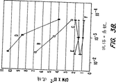
第3A図−第3B図は、125I−pGEX−3X−MN蛋白質およびいろいろな抗体を用いた放射免疫沈降実験の結果をグラフに表したものである(実施例8に記述)。放射活性蛋白質(15×103cpm/チューブ)は、次の腹水あるいは血清およびSACと沈降した。(A)MAb M75を含む腹水;(B)ウサギ抗MaTu血清;(C)正常ウサギ血清;(D)ヒト血清L8;(E)ヒト血清KH;(F)ヒト血清M7。
第4図は、MN抗原に対するラジオイムノアッセイの結果を示す(実施例8に記述)。腹水(沈降が50%の放射活性となるように希釈)は、次のものと2時間反応させた。(A)「コールド(cold)」(ラベルしていない)蛋白質pGEX−3X−MN;あるいは以下の細胞の抽出物(B)HeLa+MX;(C)Rat−2TK-;(D)HeLa;(E)ラットXC;(F)T24および(G)HEF。次に、125IでラベルしたpGEX−3X−MN蛋白質(25×103cpm/チューブ)を加え、さらに2時間インキュベートした。最後に、放射活性MAb M75をSACに吸着させ、測定した。
発明の詳細な説明
MaTu−−MXおよびMNコンポーネント
本明細書に記述するように、MaTuは2コンポーネントシステムからなっている。コンプレックスの一つの部分である外因性MXは、透過性であり、蛋白質p58Xを発現する。該蛋白質は、ヒトおよびさまざまな動物の天然の血清と反応する細胞質抗原である。もう一つのコンポーネントであるMNは、ヒト細胞に内因するものである。
MNは細胞性遺伝子であり、既知のDNA配列とはほとんど相同性がない。MNは保存性であり、多くの脊椎動物の染色体DNA内で単一のコピー遺伝子として存在している。本明細書には、MN cDNAのクローニングおよび配列決定、また融合蛋白質(MN+グルタチオンS−トランスフェラーゼのC末端からなる:アフィニティークロマトグラフィーにより容易に精製できる)の遺伝子工学的処理について記述している。
MNはHeLa細胞において双子蛋白質p54/58Nとして表現され、細胞表面および核内に局在している。p54/58Nに反応するモノクローナル抗体(MAb M75)を用いたイムノブロットにおいて54kdと58kdの2本のバンドが確認できる。これらの2本のバンドは、一種類の蛋白質に関して、グリコシル化のパターンあるいはプロセッシングの過程が異なることによるものかもしれない。(p54Nおよびp58Nは、両方ともマンノースを含むオリゴ糖残基によりグリコシル化されているが、p58Nだけはグルコサミンも含有している。)本明細書で使用する「双子蛋白質(twin protein)」とはp54/58Nを指している。
MNは、HeLa細胞の急速成長する希薄培養中では出現しないが、細胞を高密度培養中で維持することにより、あるいは、さらに効率的な手段として、細胞にMXを感染させることにより誘導される。p54/58NのみがMaTuに感染したHeLa細胞内で再生された水疱性口内炎ウイルス(vesicular surface virus, VSV)のビリオンに結合する。双子蛋白質p54/58Nはグリコシル化され、ジスルフィド結合で連結されたオリゴマーの形をとっているのに対し、p58Nはグリコシル化されておらず、ジスルフィド結合で連結されたオリゴマー形ではない。
VSVは、HeLa細胞内でp54/58Nをビリオン中に集めるが、このことは、該双子蛋白質がVSVのG蛋白質変異型の相補性と、VSV(MaTu)のプソイド型の形成に関与していることを示唆している。エンベロープを有するウイルスのみが、感染性で機能的なプソイド型を形成するための表面糖蛋白質を産生し、ビリオンの細胞への吸着および侵入という特殊な機能を発揮することができる(ザバダ(Zavada),J.,J. Gen. Virol.,63: 15-24(1982))。このことはMN遺伝子が疑似ウイルスの配列として行動することを示している。
エンベロープを有するウイルスの表面蛋白質は、VSVのプソイド型の形成に関与しており、MN双子蛋白質p54/58Nと同様に、グリコシル化されている。また、MN蛋白質は、オリゴマー(好ましくは三量体または四量体)の形成においてはウイルスの糖蛋白質と類似している。そのようなオリゴマー形成においては、S−S結合(ジスルフィド結合)の関与は必須ではないが、ビリオンの集積には必須である(クレイス(Kreis)とロディッシュ(Lodish),Cell, 46: 929-937(1986))。ジスルフィド結合は2−メルカプトエタノールを用いて還元することにより分裂する。
パストレコヴァ(Pastorekova)らにより報告されているように(Virology, 187: 620-626(1992))、メルカプトエタノールを用いて還元した後は、細胞抽出物あるいはVSV由来のp54/58Nはイムノブロットにおいて非常に類似している。還元を行わない場合、細胞抽出物のp54/58Nは150kd付近に複数のバンドとして現れ、このことは細胞が数種の異なるオリゴマー(おそらくp54:p58の比率が異なる)を含んでいることを示唆している。しかし,VSVにおいては選択的にその中の一つ、分子量約153kdに集まっている。該オリゴマーは三量体もしくは四量体であり、54kdおよび58kdの蛋白質からなっている。還元状態でのVSVサンプルの分析において54kdと58kdのバンドの強さがほぼ同等であることから、VSVビリオンにおいてはp54:p58が等モル比であることが示された。
MN蛋白質の発現は、腫瘍性疾患の診断/予後にみられる。MN双子蛋白質p54/58Nは、HeLa細胞およびスタンブリッジ(Stanbridge)の腫瘍形成性(H/F−T)ハイブリッド細胞において発現が確認されているが(スタンブリッジ(Stanbridge)ら、Somatic Cell Genet, 7: 699-712(1981)およびスタンブリッジ(Stanbridge)ら、Science, 215: 252-259(1982))、線維芽細胞あるいは非腫瘍形成性(H/F−N)ハイブリッド細胞においては確認されていない(スタンブリッジ(Stanbridge)ら、同上)。ヒト卵巣、子宮内膜および子宮頚管癌、またいくつかの良性腫瘍(乳頭腫など)を用いて行ったイムノブロットにおいてはMN蛋白質が確認されるが、正常な卵巣、子宮内膜、子宮あるいは胎盤の組織からは確認されない。MXに感染したHeLa細胞内では、微細構造の交替が顕著に行われており、このことは、細胞表面でのおびただしい糸状体の形成およびミトコンドリアの増幅を意味している。免疫金(immunogold)標識法を用いると、p54/58Nは糸状体の表面および核、特に核小体上で観察される。すなわち、MN蛋白質は正常非腫瘍細胞では産生されていないことから、腫瘍特異的であるといえる。
本明細書の実施例において、MNおよびMXは二つの異なる存在であり、互いに独立して存在することが示されている。外因性の透過性物質であるMXは、線維芽細胞あるいは非腫瘍形成性(H/F−N)ハイブリッド細胞において増殖するが、これらの細胞はMN関連蛋白質は発現しない(第2A図−第2B図)。そのような細胞内ではMXはMN蛋白質の産生を誘導しないのである。第2A図−第2B図および実施例5と6に示されるように、MXの非存在下でもMN蛋白質はHeLa細胞および他の腫瘍細胞内で産生される。しかしながら、MXはHeLa細胞内におけるMN蛋白質の強力な誘導剤である。非感染細胞内において、MXはMN蛋白質の産生を濃度にして30倍増加させた(下記の実施例5と8、実施例8の表1参照)。
MN遺伝子−−クローニングおよび塩基配列決定
第1A図−第1B図は、本項に記載されている方法に従って単離されたMN cDNAクローンの塩基配列を示している。遺伝子コドンの縮重から、一つのコドンが一つ以上のアミノ酸をコードしており(たとえば、TTA、TTG、CTT、CTC、CTAおよびCTGはいずれもロイシン(leu)というアミノ酸をコードしている)、また、たとえば第1A図−第1B図に示すように、一つのコドンが他のコドンと入れ替わるヌクレオチド配列の多様性により、本発明と実質的に同等な蛋白質およびポリペプチドが産生される。MN cDNAのヌクレオチド配列および相補的な核酸配列に関するそのような変形もすべて本発明の範ちゅうに含まれる。
さらに、本明細書に記述し、第1A図−第1B図に示しているヌクレオチド配列は、単離され、本明細書で説明しているcDNAヌクレオチド配列のうち、はっきりした構造のみを表したものである。わずかに変更されたヌクレオチド配列が見つかることもあろうし、また、たとえば、同様のエピトープを有する等の、実質的に同等なMN蛋白質およびポリペプチドをコードするように当該分野で知られた技術により変形することも可能である。そしてそのような蛋白質/ポリペプチドは本発明の目的に適合する。MN蛋白質/ポリペプチドと相同あるいはほぼ相同な蛋白質/ポリペプチドをコードする合成核酸配列のように、同等なコドンを有するDNAおよびRNAは本発明の範ちゅうに含まれる。遺伝子コードの縮重がなければ、これらの核酸配列はやはり前記cDNAヌクレオチド配列にハイブリダイズする。本明細書で説明しているように、核酸配列が修飾されたり変形される結果、MN配列およびその断片と実質的に同等の配列が作り出される。
MN遺伝子を見つけるために、MX感染HeLa細胞からλgt11によるcDNAライブラリーを調製した。MX感染HeLa細胞からの全RNAはグアニジンチオシアネートCsCl法を用いて抽出し、mRNAはオリゴdTセルロースを用いるアフィニティーにより分離した。cDNAの合成およびgt11へのそのクローニングはアマシャム(Amarsham)社のキットを用いて行ったが、EcoRI−NotIアダプターだけはストラタジーン(Stratagene)社(米国、カリフォルニア州、ラ・ホーラ(La Jolla))のものを使用した。モノクローナル抗体M75とアルカリフォスファターゼを縮合したヤギ抗マウス抗体とを組合せたイムノスクリーニングにライブラリーをかけた。このイムノスクリーニング法は、ヤング(Young)とデイヴィス(Davis)により報告されている(PNAS(USA), 80: 1194-1198(1983))。350,000のプラーク(全ライブラリーのおおよそ半分にあたる)をスクリーニングし、1個のポジティブクローンを取り出した。
ポジティブクローンをpBluescript KS(ストラタジーン(Stratagene)社)のNotI部位に組み込んでサブクローニングを行い、pBluescript-MNを作った。Erase-a-BaseTMキット(プロメガ(Promega)社、米国、ウィスコンシン州、マディソン)を使用して、方向が反対で重なる2個の欠失を作り、T7シークエンス用キット(ファルマシア(Pharmacia)社、米国、ニュージャージー州、ピスカタウェイ)を用いてジデオキシ法により配列を決定した。配列はcDNAクローンの一部を表しており、インサートの長さは1397bpであった。本配列を第1A図−第1B図に示す(SEQ ID NO.1)。配列は、大きな1290bpのオープンリーディングフレームおよびポリAシグナル(AATAAA)を含む107bpの3’非翻訳領域からなっている。該配列のもう一つの特徴は、mRNAの不安定性に関与する領域(1389番目のAUUUA)が存在することである。この領域は、ある種の腫瘍遺伝子およびリンホカインのmRNAに特異的なものである(ショウ(Shaw)とカーメン(Kamen),Cell, 46: 659-667(1986))。MNクローンの大きさと対応するmRNAのそれとをノーザンブロットにより比較すると(実施例12)、このcDNAは、その配列の5’末端から約100bpが欠損していることがわかった。
MN cDNAクローンのオープンリーディングフレームは、約48kdの推定蛋白質をコードしている(第1A図−第1B図、SEQ ID NO.2)。推定翻訳アミノ酸(AA)配列の分析では、既報の蛋白質の配列とは高い相同性を示さなかった。最も近い相同性が見られたのは、MN蛋白質およびいろいろな型の炭酸脱水素酵素のC末端である(170−200AAの重なりにおいて約30−35%)。炭酸脱水素酵素の活性部位は、Zn2+結合ドメインと同様に、MN蛋白質においてもよく保存されている。しかしながら、MN遺伝子は、ヒトゲノム由来の新規な配列であることは明らかである。
上述したように、MN遺伝子は既知の炭酸脱水素酵素といくらかの相同性を有するが、いくつかの面でそれらとは異なっている。7個の炭酸脱水素酵素が報告されている(ドッジソン(Dodgson)ら(編)、炭酸脱水素酵素(The Carbonic Anhydrases)、(プレナムプレス(Plenum Press)社、ニューヨーク/ロンドン(1991年))。それらのおのおのは7個のイントロンを含んでいるが、MN遺伝子はイントロンを含まないようである。また、既知の炭酸脱水素酵素はすべておよそ30kdの蛋白質であり、これらはMN遺伝子のp54/58N関連の産生物より小さい。さらに、炭酸脱水素酵素はMN関連蛋白質のようなオリゴマーを形成しない。
推定アミノ酸配列から、MN遺伝子の産生物は、303−313のアミノ酸位置に一つの活性なN−グリコシル化部位を有する塩基性蛋白質(pI9.08)であることが明らかである。これらの事実は、HeLa細胞由来のp54/58N蛋白質が、Endo HおよびEndo Fによる分裂(おのおの約3kdの欠損を起こす)に感受性であることに対応している。親水性プロフィルは、アミノ酸の親水性配列(371−395位)を示しており、これはプラズマ膜にかかる領域を表していると考えられ、また、分裂シグナルも含むと考えられる。該プロフィルは、p54/58N蛋白質が細胞膜に局在していることとよく一致する。MNアミノ酸配列にはPEST領域は存在しないことから、MN遺伝子の産生物は安定で永続性の蛋白質であることが示唆される(ロジャース(Rogers)ら、Science, 234: 364-368(1986))。そのような特性から、発明者らのp54/58Nの代謝ラベルが非効率的であったことを説明できる。推定アミノ酸配列はさらにほかの特徴をも示す。すなわち、10個の活性リン酸化部位と7個のミリスチル化部位および3個の抗原決定因子を有する。
p54/58N蛋白質の両方が一つの遺伝子によってコードされているか否かを確認するために、MN遺伝子の発現を特異的に阻害するアンチセンスODNsを用いた。(このようなアンチセンスODNsの使用法については、ステイン(Stein)とコーエン(Cohen)により総説されている(Cancer Res., 48: 2659-2668(1988))。)これらの実験については実施例11に詳述している。実験の結果、ODNsと共に培養したHeLa細胞においてはp54/58Nの合成がかなり阻害されていることがわかり、一方、いろいろなHeLa細胞の蛋白質の産生量はほぼ同程度に保たれていた。さらに、イムノブロットにおいて重要な結果が得られた。すなわち、ODNsによる特異的阻害はp54/58N蛋白質のの両方に影響を与えていた(実施例11)。これらのことから、MN遺伝子はHeLa細胞においてp54/58N蛋白質の両方をコードしていると結論づけられる。
クローニングされた遺伝子がp54/58N特異的蛋白質をコードしているか否かを確認するために、該遺伝子をバクテリア発現ベクターpGEX−3X(ファルマシア(Pharmacia)社、スゥエーデン、ウプサラ)にサブクローニングし、グルタチオンS−トランスフェラーゼのC末端を有する融合蛋白質を発現するように構築した。このサブクローニングは、本発明におけるMN関連蛋白質の遺伝子工学的手法の一つを示すものである。以下の記述は例示であり、如何なる意味においても本発明を限定するものではない。
融合蛋白質pGEX−3X−MNの産生
上述のpBluescript-MN由来のcDNA挿入体(インサート)は、プラスミドDNAをNotIで消化(切断(digesting))することにより切出した。該cDNA挿入体は、平滑末端を得るためにS1ヌクレアーゼで処理し、pGEX−3X(ファルマシア(Pharmacia)社)の脱リン酸化したSmaI部位にクローニングした。XL1-Blueの形質転換およびIPGTによる誘導の結果、融合蛋白質が得られた。
融合蛋白質であるMNグルタチオンS−トランスフェラーゼは、グルタチオンS−セファロース4B(ファルマシア(Pharmacia)社)を用いたアフィニティークロマトグラフィーにより精製した。10%ゲルを用いたSDS−PAGEにより分離された2つの同様なサンプルから、20ミリグラムの精製された組換え蛋白質が得られた。一つのサンプル(A)はクマジーブリリアントブルーで染色し、他方のサンプル(B)はハイボンドCメンブレン(Hybond C membrane)(アマシャム(Amarsham)社、英国、バックス、アリスバリー)にブロットした。このブロットは、125IでラベルしたMAb M75を用いてオートラジオグラフィーにより展開した。
SDS−PAGE分析から興味深い結果が得られた。すなわち、異なる分子量を有する一連の蛋白質のバンドが存在することである。本発明に従って産生された別の融合蛋白質である、β−ガラクトシダーゼMN(λ gt11溶原)においても同様なDSD−PAGEパターンが得られた。MN配列内に9個のAGGAGGコドンタンデムが存在することによる翻訳エラーのために、これらのパターンが現れたものと思われる。対応するtRNAが短いため、バクテリア遺伝子内でこれらのコドンを使用することは絶対に避けられている。かくして、外来性mRNAからのAGGAGGタンデムの翻訳の際に、+1のリボソーム上でのフレームシフトが高い頻度(約50%)で起こる(スパンヤード(Spanjaard)ら、Nuc. Acid Res., 18: 5031-5036(1990))。
イムノブロッティングにおいて、同様なパターンが得られた。すなわち、染色されたSDS−PAGEゲル上で確認された全てのバンドは、MN特異的MAb M75に反応し、このことは、全ての蛋白質のバンドがMN特異的であることを示している。また、これらの結果から、MAb M75への結合部位は、フレームシフトの影響を受けないMN蛋白質のN末端部分にあることが示された。
下記の実施例8に示すように、融合蛋白質pGEX−3X−MNは、MN特異的抗体およびMN抗原のラジオイムノアッセイに使用した。
MN蛋白質および/またはポリペプチド
本明細書で用いている「MN蛋白質および/またはポリペプチド」(MN蛋白質/ポリペプチド)とは、MN遺伝子あるいはその断片によりコードされている蛋白質および/またはポリペプチドを意味している。好ましいMN蛋白質の例は、推定アミノ酸配列が第1A図−第1B図に示されているものである(SEQ ID NO.2)。好ましいMN蛋白質/ポリペプチドは、第1A図−第1B図に示すMN蛋白質と実質的に相同性を有する蛋白質/ポリペプチドである。
「ポリペプチド」とは、ペプチド結合によるアミノ酸の共有結合鎖のことであり、本明細書では、50あるいはそれ以下のアミノ酸から構成されるものと考えている。本明細書における「蛋白質」とは、50より多くのアミノ酸から構成されるポリペプチドと定義される。
インビボ(in vivo)の腫瘍細胞から産生される蛋白質/ポリペプチドの配列が細胞培養内の腫瘍細胞から産生される蛋白質/ポリペプチドのものと異なることがある。すなわち、MN蛋白質/ポリペプチドが、アミノ酸置換、伸張、欠損、削除およびそれらの組合せ(これらに限定されるわけではないが)のようなアミノ酸配列変化を有していても、それらはすべて本発明の範ちゅうに属する。体液中に残存する蛋白質は蛋白質分解などの分解処理を受けることがある。すなわち、血清などの体液中にはかなりの削除が行われたMN蛋白質およびMNポリペプチドが見いだされる。本明細書で使用している「MN抗原」とは、MN蛋白質/ポリペプチドを包含している。
さらに、MN蛋白質およびポリペプチドのアミノ酸配列は、遺伝子工学によって変化させることもできる。1個またはそれ以上のアミノ酸を削除したり置換することができる。そのようなアミノ酸の変化も、生物学的活性に有意の変化をもたらさず、本発明の範囲に含まれる蛋白質やポリペプチドを生じさせることができる。
本発明のMN蛋白質およびポリペプチドは、本発明の方法にしたがって、さまざまな手段で調製できる。たとえば、組換え、合成、あるいはその他の生物学的手法、すなわち、長い蛋白質およびポリペプチドを酵素および/または化学的に解裂する等の方法が挙げられる。MN蛋白質を調製する好ましい方法は組換え法である。組換えによるMN蛋白質の産生のために特に好ましい方法は、融合蛋白質pGEX−3X−MNに関して上述した方法である。
MN蛋白質およびポリペプチドの組換え産生
第1A図−第1B図に示すMN蛋白質およびその断片を調製する代表的な方法は、適切なMN cDNA断片を上で例示したような適切な発現ベクターに挿入することである。本明細書に記載しているように、単離されたMN DNAのクローニングには、広範な種類の宿主−クローニングベクターの組合せを用いることができる。たとえば、有用なクローニング媒体としては次のようなものを挙げることができる。染色体DNA、非染色体DNA、合成のDNAであって、たとえば、pBR322等の各種の既知のバクテリアプラスミド、その他の大腸菌(E. Coli)プラスミドおよびそれらの誘導体、ならびに広い宿主範囲のプラスミド、たとえば、RP4やファージDNA(たとえば、NB989等のλファージの多数の誘導体)、ファージDNAの発現をコントロールする配列を有する修飾プラスミド等のプラスミドとファージDNAの組合せから作られたベクターなど。プラスミドpGEX−3Xが好ましいクローニング媒体である。
宿主として有用な細胞は真核性でも原核性でもよく、次のようなものが例示される。バクテリア宿主、たとえば、大腸菌(E. Coli)とその他のバクテリア株、酵母およびその他の菌類等、動物または植物の培養細胞等の動物あるいは植物宿主、昆虫細胞およびその他に宿主。もちろん、全ての宿主が同じ効果を有するわけではない。本明細書に記載されている原則を考慮しながら本発明の範囲から逸脱しないように、当業者によって宿主−クローニング媒体の組合せの選択がなされる。
組換えDNA分子を形成するため、選択されたDNA断片をクローニング媒体へ挿入する特定部位の決定は、さまざまな因子に影響される。これらの因子としては、発現させる蛋白質およびポリペプチドのサイズと構造、所望する蛋白質およびポリペプチドの宿主細胞の成分による内部酵素分解に対する感受性、宿主細胞蛋白質によるコンタミネーション、開始および終了コドンの局在などの発現特性、および当業者により知られているその他の因子などが挙げられる。
MN遺伝子、その断片あるいはMN遺伝子由来のcDNAを含む組換え核酸分子は宿主の形質転換に用いられるが、その形質転換により、宿主(形質転換体)にその構造蛋白質および断片を発現させ、かつ、ハイブリッドDNAがコードしている蛋白質およびポリペプチドを産生させる。組換え核酸分子はまた、MN核酸およびその断片の源としてさらに組換え核酸分子を複数生産させるために宿主の形質転換に用いられる。これらのそれぞれの使用に対する適切な宿主の選択は、当該分野で知られている多くの因子により定められている。これらの因子としては、たとえば、選択したベクターとの適合性、共生産物の毒性、所望する蛋白質およびポリペプチドの回収のしやすさ、発現特性、生物学的安全性およびコストが挙げられる。
宿主細胞として大腸菌(E. Coli)などの原核細胞を用いる場合は、DNAを取り込むことのできるコンピテントな細胞は、指数増殖期の後に細胞を回収(ハーベスト)し、続いて既知の手法であるCaCl2法で処理することにより調製できる。宿主細胞のプロトプラストを作った後、形質転換を行うことができる。
宿主細胞として原核細胞を用いる場合には、リン酸カルシウム沈降法、エレクトロポレーション、マイクロインジェクション等の従来からの機械的手法、赤血球あるいはリポソームに封入したプラスミドの挿入、リゾフォスファチジルコリン等の薬剤を用いた細胞処理、あるいはウイルスベクターの使用などの方法がとられる。
蛋白質およびポリペプチドの産生量は3つの大きな因子に左右される。(1)細胞内の遺伝子またはDNA配列のコピー数、(2)該遺伝子および配列が転写、翻訳される効率、および(3)mRNAの安定性。転写、翻訳(これらが共同して発現を司る)の効率は、核酸配列、一般には所望のコード配列の前に位置している核酸配列に依存する。これらの核酸配列、すなわち発現コントロール配列は、とりわけ、転写を開始するためにRNAポリメラーゼが相互作用する位置(プロモーター配列)、および翻訳を開始するためにリボソームがmRNA(転写産物)と結合し、相互作用する位置を定めている。そのような全ての発現コントロール配列が同じ効果を発揮するわけではない。そのため、所望する蛋白質に特異的なコード配列を近傍の核酸配列から切り出し、その代わりに既知の発現コントロールベクターに融合して希望する高レベルの発現を得られるようにするのがよい。上述の操作を行ってから、処理されたこの新しいDNA断片が多数のプラスミドまたはバクテリオファージ誘導体に挿入され、細胞内で遺伝子または配列のコピー数を増し、さらに、発現蛋白質の収量を増加させる。
発現コントロール配列として数種のものが用いられる。これらの中には、オペレーター、プロモーター、大腸菌(E. Coli)のラクトースオペロン(lac系)のリボソーム結合および相互作用配列、大腸菌(E. Coli)のトリプトファン合成系(trp系)に対応する配列、trpとlacプロモーターの融合(tac系)、λファージの主要オペレーターとプロモーター(OLPLとORPR)、およびfdファージのコート蛋白質のコントロール領域などが含まれる。これらの配列を含むDNA断片は、lacあるいはtrpオペロンを有する形質導入ファージのDNA、またはλあるいはfdファージのDNAから制限酵素を用いた開裂により切出せる。つぎに、これらの断片を操作して、必須コントロール配列が、コード配列の開始コドンの非常に近傍または並列に位置しているようになった限定された分子集団を得る。
融合体は次に、適切な宿主の形質転換あるいはトランスフェクションのためのクローニング媒体に挿入し、抗原産生量を測定する。そして、最も効率的な発現をする細胞を選択する。別の方法としては、開始コドンに接続されたlac、trpまたはλPLコントロール系を有するクローニング媒体を用い、MN蛋白質およびポリペプチドをコードする配列を含む断片に融合し、それによって遺伝子あるいは配列がクローニング媒体の開始コドンから正しく翻訳されるようにすることもできる。
本発明で使用している「組換え核酸分子」とは、少なくとも2つの核酸配列からなるハイブリッドヌクレオチド配列を指しており、ここで、第一の配列は、通常、自然状態では第二の配列と共存していない。
本明細書で使用している「発現コントロール配列」とは、構造遺伝子と結合した場合にその発現を制御または調製するヌクレオチド配列のことである。
MN蛋白質およびポリペプチドの合成および生物工学的発現
本発明のMN蛋白質およびポリペプチドは組換え法のみでなく、合成およびその他の生物学的手法によっても調製することができる。蛋白質またはポリペプチドの合成による産生は、当該分野でよく知られた方法に従って所望するアミノ酸鎖を化学的に合成していくことからなる。所望するポリペプチドまたは蛋白質を調製するためのその他の生物学的手法の例としては、所望するアミノ酸配列を含む長いMNポリペプチドまたは蛋白質を選択的に蛋白分解することが挙げられる。たとえば、長いポリペプチドまたは蛋白質を化学試薬または酵素で分解することなどである。
ペプチドの化学合成は従来技術であり、たとえば、メリフィールド(Merrifield)固相合成法(メリフィールド(Merrifield),J.,Am. Chem. Soc., 85: 2149-2154(1963)、ケント(Kent)ら、「生物学および医学における合成ペプチド(Synthetic Peptides in Biology and Medicine)29ページ」、アリターロ(Alitalo)ら編、エルセヴィール科学出版(Elsevier Science Publishers)社、1985年、およびハーグ(Haug),J.D.「ペプチド合成および保護基戦略(Peptide Synthesis and Protecting Group Strategy)」,American Biotechnology Laboratory, 5(1): 40-47(1987年、1/2月号)などにより実施される。
化学的ペプチド合成の方法には、市販の保護アミノ酸を用いる自動ペプチド合成機の使用も含まれる。合成機としては、たとえばバイオサーチ(Biosearch)社(米国、カリフォルニア州、サンラファエル)の9500型および9600型、アプライド バイオシステムズ(Applied Biosystems)社(米国、カリフォルニア州、フォスターシティー)の430型、ミリジェン(Milligen)社(ミリポア(Millipore)社の子会社、米国、マサチューセッツ州、ベッドフォード)の9050型、およびデュポン(DuPont)のRAMP(高速自動複数ペプチド合成機)(デュポンコンパス(Dupont Compass)社、米国、デラウェア州、ウィルミントン)などがある。
核酸プローブおよび試験キット
本発明の核酸プローブは、第1A図−第1図Bに示すMN cDNA配列またはMN遺伝子配列と実質的に相補的な配列からなる。本明細書で使用する「実質的に相補的」という語は、当該技術分野で広く理解されている意味と同じであり、それゆえ、通常のハイブリダイゼーションの状態の意味で用いる。ハイブリダイゼーションの程度は、相補正の精度に応じて変わり得る。
前記プローブは、MN DNAおよび/またはRNAの検出に使用でき、したがって、患者の細胞内のMN遺伝子の存在や欠如、増殖、変異、あるいは遺伝子再配列の試験に使用できる。たとえば、MN遺伝子の過剰発現は、本発明のプローブを使用したノーザンブロットにより検出できる。増幅、転座、逆位ならびに欠失などの遺伝子変化は本発明のプローブを用いることにより検出でき、このとき、該プローブは、細胞分裂中期の染色体の広がった状態でも間期の核のいずれの状態であっても、患者の細胞由来の染色体とインサイチュー(in situ)ハイブリダイゼーションする。また、本発明のプローブを用いたサザンブロットによってMN遺伝子の増幅あるいは欠失を検出することもできる。該プローブを用いた制限酵素断片長多型(RFLP)分析は、遺伝子変化、変異および欠失の検出として好ましい方法である。該プローブはまた、いろいろな組織由来のMN遺伝子から転写された各種のmRNAとハイブリダイゼーションすることにより、MN蛋白質および/またはポリペプチドならびにそれらと相同またはほぼ相同な蛋白質および/またはポリペプチドを検出するために用いられる。
このように、該プローブは診断/予後に有用である。該プローブは試験キットとして具体化でき、好ましくは、該プローブが適切なMN遺伝子またはMN mRNAターゲットとハイブリダイゼーションした時に、視覚化できる適切な手段を伴っているのがよい。そのような試験のサンプルとしては、組織標本、体液ならびに組織および細胞の抽出物が挙げられる。
分析
本発明に従う分析は、脊椎動物のサンプル、好ましくはホ乳類のサンプル、より好ましくはヒトのサンプル中のMN抗原またはMN特異的抗体の検出および/または定量である。そのようなサンプルとしては、組織標本、体液、組織抽出物および細胞抽出物が挙げられる。MN抗原は、イムノアッセイ、免疫組織学的染色、免疫電子および走査顕微鏡観察、とりわけ、免疫金沈降(immunogold)を用いる技術により検出できる。
MN抗原分析の好ましいサンプルは組織および/または細胞抽出物である(下記の実施例7と8が代表的なものである)。しかし、MN抗原は体液、とりわけ、血液、血清、プラズマ、精液、乳汁、唾液、涙、喀痰、粘液、尿、リンパ液、サイトゾル、腹水、胸水、羊水、膀胱洗浄液、気管支肺胞洗浄液、随液からも検出できる。試験前に大量の体液からMN抗原を濃縮することが望ましい。分析に好ましい体液は、試験する癌の型にもよるが、一般的に好ましい体液は、乳汁、胸水および腹水である。
血液、プラズマ、血清、リンパ液、粘液、涙、尿、髄液および唾液などの体液サンプル中の活性MN蛋白質/ポリペプチドにMN特異的抗体は血清学的に結合するが、そのような抗体は血液、プラズマおよび血清、好ましくは血清に普通にみられる。MN特異的抗体の検出のための代表的な分析は、下記の実施例8に示すものであり、その実施例では融合蛋白質pGEX−3X−MNを用いている。MN抗原およびそれらと反応するMN特異的抗体の検出および/または定量試験の結果の相関から、患者の病状の好ましいプロファイルが示される。
本発明の分析は、診断および/または予後の両方、すなわち、診断/予後である。本明細書で使用している「診断/予後」とは、臨床的状況に依存して以下の手順のそれぞれ、または、それらの手順のいくつかが重複していることを意味する。疾病の存在の判断、疾病の特徴の判断、ある疾病と他の疾病との区別、病状の帰結予想、患者の様子と症状から示される疾病からの回復の見込み、患者の病状のモニタリング、疾病の再発に関する患者のモニタリング、および/または患者に対する好ましい治療方法の決定など。本発明における診断/予後の方法はたとえば以下のような場合に有用である。腫瘍性および/または前腫瘍性疾患の存在に対する集団のスクリーニング、腫瘍性疾患の進展の危険性の判断、腫瘍性および/または前腫瘍性疾患の存在の診断、腫瘍性疾患の患者の病状のモニタリング、および/または腫瘍性疾患の経過に対する予後の判断など。
本発明は、広範な種類の腫瘍性疾患の存在のスクリーニングに有用である。たとえば、乳腺、尿路、卵巣、子宮、子宮頚管、子宮内膜、偏平細胞および腺偏平細胞などの腫瘍;頭および首の癌;神経芽細胞腫および網膜芽腫などの中胚葉性の腫瘍;骨肉腫およびユーイング肉腫(Ewing's sarcoma)などの肉腫;あるいはメラノーマなどが含まれる。特に興味があるのは婦人科の癌であり、この中には卵巣、子宮、子宮頚管、腟、陰門、子宮内膜癌が含まれるが、中でもとりわけ興味があるのは卵巣、子宮頚管および子宮内膜の癌である。同様に特に興味があるのは、胸、食道を含む胃、結腸、腎臓、前立腺、肝臓、膀胱を含む尿路系、肺、および頭と首の癌である。
本発明は、たとえば、宿主から取出した直後の細胞群を使用して、悪性腫瘍あるいは前悪性腫瘍性細胞の存在の可能性を推し量るための方法および構成物を提供する。そのような分析は、腫瘍の発見、それらの増殖の計測および疾病の診断と予後に役立つ。この分析はまた、癌の転移の存在の発見、ならびに、手術、癌の化学療法および/または放射線療法の後、全ての腫瘍組織がなくなっているか除去されているかを確認するのに役立つ。さらに、診断は、癌の化学療法および腫瘍の再発をモニターするのに役立つ。
MN抗原または抗体の存在は、多くの既に確立された診断分析を用いて検出および/または定量することができる。当業者であれば、従来から存在する任意のイムノアッセイ法を適用して、MN抗原および/または抗体を検出および/または定量することができる。実施例8に本発明の好ましい診断方法であるラジオイムノアッセイ法について詳しく述べている。もちろん、MN抗原およびMN特異的抗体の検出にはほかの多くの方式も利用可能である。たとえば、ウエスタンブロット、ELISA(酵素結合免疫吸着法)、RIA(ラジオイムノアッセイ)、競合EIA、二重抗体サンドイッチ法、その他診断研究において通常使用される全ての分析法などがある。そのようなイムノアッセイにおける結果の解釈は、抗体あるいは抗体の組合せは、MNに関係のないサンプル中に存在する他の蛋白質および蛋白質の断片とは交差反応しないという仮定に基づいている。
MN抗原検出の代表的な一つのELISA試験は、MN蛋白質/ポリペプチドに対して作られた抗体あるいはMN蛋白質を発現する細胞全体に対して作られた抗体をコートしたマイクロタイタープレートに、組織あるいは細胞抽出物のような患者のサンプルを加える方法である。ある抗原が抗体と結合するように一定時間インキュベートした後、プレートを洗浄し、酵素が結合している別の抗MN抗体を加え、反応が生じるようにインキュベートし、プレートを再洗浄する。その後、マイクロタイタープレートに酵素基質を加え、酵素が基質に作用するように一定時間インキュベートし、最終試料の吸光度を測定する。吸光度が大きく変化することは陽性の結果を示している。
患者の体液、組織および/または細胞中のMN抗原の存在を検出および/または定量するためにMN蛋白質/ポリペプチドが使用できることもイムノアッセイの分野の当業者には明かである。そのような実施態様の一つとして競合イムノアッセイがあるが、この方法では、MN蛋白質/ポリペプチドはラベルされ、MN蛋白質/ポリペプチドに特異的な抗体への該ラベル化MN蛋白質/ポリペプチドの結合に競合させるために体液を加える。そのような分析は実施例8に記載しているようなMN抗原の検出および/または定量に使用できる。
別の実施例としては、MN蛋白質またはポリペプチドに対するラベルされた抗体を用いるイムノメトリック分析がある。そのような分析においては、抗原結合抗体とコンプレックスを形成するラベル化抗体の量は、試料中のMN抗原の量と直接的に比例する。
MN特異的抗体を検出する代表的な分析は競合分析であり、この分析では、ラベルされたMN蛋白質/ポリペプチドはサンプル中の抗体、たとえば、MN蛋白質/ポリペプチドを認識するモノクローナル抗体と結合して沈降する。当業者であれば、従来から存在する任意のイムノアッセイ方法を適用してMN抗原の検出および/または定量することができる。該抗体のMN蛋白質/ポリペプチドとの結合の検出は、当業者に知られた多くの方法、たとえば、ヒトにおいては抗ヒトラベルIgGの使用などにより行うことができる。
本発明に従い、脊椎動物のサンプルからMN抗原を検出および/または定量するイムノアッセイの方法の例は、以下のステップから構成されている。
a)MN抗原に結合する一組あるいはそれ以上の組の抗体(一つの抗体かあるいは複数の抗体)(そのうち一組はラベルされているかまたはその他の手法で検出可能となっている)と脊椎動物のサンプルをインキュベートする。
b)MN抗原と該抗体からなる免疫コンプレックスが存在するかについて、インキュベートしたサンプルを調べる。
本発明に従う別のイムノアッセイ法の例は競合イムノアッセイであり、脊椎動物サンプル中のMN抗原を検出および/または定量するために使用され、以下のステップから構成されている。
a)脊椎動物のサンプルを一組あるいはそれ以上の組の抗体および一定量のラベルされたもしくは他の手段で認識できるようになっているMN蛋白質/ポリペプチドとインキュベートし、このとき、該MN蛋白質/ポリペプチドが抗体との結合において、サンプル中に存在するMN抗原と競合する。
b)インキュベートしたサンプルを調べて、抗体に結合しているラベルした/検出可能なMN蛋白質/ポリペプチドの量を測定する。
c)ステップb)の検査から、前記サンプル中に存在するMN抗原および/または前記サンプル中に存在するMN抗原の量を決定する。
望ましい特性を有する抗体(生物学的に活性な抗体の断片を含む)が調製されると、特定の抗体抗原コンプレックスの形成を判定するための広範な免疫学的分析法が可能である。多くの競合、非競合蛋白質結合分析に関して化学文献および特許明細書に記述されており、そのような分析法の多くは購入可能である。血清中の抗原の検出に適したイムノアッセイの例としては、以下の米国特許に記載されているものが含まれる。米国特許第3,791,932号、第3,817,837号、第3,839,153号、第3,850,752号、第3,850,578号、第3,853,987号、第3,867,517号、第3,879,262号、第3,901,654号、第3,935,074号、第3,984,533号、第3,996,345号、第4,034,074号、および第4,098,876号。
分析に用いれらる抗体は、ラベルされているかまたはラベルされていないものである。ラベルされていない抗体は凝集に用いられ、ラベルされた抗体は、多くの種類のラベルを施すことにより、広範な分析に使用される。
適切な検出には、放射核、酵素、補酵素、発蛍光剤、化学発光剤、色素原、酵素基質あるいは補助因子、酵素阻害剤、フリーラジカル、パーティクル、染料などのラベルを用いるものが挙げられる。そのようなラベル試薬は、既知のさまざまな分析、たとえば、ラジオイムノアッセイ、ELISA等の酵素免疫アッセイ、蛍光免疫アッセイなどに用いられる。たとえば、米国特許第3,766,162号、第3,791,932号、第3,817,837号および第4,233,402号を参照をこと。
本発明の分析において有用な抗体を調製する方法を以下に示す。以下の例は、本発明に従う代表的な分析について詳述したものである。
イムノアッセイ試験キット
今までに概要を示した分析は、MN抗原および/またはMN特異的抗体(生物学的に活性な抗体の断片を含む)を検出および/または定量する試験キットとして具体化される。MN抗原を検出および/または定量する試験キットは、MN蛋白質/ポリペプチドおよび/または、(ポリクローナルおよび/またはモノクローナルの)MN特異的抗体からなる。そのような診断/予後用の試験キットは、一組またはそれ以上の組のポリクローナルおよび/またはモノクローナル抗体から構成されており、サンドイッチ法の場合には、抗体がMN抗原のエピトープを認識し、一組は適切にラベルされているか、その他の手段により検出可能である。
ラベルされた(あるいは他の手段で検出可能な)MN蛋白質/ポリペプチドとサンプル中のMN抗原との間で抗体に対する結合に関して競合がある分析方法の試験キットは、ラベルされた蛋白質/ポリペプチドと抗体の量を組合わせて、最適の感度と精度が得られるように構成されている。
MN特異的抗体検出のための試験キットは、ラベルされた/検出可能なMN蛋白質/ポリペプチドから構成されていることが好ましく、必要であれば、たとえば、下記の実施例8に概要を示しているような好ましい分析を行うために、その他の成分を含んでいてもよい。そのような試験キットは、その他の適切な手段を含んで、従来からあるような分析に応用することもできる。
MN特異的抗体の調製
本明細書で使用している「抗体」という語は、抗体全体を指すだけでなく、生物学的に活性な抗体の断片、好ましくは抗原結合部位を有する断片をも指している。そのような抗体は、従来からの手法および/または遺伝子工学により調製できる。抗体の断片は、遺伝子操作により、好ましくは超可変領域を含む、短鎖および/または長鎖の可変領域(VHとVL)から調製されるのがよく、さらに好ましくは、VH領域とVL領域の両方から調製されるのがよい。たとえば、本明細書で使用している「抗体」という語は、ポリクローナルおよびモノクローナル抗体ならびにそれらの生物学的に活性な断片をも意味しており、とりわけ、「一価」抗体(グレニー(Glennie)ら、Nature, 295: 712(1982));共有結合性または非共有結合性凝集をするFab’およびF(ab’)2断片を含むFab蛋白質;好ましくは短鎖および長鎖の可変領域(VHとVL領域)を含み、より好ましくは超可変領域(該VHとVL領域の相補性決定領域(CDRs)としても知られている)を含む短鎖または長鎖単独;Fc蛋白質;1以上の抗体と結合可能な「ハイブリッド」抗体;定常−可変領域キメラ;異なる起源由来の長鎖および短鎖からなる「複合」免疫グロブリン;通常の組換え手法により、またはオリゴヌクレオチドの依存突然変異誘発法(ダルバディー−マクファーランド(Dalbadie-McFarland)ら、PNAS (USA), 79: 6409(1982))により調整された、特異性やその他の特性を改良した「改造」抗体などが含まれる。
治療および/またはイメージングに使用する抗体は、生物学的に活性な抗体の断片であることが好ましく、より好ましくは遺伝子工学的に処理された断片がよく、さらに好ましくはVHおよび/またはVL領域から遺伝子工学的に処理された断片がよく、さらにより好ましいのは、それらの超可変領域からなる断片である。
ポリクローナルおよびモノクローナル抗体を作出する従来からの技術は、イムノアッセイの分野では周知である。MN特異的抗体を産生するための免疫原には、MN蛋白質および/またはポリペプチド、好ましくは純粋で、MXに感染した腫瘍細胞系、たとえば、MX感染HeLa細胞およびその他の免疫原が含まれる。
抗ペプチド抗体は、ヨーロッパ特許出願公開第44,710号(1982年1月27日公開)に記載されているような当該分野の従来法によっても作出される。該方法を要約すると、第1A図−第1B図に示すようなMNアミノ酸配列からペプチドを選択し、化学的に合成し、適切な免疫原蛋白質に結合し、適切な動物、通常なウサギあるいはマウスに注入し、ポリクローナルまたはモノクローナル抗体を作らせることにより、抗ペプチド抗体が調製される。モノクローナル抗体はコーラー−ミルシュテイン(Kohler-Milstein)法に従って作出される。
従来のハイブリドーマ法だけでなく、新しい技術も本発明に従う抗体を産生するために用いることができる。たとえば、クローン作成および抗体のV遺伝子の発現にポリメラーゼ連鎖反応(PCR)法を用い、結合活性を有する断片をコードしている抗体遺伝子の選択にファージディスプレイ法を用いることにより、免疫されたマウスおよびヒト由来のPCR増幅したV遺伝子の集団から抗体の断片を単離することができた(マークス(Marks)ら、BioTechnology, 10: 779(1992年7月号)参照のこと。シャン(Chiang)ら、BioTechniques, 7(4): 360(1989);ワード(Ward)ら、Nature, 341: 544(1989年10月12日号);マークス(Marks)ら、J. Mol. Biol., 222: 581(1991);クラクソンClackson)ら、Nature, 352: 642-628(1991年8月15日号);ムリナックス(Mullinax)ら、PNAS (USA), 87: 8095(1990年10月号))。
組換え技術を用いた抗体(本明細書では生物学的に活性な抗体の断片を含む)の調製法に関する記載は下記に見出される。米国特許第4,816,567号(1989年3月28日付与)、ヨーロッパ特許出願公開番号(EP)第338,745号(1989年10月25日公開)、EP第368,684号(1990年6月16日公開)、EP第239,400号(1987年9月30日公開)、WO第90/14424号(1990年11月29日公開)、WO第90/14430号(1990年5月16日公開)、ヒューセ(Huse)ら、Science, 246: 1275(1989年12月8日号);マークス(Marks)ら、BioTechnology, 10: 779(1992年7月号);ラ・サストリー(La Sastry)ら、PNAS(USA), 86: 5728(1989年8月号);シャン(Chiang)ら、BioTechniques, 7(4): 360(1989);オーランディ(Orlandi)ら、PNAS(USA), 86: 3833(1989年5月号);ワード(Ward)ら、Nature, 341: 544(1989年10月12日号);マークス(Marks)ら、J. Mol. Biol., 222: 581(1991);フーゲンブーム(Hoogenboom)ら、Nucleic Acids Res., 19(15): 4133(1991)。
モノクローナル抗体の調製
本発明の分析で使用するモノクローナル抗体は、当該分野において既知の方法により得られる。たとえば、ガルフレ(Galfre)とミルシュテイン(Milstein)「モノクローナル抗体の調製:戦略および方法(Preparation of Monoclonal Antibodies: Strategies and Procedures)」(酵素学的方法:免疫化学的方法(Methods in Enzymology: Immunochemical Techniques)73巻1−46ページより)(ランゴーン(Langone)とヴァナティス(Vanatis)編、アカデミック・プレス(Academic Press)社(1981年)、および古典的な参考として、ミルシュテイン(Milstein)とコーラー(Kohler)、Nature, 256: 495-497(1975)がある。
本発明の代表的なハイブリドーマは、マウス細胞系の融合により調製されるが、ヒト/ヒトハイブリドーマ(オルソン(Olsson)ら、PNAS(USA), 77: 5429(1980))およびヒト/マウスハイブリドーマ(シュローム(Schlom)ら、PNAS(USA), 77: 6481(1980)、シェアマン(Shearman)ら、J. Immunol., 146: 928-935(1991)、ゴーマン(Gorman)ら、PNAS(USA), 88: 4181-4185(1991))からも調製され得る。そのようなヒト型のモノクローナル抗体は、治療およびイメージングに使用するのに好ましいモノクローナル抗体である。
本発明に用いるモノクローナル抗体は、適切なホ乳類、好ましくは齧歯類、より好ましくはウサギまたはマウスに、適切な免疫原、たとえば、MaTuに感染したHeLa細胞や、あるいは、必要であればキャリアー蛋白質をつけたMN蛋白質/ポリペプチドを用いて免疫することにより調製される。例として、MAb M75を分泌するハイブリドーマVU−M75の作出について以下に記載している。MAb M75は、多くの実験室での診断試験、たとえば、培養腫瘍細胞、臨床サンプルなどにおけるMN蛋白質/ポリペプチドの同定に役立っている。MAb M75を産生する方法により、MAb M16(IgG2Bのアイソタイプ)およびMAb M67(IgG1のアイソタイプ)も産生された。
MAb M75
モノクローナル抗体M75(MAb M75)は、マウスリンパ球ハイブリドーマVU−M75から産生されるが、該ハイブリドーマは、当初スロヴァク科学アカデミーウイルス研究所(Institute of Virology, Slovak Academy of Sciences)のハイブリドーマコレクション(Collection of Hybridomas)(チェコスロバキア、ブラティスラヴァ)に寄託され、また、アメリカン・タイプ・カルチャー・コレクション(American Type Culture Collection)(米国、メリーランド州、ロックヴィル)に1992年9月17日にATCC受入番号HB11128として寄託されたものである。
ハイブリドーマVU−M75は、ガーハード(Gerhard)、W.によって記載されている方法(「懸濁液中での細胞融合および調整培地中でのハイブリッドの発芽後生育(Fusion of cells in suspension and outgrowth of hybrids in conditioned medium)」モノクローナル抗体・ハイブリドーマ:生物学的分析の新しい方向(Monoclonal Antibodies. Hybridomas: A New Dimension in Biological Analysis)より、370ページ(ケネット(Kennet)ら編、プレナム(Plenum)社、米国、ニューヨーク))に従って産生される。BALB/CマウスをMaTuに感染したHeLa細胞で免疫し、それらの脾細胞をミエローマ細胞系NS−0と融合した。ハイブリドーマの組織培養培地をモノクローナル抗体のスクリーニングにかけた。用いた抗原は、MaTuに感染したHeLa細胞の抽出物とウサギ抗MaTu血清の免疫沈降物であるp58、および黄色ブドウ球菌(Staphylococcus aureus)細胞(SAC)のプロテインAであり(ザバダ(Zabada)とザバドバ(Zavadova)、Arch. Virol., 118: 189-197(1991))、SDS−PAGEゲルにより分離した。モノクローナル抗体は、プロテインA−セファロースを用いたアフィニティークロマトグラフィー(ハーロウ(Harlow)とレイン(Lane)、「抗体:実験室手順書(Antibodies: A Laboratory Manual)」コールドスプリングハーバー(Cold Spring Harbor)社、米国、ニューヨーク州、コールドスプリングハーバー、1988年)により、TC培地から精製された。
本発明に従ってMN蛋白質/ポリペプチドを同定するのに有用なモノクローナル抗体は、従来からの任意の方法によってラベルすることができる。たとえば、西洋ワサビペルオキシダーゼ(HRP)のような酵素、蛍光化合物、125Iなどの放射活性同位体などである。本発明に従う好ましいラベルは125Iであり、好ましい抗体ラベル化の方法は、クロラミン−T法(ハンター(Hunter)、W.H.、「ラジオイムノアッセイ(Radioimmunoassay)」実験免疫学の手引(Handbook of Experimental Immunology)より、14.1-14.40(D.W.ウェイアー(Weir)編、ブラックウェル(Blackwell)社、オックスフォード/ロンドン/エディンバラ/メルボルン、1978年)である。
MAb H460
モノクローナル抗体H460(MAb H460)は、MAb M75と同様な方法によって調製された。ただし、マウスをMaTuに感染していないHeLa細胞により免疫し、マウスの脾細胞ではなくリンパ球をミエローマ細胞系NS−0の細胞と融合した点が異なる。MAb H460は、どんなヒト細胞ともほぼ同様に反応する。
MN特異的抗体の治療への応用
本発明のMN特異的抗体、モノクローナル抗体および/またはポリクローナル抗体、好ましくはモノクローナル抗体、より好ましくはMAb M75は、腫瘍および/または前腫瘍性疾患の治療に用いることができ、単独あるいは化学療法剤または毒性剤(たとえば、リシンA等)と組み合わせて用いることができる。治療用としてより好ましいのは、本明細書に記載しているような生物学的に活性な抗体の断片である。同様に、治療用として好ましいMN特異的抗体は、ヒト型モノクローナル抗体である。
MN特異的抗体は、好ましくは生理学的に許容性の非毒性液体基剤に分散された形で、治療効果を発揮するのに十分な量が投与される。
イメージングへの抗体の応用
さらに、本発明のMN特異的抗体は、放射核などのイメージング剤と結合させた場合には、イメージングに利用できる。生物学的に活性な抗体の断片あるいはヒト型モノクローナル抗体がイメージング用としては好ましい。
たとえば、腫瘍部位や転移の位置などが患者の腫瘍組織から同定できる。適切にラベルしたあるいはイメージング剤と結合させた抗体を生理学的に許容性のキャリアーと共に患者に投与し、結合した抗体は、ラベルあるいはイメージング剤の検出に適した方法、たとえば、シンチグラフィーなどにより検出される。
アンチセンスMN核酸配列
本発明のMN遺伝子は、腫瘍遺伝子と推定され、それによってコードされている蛋白質は、腫瘍性蛋白質であると推定される。MN遺伝子から転写されたmRNAと実質的に相補性のアンチセンス核酸配列は、実施例11のアンチセンスオリゴデオキシヌクレオチド(ODNs)に代表されるものであり、MN遺伝子の発現を減少あるいは抑制するのに用いることができる(ザメクニック(Zamecnick)、P.C.「イントロダクション:遺伝子情報解読のモジュレーターとしてのオリゴヌクレオチド塩基ハイブリダイゼーション(Introduction: Oligonicleotide Base Hybridization as a Modulator of Genetic Message Readout)」1−6ページ、癌およびAIDSへのアンチセンス核酸療法の予測(Prospects for Antisense Nucleic Acid Therapy of Cancer and AIDS)より(ウィレイ−リス(Wiley-Liss)社、米国、ニューヨーク州、ニューヨーク、1991年);ウィックストーム(Wickstorm)、E.「HL−60前骨髄球性白血病細胞に対するアンチセンスDNA療法:末端配列の変化と標的配列への依存性(Antisense DNA Treatment of HL-60 Promyelocytic Leukemia Cells: Terminal Differenciation and Dependence on Target Sequence)」7−24ページ、同上;レザマン(Leserman)ら、「腫瘍遺伝子の発現に干渉するアンチセンスオリゴヌクレオチドの標的化と細胞内分布(Targeting and Intracellular Delivery of Antisense Oligonucleotides Interfering with Oncogene Expression)」25−34ページ、同上;ヨコヤマ(Yokoyama)、K.「アンチセンスRNAによる腫瘍原遺伝子c-mycの転写制御(Transcriptional Regulation of c-myc Proto-oncogene by Antisense RNA)」35−52ページ、同上;ヴァンデンベルク(van den Berg)ら、「染色体の異常世代を抑制するアンチセンスfosオリゴデオキシリボヌクレオチド(Antisense fos Oligodeoxyribonucleotides Suppress the Generation of Chromosomal Aberrations)」63−70ページ、同上;メルコーラ(Mercola)、D.「アンチセンスfosおよびfunRNA(Antisense fos and fun RNA)」83−114ページ、同上;イノウエ(Inouye)、Gene, 72: 25-34(1988)、ミラー(Miller)とティソー(Ts'o)、Ann. Reports Med. Chem., 23: 295-304(1988);ステイン(Stein)とコーエン(Cohen)、Cancer Res., 48: 2659-2668(1988);ステヴェンソン(Stevenson)とインヴァーセン(Inversen)、J. Gen. Virol., 70: 2673-2682(1989);グッドチャイルド(Goodchild)「オリゴヌクレオチドによる遺伝子発現の抑制(Inhibition of Gene Expression by Oligonucleotides)」53−77ページ、オリゴデオキシヌクレオチド:遺伝子発現のアンチセンス抑制剤(Oligodeoxynucleotides: Antisense Inhibitors of Gene Expression)、(コーエン(Cohen)、J.S.編、CRCプレス(CRC Press)社、米国、フロリダ州、ボカ・ラートン、1989年);デルヴァン(Dervan)ら、「三重らせん形成による二重らせんDNAのオリゴヌクレオチドの認識(Oligonucleotide Recognition of Double- helical DNA by Triple-helix Formation)」197−210ページ、同上;ネッカーズ(Neckers)、L.M.「細胞制御研究の道具としてのアンチセンスオリゴデオキシヌクレオチド:取込のメカニズムと腫瘍遺伝子機能の研究への応用(Antisense Oligodeoxynucleotides as a Tool for Studying Cell Regulation: Mechanisms of Uptake and Application to the Study of Oncogene Function)」211−232ページ、同上;レイトナー(Leitner)ら、PNAS(USA), 87: 3430-3434(1990);ベヴィラッカ(Bevilacqua)ら、PNAS(USA), 85: 831-835(1988);ローク(Loke)ら、Curr. Top. Microbiol. Immunol., 141: 282-288(1988);サリン(Sarin)ら、PNAS(USA), 85: 7448-7451(1988);アグラワル(Agrawal)ら、「アンチセンスオリゴヌクレオチド:化学療法およびAIDSへのアプローチの可能性(Antisense Oligonucleotides: A Possible Approach for Chemotherapy and AIDS)」核酸療法に関する生化学会国際会議(International Union of Biochemistry Conference on Nucleic Acid Therapeutics)(1991年1月13−17日、米国、フロリダ州、クリアウォタービーチ);アームストロング(Armstrong)、L.、Ber. Week、88−89ページ(1990年3月5日号);ウエイントラウブ(Weintraub)ら、Trends, 1: 22-25(1985))。そのようなアンチセンス核酸配列、好ましくはオリゴヌクレオチドは、MN mRNA、特にリボソーム結合部位と翻訳開始点の近傍でハイブリダイゼーションすることにより、mRNAの翻訳を阻害する。それゆえ、そのようなアンチセンス核酸配列を使用することは、癌の治療の一つの方法と考えられる。
本発明に従う好ましいアンチセンスオリゴヌクレオチドは、遺伝子特異的ODNsあるいはMN mRNAの5’末端に相補的なオリゴヌクレオチドである。特に好ましいのは、下記の実施例11に配列が示されているような、29−mer ODN1および19−mer ODN2である。これらのアンチセンスODNsは、MN遺伝子の発現を抑制する機能を有する多くのアンチセンス核酸配列の中の代表的なものである。当業者であれば、第1A図−第1B図の核酸配列から適切なアンチセンス核酸配列、好ましくはアンチセンスオリゴヌクレオチドを確定することができる。
ワクチン
本発明のMN蛋白質およびポリペプチドは、腫瘍性疾患に対して防御免疫を誘起でき、腫瘍形成活性を弱める効果を有するようなワクチンに組込むことができる。MN蛋白質および/またはポリペプチドは、合成あるいは組換えやその他の生物学的手法により調製されて、単量体、または多量体型のMN蛋白質の1またはそれ以上のエピトープに対応する、1またはそれ以上のアミノ酸配列から構成されるようにすることができる。つぎに、これらの蛋白質および/またはポリペプチドは、防御免疫を誘起できるワクチンに組込まれる。そのようなポリペプチドの免疫原性を上げる方法は、多量体構造に組込むこと、キーホールリンペットヘモシアニン(KLH)あるいはジフテリア毒素等の高免疫原性蛋白質キャリアーに結合させること、およびアジュバントあるいはその他の免疫応答強化剤と組み合わせて投与することを含む。
本発明に従うワクチンにおいて使用される好ましいMN蛋白質/ポリペプチドは、遺伝子工学的に処理されたMN蛋白質である。好ましい組換えMN蛋白質は、本発明に従って産生された融合蛋白質pGEX−3X−MNである。
本発明のそのようなワクチンの好ましい使用例は、MN関与性一次癌が外科的に切除された患者へ投与することである。ワクチンは患者の体内で能動免疫を誘起し、再発あるいは転移を防ぐことができる。
さらに、MN蛋白質/ポリペプチドに対する抗体に対する抗イディオタイプ抗体もワクチンとして有用であり、同様に製剤化できる。
単量体、または多量体型のMN蛋白質/ポリペプチドのエピトープに対応するアミノ酸配列は、化学的合成、あるいは遺伝的に変更された微生物やそれらの培養培地などの生物源を精製することによっても得られる(ラーナー(Lerner)「合成ワクチン(Synthetic Vaccines)」Sci. Am., 248(2): 66-74(1983)を参照)。蛋白質/ポリペプチドは、他の蛋白質/ポリペプチド(他のタンパク質の断片を含む)と組合わされて一つのアミノ酸を形成することがあり、たとえば、融合蛋白質として合成される場合や、合成または生物由来の抗原性あるいは非抗原性の他のポリペプチドに結合する場合などがある。
「MN蛋白質/ポリペプチドのエピトープに対応する」という言葉は、天然に存在する蛋白質またはポリペプチドのアミノ酸配列の変化が抗原性を与えることがあり、腫瘍性疾患に対する防御免疫および/または抗腫瘍形成性効果を付与することがあるという実際的な可能性を含むものとする。配列の変化の可能性としては、アミノ酸の置換、伸長、欠失、削除、挿入およびこれらの組合せが挙げられるが、これらに限定されるわけではない。そのような変形も本発明の範ちゅうに含まれる。ただし、それらを含有する蛋白質またはポリペプチドが免疫原性であり、そのような蛋白質またはポリペプチドによって分泌された抗体は、天然に存在するMN蛋白質またはポリペプチドと交差反応し、その分泌量は、ワクチンとして投与したときに防御免疫および/または抗腫瘍形成性活性を誘起するのに十分な量であるものとする。
そのようなワクチンの組成物は、生理学的に許容し得る基剤、たとえば、免疫的に許容される希釈剤およびキャリアー、また、フロイントの完全アジュバント(Freund's Complete Adjubant)、サポニン、明ばん等の通常用いられるアジュバントなどと組合せられる。投与は、免疫学的に有効な量のMN蛋白質またはポリペプチドで行うが、好ましい投与量ユニットは、被投与体の体重1kgあたり0.01から10.0μgの免疫学的に活性なMN蛋白質および/またはポリペプチドである。防御に有効な総投与量は、抗原量として0.1から約100μgの範囲である。
投与経路、抗原量、投与の回数と頻度はすべて最適の条件で行うが、これらは当該分野の通常の技術範囲の範ちゅうに含まれる。
以下の実施例は説明をするためのものであり、如何なる意味においても本発明を限定するものではない。
材料および方法
次の材料および方法は、以下の実施例で使用するものである。
MaTu感染および非感染HeLa細胞
MaTu剤(ザバダ(Zabada)ら、Nature New Biol., 240:124-125(1972)、ザバダ(Zabada)ら、J. Gen. Virol., 24: 327-337(1974))は、「MaTu」細胞由来であり(ウィドメイアー(Widmaier)ら、Arch. Geschwulstforsch, 44: 1-10(1974))、これは、マイトマイシンCで処理したMaTu細胞と共培養することにより、発明者らのHeLa細胞ストックに移したものであり、これにより、対照とMaTu感染細胞を対比できるようなった。MaTu細胞は、5μg/mlのマイトマイシンC(カルバイオケム(Calbiochem)社、米国、カリフォルニア州、ラ・ホーラ(La Jolla))を含む培地で37℃で3時間インキュベートした。混合培養は、培地5mlあたり、2×10⁵個のマイトマイシンC処理細胞および4×10⁵個の新鮮感受性細胞とからなるようにした。3日後に最初の継代を行い、さらに週に1−2回継代した。
対照のHeLa細胞は、ザバダ(Zabada)らの記載(Nature New Biol., 240:124-125(1972))と同様に扱った。
血清
癌患者、各種の非腫瘍性症状に苦しむ患者および健康な女性からのヒト血清は、大学院医学研究科産婦人科病院(Clinics of Obstetrics and Gynaecology at the Postgraduate Medical School)(チェコスロバキア、ブラチスラヴァ)から入手した。
ヒト血清KHは、切除後14カ月の50才の乳腺腫患者から得た。該血清は、401の血清サンプルの中で、ザバダ(Zabada)らにより記載(Nature New Biol., 240:124-125(1972))されているVSV(MaTu)のプソイド型に対する中和抗体を含有する2つの血清の内の一つである。血清L8は、パジェット病(Paget's disease)の患者から得た。血清M7は、健康な献血者から得た。
ウサギ抗MaTu血清は、MaTuに感染した生存HeLa細胞10−5×10⁷個を30日の間隔で3回ウサギに免疫することにより調製した。
RIPとPAGE
RIPとPAGEは基本的には、ザバダ(Zabada)とザバドヴァ(Zabadova)による記載(Arch. Virol., 118: 189-197(1991))に従って行ったが、本明細書に記載している実験において異なる点は、[35S]メチオニン(NEN)、10μCi/mlのメチオニン非含有MEM培地、2%のFCSを添加した3%の完全MEM培地を使用した点である。細胞の全面ペトリ皿培養を、該培地で終夜インキュベートした。
RIPには、SAC法(ケスラー(Kessler)、J.,J. Immunol., 115: 1617-1624(1975))を用いた。インキュベーションおよび遠心分離はすべて0−4℃で行った。単層細胞は、RIPA緩衝液(0.14Mの塩化ナトリウム、7.5mMのリン酸緩衝液(pH7.2)、1%のトライトンX−100(Triton X-100)、0.1%のデオキシコール酸ナトリウム、1mMのフッ化フェニルメチルスルフォニルおよびトラシロール(Trasylol)からなる)で抽出した。非特異反応を減少させるため、抗血清をウシ胎児血清(バーバシッド(Barbacid)ら、PNAS(USA), 77: 1617-1621(1980))およびSAC処理した抗原抽出物に前吸着させた。
PAGE(還元状態)には、10%のSDSゲルを用いた(レムリ(Laemmli)、Nature, 227: 680-685(1970))。対照マーカー蛋白質としては、シグマキット(Sigma Kit)(製品番号MW-SDS-200)を使用した。フルオログラフィーには、サリチル酸塩を用いた(ヘーガード(Heegaard)ら、Electrophoresis 5: 263-269(1984))。
イムノブロット
本明細書に記載しているイムノブロットは、トウビン(Towbin)らの方法(PNAS(USA) 76: 4350-4354(1979))に従って行った。蛋白質は、レムリ(Laemmli)の電気泳動緩衝液を蒸留水で1:10に希釈し、メタノールやSDSを含まない状態でゲルからニトロセルロース(シュレイカー・アンド・シュエル(Schuleicher and Schuell)者、ドイツ、ダッセル、0.45μm多孔性)に移した。移送は1.75mA/cm2で2時間半かけて行った。ブロットは、125IでラベルしたMAbと共に展開し、増感フィルターを使用してX線フィルムを−70℃で感光することによりオートラジオグラフィーを行った。
細胞培養の抽出物には少量のMN抗原しか含まれていないため、0.5から1mlの抽出物に50μlの10%SAC懸濁液(MAb M75が含まれている)を添加して濃縮した。本方法は、ヒトIgGを含む臨床サンプルのMN抗原の濃縮にも使用できる。予備対照実験の結果から、そのような方法がSAC吸着M75へのMN抗原の結合に影響しないことが示されている。組織抽出物は、組織をモルタル、乳棒、砂(分析用グレード)と共に粉砕することにより得られた。このホモジネートに、RIPA緩衝液を当初の組織に対して10:1(重量当りの容量)の割合で加えた。抽出物は、エッペンドルフ(Eppendorf)遠心分離に3分間かけ、清澄にした。
実施例1
MaTu特異的抗原の免疫蛍光法
対照細胞、および上述の方法で調製されたモノクローナル抗体を含むMaTu感染HeLa細胞について、免疫蛍光実験を行った。該モノクローナル抗体は、MaTu関連抗原に特異的である。モノクローナル抗体の存在確認には、FITC結合抗マウスIgGを用いた。細胞のギムザ(Giemsa)染色から、対照とMaTu感染HeLa細胞にははっきりとした違いはないことが示された。
MaTu関連抗原に特異的であることが事前の試験で証明されているMAbは、免疫蛍光において二つの異なる反応性を示した。第一のグループを代表するMAb M67は、MaTu感染HeLa細胞において顆粒状の細胞質蛍光反応を示したが、これはアセトンで固定した細胞のみにみられ、生細胞は蛍光を示さなかった。MAb M16も同様の蛍光を示した。M67およびM16のいずれについても、対照のHeLa細胞においてごく弱い「バックグラウンド」蛍光がみられた。
もう一つのMAbであるM75は、生きたMaTu感染HeLa細胞において顆粒状の膜蛍光反応を示し、アセトン固定細胞においては顆粒状の核蛍光反応を示した。しかしながら、M75は、非感染HaLa細胞においても、非常に弱いが類似の蛍光を示すことがあった。増殖の条件に基づく関係が見いだされた。すなわち、MaTuに非感染のHeLa細胞においては、細胞を高密度培養中で数回継代して増殖したもののみがMAb M75による両方の型の蛍光を示し、希薄培養中で増殖したものは示さない。
M75反応性の細胞表面抗原の量は、細胞蛍光測定法により測定し、その量は、細胞培養の密度およびMaTuの感染に依存していた。対照およびMaTu感染HeLa細胞は、高密度あるいは希薄培養中で12日間増殖させた。細胞は、ヴァーセン(Versene)(EDTA)によって遊離させ、MAb M75と共にあるいはMAbなしでインキュベートし、続いてFITC結合抗マウスIgGと共にインキュベートした。蛍光の強さを測定した。
抗原結合Mab M75は、誘導性のものと思われる。すなわち、該抗体は、希薄培養で増殖した対照のHeLa細胞にはなく、高密度培養でのHeLa細胞の増殖あるいはMaTuの感染によって誘起されることがわかった。これら二つの因子は、付加的あるいは共働作用効果を有することがわかった。これらの知見と本明細書に記載したその他の結果により、次のことが示唆される。すなわち、二つの異なる物質があり、その一つはM67に反応する外因性、透過性のMX、もう一つはMAb M75により検出される内因性、誘導性のMNである。
実施例2
MAb M75に反応する蛋白質のイムノブロット分析
MAb M75が、非感染およびMaTu感染HeLa細胞の両方において同じ蛋白質に反応するか否かを確かめ、さらに、該蛋白質の分子量を求めるために、それらの細胞の抽出物をPAGEおよびイムノブロッティング(上述の方法による)により分析した。高密度あるいは希薄培養中で12日間増殖させた非感染およびMaTu感染HeLa細胞を5cmのペトリ皿にまき、すべての変異型について細胞数が5×10⁵個となるようにした。2日後に細胞をRIPA緩衝液で抽出し(上述)、200μl/皿の量にした。抽出物は、6%のメルカプトエタノールを含有する2倍濃縮のレムリ(Laemmli)のサンプル緩衝液と混合し、5分間煮沸した。蛋白質は、SDS−PAGEにより分離し、ニトロセルロースにブロットした。ブロットは、125IでラベルしたMAb M75を用いて展開し、オートラジオグラフィーにかけた。
MAb M75は、54kdと58kdの2本のMN特異的蛋白質のバンドと反応し、この結果は高密度で増殖した非感染HeLa細胞、ならびにMaTu感染HeLa細胞において同じであったことから、M75は、非感染およびMaTu感染HeLa細胞の両方で同じ蛋白質を認識していることが明らかになった。細胞蛍光測定の結果と併せて考えると、抗原量は、細胞密度とMaTuの感染に依存しており、後者がより強力なp54/58Nの誘導因子である。
実施例3
インサイチュー(in situ)でのMaTu特異的抗原のラジオイムノアッセイ
M75での結果とは対照的に、別のMAbである、M67は、外因性で透過性の物質MXに特異的と思われる。M67においては、細胞が、高密度培養で増殖されたかあるいは希薄培養で増殖されたかに関係なく、対照のHeLa細胞では免疫蛍光を示さなかった。この相違は、125IでラベルしたMAbs M67およびM75を用いたラジオイムノアッセイ実験によってはっきり示された。
そのような実験においては、非感染およびMaTu感染HeLa細胞を高密度培養あるいは希薄培養で同時培養して増殖させた。培養細胞は、生きたもの(固定しない)か、固定したもの(メタノールで5分間処理後風乾)かである。培養細胞は、125IでラベルしたMAbsを6×104cm/皿加えたペトリ皿で2時間インキュベートした。その後、培養細胞をPBSで4回洗浄し、1ml/皿の2N NaOHで溶解し、γカウンターで放射活性を測定した。
本例の簡単な放射活性法は、ペトリ皿の培養細胞に直接行うものである。ラジオイムノアッセイの16の変異型から、MXおよびMNが細胞表面あるいは細胞内に存在しているかが確認でき、これら二つの抗原の発現がMaTuの感染およびペトリ皿にまく前の細胞増殖の時の密度にどのように依存しているかが確認された。生きた固定されていない細胞では、細胞表面の抗原のみがMAbsと結合できる。これらの細胞においては、M67は培養細胞の如何なる変異型とも反応を示さなかったが、一方、M75は上述の実施例1および2の結果に示すように反応した。
メタノールで細胞を固定することにより、細胞膜はMAbsを透過させるようになる。M67は、事前の細胞密度に関係なくMaTu感染HeLa細胞と反応するが、対照のHeLa細胞には結合しない。メタノール固定された細胞内のMAb M75により、希薄培養由来の非感染HeLa細胞には対応する抗原が存在しないこと、さらに、該抗原は、高密度培養による増殖あるいはMaTuの感染により誘導されることが確認された。
実施例4
動物血清に反応性またはVSVビリオンに関連するMaTu成分の同定
非感染またはMaTu感染HeLa細胞由来のRIPA抽出物、あるいは、対照またはMaTu感染HeLa細胞内で再産生された精製VSV由来のMaTu特異的蛋白質のイムノブロット分析を行うこと、抗原p58Xあるいはp54/58Nのちどの抗原が動物血清と放射免疫沈降するかということ、さらに、VSV変異株の相補性およびプソイド型(pseudotype)のビリオンの形成に関与しているかということを調べた。方法の詳細については、パストレコヴァ(Pastorekova)らによる記載(Virology, 187: 620-626(1992))を参照のこと。
MaTu感染HeLa細胞で免疫したウサギの血清は、MAb M67およびMAb M75反応性の蛋白質(p58Xおよびp54/58Nの両方)の両方と免疫沈降したのに対し、正常ウサギ、ヒツジあるいは白血病ウシの「自発的(spontaneously)」血清は、M67反応性蛋白質(p58X)とのみ免疫沈降した。一方、MaTu感染HeLa細胞により産生され、精製されたVSVは、p54/58NのM75反応性のバンドのみが存在した。このことから、MXとMNは、MaTuの独立した構成成分であると考えられ、VSV変異株に相補性であり、プソイド型のビリオンに集積されるのはp54/58Nである。
第2A図に示し、下記の実施例5に述べているように、MX抗原は、MaTu感染線維芽細胞内に存在することがわかっている。ザバダ(Zavada)とザバドバ(Zavadova)は(1991年)、MX感染線維芽細胞由来のp58のバンドは、ウサギ抗MaTu血清を用いたRIPでは検出できなかったと報告している。該血清中には、MX抗原よりもMN抗原に対する抗体が多く含まれていた。この矛盾は、感染培養細胞内におけるMXの伝播が極端に遅いことで説明できる。ザバダ(Zavada)とザバドバ(Zavadova)による報告は(1991年)、感染後6週目の線維芽細胞に関するものであり、他方、後の試験は感染4カ月後の線維芽細胞に関してである。イムノブロットの結果から、MXは、まず、感染の4週間後にはH/F−NハイブリッドおよびH/F−Tハイブリッドの両方から検出され、6週後にはHeLa細胞に検出され、線維芽細胞においては感染10週後になってようやく検出されることを発明者らは見出した。
実施例5
MN特異的およびMX特異的蛋白質の発現
第2A図−第2B図は、ヒト線維芽細胞、HeLa細胞およびH/F−Nハイブリッド細胞とH/F−Tハイブリッド細胞内におけるMN特異的およびMX特異的蛋白質の発現をグラフとして示したものであり、MX感染および非感染細胞での発現を対比している。細胞は、マイトマイシンCで処理したMX感染HeLa細胞と共培養することにより感染させた。感染および非感染細胞は、高密度培養中で3回継代して増殖させた。感染4カ月後に、感染細胞を非感染細胞と同時にペトリ皿で増殖し、高密度単相を形成させた。
全面ペトリ皿(5cm)培養した細胞にラジオイムノアッセイを直接行い、メタノールで固定した(基本的には上記の実施例3と同様)。細胞の単層はメタノールで固定し、125IでラベルしたMAbs M67(外因性のMX抗原特異的)またはM75(内因性MN抗原特異的)を6×10⁴cpm/皿加えて処理した。結合放射活性を測定した。結果は第2A図−第2B図に示す。
第2A図は、試験した4つの細胞系すべて、すなわち、ヒト胚線維芽細胞、HeLa細胞、およびH/F−NハイブリッドとH/F−Tハイブリッド細胞、にMXが透過したことを示しており、同時に、対応する4つの非感染細胞系にはMXが存在しないことを示している。第2B図は、MX感染および非感染HeLa細胞ならびにH/F−T細胞にはMN抗原が存在するが、線維芽細胞には存在しないことを示している。対照のH/F−NにはMNは全く検出されず、MaTu感染H/F−NにおいてMN抗原のバックグラウンドを越えるわずかな増加が見られただけであった。このことから、ハイブリッドにおいては、MN抗原の発現が腫瘍形成性と強く関係していることが明らかになった。
これらの結果は、イムノブロッティングにより得られた結果と一致する。MN特異的双子蛋白質p54/58Nは、イムノブロッティングによりHeLa細胞系(標準型の両方、すなわちHeLa K、およびスタンブリッジ(Stanbridge)突然変異HeLa細胞、すなわちD98/AH.2あるいはHeLa S)および腫瘍形成性H/F−Tにおいて検出されたが、線維芽細胞あるいは非腫瘍形成性H/F−Nでは、放射活性を検出するために使用したフィルムをゆっくりと長時間感光してもp54/58Nは検出されなかった。MXのHeLa細胞への感染により、p54/58N蛋白質の急激な濃度上昇がみられた。
ハイブリッド細胞H/F−NおよびH/F−Tは、エリック J.スタンブリッジ(Eric J. Stanbridge)によって作出された(スタンブリッジ(Stanbridge)ら、Somatic Cell Genetics, 7: 699-712(1981)、スタンブリッジ(Stanbridge)ら、Science, 215: 252-259(1982))。彼の本来のハイブリッドは、HeLa細胞とヒト線維芽細胞との融合によって作られ、ヌードマウスにおいては腫瘍形成性ではなかったが、形質転換細胞内のいくつかの特性、たとえば、軟寒天上での増殖など、を保持していた。
染色体11を失ったハイブリッド由来の分離体(セグレガント)が腫瘍形成性であることはまれである。これらの分離体における腫瘍形成性に関する最も妥当な説明は、染色体11が抑制遺伝子(抗腫瘍遺伝子)を有しており、これが未知の腫瘍遺伝子の発現をブロックしているということである。該腫瘍遺伝子によってコードされている腫瘍蛋白質は、ヌードマウス内で腫瘍を形成するH/Fハイブリッドの能力に対して重大な働きをする。p54/58N蛋白質は、H/Fハイブリッドの腫瘍形成性に関連があり、推定腫瘍蛋白質の一つの候補である。
実施例6
ヒト腫瘍細胞培養由来およびヒト組織の臨床標本由来のMN抗原のイムノブロット
実施例5に示したように、H/Fハイブリッド細胞内の腫瘍形成性において、MN抗原が関与することから、その他のヒト腫瘍細胞培養および臨床標本内のMN抗原の存在を調べることにした。予備実験の結果から、その他のヒト腫瘍細胞培養の抽出物内のMN抗原の濃度はHeLa細胞よりも低いことが示された。このことから、オートラジオグラフィーにおいては、長時間の感光が必要であることがわかった。これにより、上述の材料および方法:イムノブロッティングに従う方法(ここでは、MAb M75を結合させたSACを用いて沈澱させることにより、MN抗原を濃縮している)により、該方法の感度を増加させた。
細胞培養の抽出物内のMN蛋白質のイムノブロットは、次のものから調製した。(A)MX感染HeLa細胞、(B)ヒト線維芽細胞、(C)T24、(D)T47D、(E)SK−Mel 1477、および(F)MX非感染HeLa細胞。蛋白質は、3%のメルカプトエタノールを含むかまたは含まない状態(+MEまたは0MEと表す)でサンプル緩衝液に加えて加熱した後、PAGEにより分離した。つまり、おのおのの抽出物に関し、+MEについて、次に0MEについて一回の泳動を行った。レーン(A)(MX感染HeLa細胞由来の細胞培養抽出物)については直接分析を行ったが(10μl/レーン)、その他の抽出物(レーンB−E)由来の抗原については、500μlの抽出物をMAb M75とSACを用いて沈澱させることによりおのおの濃縮した。イムノブロットの結果、2つの他のヒト癌細胞系がMN関連蛋白質を含有することが示された。すなわち、T24(膀胱癌;レーンC)とT47D(乳腺癌;レーンD)である。これらの細胞は、MAb M75と反応し、還元状態では54kdと56kdの分子量を含有し、非還元状態では約153kdの分子量を有する蛋白質を含有している。これらのバンドの強度は、HeLa細胞由来のp54/58N双子蛋白質のそれの少なくとも10分の1以下である。
ヒトメラノーマ細胞由来の抽出物(SK−Mel 1477;レーンE)の還元状態では、おおよそ52kdの位置にごく弱いバンドが観察されたが、ヒト線維芽細胞抽出物(レーンB)では、還元状態でも非還元状態でもそのようなバンドはみられなかった。
外科標本を含むヒト組織抽出物のイムノブロットをMX感染HeLa細胞由来の細胞抽出物(レーンA)のそれと比較した。他のレーンの組織抽出物は次のものから調製した。(B)満期の胎盤、(C)子宮体部、(D,M)子宮内膜腺癌、(E,N)卵巣腺癌、(F,G)トロホブラスト、(H)正常卵巣、(I)子宮筋腫、(J)乳頭腫、(K)正常乳腺、(L)高増殖性子宮内膜、(O)子宮頚管癌、および(P)メラノーマ。レーンA(10μl/レーンで直接分析した)を除くその他の全ての抽出物由来のMN関連抗原は、上述のように、まず1mlの抽出物から濃縮した。MN蛋白質は、子宮内膜(レーンDとM)、卵巣(レーンEとN)および子宮頚管(レーンO)の癌について見つかった。これらの抽出物においては、MN関連蛋白質は、約48kdから約58kdの間の分子量を有する3本のバンドとして現れた。乳腺癌の組織抽出物中には別のMN関連蛋白質が存在しており、該蛋白質は、約48kdの一本のバンドとして現れた(レーンJ)。
満期の胎盤(レーンB)、正常乳腺(レーンK)、高増殖性子宮内膜(レーンL)、正常卵巣(レーンH)、および子宮筋腫(レーンI)からの抽出物では全く何も現れてこなかった。トロホブラスト(レーンFとG)およびメラノーマ(レーンP)からの抽出物においては、ごく弱いMN関連のバンドが現れただけであった。
p54/58Nに関連する抗原は、いくつかの型のヒトの癌の臨床標本内では発現するが、対応する器官の正常組織においては発現しないという所見から、MN抗原の腫瘍形成性との関係がさらにはっきりしてきた。しかしながら、ヒト腫瘍においては、腫瘍は成熟した分化後の細胞から生じるものではなく、分岐および分化能を有するある種の基幹細胞から生じると考えられるので、正常細胞が本当に適切な対照となるわけではないことに注意しなければならない。体内器官では、そのような細胞はきわめて稀である。
実施例7
動物細胞系のMN抗原
試験した全ての脊椎動物の染色体DNAにはMN遺伝子が存在していたことから、数種の動物種の正常組織由来および腫瘍由来の細胞系についてもMN関連抗原を調べた。MN関連抗原は、二つのラット細胞系から見つかった。一つは、ルイス肉腫ウイルス(Rous Sarcoma virus)により誘起されたラット横紋筋肉腫由来のXC細胞系であり、もう一つは、ラット2−Tk-細胞系である。これら両方のラット細胞系の抽出物において、一本の蛋白質のバンドがブロット上に認められた。還元ゲルおよび非還元ゲルにより得られたブロットの分子量は、それぞれ53.5kdおよび153kdであった。
MN蛋白質のイムノブロットは、(A)MX感染HeLa細胞、および(B)ラットTK-細胞系(上述のように、3%のメルカプトエタノールを含む(+ME)かまたは3%のメルカプトエタノールを含まない(0ME)状態でサンプル緩衝液に加えて加熱した後、PAGEにより分離した)から調製した。これら二つの細胞系内のMN抗原の濃度は、+MEおよび0MEの泳動においてほぼ同じであった。抽出物は直接分析した(40μl/レーン)。
XC細胞のMN関連蛋白質は、還元および非還元状態のいずれにおいてもラット2−Tk-細胞のそれと同様のパターンを示したが、濃度は30分の1であった。二つのラット細胞系(上述のイムノブロットおよび実施例4より)においてMN関連蛋白質p53.5Nが発見されたことにより、モデル系の基礎が作られた。
MN抗原を濃縮し、高感度イムノブロット法を使用しても、試験した他の動物細胞系にはいずれも検出できる量のMN抗原は含まれていなかった。MNを含有していなかった細胞は次のものである。ベロ(Vero)細胞(アフリカミドリザル);マウスL細胞;マウスNIH−3T3細胞の正常型、サル白血病ウイルス感染型あるいはハーヴェイ肉腫ウイルス(Harvey sarcoma virus)形質転換型;GR細胞(MTVにより誘起されたマウス乳腺腫細胞);およびNMG細胞(正常マウス乳腺)。
実施例8
MN特異的抗体およびMN抗原検出のための組換えMN蛋白質を用いた液相ラジオイムノアッセイ
グルタチオンSトランスフェラーゼと融合させ、上述のように調製、精製した遺伝子組換えMN蛋白質、pGEX−3X−MNを、クロラミンT法(ハンター(Hunter)ら、1978年)により125Iでラベルした。精製した蛋白質により、MN特異的抗体ならびにMN抗原の定量RIAを行うことができた。抗体の希釈および抗原の希釈はすべて、1%のウシ胎児血清(FCS)を添加したRIPA緩衝液(1%のトライトンX−100(TRITON X-100)および0.1%のデオキシコール酸ナトリウムのPBS(リン酸緩衝生理食塩水、pH7.2)溶液で行った。組織および細胞の抽出物は、1mMのフッ化フェニルメチルスルフォニルおよび1mlあたり200トリプシン阻害ユニットのトラシロール(Trasylol)(アプロチニン)を添加し、FCSを含まないRIPA緩衝液で調製した。125IでラベルしたpGEX−3X−MN(2.27μCi/μgのTCA沈降活性を有する蛋白質)は、使用前に1%のFCSを含むRIPAで希釈し、非特異結合放射活性をプロテインA固定黄色ブドウ球菌(Staphylococcus aureus)(SAC)の懸濁液で吸着した。
MN特異的抗体に関するRIAにおいては、MAbを含む腹水あるいは試験血清を125Iでラベルした蛋白質と混合し、総量が1mlとなるようにして室温で2時間反応させた。次に、50μlのSACの10%懸濁液(ケスラー(Kessler)ら、同上)を加え、得られた混合物を30分インキュベートした。最後に、SACを沈澱させ、RIPAで3回洗浄し、結合放射活性をγカウンターで測定した。
MN抗原に対する抗体の滴定の結果を第3A図−第3B図に示す。M75ハイブリドーマ細胞を含有するマウスの腹水(A)は、1:1.4×10⁻⁶希釈において50%終点を有することを示している。同時に、MX蛋白質(M16およびM67)に特異的なMAbsを含有する腹水は、1:200希釈においても125IでラベルしたpGEX−3X−MNとは沈降しなかった。正常ウサギ血清(C)は、MN抗原とははっきりとは沈降しなかった(結果は示していない)。生きたMX感染HeLa細胞で免疫したウサギから得られたウサギ抗MaTu血清(B)は、1:200に希釈したときに放射活性MN蛋白質と7%の沈降を示した。実施例4(上述)に示すようなイムノブロットにより、ウサギ抗MaTu血清は、MXおよびMNのいずれの蛋白質とも沈降を示した。
試験した180のヒト血清(90の対照、および、乳房、卵巣、子宮頚管癌の患者からの90の血清)の中からただ一つだけが、放射活性ラベルされた組換えMN蛋白質と明かに沈降した。その血清L8(D)をイムノブロット(実施例4)で再試験したが、MX感染HeLa細胞由来のp54/58Nのいずれとも沈降しなかった。さらに、KH(E)を含む6つの他のヒト血清もイムノブロットで反応しなかった。つまり、RIAでただ一つ反応したヒト血清L8は、遺伝子組換え産物とのみ反応し、HeLa細胞で発現した、生来のp54/58Nとは反応しなかった。
MN抗原に対するRIAにおいては、MAb M75の希釈液(事前の試験で50%の最大沈降放射活性(=1:1.4×10⁻⁶希釈)を示した)を細胞抽出物の希釈物と混合し、2時間反応させた。次に、125IでラベルしたpGEX−3X−MN(25×103cpm/チューブ)を加え、さらに2時間反応させた。最後に、MAb M75に結合した放射活性をSACで沈降させ、上述のように洗浄した。100%の沈降(=阻害なし)を、用いた希釈MAbによる最大放射活性結合と見なした。試験に用いた細胞抽出物中のMN抗原の濃度は、「コールド(cold)」のpGEX−3X−MNを標準として使用した阻害曲線から計算した(第4図のA)。
放射活性ラベルしたpGEX−3X−MN蛋白質とMAb M75との反応から、細胞抽出物中のMN抗原を直接的に定量することができた。第4図は、3ngの「コールド」のpGEX−3X−MN(A)が、「ホット(hot)」のpGEX−3X−MNと50%の沈降阻害を起こしたことを示すものである。同量のMN抗原が、MaTu感染HeLa細胞(B)およびラット2−Tk-細胞(C)から抽出した3×10³ngの蛋白質にも存在している。本実験のRIAで測定した細胞抽出物中の蛋白質の濃度を下記の表1に示す。細胞抽出物中のMN抗原は、ある程度の異なる大きさであるということ、また、遺伝子操作されたMN蛋白質は、さまざまな大きさの分子を含有するものであることから、計算値は絶対的なものではないことに注意されたい。
実施例9
MX抗原のRIP
p58X蛋白質のおよその濃度は、MaTu感染HeLa細胞の抽出物のRIPにより求めることができ、ここで、該抽出物は、[35S]メチオニンあるいは[14C]アミノ酸混合物で代謝的にラベルしたものである。これらの結果を表2に示す。
表2に示す結果から、p58Xは、細胞抽出物中の蛋白質のおよそ0.8%を占めることがわかった。
ラベルしたアミノ酸と共に終夜培養した培養細胞および並行培養(アミノ酸を完全に補充した「コールド」の培地でさらに24時間インキュベートしたもの)の培養細胞からも非常に近似した値が得られた。これらの結果は、得られた放射活性の値が既に平衡状態にあることを反映したものであり、取り込みの速度を表したものではないことを示しており、該値は、抽出物中のp58Xの実際の含量と大差はない。[35S]メチオニンでラベルした細胞の抽出物のp58Xの値は、[14C]アミノ酸混合物でラベルした培養の抽出液内のそれと同程度であった。
実施例10
対照およびMX感染HeLa細胞の免疫電子および走査顕微鏡法
上記の実施例1に示したように、MAb M75を用いた直接免疫蛍光法により検出されたMN抗原は、MX感染HeLa細胞の膜表面および核内あるいは高密度培養で増殖させたHaLa細胞内に存在している。MN抗原の所在をさらに明らかにさせるために、免疫電子顕微鏡法を用いた。該方法においては、MN抗原に結合したMAb M75を免疫金(immunogold)ビーズで視覚化した(ヘルゾーグ(Herzog)ら、「細胞表面域判定のためのコロイド金ラベリング法(Colloidal gold labeling for determining cell surface area)」コロイド金(Colloidal Gold)第3巻(ハヤット(Hayat),M.A.編)より、139−149ページ(アカデミックプレス(Academic Press)社、米国、カリフォルニア州、サンディエゴ)参照)。
MX非感染(対照)およびMX感染HeLa細胞の超薄片を免疫金を含むMAb M75および含まないMAb M75で染色した。(いくつかの細胞は、包埋し、切断する前にM75と免疫金で固定し、処理した。この操作により、免疫金装飾が細胞表面の抗原に対してのみ施される。いくつかの細胞は、包埋し、切断した後にのみM75と免疫金で処理した。こうすると、細胞内の抗原も装飾される。免疫金で処理しなかったいくつかの細胞は、細胞分裂の末期のものである。)
染色した細胞の免疫電子および走査顕微鏡像から、細胞内のMN抗原の局在が示され、さらに、対照およびMX感染HeLa細胞とでは微細構造にかなりの違いがあることが見出された。対照のHeLa細胞は、その表面に、金ビーズで視覚化されたMN抗原をほとんど有していないことが示された。細胞表面は比較的滑らかで、2つの小さい突起を有するだけであった。細胞質内にはミトコンドリアは見られなかった。これとは対照的に、MX感染HeLa細胞では、表面にたくさんの密集した糸状突起がみられた。ほとんどのMN抗原はこれらの糸状体の上に局在しており、該抗原は、染色に免疫金を用いた場合には免疫金によって修飾されていた。MX感染HeLa細胞の細胞質には多くのミトコンドリアを含んでいるのが見られた。MN抗原は、MX感染HeLa細胞の核にあった。MN抗原は核質の中にいくらか存在していたが(おそらくクロマチンと結合しているのであろう)、より高濃度のMN抗原は核小体に存在していた。繰り返すと、正常HeLa細胞の細胞表面は比較的滑らかであり、一方、MX感染HeLa細胞の表面には多数の糸状体と「水疱(blebs)」がある。糸状体のいくつかは、近隣の細胞とつながる橋を形成しているように見える。
インビトロ(in vitro)で形質転換された細胞とそれらの正常親細胞を比較したいくつかの例において注目すべきことは、相違点の一つが、正常細胞の表面は滑らかであるが、形質転換された細胞の表面には多数の毛髪状の糸状体を有するということである(ダーネル(Darnell)ら、「分子細胞生物学(Molecular Cell Biology)」(第2版)、サイエンスアメリカンブックス(Sci. Am. Books)、W.H.フリーマン(Freeman)株式会社、米国、ニューヨーク州(1990年))。これらの特徴からすると、本実施例の顕微鏡像で明らかにされたように、MX感染HeLa細胞は超形質転換した外観を呈している。
さらに、数種の腫瘍では、ミトコンドリアの増幅が報告されている(ベーンハード(Bernhard)、W.,「分子細胞学ハンドブック(Handbook of Molecular Cytology)」、687−715ページ、リマ・デ・ファリア(Lima de Faria)編、ノースホランド(North-Holland)出版株式会社、アムステルダム−ロンドン(1972年))。そのような増幅は、ヤーヌス緑(Janus' green)で特にミトコンドリアが強く染色されたMX感染HeLa細胞において顕著であり、一方、対照のHeLa細胞では弱くしか染色されなかった。
電子顕微鏡の熟練者によっても、腫瘍細胞に特異的な構造的特徴は見出すことができなかったことを付記しておく。
実施例11
MN遺伝子発現を阻害するアンチセンスODNs
p54/58N蛋白質の両方が一つの遺伝子によってコードされているか否かを確認するために、アンチセンスODNsを用いて以下の実験を行った。過密培養を作るために、事前に希薄培養していたHeLa細胞をまき、MN mRNAの5’末端に相補的で2つの遺伝子に特異的なODNsの非存在下および存在下で130時間インキュベートした。HeLa細胞は、10%のFCSを含むDMEM 1mlあたり8×10⁵個となるように継代した。同時に、次のようにODNsを培地に添加した。(A)29−mer ODN1(5’CGCCCAGTGGGTCATCTTCCCCAGAAGAG3’(SEQ.ID.NO.:3)、44−72位に相補的)を4μM(最終濃度)、(B)19−mer ODN2(5’GGAATCCTCCTGCATCCGG3’(SEQ.ID.NO.:4)、12−30位に相補的)を4μM(最終濃度)、(C)CDN1およびODN2をおのおの2μM(最終濃度)、(D)同様に処理したが、ODNsを加えずにインキュベートしたものであり、対照とする。130時間後に細胞から抽出物を調製し、125IでラベルしたMAbs M75を用いたイムノブロットにより分析した。細胞の蛋白質抽出物は、MAbs M75を用いたイムノブロットおよびRIAにより分析した。ODNsを添加したHeLa細胞の培養の結果、p54/58N合成がかなり阻害されていることがわかった。最終濃度4μMの19−mer ODN2が非常に効果があり、RIAで測定したところ、40%の阻害を起こした。一方、29−mer ODN1(4μM)および2つのODNsの組合せ(おのおの最終濃度2μM)では効果は低くなり、RIAで23−35%のMN関連蛋白質の減少を示した。このとき、特異的MAb H460を用いたRIAで測定した、別のHeLa細胞蛋白質の量は、全ての細胞変異種においてほぼ同程度であった。最も重要なことは、イムノブロットにおけるODNsによる特異的阻害は、p54/58N蛋白質の両方に影響を与えていることである。このことから、発明者らがクローンしたMN遺伝子は、HeLa細胞内においてp54/58N蛋白質の両方をコードしていると結論づけられる。
実施例12
腫瘍形成性細胞由来および非腫瘍形成性細胞系由来のMN mRNAのノーザンブロッティング
ヒト細胞系のMN mRNAのノーザンブロッティングを行った。全RNAは、次の細胞系からグアニジンチオシアネートCsCl法により調製した。高密度(A)および希薄(B)培養で増殖したHeLa細胞;(C)CGL1(H/F−N)ハイブリッド細胞;(D)CGL3分離体および(E)CGL4分離体(両者ともH/F−T)、ならびに(F)ヒト胚線維芽細胞。15μgのRNAを1.2%のホルムアルデヒドゲルで分離し、ハイボンドCスーパーメンブレン(Hybond C Super membrane)(アマシャム(Amersham)社)の上にブロットした。MN cDNAのNotIプローブを、ランダムプライミング(マルチプライムDNAラベリングシステム(Multiprime DNA labelling system)、(アマシャム(Amersham)社)を用いてラベルした。50%のホルムアミド存在下42℃でハイブリダイゼーションを行い、0.1%のSSPEと0.1%のSDSを用いて65℃で最終洗浄を行った。RNAラダー(RNA ladder)(0.24−9.5kb)(ベセスダ研究所(BRL)(Bethesda Research Laboratories)、米国、メリーランド州、ベセスダ)をサイズマーカーとして用いた。
検出されたのは1.5kbのMN特異的mRNAで、2つの腫瘍形成性分離体クローン、CGL3とCGL4(H/F−T)からのみであり、非腫瘍形成性ハイブリッドクローンCGL1(H/F−N)あるいは正常ヒト線維芽細胞からは検出されなかった。さらに、1.5kbのmRNAは、高密度培養で増殖したHeLa細胞からは検出されたが、希薄培養のものからは検出されなかった。
このように、MN関連蛋白質が腫瘍形成性と関連しているということについてノーザンブロッティングの結果は、上述の実施例の結果と一致している。
実施例13
各種の脊椎動物由来のゲノムDNAのサザンブロッティングによるMN遺伝子の検出
いろいろな脊椎動物のゲノムDNA中のMN遺伝子をサザンブロッティングにより検出した。次のものの染色体DNAをSstIで消化した。(A)ニワトリ、(B)ウシ、(C)ネコ、(D)MX感染HeLa細胞、(E)マウスNIH3T3細胞、(F)ヒト胎盤細胞、(G)HeLa細胞、(H)ヒツジ、(I)ヒトメラノーマ細胞、および(J)サルベロ(Vero)細胞。制限酵素による断片を、0.7%の寒天ゲルで分離し、ハイボンドNメンブレン(Hybond N membrane)(アマシャム(Amersham)社)の上にアルカリブロットした。MN cDNAプローブのラベリングおよびハイブリダイゼーションの操作は、実施例12のノーザンブロット分析と同様に行った。
染色体DNAのSstI制限酵素断片を用いたサザンブロット分析は、いずれの種においても、約1.5kbのところに唯一のバンドを示した。さらに、XhoIおよびSalIによる切断によって生じた制限酵素断片を用いたハイブリダイゼーションにおいては、おのおのの染色体DNAサンプルは4.5kbおよび4.7kbにそれぞれ1本のバンドを示した。これらの結果から、脊椎動物のゲノムにおいては、MN遺伝子は単一のコピーとして存在していることが示唆される。さらに、これらの結果は、MN遺伝子がその側面配列と共に保存性であることを示唆している。
SstIの開裂部位がMN遺伝子における自然な境界を形成し、MN mRNAのサイズがノーザンブロットにおけるMN遺伝子のサイズと同じであることから(実施例12の結果と比較されたい)、MN遺伝子にはイントロンが存在しないと推論される。この結論は、MN cDNAの制限酵素パターンとMN特異的ゲノムのSstI断片のそれとが同じであることからも支持される。
下記に挙げる材料は、アメリカンタイプカルチャーコレクション(American Type Culture Collection)(ATCC)(米国、メリーランド州、ロックヴィル、パークローン通り(Parklawn Drive)12301番地、郵便番号20852)に寄託されている。寄託は、特許手続き上の微生物の寄託の国際的承認に関するブダペスト条約(Budapest Treaty on the International Recognition of Deposited Microorganisms for the Purposes of Patent Procedure and Regulations thereunder)(ブダペスト条約)に基づいてなされた。生細胞培養の維持は寄託の日から30年間保障されている。微生物は、ブダペスト条約の規定に基づいてATCCから入手可能であり、寄託者とATCCは、関連する米国特許が付与されると制限されることなく入手できることについて合意している。寄託した株が入手可能できるとは言っても、ある政府機関がその国の特許法に基づいて許可する権利に違反して発明を実施する実施権を与えるということではない。
引用した全ての文献は、参考のために本明細書に取り入れられている。 Field of Invention
The present invention belongs to the general field of medical genetics and to the fields of biochemical engineering and immunochemistry. More specifically, the present invention relates to the identification of a novel gene, the MN gene (which is a cellular gene encoding MN protein). The inventors have discovered that the MN protein is involved in tumorigenesis. Identifying MN antigens, as well as antibodies specific for the antigens in patient samples, provides the basis for cancer diagnosis / prognosis analysis.
Background of the Invention
A novel pseudoviral body with unusual properties has been found in a vesicular stomatitis virus (vesicular virus with a heat-labile surface G protein) in HeLa cells [cell line derived from human cervical adenocarcinoma] co-cultured with human thymic carcinoma cells. surface virus, VSV) detected by the ability to capture mutants (Zavada et al.,Nature New Biol., 240 : 124 (1972); Zavada et al.,J. Gen. Viol., 24 : 327 (1974); Zavada, Arch. Viol., 50: 1 (1976); Zavada,J. Gen. Viol., 63 : 15-24 (1982); Zavada,Arch. Viol., 118 : 189 (1991). The pseudoviral body is probably human breast tumor (mammarytuIt is called MaTu because it was obtained from mor).
There was great medical interest in studying MaTu and characterizing it. MaTu appears to be a completely new type of parasitic molecule for living cells and may be derived from human tumor cells. This document describes the biological and molecular properties of MaTu that led to the discovery of the MN gene and MN protein. The inventors have discovered that MaTu consists of two components: the exogenous transport component MX and the endogenous cellular component MN. As described herein, the MN component is found as a cellular gene and has little homology with known DNA sequences. The MN gene has been found to be present in the chromosomal DNA of all vertebrates tested, and expression of the gene has been shown to be strongly involved in tumorigenicity.
Described herein is the cloning and sequencing of the MN gene in bacterial vectors and the production of the protein encoded by the MN gene. The genetically engineered MN protein, like other MN proteins / polypeptides, can be used for serological analysis to detect MN-specific antibodies according to the present invention. Furthermore, such MN proteins / polypeptides and antibodies that react with MN antigens can be used in accordance with the present invention for immunoassays to detect and / or quantify MN antigens. Such analysis is utilized for the diagnosis and / or prognosis of neoplastic and / or preneoplastic diseases.
Summary of the Invention
This specification discloses a cellular gene that is an endogenous component of MaTu, ie, the MN gene. The substantial entire cDNA sequence of the gene that is thought to have no intron is shown in FIGS. 1A-1B [SEQ ID NO.:1].
The present invention relates to the MN gene, fragments thereof and cDNA related thereto, which are useful, for example, as follows. 1) Production of MN protein / polypeptide by biochemical engineering. 2) Preparation of a nucleic acid probe for confirming the presence of the MN gene in the cells of the test material. 3) Preparation of appropriate polymerase chain reaction (PCR) primers. This is used, for example, for PCR-based analysis and nucleic acid probe production. 4) Identification of MN proteins and polypeptides, and polypeptides that are homologous or nearly homologous thereto. 5) Identification of various mRNAs transcribed from the MN gene present in various tissues and cell lines. 6) Identification of mutations in the MN gene. The present invention further relates to purified and isolated DNA consisting of the MN gene or fragments thereof, or related cDNA or fragments thereof.
Furthermore, the present invention relates to the discovery of a previously unknown protein MN (encoded by the MN gene). The expression of MN protein was induced by growing cells in high density culture, and such expression was found to be involved with tumorigenic cells.
MN protein is in vitro (in vitro) Have been found to be produced in several human tumor cell lines. For example, cell lines such as HeLa (derived from cervical cancer), T24 (derived from bladder cancer) and T47D (derived from breast cancer) and SK-Mel 1477 (derived from melanoma), tumorigenic hybrid cells, and the like. In vivo (in vivo) In some human cancer cells. For example, cervical cells, ovarian and endometrial cancer cells, and some benign tumor cells such as papillomas. MN protein is not found in non-tumorigenic hybrid cells or normal tissue cells. This suggests that the MN protein is tumor specific.
In HeLa cell lines and oncogenic HeLa cells and fibroblast hybrid (H / F / T) cell lines, the MN protein is expressed as the “twin” protein p54 / 58N. The protein is in the form of an oligomer that is glycosylated and disulfide bonded. The MN protein has a molecular weight in the range of about 40 Kd to about 70 Kd, preferably in the range of about 45 Kd to about 65 Kd, more preferably in the range of about 48 Kd to about 58 Kd, as measured by electrophoresis using a reducing gel. It is preferable to have a molecular weight of In non-reducing gels, the oligomeric MN protein has a molecular weight in the range of about 145 Kd to about 160 Kd, preferably in the range of about 150 Kd to about 155 Kd, more preferably in the range of about 152 Kd to about 154 Kd. It should have a molecular weight. The deduced amino acid sequence of the preferred MN protein in the present invention is shown in FIGS. 1A-1B.
With the discovery of the MN gene and MN protein and the substantially complementary MN gene and MN protein encoded by them, it was found that the expression of the MN protein is associated with tumorigenicity. As a result of this discovery, diagnostic and prognostic methods for cancer and precancerous conditions have been established. To identify the onset and presence of neoplastic disease by detecting and / or quantifying MN antigens in patient samples containing vertebrates, preferably mammals, more preferably human cells and tissue extracts Methods and materials are provided. Such MN antigens are also detected in body fluids.
The MN protein and the MN gene have been used for studies to elucidate molecular mechanisms of tumor formation in cancer diagnosis / prognosis, and can be applied to cancer immunotherapy.
The present invention is effective in detecting a wide range of neoplastic and / or preneoplastic diseases. Examples of neoplastic diseases include tumors such as breast, bladder, ovary, uterus, cervix, endometrium, squamous cells and adenocarcinomas; head and neck cancer; neuroblastoma and retinoblastoma Mesodermal tumors; sarcomas such as osteosarcoma and Ewing's sarcoma; and melanoma. Of particular interest are gynecological cancers including head and neck cancers, ovarian, cervical canal, sputum, endometrial and vulvar cancers; gastrointestinal cancers such as stomach, colon and esophageal cancers; bladder and There are urinary tract cancers such as kidney cancer; skin cancer; liver cancer; prostate cancer; lung cancer and breast cancer. Of particular interest are gynecological cancers; breast cancer; urinary tract cancers, especially bladder cancer; lung cancer; gastrointestinal cancers such as stomach, colon and esophageal cancers; and liver cancer. Of particular interest are gynecological cancers and breast cancer. Of particular interest in gynecological cancers are cervical, endometrial and ovarian cancers, but similar to gynecological cancers, including cervical squamous cell tumors, adenosquamous cell tumors, adenomas, etc. In addition, gynecological precancerous conditions such as posterior body cervical tissue and condyloma are also very interesting.
The invention further relates to biochemical processing of the MN gene, fragments thereof or related cDNA. For example, the gene or fragment thereof, or associated cDNA is incorporated into an appropriate expression vector, a host cell is transformed into such an expression vector, and the MN protein / polypeptide, preferably the MN protein is expressed therein. . Such recombinant proteins or polypeptides are either glycosylated or not, but are preferably glycosylated and can be purified to a substantially pure state. The invention further relates to MN proteins / polypeptides prepared synthetically or by other biochemical techniques.
The MN protein / polypeptide can be used in assays to detect MN antigens in patient samples, or serological assays for MN specific antibodies. The MN protein / polypeptide of the present invention is serologically active, immunogenic and / or antigenic. The MN protein / polypeptide can also be used as an immunogen that produces MN-specific antibodies (polyclonal and / or monoclonal) or raises a T cell immune response.
The invention further relates to MN-specific antibodies, which are used for diagnosis / prognosis and can also be used for therapy. The MN-specific antibody is used for the following applications. For example, as a component of an immunoassay for laboratory analysis by immunofluorescence microscopy or immunohistochemical staining, detection and / or quantification of MN antigen in clinical samples; an immunoblot to detect MN antigen Probe of the method; immunoelectron microscopy using colloidal gold beads showing the localization of MN protein and / or polypeptide in the cell; and genetic engineering to clone the MN gene, its fragments or related cDNA. Such MN-specific antibodies are for example in vitro (in vitro) As a constituent material of a kit for diagnosis and / or prognosis using a tissue piece. Such antibodies can also be appropriately labeled with, for example, appropriate radioactive isotopes and used in vivo (in vivo) Followed by scintigraphic tracking of antibody localization in vivo (in vivoCan be used for diagnosis / prognosis. In addition, such antibodies may be used in vivo (with or without toxic and / or cytostatic factors attached thereto).in vivo) It can also be used to treat cancer patients as a therapy. Such antibodies can also be used in vivo to detect the presence of neoplastic and / or preneoplastic diseases (in vivo). In addition, such antibodies can be used for affinity purification of MN proteins and polypeptides.
A hybridoma producing monoclonal antibody M75, a typical MN-specific antibody, was deposited with the ATCC (American Type Culture Collection, Rockville, MD, USA) on September 17, 1992 as ATCC No. HB 11128. . This M75 antibody was used to discover and identify MN protein, but it can easily detect MN antigen from formalin-fixed tissue samples by Western blot, radioimmunoassay and immunohistochemical methods. Can be used for
The present invention further provides a recombinant DNA having a DNA sequence encoding a MN protein or polypeptide, and a recombinant DNA encoding not only a MN protein or polypeptide but also an amino acid sequence of a non-MN protein or polypeptide. About. Desirably, the amino acid sequence is not immunogenic in humans and does not have general reactivity with antibodies in human body fluids. Examples of such DNA sequences include the α-peptide coding region of β-galactosidase, the sequence encoding glutathione S-transferase, and fragments thereof. Furthermore, such recombinant fusion proteins / polypeptides that are substantially pure and do not occur in nature are also included in the present invention. An example of a fusion protein of the present invention is pGEX-3X-MN.
The present invention also relates to a method of treating neoplastic and / or preneoplastic diseases comprising administering an antisense nucleic acid sequence that is substantially complementary to mRNA transcribed from the MN gene, It consists in suppressing the expression of the MN gene. The antisense nucleic acid sequence is preferably substantially complementary at the 5 'end of the MN cDNA, as shown in FIGS. 1A-1B. The antisense nucleic acid sequence is preferably an oligonucleotide.
The present invention also relates to vaccines that are sufficient as an immunogenic amount of one or more MN proteins and / or polypeptides that are substantially pure. The MN protein and / or polypeptide is dispersed in a physiologically acceptable and non-toxic base and is suitable for vertebrates, preferably mammals, for neoplastic diseases associated with MN protein expression. Use an amount that provides a sufficient immune effect in humans. The protein is produced by recombinant, synthetic or other biochemical techniques. Examples of the recombinant MN protein include fusion proteins such as pGEX-3X-MN. A characteristic use of the vaccine is to prevent recurrence and / or metastasis. For example, the vaccine can be administered to patients who have surgically removed MN-associated tumors to prevent tumor recurrence.
The present invention further relates to nucleic acid probes that are substantially complementary to the nucleic acid sequence of the MN gene. Preferred nucleic acid probes in the present invention are those whose sequence is substantially complementary to the sequence of MN cDNA as shown in FIGS. 1A-1B. The test kit (test kit) according to the present invention is composed of probes effective for diagnosis / prognosis of neoplastic and / or preneoplastic diseases. A preferred test kit is constituted by a technique for detecting or measuring the hybridization between the probe and the MN gene or the mRNA product of the MN gene by visualization or the like.
The immunoassay according to the present invention is embodied as a test kit consisting of MN protein / polypeptide and / or MN specific antibody. Such a test kit is in a solid-phase state, but is not limited thereto, and may be in a liquid-phase state, in a non-amplified state or amplified using an avidin / biotin method, etc. Based on particle assays, radiometric or fluorometric assays.
Abbreviation table
Abbreviations used in this specification are as follows.
AA-amino acid
ATCC-American Type Culture Collection
bp-base pair
BSA-bovine serum albumin
Ci-Curie
cm-centimeter
cpm-count / min
C-terminal-carboxyl terminal
° C-Celsius
DMEM-Dulbecco modified Eagle medium
EDTA-ethylenediaminetetraacetic acid
EIA-enzyme immunoassay, enzyme immunoassay
ELISA-Enzyme-linked immunosorbent assay
F-fibroblast
FCS-Fetal bovine serum
FIBR-fibroblast
FITC-fluorescein isothiocyanate
H-HeLa cells
HEF-human embryo fibroblast
HeLa K-standard HeLa cells
HeLa S-Stanbridge mutant HeLa cells D98 / AH. 2
H / FT-hybrid HeLa fibroblasts (tumorogenic), HeLa cells D98 / AH. 2 origin
H / F-N-hybrid HeLa fibroblasts (non-tumorigenic), HeLa cells D98 / AH. 2 origin
HGPRT- -Deficiency of hypoxanthine guanine phosphoribosyltransferase
HRP-Horseradish (horseradish) peroxidase
IPTG -Isopropyl-β-D-thiogalacto-pyranoside
kb-kilobase
kd-kilodalton
M-mol
mA-mA
MAb-monoclonal antibody
ME-mercaptoethanol
MEM-minimal essential medium
mg-milligram
ml-ml
mM-mmol
MTV-mammary tumor virus
N-normal concentration
ng-nanogram
N-terminal -amino terminal
ODN-oligodeoxynucleotide
PAGE-Polyacrylamide gel electrophoresis
PBS-phosphate buffered saline
PEST-acronym of proline, glutamic acid, saline, threonine
pI-isoelectric point
RIP-radioimmunoprecipitation
RIPA-Radioimmunoprecipitation assay
SAC-Protein A (S. aureus (Staphylococcus aureus) Origin)
SDS-sodium dodecyl sulfate
SDS-PAGE-Sodium dodecyl sulfate-polyacrylamide gel electrophoresis
SSPE-NaCl (0.18M), sodium phosphate (0.01M), EDTA (0.001M)
TCA-trichloroacetic acid
TC medium-Tissue culture medium
μCi-Microcurie
μg-microgram
μl-microliter
μM-micromolar
VSV-vesicular surface virus
X-MLV-heterologous murine leukemia virus
Cell line (cell line)
The cell lines used in the experimental examples described herein are shown below.
HeLa K—standard HeLa cells, aneuploid, epithelial-like cell line, isolated from human cervical adenoma (Gey et al.,Cancer Res., 12: 264 (1952), Jones et al.,Obstet. Gynecol., 38: 945-949 (1971)), B.I. Obtained from Professor Korych (Charles University, Institute of Medical Microbiology and Immunology, Prague, Czechoslovakia).
HeLa D98 / AH. 2 (or HeLa S) —mutant HeLa clones that are deficient in hypoxanthine guanine phosphoribosyltransferase (HGPRT-), Eric J. Courtesy of Eric J. Stanbridge (Department of Microbiology, College of Medicine, University of California, Urban, California, USA) and reported by Stanbridge et al. (Science, 215: 252-259 (January 15, 1982 issue)). The parent cell line of hybrid cell H / F-N is also E. coli. J. et al. Obtained from Stanbridge.
NIH-3T3-mouse fibroblast cell line, reported by Aaronson (Science, 237: 178 (1987)).
T47D-Cell line derived from human breast cancer (Keydar et al.,Eur. J. Cancer, 15: 659-670 (1979)). J. et al. Granted by Keydar (Haddasah Medical School, Jerusalem, Israel).
T24-Cell line derived from bladder cancer (Bubenik et al.,Int. J. Cancer, 11: 765-773 (1973)). J. et al. Provided by Bubenik (Insutitute of Melecular Genetics, Czechoslovak Academy of Sciences, Czechoslovakia, Prague).
HMB2-melanoma derived cell line (Svec et al.,Neoplasma, 35: 665-681 (1988)).
HEF-human embryo fibroblasts (Zavada et al.,Nature New Biology, 240: 124-125 (1972))
SIRC—Rabbit cornea-derived cell line (control and X-MLV infection) (Zavada et al.,Virology, 82: 221-231 (1977))
Vero cells-Cell lines derived from African green monkeys (Zavada et al., 1977)
Myeloma cell line NS-0-Myeloma cell lines used as fusion parent cells in the production of monoclonal antibodies (Galfre and Milstein,Methods Enzymol., 73: 3-46 (1981)
SK-Mel 1477-human melanoma cell line. K. E. Granted by Hellstron (Division of Tumor Immunology, Fred Hutchins Cancer Research Center, Seattle, Washington, USA).
XC-Rat Rhabdomyosarcoma-derived cells, induced by Rous sarcoma virus, induced rat sarcoma (Svoboda, J., National Cancer Center Research Institute Monograph Vol. 17 (Natl. Cancer Center Institute Monograph No.17) "International Conference on Avian Tumor Viruses" (ed. By JW Beard), pp.277-298 (1964). Granted by Svoboda (Insutitute of Melecular Genetics, Czechoslovak Academy of Sciences, Czechoslovakia, Prague).
Rat 2-Tk- -Thymidine kinase deficient cell line. L. Courtesy of Kutinova (Institute of Sera and Vaccines, Czechoslovakia, Prague).
CGL1-H / FN hybrid cells (HeLa D98 / AH.2 origin)
CGL2-H / FT hybrid cells (from HeLa D98 / AH.2)
CGL3-H / F-T hybrid cells (HeLa D98 / AH.2 origin)
CGL4-H / FT hybrid cells (HeLa D98 / AH.2 origin)
Nucleotide and amino acid sequence symbols
In this specification, the following symbols are used as symbols representing nucleotides.
FIG. 1A-FIG. 1B shows the nucleotide sequence isolated from the MN cDNA clone as described herein and the deduced amino acid sequence encoded by the cDNA [SEQ ID NO.:1 and 2 respectively] Represents. The sequence data has been sent to the EMBL Data Library (Heidelberg, Germany) and is available as accession number X66839.
Figures 2A-2B show the expression of MN-specific and MX-specific proteins in human fibroblasts (F), HeLa cells (H) and H / F-N and H / F-T hybrid cells. It is shown in a graph, and the expression in MX infected cells and MX non-infected cells are compared. Example 5 details the operations and results.
3A-3B are:125FIG. 5 is a graph showing the results of radioimmunoprecipitation experiments using I-pGEX-3X-MN protein and various antibodies (described in Example 8). Radioactive protein (15 × 10Threecpm / tube) settled with the following ascites or serum and SAC. (A) Ascites fluid containing MAb M75; (B) Rabbit anti-MaTu serum; (C) Normal rabbit serum; (D) Human serum L8; (E) Human serum KH; (F) Human serum M7.
FIG. 4 shows the results of a radioimmunoassay for MN antigen (described in Example 8). Ascites (diluted so that the sedimentation was 50% radioactivity) was reacted with the following for 2 hours. (A) “cold” (unlabeled) protein pGEX-3X-MN; or an extract of the following cells (B) HeLa + MX; (C) Rat-2TK-(D) HeLa; (E) rat XC; (F) T24 and (G) HEF. next,125PGEX-3X-MN protein labeled with I (25 × 10 5Threecpm / tube) was added and incubated for an additional 2 hours. Finally, radioactive MAb M75 was adsorbed on SAC and measured.
Detailed Description of the Invention
MaTu--MX and MN components
As described herein, MaTu consists of a two component system. One part of the complex, exogenous MX, is permeable and expresses protein p58X. The protein is a cytoplasmic antigen that reacts with the natural serum of humans and various animals. Another component, MN, is endogenous to human cells.
MN is a cellular gene and has little homology with known DNA sequences. MN is conserved and exists as a single copy gene in the chromosomal DNA of many vertebrates. This document describes the cloning and sequencing of MN cDNA and the genetic engineering of the fusion protein (consisting of the C-terminus of MN + glutathione S-transferase: which can be easily purified by affinity chromatography).
MN is expressed as a twin protein p54 / 58N in HeLa cells and is localized on the cell surface and in the nucleus. Two bands of 54 kd and 58 kd can be confirmed in an immunoblot using a monoclonal antibody (MAb M75) that reacts with p54 / 58N. These two bands may be due to different glycosylation patterns or processing processes for a single protein. (P54N and p58N are both glycosylated by oligosaccharide residues containing mannose, but only p58N also contains glucosamine.) As used herein, "twin protein" p54 / 58N.
MN does not appear in rapidly growing lean cultures of HeLa cells, but is induced by maintaining the cells in a high density culture or by infecting the cells with MX as a more efficient means. Only p54 / 58N binds to the virion of vesicular surface virus (VSV) regenerated in HeLa cells infected with MaTu. The twin protein p54 / 58N is glycosylated and takes the form of an oligomer linked by disulfide bonds, whereas p58N is not glycosylated and is not in the form of an oligomer linked by disulfide bonds.
VSV collects p54 / 58N in virions in HeLa cells, which is responsible for the complementation of the VSV G protein variant and the pseudoform of VSV (MaTu). Suggests that. Only enveloped viruses can produce surface glycoproteins that form infectious and functional pseudotypes and can perform the special function of virion adsorption and entry into cells (Zavada) , J.,J. Gen. Virol., 63: 15-24 (1982)). This indicates that the MN gene acts as a pseudoviral sequence.
The enveloped surface protein of the virus is involved in the formation of the pseudotype of VSV and is glycosylated, similar to the MN twin protein p54 / 58N. The MN protein is similar to a viral glycoprotein in the formation of oligomers (preferably trimers or tetramers). In such oligomer formation, participation of SS bonds (disulfide bonds) is not essential, but is essential for virion accumulation (Kreis and Lodish,Cell, 46: 929-937 (1986)). Disulfide bonds are disrupted by reduction with 2-mercaptoethanol.
As reported by Pastorekova et al. (Virology, 187: 620-626 (1992)), after reduction with mercaptoethanol, p54 / 58N from cell extracts or VSV is very similar in immunoblots. Without reduction, the cell extract p54 / 58N appears as multiple bands around 150 kd, suggesting that the cells contain several different oligomers (possibly with different p54: p58 ratios). is doing. However, in VSV, one of them, the molecular weight is about 153 kd. The oligomer is a trimer or a tetramer, and consists of 54 kd and 58 kd proteins. In the analysis of the VSV sample in the reduced state, the intensities of the 54 kd and 58 kd bands were approximately equal, indicating that p54: p58 is an equimolar ratio in the VSV virion.
MN protein expression is found in the diagnosis / prognosis of neoplastic disease. The expression of MN twin protein p54 / 58N has been confirmed in HeLa cells and Stanbridge tumorigenic (H / FT) hybrid cells (Stanbridge et al.,Somatic Cell Genet, 7: 699-712 (1981) and Stanbridge et al.Science, 215: 252-259 (1982)), not confirmed in fibroblasts or non-tumorigenic (H / FN) hybrid cells (Stanbridge et al., Ibid). Immunoblots performed with human ovary, endometrium and cervical cancer, and some benign tumors (such as papillomas) show MN protein, but normal ovary, endometrium, uterus or It is not confirmed from the tissue of the placenta. In HeLa cells infected with MX, the alternation of fine structure is prominent, which means the formation of numerous filaments on the cell surface and the amplification of mitochondria. Using the immunogold labeling method, p54 / 58N is observed on the surface and nuclei of the filaments, especially on the nucleolus. That is, since MN protein is not produced in normal non-tumor cells, it can be said to be tumor specific.
In the examples herein, MN and MX are shown to be two different entities and exist independently of each other. MX, an exogenous permeant, grows in fibroblasts or non-tumorigenic (H / F-N) hybrid cells, but these cells do not express MN-related proteins (FIGS. 2A-2B). Figure). In such cells, MX does not induce the production of MN protein. As shown in FIGS. 2A-2B and Examples 5 and 6, MN protein is produced in HeLa cells and other tumor cells even in the absence of MX. However, MX is a potent inducer of MN protein in HeLa cells. In uninfected cells, MX increased the production of MN protein by a factor of 30 (see Examples 5 and 8 below, Table 1 in Example 8).
MN gene--cloning and sequencing
FIGS. 1A-1B show the nucleotide sequences of MN cDNA clones isolated according to the method described in this section. Due to the degeneracy of gene codons, one codon encodes one or more amino acids (for example, TTA, TTG, CTT, CTC, CTA, and CTG all encode the amino acid leucine). In addition, as shown in FIGS. 1A to 1B, for example, proteins and polypeptides substantially equivalent to the present invention are produced due to the diversity of nucleotide sequences in which one codon is replaced with another codon. All such variations on the nucleotide sequence of MN cDNA and complementary nucleic acid sequences are also included within the scope of the present invention.
In addition, the nucleotide sequences described herein and shown in FIGS. 1A-1B are cDNA nucleotide sequences that have been isolated and described herein and represent only distinct structures. It is. Slightly altered nucleotide sequences may be found and modified by techniques known in the art to encode substantially equivalent MN proteins and polypeptides, such as having similar epitopes. It is also possible to do. And such proteins / polypeptides meet the purpose of the present invention. DNAs and RNAs having equivalent codons, such as synthetic nucleic acid sequences encoding proteins / polypeptides that are homologous or nearly homologous to MN proteins / polypeptides are included within the scope of the present invention. Without the degeneracy of the genetic code, these nucleic acid sequences still hybridize to the cDNA nucleotide sequence. As described herein, the nucleic acid sequence is modified or deformed, resulting in a sequence that is substantially equivalent to the MN sequence and fragments thereof.
To find the MN gene, a cDNA library with λgt11 was prepared from MX-infected HeLa cells. Total RNA from MX infected HeLa cells was extracted using the guanidine thiocyanate CsCl method and mRNA was separated by affinity using oligo dT cellulose. The synthesis of the cDNA and its cloning into gt11 was performed using a kit from Amarsham.EcoRI−NotIOnly the adapter from Stratagene (La Jolla, Calif., USA) was used. The library was subjected to immunoscreening combining monoclonal antibody M75 and goat anti-mouse antibody condensed with alkaline phosphatase. This immunoscreening method has been reported by Young and Davis (PNAS (USA), 80: 1194-1198 (1983)). 350,000 plaques (approximately half of the entire library) were screened and one positive clone was picked.
Positive clones from pBluescript KS (Stratagene)NotIPBluescript-MN was made by incorporating into the site and subcloning. Erase-a-BaseTMA kit (Promega, Madison, Wisconsin, USA) was used to make two overlapping deletions in opposite directions and a T7 sequencing kit (Pharmacia, Piscataway, NJ, USA) The sequence was determined by the dideoxy method. The sequence represents part of a cDNA clone and the length of the insert was 1397 bp. This sequence is shown in FIGS. 1A-1B (SEQ ID NO. 1). The sequence consists of a 107 bp 3 'untranslated region containing a large 1290 bp open reading frame and a poly A signal (AATAAA). Another feature of the sequence is that a region (1389 AUUUA) involved in mRNA instability exists. This region is specific for certain oncogenes and lymphokine mRNA (Shaw and Kamen,Cell, 46: 659-667 (1986)). When the size of the MN clone was compared with that of the corresponding mRNA by Northern blotting (Example 12), it was found that this cDNA lacked about 100 bp from the 5 'end of the sequence.
The open reading frame of the MN cDNA clone encodes a predicted protein of approximately 48 kd (FIGS. 1A-1B, SEQ ID NO. 2). Analysis of the predicted translated amino acid (AA) sequence did not show high homology with the previously reported protein sequences. The closest homology was found at the C-terminus of the MN protein and various types of carbonic acid dehydrogenases (approximately 30-35% at 170-200 AA overlap). The active site of carbonic acid dehydrogenase is Zn2+Like the binding domain, it is well conserved in the MN protein. However, it is clear that the MN gene is a novel sequence derived from the human genome.
As noted above, the MN gene has some homology with known carbonic dehydrogenases, but differs from them in several aspects. Seven carbonic acid dehydrogenases have been reported (Dodgson et al. (Ed.), Carbonic acid dehydrogenase (The Carbonic Anhydrases), (Plenum Press, New York / London (1991)). Each of them contains 7 introns, but the MN gene does not appear to contain any introns. Also, all known carbonic dehydrogenases are approximately 30 kd proteins, which are smaller than the p54 / 58N related products of the MN gene. Furthermore, carbonic dehydrogenase does not form oligomers like MN-related proteins.
From the deduced amino acid sequence, it is clear that the product of the MN gene is a basic protein (pI9.08) having one active N-glycosylation site at amino acid positions 303-313. These facts correspond to the p54 / 58N protein from HeLa cells being sensitive to division by Endo H and Endo F, each causing a loss of about 3 kd. The hydrophilic profile shows the hydrophilic sequence of amino acids (positions 371-395), which is thought to represent a region that spans the plasma membrane and is also thought to include a splitting signal. The profile is in good agreement with the localization of the p54 / 58N protein in the cell membrane. The absence of a PEST region in the MN amino acid sequence suggests that the product of the MN gene is a stable and permanent protein (Rogers et al.,Science, 234: 364-368 (1986)). Such properties can explain the inefficiency of our metabolic label for p54 / 58N. The deduced amino acid sequence also shows other characteristics. That is, it has 10 active phosphorylation sites, 7 myristylation sites and 3 antigenic determinants.
To confirm whether both p54 / 58N proteins are encoded by a single gene, antisense ODNs that specifically inhibit MN gene expression were used. (The use of such antisense ODNs has been reviewed by Stein and Cohen (Cancer Res., 48: 2659-2668 (1988)). These experiments are detailed in Example 11. As a result of the experiment, it was found that the synthesis of p54 / 58N was considerably inhibited in HeLa cells cultured with ODNs, while the protein production amounts of various HeLa cells were maintained at almost the same level. Furthermore, important results were obtained in immunoblots. That is, specific inhibition by ODNs affected both p54 / 58N proteins (Example 11). From these, it can be concluded that the MN gene encodes both p54 / 58N proteins in HeLa cells.
In order to confirm whether the cloned gene encodes a p54 / 58N-specific protein, the gene is subcloned into the bacterial expression vector pGEX-3X (Pharmacia, Sweden, Uppsala) and glutathione S -Constructed to express a fusion protein with the C-terminus of the transferase. This subcloning represents one of genetic engineering techniques for MN-related proteins in the present invention. The following description is illustrative and is not intended to limit the invention in any way.
Production of fusion protein pGEX-3X-MN
The above pBluescript-MN-derived cDNA insert (insert)NotIThe digestion was performed by digestion (digesting). The cDNA insert was treated with S1 nuclease to obtain blunt ends and dephosphorylated pGEX-3X (Pharmacia)SmaICloned into the site. As a result of transformation of XL1-Blue and induction by IPGT, a fusion protein was obtained.
The fusion protein MN glutathione S-transferase was purified by affinity chromatography using glutathione S-Sepharose 4B (Pharmacia). From two similar samples separated by SDS-PAGE using a 10% gel, 20 milligrams of purified recombinant protein was obtained. One sample (A) was stained with Coomassie brilliant blue and the other sample (B) was blotted onto a Hybond C membrane (Amarsham, Bucks, Alicebury, UK). This blot is125Developed by autoradiography using MAb M75 labeled with I.
Interesting results were obtained from SDS-PAGE analysis. That is, there are a series of protein bands with different molecular weights. Similar DSD-PAGE patterns were obtained with β-galactosidase MN (λ gt11 lysogen), another fusion protein produced according to the present invention. These patterns appear to have appeared due to translation errors due to the presence of nine AGGAGGG codon tandems in the MN sequence. The use of these codons in bacterial genes is absolutely avoided because the corresponding tRNA is short. Thus, during translation of the AGGAGG tandem from exogenous mRNA, a frameshift on the +1 ribosome occurs at a high frequency (approximately 50%) (Spanjaard et al.,Nuc. Acid Res., 18: 5031-5036 (1990)).
Similar patterns were obtained in immunoblotting. That is, all bands identified on the stained SDS-PAGE gel reacted with the MN-specific MAb M75, indicating that all protein bands were MN-specific. Moreover, these results showed that the binding site to MAb M75 is in the N-terminal part of the MN protein that is not affected by the frameshift.
As shown in Example 8 below, the fusion protein pGEX-3X-MN was used for radioimmunoassay of MN-specific antibodies and MN antigens.
MN protein and / or polypeptide
As used herein, “MN protein and / or polypeptide” (MN protein / polypeptide) means a protein and / or polypeptide encoded by the MN gene or a fragment thereof. An example of a preferred MN protein is that whose deduced amino acid sequence is shown in FIGS. 1A-1B (SEQ ID NO. 2). A preferred MN protein / polypeptide is a protein / polypeptide having substantial homology to the MN protein shown in FIGS. 1A-1B.
A “polypeptide” is a covalent chain of amino acids by peptide bonds, and is considered herein to be composed of 50 or fewer amino acids. As used herein, “protein” is defined as a polypeptide composed of more than 50 amino acids.
In vivo (in vivoThe protein / polypeptide sequence produced from the tumor cells of) may differ from that of the protein / polypeptide produced from the tumor cells in the cell culture. That is, even if the MN protein / polypeptide has amino acid sequence changes such as, but not limited to, amino acid substitutions, extensions, deletions, deletions and combinations thereof, they are all Belongs to the category. Proteins remaining in body fluids may undergo degradation treatments such as proteolysis. That is, MN protein and MN polypeptide that have been considerably deleted are found in body fluids such as serum. As used herein, “MN antigen” includes MN protein / polypeptide.
Furthermore, the amino acid sequences of MN proteins and polypeptides can be altered by genetic engineering. One or more amino acids can be deleted or substituted. Such amino acid changes also do not result in significant changes in biological activity and can result in proteins and polypeptides within the scope of the present invention.
The MN proteins and polypeptides of the present invention can be prepared by various means according to the methods of the present invention. For example, recombinant, synthetic, or other biological techniques, such as enzymatic and / or chemical cleavage of long proteins and polypeptides. A preferred method of preparing the MN protein is a recombinant method. A particularly preferred method for recombinantly producing MN protein is the method described above with respect to the fusion protein pGEX-3X-MN.
Recombinant production of MN proteins and polypeptides
A typical method for preparing the MN protein and fragments thereof shown in FIGS. 1A-1B is to insert the appropriate MN cDNA fragment into a suitable expression vector as exemplified above. As described herein, a wide variety of host-cloning vector combinations can be used to clone isolated MN DNA. For example, useful cloning media include the following. Chromosomal DNA, non-chromosomal DNA, synthetic DNA, such as various known bacterial plasmids such as pBR322, other E. coli (E. Coli) Plasmids and their derivatives, as well as plasmids in a wide host range, such as RP4, phage DNA (eg, many derivatives of lambda phage such as NB989), modified plasmids having sequences that control the expression of phage DNA, and the like A vector made from a combination of phage DNA. Plasmid pGEX-3X is a preferred cloning medium.
A cell useful as a host may be eukaryotic or prokaryotic, and examples thereof include the following. Bacterial hosts such as E. coli (E. Coli) And other bacterial strains, yeast and other fungi, animals or plant hosts such as cultured cells of animals or plants, insect cells and other hosts. Of course, not all hosts have the same effect. Selection of a host-cloning medium combination will be made by one skilled in the art without departing from the scope of the present invention in view of the principles set forth herein.
The determination of the specific site at which a selected DNA fragment is inserted into a cloning medium to form a recombinant DNA molecule is affected by various factors. These factors include the size and structure of the protein and polypeptide to be expressed, the sensitivity of the desired protein and polypeptide to internal enzyme degradation by host cell components, contamination by host cell proteins, localization of initiation and termination codons, etc. Expression characteristics, and other factors known by those skilled in the art.
A recombinant nucleic acid molecule containing the MN gene, a fragment thereof or cDNA derived from the MN gene is used for transformation of the host, and by the transformation, the structural protein and fragment are expressed in the host (transformant), and Proteins and polypeptides encoded by the hybrid DNA are produced. Recombinant nucleic acid molecules are also used in host transformation to further produce multiple recombinant nucleic acid molecules as a source of MN nucleic acid and fragments thereof. The selection of an appropriate host for each of these uses is determined by a number of factors known in the art. These factors include, for example, compatibility with the selected vector, toxicity of the co-product, ease of recovery of the desired protein and polypeptide, expression characteristics, biological safety and cost.
E. coli (host cell)E. ColiWhen prokaryotic cells such as) are used, competent cells that can take up DNA are harvested after the exponential growth phase, followed by CaCl, a known technique.2It can be prepared by processing by the method. After making a protoplast of the host cell, transformation can be performed.
When prokaryotic cells are used as host cells, conventional mechanical methods such as calcium phosphate precipitation, electroporation, and microinjection, insertion of plasmids encapsulated in erythrocytes or liposomes, and drugs such as lysophosphatidylcholine are used. Cell treatment or use of viral vectors.
Protein and polypeptide production depends on three major factors. (1) the number of copies of the gene or DNA sequence in the cell, (2) the efficiency with which the gene and sequence are transcribed and translated, and (3) the stability of the mRNA. The efficiency of transcription, translation (which together regulate expression) depends on the nucleic acid sequence, generally the nucleic acid sequence located in front of the desired coding sequence. These nucleic acid sequences, ie expression control sequences, are inter alia linked to the position (promoter sequence) where RNA polymerase interacts to initiate transcription and the ribosome binds to mRNA (transcript) to initiate translation. The position to act is defined. Not all such expression control sequences have the same effect. Therefore, a coding sequence specific for the desired protein should be excised from nearby nucleic acid sequences and instead fused to a known expression control vector to obtain the desired high level expression. After performing the above operations, this treated new DNA fragment is inserted into a number of plasmids or bacteriophage derivatives, increasing the gene or sequence copy number in the cell and further increasing the yield of expressed protein.
Several types of expression control sequences are used. Among these are operators, promoters, E. coli (E. Coli) Lactose operon (lac system) ribosome binding and interaction sequence, E. coli (E. Coli) Sequence corresponding to tryptophan synthesis system (trp system), fusion of trp and lac promoter (tac system), major operator of λ phage and promoter (OLPLAnd ORPR), And the control region of the coat protein of fd phage. A DNA fragment containing these sequences can be excised from the DNA of a transducing phage having a lac or trp operon, or from the DNA of a λ or fd phage using a restriction enzyme. These fragments are then manipulated to obtain a limited population of molecules in which the essential control sequences are located very near or in parallel with the start codon of the coding sequence.
The fusion is then inserted into a cloning medium for transformation or transfection of an appropriate host and the amount of antigen produced is measured. Then, the cells that express the most efficient are selected. Alternatively, lac, trp or λP connected to the start codonLCloning media with a control system can be used and fused to a fragment containing sequences encoding MN protein and polypeptide so that the gene or sequence is correctly translated from the start codon of the cloning media.
As used herein, a “recombinant nucleic acid molecule” refers to a hybrid nucleotide sequence consisting of at least two nucleic acid sequences, where the first sequence is usually the second sequence in nature. They do not coexist.
As used herein, an “expression control sequence” is a nucleotide sequence that controls or prepares its expression when bound to a structural gene.
Synthesis and biotechnological expression of MN proteins and polypeptides
The MN proteins and polypeptides of the present invention can be prepared not only by recombinant methods, but also by synthetic and other biological techniques. Production by protein or polypeptide synthesis consists of chemically synthesizing the desired amino acid chain according to methods well known in the art. Examples of other biological techniques for preparing the desired polypeptide or protein include selective proteolysis of long MN polypeptides or proteins containing the desired amino acid sequence. For example, degradation of long polypeptides or proteins with chemical reagents or enzymes.
Chemical synthesis of peptides is a conventional technique, for example, Merrifield solid phase synthesis (Merrifield, J.,Am. Chem. Soc., 85: 2149-2154 (1963), Kent et al., “Synthetic peptides in biology and medicine (Synthetic Peptides in Biology and Medicine) 29 ", edited by Alitalo et al., Elsevier Science Publishers, 1985, and Haug, J. et al. D. “Peptide Synthesis and Protecting Group Strategy”,American Biotechnology Laboratory, 5 (1): 40-47 (1987, January issue) etc.
Chemical peptide synthesis methods also include the use of an automated peptide synthesizer using commercially available protected amino acids. Examples of synthesizers include 9500 and 9600 models from Biosearch (San Rafael, CA, USA), 430 models from Applied Biosystems (Foster City, CA, USA), and MilliGen. (Milligen) (subsidiary of Millipore, Model 9050 from Bedford, Massachusetts, USA) and DuPont RAMP (high-speed automatic multiple peptide synthesizer) (Dupont Compass, USA , Wilmington, Delaware).
Nucleic acid probes and test kits
The nucleic acid probe of the present invention comprises a sequence substantially complementary to the MN cDNA sequence or MN gene sequence shown in FIGS. 1A to 1B. As used herein, the term “substantially complementary” has the same meaning as is widely understood in the art and is therefore used in the sense of normal hybridization conditions. The degree of hybridization can vary depending on the accuracy of the phase correction.
Said probes can be used for the detection of MN DNA and / or RNA and can therefore be used to test for the presence or absence of MN genes, proliferation, mutations or gene rearrangements in patient cells. For example, overexpression of the MN gene can be detected by Northern blot using the probe of the present invention. Genetic changes such as amplification, translocation, inversion, and deletion can be detected by using the probe of the present invention, and the probe can detect any of the interphase nuclei even in the expanded state of the chromosome in the middle cell division. Even in the state, chromosomes and in situ from patient cells (in situ) Hybridize. Further, amplification or deletion of the MN gene can also be detected by Southern blotting using the probe of the present invention. Restriction enzyme fragment length polymorphism (RFLP) analysis using the probe is a preferred method for detecting genetic changes, mutations and deletions. The probe also detects MN proteins and / or polypeptides and proteins homologous or nearly homologous to them by hybridizing with various mRNAs transcribed from MN genes from various tissues. Used for.
Thus, the probe is useful for diagnosis / prognosis. The probe can be embodied as a test kit, preferably with appropriate means that can be visualized when the probe is hybridized to an appropriate MN gene or MN mRNA target. Samples for such tests include tissue specimens, body fluids and tissue and cell extracts.
analysis
The analysis according to the invention is the detection and / or quantification of MN antigens or MN specific antibodies in vertebrate samples, preferably mammal samples, more preferably human samples. Such samples include tissue specimens, body fluids, tissue extracts and cell extracts. The MN antigen can be detected by techniques using immunoassay, immunohistological staining, immunoelectron and scanning microscopy, especially immunogold precipitation.
Preferred samples for MN antigen analysis are tissue and / or cell extracts (examples 7 and 8 below are representative). However, MN antigen is a bodily fluid, especially blood, serum, plasma, semen, milk, saliva, tears, sputum, mucus, urine, lymph, cytosol, ascites, pleural effusion, amniotic fluid, bladder lavage fluid, bronchoalveolar lavage fluid, paralyzed fluid Can also be detected. It is desirable to concentrate MN antigen from a large volume of body fluid prior to testing. Preferred body fluids for analysis depend on the type of cancer being tested, but generally preferred body fluids are milk, pleural effusion and ascites.
Although MN-specific antibodies bind serologically to active MN proteins / polypeptides in body fluid samples such as blood, plasma, serum, lymph, mucus, tears, urine, spinal fluid and saliva, such antibodies are blood Commonly found in plasma and serum, preferably serum. A representative analysis for detection of MN-specific antibodies is shown in Example 8 below, in which the fusion protein pGEX-3X-MN is used. Correlation of the results of detection and / or quantitative testing of MN antigens and MN-specific antibodies that react with them indicates a favorable profile of patient pathology.
The analysis of the present invention is both diagnostic and / or prognostic, ie diagnostic / prognostic. As used herein, “diagnosis / prognosis” means that each of the following procedures, or some of those procedures, overlap depending on the clinical situation. Determining the presence of a disease, determining the characteristics of a disease, distinguishing between one disease and another, predicting the outcome of a disease state, predicting recovery from the disease indicated by the patient's condition and symptoms, monitoring the patient's condition, Such as monitoring the patient for recurrence and / or determining the preferred treatment method for the patient. The diagnosis / prognosis method in the present invention is useful in the following cases, for example. Screening the population for the presence of neoplastic and / or pre-neoplastic diseases, determining the risk of development of neoplastic diseases, diagnosing the presence of neoplastic and / or pre-neoplastic diseases, monitoring the pathology of patients with neoplastic diseases And / or prognostic decisions about the course of neoplastic disease.
The present invention is useful for screening for the presence of a wide variety of neoplastic diseases. For example, tumors such as mammary gland, urinary tract, ovary, uterus, cervix, endometrium, squamous cells and glandular flat cells; head and neck cancer; mesodermal tumors such as neuroblastoma and retinoblastoma Sarcomas such as osteosarcoma and Ewing's sarcoma; or melanoma. Of particular interest are gynecological cancers, including ovary, uterus, cervical canal, sputum, vulva, endometrial cancer, but of particular interest are ovary, cervical canal And endometrial cancer. Also of particular interest are breast, stomach including esophagus, colon, kidney, prostate, liver, urinary system including bladder, lung, and head and neck cancer.
The present invention provides methods and compositions for inferring the possibility of the presence of a malignant or pre-malignant neoplastic cell, for example, using a cell population immediately after removal from a host. Such analysis is useful for tumor discovery, measuring their growth and diagnosing and prognosing disease. This analysis also helps to detect the presence of cancer metastases and to confirm whether all tumor tissue is missing or removed after surgery, cancer chemotherapy and / or radiation therapy. In addition, the diagnosis helps to monitor cancer chemotherapy and tumor recurrence.
The presence of MN antigen or antibody can be detected and / or quantified using a number of established diagnostic assays. One skilled in the art can apply any conventional immunoassay method to detect and / or quantify MN antigens and / or antibodies. Example 8 details the radioimmunoassay method which is the preferred diagnostic method of the present invention. Of course, many other schemes are available for the detection of MN antigens and MN specific antibodies. For example, Western blots, ELISA (enzyme linked immunosorbent assay), RIA (radioimmunoassay), competitive EIA, double antibody sandwich method, and all other analytical methods commonly used in diagnostic research. Interpretation of results in such immunoassays is based on the assumption that antibodies or antibody combinations do not cross-react with other proteins and protein fragments present in samples unrelated to MN.
One typical ELISA test for MN antigen detection involves the use of tissue or microtiter plates coated with antibodies made against MN protein / polypeptide or antibodies made against whole cells expressing MN protein. A method of adding a patient sample such as a cell extract. After incubating for a period of time so that an antigen binds to the antibody, the plate is washed, another anti-MN antibody conjugated with the enzyme is added, incubated for a reaction to occur, and the plate is washed again. Thereafter, the enzyme substrate is added to the microtiter plate, and the enzyme is incubated for a certain period of time so that the enzyme acts on the substrate, and the absorbance of the final sample is measured. A large change in absorbance indicates a positive result.
It will also be apparent to those skilled in the immunoassay art that MN proteins / polypeptides can be used to detect and / or quantify the presence of MN antigens in patient fluids, tissues and / or cells. One such embodiment is a competitive immunoassay, in which the MN protein / polypeptide is labeled and binding of the labeled MN protein / polypeptide to an antibody specific for the MN protein / polypeptide. Add body fluid to compete. Such an analysis can be used for the detection and / or quantification of MN antigen as described in Example 8.
Another example is immunometric analysis using labeled antibodies against MN protein or polypeptide. In such an analysis, the amount of labeled antibody that forms a complex with the antigen-binding antibody is directly proportional to the amount of MN antigen in the sample.
A typical assay for detecting MN-specific antibodies is a competitive assay, in which the labeled MN protein / polypeptide binds to an antibody in the sample, eg, a monoclonal antibody that recognizes the MN protein / polypeptide. And sink. Those skilled in the art can apply any conventional immunoassay method to detect and / or quantify MN antigen. Detection of the binding of the antibody to the MN protein / polypeptide can be performed by many methods known to those skilled in the art, for example, the use of anti-human labeled IgG in humans.
An example of an immunoassay method for detecting and / or quantifying MN antigen from a vertebrate sample according to the present invention consists of the following steps.
a) one or more sets of antibodies (one antibody or multiple antibodies) that bind to the MN antigen (of which one set is labeled or otherwise detectable) and vertebrates Incubate the sample.
b) Examine the incubated sample for the presence of an immune complex consisting of the MN antigen and the antibody.
Another example of an immunoassay method according to the present invention is a competitive immunoassay, which is used to detect and / or quantify MN antigens in vertebrate samples and consists of the following steps.
a) Incubating a vertebrate sample with one or more sets of antibodies and an amount of labeled or otherwise available MN protein / polypeptide, wherein the MN protein / The polypeptide competes with the MN antigen present in the sample for binding to the antibody.
b) Examine the incubated sample to determine the amount of labeled / detectable MN protein / polypeptide bound to the antibody.
c) From the test of step b), determine the amount of MN antigen present in the sample and / or the amount of MN antigen present in the sample.
Once antibodies with the desired properties (including biologically active antibody fragments) are prepared, a wide range of immunological assays are possible to determine the formation of specific antibody-antigen complexes. Many competitive and non-competitive protein binding assays are described in the chemical literature and patent specifications, and many such analytical methods are commercially available. Examples of immunoassays suitable for the detection of antigens in serum include those described in the following US patents. U.S. Pat. Nos. 4,034,074 and 4,098,876.
The antibody used in the analysis is labeled or unlabeled. Unlabeled antibodies are used for aggregation, and labeled antibodies are used for extensive analysis by applying many types of labels.
Suitable detection includes those using labels such as radionuclear, enzyme, coenzyme, fluorophore, chemiluminescent agent, chromogen, enzyme substrate or cofactor, enzyme inhibitor, free radical, particle, dye, etc. . Such labeling reagents are used in various known analyzes, for example, radioimmunoassays, enzyme immunoassays such as ELISA, fluorescent immunoassays, and the like. See, for example, U.S. Pat. Nos. 3,766,162, 3,791,932, 3,817,837, and 4,233,402.
Methods for preparing antibodies useful in the analysis of the present invention are shown below. The following example details a representative analysis according to the present invention.
Immunoassay test kit
The analysis outlined so far is embodied as a test kit for detecting and / or quantifying MN antigens and / or MN-specific antibodies (including fragments of biologically active antibodies). A test kit for detecting and / or quantifying MN antigens consists of MN protein / polypeptide and / or MN-specific antibodies (polyclonal and / or monoclonal). Such diagnostic / prognostic test kits are composed of one or more sets of polyclonal and / or monoclonal antibodies, and in the case of the sandwich method, the antibodies recognize epitopes of the MN antigen, and The set is appropriately labeled or can be detected by other means.
A test kit for an analytical method that competes for binding to an antibody between a labeled (or detectable by other means) MN protein / polypeptide and an MN antigen in a sample comprises a labeled protein / polypeptide and Combining the amount of antibody, it is configured to obtain optimum sensitivity and accuracy.
The test kit for detection of MN-specific antibodies is preferably composed of labeled / detectable MN protein / polypeptide, if necessary, for example as outlined in Example 8 below. Other components may be included in order to perform such a preferred analysis. Such a test kit can also be applied to such conventional analysis, including other suitable means.
Preparation of MN-specific antibodies
As used herein, the term “antibody” refers not only to whole antibodies, but also to biologically active fragments of antibodies, preferably those having an antigen binding site. Such antibodies can be prepared by conventional techniques and / or genetic engineering. Antibody fragments are obtained by genetic engineering, preferably short and / or long variable regions (VHAnd VL), More preferably VHRegion and VLIt should be prepared from both areas. For example, as used herein, the term “antibody” also means polyclonal and monoclonal antibodies and biologically active fragments thereof, in particular “monovalent” antibodies (Glennie Et al.Nature, 295: 712 (1982)); Fab 'and F (ab') with covalent or non-covalent aggregation2Fab protein containing fragments; preferably short and long variable regions (VHAnd VLRegion), more preferably a hypervariable region (the VHAnd VLShort or long chains alone (also known as region complementarity determining regions (CDRs)); Fc proteins; “hybrid” antibodies capable of binding to one or more antibodies; constant-variable region chimeras; A “complex” immunoglobulin consisting of a long chain and a short chain of: by conventional recombinant techniques or by oligonucleotide-dependent mutagenesis (Dalbadie-McFarland et al.,PNAS (USA), 79: 6409 (1982)), including “modified” antibodies with improved specificity and other properties.
The antibody used for therapy and / or imaging is preferably a fragment of a biologically active antibody, more preferably a genetically engineered fragment, more preferably VHAnd / or VLFragments that have been genetically engineered from the region are preferred, and even more preferred are fragments consisting of these hypervariable regions.
Conventional techniques for generating polyclonal and monoclonal antibodies are well known in the field of immunoassays. Immunogens for producing MN-specific antibodies include MN proteins and / or polypeptides, preferably pure, MX-infected tumor cell lines, such as MX-infected HeLa cells and other immunogens.
Anti-peptide antibodies are also generated by conventional methods in the art as described in European Patent Application Publication No. 44,710 (published January 27, 1982). To summarize the method, a peptide is selected from the MN amino acid sequence as shown in FIGS. 1A-1B, chemically synthesized, bound to the appropriate immunogenic protein, and the appropriate animal, normal rabbit or mouse. The anti-peptide antibody is prepared by injecting the antibody into a polyclonal or monoclonal antibody. Monoclonal antibodies are produced according to the Kohler-Milstein method.
New techniques as well as conventional hybridoma methods can be used to produce antibodies according to the present invention. For example, by using the polymerase chain reaction (PCR) method for cloning and expression of the V gene of an antibody and the phage display method for selecting an antibody gene encoding a fragment having binding activity, Antibody fragments could be isolated from a population of PCR-amplified V genes from humans (Marks et al.,BioTechnology, 10: See 779 (July 1992). Shan et al.BioTechniques, 7 (4): 360 (1989); Ward et al.,Nature, 341: 544 (October 12, 1989 issue); Marks et al.,J. Mol. Biol., 222: 581 (1991);Nature, 352: 642-628 (August 15, 1991 issue); Mullinax et al.,PNAS (USA),87: 8095 (October 1990 issue)).
A description of how to prepare antibodies (including biologically active antibody fragments herein) using recombinant techniques is found below. US Patent No. 4,816,567 (granted on March 28, 1989), European Patent Application Publication Number (EP) No. 338,745 (published October 25, 1989), EP 368,684 (published June 16, 1990), EP No. 239,400 (published September 30, 1987), WO 90/14424 (published November 29, 1990), WO 90/14430 (published May 16, 1990), Huse Et al.Science, 246: 1275 (December 8, 1989 issue); Marks et al.,BioTechnology, 10: 779 (July 1992); La Sastry et al.,PNAS(USA),86: 5728 (August 1989 issue); Chiang et al.BioTechniques, 7 (4): 360 (1989); Orlandoi et al.PNAS(USA),86: 3833 (May 1989 issue); Ward et al.,Nature, 341: 544 (October 12, 1989 issue); Marks et al.,J. Mol. Biol., 222: 581 (1991); Hoogenboom et al.,Nucleic Acids Res., 19 (15): 4133 (1991).
Preparation of monoclonal antibodies
Monoclonal antibodies used in the analysis of the present invention can be obtained by methods known in the art. For example, Galfre and Milstein “Preparation of Monoclonal Antibodies: Strategies and Procedures” (enzymatic method: immunochemical method (Methods in Enzymology: Immunochemical TechniquesPp. 73-46) (Langone and Vanatis, Academic Press (1981), and for classic reference, Milstein and Kohler) ,Nature, 256: 495-497 (1975).
Representative hybridomas of the present invention are prepared by fusion of mouse cell lines, but human / human hybridomas (Olsson et al.,PNAS(USA),77: 5429 (1980)) and human / mouse hybridomas (Schlom et al.,PNAS(USA),77: 6481 (1980), Shearman et al.J. Immunol., 146: 928-935 (1991), Gorman et al.PNAS(USA),88: 4181-4185 (1991)). Such human monoclonal antibodies are preferred monoclonal antibodies for use in therapy and imaging.
Monoclonal antibodies for use in the present invention can be obtained from suitable mammals, preferably rodents, more preferably rabbits or mice, HeLa cells infected with a suitable immunogen, such as MaTu, or, if necessary, a carrier. Prepared by immunization with protein-attached MN protein / polypeptide. As an example, the generation of hybridoma VU-M75 secreting MAb M75 is described below. MAb M75 has helped in the identification of MN proteins / polypeptides in many laboratory diagnostic tests, eg, cultured tumor cells, clinical samples, etc. The method of producing MAb M75 also produced MAb M16 (IgG2B isotype) and MAb M67 (IgG1 isotype).
MAb M75
Monoclonal antibody M75 (MAb M75) is produced from murine lymphocyte hybridoma VU-M75, which was originally collected from the Hybridomas Collection at the Institute of Virology, Slovak Academy of Sciences. ) (Czechoslovakia, Bratislava), and the American Type Culture Collection (Rockville, Maryland, USA) on September 17, 1992, ATCC accession number HB11128 It was deposited as.
Hybridoma VU-M75 is described in Gerhard, W., et al. ("Fusion of cells in suspension and outgrowth of hybrids in conditioned medium") monoclonal antibody hybridoma: biological New direction of analysis (Monoclonal Antibodies. Hybridomas: A New Dimension in Biological Analysis), 370 pages (edited by Kennet et al., Plenum, New York, USA). BALB / C mice were immunized with HeLa cells infected with MaTu and their splenocytes were fused with the myeloma cell line NS-0. Hybridoma tissue culture media were subjected to monoclonal antibody screening. Antigens used were p58, an immunoprecipitate of an extract of MaLa-infected HeLa cells and rabbit anti-MaTu serum, and S. aureus (Staphylococcus aureus) Protein A of cells (SAC) (Zabada and Zavadova)Arch. Virol., 118: 189-197 (1991)), and separated by SDS-PAGE gel. Monoclonal antibodies include affinity chromatography using protein A-Sepharose (Harlow and Lane), “Antibodies: A Laboratory Manual”, Cold Spring Harbor, Purified from TC medium by Cold Spring Harbor, New York, USA (1988).
Monoclonal antibodies useful for identifying MN proteins / polypeptides according to the present invention can be labeled by any conventional method. For example, an enzyme such as horseradish peroxidase (HRP), a fluorescent compound,125And radioactive isotopes such as I. Preferred labels according to the present invention are125A preferred antibody labeling method is I. Chloramine-T method (Hunter, WH, "Radioimmunoassay" Handbook of Experimental Immunology, 14.1-14.40). (D.W. Weir, Blackwell, Oxford / London / Edinburgh / Melbourne, 1978).
MAb H460
Monoclonal antibody H460 (MAb H460) was prepared by a method similar to MAb M75. However, the difference is that mice were immunized with HeLa cells not infected with MaTu, and lymphocytes rather than mouse spleen cells were fused with cells of the myeloma cell line NS-0. MAb H460 reacts almost similarly to any human cell.
Application of MN-specific antibodies to therapy
The MN-specific antibody, monoclonal antibody and / or polyclonal antibody, preferably monoclonal antibody, more preferably MAb M75 of the present invention can be used for the treatment of tumors and / or preneoplastic diseases, either alone or as chemotherapeutic agents or It can be used in combination with a toxic agent (for example, ricin A and the like). More preferred for therapeutic use are fragments of biologically active antibodies as described herein. Similarly, a preferred MN-specific antibody for therapeutic use is a human monoclonal antibody.
The MN-specific antibody is administered in an amount sufficient to exert a therapeutic effect, preferably in a form dispersed in a physiologically acceptable non-toxic liquid base.
Application of antibodies to imaging
Furthermore, the MN-specific antibody of the present invention can be used for imaging when bound to an imaging agent such as a radionuclide. Biologically active antibody fragments or human monoclonal antibodies are preferred for imaging.
For example, the tumor site and the location of metastasis can be identified from the patient's tumor tissue. An antibody that is appropriately labeled or conjugated to an imaging agent is administered to a patient with a physiologically acceptable carrier, and the bound antibody is detected by a method suitable for detection of the label or imaging agent, such as scintigraphy. The
Antisense MN nucleic acid sequence
The MN gene of the present invention is presumed to be an oncogene, and the protein encoded thereby is presumed to be a neoplastic protein. Antisense nucleic acid sequences substantially complementary to mRNA transcribed from the MN gene are typified by the antisense oligodeoxynucleotides (ODNs) of Example 11, and reduce or suppress the expression of the MN gene. (Zamecnick, PC, “Introduction: Oligonucleotide Base Hybridization as a Modulator of Genetic Message Readout” page 1-6, Antisense nucleic acid therapy prediction for cancer and AIDS (Prospects for Antisense Nucleic Acid Therapy of Cancer and AIDS) (Wiley-Liss, New York, NY, USA, 1991); Wickstorm, E .; “Antisense DNA Treatment of HL-60 Promyelocytic Leukemia Cells: Terminal Differenciation and Dependence on Target Sequence” 7- 24, ibid .; Leserman et al., “Targeting and Intracellular Delivery of Antisense Oligonucleotides Interfering with Oncogene Expression,” p. 25-34, ibid. Yokoyama, K .; "Oncogenes by antisense RNAc-mycTranscriptional Regulation ofc-myc Proto-oncogene by Antisense RNA ”, pages 35-52, ibid .; van den Berg et al.,“ Antisense that suppresses abnormal generation of chromosomes ”fosOligodeoxyribonucleotide (Antisensefos Oligodeoxyribonucleotides Suppress the Generation of Chromosomal Aberrations) ”pages 63-70, ibid .; Mercola, D. et al. "AntisensefosandfunRNA (Antisensefos andfun RNA) ”pages 83-114, ibid .; InouyeGene, 72: 25-34 (1988), Miller and Ts'o,Ann. Reports Med. Chem., 23: 295-304 (1988); Stein and Cohen,Cancer Res., 48: 2659-2668 (1988); Stevenson and Inversen,J. Gen. Virol., 70: 2673-2682 (1989); Goodchild “Inhibition of Gene Expression by Oligonucleotides” pages 53-77, oligodeoxynucleotides: antisense inhibitors of gene expression (Oligodeoxynucleotides: Antisense Inhibitors of Gene Expression), (Cohen, J. S., edited by CRC Press, Boca Raton, Florida, USA, 1989); Dervan et al., “Double-stranded DNA by triple-helix formation. Oligonucleotide Recognition of Double-helical DNA by Triple-helix Formation, pp. 197-210, ibid .; Neckers, L. et al. M.M. “Antisense Oligodeoxynucleotides as a Tool for Studying Cell Regulation: Mechanisms of Uptake and Application to the Study of Oncogene Function” 211-232, ibid .; Leitner et al.,PNAS(USA),87: 3430-3434 (1990); Bevilacqua et al.,PNAS(USA),85: 831-835 (1988); Loke et al.,Curr. Top. Microbiol. Immunol., 141: 282-288 (1988); Sarin et al.PNAS(USA),85: 7448-7451 (1988); Agrawal et al., “Antisense Oligonucleotides: A Possible Approach for Chemotherapy and AIDS”, Biochemical Society International Conference on Nucleic Acid Therapy (International Union of Biochemistry Conference on Nucleic Acid Therapeutics) (January 13-17, 1991, Clearwater Beach, Florida, USA); Armstrong, L. ,Ber. Week88-89 (March 5, 1990 issue); Weintraub et al.,Trends, 1: 22-25 (1985)). Such antisense nucleic acid sequences, preferably oligonucleotides, inhibit the translation of the mRNA by hybridizing in the vicinity of the MN mRNA, particularly the ribosome binding site and the translation initiation site. Therefore, the use of such antisense nucleic acid sequences is considered one method of cancer treatment.
Preferred antisense oligonucleotides according to the present invention are those complementary to the 5 'end of gene specific ODNs or MN mRNA. Particularly preferred are 29-mer ODN1 and 19-mer ODN2, as shown in the sequence in Example 11 below. These antisense ODNs are representative among many antisense nucleic acid sequences having a function of suppressing the expression of the MN gene. One skilled in the art can determine an appropriate antisense nucleic acid sequence, preferably an antisense oligonucleotide, from the nucleic acid sequences of FIGS. 1A-1B.
vaccine
The MN protein and polypeptide of the present invention can be incorporated into a vaccine capable of inducing protective immunity against neoplastic diseases and having an effect of weakening tumorigenic activity. One or more MN proteins and / or polypeptides may be prepared by synthesis or recombination or other biological techniques to correspond to one or more epitopes of monomeric or multimeric MN protein. It can be comprised from the amino acid sequence. These proteins and / or polypeptides are then incorporated into vaccines that can induce protective immunity. Methods for increasing the immunogenicity of such polypeptides include incorporation into multimeric structures, conjugation to highly immunogenic protein carriers such as keyhole limpet hemocyanin (KLH) or diphtheria toxin, and adjuvants or other Administration in combination with an immune response enhancer.
A preferred MN protein / polypeptide for use in a vaccine according to the invention is a genetically engineered MN protein. A preferred recombinant MN protein is the fusion protein pGEX-3X-MN produced according to the present invention.
A preferred use of such a vaccine of the present invention is to administer to a patient whose MN-associated primary cancer has been surgically removed. Vaccines can induce active immunity in the patient's body and prevent recurrence or metastasis.
Furthermore, anti-idiotypic antibodies against antibodies against MN protein / polypeptide are also useful as vaccines and can be similarly formulated.
Amino acid sequences corresponding to epitopes of monomeric or multimeric MN proteins / polypeptides can also be obtained by chemical synthesis or purification of biological sources such as genetically modified microorganisms and their culture media. (Lerner “Synthetic Vaccines”)Sci. Am., 248(2): See 66-74 (1983)). A protein / polypeptide may be combined with other proteins / polypeptides (including fragments of other proteins) to form one amino acid, eg, synthesized as a fusion protein, synthesized or biologically derived And other antigenic or non-antigenic polypeptides.
The term “corresponds to an epitope of a MN protein / polypeptide” means that a change in the amino acid sequence of a naturally occurring protein or polypeptide may confer antigenicity and prevent immunity and / or anti-tumor formation against neoplastic diseases It shall include the practical possibility of imparting sexual effects. Possible sequence changes include, but are not limited to, amino acid substitutions, extensions, deletions, deletions, insertions, and combinations thereof. Such variations are also included in the scope of the present invention. However, the proteins or polypeptides containing them are immunogenic, and antibodies secreted by such proteins or polypeptides cross-react with naturally occurring MN proteins or polypeptides, and the amount secreted is The amount should be sufficient to induce protective immunity and / or anti-tumorigenic activity when administered as a vaccine.
Such vaccine compositions usually contain physiologically acceptable bases such as immunologically acceptable diluents and carriers, as well as Freund's Complete Adjubant, saponins, alum etc. It is combined with the adjuvant etc. which are used. Administration is carried out with an immunologically effective amount of MN protein or polypeptide, but preferred dosage units are 0.01 to 10.0 μg of immunologically active MN protein and / or poly / kg body weight of the subject. It is a peptide. The total effective dose for protection is in the range of 0.1 to about 100 μg of antigen.
The route of administration, the amount of antigen, the frequency and frequency of administration are all carried out under optimum conditions, and these are included in the normal technical scope of the field.
The following examples are for illustrative purposes and are not intended to limit the invention in any way.
Materials and methods
The following materials and methods are those used in the following examples.
MaTu infected and uninfected HeLa cells
MaTu agents (Zabada et al.,Nature New Biol., 240: 124-125 (1972), Zabada et al.J. Gen. Virol., 24: 327-337 (1974)) is derived from “MaTu” cells (Widmaier et al.,Arch. Geschwulstforsch, 44: 1-10 (1974)), which was co-cultured with MaTu cells treated with mitomycin C and transferred to our HeLa cell stock, which contrasted control and MaTu infected cells. I could do it. MaTu cells were incubated at 37 ° C. for 3 hours in medium containing 5 μg / ml mitomycin C (Calbiochem, La Jolla, Calif., USA). Mixed culture is 2 x 10 per 5 ml of medium.FiveMitomycin C-treated cells and 4 × 10FiveOf fresh sensitive cells. The first passage was performed 3 days later, and was further passaged 1-2 times a week.
Control HeLa cells were described by Zabada et al. (Nature New Biol., 240: 124-125 (1972)).
serum
Human sera from cancer patients, patients suffering from various non-neoplastic symptoms and healthy women are obtained from the Clinics of Obstetrics and Gynaecology at the Postgraduate Medical School (Czechoslovakia, Bratislava) did.
Human serum KH was obtained from a 50 year old mammary tumor patient 14 months after resection. The serum was described by Zabada et al. In 401 serum samples (Nature New Biol., 240: 124-125 (1972)) is one of two sera containing neutralizing antibodies to the pseudo type of VSV (MaTu). Serum L8 was obtained from a patient with Paget's disease. Serum M7 was obtained from a healthy blood donor.
Rabbit anti-MaTu sera were generated from live HeLa cells infected with MaTu.7Individuals were prepared by immunizing rabbits three times at 30 day intervals.
RIP and PAGE
RIP and PAGE are basically described by Zabada and Zabadova (Arch. Virol., 118: 189-197 (1991)), but the differences in the experiments described herein are:35S] methionine (NEN), 10 μCi / ml methionine-free MEM medium, 3% complete MEM medium supplemented with 2% FCS. Full-face petri dish cultures of cells were incubated overnight in the medium.
RIP includes the SAC method (Kessler, J.,J. Immunol., 115: 1617-1624 (1975)). All incubations and centrifugations were performed at 0-4 ° C. Monolayer cells consist of RIPA buffer (0.14 M sodium chloride, 7.5 mM phosphate buffer (pH 7.2), 1% Triton X-100, 0.1% sodium deoxycholate, Extraction with 1 mM phenylmethylsulfonyl fluoride and Trasylol). In order to reduce non-specific reactions, antiserum is treated with fetal bovine serum (Barbacid et al.PNAS(USA),77: 1617-1621 (1980)) and SAC-treated antigen extracts.
For PAGE (reduced state), 10% SDS gel was used (Laemmli,Nature, 227: 680-685 (1970)). As a control marker protein, Sigma Kit (Product No. MW-SDS-200) was used. Fluorography uses salicylate (Heegaard et al.,Electrophoresis 5: 263-269 (1984)).
Immunoblot
The immunoblot described herein was performed according to the method of Towbin et al.PNAS(USA)76: 4350-4354 (1979)). Proteins were diluted 1:10 in Laemmli electrophoresis buffer with distilled water, and the gel was run from nitrocellulose (Schuleicher and Schuell, Germany, (Dassel, 0.45 μm porous). Transfer 1.75mA / cm2It took two and a half hours. The blot125Autoradiography was performed by developing with MAb labeled with I and exposing the X-ray film at -70 ° C using an intensifying filter.
Since the cell culture extract contained only a small amount of MN antigen, 50 μl of 10% SAC suspension (containing MAb M75) was added to the 0.5 to 1 ml extract and concentrated. The method can also be used to enrich MN antigen in clinical samples containing human IgG. The results of preliminary control experiments indicate that such methods do not affect the binding of MN antigen to SAC adsorbed M75. The tissue extract was obtained by grinding the tissue with mortar, pestle, sand (analytical grade). To this homogenate, RIPA buffer was added at a ratio of 10: 1 (volume per weight) to the original tissue. The extract was clarified by centrifuging in an Eppendorf for 3 minutes.
Example 1
Immunofluorescence of MaTu specific antigen
Immunofluorescence experiments were performed on control cells and MaTu-infected HeLa cells containing monoclonal antibodies prepared by the method described above. The monoclonal antibody is specific for a MaTu associated antigen. For confirmation of the presence of the monoclonal antibody, FITC-conjugated anti-mouse IgG was used. Giemsa staining of the cells showed that there was no clear difference between the control and MaTu infected HeLa cells.
MAbs that have been demonstrated in previous studies to be specific for MaTu related antigens showed two different reactivities in immunofluorescence. MAb M67, representing the first group, showed a granular cytoplasmic fluorescence response in MaTu-infected HeLa cells, which was seen only in cells fixed with acetone, and live cells did not show fluorescence. MAb M16 also showed similar fluorescence. For both M67 and M16, very weak “background” fluorescence was seen in control HeLa cells.
Another MAb, M75, showed a granular membrane fluorescence reaction in live MaTu-infected HeLa cells and a granular nuclear fluorescence reaction in acetone-fixed cells. However, M75 could show very weak but similar fluorescence in uninfected HaLa cells. A relationship based on growth conditions was found. That is, in HeLa cells uninfected with MaTu, only those cells that were propagated several times in high-density culture showed both types of fluorescence with MAb M75, while those that grew in dilute cultures were not shown. Absent.
The amount of M75-reactive cell surface antigen was measured by cytofluorimetry, and the amount was dependent on cell culture density and MaTu infection. Control and MaTu infected HeLa cells were grown for 12 days in high density or dilute cultures. Cells were released by Versene (EDTA) and incubated with or without MAb M75 followed by FITC-conjugated anti-mouse IgG. The intensity of fluorescence was measured.
Antigen binding Mab M75 appears to be inducible. That is, it was found that the antibody was not found in control HeLa cells grown in dilute culture, but was induced by proliferation of HeLa cells in high-density culture or MaTu infection. These two factors have been found to have additive or synergistic effects. These findings and other results described herein suggest the following. That is, there are two different substances, one of which is exogenous, permeable MX that reacts to M67, and the other is endogenous, inducible MN detected by MAb M75.
Example 2
Immunoblot analysis of proteins reacting with MAb M75
To determine whether MAb M75 reacts with the same protein in both uninfected and MaTu infected HeLa cells, and to determine the molecular weight of the protein, extracts of those cells were PAGE and immunoblotted (described above). According to the method). Uninfected and MaTu-infected HeLa cells grown for 12 days in high-density or dilute cultures are seeded in 5 cm Petri dishes and 5 × 10 cells for all variants.FiveI tried to be a piece. Two days later, the cells were extracted with RIPA buffer (described above) to a volume of 200 μl / dish. The extract was mixed with 2 × concentrated Laemmli sample buffer containing 6% mercaptoethanol and boiled for 5 minutes. Proteins were separated by SDS-PAGE and blotted onto nitrocellulose. The blot125Development with Ib-labeled MAb M75 and autoradiography.
MAb M75 reacted with two MN-specific protein bands of 54 kd and 58 kd, and this result was the same in uninfected HeLa cells grown at high density, as well as MaTu infected HeLa cells. It was found that both uninfected and MaTu infected HeLa cells recognize the same protein. When combined with the results of cell fluorescence measurements, the amount of antigen depends on cell density and MaTu infection, the latter being a more potent inducer of p54 / 58N.
Example 3
In situ radioimmunoassay of MaTu specific antigen
In contrast to the results with M75, another MAb, M67, appears to be specific for the exogenous and permeable substance MX. In M67, no control HeLa cells showed immunofluorescence, regardless of whether the cells were grown in high density culture or in dilute culture. This difference125Clearly demonstrated by radioimmunoassay experiments with I-labeled MAbs M67 and M75.
In such experiments, uninfected and MaTu-infected HeLa cells were grown by co-culturing in high density or dilute culture. Cultured cells are either live (not fixed) or fixed (treated with methanol for 5 minutes and air dried). Cultured cells1256x10 MAbs labeled with IFourIncubate for 2 hours in petri dishes with cm / dish. Thereafter, the cultured cells were washed 4 times with PBS, dissolved in 1 ml / dish of 2N NaOH, and the radioactivity was measured with a γ counter.
The simple radioactivity method of this example is performed directly on cultured cells in a Petri dish. The 16 variants of the radioimmunoassay confirm whether MX and MN are present on the cell surface or in the cell, and the expression of these two antigens is the density at the time of MaTu infection and cell growth prior to seeding in a Petri dish. It was confirmed how it depends on. In living, non-fixed cells, only cell surface antigens can bind to MAbs. In these cells, M67 did not react with any of the cultured cell variants, whereas M75 reacted as shown in the results of Examples 1 and 2 above.
By fixing the cells with methanol, the cell membrane becomes permeable to MAbs. M67 reacts with MaTu-infected HeLa cells regardless of prior cell density, but does not bind to control HeLa cells. MAb M75 in cells fixed with methanol indicates that the corresponding antigen does not exist in non-infected HeLa cells derived from dilute culture, and that the antigen is induced by growth by high-density culture or MaTu infection. confirmed.
Example 4
Identification of MaTu components reactive with animal serum or related to VSV virions
Perform immunoblotting analysis of RIPA extracts from uninfected or MaTu-infected HeLa cells, or purified VSV-derived MaTu-specific proteins regenerated in control or MaTu-infected HeLa cells, antigen p58X or p54 / 58N It was investigated which antigens were radioimmunoprecipitated with animal sera, as well as whether they were involved in the complementation of VSV mutants and the formation of pseudotype virions. Details of the method are described by Pastorekova et al. (Virology, 187: 620-626 (1992)).
Sera from rabbits immunized with MaTu-infected HeLa cells were immunoprecipitated with both MAb M67 and MAb M75 reactive proteins (both p58X and p54 / 58N), whereas “spontaneous” in normal rabbits, sheep or leukemic cattle. Spontaneously "sera were immunoprecipitated only with M67-reactive protein (p58X). On the other hand, VSV produced and purified by MaTu-infected HeLa cells only had an M75-reactive band of p54 / 58N. From this, MX and MN are considered to be independent components of MaTu, are complementary to the VSV mutant, and are accumulated in pseudo type virions at p54 / 58N.
As shown in FIG. 2A and described in Example 5 below, the MX antigen is known to be present in MaTu-infected fibroblasts. Zavada and Zavadova (1991) report that the p58 band from MX-infected fibroblasts could not be detected by RIP using rabbit anti-MaTu serum. The serum contained more antibodies against the MN antigen than the MX antigen. This discrepancy can be explained by the extremely slow propagation of MX in infected cultured cells. A report by Zavada and Zavadova (1991) relates to fibroblasts 6 weeks after infection, while later studies relate to fibroblasts 4 months after infection. From the immunoblot results, MX was first detected from both H / F-N and H / F-T hybrids 4 weeks after infection, 6 weeks later in HeLa cells, and in fibroblasts The inventors have found that can only be detected 10 weeks after infection.
Example 5
Expression of MN-specific and MX-specific proteins
Figures 2A-2B are graphs showing the expression of MN-specific and MX-specific proteins in human fibroblasts, HeLa cells and H / F-N hybrid cells and H / FT hybrid cells. Contrasting expression in MX infected and uninfected cells. Cells were infected by co-culturing with MX infected HeLa cells treated with mitomycin C. Infected and uninfected cells were propagated by passage 3 times in high density culture. Four months after infection, infected cells were grown in petri dishes at the same time as non-infected cells to form a dense single phase.
Radioimmunoassay was directly performed on cells cultured on the whole Petri dish (5 cm), and fixed with methanol (basically as in Example 3 above). The cell monolayer is fixed with methanol,125MAbs M67 labeled with I (exogenous MX antigen specific) or M75 (endogenous MN antigen specific) 6 × 10 6FourProcessed with cpm / dish added. Bound radioactivity was measured. The results are shown in FIGS. 2A-2B.
FIG. 2A shows that MX penetrated all four cell lines tested: human embryo fibroblasts, HeLa cells, and H / F-N and H / F-T hybrid cells. At the same time, it shows that MX is not present in the corresponding four uninfected cell lines. FIG. 2B shows that MN antigen is present in MX infected and uninfected HeLa cells and H / F-T cells but not in fibroblasts. No MN was detected in the control H / F-N, and there was only a slight increase over the background of MN antigen in MaTu-infected H / F-N. This revealed that in the hybrid, the expression of MN antigen is strongly related to tumorigenicity.
These results are consistent with the results obtained by immunoblotting. The MN-specific twin protein p54 / 58N is produced by immunoblotting in the HeLa cell line (both standard types, ie HeLa K, and Stanbridge mutant HeLa cells, ie D98 / AH.2 or HeLa S) and tumorigenesis. In fibroblasts or non-tumorigenic H / F-N, p54 / 58N is not detected even if the film used to detect radioactivity is exposed to light slowly over a long period of time. Not detected. A sharp increase in the concentration of p54 / 58N protein was observed due to infection of MX with HeLa cells.
Hybrid cells H / F-N and H / F-T are described in Eric J. et al. Created by Eric J. Stanbridge (Stanbridge et al.,Somatic Cell Genetics, 7: 699-712 (1981), Stanbridge et al.,Science, 215: 252-259 (1982)). His original hybrid was made by the fusion of HeLa cells and human fibroblasts and was not tumorigenic in nude mice, but some properties within transformed cells such as on soft agar Proliferation, etc. were retained.
Isolates from hybrids that have lost chromosome 11 (segregant) are rarely tumorigenic. The most reasonable explanation for tumorigenicity in these isolates is that chromosome 11 has a suppressor gene (anti-tumor gene), which blocks the expression of unknown oncogenes. The oncoprotein encoded by the oncogene plays a critical role in the ability of H / F hybrids to form tumors in nude mice. The p54 / 58N protein is related to the tumorigenicity of the H / F hybrid and is one candidate putative tumor protein.
Example 6
Immunoblot of MN antigen from human tumor cell cultures and clinical specimens of human tissues
As shown in Example 5, since MN antigen is involved in tumorigenicity in H / F hybrid cells, it was decided to examine the presence of MN antigen in other human tumor cell cultures and clinical specimens. Preliminary results showed that the concentration of MN antigen in other human tumor cell culture extracts was lower than HeLa cells. From this, it was found that long time exposure is necessary in autoradiography. As a result,Materials and Methods: ImmunoblottingThe sensitivity of the method was increased by the method according to, where the MN antigen is enriched by precipitation with SAC conjugated with MAb M75.
An immunoblot of MN protein in cell culture extracts was prepared from: (A) MX infected HeLa cells, (B) human fibroblasts, (C) T24, (D) T47D, (E) SK-Mel 1477, and (F) MX non-infected HeLa cells. Proteins were added to sample buffer with or without 3% mercaptoethanol (represented as + ME or 0ME), heated and then separated by PAGE. That is, for each extract, one run was performed for + ME and then for 0ME. Lane (A) (cell culture extract derived from MX infected HeLa cells) was analyzed directly (10 μl / lane), while antigens from other extracts (lanes BE) were extracted with 500 μl. Were each concentrated by precipitation with MAb M75 and SAC. Immunoblot results showed that two other human cancer cell lines contain MN-related proteins. That is, T24 (bladder cancer; lane C) and T47D (breast cancer; lane D). These cells react with MAb M75 and contain a protein with a molecular weight of 54 kd and 56 kd in the reduced state and a molecular weight of about 153 kd in the non-reduced state. The intensity of these bands is at least 10 times less than that of the p54 / 58N twin protein from HeLa cells.
In the reduced state of the human melanoma cell-derived extract (SK-Mel 1477; lane E), a very weak band was observed at a position of approximately 52 kd, whereas in the human fibroblast extract (lane B), the reduced state was also observed. Such a band was not observed even in the non-reduced state.
An immunoblot of human tissue extracts including surgical specimens was compared to that of cell extracts derived from MX infected HeLa cells (lane A). The other lane tissue extracts were prepared from: (B) full term placenta, (C) uterine body, (D, M) endometrial adenocarcinoma, (E, N) ovarian adenocarcinoma, (F, G) trophoblast, (H) normal ovary, (I) Uterine fibroids, (J) papilloma, (K) normal mammary gland, (L) highly proliferating endometrium, (O) cervical cancer, and (P) melanoma. MN-related antigens from all other extracts except Lane A (analyzed directly at 10 μl / lane) were first concentrated from 1 ml extract as described above. MN protein was found for cancer of the endometrium (lanes D and M), ovary (lanes E and N) and cervix (lane O). In these extracts, the MN-related protein appeared as three bands with a molecular weight between about 48 kd and about 58 kd. There was another MN-related protein in the breast cancer tissue extract, which appeared as a single band of about 48 kd (lane J).
Extracts from full-term placenta (lane B), normal mammary gland (lane K), hyperproliferative endometrium (lane L), normal ovary (lane H), and uterine fibroids (lane I) may show nothing at all. There wasn't. Only very weak MN-related bands appeared in extracts from trophoblasts (lanes F and G) and melanoma (lane P).
The antigen associated with p54 / 58N is expressed in clinical specimens of several types of human cancers but not in normal tissues of the corresponding organs, suggesting a relationship with the tumorigenicity of MN antigens. It has become clearer. However, in human tumors, normal cells are not really a suitable control because tumors do not arise from mature post-differentiation cells, but are thought to arise from certain stem cells with branching and differentiation potential. Note that there is no. In internal organs, such cells are extremely rare.
Example 7
MN antigen of animal cell line
Since the MN gene was present in the chromosomal DNA of all vertebrates tested, MN-related antigens were also examined in cell lines derived from normal tissues and tumors of several animal species. MN-related antigens were found from two rat cell lines. One is an XC cell line derived from rat rhabdomyosarcoma induced by Rous Sarcoma virus, and the other is rat 2-Tk.-Cell line. In both rat cell line extracts, a single protein band was observed on the blot. The molecular weights of the blots obtained with reducing and non-reducing gels were 53.5 kd and 153 kd, respectively.
An immunoblot of MN protein was performed using (A) MX infected HeLa cells and (B) rat TK.-Prepared from cell lines (as described above, with 3% mercaptoethanol (+ ME) or without 3% mercaptoethanol (0ME), added to sample buffer, heated and then separated by PAGE) did. The concentration of MN antigen in these two cell lines was approximately the same in the + ME and 0ME runs. Extracts were analyzed directly (40 μl / lane).
XC cell MN-related protein is expressed in rat 2-Tk in both reduced and non-reduced states.-It showed a pattern similar to that of the cells, but the concentration was 1/30. The discovery of the MN-related protein p53.5N in two rat cell lines (from the above immunoblot and Example 4) created the basis for the model system.
Even when MN antigen was concentrated and sensitive immunoblotting was used, none of the other animal cell lines tested contained detectable amounts of MN antigen. Cells that did not contain MN are: Vero cell (African green monkey); mouse L cell; mouse NIH-3T3 cell normal type, simian leukemia virus-infected type or Harvey sarcoma virus transformed type; GR cell (MTV-induced mouse) Mammoma cells); and NMG cells (normal mouse mammary gland).
Example 8
Solution-phase radioimmunoassay using recombinant MN protein for MN-specific antibody and MN antigen detection
A recombinant MN protein, pGEX-3X-MN, fused with glutathione S transferase and prepared and purified as described above, was prepared by the chloramine T method (Hunter et al., 1978).125Labeled with I. Quantitative RIA of MN-specific antibodies and MN antigens could be performed with the purified protein. All antibody dilutions and antigen dilutions were performed using RIPA buffer (1% TRITON X-100) and 0.1% sodium deoxycholate in PBS with 1% fetal calf serum (FCS). Tissue and cell extracts were supplemented with 1 mM phenylmethylsulfonyl fluoride and 200 trypsin inhibitor units of Trasylol (aprotinin) per ml, Prepared with RIPA buffer without FCS.125I-labeled pGEX-3X-MN (2.27 μCi / μg protein with TCA precipitation activity) was diluted with RIPA containing 1% FCS before use, and non-specifically bound radioactivity was converted to protein A-fixed S. aureus. (Staphylococcus aureus) (SAC) suspension.
In RIA for MN-specific antibodies, ascites containing MAb or test serum125The mixture was mixed with the protein labeled with I and reacted at room temperature for 2 hours so that the total amount was 1 ml. Next, 50 μl of a 10% suspension of SAC (Kessler et al., Ibid.) Was added and the resulting mixture was incubated for 30 minutes. Finally, SAC was precipitated, washed 3 times with RIPA, and bound radioactivity was measured with a γ counter.
The results of titration of antibodies against the MN antigen are shown in FIGS. 3A-3B. The ascites of mice containing M75 hybridoma cells (A) is 1: 1.4 × 10⁻⁶Shows 50% endpoint at dilution. At the same time, ascites containing MAbs specific for MX proteins (M16 and M67) can be obtained at 1: 200 dilution.125It did not precipitate with pGEX-3X-MN labeled with I. Normal rabbit serum (C) did not precipitate clearly with MN antigen (results not shown). Rabbit anti-MaTu serum (B) obtained from rabbits immunized with live MX-infected HeLa cells showed 7% precipitation with radioactive MN protein when diluted 1: 200. By immunoblotting as shown in Example 4 (described above), the rabbit anti-MaTu serum showed precipitation with both MX and MN proteins.
Only one of the 180 human sera tested (90 controls and 90 sera from patients with breast, ovarian, cervical cancer) is clearly labeled as a radiolabeled recombinant MN protein. Sedimented. The serum L8 (D) was retested by immunoblot (Example 4) and did not precipitate with any of p54 / 58N from MX infected HeLa cells. In addition, six other human sera containing KH (E) did not react in the immunoblot. That is, human serum L8 that reacted only once with RIA reacted only with the gene recombination product and did not react with native p54 / 58N expressed in HeLa cells.
For RIA against MN antigen, a dilution of MAb M75 (50% maximum sedimentation radioactivity (= 1: 1.4 × 10⁻⁶Diluted) was mixed with the cell extract dilution and allowed to react for 2 hours. next,125PGEX-3X-MN labeled with I (25 × 10Threecpm / tube) was added and allowed to react for another 2 hours. Finally, radioactivity bound to MAb M75 was precipitated with SAC and washed as described above. 100% sedimentation (= no inhibition) was considered as the maximum radioactive binding by the diluted MAb used. The concentration of MN antigen in the cell extract used in the study was calculated from an inhibition curve using “cold” pGEX-3X-MN as a standard (A in FIG. 4).
From the reaction of radiolabeled pGEX-3X-MN protein with MAb M75, the MN antigen in the cell extract could be directly quantified. FIG. 4 shows that 3 ng of “cold” pGEX-3X-MN (A) caused 50% sedimentation inhibition with “hot” pGEX-3X-MN. The same amount of MN antigen is expressed in MaTu infected HeLa cells (B) and rat 2-Tk.-3 x 10 extracted from cells (C)ThreeIt is also present in ng proteins. The concentration of the protein in the cell extract measured by RIA in this experiment is shown in Table 1 below. Since the MN antigen in the cell extract is of a somewhat different size, and the genetically engineered MN protein contains molecules of various sizes, the calculated value is absolute Note that it is not a thing.
Example 9
MX antigen RIP
The approximate concentration of p58X protein can be determined by RIP of an extract of MaTu infected HeLa cells, where the extract is [35S] methionine or [14C] Metabolically labeled with an amino acid mixture. These results are shown in Table 2.
From the results shown in Table 2, it was found that p58X accounts for approximately 0.8% of the protein in the cell extract.
Approximate values were also obtained from cultured cells cultured overnight with labeled amino acids and cultured cells in parallel culture (incubated for 24 hours in “cold” medium fully supplemented with amino acids). These results reflect that the radioactivity values obtained are already in equilibrium and do not represent the rate of uptake, which values are p58X in the extract. There is no big difference from the actual content of. [35The value of p58X for the cell extract labeled with S] methionine is [14C] comparable to that in culture extracts labeled with amino acid mixtures.
Example 10
Immunoelectrons and scanning microscopy of control and MX infected HeLa cells
As shown in Example 1 above, MN antigen detected by direct immunofluorescence using MAb M75 was found in the membrane surface of MX-infected HeLa cells and in HaLa cells grown in the nucleus or in high-density culture. Existing. Immunoelectron microscopy was used to further reveal the location of the MN antigen. In this method, MAb M75 bound to MN antigen was visualized with immunogold beads (Herzog et al., “Colloidal gold labeling for determining cell surface area) "colloidal gold (Colloidal gold) Volume 3 (Hayat, MA, edited by pp. 139-149 (Academic Press, San Diego, Calif., USA)).
Non-MX infected (control) and ultra-thin slices of MX infected HeLa cells were stained with MAb M75 with and without immune gold. (Some cells were embedded, fixed with M75 and immune gold before being cut, and processed. This procedure provided immune gold decoration only for cell surface antigens. Were treated with M75 and immune gold only after embedding and cutting, which also decorated the intracellular antigens.Some cells that were not treated with immune gold were at the end of cell division. is there.)
Immunoelectrons and scanning micrographs of the stained cells showed the localization of the MN antigen in the cells, and further found that there are considerable differences in microstructure between control and MX infected HeLa cells. Control HeLa cells were shown to have little MN antigen visualized with gold beads on their surface. The cell surface was relatively smooth and only had two small protrusions. No mitochondria were found in the cytoplasm. In contrast, MX-infected HeLa cells had many dense filamentous processes on the surface. Most MN antigens were localized on these filaments, and the antigen was modified by immunity when immunity was used for staining. The cytoplasm of MX infected HeLa cells was found to contain many mitochondria. The MN antigen was in the nucleus of MX infected HeLa cells. Although some MN antigen was present in the nucleus (probably bound to chromatin), a higher concentration of MN antigen was present in the nucleolus. To reiterate, the cell surface of normal HeLa cells is relatively smooth, while the surface of MX infected HeLa cells has numerous filaments and “blebs”. Some of the filaments appear to form bridges that connect with neighboring cells.
In vitro (in vitroNote that in some examples comparing cells transformed with) and their normal parent cells, one difference is that the surface of the transformed cells is smooth, while the surface of the transformed cells is smooth. Has a large number of hair-like filaments (Darnell et al., “Molecular Cell Biology” (2nd edition), Science American Books (Sci. Am. Books), WH Freeman Corporation, New York, USA (1990)). From these characteristics, as revealed by the microscopic image of this example, MX-infected HeLa cells have a super-transformed appearance.
In addition, mitochondrial amplification has been reported in several tumors (Bernhard, W., “Handbook of Molecular Cytology”, pages 687-715, Lima de Faria ( Lima de Faria), North-Holland Publishing Co., Amsterdam-London (1972)). Such amplification was prominent in MX infected HeLa cells with strong mitochondrial staining with Janus' green, while it was only weakly stained with control HeLa cells.
It should be noted that no structural features specific to tumor cells could be found even by electron microscope experts.
Example 11
Antisense ODNs that inhibit MN gene expression
In order to confirm whether both p54 / 58N proteins are encoded by one gene, the following experiment was performed using antisense ODNs. To make confluent cultures, HeLa cells previously diluted were seeded and incubated for 130 hours in the absence and presence of ODNs complementary to the 5 'end of MN mRNA and specific for the two genes. HeLa cells are 8 x 10 per ml DMEM containing 10% FCS.FiveIt was subcultured to be individual. At the same time, ODNs were added to the medium as follows. (A) 29-mer ODN1 (5′CGGCCCAGTGGGTCATTCTCCCCAGAAGAG3 ′ (SEQ.ID.NO.:3), complementary to positions 44-72) 4 μM (final concentration), (B) 19-mer ODN2 (5′GGAATCCTCCTCATCCCG3 ′ (SEQ. ID. NO .: 4), complementary to positions 12-30), 4 μM (final concentration), (C) CDN1 and ODN2 each 2 μM (final concentration), (D) were treated in the same way, but ODNs Incubated without adding and serves as a control. Prepare the extract from the cells after 130 hours,125Analysis was by immunoblot using I-labeled MAbs M75. Cellular protein extracts were analyzed by immunoblot and RIA using MAbs M75. As a result of culturing HeLa cells supplemented with ODNs, it was found that p54 / 58N synthesis was significantly inhibited. 19-mer ODN2 at a final concentration of 4 μM was very effective and caused 40% inhibition as measured by RIA. On the other hand, the combination of 29-mer ODN1 (4 μM) and two ODNs (each final concentration of 2 μM) was less effective and showed a 23-35% reduction in MN-related protein by RIA. At this time, the amount of another HeLa cell protein measured by RIA using specific MAb H460 was almost the same in all cell variants. Most importantly, specific inhibition by ODNs in the immunoblot affects both p54 / 58N proteins. From this, it can be concluded that the MN gene cloned by the inventors encodes both p54 / 58N proteins in HeLa cells.
Example 12
Northern blotting of MN mRNA from tumorigenic and non-tumorigenic cell lines
Northern blotting of human cell line MN mRNA was performed. Total RNA was prepared from the following cell lines by the guanidine thiocyanate CsCl method. HeLa cells grown in high density (A) and dilute (B) cultures; (C) CGL1 (H / F-N) hybrid cells; (D) CGL3 and (E) CGL4 isolates (both H / F -T), and (F) human embryonic fibroblasts. 15 μg of RNA was separated on a 1.2% formaldehyde gel and blotted onto a Hybond C Super membrane (Amersham). MN cDNANotI probes were labeled using random priming (Multiprime DNA labeling system, (Amersham)). Hybridization was performed at 42 ° C. in the presence of 50% formamide and 0.1% SSPE. And 0.1% SDS at 65 ° C. RNA ladder (0.24-9.5 kb) (Bethesda Research Laboratories, Bethesda, MD, USA) Was used as a size marker.
Only a 1.5 kb MN-specific mRNA was detected from two oncogenic isolate clones, CGL3 and CGL4 (H / F-T), and non-tumorigenic hybrid clone CGL1 (H / F- N) or not detected from normal human fibroblasts. Furthermore, 1.5 kb mRNA was detected from HeLa cells grown in high density culture, but not from dilute cultures.
Thus, the northern blotting results are consistent with the results of the above examples regarding that MN-related proteins are associated with tumorigenicity.
Example 13
Detection of MN gene by Southern blotting of genomic DNA from various vertebrates
The MN gene in the genomic DNA of various vertebrates was detected by Southern blotting. Chromosomal DNA of:SstDigested with I. (A) chicken, (B) cow, (C) cat, (D) MX infected HeLa cell, (E) mouse NIH3T3 cell, (F) human placental cell, (G) HeLa cell, (H) sheep, (I) ) Human melanoma cells and (J) Vero cells. The restriction enzyme fragments were separated on a 0.7% agar gel and alkaline blotted onto a Hybond N membrane (Amersham). The labeling and hybridization operations of the MN cDNA probe were performed in the same manner as the Northern blot analysis in Example 12.
Of chromosomal DNASstSouthern blot analysis using the I restriction enzyme fragment showed a unique band at approximately 1.5 kb in all species. further,XhoI andSalIn hybridization using restriction enzyme fragments generated by cleavage with I, each chromosomal DNA sample showed one band at 4.5 kb and 4.7 kb, respectively. These results suggest that the MN gene exists as a single copy in the vertebrate genome. Furthermore, these results suggest that the MN gene is conserved with its flanking sequences.
SstSince the cleavage site of I forms a natural boundary in the MN gene and the size of the MN mRNA is the same as the size of the MN gene in the Northern blot (compare with the results of Example 12), the MN gene has an intron. Is inferred not to exist. This conclusion is based on the restriction enzyme pattern of MN cDNA and the MN-specific genome.SstIt is also supported by the fact that the I fragment is the same.
The following materials have been deposited with the American Type Culture Collection (ATCC) (123kl Park Drive, Rockville, Maryland, USA, 12301, ZIP Code 20852). The deposit was made under the Budapest Treaty on the International Recognition of the Deposited Microorganisms for the Purposes of Patent Procedure and Regulations thereunder (Budapest Convention). Maintenance of live cell culture is guaranteed for 30 years from the date of deposit. Microorganisms are available from the ATCC under the provisions of the Budapest Treaty, and the depositor and ATCC have agreed that they can be obtained without restriction when the relevant US patent is granted. Saying that the deposited stock is available does not mean that a government agency has the right to practice the invention in violation of the right to grant under the patent laws of that country.
All references cited are incorporated herein for reference.
Claims (19)
(B)ストリンジェントな条件下で、前記配列番号1の配列または前記配列番号1の配列に相補的な配列にハイブリダイズするヌクレオチド配列であって、
アメリカン タイプ カルチャー コレクション(ATCC)にATCC番号HB 11128として寄託されているハイブリドーマから産生されるMN−特異的モノクロナール抗体M75が特異的に結合する蛋白質またはポリペプチドをコードするヌクレオチド配列、または
前記配列番号1の配列によってコードされるアミノ酸配列を有する蛋白質またはポリペプチドと同じ免疫原性を有する蛋白質またはポリペプチドをコードするヌクレオチド配列;および
(C)遺伝暗号の縮重のせいで、前記配列番号1のヌクレオチド配列または前記配列番号1の配列に相補的なヌクレオチド配列、あるいは前記(B)のヌクレオチド配列と、コドン配列において異なっているヌクレオチド配列;
よりなる群から選択されるヌクレオチド配列からなる核酸配列を有するMN遺伝子:
配列番号1:
(B) a nucleotide sequence that hybridizes under stringent conditions to the sequence of SEQ ID NO: 1 or a sequence complementary to the sequence of SEQ ID NO: 1,
Nucleotide sequence encoding a protein or polypeptide to which MN-specific monoclonal antibody M75 produced from a hybridoma deposited with the American Type Culture Collection (ATCC) as ATCC No. HB 11128 specifically binds, or said SEQ ID NO: A nucleotide sequence encoding a protein or polypeptide having the same immunogenicity as a protein or polypeptide having an amino acid sequence encoded by one sequence; and (C) due to the degeneracy of the genetic code, A nucleotide sequence or a nucleotide sequence complementary to the sequence of SEQ ID NO: 1, or a nucleotide sequence that differs from the nucleotide sequence of (B) in a codon sequence;
A MN gene having a nucleic acid sequence consisting of a nucleotide sequence selected from the group consisting of:
SEQ ID NO: 1:
(a)前記配列番号1に示されるヌクレオチド配列;
(b)ストリンジェントな条件下で、前記配列番号1の配列にハイブリダイズするヌクレオチド配列;および
(c)遺伝暗号の縮重のせいで、前記配列番号1のヌクレオチド配列または前記(b)のヌクレオチド配列とコドン配列において異なっているヌクレオチド配列;
よりなる群から選択されるヌクレオチド配列を有することを特徴とする請求項1記載のMN遺伝子。The nucleic acid sequence is:
(A) the nucleotide sequence shown in SEQ ID NO: 1;
(B) a nucleotide sequence that hybridizes to the sequence of SEQ ID NO: 1 under stringent conditions; and (c) the nucleotide sequence of SEQ ID NO: 1 or the nucleotide of (b) because of the degeneracy of the genetic code. Nucleotide sequences that differ in sequence and codon sequence;
The MN gene according to claim 1, which has a nucleotide sequence selected from the group consisting of:
アメリカン タイプ カルチャー コレクション(ATCC)にATCC番号HB 11128として寄託されているハイブリドーマから産生されるMN−特異的モノクロナール抗体M75が特異的に結合するか、または
前記配列番号2に示されるアミノ酸配列を有する蛋白質またはポリペプチドと同じ免疫原性を有することを特徴とするMN蛋白質またはMNポリペプチド:
配列番号2:
MN-specific monoclonal antibody M75 produced from a hybridoma deposited with the American Type Culture Collection (ATCC) as ATCC No. HB 11128 specifically binds or has the amino acid sequence shown in SEQ ID NO: 2 MN protein or polypeptide characterized by having the same immunogenicity as a protein or polypeptide:
SEQ ID NO: 2:
b.前記単細胞宿主を培養してMN蛋白質またはMNポリペプチドを発現させ;
c.前記MN蛋白質またはMNポリペプチドを抽出および単離する;
各工程を含むことを特徴とする、MN蛋白質またはMNポリペプチドを組換えにより産生する方法。a. Transforming a unicellular host with the vector of claim 4;
b. Culturing said single cell host to express MN protein or MN polypeptide;
c. Extracting and isolating said MN protein or MN polypeptide;
A method for producing a MN protein or MN polypeptide recombinantly, comprising each step.
b.前記サンプル中の抗原に対する前記抗体の結合を検出および/または定量する;各工程を含むことを特徴とする、脊椎動物サンプル中のMN抗原を検出および/または定量する方法。a. Contacting a vertebrate sample with one or more antibodies of claim 10;
b. A method for detecting and / or quantifying MN antigen in a vertebrate sample, comprising detecting and / or quantifying the binding of said antibody to an antigen in said sample;
b.前記サンプル中の抗体に対する前記MN蛋白質またはMNポリペプチドの結合を検出および/または定量する;
各工程を含むことを特徴とする、脊椎動物サンプル中のMN特異的抗体を検出および/または定量する方法。a. Incubating a vertebrate sample in contact with the MN protein or polypeptide of claim 6;
b. Detecting and / or quantifying the binding of the MN protein or MN polypeptide to antibodies in the sample;
A method for detecting and / or quantifying an MN-specific antibody in a vertebrate sample, characterized in that it comprises each step.
Applications Claiming Priority (5)
| Application Number | Priority Date | Filing Date | Title |
|---|---|---|---|
| CS70992 | 1992-03-11 | ||
| CS709-92 | 1992-03-11 | ||
| US07/964,589 US5387676A (en) | 1992-03-11 | 1992-10-21 | MN gene and protein |
| US964,589 | 1992-10-21 | ||
| PCT/US1993/002024 WO1993018152A1 (en) | 1992-03-11 | 1993-03-08 | Mn gene and protein |
Related Child Applications (1)
| Application Number | Title | Priority Date | Filing Date |
|---|---|---|---|
| JP2005297594A Division JP4624900B2 (en) | 1992-03-11 | 2005-10-12 | MN gene and MN protein |
Publications (2)
| Publication Number | Publication Date |
|---|---|
| JPH07508160A JPH07508160A (en) | 1995-09-14 |
| JP3755536B2 true JP3755536B2 (en) | 2006-03-15 |
Family
ID=25745325
Family Applications (3)
| Application Number | Title | Priority Date | Filing Date |
|---|---|---|---|
| JP51592893A Expired - Lifetime JP3755536B2 (en) | 1992-03-11 | 1993-03-08 | MN gene and MN protein |
| JP2005297594A Expired - Lifetime JP4624900B2 (en) | 1992-03-11 | 2005-10-12 | MN gene and MN protein |
| JP2010200720A Expired - Lifetime JP4865896B2 (en) | 1992-03-11 | 2010-09-08 | MN gene and MN protein |
Family Applications After (2)
| Application Number | Title | Priority Date | Filing Date |
|---|---|---|---|
| JP2005297594A Expired - Lifetime JP4624900B2 (en) | 1992-03-11 | 2005-10-12 | MN gene and MN protein |
| JP2010200720A Expired - Lifetime JP4865896B2 (en) | 1992-03-11 | 2010-09-08 | MN gene and MN protein |
Country Status (8)
| Country | Link |
|---|---|
| US (5) | US5387676A (en) |
| EP (1) | EP0637336B1 (en) |
| JP (3) | JP3755536B2 (en) |
| AU (1) | AU669694B2 (en) |
| CA (1) | CA2131826C (en) |
| DE (1) | DE69325577T2 (en) |
| NO (1) | NO314191B1 (en) |
| WO (1) | WO1993018152A1 (en) |
Families Citing this family (57)
| Publication number | Priority date | Publication date | Assignee | Title |
|---|---|---|---|---|
| US20030206899A1 (en) * | 1991-03-29 | 2003-11-06 | Genentech, Inc. | Vascular endothelial cell growth factor antagonists |
| US5981711A (en) * | 1992-03-11 | 1999-11-09 | Institute Of Virology, Slovak Academy Of Sciences | MN-specific antibodies and hybridomas |
| US6297041B1 (en) * | 1992-03-11 | 2001-10-02 | Institute Of Virology, Slovak Academy Of Sciences | MN gene and protein |
| US6027887A (en) * | 1992-03-11 | 2000-02-22 | Institute Of Virology, Solvak Academy Of Sciences | MN gene and protein |
| US5972353A (en) * | 1992-03-11 | 1999-10-26 | Institute Of Virology, Slovak Academy Of Sciences | MN proteins, polypeptides, fusion proteins and fusion polypeptides |
| US6297051B1 (en) | 1997-01-24 | 2001-10-02 | Institute Of Virology, Slovak Academy Of Sciences | MN gene and protein |
| US6204370B1 (en) * | 1992-03-11 | 2001-03-20 | Institute Of Virology, Slovak Academy Of Sciences | MN gene and protein |
| US6774117B1 (en) | 1992-03-11 | 2004-08-10 | Institute Of Virology, Slovak Academy Of Sciences | MN gene and protein |
| US6093548A (en) * | 1992-03-11 | 2000-07-25 | Institute Of Virology, Slovak Academy Of Sciences | Detection and quantitation of MN-specific antibodies. |
| US6069242A (en) * | 1992-03-11 | 2000-05-30 | Institute Of Virology, Slovak Academy Of Sciences | MN gene and protein |
| US5989838A (en) * | 1992-03-11 | 1999-11-23 | Institute Of Virology, Slovak Academy Of Sciences | Immunological methods of detecting MN proteins and MN polypeptides |
| US5387676A (en) * | 1992-03-11 | 1995-02-07 | Ciba Corning Diagnostics Corp. | MN gene and protein |
| US5955075A (en) * | 1992-03-11 | 1999-09-21 | Institute Of Virology, Slovak Academy Of Sciences | Method of inhibiting tumor growth using antibodies to MN protein |
| DE69536047D1 (en) * | 1994-06-15 | 2010-04-01 | Inst Virology | MN gene and protein |
| WO1995034650A2 (en) * | 1994-06-15 | 1995-12-21 | Ciba Corning Diagnostics Corp. | Mn gene and protein |
| US5744460A (en) * | 1996-03-07 | 1998-04-28 | Novartis Corporation | Combination for treatment of proliferative diseases |
| US5858673A (en) * | 1996-06-24 | 1999-01-12 | Charlotte-Mecklenburg Hospital Authority | Method for detecting prostate cells |
| US6087098A (en) | 1997-04-15 | 2000-07-11 | The Trustees Of Columbia University In The City Of New York | Enhanced reverse transcriptase polymerase chain assay to detect MN in patients with renal cell carcinoma |
| US7713704B1 (en) | 1998-10-23 | 2010-05-11 | Institute Of Virology Of The Slovak Academy Of Science | MN gene and protein |
| US6379907B1 (en) | 1999-08-05 | 2002-04-30 | The Regents Of The University Of California | Diagnostic method using expression of MN/CA9 protein in AGUS Pap smears |
| US6403327B1 (en) | 1999-08-05 | 2002-06-11 | The Regents Of The University Of California | Diagnostic method using expression of MN/CA9 protein in ASCUS Pap smears |
| US20080146780A1 (en) * | 1999-10-22 | 2008-06-19 | Jan Zavada | MN Gene and Protein |
| US20100135980A1 (en) * | 2000-10-16 | 2010-06-03 | Rodriguez Victorio C | Therapeutic and prophylactic uses of cell specific carbonic anhydrase enzymes in treating aging, disorders of aging, cancer, as growth factors, and as an alternative to stem cell therapy |
| EP1733736B1 (en) | 2001-02-07 | 2015-02-25 | Wilex AG | Method of producing recombinant antibodies against tumours |
| US20040077081A1 (en) * | 2001-02-07 | 2004-04-22 | Egbert Oosterwijk | Hybridoma cell line g250 and its use for producing monoclonal antibodies |
| AU2002252724A1 (en) * | 2001-04-23 | 2002-11-05 | Quark Biotech, Inc. | The 76a11 polypeptide and uses thereof |
| CA2467738A1 (en) * | 2001-12-03 | 2003-06-12 | Abgenix, Inc. | Antibodies against carboxic anhydrase ix (ca ix) tumor antigen |
| US7910549B2 (en) * | 2001-12-13 | 2011-03-22 | Institute Of Virology Of The Slovak Academy Of Sciences | MN gene and protein |
| ATE444308T1 (en) | 2002-02-13 | 2009-10-15 | Ludwig Inst Cancer Res | FUSION PROTEINS OF HUMANIZED G250 SPECIFIC ANTIBODIES AND USES THEREOF |
| SI1487972T1 (en) | 2002-02-21 | 2016-03-31 | Institute Of Virology Slovak Academy Of Sciences | Mn/ca ix-specific monoclonal antibodies generated from mn/ca ix-deficient mice and methods of use |
| HUE036891T2 (en) * | 2002-02-21 | 2018-08-28 | Inst Virology | Mn/ca ix-specific monoclonal antibodies generated from mn/ca ix-deficient mice and methods of use |
| US20090162382A1 (en) * | 2002-03-01 | 2009-06-25 | Bernett Matthew J | Optimized ca9 antibodies and methods of using the same |
| WO2003089659A1 (en) * | 2002-04-16 | 2003-10-30 | The Regents Of The University Of California | Methods or renal cell carcinoma prognosis and treatment selection with carbonic anhydrase ix |
| US20030232030A1 (en) * | 2002-06-12 | 2003-12-18 | L'oreal | Compositions containing at least one oil structured with at least one silicone-polyamide polymer, and at least one gelling agent and methods of using the same |
| AU2003280442B2 (en) | 2002-07-01 | 2009-02-05 | Wilex Ag | Co-administration of CG250 and IL-2 or IFN-alpha for treating cancer such as renal cell carcinomas |
| US20090252738A1 (en) * | 2002-08-23 | 2009-10-08 | Novartis Vaccines And Diagnostics, Inc. | Compositions and methods of therapy for cancers characterized by expression of the tumor-associated antigen mn/ca ix |
| HUE028349T2 (en) | 2002-11-26 | 2016-12-28 | Inst Virology | Ca ix-specific inhibitors |
| WO2005037083A2 (en) * | 2003-10-16 | 2005-04-28 | Bayer Healthcare | Mn/ca ix and cancer prognosis |
| AU2005241093B2 (en) * | 2004-05-04 | 2008-06-05 | Institute Of Virology Of Slovak Academy Of Sciences | MN/CA IX/ CA9 and renal cancer prognosis |
| CN101052416B (en) | 2004-07-02 | 2013-07-10 | 威丽克斯股份公司 | Improved adjuvant therapy of G250-expressing tumors |
| EP1835929B8 (en) | 2005-01-06 | 2016-07-27 | Novo Nordisk A/S | Anti-kir combination treatments and methods |
| DOP2006000277A (en) | 2005-12-12 | 2007-08-31 | Bayer Pharmaceuticals Corp | ANTI MN ANTIBODIES AND METHODS FOR USE |
| US20080038251A1 (en) * | 2006-03-20 | 2008-02-14 | Silvia Pastorekova | MN/CA IX and MAPK inhibition |
| JP5479099B2 (en) * | 2006-10-12 | 2014-04-23 | インスティトゥート オブ ヴァイロロジー | MN / CA9 splice variant |
| US7820159B2 (en) * | 2006-10-31 | 2010-10-26 | Instiute of Virology of the Slovak Academy of Sciences | MN/CA IX and EGFR pathway inhibition |
| US8097423B2 (en) * | 2007-07-30 | 2012-01-17 | Institute Of Virology | MN/CA IX and breast cancer therapy |
| US7919006B2 (en) * | 2007-10-31 | 2011-04-05 | Freescale Semiconductor, Inc. | Method of anti-stiction dimple formation under MEMS |
| EP2219668A1 (en) * | 2007-11-02 | 2010-08-25 | Wilex AG | Binding epitopes for g250 antibody |
| RU2497543C2 (en) * | 2007-11-02 | 2013-11-10 | Хелт Рисерч Инк. | Compositions and methods for using ca9 protein for immune system stimulation |
| AU2010297311B2 (en) | 2009-09-15 | 2013-11-21 | Wilex Ag | Selective detection of bone metastases in renal clear cell carcinoma |
| US8993347B2 (en) | 2009-12-17 | 2015-03-31 | Cornell University | Methods for detecting antibodies in mucosal samples and device for sampling mucosal material |
| CN105377295A (en) | 2013-02-22 | 2016-03-02 | 威丽克斯股份公司 | CAIX stratification based cancer treatment |
| US9616114B1 (en) | 2014-09-18 | 2017-04-11 | David Gordon Bermudes | Modified bacteria having improved pharmacokinetics and tumor colonization enhancing antitumor activity |
| US10252231B2 (en) | 2014-10-31 | 2019-04-09 | Massachusetts Institute Of Technology | Compositions and methods for forming emulsions |
| US11180535B1 (en) | 2016-12-07 | 2021-11-23 | David Gordon Bermudes | Saccharide binding, tumor penetration, and cytotoxic antitumor chimeric peptides from therapeutic bacteria |
| US11129906B1 (en) | 2016-12-07 | 2021-09-28 | David Gordon Bermudes | Chimeric protein toxins for expression by therapeutic bacteria |
| JP2023508826A (en) | 2019-11-05 | 2023-03-06 | マブプロ エー.エス. | HUMANIZED ANTI-CA IX ANTIBODY, AND METHODS OF USING SAME |
Family Cites Families (6)
| Publication number | Priority date | Publication date | Assignee | Title |
|---|---|---|---|---|
| US5225539A (en) * | 1986-03-27 | 1993-07-06 | Medical Research Council | Recombinant altered antibodies and methods of making altered antibodies |
| EP0366707A1 (en) * | 1987-05-06 | 1990-05-09 | OOSTERWIJK, Egbert | Monoclonal antibodies to renal cell carcinoma |
| IL162181A (en) * | 1988-12-28 | 2006-04-10 | Pdl Biopharma Inc | A method of producing humanized immunoglubulin, and polynucleotides encoding the same |
| US5981711A (en) * | 1992-03-11 | 1999-11-09 | Institute Of Virology, Slovak Academy Of Sciences | MN-specific antibodies and hybridomas |
| US5387676A (en) * | 1992-03-11 | 1995-02-07 | Ciba Corning Diagnostics Corp. | MN gene and protein |
| WO1995034650A2 (en) * | 1994-06-15 | 1995-12-21 | Ciba Corning Diagnostics Corp. | Mn gene and protein |
-
1992
- 1992-10-21 US US07/964,589 patent/US5387676A/en not_active Expired - Lifetime
-
1993
- 1993-03-08 JP JP51592893A patent/JP3755536B2/en not_active Expired - Lifetime
- 1993-03-08 DE DE69325577T patent/DE69325577T2/en not_active Expired - Lifetime
- 1993-03-08 AU AU37927/93A patent/AU669694B2/en not_active Expired
- 1993-03-08 EP EP93907263A patent/EP0637336B1/en not_active Expired - Lifetime
- 1993-03-08 WO PCT/US1993/002024 patent/WO1993018152A1/en active IP Right Grant
- 1993-03-08 CA CA002131826A patent/CA2131826C/en not_active Expired - Lifetime
- 1993-12-30 US US08/177,093 patent/US6051226A/en not_active Expired - Lifetime
-
1994
- 1994-09-09 NO NO19943344A patent/NO314191B1/en not_active IP Right Cessation
- 1994-11-07 US US08/335,469 patent/US6004535A/en not_active Expired - Lifetime
-
2005
- 2005-10-12 JP JP2005297594A patent/JP4624900B2/en not_active Expired - Lifetime
-
2007
- 2007-02-13 US US11/706,101 patent/US7816496B2/en not_active Expired - Fee Related
- 2007-10-31 US US11/932,697 patent/US20080221309A1/en not_active Abandoned
-
2010
- 2010-09-08 JP JP2010200720A patent/JP4865896B2/en not_active Expired - Lifetime
Also Published As
| Publication number | Publication date |
|---|---|
| EP0637336A1 (en) | 1995-02-08 |
| US7816496B2 (en) | 2010-10-19 |
| US6051226A (en) | 2000-04-18 |
| WO1993018152A1 (en) | 1993-09-16 |
| NO943344L (en) | 1994-09-09 |
| CA2131826C (en) | 2000-05-16 |
| US5387676A (en) | 1995-02-07 |
| NO943344D0 (en) | 1994-09-09 |
| JPH07508160A (en) | 1995-09-14 |
| EP0637336B1 (en) | 1999-07-07 |
| AU3792793A (en) | 1993-10-05 |
| CA2131826A1 (en) | 1993-09-16 |
| DE69325577D1 (en) | 1999-08-12 |
| US6004535A (en) | 1999-12-21 |
| US20070255043A1 (en) | 2007-11-01 |
| DE69325577T2 (en) | 2000-01-13 |
| JP4624900B2 (en) | 2011-02-02 |
| NO314191B1 (en) | 2003-02-10 |
| AU669694B2 (en) | 1996-06-20 |
| US20080221309A1 (en) | 2008-09-11 |
| JP4865896B2 (en) | 2012-02-01 |
| JP2011036254A (en) | 2011-02-24 |
| JP2006083179A (en) | 2006-03-30 |
Similar Documents
| Publication | Publication Date | Title |
|---|---|---|
| JP3755536B2 (en) | MN gene and MN protein | |
| US7186514B2 (en) | MN gene and protein | |
| US5843674A (en) | Anti-human tyrosinase monoclonal antibody | |
| US8759493B2 (en) | MN gene and protein | |
| JPH04503012A (en) | C-erbB-2 external domain: GP75 | |
| JPH09509570A (en) | Diagnosis of metastatic cancer by MTS-1 gene | |
| CA2192678C (en) | Mn gene and protein | |
| EP0344134B1 (en) | Monoclonal antibody specific to a fibronectin sequence expressed in transformed cells, hybridoma secreting said antibody and use of said monoclonal antibody in the diagnosis of tumors | |
| WO1995034650A9 (en) | Mn gene and protein | |
| JPH08506801A (en) | Peptides corresponding to CD44 exon 6, antibodies specific to the peptides, and methods of using these antibodies for tumor diagnosis | |
| KR100282284B1 (en) | MN genes and proteins | |
| JP3977416B2 (en) | MN gene and MN protein | |
| JP3977416B6 (en) | MN gene and MN protein | |
| US20080214787A1 (en) | MN Gene and Protein | |
| JPH08280383A (en) | Preparation of monoclonal antibody to propagation-related antigen of cell nucleus |
Legal Events
| Date | Code | Title | Description |
|---|---|---|---|
| A601 | Written request for extension of time |
Free format text: JAPANESE INTERMEDIATE CODE: A601 Effective date: 20040316 |
|
| A602 | Written permission of extension of time |
Free format text: JAPANESE INTERMEDIATE CODE: A602 Effective date: 20040510 |
|
| A521 | Request for written amendment filed |
Free format text: JAPANESE INTERMEDIATE CODE: A523 Effective date: 20040615 |
|
| A131 | Notification of reasons for refusal |
Free format text: JAPANESE INTERMEDIATE CODE: A131 Effective date: 20050125 |
|
| A521 | Request for written amendment filed |
Free format text: JAPANESE INTERMEDIATE CODE: A523 Effective date: 20050425 |
|
| A02 | Decision of refusal |
Free format text: JAPANESE INTERMEDIATE CODE: A02 Effective date: 20050614 |
|
| A521 | Request for written amendment filed |
Free format text: JAPANESE INTERMEDIATE CODE: A523 Effective date: 20050912 |
|
| A911 | Transfer to examiner for re-examination before appeal (zenchi) |
Free format text: JAPANESE INTERMEDIATE CODE: A911 Effective date: 20050929 |
|
| TRDD | Decision of grant or rejection written | ||
| A01 | Written decision to grant a patent or to grant a registration (utility model) |
Free format text: JAPANESE INTERMEDIATE CODE: A01 Effective date: 20051206 |
|
| A61 | First payment of annual fees (during grant procedure) |
Free format text: JAPANESE INTERMEDIATE CODE: A61 Effective date: 20051213 |
|
| R150 | Certificate of patent or registration of utility model |
Free format text: JAPANESE INTERMEDIATE CODE: R150 |
|
| FPAY | Renewal fee payment (event date is renewal date of database) |
Free format text: PAYMENT UNTIL: 20100106 Year of fee payment: 4 |
|
| FPAY | Renewal fee payment (event date is renewal date of database) |
Free format text: PAYMENT UNTIL: 20100106 Year of fee payment: 4 |
|
| FPAY | Renewal fee payment (event date is renewal date of database) |
Free format text: PAYMENT UNTIL: 20100106 Year of fee payment: 4 |
|
| FPAY | Renewal fee payment (event date is renewal date of database) |
Free format text: PAYMENT UNTIL: 20110106 Year of fee payment: 5 |
|
| FPAY | Renewal fee payment (event date is renewal date of database) |
Free format text: PAYMENT UNTIL: 20120106 Year of fee payment: 6 |
|
| FPAY | Renewal fee payment (event date is renewal date of database) |
Free format text: PAYMENT UNTIL: 20130106 Year of fee payment: 7 |
|
| FPAY | Renewal fee payment (event date is renewal date of database) |
Free format text: PAYMENT UNTIL: 20130106 Year of fee payment: 7 |
|
| EXPY | Cancellation because of completion of term |