EP0548418A1 - Non-invasive determination of glucose concentration in body of patient - Google Patents
Non-invasive determination of glucose concentration in body of patient Download PDFInfo
- Publication number
- EP0548418A1 EP0548418A1 EP91311421A EP91311421A EP0548418A1 EP 0548418 A1 EP0548418 A1 EP 0548418A1 EP 91311421 A EP91311421 A EP 91311421A EP 91311421 A EP91311421 A EP 91311421A EP 0548418 A1 EP0548418 A1 EP 0548418A1
- Authority
- EP
- European Patent Office
- Prior art keywords
- radiation
- expression
- glucose
- wavelength
- patient
- Prior art date
- Legal status (The legal status is an assumption and is not a legal conclusion. Google has not performed a legal analysis and makes no representation as to the accuracy of the status listed.)
- Granted
Links
- WQZGKKKJIJFFOK-GASJEMHNSA-N Glucose Natural products OC[C@H]1OC(O)[C@H](O)[C@@H](O)[C@@H]1O WQZGKKKJIJFFOK-GASJEMHNSA-N 0.000 title claims abstract description 85
- 239000008103 glucose Substances 0.000 title claims abstract description 85
- 230000005855 radiation Effects 0.000 claims abstract description 114
- 239000004020 conductor Substances 0.000 claims abstract description 25
- 238000001228 spectrum Methods 0.000 claims abstract description 20
- 238000012545 processing Methods 0.000 claims abstract description 4
- 238000000034 method Methods 0.000 claims description 24
- 238000010521 absorption reaction Methods 0.000 abstract description 14
- 230000000149 penetrating effect Effects 0.000 abstract description 2
- 230000006870 function Effects 0.000 description 35
- 239000008280 blood Substances 0.000 description 28
- 210000004369 blood Anatomy 0.000 description 27
- 230000005540 biological transmission Effects 0.000 description 13
- 239000000523 sample Substances 0.000 description 13
- NOESYZHRGYRDHS-UHFFFAOYSA-N insulin Chemical compound N1C(=O)C(NC(=O)C(CCC(N)=O)NC(=O)C(CCC(O)=O)NC(=O)C(C(C)C)NC(=O)C(NC(=O)CN)C(C)CC)CSSCC(C(NC(CO)C(=O)NC(CC(C)C)C(=O)NC(CC=2C=CC(O)=CC=2)C(=O)NC(CCC(N)=O)C(=O)NC(CC(C)C)C(=O)NC(CCC(O)=O)C(=O)NC(CC(N)=O)C(=O)NC(CC=2C=CC(O)=CC=2)C(=O)NC(CSSCC(NC(=O)C(C(C)C)NC(=O)C(CC(C)C)NC(=O)C(CC=2C=CC(O)=CC=2)NC(=O)C(CC(C)C)NC(=O)C(C)NC(=O)C(CCC(O)=O)NC(=O)C(C(C)C)NC(=O)C(CC(C)C)NC(=O)C(CC=2NC=NC=2)NC(=O)C(CO)NC(=O)CNC2=O)C(=O)NCC(=O)NC(CCC(O)=O)C(=O)NC(CCCNC(N)=N)C(=O)NCC(=O)NC(CC=3C=CC=CC=3)C(=O)NC(CC=3C=CC=CC=3)C(=O)NC(CC=3C=CC(O)=CC=3)C(=O)NC(C(C)O)C(=O)N3C(CCC3)C(=O)NC(CCCCN)C(=O)NC(C)C(O)=O)C(=O)NC(CC(N)=O)C(O)=O)=O)NC(=O)C(C(C)CC)NC(=O)C(CO)NC(=O)C(C(C)O)NC(=O)C1CSSCC2NC(=O)C(CC(C)C)NC(=O)C(NC(=O)C(CCC(N)=O)NC(=O)C(CC(N)=O)NC(=O)C(NC(=O)C(N)CC=1C=CC=CC=1)C(C)C)CC1=CN=CN1 NOESYZHRGYRDHS-UHFFFAOYSA-N 0.000 description 12
- 206010012601 diabetes mellitus Diseases 0.000 description 11
- 230000000875 corresponding effect Effects 0.000 description 8
- 238000005259 measurement Methods 0.000 description 8
- 230000003595 spectral effect Effects 0.000 description 8
- 230000037406 food intake Effects 0.000 description 7
- XLYOFNOQVPJJNP-UHFFFAOYSA-N water Substances O XLYOFNOQVPJJNP-UHFFFAOYSA-N 0.000 description 7
- 102000004877 Insulin Human genes 0.000 description 6
- 108090001061 Insulin Proteins 0.000 description 6
- 229940125396 insulin Drugs 0.000 description 6
- 230000004044 response Effects 0.000 description 6
- 230000008569 process Effects 0.000 description 5
- 208000013016 Hypoglycemia Diseases 0.000 description 4
- 230000007423 decrease Effects 0.000 description 4
- 230000009977 dual effect Effects 0.000 description 4
- 238000011156 evaluation Methods 0.000 description 4
- 239000000835 fiber Substances 0.000 description 4
- 201000001421 hyperglycemia Diseases 0.000 description 4
- 230000002218 hypoglycaemic effect Effects 0.000 description 4
- 210000002381 plasma Anatomy 0.000 description 4
- 238000004458 analytical method Methods 0.000 description 3
- 235000005911 diet Nutrition 0.000 description 3
- 230000037213 diet Effects 0.000 description 3
- 210000000624 ear auricle Anatomy 0.000 description 3
- 238000012544 monitoring process Methods 0.000 description 3
- 239000000243 solution Substances 0.000 description 3
- 241000894007 species Species 0.000 description 3
- 210000005069 ears Anatomy 0.000 description 2
- 230000003287 optical effect Effects 0.000 description 2
- 210000000496 pancreas Anatomy 0.000 description 2
- 239000007787 solid Substances 0.000 description 2
- 238000012360 testing method Methods 0.000 description 2
- 206010003210 Arteriosclerosis Diseases 0.000 description 1
- 201000004569 Blindness Diseases 0.000 description 1
- 206010010071 Coma Diseases 0.000 description 1
- LKDRXBCSQODPBY-VRPWFDPXSA-N D-fructopyranose Chemical group OCC1(O)OC[C@@H](O)[C@@H](O)[C@@H]1O LKDRXBCSQODPBY-VRPWFDPXSA-N 0.000 description 1
- 206010020772 Hypertension Diseases 0.000 description 1
- 208000007976 Ketosis Diseases 0.000 description 1
- 241001465754 Metazoa Species 0.000 description 1
- 241000283973 Oryctolagus cuniculus Species 0.000 description 1
- 208000001647 Renal Insufficiency Diseases 0.000 description 1
- BQCADISMDOOEFD-UHFFFAOYSA-N Silver Chemical compound [Ag] BQCADISMDOOEFD-UHFFFAOYSA-N 0.000 description 1
- 208000006011 Stroke Diseases 0.000 description 1
- 240000007591 Tilia tomentosa Species 0.000 description 1
- 208000011775 arteriosclerosis disease Diseases 0.000 description 1
- 230000002238 attenuated effect Effects 0.000 description 1
- 238000010241 blood sampling Methods 0.000 description 1
- 235000019577 caloric intake Nutrition 0.000 description 1
- 230000008859 change Effects 0.000 description 1
- 238000006243 chemical reaction Methods 0.000 description 1
- 239000013626 chemical specie Substances 0.000 description 1
- 239000000470 constituent Substances 0.000 description 1
- 238000007796 conventional method Methods 0.000 description 1
- 230000002596 correlated effect Effects 0.000 description 1
- 238000010586 diagram Methods 0.000 description 1
- 201000010099 disease Diseases 0.000 description 1
- 208000037265 diseases, disorders, signs and symptoms Diseases 0.000 description 1
- 238000006073 displacement reaction Methods 0.000 description 1
- 229940079593 drug Drugs 0.000 description 1
- 239000003814 drug Substances 0.000 description 1
- 230000000694 effects Effects 0.000 description 1
- 210000002615 epidermis Anatomy 0.000 description 1
- 239000012530 fluid Substances 0.000 description 1
- 208000019622 heart disease Diseases 0.000 description 1
- 238000002347 injection Methods 0.000 description 1
- 239000007924 injection Substances 0.000 description 1
- 230000004140 ketosis Effects 0.000 description 1
- 201000006370 kidney failure Diseases 0.000 description 1
- 230000007774 longterm Effects 0.000 description 1
- 238000012986 modification Methods 0.000 description 1
- 230000004048 modification Effects 0.000 description 1
- 239000013307 optical fiber Substances 0.000 description 1
- 235000015205 orange juice Nutrition 0.000 description 1
- 230000008520 organization Effects 0.000 description 1
- 230000002028 premature Effects 0.000 description 1
- 229910052709 silver Inorganic materials 0.000 description 1
- 239000004332 silver Substances 0.000 description 1
- 210000003491 skin Anatomy 0.000 description 1
- 238000004611 spectroscopical analysis Methods 0.000 description 1
- 239000000126 substance Substances 0.000 description 1
- 208000024891 symptom Diseases 0.000 description 1
- 210000003371 toe Anatomy 0.000 description 1
- 238000001845 vibrational spectrum Methods 0.000 description 1
- 238000004260 weight control Methods 0.000 description 1
- 210000000707 wrist Anatomy 0.000 description 1
Images


Classifications
-
- A—HUMAN NECESSITIES
- A61—MEDICAL OR VETERINARY SCIENCE; HYGIENE
- A61B—DIAGNOSIS; SURGERY; IDENTIFICATION
- A61B5/00—Measuring for diagnostic purposes; Identification of persons
- A61B5/145—Measuring characteristics of blood in vivo, e.g. gas concentration, pH value; Measuring characteristics of body fluids or tissues, e.g. interstitial fluid, cerebral tissue
- A61B5/1455—Measuring characteristics of blood in vivo, e.g. gas concentration, pH value; Measuring characteristics of body fluids or tissues, e.g. interstitial fluid, cerebral tissue using optical sensors, e.g. spectral photometrical oximeters
-
- A—HUMAN NECESSITIES
- A61—MEDICAL OR VETERINARY SCIENCE; HYGIENE
- A61B—DIAGNOSIS; SURGERY; IDENTIFICATION
- A61B5/00—Measuring for diagnostic purposes; Identification of persons
- A61B5/145—Measuring characteristics of blood in vivo, e.g. gas concentration, pH value; Measuring characteristics of body fluids or tissues, e.g. interstitial fluid, cerebral tissue
- A61B5/14532—Measuring characteristics of blood in vivo, e.g. gas concentration, pH value; Measuring characteristics of body fluids or tissues, e.g. interstitial fluid, cerebral tissue for measuring glucose, e.g. by tissue impedance measurement
-
- G—PHYSICS
- G01—MEASURING; TESTING
- G01J—MEASUREMENT OF INTENSITY, VELOCITY, SPECTRAL CONTENT, POLARISATION, PHASE OR PULSE CHARACTERISTICS OF INFRARED, VISIBLE OR ULTRAVIOLET LIGHT; COLORIMETRY; RADIATION PYROMETRY
- G01J3/00—Spectrometry; Spectrophotometry; Monochromators; Measuring colours
- G01J3/28—Investigating the spectrum
- G01J3/42—Absorption spectrometry; Double beam spectrometry; Flicker spectrometry; Reflection spectrometry
- G01J3/433—Modulation spectrometry; Derivative spectrometry
Definitions
- This invention relates to the determination of the glucose in the blood of a patient who is suspected of suffering from diabetes or to control the treatment or medication of patients who already suffer from diabetes. It has particular relationship to such determination without drawing blood from the patient, i.e., by a non-invasive process and by non-invasive means.
- Type 1 Diabetics whose pancreas secretes no insulin
- Type 2 Diabetics whose pancreas secretes insufficient insulin or secretes it too late.
- Most of the Type 2 Diabetics can be controlled with proper diet and weight control.
- Some of the Type 2 Diabetics and all of the Type 1 Diabetics require one or more shots of insulin per day.
- Insulin controls the body's utilization of glucose or sugar in the blood and, in the correct concentrations, prevents hyperglycemia (excess glucose) which, if left uncorrected, can lead to ketosis, coma and death.
- Glucose determination is also indispensable for sufferers from hypoglycemia who must ingest glucose containing fluids, such as orange juice, if the glucose in their blood decreases to a low level.
- Hyperglycemia in the diabetic is strongly suspected of being responsible for the long-term effects of diabetes which include heart disease, arteriosclerosis, blindness, stroke, hypertension, kidney failure, and premature death. Severe hypoglycemia has similar drastic consequences.
- the blood glucose level may vary between 60 and 130 milligrams per deciliter, a variance exceeding 100%; whereas, in a diabetic, the levels may vary from time to time from 40 to 500 milligrams per deciliter, a variance of 1150% for hyperglycemia.
- 60 milligrams per deciliter indicates that treatment is necessary; the glucose may reach a dangerous level of 20 milligrams per deciliter.
- the only practicable, reliable method currently available for monitoring blood glucose is by means of blood sampling.
- the diabetic pricks his epidermis with a needle, usually in the finger, draws a drop of blood, and absorbs the blood on a chemically treated strip of paper. He can then read the glucose level by placing the strip in a glucometer (a spectrophotometer which reads glucose concentrations); or he can compare the color change of the strip with a calibrated color chart.
- glucometer a spectrophotometer which reads glucose concentrations
- the direct reading instruments are more accurate.
- Other methods include measuring the electrical resistance of the strip with a glucometer which is an ohmeter calibrated in milligrams per deciliter. For effective control, some diabetics must utilize a finger prick four or more times a day.
- Dahne suffers from the disadvantage that its process lacks the precision demanded for the effective monitoring of glucose concentration.
- the range of concentration over which the glucose is in practice monitored for effective control of the patient is between 40 and 500 milligrams per deciliter, but even lower concentrations may be encountered in hyperglycemia or hypoglycemia.
- a concentration appreciably greater than 120 milligrams per deciliter indicates a diabetic condition and treatment by diet or insulin.
- the highest concentration of glucose which was measured was one molar (1M) and the lowest concentration of glucose which was measured was 0.05 molar (page 18).
- the chemical formula for glucose is C6H12O6. In a 1M solution of glucose, there are 180.16 grams per liter or 18,000 milligrams per deciliter.
- This invention arises from the realization that in any expression, for example, in a graph, of the concentration of glucose in blood as a function of the wavelengths over which an analysis is carried out, the measure of the maximum concentration, which must be precisely determined, is often obscured by the presence of other chemical species. It has been realized in arriving at this invention that the specific species which must be measured may be emphasized and readily determined by deriving a new function, the nth derivative with respect to wavelength of the expression defining the concentration of glucose as a function of wavelength, over a defined region of this nth derivative.
- this magnitude may be converted into glucose concentration by reference to the magnitude for log 1/T of the known specimens and the appropriate derivatives corresponding to these known specimens, i.e., by calibrating the apparatus with known specimens containing known concentrations of the species to be measured.
- I o is the intensity of the radiation incident on the portion of the patient's body and I is the resulting radiation transmitted through or scattered by the portion.
- the concentration of the glucose is evaluated at or near the maximum or minimum of the second derivative of log 1/T as a function of the wavelength, but other points along the function may be selected in accordance with this invention.
- the evaluation may be taken at a point displaced from a reference point or region which may be referred to as a "crossover" point or region.
- the crossover region is the region in which functions for a large number of spectra intersect.
- the evaluation may be taken at a convenient point sufficiently displaced from this region to yield a reliable measurement of the concentration for a specimen.
- the selected evaluation point or region may be a maximum or minimum point since, at this point, there is a maximum displacement in concentration either positively or negatively from the crossover point.
- An evaluation point may also be selected based on statistical determination of the maximum coefficient of correlation along an expression or function between known spectra and the functions serving as a basis for comparison.
- the non-invasive measurement of the concentration of glucose in blood is performed with a near-infrared radiation source, a fiber-optic probe, a spectrum analyzer with a detector and a data processor.
- the fiber-optic probe consists of a dual conductor of near-infrared radiation which is used in either the transmission or scattering mode. Radiation from the near-infrared source is transmitted through one of the dual conductors, the end of which is placed in contact with a portion of the patient's body. The radiation transmitted into the body undergoes scattering and characteristic absorption depending on the identity of the species present.
- a portion of the radiation having undergone scattering and absorption is back scattered from the body and collected and transmitted back to the spectral analyzer/detector system by the other fiber-optic conductor.
- the sensing end of the fiber-optic probe, placed in contact with the body, is arranged so that either a transmission or a scattering measurement is performed.
- the sending end of the fiber-optic probe is arranged so that the near-infrared radiation from the source can be passed through the portion of the body which may be the ear lobe, tongue or webbing between the fingers or toes and its spectral absorption characteristics measured.
- a bifurcated fiber-optic probe is used.
- the bifurcated probe consists of two separate bundles of fibers, one bundle being centrally located and the other bundles being disposed in any configuration surrounding the central bundle.
- the sensing end of the probe is placed in direct contact with an outer surface of the body.
- Near-infrared radiation from the fibers connected to the source is transmitted through that portion of the body undergoing both characteristic spectral absorption and scattering. Some of the scattered radiation which has traveled through the body experiencing absorption is collected by the optical fibers in the configuration and then transmitted to the spectrum analyzer/detector.
- the spectrum analyzer for this application can consist of a dispersive spectrometer with a prism or diffraction grating, a set of optical filters, a scanning interferometer, a stationary interferometer, or it may consist of a Hadamard transform spectrometer.
- Hadamard transform spectroscopy is described in a paper by Hammaker et al. in Vibrational Spectra and Structure , Vol. 15, Nov. 1986, edited by J.R. Durig, Elseviere Press, Amsterdam, Holland.
- Spectrometers are disclosed in Fateley patents US 4,615,619, US 4,750,834 and US 4,799,795.
- the purpose of the spectrum analyzer is to disperse the near-infrared radiation passing through the body into its spectral components to distinguish and quantify those particular spectral components that are characteristic of blood glucose.
- the characteristic near-infrared absorption by the glucose can be related directly to its concentration in blood.
- the data processor receives the output signal from the spectral analyzer, calculates the concentration of blood glucose, and formats the output to a display or recording device giving blood glucose concentration in selected units.
- a microprocessor in the data processor is used to perform data processing and control the operation of the spectral analyzer.
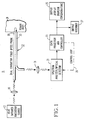
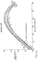
- Fig. 1 shows apparatus 11 for the non-invasive determination of the glucose concentration in a patient.
- This apparatus includes a source 13 of near-infrared radiation, a lens system 15, a dual conductor fiber-optic probe 17, a second lens system 19, a spectrum analyzer/detector 21, a data processor and controller 23, an output display device 25 and an output recorder 27.
- the source may produce radiation over the range from 700 to 3000 nm.
- the data presented in this application was produced using a Model 6250 System Near Infrared Spectrometer acquired from Pacific Scientific Instrument Division of Pacific Scientific, Ltd. The address of this Instrument Division is 2431 Linden Lane, Silver Spring, Md 20910.
- the lens systems 15 and 19 are represented by single-lens symbols.
- the fiber-optic probe 17 includes an input radiation conductor 29 for transmitting radiation to a portion 31, for example, an ear lobe or wrist, of the patient's body and an output radiation conductor 33 for deriving the resulting radiation from the portion 31.
- the output end of the input conductor 29 and the input or sensing end of the output conductor 33 are in firm contact with the outer surface of the portion 31 of the patient's body. While each conductor 29 and 33 is represented by a symbol for a single conductor, each radiation conductor, in actual practice of this invention, includes bundles of optic fibers.
- Fig. 1 discloses apparatus in which scattered radiation is analyzed, the analysis of transmitted radiation, i.e., the input radiation less the back scattered and absorbed radiation, plus any forward scattered radiation, is within the scope of equivalents of this invention.
- the ends of conductors 29 and 33 instead of being side-by-side in contact with adjacent surfaces of the body portion 31, would be in contact with the outer surfaces on opposite sides of the body portion 31, for example, with opposite surfaces of the ear lobe.
- the radiation, which is in this case passed through conductor 33, is predominantly the radiation from the source 13 less the radiation scattered and absorbed by the molecules of the water in the blood, the glucose and other constituents of the blood.
- the skin also contributes to the scattering and absorption.
- the resulting back scattered radiation emitted by the body portion 31 is passed by output conductor 33 and lens system 19 to the spectrum analyzer/detector 21 where this radiation is spread into a spectrum consisting of the wavelength distribution of the radiation intensity.
- This radiation is converted by an optical detector to electrical signals which are transmitted to the data processor 23.
- the operation of the spectrum analyzer in producing the spectrum is coordinated with the operation of the data processor through feedback channel 35, so that for each wavelength the data processor produces an electrical signal whose magnitude corresponds to the intensity of radiation at each wavelength. In this way an expression or spectrum of the resulting scattered radiation as a function of wavelength characteristic of the glucose concentration is produced. This expression appears on the output display 25 and is entered in the output recorder 27.
- the data processor computes the nth derivative of the expression for the spectrum expanded over a small subrange of wavelengths of the function defining the expression at a correlated point selected by statistical multi-variant calibration procedures.
- the glucose concentration is determined from the magnitude of the absorption, i.e., the reciprocal of the transmission, at this point and the relationship of the absorption, to concentration at this point as determined by calibration.
- the determination is simplified by computing the concentration at a maximum or minimum point of the second derivative, i.e., near a maximum or minimum point of the function itself. The minimum is preferred because in this region the transmission is a maximum.
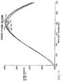
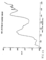
- Fig. 2 shows a family of curves or spectra for the different concentrations of glucose in the blood which were measured. The curves are labeled to show the concentrations, the numbers 86, 130, 173, 219, 250, 360 are in milligrams per deciliter.
- Fig. 2 has two sets of stationary points, 37 a maximum and 39 a minimum. In the region 40, between the minimum near 1300 nm and the maximum 37, there is a crossover region.
- the maximum points 37 correspond to low magnitudes of I and the minimum points to high magnitudes of I.
- the minimum points 39 identify a frequency at which the transmission for the various concentrations is a maximum.
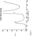
- the second derivative was determined for the very narrow subregion between lines 41 and 43, corresponding to wavelengths 1620 and 1700 nanometers and a highly blown up or enlarged display of this function was produced.
- This graph is shown in Fig. 3.
- the second derivative of log 1/T, as a function of wavelength, is plotted vertically and wavelength horizontally. The enlargement results in a family of curves, one for each concentration, as indicated. The maximum points of these curves are at about 1660 nanometers.
- Fig. 3 shows that in the region 44 of the maximum of the second derivative, at 1660 nanometers, there is a substantial spread between the curves. The spread provides a convenient scale for determining glucose concentration.
- the glucose concentration of the samples was determined by conventional methods and compared with the concentrations measured in the practice of this invention.
- the measured concentrations were derived by the data processor at wavelength of about 1660 nanometers.
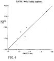
- Fig. 4 the known concentration is plotted vertically in 102 milligrams per deciliter and the measured concentration is plotted horizontally in 102 per deciliter.
- the 45° line shows that there is a precise correspondence between the actual concentrations and the concentrations measured in the practice of this invention.
- concentrations of glucose in the range actually encountered in diabetics can be precisely measured.
- an expression such as is shown in Fig. 3, or a like expression for scattering rather than transmission, may be entered into the memory of data processor 23 of the apparatus shown in Fig. 1.
- a patient suspected of suffering from diabetes is subjected to non-invasive testing as disclosed with reference to Fig. 1.
- the resulting second derivative of the expression of log 1/T as a function of wavelength is evaluated against the expression in the memory of the computer to determine the glucose concentration of the patient.
- a narrow region of the graph corresponding to Fig. 2 for the patient may be blown up in the critical region shown by the graph corresponding to Fig. 3 for the patient and the concentration computed from log 1/T at the minimum point.
- Fig. 5 is a graph in which the second derivative of log 1/T is plotted as a function of wavelength for various concentrations, 98, 145, 189, 219, 278, 322 milligrams per deciliter, of glucose in blood plasma.
- concentrations 98, 145, 189, 219, 278, 322 milligrams per deciliter
- Fig. 6 shows that, in this case also, there is close correlation between the concentrations computed from Fig. 5, plotted horizontally, and the known concentrations, plotted vertically. The concentrations are expressed in 102 milligrams per deciliter.
- Fig. 7 is a single line graph in which log 1/T is plotted vertically against wavelength plotted horizontally for several concentrations of glucose in water. This graph is significant because the major component of animal blood is water. The similarity between Fig. 7 and Fig. 2 is striking. Fig. 7 has a maximum 51 between about 1400 and 1500 nm and a sharp rise 53 at about 1900 nm. These regions correspond to the maximum 37 and the sharp rise 57 in Fig. 2.
- Fig. 8 the second derivative as a function of wavelength of the function in Fig. 7 is plotted in wavelength range between 1650 nm and 1680 nm. This range is near the minimum 61 at about 1680 nm of the function shown in Fig. 8. The spread near 1665 nm between the curves for the different concentrations is notable.
- the numbers shown in Fig. 8 are in milligrams per deciliter.
- Fig. 9 shows the close correlation between the known concentrations and the measured concentrations.
- Fig. 10 is based on data produced by actual measurement, with the apparatus according to this invention, by transmission of radiation through the ear of a host of the concentration of glucose as a function of wavelength after ingestion of 75 grams of glucose by the host.
- log 1/T is plotted vertically and the wavelength horizontally. The similarity between Figs. 2 and 7 and Fig. 10 is significant.
- Fig. 11 is a graph showing the response of the sensor of the apparatus shown in Fig. 1 as a function of time as the ingested glucose is absorbed in the blood. Response is plotted vertically and time in minutes after ingestion horizontally. Initially, there is a decrease in the response. Then the response rises gradually to between 0.207 and 0.208 at about 40 minutes after ingestion. There is then a gradual small decrease at about 50 minutes and a further rise followed by a decrease.
- Fig. 12 the second derivative as a function of wavelength of log 1/T is plotted for the glucose concentration.
- the family of curves presents the second derivative function at different times in minutes after ingestion of the glucose.
- the curves are expanded over a narrow range between 1670 and 1690nm.
- the curves of the family corresponding to the concentrations at different times in minutes after ingestion are labeled.
- Fig. 13 log 1/R is plotted as a function of wavelength for solid glucose. The data were derived by measuring the radiation scattered from the glucose by reflection. R is the intensity of the reflected radiation. The similarity between Fig. 13 and Figs. 2, 7 and 10 is notable.
Landscapes
- Physics & Mathematics (AREA)
- Health & Medical Sciences (AREA)
- Life Sciences & Earth Sciences (AREA)
- Spectroscopy & Molecular Physics (AREA)
- Medical Informatics (AREA)
- Animal Behavior & Ethology (AREA)
- Pathology (AREA)
- Engineering & Computer Science (AREA)
- Biomedical Technology (AREA)
- Heart & Thoracic Surgery (AREA)
- Optics & Photonics (AREA)
- Molecular Biology (AREA)
- Surgery (AREA)
- Biophysics (AREA)
- General Health & Medical Sciences (AREA)
- Public Health (AREA)
- Veterinary Medicine (AREA)
- Emergency Medicine (AREA)
- General Physics & Mathematics (AREA)
- Measurement Of The Respiration, Hearing Ability, Form, And Blood Characteristics Of Living Organisms (AREA)
- Investigating Or Analysing Materials By Optical Means (AREA)
Abstract
Description
- This invention relates to the determination of the glucose in the blood of a patient who is suspected of suffering from diabetes or to control the treatment or medication of patients who already suffer from diabetes. It has particular relationship to such determination without drawing blood from the patient, i.e., by a non-invasive process and by non-invasive means.
- There is widespread demand for non-invasive determination of glucose in patients. In the United States there are approximately ten million diabetics. Two million of these are
Type 1 Diabetics, whose pancreas secretes no insulin; and eight million of these are Type 2 Diabetics, whose pancreas secretes insufficient insulin or secretes it too late. Most of the Type 2 Diabetics can be controlled with proper diet and weight control. Some of the Type 2 Diabetics and all of theType 1 Diabetics require one or more shots of insulin per day. Insulin controls the body's utilization of glucose or sugar in the blood and, in the correct concentrations, prevents hyperglycemia (excess glucose) which, if left uncorrected, can lead to ketosis, coma and death. Glucose determination is also indispensable for sufferers from hypoglycemia who must ingest glucose containing fluids, such as orange juice, if the glucose in their blood decreases to a low level. - Hyperglycemia in the diabetic is strongly suspected of being responsible for the long-term effects of diabetes which include heart disease, arteriosclerosis, blindness, stroke, hypertension, kidney failure, and premature death. Severe hypoglycemia has similar drastic consequences. In a normal person, the blood glucose level may vary between 60 and 130 milligrams per deciliter, a variance exceeding 100%; whereas, in a diabetic, the levels may vary from time to time from 40 to 500 milligrams per deciliter, a variance of 1150% for hyperglycemia. For hypoglycemia, 60 milligrams per deciliter indicates that treatment is necessary; the glucose may reach a dangerous level of 20 milligrams per deciliter. These large swings of glucose levels must be avoided to prevent the symptoms and complications of the disease. To avoid the swings, the diabetic must be able to conveniently monitor his blood glucose level, and then vary his caloric intake, diet and insulin to control the level. For effective control, frequent blood glucose monitoring is necessary.
- The only practicable, reliable method currently available for monitoring blood glucose is by means of blood sampling. The diabetic pricks his epidermis with a needle, usually in the finger, draws a drop of blood, and absorbs the blood on a chemically treated strip of paper. He can then read the glucose level by placing the strip in a glucometer (a spectrophotometer which reads glucose concentrations); or he can compare the color change of the strip with a calibrated color chart. The direct reading instruments are more accurate. Other methods include measuring the electrical resistance of the strip with a glucometer which is an ohmeter calibrated in milligrams per deciliter. For effective control, some diabetics must utilize a finger prick four or more times a day.
- It is desirable to dispense with the drawing and analyzing of blood and it is an object of this invention to achieve this purpose, providing for effective non-invasive determination of glucose concentration in a patient suspected to suffer from, or already suffering from, diabetes.
- European Publication 0 160 768, dated November 13, 1985, to Clause Dahne and Daniel Cross, is typical of the prior art relating to the non-invasive determination of glucose concentration. In Dahne, a beam of radiation in selected bands, 1575, 1765, 2100 and 2270 ± 15 nanometers is impinged on a portion of the patient's body, penetrating into the portion, and the radiation resulting from the reaction within the body on the incident radiation is analyzed photometrically for the presence quantitatively of glucose. The resulting radiation which is analyzed may be scattered radiation or the transmitted radiation which, in effect, is the incident radiation less the predominant fraction of the scattered radiation and the radiation absorbed by the portion of the body.
- Dahne suffers from the disadvantage that its process lacks the precision demanded for the effective monitoring of glucose concentration. The range of concentration over which the glucose is in practice monitored for effective control of the patient is between 40 and 500 milligrams per deciliter, but even lower concentrations may be encountered in hyperglycemia or hypoglycemia. A concentration appreciably greater than 120 milligrams per deciliter indicates a diabetic condition and treatment by diet or insulin. In the actual practice of Dahne's process, the highest concentration of glucose which was measured was one molar (1M) and the lowest concentration of glucose which was measured was 0.05 molar (page 18). The chemical formula for glucose is C₆H₁₂O₆. In a 1M solution of glucose, there are 180.16 grams per liter or 18,000 milligrams per deciliter. In 0.05 mole, there are 0.9 grams or 900 milligrams per deciliter. This is far out of the range of glucose concentrations which must be measured for effective control and, indeed, out of the range which is compatible with life. There is no evidence in Dahne that its process is more effective than is indicated by its tests.
- It is accordingly an object of this invention to overcome the disadvantages and drawbacks of the prior art and to provide for the precise effective non-invasive determination of the concentration of glucose in a patient, taking into consideration the concentrations actually involved in such determination.
- This invention arises from the realization that in any expression, for example, in a graph, of the concentration of glucose in blood as a function of the wavelengths over which an analysis is carried out, the measure of the maximum concentration, which must be precisely determined, is often obscured by the presence of other chemical species. It has been realized in arriving at this invention that the specific species which must be measured may be emphasized and readily determined by deriving a new function, the nth derivative with respect to wavelength of the expression defining the concentration of glucose as a function of wavelength, over a defined region of this nth derivative. Once the magnitude of the nth derivative with respect to a zero reference is determined, this magnitude may be converted into glucose concentration by reference to the magnitude for
log 1/T of the known specimens and the appropriate derivatives corresponding to these known specimens, i.e., by calibrating the apparatus with known specimens containing known concentrations of the species to be measured.
where Io is the intensity of the radiation incident on the portion of the patient's body and I is the resulting radiation transmitted through or scattered by the portion. - In the specific practice of this invention, the concentration of the glucose is evaluated at or near the maximum or minimum of the second derivative of
log 1/T as a function of the wavelength, but other points along the function may be selected in accordance with this invention. In particular, the evaluation may be taken at a point displaced from a reference point or region which may be referred to as a "crossover" point or region. The crossover region is the region in which functions for a large number of spectra intersect. The evaluation may be taken at a convenient point sufficiently displaced from this region to yield a reliable measurement of the concentration for a specimen. Advantageously, the selected evaluation point or region may be a maximum or minimum point since, at this point, there is a maximum displacement in concentration either positively or negatively from the crossover point. An evaluation point may also be selected based on statistical determination of the maximum coefficient of correlation along an expression or function between known spectra and the functions serving as a basis for comparison. - Specifically, the non-invasive measurement of the concentration of glucose in blood is performed with a near-infrared radiation source, a fiber-optic probe, a spectrum analyzer with a detector and a data processor. The fiber-optic probe consists of a dual conductor of near-infrared radiation which is used in either the transmission or scattering mode. Radiation from the near-infrared source is transmitted through one of the dual conductors, the end of which is placed in contact with a portion of the patient's body. The radiation transmitted into the body undergoes scattering and characteristic absorption depending on the identity of the species present. A portion of the radiation having undergone scattering and absorption is back scattered from the body and collected and transmitted back to the spectral analyzer/detector system by the other fiber-optic conductor. The sensing end of the fiber-optic probe, placed in contact with the body, is arranged so that either a transmission or a scattering measurement is performed. In the transmission mode, the sending end of the fiber-optic probe is arranged so that the near-infrared radiation from the source can be passed through the portion of the body which may be the ear lobe, tongue or webbing between the fingers or toes and its spectral absorption characteristics measured. This is accomplished by placing the body section between the opposite ends of the dual fiber so that radiation from the fiber-optic conductor connected to the near-infrared source passes through the body section to the other fiber-optic conductor which transmits the attenuated radiation to the spectral analyzer/detector. In the scattering mode, a bifurcated fiber-optic probe is used. The bifurcated probe consists of two separate bundles of fibers, one bundle being centrally located and the other bundles being disposed in any configuration surrounding the central bundle. To measure blood glucose, the sensing end of the probe is placed in direct contact with an outer surface of the body. Near-infrared radiation from the fibers connected to the source is transmitted through that portion of the body undergoing both characteristic spectral absorption and scattering. Some of the scattered radiation which has traveled through the body experiencing absorption is collected by the optical fibers in the configuration and then transmitted to the spectrum analyzer/detector.
- The spectrum analyzer for this application can consist of a dispersive spectrometer with a prism or diffraction grating, a set of optical filters, a scanning interferometer, a stationary interferometer, or it may consist of a Hadamard transform spectrometer. Hadamard transform spectroscopy is described in a paper by Hammaker et al. in Vibrational Spectra and Structure, Vol. 15, Nov. 1986, edited by J.R. Durig, Elseviere Press, Amsterdam, Holland. Spectrometers are disclosed in Fateley patents US 4,615,619, US 4,750,834 and US 4,799,795. The purpose of the spectrum analyzer is to disperse the near-infrared radiation passing through the body into its spectral components to distinguish and quantify those particular spectral components that are characteristic of blood glucose. The characteristic near-infrared absorption by the glucose can be related directly to its concentration in blood.
- The data processor receives the output signal from the spectral analyzer, calculates the concentration of blood glucose, and formats the output to a display or recording device giving blood glucose concentration in selected units. A microprocessor in the data processor is used to perform data processing and control the operation of the spectral analyzer.
- To investigate and demonstrate the practical utility of the invention, near-infrared measurements were performed in water and different concentrations of glucose in water, blood plasma, whole blood and different concentrations of glucose in whole blood, human ears, and rabbit ears. It was found that the second derivative of the expression for the absorption of the radiation as a function of wavelength about the near-infrared band of glucose near 1660 nm, which occurs on the higher-frequency side of the water-absorption band at 1450 nm, yielded effective data from which precise concentration could be derived. In the practice of this invention, in its broad aspects, measurements are made over the range of wavelengths from 700 to 3000 nm.
- For a better understanding of this invention, both as to its organization and as to its method of operation, together with additional objects and advantages thereof, reference is made to the following description, taken in connection with the accompanying drawings, in which:
- Figure 1 is a block diagram showing an embodiment of this invention with which the method of this invention is practiced;
- Fig. 2 is a graph presenting the log of the reciprocal of the transmission of radiation through specimens, i.e., the absorption for the specimens, as a function of the wavelength in the near-infrared for whole-blood solutions of different concentrations of glucose;
- Fig. 3 is a graph presenting the second derivative of the function shown in Fig. 2 as a function of wavelength expanded over a small subrange of the wavelengths of Fig. 2;
- Fig. 4 is a graph in which the concentration of glucose in whole blood determined in the practice of this invention from the data derived from the graph shown in Figs. 2 and 3 is plotted against the corresponding known concentrations;
- Fig. 5 is a graph similar to Fig. 3 presenting expanded over a small wavelength range the second derivative of the log of the reciprocal of the transmission of radiation through specimens as a function of the wavelength from different concentrations of glucose in blood plasma;
- Fig. 6 is a graph similar to Fig. 4 in which the concentration of glucose in blood plasma, determined in the practice of this invention from the data on which Fig. 5 is based, is plotted against the corresponding known concentrations;
- Fig. 7 is a graph showing as a single line the log of the reciprocal of the transmission of radiation through specimens as a function of the wavelength for water solutions of different concentrations of glucose;
- Fig. 8 is a graph similar to Fig. 3 presenting expanded over a small wavelength range the second derivative of the functions shown in Fig. 7 as a function of wavelength;
- Fig. 9 is a graph similar to Figs. 4 and 6 in which the concentration of glucose in water, determined in the practice of this invention from the data on which Figs. 7 and 8 is based, is plotted against the corresponding known concentrations;
- Fig. 10 is a graph presenting the reciprocal of the log of radiation in the near-infrared through the ear of a human host as a function of wavelength after ingestion by the host of glucose;
- Fig. 11 is an expanded graph over a small range of wavelength presenting the response of the sensor as plotted in Fig. 10 over a time interval after the ingestion of glucose;
- Fig. 12 is a graph of a family of curves, each curve plotted at a different time after injection of glucose, presenting the second derivative of the response (
Log 1/T) of the sensor as a function of wavelength in the measurement of the transmission through the ear of the host; Figs. 10 and 11 are based on Fig. 12; and - Fig. 13 is a graph presenting the log of the reciprocal of the reflection from solid glucose of radiation in the near-infrared as a function of wavelength.
- Fig. 1 shows
apparatus 11 for the non-invasive determination of the glucose concentration in a patient. This apparatus includes asource 13 of near-infrared radiation, alens system 15, a dual conductor fiber-optic probe 17, asecond lens system 19, a spectrum analyzer/detector 21, a data processor andcontroller 23, anoutput display device 25 and anoutput recorder 27. As stated the source may produce radiation over the range from 700 to 3000 nm. The data presented in this application was produced using a Model 6250 System Near Infrared Spectrometer acquired from Pacific Scientific Instrument Division of Pacific Scientific, Ltd. The address of this Instrument Division is 2431 Linden Lane, Silver Spring, Md 20910. Thelens systems optic probe 17 includes aninput radiation conductor 29 for transmitting radiation to a portion 31, for example, an ear lobe or wrist, of the patient's body and anoutput radiation conductor 33 for deriving the resulting radiation from the portion 31. The output end of theinput conductor 29 and the input or sensing end of theoutput conductor 33 are in firm contact with the outer surface of the portion 31 of the patient's body. While eachconductor - Radiation from the
source 13 is directed by thelens system 15 intoconductor 29 and, at its outlet, is projected into the portion 31. This incident radiation induces scattered radiation within the body portion 31, some of which passes through the end ofconductor 33 and through the conductor and is directed bylens system 19 into the spectrum analyzer/detector. While Fig. 1 discloses apparatus in which scattered radiation is analyzed, the analysis of transmitted radiation, i.e., the input radiation less the back scattered and absorbed radiation, plus any forward scattered radiation, is within the scope of equivalents of this invention. In this case, the ends ofconductors conductor 33, is predominantly the radiation from thesource 13 less the radiation scattered and absorbed by the molecules of the water in the blood, the glucose and other constituents of the blood. The skin also contributes to the scattering and absorption. - With the apparatus as shown in Fig. 1, the resulting back scattered radiation emitted by the body portion 31 is passed by
output conductor 33 andlens system 19 to the spectrum analyzer/detector 21 where this radiation is spread into a spectrum consisting of the wavelength distribution of the radiation intensity. This radiation is converted by an optical detector to electrical signals which are transmitted to thedata processor 23. The operation of the spectrum analyzer in producing the spectrum is coordinated with the operation of the data processor throughfeedback channel 35, so that for each wavelength the data processor produces an electrical signal whose magnitude corresponds to the intensity of radiation at each wavelength. In this way an expression or spectrum of the resulting scattered radiation as a function of wavelength characteristic of the glucose concentration is produced. This expression appears on theoutput display 25 and is entered in theoutput recorder 27. In accordance with this invention, in its broader aspects, the data processor computes the nth derivative of the expression for the spectrum expanded over a small subrange of wavelengths of the function defining the expression at a correlated point selected by statistical multi-variant calibration procedures. The glucose concentration is determined from the magnitude of the absorption, i.e., the reciprocal of the transmission, at this point and the relationship of the absorption, to concentration at this point as determined by calibration. In the specific practice of this invention as disclosed herein, the determination is simplified by computing the concentration at a maximum or minimum point of the second derivative, i.e., near a maximum or minimum point of the function itself. The minimum is preferred because in this region the transmission is a maximum. - A number of samples of blood were taken from one of the applicants and an associate. Neither man suffers from diabetes. The samples were doped with glucose of the concentrations in milligrams per deciliter shown in Figs. 2 and 3. Each sample was analyzed as described above in a Yellow Spring Instrument glucose analyzer. The near-infrared measurements were performed using the Pacific Scientific Model 6250 linear infrared spectrometer. The absorption method was used. Radiation in the near-infrared was transmitted through each sample and an expression or spectrum was produced, as shown in Fig. 2. In this view, log 1/T is plotted vertically as a function of wavelength plotted horizontally.

where I is the intensity of the radiation transmitted or the transmission through the sample, Io is the intensity of radiation incident upon the sample.
The plot is over the limited range between 1100 and 1900 nanometers. Fig. 2 shows a family of curves or spectra for the different concentrations of glucose in the blood which were measured. The curves are labeled to show the concentrations, thenumbers region 40, between the minimum near 1300 nm and the maximum 37, there is a crossover region. The maximum points 37 correspond to low magnitudes of I and the minimum points to high magnitudes of I. The minimum points 39 identify a frequency at which the transmission for the various concentrations is a maximum. - It was found by further analysis that, in the region of this
minimum region 39, the concentration of the glucose is readily determinable. The second derivative was determined for the very narrow subregion betweenlines 41 and 43, corresponding towavelengths log 1/T, as a function of wavelength, is plotted vertically and wavelength horizontally. The enlargement results in a family of curves, one for each concentration, as indicated. The maximum points of these curves are at about 1660 nanometers. Fig. 3 shows that in the region 44 of the maximum of the second derivative, at 1660 nanometers, there is a substantial spread between the curves. The spread provides a convenient scale for determining glucose concentration. - To evaluate the reliability of the practice of this invention, the glucose concentration of the samples was determined by conventional methods and compared with the concentrations measured in the practice of this invention. The measured concentrations were derived by the data processor at wavelength of about 1660 nanometers. In Fig. 4 the known concentration is plotted vertically in 10² milligrams per deciliter and the measured concentration is plotted horizontally in 10² per deciliter. In the resulting graph, the 45° line shows that there is a precise correspondence between the actual concentrations and the concentrations measured in the practice of this invention.
- By the process in accordance with this invention as described above, concentrations of glucose in the range actually encountered in diabetics can be precisely measured. In practice, an expression such as is shown in Fig. 3, or a like expression for scattering rather than transmission, may be entered into the memory of
data processor 23 of the apparatus shown in Fig. 1. A patient suspected of suffering from diabetes is subjected to non-invasive testing as disclosed with reference to Fig. 1. The resulting second derivative of the expression oflog 1/T as a function of wavelength is evaluated against the expression in the memory of the computer to determine the glucose concentration of the patient. Alternatively, a narrow region of the graph corresponding to Fig. 2 for the patient may be blown up in the critical region shown by the graph corresponding to Fig. 3 for the patient and the concentration computed fromlog 1/T at the minimum point. - Fig. 5 is a graph in which the second derivative of
log 1/T is plotted as a function of wavelength for various concentrations, 98, 145, 189, 219, 278, 322 milligrams per deciliter, of glucose in blood plasma. In this case again, there is a substantial spread between the curves for the different concentrations at the maximum in the region of 1660 nm. Fig. 6 shows that, in this case also, there is close correlation between the concentrations computed from Fig. 5, plotted horizontally, and the known concentrations, plotted vertically. The concentrations are expressed in 10² milligrams per deciliter. - Fig. 7 is a single line graph in which log 1/T is plotted vertically against wavelength plotted horizontally for several concentrations of glucose in water. This graph is significant because the major component of animal blood is water. The similarity between Fig. 7 and Fig. 2 is striking. Fig. 7 has a maximum 51 between about 1400 and 1500 nm and a
sharp rise 53 at about 1900 nm. These regions correspond to the maximum 37 and thesharp rise 57 in Fig. 2. - In Fig. 8 the second derivative as a function of wavelength of the function in Fig. 7 is plotted in wavelength range between 1650 nm and 1680 nm. This range is near the minimum 61 at about 1680 nm of the function shown in Fig. 8. The spread near 1665 nm between the curves for the different concentrations is notable. The numbers shown in Fig. 8 are in milligrams per deciliter.
- Fig. 9 shows the close correlation between the known concentrations and the measured concentrations.
- Fig. 10 is based on data produced by actual measurement, with the apparatus according to this invention, by transmission of radiation through the ear of a host of the concentration of glucose as a function of wavelength after ingestion of 75 grams of glucose by the host. In Fig. 10,
log 1/T is plotted vertically and the wavelength horizontally. The similarity between Figs. 2 and 7 and Fig. 10 is significant. - Fig. 11 is a graph showing the response of the sensor of the apparatus shown in Fig. 1 as a function of time as the ingested glucose is absorbed in the blood. Response is plotted vertically and time in minutes after ingestion horizontally. Initially, there is a decrease in the response. Then the response rises gradually to between 0.207 and 0.208 at about 40 minutes after ingestion. There is then a gradual small decrease at about 50 minutes and a further rise followed by a decrease.
- In Fig. 12 the second derivative as a function of wavelength of
log 1/T is plotted for the glucose concentration. The family of curves presents the second derivative function at different times in minutes after ingestion of the glucose. The curves are expanded over a narrow range between 1670 and 1690nm. The curves of the family corresponding to the concentrations at different times in minutes after ingestion are labeled. - In Fig. 13,
log 1/R is plotted as a function of wavelength for solid glucose. The data were derived by measuring the radiation scattered from the glucose by reflection. R is the intensity of the reflected radiation. The similarity between Fig. 13 and Figs. 2, 7 and 10 is notable. - While a preferred embodiment and preferred practice of this invention has been disclosed herein, many modifications thereof are feasible. This invention is not to be restricted except insofar as is necessitated by the spirit of the prior art.
- For example, although the method described utilises radiation in the rear infra red range other frequency ranges may be used.
Claims (13)
- A method of non-invasive determination of the concentration of glucose in a patient with apparatus including a source of radiation of predetermined wavelengths; the said method comprising: projecting radiation from said source on a portion of the body of said patient, varying the wavelengths of the radiation projected on said portion of said body over a limited range of predetermined wavelengths, sensing the resulting radiation emitted from said portion of said body, deriving from said sensed resulting radiation a first expression for the magnitude of said sensed radiation as a function of the wavelength to locate a region of said expression where the expression shows maximum influence by the portion of said body on the resulting radiation, responsive to the content of glucose in said patient of the portion of said body on the radiation incident on said portion of said body, deriving a second expression for the nth derivative of said function with respect to wavelength about a narrow range of predetermined wavelengths of the radiation of said region, producing an enlargement of said second expression over said narrow range of wavelengths, and determining said concentration of said glucose from the magnitude at a selected point of said enlargement of said second expression.
- The method of non-invasive determination of the concentration of the glucose in a patient with apparatus including a source of radiation of predetermined wavelengths; said method comprising: projecting radiation of said predetermined wavelengths on a portion of the body of said patient, varying the wavelength of said projected radiation over a narrow range of wavelengths about the wavelength of approximately 1660 nanometers, sensing the resulting radiation emitted from said portion of said body, deriving an expression for the magnitude of the resultng radiation as a function of the wavelength of said radiation, and determining said magnitude of the glucose by evaluating the nth derivative of said expression in an enlarged narrow range at about 1660 nanometers to determine the magnitude of the resulting radiation at the wavelength about 1660 nanometers at which the influence of said portion of said body on the radiation from said source incident thereon is a maximum, thereby to determine the concentration of the glucose in said patient.
- The method of non-invasive determination of the concentration of the glucose in a patient with apparatus including a source of radiation of predetermined wavelengths; the said method comprising: projecting from said source radiation of said predetermined wavelength on a portion of the body of said patient, varying the wavelength of the radiation projected on said portion of said body over a limited range of wavelengths about the wavelength of approximately 1660 nanometers, sensing the resulting radiation emitted by said portion of said body to derive an expression which is a measure of the magnitude of said resulting radiation as a function of the wavelength of the impinging radiation having a stationary point in the region of 1660 nanometers, deriving the nth order derivative of said expression over a narrow range of wavelengths immediately in the region of 1660 nanometers, and determining the concentration of the glucose from said stationary point of said nth order derivative.
- The method of claim 1 or 2 wherein the narrow range is between about 1640 and 1670 nanometers.
- The method of claim 3 wherein the limited range is between about 1100 and 1900 nanometers and the narrow range is between about 1640 and 1670 nanometers.
- The method of claim 1, 2 or 3 wherein the nth derivative is the second derivative.
- Apparatus for non-invasive determination of the concentration of glucose in a patient including: a source of radiation, a first fiber-optic conductor of radiation adapted to be connected to an external surface of a portion of the body of said patient, means positioning said first fiber-optic conductor to transmit radiation from said source to said portion through said surface as incident radiation thereon, a second fiber-optic conductor of radiation adapted to be connected to an external surface of said portion of the body of the patient to transmit radiation emitted from said portion resulting from the impingement of said incident radiation, first deriving means, connected to said second fiber-optic conductor, to derive from said resulting radiation a first continuous expression defining the magnitude of said resulting radiation as a function of wavelength of said radiation, and a second deriving means, connected to said first deriving means, for deriving from the first expression a second expression defining the nth derivative of the first expression as a function of the wavelength of the radiation, and means, connected to said second deriving means, for processing the resulting radiation from said portion in dependence upon said continuous expression to determine said concentration.
- The apparatus of claim 7 wherein the deriving means includes means for scanning the resulting radiation continuously over a predetermined range of wavelengths and means, responsive to said scanning means, to derive the expression.
- The apparatus of claim 7 wherein the deriving means includes a spectrum analyzer for analyzing the spectrum of the resulting radiation emitted by the portion of the body of the patient and also includes means, connected to said analyzer, for deriving the expression for said resulting radiation as a function of the wavelength.
- The method of non-invasive determination of the concentration of glucose in a patient with apparatus including a source of radiation of predetermined wavelengths; the said method comprising: projecting radiation from said source on a portion of the body of said patient, varying the wavelengths of the radiation projected on said portion of said body over a limited range of said predetermined wavelengths, sensing the resulting radiation emitted from said portion of said body, deriving from said sensed resulting radiation a first expression for the magnitude of said sensed radiation as a function of the wavelength to locate a region of said first expression where said expression shows maximum influence by the portion of said body on the resulting radiation responsive to the content of glucose in said patient on the radiation incident on said portion of said body, deriving a second expression for the nth derivative of said expression with respect to wavelength about a narrow range of wavelengths of said region, and determining said concentration of said glucose from the magnitude of said second expression at a selected point of said narrow range.
- The appratus of claim 7 wherein the processing means is controlled by the second expression to determine the concentration.
- The method according to any one of claims 1 to 8 and 12 wherein said predetermined wave lengths are in the rear infra red.
- Apparatus according to any one of claims 7 to 9 and 11, wherein said predetermined wavelengths are in the rear infra red.
Priority Applications (8)
| Application Number | Priority Date | Filing Date | Title |
|---|---|---|---|
| US07/482,792 US5070874A (en) | 1990-01-30 | 1990-01-30 | Non-invasive determination of glucose concentration in body of patients |
| CA002056868A CA2056868C (en) | 1990-01-30 | 1991-12-03 | Non-invasive determination of glucose concentration in body of patient |
| AU88392/91A AU643855B2 (en) | 1990-01-30 | 1991-12-04 | Non-invasive determination of glucose concentration in body of patient |
| EP91311421A EP0548418B1 (en) | 1990-01-30 | 1991-12-09 | Non-invasive determination of glucose concentration in body of patient |
| DE69125942T DE69125942T2 (en) | 1990-01-30 | 1991-12-09 | Non-invasive determination of the glucose content in a patient's body |
| AT91311421T ATE152338T1 (en) | 1990-01-30 | 1991-12-09 | NON-INVASIVE DETERMINATION OF GLUCOSE CONTENT IN A PATIENT'S BODY |
| ES91311421T ES2101728T3 (en) | 1990-01-30 | 1991-12-09 | NON-INVASIVE DETERMINATION OF THE CONCENTRATION OF GLUCOSE IN THE BODY OF A PATIENT. |
| DK91311421.1T DK0548418T3 (en) | 1990-01-30 | 1991-12-09 | Non-invasive determination of glucose concentration in the body of a patient. |
Applications Claiming Priority (4)
| Application Number | Priority Date | Filing Date | Title |
|---|---|---|---|
| US07/482,792 US5070874A (en) | 1990-01-30 | 1990-01-30 | Non-invasive determination of glucose concentration in body of patients |
| CA002056868A CA2056868C (en) | 1990-01-30 | 1991-12-03 | Non-invasive determination of glucose concentration in body of patient |
| AU88392/91A AU643855B2 (en) | 1990-01-30 | 1991-12-04 | Non-invasive determination of glucose concentration in body of patient |
| EP91311421A EP0548418B1 (en) | 1990-01-30 | 1991-12-09 | Non-invasive determination of glucose concentration in body of patient |
Publications (2)
| Publication Number | Publication Date |
|---|---|
| EP0548418A1 true EP0548418A1 (en) | 1993-06-30 |
| EP0548418B1 EP0548418B1 (en) | 1997-05-02 |
Family
ID=27424036
Family Applications (1)
| Application Number | Title | Priority Date | Filing Date |
|---|---|---|---|
| EP91311421A Expired - Lifetime EP0548418B1 (en) | 1990-01-30 | 1991-12-09 | Non-invasive determination of glucose concentration in body of patient |
Country Status (8)
| Country | Link |
|---|---|
| US (1) | US5070874A (en) |
| EP (1) | EP0548418B1 (en) |
| AT (1) | ATE152338T1 (en) |
| AU (1) | AU643855B2 (en) |
| CA (1) | CA2056868C (en) |
| DE (1) | DE69125942T2 (en) |
| DK (1) | DK0548418T3 (en) |
| ES (1) | ES2101728T3 (en) |
Cited By (4)
| Publication number | Priority date | Publication date | Assignee | Title |
|---|---|---|---|---|
| EP0623307A1 (en) * | 1993-05-07 | 1994-11-09 | Diasense, Inc. | Non-invasive determination of constituent concentration using non-continuous radiation |
| WO1995013739A1 (en) * | 1993-11-16 | 1995-05-26 | Jenoptik Gmbh | Method and device for the non-invasive transcutanean determination of the concentrations of substances in human body fluids or tissues |
| WO1995022046A1 (en) * | 1994-02-14 | 1995-08-17 | University Of Iowa Research Foundation | Method and apparatus for non-invasive detection of physiological chemicals, particularly glucose |
| EP0762109A2 (en) * | 1995-08-30 | 1997-03-12 | Kyoto Dai-ichi Kagaku Co., Ltd. | Method of and apparatus for measuring lactic acid in organism |
Families Citing this family (218)
| Publication number | Priority date | Publication date | Assignee | Title |
|---|---|---|---|---|
| US6066847A (en) * | 1989-01-19 | 2000-05-23 | Futrex Inc. | Procedure for verifying the accuracy of non-invasive blood glucose measurement instruments |
| US5204532A (en) * | 1989-01-19 | 1993-04-20 | Futrex, Inc. | Method for providing general calibration for near infrared instruments for measurement of blood glucose |
| US5237178A (en) * | 1990-06-27 | 1993-08-17 | Rosenthal Robert D | Non-invasive near-infrared quantitative measurement instrument |
| CA2028261C (en) * | 1989-10-28 | 1995-01-17 | Won Suck Yang | Non-invasive method and apparatus for measuring blood glucose concentration |
| DE3938759A1 (en) * | 1989-11-23 | 1991-05-29 | Philips Patentverwaltung | NON-INVASIVE OXIMETER ARRANGEMENT |
| US5070874A (en) * | 1990-01-30 | 1991-12-10 | Biocontrol Technology, Inc. | Non-invasive determination of glucose concentration in body of patients |
| US5246004A (en) * | 1990-02-02 | 1993-09-21 | Angiomedics Ii, Inc. | Infrared cholesterol sensor |
| US5222495A (en) * | 1990-02-02 | 1993-06-29 | Angiomedics Ii, Inc. | Non-invasive blood analysis by near infrared absorption measurements using two closely spaced wavelengths |
| US5222496A (en) * | 1990-02-02 | 1993-06-29 | Angiomedics Ii, Inc. | Infrared glucose sensor |
| US5574283A (en) * | 1990-06-27 | 1996-11-12 | Futrex, Inc. | Non-invasive near-infrared quantitative measurement instrument |
| US5324979A (en) * | 1990-09-26 | 1994-06-28 | Futrex, Inc. | Method and means for generating synthetic spectra allowing quantitative measurement in near infrared measuring instruments |
| US5219400A (en) * | 1991-06-11 | 1993-06-15 | The United States Of America As Represented By The Secretary Of The Army | Noninvasive method for quantitation of oxyhemoglobin saturation by near-infrared reflectance spectrophotometry |
| JPH05228098A (en) * | 1992-02-20 | 1993-09-07 | Asahi Optical Co Ltd | Thermoendoscope |
| DE69232711T2 (en) * | 1992-02-28 | 2003-02-06 | Theodore E. Cadell | DEVICE AND METHOD FOR NON-INVASIVE QUANTITY DETERMINATION OF INGREDIENTS IN BLOOD OR TISSUE |
| US5377674A (en) * | 1992-05-08 | 1995-01-03 | Kuestner; J. Todd | Method for non-invasive and in-vitro hemoglobin concentration measurement |
| US5434412A (en) * | 1992-07-15 | 1995-07-18 | Myron J. Block | Non-spectrophotometric measurement of analyte concentrations and optical properties of objects |
| US5818048A (en) * | 1992-07-15 | 1998-10-06 | Optix Lp | Rapid non-invasive optical analysis using broad bandpass spectral processing |
| US5424545A (en) * | 1992-07-15 | 1995-06-13 | Myron J. Block | Non-invasive non-spectrophotometric infrared measurement of blood analyte concentrations |
| US5433197A (en) * | 1992-09-04 | 1995-07-18 | Stark; Edward W. | Non-invasive glucose measurement method and apparatus |
| US5762609A (en) * | 1992-09-14 | 1998-06-09 | Sextant Medical Corporation | Device and method for analysis of surgical tissue interventions |
| US5772597A (en) * | 1992-09-14 | 1998-06-30 | Sextant Medical Corporation | Surgical tool end effector |
| US5746210A (en) * | 1993-02-26 | 1998-05-05 | David A. Benaron | Device and method for detection, localization, and characterization of inhomogeneities in turbid media |
| US6172743B1 (en) | 1992-10-07 | 2001-01-09 | Chemtrix, Inc. | Technique for measuring a blood analyte by non-invasive spectrometry in living tissue |
| US5360004A (en) * | 1992-12-09 | 1994-11-01 | Diasense, Inc. | Non-invasive determination of analyte concentration using non-continuous radiation |
| US5379764A (en) * | 1992-12-09 | 1995-01-10 | Diasense, Inc. | Non-invasive determination of analyte concentration in body of mammals |
| US5398681A (en) * | 1992-12-10 | 1995-03-21 | Sunshine Medical Instruments, Inc. | Pocket-type instrument for non-invasive measurement of blood glucose concentration |
| DE4242083C2 (en) * | 1992-12-14 | 1995-07-20 | Marbach Hermann Dipl Ing | Sensor device for reproducible, non-invasive measurement of blood glucose |
| US5615672A (en) * | 1993-01-28 | 1997-04-01 | Optiscan, Inc. | Self-emission noninvasive infrared spectrophotometer with body temperature compensation |
| US5515847A (en) * | 1993-01-28 | 1996-05-14 | Optiscan, Inc. | Self-emission noninvasive infrared spectrophotometer |
| US5447159A (en) * | 1993-02-03 | 1995-09-05 | Massachusetts Institute Of Technology | Optical imaging for specimens having dispersive properties |
| US5487384A (en) * | 1993-02-25 | 1996-01-30 | Blue Marble Research, Inc. | Kinematic assay of plasma glucose concentration without blood sampling |
| US5987346A (en) | 1993-02-26 | 1999-11-16 | Benaron; David A. | Device and method for classification of tissue |
| US5343045A (en) * | 1993-06-11 | 1994-08-30 | Ontario Hydro | Method and device for measuring moisture content |
| US5383452A (en) * | 1993-07-01 | 1995-01-24 | Buchert; Janusz | Method, apparatus and procedure for non-invasive monitoring blood glucose by measuring the polarization ratio of blood luminescence |
| US5562729A (en) * | 1994-11-01 | 1996-10-08 | Biocontrol Technology, Inc. | Heart valve |
| US5743262A (en) * | 1995-06-07 | 1998-04-28 | Masimo Corporation | Blood glucose monitoring system |
| SG38866A1 (en) * | 1995-07-31 | 1997-04-17 | Instrumentation Metrics Inc | Liquid correlation spectrometry |
| US5655530A (en) * | 1995-08-09 | 1997-08-12 | Rio Grande Medical Technologies, Inc. | Method for non-invasive blood analyte measurement with improved optical interface |
| US6212424B1 (en) | 1998-10-29 | 2001-04-03 | Rio Grande Medical Technologies, Inc. | Apparatus and method for determination of the adequacy of dialysis by non-invasive near-infrared spectroscopy |
| US6240306B1 (en) | 1995-08-09 | 2001-05-29 | Rio Grande Medical Technologies, Inc. | Method and apparatus for non-invasive blood analyte measurement with fluid compartment equilibration |
| US6152876A (en) * | 1997-04-18 | 2000-11-28 | Rio Grande Medical Technologies, Inc. | Method for non-invasive blood analyte measurement with improved optical interface |
| US5827181A (en) * | 1995-09-07 | 1998-10-27 | Hewlett-Packard Co. | Noninvasive blood chemistry measurement method and system |
| US5760399A (en) * | 1995-10-02 | 1998-06-02 | Foss Nirsystems, Inc. | Measurement of transmission spectra of pharmaceutical tablets |
| US6025597A (en) * | 1995-10-17 | 2000-02-15 | Optiscan Biomedical Corporation | Non-invasive infrared absorption spectrometer for measuring glucose or other constituents in a human or other body |
| US6072180A (en) * | 1995-10-17 | 2000-06-06 | Optiscan Biomedical Corporation | Non-invasive infrared absorption spectrometer for the generation and capture of thermal gradient spectra from living tissue |
| US6002954A (en) * | 1995-11-22 | 1999-12-14 | The Regents Of The University Of California | Detection of biological molecules using boronate-based chemical amplification and optical sensors |
| ATE278801T1 (en) | 1995-11-22 | 2004-10-15 | Medtronic Minimed Inc | DETECTION OF BIOLOGICAL MOLECULES USING CHEMICAL AMPLIFICATION AND OPTICAL SENSOR |
| US6766183B2 (en) | 1995-11-22 | 2004-07-20 | Medtronic Minimed, Inc. | Long wave fluorophore sensor compounds and other fluorescent sensor compounds in polymers |
| US6405069B1 (en) | 1996-01-31 | 2002-06-11 | Board Of Regents, The University Of Texas System | Time-resolved optoacoustic method and system for noninvasive monitoring of glucose |
| US6040578A (en) * | 1996-02-02 | 2000-03-21 | Instrumentation Metrics, Inc. | Method and apparatus for multi-spectral analysis of organic blood analytes in noninvasive infrared spectroscopy |
| US6219565B1 (en) | 1996-02-05 | 2001-04-17 | Diasense, Inc. | Methods and apparatus for non-invasive glucose sensing: non-invasive probe |
| EP0955867A1 (en) * | 1996-02-23 | 1999-11-17 | Diasense, Inc. | Method and apparatus for non-invasive blood glucose sensing |
| JP3599878B2 (en) * | 1996-03-01 | 2004-12-08 | テルモ株式会社 | Blood glucose meter |
| US5666956A (en) * | 1996-05-20 | 1997-09-16 | Buchert; Janusz Michal | Instrument and method for non-invasive monitoring of human tissue analyte by measuring the body's infrared radiation |
| DE19629342C2 (en) * | 1996-07-20 | 1999-09-02 | Epsa Elektronik Und Praezision | Method and arrangement for the non-invasive, transcutaneous determination of substance concentrations in body tissues |
| AU5446098A (en) | 1996-11-21 | 1998-06-10 | Lawrence Livermore National Laboratory | Detection of biological molecules using boronate-based chemical amplification and optical sensors |
| JP4212007B2 (en) * | 1996-11-26 | 2009-01-21 | パナソニック電工株式会社 | Blood component concentration analyzer |
| US5910109A (en) | 1997-02-20 | 1999-06-08 | Emerging Technology Systems, Llc | Non-invasive glucose measuring device and method for measuring blood glucose |
| US6309884B1 (en) | 1997-02-26 | 2001-10-30 | Diasense, Inc. | Individual calibration of blood glucose for supporting noninvasive self-monitoring blood glucose |
| US6113541A (en) * | 1997-03-07 | 2000-09-05 | Agilent Technologies, Inc. | Noninvasive blood chemistry measurement method and system |
| TW352335B (en) * | 1997-03-25 | 1999-02-11 | Matsushita Electric Works Ltd | Method of determining a glucose concentration in a target by using near-infrared spectroscopy |
| US5961451A (en) * | 1997-04-07 | 1999-10-05 | Motorola, Inc. | Noninvasive apparatus having a retaining member to retain a removable biosensor |
| US6628809B1 (en) | 1999-10-08 | 2003-09-30 | Lumidigm, Inc. | Apparatus and method for identification of individuals by near-infrared spectrum |
| US7890158B2 (en) | 2001-06-05 | 2011-02-15 | Lumidigm, Inc. | Apparatus and method of biometric determination using specialized optical spectroscopy systems |
| US6560352B2 (en) | 1999-10-08 | 2003-05-06 | Lumidigm, Inc. | Apparatus and method of biometric identification or verification of individuals using optical spectroscopy |
| US6246893B1 (en) * | 1997-06-12 | 2001-06-12 | Tecmed Incorporated | Method and device for glucose concentration measurement with special attention to blood glucose determinations |
| US7383069B2 (en) * | 1997-08-14 | 2008-06-03 | Sensys Medical, Inc. | Method of sample control and calibration adjustment for use with a noninvasive analyzer |
| AU3311099A (en) * | 1998-02-25 | 1999-09-15 | Ohio University | Near infrared-transmission spectroscopy of tongue tissue |
| US6391005B1 (en) | 1998-03-30 | 2002-05-21 | Agilent Technologies, Inc. | Apparatus and method for penetration with shaft having a sensor for sensing penetration depth |
| US6728560B2 (en) | 1998-04-06 | 2004-04-27 | The General Hospital Corporation | Non-invasive tissue glucose level monitoring |
| US20020091324A1 (en) * | 1998-04-06 | 2002-07-11 | Nikiforos Kollias | Non-invasive tissue glucose level monitoring |
| US6505059B1 (en) | 1998-04-06 | 2003-01-07 | The General Hospital Corporation | Non-invasive tissue glucose level monitoring |
| US7899518B2 (en) | 1998-04-06 | 2011-03-01 | Masimo Laboratories, Inc. | Non-invasive tissue glucose level monitoring |
| US6097975A (en) * | 1998-05-13 | 2000-08-01 | Biosensor, Inc. | Apparatus and method for noninvasive glucose measurement |
| US6157041A (en) | 1998-10-13 | 2000-12-05 | Rio Grande Medical Technologies, Inc. | Methods and apparatus for tailoring spectroscopic calibration models |
| US7098037B2 (en) | 1998-10-13 | 2006-08-29 | Inlight Solutions, Inc. | Accommodating subject and instrument variations in spectroscopic determinations |
| US6441388B1 (en) | 1998-10-13 | 2002-08-27 | Rio Grande Medical Technologies, Inc. | Methods and apparatus for spectroscopic calibration model transfer |
| US7299080B2 (en) * | 1999-10-08 | 2007-11-20 | Sensys Medical, Inc. | Compact apparatus for noninvasive measurement of glucose through near-infrared spectroscopy |
| US6370407B1 (en) | 1999-07-27 | 2002-04-09 | Tecmed, Incorporated | System for improving the sensitivity and stability of optical polarimetric measurements |
| WO2001016582A1 (en) * | 1999-08-30 | 2001-03-08 | Euro-Celtique S.A. | In situ methods for measuring the release of a substance from a dosage form |
| US6673625B2 (en) | 1999-09-15 | 2004-01-06 | The Regents Of The University Of California | Saccharide sensing molecules having enhanced fluorescent properties |
| US6682938B1 (en) | 1999-09-15 | 2004-01-27 | The Regents Of The University Of California | Glucose sensing molecules having selected fluorescent properties |
| US6816605B2 (en) | 1999-10-08 | 2004-11-09 | Lumidigm, Inc. | Methods and systems for biometric identification of individuals using linear optical spectroscopy |
| US6563585B1 (en) * | 1999-11-24 | 2003-05-13 | University Of Maryland Biotechnology Institute | Ratiometric fluorometer |
| US20070179367A1 (en) * | 2000-05-02 | 2007-08-02 | Ruchti Timothy L | Method and Apparatus for Noninvasively Estimating a Property of an Animal Body Analyte from Spectral Data |
| US7519406B2 (en) | 2004-04-28 | 2009-04-14 | Sensys Medical, Inc. | Noninvasive analyzer sample probe interface method and apparatus |
| US7606608B2 (en) | 2000-05-02 | 2009-10-20 | Sensys Medical, Inc. | Optical sampling interface system for in-vivo measurement of tissue |
| US6549861B1 (en) | 2000-08-10 | 2003-04-15 | Euro-Celtique, S.A. | Automated system and method for spectroscopic analysis |
| WO2002016905A2 (en) | 2000-08-21 | 2002-02-28 | Euro-Celtique, S.A. | Near infrared blood glucose monitoring system |
| WO2002024065A1 (en) | 2000-09-22 | 2002-03-28 | Knobbe, Lim & Buckingham | Method and apparatus for real-time estimation and control of pysiological parameters |
| US8641644B2 (en) | 2000-11-21 | 2014-02-04 | Sanofi-Aventis Deutschland Gmbh | Blood testing apparatus having a rotatable cartridge with multiple lancing elements and testing means |
| AU2002251944A1 (en) * | 2001-02-15 | 2002-09-04 | Medtronic Minimed, Inc. | Polymers functionalized with fluorescent boronate motifs |
| US6983176B2 (en) | 2001-04-11 | 2006-01-03 | Rio Grande Medical Technologies, Inc. | Optically similar reference samples and related methods for multivariate calibration models used in optical spectroscopy |
| US7043288B2 (en) * | 2002-04-04 | 2006-05-09 | Inlight Solutions, Inc. | Apparatus and method for spectroscopic analysis of tissue to detect diabetes in an individual |
| US6574490B2 (en) | 2001-04-11 | 2003-06-03 | Rio Grande Medical Technologies, Inc. | System for non-invasive measurement of glucose in humans |
| US6862091B2 (en) | 2001-04-11 | 2005-03-01 | Inlight Solutions, Inc. | Illumination device and method for spectroscopic analysis |
| US6865408B1 (en) | 2001-04-11 | 2005-03-08 | Inlight Solutions, Inc. | System for non-invasive measurement of glucose in humans |
| US7126682B2 (en) | 2001-04-11 | 2006-10-24 | Rio Grande Medical Technologies, Inc. | Encoded variable filter spectrometer |
| US8337419B2 (en) | 2002-04-19 | 2012-12-25 | Sanofi-Aventis Deutschland Gmbh | Tissue penetration device |
| EP1404235A4 (en) | 2001-06-12 | 2008-08-20 | Pelikan Technologies Inc | Method and apparatus for lancet launching device integrated onto a blood-sampling cartridge |
| ATE450210T1 (en) | 2001-06-12 | 2009-12-15 | Pelikan Technologies Inc | SELF-OPTIMIZING LANCET DEVICE WITH ADAPTATION AGENT FOR TIME Fluctuations in SKIN PROPERTIES |
| US7041068B2 (en) | 2001-06-12 | 2006-05-09 | Pelikan Technologies, Inc. | Sampling module device and method |
| US9427532B2 (en) | 2001-06-12 | 2016-08-30 | Sanofi-Aventis Deutschland Gmbh | Tissue penetration device |
| US7033371B2 (en) | 2001-06-12 | 2006-04-25 | Pelikan Technologies, Inc. | Electric lancet actuator |
| US9226699B2 (en) | 2002-04-19 | 2016-01-05 | Sanofi-Aventis Deutschland Gmbh | Body fluid sampling module with a continuous compression tissue interface surface |
| US7981056B2 (en) | 2002-04-19 | 2011-07-19 | Pelikan Technologies, Inc. | Methods and apparatus for lancet actuation |
| US9795747B2 (en) | 2010-06-02 | 2017-10-24 | Sanofi-Aventis Deutschland Gmbh | Methods and apparatus for lancet actuation |
| US7045361B2 (en) | 2001-09-12 | 2006-05-16 | Medtronic Minimed, Inc. | Analyte sensing via acridine-based boronate biosensors |
| US6731961B2 (en) | 2001-11-09 | 2004-05-04 | Optiscan Biomedical Corp. | Method for transforming phase spectra to absorption spectra |
| US20030175806A1 (en) * | 2001-11-21 | 2003-09-18 | Peter Rule | Method and apparatus for improving the accuracy of alternative site analyte concentration measurements |
| US6825044B2 (en) * | 2001-11-21 | 2004-11-30 | Optiscan Biomedical Corporation | Method for adjusting a blood analyte measurement |
| US8504128B2 (en) | 2002-03-08 | 2013-08-06 | Glt Acquisition Corp. | Method and apparatus for coupling a channeled sample probe to tissue |
| US7697966B2 (en) | 2002-03-08 | 2010-04-13 | Sensys Medical, Inc. | Noninvasive targeting system method and apparatus |
| EP1499231A4 (en) * | 2002-03-08 | 2007-09-26 | Sensys Medical Inc | Compact apparatus for noninvasive measurement of glucose through near-infrared spectroscopy |
| US8718738B2 (en) | 2002-03-08 | 2014-05-06 | Glt Acquisition Corp. | Method and apparatus for coupling a sample probe with a sample site |
| US6654125B2 (en) | 2002-04-04 | 2003-11-25 | Inlight Solutions, Inc | Method and apparatus for optical spectroscopy incorporating a vertical cavity surface emitting laser (VCSEL) as an interferometer reference |
| US7027848B2 (en) * | 2002-04-04 | 2006-04-11 | Inlight Solutions, Inc. | Apparatus and method for non-invasive spectroscopic measurement of analytes in tissue using a matched reference analyte |
| US7976476B2 (en) | 2002-04-19 | 2011-07-12 | Pelikan Technologies, Inc. | Device and method for variable speed lancet |
| US7909778B2 (en) | 2002-04-19 | 2011-03-22 | Pelikan Technologies, Inc. | Method and apparatus for penetrating tissue |
| US9314194B2 (en) | 2002-04-19 | 2016-04-19 | Sanofi-Aventis Deutschland Gmbh | Tissue penetration device |
| US7232451B2 (en) | 2002-04-19 | 2007-06-19 | Pelikan Technologies, Inc. | Method and apparatus for penetrating tissue |
| US7674232B2 (en) | 2002-04-19 | 2010-03-09 | Pelikan Technologies, Inc. | Method and apparatus for penetrating tissue |
| US7175642B2 (en) | 2002-04-19 | 2007-02-13 | Pelikan Technologies, Inc. | Methods and apparatus for lancet actuation |
| US7547287B2 (en) | 2002-04-19 | 2009-06-16 | Pelikan Technologies, Inc. | Method and apparatus for penetrating tissue |
| US7297122B2 (en) | 2002-04-19 | 2007-11-20 | Pelikan Technologies, Inc. | Method and apparatus for penetrating tissue |
| US7892183B2 (en) | 2002-04-19 | 2011-02-22 | Pelikan Technologies, Inc. | Method and apparatus for body fluid sampling and analyte sensing |
| US9248267B2 (en) | 2002-04-19 | 2016-02-02 | Sanofi-Aventis Deustchland Gmbh | Tissue penetration device |
| US7491178B2 (en) | 2002-04-19 | 2009-02-17 | Pelikan Technologies, Inc. | Method and apparatus for penetrating tissue |
| US8702624B2 (en) | 2006-09-29 | 2014-04-22 | Sanofi-Aventis Deutschland Gmbh | Analyte measurement device with a single shot actuator |
| US8267870B2 (en) | 2002-04-19 | 2012-09-18 | Sanofi-Aventis Deutschland Gmbh | Method and apparatus for body fluid sampling with hybrid actuation |
| US8221334B2 (en) | 2002-04-19 | 2012-07-17 | Sanofi-Aventis Deutschland Gmbh | Method and apparatus for penetrating tissue |
| US8360992B2 (en) | 2002-04-19 | 2013-01-29 | Sanofi-Aventis Deutschland Gmbh | Method and apparatus for penetrating tissue |
| US8784335B2 (en) | 2002-04-19 | 2014-07-22 | Sanofi-Aventis Deutschland Gmbh | Body fluid sampling device with a capacitive sensor |
| US7892185B2 (en) | 2002-04-19 | 2011-02-22 | Pelikan Technologies, Inc. | Method and apparatus for body fluid sampling and analyte sensing |
| US8579831B2 (en) | 2002-04-19 | 2013-11-12 | Sanofi-Aventis Deutschland Gmbh | Method and apparatus for penetrating tissue |
| US7708701B2 (en) | 2002-04-19 | 2010-05-04 | Pelikan Technologies, Inc. | Method and apparatus for a multi-use body fluid sampling device |
| US7901362B2 (en) | 2002-04-19 | 2011-03-08 | Pelikan Technologies, Inc. | Method and apparatus for penetrating tissue |
| US9795334B2 (en) | 2002-04-19 | 2017-10-24 | Sanofi-Aventis Deutschland Gmbh | Method and apparatus for penetrating tissue |
| US7229458B2 (en) | 2002-04-19 | 2007-06-12 | Pelikan Technologies, Inc. | Method and apparatus for penetrating tissue |
| US7331931B2 (en) | 2002-04-19 | 2008-02-19 | Pelikan Technologies, Inc. | Method and apparatus for penetrating tissue |
| US7620212B1 (en) | 2002-08-13 | 2009-11-17 | Lumidigm, Inc. | Electro-optical sensor |
| US20040133086A1 (en) * | 2002-09-10 | 2004-07-08 | Ciurczak Emil W. | Apparatus and method for non-invasive measurement of blood constituents |
| CN100483106C (en) * | 2002-09-29 | 2009-04-29 | 天津市先石光学技术有限公司 | Optical method for detecting discerptible medium skin layer and deep layer information |
| US7174198B2 (en) * | 2002-12-27 | 2007-02-06 | Igor Trofimov | Non-invasive detection of analytes in a complex matrix |
| US8574895B2 (en) | 2002-12-30 | 2013-11-05 | Sanofi-Aventis Deutschland Gmbh | Method and apparatus using optical techniques to measure analyte levels |
| US20040138539A1 (en) * | 2003-01-07 | 2004-07-15 | Jay Paul R. | Non-invasive blood monitor |
| US7627151B2 (en) | 2003-04-04 | 2009-12-01 | Lumidigm, Inc. | Systems and methods for improved biometric feature definition |
| US7539330B2 (en) | 2004-06-01 | 2009-05-26 | Lumidigm, Inc. | Multispectral liveness determination |
| US7545963B2 (en) | 2003-04-04 | 2009-06-09 | Lumidigm, Inc. | Texture-biometrics sensor |
| CA2521304A1 (en) | 2003-04-04 | 2004-10-21 | Lumidigm, Inc. | Multispectral biometric sensor |
| US7394919B2 (en) | 2004-06-01 | 2008-07-01 | Lumidigm, Inc. | Multispectral biometric imaging |
| US7668350B2 (en) | 2003-04-04 | 2010-02-23 | Lumidigm, Inc. | Comparative texture analysis of tissue for biometric spoof detection |
| US7751594B2 (en) | 2003-04-04 | 2010-07-06 | Lumidigm, Inc. | White-light spectral biometric sensors |
| US7460696B2 (en) | 2004-06-01 | 2008-12-02 | Lumidigm, Inc. | Multispectral imaging biometrics |
| US7347365B2 (en) | 2003-04-04 | 2008-03-25 | Lumidigm, Inc. | Combined total-internal-reflectance and tissue imaging systems and methods |
| US7283242B2 (en) * | 2003-04-11 | 2007-10-16 | Thornton Robert L | Optical spectroscopy apparatus and method for measurement of analyte concentrations or other such species in a specimen employing a semiconductor laser-pumped, small-cavity fiber laser |
| US7633621B2 (en) * | 2003-04-11 | 2009-12-15 | Thornton Robert L | Method for measurement of analyte concentrations and semiconductor laser-pumped, small-cavity fiber lasers for such measurements and other applications |
| US7271912B2 (en) * | 2003-04-15 | 2007-09-18 | Optiscan Biomedical Corporation | Method of determining analyte concentration in a sample using infrared transmission data |
| US6958039B2 (en) | 2003-05-02 | 2005-10-25 | Oculir, Inc. | Method and instruments for non-invasive analyte measurement |
| US6975892B2 (en) | 2003-10-21 | 2005-12-13 | Oculir, Inc. | Methods for non-invasive analyte measurement from the conjunctiva |
| US6968222B2 (en) | 2003-05-02 | 2005-11-22 | Oculir, Inc. | Methods and device for non-invasive analyte measurement |
| US8262614B2 (en) | 2003-05-30 | 2012-09-11 | Pelikan Technologies, Inc. | Method and apparatus for fluid injection |
| ES2490740T3 (en) | 2003-06-06 | 2014-09-04 | Sanofi-Aventis Deutschland Gmbh | Apparatus for blood fluid sampling and analyte detection |
| WO2006001797A1 (en) | 2004-06-14 | 2006-01-05 | Pelikan Technologies, Inc. | Low pain penetrating |
| WO2005007215A2 (en) * | 2003-07-09 | 2005-01-27 | Glucolight Corporation | Method and apparatus for tissue oximetry |
| US8282576B2 (en) | 2003-09-29 | 2012-10-09 | Sanofi-Aventis Deutschland Gmbh | Method and apparatus for an improved sample capture device |
| WO2005037095A1 (en) | 2003-10-14 | 2005-04-28 | Pelikan Technologies, Inc. | Method and apparatus for a variable user interface |
| US20060224057A1 (en) * | 2003-10-21 | 2006-10-05 | Oculir, Inc. | Methods for non-invasive analyte measurement |
| WO2005050176A1 (en) * | 2003-11-10 | 2005-06-02 | Zaidanhozin Sinsangyosozokenkyukiko | Visible/near-infrared spectrometry and its device |
| US7263213B2 (en) | 2003-12-11 | 2007-08-28 | Lumidigm, Inc. | Methods and systems for estimation of personal characteristics from biometric measurements |
| WO2005065414A2 (en) | 2003-12-31 | 2005-07-21 | Pelikan Technologies, Inc. | Method and apparatus for improving fluidic flow and sample capture |
| US7822454B1 (en) | 2005-01-03 | 2010-10-26 | Pelikan Technologies, Inc. | Fluid sampling device with improved analyte detecting member configuration |
| US7510849B2 (en) * | 2004-01-29 | 2009-03-31 | Glucolight Corporation | OCT based method for diagnosis and therapy |
| KR20060129507A (en) * | 2004-03-06 | 2006-12-15 | 카리스토 메디컬, 아이엔씨. | Methods and devices for non-invasively measuring quantitative information of substances in living organisms |
| US8868147B2 (en) | 2004-04-28 | 2014-10-21 | Glt Acquisition Corp. | Method and apparatus for controlling positioning of a noninvasive analyzer sample probe |
| EP1751546A2 (en) | 2004-05-20 | 2007-02-14 | Albatros Technologies GmbH & Co. KG | Printable hydrogel for biosensors |
| US20110163163A1 (en) * | 2004-06-01 | 2011-07-07 | Lumidigm, Inc. | Multispectral barcode imaging |
| US8229185B2 (en) | 2004-06-01 | 2012-07-24 | Lumidigm, Inc. | Hygienic biometric sensors |
| US7508965B2 (en) | 2004-06-01 | 2009-03-24 | Lumidigm, Inc. | System and method for robust fingerprint acquisition |
| US9820684B2 (en) | 2004-06-03 | 2017-11-21 | Sanofi-Aventis Deutschland Gmbh | Method and apparatus for a fluid sampling device |
| US9775553B2 (en) | 2004-06-03 | 2017-10-03 | Sanofi-Aventis Deutschland Gmbh | Method and apparatus for a fluid sampling device |
| US7254429B2 (en) * | 2004-08-11 | 2007-08-07 | Glucolight Corporation | Method and apparatus for monitoring glucose levels in a biological tissue |
| US8787630B2 (en) | 2004-08-11 | 2014-07-22 | Lumidigm, Inc. | Multispectral barcode imaging |
| US8036727B2 (en) * | 2004-08-11 | 2011-10-11 | Glt Acquisition Corp. | Methods for noninvasively measuring analyte levels in a subject |
| US8652831B2 (en) | 2004-12-30 | 2014-02-18 | Sanofi-Aventis Deutschland Gmbh | Method and apparatus for analyte measurement test time |
| BRPI0606662A2 (en) * | 2005-01-12 | 2010-02-09 | King Pharmaceuticals Res & Dev | methods for diagnosing myocardial dysfunction, coronary artery disease, ventricular dysfunction caused by coronary artery disease, perfusion abnormalities, for diagnosing the presence and assessing the severity of coronary artery disease and the severity of ventricular dysfunction and myocardial dysfunction and kit |
| EP1874178A4 (en) | 2005-04-13 | 2009-12-09 | Glucolight Corp | Method for data reduction and calibration of an oct-based blood glucose monitor |
| US7801338B2 (en) | 2005-04-27 | 2010-09-21 | Lumidigm, Inc. | Multispectral biometric sensors |
| US8140139B2 (en) * | 2005-06-14 | 2012-03-20 | Dominion Assets, Llc | Method and apparatus for the non-invasive sensing of glucose in a human subject |
| US8292920B2 (en) | 2005-11-10 | 2012-10-23 | Tyco Healthcare Group Lp | Sickle needle and method |
| US8355545B2 (en) | 2007-04-10 | 2013-01-15 | Lumidigm, Inc. | Biometric detection using spatial, temporal, and/or spectral techniques |
| KR101349892B1 (en) | 2006-07-19 | 2014-01-13 | 루미다임 인크. | Multibiometric multispectral imager |
| US7995808B2 (en) | 2006-07-19 | 2011-08-09 | Lumidigm, Inc. | Contactless multispectral biometric capture |
| US8175346B2 (en) | 2006-07-19 | 2012-05-08 | Lumidigm, Inc. | Whole-hand multispectral biometric imaging |
| US7804984B2 (en) | 2006-07-31 | 2010-09-28 | Lumidigm, Inc. | Spatial-spectral fingerprint spoof detection |
| US7801339B2 (en) | 2006-07-31 | 2010-09-21 | Lumidigm, Inc. | Biometrics with spatiospectral spoof detection |
| US8180419B2 (en) * | 2006-09-27 | 2012-05-15 | Nellcor Puritan Bennett Llc | Tissue hydration estimation by spectral absorption bandwidth measurement |
| US8285010B2 (en) | 2007-03-21 | 2012-10-09 | Lumidigm, Inc. | Biometrics based on locally consistent features |
| US9080942B2 (en) | 2007-04-18 | 2015-07-14 | The Research Foundation for State University of New York | Flexible multi-moduled nanoparticle-structured sensor array on polymer substrate and methods for manufacture |
| US7734321B2 (en) * | 2007-07-13 | 2010-06-08 | All Protect, Llc | Apparatus for non-invasive spectroscopic measurement of analytes, and method of using the same |
| WO2009111542A2 (en) | 2008-03-04 | 2009-09-11 | Glucolight Corporation | Methods and systems for analyte level estimation in optical coherence tomography |
| EP2265324B1 (en) | 2008-04-11 | 2015-01-28 | Sanofi-Aventis Deutschland GmbH | Integrated analyte measurement system |
| CN101292875B (en) * | 2008-06-06 | 2010-07-14 | 天津市先石光学技术有限公司 | Method for measuring component concentration with reference wavelength |
| US9375169B2 (en) | 2009-01-30 | 2016-06-28 | Sanofi-Aventis Deutschland Gmbh | Cam drive for managing disposable penetrating member actions with a single motor and motor and control system |
| US10475529B2 (en) | 2011-07-19 | 2019-11-12 | Optiscan Biomedical Corporation | Method and apparatus for analyte measurements using calibration sets |
| EP2471023A1 (en) | 2009-08-26 | 2012-07-04 | Lumidigm, Inc. | Multiplexed biometric imaging and dual-imager biometric sensor |
| US8570149B2 (en) | 2010-03-16 | 2013-10-29 | Lumidigm, Inc. | Biometric imaging using an optical adaptive interface |
| US8965476B2 (en) | 2010-04-16 | 2015-02-24 | Sanofi-Aventis Deutschland Gmbh | Tissue penetration device |
| US8235897B2 (en) | 2010-04-27 | 2012-08-07 | A.D. Integrity Applications Ltd. | Device for non-invasively measuring glucose |
| JP5982364B2 (en) | 2010-06-22 | 2016-08-31 | センスペック・ゲゼルシャフト・ミット・ベシュレンクテル・ハフツングSenspec Gmbh | Apparatus and method for identifying and monitoring components or characteristics of a measurement medium, in particular physiological blood values |
| US9693694B2 (en) | 2012-11-21 | 2017-07-04 | Folim G. Halaka | Cancer cell detection using dielectrophoretic dynamic light scattering (DDLS) spectroscopy |
| US8843186B2 (en) * | 2012-11-21 | 2014-09-23 | Folim G. Halaka | Non-invasive reagentless glucose determination |
| US9977947B2 (en) * | 2015-08-25 | 2018-05-22 | Gingy Technology Inc. | Fingerprint identification method and device thereof |
| WO2016054079A1 (en) | 2014-09-29 | 2016-04-07 | Zyomed Corp. | Systems and methods for blood glucose and other analyte detection and measurement using collision computing |
| US9554738B1 (en) | 2016-03-30 | 2017-01-31 | Zyomed Corp. | Spectroscopic tomography systems and methods for noninvasive detection and measurement of analytes using collision computing |
| US10512425B2 (en) | 2016-08-04 | 2019-12-24 | United States Of America As Represented By The Secretary Of The Navy | Dermatologically non-abrasive blood testing using an interferometry optical design |
| CN108051423A (en) * | 2017-11-24 | 2018-05-18 | 武汉比色光电科技有限公司 | A kind of blood optical detecting method and device |
| ES2774983B2 (en) | 2019-01-22 | 2021-06-10 | Univ Sevilla | PORTABLE DEVICE AND METHOD FOR NON-INVASIVE ESTIMATION OF GLUCOSE LEVEL IN BLOOD |
Citations (5)
| Publication number | Priority date | Publication date | Assignee | Title |
|---|---|---|---|---|
| US4791296A (en) * | 1987-08-04 | 1988-12-13 | Inmos Corporation | Fast method of measuring phosphorous concentration in PSG and BPSG films |
| WO1990007905A1 (en) * | 1989-01-19 | 1990-07-26 | Futrex, Inc. | Non-invasive measurement of blood glucose |
| EP0404562A2 (en) * | 1989-06-21 | 1990-12-27 | University Of New Mexico | Method of and apparatus for determining the similarity of a biological analyte from a model constructed from known biological fluids |
| WO1991011136A1 (en) * | 1990-02-02 | 1991-08-08 | Boston Advanced Technologies, Inc. | Systems for material analysis based on reflectance ratio detection |
| US5070874A (en) * | 1990-01-30 | 1991-12-10 | Biocontrol Technology, Inc. | Non-invasive determination of glucose concentration in body of patients |
Family Cites Families (1)
| Publication number | Priority date | Publication date | Assignee | Title |
|---|---|---|---|---|
| ATE42673T1 (en) * | 1984-05-04 | 1989-05-15 | Kurashiki Boseki Kk | SPECTROPHOTOMETRIC DEVICE FOR THE BLOOD-FREE DETERMINATION OF GLUCOSE IN LIVING TISSUE. |
-
1990
- 1990-01-30 US US07/482,792 patent/US5070874A/en not_active Expired - Lifetime
-
1991
- 1991-12-03 CA CA002056868A patent/CA2056868C/en not_active Expired - Fee Related
- 1991-12-04 AU AU88392/91A patent/AU643855B2/en not_active Ceased
- 1991-12-09 AT AT91311421T patent/ATE152338T1/en not_active IP Right Cessation
- 1991-12-09 EP EP91311421A patent/EP0548418B1/en not_active Expired - Lifetime
- 1991-12-09 DE DE69125942T patent/DE69125942T2/en not_active Expired - Fee Related
- 1991-12-09 DK DK91311421.1T patent/DK0548418T3/en active
- 1991-12-09 ES ES91311421T patent/ES2101728T3/en not_active Expired - Lifetime
Patent Citations (5)
| Publication number | Priority date | Publication date | Assignee | Title |
|---|---|---|---|---|
| US4791296A (en) * | 1987-08-04 | 1988-12-13 | Inmos Corporation | Fast method of measuring phosphorous concentration in PSG and BPSG films |
| WO1990007905A1 (en) * | 1989-01-19 | 1990-07-26 | Futrex, Inc. | Non-invasive measurement of blood glucose |
| EP0404562A2 (en) * | 1989-06-21 | 1990-12-27 | University Of New Mexico | Method of and apparatus for determining the similarity of a biological analyte from a model constructed from known biological fluids |
| US5070874A (en) * | 1990-01-30 | 1991-12-10 | Biocontrol Technology, Inc. | Non-invasive determination of glucose concentration in body of patients |
| WO1991011136A1 (en) * | 1990-02-02 | 1991-08-08 | Boston Advanced Technologies, Inc. | Systems for material analysis based on reflectance ratio detection |
Cited By (6)
| Publication number | Priority date | Publication date | Assignee | Title |
|---|---|---|---|---|
| EP0623307A1 (en) * | 1993-05-07 | 1994-11-09 | Diasense, Inc. | Non-invasive determination of constituent concentration using non-continuous radiation |
| WO1995013739A1 (en) * | 1993-11-16 | 1995-05-26 | Jenoptik Gmbh | Method and device for the non-invasive transcutanean determination of the concentrations of substances in human body fluids or tissues |
| WO1995022046A1 (en) * | 1994-02-14 | 1995-08-17 | University Of Iowa Research Foundation | Method and apparatus for non-invasive detection of physiological chemicals, particularly glucose |
| EP0762109A2 (en) * | 1995-08-30 | 1997-03-12 | Kyoto Dai-ichi Kagaku Co., Ltd. | Method of and apparatus for measuring lactic acid in organism |
| EP0762109A3 (en) * | 1995-08-30 | 1997-09-17 | Kyoto Daiichi Kagaku Kk | Method of and apparatus for measuring lactic acid in organism |
| US5757002A (en) * | 1995-08-30 | 1998-05-26 | Kyoto Dai-Ichi Kagaku Co., Ltd. | Method of and apparatus for measuring lactic acid in organism |
Also Published As
| Publication number | Publication date |
|---|---|
| DE69125942T2 (en) | 1997-10-30 |
| DE69125942D1 (en) | 1997-06-05 |
| US5070874A (en) | 1991-12-10 |
| DK0548418T3 (en) | 1997-07-28 |
| ATE152338T1 (en) | 1997-05-15 |
| AU643855B2 (en) | 1993-11-25 |
| AU8839291A (en) | 1993-06-10 |
| EP0548418B1 (en) | 1997-05-02 |
| ES2101728T3 (en) | 1997-07-16 |
| CA2056868C (en) | 1996-03-26 |
| CA2056868A1 (en) | 1993-06-04 |
Similar Documents
| Publication | Publication Date | Title |
|---|---|---|
| US5070874A (en) | Non-invasive determination of glucose concentration in body of patients | |
| US5379764A (en) | Non-invasive determination of analyte concentration in body of mammals | |
| US5028787A (en) | Non-invasive measurement of blood glucose | |
| US5086229A (en) | Non-invasive measurement of blood glucose | |
| US5666956A (en) | Instrument and method for non-invasive monitoring of human tissue analyte by measuring the body's infrared radiation | |
| US5747806A (en) | Method and apparatus for multi-spectral analysis in noninvasive nir spectroscopy | |
| US6040578A (en) | Method and apparatus for multi-spectral analysis of organic blood analytes in noninvasive infrared spectroscopy | |
| EP0776628A2 (en) | Intraocular substance measuring apparatus | |
| EP0828533B1 (en) | Method and apparatus for rapid non-invasive determination of blood composition parameters | |
| JPH03114441A (en) | Method and apparatus for determining similarity of organism material to be analyzed by model made of known organism fluid | |
| AU1839595A (en) | Method and apparatus for non-invasive detection of physiological chemicals, particularly glucose | |
| EP0630203A1 (en) | Non-invasive device and method for determining concentrations of various components of blood or tissue | |
| US8970843B2 (en) | Optical assembly and method for determining analyte concentration | |
| EP1137364A2 (en) | Infrared atr glucose measurement system | |
| US20060211926A1 (en) | Non-invasive Raman measurement apparatus with broadband spectral correction | |
| JPH09113439A (en) | Method and apparatus for measuring organic component | |
| JPH09159606A (en) | Liquid-correlation spectrometry | |
| JPH05161628A (en) | Non-ivasion measurement method of concentration of glucose of patient | |
| AU713502C (en) | Method and apparatus for multi-spectral analysis in noninvasive NIR spectroscopy |
Legal Events
| Date | Code | Title | Description |
|---|---|---|---|
| PUAI | Public reference made under article 153(3) epc to a published international application that has entered the european phase |
Free format text: ORIGINAL CODE: 0009012 |
|
| AK | Designated contracting states |
Kind code of ref document: A1 Designated state(s): AT BE CH DE DK ES FR GB GR IT LI LU NL SE |
|
| 17P | Request for examination filed |
Effective date: 19931213 |
|
| 17Q | First examination report despatched |
Effective date: 19950505 |
|
| GRAG | Despatch of communication of intention to grant |
Free format text: ORIGINAL CODE: EPIDOS AGRA |
|
| GRAH | Despatch of communication of intention to grant a patent |
Free format text: ORIGINAL CODE: EPIDOS IGRA |
|
| GRAH | Despatch of communication of intention to grant a patent |
Free format text: ORIGINAL CODE: EPIDOS IGRA |
|
| GRAA | (expected) grant |
Free format text: ORIGINAL CODE: 0009210 |
|
| AK | Designated contracting states |
Kind code of ref document: B1 Designated state(s): AT BE CH DE DK ES FR GB GR IT LI LU NL SE |
|
| PG25 | Lapsed in a contracting state [announced via postgrant information from national office to epo] |
Ref country code: GR Free format text: LAPSE BECAUSE OF FAILURE TO SUBMIT A TRANSLATION OF THE DESCRIPTION OR TO PAY THE FEE WITHIN THE PRESCRIBED TIME-LIMIT Effective date: 19970502 |
|
| REF | Corresponds to: |
Ref document number: 152338 Country of ref document: AT Date of ref document: 19970515 Kind code of ref document: T |
|
| REG | Reference to a national code |
Ref country code: CH Ref legal event code: EP |
|
| REF | Corresponds to: |
Ref document number: 69125942 Country of ref document: DE Date of ref document: 19970605 |
|
| REG | Reference to a national code |
Ref country code: CH Ref legal event code: NV Representative=s name: ISLER & PEDRAZZINI AG |
|
| REG | Reference to a national code |
Ref country code: ES Ref legal event code: FG2A Ref document number: 2101728 Country of ref document: ES Kind code of ref document: T3 |
|
| REG | Reference to a national code |
Ref country code: DK Ref legal event code: T3 |
|
| ET | Fr: translation filed | ||
| PGFP | Annual fee paid to national office [announced via postgrant information from national office to epo] |
Ref country code: SE Payment date: 19971203 Year of fee payment: 7 |
|
| PGFP | Annual fee paid to national office [announced via postgrant information from national office to epo] |
Ref country code: AT Payment date: 19971210 Year of fee payment: 7 |
|
| PGFP | Annual fee paid to national office [announced via postgrant information from national office to epo] |
Ref country code: CH Payment date: 19971211 Year of fee payment: 7 |
|
| PGFP | Annual fee paid to national office [announced via postgrant information from national office to epo] |
Ref country code: DK Payment date: 19971215 Year of fee payment: 7 |
|
| PGFP | Annual fee paid to national office [announced via postgrant information from national office to epo] |
Ref country code: NL Payment date: 19971231 Year of fee payment: 7 |
|
| PGFP | Annual fee paid to national office [announced via postgrant information from national office to epo] |
Ref country code: BE Payment date: 19980115 Year of fee payment: 7 |
|
| PGFP | Annual fee paid to national office [announced via postgrant information from national office to epo] |
Ref country code: LU Payment date: 19980206 Year of fee payment: 7 |
|
| PLBE | No opposition filed within time limit |
Free format text: ORIGINAL CODE: 0009261 |
|
| STAA | Information on the status of an ep patent application or granted ep patent |
Free format text: STATUS: NO OPPOSITION FILED WITHIN TIME LIMIT |
|
| 26N | No opposition filed | ||
| PG25 | Lapsed in a contracting state [announced via postgrant information from national office to epo] |
Ref country code: LU Free format text: LAPSE BECAUSE OF NON-PAYMENT OF DUE FEES Effective date: 19981209 Ref country code: DK Free format text: LAPSE BECAUSE OF NON-PAYMENT OF DUE FEES Effective date: 19981209 Ref country code: AT Free format text: LAPSE BECAUSE OF NON-PAYMENT OF DUE FEES Effective date: 19981209 |
|
| PG25 | Lapsed in a contracting state [announced via postgrant information from national office to epo] |
Ref country code: SE Free format text: LAPSE BECAUSE OF NON-PAYMENT OF DUE FEES Effective date: 19981210 |
|
| PG25 | Lapsed in a contracting state [announced via postgrant information from national office to epo] |
Ref country code: LI Free format text: LAPSE BECAUSE OF NON-PAYMENT OF DUE FEES Effective date: 19981231 Ref country code: CH Free format text: LAPSE BECAUSE OF NON-PAYMENT OF DUE FEES Effective date: 19981231 Ref country code: BE Free format text: LAPSE BECAUSE OF NON-PAYMENT OF DUE FEES Effective date: 19981231 |
|
| BERE | Be: lapsed |
Owner name: DIASENSE INC. Effective date: 19981231 |
|
| PG25 | Lapsed in a contracting state [announced via postgrant information from national office to epo] |
Ref country code: NL Free format text: LAPSE BECAUSE OF NON-PAYMENT OF DUE FEES Effective date: 19990701 |
|
| REG | Reference to a national code |
Ref country code: CH Ref legal event code: PL |
|
| NLV4 | Nl: lapsed or anulled due to non-payment of the annual fee |
Effective date: 19990701 |
|
| REG | Reference to a national code |
Ref country code: DK Ref legal event code: EBP |
|
| PGFP | Annual fee paid to national office [announced via postgrant information from national office to epo] |
Ref country code: FR Payment date: 20011212 Year of fee payment: 11 |
|
| PGFP | Annual fee paid to national office [announced via postgrant information from national office to epo] |
Ref country code: ES Payment date: 20011214 Year of fee payment: 11 |
|
| PGFP | Annual fee paid to national office [announced via postgrant information from national office to epo] |
Ref country code: GB Payment date: 20011227 Year of fee payment: 11 |
|
| REG | Reference to a national code |
Ref country code: GB Ref legal event code: IF02 |
|
| PGFP | Annual fee paid to national office [announced via postgrant information from national office to epo] |
Ref country code: DE Payment date: 20020129 Year of fee payment: 11 |
|
| PG25 | Lapsed in a contracting state [announced via postgrant information from national office to epo] |
Ref country code: GB Free format text: LAPSE BECAUSE OF NON-PAYMENT OF DUE FEES Effective date: 20021209 |
|
| PG25 | Lapsed in a contracting state [announced via postgrant information from national office to epo] |
Ref country code: ES Free format text: LAPSE BECAUSE OF NON-PAYMENT OF DUE FEES Effective date: 20021210 |
|
| PG25 | Lapsed in a contracting state [announced via postgrant information from national office to epo] |
Ref country code: DE Free format text: LAPSE BECAUSE OF NON-PAYMENT OF DUE FEES Effective date: 20030701 |
|
| GBPC | Gb: european patent ceased through non-payment of renewal fee | ||
| PG25 | Lapsed in a contracting state [announced via postgrant information from national office to epo] |
Ref country code: FR Free format text: LAPSE BECAUSE OF NON-PAYMENT OF DUE FEES Effective date: 20030901 |
|
| REG | Reference to a national code |
Ref country code: FR Ref legal event code: ST |
|
| REG | Reference to a national code |
Ref country code: ES Ref legal event code: FD2A Effective date: 20021210 |
|
| PG25 | Lapsed in a contracting state [announced via postgrant information from national office to epo] |
Ref country code: IT Free format text: LAPSE BECAUSE OF NON-PAYMENT OF DUE FEES Effective date: 20051209 |