EP0512112B1 - Hiv-induzierte synzytien blockierender anti-cd-4-antikörper - Google Patents
Hiv-induzierte synzytien blockierender anti-cd-4-antikörper Download PDFInfo
- Publication number
- EP0512112B1 EP0512112B1 EP92903295A EP92903295A EP0512112B1 EP 0512112 B1 EP0512112 B1 EP 0512112B1 EP 92903295 A EP92903295 A EP 92903295A EP 92903295 A EP92903295 A EP 92903295A EP 0512112 B1 EP0512112 B1 EP 0512112B1
- Authority
- EP
- European Patent Office
- Prior art keywords
- antibody
- cells
- seq
- hiv
- human
- Prior art date
- Legal status (The legal status is an assumption and is not a legal conclusion. Google has not performed a legal analysis and makes no representation as to the accuracy of the status listed.)
- Expired - Lifetime
Links
Images


Classifications
-
- C—CHEMISTRY; METALLURGY
- C07—ORGANIC CHEMISTRY
- C07K—PEPTIDES
- C07K16/00—Immunoglobulins [IG], e.g. monoclonal or polyclonal antibodies
- C07K16/18—Immunoglobulins [IG], e.g. monoclonal or polyclonal antibodies against material from animals or humans
- C07K16/28—Immunoglobulins [IG], e.g. monoclonal or polyclonal antibodies against material from animals or humans against receptors, cell surface antigens or cell surface determinants
- C07K16/2803—Immunoglobulins [IG], e.g. monoclonal or polyclonal antibodies against material from animals or humans against receptors, cell surface antigens or cell surface determinants against the immunoglobulin superfamily
- C07K16/2812—Immunoglobulins [IG], e.g. monoclonal or polyclonal antibodies against material from animals or humans against receptors, cell surface antigens or cell surface determinants against the immunoglobulin superfamily against CD4
-
- A—HUMAN NECESSITIES
- A61—MEDICAL OR VETERINARY SCIENCE; HYGIENE
- A61P—SPECIFIC THERAPEUTIC ACTIVITY OF CHEMICAL COMPOUNDS OR MEDICINAL PREPARATIONS
- A61P31/00—Antiinfectives, i.e. antibiotics, antiseptics, chemotherapeutics
-
- A—HUMAN NECESSITIES
- A61—MEDICAL OR VETERINARY SCIENCE; HYGIENE
- A61P—SPECIFIC THERAPEUTIC ACTIVITY OF CHEMICAL COMPOUNDS OR MEDICINAL PREPARATIONS
- A61P31/00—Antiinfectives, i.e. antibiotics, antiseptics, chemotherapeutics
- A61P31/12—Antivirals
-
- A—HUMAN NECESSITIES
- A61—MEDICAL OR VETERINARY SCIENCE; HYGIENE
- A61P—SPECIFIC THERAPEUTIC ACTIVITY OF CHEMICAL COMPOUNDS OR MEDICINAL PREPARATIONS
- A61P31/00—Antiinfectives, i.e. antibiotics, antiseptics, chemotherapeutics
- A61P31/12—Antivirals
- A61P31/14—Antivirals for RNA viruses
- A61P31/18—Antivirals for RNA viruses for HIV
-
- A—HUMAN NECESSITIES
- A61—MEDICAL OR VETERINARY SCIENCE; HYGIENE
- A61P—SPECIFIC THERAPEUTIC ACTIVITY OF CHEMICAL COMPOUNDS OR MEDICINAL PREPARATIONS
- A61P37/00—Drugs for immunological or allergic disorders
- A61P37/02—Immunomodulators
- A61P37/04—Immunostimulants
-
- A—HUMAN NECESSITIES
- A61—MEDICAL OR VETERINARY SCIENCE; HYGIENE
- A61K—PREPARATIONS FOR MEDICAL, DENTAL OR TOILETRY PURPOSES
- A61K38/00—Medicinal preparations containing peptides
-
- C—CHEMISTRY; METALLURGY
- C07—ORGANIC CHEMISTRY
- C07K—PEPTIDES
- C07K2317/00—Immunoglobulins specific features
- C07K2317/20—Immunoglobulins specific features characterized by taxonomic origin
- C07K2317/24—Immunoglobulins specific features characterized by taxonomic origin containing regions, domains or residues from different species, e.g. chimeric, humanized or veneered
-
- C—CHEMISTRY; METALLURGY
- C07—ORGANIC CHEMISTRY
- C07K—PEPTIDES
- C07K2319/00—Fusion polypeptide
-
- C—CHEMISTRY; METALLURGY
- C07—ORGANIC CHEMISTRY
- C07K—PEPTIDES
- C07K2319/00—Fusion polypeptide
- C07K2319/01—Fusion polypeptide containing a localisation/targetting motif
- C07K2319/02—Fusion polypeptide containing a localisation/targetting motif containing a signal sequence
Definitions
- This invention relates to anti-CD4 antibody homologs, to DNA sequences encoding those homologs, to prophylactic, immunotherapeutic and diagnostic compositions comprising those homologs, and to methods for preventing or treating diseases in mammals, including humans, caused by infective agents whose primary targets are CD4 + lymphocytes.
- diseases include acquired immune deficiency syndrome ("AIDS"), AIDS related complex, and human immunodeficiency virus infection.
- AIDS acquired immune deficiency syndrome
- AIDS related complex AIDS related complex
- human immunodeficiency virus infection human immunodeficiency virus infection.
- CD4 is a cell surface glycoprotein of CD4 + T lymphocytes (helper/inducer cells).
- CD4 + lymphocytes are critical regulatory cells of the human immune system. They mediate T cell proliferation, lymphokine release and helper cell interactions affecting immunoglobulin release.
- the primary targets of certain infective agents, including the human immunodeficiency virus (“HIV”), are cells bearing the CD4 glycoprotein.
- Such cells include CD4 + lymphocytes, macrophages and certain brain cells.
- This application uses the generic term human immunodeficiency virus (“HIV”) to refer to independent isolates from AIDS patients and to laboratory strains derived therefrom.
- HIV includes viruses elsewhere identified as human T cell lymphotrophic virus type III ("HTLV-III"), lymphadenopathy-associated virus (“LAV”) and AIDS-associated retrovirus ("ARV").
- HTLV-III human T cell lymphotrophic virus type III
- LAV lymphadenopathy-associated virus
- ARV AIDS-associated retrovirus
- CD4 + lymphocytes Upon infection with HIV, CD4 + lymphocytes are rendered non-functional and become depleted. This T cell depletion has been attributed both to recurrent cycles of infection resulting from lysis of infected cells and to fusion (syncytia formation) between CD4 + infected and uninfected cells [J. Sodroski et al., "Role Of The HTLV-III/LAV Envelope In Syncytium Formation And Cytopathicity", Nature , 322, pp. 470-74 (1986)]. The depletion of CD4 + lymphocytes leads to immunosuppression, with the patient becoming susceptible to a wide range of opportunistic infections and malignancies.
- AIDS acquired immunodeficiency syndrome
- AIDS related complex ARC
- ARC AIDS related complex
- the major surface (envelope) protein of HIV is produced as a precursor polypeptide (gp160) which, in mature form, is cleaved into a large heavily glycosylated exterior membrane protein having about 481 amino acids -- gp120 -- and a smaller transmembrane protein of about 345 amino acids, which may be glycosylated -- gp41 [L. Ratner et al., "Complete Nucleotide Sequence Of The AIDS Virus, HTLV-III", Nature , 313, pp. 277-84 (1985)].
- HIV gp120 selectively binds to CD4 epitope(s), thus targeting HIV to CD4 + cells [A. G. Dalgleish et al., "The CD4 (T4) Antigen Is An Essential Component Of The Receptor For The AIDS Retrovirus", Nature , 312, pp. 763-67 (1984); D. Klatzmann et al., "T-Lymphocyte T4 Molecule Behaves As The Receptor For Human Retrovirus LAV", Nature , 312, pp. 767-68 (1984)].
- the binding of HIV gp120 to cell surface CD4 is thought to initiate infection of the CD4 + cell.
- Mature CD4 (also known as T4) is a 433 amino acid glycoprotein displaying a molecular mass of 55,000 to 62,000 daltons, having an extracellular domain (approximately AA 1 -AA 375 ), a membrane spanning domain (approximately AA 376 -AA 395 ) and a cytoplasmic tail (approximately (AA 396 -AA 433 ).
- CD4 is synthesized as a pre-protein with a 25 amino acid signal sequence. The nucleotide sequence and deduced amino acid sequence for cDNA encoding full length human CD4 have been reported [P. J.
- the CD4 extracellular domain consists of four tandem regions having homology to immunoglobulin V regions --V1 (spanning approximately AA 1 -AA 100 ), V2 (spanning approximately AA 101 -AA 180 ), V3 (spanning approximately AA 181 -AA 290 ) and V4 (spanning approximately AA 291 -AA 375 [see Maddon et al., Cell , supra ; J. Wang et al., "Atomic Structure Of A Fragment Of Human CD4 Containing Two Immunoglobulin-Like Domains", Nature , 348, pp. 411-18 (1990)].
- the CD4 V1 region has been identified as the binding site of HIV gp120 [J. Arthos et al., "Identification Of The Residues In Human CD4 Critical For The Binding Of HIV", Cell , 57, pp. 469-81 (1989); T. Mizukami et al., "Binding Region For Human Immunodeficiency Virus (HIV) And Epitopes For HIV-Blocking Monoclonal Antibodies Of The CD4 Molecule Defined By Site-Directed Mutagenesis", Proc. Natl. Acad. Sci. USA , 85, pp. 9273-77 (1988); A. Peterson and B.
- anti-CD4 antibodies to prevent or treat HIV infection by blocking HIV gp120 binding to CD4 has led a number of workers to explore the ability of certain anti-CD4 antibodies to block HIV-induced syncytia formation between CD4 + cells, as well to study the immunosuppressiveness of some of those antibodies.
- the CD4 V1-specific antibodies Leu3A and OKT4A have been reported to be effective blockers of HIV-induced syncytia formation [Q. J. Sattentau, "Epitopes Of The CD4 Antigen And HIV Infection", Science , 234, pp. 1120-23 (1986); B. A. Jameson et al., "Location And Chemical Synthesis Of A Binding Site For HIV-1 On The CD4 Protein", Science , 240, pp. 1335-39 (1988); Peterson and Seed, Cell , supra ].
- these antibodies have serious drawbacks for use as a pharmaceutical for treatment of HIV infection.
- OKT4A has been reported to be quite immunosuppressive, which is not a desirable characteristic for a putative AIDS therapeutic [D. Lamarre et al., "Class II MHC Molecules And The HIV gp120 Envelope Protein Interact With Functionally Distinct Regions Of The CD4 Molecule", EMBO J. , 8, pp. 3271-77 (1989); W. E. Biddison et at., "Possible Involvement Of The OKT4 Molecule In T Cell Recognition Of Class II HLA Antigens", J. Exp. Med. , 156, pp. 1065-76 (1982)].
- Anti-CD4 monoclonal antibodies that are specific for other CD4 epitopes or domains have been studied, but the published reports on these antibodies also evidence drawbacks for use as AIDS therapeutics.
- OKT4B which has been reported to be specific for the V2 domain of CD4 [T. Kieber-Emmons et at., "The gp120-CD4 Interface: Structural, Immunological And Pathological Considerations", Biochim. Biophys. Acta , 989, pp. 281-300 (1989)].
- OKT4B significantly interferes with HIV gp120 binding to CD4 [McDougal et at., J. Immunol. , supra ; K. Lundin et at., "A Specific Assay Measuring Binding Of 125 I-Gp120 From HIV To T4 + /CD4 + Cells", J. Immunol.
- OKT4B has been reported to be significantly more immunosuppressive than OKT4A [Lamarre et at., EMBO J. , supra ]. Moreover, OKT4B is a relatively weak blocker of HIV-induced syncytia formation, as compared to OKT4A [Sattentau et al., Science , supra ].
- anti-CD4 antibodies that have been reported to have some effect on HIV-induced syncytia formation, and also have been reported to bind to CD4 epitopes distinct from the epitopes bound by OKT4A and Leu3A, include MT151, VIT4 and MT321 [Sattentau et al., Science , supra ].
- MT151, VIT4 and MT321 include MT151, VIT4 and MT321 [Sattentau et al., Science , supra ].
- these closely related antibodies recognize conformational-dependent CD4 epitopes that overlap with the CD4 epitope involved in gp120 binding [Q. J. Sattentau et al., "Structural Analysis Of The Human Immunodeficiency Virus-Binding Domain Of CD4", J. Exp. Med. , 170, pp.
- OKT4 Another anti-CD4 antibody that has been studied is OKT4, which is specific for the V3V4 domain of CD4 [E. A. Berger et al., "A Soluble Recombinant Polypeptide Comprising The Amino-Terminal Half Of The Extracellular Region Of The CD4 Molecule Contains An Active Binding Site For Human Immunodeficiency Virus", Proc. Natl. Acad. Sci. USA , 85, pp. 2357-61 (1988)].
- OKT4 has even greater drawbacks as a therapeutic agent for HIV infection than OKT4A, Leu3A, OKT4B and other known anti-CD4 antibodies.
- OKT4 has been reported to be a "nonblocker" of HIV-induced syncytia formation [Sattentau, Science , supra ].
- the present invention generally solves many of the problems referred to above by providing, for the first time, antibody homologs, preferably monoclonal antibodies, that bind to human CD4 without significantly blocking binding of HIV gp120 to human CD4 and that display one of the following sets of properties: (1) block HIV-induced syncytia formation between CD4 + cells at least about as well as OKT4A (a commercially available CD4 V1-specific anti-CD4 monoclonal antibody); (2) block HIV-induced syncytia formation between CD4 + cells better than OKT4 (a commercially available CD4 V3V4-specific anti-CD4 monoclonal antibody) and are less immunosuppressive than OKT4A in an in vitro tetanus toxoid-specific proliferation assay; or (3) block HIV-induced syncytia formation between CD4 + cells at least about as well as OKT4A and are less immunosuppressive than OKT4A in an in vitro tetanus toxoid-
- anti-CD4 antibody homologs of this invention will bind to a human CD4 fragment consisting of CD4 AA 1 -AA 180 (i.e., they are V1V2-specific), but will not bind to a human CD4 fragment consisting of CD4 AA 1 -AA 113 (i.e., they are not solely V1-specific). Similarly, certain antibody homologs of this invention do not significantly inhibit the binding of CD4 V1-specific antibodies to human CD4. The preferred anti-CD4 antibody homologs of this invention also inhibit infection of CD4 + cells by HIV.
- Other preferred antibody homologs of this invention display one or more characteristics selected from causing no significant (a) decrease in the number of circulating CD4 + cells in vivo, (b) modulation of CD4 from the surface of CD4 + cells in vivo, (c) decrease in peripheral white blood cell counts in vivo, and (d) decrease in the antibody titer elicited in response to foreign antigens in vivo.
- the present invention provides anti-CD4 antibody homologs of many types. These include monoclonal antibodies, recombinant antibodies, recombinant chimeric antibodies and recombinant humanized antibodies.
- the provided antibody homologs preferably are intact immunoglobulin molecules having two each of heavy and light chains, but may consist of one or more light chains, one or more heavy chains or combinations thereof.
- the anti-CD4 antibody homologs of this invention may be in the form of Fab fragments, Fab' fragments, F(ab) 2 fragments, F(v) fragments or any other immunoglobulin fragment having the above-described properties.
- preferred anti-CD4 antibody homologs of this invention are the mouse monoclonal antibodies designated 5A8, 1F8 and 5F2. 5A8 is most preferred.
- This invention provides DNA sequences encoding the 5A8 heavy chain variable region and the 5A8 light chain variable region. Also provided are DNA sequences encoding 5A8-chimeric and 5A8-humanized recombinant antibodies utilizing those DNA sequences, or portions thereof, as well as the polypeptides encoded by those DNA sequences. Recombinant DNA molecules comprising those DNA sequences and expression control sequences operatively linked thereto are also provided.
- This invention provides 5A8-mimetic agents, which are peptides, semi-peptidic compounds or non-peptidic compounds that significantly block binding of monoclonal antibody 5A8 to human CD4.
- 5A8-mimetic agents of this invention inhibit HIV-induced syncytia formation between CD4 + cells, inhibit infection of CD4 + cells by HIV, or both.
- a 5A8-mimetic agent displays the properties of an antibody homolog of this invention.
- the present invention also provides anti-CD4 antibody homologs that are linked to one or more substances selected from antibody homologs and 5A8-mimetic agents of this invention, detectable agents, cytotoxic agents and pharmaceutical agents.
- the properties of the anti-CD4 antibody homologs and 5A8-mimetic agents of the present invention make them especially useful in the detection, prophylaxis and treatment in humans of diseases caused by infective agents whose primary targets are CD4 + cells, for example, the HIV-related diseases ARC and AIDS.
- the present invention provides diagnostic, prophylactic and therapeutic compositions comprising the above-described anti-CD4 antibody homologs and 5A8-mimetic agents, useful for detecting, preventing and treating in humans diseases caused by infective agents whose primary targets are CD4 + cells, for example, the HIV-related diseases ARC and AIDS, as well as methods using these compositions.
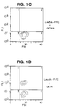
- Figure 1 depicts fluorescence activated cell sorter ("FACS") profiles of human peripheral blood lymphocytes stained with the CD4 V1-specific monoclonal antibody Leu3A labelled with fluorescein isothiocyanate ("Leu3A-FITC") in the presence of buffer ( Figure 1A), unlabelled Leu3A ( Figure 1B), the CD4 V1-specific monoclonal antibody OKT4A ( Figure 1C), the CD4 V3V4-specific monoclonal antibody OKT4 ( Figure 1D) or monoclonal antibody 5A8 of this invention ( Figure 1E). Data are plotted as log fluorescence intensity (y-axis) as a function of forward scatter (x-axis).
- FACS fluorescence activated cell sorter
- Figure 2 depicts immunofluorescent staining of the human CD4 + cell line H9 with an antibody homolog of this invention (5A8) and the anti-CD4 monoclonal antibodies Leu3A, OKT4A and OKT4, with and without preincubation with excess HIV gp120.
- the dotted lines signify staining with an anti-CD4 monoclonal antibody and the solid lines signify staining with irrelevant negative control mouse antibodies in the absence (left profiles) or presence (right profiles) of excess HIV gp120.
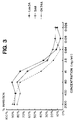
- Figure 3 depicts percent inhibition of syncytia formation (quantified as 51 Cr release) as a function of antibody concentration (0.026-2,000 ng/ml) for a monoclonal antibody of this invention (5A8) and two CD4 V1-specific monoclonal antibodies (Leu3A and OKT4A).
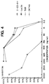
- Figure 4 depicts percent inhibition of syncytia formation (quantified as 51 Cr release) as a function of antibody concentration (3.2-10,000 ng/ml) for a monoclonal antibody of this invention (5A8), two CD4 V1-specific monoclonal antibodies (Leu3A and OKT4A) and an "OKT4-like" monoclonal antibody specific for CD4 V3V4 (2-103-D11).
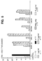
- Figures 5-6 depict the inhibition of tetanus toxoid ("TT")-induced proliferation of human peripheral blood lymphocytes (quantified as 3 H-thymidine incorporation and identified as "cpm x 10E3 (Thousands)") caused by an antibody homolog of this invention (monoclonal antibody 5A8) and various control antibodies (the CD4 V1-specific antibodies OKT4A and Leu3A and the isotype-matched control antibodies MOPC 21 and UPC 10) on days four ( Figure 5) and five ( Figure 6) of the assay.
- the bars labelled "TT Control” show the baseline growth of the cells without TT stimulation (open bar) and the TT response without any antibody (solid bar).
- Figures 5-6 also show the effect on proliferation after TT stimulation (i.e., inhibition of TT response) caused by the various antibodies at concentrations of 10 ⁇ g/ml (bars with dots) (MOPC 21 and 5A8 only), 1 ⁇ g/ml (bars with diagonal stripes) (every antibody) and 0.1 ⁇ g/ml (bars with crosshatching) (every antibody).
- Figure 7 depicts the peripheral white blood cell count of rhesus monkeys at various time points before, during and after a course of treatment with 5A8 (solid lines, Mn 261-85 and Mn 251-87) or the isotype-matched control monoclonal antibody MOPC21 (dashed lines, Mn 265-85 and Mn 265-87).
- the y-axes depict white blood cells (in thousands) per microliter of blood.
- the x-axes depict days, where 5A8 or MOPC21 was injected on days 0, 2 and 4.
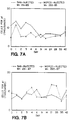
- Figure 8 depicts the number of peripheral CD4 + lymphocytes in rhesus monkeys at various time points before, during and after a course of treatment with 5A8 (solid lines, Mn 261-85 (top panel) and Mn 251-87 (bottom panel)) or the isotype-matched control monoclonal antibody MOPC21 (dashed lines, Mn 265-85 (top panel) and Mn 265-87 (bottom panel)).
- CD4 + cell number is quantitated by FACS analysis after staining with the fluorescently labelled CD4 V3V4-specific monoclonal antibody FITC-OKT4 (open and closed circles) or by FACS analysis after incubating isolated cells with excess 5A8 in vitro and then staining with fluorescently labelled goat anti-mouse Ig (open and closed triangles).
- the y-axes depict CD4 + cell number per microliter.
- the x-axes depict days, where 5A8 or MOPC21 was injected on days 0, 2 and 4.
- Figure 9 depicts the anti-tetanus toxoid titers of rhesus monkeys at various time points before, during and after treatment with 5A8 (solid lines, Mn 261-85 and Mn 251-87) or the isotype-matched control monoclonal antibody MOPC21 (dotted lines, Mn 265-85 and Mn 265-87).
- the y-axis depicts units per milliliter blood of anti-tetanus toxoid antibodies established relative to a standard anti-tetanus toxoid immune serum.
- the x-axis depicts days, where 5A8 or MOPC21 was injected on days 0, 2 and 4 and tetanus toxoid was injected on days 3 and 21.
- the bar beneath days 0-9 indicates the time period during which there was saturation coating of CD4 by 5A8 in vivo.
- CD4 means any CD4 protein encoded by a naturally occurring CD4 gene.
- CD4 + cells are cells that present the CD4 glycoprotein on their surface. Such cells include CD4 + T lymphocytes and CD4 + mammalian tissue culture cells, e.g., H9 and C8166 cells.
- CD4 V1 is the region of CD4 spanning AA 1 -AA 113 .
- CD4 V1V2 is the region of CD4 spanning AA 1 -AA 180 .
- CD4 V3V4" is the region of CD4 spanning AA 181 -AA 375 .
- recombinant soluble CD4" or “rsCD4" is a polypeptide consisting of AA 1 -AA 375 (i.e., regions V1-V4) of human CD4.
- CD4 V1-specific antibody is an antibody that binds to an epitope in the V1 region of CD4.
- CD4 V3V4-specific antibody is an antibody that binds to an epitope in the V3V4 region of CD4.
- an "antibody homolog” is a protein comprising one or more polypeptides selected from immunoglobulin light chains, immunoglobulin heavy chains, and antigen-binding fragments thereof, which are capable of binding to one or more antigens.
- the component polypeptides of an antibody homolog composed of more than one polypeptide may optionally be disulfide-bound or otherwise covalently crosslinked.
- antibody homologs include intact immunoglobulins of types IgA, IgG, IgE, IgD, IgM (as well as subtypes thereof), wherein the light chains of the immunoglobulin may be of types kappa or lambda.
- Antibody homologs also include portions of intact immunoglobulins that retain antigen-binding specificity, for example, Fab fragments, Fab' fragments, F(ab') 2 fragments, F(v) fragments, heavy chain monomers or dimers, light chain monomers or dimers, dimers consisting of one heavy and one light chain, and the like.
- a "humanized recombinant antibody homolog” is an antibody homolog initially derived from a nonhuman mammal in which recombinant DNA technology has been used to substitute some or all of the amino acids not required for CD4 binding with amino acids from corresponding regions of a human immunoglobulin light or heavy chain.
- a "chimeric recombinant antibody homolog” is an antibody homolog derived initially from a nonhuman mammal, in which recombinant DNA technology has been used to replace all or part of the hinge and constant regions of the light chain, the heavy chain or both, with corresponding regions from an immunoglobulin light chain or heavy chain of a mammal of a different species, preferably a human.
- an antibody homolog that "does not significantly block binding of a CD4 V1-specific antibody to human CD4" is one that causes no more than a 30% reduction in the binding of the CD4 V1-specific antibody either to human rsCD4 (CD4 V1-V4) or to human CD4 displayed on a CD4 + cell.
- an antibody homolog that "does not significantly block binding of HIV gp120 to human CD4" is one that causes no more than a 30% reduction in the binding of HIV gp120 either to human rsCD4 (CD4 V1-V4) or to human CD4 displayed on a CD4 + cell.
- OKT4 is the anti-CD4 mouse monoclonal antibody commercially available from Ortho Diagnostic Systems, Raritan, NJ under catalog number 7042.
- OKT4A is the anti-CD4 mouse monoclonal antibody commercially available from Ortho Diagnostic Systems, Raritan, NJ under catalog number 7142.
- a “5A8-mimetic agent” is a compound that causes at least a 30% reduction in the binding of monoclonal antibody 5A8 either to human rsCD4 (CD4 V1-V4) or to human CD4 displayed on a CD4 + cell.
- an antibody homolog "causing no significant decrease in the number of circulating CD4 + cells in vivo" is an antibody homolog which, within 24 hours after administration to a mammal having normal immune function, causes less than a 50% decrease in the number of circulating CD4 + cells, as compared either to the pre-administration number for that mammal or to the number in a control mammal to whom an isotype-matched antibody homolog of irrelevant specificity has been administered instead of an antibody homolog of this invention.
- an antibody homolog "causing no significant modulation of CD4 from the surface of CD4 + cells in vivo" is an antibody homolog which, within 24 hours after administration to a mammal having normal immune function, causes less than a ten-fold decrease in the number of CD4 molecules on the surface of the mammal's CD4 + cells, as compared either to the pre-administration number for that mammal or to the number in a control mammal to whom an isotype-matched antibody homolog of irrelevant specificity has been administered instead of an antibody homolog of this invention.
- an antibody homolog "causing no significant decrease in circulating peripheral white blood cell counts in vivo" is an antibody homolog which, within 24 hours after administration to a mammal having normal immune function, causes less than a 50% decrease in the number of circulating peripheral white blood cells, as compared either to the pre-administration number for that mammal or to the number in a control mammal to whom an isotype-matched antibody homolog of irrelevant specificity has been administered instead of an antibody homolog of this invention.
- peripheral white blood cells include B lymphocytes, T lymphocytes (CD4 + or CD8 + ) and monocytes.
- an antibody homolog "causing no significant decrease in the antibody titer elicited in response to foreign antigens" is an antibody homolog which, upon administration to a mammal having normal immune function, causes less than a ten-fold decrease in the antibody titer elicited in response to a subsequently administered foreign antigen, as compared to the antibody titer elicited by the foreign antigen in a control mammal to whom an isotype-matched antibody homolog of irrelevant specificity has been administered instead of an antibody homolog of this invention.
- a "foreign antigen” is a molecule (e.g., a viral or bacterial polypeptide) that is not encoded by the genome of that mammal.
- the antibody homologs according to one embodiment of the present invention (a) bind to human CD4; (b) do not significantly block binding of HIV gp120 to human CD4; and (c) block HIV-induced syncytia formation between CD4 + cells at least about as well as OKT4A.
- Another embodiment of the present invention relates to antibody homologs that (a) bind to human CD4; (b) do not significantly block binding of HIV gp120 to human CD4; (c) block HIV-induced syncytia formation between CD4 + cells better than OKT4; and (d) are less immunosuppressive than OKT4A in an in vitro tetanus toxoid-specific proliferation assay.
- a further embodiment relates to antibody homologs that (a) bind to human CD4; (b) do not significantly block binding of HIV gp120 to human CD4; (c) block HIV-induced syncytia formation between CD4 + cells at least about as well as OKT4A and (d) are less immunosuppressive than OKT4A in an in vitro tetanus toxoid-specific proliferation assay.
- antibody homologs of this invention bind to a polypeptide consisting of human CD4 V1V2 (i.e., they are V1V2-specific), but do not bind to a polypeptide consisting of human CD4 V1 (i.e., they are not solely V1-specific). Similarly, certain antibody homologs of this invention do not significantly block the binding of CD4 V1-specific antibodies, such as OKT4A and Leu3A, to human CD4.
- the preferred antibody homologs of this invention inhibit infection of CD4 + cells by HIV.
- Other preferred antibody homologs of this invention display one or more characteristics selected from: (a) causing no significant decrease in the number of circulating CD4 + cells in vivo; (b) causing no significant modulation of CD4 from the surface of CD4 + cells in vivo; (c) causing no significant decrease in circulating peripheral white blood cell counts in vivo; (d) causing no significant decrease in the antibody titer elicited in response to foreign antigens in vivo; and (e) causing no consistent or persistent inhibition ex vivo of T cell proliferative responses to mitogens and xenogeneic cells.
- the most preferred antibody homologs of this invention display all of the above-described properties.
- Specific antibody homologs according to the present invention include the mouse monoclonal antibodies designated 5A8 (IgG 1 ), 1F8 (IgG 1 ) and 5F2 (IgG 1 ), described infra . 5A8 is preferred.
- the antibody homologs of this invention are prophylactic and therapeutic for AIDS, ARC and HIV infection because they inhibit HIV-induced syncytia formation between CD4 + cells, thus slowing spread of the virus to uninfected cells.
- the antibody homologs of this invention to be particularly useful because they should prevent syncytia formation even after HIV gp120 has bound to CD4 displayed at the surface of CD4 + lymphocytes.
- the antibody homologs of this invention that also inhibit infection of CD4 + cells by HIV are particularly useful.
- the antibody homologs of this invention are noncompetitive blockers of virus binding, we believe their therapeutic and prophylactic effects to be independent of plasma virus levels.
- the ordinary skilled worker may easily determine, using well known methods, whether a particular antibody homolog (or preparation comprising an antibody homolog) displays the properties described above, and thus may identify antibody homologs according to the present invention.
- any conventional binding assay may be used.
- Useful CD4 binding assays include FACS analysis, ELISA assays, radioimmunoassays and the like, which detect binding of antibody homologs to human CD4.
- Full-length and soluble forms of human CD4 useful in such assays are described in PCT patent application PCT/US88/02940, which is herein incorporated by reference. Reference should also be made to D. R. Littman et al., Cell , 55, p. 541 (1988), which describes the correct signal sequence cleavage site of pre-human CD4 and which was published after the filing date of PCT/US88/02940.
- the binding of an antibody homolog to CD4, or to soluble fragments thereof, such as rsCD4 may conveniently be detected through the use of a secondary antibody specific for immunoglobulins of the species from which the antibody homolog was derived.
- any suitable competition assay may be used.
- Useful assays include, for example, ELISA assays, radioimmunoassays and the like that quantify the ability of the antibody homolog to cross block with HIV gp120 for binding to human CD4.
- the ability of excess HIV gp120 to block binding of labelled human rsCD4 to immobilized antibody homolog is measured.
- the ability of an antibody homolog to bind to human CD4 is evaluated by testing its ability to bind to human CD4 + cells.
- the preferred CD4 + cells for use in determining whether a particular antibody homolog binds to human CD4 are mammalian tissue culture cells transformed with DNA encoding full-length human CD4 and expressing the CD4 on their surface. Such cells include, for example, the CHO cells transformed with DNA encoding full-length CD4 which are described in R. A. Fisher et al., "HIV Infection Is Blocked In vitro By Recombinant Soluble CD4", Nature , 331, pp. 76-78 (1988) ("r-CD4-CHO cells”).
- Suitable r-CD4-CHO cells were deposited with the In Vitro International, Inc. culture collection in Linthicum, MD, under accession number IVI-10260 and transferred to the American Type Culture Collection, Rockville, MD, under accession number CRL 10884.
- Binding of the antibody homolog to the CD4 + cell preferably is detected by staining the cells with a fluorescently labelled secondary antibody specific for immunoglobulins of the same species from which the antibody homolog being tested is derived.
- a fluorescence activated cell sorter (“FACS") is used to detect and quantify any binding. See generally H. M. Shapiro, Practical Flow Cytometry , Alan R. Liss, Inc., New York, New York (1985).
- the ability of an antibody homolog to block binding of HIV gp120 to human CD4 is determined by preincubating excess HIV gp120 with CD4 + cells, and quantifying the degree to which the bound HIV gp120 blocks binding of the antibody homolog to the cells. Binding of the antibody homolog to the CD4 + cells is quantified by FACS analysis, using a fluorescently labelled secondary antibody specific for immunoglobulins of the same species from which the antibody homolog being tested is derived.
- the HIV gp120 used in the above assays may be provided by cells infected with HIV, by HIV itself, by host cells transformed with the gene for HIV gp120, or by isolated gp120.
- a purified, soluble HIV gp120 isolated from a unicellular host transformed with a truncated HIV gp160 gene encoding HIV gp120 will be used.
- Purified, recombinant HIV gp120 is commercially available, for example, from Repligen (Cambridge, MA) and from Celltech (Berkshire, United Kingdom).
- Suitable soluble, recombinant HIV gp120 polypeptides are produced by Spodoptera frugiperda cells transformed with recombinant baculovirus carrying the DNA sequence defined by SEQ ID NO:3.
- any known syncytia assay may be used.
- an HIV laboratory isolate or HIV-infected CD4 + tissue culture cells e.g., H9 are added to cultures of C8166 cells, and varying amounts of the antibody homolog are added to all but the control cultures.
- To control cultures are added similar varying amounts of OKT4 or OKT4A.
- Some control cultures (negative controls) are supplemented with nothing, or with an irrelevant antibody of the same isotype as the antibody homolog. After incubation, all the cultures are scored by visual inspection for syncytia. In this way, the ability of the antibody homolog to block syncytia formation is compared to the syncytia blocking abilities of OKT4 and OKT4A.
- An alternative method for comparing the syncytia blocking abilities of an antibody homolog with that of OKT4 and OKT4A is the following.
- Transformed tissue culture cells expressing recombinant HIV envelope glycoprotein (i.e., HIV gp120/gp41 after post-translational cleavage of HIV gp160) on their surface are incubated with human CD4 + cells containing a detectable substance (e.g., Jurkat cells labelled with 51 Cr) in the presence of OKT4, OKT4A, antibody homolog to be tested or no antibody. After incubation, the culture supernatants are assayed for the detectable substance (e.g., measuring ⁇ -irradiation for 51 Cr).
- a detectable substance e.g., Jurkat cells labelled with 51 Cr
- the most preferred transformed tissue culture cell for the above assay is a CHO cell expressing recombinant HIV envelope glycoprotein on its surface ("r-gp160-CHO cell").
- r-gp160-CHO cell a CHO cell expressing recombinant HIV envelope glycoprotein on its surface
- Such r-gp160-CHO cells are exemplified by cultures which were deposited in the In Vitro International, Inc. culture collection, Linthicum, MD, under accession number IVI-10261 and transferred to the American Type Culture Collection, Rockville, MD, under accession number CRL 10885.
- the deposited cells are transformed with the DNA sequence defined herein by SEQ ID NO:1.
- ELISA assays To determine whether a particular antibody homolog either does not bind to a polypeptide consisting of the human CD4 V1 region or whether it does bind to a polypeptide consisting of the human CD4 V1V2 region, ELISA assays, radioimmunoassays (RIAs), FACS analysis or the like are useful. In such assays, the ability of the antibody homolog to bind to either of those polypeptides may be detected through use of a secondary antibody specific for immunoglobulins of the species from which the antibody homolog was derived.
- binding is determined through inhibition studies with CD4 V1 and CD4 V1V2 polypeptides as competitors for binding of the antibody homolog to human CD4 displayed on the surface of a CD4 + cell, using FACS analysis to quantify any inhibition.
- determining whether a particular antibody homolog binds or does not bind to a CD4 V1 polypeptide or CD4 V1V2 polypeptide is accomplished using an RIA competition assay in which the antibody homolog is bound to the plate (directly or indirectly), and the CD4 V1 polypeptide or CD4 V1V2 polypeptide simultaneously competes with detectably labelled rsCD4 for binding to the antibody homolog.
- the amount of bound rsCD4 is compared with the amount of rsCD4 bound in the absence of the CD4 V1 or CD4 V1V2 polypeptide, allowing calculation of the percent inhibition of rsCD4 binding caused by the CD4 V1 or CD4 V1V2 polypeptide.
- the preferred polypeptides consisting of the human CD4 V1 or CD4 V1V2 regions are CD4 AA 3 -AA 115 or CD4 AA 3 -AA 182 , respectively, of the CD4 sequences set forth in PCT patent application PCT/US88/02940 or in Maddon et al., Cell , supra . Using the corrected numbering system of Littman et al., Cell , supra , these polypeptides correspond respectively to human CD4 AA 1 -AA 113 and human CD4 AA 1 -AA 180 .
- a suitable CD4 V1 polypeptide is described in B. H.
- any indication of HIV infection could be monitored.
- Useful indicators of HIV infection include, for example, secretion of HIV core antigen p24 by cells chronically infected by HIV and HIV-induced syncytia formation.
- inhibition of HIV infection is determined by comparing HIV p24 levels in the presence and absence of the antibody homolog in HIV-infected CD4 + cell cultures.
- the number of circulating CD4 + cells isolated from a mammal within 24 hours after administration of the antibody homolog to a mammal having normal immune function is quantified, and compared to the pre-administration number or the number in a control mammal to whom an isotype-matched antibody homolog of irrelevant specificity has been administered instead of an antibody homolog of this invention.
- Quantification of CD4 + cells may be accomplished, for example, by staining the cells with fluorescently labelled anti-CD4 antibodies that do not cross block with the antibody homolog for binding to CD4 (e.g., OKT4), followed by FACS analysis.
- the number of CD4 molecules on CD4 + cells isolated from a mammal within 24 hours after administration of the antibody homolog to a mammal having normal immune function is quantified, and compared to the pre-administration number in that mammal or the number in a control mammal to whom an isotype-matched antibody homolog of irrelevant specificity has been administered instead of an antibody homolog of this invention.
- Quantification of cell surface CD4 may be accomplished, for example, by staining the cells with fluorescently labelled anti-CD4 antibodies that do not cross block with the antibody homolog for binding to CD4 (e.g., OKT4), followed by FACS analysis.
- a particular antibody homolog causes no significant decrease in circulating peripheral white blood cell counts in vivo
- blood drawn within 24 hours after administration of the antibody homolog to a mammal having normal immune function is fractionated by conventional means and white blood cell counts are obtained (e.g., by staining blood smears). That number is compared to the pre-administration number for that mammal or to the number in a control mammal to whom an isotype-matched antibody of irrelevant specificity has been administered instead of an antibody homolog of this invention.
- the antibody titer elicited in a mammal having normal immune function by a foreign antigen is determined after administration of the antibody homolog, and compared to the titer elicited in a control mammal to whom an isotype-matched antibody homolog has been administered instead of an antibody homolog of this invention.
- the antibody titer for the foreign antigen may conveniently be determined, for example, by performing an ELISA using microtiter plates coated with the foreign antigen.
- Useful mitogens for this purpose include Concanavalin A and phytohemagglutinin.
- Useful xenogeneic cells include any cells (e.g., lymphocytes) derived from a species other than the species of the mammal to which the antibody homolog was administered. Proliferative response may be measured, for example, by 3 H-thymidine incorporation into the various T cell samples after incubation in vitro with mitogens or irradiated xenogeneic cells, which cannot proliferate.
- Antibody homologs according to the present invention may be of many types, the critical criterion being that the antibody homolog display one of the combinations of properties described above.
- antibody homologs of this invention may be intact monoclonal antibodies, intact recombinant antibodies, intact chimeric recombinant antibodies, intact humanized recombinant antibodies, or antigen-binding portions of the foregoing antibodies.
- the most preferred antibody homologs of the present invention are intact monoclonal antibodies produced by hybridoma cells.
- the technology for producing monoclonal antibodies is well known [see generally E. A. Lerner, "How To Make A Hybridoma”, Yale J. Biol. Med. , 54, pp. 387-402 (1981); M. L. Gefter et al., "A Simple Method For Polyethylene Glycol-Promoted Hybridization Of Mouse Myeloma Cells", Somatic Cell Genet. , 3, pp. 231-36 (1977)].
- an immortal cell line typically myeloma cells
- lymphocytes typically splenocytes
- a preparation comprising human CD4 or a V1V2-containing fragment thereof
- the culture supernatants of the resulting hybridoma cells are screened as described above for antibody homologs according to this invention.
- Useful CD4-comprising preparations include human CD4-bearing cells (e.g., CD4 + lymphocytes), fractions of such cells comprising CD4, partially purified or substantially pure human CD4 isolated from natural sources, and unpurified, partially purified or substantially pure human CD4 or V1V2-containing fragments thereof derived from recombinant host cells.
- Recombinant host cells expressing V1V2-containing fragments of CD4 are described, for example, in PCT patent application PCT/US88/02940.
- the preferred immunogen is CD4 + cells produced by transforming CD4 - mammalian tissue culture cells with DNA encoding full-length human CD4.
- the immunogen is a CHO cell transformed with the DNA sequence encoding CD4 AA -23 -AA 435 set forth in Maddon et al., Cell , supra (i.e., CD4 AA -25 -AA 433 using the corrected numbering system set forth in Littman et al., Cell , supra ).
- Such a cell referred to herein as an "r-CD4-CHO cell"
- r-CD4-CHO cells as the immunogen, rather than "natural" CD4 + cells, isolated CD4 or truncated forms of CD4 (e.g., rsCD4), increases the likelihood of obtaining anti-CD4 antibody homologs according to the present invention from a particular fusion.
- This increased likelihood as compared to "natural" CD4 + cells to be due possibly to the fact that there are no naturally occurring T cell surface molecules other than CD4 displayed on the surface of the r-CD4-CHO cells which, if present, might associate with the CD4 and by virtue of that association render certain CD4 epitopes inaccessible.
- this increased likelihood as compared to isolated CD4 or fragments thereof to be due possibly to a more favorable conformation assumed by the CD4 on the surface of the r-CD4-CHO cells than that assumed by isolated CD4 and fragments thereof.
- Immunization may be accomplished using standard procedures.
- the unit dose and immunization regimen depend on the species of mammal immunized, its immune status, the body weight of the mammal, and the CD4 content of the CD4 preparation administered.
- the CD4 preparation will be administered with an adjuvant, such as complete or incomplete Freund's adjuvant.
- each dose of the CD4 preparation used for immunization of mice contains at least about 2 x 10 5 cells, preferably about 3 x 10 6 to 2 x 10 7 r-CD4-CHO cells. Most preferably, a unit dose contains about 5 x 10 6 r-CD4-CHO cells.
- the mouse is immunized intraperitoneally on day 0 with r-CD4-CHO cells in complete Freund's adjuvant.
- the mouse is given a first boost intraperitoneally with r-CD4-CHO cells without adjuvant, 14 days to 6 months after the initial immunization, and preferably 15 to 20 days after the first immunization. Additional boosts may be administered if desired.
- the final boost (r-CD4-CHO cells without adjuvant) is administered intravenously. Typically, this final boost is administered 1 to 6 months, and preferably 39-44 days, after the initial immunization.
- the mammal is sacrificed, its spleen removed and the splenocytes (including lymphocytes) are prepared for fusion with the immortal cell line.
- the immunized mammals may be bled between boosts and the serum from each blood sample assayed for antibody homologs according to the present invention using the screening assays described above.
- the lymphocytes used in the production of hybridoma cells typically are isolated from immunized mammals whose sera have already tested positive for the presence of antibody homologs according to this invention.
- the immortal cell line (e.g., a myeloma cell line) is derived from the same mammalian species as the lymphocytes.
- Useful mammals include mice and rats.
- both the immortal cell line and the lymphocytes are derived from an inbred mouse of strain BALB/c (Jackson Labs, Bar Harbour, ME).
- Preferred immortal cell lines are mouse myeloma cell lines that are sensitive to culture medium containing hypoxanthine, aminopterin and thymidine ("HAT medium”).
- the most preferred mouse myeloma cell line is P3X63-AG8.653 (ATCC, Rockville, MD, catalog no. CRL 1580).
- HAT-sensitive mouse myeloma cells are fused to mouse splenocytes using polyethylene glycol (“PEG”), preferably PEG-3350. Hybridoma cells resulting from the fusion are then selected using HAT medium, which kills unfused and unproductively fused myeloma cells (unfused splenocytes die after several days because they are not transformed).
- PEG polyethylene glycol
- Hybridoma cells producing an antibody homolog are detected by screening the hybridoma culture supernatants using the screening assays described above.
- the primary screen will select antibodies that bind to human CD4 displayed on the surface of CD4 + cells. Such binding is detected by use of fluorescently labelled secondary antibodies specific for mouse immunoglobulins, and quantified by FACS analysis.
- hybridoma cells that have tested positive in the above screening assays are cultured in a nutrient medium under conditions and for a time sufficient to allow the hybridoma cells to secrete the monoclonal antibodies into the culture medium.
- Tissue culture techniques and culture media suitable for hybridoma cells are well known [see, e.g., Lerner, Yale J. Biol. Med. , supra ].
- Conditioned hybridoma culture supernatant containing the antibody homologs is collected.
- the desired antibody homolog may be produced by injecting the hybridoma cells into the peritoneal cavity of an unimmunized mouse.
- the hybridoma cells proliferate in the peritoneal cavity, secreting the antibody homolog, which accumulates as ascites fluid [see Lerner, Yale J. Biol. Med. , supra ].
- the antibody homolog is harvested by withdrawing the ascites fluid from the peritoneal cavity with a syringe.
- monoclonal antibodies which are antibody homologs according to this invention may be purified with ease from conditioned hybridoma culture supernatant or from ascites.
- Antibody homologs according to the present invention may be recombinant monoclonal antibodies produced by host cells transformed with DNA encoding immunoglobulin light and heavy chains according to this invention.
- Recombinant antibodies may be produced by well known genetic engineering techniques. See, e.g., United States patent 4,816,397, which is incorporated herein by reference.
- recombinant antibodies may be produced by cloning cDNA or genomic DNA encoding the immunoglobulin light and heavy chains of the desired antibody from a hybridoma cell that produces an antibody homolog according to this invention.
- the cDNA or genomic DNA encoding those polypeptides is then inserted into expression vectors so that both genes are operatively linked to their own transcriptional and translational expression control sequences.
- the expression vector and expression control sequences are chosen to be compatible with the expression host cell used. Typically, both genes are inserted into the same expression vector.
- This invention provides DNA sequences encoding the 5A8 light chain variable region (SEQ ID NO:14) and the 5A8 heavy chain variable region (SEQ ID NO:9), portions thereof that encode an antibody homolog according to this invention, and DNA sequences degenerate to any of those DNA sequences. Also provided are recombinant DNA molecules comprising one or more of the foregoing DNA sequences and one or more expression control sequences operatively linked thereto.
- Prokaryotic or eukaryotic cells may be used as expression hosts. Expression in eukaryotic host cells is preferred because such cells are more likely than prokaryotic cells to assemble and secrete a properly folded and immunologically active antibody. However, any antibody produced that is inactive due to improper folding may be renaturable according to well known methods [P. S. Kim and R. L. Baldwin, "Specific Intermediates In The Folding Reactions Of Small Proteins And The Mechanism Of Protein Folding", Ann. Rev. Biochem. , 51, pp. 459-89 (1982)]. It is possible that the host cells will produce portions of intact antibodies, such as light chain dimers or heavy chain dimers, which also are antibody homologs according to the present invention.
- DNA encoding the recombinant antibodies described above may be used as the starting point for producing chimeric or humanized recombinant antibodies.
- Chimeric recombinant antibodies are produced by transforming a host cell with a suitable expression vector comprising DNA encoding the desired immunoglobulin light and heavy chains in which all or some of the DNA encoding the hinge and constant regions of the heavy and/or the light chain have been substituted with DNA from the corresponding region of an immunoglobulin light or heavy chain of a different species.
- a suitable expression vector comprising DNA encoding the desired immunoglobulin light and heavy chains in which all or some of the DNA encoding the hinge and constant regions of the heavy and/or the light chain have been substituted with DNA from the corresponding region of an immunoglobulin light or heavy chain of a different species.
- substitution of corresponding human sequences is preferred.
- An exemplary chimeric recombinant antibody has mouse variable regions and human hinge and constant regions. See generally, United States patent 4,816,397 and S.
- amino acid sequence of the light chain variable region is SEQ ID NO:15 and the amino acid sequence of the heavy chain variable region is SEQ ID NO:10, respectively.
- Humanized recombinant antibodies are produced by transforming a host cell with a suitable expression vector comprising DNA encoding the desired nonhuman immunoglobulin light and heavy chains in which all or some of the DNA encoding amino acids not involved in CD4 binding have been substituted with DNA from the corresponding region of a desired human immunoglobulin light or heavy chain. See generally, P. T. Jones et al., "Replacing The Complementarity-Determining Regions In A Human Antibody With Those From A Mouse", Nature , 321, pp. 522-25 (1986).
- humanized recombinant antibodies of this invention have the 5A8 complementarity determining regions ("CDRs"). These include humanized recombinant antibodies wherein:
- Another preferred embodiment provides humanized recombinant antibody homologs having 5A8 CDRs wherein the amino acid sequence of the light chain variable region is AA 1 -AA 112 of SEQ ID NO:56 and the amino acid sequence of the heavy chain variable region is AA 1 -AA 122 of SEQ ID NO:45.
- a further preferred embodiment provides humanized recombinant antibody homologs having 5A8 CDRs wherein the amino acid sequence of the light chain is AA 1 -AA 219 of SEQ ID NO:56 and the amino acid sequence of the heavy chain is AA 1 -AA 448 of SEQ ID NO:45.
- This invention provides DNA sequences encoding a pre-5A8 humanized light chain (SEQ ID NO:55) and a pre-5A8 humanized heavy chain (SEQ ID NO:44), portions of the foregoing DNA sequences that encode antibody homologs according to this invention, and DNA sequences degenerate to the foregoing DNA sequences. Also provided are recombinant DNA molecules comprising one or more of the foregoing DNA sequences and one or more expression control sequences operatively linked thereto.
- Preferred recombinant DNA molecules include pMDR1007 (pre-5A8 humanized light chain) and pMDR1002 (pre-5A8 humanized heavy chain), deposited with the American Type Culture Collection, Rockville, MD, under accession numbers ATCC 68846 and ATCC 68847, respectively.
- the present invention also provides antibody homologs that are not intact antibodies.
- Such homologs may be derived from any of the antibody homologs described above.
- antigen-binding fragments, as well as full-length monomeric, dimeric or trimeric polypeptides derived from the above-described antibodies are themselves antibody homologs according to the present invention.
- Useful antibody homologs of this type include Fab fragments, Fab' fragments, F(ab') 2 fragments, F(v) fragments, heavy chain monomers or dimers, light chain monomers or dimers, dimers consisting of one heavy and one light chain, and the like. We believe that the foregoing fragments, other than Fab fragments of 5A8, generally are useful antibody homologs according to the present invention.
- Antibody fragments may be produced by chemical methods, e.g., by cleaving an intact antibody with a protease, such as pepsin or papain, and optionally treating the cleaved product with a reducing agent.
- useful fragments may be produced by using host cells transformed with truncated heavy and/or light chain genes.
- Heavy and light chain monomers may be produced by treating an intact antibody with a reducing agent, such as dithiothreitol, followed by purification to separate the chains.
- Heavy and light chain monomers may also be produced by host cells transformed with DNA encoding either the desired heavy chain or light chain, but not both. See, e.g., E. S.
- this invention provides derivatized antibody homologs in which any of the antibody homologs of this invention are functionally linked (by chemical coupling, genetic fusion or otherwise) to one or more members independently selected from the group consisting of antibody homologs of this invention, 5A8-mimetic agents of this invention, detectable agents, cytotoxic agents and pharmaceutical agents.
- One type of derivatized antibody homolog is produced by crosslinking two or more antibody homologs (of the same type or of different types).
- Suitable crosslinkers include those that are heterobifunctional, having two distinctly reactive groups separated by an appropriate spacer (e.g., m-maleimidobenzoyl-N-hydroxysuccinimide ester) or homobifunctional (e.g., disuccinimidyl suberate).
- an appropriate spacer e.g., m-maleimidobenzoyl-N-hydroxysuccinimide ester
- homobifunctional e.g., disuccinimidyl suberate
- Useful detectable agents include fluorescent compounds.
- Exemplary fluorescent detectable agents include fluorescein, fluorescein isothiocyanate, rhodamine, 5-dimethylamine-1-naphthalenesulfonyl chloride, phycoerythrin and the like.
- the antibody homologs of this invention may also be derivatized with detectable enzymes, such as alkaline phosphatase, horseradish peroxidase, glucose oxidase and the like.
- detectable enzymes such as alkaline phosphatase, horseradish peroxidase, glucose oxidase and the like.
- the antibody homolog is derivatized with a detectable enzyme, it is detected by adding additional reagents that the enzyme uses to produce a detectable reaction product.
- the detectable agent horseradish peroxidase is present, the addition of hydrogen peroxide and diaminobenzidine leads to a colored reaction product, which is detectable.
- the antibody homolog may also be derivatized with biotin, and detected through indirect measurement of avidin binding.
- the present invention also provides antibody homologs linked to one or more pharmaceutical agents.
- useful pharmaceutical agents include biologically active peptides, polypeptides and proteins, such as antibody homologs specific for a human polypeptide other than CD4.
- Other useful pharmaceutical agents include non-proteinaceous pharmaceutical agents such as HIV reverse transcriptase inhibitors (e.g., 3'-azido-2',3'-dideoxythymidine (“AZT”) and 2',3'-dideoxyinosine (“DDI”) and other antiviral compounds, or immunosuppressive agents (e.g., cyclosporin or FK506).
- the present invention also provides cells and cell cultures that produce the antibody homologs of this invention.
- Such cells include hybridoma cells that produce monoclonal antibodies which are antibody homologs of this invention, as well as transformed host cells that produce recombinant antibody homologs of this invention.
- Preferred cells according to this invention include those that produce the monoclonal antibodies referred to herein as "5A8", “1F8” and “5F2".
- the most preferred cell line produces monoclonal antibody 5A8.
- the hybridoma cell lines producing 5A8, 1F8 and 5F2 were deposited in the In Vitro International, Inc. culture collection in Linthicum, MD, under accession numbers IVI-10257, IVI-10258 and IVI-10259, respectively and transferred to the American Type Culture Collection, Rockville, MD, under accession numbers HB 10881, HB 10882 and HB 10883, respectively.
- Other preferred cells are host cells producing the 5A8 chimeric and humanized recombinant antibodies described supra .
- Eukaryotic host cells transformed with pMDR1007 (pre-5A8 humanized light chain) and pMDR1002 (pre-5A8 humanized heavy chain) are most preferred for producing 5A8 humanized antibodies.
- the present invention provides a method of producing the antibody homologs of the present invention by culturing the cells of this invention, which produce the antibody homologs. Methods of culturing such cells and isolating the antibody homologs they produce are well known. These methods include tissue culture techniques, as well as the generation of ascites.
- Preferred cells to be used as an immunogen for this process are the r-CD4-CHO cells described above.
- the present invention provides 5A8-mimetic agents, which are compounds that cause at least a 30% reduction in the binding of monoclonal antibody 5A8 either to human rsCD4 or to human CD4 displayed on the surface of a CD4 + cell.
- 5A8-mimetic agents may be peptides, semi-peptidic compounds or non-peptidic compounds.
- Preferred 5A8-mimetic agents inhibit HIV-induced syncytia formation between CD4 + cells, inhibit infection of CD4 + cells by HIV, or both.
- the most preferred 5A8-mimetic agents display the properties of one or more antibody homologs of this invention.
- 5A8-mimetic agents of this invention may be produced by synthesizing a plurality of peptides (e.g., 5-20 amino acids in length), semi-peptidic compounds or non-peptidic, organic compounds, and then screening those compounds for their ability (1) to cross block with 5A8 for binding to human CD4, (2) to inhibit HIV-induced syncytia formation between CD4 + cells, and (3) to inhibit infection of CD4 + cells by HIV.
- peptides e.g., 5-20 amino acids in length
- semi-peptidic compounds or non-peptidic, organic compounds screening those compounds for their ability (1) to cross block with 5A8 for binding to human CD4, (2) to inhibit HIV-induced syncytia formation between CD4 + cells, and (3) to inhibit infection of CD4 + cells by HIV.
- the antibody homologs and the 5A8-mimetic agents of this invention are useful in prophylactic and therapeutic compositions for preventing and treating diseases caused by infective agents whose primary targets are CD4 + lymphocytes.
- diseases include AIDS, ARC and HIV infection.
- Preferred pharmaceutical compositions of this invention for administration to humans include, as antibody homologs, one or more of the mouse monoclonal antibodies 5A8, 1F8 or 5F2, 5A8 mouse/human chimeric recombinant antibodies, 5A8 humanized recombinant antibodies or antigen-binding portions of those antibodies.
- Pharmaceutical compositions comprising 5A8 mouse/human chimeric recombinant antibodies or 5A8 humanized recombinant antibodies are most preferred.
- compositions of this invention may further comprise other therapeutics for the prophylaxis or treatment of AIDS, ARC and HIV infection.
- the antibody homologs of this invention may be used in combination with anti-retroviral agents that block reverse transcriptase, such as AZT, DDI, HPA-23, phosphonoformate, suramin, ribavirin and dideoxycytidine, or with agents that inhibit the HIV protease.
- compositions of this invention may further comprise anti-viral agents such as interferons, including alpha interferon, beta interferon and gamma interferon, or glucosidase inhibitors such as castanospermine, or immunosuppressive agents such as adrenal corticosteroids, azathrioprine, cyclosporin or FK506.
- anti-viral agents such as interferons, including alpha interferon, beta interferon and gamma interferon, or glucosidase inhibitors such as castanospermine, or immunosuppressive agents such as adrenal corticosteroids, azathrioprine, cyclosporin or FK506.
- the antibody homologs of this invention may advantageously be administered in combination with agents that significantly block binding of HIV gp120 to CD4.
- agents include, for example, anti-CD4 antibodies that are V1-specific (e.g., Leu3A), CD4 V1 polypeptides and antibodies (anti-idio
- one or more antibody homologs or 5A8-mimetic agents may be used in combination with two or more of the foregoing therapeutic agents.
- Such combination therapies may advantageously utilize lower dosages of the administered therapeutic agents, thus avoiding possible toxicities or complications associated with the various monotherapies.
- compositions of this invention comprise an immunotherapeutically effective amount of one or more antibody homologs or 5A8-mimetic agents according to this invention, or derivatized form(s) thereof and, preferably, a pharmaceutically acceptable carrier.
- immunotherapeutically effective amount is meant an amount capable of lessening the spread, severity or immunocompromising effects of AIDS, ARC or HIV infection, or of other diseases caused by infective agents whose primary targets are CD4 + lymphocytes.
- pharmaceutically acceptable carrier is meant a carrier that does not cause an allergic reaction or other untoward effect in patients to whom it is administered.
- Suitable pharmaceutically acceptable carriers include, for example, one or more of water, saline, phosphate buffered saline, dextrose, glycerol, ethanol and the like, as well as combinations thereof. Pharmaceutically acceptable carriers may further comprise minor amounts of auxiliary substances such as wetting or emulsifying agents, preservatives or buffers, which enhance the shelf life or effectiveness of the antibody homolog or 5A8-mimetic agent.
- compositions of this invention may be in a variety of forms. These include, for example, solid, semi-solid and liquid dosage forms, such as tablets, pills, powders, liquid solutions, dispersions or suspensions, liposomes, suppositories, injectable and infusible solutions.
- solid, semi-solid and liquid dosage forms such as tablets, pills, powders, liquid solutions, dispersions or suspensions, liposomes, suppositories, injectable and infusible solutions.
- the preferred form depends on the intended mode of administration and therapeutic application.
- the preferred compositions are in the form of injectable or infusible solutions.
- compositions of this invention are similar to those used for passive immunization of humans with other antibodies.
- the preferred mode of administration is parenteral.
- the immunotherapeutically effective amount of antibody homolog or 5A8-mimetic agent of this invention will depend, inter alia, upon the administration schedule, the unit dose of antibody homolog or 5A8-mimetic agent administered, whether the antibody homolog or 5A8-mimetic agent is administered in combination with other therapeutic agents, the immune status and health of the patient, and the therapeutic activity of the particular antibody homolog or 5A8-mimetic agent administered.
- immunotherapeutically effective amounts per unit dose of an antibody homolog which is an intact antibody range from about 0.1 to 10 mg/kg patient weight, preferably 2 mg/kg patient weight.
- Unit doses should be administered from twice each day to once every two weeks until an antiviral effect is observed, preferably once every two weeks.
- the antiviral effect may be measured by a variety of methods, including viral load, lymphocyte counts and clinical signs and symptoms. It will be recognized, however, that lower or higher dosages and other administration schedules may be employed.
- Treatment regimens for antibody homologs that are not intact antibodies and for 5A8-mimetic agents may differ, depending on their size and pharmaceutical properties.
- the immunogen used to generate monoclonal antibodies 1F8, 5A8 and 5F2 was CHO cells transformed with DNA encoding full-length human CD4 expressed on the cell surface ("r-CD4-CHO cells").
- the CD4 sequence used to transform the CHO cells is set forth in Maddon et al., Cell , supra . These cells are described in R. A. Fisher et al., "HIV Infection Is Blocked In Vitro By Recombinant Soluble CD4", Nature , 331, pp. 76-78 (1988), and were deposited with the In Vitro International, Inc. culture collection, Linthicum, MD, under accession number IVI-10260 and transferred to American Type Culture Collection, Rockville, MD, under accession number CRL 10884.
- the r-CD4-CHO cells were maintained at 37°C, under 5% CO 2 , in ⁇ - MEM media (Gibco, Grand Island, NY) supplemented with 10% dialyzed fetal calf serum ("FCS") (North American Biologics, Miami, FL, cat. no. 33189) and 30 nM methotrexate.
- FCS dialyzed fetal calf serum
- Cells were prepared for injection by substituting the above culture medium with phosphate buffered (Ca + - and Mg + -free) saline (“PBS”) supplemented with 5 mM EDTA, and harvesting the cells in that buffer.
- PBS phosphate buffered
- the harvested cells were pelleted by centrifugation at 500 x g av for about 5 minutes, washed once by resuspending the pellet in PBS and centrifuging as before, counted, and adjusted to the appropriate volume for injection by resuspending the cell pellet in PBS.
- mice Two female BALB/c J mice, about 4-6 weeks old (Jackson Labs, Bar Harbour, ME), were immunized with r-CD4-CHO cells as follows.
- the first mouse was primed intraperitoneally (i.p.) on day 0 with a 1:1 emulsion of r-CD4-CHO cells in PBS with complete Freunds adjuvant ("CFA") (Colorado Serum Co., Denver, CO, cat. no. C51450), boosted i.p. on day 20 with r-CD4-CHO cells in PBS without adjuvant, and finally boosted intravenously on day 44 with r-CD4-CHO cells in PBS, without adjuvant.
- CFA complete Freunds adjuvant
- the other mouse was primed i.p. on day 0, boosted i.p.
- each injection contained approximately 3 x 10 6 to 2 x 10 7 r-CD4-CHO cells in a volume of approximately 250 ⁇ l.
- mice were sacrificed and each spleen was removed and placed in approximately 5 ml of serum-free DMEM (Gibco, cat. no. 430-2100) in a petri dish.
- the splenic capsules were cut and the spleen cells were teased out into the medium.
- the spleen cell suspensions were transferred to one 15 ml conical bottom tube and allowed to settle for about 5 minutes.
- the supernatant was removed to a fresh 15 ml conical bottom tube and placed in an incubator (37°C, 5% CO 2 ) until the fusion.
- the spleen cells from both mice were pooled.
- a 5-ml single cell suspension of control spleen feeder cells was prepared from an unimmunized mouse essentially as described above for the immunized spleen cells, and placed in an incubator (37°C, 5% CO 2 ) until needed.
- the fusion partner for the immunized spleen cells was a hypoxanthine/aminopterin/thymidine ("HAT")-sensitive, non-secreting mouse myeloma cell line --P3X63-AG8.653 (ATCC, Rockville, MD, cat. no. CRL 1580).
- HAT hypoxanthine/aminopterin/thymidine
- P3X63-AG8.653 ATCC, Rockville, MD, cat. no. CRL 1580.
- the P3X63-AG8.653 cells were maintained in DMEM/10% FCS (37°C, 5% CO 2 ), ensuring that they were in a logarithmic growing phase on the day of the fusions.
- the fusion protocol used was a hybrid of the protocols set forth in Lerner, Yale J. Biol. Med. , supra and Gefter et al., Somatic Cell Genet. , supra .
- the pooled spleen cells prepared as described above were washed three times with PBS, resuspended in approximately 10 ml of PBS and counted.
- the logarithmic phase P3X63-AG8.653 cells were washed three times with PBS and counted.
- the myeloma cells were resuspended to 1 X 10 7 cells/ml in PBS.
- spleen cells were mixed with 3 x 10 7 myeloma cells in a 50 ml conical bottom polypropylene tube, and the cells were washed once with PBS. The ratio of spleen cells to myeloma cells was 5:1.
- the tubes were centrifuged at 500 x g av for 5 minutes to pellet the cells. After aspiration of the supernatants, the pellets were gently resuspended by tapping the bottom of the tubes. The tubes were then placed in a beaker of 37°C water. All subsequent fusion steps were carried out in this beaker.
- HAT selection medium Fifty ml of "HAT selection medium” containing 400 ⁇ l of the 5-ml suspension of control spleen feeder cells (prepared as described above) were added stepwise to each pellet, with occasional mixing.
- "HAT selection medium” was prepared by diluting a 50x HAT stock (Sigma Chemical Co., St. Louis, MO, cat. no. H-0262) 50-fold in DMEM/10% FCS (final concentrations: 10 ⁇ M hypoxanthine, 0.4 ⁇ M aminopterin, 16 ⁇ M thymidine).
- Each 50-ml cell suspension was plated into 5 96-well flat-bottom microtiter plates, with ⁇ 100 ⁇ l/well. The plates were kept in an incubator at 37°C, 5% CO 2 . On day 2 post-fusion, the cells were fed by addition of 100 ⁇ l HAT selection medium per well. On day 10 post-fusion, the medium was aspirated and 200 ⁇ l HAT selection medium was added to each well. Supernatants were withdrawn for primary screening on days 12, 14, 16 and 19 post-fusion, from wells containing clones. The fusion efficiency was 34% (327 out of 960 possible wells developed clones that were screened).
- the primary screen used was a radioimmunoassay (RIA) designed to detect antibodies that bind to human rsCD4.
- RIA radioimmunoassay
- affinity-purified goat anti-mouse IgG F C fragment-specific
- PBS PBS-containing bovine serum
- the goat anti-mouse IgG was removed from the plates and the wells were blocked with 100 ⁇ l/well of 5% FCS/PBS for 1 hour at room temperature.
- neat hybridoma culture supernatant was added to the wells (50 ⁇ l/well) and incubated 1 hour at room temperature. The plates were washed 3 times with PBS/0.05% Tween-20.
- the 125 I-rsCD4 used in the primary screening assay consisted of purified, 125 I-labelled human CD4 AA 3 -AA 377 of the CD4 sequence set forth in Maddon et al., Cell , supra (i.e., human CD4 AA 1 -AA 375 according to the corrected numbering system of Littman et al., Cell , supra ).
- the rsCD4 was produced by transformed CHO cells substantially as described in Fisher et al., Nature , supra .
- the rsCD4 produced by those cells may be purified as described in Fisher et al., Nature , supra .
- the purified rsCD4 was labelled with 125 I according to the Bolton-Hunter method, substantially as described by the supplier of the Bolton-Hunter reagent (New England Nuclear, Boston, MA).
- the quality of the 125 I-rsCD4 was routinely monitored by confirming that the labelling procedure did not destroy the epitopes recognized by the commercially available CD4 V1-specific monoclonal antibodies Leu3A (Becton Dickinson, Mountain View, CA, cat. no. 7326) and OKT4A (Ortho Diagnostic Systems, Raritan, N.J., cat. no. 7124) and the CD4 V3V4-specific monoclonal antibody OKT4 (Ortho Diagnostic Systems, cat. no. 7024).
- Clones were considered to be positive on primary screening if they were approximately 10-fold over background in the RIA. By this criterion, approximately 6% of the clones (21) were positive. The 21 positive clones were pulled and expansion was attempted; 13 clones survived expansion and were stored frozen. Many of these 13 clones were successfully subcloned, including 1F8, 5A8 and 5F2. Expansion, subcloning and freezing of the cells was conducted substantially as described in Lerner, Yale J. Biol Med. , supra .
- the first secondary screen we conducted was designed to determine whether the antibodies recognized native CD4 epitopes. This was accomplished by FACS analysis of cell surface CD4 displayed on CD4 + Jurkat cells and r-CD4-CHO cells stained with the monoclonal antibodies, visualizing binding with fluorescently labelled goat anti-mouse second antibody, substantially as described below. All 13 antibodies recognized cell surface CD4.
- competition assays as described below with a CD4 V1 polypeptide and a CD4 V1V2 polypeptide, with commercially available CD4 V1-specific and CD4 V3V4-specific antibodies, and with HIV gp120.
- an RIA was performed in which each microtiter plate well was coated with an anti-mouse Ig, blocked, and then incubated with one of the hybridoma culture supernatants being tested. Next, a mixture of CD4-V1 or CD4-V1V2 and 125 I-rsCD4 was added. After incubation, the wells were washed and the bound 125 I-rsCD4 was detected in a gamma counter.
- microtiter plates (96-well) were coated overnight at 4°C with 50 ⁇ l/well of affinity-purified goat anti-mouse IgG (Fc fragment-specific) (Cappell). Wells were washed 3 times with PBS/0.05% Tween-20TM, and then blocked by incubating 1 hour with 100 ⁇ l/well of 5% FCS/PBS. After removing the blocking buffer, the wells were incubated 1 hour at room temperature with 50 ⁇ l/well neat hybridoma culture supernatant (5-20 ⁇ g/ml Ig).
- affinity-purified goat anti-mouse IgG Fc fragment-specific
- control wells were incubated with 50 ⁇ l/well OKT4 (10 ⁇ g/ml in PBS/5% FCS), OKT4A (500 ng/ml in PBS/5% FCS) or Leu3A (500 ng/ml in PBS/5% FCS).
- the plates were washed 3 times with PBS/0.05% Tween-20TM.
- CD4-V1 and CD4-V1V2 (final concentrations 5 ⁇ g/ml) were separately pre-incubated with approximately 20,000 cpm 125 I-rsCD4 (prepared as described above), for approximately 35-45 minutes at room temperature. Those pre-incubated mixtures were then incubated in the wells for 1-2 hours at room temperature. Following the incubation, the wells were washed 3 times with PBS/0.05% Tween-20TM and then counted for radioactivity.
- the CD4-V1 used in this experiment consisted of AA 3 -AA 115 and/or AA 3 -AA 113 of the CD4 sequence set forth in Figure 16 of PCT patent application PCT/US88/02940, which correspond respectively to CD4 AA 1 -AA 113 and AA 1 -AA 111 according to the corrected numbering system of Littman et al., Cell , supra .
- These CD4 V1 polypeptides were produced and purified as described in B. H. Chao et al., "A 113-Amino Acid Fragment Of CD4 produced In Escherichia coli Blocks Human Immunodeficiency Virus-Induced Cell Fusion", J. Biol. Chem. , 264, pp. 5812-17 (1989).
- the CD4-V1V2 used in this experiment consisted of AA 3 -AA 182 of the CD4 sequence set forth in Figure 16 of PCT patent application PCT/US88/02940, which corresponds to CD4 AA 1 -AA 180 according to the corrected numbering system of Littman et al., Cell , supra .
- the CD4 V1V2 polypeptide was bacterially produced using standard molecular biology techniques, in a manner analogous to the protocol used for production of the CD4 V1 polypeptide described in Chao et al., J. Biol. Chem. , supra .
- a plasmid carrying DNA encoding the CD4 V1V2 polypeptide was transfected into E . coli , strain A89 [S. A. Goff et al., "Heat Shock Regulatory Gene htpR Influences Rates of protein Degradation And Expression Of The lon Gene In Escherichia coli ", Proc. Natl. Acad. Sci. USA , 81, pp. 6647-51 (1984)].
- the cells were fermented under standard conditions in a 10-liter fermenter, harvested using a Pelikon concentration system followed by centrifugation at 3000 rpm in a Beckman J-6M centrifuge. The cell pellet was stored frozen at -20°C until use.
- the cells were thawed and suspended in 8 ml buffer (20 mM Tris-HCl, pH 7.7/1 mM Na 2 EDTA) per gram wet cell weight. The cells were broken by 3 passes through a french press. The insoluble material containing the CD4-V1V2 was pelleted by centrifugation. The pellets were washed 3 times with 1 M urea/15 mM sodium acetate, pH 5.5 (15-20 ml per gram wet cell weight), followed by 2 washes with 20 mM Tris-HCl, pH 7.7/1 mM Na 2 EDTA.
- the resulting washed pellet was extracted overnight at room temperature with 25 ml per gram of wet cell weight using 6 M guanidine-HCl/10 mM DTT/20 mM Tris-HCl, pH 7.7.
- the extract was clarified by centrifuging at 48,000 x g av for 45 minutes at room temperature.
- the clarified extract was diluted 1:60 v/v into cold (4°C) 20 mM Tris-HCl, pH 7.7, and bovine serum albumin ("BSA") was added to a final concentration of 0.5 mg/ml. All subsequent steps were performed at 4°C.
- the pH was lowered to 7.0 with 6 N HCl, and the suspension was clarified by passage through a 5 micron hydrophilic MillidiskTM filter (Millipore, Bedford, MA, cat. no. MCSC 40S 01) followed by passage through a Durapore 0.2 micron filter (Millipore, cat. no. CVGL 01T P3).
- the clarified preparation was loaded onto an S-SepharoseTM Fast FlowTM column (Pharmacia LKB Biotechnology, Inc., Piscataway, NJ), pre-equilibrated with 20 mM Tris-HCl, pH 7.0, at a ratio of 1 volume S-Sepharose Fast FlowTM gel per 100 volumes load.
- the column was washed with 20 mM Tris-HCl, pH 7.0, and the bound proteins were eluted with 20 mM Tris-HCl, pH 7.7/300 mM NaCl.
- the eluate pool containing the CD4-V1V2 was diluted with an equal volume of 20 mM Tris-HCl, pH 7.7, and then loaded onto an anti-CD4 monoclonal antibody affinity column (described below), pre-equilibrated with 20 mM Tris-HCl, pH 7.7/150 mM NaCl.
- the column was washed with 20 mM Tris-HCl, pH 7.7/150 mM NaCl followed by 20 mM Tris-HCl, pH 7.7/1 M NaCl.
- the bound proteins were eluted with 50 mM glycine/250 mM NaCl, pH 3.0.
- the eluted material was dialysed into 20 mM HEPES, pH 6.8, and concentrated on a PM 10 membrane in an Amicon ultrafiltration cell.
- the purified, concentrated CD4-V1V2 (approximately 0.5 - 3.0 mg/ml) was stored at -70°C until use.
- the anti-CD4 monoclonal antibody affinity column was prepared using one of the CD4 V1-specific monoclonal antibodies produced from the fusions described above.
- the monoclonal antibody was purified from ascites by ammonium sulfate fractionation and anion exchange chromatography, concentrated by ultrafiltration, dialysed into 0.1 M sodium borate (pH 8.0)/0.5 M sodium chloride, and concentrated again by ultrafiltration to a concentration of approximately 5 mg/ml.
- the purified anti-CD4 monoclonal antibody (10 ml containing approximately 50 mg protein) was coupled to 4 g of CNBr-SepharoseTM according to the manufacturer's directions (Sigma Chemical Co., cat. no. C9142), yielding 12 ml of derivatized resin.
- each microtiter plate well was coated with anti-mouse Ig, blocked, and then incubated with 5A8 culture supernatant (5-20 ⁇ g/ml), OKT4A (500 ng/ml), OKT4 (10 ⁇ g/ml) or Leu3A (500 ng/ml). The wells were then washed, and blocked with the 100 ⁇ l/well mouse Ig (50 ⁇ g/ml) in PBS for 30 minutes to 1 hour at room temperature.
- Competitor antibody i.e., OKT4A, OKT4 or Leu3A at twice the above concentrations
- 125 I-rsCD4 was pre-incubated with 125 I-rsCD4, and each mixture was added to the wells at 50 ⁇ l/well. After incubating 1 hour at room temperature, the wells were washed and the bound 125 I-rsCD4 was quantified in a gamma counter.
- Table III lists the percent inhibition of binding of 125 I-rsCD4 to the immobilized anti-CD4 antibodies 5A8, OKT4, OKT4A and Leu3A by the soluble anti-CD4 antibodies Leu3A, OKT4A and OKT4.
- the 125 I-rsCD4 used in this experiment consisted of purified, 125 I-labelled human CD4 AA 3 -AA 377 of the CD4 sequence set forth in Figure 16 of PCT patent application PCT/US88/02940 (rather than the sequence set forth in Maddon et al., Cell , supra ), which corresponds to CD4 AA 1 -AA 375 according to the corrected numbering system of Littman et al., Cell , supra .
- Table IV lists the percent inhibition of binding of 125 I-rsCD4 to immobilized 5A8 by the soluble anti-CD4 antibodies 1F8, 5F2, 5A8, OKT4 and Leu3A. TABLE IV 5A8 Cross Blocking: Anti-CD4 Monoclonal Antibodies Preincubated In Solution With rsCD4 Soluble Anti-CD4 Monoclonal Antibody % Inhibition rsCD4 Binding 1F8 97 5A8 98 5F2 89 OKT4 4 Leu3A 0
- Figure 1 displays the results of the cell surface CD4 cross blocking experiment.
- Human peripheral blood lymphocytes (“PBLs") were incubated with buffer ( Figure 1A), Leu3A (Figure 1B), OKT4A (Figure 1C), OKT4 ( Figure 1D) or 5A8 ( Figure 1E).
- the cells then were washed and incubated with Leu3A labelled with fluorescein isothiocyanate ("Leu3A-FITC"). After washing again, the cells were analyzed using a FACStar fluorescence activated cell sorter. The results of the this FACS analysis are depicted in Figure 1.
- the data demonstrate that 5A8 does not significantly block binding of the V1-specific antibody Leu3A to cell surface CD4.
- the protocol for this experiment follows.
- human PBLs were isolated by diluting 10 ml blood two-fold with unsupplemented RPMI-1640 media and layering the mixture over 10 ml of Lymphocyte Separation Media (Organon Teknika, Durham, NC, cat. no. 36427) in a 50-ml tube at room temperature. The tube was centrifuged at room temperature for 30 minutes at 400 x g av . PBLs were collected from the interface, washed two times with unsupplemented RPMI-1640 media and counted. For each sample, 3 x 10 5 PBLs were suspended in 50 ⁇ l of staining buffer (PBS/1% BSA/0.02% sodium azide) per tube and washed one time with staining buffer.
- staining buffer PBS/1% BSA/0.02% sodium azide
- the staining buffer was aspirated from the pelleted cells, and antibody reagent was added to each pellet.
- the antibody reagent was 50 ⁇ l conditioned hybridoma cell culture supernatant containing approximately 5-20 ⁇ g/ml 5A8.
- the antibody reagents for the control antibodies were 2.5 ⁇ g/ml solutions in staining buffer: 60 ⁇ l/tube Leu3A; 30 ⁇ l/tube OKT4A; 30 ⁇ l/tube OKT4.
- one cell sample received 50 ⁇ l of staining buffer without antibody.
- the soluble, recombinant, HIV gp120 used in these assays was produced using a baculovirus vector/insect cell culture system.
- the vector used to transfer the truncated HIV-1 gp120 gene into Autographa californica nuclear polyhedrosis virus (baculovirus) was a derivative of vector pAc373 [G. E. Smith et al., "Modification And Secretion Of Human Interleukin 2 Produced In Insect Cells By A Baculovirus Expression Vector", Proc. Natl. Acad. Sci. USA , 82, pp. 8404-08 (1985)].
- DNA sequences encoding the signal peptide and pro-peptide of human tissue plasminogen activator and a C-terminal methionine were ligated in-frame to HIV-1 DNA sequences encoding AA 13 -AA 471 of gp160 (see SEQ ID NO:1 and SEQ ID NO:2), followed by DNA encoding leucine and serine (SEQ ID NO:3), in the pAc373 vector.
- the resulting vector and wild type baculovirus were co-transfected into Spodoptera frugiperda (sf9) cells, and the recombinant baculovirus expressing the two HIV r-gp120 polypeptides was isolated according to the methods described in M. D. Summers and G. E.
- SEQ ID NO:3 in the Spodoptera frugiperda cells resulted in production of two different r-gp120 polypeptides: 70% of the polypeptides extended from AA 33 to AA 498 of SEQ ID NO:4 and 30% of the polypeptides extended from AA 23 to AA 498 of SEQ ID NO:4.
- the conditioned insect cell culture supernatant was clarified by filtration through a Millipore Durapore hydrophilic 2 micron filter cartridge.
- the clarified filtrate was diluted with an equal volume of 40 mM MES, pH 5.5 and loaded onto an S-Sepharose Fast Flow column (pre-equilibrated with 20 mM MES, pH 5.5) at a ratio of 1 ml resin per 100 ml conditioned culture supernatant.
- the column was washed with 20 mM MES, pH 5.5, followed by 20 mM Tris-HCl, pH 7.0.
- the bound proteins were eluted with 20 mM Tris-HCl, pH 7.0/300 mM NaCl.
- the eluate pool, containing HIV r-gp120 was diluted with an equal volume of 20 mM Tris-HCl, pH 7.0 and applied to a human rsCD4 affinity column.
- the affinity column contained purified human rsCD4 (described above) immobilized on Affi-Gel 10 resin at 5 mg rsCD4 per ml resin, substantially according to the resin manufacturer's directions (Bio-Rad Laboratories, Richmond, CA, cat. no. 153-6046).
- the column was washed with 5 column volumes of PBS, then with 5 column volumes PBS/500 mM NaCl, and then with 5 volumes PBS.
- the bound HIV r-gp120 polypeptides were eluted with 50 mM sodium citrate, pH 3.0/250 mM NaCl. The eluted fractions were immediately neutralized by addition of one tenth volume of 1 M Tris-HCl, pH 7. The purified HIV r-gp120 polypeptides (approximately 0.2-0.3 mg/ml) were stored at -70°0 until use.
- the RIA competition experiment was conducted substantially as described above for the CD4-V1 and CD4-V1V2 competition experiments. Specifically, each microtiter plate well was coated with anti-mouse Ig, blocked and then incubated with hybridoma culture supernatant (1F8, 5A8 or 5F2), OKT4, OKT4A or Leu3A, as described above.
- hybridoma culture supernatant (1F8, 5A8 or 5F2
- OKT4A or Leu3A as described above.
- the purified baculovirus r-gp120 (final concentration 2.5 ⁇ g/ml) and 125 I-rsCD4 were pre-incubated together 0.5 hour and then the mixture was added to each well. After incubation, the wells were washed and the bound 125 I-rsCD4 was detected using a gamma counter.
- Table V lists the percent inhibition of human rsCD4 binding to three antibody homologs of this invention (1F8, 5A8 and 5F2), two CD4 V1-specific monoclonal antibodies (Leu3A and OKT4A), and a CD4 V3V4-specific monoclonal antibody (OKT4) caused by purified HIV r-gp120.
- two cell samples received antibody reagents containing 10 ⁇ g/ml in staining buffer of an antibody of irrelevant specificity -- purified mouse myeloma proteins from MOPC 21 cells (IgG 1 ) (Litton Bionetics, Inc., Washington, SC, cat. no. 8402-03) or from UPC-10 cells (IgG 2a ) (Litton Bionetics, Inc., cat. no. 8402-24).
- MOPC 21 cells IgG 1
- UPC-10 cells IgG 2a
- the cell samples were incubated with excess r-gp120 (saturating the cell surface CD4 molecules) before staining with the anti-CD4 monoclonal antibodies as described above. Specifically, 50 ⁇ l staining buffer containing 10 ⁇ g r-gp120 was added to each cell sample. After a 30 minute preincubation with r-gp120 on ice, the above antibody reagents were added, and the staining procedure was continued as described above. The cells were analyzed on a FACStar, as above.
- Figure 2 displays the results of this experiment.
- Each profile displays relative cell number (linear scale, x-axis) versus log fluorescence intensity of the H9 cells. Log fluorescence intensity is directly proportional to the density of stained molecules per cell.
- the profiles in the left column relate to the set of experiments in which the anti-CD4 antibodies were added directly to the H9 cells.
- the profiles in the right column relate to the set of experiments in which the cells were preincubated with excess HIV r-gp120 before adding the anti-CD4 antibodies.
- the dotted lines represent staining with the anti-CD4 monoclonal antibodies.
- the solid lines represent staining by the irrelevant negative control mouse IgG antibodies.
- the UPC-10 immunoglobulin (IgG 2 ) served as a control for OKT4A (IgG 2a ) and OKT4 (IgG 2b ), and the MOPC-21 immunoglobulin (IgG 1 ) served as a control for 5A8 (IgG 1 ).
- C8166 is a CD4 + transformed human umbilical cord blood lymphocyte cell line, described in J. Sodroski et al., "Role Of The HTLV-III/LAV Envelope In Syncytium Formation And Cytopathicity", Nature , 322, pp. 470-74 (1986). Both cell lines were a gift from Dr. Robert T. Schooley, University of Colorado Medical School, Denver, CO, and are also available from the AIDS Reference Reagent Program, National Institutes of Health, Bethesda, MD.
- R-20 media RPMI 1640 culture media containing 10 mM HEPES (pH 6.8) and 2 mM glutamine, supplemented with 20% FCS
- 50 ⁇ l hybridoma cell culture supernatant containing approximately 5-20 ⁇ g/ml of an antibody homolog of this invention.
- the culture supernatants were tested at serial two-fold dilutions in R-20 media from 1:2 to 1:256.
- the maximum number of syncytia per well was established in the absence of any added anti-CD4 antibody-containing culture supernatant.
- 100 ⁇ l of culture supernatant containing approximately 5-20 ⁇ g/ml of an isotype-matched irrelevant mouse immunoglobulin (hybridoma culture supernatant) was added as a negative control for any non-specific blocking effects.
- the CD4 V1-specific monoclonal antibody Leu3A was tested at a number of concentrations, ranging from 2.5 ⁇ g/ml down to 0.04 ⁇ g/ml.
- the concentration of OKT4 (a CD4 V3V4-specific antibody) required to cause 50% blocking of syncytia formation ranged from 1-10 ⁇ g/ml.
- the above assay was also performed several times with purified 5A8, purified Leu3A, and purified 5A8 together with purified Leu3A.
- Leu3A and 5A8 were administered together, synergistic inhibition was observed.
- both Leu3A and 5A8 inhibited syncytia formation 50% at a concentration of 0.02 ⁇ g/ml when used alone.
- 0.008 ⁇ g/ml of either Leu3A or 5A8 alone we observed 30% and 20% syncytia blocking, respectively.
- Leu3A and 5A8 were administered together only 0.004 ⁇ g/ml of each was required for 50% inhibition. Similar synergistic effects were observed in 3 out of 4 additional experiments.
- r-gp160-CHO cells we accurately quantified the ability of purified 5A8 to block HIV-induced syncytia formation between human CD4 + Jurkat cells and transformed CHO cells expressing recombinant gp160 on their surface ("r-gp160-CHO cells").
- the Jurkat cells were a gift of Dr. Timothy Springer, Dana Farber Cancer Institute, Boston, MA, and are also available from American Type Culture Collection, Rockville, MD, cat. no. TIB152.
- the r-gp160-CHO cells were deposited with the In Vitro International Inc. culture collection in Linthicum, MD, under accession number IVI-10261 and were transferred to the American Type Culture Collection, Rockville, MD, under accession number CRL 10885.
- the nucleotide sequence of the HIV gp160 gene used to transform those cells is set forth in SEQ ID NO:1, and the amino acid sequence of the gp120/gp41 polypeptides it encodes also is set forth in SEQ ID NO:1, as well as in SEQ ID NO:2.
- this assay we also used this assay to test the syncytia-blocking ability of two CD4 V1-specific monoclonal antibodies (OKT4A and Leu3A) and one CD4 V3V4-specific monoclonal antibody (2-103-D11, similar to OKT4 in behavior).
- the purified 5A8 antibody used in this assay was prepared as follows. 5A8 hybridoma cells (1 x 10 6 ) were injected intraperitoneally into Balb/c mice (primed with pristane), to generate ascites. 5A8 ascites was stored frozen at -20°C until use. After thawing, the 5A8 ascites was centrifuged at approximately 500 x g av for 20 minutes at room temperature. The supernatant was filtered through a 0.45 ⁇ M MilliporeTM filter, and then diluted six-fold and purified according to the manufacturer's directions using an Affi-GelTM Protein A MAPSII column (Bio-Rad Laboratories, cat. no. 1536159). The sample was then dialyzed extensively against phosphate buffered saline at 4°C. The dialyzed 5A8 preparation was divided into aliquots and stored at -70°C until use.
- r-gp160-CHO cells were plated into 96-well (flat bottomed) microtiter plates at 1.5 x 10 4 cells/well in 100 ⁇ l media ( ⁇ - MEM supplemented with 10% dialyzed FCS, 4 mM L-glutamine and 500 nM methotrexate), and incubated overnight at 37°C in a 5% CO 2 atmosphere. After incubation, the plates were flipped onto absorbent paper to remove the media from the wells and 100 ⁇ l of antibody reagent was added to each well (each antibody concentration was tested in triplicate); these samples yield "test cpm".
- microtiter plates were incubated at 37°C in a 5% CO 2 atmosphere for 17-19 hours. After the incubation, the wells were visually inspected under a light microscope for syncytia lysis in the positive control wells and inhibition of lysis in the wells containing anti-CD4 antibodies expected to block syncytia formation. The plates were centrifuged three to four minutes at approximately 400 x g av . Supernatant (100 ⁇ l) was withdrawn from each well without touching the bottom and placed in Titertube Microtubes (Bio-Rad Laboratories, cat. no. 223-9390). The Microtubes were then transferred individually to counting vials and the cpm emitted by each vial was measured in a gamma counter (Beckman).
- total cpm 6 x 10 4 of the 51 Cr-labelled Jurkat cells were added to a scintillation vial, and then lysed in the vial by adding 0.2 N NaOH and 1% sodium dodecyl sulfate in PBS. The cpm emitted by this control vial was then measured in the gamma counter.
- the 51 Cr-labelled Jurkat cells were prepared as follows. Jurkat cells from flasks were spun down, resuspended in fresh growth media (RPMI media supplemented with 10% heat-inactivated FCS, 2 mM L-glutamine and 100 U/ml penicillin G and streptomycin) and counted. Fresh 51 Cr was added to 50-ml conical bottom sterile tubes at the following ratios: (1) 100 ⁇ Ci 51 Cr (100 ⁇ l) added to 900 ⁇ l media containing up to 5 x 10 6 cells; (2) 200-250 ⁇ Ci 51 Cr (200-250 ⁇ l) added to 800-750 ⁇ l media containing from 5 x 10 6 up to 10 x 10 6 cells.
- the cells were incubated at 37°C under 5% CO 2 with loose caps for 45 minutes, with occasional gentle shaking. After incubation, the cells were washed three times in their growth media (30 ml per wash). The washed cells were resuspended in 1 ml of growth media, counted, and added to the syncytia assays. The cells were used in the assay within 30 minutes of their preparation.
- Figures 3 and 4 display the results of these experiments, plotting percent syncytia inhibition (calculated as described above) versus concentration (ng/ml) of antibody.
- Figure 3 shows the effect on syncytia formation of 0.026 to 2,000 ng/ml of 5A8, Leu3A and OKT4A.
- Figure 3 demonstrates that the efficacy of 5A8 is comparable to that of Leu3A and OKT4A, with 50% syncytia blocking achieved at approximately 10 ng/ml.
- Figure 4 shows the effect on syncytia formation of 3.2 to 10,000 ng/ml of 5A8, Leu3A, OKT4A and the "OKT4-like" monoclonal antibody 2-103-D11.
- Figure 4 demonstrates that the CD4 V3V4-specific antibody (2-103-D11) blocked syncytia formation only at a concentration several orders of magnitude higher than that required by 5A8.
- TCID 50 200 times the lowest dilution of virus that reproducibly infects one half the cultures inoculated
- HTLV-III B HIV isolate HTLV-III B [Robert-Guroff et al., Nature , supra ] in R-20 media was added to 20 ⁇ l of an undiluted hybridoma culture supernatant containing anti-CD4 monoclonal antibodies or an irrelevant mouse IgG 1 -containing culture supernatant (as negative control) and 4 x 10 4 C8166 cells in 10 ⁇ l R-20 media.
- HTLV-III B was a gift from Dr. Robert T.
- the wells from both experiments were visually scored under a light microscope four to eight days later for the onset of infection, as evidenced by the presence of syncytia.
- a well was scored as positive for infection if it contained more than 5 syncytia, with each syncytium being greater than 3 cell diameters in diameter.
- Table VII displays the effects of 5A8 (5-20 ⁇ g/ml), 1F8 (5-20 ⁇ g/ml), 5F2 ( ⁇ 5 ⁇ g/ml), the CD4 V3V4-specific mouse monoclonal IgG 1 2-103-D11 ( ⁇ 5 ⁇ g/ml) and an irrelevant mouse IgG 1 culture supernatant (5-20 ⁇ g/ml) on the infection by 200 TCID 50 of HIV of CD4 + C8166 cells, without preincubation of the cells and the antibodies.
- Table VIII displays the effects of the same antibodies, at the same concentrations, on the infection by 100 TCID 50 of HIV, with preincubation of the cells and the antibodies.
- Tables VII and VIII demonstrate that 5A8 was able to block reproducibly the establishment of HIV infection in CD4 + cells. 1F8 and 5F2 also showed evidence of being able to block HIV infection.
- the antibody reagents used in the experiment presented in Table IX were 5A8-containing hybridoma culture supernatant (5-20 ⁇ g/ml) and an isotype-matched IgG 1 -containing culture supernatant (5-20 ⁇ g/ml) as a negative control, both diluted in R-20 media.
- the antibody reagents used in the experiment presented in Table X were purified 5A8 (described above), purified Leu3A as a positive control, and isotype-matched, purified mouse IgG 1 MOPC-21 as a negative control (Litton Bionetics, cat. no. 8402-031), all diluted in R-20 media.
- the PHA-activated human PBLs were prepared as follows. Whole Blood (Red Cross) was layered on Histopaque (Sigma Chemical Co., cat. no. 1077-1), and centrifuged at 400 x g av for 40 minutes, at room temperature. PBLs were collected from the interface, washed three times in unsupplemented RPMI-1640 media, and counted. The PBLs were then resuspended at 4 x 10 6 cell/ml in R-3 media supplemented with 1 ⁇ g/ml PHA-P (Difco, Springfield, NJ, cat. no. DF 3110 56 4), and incubated for 3 days. After the incubation, the cells were frozen (10% DMSO in R-20 media) and stored at -80°C until use. Before use, the activated PBLs were thawed and cultured in R-3 media for 5 days.
- a well was classified as "infected" if it contained greater than 0.1 ng/ml HIV p24.
- Tables IX and X demonstrate that 5A8 is an extremely effective blocker of infection by an HIV patient isolate. 5A8-containing culture supernatant blocked infection even at a 1:16 dilution. Purified 5A8 blocked infection by the patient virus isolate at a concentration as low as 0.08 ⁇ g/ml, the lowest concentration tested, which was comparable to Leu3A.
- T cell proliferation assay modified after the protocol described in E. G. Engleman et al., "Activation Of Human T Lymphocyte Subsets: Helper and Suppressor/Cytotoxic T Cells Recognize And Respond To Distinct Histocompatibility Antigens", J. Immunol. , 127, pp. 2124-29 (1981) ("Tetanus Toxoid” or "TT” assay).
- human PBLs are treated with tetanus toxoid, which stimulates their proliferation. If an immunosuppressive substance is present when the tetanus toxoid is added, then proliferation will be impeded. Proliferation is quantified as 3 H-thymidine incorporation by the cells.
- Human PBLs were isolated by diluting 10 ml blood two-fold with unsupplemented RPMI-1640 media and layering the mixture over 10 ml of Lymphocyte Separation Media in a 50-ml tube at room temperature. The tube was centrifuged at room temperature for 30 minutes at 400 x g av . PBLs were collected from the interface, washed two times with unsupplemented RPMI-1640 media, counted, and used in the Tetanus Toxoid assay as described below.
- Tetanus Toxoid assay was performed in 96-well flat bottom microtiter plates, with each well containing human PBLs and tetanus toxoid. Antibody reagents were added to some of the wells, with the total volume in each well being 200 ⁇ l. Wells containing only cells (no antibody reagent or tetanus toxoid) constituted the "background" for this assay.
- the assay was performed in triplicate. Assay components were added in the following order: (1) 50 ⁇ l "assay media" (RPMI-1640 supplemented with 10% heat-inactivated fetal bovine serum (Hazelton Biologics, Inc., Lenexa, KS, cat. no.
- the tetanus toxoid was obtained as a concentrate from the Massachusetts Public Health Biologic Laboratory, Jamaica Plain, MA, and was dialyzed at 4°C against 4 L of PBS for over 48 hours before use.
- the CD4 V1-specific antibodies OKT4A (isotype IgG 2a ) and Leu3A (isotype IgG 1 ) were obtained from Ortho Diagnostic Systems and Becton Dickinson, respectively.
- the purified 5A8 (isotype IgG 1 ) was produced as described above.
- the purified MOPC 21 control antibody (Litton Bionetics) is isotype matched to 5A8 and Leu3A.
- the purified UPC 10 antibody is isotype matched to OKT4A.
- the plates were incubated at 37°C in a 5% CO 2 atmosphere for four and five days. On the fourth and fifth days, the plates were pulsed with 1 ⁇ Ci of 3 H-thymidine (NEN Research Products, cat. no. NET-027) for 6-18 hours at 37°C, 5% CO 2 . After incubation with label, the plates were harvested with a PHD Cell Harvester (Cambridge Technologies, Inc., Cambridge, MA), and filters containing the radioactive DNA were transferred to scintillation vials, to which scintillation fluid was added (3 ml per vial) (Scintiverse, Fisher Scientific Co., cat. no. SK12-4). The radioactivity of the vials was counted in a ⁇ -counter (Beckman Model LS 3801).
- Figures 5 and 6 show the effect on proliferation (identified as cpm x 10 3 ) of 5A8 and the various control antibodies.
- the bars labelled "TT Control” show the baseline growth of the cells without TT stimulation (open bar) and the TT response without any antibody (solid bar). The effect on proliferation of antibody only, without TT stimulation, is indicated by the bars with large spots.
- the figures also show the effect on proliferation after TT stimulation (i.e., inhibition of TT response) of the various antibodies at concentrations of 10 ⁇ g/ml (bars with dots) (MOPC 21 and 5A8 only), 1 ⁇ g/ml (bars with diagonal stripes) (every antibody) and 0.1 ⁇ g/ml (bars with crosshatching) (every antibody).
- 5A8 was found to recognize nonreduced, SDS-denatured CD4 on a Western blot. However, 5A8 did not bind to reduced CD4 or to proteolytic fragments of CD4 (five different proteases were used). Thus, 5A8 appears to recognize a conformational epitope of CD4. This result is consistent with the fact that 5A8 was raised against r-CD4 + -CHO cells, which presumably display CD4 having its native conformation, rather than against denatured CD4 or CD4 fragments.
- Monoclonal antibody 5A8 or a control isotype-matched monoclonal antibody (MOPC21) was administered to rhesus macaque monkeys in order to assess the impact of 5A8 on normal immune function in vivo. Several indicia of immune function were examined before and at various time points after 5A8 or MOPC21 infusion.
- 5A8 caused (a) a decrease in the number of circulating CD4 + cells in vivo, (b) a modulation of CD4 from the surface of CD4 + cells, (c) a decrease in the circulating peripheral white blood cell counts in vivo or (d) a decrease in the antibody titer elicited in response to a foreign antigen (tetanus toxoid) in vivo.
- CD4 + cell count was determined by FACS analysis, both after direct staining with FITC-OKT4 and after indirect staining with 5A8 (i.e., in vitro incubation with excess 5A8 followed by FITC-goat anti-mouse Ig).
- the protocols used were analogous to the FACS protocols described above. The results of these experiments are depicted in Figure 8.
- the number of circulating CD4 + cells in the 5A8-injected animals and the control animals did not change significantly after injection of the antibodies. Moreover, the intensity of staining for CD4 was not significantly different between the 5A8-injected and control monkeys. All other blood cell parameters, including CD2, CD8 and CD20 staining, were similar between 5A8-injected and control animals (data not shown).
- Genomic DNA was prepared from the murine hybridoma cell line 5A8 (ATCC accession number HB 10881) substantially as described in J. Sambrook et al., Molecular Cloning , ch. 9 (Cold Spring Harbor Laboratory Press 1989). DNA encoding the variable region of the 5A8 immunoglobulin heavy chain (5A8 V H ) was isolated from this genomic DNA library as described below.
- DNA encoding the 5A8 heavy chain variable region was amplified from the genomic DNA by polymerase chain reaction (PCR) using VH01 (SEQ ID NO:5) and VH02 (SEQ ID NO:6) as primers. These primers are identical to the VH1BACK and VH1FOR primers, respectively, described in Orlandi et al., "Cloning Immunoglobulin Variable Domains For Expression By The Polymerase Chain Reaction", Proc. Natl. Acad. Sci. USA , 86, pp. 3833-37 (1989), except that VH02 lacks the two 3' nucleotides of VH1FOR.
- VH01 and VH02 (400 pmoles each) were dried down and then resuspended in 50 ⁇ l 1x Kinase Buffer (10x Kinase Buffer is 0.66 M Tris-HCl, pH 7.6, 10 mM ATP, 10 mM spermidine, 100 mM MgCl 2 , 150 mM dithiothreitol, 2 mg/ml gelatin).
- T4 polynucleotide kinase (1 ⁇ l) (New England Biolabs, 10 U/ ⁇ l) was added and the reaction was incubated at 37°C for 30 minutes, and then at 70°C for 5 minutes.
- the PCR reaction mixture contained 50 pmoles of each kinased primer (VH01 and VH02), 1 ⁇ g genomic 5A8 DNA and 1.25 mM of each dXTP in 1x PCR Buffer (10x PCR Buffer is 100 mM Tris-HCl, pH 8.3, 500 mM KCl, 15 mM MgCl 2 , 0.01% gelatin).
- Taq polymerase 0.5 ⁇ l
- Perkin Elmer-Cetus 5 U/ ⁇ l
- the PCR reaction products were fractionated on a 1% agarose gel by standard methods and the band corresponding to the expected size of immunoglobulin variable region (330 bp) was excised.
- the DNA in that band was eluted by the GENECLEAN II® technique (Bio101 Inc., LaJolla, CA), ethanol precipitated and subsequently resuspended in 10 ⁇ l TE buffer (10 mM Tris-HCl, 1 mM Na 2 EDTA).
- the purified PCR fragment was incubated at room temperature for 20 minutes with the Klenow fragment of DNA polymerase I (1 ⁇ l) (New England Biolabs, 5 U/ ⁇ l) and 0.125 mM of each dXTP in 1x Ligation Buffer (10x Ligation Buffer is 0.5 M Tris-HCl, pH 7.6, 100 mM MgCl 2 , 100 mM dithiothreitol, 500 ⁇ g/ml gelatin), at a final reaction volume of 15 ⁇ l. The reaction was terminated by incubation at 65°C for 5 minutes.
- Vector pNN03 was constructed by removing the synthetic polylinker sequence of pUC8 (Pharmacia, Piscataway, NJ) by endonuclease cleavage, and replacing it with a novel synthetic polylinker (SEQ ID NO:57).
- the DNA sequence of pNN03 is set forth as SEQ ID NO:7.
- Vector pNN03 was prepared for ligation by linearizing with Eco R5, dephosphorylating the 5' ends with calf alkaline phosphatase, fractionating on a low melting temperature agarose gel, and excising the band corresponding to linearized pNN03 (2.7 kb). The linearized, dephosphorylated pNN03 was then ligated to the Klenow-treated PCR 5A8 V H fragment using T4 DNA ligase.
- the ligation mixture was used to transform E . coli JA221 (ATCC 33875) to ampicillin resistance. Colonies were grown up and mini-prep DNA was prepared. The recombinant plasmids were screened for the presence of an approximately 450 bp Sac 2 insert. In addition, the amino acid sequence encoded by the PCR fragment was compared to the experimentally determined amino terminal amino acid sequence of the 5A8 heavy chain. By these means, the insert of vector pMDR904 was confirmed to contain a DNA sequence encoding AA 2 -AA 122 of the 5A8 heavy chain variable region. E . coli harboring pMDR904 have been deposited with the American Type Culture Collection, Rockville, MD, under accession number ATCC 68848.
- SEQ ID NO:8 sets forth the DNA sequence of the pMDR904 insert. Nucleotides 3 to 365 of SEQ ID NO:8 encode AA 2 -AA 122 of the 5A8 V H sequence. Vector pMDR904 does not contain the codon for AA 1 of 5A8 V H . This codon is "built back" using an oligonucleotide when a 5A8 V H expression vector is made.
- CDR1 is AA 31 -AA 35
- CDR2 is AA 50 -AA 66
- CDR3 is AA 99 -AA 111 , all with respect to SEQ ID NO:10.
- Total RNA was prepared from the murine hybridoma cell line 5A8 (ATCC accession number HB 1088) according to a conventional guanidinium isothiocyanate protocol. Poly(A) + RNA was prepared from the total RNA using an oligo-d(T) column (Clontech), also according to conventional procedures.
- cDNA was prepared from the poly(A) + RNA as follows. Oligonucleotides ACE149 (SEQ ID No:11) and ACE150 (SEQ ID NO:12) (200 pmoles each) were incubated separately with 1 ⁇ l T4 polynucleotide kinase (New England Biolabs, 10 U/ ⁇ l) in 100 ⁇ l of 1x Kinase Buffer. 5A8 poly(A) + RNA (25 ⁇ g) was dried down, and then incubated 10 minutes at room temperature with a mixture of: 5 ⁇ l of each kinased oligonucleotide, 2 ⁇ l 25 mM methylmercury hydroxide and 10 ⁇ l water.
- DNA encoding the 5A8 light chain variable region was amplified from the 5A8 cDNA by PCR using ACE149 (SEQ ID NO:11) and ACE150 (SEQ ID NO:12) as primers.
- the PCR reaction mixture contained 50 pmoles of each kinased primer, 5 ⁇ l 5A8 cDNA, 8 ⁇ l 2.5 mM dXTPs, 0.5 ⁇ l Taq polymerase (5 U/ ⁇ l) and 10 ⁇ l 10x PCR Buffer.
- the PCR reaction conditions were: 30 cycles of incubation at 94°C for 1 minute, 37°C for 1 minute, and 72°C for 2 minutes.
- the completed PCR reaction mixture was extracted once with ether, once with phenol:chloroform (1:1) saturated with TE buffer, and then ethanol precipitated.
- the ethanol precipitate was resuspended in 100 ⁇ l of buffer containing 0.5 mM dXTPs, 100 mM NaCl, 10 mM Tris-HCl, pH 8.0, 1 mM Na 2 EDTA, 100 ⁇ g/ml gelatin and 10 mM dithiothreitol. Klenow fragment of DNA polymerase I (1 ⁇ l, 5 U/ ⁇ l) was added and the mixture was incubated at room temperature for 10 minutes.
- the reaction mixture was fractionated on a 2% agarose gel by standard methods and the band corresponding to the expected size of immunoglobulin variable region (350 bp) was excised.
- the DNA in that band was eluted by the GENECLEAN II® technique.
- Vector pUC19 was prepared for ligation by linearizing with Sma 1, dephosphorylating the 5' ends with calf alkaline phosphatase, fractionating on a low melting temperature agarose gel, and excising the band corresponding to linearized pUC19 (2.7 kb).
- the linearized, dephosphorylated pUC19 was then ligated to the Klenow-treated PCR 5A8 V L fragment using T4 DNA ligase.
- the ligation mixture was used to transform E . coli JA221 to ampicillin resistance. Colonies were grown up and mini-prep DNA was prepared. The recombinant plasmids were screened for the presence of an approximately 400 bp Eco RI/ Hind 3 fragment. In addition, the amino acid sequence encoded by the PCR fragment was compared to the experimentally determined amino terminal amino acid sequence of the 5A8 light chain. By these means, the insert of vector pMDR927 was confirmed to contain a DNA sequence encoding AA 1 -AA 111 of the 5A8 light chain variable region. E . coli harboring pMDR927 have been deposited with the American Type Culture Collection, Rockville, MD, under accession number ATCC 68849.
- SEQ ID NO:13 sets forth the DNA sequence of the pMDR927 insert. Nucleotides 4 to 336 of SEQ ID NO:13 encode AA 1 -AA 111 of the 5A8 V L sequence. Vector pMDR927 does not contain the codon for AA 112 of 5A8 V L . That codon is "built back" using an oligonucleotide when a 5A8 V L expression vector is made.
- CDR1 is AA 24 -AA 40
- CDR2 is AA 56 -AA 62
- CDR3 is AA 95 -AA 102 , all with respect to SEQ ID NO:15.
- the amino acid sequences of the 5A8 V H and V L regions were compared to the known human immunoglobulin sequences to find the human framework sequences most closely matching the murine 5A8 framework sequences.
- Humanized versions of the 5A8 heavy and light chain variable regions then were designed by grafting the CDRs of the 5A8 V H and V L onto the human frameworks. These humanized variable regions were then ligated to DNA encoding human IgG 4 heavy chain and kappa light chain constant regions, respectively, and inserted into eukaryotic expression vectors.
- the 5A8 heavy chain variable region was humanized by PCR mutagenesis.
- Three PCR reactions were carried out using three different pairs of oligonucleotide PCR primers: (a) primers 312-56 (SEQ ID NO:16) and 312-57 (SEQ ID NO:17); (b) primers 312-58 (SEQ ID NO:18) and 312-59 (SEQ ID NO:19); and (c) 312-60 (SEQ ID NO:20) and 312-61 (SEQ ID NO:21).
- Each PCR reaction contained 30 pmoles of each of the appropriate primers, 1 ⁇ g pMDR904 (insert sequence is SEQ ID NO:8), 16 ⁇ l 1.25 mM dXTPs, 0.5 ⁇ l Taq polymerase (5 U/ ⁇ l) and 10 ⁇ l 10x PCR Buffer.
- the incubation conditions for PCR were 10 cycles of incubation for 3 minutes at 94°C, 2 minutes at 45°C and 2 minutes at 72°C.
- the PCR products were fractionated through a 2% agarose gel, and the bands corresponding to approximately 160 bp, 175 bp and 165 bp were excised.
- the three PCR fragments were eluted separately using GENECLEAN II®, into 100 ⁇ l aliquots of TE buffer.
- the three eluted PCR fragments (10 ⁇ l each) were combined and subjected to a second round of PCR using oligonucleotide primers 312-56 (SEQ ID NO:16) and 312-61 (SEQ ID NO:21) for 20 cycles of incubation for 3 minutes at 94°C, 2 minutes at 72°C and 2 minutes at 72°C, with the same reaction conditions used in the first round of PCR.
- the PCR products were fractionated through a 2% agarose gel, and the band corresponding to approximately 365 bp was excised.
- the PCR fragment was eluted using GENECLEAN II® and the eluate was ethanol precipitated. The ethanol precipitate was resuspended in 20 ⁇ l TE buffer, and treated with the Klenow fragment of DNA polymerase I as described above.
- Vector pNN03 was prepared for ligation by linearizing with Eco R5, dephosphorylating the 5' ends with calf alkaline phosphatase, fractionating on a low melting temperature agarose gel, and excising the band corresponding to linearized pNN03 (2.7 kb).
- the linearized, dephosphorylated pNN03 was then ligated to the Klenow-treated PCR 5A8 humanized V H fragment using T4 DNA ligase.
- E . coli JA221(Iq) cells were deposited with the American Type Culture Collection, Rockville, MD, and have been assigned accession number ATCC 68845. Colonies were grown up and mini-prep DNA was prepared. The recombinant plasmids were screened for the presence of an approximately 400 bp Sac 2 fragment. DNA sequence analysis revealed that none of the plasmids had the correct sequence, but one plasmid -- pMDR989-15 (insert sequence is SEQ ID NO:22) -- had approximately 50% of the mutations necessary to convert the murine 5A8 V H sequence to the designed humanized sequence. Therefore, pMDR989-15 was subjected to a second two rounds of PCR as described immediately above. Thereafter, the insert of vector pMDR991 was identified by DNA sequence analysis to have the designed 5A8 V H humanized sequence.
- SEQ ID NO:23 sets forth the DNA sequence of the pMDR991 insert. Nucleotides 1 to 357 of SEQ ID NO:23 encode AA 2 -AA 120 of the humanized 5A8 V H sequence. Vector pMDR991 does not contain codons for AA 1 and AA 121 -AA 122 of humanized 5A8 V H . As described below, these codons were "built back" using oligonucleotides when the humanized 5A8 V H eukaryotic expression vector was made.
- the humanized 5A8 light chain variable region was prepared by ligating 10 oligonucleotides, which together span the entire humanized 5A8 V L region, and screening for constructs having the correct sequence. The protocol is described in more detail below.
- Oligonucleotides 312-62 through 312-71 (SEQ ID NO:24 through SEQ ID NO:33, respectively) (20 pmoles each) were dried down, and separately resuspended in 20 ⁇ l 1x Kinase Buffer containing 1 mM ATP and 1 ⁇ l T4 polynucleotide kinase (10 U/ ⁇ l). The kinase reaction mixture was incubated for 1 hour at 37°C. The reaction was terminated by incubating at 70°C for 5 minutes.
- kinase-treated oligonucleotides (0.4 ⁇ g each) were combined with each other and with 25 ⁇ l 10 mM ATP and 2 ⁇ l T4 DNA ligase (10 U/ ⁇ l), and the reaction mixture was incubated at room temperature for 6 hours.
- the ligation reaction mixture was extracted with phenol:chloroform (1:1) saturated with TE buffer, and then ethanol precipitated.
- the ethanol precipitate was resuspended in 50 ⁇ l 1x 150 mM Restriction Enzyme Buffer (10x 150 mM Restriction Enzyme Buffer is 100 mM Tris-HCl, pH 8.0, 1.5 M NaCl, 100 mM MgCl 2 , 1 mg/ml gelatin, 10 mM dithiothreitol) and incubated with restriction enzymes Bgl 2 and Asp 718 for 16 hours at 37°C.
- the digestion products were electrophoresed through a 2% agarose gel, and the band corresponding to 330 bp was excised.
- the fragment was eluted using GENECLEAN II® and the eluate was ethanol precipitated.
- the ethanol precipitate was resuspended in 20 ⁇ l TE buffer.
- Vector pNN03 was prepared for ligation by linearizing with Asp 718 and Bgl 2, dephosphorylating the 5' ends with calf alkaline phosphatase, fractionating on a low melting temperature agarose gel, and excising the band corresponding to linearized pNN03 (2.7 kb).
- the linearized, dephosphorylated pNN03 was then ligated to the 330 bp oligonucleotide fragment encoding the humanized V L region using T4 DNA ligase.
- the ligation mixture was used to transform E . coli JA221(Iq) to ampicillin resistance. Colonies were grown up and mini-prep DNA was prepared. The recombinant plasmids were screened for the presence of an approximately 400 bp Sac 2 fragment. DNA sequence analysis identified vector pMDR1003 as having the designed humanized sequence for the 5A8 kappa light chain variable region.
- SEQ ID NO:34 sets forth the DNA sequence of the pMDR1003 insert. Nucleotides 11 to 343 of SEQ ID NO:34 encode AA 1 -AA 111 of the humanized 5A8 V L sequence. Vector pMDR1003 does not contain the codon for AA 112 of humanized 5A8 V L . As described below, that codon was "built back" using an oligonucleotide when the humanized 5A8 V L eukaryotic expression vector was made.
- a eukaryotic expression vector carrying 5A8 humanized heavy chain (pMDR1002) was constructed by ligating intermediate plasmids pMDR1001, pBAG101 and pSAB132, as described below.
- Intermediate plasmid pMDR1001 was constructed to carry DNA encoding the human HG3 immunoglobulin heavy chain signal sequence (see Kabat, Sequence of Proteins of Immunological Interest , p. 460 (4th ed.)), AA 1 -AA 122 of the humanized 5A8 heavy chain variable region, followed by an RNA 5' splice site.
- Plasmid pMDR1001 was constructed from pLCB7 and pMDR991. Plasmid pLCB7, in turn, was constructed from pLCB6 and pMDR904.
- Plasmid pLCB6 was designed to carry DNA encoding the human immunoglobulin heavy chain signal sequence, an RNA 5' splice site and restriction sites for Pst 1 and Bst E2.
- oligonucleotides 312-45 SEQ ID NO:35
- 312-50 SEQ ID NO:36
- 312-46 SEQ ID NO:58 through SEQ ID NO:61, respectively
- polynucleotide kinase and ligated together as described above.
- the ligated oligonucleotide was itself ligated into a 2701 bp Asp 718/ Bam H1 fragment of plasmid pNN03, giving pLCB6.
- the DNA sequence of the pLCB6 insert is SEQ ID NO:37.
- Plasmid pLCB6 was cleaved with Pst 1 and Bst E2, and a 2820 bp fragment was isolated. That fragment was ligated to the 346 bp Pst 1/ Bst E2 fragment of pMDR904. Plasmid pLCB7 was identified by the presence of a 397 bp Sty 1 restriction fragment. The DNA sequence of the pLCB7 insert is SEQ ID NO:38.
- Plasmids pLCB7 and pMDR991 were individually cleaved with Pst 1 and Bst E2, and 2820 bp (vector) and 337 bp (insert) fragments, respectively, were isolated following separate fractionation through low temperature melting agarose. These fragments were then ligated together. Plasmid pMDR1001 was identified by the presence of a diagnostic Hae 2 restriction pattern (1871 bp, 523 bp, 393 bp and 370 bp). The DNA sequence of the pMDR1001 insert is SEQ ID NO:39.
- Plasmid pMDR1001 was cleaved with Not 1 and Hind 3 and the 443 bp fragment containing the plasmid insert was isolated by fractionation through low temperature melting agarose.
- Intermediate plasmid pSAB132 was constructed to be a general purpose eukaryotic expression shuttle vector. It carries the human cytomegalovirus immediate early promoter and enhancer. The construction of pSAB132 is described at pages 55-56 of commonly assigned United States patent application 07/770,967. The entire sequence of pSAB132 is set forth as SEQ ID NO:40.
- Vector pSAB132 was linearized with Not 1. The 7913 bp linearized vector then was dephosphorylated and isolated by fractionation through low temperature melting agarose.
- Intermediate plasmid pBAG101 was constructed to carry genomic DNA encoding the human IgG 4 heavy chain constant region.
- Genomic DNA was prepared from human placenta substantially as described in J. Sambrook et al., Molecular Cloning , ch. 9 (Cold Spring Harbor Laboratory Press 1989).
- DNA encoding the IgG 4 constant region was amplified as described above from the genomic DNA by PCR using oligonucleotides 370-38 (SEQ ID NO:41) and 370-40 (SEQ ID NO:42) as primers.
- Plasmid pBAG101 was identified by the presence of 3887 bp and 3896 bp Bgl 2 restriction fragments.
- the DNA sequence of the pBAG101 insert is SEQ ID NO:43.
- the DNA sequence encoding the human IgG 4 heavy chain constant region in the pBAG101 insert was obtained from the corresponding sequence of Genbank Accession Number K01316-PR:HUMIGCD2.
- Plasmid pBAG101 was cleaved with Not 1 and Hind 3 and the 2109 bp fragment containing the plasmid insert was isolated by fractionation through low temperature melting agarose.
- pMDR1002 Three fragments were ligated together to generate pMDR1002: (a) 443 bp Not 1/ Hind 3 fragment of pMDR1001; (b) 7913 bp Not 1 linearized pSAB132; and (c) 2109 bp Not 1/ Hind 3 fragment of pBAG101.
- the ligation mixture was used to transform E . coli JA221(Iq) to ampicillin resistance. Colonies were grown up and mini-prep DNA was prepared. The recombinant plasmids were screened for the presence of approximately 1276 bp and 9160 bp Eco R1 fragments, identifying pMDR1002.
- the DNA sequence of pMDR1002's insert is set forth as SEQ ID NO:44.
- the amino acid sequence encoded by that DNA sequence is set out as SEQ ID NO:45.
- the pMDR1002 insert comprises DNA encoding, in 5' to 3' order, (1) the immunoglobulin signal sequence (nucleotides 12-68 of SEQ ID NO:44, corresponding to AA -19 -AA -1 of SEQ ID NO:45), (2) AA 1 -AA 122 of the designed humanized sequence for the 5A8 heavy chain variable region (nucleotides 69-434 of SEQ ID NO:44, corresponding to AA 1 -AA 122 of SEQ ID NO:45), followed by genomic DNA (nucleotides 27 to 2525 of Genbank accession number K01316) encoding AA 114 -AA 478 (Kabat numbering) of the human IgG 4 heavy chain (i.e., the heavy chain constant region) (nucleotides 712-1005
- E . coli harboring pMDR1002 have been deposited with the American Type Culture Collection, Rockville, MD, under accession number ATCC 68847.
- a eukaryotic expression vector carrying 5A8 humanized light chain (pMDR1007) was constructed by ligating intermediate plasmids pMDR1006 and pSAB132. Plasmid pMDR1006 was in turn constructed by ligating intermediate plasmids pMDR985, pMDR986 and pMDR1003. The specific construction strategy is discussed below.
- Intermediate plasmid pMDR985 was constructed to carry DNA encoding a prototypical human immunoglobulin kappa light chain signal sequence.
- oligonucleotides 360-81 SEQ ID NO:46
- 360-82 SEQ ID NO:47
- the ligated oligonucleotide was itself ligated into a 2707 bp Hind 3/ Eco R5 fragment of plasmid pNN03 that had been dephosphorylated by calf alkaline phosphatase.
- the ligation mixture was used to transform E . coli JA221(Iq) to ampicillin resistance. Colonies were grown up and mini-prep DNA was prepared.
- the recombinant plasmids were screened for the presence of approximately 461 bp and 2321 bp Eco O109 I fragments, identifying pMDR985.
- the DNA sequence of pMDR985's insert is set forth as SEQ ID NO:48.
- DNA sequence analysis confirmed pMDR985 to have the correct codons for the signal sequence (nucleotides 9 to 77 of SEQ ID NO:48); however, due to a cloning artifact the designed Hind 3 site (5' of the insert) was not reconstituted in pMDR985.
- Plasmid pMDR985 was cleaved with Aat 2 and Eco R5 and the 572 bp fragment containing the plasmid insert was isolated by fractionation through low temperature melting agarose.
- Intermediate plasmid pMDR986 was constructed to carry genomic DNA encoding AA 108 -AA 214 (Kabat numbering) of a human kappa light chain (i.e., the light chain constant region).
- Genomic DNA was prepared from human placenta [substantially as described in J. Sambrook et al., Molecular Cloning , ch. 9 (Cold Spring Harbor Laboratory Press 1989)]. The genomic DNA was cleaved with Eco RI and a 2.5 kb Eco RI fragment comprising the kappa chain constant region (Genbank Accession Number J00241-PR:HUMIGKC3) was cloned into the Eco R1 site of pUC8, generating pAB8.
- DNA encoding the kappa constant region (AA 108 -AA 214 ) was amplified as described above from pAB8 by PCR using oligonucleotides 370-54 (SEQ ID NO:49) and 370-55 (SEQ ID NO:50) as primers.
- the approximately 1240 bp PCR fragment was isolated and ligated into Eco R5 linearized pNN03 that had been dephosphorylated.
- Plasmid pSAB153 was identified by the presence of a 1314 bp Xmn 1 restriction fragment.
- the DNA sequence of the pSAB153 insert is SEQ ID NO:51.
- Plasmid pMDR986 was constructed from pSAB153 as follows. The kappa constant region was amplified from pSAB153 by PCR using oligonucleotides 360-83 (SEQ ID NO:52) and 370-55 (SEQ ID NO:50) as primers. The approximately 1276 bp PCR fragment was isolated by GENECLEAN II® elution, followed by electrophoresis through agarose, ethanol precipitation and resuspension in 20 ⁇ l TE buffer. The purified PCR fragment was treated with the Klenow fragment of DNA polymerase I and ligated to Eco R5 linearized pNN03 that had been dephosphorylated by calf alkaline phosphatase.
- the ligation mixture was used to transform E . coli JA221(Iq) cells.
- DNA was prepared from ampicillin resistant colonies and screened by Eco O109 I restriction digestion. Plasmid pMDR986 was identified by the presence of a 1122 bp Eco O109 I restriction fragment. The pMDR986 plasmid then was used to transform E . coli GM2929, which does not methylate its DNA (a dam-13, dcm-6 strain).
- the DNA sequence of the pMDR986 insert is SEQ ID NO:53.
- DNA sequence analysis confirmed pMDR986 to have the correct exons to encode AA 108 -AA 214 of the human kappa chain. However, due to a cloning artifact the designed Not 1 site 3' of the constant region was not reconstituted in pMDR986 and a Pvu 1 site was created instead.
- Plasmid pMDR986 was cleaved with Aat 2 and Bcl 1, treated with calf alkaline phosphatase, and the 3443 bp fragment was isolated by fractionation through low temperature melting agarose.
- plasmid pMDR1003 The construction of plasmid pMDR1003 is described above. It carries DNA encoding humanized 5A8 kappa light chain variable region. The DNA sequence of the pMDR1003 insert is set forth as SEQ ID NO:34.
- Plasmid pMDR1003 was cleaved with Eco R5 and Bgl 2 and the 326 bp fragment containing the plasmid insert was isolated by fractionation through low temperature melting agarose.
- Vector pMDR1007 was constructed from pSAB132 (SEQ ID NO:40) and pMDR1006 (insert DNA sequence is SEQ ID NO:54).
- pMDR1006 Three fragments were ligated together to generate pMDR1006: (a) 572 bp Aat 2/ Eco R5 fragment of pMDR985; (b) 3442 bp Aat 2/ Bcl 1 fragment of pMDR986; and (c) 326 bp Eco R5/ Bgl 2 fragment of pMDR1003.
- the ligation mixture was used to transform E . coli JA221(Iq) [source?] to ampicillin resistance. Colonies were grown up and mini-prep DNA was prepared.
- the recombinant plasmids were screened for the presence of diagnostic Eco O109 I restriction fragments (2741 bp, 767 bp and 461 bp, 243 bp and 129 bp), identifying pMDR1006.
- the DNA sequence of pMDR1006's insert is set forth as SEQ ID NO:54.
- Plasmid pMDR1006 was cleaved with Not 1 and the 1693 bp fragment was isolated.
- the Not 1 fragment was ligated into Not 1 linearized pSAB132 (SEQ ID NO:40) that had previously been dephosphorylated by calf alkaline phosphatase.
- the dephosphorylated reaction mixture was fractionated through low temperature melting agarose, and then used to transform E . coli JA221(Iq) to ampicillin resistance. Colonies were grown up and mini-prep DNA was prepared.
- the recombinant plasmids were screened for a diagnostic Apa L1 digestion pattern (fragments of 6249 bp, 1593 bp, 1246 bp, and 498 bp), identifying pMDR1007.
- the DNA sequence of pMDR1007's insert is set forth as SEQ ID NO:55.
- the amino acid sequence encoded by that DNA sequence is set out as SEQ ID NO:56.
- the pMDR1007 insert comprises DNA encoding, in 5' to 3' order, (1) the immunoglobulin kappa chain signal sequence (nucleotides 35-100 of SEQ ID NO:55, corresponding to AA -22 -AA -1 of SEQ ID NO:56), (2) AA 1 -AA 112 of the designed humanized sequence for the 5A8 light chain variable region (nucleotides 101-436 of SEQ ID NO:55, corresponding to AA 1 -AA 112 of SEQ ID NO:56), followed by genomic DNA (nucleotides 1 to 1201 of Genbank Accession Number J00241-PR:HUMIGKC3) encoding AA 108 -AA 214 (Kabat numbering) of the human kappa light chain (i.e., the light chain constant
- E . coli harboring pMDR1007 have been deposited with the American Type Culture Collection, Rockville, MD, under accession number ATCC 68846.
- COS7 cells were transfected with expression vectors pMDR1007 and pMDR1002 by electroporation and the cells were cultured for 48 hours. The cells were then radiolabelled with 35 S-methionine. Then, cell extracts and conditioned culture media were incubated with Protein A-SepharoseTM. The Protein A-SepharoseTM was washed and the bound proteins were eluted with SDS polyacrylamide gel electrophoresis (PAGE) loading buffer, and analyzed by PAGE. Assembled immunoglobulin molecules were detected in the conditioned media at very low yields, as evidenced by Protein A precipitation of immunoglobulin light chain.
- PAGE polyacrylamide gel electrophoresis
- Cells and antibody homologs according to the present invention are exemplified by cultures deposited under the Budapest Treaty in the In Vitro International, Inc. culture collection in Linthicum, Maryland, U.S.A. on November 20, 1990, and identified as:
- DNA sequences and recombinant DNA molecules according to the present invention are exemplified by cultures deposited under the Budapest Treaty with the American Type Culture Collection, Rockville, Maryland, U.S.A. on November 21, 1991, and identified as:
Landscapes
- Health & Medical Sciences (AREA)
- Chemical & Material Sciences (AREA)
- Immunology (AREA)
- Life Sciences & Earth Sciences (AREA)
- Organic Chemistry (AREA)
- Medicinal Chemistry (AREA)
- General Health & Medical Sciences (AREA)
- Nuclear Medicine, Radiotherapy & Molecular Imaging (AREA)
- Veterinary Medicine (AREA)
- Public Health (AREA)
- Animal Behavior & Ethology (AREA)
- Pharmacology & Pharmacy (AREA)
- General Chemical & Material Sciences (AREA)
- Chemical Kinetics & Catalysis (AREA)
- Oncology (AREA)
- Communicable Diseases (AREA)
- Molecular Biology (AREA)
- Virology (AREA)
- Biochemistry (AREA)
- Biophysics (AREA)
- Proteomics, Peptides & Aminoacids (AREA)
- Genetics & Genomics (AREA)
- AIDS & HIV (AREA)
- Tropical Medicine & Parasitology (AREA)
- Engineering & Computer Science (AREA)
- Bioinformatics & Cheminformatics (AREA)
- Medicines Containing Antibodies Or Antigens For Use As Internal Diagnostic Agents (AREA)
- Peptides Or Proteins (AREA)
- Preparation Of Compounds By Using Micro-Organisms (AREA)
- Micro-Organisms Or Cultivation Processes Thereof (AREA)
- Medicines That Contain Protein Lipid Enzymes And Other Medicines (AREA)
- Pharmaceuticals Containing Other Organic And Inorganic Compounds (AREA)
Claims (37)
- Antikörper-Homolog, das(a) an menschliches CD4 bindet;(b) nicht signifikant die Bindung von HIV-gp120 an menschliches CD4 blockiert; und(c) HIV-induzierte Syncytienerzeugung zwischen CD4+-Zellen mindestens so gut wie OKT4A blockiert.
- Antikörper-Homolog, das(a) an menschliches CD4 bindet;(b) nicht signifikant die Bindung von HIV-gp120 an menschliches CD4 blockiert;(c) HIV-induzierte Syncytienerzeugung zwischen CD4+-Zellen besser als OKT4 blockiert; und(d) in einem in vitro-Tetanus-Toxoid-spezifischen Vermehrungstest weniger immunsuppressiv ist als OKT4A.
- Antikörper-Homolog, das(a) an menschliches CD4 bindet;(b) nicht signifikant die Bindung von HIV-gp120 an menschliches CD4 blockiert;(c) HIV-induzierte Syncytienerzeugung zwischen CD4+-Zellen wenigstens so gut wie OKT4A blockiert; und(d) in einem in vitro-Tetanus-Toxoid-spezifischen Vermehrungstest weniger immunsuppressiv ist als OKT4A.
- Antikörper-Homolog nach einem der Ansprüche 1 bis 3, wobei das Antikörper-Homolog nicht an ein aus menschlichem CD4-V1 bestehendes Polypeptid bindet.
- Antikörper-Homolog nach Anspruch 4, wobei das Antikörper-Homolog an ein aus menschlichem CD4-V1V2 bestehendes Polypeptid bindet.
- Antikörper-Homolog nach einem der Ansprüche 1 bis 3, wobei das Antikörper-Homolog nicht signifikant die Bindung von CD4-V1-spezifischen Antikörpern an menschliches CD4 blockiert.
- Antikörper-Homolog nach einem der Ansprüche 1 bis 3, wobei das Antikörper-Homolog die Infektion von CD4+-Zellen mit HIV inhibiert.
- Antikörper-Homolog nach einem der Ansprüche 1 bis 3, wobei das Antikörper-Homolog ein oder mehrere Kennzeichen zeigt, ausgewählt aus:(a) es verursacht keine signifikante Abnahme in der Zahl von zirkulierenden CD4+-Zellen in vivo;(b) es verursacht keine signifikante Modulation von CD4 auf der Oberfläche von CD4+-Zellen in vivo;(c) es verursacht keine signifikante Abnahme in der Zahl der zirkulierenden peripheren Leukocyten in vivo; und(d) es verursacht keine signifikante Abnahme des Antikörpertiters, der als Antwort auf fremde Antigene in vivo ausgelöst wird.
- Antikörper-Homolog, das von einem Hybridom stammt, ausgewählt aus den Hybridomen mit den Hinterlegungsnummern HB 10881 (5A8-2F6), HB 10882 (7-27-IF8-2B4) und HB 10883 (7-27-5F2-2C5).
- Antikörper-Homolog nach einem der Ansprüche 1 bis 3 oder 9, wobei das Antikörper-Homolog ein intakter monoclonaler Antikörper ist.
- Antikörper-Homolog nach einem der Ansprüche 1 bis 3 oder 9, wobei das Antikörper-Homolog ausgewählt ist aus Fab-Fragmenten, Fab'-Fragmenten, F(ab')2-Fragmenten und F(v)-Fragmenten.
- Antikörper-Homolog nach einem der Ansprüche 1 bis 3, das ein humanisiertes rekombinantes Antikörper-Homolog ist.
- Humanisiertes rekombinantes Antikörper-Homolog nach Anspruch 12, wobei die Aminosäuresequenzen der CDRs wie folgt sind:(a) die leichte Kette CDR1 ist AA24-AA40 von Seq ID NO:15;(b) die leichte Kette CDR2 ist AA56-AA62 von Seq ID NO:15;(c) die leichte Kette CDR3 ist AA95-AA102 von Seq ID NO:15;(d) die schwere Kette CDR1 ist AA31-AA35 von Seq ID NO:10;(e) die schwere Kette CDR2 ist AA50-AA66 von Seq ID NO:10; und(f) die schwere Kette CDR3 ist AA99-AA111 von Seq ID NO:10.
- Humanisiertes rekombinantes Antikörper-Homolog nach Anspruch 13, wobei:(a) die Aminosäuresequenz des variablen Bereichs der leichten Kette AA1-AA112 von SEQ ID NO:56 ist; und(b) die Aminosäuresequenz des variablen Bereichs der schweren Kette AA1-AA122 von SEQ ID NO:45 ist.
- Humanisiertes rekombinantes Antikörper-Homolog nach Anspruch 14, wobei:(a) die Aminosäuresequenz der leichten Kette AA1-AA219 von SEQ ID NO:56 ist; und(b) die Aminosäuresequenz der schweren Kette AA1-AA448 von SEQ ID NO:45 ist.
- Antikörper-Homolog nach einem der Ansprüche 1 bis 3, das ein chimäres rekombinantes Antikörper-Homolog ist.
- Chimäres rekombinantes Antikörper-Homolog nach Anspruch 16, wobei:(a) die Aminosäuresequenz des variablen Bereichs der leichten Kette SEQ ID NO:15 ist; und(b) die Aminosäuresequenz des variablen Bereichs der schweren Kette SEQ ID NO:10 ist.
- Derivatisiertes Antikörper-Homolog, umfassend ein Antikörper-Homolog nach einem der Ansprüche 1 bis 3 oder 9, verbunden mit einem oder mehreren Mitgliedern unabhängig voneinander ausgewählt aus der Gruppe von Antikörper-Homologen nach den Ansprüchen 1 bis 3 und 9, 5A8-mimetischen Mitteln nach den Ansprüchen 35 bis 37, nachweisbaren Mitteln, cytotoxischen Mitteln und Arzneimitteln.
- Derivatisiertes Antikörper-Homolog nach Anspruch 18, wobei das Arzneimittel ausgewählt ist aus Immunglobulinen und Teilen davon, die an menschliche Peptide, Polypeptide oder Proteine binden.
- Verfahren zur Herstellung einer Zusammensetzung für die Vorbeugung oder Behandlung einer Krankheit in einem Säuger, verursacht durch einen Infektionserreger, dessen primäres Ziel CD4+-Zellen sind, umfassend das Kombinieren einer immuntherapeutisch wirksamen Menge eines oder mehrerer Mitglieder, aus der Gruppe Antikörper-Homologe nach den Ansprüchen 1 bis 17, derivatisierte Antikörper-Homologe nach den Ansprüchen 18 bis 19 und 5A8-mimetische Mittel nach den Ansprüchen 35 bis 37, gegebenenfalls mit einem pharmazeutisch verträglichen Träger.
- Verfahren nach Anspruch 20, wobei der Säuger ein Mensch ist und der Infektionserreger HIV ist.
- Verfahren nach Anspruch 20 oder 21, weiter umfassend ein oder mehrere Mittel, die signifikant die Bindung von HIV-gp120 an CD4 blockieren.
- Verfahren nach Anspruch 22, wobei das Mittel ausgewählt ist aus CD4-V1-spezifischen Antikörper-Homologen, CD4 V1-Polypeptiden und anti-HIV-gp120-Antikörper-Homologen.
- Verwendung der nach einem der Ansprüche 20 bis 23 hergestellten Zusammensetzung für die Herstellung eines Arzneimittels zur Vorbeugung oder Behandlung einer Krankheit in einem Säuger, verursacht durch einen Infektionserreger, dessen primäres Ziel CD4+-Lymphocyten sind.
- DNA-Sequenz, ausgewählt aus der Gruppe: SEQ ID NO:14 (5A8-variabler Bereich der leichten Kette), SEQ ID NO:9 (5A8-variabler Bereich der schweren Kette), SEQ ID NO:55 (pre-5A8-humanisierte leichte Kette) und SEQ ID NO:44 (pre-5A8-humanisierte schwere Kette); Teile einer der vorstehenden DNA-Sequenzen, die ein Antikörper-Homolog nach den Ansprüchen 1 bis 3 codieren; und DNA-Sequenzen, die zu einer der vorstehenden DNA-Sequenzen degeneriert sind.
- Rekombinantes DNA-Molekül, umfassend ein oder mehrere DNA-Sequenzen nach Anspruch 25 und eine oder mehrere damit funktionell verbundene Expressionskontrollsequenzen.
- Rekombinantes DNA-Molekül nach Anspruch 26, das ausgewählt ist aus pMDR1007 (ATCC 68846) und pMDR1002 (ATCC 68847).
- Zelle, die ein Antikörper-Homolog nach den Ansprüchen 1 bis 17 produziert.
- Zelle nach Anspruch 28, die eine Hybridomzelle ist.
- Zelle nach Anspruch 29, die ausgewählt ist aus Zellen mit den Hinterlegungsnummern HB 10881 (5A8-2F6), HB 10882 (7-27-IF8-2B4) und HB 10883 (7-27-5F2-2C5).
- Zelle nach Anspruch 28, die eine Wirtszelle ist, die mit einem oder mehreren rekombinanten DNA-Molekülen nach Anspruch 26 oder 27 transformiert ist.
- Verfahren zur Herstellung eines Antikörper-Homologs nach einem der Ansprüche 1 bis 17, umfassend das Züchten einer Zelle nach einem der Ansprüche 28 bis 31.
- Verfahren zur Herstellung einer Zelle nach Anspruch 29, umfassend den Schritt der Immunisierung eines nichtmenschlichen Säugers mit CD4+-Zellen, die CD4 auf ihrer Oberfläche exprimieren und durch die Transformation von CD4--Säugergewebekulturzellen mit das vollständige menschliche CD4 codierende DNA hergestellt wurden.
- Verfahren nach Anspruch 33, wobei die CD4+-Zellen die Hinterlegungsnummer CRL 10884 haben.
- 5A8-mimetisches Mittel, das HIV-induzierte Syncytienerzeugung zwischen CD4+-Zellen inhibiert.
- 5A8-mimetisches Mittel, das die Infektion von CD4+-Zellen mit HIV inhibiert.
- 5A8-mimetisches Mittel, das die Eigenschaften eines Antikörper-Homologs nach einem der Ansprüche 1 bis 8 zeigt.
Applications Claiming Priority (3)
| Application Number | Priority Date | Filing Date | Title |
|---|---|---|---|
| US61854290A | 1990-11-27 | 1990-11-27 | |
| US618542 | 1990-11-27 | ||
| PCT/US1991/008843 WO1992009305A1 (en) | 1990-11-27 | 1991-11-27 | Anti cd-4 antibodies blocking hiv-induced syncytia |
Publications (2)
| Publication Number | Publication Date |
|---|---|
| EP0512112A1 EP0512112A1 (de) | 1992-11-11 |
| EP0512112B1 true EP0512112B1 (de) | 1997-05-28 |
Family
ID=24478147
Family Applications (1)
| Application Number | Title | Priority Date | Filing Date |
|---|---|---|---|
| EP92903295A Expired - Lifetime EP0512112B1 (de) | 1990-11-27 | 1991-11-27 | Hiv-induzierte synzytien blockierender anti-cd-4-antikörper |
Country Status (12)
| Country | Link |
|---|---|
| US (1) | US5871732A (de) |
| EP (1) | EP0512112B1 (de) |
| JP (5) | JPH05505112A (de) |
| AT (1) | ATE153536T1 (de) |
| AU (1) | AU662891B2 (de) |
| CA (1) | CA2073031C (de) |
| DE (1) | DE69126301T2 (de) |
| DK (1) | DK0512112T3 (de) |
| ES (1) | ES2101834T3 (de) |
| GR (1) | GR3024274T3 (de) |
| OA (1) | OA10079A (de) |
| WO (1) | WO1992009305A1 (de) |
Cited By (5)
| Publication number | Priority date | Publication date | Assignee | Title |
|---|---|---|---|---|
| US7951920B2 (en) | 2006-08-17 | 2011-05-31 | Roche Palo Alto Llc | Conjugate of an antibody against CCR5 and an antifusogenic peptide |
| CN102321653A (zh) * | 2011-07-18 | 2012-01-18 | 清华大学 | 人鼠嵌合型cd4质粒及其多抗在抑制hiv感染中的应用 |
| EP2526972A1 (de) * | 2007-07-20 | 2012-11-28 | F. Hoffmann-La Roche AG | Konjugat eines gegen CD4 gerichteten Antikörpers und antifusiogenen Peptiden |
| US9504755B2 (en) | 2008-08-11 | 2016-11-29 | Nektar Therapeutics | Multi-arm polymeric alkanoate conjugates |
| US10098865B2 (en) | 2010-12-22 | 2018-10-16 | Nektar Therapeutics | Multi-arm polymeric prodrug conjugates of taxane-based compounds |
Families Citing this family (55)
| Publication number | Priority date | Publication date | Assignee | Title |
|---|---|---|---|---|
| US5651970A (en) * | 1993-03-19 | 1997-07-29 | Allen; Allen D. | Method for inhibiting disease associated with the human immunodeficiency virus through the use of monoclonal antibodies directed against anti-self cytotoxic T-lymphocytes or their lytics |
| DE4435950A1 (de) * | 1994-10-07 | 1996-04-11 | Hoechst Ag | Copolymerisate mit cyclischen oder polycyclischen Monomeren mit einer speziellen Isomerenverteilung und deren Einsatz in Beschichtungsmitteln |
| US5961976A (en) * | 1996-06-03 | 1999-10-05 | United Biomedical, Inc. | Antibodies against a host cell antigen complex for pre- and post-exposure protection from infection by HIV |
| AU3044697A (en) * | 1997-06-24 | 1999-01-04 | Norman Godin | A composition for specific immunoprotection and method for obtaining said comp osition |
| US6420545B1 (en) * | 1999-05-24 | 2002-07-16 | The Trustees Of The University Of Pennsylvania | CD4-independent HIV envelope proteins as vaccines and therapeutics |
| JP2003533968A (ja) * | 1999-09-03 | 2003-11-18 | ヒューマン ジノーム サイエンシーズ, インコーポレイテッド | 免疫グロブリンスーパーファミリーポリヌクレオチド、ポリペプチド、および抗体 |
| AU2265701A (en) * | 1999-12-16 | 2001-06-25 | Tanox, Inc. | Anti-hiv-1 conjugates for treatment of hiv disease |
| EP1543166A4 (de) * | 2002-09-27 | 2009-10-28 | Genentech Inc | Synergetische zusammensetzungen zur vorbeugung und behandlung des erworbenen immunschwäche-syndroms |
| EP1460088A1 (de) * | 2003-03-21 | 2004-09-22 | Biotest AG | Humanisierter Antikörper gegen CD4 mit immunsuppressiven Merkmalen |
| WO2007094983A2 (en) * | 2006-02-03 | 2007-08-23 | Tanox, Inc. | Methods and compositions for the inhibition of hiv infection of t cells |
| AU2007286451A1 (en) * | 2006-08-17 | 2008-02-21 | F. Hoffmann-La Roche Ag | A conjugate of an antibody against CCR5 and an antifusogenic peptide |
| CL2008000707A1 (es) | 2007-03-13 | 2008-09-22 | Hoffmann La Roche | Conjugado de polipeptidos antifusogenicos y polipeptidos derivados de la cabeza globular del factor del complemento c1q; composicion farmaceutica que lo comprende; su uso para tratar infecciones viricas; y metodo de produccion. |
| TWI570135B (zh) | 2007-04-27 | 2017-02-11 | 建南德克公司 | 高效、穩定且非免疫抑制之抗-cd4抗體 |
| BRPI0909048A2 (pt) * | 2008-03-13 | 2015-11-24 | Biotest Ag | composição farmacêutica, e, método de tratamento de uma doença autoimune |
| HUE028962T2 (en) * | 2008-03-13 | 2017-01-30 | Biotest Ag | Active ingredient for treating disease |
| ES2610327T3 (es) * | 2008-03-13 | 2017-04-27 | Biotest Ag | Régimen de dosificación de anticuerpos anti-CD4 para el tratamiento de enfermedades autoinmunitarias |
| KR20110061630A (ko) * | 2008-09-29 | 2011-06-09 | 바이오테스트 아게 | 질병 치료용 조성물 |
| EP2246364A1 (de) * | 2009-04-29 | 2010-11-03 | Pierre Fabre Médicament | Antikörper gegen CXCR4 zur Behandlung von HIV |
| GB0920944D0 (en) | 2009-11-30 | 2010-01-13 | Biotest Ag | Agents for treating disease |
| EP2550362B1 (de) | 2010-03-25 | 2017-01-04 | Oregon Health&Science University | Cmv-glycoproteine und rekombinante vektoren |
| US10894087B2 (en) | 2010-12-22 | 2021-01-19 | Nektar Therapeutics | Multi-arm polymeric prodrug conjugates of cabazitaxel-based compounds |
| US20140088021A1 (en) | 2011-05-27 | 2014-03-27 | Nektar Therapeutics | Water-Soluble Polymer-Linked Binding Moiety and Drug Compounds |
| CA2832109C (en) | 2011-06-10 | 2021-07-06 | Oregon Health & Science University | Cmv glycoproteins and recombinant vectors |
| EP2568289A3 (de) | 2011-09-12 | 2013-04-03 | International AIDS Vaccine Initiative | Immunselektion von rekombinantem vesikulärem Stomatitisvirus mit Expression von HIV-1-Proteinen durch Breitbandneutralisierungs-Antikörper |
| US9402894B2 (en) | 2011-10-27 | 2016-08-02 | International Aids Vaccine Initiative | Viral particles derived from an enveloped virus |
| EP2679596B1 (de) | 2012-06-27 | 2017-04-12 | International Aids Vaccine Initiative | HIV-1 Env-proteinvariante |
| ES2862950T3 (es) | 2012-12-18 | 2021-10-08 | Univ Rockefeller | Anticuerpos anti-CD4 modificados con glicanos para la prevención y terapia del VIH |
| EP2848937A1 (de) | 2013-09-05 | 2015-03-18 | International Aids Vaccine Initiative | Verfahren zur Identifizierung neuartiger HIV-1-Immunogene |
| US10058604B2 (en) | 2013-10-07 | 2018-08-28 | International Aids Vaccine Initiative | Soluble HIV-1 envelope glycoprotein trimers |
| RU2762315C2 (ru) | 2014-09-16 | 2021-12-17 | Юнайтед Байофарма, Инк. | Лечение и функциональное излечение вич-инфекции моноклональными антителами к cd4, опосредующими конкурентное ингибирование входа вич |
| EP3069730A3 (de) | 2015-03-20 | 2017-03-15 | International Aids Vaccine Initiative | Lösliche hiv-1-hüllglykoproteintrimere |
| EP3072901A1 (de) | 2015-03-23 | 2016-09-28 | International Aids Vaccine Initiative | Lösliche hiv-1-hüllglykoproteintrimere |
| HK1253694A1 (zh) | 2015-06-01 | 2019-06-28 | Medigene Immunotherapies Gmbh | T细胞受体文库 |
| WO2016193300A1 (en) | 2015-06-01 | 2016-12-08 | Medigene Immunotherapies Gmbh | Method for generating antibodies against t cell receptor |
| US9925258B2 (en) | 2015-10-02 | 2018-03-27 | International Aids Vaccine Initiative | Replication-competent VSV-HIV Env vaccines |
| HUE053674T2 (hu) | 2015-12-23 | 2021-07-28 | Medigene Immunotherapies Gmbh | Antigén-specifikus TCR receptorok új generációja |
| BR112019002734A2 (pt) | 2016-08-13 | 2019-05-14 | Ubi Ip Holdings | tratamento e remissão virológica sustentada de infecção por hiv por anticorpos para cd4 em pacientes estabilizados por haart |
| CN112175087B (zh) * | 2019-07-02 | 2022-05-13 | 深圳市迈加瑞生物技术有限公司 | 一种抗CD4和TGFβ的双特异性抗体、其药物组合及其用途 |
| TWI845745B (zh) * | 2019-08-19 | 2024-06-21 | 洛克菲勒大學 | 具改良藥物動力學之經酸鹼度依賴性抗原結合活性工程化之抗人類免疫缺乏病毒抗體 |
| WO2021062546A1 (en) * | 2019-10-01 | 2021-04-08 | Taimed Biologics Inc. | Use of ibalizumab for the treatment of hiv-2 infection |
| CN112500491B (zh) | 2020-12-18 | 2022-04-08 | 深圳市迈加瑞生物技术有限公司 | 一种特异性中和辅助性T细胞TGF-β信号的双特异性抗体、其药物组合及其用途 |
| WO2023015217A1 (en) | 2021-08-04 | 2023-02-09 | Sana Biotechnology, Inc. | Use of cd4-targeted viral vectors |
| US20250059560A1 (en) | 2021-12-16 | 2025-02-20 | Sana Biotechnology, Inc. | Methods and systems of particle production |
| WO2023133595A2 (en) | 2022-01-10 | 2023-07-13 | Sana Biotechnology, Inc. | Methods of ex vivo dosing and administration of lipid particles or viral vectors and related systems and uses |
| US20250144235A1 (en) | 2022-02-02 | 2025-05-08 | Sana Biotechnology, Inc. | Methods of repeat dosing and administration of lipid particles or viral vectors and related systems and uses |
| US20250222027A1 (en) | 2022-04-01 | 2025-07-10 | Sana Biotechnology, Inc. | Cytokine receptor agonist and viral vector combination therapies |
| WO2024026377A1 (en) | 2022-07-27 | 2024-02-01 | Sana Biotechnology, Inc. | Methods of transduction using a viral vector and inhibitors of antiviral restriction factors |
| US20260055146A1 (en) | 2022-08-24 | 2026-02-26 | Sana Biotechnology, Inc. | Delivery of heterologous proteins |
| US20260098062A1 (en) | 2022-09-21 | 2026-04-09 | Sana Biotechnology, Inc. | Lipid particles comprising variant paramyxovirus attachment glycoproteins and uses thereof |
| CN120569191A (zh) | 2022-11-23 | 2025-08-29 | 乔治亚大学研究基金公司 | 用于增加免疫应答的组合物及其使用方法 |
| WO2024119157A1 (en) | 2022-12-02 | 2024-06-06 | Sana Biotechnology, Inc. | Lipid particles with cofusogens and methods of producing and using the same |
| WO2024220597A2 (en) | 2023-04-18 | 2024-10-24 | Sana Biotechnology, Inc. | Digital droplet based assay for detecting replication competent lentiviral vector |
| KR20260016952A (ko) | 2023-05-23 | 2026-02-04 | 사나 바이오테크놀로지, 인크. | 탠덤 퓨소젠 및 관련 지질 입자 |
| CN117659192A (zh) * | 2023-10-19 | 2024-03-08 | 深圳市福元生物科技有限公司 | Cd4纳米抗体的制备及其应用 |
| WO2025184529A1 (en) | 2024-03-01 | 2025-09-04 | Sana Biotechnology, Inc. | Viral particles with fusogen display and related compositions and methods |
Family Cites Families (11)
| Publication number | Priority date | Publication date | Assignee | Title |
|---|---|---|---|---|
| US4515895A (en) * | 1979-04-26 | 1985-05-07 | Ortho Pharmaceutical Corporation | Hybrid cell line for producing monoclonal antibody to human helper T cells |
| US4652447A (en) * | 1979-04-26 | 1987-03-24 | Ortho Pharmaceutical Corporation | Methods and compositions using monoclonal antibody to human helper T cells |
| US4381295A (en) * | 1979-04-26 | 1983-04-26 | Ortho Pharmaceutical Corporation | Monoclonal antibody to human helper T cells and methods of preparing same |
| GB8308235D0 (en) * | 1983-03-25 | 1983-05-05 | Celltech Ltd | Polypeptides |
| US4653020A (en) * | 1983-10-17 | 1987-03-24 | International Business Machines Corporation | Display of multiple data windows in a multi-tasking system |
| NZ215865A (en) * | 1985-04-22 | 1988-10-28 | Commw Serum Lab Commission | Method of determining the active site of a receptor-binding analogue |
| KR890701605A (ko) * | 1987-07-04 | 1989-12-21 | 원본미기재 | Dna 서열, 재조합 dna 분자 및 용해성 t4 단백질의 제조방법 |
| US4908203A (en) * | 1987-09-09 | 1990-03-13 | Johnson & Johnson | Method for inducing HIV neutralizing antibodies using an internal image idiotope |
| AU631357B2 (en) * | 1987-10-14 | 1992-11-26 | United States of America, as represented by the Secretary, U.S. Department of Commerce, The | New anti-receptor peptides and therapeutic agents |
| WO1990004978A1 (en) * | 1988-11-01 | 1990-05-17 | Becton Dickinson And Company | Method of directing immune response to a viral binding site |
| GB8928874D0 (en) * | 1989-12-21 | 1990-02-28 | Celltech Ltd | Humanised antibodies |
-
1991
- 1991-11-27 DE DE69126301T patent/DE69126301T2/de not_active Expired - Lifetime
- 1991-11-27 AT AT92903295T patent/ATE153536T1/de not_active IP Right Cessation
- 1991-11-27 WO PCT/US1991/008843 patent/WO1992009305A1/en not_active Ceased
- 1991-11-27 EP EP92903295A patent/EP0512112B1/de not_active Expired - Lifetime
- 1991-11-27 CA CA002073031A patent/CA2073031C/en not_active Expired - Lifetime
- 1991-11-27 DK DK92903295.1T patent/DK0512112T3/da active
- 1991-11-27 ES ES92903295T patent/ES2101834T3/es not_active Expired - Lifetime
- 1991-11-27 AU AU91543/91A patent/AU662891B2/en not_active Expired
- 1991-11-27 US US07/916,098 patent/US5871732A/en not_active Expired - Lifetime
- 1991-11-27 JP JP92503666A patent/JPH05505112A/ja not_active Withdrawn
-
1992
- 1992-07-27 OA OA60249A patent/OA10079A/en unknown
-
1997
- 1997-07-30 GR GR970401923T patent/GR3024274T3/el unknown
-
2002
- 2002-03-08 JP JP2002064584A patent/JP2002356499A/ja not_active Withdrawn
-
2004
- 2004-03-04 JP JP2004061381A patent/JP2005041859A/ja not_active Withdrawn
-
2007
- 2007-01-22 JP JP2007012057A patent/JP4642035B2/ja not_active Expired - Lifetime
- 2007-01-22 JP JP2007012058A patent/JP2007151556A/ja not_active Withdrawn
Non-Patent Citations (6)
| Title |
|---|
| BIOCHIMICA & BIOPHYSICA ACTA, vol. 989, 1989; T. KIEBER-EMMONS et al., pp. 281-300/ * |
| CELL, vol. 60, March 1990; D. CAMERINI et al., pp. 747-754/ * |
| IMMUNOLOGY, vol. 71, 1990; D. WILKS et al., pp. 10-15/ * |
| JOURNAL OF BIOLOGICAL CHEMISTRY, vol. 266, no. 9, March 1991; A. TRUNEH et al., pp. 5942-5948/ * |
| JOURNAL OF EXPERIMENTAL MEDICINE, vol. 172, October 1990; F. CELADA et al., pp. 1143-1150; and D. HEALEY et al., pp. 1233-1242/ * |
| JOURNAL OF VIROLOGY, vol. 62, no. 11, November 1988; M. A. SKINNER et al., pp. 4195-4200/ * |
Cited By (7)
| Publication number | Priority date | Publication date | Assignee | Title |
|---|---|---|---|---|
| US7951920B2 (en) | 2006-08-17 | 2011-05-31 | Roche Palo Alto Llc | Conjugate of an antibody against CCR5 and an antifusogenic peptide |
| EP2526972A1 (de) * | 2007-07-20 | 2012-11-28 | F. Hoffmann-La Roche AG | Konjugat eines gegen CD4 gerichteten Antikörpers und antifusiogenen Peptiden |
| US9504755B2 (en) | 2008-08-11 | 2016-11-29 | Nektar Therapeutics | Multi-arm polymeric alkanoate conjugates |
| US10039737B2 (en) | 2008-08-11 | 2018-08-07 | Nektar Therapeutics | Multi-arm polymeric alkanoate conjugates |
| US10098865B2 (en) | 2010-12-22 | 2018-10-16 | Nektar Therapeutics | Multi-arm polymeric prodrug conjugates of taxane-based compounds |
| CN102321653A (zh) * | 2011-07-18 | 2012-01-18 | 清华大学 | 人鼠嵌合型cd4质粒及其多抗在抑制hiv感染中的应用 |
| CN102321653B (zh) * | 2011-07-18 | 2013-11-20 | 清华大学 | 人鼠嵌合型cd4质粒及其多抗在抑制hiv感染中的应用 |
Also Published As
| Publication number | Publication date |
|---|---|
| ATE153536T1 (de) | 1997-06-15 |
| ES2101834T3 (es) | 1997-07-16 |
| GR3024274T3 (en) | 1997-10-31 |
| JP2007175055A (ja) | 2007-07-12 |
| AU9154391A (en) | 1992-06-25 |
| AU662891B2 (en) | 1995-09-21 |
| JP2007151556A (ja) | 2007-06-21 |
| CA2073031A1 (en) | 1992-05-28 |
| US5871732A (en) | 1999-02-16 |
| EP0512112A1 (de) | 1992-11-11 |
| HK1007686A1 (en) | 1999-04-23 |
| DE69126301T2 (de) | 1998-01-02 |
| JP2005041859A (ja) | 2005-02-17 |
| OA10079A (en) | 1996-12-18 |
| JP2002356499A (ja) | 2002-12-13 |
| JP4642035B2 (ja) | 2011-03-02 |
| DK0512112T3 (da) | 1997-09-29 |
| WO1992009305A1 (en) | 1992-06-11 |
| JPH05505112A (ja) | 1993-08-05 |
| CA2073031C (en) | 2002-10-01 |
| DE69126301D1 (de) | 1997-07-03 |
Similar Documents
| Publication | Publication Date | Title |
|---|---|---|
| EP0512112B1 (de) | Hiv-induzierte synzytien blockierender anti-cd-4-antikörper | |
| US6384198B1 (en) | Methods for inhibiting the production of HIV-1 retrovirus using monoclonal antibodies and Fv specific for CD2 antigen | |
| JP3121823B2 (ja) | 抗体におけるまたは抗体に関する改良 | |
| US5846534A (en) | Antibodies to the antigen campath-1 | |
| US5637481A (en) | Expression vectors encoding bispecific fusion proteins and methods of producing biologically active bispecific fusion proteins in a mammalian cell | |
| US5817767A (en) | Synergistic composition of CD4-based protein and anti-HIV-1 antibody, and methods of using same | |
| EP0503648A1 (de) | CD2 bindender Bereich für das Lymphocyten-Funktion assoziierte Antigen-3 | |
| US6482919B2 (en) | Expression vectors encoding bispecific fusion proteins and methods of producing biologically active bispecific fusion proteins in a mammalian cell | |
| EP0414178B1 (de) | Nichtmenschliche primaten CD4-Polypeptide und menschliche glykosilierbare CD4-Moleküle | |
| EP0910659A2 (de) | Antikörper gegen einen komplex aus cd4 und einer chemokinrezeptordomäne, sowie deren verwendung gegen hiv infektionen | |
| US6326004B1 (en) | Antiviral methods using fragments of human rhinovirus receptor (ICAM-1) | |
| US6143298A (en) | Soluble truncated forms of ICAM-1 | |
| HK1007686B (en) | Anti cd-4 antibodies blocking hiv-induced syncytia | |
| Bès et al. | The chimeric mouse-human anti-CD4 Fab 13B8. 2 expressed in baculovirus inhibits both antigen presentation and HIV-1 promoter activation | |
| JP2011024593A (ja) | Aids、arcおよびhiv感染の予防および治療に有用な抗cd4抗体ホモログ |
Legal Events
| Date | Code | Title | Description |
|---|---|---|---|
| PUAI | Public reference made under article 153(3) epc to a published international application that has entered the european phase |
Free format text: ORIGINAL CODE: 0009012 |
|
| 17P | Request for examination filed |
Effective date: 19920819 |
|
| AK | Designated contracting states |
Kind code of ref document: A1 Designated state(s): AT BE CH DE DK ES FR GB GR IT LI LU NL SE |
|
| 17Q | First examination report despatched |
Effective date: 19941027 |
|
| GRAG | Despatch of communication of intention to grant |
Free format text: ORIGINAL CODE: EPIDOS AGRA |
|
| GRAH | Despatch of communication of intention to grant a patent |
Free format text: ORIGINAL CODE: EPIDOS IGRA |
|
| GRAH | Despatch of communication of intention to grant a patent |
Free format text: ORIGINAL CODE: EPIDOS IGRA |
|
| GRAA | (expected) grant |
Free format text: ORIGINAL CODE: 0009210 |
|
| AK | Designated contracting states |
Kind code of ref document: B1 Designated state(s): AT BE CH DE DK ES FR GB GR IT LI LU NL SE |
|
| REF | Corresponds to: |
Ref document number: 153536 Country of ref document: AT Date of ref document: 19970615 Kind code of ref document: T |
|
| REG | Reference to a national code |
Ref country code: CH Ref legal event code: EP |
|
| REF | Corresponds to: |
Ref document number: 69126301 Country of ref document: DE Date of ref document: 19970703 |
|
| REG | Reference to a national code |
Ref country code: ES Ref legal event code: FG2A Ref document number: 2101834 Country of ref document: ES Kind code of ref document: T3 |
|
| ET | Fr: translation filed | ||
| REG | Reference to a national code |
Ref country code: CH Ref legal event code: NV Representative=s name: E. BLUM & CO. PATENTANWAELTE |
|
| REG | Reference to a national code |
Ref country code: DK Ref legal event code: T3 |
|
| REG | Reference to a national code |
Ref country code: GR Ref legal event code: FG4A Free format text: 3024274 |
|
| PLBE | No opposition filed within time limit |
Free format text: ORIGINAL CODE: 0009261 |
|
| STAA | Information on the status of an ep patent application or granted ep patent |
Free format text: STATUS: NO OPPOSITION FILED WITHIN TIME LIMIT |
|
| 26N | No opposition filed | ||
| REG | Reference to a national code |
Ref country code: GB Ref legal event code: IF02 |
|
| NLT1 | Nl: modifications of names registered in virtue of documents presented to the patent office pursuant to art. 16 a, paragraph 1 |
Owner name: BIOGEN IDEC MA, INC. Owner name: BIOGEN IDEC MA INC. |
|
| REG | Reference to a national code |
Ref country code: FR Ref legal event code: CD |
|
| REG | Reference to a national code |
Ref country code: CH Ref legal event code: PFA Owner name: BIOGEN IDEC MA INC. Free format text: BIOGEN, INC.#14 CAMBRIDGE CENTER#CAMBRIDGE MASSACHUSETTS 02142 (US) -TRANSFER TO- BIOGEN IDEC MA INC.#14 CAMBRIDGE CENTER#CAMBRIDGE, MA 02142 (US) |
|
| REG | Reference to a national code |
Ref country code: CH Ref legal event code: PFA Owner name: BIOGEN IDEC MA INC. Free format text: BIOGEN IDEC MA INC.#14 CAMBRIDGE CENTER#CAMBRIDGE, MA 02142 (US) -TRANSFER TO- BIOGEN IDEC MA INC.#14 CAMBRIDGE CENTER#CAMBRIDGE, MA 02142 (US) |
|
| PGFP | Annual fee paid to national office [announced via postgrant information from national office to epo] |
Ref country code: NL Payment date: 20101123 Year of fee payment: 20 Ref country code: FR Payment date: 20101202 Year of fee payment: 20 Ref country code: DK Payment date: 20101125 Year of fee payment: 20 Ref country code: AT Payment date: 20101103 Year of fee payment: 20 |
|
| PGFP | Annual fee paid to national office [announced via postgrant information from national office to epo] |
Ref country code: LU Payment date: 20101130 Year of fee payment: 20 Ref country code: DE Payment date: 20101126 Year of fee payment: 20 Ref country code: CH Payment date: 20101124 Year of fee payment: 20 |
|
| PGFP | Annual fee paid to national office [announced via postgrant information from national office to epo] |
Ref country code: IT Payment date: 20101124 Year of fee payment: 20 Ref country code: SE Payment date: 20101126 Year of fee payment: 20 Ref country code: GR Payment date: 20101129 Year of fee payment: 20 Ref country code: GB Payment date: 20101124 Year of fee payment: 20 Ref country code: BE Payment date: 20101117 Year of fee payment: 20 |
|
| PGFP | Annual fee paid to national office [announced via postgrant information from national office to epo] |
Ref country code: ES Payment date: 20101125 Year of fee payment: 20 |
|
| REG | Reference to a national code |
Ref country code: DE Ref legal event code: R071 Ref document number: 69126301 Country of ref document: DE |
|
| REG | Reference to a national code |
Ref country code: DE Ref legal event code: R071 Ref document number: 69126301 Country of ref document: DE |
|
| BE20 | Be: patent expired |
Owner name: *BIOGEN IDEC MA INC. Effective date: 20111127 |
|
| REG | Reference to a national code |
Ref country code: CH Ref legal event code: PL |
|
| REG | Reference to a national code |
Ref country code: NL Ref legal event code: V4 Effective date: 20111127 |
|
| REG | Reference to a national code |
Ref country code: DK Ref legal event code: EUP |
|
| REG | Reference to a national code |
Ref country code: GB Ref legal event code: PE20 Expiry date: 20111126 |
|
| REG | Reference to a national code |
Ref country code: SE Ref legal event code: EUG |
|
| PG25 | Lapsed in a contracting state [announced via postgrant information from national office to epo] |
Ref country code: NL Free format text: LAPSE BECAUSE OF EXPIRATION OF PROTECTION Effective date: 20111127 |
|
| REG | Reference to a national code |
Ref country code: GR Ref legal event code: MA Ref document number: 970401923 Country of ref document: GR Effective date: 20111128 |
|
| REG | Reference to a national code |
Ref country code: AT Ref legal event code: MK07 Ref document number: 153536 Country of ref document: AT Kind code of ref document: T Effective date: 20111127 |
|
| PG25 | Lapsed in a contracting state [announced via postgrant information from national office to epo] |
Ref country code: GB Free format text: LAPSE BECAUSE OF EXPIRATION OF PROTECTION Effective date: 20111126 |
|
| PG25 | Lapsed in a contracting state [announced via postgrant information from national office to epo] |
Ref country code: DE Free format text: LAPSE BECAUSE OF EXPIRATION OF PROTECTION Effective date: 20111128 |
|
| REG | Reference to a national code |
Ref country code: ES Ref legal event code: FD2A Effective date: 20130724 |
|
| PG25 | Lapsed in a contracting state [announced via postgrant information from national office to epo] |
Ref country code: ES Free format text: LAPSE BECAUSE OF EXPIRATION OF PROTECTION Effective date: 20111128 |