EP0512064B1 - Extrazelluläre teilstücke humaner ige-immunoglobulin-ankerpeptide und dafür spezifische antikörper - Google Patents
Extrazelluläre teilstücke humaner ige-immunoglobulin-ankerpeptide und dafür spezifische antikörper Download PDFInfo
- Publication number
- EP0512064B1 EP0512064B1 EP91904413A EP91904413A EP0512064B1 EP 0512064 B1 EP0512064 B1 EP 0512064B1 EP 91904413 A EP91904413 A EP 91904413A EP 91904413 A EP91904413 A EP 91904413A EP 0512064 B1 EP0512064 B1 EP 0512064B1
- Authority
- EP
- European Patent Office
- Prior art keywords
- ige
- cells
- antibodies
- human
- membrane
- Prior art date
- Legal status (The legal status is an assumption and is not a legal conclusion. Google has not performed a legal analysis and makes no representation as to the accuracy of the status listed.)
- Expired - Lifetime
Links
- 108090000765 processed proteins & peptides Proteins 0.000 title claims description 138
- 102000004196 processed proteins & peptides Human genes 0.000 title claims description 53
- 238000004873 anchoring Methods 0.000 title claims description 30
- 108060003951 Immunoglobulin Proteins 0.000 title abstract description 19
- 102000018358 immunoglobulin Human genes 0.000 title abstract description 19
- 239000012528 membrane Substances 0.000 claims abstract description 83
- 108010029485 Protein Isoforms Proteins 0.000 claims description 62
- 102000001708 Protein Isoforms Human genes 0.000 claims description 62
- 239000002773 nucleotide Substances 0.000 claims description 16
- 125000003729 nucleotide group Chemical group 0.000 claims description 16
- 239000012634 fragment Substances 0.000 claims description 14
- 241001529936 Murinae Species 0.000 claims description 5
- 108010021625 Immunoglobulin Fragments Proteins 0.000 claims description 3
- 102000008394 Immunoglobulin Fragments Human genes 0.000 claims description 3
- 230000001900 immune effect Effects 0.000 claims description 2
- 238000012986 modification Methods 0.000 claims 1
- 230000004048 modification Effects 0.000 claims 1
- 210000003719 b-lymphocyte Anatomy 0.000 abstract description 51
- 108020004999 messenger RNA Proteins 0.000 abstract description 32
- 210000003651 basophil Anatomy 0.000 abstract description 15
- 206010020751 Hypersensitivity Diseases 0.000 abstract description 14
- 238000002560 therapeutic procedure Methods 0.000 abstract description 13
- 229940072221 immunoglobulins Drugs 0.000 abstract description 12
- 230000001404 mediated effect Effects 0.000 abstract description 11
- 239000002596 immunotoxin Substances 0.000 abstract description 9
- 210000004899 c-terminal region Anatomy 0.000 abstract description 6
- 229940051026 immunotoxin Drugs 0.000 abstract description 6
- 230000002637 immunotoxin Effects 0.000 abstract description 4
- 231100000608 immunotoxin Toxicity 0.000 abstract description 4
- 230000000890 antigenic effect Effects 0.000 abstract description 3
- 238000003745 diagnosis Methods 0.000 abstract description 3
- 210000004698 lymphocyte Anatomy 0.000 abstract description 3
- 230000000903 blocking effect Effects 0.000 abstract description 2
- 210000004027 cell Anatomy 0.000 description 144
- 239000000523 sample Substances 0.000 description 31
- 239000002299 complementary DNA Substances 0.000 description 26
- 241000699666 Mus <mouse, genus> Species 0.000 description 25
- 230000027455 binding Effects 0.000 description 25
- 108090000623 proteins and genes Proteins 0.000 description 25
- 108020004414 DNA Proteins 0.000 description 23
- 239000000427 antigen Substances 0.000 description 20
- 108091007433 antigens Proteins 0.000 description 20
- 102000036639 antigens Human genes 0.000 description 20
- 230000002163 immunogen Effects 0.000 description 20
- 238000000034 method Methods 0.000 description 18
- 238000002965 ELISA Methods 0.000 description 17
- 238000003752 polymerase chain reaction Methods 0.000 description 17
- 125000003275 alpha amino acid group Chemical group 0.000 description 16
- 150000001413 amino acids Chemical class 0.000 description 16
- 210000000170 cell membrane Anatomy 0.000 description 16
- 206010035226 Plasma cell myeloma Diseases 0.000 description 15
- 201000000050 myeloid neoplasm Diseases 0.000 description 15
- 125000000539 amino acid group Chemical group 0.000 description 14
- 108010045069 keyhole-limpet hemocyanin Proteins 0.000 description 13
- 239000000047 product Substances 0.000 description 13
- NTYJJOPFIAHURM-UHFFFAOYSA-N Histamine Chemical compound NCCC1=CN=CN1 NTYJJOPFIAHURM-UHFFFAOYSA-N 0.000 description 12
- 210000003630 histaminocyte Anatomy 0.000 description 12
- 230000009257 reactivity Effects 0.000 description 12
- 241000283707 Capra Species 0.000 description 11
- 241000699670 Mus sp. Species 0.000 description 11
- 208000026935 allergic disease Diseases 0.000 description 11
- 230000007815 allergy Effects 0.000 description 11
- 230000010056 antibody-dependent cellular cytotoxicity Effects 0.000 description 11
- 239000000126 substance Substances 0.000 description 11
- 239000013615 primer Substances 0.000 description 10
- 108091032973 (ribonucleotides)n+m Proteins 0.000 description 9
- 238000004458 analytical method Methods 0.000 description 9
- 238000003556 assay Methods 0.000 description 9
- 231100000433 cytotoxic Toxicity 0.000 description 9
- 230000001472 cytotoxic effect Effects 0.000 description 9
- 230000004927 fusion Effects 0.000 description 9
- 230000003248 secreting effect Effects 0.000 description 9
- 210000002966 serum Anatomy 0.000 description 9
- 102000019260 B-Cell Antigen Receptors Human genes 0.000 description 8
- 108010012919 B-Cell Antigen Receptors Proteins 0.000 description 8
- 108091028043 Nucleic acid sequence Proteins 0.000 description 8
- 238000013459 approach Methods 0.000 description 8
- 239000003795 chemical substances by application Substances 0.000 description 8
- 210000000265 leukocyte Anatomy 0.000 description 8
- 239000007790 solid phase Substances 0.000 description 8
- 241000894007 species Species 0.000 description 8
- 108700024394 Exon Proteins 0.000 description 7
- 230000007423 decrease Effects 0.000 description 7
- 230000000694 effects Effects 0.000 description 7
- 238000000684 flow cytometry Methods 0.000 description 7
- 238000003119 immunoblot Methods 0.000 description 7
- 238000002360 preparation method Methods 0.000 description 7
- 239000000370 acceptor Substances 0.000 description 6
- 230000002378 acidificating effect Effects 0.000 description 6
- 239000013566 allergen Substances 0.000 description 6
- 208000006673 asthma Diseases 0.000 description 6
- 239000012228 culture supernatant Substances 0.000 description 6
- 238000002474 experimental method Methods 0.000 description 6
- 229960001340 histamine Drugs 0.000 description 6
- 210000004408 hybridoma Anatomy 0.000 description 6
- 230000003053 immunization Effects 0.000 description 6
- 238000010166 immunofluorescence Methods 0.000 description 6
- 238000001727 in vivo Methods 0.000 description 6
- 229940092253 ovalbumin Drugs 0.000 description 6
- 238000012216 screening Methods 0.000 description 6
- 238000012163 sequencing technique Methods 0.000 description 6
- 230000001225 therapeutic effect Effects 0.000 description 6
- 101150067056 Epsilon gene Proteins 0.000 description 5
- 241000713772 Human immunodeficiency virus 1 Species 0.000 description 5
- 108010058846 Ovalbumin Proteins 0.000 description 5
- 241000700159 Rattus Species 0.000 description 5
- 239000003153 chemical reaction reagent Substances 0.000 description 5
- 238000010367 cloning Methods 0.000 description 5
- 238000000586 desensitisation Methods 0.000 description 5
- LOKCTEFSRHRXRJ-UHFFFAOYSA-I dipotassium trisodium dihydrogen phosphate hydrogen phosphate dichloride Chemical compound P(=O)(O)(O)[O-].[K+].P(=O)(O)([O-])[O-].[Na+].[Na+].[Cl-].[K+].[Cl-].[Na+] LOKCTEFSRHRXRJ-UHFFFAOYSA-I 0.000 description 5
- 230000002209 hydrophobic effect Effects 0.000 description 5
- 125000003588 lysine group Chemical group [H]N([H])C([H])([H])C([H])([H])C([H])([H])C([H])([H])C([H])(N([H])[H])C(*)=O 0.000 description 5
- 239000002953 phosphate buffered saline Substances 0.000 description 5
- 238000010186 staining Methods 0.000 description 5
- 238000012360 testing method Methods 0.000 description 5
- 239000013598 vector Substances 0.000 description 5
- 239000003298 DNA probe Substances 0.000 description 4
- 241000282412 Homo Species 0.000 description 4
- 108010019476 Immunoglobulin Heavy Chains Proteins 0.000 description 4
- 102000006496 Immunoglobulin Heavy Chains Human genes 0.000 description 4
- NWIBSHFKIJFRCO-WUDYKRTCSA-N Mytomycin Chemical compound C1N2C(C(C(C)=C(N)C3=O)=O)=C3[C@@H](COC(N)=O)[C@@]2(OC)[C@@H]2[C@H]1N2 NWIBSHFKIJFRCO-WUDYKRTCSA-N 0.000 description 4
- 230000000172 allergic effect Effects 0.000 description 4
- 208000010668 atopic eczema Diseases 0.000 description 4
- 230000001461 cytolytic effect Effects 0.000 description 4
- 239000012636 effector Substances 0.000 description 4
- 239000000499 gel Substances 0.000 description 4
- 230000028993 immune response Effects 0.000 description 4
- 238000002649 immunization Methods 0.000 description 4
- 238000004519 manufacturing process Methods 0.000 description 4
- 229920001223 polyethylene glycol Polymers 0.000 description 4
- 102000004169 proteins and genes Human genes 0.000 description 4
- 108020003175 receptors Proteins 0.000 description 4
- 102000005962 receptors Human genes 0.000 description 4
- 230000009870 specific binding Effects 0.000 description 4
- 230000008685 targeting Effects 0.000 description 4
- 238000001262 western blot Methods 0.000 description 4
- 108010032595 Antibody Binding Sites Proteins 0.000 description 3
- 241000244186 Ascaris Species 0.000 description 3
- 108091003079 Bovine Serum Albumin Proteins 0.000 description 3
- 102000014914 Carrier Proteins Human genes 0.000 description 3
- 108010078791 Carrier Proteins Proteins 0.000 description 3
- 241000588724 Escherichia coli Species 0.000 description 3
- 108010087819 Fc receptors Proteins 0.000 description 3
- 102000009109 Fc receptors Human genes 0.000 description 3
- 102100039620 Granulocyte-macrophage colony-stimulating factor Human genes 0.000 description 3
- 239000000232 Lipid Bilayer Substances 0.000 description 3
- 241000282560 Macaca mulatta Species 0.000 description 3
- 239000000020 Nitrocellulose Substances 0.000 description 3
- 238000000636 Northern blotting Methods 0.000 description 3
- 241000283973 Oryctolagus cuniculus Species 0.000 description 3
- 239000002202 Polyethylene glycol Substances 0.000 description 3
- 210000001744 T-lymphocyte Anatomy 0.000 description 3
- 239000002671 adjuvant Substances 0.000 description 3
- 230000015572 biosynthetic process Effects 0.000 description 3
- 229940098773 bovine serum albumin Drugs 0.000 description 3
- 230000000295 complement effect Effects 0.000 description 3
- 238000010276 construction Methods 0.000 description 3
- 238000004132 cross linking Methods 0.000 description 3
- 230000000779 depleting effect Effects 0.000 description 3
- 229940079593 drug Drugs 0.000 description 3
- 239000003814 drug Substances 0.000 description 3
- 239000003623 enhancer Substances 0.000 description 3
- 238000001502 gel electrophoresis Methods 0.000 description 3
- 210000005260 human cell Anatomy 0.000 description 3
- 238000009396 hybridization Methods 0.000 description 3
- 230000008076 immune mechanism Effects 0.000 description 3
- 238000003125 immunofluorescent labeling Methods 0.000 description 3
- 238000009169 immunotherapy Methods 0.000 description 3
- 238000002347 injection Methods 0.000 description 3
- 239000007924 injection Substances 0.000 description 3
- 210000002540 macrophage Anatomy 0.000 description 3
- 239000000203 mixture Substances 0.000 description 3
- 229920001220 nitrocellulos Polymers 0.000 description 3
- 210000003819 peripheral blood mononuclear cell Anatomy 0.000 description 3
- 102000013415 peroxidase activity proteins Human genes 0.000 description 3
- 108040007629 peroxidase activity proteins Proteins 0.000 description 3
- 230000000144 pharmacologic effect Effects 0.000 description 3
- 239000013641 positive control Substances 0.000 description 3
- 230000001105 regulatory effect Effects 0.000 description 3
- 210000004989 spleen cell Anatomy 0.000 description 3
- 239000000758 substrate Substances 0.000 description 3
- 208000024891 symptom Diseases 0.000 description 3
- 239000003053 toxin Substances 0.000 description 3
- 231100000765 toxin Toxicity 0.000 description 3
- 108700012359 toxins Proteins 0.000 description 3
- 238000011282 treatment Methods 0.000 description 3
- 229920000936 Agarose Polymers 0.000 description 2
- 238000011725 BALB/c mouse Methods 0.000 description 2
- 241000282472 Canis lupus familiaris Species 0.000 description 2
- 206010057248 Cell death Diseases 0.000 description 2
- 108091026890 Coding region Proteins 0.000 description 2
- 108020003215 DNA Probes Proteins 0.000 description 2
- 102000004190 Enzymes Human genes 0.000 description 2
- 108090000790 Enzymes Proteins 0.000 description 2
- 241000283086 Equidae Species 0.000 description 2
- 241000701959 Escherichia virus Lambda Species 0.000 description 2
- 241000282326 Felis catus Species 0.000 description 2
- SXRSQZLOMIGNAQ-UHFFFAOYSA-N Glutaraldehyde Chemical compound O=CCCCC=O SXRSQZLOMIGNAQ-UHFFFAOYSA-N 0.000 description 2
- 101000878605 Homo sapiens Low affinity immunoglobulin epsilon Fc receptor Proteins 0.000 description 2
- 108010001336 Horseradish Peroxidase Proteins 0.000 description 2
- OTXBNHIUIHNGAO-UWVGGRQHSA-N Leu-Lys Chemical compound CC(C)C[C@H](N)C(=O)N[C@H](C(O)=O)CCCCN OTXBNHIUIHNGAO-UWVGGRQHSA-N 0.000 description 2
- 102100038007 Low affinity immunoglobulin epsilon Fc receptor Human genes 0.000 description 2
- 241000124008 Mammalia Species 0.000 description 2
- 241001465754 Metazoa Species 0.000 description 2
- 108020005038 Terminator Codon Proteins 0.000 description 2
- 230000003321 amplification Effects 0.000 description 2
- 238000010171 animal model Methods 0.000 description 2
- 239000011324 bead Substances 0.000 description 2
- 230000008901 benefit Effects 0.000 description 2
- 230000001413 cellular effect Effects 0.000 description 2
- 238000006243 chemical reaction Methods 0.000 description 2
- 230000009089 cytolysis Effects 0.000 description 2
- 238000007822 cytometric assay Methods 0.000 description 2
- 230000001086 cytosolic effect Effects 0.000 description 2
- 238000003379 elimination reaction Methods 0.000 description 2
- 239000013604 expression vector Substances 0.000 description 2
- 208000024711 extrinsic asthma Diseases 0.000 description 2
- 101150106093 gpt gene Proteins 0.000 description 2
- 210000003714 granulocyte Anatomy 0.000 description 2
- 239000001963 growth medium Substances 0.000 description 2
- 230000036737 immune function Effects 0.000 description 2
- 208000015181 infectious disease Diseases 0.000 description 2
- 230000005764 inhibitory process Effects 0.000 description 2
- 108010034529 leucyl-lysine Proteins 0.000 description 2
- 239000011159 matrix material Substances 0.000 description 2
- 229960004857 mitomycin Drugs 0.000 description 2
- 238000003199 nucleic acid amplification method Methods 0.000 description 2
- 244000045947 parasite Species 0.000 description 2
- 235000020030 perry Nutrition 0.000 description 2
- 229920000642 polymer Polymers 0.000 description 2
- QZAYGJVTTNCVMB-UHFFFAOYSA-N serotonin Chemical compound C1=C(O)C=C2C(CCN)=CNC2=C1 QZAYGJVTTNCVMB-UHFFFAOYSA-N 0.000 description 2
- 210000000952 spleen Anatomy 0.000 description 2
- 238000012289 standard assay Methods 0.000 description 2
- 230000004936 stimulating effect Effects 0.000 description 2
- 230000000638 stimulation Effects 0.000 description 2
- 239000006228 supernatant Substances 0.000 description 2
- 210000004881 tumor cell Anatomy 0.000 description 2
- 125000003088 (fluoren-9-ylmethoxy)carbonyl group Chemical group 0.000 description 1
- WQQBUTMELIQJNY-UHFFFAOYSA-N 1-[4-(2,5-dioxo-3-sulfopyrrolidin-1-yl)oxy-2,3-dihydroxy-4-oxobutanoyl]oxy-2,5-dioxopyrrolidine-3-sulfonic acid Chemical compound O=C1CC(S(O)(=O)=O)C(=O)N1OC(=O)C(O)C(O)C(=O)ON1C(=O)CC(S(O)(=O)=O)C1=O WQQBUTMELIQJNY-UHFFFAOYSA-N 0.000 description 1
- ZIIUUSVHCHPIQD-UHFFFAOYSA-N 2,4,6-trimethyl-N-[3-(trifluoromethyl)phenyl]benzenesulfonamide Chemical compound CC1=CC(C)=CC(C)=C1S(=O)(=O)NC1=CC=CC(C(F)(F)F)=C1 ZIIUUSVHCHPIQD-UHFFFAOYSA-N 0.000 description 1
- 108020005065 3' Flanking Region Proteins 0.000 description 1
- YRNWIFYIFSBPAU-UHFFFAOYSA-N 4-[4-(dimethylamino)phenyl]-n,n-dimethylaniline Chemical compound C1=CC(N(C)C)=CC=C1C1=CC=C(N(C)C)C=C1 YRNWIFYIFSBPAU-UHFFFAOYSA-N 0.000 description 1
- 108020005029 5' Flanking Region Proteins 0.000 description 1
- 108010066676 Abrin Proteins 0.000 description 1
- 208000035285 Allergic Seasonal Rhinitis Diseases 0.000 description 1
- 206010002198 Anaphylactic reaction Diseases 0.000 description 1
- 241000244188 Ascaris suum Species 0.000 description 1
- 102000004506 Blood Proteins Human genes 0.000 description 1
- 108010017384 Blood Proteins Proteins 0.000 description 1
- 101710132601 Capsid protein Proteins 0.000 description 1
- 241000282693 Cercopithecidae Species 0.000 description 1
- 108020004705 Codon Proteins 0.000 description 1
- 239000004971 Cross linker Substances 0.000 description 1
- 230000004544 DNA amplification Effects 0.000 description 1
- 239000003155 DNA primer Substances 0.000 description 1
- 238000001712 DNA sequencing Methods 0.000 description 1
- 240000006497 Dianthus caryophyllus Species 0.000 description 1
- 235000009355 Dianthus caryophyllus Nutrition 0.000 description 1
- 108010053187 Diphtheria Toxin Proteins 0.000 description 1
- 102000016607 Diphtheria Toxin Human genes 0.000 description 1
- 108700004714 Gelonium multiflorum GEL Proteins 0.000 description 1
- 101000946889 Homo sapiens Monocyte differentiation antigen CD14 Proteins 0.000 description 1
- 101000738771 Homo sapiens Receptor-type tyrosine-protein phosphatase C Proteins 0.000 description 1
- 101000716102 Homo sapiens T-cell surface glycoprotein CD4 Proteins 0.000 description 1
- 108700005091 Immunoglobulin Genes Proteins 0.000 description 1
- 102000013463 Immunoglobulin Light Chains Human genes 0.000 description 1
- 108010065825 Immunoglobulin Light Chains Proteins 0.000 description 1
- 238000012404 In vitro experiment Methods 0.000 description 1
- 102100034343 Integrase Human genes 0.000 description 1
- KDXKERNSBIXSRK-UHFFFAOYSA-N Lysine Natural products NCCCCC(N)C(O)=O KDXKERNSBIXSRK-UHFFFAOYSA-N 0.000 description 1
- 201000003791 MALT lymphoma Diseases 0.000 description 1
- 102100035877 Monocyte differentiation antigen CD14 Human genes 0.000 description 1
- 241001045988 Neogene Species 0.000 description 1
- 241001126259 Nippostrongylus brasiliensis Species 0.000 description 1
- 239000004677 Nylon Substances 0.000 description 1
- 238000012408 PCR amplification Methods 0.000 description 1
- 229930040373 Paraformaldehyde Natural products 0.000 description 1
- 102000015439 Phospholipases Human genes 0.000 description 1
- 108010064785 Phospholipases Proteins 0.000 description 1
- 229920001213 Polysorbate 20 Polymers 0.000 description 1
- 241000589516 Pseudomonas Species 0.000 description 1
- 238000002123 RNA extraction Methods 0.000 description 1
- 108010092799 RNA-directed DNA polymerase Proteins 0.000 description 1
- 102100037422 Receptor-type tyrosine-protein phosphatase C Human genes 0.000 description 1
- 108010039491 Ricin Proteins 0.000 description 1
- 229920002684 Sepharose Polymers 0.000 description 1
- 238000012300 Sequence Analysis Methods 0.000 description 1
- FAPWRFPIFSIZLT-UHFFFAOYSA-M Sodium chloride Chemical compound [Na+].[Cl-] FAPWRFPIFSIZLT-UHFFFAOYSA-M 0.000 description 1
- QAOWNCQODCNURD-UHFFFAOYSA-N Sulfuric acid Chemical compound OS(O)(=O)=O QAOWNCQODCNURD-UHFFFAOYSA-N 0.000 description 1
- 102100036011 T-cell surface glycoprotein CD4 Human genes 0.000 description 1
- 108091036066 Three prime untranslated region Proteins 0.000 description 1
- 238000010521 absorption reaction Methods 0.000 description 1
- 239000002253 acid Substances 0.000 description 1
- 150000007513 acids Chemical class 0.000 description 1
- 238000001042 affinity chromatography Methods 0.000 description 1
- 239000011543 agarose gel Substances 0.000 description 1
- 230000002009 allergenic effect Effects 0.000 description 1
- 208000030961 allergic reaction Diseases 0.000 description 1
- 125000003277 amino group Chemical group 0.000 description 1
- 230000036783 anaphylactic response Effects 0.000 description 1
- 208000003455 anaphylaxis Diseases 0.000 description 1
- 238000000137 annealing Methods 0.000 description 1
- 230000003302 anti-idiotype Effects 0.000 description 1
- 238000009175 antibody therapy Methods 0.000 description 1
- 238000011230 antibody-based therapy Methods 0.000 description 1
- 238000000149 argon plasma sintering Methods 0.000 description 1
- 210000003912 basophilic leucocyte Anatomy 0.000 description 1
- VYLDEYYOISNGST-UHFFFAOYSA-N bissulfosuccinimidyl suberate Chemical compound O=C1C(S(=O)(=O)O)CC(=O)N1OC(=O)CCCCCCC(=O)ON1C(=O)C(S(O)(=O)=O)CC1=O VYLDEYYOISNGST-UHFFFAOYSA-N 0.000 description 1
- 210000004369 blood Anatomy 0.000 description 1
- 239000008280 blood Substances 0.000 description 1
- 210000000601 blood cell Anatomy 0.000 description 1
- 208000030303 breathing problems Diseases 0.000 description 1
- 239000000872 buffer Substances 0.000 description 1
- 238000004113 cell culture Methods 0.000 description 1
- 239000013592 cell lysate Substances 0.000 description 1
- 239000006285 cell suspension Substances 0.000 description 1
- 238000012512 characterization method Methods 0.000 description 1
- 239000003638 chemical reducing agent Substances 0.000 description 1
- 239000011248 coating agent Substances 0.000 description 1
- 238000000576 coating method Methods 0.000 description 1
- 230000001268 conjugating effect Effects 0.000 description 1
- 230000021615 conjugation Effects 0.000 description 1
- 238000007796 conventional method Methods 0.000 description 1
- 210000004748 cultured cell Anatomy 0.000 description 1
- 238000012258 culturing Methods 0.000 description 1
- 230000001186 cumulative effect Effects 0.000 description 1
- 108091092330 cytoplasmic RNA Proteins 0.000 description 1
- 210000001151 cytotoxic T lymphocyte Anatomy 0.000 description 1
- 229940127089 cytotoxic agent Drugs 0.000 description 1
- 239000002254 cytotoxic agent Substances 0.000 description 1
- 231100000599 cytotoxic agent Toxicity 0.000 description 1
- 230000010013 cytotoxic mechanism Effects 0.000 description 1
- 230000006378 damage Effects 0.000 description 1
- 230000001419 dependent effect Effects 0.000 description 1
- 238000013461 design Methods 0.000 description 1
- 238000001514 detection method Methods 0.000 description 1
- 238000010790 dilution Methods 0.000 description 1
- 239000012895 dilution Substances 0.000 description 1
- 231100000673 dose–response relationship Toxicity 0.000 description 1
- 238000004520 electroporation Methods 0.000 description 1
- 230000008030 elimination Effects 0.000 description 1
- 238000005516 engineering process Methods 0.000 description 1
- 230000002708 enhancing effect Effects 0.000 description 1
- 210000003527 eukaryotic cell Anatomy 0.000 description 1
- 235000013861 fat-free Nutrition 0.000 description 1
- 210000002950 fibroblast Anatomy 0.000 description 1
- 239000012997 ficoll-paque Substances 0.000 description 1
- GNBHRKFJIUUOQI-UHFFFAOYSA-N fluorescein Chemical compound O1C(=O)C2=CC=CC=C2C21C1=CC=C(O)C=C1OC1=CC(O)=CC=C21 GNBHRKFJIUUOQI-UHFFFAOYSA-N 0.000 description 1
- 235000013305 food Nutrition 0.000 description 1
- 230000006870 function Effects 0.000 description 1
- 238000010353 genetic engineering Methods 0.000 description 1
- 210000004602 germ cell Anatomy 0.000 description 1
- ZJYYHGLJYGJLLN-UHFFFAOYSA-N guanidinium thiocyanate Chemical compound SC#N.NC(N)=N ZJYYHGLJYGJLLN-UHFFFAOYSA-N 0.000 description 1
- 208000002672 hepatitis B Diseases 0.000 description 1
- 210000005104 human peripheral blood lymphocyte Anatomy 0.000 description 1
- 230000009610 hypersensitivity Effects 0.000 description 1
- 210000000987 immune system Anatomy 0.000 description 1
- 229940127121 immunoconjugate Drugs 0.000 description 1
- 230000002871 immunocytoma Effects 0.000 description 1
- 239000002955 immunomodulating agent Substances 0.000 description 1
- 230000001939 inductive effect Effects 0.000 description 1
- 230000003834 intracellular effect Effects 0.000 description 1
- 210000003734 kidney Anatomy 0.000 description 1
- 230000007774 longterm Effects 0.000 description 1
- 210000004072 lung Anatomy 0.000 description 1
- 210000001165 lymph node Anatomy 0.000 description 1
- 230000007246 mechanism Effects 0.000 description 1
- HPNSFSBZBAHARI-UHFFFAOYSA-N micophenolic acid Natural products OC1=C(CC=C(C)CCC(O)=O)C(OC)=C(C)C2=C1C(=O)OC2 HPNSFSBZBAHARI-UHFFFAOYSA-N 0.000 description 1
- 230000005012 migration Effects 0.000 description 1
- 238000013508 migration Methods 0.000 description 1
- 239000008267 milk Substances 0.000 description 1
- 210000004080 milk Anatomy 0.000 description 1
- 235000013336 milk Nutrition 0.000 description 1
- 210000001616 monocyte Anatomy 0.000 description 1
- 210000005087 mononuclear cell Anatomy 0.000 description 1
- 238000010172 mouse model Methods 0.000 description 1
- HPNSFSBZBAHARI-RUDMXATFSA-N mycophenolic acid Chemical compound OC1=C(C\C=C(/C)CCC(O)=O)C(OC)=C(C)C2=C1C(=O)OC2 HPNSFSBZBAHARI-RUDMXATFSA-N 0.000 description 1
- 229960000951 mycophenolic acid Drugs 0.000 description 1
- 239000007922 nasal spray Substances 0.000 description 1
- 229940097496 nasal spray Drugs 0.000 description 1
- 230000010807 negative regulation of binding Effects 0.000 description 1
- 101150091879 neo gene Proteins 0.000 description 1
- 229920001778 nylon Polymers 0.000 description 1
- 229940043515 other immunoglobulins in atc Drugs 0.000 description 1
- 230000000242 pagocytic effect Effects 0.000 description 1
- 229920002866 paraformaldehyde Polymers 0.000 description 1
- 230000001575 pathological effect Effects 0.000 description 1
- 210000005259 peripheral blood Anatomy 0.000 description 1
- 239000011886 peripheral blood Substances 0.000 description 1
- 210000005105 peripheral blood lymphocyte Anatomy 0.000 description 1
- 230000002093 peripheral effect Effects 0.000 description 1
- 230000035790 physiological processes and functions Effects 0.000 description 1
- 239000013612 plasmid Substances 0.000 description 1
- 108700028325 pokeweed antiviral Proteins 0.000 description 1
- 230000008488 polyadenylation Effects 0.000 description 1
- 235000010486 polyoxyethylene sorbitan monolaurate Nutrition 0.000 description 1
- 239000000256 polyoxyethylene sorbitan monolaurate Substances 0.000 description 1
- 230000008569 process Effects 0.000 description 1
- 230000035755 proliferation Effects 0.000 description 1
- 230000002285 radioactive effect Effects 0.000 description 1
- 239000000700 radioactive tracer Substances 0.000 description 1
- 210000002345 respiratory system Anatomy 0.000 description 1
- 108091008146 restriction endonucleases Proteins 0.000 description 1
- 238000010839 reverse transcription Methods 0.000 description 1
- 230000028327 secretion Effects 0.000 description 1
- 230000035945 sensitivity Effects 0.000 description 1
- 229940076279 serotonin Drugs 0.000 description 1
- 238000004513 sizing Methods 0.000 description 1
- 239000011780 sodium chloride Substances 0.000 description 1
- 238000002415 sodium dodecyl sulfate polyacrylamide gel electrophoresis Methods 0.000 description 1
- 239000000243 solution Substances 0.000 description 1
- 238000001179 sorption measurement Methods 0.000 description 1
- 239000007921 spray Substances 0.000 description 1
- 238000010561 standard procedure Methods 0.000 description 1
- 150000003431 steroids Chemical class 0.000 description 1
- 230000001629 suppression Effects 0.000 description 1
- 238000003786 synthesis reaction Methods 0.000 description 1
- 210000001519 tissue Anatomy 0.000 description 1
- 230000009466 transformation Effects 0.000 description 1
- 238000011144 upstream manufacturing Methods 0.000 description 1
Images


Classifications
-
- C—CHEMISTRY; METALLURGY
- C07—ORGANIC CHEMISTRY
- C07K—PEPTIDES
- C07K14/00—Peptides having more than 20 amino acids; Gastrins; Somatostatins; Melanotropins; Derivatives thereof
- C07K14/435—Peptides having more than 20 amino acids; Gastrins; Somatostatins; Melanotropins; Derivatives thereof from animals; from humans
- C07K14/705—Receptors; Cell surface antigens; Cell surface determinants
-
- A—HUMAN NECESSITIES
- A61—MEDICAL OR VETERINARY SCIENCE; HYGIENE
- A61K—PREPARATIONS FOR MEDICAL, DENTAL OR TOILETRY PURPOSES
- A61K47/00—Medicinal preparations characterised by the non-active ingredients used, e.g. carriers or inert additives; Targeting or modifying agents chemically bound to the active ingredient
- A61K47/50—Medicinal preparations characterised by the non-active ingredients used, e.g. carriers or inert additives; Targeting or modifying agents chemically bound to the active ingredient the non-active ingredient being chemically bound to the active ingredient, e.g. polymer-drug conjugates
- A61K47/51—Medicinal preparations characterised by the non-active ingredients used, e.g. carriers or inert additives; Targeting or modifying agents chemically bound to the active ingredient the non-active ingredient being chemically bound to the active ingredient, e.g. polymer-drug conjugates the non-active ingredient being a modifying agent
- A61K47/62—Medicinal preparations characterised by the non-active ingredients used, e.g. carriers or inert additives; Targeting or modifying agents chemically bound to the active ingredient the non-active ingredient being chemically bound to the active ingredient, e.g. polymer-drug conjugates the non-active ingredient being a modifying agent the modifying agent being a protein, peptide or polyamino acid
- A61K47/64—Drug-peptide, drug-protein or drug-polyamino acid conjugates, i.e. the modifying agent being a peptide, protein or polyamino acid which is covalently bonded or complexed to a therapeutically active agent
- A61K47/643—Albumins, e.g. HSA, BSA, ovalbumin or a Keyhole Limpet Hemocyanin [KHL]
-
- A—HUMAN NECESSITIES
- A61—MEDICAL OR VETERINARY SCIENCE; HYGIENE
- A61P—SPECIFIC THERAPEUTIC ACTIVITY OF CHEMICAL COMPOUNDS OR MEDICINAL PREPARATIONS
- A61P37/00—Drugs for immunological or allergic disorders
- A61P37/08—Antiallergic agents
-
- C—CHEMISTRY; METALLURGY
- C07—ORGANIC CHEMISTRY
- C07K—PEPTIDES
- C07K16/00—Immunoglobulins [IGs], e.g. monoclonal or polyclonal antibodies
-
- A—HUMAN NECESSITIES
- A61—MEDICAL OR VETERINARY SCIENCE; HYGIENE
- A61K—PREPARATIONS FOR MEDICAL, DENTAL OR TOILETRY PURPOSES
- A61K38/00—Medicinal preparations containing peptides
-
- A—HUMAN NECESSITIES
- A61—MEDICAL OR VETERINARY SCIENCE; HYGIENE
- A61K—PREPARATIONS FOR MEDICAL, DENTAL OR TOILETRY PURPOSES
- A61K39/00—Medicinal preparations containing antigens or antibodies
-
- C—CHEMISTRY; METALLURGY
- C07—ORGANIC CHEMISTRY
- C07K—PEPTIDES
- C07K2319/00—Fusion polypeptide
Definitions
- the immediate-type hypersensitivities such as extrinsic asthma, hay fever, and allergic responses to certain foods or drugs, are mediated primarily by one isotype of the immunoglobulins, i . e. , IgE.
- IgE immunoglobulins
- the allergen binds to the IgE which is bound to receptors on the surface of mast cells and basophilic leukocytes (basophils).
- the binding of the allergen causes crosslinking of the surface IgE molecules, and hence the underlying receptors for the Fc portion of IgE (Fc ⁇ R), thereby triggering the release of pharmacologic mediators such as histamine, the slow-reacting substance of anaphylaxis (SRA), and serotonin.
- pharmacologic mediators such as histamine, the slow-reacting substance of anaphylaxis (SRA), and serotonin.
- SRA slow-reacting substance of anaphylaxis
- serotonin serotonin
- IgE is secreted by a particular class of B cells, which also express IgE on their surface. In individuals sensitized to specific allergens, the allergen-specific IgE is continuously produced by these B cells. Nevertheless, individuals who have no secreted IgE in their systems (and no IgE producing B cells) appear to live normally, indicating that IgE is not essential in the immune response. IgE may, however, be useful in fighting infection by parasites.
- IgE binds to the Fc ⁇ R receptors on the surface of basophils and mast cells very strongly, with an association constant, Ka, of about 1 x 10 10 liter/mole. Even though IgE is not synthesized by basophils and mast cells, the very strong and stable association of IgE with Fc ⁇ R means that IgE is virtually always present and exposed on the surface of these cells. Thus, an immunotherapeutic agent targeting the IgE on B cells must not react with the IgE on basophils and mast cells, in order to avoid cross-linking this IgE and the underlying Fc ⁇ R and thereby triggering an allergic reaction.
- isoform I an isoform of the membrane anchoring peptide of human ⁇ -chain and a nucleotide sequence coding therefor, as well as monoclonal antibody against isoform I are known.
- Immunoglobulins consist of two peptide chains, a heavy chain and a light chain. In IgE, the heavy chain is designated as the ⁇ chain.
- Membrane anchoring peptides extend from the C terminus of the heavy chains of the immunoglobulins and affix the associated immunoglobulin to the cell membrane surface. These membrane anchoring peptides can be divided into three segments in terms of locations in relation to the plasma membrane. The middle segments have 25 hydrophobic and uncharged amino acid residues, suggesting that they are in the membrane lipid bilayer. The C-terminal hydrophilic segments have 3-28 amino acid residues, suggesting that they are intracellular. The segments toward the N-termini contain about 13 to 67 amino acid residues, and are highly acidic and hydrophilic, suggesting that they are on the extracellular surface of the plasma membrane.
- the extracellular segments of these peptides are unique for different isotypes. Therefore, the extracellular segment of the ⁇ chain membrane anchoring peptide forms, in whole or in part, an epitope unique to the B cells which produce IgE.
- this membrane-bound immunoglobulin isotype specific (" migis ") extracellular epitope is not present on secreted, soluble IgE (or on IgE bound to the Fc ⁇ R) because only the IgE which is bound to the surface of B cells contains the membrane anchoring peptide as part of its heavy chain.
- the antibodies of the invention bind to the migis epitopes on the surface of IgE-bearing B cells. These B cells can then be eliminated or controlled by a number of immune mechanisms. These antibodies can be used in in vivo or extracorporeal allergy therapy, and in diagnosis, as described further below.
- the immune complex may create problems with kidney or other physiological functions.
- isoform I (isoform I is not subject matter of the present invention) shows that it has 67 amino acid residues, and a 15 amino acid peptide segment toward the N-terminus. This 15 amino acid segment is proposed to be extracellular and to form, entirely or in-part, the migis epitope.
- Isoform II has 119 amino acid residues, 67 of which are towards the N terminus and form the proposed extracellular segment
- Isoform III having 45 amino acid residues, is secreted and does not have a membrane-bound extracellular segment.
- Isoform III is not subject matter of the present invention.
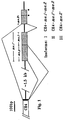
- Fig. 1 shows three different mRNA splicings which create three different proposed isoforms of human ⁇ membrane anchoring peptide (I, II, and III). "*" indicates a stop codon in the reading frame.
- the short solid-black segment at the end of the CH4 domain represents the C-terminus of the secreted ⁇ chain.
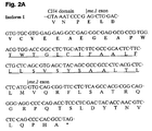
- Fig. 2A shows the peptide-coding nucleotide sequence and the deduced amino acid sequence of isoform I (which are not subject matter of the present invention) of the membrane anchoring peptide of human ⁇ chain.
- the underlined segment is the hydrophobic amino acid stretch thought to span the membrane lipid bilayer; the bold-faced segment is the portion presumably exposed to the exterior cell surface.
- Fig. 2B shows the peptide-coding nucleotide sequence and the deduced amino acid sequence of isoform II of the membrane anchoring peptide of human ⁇ chain (which are subject matter of the present invention).
- the bold-faced segment indicates amino acid sequences unique to isoform II.
- Fig. 2C shows the peptide-coding nucleotide sequence and the deduced amino acid sequence of isoform III, which is in the membrane anchoring region of human ⁇ chain (not subject matter of the present invention).
- the bold-faced segment indicates amino acid sequences unique to isoform III.
- Figs. 3A and 3B respectively show the locations of the DNA probes which can be used for screening the cDNA library for clones containing the human ⁇ chain membrane anchoring peptides, for secreted and membrane-bound IgE.
- Fig. 4 shows the binding of HEM7 to various migis peptides.
- Results are the means of duplicates from one representative of three ELISAs, using microtiter plates coated with polyclonal human serum IgE ( ⁇ ) or synthetic migis peptides, ⁇ migis - ⁇ ; ⁇ , migis - ⁇ ; ⁇ , migis - ⁇ , ⁇ , migis- ⁇ ; , ⁇ , migis - ⁇ .
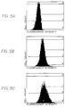
- Fig. 5C shows binding of HEM7 to IgE-secreting cells as determined by fluorescence flow cytometry.
- Fig. 5B shows binding of TES-19, a mAb to secreted IgE (positive control), with binding at equivalent concentrations (10 ⁇ g/ml) to SKO-007 cells.
- Fig. 5A is control without antibody added.
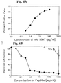
- Fig. 6A shows the concentration-dependent binding of HEM7 to IgE-secreting SKO-007 cells at various concentrations.
- Fig. 6B shows the specific inhibition of HEM7 binding to SKO-007 cells by migis - ⁇ peptide.
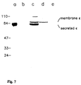
- Fig. 7 Western immunoblotting analysis of membrane-bound IgE with HEM7. The relative migration of the M.W. markers is shown on the left, and the positions of membrane bound ⁇ and secreted ⁇ on the right.
- a serum IgE probed by polyclonal anti- ⁇
- b serum IgE probed by HEM7
- c plasma membranes of SKO-007 cells probed by polyclonal anti- ⁇
- d plasma membranes of SKO-007 cells probed by HEM7
- e plasma membranes of SKO-007 cells probed by HEM7 in the presence of migis - ⁇ peptide at 100 ⁇ g/ml.
- Membrane-bound immunoglobulins on B cells differ from the secretory, soluble immunoglobulins synthesized by the same B cells in that the former have an extra peptidic piece that anchors them onto the B cell surface.
- the membrane-bound immunoglobulins on B cells from different species, for which amino acid sequences have been determined, have extra isotype-specific regions that anchor the immunoglobulins to the membrane. These peptidic regions have lengths ranging from 41 to 130 amino acids and can be divided into three segments. There is a middle segment of 25 hydrophobic and uncharged amino acids, which is believed to be located in the cytoplasmic membrane bilayer.
- the length and the hydrophilic and highly charged nature of the extracellular segment indicate that this segment is exposed and accessible to antibodies.
- the antigenic epitopes located on the extracellular segment of the membrane-bound region of immunoglobulin heavy chains are designated herein as the migis epitopes.
- the migis epitopes allow for developing several types of monoclonal or polyclonal antibody-based therapies and diagnoses for IgE-mediated allergic diseases.
- the membrane anchoring peptide has three segments which are distinguishable based upon their locations in relation to the plasma membrane.
- the shortest migis peptides have 13 amino acid residues (mouse and human ⁇ chains). See Table 1.
- the migis peptides of all immunoglobulins contain high proportions of charged amino acid residues, almost entirely acidic residues, as shown in Table 2.
- the proportions of charged amino acid residues and polar hydrophilic residues account for very high percentages of the amino acid composition of the migis peptides (Table 3). Thus, it is proposed that all the migis peptides are exposed and long enough to be accessible by antibodies.
- Table 3 Composition of charged amino acid residues and polar, hydrophilic amino acid residues of the migis peptides.
- Acidic residues Basic residues Polar residues hydrophilic residues Proportion of hydrophilic residues % # Amino acid residues Mouse IgE 10 0 2 12 63 Rat IgE1 10 0 2 12 63 Mouse IgG 1 6 0 4 10 56 Mouse IgG 2a 7 0 2 9 50 Mouse IgG 2b 7 1 1 9 50 Mouse IgG 3 6 0 4 10 56 Mouse IgM 6 0 2 8 61 Human IgM 6 0 1 7 54 Human IgD 6 1 8 15 56 Mouse IgD 7 0.5 9 16.5 63 Acidic residues: E (Glu), D (Asp) Basic residues: K (Lys), R (Arg), H (His); His is partially charged. Polar residues: S (Ser), T (Thr), C (Cys), Q (Gln), N (Asn)
- a number of well established procedures can be applied to determine the DNA sequence corresponding to the human ⁇ chain migis peptides.
- One approach is to start with the mRNA preparation of a human myeloma cell line which expresses IgE on the surface. SKO-007 cells can be employed for this purpose.
- the mRNA preparation one can establish a cDNA library by employing lambda phage or plasmids as cloning vectors.
- a preferred method for constructing the cDNA library is with the cDNA Library Construction System Kit - Librarian I developed and commercialized by Invitrogen (San Diego, CA).
- a stepwise detailed instruction manual is provided for RNA isolation from cells, reverse transcription, second strand synthesis, linker ligation, agarose gel sizing of cDNA, electroelution to purify cDNA, vector ligation, and transformation of E . coli .
- the vector used in this library is pCDM8.
- probe A is a 1.1 kb long U266 cDNA covering most of length of ⁇ mRNA (no membrane-bound segment).
- Probe B is developed by taking advantage of the probable fact that the end of the CH4 domain is truncated in the human ⁇ chain membrane anchoring peptide. The truncation occurs when the gene segments of the CH4 domain and the membrane-bound domain are translocated.
- the loss of the C-termini also occurs with the membrane bound forms of other immunoglobulins, including ⁇ and ⁇ , which contain CH4 domains.
- the most possible splicing donor site is intracodon GT, 71 bp 5' of the termination codon TGA.
- Another GT which is not intracodon and less likely a splicing donor site, is closer to the terminus (24 bp 5' to the termination codon).
- Probe B will react with the secreted form of the ⁇ chain gene and not the membrane-bound form of ⁇ chain gene.
- probe C (Fig. 3B) was based on the finding that the transmembrane segment of the membrane anchoring peptides is very conserved among all the immunoglobulin genes so far sequenced. There is a segment of peptide and corresponding coding DNA within this transmembrane segment that is nearly identical among all immunoglobulins. As shown in Table 4, the consensus DNA sequence with the eight combinations was used as probe C.
- Probe D which represents a segment upstream of the most probable splicing donor site, GT, consists of 36 bp. This probe should react with ⁇ chain gene of both the secreted and membrane-bound forms.
- Table 5 summarizes the pattern of reactivities of clones containing ⁇ genes of secreted or membrane-bound forms with the four probes. Table 5. The reactivity of ⁇ gene-containing cDNA clones with probes A, B, C, and D. ⁇ Secreted ⁇ Membrane-bound Probe A + + Probe B + - Probe C - + Probe D + +
- the library size needed to clone the membrane-bound ⁇ chain depends on how abundant the mRNA is. Assuming secreted IgE comprises 0.1% of the SKO-007 poly A + RNA, the library size should be about 5,000 independent recombinant clones to have a 99% probability to isolate a positive clone. In IgE-producing rat immunocytoma IR2 and IR162 cells, mRNA for the membrane-bound form of ⁇ chain was found to be more than 2% of that of the secreted form.
- the cDNA library size needed to isolate the membrane-bound ⁇ chain is about 250,000. In a preferred procedure, a larger number of clones (about 1,000,000) are screened.
- PCR polymerase chain reaction
- the strategy is to amplify both the secreted and membrane-bound forms of ⁇ chains.
- Two primers are to be used, one is oligo.dT (25-30-mers) and one is the oligomer corresponding to probe D in Figure 3.
- Probe D is located 5' to the most probable splicing donor site and therefore primes both the secreted and membrane-bound forms of ⁇ mRNA and DNA. After sufficient amplification, the two populations of DNA fragments are resolved by gel electrophoresis. The secreted form of the ⁇ chain can be distinguished by its reactivity with probe B. The purified DNA's are then subjected to DNA sequencing.
- U266 ⁇ chain cDNA (U266 being the parent cell line of SKO-007 with the same mRNA and cDNA) can be used to work out some preliminary annealing conditions between template DNA and oligo-primers.
- Another approach for obtaining a DNA clone containing genes encoding the membrane-bound segments is to screen the human genomic DNA library.
- a preferred source for this human genomic library is constructed using human lung fibroblast WI38 cells provided by Stratagene (La Jolla, CA). The genes are in lambda vector and the inserted DNAs have average sizes of 15K bp. Identification of the clones can be achieved by hybridization with U266 ⁇ chain cDNA. The location of the gene segment corresponding to the membrane anchoring peptide can be determined by using a probe prepared from the homologous mouse gene of the transmembrane segment (probe C of Figure 3 and Table 4). The sequence of the gene segment encoding the membrane anchoring peptide is then determined.
- the nucleotide sequence of genomic DNA encompassing the encoding segments for the membrane anchoring peptide of human membrane bound ⁇ chain was determined by screening the human genomic library as described above.
- the sequences of Isoforms I, II and III are shown respectively in Figs. 2A, 2B and 2C, along with the deduced amino acid sequences for portions of the membrane anchoring peptide.
- the assignment of the exons was made by identifying the nucleotides for splicing donors and acceptors (as shown in Fig. 1) and by comparison to the published homologous sequences of mouse membrane-bound ⁇ chain and of immunoglobulins of other classes.
- isoform I the migis peptide is identified as the first fifteen amino acids encoded by membrane exon I, as indicated by the bold-faced amino acids in Fig. 2A. This precedes a stretch of about 25 hydrophobic amino acids (underlined in Fig. 2A) which form the transmembrane region.
- Two possible structures of migis peptides are shown below.
- the initial nucleotide sequencing was performed on the cDNA derived from mRNA isolated from human cells expressing membrane-bound IgE.
- a commercially available human IgE expressing myeloma, SKO-007 from the American Type Culture Collection (“ATCC”) Rockville Maryland, was used.
- DNA segments of cDNA regarded as pertinent to identification and characterization of the transmembrane regions of human ⁇ chain were amplified by PCR, as described further below.
- a cell line secreting a hu/mu chimeric IgE and expressing membrane-bound IgE was generated to use in determining the reactivities of monoclonal antibodies with membrane-bound IgE on B cells.
- the constant regions of human ⁇ and K genomic DNA and the variable regions of genomic DNAs of the heavy and light chains of a monoclonal antibody, BAT123 were used.
- variable region genes of BAT123 had been isolated from the functional heavy and light chain loci and used in the construction of murine/human ( ⁇ 1/ ⁇ ) fusion genes for the production of chimeric BAT123 (hu ⁇ 1/ ⁇ ). See International Patent Application No. PCT/US88/01797. By replacing the human ⁇ constant region with the ⁇ constant region in the heavy chain expression vector, a chimeric BAT123 (hu ⁇ , ⁇ ) with an antigen binding region derived from BAT123, was produced in a similar approach.
- a ⁇ phage clone containing the human germ line ⁇ constant region was identified with a probe representing a segment of the constant domains (CH1-4) of ⁇ chain. From this phage, a 6.4 kb DNA segment containing domains CH1 to CH4 and a 2.5 kb 3'-flanking sequence was subcloned into pUC19. By analogy to the reported mouse and rat ⁇ -loci information, the presumed membrane exons were estimated to be located within the 1 Kb SacI fragment at the 3'-end of the ⁇ gene. The 1 Kb SacI fragment was subcloned and sequenced to establish the presence of any membrane exon-like sequences.
- the 6.4 kb DNA segment containing ⁇ domains CH1 to CH4 and the membrane exons was linked to the BAT123 V H gene to give the chimeric mouse/human ⁇ gene.
- This chimeric ⁇ gene, together with the chimeric ⁇ gene, were co-transfected into Sp2/0 cells by electroporation.
- the transfected cells were selected by the gpt and neo gene activities in the presence of mycophenolic acid and G418. The procedure was similar to that described in International Patent Application No. PCT/US88/01797.
- Stable transformants were established and analyzed for IgE secretion by ELISA, and for membrane IgE expression (by fluorescence flow cytometry).
- a clone, SE-44 was chosen for further studies.
- the cumulative IgE concentration in the culture supernatant of the SE-44 cells at 10 6 /ml was established to be 40 ⁇ g/ml.
- the 1 kb SacI segment was separated into three portions utilizing the restriction enzyme ApaI .
- the two 250 bp fragments containing membrane exon 1 and its 5'-flanking region were used as the probe specific for exon 1.
- the 400 bp fragment containing exon 2 and the 3'-untranslated region was used as the exon 2 probe.
- These probes were used separately in Northern analyses to hybridize with cytoplasmic RNAs. Both probes yielded similar results and lit up messages of 3,000 and 3,600 nucleotides in length for SE-44 and SKO-007, respectively. The observation that SE-44 cells expressed shorter membrane-IgE messages than SKO-007 cells was expected.
- the chimeric ⁇ gene used the SV40-derived t/pA signal present in the gpt gene construct for expression.
- the SKO-007 membrane IgE messages probably represent the normal intact transcripts using the endogenous ⁇ -locus t/pA signal which is located 600 bp (estimation based on the size difference of the two messages) downstream from the 3'-end of the 1 kb SacI fragment.
- Northern analysis therefore suggests that both exons 1 and 2 are transcribed in these cells.
- Binding inhibition assays were used to demonstrate that the chimeric BAT123 (human ⁇ , ⁇ ) bound to gp120 with an affinity constant comparable to that of BAT123 or chimeric BAT123 (hu ⁇ 1, ⁇ ).
- chimeric BAT123 (hu ⁇ 1, ⁇ ) the replacement of mouse C ⁇ 1 in in BAT123 with human C ⁇ 1 or human ⁇ did not affect its antigen-binding affinity significantly.
- the cell line SKO-007 which also expresses human ⁇ chain on its cell surface and which is a subclone of U266, was obtained from the ATCC.
- U266 was a myeloma cell line established from a blood sample of a myeloma patient.
- mRNA was reverse-transcribed using the oligo-dT primer into cDNA, which was then used as the template in PCR to amplify the pertinent segments covering the 3' end of the CH4 exon and the membrane exons.
- oligonucleotide primers with the following sequences, were used in the PCR:
- the major products derived from PCR were either subjected to direct sequencing or cloned into a Bluescript II vector.
- the nucleotide sequences were determined for several clones derived from each individual band.
- the electrophoretic patterns of the PCR products and the sequencing data indicated unexpectedly the existence in both SKO-007 and SE44 cells of RNA species other than the one derived from the splicing of CH4 domain to the previously predicted me.1 and me.2 exons.
- primers #1 and #2 were used, the dominant PCR product was a segment originating from the direct RNA splicing of CH4 domain to me.2 , using the predicted donor and acceptor sites, leaving out the me.1 domain (Fig. 1).
- isoform I contains CH4- me.1-me.2
- isoform II contains CH4 -me.1'(me.p+me.l)-me.2
- isoform III contains CH4- me.2'.
- isoform III is substantially smaller than isoform I or II and was resolved from isoform I and/or II in the electrophoretic gel
- the me.2 probe revealed two bands, one with and one without me.1' (or me.1 ) exon.
- the me.1 probe could hybridize with both isoforms I and II, which were not resolvable in the gel, the Northern blotting analysis did not establish the presence of isoform ImRNA.
- the analyses with me.p and me.2 probes suggest convincingly the presence of mRNA's of isoforms II and III.
- the intensity of bands also suggest that the amounts of mRNA's of these isoforms and their relative proportions are different in SKO-007 and SE44 cells.
- isoform II Based on the nucleotide sequences of the PCR-amplified segments, the corresponding amino acid sequences for isoforms II and III were deduced and compared to that of isoform I.
- the reading frame of isoform II is the same as isoform I.
- the extra 52 a.a. (bold-faced in Fig. 2B) lengthens the extracellular segment of the membrane-anchor peptide to a total of 67 a.a. from 15 a.a. in isoform I (Fig. 2A).
- the omission of the me.1 (length 122 bp, not a multiple of 3) causes the reading frame of me.2' segment in isoform III to be shifted (Fig. 2C); the peptide coding sequence is lengthened from 81 bp (encoding 27 a.a.) to 134 bp (encoding 45 a.a).
- Fig. 2C The corresponding peptide of isoform III (Fig. 2C) does not contain the hydrophobic stretch of 25 a.a. thought to span the membrane lipid bilayer (the segment is encoded by me.1 ). This suggests that it is secreted, and is not membrane-bound.
- Immunogenic peptides based on isoform I may be in either the monomeric or dimeric form shown above.
- Immunogenic peptides based on isoform II may be in either the monomeric or dimeric form.
- immunogenic peptides immunogenic peptides based on isoform II being designated herein as the peptides of the invention
- RaMPS system DuPont DeNemours & Co.
- recombinant peptides or immunoglobulin heavy chains (or portions thereof) containing isoforms I, II, or III may be biosynthesized by expressing in E. coli or eukaryotic cells the gene segments containing the coding sequence of these peptides.
- a synthetic peptide segment When using a synthetic peptide segment as an immunogen, it is usually more effective to conjugate it to a protein carrier, for example, hepatitis B surface antigen, core antigen, or preferably keyhole limpet hemocyanin (KLH). If the peptidic segment lacks a lysine residue or if the lysine residue is in the middle part of the segment, it is desirable to add a lysine residue at the C-terminal end. Because the N-terminus already has an ⁇ -amino group, the modified synthetic peptidic will have two available amino groups for linking.
- a protein carrier for example, hepatitis B surface antigen, core antigen, or preferably keyhole limpet hemocyanin (KLH).
- KLH keyhole limpet hemocyanin
- peptides can be conjugated to each molecule of the carrier protein.
- KLH a preferred molar ratio for peptide/KLH is 10.
- Cross-linkers such as glutaraldehyde or bis (sulfosuccinimidyl) suberate or preferably disulfosuccinimidyl tartrate (Catalogue #21579, 20591, Pierce Chemical Co., Rockford, IL) can be used.
- these peptides can be used to make monoclonal antibodies which are specific for them, using the protocol described further below. Specific examples of making monoclonal antibodies to the migis epitope of human ⁇ chain appear below and in priority U.S. Patent Application Serial Nos. 07/531,787, filed June 1, 1990, and 07/468,766, filed on January 23, 1990.
- the immunogenic peptides of the invention can also be used to immunize rabbits, goats, rats, or mice (or even another human being) to prepare polyclonal antibodies to the extracellular migis- ⁇ epitopes.
- Monoclonal antibodies that react with the peptides of the invention can be further screened for positive specific-reactivity with cells bearing a specific isotype. The monoclonal antibodies can then be applied in vivo .
- Polyclonal antibodies made against peptides of the invention generally contain almost entirely antibodies that react with the synthetic peptide but not the native molecules. Whether the polyclonal antibodies made against synthetic peptides can react with intact cells must be tested.
- the immunogen may be the membrane-bound immunoglobulin isolated from the plasma membrane of immunoglobulin-bearing myeloma cells, such as the IgG-expressing IM-9 cell line, or it may be the myeloma cells themselves.
- Transfectomas which are developed by transfecting mouse myeloma cells with genes of human immunoglobulin heavy chains and light chains and which express on their cell surface membrane-bound immunoglobulins, may also be used as immunogens.
- Lymphocytes from the spleen or lymph nodes of immune mice and rats can also be used to prepare hybridomas secreting monoclonal antibodies specific for the extracellular migis - ⁇ epitopes.
- a preferred fusion protocol is to fuse immune spleen cells of mice with non-secreting mouse myeloma cells, such as NS-1 or Sp2/0 cells, using polyethylene glycol.
- a preferred immunization protocol for preparing monoclonal antibodies is to inject into each mouse 50 ⁇ g of the conjugate of KLH and the recombinant or synthetic peptides of the invention in complete Freund's adjuvant. Two and four weeks later, the same amount of antigen is given subcutaneously in incomplete Freund's adjuvant. After about six weeks, the fourth antigen injection is given intraperitoneally in saline. Mice are sacrificed 4 days after the last injection and the spleens are removed for preparing single cell suspensions for fusion with myeloma cells.
- a similar protocol can be used for immunization with purified native human membrane-bound immunoglobulins (having attached membrane anchoring peptide segments) isolated from the plasma membrane of immunoglobulin-bearing human myeloma cells, such as IM-9 cells.
- immunoglobulin-bearing human myeloma cells such as IM-9 cells.
- I x 10 7 cells are injected intraperitoneally at two week intervals.
- the fusion procedure with polyethylene glycol and other various procedures concerning cloning and hybridoma culturing have been well established.
- the preferred fusion procedure is the well-known one described by Hudson, L and Hay, F.C. (Practical Immunology, 2nd edition, pp. 303-313, 1980, Blackwell Publishing Co., Boston).
- the screening of hybridomas for monoclonal antibodies (or the identification of polyclonal antibodies) reactive with the extracellular migis - ⁇ epitopes can be performed with an enzyme-linked immunosorbent assay (ELISA) using the synthetic peptide as the solid phase antigen.
- ELISA enzyme-linked immunosorbent assay
- a preferred solid phase antigen is the conjugate of a peptide of the invention with a carrier protein different from that used in the immunogen, such as bovine serum albumin or ovalbumin.
- Monoclonal antibodies specific for a particular peptide of the invention (corresponding to isoform II) can then be screened for specific binding to B cell lines and B cells expressing isoform II by using immunofluorescence flow cytometric analyses.
- the migis - ⁇ epitope-specific monoclonal antibodies which are first obtained will be murine-derived, and thus may be immunogenic or allergenic in human therapy. It is therefore desirable to produce chimeric antibodies (having an animal variable region and a human constant region), or to use human expression vectors (Stratagene Corp., La Jolla, California) to produce fragments of human antibodies (V H , V L, F v , Fd, Fab, or F(ab') 2 ) and then construct whole human antibodies using techniques similar to those for producing chimeric antibodies. In addition, one can create antibodies in which the entire constant portion and most of the variable region are human-derived, and only the antigen binding site is derived from some other mammal.
- Monoclonal antibodies specific for the migis - ⁇ epitopes can be used to reduce or eliminate the B cells expressing IgE by antibody-dependent cellular cytotoxicity (ADCC), complement-mediated cytolysis, or other cytolytic or regulatory immune mechanisms.
- ADCC antibody-dependent cellular cytotoxicity
- antibodies of certain IgG subclasses such as mouse IgG 2a and human IgG 1 and IgG 3
- ADCC antibody-dependent cellular cytotoxicity
- chimeric antibodies bearing human ⁇ -1 or ⁇ -3 chains, or human IgG 1 or IgG 3 antibodies can be used to down-regulate or lyse B cells expressing IgE. These antibodies will not bind to the secreted form of IgE or to IgE bound to the surface of basophils or mast cells.
- the mAbs of the invention can also be used as targeting agents for cytotoxic cells.
- the mAbs of the invention can also be used as carrier agents of cytotoxic drugs or for delivering an effector substance, by conjugating the mAbs to these substances.
- a toxin-antibody conjugate will bind and directly kill B cells producing IgE, but not B cells producing other isotypes.
- These toxins are cytolytic or cytotoxic agents, including cytotoxic steroids, gelonin, abrin, ricin, Pseudomonas toxin, diphtheria toxin, pokeweed antiviral peptide, tricathecums, radioactive nuclides, and membrane-lytic enzymes (such as phospholipase).
- the antibody and the agent can be conjugated by chemical or by genetic engineering techniques.
- the toxin-antibody conjugates may be used alone or in combination with the free antibodies of the invention.
- the antibodies of the invention are administered systemically, and preferably intravenously. They can be administered in any pharmaceutically acceptable vehicle.
- Another therapeutic alternative involves active immunization, wherein antibodies specific to the migis - ⁇ epitopes are endogenously produced in vivo . These endogenously produced antibodies bind the migis - ⁇ epitopes and cause destruction of the associated B cells. Production of such antibodies can be induced either by administering an immunogenic migis peptide of the invention, or a paratope-specific, anti-idiotypic antibody. Anti-idiotype antibodies against the paratope of the antibodies of the invention bear the internal image of the migis- ⁇ epitopes. These anti-idiotypic antibodies can be used to actively immunize against the migis - ⁇ epitopes and induce the endogenous formation of antibodies against the migis - ⁇ epitopes.
- Such paratope-specific, anti-idiotyptic antibodies are administered to a patient in an immunogenic amount sufficient to induce the formation of antibodies against B cells expressing IgE.
- These anti-idiotypic antibodies are preferably administered as chimeric antibodies or human antibodies, to minimize any immune response against them. They may also be any of the antibody fragments, V H , V L , F V , Fd, Fab, or F(ab') 2 (which also may be chimeric or human in nature).
- GM-CSF granulocyte monocyte-colony stimulating factor
- M-CSF monocyte-colony stimulating factor
- Derivative antibodies can be made which draw cytotoxic cells such as macrophages or cytotoxic T cells toward the targeted immunoglobulin-expressing B cells.
- These derivative antibodies include bi-specific antibodies having a specificity for a receptor of a cytotoxic cell and a specificity for the targeted IgE-expressing B cells.
- Such hybrid bi-specific antibodies can include two different Fab moieties, one Fab moiety having antigen specificity for the targeted migis - ⁇ epitopes, and the other Fab moiety having antigen specificity for a surface antigen of a cytotoxic cell, such as CD3 or CD8.
- the bi-specific antibodies of the invention can be a single antibody having two specificities, or a heteroaggregate of two or more antibodies or antibody fragments. See , e.g. , C. Reading, U.S. Patent Nos. 4,474,893 and 4,714,681; Segal et al. , U.S. Patent No. 4,676,980.
- monoclonal antibodies of the invention can be used for in vivo applications, they may also be used in extra-corporeal ex-vivo applications.
- the IgE-bearing B cells in the circulation of the patients can be removed by an affinity matrix (antibody immobilized on a solid phase) which is conjugated with the monoclonal antibodies of the invention.
- Another use for the antibodies of the invention is for determining numbers and relative proportions of B lymphocpes bearing particular isotypes in mixed leukocyte populations.
- the migis - ⁇ specific antibodies will not react with cells which bear secreted immunoglobulins via such cells' Fc receptors.
- Such cells include macrophage and activated T cells.
- the profile of the B cells may indicate the allergic status of the individual, and whether further depletion of IgE-bearing B cells is desirable. The same information can also indicate how much antibody is needed to deplete a substantial portion of B cells bearing IgE.
- antibodies can be used in standard assays which are used to determine cell surface antigens.
- the antibodies are contacted with a sample of the leukocytes to be tested under conditions which allow the antibodies to bind IgE-bearing cells in the sample.
- the cells are then examined for binding of antibody. This can be accomplished by conventional cell staining procedures, for example, a fluorescently labeled second antibody can be used to detect binding of antibody.
- the monoclonals can also be further characterized.
- An immunofluorescence assay could be used to determine whether the antibodies of the invention bind to basophils.
- An immunofluorescence assay could also be used to determine whether the antibodies bind to mast cells, and to determine whether the antibodies of the invention react with SKO-007 myeloma cells, IgE-bearing B cells, and transfectomas expressing human/murine chimeric IgE.
- SKO-007 myeloma cells IgE-bearing B cells
- transfectomas expressing human/murine chimeric IgE The results for the HEM7 rnAb to isoform I are shown below in Fig. 6.
- An ELISA is used to determine reactivity with synthetic migis- ⁇ peptides and with soluble IgE. Table 6.
- the substances of the invention are likely to be tested on animal model systems. Two of the most relevant systems are the following.
- the monoclonal antibodies of this invention which are specific for human migis peptides and their related substances (some of which are described further below) are intended for use to treat patients with various IgE-mediated allergies (see section 6 below). Among these allergies, extrinsic asthma is a more serious form.
- An experimental model system for studying asthma has been established in rhesus monkeys.
- the various substances of this invention can be tested in the asthma/rhesus monkey model system.
- the ascaris sensitive monkeys are given the experimental treatment or control treatment and measurements are made to determine:
- mice are not known to develop allergic symptoms naturally. However, for demonstrating the pharmacologic mechanisms of the intended therapy by depleting IgE-bearing B cells and IgE, the mouse can serve as an excellent model.
- the extracellular mouse ⁇ chain migis peptide has already been sequenced. Ishida, N. et al. , EMBO J. 1 :1117-1123 (1982)
- the 19 amino acid residue peptide is:
- This peptide is synthesized in several forms, including one that has extra Leu-Lys residues at the C-terminus.
- the peptide and its KLH conjugate are used as antigens to immunize rabbits and goats.
- the antisera are collected.
- the antigen-specific antibodies are purified using a column of Sepharose 4B conjugated with the peptide (with Leu-Lys addition) or with peptide linked to bovine serum albumin.
- Normal mice are injected intravaneously (i.v.) or intraperitoneally (i.p.) with the purified antibodies (or their related substances), with the peptide (with Leu-Lys addition), or with peptide linked to bovine serum albumin.
- the mice are preferably immunized with the mouse migis - ⁇ peptide conjugated to a carrier protein, such as keyhole limpet hemocyanin.
- mice may also be challenged by infection with a parasite, Nippostrongylus brasiliensis , which is known to induce large quantities of IgE. Snapper, C.M. et al. , Immunol. Rev. 102 :51-75 (1988).
- the questions to be addressed include the following:
- Antibodies specific for the migis - ⁇ epitopes bind IgE on the surface of IgE-producing B cells and not on basophils and mast cells. This differential binding of IgE-bearing cell types provides the basis for therapeutic uses of the antibodies.
- the antibodies specific for migis - ⁇ epitopes can be used to treat IgE-mediated allergies in humans or other animals (e.g. dogs, cats and horses).
- the antibodies can be used therapeutically in several ways, including as effector agents mediating an immune function, as carrier agents of toxins or cytotoxic drugs, for delivering an effector substance, or as targeting agents for cytotoxic cells.
- Antibodies of certain IgG subclasses can be used to reduce or eliminate the IgE-bearing B cells by ADCC, complement-mediated cytolysis, or other cytolytic or regulatory immune mechanisms. These antibodies can also be used as effector agents mediating an immune function or as targeting agents for cytotoxic cells.
- the antibodies can be systemically administered, preferably intravaneously, as free antibodies to patients afflicted with IgE-mediated allergy in amounts sufficient to eliminate substantially IgE-producing cells and consequently, to substantially eliminate IgE.
- the antibodies can also be administered nasally.
- nasal channels and the respiratory tract are areas in which active mast cells are concentrated.
- the IgE-producing B cells and free IgE in the extravascular space of these tissues may have better access to the basophils and mast cells than IgE-producing B cells and IgE in other parts of the body.
- Nasal administration e.g., by nasal spray
- the antibodies can also be administered ocularly.
- the mAbs of the invention may be used therapeutically in humans, and related mAbs against corresponding migis epitopes may be used therapeutically in other mammals, such as dogs, cats and horses.
- the human or humanized antibodies (and fragments) including chimeric antibodies are preferred. Human and humanized antibodies are less immunogenic in humans than non-human antibodies. Consequently, they are better suited for in vivo administration, especially when repeated or long term administration is necessary.
- Immunotherapies employing the antibodies of this invention may be used in combination with conventional desensitization immunotherapy.
- desensitization with allergen may be performed in conjunction with the administration of either anti- migis - ⁇ antibodies or antibody-toxin conjugates discussed above to substantially eliminate IgE producing cells.
- Desensitization induces IgG production against the allergen/immunogen. Inducing such IgG production may be most effective as an allergy therapy when IgE-producing B cells are substantially depleted.
- the combination of antibody and desensitization therapy is attractive because although the IgE-producing B cells may only be temporarily depleted (for a few weeks or months) by the anti- migis antibody, and will eventually re-populate, the desensitization effect may last much longer.
- GM-CSF granulocyte monocyte-colony stimulation factor
- M-CSF monocyte-colony stimulation factor
- Antibodies specific for a migis - ⁇ epitope can be combined with one or more of the immunotoxins noted above, thereby forming an antibody-immunotoxin conjugate which specifically targets IgE-producing B cells.
- the immunotoxins may be used alone or in combination with free anti- migis antibodies.
- the migis - ⁇ -specific monoclonal antibodies can be used for in vivo therapy they may also be used in extra-corporeal ex - vivo therapy.
- the IgE in the circulation of allergic patients can be removed by an affinity matrix (antibody immobilized on a solid phase) that is conjugated with the monoclonal antibodies of this invention. Because antibodies may leak out from the affinity column and enter into the circulation of the patient, the monoclonal antibodies of the invention are preferable to other antibodies that can induce histamine release from basophils and mast cells.
- Another use for the antibodies of the invention is for determining numbers and relative proportions of IgE-bearing B lymphocytes in mixed leukocyte populations.
- the migis specific antibodies will not react with cells which bear secreted immunoglobulins via such cells' Fc receptors.
- Such cells include macrophages and activated T cells.
- the profile of the B cells may indicate the immune status of the individual. The same information can also indicate how much antibody is needed to deplete a substantial portion of B cells bearing a particular isotype, where some of those B cells are tumorous.
- antibodies can be used in standard assays which are used to determine cell surface antigens.
- the antibodies are contacted with a sample of the leukocytes to be tested under conditions which allow the antibodies to bind isotype-bearing cells in the sample.
- the cells are then examined for binding of antibody. This can be accomplished by conventional cell staining procedures, for example, a fluorescently labeled second antibody can be used to detect binding of antibody.
- Monoclonal antibodies against an epitope unique to membrane-bound IgE but not secreted IgE were prepared by a standard procedure for preparing hybridomas, as described in the Detailed Description of the Invention.
- the immunogen for immunizing BALB/c mice was the above-described SE-44 transfectoma cells. The mice were injected intraperitoneally 3 times at 2 weeks intervals with 1x10 7 SE-44 cells that were treated with 1mM mitomycin C for 30 minutes at 37°C prior to injection.
- the human migis - ⁇ peptide Glu ⁇ Leu ⁇ Asp ⁇ Val ⁇ Cys ⁇ Val ⁇ Glu ⁇ Glu ⁇ Ala ⁇ Glu ⁇ Gly ⁇ Glu ⁇ Ala ⁇ Pro ⁇ Trp dimerized was used as coating antigen for ELISA.
- the positive clones were characterized in additional assays with other peptides and SE-44 cells and control cell lines.
- Solid-phase antigen (2 ⁇ g/ml) A 450 migis - ⁇ peptide - ovalbumin 2.707 HIV-1 peptide*- ovalbumin 0.011 migis - ⁇ peptide - KLH 2.773 HIV-1 peptide - KLH 0.002 KLH 0.005 *The HIV-1 peptide was a 15-mer peptide representing a segment of gp120 of HTLV-IIIB strain of HIV-1. This peptide is reactive with BAT123 monoclonal antibody.
- E46-13-3 and other monoclonal antibodies were further analyzed for reactivities with SE-44 cells compared to various control cell lines, including Sp2/0, the parent cell line for the transfectoma SE-44. Included in the control was the IM-9 cell line, which expresses IgG and CD23 on the cell surface, and the DAKIKI cell line, which expresses IgA on its surface.
- the tests were carried out with flow cytometric analyses with FITC-goat-anti-mouse IgG using an EPICS system. The results (shown below in Table 8) show clearly that E46-13-3 stained SE-44 specifically. Table 8. Live cell staining studies of E46-13-3.
- the migis peptides of all five human immunoglobulin heavy chain isotypes (including migis - ⁇ ) with an additional C-terminal lysine residue were synthesized.
- Conjugates of these migis peptides with keyhole limpet hemocyanin (KLH;Sigma) or with ovalbumin (Sigma) were prepared by cross-linking 1 mg/ml each with 0.04% glutaraldehyde (Sigma) in phosphate buffered saline (PBS), pH 7.4, for 16h at 4°C, and dialyzed to PBS. Under these conditions more than 90% of the peptide was cross-linked.
- mice were then immunized subcutaneously and intraperitoneally with 100 ⁇ g migis - ⁇ -KLH in Freund's adjuvant 4 times and then intraperitoneally twice with mitomycin C (Sigma)-treated (20 mg/ml for 20h) SE44 mouse myeloma cells (which express human IgE on the cell surface). Spleen cells were fused with Sp 2/0 cells using polyethylene glycol (Carbowax, Fisher). Supernatants of growing hybrids were screened in enzyme-linked immunosorbent assay (ELISA) for reactivity to migis - ⁇ ovalbumin. Positive wells were then tested for ability to bind to cell surface IgE by indirect immunofluorescence flow cytometric analyses.
- ELISA enzyme-linked immunosorbent assay
- the HEM7 hybridoma secreted an antibody showing specificity in these assays and was subcloned by limiting dilution.
- Antibodies from culture supernatants were purified by protein A (Repligen)-affinity chromatography.
- the purified mAb HEM7 was analyzed for specific binding to the migis - ⁇ peptide and to mIgE.
- the migis peptides of the four other heavy chain isotypes (IgG, IgA, IgM, and IgD) were also synthesized and the reactivity of HEM7 with these peptides was examined.
- These migis peptides are at least 13 a.a. long and apparently hydrophilic, containing 6 or 7 acidic amino acids.
- the migis peptides show a great degree of heterogeneity; although migis - ⁇ and migis - ⁇ are the most homologous to migis - ⁇ .
- ELISA microtiter plates (Immulon 2, Dynatech) were coated at 1 ⁇ g/ml PBS with migis - ⁇ peptide, and the migis - ⁇ , migis - ⁇ , migis - ⁇ , migis - ⁇ peptides, as well as with migis - ⁇ peptide-ovalbumin conjugate, or with human serum IgE (Ventrex), or other serum proteins (Jackson Immunoresearch), for 16h at 22°C. Plates were post-coated with blocking buffer, containing 5% non-fat dry milk (Carnation), 0.05% Tween 20 (Sigma) in PBS for 1h at 22°C.
- blocking buffer containing 5% non-fat dry milk (Carnation), 0.05% Tween 20 (Sigma) in PBS for 1h at 22°C.
- TMB tetramethyl benzidine
- HEM7 was shown to bind to migis - ⁇ peptide reaching maximal binding at 100 ng/ml antibody concentration. It did not bind to any measurable extent to the migis peptides of the other four isotypes (Fig. 4). Furthermore, HEM7 even at 10 ⁇ g/ml did not bind to soluble, secreted IgE purified from human serum (Fig. 4). Nor did it bind to IgE purified from supernatants of SE44 cells, or to other human Igs or serum substances (data not shown).
- the purified HEM7 mAb was also tested for its ability to bind to IgE-bearing SKO-007 cells in immunofluorescence flow cytometric assays.
- the immunofluorescence flow cytometric assays were carried out as follows. Cultures of B cells secreting various human immunoglobulins and other human blood cell lines were obtained from the ATCC. Peripheral blood was obtained by venipuncture from adult volunteers and mononuclear cells were isolated using Ficoll-Paque (Pharmacia). Cultured cells were stained with antibodies and fluorescence quantitated by flow cytometry (EPICS-V, Coulter Diagnostics). Peripheral blood lymphocytes and monocytes were gated by their light scattering properties.
- Presence of surface markers was confirmed using mAb reagents specific for IgE(HP6029), IgG(HP 6046), IgM(HP6081), IgD(JA11), and IgA(2D7) (all from Zymed), and for other leukocyte surface markers (CD4, CD14, CD45 anad CD23) (from Becton-Dickinson).
- HEM7 was able to bind to SKO-007 cells (obtained from the ATCC) as shown in the flow cytometric profiles in Figs. 5A-5C.
- this immunofluorescence staining histogram of SKO-007 cells using HEM7 mAb it can be seen that the fluorescence of the entire SKO-007 cell population shifted to higher levels, indicating that all cells are reactive with HEM7.
- the immunofluorescence staining is comparable to that observed with TES-19, a mAb specific for human IgE, used as a positive control.
- HEM7 was also tested for its ability to bind to human cells bearing other surface Ig. Among the various human cell lines tested, HEM7 was able to bind to SKO-007 cells and to the related U266 cells that bear mIgE, but not to B cell lines expressing IgM, IgD, IgG, or IgA. The mAb also did not bind to any other cells tested, including a T cell line, a monocyte-like cell line, and peripheral blood mononuclear cells (Table 9).
- Proteins in purified polyclonal human serum IgE (0.1 ⁇ g, Ventrex) and a plasma membrane fraction from SKO-007 cells (25 ⁇ g) were fractionated on 10% SDS-PAGE and electroblotted onto nitrocellulose.
- the filters were incubated with either peroxidase-conjugated polyclonal goat anti-human ⁇ chain at 2.5 ⁇ g/ml, or with HEM7 at 25 ⁇ g/ml, followed by peroxidase-conjugated goat anti-mouse IgG, and TMB with membrane enhancer substrate (enzyme conjugated antibodies and substrate from Kirkegaard and Perry).
- M. W. markers were from BioRad.
- HEM7 was also checked to determine if it would cause histamine release from peripheral human blood basophils, which cells were taken from four individuals. Each assay was run using various concentrations of TES-17 mAb (a mAb specific for human IgE which induces histamine release from leukocytes) and HEM7. Polyclonal antibodies to human IgE (which also induce histamine release) served, in addition to TES-17, as an additional positive control. Goat anti-mouse IgG was added to some of the tests as an enhancer.
- HEM7 did not cause histamine release in a mAb dose-dependent fashion, irrespective of whether the enhancer was present.
- the predicted isoform II contains 52 additional amino acids between C ⁇ 4 and the 15 amino acid segment bound by HEM7.
- Monoclonal and polyclonal antibodies were prepared to 36 amino acid synthetic peptide corresponding to residues 6 to 40 of the 52 amino acid segment. Both in ELISA and on immunoblots, the antibodies react with IgE from cell lysates but not with IgE from cell culture supernatants, suggesting that isoform II exists on cells and is not secreted.
- the 15 amino acid band recognized by HEM7 co-migrates with that recognized by the antibodies to the 36 amino acid segment.
- the predicted ⁇ chain encoded by the mRNA with CH4- me.2' splicing (designated as isoform III above) is referred to below as ⁇ 2
- the conventional ⁇ chain of secreted IgE (designated as isoform I above) is referred to as ⁇ 1
- IgE molecules containing one or two ⁇ 2 chains are referred to as IgE 2
- IgE molecules containing only ⁇ chains are IgE 1 .
- the PBMC from an allergic patient whose serum IgE concentration was about 1 ⁇ g/ml (about 10-20 times average), were isolated and the total RNA from these cells was prepared.
- PCR was performed using a pair of primers, one of which was located in CH4 domain and the other in the me.2 domain.
- the PCR-amplified products were analyzed by gel electrophoresis and the DNA in the major bands were subcloned and their nucleotide sequences determined.
- the major PCR product using the cDNA template derived from the RNA of SKO-007 and SE44 cells was a segment of ⁇ 2 mRNA.
- the same segment was also the major product with the cDNA from the human PBMC preparation.
- An autoradiograph showed that the 32 P-labeled DNA probe corresponding to an me.2 segment hybridized with the ⁇ 2 segment. It also hybridized with two other bands, corresponding to two segments derived from two isoforms of membrane-bound ⁇ chain mRNA. Nucleotide sequencing of DNA fragments cloned from these bands confirmed this conclusion.
- the peptide had the sequence: SHAAGEAPDLPRLHQRPPAPRLAAGHSRSTRPS
- the peptide designated the "E2T peptide,” was conjugated to KLH and used to immunize mice, using a standard protocol described above. Monoclonal antibodies were made which bound to the E2T peptide specifically. These mAbs also bound to ⁇ 2 , as determined on immunoblots.
- the resulting antibodies specific for E2T were affinity purified by a small column of agarose beads conjugated with E2T.
- These various reagents were then used in ELISA and immunoblotting assays to detect ⁇ 2 chain and IgE 2 .
- ⁇ 2 chain could be detected in the culture supernatant of SE44 cells, and to less extent, in that of SKO-007 cell.
- the substance was absent in the media of SP2/0 cells and of CAG1-51-4 cells.
- variable regions of the heavy and the light chains and the antigen specificity of the chimeric IgE ( ⁇ , ⁇ ) secreted by SE44 cells and of the chimeric IgG ( ⁇ 1, ⁇ ) secreted by CAG1-51-4 cells are identical, the binding of the ⁇ 2 chain or IgE 2 from the media of SE44 and SKO-007 cells to the solid phase was not due to the variable regions.
- the ⁇ chain in the culture supernatant of SE44 cells did not bind to rabbit antibodies specific for an irrelevant antigen.
- ⁇ m chain membrane-bound ⁇ chain
- ⁇ m chain membrane-bound ⁇ chain
- the recombinant ⁇ m which extended from CH2 domain to the membrane anchoring peptide, was produced in E . coli and purified by affinity column.
- An ELISA showed that mAb E11-4-70, which reacts with ⁇ 1 chain, and mAb HEM7, which is specific for the extracellular 15 a.a. segment of the membrane anchor peptide, reacted with the recombinant ⁇ m , while anti-E2T did not.
- the IgE secreted by SE44 cells into the culture medium was affinity purified by a mAb, TES-61, which is presumed to be specific for CH domains of IgE.
- the purified IgE was gel electrophoresed with or without treatment of reducing agents, transblotted onto nitrocellulose filters, and reacted with various antibodies to be studied.
- both horseradish peroxidase-conjugated goat anti-human IgE and anti-E2T stained a band of about 200 Kd, indicating that ⁇ 2 chain was present in IgE molecules and not as single chains.
- anti-E2T reacted with substances with M.W. at the higher end of those bands stained by goat anti-human IgE.
Landscapes
- Health & Medical Sciences (AREA)
- Life Sciences & Earth Sciences (AREA)
- Chemical & Material Sciences (AREA)
- Organic Chemistry (AREA)
- Medicinal Chemistry (AREA)
- Proteomics, Peptides & Aminoacids (AREA)
- General Health & Medical Sciences (AREA)
- Bioinformatics & Cheminformatics (AREA)
- Immunology (AREA)
- Engineering & Computer Science (AREA)
- Molecular Biology (AREA)
- Biochemistry (AREA)
- Pharmacology & Pharmacy (AREA)
- Biophysics (AREA)
- Veterinary Medicine (AREA)
- Public Health (AREA)
- Animal Behavior & Ethology (AREA)
- Genetics & Genomics (AREA)
- Cell Biology (AREA)
- General Chemical & Material Sciences (AREA)
- Toxicology (AREA)
- Zoology (AREA)
- Gastroenterology & Hepatology (AREA)
- Pulmonology (AREA)
- Chemical Kinetics & Catalysis (AREA)
- Epidemiology (AREA)
- Nuclear Medicine, Radiotherapy & Molecular Imaging (AREA)
- Peptides Or Proteins (AREA)
- Medicines Containing Antibodies Or Antigens For Use As Internal Diagnostic Agents (AREA)
- Preparation Of Compounds By Using Micro-Organisms (AREA)
- Medicines Containing Material From Animals Or Micro-Organisms (AREA)
- Micro-Organisms Or Cultivation Processes Thereof (AREA)
- Measuring Or Testing Involving Enzymes Or Micro-Organisms (AREA)
Claims (6)
- Isoform des Membranankerpeptids einer menschlichen ε-Kette aufweisend ein Peptidsegment mit der Sequenzwenn die Isoform die Isoform II ist, oder Modifizierungen des Peptidsegments, die die immunologischen Eigenschaften des Peptids nicht wesentlich ändern, aufweisend.

- Nukleotidfragment, das für irgendeines der Peptidsegmente gemäß Anspruch 1 codiert.
- DNA-Fragment nach Anspruch 2, das die Sequenz:oder eine entsprechende Sequenz, die für das gleiche Peptid codiert, enthält.

- Monoklonaler Antikörper gegen eines der Peptide gemäß Anspruch 1, wobei Antikörper gegen Isoform I des Membranankerpeptids der menschlichen ε-Kette ausgenommen sind.
- Monoklonaler Antikörper nach Anspruch 4, der menschlicher oder chimärer Maus/Mensch-Herkunft oder ein Antikörperfragment ist.
- Anti-idiotypischer Antikörper zu einem der Antikörper gemäß Anspruch 4.
Applications Claiming Priority (7)
| Application Number | Priority Date | Filing Date | Title |
|---|---|---|---|
| US468766 | 1990-01-23 | ||
| US07/468,766 US5260416A (en) | 1987-12-31 | 1990-01-23 | Antigenic epitopes present on membrane-bound but not secreted IgE |
| US07/515,604 US5274075A (en) | 1987-12-31 | 1990-04-27 | Newly identified human epsilon immunoglobulin peptides and related products |
| US63007790A | 1990-12-19 | 1990-12-19 | |
| US630077 | 1990-12-19 | ||
| PCT/US1991/000491 WO1991011456A1 (en) | 1990-01-23 | 1991-01-23 | EXTRACELLULAR SEGMENTS OF HUMAN ε IMMUNOGLOBULIN ANCHORING PEPTIDES AND ANTIBODIES SPECIFIC THEREFOR |
| US515604 | 1995-08-16 |
Publications (3)
| Publication Number | Publication Date |
|---|---|
| EP0512064A1 EP0512064A1 (de) | 1992-11-11 |
| EP0512064A4 EP0512064A4 (en) | 1993-02-17 |
| EP0512064B1 true EP0512064B1 (de) | 1997-06-18 |
Family
ID=27413084
Family Applications (1)
| Application Number | Title | Priority Date | Filing Date |
|---|---|---|---|
| EP91904413A Expired - Lifetime EP0512064B1 (de) | 1990-01-23 | 1991-01-23 | Extrazelluläre teilstücke humaner ige-immunoglobulin-ankerpeptide und dafür spezifische antikörper |
Country Status (12)
| Country | Link |
|---|---|
| EP (1) | EP0512064B1 (de) |
| JP (1) | JP3449712B2 (de) |
| AT (1) | ATE154637T1 (de) |
| AU (2) | AU645783B2 (de) |
| CA (1) | CA2074089C (de) |
| DE (1) | DE69126607T2 (de) |
| DK (1) | DK0512064T3 (de) |
| ES (1) | ES2107454T3 (de) |
| GR (1) | GR3024563T3 (de) |
| HK (1) | HK1002034A1 (de) |
| SG (1) | SG70982A1 (de) |
| WO (1) | WO1991011456A1 (de) |
Families Citing this family (24)
| Publication number | Priority date | Publication date | Assignee | Title |
|---|---|---|---|---|
| AU685057B2 (en) * | 1992-07-08 | 1998-01-15 | Unilever Plc | Process for immobilizing enzymes to the cell wall of a microbial cell by producing a fusion protein |
| ATE228536T1 (de) * | 1993-03-11 | 2002-12-15 | Tanox Biosystems Inc | Antigene epitope von ige repräsentierenden peptiden auf der b-zell-oberfläche aber nicht auf oberflächen von basophilen |
| US5552537A (en) * | 1993-03-24 | 1996-09-03 | The Regents Of The University Of California | IgE isoforms and methods of use |
| US6267958B1 (en) | 1995-07-27 | 2001-07-31 | Genentech, Inc. | Protein formulation |
| IL116436A (en) * | 1995-12-18 | 2006-12-31 | Yissum Res Dev Co | Fc?Á-PE CHIMERIC PROTEIN FOR TARGETED TREATMENT OF ALLERGY RESPONSES AND |
| CA2245497C (en) | 1996-03-01 | 2009-02-24 | Novartis Ag | Peptide immunogens for vaccination against and treatment of allergy |
| US6913749B2 (en) | 1998-11-02 | 2005-07-05 | Resistentia Pharmaceuticals Ab | Immunogenic polypeptides for inducing anti-self IgE responses |
| GB9908533D0 (en) | 1999-04-14 | 1999-06-09 | Novartis Ag | Organic compounds |
| US7754208B2 (en) | 2001-01-17 | 2010-07-13 | Trubion Pharmaceuticals, Inc. | Binding domain-immunoglobulin fusion proteins |
| CA2486464A1 (en) | 2002-05-21 | 2003-11-27 | Resistentia Pharmaceuticals Ab | Chimeric ige polypeptides and host cells |
| ES2539250T3 (es) | 2005-07-25 | 2015-06-29 | Emergent Product Development Seattle, Llc | Reducción de células B mediante el uso de moléculas de unión específica a CD37 y de unión específica a CD20 |
| US8137670B2 (en) | 2005-09-29 | 2012-03-20 | Medimmune, Llc | Method of identifying membrane IgE specific antibodies and use thereof for targeting IgE producing precursor cells |
| JP2009536217A (ja) * | 2006-05-03 | 2009-10-08 | ガスリー ファンデーション フォー エデュケーション アンド リサーチ | B細胞障害の治療における免疫グロブリン結合細胞表面決定基 |
| CA3149553C (en) | 2006-06-12 | 2023-11-21 | Aptevo Research And Development Llc | Single-chain multivalent binding proteins with effector function |
| AR065368A1 (es) | 2007-02-15 | 2009-06-03 | Astrazeneca Ab | Anticuerpos para moleculas de ige |
| WO2008116149A2 (en) | 2007-03-22 | 2008-09-25 | Genentech, Inc. | Apoptotic anti- ige antibodies binding the membrane-bound ige |
| SI2132228T1 (sl) | 2008-04-11 | 2011-10-28 | Emergent Product Dev Seatle | CD37 imunoterapevtik in kombinacija z njegovim bifunkcionalnim kemoterapevtikom |
| WO2010097012A1 (en) | 2009-02-25 | 2010-09-02 | Tsewen Chang | ANTI-CεmX ANTIBODIES CAPABLE OF BINDING TO HUMAN mIgE ON B LYMPHOCYTES |
| US9140698B2 (en) | 2011-04-06 | 2015-09-22 | The Board Of Regents Of The University Of Texas System | Modulating bacterial MAM polypeptides in pathogenic disease |
| EP2838919A4 (de) | 2012-04-20 | 2016-03-23 | Academia Sinica | An den übergang zwischen ch4- und c-epsilon-mx-domänen bindende anti-mige-antikörper |
| US10196458B2 (en) * | 2013-07-26 | 2019-02-05 | The Regents Of The University Of California | Anti-immunoglobulin E antibodies and methods of using thereof |
| EP3077000A4 (de) * | 2013-12-05 | 2017-06-14 | Immunwork Inc. | Anti-human-miga-antikörper zur lyse von mlga-b lymphozyten und verringerung der iga-produktion |
| EA201890613A1 (ru) | 2015-09-21 | 2018-10-31 | Аптево Рисёрч Энд Девелопмент Ллс | Полипептиды, связывающие cd3 |
| CA3079992A1 (en) | 2017-10-31 | 2019-05-09 | Oneness Biotech Co. Ltd. | Treating ige-mediated allergic diseases |
Family Cites Families (2)
| Publication number | Priority date | Publication date | Assignee | Title |
|---|---|---|---|---|
| PT89107A (pt) * | 1987-11-30 | 1989-11-30 | Idec Pharma Corp | Metodo e meios para a seleccao de anticorpos anti-idiotipos e seu uso para o diagnostico, monitorizacao, tratamento e/ou prevencao de doencas cancerosas, da autoimunidade ou infecciosas |
| DE122006000007I2 (de) * | 1987-12-31 | 2008-03-27 | Tanox Inc | -tragenden b-lymphocyten befinden. |
-
1991
- 1991-01-23 ES ES91904413T patent/ES2107454T3/es not_active Expired - Lifetime
- 1991-01-23 EP EP91904413A patent/EP0512064B1/de not_active Expired - Lifetime
- 1991-01-23 AU AU73179/91A patent/AU645783B2/en not_active Expired
- 1991-01-23 DK DK91904413.1T patent/DK0512064T3/da active
- 1991-01-23 SG SG1996006487A patent/SG70982A1/en unknown
- 1991-01-23 AT AT91904413T patent/ATE154637T1/de not_active IP Right Cessation
- 1991-01-23 CA CA002074089A patent/CA2074089C/en not_active Expired - Lifetime
- 1991-01-23 WO PCT/US1991/000491 patent/WO1991011456A1/en active IP Right Grant
- 1991-01-23 JP JP50484591A patent/JP3449712B2/ja not_active Expired - Lifetime
- 1991-01-23 DE DE69126607T patent/DE69126607T2/de not_active Expired - Lifetime
-
1993
- 1993-12-03 AU AU52148/93A patent/AU5214893A/en not_active Abandoned
-
1997
- 1997-08-28 GR GR970402202T patent/GR3024563T3/el unknown
-
1998
- 1998-02-12 HK HK98101073A patent/HK1002034A1/xx not_active IP Right Cessation
Also Published As
| Publication number | Publication date |
|---|---|
| EP0512064A4 (en) | 1993-02-17 |
| WO1991011456A1 (en) | 1991-08-08 |
| ATE154637T1 (de) | 1997-07-15 |
| EP0512064A1 (de) | 1992-11-11 |
| DE69126607D1 (de) | 1997-07-24 |
| AU7317991A (en) | 1991-08-21 |
| ES2107454T3 (es) | 1997-12-01 |
| JPH05504482A (ja) | 1993-07-15 |
| HK1002034A1 (en) | 1998-07-24 |
| JP3449712B2 (ja) | 2003-09-22 |
| CA2074089A1 (en) | 1991-07-24 |
| GR3024563T3 (en) | 1997-12-31 |
| CA2074089C (en) | 2001-04-10 |
| SG70982A1 (en) | 2000-03-21 |
| DE69126607T2 (de) | 1998-01-15 |
| AU5214893A (en) | 1994-03-03 |
| AU645783B2 (en) | 1994-01-27 |
| DK0512064T3 (da) | 1997-12-15 |
Similar Documents
| Publication | Publication Date | Title |
|---|---|---|
| US5342924A (en) | Extracellular segments of human ε immunoglobulin anchoring peptides and antibodies specific therefor | |
| EP0512064B1 (de) | Extrazelluläre teilstücke humaner ige-immunoglobulin-ankerpeptide und dafür spezifische antikörper | |
| US5254671A (en) | Extracellular segments of human e immunoglobulin anchoring peptides and antibodies specific therefor | |
| US5091313A (en) | Antigenic epitopes of IgE present on B cell but not basophil surface | |
| US5231026A (en) | DNA encoding murine-human chimeric antibodies specific for antigenic epitopes of IgE present on the extracellular segment of the membrane domain of membrane-bound IgE | |
| US5292867A (en) | Chimeric monoclonal antibodies which bind to the extracellular segment of the membrane-bound domain of a human membrane-bound immunoglobulin | |
| AU645256B2 (en) | Antigenic epitopes present on membrane-bound but not secreted IgA | |
| CA1340233C (en) | Antigenic epitopes of immunoglobulin e present on b cell but not basophil surface | |
| WO1992007574A1 (en) | Glycoproteins associated with membrane-bound immunoglobulins as antibody targets on b cells | |
| US5281699A (en) | Treating B cell lymphoma or leukemia by targeting specific epitopes on B cell bound immunoglobulins | |
| US5260416A (en) | Antigenic epitopes present on membrane-bound but not secreted IgE | |
| US5298420A (en) | Antibodies specific for isotype specific domains of human IgM and human IgG expressed or the B cell surface | |
| US5252467A (en) | Method of making antibodies to antigenic epitopes of IGE present on B cells but not basophil cell surface or secreted, soluble IGE | |
| CA2065414C (en) | Treatment of autoimmune disease | |
| US5079344A (en) | Antigenic epitopes present on membrane-bound but not secreted IgA | |
| US5274075A (en) | Newly identified human epsilon immunoglobulin peptides and related products | |
| US5362643A (en) | Antibodies to epitopes present on membrane-bound but not secreted IGA | |
| US5310875A (en) | Peptides corresponding to membrane-bound IgA | |
| US5484907A (en) | Nucleotides coding for the extracellular membrane-bound segment of IgA | |
| WO1994020533A1 (en) | PEPTIDES REPRESENTING ANTIGENIC EPITOPES OF IgE PRESENT ON B CELL BUT NOT BASOPHIL SURFACE | |
| AU618317C (en) | Unique antigenic epitopes on IgE-bearing B lymphocytes |
Legal Events
| Date | Code | Title | Description |
|---|---|---|---|
| PUAI | Public reference made under article 153(3) epc to a published international application that has entered the european phase |
Free format text: ORIGINAL CODE: 0009012 |
|
| 17P | Request for examination filed |
Effective date: 19920715 |
|
| AK | Designated contracting states |
Kind code of ref document: A1 Designated state(s): AT BE CH DE DK ES FR GB GR IT LI LU NL SE |
|
| A4 | Supplementary search report drawn up and despatched |
Effective date: 19921230 |
|
| AK | Designated contracting states |
Kind code of ref document: A4 Designated state(s): AT BE CH DE DK ES FR GB GR IT LI LU NL SE |
|
| 17Q | First examination report despatched |
Effective date: 19950818 |
|
| GRAG | Despatch of communication of intention to grant |
Free format text: ORIGINAL CODE: EPIDOS AGRA |
|
| GRAH | Despatch of communication of intention to grant a patent |
Free format text: ORIGINAL CODE: EPIDOS IGRA |
|
| GRAH | Despatch of communication of intention to grant a patent |
Free format text: ORIGINAL CODE: EPIDOS IGRA |
|
| GRAA | (expected) grant |
Free format text: ORIGINAL CODE: 0009210 |
|
| AK | Designated contracting states |
Kind code of ref document: B1 Designated state(s): AT BE CH DE DK ES FR GB GR IT LI LU NL SE |
|
| REF | Corresponds to: |
Ref document number: 154637 Country of ref document: AT Date of ref document: 19970715 Kind code of ref document: T |
|
| REG | Reference to a national code |
Ref country code: CH Ref legal event code: EP |
|
| REF | Corresponds to: |
Ref document number: 69126607 Country of ref document: DE Date of ref document: 19970724 |
|
| ET | Fr: translation filed | ||
| REG | Reference to a national code |
Ref country code: CH Ref legal event code: NV Representative=s name: MICHELI & CIE INGENIEURS-CONSEILS |
|
| REG | Reference to a national code |
Ref country code: GR Ref legal event code: FG4A Free format text: 3024563 |
|
| REG | Reference to a national code |
Ref country code: ES Ref legal event code: FG2A Ref document number: 2107454 Country of ref document: ES Kind code of ref document: T3 |
|
| REG | Reference to a national code |
Ref country code: DK Ref legal event code: T3 |
|
| PLBE | No opposition filed within time limit |
Free format text: ORIGINAL CODE: 0009261 |
|
| STAA | Information on the status of an ep patent application or granted ep patent |
Free format text: STATUS: NO OPPOSITION FILED WITHIN TIME LIMIT |
|
| 26N | No opposition filed | ||
| REG | Reference to a national code |
Ref country code: GB Ref legal event code: IF02 |
|
| PGFP | Annual fee paid to national office [announced via postgrant information from national office to epo] |
Ref country code: AT Payment date: 20030110 Year of fee payment: 13 |
|
| PGFP | Annual fee paid to national office [announced via postgrant information from national office to epo] |
Ref country code: NL Payment date: 20030120 Year of fee payment: 13 Ref country code: FR Payment date: 20030120 Year of fee payment: 13 |
|
| PGFP | Annual fee paid to national office [announced via postgrant information from national office to epo] |
Ref country code: DK Payment date: 20030124 Year of fee payment: 13 |
|
| PGFP | Annual fee paid to national office [announced via postgrant information from national office to epo] |
Ref country code: CH Payment date: 20030127 Year of fee payment: 13 Ref country code: LU Payment date: 20030127 Year of fee payment: 13 Ref country code: SE Payment date: 20030127 Year of fee payment: 13 |
|
| PGFP | Annual fee paid to national office [announced via postgrant information from national office to epo] |
Ref country code: BE Payment date: 20030128 Year of fee payment: 13 |
|
| PGFP | Annual fee paid to national office [announced via postgrant information from national office to epo] |
Ref country code: GR Payment date: 20030130 Year of fee payment: 13 Ref country code: ES Payment date: 20030130 Year of fee payment: 13 |
|
| PG25 | Lapsed in a contracting state [announced via postgrant information from national office to epo] |
Ref country code: LU Free format text: LAPSE BECAUSE OF NON-PAYMENT OF DUE FEES Effective date: 20040123 Ref country code: AT Free format text: LAPSE BECAUSE OF NON-PAYMENT OF DUE FEES Effective date: 20040123 |
|
| PG25 | Lapsed in a contracting state [announced via postgrant information from national office to epo] |
Ref country code: SE Free format text: LAPSE BECAUSE OF NON-PAYMENT OF DUE FEES Effective date: 20040124 Ref country code: ES Free format text: LAPSE BECAUSE OF NON-PAYMENT OF DUE FEES Effective date: 20040124 |
|
| PG25 | Lapsed in a contracting state [announced via postgrant information from national office to epo] |
Ref country code: BE Free format text: LAPSE BECAUSE OF NON-PAYMENT OF DUE FEES Effective date: 20040131 Ref country code: LI Free format text: LAPSE BECAUSE OF NON-PAYMENT OF DUE FEES Effective date: 20040131 Ref country code: CH Free format text: LAPSE BECAUSE OF NON-PAYMENT OF DUE FEES Effective date: 20040131 |
|
| PG25 | Lapsed in a contracting state [announced via postgrant information from national office to epo] |
Ref country code: DK Free format text: LAPSE BECAUSE OF NON-PAYMENT OF DUE FEES Effective date: 20040202 |
|
| BERE | Be: lapsed |
Owner name: *TANOX BIOSYSTEMS INC. Effective date: 20040131 |
|
| PG25 | Lapsed in a contracting state [announced via postgrant information from national office to epo] |
Ref country code: NL Free format text: LAPSE BECAUSE OF NON-PAYMENT OF DUE FEES Effective date: 20040801 |
|
| PG25 | Lapsed in a contracting state [announced via postgrant information from national office to epo] |
Ref country code: GR Free format text: LAPSE BECAUSE OF NON-PAYMENT OF DUE FEES Effective date: 20040804 |
|
| EUG | Se: european patent has lapsed | ||
| REG | Reference to a national code |
Ref country code: DK Ref legal event code: EBP |
|
| REG | Reference to a national code |
Ref country code: CH Ref legal event code: PL |
|
| PG25 | Lapsed in a contracting state [announced via postgrant information from national office to epo] |
Ref country code: FR Free format text: LAPSE BECAUSE OF NON-PAYMENT OF DUE FEES Effective date: 20040930 |
|
| NLV4 | Nl: lapsed or anulled due to non-payment of the annual fee |
Effective date: 20040801 |
|
| REG | Reference to a national code |
Ref country code: FR Ref legal event code: ST |
|
| PG25 | Lapsed in a contracting state [announced via postgrant information from national office to epo] |
Ref country code: IT Free format text: LAPSE BECAUSE OF NON-PAYMENT OF DUE FEES Effective date: 20050123 |
|
| REG | Reference to a national code |
Ref country code: ES Ref legal event code: FD2A Effective date: 20040124 |
|
| PGFP | Annual fee paid to national office [announced via postgrant information from national office to epo] |
Ref country code: GB Payment date: 20100120 Year of fee payment: 20 Ref country code: DE Payment date: 20100121 Year of fee payment: 20 |
|
| REG | Reference to a national code |
Ref country code: GB Ref legal event code: PE20 Expiry date: 20110122 |
|
| PG25 | Lapsed in a contracting state [announced via postgrant information from national office to epo] |
Ref country code: GB Free format text: LAPSE BECAUSE OF EXPIRATION OF PROTECTION Effective date: 20110122 |
|
| PG25 | Lapsed in a contracting state [announced via postgrant information from national office to epo] |
Ref country code: DE Free format text: LAPSE BECAUSE OF EXPIRATION OF PROTECTION Effective date: 20110123 |