EP0509841A2 - Système de co-expression d'un gène de la protéine disolfure isomerase et d'un gène d'un polypeptide utile et procédé de production de ce polypeptide en utilisant un tel système - Google Patents
Système de co-expression d'un gène de la protéine disolfure isomerase et d'un gène d'un polypeptide utile et procédé de production de ce polypeptide en utilisant un tel système Download PDFInfo
- Publication number
- EP0509841A2 EP0509841A2 EP92303492A EP92303492A EP0509841A2 EP 0509841 A2 EP0509841 A2 EP 0509841A2 EP 92303492 A EP92303492 A EP 92303492A EP 92303492 A EP92303492 A EP 92303492A EP 0509841 A2 EP0509841 A2 EP 0509841A2
- Authority
- EP
- European Patent Office
- Prior art keywords
- gene
- protein
- pdi
- polypeptide
- dna
- Prior art date
- Legal status (The legal status is an assumption and is not a legal conclusion. Google has not performed a legal analysis and makes no representation as to the accuracy of the status listed.)
- Withdrawn
Links
Images


Classifications
-
- C—CHEMISTRY; METALLURGY
- C07—ORGANIC CHEMISTRY
- C07K—PEPTIDES
- C07K14/00—Peptides having more than 20 amino acids; Gastrins; Somatostatins; Melanotropins; Derivatives thereof
- C07K14/37—Peptides having more than 20 amino acids; Gastrins; Somatostatins; Melanotropins; Derivatives thereof from fungi
- C07K14/39—Peptides having more than 20 amino acids; Gastrins; Somatostatins; Melanotropins; Derivatives thereof from fungi from yeasts
- C07K14/395—Peptides having more than 20 amino acids; Gastrins; Somatostatins; Melanotropins; Derivatives thereof from fungi from yeasts from Saccharomyces
-
- C—CHEMISTRY; METALLURGY
- C07—ORGANIC CHEMISTRY
- C07K—PEPTIDES
- C07K14/00—Peptides having more than 20 amino acids; Gastrins; Somatostatins; Melanotropins; Derivatives thereof
- C07K14/435—Peptides having more than 20 amino acids; Gastrins; Somatostatins; Melanotropins; Derivatives thereof from animals; from humans
- C07K14/76—Albumins
- C07K14/765—Serum albumin, e.g. HSA
-
- C—CHEMISTRY; METALLURGY
- C12—BIOCHEMISTRY; BEER; SPIRITS; WINE; VINEGAR; MICROBIOLOGY; ENZYMOLOGY; MUTATION OR GENETIC ENGINEERING
- C12N—MICROORGANISMS OR ENZYMES; COMPOSITIONS THEREOF; PROPAGATING, PRESERVING, OR MAINTAINING MICROORGANISMS; MUTATION OR GENETIC ENGINEERING; CULTURE MEDIA
- C12N15/00—Mutation or genetic engineering; DNA or RNA concerning genetic engineering, vectors, e.g. plasmids, or their isolation, preparation or purification; Use of hosts therefor
- C12N15/09—Recombinant DNA-technology
- C12N15/63—Introduction of foreign genetic material using vectors; Vectors; Use of hosts therefor; Regulation of expression
- C12N15/79—Vectors or expression systems specially adapted for eukaryotic hosts
- C12N15/80—Vectors or expression systems specially adapted for eukaryotic hosts for fungi
- C12N15/81—Vectors or expression systems specially adapted for eukaryotic hosts for fungi for yeasts
-
- C—CHEMISTRY; METALLURGY
- C12—BIOCHEMISTRY; BEER; SPIRITS; WINE; VINEGAR; MICROBIOLOGY; ENZYMOLOGY; MUTATION OR GENETIC ENGINEERING
- C12N—MICROORGANISMS OR ENZYMES; COMPOSITIONS THEREOF; PROPAGATING, PRESERVING, OR MAINTAINING MICROORGANISMS; MUTATION OR GENETIC ENGINEERING; CULTURE MEDIA
- C12N15/00—Mutation or genetic engineering; DNA or RNA concerning genetic engineering, vectors, e.g. plasmids, or their isolation, preparation or purification; Use of hosts therefor
- C12N15/09—Recombinant DNA-technology
- C12N15/87—Introduction of foreign genetic material using processes not otherwise provided for, e.g. co-transformation
- C12N15/90—Stable introduction of foreign DNA into chromosome
-
- C—CHEMISTRY; METALLURGY
- C12—BIOCHEMISTRY; BEER; SPIRITS; WINE; VINEGAR; MICROBIOLOGY; ENZYMOLOGY; MUTATION OR GENETIC ENGINEERING
- C12N—MICROORGANISMS OR ENZYMES; COMPOSITIONS THEREOF; PROPAGATING, PRESERVING, OR MAINTAINING MICROORGANISMS; MUTATION OR GENETIC ENGINEERING; CULTURE MEDIA
- C12N9/00—Enzymes; Proenzymes; Compositions thereof; Processes for preparing, activating, inhibiting, separating or purifying enzymes
- C12N9/90—Isomerases (5.)
-
- C—CHEMISTRY; METALLURGY
- C07—ORGANIC CHEMISTRY
- C07K—PEPTIDES
- C07K2319/00—Fusion polypeptide
-
- C—CHEMISTRY; METALLURGY
- C07—ORGANIC CHEMISTRY
- C07K—PEPTIDES
- C07K2319/00—Fusion polypeptide
- C07K2319/01—Fusion polypeptide containing a localisation/targetting motif
- C07K2319/02—Fusion polypeptide containing a localisation/targetting motif containing a signal sequence
Definitions
- This invention relates to a co-expression system which comprises a gene coding for protein disulfide isomerase (PDI) and a foreign gene coding for a useful polypeptide and to a process for the production of said polypeptide using said system.
- PDI is an enzyme which enhances formation of the higher-order structure of polypeptides through its function of catalyzing the exchange reaction of a disulfide bond(s) in the polypeptides.
- PDI is soluble in water and can be isolated relatively easily from the liver of mammals, its properties as a catalyst have been studied in detail.
- PDI catalyzes the exchange reaction between thiol/disulfide bonds and is capable of undergoing formation, isomerization or reduction of the disulfide bond in protein substrates (Freedman, Cell , vol.57, pp.1069 - 1072, l989).
- PDI enhances the formation or exchange reaction of the disulfide linkage(s) in molecules of a single domain protein such as RNase and of a multiple domain protein such as serum albumin, or enhances the formation of an intermolecular disulfiode bond(s) in a protein having a subinit structure such as immunoglobulin, procollagen or the like (Freedman, Nature, vol.329, p.196, 1987).
- PDI from mammals exists usually as a homodimer of the polypeptide having a molecular weight of about 57,000 and shows a highly acidic pI value (4.2 to 4.3).
- the PDI gene from rat liver has been isolated.
- the amino acid sequence deduced from the DNA sequence of the PDI gene indicated that PDI has an intramolecular duplicate structure consisting of two homologous units.
- One of these two homologous units has a homology to the amino acid sequence of thioredoxin, indicating that its active has an amino acid sequence similar to that of thioredoxin (Edman et al., Nature, vol.317, pp.267 - 270, 1985).
- Thioredoxin enhances the reduction of a disulfide bond in insulin and the exchange reaction of a disulfide bond in RNase in vivo , which indicate that thioredoxin plays a similar role to PDI in the in vivo folding process proteins (Pigict and Schuster, Proc. Natl. Acad. Sci. , USA, vol.83, pp7643 - 7647, l986).
- PDI is localized abundantly in the endoplasmic reticulum through which a protein is known to pass during its secretion.
- PDI concerns with the formation of a disulfide bond(s) in secretory proteins newly synthesized within cells.
- PDI In addition to the disulfide bond formation, PDI concerns with other post-translational modifications of proteins.
- the polyfunctional property of PDI in connection with the protein modifications has been suggested on the basis of its homology to a catalytic unit, ⁇ -subinit, of prolyl-4-hydroxylase which catalyzes hydroxylation of proline residues in collagen, to a glycosylation site binding protein that recognizes a signal Asn-X-Ser/Thr of a peptide to which a sugar chain is bound during N-glycosylation process of synthetic protein (Pihlajaniemi et al., EMBO J., vol.6, pp.643 - 649, 1987; Geetha-Habib et al., Cell, vol.54, pp.l053 -l060, 1988), to a thyroid hormone binding protein (triiodo-L-thyronine binding protein; Cheng et al., J.
- phospholipase C an enzyme which hydrolyzes phosphatidylinositol-4,5-bisphosphate into l,2-diacyl glycerol and inositol-l,4,5-triphosphate, has a domain homologous to PDI in its molecule (Bennett et al., Nature, vol.334, pp.268 - 270, l988).
- PDI and PDI-like molecules seem to concern in a markedly wide range of vital phenomena, both intracellularly and extracellularly.
- a main effect of PDI is to form a protein (or a protein aggregate) having a natural higher-order structure by catalyzing the isomerization of an intramolecular or intermolecular disulfide bond(s).
- an almost stoichiometric amount of PDI is required to attain an optimum reaction rate. It is expected therefore that an intramolecular or intermolecular isomerization rate of a disulfide bond will be slow when a disulfide isomerase has a low activity, and such a slow reaction rate will entail a low formation efficiency of a protein having a suitable disulfide bond(s).
- a disulfide bond which is necessary for the suitable folding of a polypeptide will be inhibited.
- a condition is generated for example in prokaryotic cells which have no compartments.
- prokaryotic cells and eukaryotic cells may be different from each other in terms of factors concerning the formation of a disulfide bond and of conditions which enable its formation.
- useful proteins most of them are secretory proteins
- a host cell should have a suitable compartment and a large amount of a disulfide-forming enzyme (i.e., disulfide isomerase) which has a high affinity for the compartment.
- a disulfide-forming enzyme i.e., disulfide isomerase
- a protein disulfide isomerase PDI
- grp78 glucose-regulated protein 78
- Bip immunoglobulin heavy chain binding protein
- grp94 glucose-regulated protein 94
- this sequence acts as a signal for allowing proteins to localize in the endoplasmic reticulum, because a mutant protein in which grp78 was deleted is secreted extracellularly and because lysozyme, in spite of its secretory nature, can stay in the endoplasmic reticulum when the "HDEL" sequence is bound to its C-terminus (Munro, S. and Pelham, H.R.B., Cell, vol.48, p.899, 1987; Pelham, H.R.B., Hardwick, K.G. and Lewis, M.J., EMBO J., vol.7 p.1757, l988).
- an endoplasmic reticulum or Goldi body contains acceptor molecules specific for the "KDEL” or "HDEL” sequence, and that the acceptor controls localization of a protein having such a sequence on its C-terminus.
- an acceptor for the signal "HDEL” was identified in yeast by the analysis of a yeast mutant erd2 in which a protein having "HDEL” sequence foes not stay in the endoplasmic reticulum but transfer along its secretion process, and the same acceptor was also identified in mammals by the analysis in which anti-idiotype antibodies specific for "KDEL” sequence were used (Semenza, J.C., Hardwick, K.G., Dean, N. and Pelham, H.R.B., Cell , vol.6l, p.l349, 1990; Vaux, D., Tooze, J. and Fuller, S., Nature, vol.345, p.495, 1990).
- a transformant comprising the following expression units integrated on yeast chromosome in a co-expressible state: an expression unit containing a gene coding for a receptor protein ERD2 from yeast or analog thereof which is capable of associating with (or binding to) a protein localizing in endoplasmic reticulum and having a signal for staying therein; and an expression unit containing a gene coding for said protein localizing in endoplasmic reticulum and having a signal for staying therein.
- the gene coding for a protein localizing in endoplasmic reticulum and having a signal for staying therein is selected from the group consisting of a human protein disulfide isomerase (PDI) gene and a fusion gene which is composed of a DNA fragment coding for a human serum albumin (HSA) prepro-sequence and the isomerase gene.
- said fusion gene has a base sequence coding for the -24 to +49l amino acid sequence shown in SEQ ID NO:2.
- embodiments of the present invention provide a transformant comprising an expression unit containing a foreign gene coding for a polypeptide which is a subject of function of said protein localizing in endoplasmic reticulum, other than the two expression units described above, the three expression units being integrated on yeast chromosome.
- a fusion gene for use in an expression of PDI which is composed of a DNA fragment coding for a HSA prepro-sequence and a gene coding for the PDI.
- Yet another aspect of the present invention provides a process for the production of a polypeptide which comprises co-expressing a human PDI gene and a foreign gene coding for the polypeptide to be produced, in the above-described transformant comprising these genes so as to produce the polypeptide.
- Embodiments of the present invention desirably further provide a process for the production of a polypeptide which comprises secreting it predominantly out of the transformant cell through the co-expression of said genes.
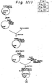
- Fig. 1 illustrates a construction of the expression plasmid pAHhPDILyl.
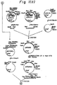
- Fig. 2 shows a boundary of the HSA prepro-sequence and the PDI gene on an expression plasmid.
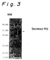
- Fig. 3 is a photograph showing the result of SDS-polyacrylamide gel electrophoresis of an expressed and secreted crude recombinant human PDI, wherein lane l is a molecular weight marker, lane 2 is pAH/AH22 (control) and lane 3 is pAHhPDILyl/AH22.
- Fig. 4 illustrates the separation of a recombinant human PDI by hydrophobic column chromatography.
- Fig. 5 shows the result of SDS-polyacrylamide gel electrophoresis of a purified recombinant human PDI, wherein the numbers at the bottom correspond to the fraction numbers of the hydrophobic column chromatography shown in FIg. 4, and M is a molecular weight marker.
- Fig. 6 is a photograph of SDS electrophoresis showing expression of human PDI in the yeast strain HIS23.
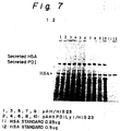
- Fig. 7 is a photograph of SDS-polyacrylamide gel electrophoresis showing secretion of HSA by co-expression of human PDI and HSA in the yeast strain HIS23.
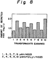
- Fig. 8 shows the result of densitometric determination of the amount of secreted HSA using the SDS-polyacrylamide gel electrophoresis gel of Fig. 7.
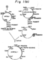
- Fig. 9 illustrates a preparation of the Xho I- BAM HI restriction fragment of YERD2.
- Fig. 10 illustrates a construction of the expression plasmid pIVTRPGAPYERD2.
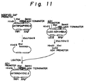
- Fig. 11 illustrates a construction of the expression plasmid pIVTRPADHYERD2.
- Fig. 12 is a photograph showing the result of SDS-polyacrylamide gel electrophoresis of human PDI and HSA which were secreted from the strain SN35A-1PU capable of expressing both human PDI and HSA and from the strain SN35A-1PUAET introduced a yeast ERD2 expression system into the SN35A-1PU, wherein lane 1 is a supernatant of the culture of SN35A-1PU and lane 2 is a supernatant of the culture of SN35A-1PUAET.
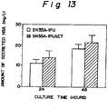
- Fig. 13 shows the result of densitometric determination of an amount of HSA secreted from strain SN35A-1PU and SN35A-1PUAET using SDS-electrophoresis gels.
- the present invention has been completed by finding a fusion gene for expression of PDI which is composed of a DNA fragment coding for human serum albumin (HSA) prepro-sequence and a gene coding for human PDI, as wall as a co-expression system comprising PDI gene, yeast ERD2 gene and a certain polypeptide gene.
- HSA human serum albumin
- Clones containing a human PDI cDNA are isolated from the human liver and placenta ⁇ gt11 cDNA libraries (Clontech, US) by the following procedures: An E. coli stain in infected with phage from the human liver and placenta ⁇ gt11 cDNA libraries, after which DNAs from the phage grown are fixed on a filter. Separately from this, positive clones are screened by hybridization using a 40 mer synthetic oligomer DNA as a probe which corresponds to the complementary strand of a nucleic acid sequence (243-282) of human proline 4-hydroxylase (the same protein as PDI) cDNA (Pihlajaniemi, T.
- the phage DNA obtained is digested with Eco RI, and the resultant 150 bp insert DNa is used as a probe for screening the PDI cDNA. Using the probe, the phage DNAs fixed on the filter are screened to isolate positive clones.
- a fusion gene for use in the expression and production of PDI which is composed of a DNA fragment coding for a HSA prepro-sequence and the aforementionned human PDI gene.
- the fusion gene is constructed in general by arranging the preprosequence-encoding DNA fragment at the upstream side of the PDI gene.
- a leader sequence for transporting human PDI into an appropriate compartment is not always limited to the HSA prepro-sequence, and other signal sequences or prepro-sequences may also be used as the leader sequence.
- said fusion gene may be prepared as follows: The aforementioned clones pHPDI16 and pHPDIp4 DNAs are double-digested with Eco RI/ Pst I and Pst I/ Bam HI, respectively, to produce DNA fragments of about 490 bp and about 1.3 kbp respectively, the fragments recovered are ligated with a plasmid vector pUCll9 which was ligated with Eco RI and Bam HI to produce phPDIEB in which a Nae I cleavage site is then introduced into the boundary between the PDI signal sequence and the PDI sequence by the Kunkel's method (Kunkel, T.A., Proc. Natl. Acad. Sci.
- pUC119 is digested with Eco RI, and the resultant digest is ligated with the following Xho I linker:
- the digest is ligated with a prepro-sequence of HSA to produce pUC119Sig which is subsequently digested with St uI and Hind III to give a DNA fragment of about 3.2 kb
- pUC119Sig which is subsequently digested with St uI and Hind III to give a DNA fragment of about 3.2 kb
- a method for synthesizing the HSA prepro-sequence willl be described later in Examples.
- phPDILyl which is in turn digested with Eco RI, blunt-ended with Klenow fragment, and digested with Bam HI, thereby giving a fusion gene in which a leader sequence is modified and in which the human PDI gene is fused to the downstream side of the HSA prepro-sequence (Fig. 2).
- Process for the preparation of the fusion gene according to the present invention is not limited to the above-described techniques, provided that said fusion gene has an ability for expressing PDI.
- analogs of the inventive fusion gene are not included within the scope of the present invention, it is obvious that they can be prepared easily from a corresponding gene of any animal origin other than human.
- said fusion gene has a DNa sequence coding for the -24 to +49l amino acid sequence shown in the SEQ ID NO. 2.
- a DNA sequence coding for the +1 to +491 amino acid sequence (Asp l --Leu 49l ) shown in SEQ ID NO:1 may also be applied.
- all genes which substantially have the same function as that of said DNA sequences, for example, derivatives having nucleotide sequences based on the degeneracy of codon are included within the scope of the present invention.
- an example of such a fusion gene includes the sequence between nucleotide 1 to nucleotide 1545 shown in SEQ ID NO.2.
- An expression vector used for the insertion of the linked gene of the present invention thereinto should replicate in a host cell and have the ability for expressing therein.
- a useful vector contains replicon, and regulatory sequences which are derivated from a species compatible with a host to be used, as well as a replication origin and a marker sequence which enable selection of a phenotype from transformed cells.
- the plasmid pJDB-ADH-HSA-A As a vector for use in the construction of the expression vector, the plasmid pJDB-ADH-HSA-A (FIg. 1) which has been disclosed in Japanese Patent Application Laying-Open (KOKAI) No. 2-117384 filed by the present applicant may be used conveniently.
- This plasmid contains HSA cDNA, as well as yeast alcohol dehydrogenase I (ADH I) promoter, ADH I terminator, ampicillin resistance gene (Amp r ) and Leu2 gene.
- the HSA cDNA is removed from this plasmid by digesting it with Xho I, blunting with Klenow fragment, and then digesting with BamH I.
- the 5′-end of the DNA fragment of about 8 kb thus obtained is dephosphorylated, and the resultant fragment is ligated with the aforementioned fusion gene of the present invention to give the expression vector pAHhPDILyl.
- other type of vectors can be used provided that they are capable of expressing the fusion gene.
- Examples of host for use in the expression of human PDI include prokaryotes such as E. coli, Bacillus substilis, etc and eukaryotes such as yeast, etc.
- a host cell capable of secreting the mature PDI via processing.
- the host cell is such as Saccharomyces cerevisiae , more preferably yeast strain AH22. It is obvious that eukaryotes other than yeast, for example animal cells, can be used as the host cell.
- Incorporation of the expression vector into a host cell can be carried out easily by conventional means such as calcium chloride, protoplast (or spheroplast)-polyethylkene glycol, electroporation, etc.
- a desired transformant may be obtained by culturing transformant cells on SD(-Leu) plate and screening colonies grown on the plate.
- a process for the production of a recombinant type human PDI comprises the steps of: constructing an expression vector which can replicate in a host cell and express the fusion gene of the present invention therein; isolating a host cell transformed with said expression vector; culturing the obtained transformant under such conditions that the fusion gene can be expressed, thereby secreting said recombinant human PDI; and recovering the recombinant PDI.
- the recombinant human PDI can be purified easily by separating the transformed cells from a cultured medium by centrifugation, disrupting the cells if necessary, concentrating the supernatant by ultrafiltration or the like, and then subjecting the concentrate to hydrophobic column chromatography.
- TSK-gel Phenyl-5PW hydrophobic column Tosoh, Japan
- the recombinant human PDI may be eluted by linear gradient of from 0.85 to 0 M ammonium sulfate in Borate buffer (Ph 8.0) containing KCl (Fig. 4).
- the purified recombinant human PDI has a molecular weight of about 55 kDa based on the SDS-polyacrylamide gel electrophoresis analysis (Fig. 5), and practically has PDI activity as the results of determination of a degree of the refolding of scrambled rebonuclease A (see Examples).
- the recombinant human PDI thus prepared has the same amino acid sequence except that its N-terminal amino acid is changed from Asp to Gly, as shown in SEQ ID NO:3.
- the present invention further provides a transformant comprising a fusion gene which is composed of a human PDI gene and a DNA fragment coding for a HSA prepro-sequence, and a foreign gene coding for a polypeptide to be produced, in a co-expressible state.
- the fusion gene and the foreign gene in the transformant may be located on the same or different chromosome(s), provided that they are mutually present in a co-expressible state. Transformation of a host cell can be carried out for example by inserting the fusion gene and the foreign gene into the same or different vector(s) and introducing the resulting vector into the host cell by conventional means as lithium chloride, protoplast (or spheroplast)-polyethylene glycol, electroporation, etc.
- the foreign gene may encode a polypeptide of any type, provided that the polypeptide contains at least one disulfide linkage because the catalyst effect of an amplified and expressed PDI, that is acceleration of the formation or exchange reaction of a disulfide bond(s) in polypeptide, is directly exhibited.
- the present invention can be applied to a case which the PDI activity exerts influence on proteins relating to gene expression, polypeptide folding or transport, thereby indirectly improving the productivity of PDI.
- the foreign gene is gene coding for HSA.
- polypeptide as used herein means a short- or long-chain peptide and protein.
- hosts include prokaryotes such as E. coli, Bacillus subtilis , etc and eukaryotes such as yeast, animal cell, etc.
- prokaryotes such as E. coli, Bacillus subtilis , etc
- eukaryotes such as yeast, animal cell, etc.
- Preferred is a host cell capable a mature polypeptide through post-translational modification and processing, more preferably eukaryotes, and most preferably yeast.
- the present invention also provides a process for producing a polypeptide, which comprises the following steps of: co-expressing a human PDI gene and a foreign gene coding for the polypeptide to be produced, in the above-described transformant so as to produce the polypeptide; and recovering the polypeptide.
- HSA and PDI are co-expressed within an HSA-producing yeast strain (pAHhPDILy1/HIS23) transformed with a human PDI expression plasmid in an appropriate medium
- a secretion level of HSA practically increase by about 60% in average in comparison with the case of a non-transformed HSA-producing yeast strain (pAH/HIS23) (Fig. 8).
- HSA is a protein containing 17 disulfide bonds. It is known also that formation of its higher-order structure is enhanced in the presence of a stoichiometric amount of PDI in in vitro refolding experiments of a denatured protein.
- HSA is secreted from the yeast strain HIS23 as a water-soluble molecule, but some of the HSA molecules are also detectable within the yeast cell.
- the intracellular HSA was analysed by SDS-polyacrylamide gel electrophotoresis, it was ditected as a single band with the same mobility as that of a normal HSA molecule in the presence of a reducing agent, while detected as dicontinuous bands having a large molecular weight than normal HSA in the absence of a reducing agent, clearly showing a different behavior from that of normal HSA.
- the increment of HSA production level is base on the direct influence of PDI on HSA molecules by their coexistence in endoplasmic reticulum, because the increase in the amount of secreted HSA has a correlation to an increased level of the secretion of human PDI out of the yeast cell based upon the fusion of the PDI gene with the HSA prepro-sequence which plays a role in the intracellular transport into endoplasmic reticulum through a transmembrane process.
- the effect of the co-expression of PDI on an increment of the amount of secreted HSA is based on the direct influence of PDI on the formation of the higher-order structure of HSA.
- a similar secretion-improving effect can also be expected in other general secretory proteins in which the formation of a disulfide bond(s) contributes to the formation and stability of their higher-order structures, by highly amplified co-expression of PDI in the same host cell.
- the present invention also provides a transformant comprising the following expression units integrated on yeast chromosome in a co expressible state: an expression unit containing a gene coding for a receptor protein ERD2 from yeast or analog thereof which is capable of binding to a protein localizing in endoplasmic reticulum and having a signal for staying therein; and an expression unit containing a gene coding for said protein localizing in endoplasmic reticulum and having a signal for staying therein.
- Such a transformant is useful for the preparation or another transformant cell capable of secreting a useful polypeptide predominantly out of a transformant, by transforming its host cell with an expression vector which contains a foreign gene coding for the polypeptide, said polypeptide being a subject of function of the aforesaid protein localizing in endoplasmic reticulum.
- protein localizing in endoplasmic reticulum and having a signal for staying therein is intended to include any protein which can localize in the endoplasmic reticulum cavity after in vivo protein synthesis, which has a signal for staying in endoplasmic reticulum, such as amino acid sequence "KDEL”, “HDEL”, “DDEL”, “ADEL”, “SDEL”, “RDEL”, KEEL”, “QEDL”, “HIEL”, “HTEL”, KQDL” and the like, on the C-terminus of the protein, and which is capable of binding to a receptor protein ERD2 from yeast or analog thereof.
- PDI glucose-regulated protein 78
- grp94 glucose-regulated protein 94
- Preferred is a protein having a useful function for a polypeptide, more preferably PDI.
- the PDI is known as an enzyme which catalyzes the exchange reaction of a thiol/disulfide bond and rises a rate of the refolding of a denatured protein ( SChein, Bio/technology, vol.7, pp. ll4l - ll48, 1989; Freedman, Cell , vol.57, pp.1069 - 1072, 1989).
- the above-mentioned protein localizing in endoplasmic reticulum also includes a protein which has no said signal natively on its C-terminus but has been modified by genetic engineering techniques so as to provide it with the signal.
- acceptor protein ERD2 from yeast or analog thereof means an acceptor protein from yeast which is capable of accepting any protein localyzing in endoplasmic reticulum and having a signal for staying therein.
- An example of the DNA sequence encoding the ERD2 protein is a sequence reported by Semenza et al. (Semenza, J.C., Hardwick, K.G., Dean, N. and Pelham, H.R.B., Cell , vol.61, p.1349, 1990) which is included herein as reference, although any other modified DNa sequence may also be included provided that the modification does not spoil the function of the ERD2.
- Such a modified DNa sequence is, for example, a DNA sequence coding for a "KDEL" acceptor from mammals which has a high homology to the ERD2 protein (LEwis, M.J. and Pelham, H.R.B., Nature , vol.348, p.162, 1990).
- a gene encoding the yeast ERD2 is obtained by the polymerase chain reaction (PCR) technique (Mullis, K.B. and Faloona, F., Meth. Enzymol., vol.155 p.335, 1987) using a genomic DNA prepared from the yeast strain S288C as a template.Primers used are : 5′-TTTTTCTCGAGTAAGCAATGAATCCGTT-3′ and 5′-AAAAAGGATCCTGCAACACTATTAAA-3′, which were designed based on the DNA sequence of the yeast ERD2 gene (Semenza, J.C., Hardwick, K.G., Dean, N. and Pelham,H.R.B., Cell , vol.6l, p.1349, 1990). The ERD2 gene obtained is inserted into a Xho I/ Bam HI site of the plasmid vector BluescriptII SK+ and then subcloned (Fig. 9).
- PCR polymerase chain reaction
- a vector for use in the incorporation of the ERD2 gene is capable of expressing said gene in a host cell and of replicating itself therein along with the replication of the host chromosome after its integration on the chromosome.
- the vector contains a marker sequence which enables selection of a phenotype from transformed cells.
- plasmid pRG-UAS1-N7-TLY1-304 may be used conveniently (A procedure for constructing the plasmid will be described in detail in Example.).
- This plasmid contains human serum albumin (HSA) cDNA, as well as a modified promoter derived from a yeast glyceraldehyde-3-phosphate dehydrogenase (GAP) promoter, a yeast alcohol dehydrogenase I (ADH I) terminator, an ampicillin resistance gene (Amp r ) and a TRP1 gene.
- HSA human serum albumin
- GAP yeast glyceraldehyde-3-phosphate dehydrogenase
- ADH I yeast alcohol dehydrogenase I
- Amp r ampicillin resistance gene
- the HSA cDNA is removed from this plasmid by digesting it with Xho I and Bam HI, which is then ligated with the yeast ERD2 gene to give the vector pIVTRPGAPYERD2 (FIg. 10). Thereafter, the pIVTRPGAPYERD2 can be converted into expression plasmid pIVTRPADHYERD2 by digesting pIVTRPGAPYERD2 with Hind III and Xho I to remove the modified GAP promoter and by ligating the resultant fragment with a yeast ADH promoter (Fig. 11). Any other type of vectors which function for the expression of the ERD2 gene may be used instead of the pRG-UAS1-N7-TLY1-304.
- promoter sequences useful in the expression vector include 3-phosphoglycerate kinase promoter (Hitzenman et al., J. Biol. Chem. , vol.225, p.2073, 1980) and promoters for the glycolytic enzymes such as enolase, glyceraldehyde-3-phosphate dehydrogenase, hexokinase, etc (Hess et al., J. Adv Enzyme Reg., vol.7 p.149, 1968; Holland et al., iochemistry, vol.17, p.4900, 1978). Any terminator sequence compatible with a yeast strain may be used in the expression vector, examples of which are terminators for the above enzymes.
- the expression vector may further contain a yeast-originated gene such as the TRP1. This gene can be used as a phenotype selection marker when a desired transformant is isolated, and it can also function in order to occur the homologous recombination of an ERD2 expression unit on yeast chromosome when the vector is integrated onto the chromosome.
- the expression vector may also contain a yeast-compatible replication origin, a ribosome binding site, a marker sequence such as antibiotic resistant gene, and other useful sequences.
- a preferred example of the above-mentioned gene coding for a protein localizing in endoplasmic reticulum and having a signal for staying therein is a PDI gene or a fused gene which is composed of the PDI gene and a HSA prepro-sequence.
- the PDI gene used in the present invention include an eukaryote PDI gene, particularly mammal PDI gene, more particularly human PDI gene, and its derivatives (substitution , addition, modification, deletion, etc).
- the human PDI gene or its derivative has for example a DNA sequence coding for the amino acid sequence shown in SEQ ID NO:1 or SEQ ID NO:3.
- a strain belonging to the genus Saccharomyces such as Saccharomyces cerevisiae
- Saccharomyces cerevisiae may be used as a yeast host. It is obvious that eukaryotes other than yeast, for example animal cells, can also be used as the host.
- Transformation can be carried out easily by conventional means such as lithium chloride, protoplast(or spheroplast)-polyethylene glycol, electroporation, etc.
- the transformant of this invention contains TRPl gene, it may be isolated by culturing the transformed cell on an SD (-Leu, -His, -Ade, -Ura, -Trp) plate and screening colonies on the plate.
- SD -Leu, -His, -Ade, -Ura, -Trp
- the present invention also provides a transformant comprising the following expression units integrated on yeast chromosome in a co-expressible state: an expression unit containing a gene coding for a receptor protein ERD2 from yeast or analog thereof which is capable of binding to a protein localizing in endoplasmic reticulum and having a signal for staying therein; an expression unit containing a gene coding for said protein localizing in endoplasmic reticulum and having a signal for staying therein; and an expression unit containing a foreign gene coding for a polypeptide which is a object of function of said protein localizing in endoplasmic reticulum and having a signal for staying therein.
- the expression unit comprising a gene encoding a ERD2 or analog thereof and the expression unit; the expression unit comprising a gene encoding the aforesaid protein localizing in endoplasmic reticulum; the yeast host; and the transformation process, may directly be applied to this case.
- HSA gene may preferably be used as the gene encoding a polypeptide.
- the ERD2 gene and other above-described genes which are carried in the two types of the transformants of the present invention may be located on the same or different genome(s) in the host cell, provided that they are mutually present in a co-expressible state.
- the ERD2 gene and other above-described genes may be inserted into the same vector or preferably different vectors, incorporated into the same host cell, and then integrated into the host chromosome by homologous recombination.
- Each of the expression units containing the ERD2 gene and other above-described genes may be contained in plural numbers in the chromosome, provided that they are mutually in a co-expressible state.
- the transformant includes the HSA-highly producing yeast SN35A-1 PUAET.
- This yeast strain is obtained by incorporating an expression vector containing the yeast ERD2 gene into the SN35A-1PU (obtained by introducing a human PDI expression unit into the locus ura 3 of a HSA-highly secreting yeast strain SN35A (Japanese Patent Application No. 3-2226107); see Examples) and then by integrating the ERD2 gene into the trp 1 site ion the yeast chromosome by homologous recombination.
- the yeast transformant SN35A-lPUAET thus obtained can express the yeast ERD2, human PDI and HSA simultaneously.
- the present invention also provides a process for the production of a polypeptide, which comprises the following steps of: culturing a transformant containing the aforementioned three expression units in an appropriate medium, and bringing about co-expression such that the polypeptide is predominantly secreted out of the transformant cell, the polypeptide being a subject of function of a protein which localizes in endoplasmic reticulum and has a signal for staying therein, while both an acceptor protein ERD2 from yeast or analog thereof which is capable of associating with said protein localizing in endoplasmic reticulum, and said protein as a ligand for the ERD2 remain in endoplasmic reticulum; and recovering said polypeptide secreted.
- the expression of the ERD2 protein or analog thereof and of the protein localizing in endoplasmic reticulum are controlled by a suitable regulator sequence.
- both the repressed secretion of PDI and the enhanced secretion of HSA by co-expression of the ERD2 may be explained as follows:
- the ERD2 localized in the endoplasmic reticulum acts as an acceptor of said signal which is attached to the C-terminus of PDI.
- a strong promoter ex. modified GAP promoter herein
- the amount of intracellularly expressed PDI exceeds the PDI-binding capacity of ERD2, and accordingly the excess PDI molecules "over flow" outside the host cell .
- ERD2 improves the aforesaid PDI-retention efficiency in the endoplasmic reticulum, and thereby an amount of secreted HSA as a substrate of PDI, increases.
- the improvement of a PDI-retention efficiency in the endoplasmic reticulum by the co-expression of a yeast ERD2 gene provides a system in which the PDI can function maximally by allowing a large quantity of expressed PDI to localize within a suitable intracellular compartment where the PDI can function naturally.
- the present invention has a general utility value for the purpose of allowing any polypeptide having a signal for staying in endoplasmic reticulum which is acceptable by ERD2, to work efficiently on a foreign polypeptide as its substrate like the case of PDI.
- the present invention has established for the first time a means in which the secretion of PDI is repressed and the effect of PDI to increase the secretion of serum albumin is improved, by constructing a large scale expression system of a yeast ERD2 gene and by applying this system to expression systems of human PDI and HSA.
- the process of the present invention can be used as a means to improve the above-described retention efficiency in conjunction with a large scale expression system of a protein localizing in the endoplasmic reticulum of an eukaryote, as well as a means to improve a production efficiency of a useful polypeptide by introducing the co-expression system into a production system of the polypeptide which is a substrate of said protein.
- the resulting cells were added to 50 ml of an LB top agar medium (LB medium, 10mM% MgCl2 and 0.7% agarose), and then mixed and inoculed on a LB agar plate (23 cm x 23 cm). After solidifying the top agar medium, the plate was incubated overnight at 37°C so as to grow the phage particles. The phage particles obtained were transferred onto a filter (Hybond-N, Amersham).
- a 40 mer oligomer DNA (5′-TGGCGTCCACCTTGGCCAACCTGATCTCGGAACCTTCTGC-3′) which corresponds to the complementary chain of the 243-282 base sequence of human proline-4-hydroxylase (the same protein of PDI) cDNA (Pihlajaniemi, T. et al., EMBO J. vol.6, p.643, 1987) was synthesized using an automatic DNA synthesizer (Model 380B, Applied Biosystems).
- the 5′-end of the synthesized DNA was labeled by phosphorylation, by incubating 20 pmoles of the DNA at 37°C for 60 minutes in 50 ⁇ l of mM Tris-HCl (pH 7.5) buffer containing 10 mM MgCl2, 5 mM dithiothreitol, l00 ⁇ Ci [ ⁇ -32p] ATP ( ⁇ 3000 ci/mmol, Amersham) and 12 units of T4 polynucleotidee kinase (Takara Shuzo, Japan).
- the filter was further soaked in a hybridization solution (prepared by supplementing the prehybridization solution with about l06 cpm/ml of the aforementioned labeled DNA) for 15 hours at 37°C.
- the resulting filter was washed with 2 x SSC solution at room temperature and then with 2 x SSC + 0.l% sodium dodecyl sarcosinate solution at 42°C for 30 minutes (followed by its exposure to an X-ray film (XAR-5 Kodak) overnight at -80°C After development of the film, 8 positive signals were detected by the primary screening.
- Phage particles corresponding to those signals signals were recovered from the aforesaid plate by cutting it out as gel sections, soaked each of the gel section in 1 ml of SM buffer (100 mM NaCl, l0 mM MgCl2, 50 mM Tris-HCl (pH 7.5) and 0.01% gelatin), and left overnight at 4°C so as to recover the phage from the gel into the solution.
- SM buffer 100 mM NaCl, l0 mM MgCl2, 50 mM Tris-HCl (pH 7.5) and 0.01% gelatin
- a phage DNA was prepared from the positive clone obtained finally by the method of Leder et al. (Leder, P., Tiemeir, D. and Enquist, L., Science, vol.196 p.175, l977).
- the thus prepared phage DNA (l/5 vol) was digested at 37°C for 1 hour in 50 ⁇ l of the digestion solution consisting of 100 mM Tris-HCl (pH 7.5), 100 mM NaCl, 6mM MgCl2, 6mM mercaptoethanol, 0.1% gelatin, 20 ⁇ g/ml of ribonuclease A and 20 units of Eco RI (Nippon Gene, Japan).
- this positive clone contains an insert DNA fragment of about 150 bp.
- the insert DNA was separated and purified using glass powder (Gene CleanTM, Bio-l01). About 20 ng of the recovered DNA fragment and about 100 ng of pUC19 vector which has been digested with Eco RI were added to the mixture of liquid A 20 ⁇ l and liquid B 4 ⁇ l from the DNA ligation kit (Takara Shuzo, Japan), and the resulting mixture was then incubated at l6°C for 15 hours to obtain a recombinant plasmid in which both DNA fragments were linked together. Using 10 ⁇ l of this reaction mixture, transformation of E.
- coli strain TGl was carried out by the Mandel's method (Mandel, M. and Higa, A., J. Mol. Biol., col.53, p.154, l970). The transformant thus obtained was cultured overnight at 37°C in 100 ml of LB medium supplemented with 25 ⁇ g/ml of ampicillin, and a plasmid DNA was purified from the cultured cells by alkaline lysis method (Birnboim, H.C. and Doly, J., Nucleic Acids Res., vol.7, p.l5l3, 1979).
- 10 ⁇ g of the plasmid DNA was digested at 37°C for l hour in 200 ⁇ l of the digestion solution consisting of 100mM Tris-HCl (pH 7.5). l00 mM NaCl, 6 mM mercaptoethanol, 0,l% gelatin and 100 units of Eco RI (Nippon Gene, Japan).
- the digest was extracted with phenol, concentrated by ethanol precipitation and then subjected to 0.8% agarose gel electrophoresis. Thereafter, an insert DNA fragment of about l50 bp was recovered by glass powder technique, for use as a probe in the following PDI cDNA screening.
- screening were carried out again from about 50,000 clones of human Live ⁇ gt11 cDNA library and about 50,000 clones of human placenta ⁇ gt11 cDNA libraries (Clontech). Filters on which phage DNA molecules of the two libraries were fixed were prepared in the same manner as described in the foregoing.
- the resulting filters were washed with 2 x SSC solution at room temperature and then with 0.5 x SSC + 0.1% sodium dodecyl sarcosinate solution at 65°C for 1 hour, followed by their exposure to X-ray films (XAR-5,Kodak) overnight at -80°C. After development of the films, 6 positive signals were found from the liver cDNA library, and 5 positive signals from the placenta cDNA library. By subjecting these clones to second and third screenings, 4 positive clones were isolated from the liver cDNA library, and 3 positive clones from the placenta cDNA library.
- the Eco RI insert DNA fragments of the obtained 7 clones were separately subcloned into an Eco RI site of plasmid vector pUCl9 in the same manner as described above in order to make restriction maps for the inserts of the 7 clones.
- 4 clones obtained from the liver cDNA library and 2 clones from the placenta cDNA library were found to overlap one another.
- a plasmid for use in the expression of human PDI in yeast was constructed by the following procesure, using the above two clones, pHPDIl6 and pHPDIp4, which encode human PDI cDNA (Fig. 1): About 1 ⁇ g of pHPDIl6 DNA prepared by the alkaline lysis method was digested at 327°C for l hour in 20 ⁇ l of the digestion solution consisting of l0 mM Tris-HCl (pH 7.5), 100 mM NaCl, 6 mM MgCl2, 6 mM mercaptoethanol, 0.1% gelatin, 10 units of Eco RI (Nippon Gene) and 10 units of Pst I (Nippon Gene).
- the resulting digest was subjected to 0.8% agarose gel electrophoresis and then to the glass powder method to separate and purify a DNA fragment of about 490 bp which corresponds to a 5′-end Eco RI- Pst I fragment of the PDI cDNA Separately from this, about 1 ⁇ g of pHPDIp4 was digested at 37°C for 1 hour in 20 ⁇ l of the digestion solution consisting of l0 mM Tris-HCl (pH 7.5), l00 mM NaCl, 6 mM MgCl2, 6 mM mercaptoethanol, 0.1% gelatin, 10 units of Pst I (Nippon Gene) and 10 units of Bam HI (Nippon Gene).
- the resulting digest was treated in the same manner as described above to separate and purify a DNA fragment of about 1.3 kb which corresponds to a 3′-end Pst I- Bam HI fragment of the PDI cDNA.
- the thus recovered two DNA fragments (about 50 ng for each) were ligated with about 20 ng of plasmid vector pUC ll9 which has been digested in a linear form which Eco RI and Bam HI, by incubating these DNA samples at l6°C for 15 hours in the mixture of 25 ⁇ l of Liquid A and 5 ⁇ l of Liquid B of the DNA ligation kit (Takara Shuzo). With 10 ⁇ l of the reaction mixture obtained, a component E.
- coli strain MV1190 cell was transformed by the calcium chloride technique.
- the transformed cell was cultured overnight at 37°C on an X-Gal plate (LB medium containing 1.5% agar further supplemented with 50 ⁇ g/ml of 5-bromo-4-chloro-3-indolyl- ⁇ -D-galactopyranoside, 80 ⁇ g/ml of isopopyl- ⁇ -D-thiogalactopyranoside and 25 ⁇ g/ml of ampicillin) of 90 mm in diameter.
- plasmid DNAs were prepared from the colonies by the alkaline lysis method, and the DNAs were analysed using restriction enzymes, thereby selecting a transformant which carries a target plasmid.
- the thus obtained plasmid was named phPDIEB.
- a Nae I cleavage site was introduced into the boundary region between the PDI signal sequence and the PDI sequence itself on the DNA by the method of Kunkel (Kunkel, T.A., Proc. Natl. Acad. Sci., USA, vol.82, p.448, 1985).
- E. coli strain BW313 competent cell was transformed with the phPDIEB DNA by calcium chloride technique.
- a single colony of the resultant transformant was pre-cultured overnight at 37°C in 2 x YT meduium (l.6% Bacto-trypton, 0.5% NaCl and 1% Bacto-Yeast Extract) supplemented with 150 ⁇ g/ml of ampicillin.
- One ml of the pre-culture was inoculated into 50 ml of the 2 x YT medium supplemented with 150 ⁇ g/ml of ampicillin, followed by its culture at 37°C.
- turbidity (OD600) of the medium reached around 0.3
- the obtained culture was subjected to centrifugation, and the supernatant recovered was mixed with 1/5 volume of a solution containing 2.5 M NaCl and 20% polyethylene glycol #6000. After stirring, the mixture was left for 15 minutes at room temperature.
- the precipitate obtained by centrifugation was dissolved in 5 ml of the TE buffer (pH 8.0) consisting of 10 mM Tris-HCl and l mM EDTA, mixed with an equal volume of neutral phenol with stirring, and then centrifuged to recover an aqueous layer. To the layer was added an equal volume of chloroform with stirring. The mixture was further subjected to centrifugation to recover an aqueous layer.
- the aqueous layer was then mixed with l/l0 volume of 3 M sodium acetate and 2.5 volume of ethanol. After stirring, the mixture was left for 30 minutes at -80°C, followed by centrifugation in order to recover DNA as precipitate.
- the DNA was washed with 70% ethanol, dried under a reduced pressure and then dissolved in 100 ⁇ l of the TE buffer.
- a desired mutation i.e., introduction of a Nae I site
- 10 pmol of a synthetic oligonucleotide (5′-CGGGGGCGCCGGCGCGC-3′, Takara Shuzo) for use in the introduction of a mutation was incubated at 37°C for 15 minutes in 10 ⁇ l of the phosphorylation solution which consists of l00 mM Tris-HCl (pH 8.0), 10 mM MgCl2, 7 mM dithiothreitol, 1 mM ATP and 10 units of T4 polynucleotide kinase (Takara Shuzo), followed by heating at 70°C for 10 minutes in order to deactivate the T4 polynucleotide kinase.
- the phosphorylation solution which consists of l00 mM Tris-HCl (pH 8.0), 10 mM MgCl2, 7 mM dithiothreitol, 1 mM ATP
- 0.2 pmol of the above-described phPDIEB-originated single-stranded DNA and l ⁇ l of an annealing buffer Site-directed mutagenesis system MutanTM-K,Takara Shuzo
- a annealing buffer Site-directed mutagenesis system MutanTM-K,Takara Shuzo
- a complementary chain synthesis was carried out by mixing the reaction mixture with 25 ⁇ l of a chain elongation solution (Site-directed mutagenesis system MutanTM-K, Takara Shuzo), 60 units of E.coli DNA ligase (MutantTM-K, Takara Shuzo) and 1 unit of T4 DNA polymerase (MutantTM-K, Takara Shuzo), and by incubating the resulting mixture at 25°C for 2 hours.
- the reaction was determined by adding 3 ⁇ l of 0.2 M EDTA (pH 8.0) and heating the mixture at 65°C for 5 minutes. 3 ⁇ l of the DNA solution obtained was mixed with 30 ⁇ l of a suspension of E.
- 2 ⁇ g of the phPDINae DNA prepared by the alkaline lysis method was digested at 37°C for 4 hours in 30 ⁇ l of the digestion solution consisting of 10 mM Tris-HCl (pH 8.0), 20 mM NaCl, 7 mM MgCl2, 7 units of Nae I (Nippon Gene) and 10 units of Hind III (Takara Shuzo).
- the resulting digest was subjected to 0.8% agarose gel electrophoresis and then treated by the glass powder technique so as to separate and purify a DNA fragment of about 1.7 kb.
- a plasmid, pUCll9Sig, containing a DNA fragment which encodes a human serum albumin prepro-sequence and is composed of codons often utilized in yeast was constructed in the following manner (Fig. 1): One ⁇ g of plasmid vector pUCll9 DNA was digested at 37°C for 1 hour in 20 ⁇ l of the digestion solution which consist of l00 mM Tris-Hcl (pH 7.5), 10 mM MgCl2, 50 mM NaCl and l2 units of Eco RI (Nippon Gene), followed by heating at 70°C for 5 minutes in order to deactivate the enzyme.
- the DNA was then incubated overnight at 16°C in 30 ⁇ l of the ligation solution which consisted of 66 mM Tris-HCl (pH 7.5), 6.6 mM mgCl2, 10 mM dithiothreitol, 0.1 mM ATP and 300 units of T4 DNA ligase (Takara Shuzo), together with an equal molar amount of a Xho I linker containing a Xho I site and consisting of the following sequence:
- E. coli JM107 competent cell was carried out by the calcium chloride method.
- the transformed cell was cultured overnight at 37°C on a LB plate supplemented with 50 ⁇ g/ml of ampicillin.
- Plasmid DNAs were prepared by the alkaline lysis method from the colonies on the plate and analysed using restriction enzymes. In this way, a plasmid DNA molecule in which the Xho I linker has been inserted into pUC119 Eco RI site was selected.
- oligonucleotides were synthesized using an automatic DNA synthesizer (380B, Applied Biosystems).
- Each 5′-end of these oligonucleotides was phosphorylated by incubating about 30 pmol of each sample at 37°C for 1 hour in the solution consisting of 50 mM Tris-HCl (pH 7.6), 10 mM MgCl2, 5 mM dithiothreitol, 0.2 mM ATP and 6 units of T4 polynucleotide kinase (Takara Shuzo).
- the oligonucleotide solutions obtained were combined (100 ⁇ l in total volume) and annealed by leaving the combined solution for 5 minutes in a water bath of l00°C, followed by cooling down to room temperature.
- One ⁇ g of the above Xho I linker-introducing vector plasmid was digested at 37°C for l hour in 20 ⁇ l of the digestion solution consisting of 100 mM Tris-HCl (pH 7.5), l0 mM MgCl2, l00 mM NaCl, 10 units of Bam HI (Nippon Gene) and 12 units of Xho I (Takara Shuzo), followed by phenol extraction and ethanol precipitation to recover a DNA fragment.
- the fragment obtained was incubated overnight at l6°C in 30 ⁇ l of the ligation solution which consist of 66 Tris-HCl (pH 7.5), 6.6 mM MgCl2, l0 mM dithiothreitol, 0.l mM ATP and 300 units of T4 DNA ligase (Takara Shuzo), together with an equal molar amount of the double-stranded DNA fragment obtained by ligating the four oligonucleotides.
- transformation of E. coli JM10l7 competent cells was carried out by the calcium chloride method.
- the transformed cells were cultured overnight at 37°C on LB medium containing 50 ⁇ g/ml of ampicillin. Plasmid DNAs prepared from colonies on the plate were analysed using restriction enzymes so as to select a transformant containing a desired recombinant plasmid.
- the obtained plasmid was named pUCll9Sig.
- a DNA was prepared from plasmid pUC119Sig by the alkaline lysis method. 2 ⁇ g of the DNA was digested at 37°C for 4 hours in the digestion solution which consist of l0 mM Tris-HCl (pH 8.0), l00 mM NaCl, 7 mM MgCl2, 8 units of Stu I (Nippon Gene) and l0 units of Hind III (Takara Shuzo), subjected to 0.8% agarose gel electrophoresis and then treated by the glass powder technique to separate and purify a DNA fragment of about 3.2 kb.
- the 1.7 kb DNA fragment (about 50 ng) derived from phPDINae was reacted with the 3.2 kb DNA fragment (about 50 ng) from pUC119Sig at l6°C for 30 minutes in the ligation kit solution of Takara Shuzo (Japan) (a mixture of liquid A 30 ⁇ l + Liquid B 6 ⁇ l).
- Takara Shuzo a mixture of liquid A 30 ⁇ l + Liquid B 6 ⁇ l.
- transformation of E. coli HB101 competent cells was carried out by the calcium chloride method.
- the transformed cells were cultured overnight at 37°C on a LB plate supplemented with 50 ⁇ g/ml of ampicillin.
- Plasmid DNAs were prepared by the alkaline lysis method from colonies grown on the plate and analysed using restriction enzymes select a recombinant plasmid in which the human PDI itself was linked to the downstream side of the human serum albumin prepro-sequence (Fig. 2) .
- the obtained plasmid was named phPDILyl.
- a human PDI expression plasmid was constructed in the following manner, such that the leader sequence modified type PDI can express under the control of a promoter of yeast alcohol dehydrogenase I gene: 7 ⁇ l phPDILyl DNA prepared by the alkaline lysis method was digested at 37°C for 2 hours in l00 ⁇ l of the digestion solution which consists of l00 mM Tris-HCl (pH 8.0), 100 mM NaCl, 7 mM MgCl2 and 40 units of Eco RI (Nippon Gene). The resulting solution was mixed with an equal volume of a pheno/chloroform mixture (a mixture of saturated phenol with an equal volume of chloroform).
- a pheno/chloroform mixture a mixture of saturated phenol with an equal volume of chloroform.
- the thus prepared solution was subjected twice to phenol/chloroform extraction, and the resulting aqueous layer was mixed with l/l0 volume of 3 M sodium solution acetate (pH 5.3) and 2.5 volume of ethanol. The mixture was left for 1 hour at -40°C and then subjected to centrifugation. The pellet was washed with 70% ethanol, dried under a reduced pressure and then dissolved in 50 ⁇ l of Klenow buffer solution (Deletion Kit for Kilo-Sequence, Takara Shuzo). Thereafter, to the solution obtained was added 4 units of Klenow fragment (Takara Shuzo), and the mixture was incubated at 37°C for 45 minutes to blunt the Eco RI cleavage site.
- the reaction mixture was then subjected twice to phenol/chloroform extraction, and the resulting aqueous layer was mixed with 1/10 volume of 3 M sodium acetate (pH 5.3)and 2.5 volume of ethanol.
- the mixture was left for l hour at -40°C and then centrifuged.
- the pellet was washed with 70% ethanol, dried under a reduced pressure and then dissolved in 40 ⁇ l of the solution consists of 10 mM Tris-HCl (pH 8.0), 60 mM NaCL, 7 mM MgCl2 and l0 units of Bam HI (Nippon Gene).
- the DNA solution obtained was subjected to 0.8% agarose gel electrophoresis and then treated by the glass powder technique so as to separate and purify a DNA fragment of about l.8 kb.
- 5 ⁇ l of pJDB-ADH-HSA-A DNA (Japanese Patent Application Laying-Open (KOKAI) No. 2-ll7384) prepared by the alkaline lysis method was digested at 37°C for 2 hours in l00 ⁇ l of the digestion solution which consists of 10 mM Tris-HCl (pH 8.0), 100 mM NaCl, 7 mM MgCl2 and 24 units of Xho I (Takara Shuzo).
- the reaction mixture was then subjected to phenol/chloroform extraction, and the aqueous layer separated was mixed with l/l0 volume of 3 M sodium acetate (pH 5.3) and 2.5 volume of ethanol. The mixture was left for 2 hours at -40 °C and then centrifuged to recover DNA as precipitate. The DNA precipitate was washed with 70% ethanol, dried under a reduced pressure and then dissolved in 50 ⁇ l of Klenow buffer solution (Deletion Kit for Kilo-Sequence, Takara Shuzo). Thereafter, to the solution obtained was added 4 units of Klenow fragment (Takara Shuzo), and the mixture was incubated at 37°C for 45 minutes to blunt the Xho I cleavage site.
- the reaction mixture was then subjected twice to phenol/chloroform extraction, and the aqueous layer separated was mixed with 1/10 volume 3 M sodium acetate (pH 5.3) and 2.5 volume of ethanol. The mixture was left for 1 hour at -40°C before centrifugation.
- the DNA pellet removed was washed with 70% ethanol, dried under a reduced pressure and then dissolved in 40 ⁇ l of the solution which consist of 10 mM Tris-HCl (Ph 8.0), 60 mM NaCl, 7mM MgCl2 and l0 units of Bam HI (Nippon Gene). The solution obtained was incubated at 37°C for 75 minutes to digest the DNA.
- the prepared DNA solution was subjected to 0.8% agarose gel electrophoresis and then treated by the powder gas technique so as to separate and purify a DNA fragment of about 8 kb.
- the thus obtained phPDILyl1-originated 1.8 kb DNA fragment (about 50 ng) and pJDB-ADH-HSA-A-originated 8 kb DNA fragment (about 50 ng) were incubated at 16°C for 2.5 hours in the DNA ligation kit solution of Takara Shuzo (a mixture of Liquid A 30 ⁇ l + Liquid B 6 ⁇ l) in order to ligate the two DNA fragments.
- Takara Shuzo a mixture of Liquid A 30 ⁇ l + Liquid B 6 ⁇ l
- coli strain C600 was carried out by the calcium chloride technique. The transformed cells were cultured overnight at 37°C on a LB plate supplemented with 50 ⁇ g/ml of ampicillin. Plasmid DNAs were prepared by the alkaline lysis method from colonies grown on the plate and analysed using restriction enzymes in order to select a transformant carrying a plasmid in which the leader sequence modified type PDI sequence was linked to the downstream side of the alcohol dehydrogenase I promoter. The constructed PDI expression plasmid was named pAHhPDILyl. As the results of the plasmid construction, the N-terminal amino acid of the mature PDI protein was changed from Asp to Gly.
- a control plasmid for use in experiments of the human PDI expression was constructed in the following manner: 5 ⁇ l of pJDB-ADH-HSA-A DNA prepared by the alkaline lysis method was digested at 37°C for 2 hours in 100 ⁇ l of the digestion solution which consists of 10 mM Tris-HCl, 100 mM NaCl, 7 mM MgCl2, 24 units of Xho I (Takara Shuzo) and 29 units of Bam HI (Nippon Gene). The reaction mixture obtained was subjected twice to phenol/chloroform extraction, and to the aqueous layer was added 1/10 vol of 3 M sodium acetate (pH 5.3) and 2.5 vol of ethanol.
- the mixture obtained was then left for 2 hours at -40°C and then centrifuged to recover DNA as pellet.
- the DNA pellet was washed with 70% ethanol, dried under a reduced pressure and then dissolved in 50 ⁇ l of Klenow buffer solution (Deletion Kit for Kilo-Sequence, Takara Shuzo). Thereafter, to the obtained solution was added a 4 units of Klenow fragment (Takara Shuzo), and the mixture was incubated at 37°C for 45 minutes to blunt the Xho I and Bam HI cleavage sites.
- the reaction mixture was then subjected twice to phenol/chloroform extraction, and the aqueous layer separated was mixed with 1/10 vol of 3 M sodium acetate (pH 5.3) and 2.5 vol of ethanol.
- the mixture was left for 1 hour at -40°C before centrifugation.
- the DNA pellet removed was dried under a reduced pressure and dissolved in 20 ⁇ l of the TE buffer. Thereafter, the DNA solution was subjected to 0.8% agarose gel electrophoresis and then treated by the glass powder technique to separate and purify a DNA fragment of about 8 kb.
- the thus obtained DNA fragment (about 50 ng) was mixed with the mixture of liquid A 30 ⁇ l + Liquid B 6 ⁇ l from the DNA ligation kit (Takara Shuzo), and incubated overnight at 16°C so as to cyclize it by self-ligation. Using 10 ⁇ l of the prepared DNA solution, transformation of E.
- Plasmid DNAs were prepared by the alkaline lysis technique from the colonies grown on the plate, and analyzed using restriction enzymes in order to select a desired control plasmid.
- the constructed plasmid was named pAH
- human PDI expression plasmid pAHhPDILyl constructed above, an expression of human PDI yeast was carried out in the following manner: A single colony of yeast strain AH22 obtained by culturing it on a YPD plate (2% Bacto-pepton, 1% yeast extract, 2% glucose and 1.5% agar) was inoculated into 5 ml of a YPD medium (2% Bacto-pepton 1% yeast extract and 2% glucose) and cultured at 30°C for 24 hours with shaking. This pre-culture (0.9 ml) was inoculated into 45 ml of the YPD medium and cultured at 30°C with shaking.
- the main culture was subjected to a low speed centrifugation to recover yeast cells as precipitate.
- the cells removed were suspended in 3 ml of 0.2 M LiSCN, and the cell suspension (l ml) was centrifuged to recover the cells.
- To the cells were subsequently added 46 ⁇ l of 50% PEG #4000, 10 ⁇ l of LiSCN and 10 ⁇ l of a pAHhPDILyl DNA solution (27 ⁇ g as DNA) prepared by the alkaline lysis method. After mixing them by pipetting the mixture was left overnight at 30°C, followed by its suspension in l ml of sterile water. The suspension was then centrifuged to recover cells as pellet.
- the pellet was resuspended in l00 ⁇ l of sterile water and cultured at 30°C after the inoculation of its suspension onto a SD(-Leu) plate (SD(-Leu) medium (0.67% Bacto-nitrogen base, 2% glucose, 20 mg/l of adenine, 20 mg/l of uracil, 20 mg/l of tryptophan, 20 mg/l of histidine, 20 mg/l of arginine, 20 mg/l of methionine, 30 mg/l of tyrosine, 30 mg/l of isoleucine, 30 mg/l of lysine, 50 mg/l of phenylalanine, 100 mg/l of aspartic acid, 100 mg/l glutamic acid, l50 mg/l of vaseline, 200 mg/l of threonine and 375 mg/l of serine (amino acids from Wako Pure Chemical Industries, Japan)) + 1.5% agar).
- SD(-Leu) medium 0.6
- a transformant from the 5-days culture was inoculated into 5 ml of SD (-Leu) medium and cultured at 30°C for 2 days with shaking. l00 ⁇ l of the obtained pre-culture was then inoculated into 5 ml of the YPD medium and cultured at 30°C for 24 hours with shaking. 1.5 ml of the resulting main culture was centrifuged to recover 500 ⁇ l of supernatant which was subsequently mixed with the equal volume of ethanol and then left for 1 hour in an ice bath. The mixture was centrifuged so as to recover products secreted out of the yeast cells as a pellet which was than dried under a reduced pressure.
- the pellet was dissolved in 10 ⁇ l of a sample buffer for SDS-PAGE (125 mM Tris-HCl (pH 6.8), 4% SDS, 20% glycerol, l0% ⁇ -mercaptoethanol and 0.01% Bromophenol Blue). After boiling for 5 minutes, the treated sample was subjected to electrophoresis on SDS/PAGE plate 10/20 (Daiichi Kagaku Yakuhin, Japan). The resulting gel was stained with a staining solution (0.15% Coomassie Brilliant Blue, l0% acetic acid and 40% methanol) and then soaked in a decoloring solution (10% acetic acid and 40% methanol) to visualize an expressed product.
- a staining solution 0.15% Coomassie Brilliant Blue, l0% acetic acid and 40% methanol
- a large-scale culture was carried out in the following manner in order to examine chemical properties of the expressed and secreted protein:
- a single colony of the pAHhPDILyl-carrying yeast strain AH22 was inoculated into 80 ml of the SD (-Leu) medium and cultured at 30°C for 2 days with shaking.
- the obtained pre-culture was then inoculated into 4 liters of a YPD-phosphate medium (YPD medium, 6 g/l of Na2HPO4, and 3 g/l of KH2PO4, pH 7.0) and cultured at 30°C for 24 hours with shaking.
- the resulting main culture was centrifuged to removed the supernatant which was used for the purification of the secreted expression product.
- the culture (4 liters) obtained by culturing the recombinant yeast was concentrated to 1/40 (final volume, l00 ml) using a Millipore-Millitan ultrafiltration apparatus (nominal molecular weight, 30,000 cut-off), and then subjected to TSK-gel Phenyl-5PW hydrophobic column chromatography so as to isolate human PDI.
- the elution was carried out in 10 mM borate-10 KCl buffer (pH 8.0) containing 0.05% NaN3, with a linear gradient from 0.85 M to 0 M of ammonium sulfate over 125 minutes.
- the flow rate was 2 ml/min.
- the result is shown in Fig. 4. In Fig.
- the result of SDS-electrophoresis of the isolated human PDI is illustrated.
- the human PDI was purified almost homogeneously by the hydrophobic column chromatography without a loss of its activity. Any UV-absorbing substance in the YPD medium could be removed markedly efficiently by the chromatography.
- PDI assay was carried out by measuring its effect to enhance the refolding of scrambled ribonuclease A (RNase A) which has been prepared by reduction, denaturation and re-oxidation steps. Refolding degree of the scrambled RNase A was determined by measuring a degree of the restoration of its enzyme activity. The following describes the assay procedure illustratively:
- RNase A 120 mg was dissolved in 3 ml of 0.l M Tris-HCl buffer (pH 8.6) containing 6 M guanidine hydrochloride and 0.15 M dithiothreitol, and then reduced under nitrogen atmosphere at room temperature for 15 hours.
- the reduced product was applied to a Sephadex G-25 column (15 mm ⁇ x 38 cm) equilibrated with 0.01 N HCL, thereby removing the reducing agent.
- To the desalting product was added guanidine hydrochloride to a final concentration of 6 M. After adjusting its pH value to 9.0 with Tris, the mixture was subjected to an exchange reaction of a S-S bond(s) in the dark at 4°C for 14 days. The thus prepared sample was stored at -80°C for use as the scramble RNase A.
- l0 ⁇ l of l dithiothreitol is added to 20 ml of 55 mM phosphate buffer (pH 7.5) in which any dissolving air was replaced with nitrogen gas.
- l0 ⁇ l of this solution is added to 420 ⁇ l of 55 mM phosphate buffer (pH 7.5) mixed with 20 ⁇ l of an enzyme sample, and the mixture is left for 5.5 minutes at 30°C.
- To this solution is added 50 ⁇ l of the scrambled RNase A solution prepared above, followed by the enzymatic reaction at 30°C for 15:5 minutes.
- transformation of the HSA-producing yeast strain HIS23 (Japanese Patent Application No. 2-57885 filed by the present applicant, Bikoken-Kin-Ki No. 11351 (FERM P-1138)) was carried out in the following manner: A single colony of the HSA-expressing yeast strain HIS23 obtained by culturing it on a YPD plate (2% Bacto-trypton, l% Bacto-yeast extracts, 2% glucose and l.5% agar) was inoculated into 5 ml of a YPD medium (2% Bacto-trypton, 1% yeast extract and 2% glucose) and cultured at 30°C for 24 hours with shaking.
- a YPD medium 2% Bacto-trypton, 1% yeast extract and 2% glucose
- One ml of the obtained pre-culture was inoculated into 50 ml of the YPD medium and cultured at 30°C with shaking. When turbidity at OD600 reached about 0.5, the main culture was subjected to a low speed centrifugation to recover the yeast cells as pellet. To the pellet were added 46 ⁇ l of 50% polyethylene glycol #4000, 10 ⁇ l of LiSCN and l0 ⁇ l of the human PDI expression plasmid pAHhPDILyl DNA solution (about 20 ⁇ g as DNA) prepared by the alkaline lysis method (Birnboim, H.C. and Doly, J., Nucleic Acid Res., vol.7 p.l513 l979).
- a transformant (pAH,HIS23) obtained using the plasmid pAH which has been prepared by removing the PDI cDNA moiety from pAHhPDILyl was used as a control.
- the single colony grown on the plate was inoculated into 5 ml of the SD (-His, -Leu) medium and cultured at 30°C for 2 days with shaking. l00 ⁇ l of the pre-culture was then inoculated into 5 ml of the YPD medium and cultured at 30°C for 24 hours with shaking.
- secretion of HSA was increased by about 60% in average due to the co-expression of PDI in the yeast strain HIS23.
- yeast ERD2 gene was carried out through the polymerase chain reaction (PCR) technique (Mullis, K.B. and Faloona, F., Meth. Enzymol., vol.155, p.335, l987) as follows: A single colony of yeast strain S288C was inoculated into 2 ml of YPD medium (2% Bacto-trypton, l% Bacto-yeast extracts and 2% glucose) and cultured at 30°C for 24 hours.
- PCR polymerase chain reaction
- Yeast cells recovered by centrifugation from the culture were washed with 1 ml of a sorbitol solution (l M sorbitol and 50 mM K2HPO4/KH2PO4; pH 6.85) and then subjected again to centrifugation.
- the cells collected were suspended in l ml of the sorbitol solution, and the cell suspension was mixed with 40 ⁇ l of Zymolyase solution (l0 mg/ml) and 1 ⁇ l of ⁇ -mercaptoethanol, and maintained at 37°C for 30 minutes.
- the precipitate obtained by ethanol precipitation was recovered by centrifugation.
- the precipitate was dissolved in 400 ⁇ l of TE buffer (l0 mM Tris-HCl, pH 8.0 and 1 mM EDTA), and the solution was mixed with 240 ⁇ l of 20% PEG solution (20% polyethylene glycol and 2.5 M NaCl).
- the precipitate was recovered by centrifugation which was subsequently washed with 70% ethanol and dissolved in 400 ⁇ l of the TE buffer .
- the solution obtained was extracted twice with phenolchloroform, and a genomic DNA of the yeast strain S288C was recovered by ethanol precipitation of the separated aqueous layer.
- a reaction mixture for PCR reaction was prepared by mixing together l0 ⁇ l of S228C genomic DNA (0.02 ⁇ g), 5 ⁇ g of primer I (0.25 ⁇ g), 5 ⁇ l of primer II (0.25 ⁇ g), 0.5 ⁇ g of TaqI polymerase (Gene AmpTM DNA Amplification Reagent Kit, Perkin Elmer Cetus), 10 ⁇ l of the reaction buffer concentrated to l/10 (the same kit jus described), dNTP mixture (l.25 mM for each, the same kit just described) and 53.5 ⁇ l of sterile water.
- a PCR reaction was carried out in the mixture using a DNA Thermal Cycler (Perkin Elmer Cetus).
- the reaction conditions employed are: denaturation of DNA, 94°C for 1 minute; annealing, 50°C for 2 minutes; and polymerase chain elongation reaction, 72°C for 3 minutes. After 30 cycles of the reaction steps, the final reaction was carried out at 72°C for 7 minutes.
- the primers I and II have the following sequences:
- the reaction mixture obtained was extracted with phenol/chloroform, and the aqueous layer separated was subjected to ethanol precipitation to recover DNA.
- the DNA was incubated at 37°C for 2 hours in 20 ⁇ l of the digestion solution which consist of 10 mM Tris-HCl (pH 8.0), 100 mM NaCl, 7 mM MgCl2, 5 units of Xho I (Takara Shuzo) and 5 units of Bam HI (Takara Shuzo).
- the digest was subjected to 0.8% agarose gel electrophoresis and then treated by the glass powder technique (Gene CleanTM, Bio-10l) to separate and purify a DNA fragment of about 0.7 kb.
- a plasmid vector BluescriptII SK+ which has been digested with Xho I and Bam HI were mixed with a ligation solution (Liquid A 30 ⁇ l + Liquid B 6 ⁇ l from the DNA ligation kit, Takara Shuzo), and the mixture was incubated at 16°C for 2 hours so as to ligate the DNA fragment with the plasmid vector.
- a ligation solution Liquid A 30 ⁇ l + Liquid B 6 ⁇ l from the DNA ligation kit, Takara Shuzo
- transformation of E. coli strain XLl-Blue was carried out.
- the DNA fragment thus subcloned was further subcloned into small fragments before its DNA sequencing. As the results, it was confirmed that the DNA fragment contained the yeast ERD2 gene.
- the base sequence and deduced amino acid sequence coincided with those reported by Semenza et al. (Semenza, J.C., Hardwick, K.G., Dean, N. and Pelham, H.R.B., Cell , vol.61, p.l349, l990), except that the reported codon for the Leu of position 52 was "TTG" while that of the clone of this invention was "TTA".
- a vector for use in the integration of ERD2 expression unit into yeast chromosome was constructed using the aforementioned plasmid pYERD2 by the following procedure(Figs. 9 to 11) : 0.5 ⁇ g of the plasmid vector pRS304 DNA (Sikorski, R.S. and Hieter, P., Genetics, vol.l22, p.1, l989) prepared by the alkaline lysis method (Birnboim, H.C.
- 0.5 ⁇ g of the ADH I transcription terminator cassette vector pUC-ATE DNA Japanese Patent Application Laying-Open (KOKAI) No. 2-ll7384 filed by the present applicant
- the digestion solution which consists of l0 mM Tris-HCl (pH 8.0), 150 mM NaCL, 7 mM MgCl2, 5 units of Sal I (Takara Shuzo) and 5 units of Bam HI (Takara Shuzo).
- the resulting digest was subjected to l% agarose gel electrophoresis and then treated by the glass powder technique to separate and purify a DNA fragment of about 0.4 kb.
- the thus recovered DNA fragments (about 50 ng for each) were mixed with a ligation solution (Liquid A 30 ⁇ l + Liquid B 6 ⁇ l from the DNA ligation kit, Takara Shuzo), and incubated at 16°C for 2 hours to ligate and cyclize the DNA fragments.
- a ligation solution Liquid A 30 ⁇ l + Liquid B 6 ⁇ l from the DNA ligation kit, Takara Shuzo
- the E. coli strain XL1-Blue was transformed.
- HSA expression vector pRG-UAS1-N7-TLY1-305 DNA Japanese Patent Application No. 3-l88794 filed by the present applicant
- the digestion solution consist of 10 mM Tris-HCl (pH 8.0), 100 mM NaCl, 7 mM MgCl2, 10 units of Not I (Toyobo) and 5 units of Bam HI (Takara Shuzo).
- the resulting digest was subjected to 0.7% agarose gel electrophoresis and then treated by the glass powder technique to separate and purify a DNA fragment of about 4 kb.
- 0.5 ⁇ g of the pRS304-ATE DNA was digested at 37°C for 2 hours in 30 ⁇ l of the digestion solution which consists of l0 mM Tris-HCl (pH 8.0), 100 mM NaCl, 7 mM MgCl2, l0 units of Not I (Toyobo) and 5 units of Bam HI (Takara Shuzo).
- the resulting digest was subjected to 0.7% agarose gel electrophoresis and then treated by the glass powder technique to separate and purify a DNA fragment of about 5 kb.
- the thus recovered DNA fragments (about 50 ng for each) were mixed with a ligation solution (Liquid A 30 ⁇ l + Liquid B 6 ⁇ l from the DNA ligation kit, Takara Shuzo), and incubated at l6°C for 2 hours to ligate and cyclize the DNA fragments.
- a ligation solution Liquid A 30 ⁇ l + Liquid B 6 ⁇ l from the DNA ligation kit, Takara Shuzo
- transformation of the E. coli strain XLl-Blue was carried out.
- 0.5 ⁇ h of the aforementioned plasmid pYERD2 DNA prepared by the alkaline lysis method was digested at 37°C for 2 hours in 30 ⁇ l of the digestion solution which consists of 10 mM Tris-HCl (pH 8.0), l00 mM NaCl, 7 mM MgCl2, 5 units of Xho I (Takara Shuzo) and 5 units of Bam HI (Takara Shuzo).
- the resulting digest was subjected to 0.8% agarose gel electrophoresis and then treated by the glass powder technique to separate and purify a DNA fragment of about 0.7 kb.
- 0.5 ⁇ g of the DNA prepared by the alkaline lysis method from the aforementioned human HSA integration vector pRG-U1-N7-TLY-304 was digested at 37°C for 2 hours in 30 ⁇ l of the digestion solution which consists of l0 mM Tris-HCl (pH 8.0), l00 mM NaCl, 7 mM MgCl2, 5 units of Xho I (Takara Shuzo) and 5 units of Bam HI (Takara SHuzo). Following 0.8% agarose gel electrophoresis of the resultant digest, a DNA fragment of about 6.3 kb was purified by the glass powder technique.
- the thus recovered DNA fragments (about 50 ng for each) were mixed with the ligation solution (Liquid A 30 ⁇ l + Liquid B ⁇ l, Takara Shuzo), and incubated at 16°C for 2 hours to ligate the DNA fragments.
- the ligation solution Liquid A 30 ⁇ l + Liquid B ⁇ l, Takara Shuzo
- the E. coli strain HBl01 was transformed.
- One ⁇ g of the pIVTRPGAPYERD2 DNA prepared by the alkaline lysis method was digested at 37°C for 4 hours in 30 ⁇ l of the digestion solution which consists of l0 mM Tris-HCl (pH 8.0), l00 mM NaCl, 7 mM MgCl2, 5 units of Xho I (Takara Shuzo) and 5 units of Hind III (Takara Shuzo).
- the resulting digest was subjected to 0.8% agarose gel electrophoresis and then treated by the glass powder technique to separate and purify a DNA fragment of about 6.8 kb.
- l ⁇ g of DNA prepared by the alkaline lysis method from the HSA expression vector pJDB-ADH-HSA-A (Japanese Patent Application Laying-Open (KOKAI) No. 2-1l7384) was digested at 37°C for 4 hours in 30 ⁇ l of the digestion solution which consist of 10 mM Tris-HCL (pH 8.0), 100 mM NaCL, 7 mM MgCl2, 5 units of Xho I (Takara Shuzo) and 5 units of Hind III (Takara Shuzo).
- the resulting digest was subjected to 0.8% agarose gel electrophoresis and then treated by the glass powder technique to separate and purify a DNA fragment (yeast alcohol dehydrogenase I promoter) of about 1.4 kb.
- the thus recovered DNA fragments (about 50 ng for each) were mixed with the ligation solution (Liquid A 30 ⁇ l + Liquid B 6 ⁇ l from the DAN ligation kit, Takara Shuzo), and then inoculed at l6° for 2 hours so as to ligate the DNA fragments.
- the ligation solution Liquid A 30 ⁇ l + Liquid B 6 ⁇ l from the DAN ligation kit, Takara Shuzo
- transformation of the E. coli strain HB101 was carried out.
- the HSA expression vector pRG-UASl-N7-TLY1-305 (Japanese Patent Application No. 3-l88794) was digested with Hind III and Xho I, and the digest was subjected to 0.7% agarose gel electrophoresis to separate a DNA fragment of about 10 kb which was subsequently purified using Gene Clean.
- the plasmid pSNAD3AX (Japanese Patent Application No. 3-l36657 filed by the present applicant) was digested with Hind III and Xho I, and the digest was subjected to 1% agarose gel electrophoresis to separate a DNA fragment of about 0.5 kb which was subsequently purified using Gene Clean.
- the plasmid pSNAD3AX contains a promoter which is composed of a hybrid UAS consisting of a UAS fragment from E. coli chromosome and ADH II UAS an ADH I transcription initiation region.
- the 0.5 kb fragment was then ligated with the above 10 kb fragment using T4 DNA ligase to give a cyclized product, and the aforementioned E. coli strain XL1-Blue was transformed with the product to prepare a transformant carrying the HSA expression vector pRG-SNAD3AX-TLYl-305.
- the plasmid vector pRS303 (Sikorski and Hieter, 1989) was digested with Bam HI and Xho I, and the digest was subjected to 0.7% agarose gel electrophoresis to separate a DNA fragment to about 4.3 kb which was subsequently purified using Gene Clean.
- the ADH I transcription terminator cassette vector pUC-ATE Japanese Patent Application Laying-Open (KOKAI) No. 2-117384
- was digested with Bam HI and Sal I was digested with Bam HI and Sal I, and the digest was subjected to 1% agarose gel electrophoresis to separate a DNA fragment of about 0.4 kb which was further purified using Gene Clean.
- the 0.4 kb fragment was ligated with the above 4.3 kb fragment using T4 DNA ligase to give a cyclized product, and the E. coli strain XLl-Blue was transformed with the product to prepare a transformant carrying the plasmid pRS303-ATE.
- the HSA expression vector pRG-SNAD3AX-TLY1-305 obtained above was digested with Not I and Bam HI, and the digest was subjected to 0.7% agarose gel electrophoresis to isolate a DNA fragment of about 4 kb which was subsequently purified using Gene Clean.
- the plasmid pRS303-ATE obtained above was digested with Not I and Bam I, and the digest was subjected to 0.7% agarose gel electrophoresis to separate a DNA fragment of about 4 kb which was subsequently purified Gene Clean.
- This fragment was ligated with the above 4 kb fragment from the pRG-SNAD3AX-TLYl-305 using T4 DNA ligase to give a cyclized product, and the E. coli strain XLl-Blue was transformed with the product to prepare a transformant carrying the HSA expression vector pRG-SNAD3AX-TLYl-305.
- the main culture obtained was subjected to centrifugation to recover cells which were subsequently washed with 0.5 ml of 0.l M lithium acetate solution containing l0 mM Tris-HCl (pH 7.5) and 1 mM EDTA.
- the cells suspended were again collected by centrifugation, resuspended in 70 ⁇ l of the 0.l M lithium acetate solution, and then incubated at 30°C for 1 hour.
- the thus treated cell suspension was mixed with 20 ⁇ l of the just described heat-treated reaction mixture, and the mixture was incubated at 30°C for 30 minutes.
- To the mixture was added 500 ⁇ l of 0.l M lithium acetate solution containing 40% polyethylene glycol (average molecular weight, 4000), followed by fully mixing using a Pipette-man. After incubation at 30°C for 45 minutes, the cell suspension was warmed at 42°C for 5 minutes, mixed with 500 ⁇ l of sterile water, and then centrifuged to recover the cells.
- the obtained cells were resuspended in 100 ⁇ l of sterile water and spreaded over the SD (-His) agar medium which consist of 20 ⁇ g/ml of adenine sulfate, 20 ⁇ g/ml of arginine hydrochloride, 20 ⁇ g/ml of methionine, 20 ⁇ g/ml of thryptophan, 20 ⁇ g/ml of uracil, 30 ⁇ g/ml of isoleucine, 30 ⁇ g/ml of lysine hydrochloride, 30 ⁇ g/ml of tyrosine, 50 ⁇ g/ml of phenylalanine, 60 ⁇ g/ml of leucine, 150 ⁇ g/ml of valine, 0.67 amino acid-free Yeast Nitrogen Base, 2% glucose and 2% agar.
- the DNA precipitate was dissolved in 50 ⁇ l of Klenow buffer solution (Deletion Kit for Kilo-Sequence, Takara Shuzo), mixed with 4 units of Klenow fragment (Takara Shuzo), and then incubated at 37°C for 45 minutes to blunt the Eco RI cleavage site. This reaction mixture was subjected to phenol/chloroform and then to ethanol precipitation to recover DNA.
- the DNA obtained was dissolved in 30 ⁇ l of a digestion solution which consists of l0 mM Tris-HCl (pH 8.0), l00 mM NaCl, 7 mM MgCl2 and 10 units of Bam HI (Nippon Gene), and then incubated at 37°C for 2 hours so as to digest the DNA. Thereafter, the resulting digest was subjected to 0.8% agarose gel electrophoresis to separate a DNA fragment of about l.8 kb which was further purified using Gene Clean.
- a digestion solution which consists of l0 mM Tris-HCl (pH 8.0), l00 mM NaCl, 7 mM MgCl2 and 10 units of Bam HI (Nippon Gene)
- 3 ⁇ g of the plasmid pRG-UASl-N7-TLYl-305 DNA prepared by the alkaline lysis technique was digested at 37°C for 2 hours in l00 ⁇ l of the digestion solution which consists of 10 mM Tris-HCl (pH 8.0), l00 mM NaCl, 7 mM MgCl2 and 24 units of Xho I (Takara Shuzo).
- the resulting digest was subjected to phenol/chloroform extraction and then to ethanol precipitation to recover the digested DNA.
- the DNA precipitate was dissolved in 50 ⁇ l of the Klenow buffer solution.
- the solution obtained was mixed with 4 units of the Klenow fragment and incubated at 37°C for 45 minutes to blunt the Xho I cleavage site.
- the reaction mixture was then subjected to phenol/chloroform extraction and to ethanol precipitation so as to recover DNA.
- the DNA was dissolved in 40 ⁇ l of the digestion solution which consists of 10 mM Tris-HCl (pH 8.0), 100 mM NaCl, 7 mM MgCl2 and l0 units of Bam HI (Nippon Gene ), and then incubated at 37°C for 2 hours to digest the DNA. After ethanol precipitation, the digested DNA precipitate was subjected to 0.8% agarose gel electrophoresis to separate a DNA fragment of about 8 kb which was subsequently purified using Gene Clean.
- 2 ⁇ g of the pIVLEUGAPhPDILyl plasmid DNA prepared by the alkaline lysis technique was digested at 37°C for 2 hours in 40 ⁇ l of the digestion solution which consists of l0 mM Tris-HCl (pH 8.0), 60 mM NaCl, 7 mM MgCl2, 10 units of Bam HI (Nippon Gene) and l0 units of Hind III (Nippon Gene).
- the resulting digest was subjected to 0.8% agarose gel electrophoresis to separate a DNA fragment of about 2.5 kb which was subsequently purified using Gene Clean.
- the two DNA fragments (about 50 ng for each) were mixed with the ligation solution (Liquid A 30 ⁇ l + Liquid B 6 ⁇ l from the DNA ligation kit, Takara Shuzo), and then incubated at 16°C for 1 hour so as to ligate the DNA fragments.
- the ligation solution Liquid A 30 ⁇ l + Liquid B 6 ⁇ l from the DNA ligation kit, Takara Shuzo
- transformation of the E. coli strain JM109 was carried out by the calcium chloride technique.
- a transformant carrying the desired plasmid (named pGAPhPDILyl) was selected for use in the following vector construction.
- Plasmid vector pRS306 (Sikorski and Hierter, 1989) was digested with Bam HI and Xho I, and the resulting digest was subjected to 0.7% agarose gel electrophoresis to separate a DNA fragment of 4.3 kb which was subsequently purified using Gene Clean. Separately from this, the ADH transcription terminator cassette vector pUC-ATE DNA (Japanese Patent Application Laying-Open (KOKAI) No.
- the HSA expression vector pRG-UASl-N7-TLY1-305 was digested with Not I and Bam HI, and the resulting digest was subjected to 0.7% agarose gel electrophoresis to separate a DNA fragment of about 4 kb which was subsequently purified using Gene Clean.
- T4 DNA ligase the thus recovered DNA fragment was ligated with a Not I- Bam HI fragment of about 5 kb prepared from the above plasmid pRS306-ATE.
- transformation of the E. coli strain XLl-Blue was carried out to prepare a transformant carrying the HSA expression vector pRG-UASl-N7-TLYl-306.
- HSA integration vector pRG-UASl-N7-TKLYl-306 DNA prepared by the alkaline technique was digested at 37°C for 2 hours in 30 ⁇ l of the digestion solution which consists of 10 mM Tris-HCl (pH 0.8), 60 mM NaCl, 7mM MgCl2, 5 units of Bam Hi (Nippon Gene) and 5 unit of Hind III (Nippon Gene).
- the resulting digest was subjected to 0.8% agarose gel electrophresis and then treated by the glass powder technique to separate and purify a DNA fragment of about 8.5 kb.
- 2 ⁇ g of the plasmid, pIVLEUGAPhPDILyl DNA prepared by the alkaline lysis technique was digested at 37°C for 2 hours in 30 ⁇ l of the digestion solution which consists of 10 mM Tris-HCl (pH 8.0), 60 mM NaCl, 7 mM MgCl2, 5 units of Bam HI (Nippon Gene) and 5 units of Hinf III (Nippon Gene).
- the resulting digest was subjected to 0.8% agarose gel electrophoresis and then treated by the glass powder technique to separate and purify a DNA fragment of about 2.5 kb.
- the two DNA fragments thus prepared (about 50 ng for each) were mixed with the ligation solution (Liquid A 30 ⁇ l + Liquid B 6 ⁇ l, Takara Shuzo), and then incubated at 16°C for 1.5 hours so as to ligate the DNA fragments.
- the ligation solution Liquid A 30 ⁇ l + Liquid B 6 ⁇ l, Takara Shuzo
- transformation of the E. coli strain MV1l90 was carried out.
- transformation of the HSA-highly producing yeast strain SN35A was carried out in the following manner: 30 ⁇ g of the plasmid pIVURAGAPhPDILyl DNA prepared by the alkaline lysis technique was digested overnight at 37°C in 400 ⁇ l of the digestion solution which consists of 10 mM Tris-HCl (pH 8.0), 100 mM NaCl, 7 mM MgCL2 and 100 units of Eco RV (Takara Shuzo Co., Ltd.). The reaction mixture was subjected to phenol/chloroform extraction and to ethanol precipitation so as to recover the digested DNA fragment which was then dissolved in 10 ⁇ l of TE buffer.
- a single colony of the HSA-highly secreting Strain SN35A was inoculated into 5 ml of YPD medium and cultured overnight at 30°C with shaking. l00 ⁇ l of the pre-culture was inoculated into 10 ml of the YPD medium, and cultured at 30°C with shaking until turbidity at OF600 reached about 0.5. The main culture obtained was subjected to centrifugation to recover the yeast cells which were subsequently washed with 1 ml of 0.l M lithium acetate solution. The cells were then collected by centrifugation, resuspended in 50 ⁇ l of the 0.1 M lithium acetate solution and then incubated at 30°C for 1 hour.
- the cell suspension (70 ⁇ l) was mixed with l0 ⁇ l of the just described DNA solution, and the mixture was incubated at 30°C for 30 minutes. To the mixture was then added 500 ⁇ l of the aforementioned PEG solution. After incubation at 30°C for 45 minutes, the mixtures was warmed at 42°C for 5 minutes, mixed with 500 ⁇ l of sterile water and then subjected to centrifugation to recover the cells.
- the collected cells were plated on to the SD (-Leu, -His, -Ade, -Ura) agar medium which consists of 20 ⁇ g/ml of arginine hydrochloride, 20 ⁇ g/ml of methionine, 20 ⁇ g/ml of tryptophan, 30 ⁇ g/ml of isoleucine, 30 ⁇ g/ml of lysine hydrochloride, 30 ⁇ g/ml of tyrosine, 50 ⁇ g/ml of phenylalanine, 100 ⁇ g/ml of aspartic acid, 100 ⁇ g/ml of glutamic acid, 150 ⁇ g/ml of valine, 200 ⁇ g/ml of threonine, 375 ⁇ g/ml of serine, 0.67% Bacto-yeast nitrogen base, 2% glucose and 1.5% agar. After the culture of the yeast cells at 30°C for 3 days, a colony grown on the plate was picked up as
- ERD2 expression unit into the locus TRPl on the chromosome of the HSA/human PDI co-expression yeast strain, SN35A-1PU
- transformation of the HSA/human PDI co-expression yeast strain SN35A-1PU was carried out in the following manner: A single colony of the strain SN35A-1PU was inoculated into YPD medium and cultured overnight at 30°C with shaking. 100 ⁇ l of the obtained pre-culture was inoculated into l0 ml of the YPD medium, and cultured at 30°C with shaking until turbidity at OD600 reached about 0.5.
- the main culture was subjected to centrifugation to recover the yeast cells which where subsequently washed with 1 ml of 0.l M lithium acetate solution (0.1 M lithium acetate, 10 mM Tris-HCl (pH 7.5) and 1 mM EDTA).
- 0.l M lithium acetate solution 0.1 M lithium acetate, 10 mM Tris-HCl (pH 7.5) and 1 mM EDTA.
- the cells were then collected by centrifugation, suspended in 50 ⁇ l of the 0.1 M lithium acetate solution and then incubated at 30°C for l hour.
- To the cell suspension (70 ⁇ l) was added 30 ⁇ g of the ERD2 expression unit integration vector DNA (pIVTRPADHYERD2), and the mixture was incubated at 30°C for 30 minutes.
- a PEG solution 50% polyethylene glycol #4000, 0.1 M lithium acetate, l0 mM Tris-HCl (pH 7.5) and l mM EDTA
- the cell suspension was warmed at 42°C for 5 minutes, mixed with 500 ⁇ l of sterile water and then centrifuged to recover the yeast cells.
- the cells collected were plated and cultured on the SD (-Leu, -His, -Ade, -Ura, -Trp) plate which consists of 20 ⁇ g/mlof arginine hydrochloride, 20 ⁇ g/ml of methionine, 30 ⁇ g/ml of tyrosin, 30 ⁇ g/ml of isoleucine, 30 ⁇ g/ml of lysine hydrochloride, 50 ⁇ g/ml of phenylalanine, 100 ⁇ g/ml of aspartic acid, l00 ⁇ g/ml of glutamic acid, l50 ⁇ g/ml of valine, 200 ⁇ g/ml of threonine, 375 ⁇ g/ml of serine, 0.67% Bacto-nitrogen Base, 2% glucose and l.5% agar. These amino acids used supplied from Wako Pure Chemical Industries. After culturing the cells at 30°C, a colony grown on
- HSA HSA
- a staining solution 0.15% Coomassie Brilliant Blue, 10% acetic acid and 40% methanol
- a decoloring solution 10% acetic acid and 40% methanol
- the amount of secreted HSA from each strain was determined using a densitometer (IMAGE ANALYSIS SYSTEM, TEFCO).
- IMAGE ANALYSIS SYSTEM TEFCO
- the amount of PDI secreted into the medium decreased sharply due to the co-expression of ERD2 while the secretion of HSA was increased by 26% in average after culturing for 24 hours and by 17% in average after culturing for 48 hours.
Applications Claiming Priority (4)
| Application Number | Priority Date | Filing Date | Title |
|---|---|---|---|
| JP3114074A JPH0638771A (ja) | 1990-10-31 | 1991-04-18 | ヒトプロテインジスルフィドイソメラーゼ遺伝子の発現方法および該遺伝子との共発現によるポリペプチドの製造方法 |
| JP114074/91 | 1991-04-18 | ||
| JP3311601A JPH05317037A (ja) | 1991-10-30 | 1991-10-30 | 酵母erd2遺伝子とそのリガンドとしての小胞体局在蛋白質をコードする遺伝子とを含む共発現系及びそれを利用する有用ポリペプチドの製造法 |
| JP311601/91 | 1991-10-30 |
Publications (2)
| Publication Number | Publication Date |
|---|---|
| EP0509841A2 true EP0509841A2 (fr) | 1992-10-21 |
| EP0509841A3 EP0509841A3 (en) | 1993-08-18 |
Family
ID=26452916
Family Applications (1)
| Application Number | Title | Priority Date | Filing Date |
|---|---|---|---|
| EP19920303492 Withdrawn EP0509841A3 (en) | 1991-04-18 | 1992-04-16 | Co-expression system of protein disulfide isomerase gene and useful polypeptide gene and process for producing the polypeptide using its system |
Country Status (2)
| Country | Link |
|---|---|
| US (1) | US5578466A (fr) |
| EP (1) | EP0509841A3 (fr) |
Cited By (24)
| Publication number | Priority date | Publication date | Assignee | Title |
|---|---|---|---|---|
| WO1994003620A2 (fr) * | 1992-07-29 | 1994-02-17 | Chiron Corporation | Procede permettant d'accroitre l'expression de proteines virales |
| EP0746611A1 (fr) * | 1992-06-12 | 1996-12-11 | Merck & Co. Inc. | PROCEDE D'AUGMENTATION DE LA PRODUCTION DE PROTEINES RECOMBINANTES LIEES AU BISULFURE A L'AIDE DE $i(SACCHAROMYCES CEREVISIAE) |
| US5639635A (en) * | 1994-11-03 | 1997-06-17 | Genentech, Inc. | Process for bacterial production of polypeptides |
| WO1997043426A1 (fr) * | 1996-05-16 | 1997-11-20 | Incyte Pharmaceuticals, Inc. | Proteine humaine disulfure isomerase |
| US5789199A (en) * | 1994-11-03 | 1998-08-04 | Genentech, Inc. | Process for bacterial production of polypeptides |
| WO1998037208A1 (fr) * | 1997-02-19 | 1998-08-27 | Chiron S.P.A. | Expression de proteines heterologues |
| US6001632A (en) * | 1996-05-15 | 1999-12-14 | Incyte Pharmaceuticals, Inc. | Human protein disulfide isomerase |
| US6027888A (en) * | 1996-04-05 | 2000-02-22 | Board Of Regents, The University Of Texas System | Methods for producing soluble, biologically-active disulfide-bond containing eukaryotic proteins in bacterial cells |
| WO2000022100A2 (fr) * | 1998-10-15 | 2000-04-20 | E.I. Du Pont De Nemours And Company | Proteine disulfure isomerase d'arthropode |
| US6083715A (en) * | 1997-06-09 | 2000-07-04 | Board Of Regents, The University Of Texas System | Methods for producing heterologous disulfide bond-containing polypeptides in bacterial cells |
| EP1270730A1 (fr) * | 2000-03-14 | 2003-01-02 | Itoham Foods Inc. | Procede de preparation d'un polypeptide comprenant une liaison disulfure |
| EP1354956A1 (fr) * | 2000-12-28 | 2003-10-22 | Toyota Jidosha Kabushiki Kaisha | Procede de production d'alcool de prenyl |
| EP1354955A1 (fr) * | 2000-12-28 | 2003-10-22 | Toyota Jidosha Kabushiki Kaisha | Proc d de production d'alcool pr nyle |
| US6686179B2 (en) | 1992-01-31 | 2004-02-03 | Aventis Behring L.L.C. | Fusion polypeptides of human serum albumin and a therapeutically active polypeptide |
| US6905688B2 (en) | 2000-04-12 | 2005-06-14 | Human Genome Sciences, Inc. | Albumin fusion proteins |
| US6946134B1 (en) | 2000-04-12 | 2005-09-20 | Human Genome Sciences, Inc. | Albumin fusion proteins |
| US7045318B2 (en) | 1995-12-30 | 2006-05-16 | Delta Biotechnology Limited | Recombinant fusion proteins to growth hormone and serum albumin |
| WO2006067511A1 (fr) | 2004-12-23 | 2006-06-29 | Novozymes Delta Limited | Technique d'expression genetique |
| EP2048236A1 (fr) | 2003-12-23 | 2009-04-15 | Novozymes Biopharma DK A/S | Technique d'expression genique |
| US7847079B2 (en) | 2001-12-21 | 2010-12-07 | Human Genome Sciences, Inc. | Albumin fusion proteins |
| US8252551B2 (en) | 2003-12-23 | 2012-08-28 | Novozymes Biopharma Dk A/S | 2-micron family plasmid and use thereof |
| EP2808388A4 (fr) * | 2012-01-23 | 2015-11-18 | Asahi Glass Co Ltd | Vecteur d'expression et procédé de production de protéines |
| WO2020097385A1 (fr) | 2018-11-08 | 2020-05-14 | Sutro Biopharma, Inc. | Souches d'e. coli ayant un cytoplasme oxydant |
| WO2021222719A1 (fr) | 2020-04-30 | 2021-11-04 | Sutro Biopharma, Inc. | Procédés de production d'anticorps de pleine longueur au moyen d'e. coli |
Families Citing this family (4)
| Publication number | Priority date | Publication date | Assignee | Title |
|---|---|---|---|---|
| IL86455A0 (en) * | 1987-06-01 | 1988-11-15 | Takeda Chemical Industries Ltd | Polypeptide and production thereof |
| DE69329202T2 (de) * | 1992-10-02 | 2001-03-29 | Res Corp Technologies Inc | Methoden der erhöhung der sekretion von überexpremierten proteinen |
| CA2714166A1 (fr) * | 2008-02-20 | 2009-08-27 | Glycofi, Inc. | Vecteurs et souches de levure pour la production de proteines |
| EP3453763B1 (fr) * | 2016-05-04 | 2021-12-22 | Shenzhen Protgen Ltd. | Construction de levure pichia pastoris génétiquement modifiées pour l'expression élevée d'albumine sérique humaine recombinante |
Citations (2)
| Publication number | Priority date | Publication date | Assignee | Title |
|---|---|---|---|---|
| EP0293793A2 (fr) * | 1987-06-01 | 1988-12-07 | Takeda Chemical Industries, Ltd. | Polypeptide et sa production |
| WO1989007140A1 (fr) * | 1988-02-05 | 1989-08-10 | Commonwealth Scientific And Industrial Research Or | Systeme d'expression par genes (en particulier pour la proteine de rotavirus vp7) employant un peptide de signal etranger et eventuellement une sequence d'ancrage transmembrane |
Family Cites Families (1)
| Publication number | Priority date | Publication date | Assignee | Title |
|---|---|---|---|---|
| JP2791418B2 (ja) * | 1987-12-02 | 1998-08-27 | 株式会社ミドリ十字 | 異種蛋白質の製造方法、組換えdna、形質転換体 |
-
1992
- 1992-04-16 EP EP19920303492 patent/EP0509841A3/en not_active Withdrawn
- 1992-04-17 US US07/872,673 patent/US5578466A/en not_active Expired - Fee Related
Patent Citations (2)
| Publication number | Priority date | Publication date | Assignee | Title |
|---|---|---|---|---|
| EP0293793A2 (fr) * | 1987-06-01 | 1988-12-07 | Takeda Chemical Industries, Ltd. | Polypeptide et sa production |
| WO1989007140A1 (fr) * | 1988-02-05 | 1989-08-10 | Commonwealth Scientific And Industrial Research Or | Systeme d'expression par genes (en particulier pour la proteine de rotavirus vp7) employant un peptide de signal etranger et eventuellement une sequence d'ancrage transmembrane |
Non-Patent Citations (2)
| Title |
|---|
| BIOCHEMICAL AND BIOPHYSICAL RESEARCH COMMUNICATIONS vol. 146, 1987, DULUTH, MINNESOTA US pages 1485 - 1492 YAMAUCHI K., YAMAMOTO T., HAYASHI H., KOYA S., TAKIKAWA H., TOYOSHIMA K., HORIUCHI R.; * |
| CELL. vol. 61, no. 7, 29 June 1990, CAMBRIDGE, NA US pages 1349 - 1357 Semenza JC; Hardwick KG; Dean N; Pelham HR 'ERD2, a yeast gene required for the receptor-mediated retrieval of luminal ER proteins from the secretory pathway.' * |
Cited By (52)
| Publication number | Priority date | Publication date | Assignee | Title |
|---|---|---|---|---|
| US6686179B2 (en) | 1992-01-31 | 2004-02-03 | Aventis Behring L.L.C. | Fusion polypeptides of human serum albumin and a therapeutically active polypeptide |
| EP0746611A4 (fr) * | 1992-06-12 | 1997-03-19 | Merck & Co Inc | PROCEDE D'AUGMENTATION DE LA PRODUCTION DE PROTEINES RECOMBINANTES LIEES AU BISULFURE A L'AIDE DE -i(SACCHAROMYCES CEREVISIAE) |
| EP0746611A1 (fr) * | 1992-06-12 | 1996-12-11 | Merck & Co. Inc. | PROCEDE D'AUGMENTATION DE LA PRODUCTION DE PROTEINES RECOMBINANTES LIEES AU BISULFURE A L'AIDE DE $i(SACCHAROMYCES CEREVISIAE) |
| US5767250A (en) * | 1992-07-29 | 1998-06-16 | Chiron Corporation | Complexes comprising truncated CMV gH polypeptides and escort proteins |
| WO1994003620A2 (fr) * | 1992-07-29 | 1994-02-17 | Chiron Corporation | Procede permettant d'accroitre l'expression de proteines virales |
| EP1298215A2 (fr) * | 1992-07-29 | 2003-04-02 | Aventis Pasteur, S.A. | Production de glycoprotéine H de CMV recombinante immunologiquement réactive et tronquée |
| WO1994003620A3 (fr) * | 1992-07-29 | 1994-05-11 | Procede permettant d'accroitre l'expression de proteines virales | |
| EP1298215A3 (fr) * | 1992-07-29 | 2007-09-26 | Aventis Pasteur, S.A. | Production de glycoprotéine H de CMV recombinante immunologiquement réactive et tronquée |
| US5639635A (en) * | 1994-11-03 | 1997-06-17 | Genentech, Inc. | Process for bacterial production of polypeptides |
| US5789199A (en) * | 1994-11-03 | 1998-08-04 | Genentech, Inc. | Process for bacterial production of polypeptides |
| US7045318B2 (en) | 1995-12-30 | 2006-05-16 | Delta Biotechnology Limited | Recombinant fusion proteins to growth hormone and serum albumin |
| US6027888A (en) * | 1996-04-05 | 2000-02-22 | Board Of Regents, The University Of Texas System | Methods for producing soluble, biologically-active disulfide-bond containing eukaryotic proteins in bacterial cells |
| US6001632A (en) * | 1996-05-15 | 1999-12-14 | Incyte Pharmaceuticals, Inc. | Human protein disulfide isomerase |
| WO1997043426A1 (fr) * | 1996-05-16 | 1997-11-20 | Incyte Pharmaceuticals, Inc. | Proteine humaine disulfure isomerase |
| US6361969B1 (en) | 1997-02-19 | 2002-03-26 | Chiron S.P.A. | Expression of heterologous proteins |
| WO1998037208A1 (fr) * | 1997-02-19 | 1998-08-27 | Chiron S.P.A. | Expression de proteines heterologues |
| US6083715A (en) * | 1997-06-09 | 2000-07-04 | Board Of Regents, The University Of Texas System | Methods for producing heterologous disulfide bond-containing polypeptides in bacterial cells |
| WO2000022100A3 (fr) * | 1998-10-15 | 2000-11-30 | Du Pont | Proteine disulfure isomerase d'arthropode |
| WO2000022100A2 (fr) * | 1998-10-15 | 2000-04-20 | E.I. Du Pont De Nemours And Company | Proteine disulfure isomerase d'arthropode |
| US6670166B1 (en) | 1998-10-15 | 2003-12-30 | E. I. Du Pont De Nemours And Company | Arthropod protein disulfide isomerases |
| EP1270730A1 (fr) * | 2000-03-14 | 2003-01-02 | Itoham Foods Inc. | Procede de preparation d'un polypeptide comprenant une liaison disulfure |
| EP1270730A4 (fr) * | 2000-03-14 | 2003-05-14 | Itoham Foods Inc | Procede de preparation d'un polypeptide comprenant une liaison disulfure |
| US7037684B2 (en) | 2000-03-14 | 2006-05-02 | Itoham Foods Inc. | Process for producing polypeptide having disulfide bond |
| US6946134B1 (en) | 2000-04-12 | 2005-09-20 | Human Genome Sciences, Inc. | Albumin fusion proteins |
| US6994857B2 (en) | 2000-04-12 | 2006-02-07 | Human Genome Sciences, Inc. | Albumin fusion proteins |
| US6926898B2 (en) | 2000-04-12 | 2005-08-09 | Human Genome Sciences, Inc. | Albumin fusion proteins |
| US6905688B2 (en) | 2000-04-12 | 2005-06-14 | Human Genome Sciences, Inc. | Albumin fusion proteins |
| US7501268B2 (en) | 2000-12-28 | 2009-03-10 | Toyota Jidosha Kabushiki Kaisha | Methods of producing prenyl alcohols |
| EP1354956A4 (fr) * | 2000-12-28 | 2004-10-27 | Toyota Motor Co Ltd | Procede de production d'alcool de prenyl |
| EP1354955A1 (fr) * | 2000-12-28 | 2003-10-22 | Toyota Jidosha Kabushiki Kaisha | Proc d de production d'alcool pr nyle |
| EP1354955A4 (fr) * | 2000-12-28 | 2005-01-05 | Toyota Motor Co Ltd | Proc d de production d'alcool pr nyle |
| EP1354956A1 (fr) * | 2000-12-28 | 2003-10-22 | Toyota Jidosha Kabushiki Kaisha | Procede de production d'alcool de prenyl |
| US8759046B2 (en) | 2000-12-28 | 2014-06-24 | Toyota Jidosha Kabushiki Kaisha | Process for producing prenyl alcohols |
| US7847079B2 (en) | 2001-12-21 | 2010-12-07 | Human Genome Sciences, Inc. | Albumin fusion proteins |
| US9221896B2 (en) | 2001-12-21 | 2015-12-29 | Human Genome Sciences, Inc. | Albumin fusion proteins |
| US8993517B2 (en) | 2001-12-21 | 2015-03-31 | Human Genome Sciences, Inc. | Albumin fusion proteins |
| US8071539B2 (en) | 2001-12-21 | 2011-12-06 | Human Genome Sciences, Inc. | Albumin fusion proteins |
| US8211439B2 (en) | 2001-12-21 | 2012-07-03 | Human Genome Sciences, Inc. | Albumin fusion proteins comprising insulin polypeptides |
| US9296809B2 (en) | 2001-12-21 | 2016-03-29 | Human Genome Sciences, Inc. | Albumin fusion proteins |
| US8252739B2 (en) | 2001-12-21 | 2012-08-28 | Human Genome Sciences, Inc. | Albumin fusion proteins |
| US8513189B2 (en) | 2001-12-21 | 2013-08-20 | Human Genome Sciences, Inc. | Albumin fusion proteins |
| US8252551B2 (en) | 2003-12-23 | 2012-08-28 | Novozymes Biopharma Dk A/S | 2-micron family plasmid and use thereof |
| CN101128587B (zh) * | 2003-12-23 | 2014-02-12 | 诺维信生物制药丹麦公司 | 基因表达技术 |
| US8969064B2 (en) | 2003-12-23 | 2015-03-03 | Novozymes Biopharma Dk A/S | Gene expression technique |
| US9057061B2 (en) | 2003-12-23 | 2015-06-16 | Novozymes Biopharma Dk A/S | Gene expression technique |
| EP2048236A1 (fr) | 2003-12-23 | 2009-04-15 | Novozymes Biopharma DK A/S | Technique d'expression genique |
| EP2330200A2 (fr) | 2004-12-23 | 2011-06-08 | Novozymes Biopharma DK A/S | Technique d'expression génique |
| WO2006067511A1 (fr) | 2004-12-23 | 2006-06-29 | Novozymes Delta Limited | Technique d'expression genetique |
| EP2808388A4 (fr) * | 2012-01-23 | 2015-11-18 | Asahi Glass Co Ltd | Vecteur d'expression et procédé de production de protéines |
| US10174304B2 (en) | 2012-01-23 | 2019-01-08 | AGC Inc. | Expression vector and method for producing protein |
| WO2020097385A1 (fr) | 2018-11-08 | 2020-05-14 | Sutro Biopharma, Inc. | Souches d'e. coli ayant un cytoplasme oxydant |
| WO2021222719A1 (fr) | 2020-04-30 | 2021-11-04 | Sutro Biopharma, Inc. | Procédés de production d'anticorps de pleine longueur au moyen d'e. coli |
Also Published As
| Publication number | Publication date |
|---|---|
| US5578466A (en) | 1996-11-26 |
| EP0509841A3 (en) | 1993-08-18 |
Similar Documents
| Publication | Publication Date | Title |
|---|---|---|
| EP0509841A2 (fr) | Système de co-expression d'un gène de la protéine disolfure isomerase et d'un gène d'un polypeptide utile et procédé de production de ce polypeptide en utilisant un tel système | |
| Zhouravleva et al. | Termination of translation in eukaryotes is governed by two interacting polypeptide chain release factors, eRF1 and eRF3. | |
| Tillmann et al. | cDNA clones of the auxin‐binding protein from corn coleoptiles (Zea mays L.): isolation and characterization by immunological methods. | |
| Zhang et al. | cDNA cloning and expression of rat and human protein geranylgeranyltransferase type-I. | |
| Schlenstedt et al. | Mutants in a yeast Ran binding protein are defective in nuclear transport. | |
| Preker et al. | The FIP1 gene encodes a component of a yeast pre-mRNA polyadenylation factor that directly interacts with poly (A) polymerase | |
| Aoki et al. | Analysis of the substrate binding sites of human galactosyltransferase by protein engineering. | |
| Tuteja et al. | Human DNA helicase IV is nucleolin, an RNA helicase modulated by phosphorylation | |
| Liang et al. | Effects of alternate RNA splicing on glucokinase isoform activities in the pancreatic islet, liver, and pituitary. | |
| Jasin et al. | Dispensable pieces of an aminoacyl tRNA synthetase which activate the catalytic site | |
| Krishna Rao et al. | cDNA for R-cognin: homology with a multifunctional protein. | |
| Gray et al. | Pseudomonas aeruginosa secretes and correctly processes human growth hormone | |
| Airenne et al. | Production of recombinant avidin in Escherichia coli | |
| AU674741B2 (en) | Methods and DNA expression systems for over-expression of proteins in host cells | |
| Shiba et al. | Human alanyl-tRNA synthetase: conservation in evolution of catalytic core and microhelix recognition | |
| JPH0787789B2 (ja) | リポコルチンをコードするdna分子および形質転換宿主 | |
| EP1270730A1 (fr) | Procede de preparation d'un polypeptide comprenant une liaison disulfure | |
| PT614982E (pt) | Vector recombinante e o seu uso para a preparacao exocelular de anticorpos sob a forma de molecula unica a partir de bacillus subtilis | |
| Roos et al. | ENP1, an essential gene encoding a nuclear protein that is highly conserved from yeast to humans | |
| JPH07163373A (ja) | マルチクローニングベクター、発現ベクター、および異種蛋白質の生産 | |
| Mercier et al. | Amino terminal sequence of the precursor of ovine α-lactalbumin | |
| JPH0638771A (ja) | ヒトプロテインジスルフィドイソメラーゼ遺伝子の発現方法および該遺伝子との共発現によるポリペプチドの製造方法 | |
| EP0264118B1 (fr) | Méthode de préparation de protéines | |
| EP0672753A1 (fr) | Vecteur de multiclonage, vecteur d'expression, et production d'une proteine exogene au moyen dudit vecteur d'expression | |
| EP0745123A1 (fr) | pp32: PROTEINE ASSOCIEE A CD45 RECEMMENT IDENTIFIEE |
Legal Events
| Date | Code | Title | Description |
|---|---|---|---|
| PUAI | Public reference made under article 153(3) epc to a published international application that has entered the european phase |
Free format text: ORIGINAL CODE: 0009012 |
|
| AK | Designated contracting states |
Kind code of ref document: A2 Designated state(s): CH DE FR GB LI |
|
| PUAL | Search report despatched |
Free format text: ORIGINAL CODE: 0009013 |
|
| AK | Designated contracting states |
Kind code of ref document: A3 Designated state(s): CH DE FR GB LI |
|
| 17P | Request for examination filed |
Effective date: 19940131 |
|
| 17Q | First examination report despatched |
Effective date: 19970523 |
|
| STAA | Information on the status of an ep patent application or granted ep patent |
Free format text: STATUS: THE APPLICATION IS DEEMED TO BE WITHDRAWN |
|
| 18D | Application deemed to be withdrawn |
Effective date: 19980911 |