EP0507170A2 - Calcium channel subtype specific of human neuronal tissue and its use - Google Patents
Calcium channel subtype specific of human neuronal tissue and its use Download PDFInfo
- Publication number
- EP0507170A2 EP0507170A2 EP92104970A EP92104970A EP0507170A2 EP 0507170 A2 EP0507170 A2 EP 0507170A2 EP 92104970 A EP92104970 A EP 92104970A EP 92104970 A EP92104970 A EP 92104970A EP 0507170 A2 EP0507170 A2 EP 0507170A2
- Authority
- EP
- European Patent Office
- Prior art keywords
- cdna
- clone
- calcium
- calcium channel
- channel
- Prior art date
- Legal status (The legal status is an assumption and is not a legal conclusion. Google has not performed a legal analysis and makes no representation as to the accuracy of the status listed.)
- Granted
Links
- 102000003922 Calcium Channels Human genes 0.000 title claims abstract description 36
- 108090000312 Calcium Channels Proteins 0.000 title claims abstract description 36
- 230000001537 neural effect Effects 0.000 title claims abstract description 14
- 239000011575 calcium Substances 0.000 claims description 31
- 238000012360 testing method Methods 0.000 claims description 10
- 239000005557 antagonist Substances 0.000 claims description 3
- 239000003446 ligand Substances 0.000 abstract description 7
- 238000003556 assay Methods 0.000 abstract description 5
- 230000003185 calcium uptake Effects 0.000 abstract description 3
- 230000001270 agonistic effect Effects 0.000 abstract description 2
- 230000003042 antagnostic effect Effects 0.000 abstract description 2
- 239000002299 complementary DNA Substances 0.000 description 60
- 108091006146 Channels Proteins 0.000 description 20
- 108090000623 proteins and genes Proteins 0.000 description 18
- 210000004027 cell Anatomy 0.000 description 16
- OYPRJOBELJOOCE-UHFFFAOYSA-N Calcium Chemical compound [Ca] OYPRJOBELJOOCE-UHFFFAOYSA-N 0.000 description 12
- 229910052791 calcium Inorganic materials 0.000 description 12
- 102000004169 proteins and genes Human genes 0.000 description 12
- 239000002773 nucleotide Substances 0.000 description 11
- 125000003729 nucleotide group Chemical group 0.000 description 11
- 210000002027 skeletal muscle Anatomy 0.000 description 10
- OPTASPLRGRRNAP-UHFFFAOYSA-N cytosine Chemical compound NC=1C=CNC(=O)N=1 OPTASPLRGRRNAP-UHFFFAOYSA-N 0.000 description 9
- 210000002569 neuron Anatomy 0.000 description 8
- 238000012216 screening Methods 0.000 description 8
- 108020004414 DNA Proteins 0.000 description 7
- OIRDTQYFTABQOQ-KQYNXXCUSA-N adenosine Chemical compound C1=NC=2C(N)=NC=NC=2N1[C@@H]1O[C@H](CO)[C@@H](O)[C@H]1O OIRDTQYFTABQOQ-KQYNXXCUSA-N 0.000 description 7
- 230000001086 cytosolic effect Effects 0.000 description 7
- 230000037430 deletion Effects 0.000 description 6
- 238000012217 deletion Methods 0.000 description 6
- 230000003834 intracellular effect Effects 0.000 description 6
- 239000013598 vector Substances 0.000 description 6
- 241000283973 Oryctolagus cuniculus Species 0.000 description 5
- 108091033411 PCA3 Proteins 0.000 description 5
- 210000004102 animal cell Anatomy 0.000 description 5
- 230000001419 dependent effect Effects 0.000 description 5
- 108020003175 receptors Proteins 0.000 description 5
- 102000005962 receptors Human genes 0.000 description 5
- BHPQYMZQTOCNFJ-UHFFFAOYSA-N Calcium cation Chemical compound [Ca+2] BHPQYMZQTOCNFJ-UHFFFAOYSA-N 0.000 description 4
- 241000252233 Cyprinus carpio Species 0.000 description 4
- 241000701959 Escherichia virus Lambda Species 0.000 description 4
- 108091036066 Three prime untranslated region Proteins 0.000 description 4
- 239000000556 agonist Substances 0.000 description 4
- 229910001424 calcium ion Inorganic materials 0.000 description 4
- 238000010367 cloning Methods 0.000 description 4
- 239000012634 fragment Substances 0.000 description 4
- 239000012528 membrane Substances 0.000 description 4
- 108020004999 messenger RNA Proteins 0.000 description 4
- 238000010369 molecular cloning Methods 0.000 description 4
- 239000013612 plasmid Substances 0.000 description 4
- 238000002360 preparation method Methods 0.000 description 4
- 229940127291 Calcium channel antagonist Drugs 0.000 description 3
- 108091026890 Coding region Proteins 0.000 description 3
- 239000000020 Nitrocellulose Substances 0.000 description 3
- 108700026244 Open Reading Frames Proteins 0.000 description 3
- 230000008859 change Effects 0.000 description 3
- 229940104302 cytosine Drugs 0.000 description 3
- 230000000694 effects Effects 0.000 description 3
- 239000013604 expression vector Substances 0.000 description 3
- 238000009396 hybridization Methods 0.000 description 3
- 230000004941 influx Effects 0.000 description 3
- 230000028161 membrane depolarization Effects 0.000 description 3
- 238000000034 method Methods 0.000 description 3
- 229920001220 nitrocellulos Polymers 0.000 description 3
- 230000001105 regulatory effect Effects 0.000 description 3
- 210000001519 tissue Anatomy 0.000 description 3
- YNGDWRXWKFWCJY-UHFFFAOYSA-N 1,4-Dihydropyridine Chemical class C1C=CNC=C1 YNGDWRXWKFWCJY-UHFFFAOYSA-N 0.000 description 2
- 108091023043 Alu Element Proteins 0.000 description 2
- 201000006474 Brain Ischemia Diseases 0.000 description 2
- 239000002126 C01EB10 - Adenosine Substances 0.000 description 2
- OYPRJOBELJOOCE-BKFZFHPZSA-N Calcium-45 Chemical compound [45Ca] OYPRJOBELJOOCE-BKFZFHPZSA-N 0.000 description 2
- 206010008120 Cerebral ischaemia Diseases 0.000 description 2
- ZHNUHDYFZUAESO-UHFFFAOYSA-N Formamide Chemical compound NC=O ZHNUHDYFZUAESO-UHFFFAOYSA-N 0.000 description 2
- 102000004016 L-Type Calcium Channels Human genes 0.000 description 2
- 108090000420 L-Type Calcium Channels Proteins 0.000 description 2
- 108091081024 Start codon Proteins 0.000 description 2
- 229960005305 adenosine Drugs 0.000 description 2
- 230000000903 blocking effect Effects 0.000 description 2
- 210000004899 c-terminal region Anatomy 0.000 description 2
- 230000004094 calcium homeostasis Effects 0.000 description 2
- 230000000747 cardiac effect Effects 0.000 description 2
- 230000001413 cellular effect Effects 0.000 description 2
- 206010008118 cerebral infarction Diseases 0.000 description 2
- 239000003623 enhancer Substances 0.000 description 2
- 238000011990 functional testing Methods 0.000 description 2
- YFHXZQPUBCBNIP-UHFFFAOYSA-N fura-2 Chemical compound CC1=CC=C(N(CC(O)=O)CC(O)=O)C(OCCOC=2C(=CC=3OC(=CC=3C=2)C=2OC(=CN=2)C(O)=O)N(CC(O)=O)CC(O)=O)=C1 YFHXZQPUBCBNIP-UHFFFAOYSA-N 0.000 description 2
- 210000001320 hippocampus Anatomy 0.000 description 2
- 230000037431 insertion Effects 0.000 description 2
- 238000003780 insertion Methods 0.000 description 2
- 210000000663 muscle cell Anatomy 0.000 description 2
- 230000035772 mutation Effects 0.000 description 2
- 230000026731 phosphorylation Effects 0.000 description 2
- 238000006366 phosphorylation reaction Methods 0.000 description 2
- 238000012545 processing Methods 0.000 description 2
- 230000002285 radioactive effect Effects 0.000 description 2
- 108091008146 restriction endonucleases Proteins 0.000 description 2
- 239000000126 substance Substances 0.000 description 2
- DIGQNXIGRZPYDK-WKSCXVIASA-N (2R)-6-amino-2-[[2-[[(2S)-2-[[2-[[(2R)-2-[[(2S)-2-[[(2R,3S)-2-[[2-[[(2S)-2-[[2-[[(2S)-2-[[(2S)-2-[[(2R)-2-[[(2S,3S)-2-[[(2R)-2-[[(2S)-2-[[(2S)-2-[[(2S)-2-[[2-[[(2S)-2-[[(2R)-2-[[2-[[2-[[2-[(2-amino-1-hydroxyethylidene)amino]-3-carboxy-1-hydroxypropylidene]amino]-1-hydroxy-3-sulfanylpropylidene]amino]-1-hydroxyethylidene]amino]-1-hydroxy-3-sulfanylpropylidene]amino]-1,3-dihydroxypropylidene]amino]-1-hydroxyethylidene]amino]-1-hydroxypropylidene]amino]-1,3-dihydroxypropylidene]amino]-1,3-dihydroxypropylidene]amino]-1-hydroxy-3-sulfanylpropylidene]amino]-1,3-dihydroxybutylidene]amino]-1-hydroxy-3-sulfanylpropylidene]amino]-1-hydroxypropylidene]amino]-1,3-dihydroxypropylidene]amino]-1-hydroxyethylidene]amino]-1,5-dihydroxy-5-iminopentylidene]amino]-1-hydroxy-3-sulfanylpropylidene]amino]-1,3-dihydroxybutylidene]amino]-1-hydroxy-3-sulfanylpropylidene]amino]-1,3-dihydroxypropylidene]amino]-1-hydroxyethylidene]amino]-1-hydroxy-3-sulfanylpropylidene]amino]-1-hydroxyethylidene]amino]hexanoic acid Chemical compound C[C@@H]([C@@H](C(=N[C@@H](CS)C(=N[C@@H](C)C(=N[C@@H](CO)C(=NCC(=N[C@@H](CCC(=N)O)C(=NC(CS)C(=N[C@H]([C@H](C)O)C(=N[C@H](CS)C(=N[C@H](CO)C(=NCC(=N[C@H](CS)C(=NCC(=N[C@H](CCCCN)C(=O)O)O)O)O)O)O)O)O)O)O)O)O)O)O)N=C([C@H](CS)N=C([C@H](CO)N=C([C@H](CO)N=C([C@H](C)N=C(CN=C([C@H](CO)N=C([C@H](CS)N=C(CN=C(C(CS)N=C(C(CC(=O)O)N=C(CN)O)O)O)O)O)O)O)O)O)O)O)O DIGQNXIGRZPYDK-WKSCXVIASA-N 0.000 description 1
- 108091032973 (ribonucleotides)n+m Proteins 0.000 description 1
- FHGWEHGZBUBQKL-UHFFFAOYSA-N 1,2-benzothiazepine Chemical compound S1N=CC=CC2=CC=CC=C12 FHGWEHGZBUBQKL-UHFFFAOYSA-N 0.000 description 1
- 102000000584 Calmodulin Human genes 0.000 description 1
- 108010041952 Calmodulin Proteins 0.000 description 1
- 102000034573 Channels Human genes 0.000 description 1
- 108020004705 Codon Proteins 0.000 description 1
- 108091035707 Consensus sequence Proteins 0.000 description 1
- 101150048270 DHPS gene Proteins 0.000 description 1
- 108090000790 Enzymes Proteins 0.000 description 1
- 102000004190 Enzymes Human genes 0.000 description 1
- 206010061216 Infarction Diseases 0.000 description 1
- 108091026898 Leader sequence (mRNA) Proteins 0.000 description 1
- 102000003792 Metallothionein Human genes 0.000 description 1
- 108090000157 Metallothionein Proteins 0.000 description 1
- 108020005196 Mitochondrial DNA Proteins 0.000 description 1
- 230000004988 N-glycosylation Effects 0.000 description 1
- 206010029260 Neuroblastoma Diseases 0.000 description 1
- 101150090068 PMII gene Proteins 0.000 description 1
- 102000035195 Peptidases Human genes 0.000 description 1
- 108091005804 Peptidases Proteins 0.000 description 1
- 102000015439 Phospholipases Human genes 0.000 description 1
- 108010064785 Phospholipases Proteins 0.000 description 1
- 239000004365 Protease Substances 0.000 description 1
- 102000001253 Protein Kinase Human genes 0.000 description 1
- 108091034057 RNA (poly(A)) Proteins 0.000 description 1
- 108020005067 RNA Splice Sites Proteins 0.000 description 1
- 238000012300 Sequence Analysis Methods 0.000 description 1
- 208000006011 Stroke Diseases 0.000 description 1
- 108020005038 Terminator Codon Proteins 0.000 description 1
- FVECELJHCSPHKY-UHFFFAOYSA-N Veratridine Natural products C1=C(OC)C(OC)=CC=C1C(=O)OC1C2(O)OC34CC5(O)C(CN6C(CCC(C)C6)C6(C)O)C6(O)C(O)CC5(O)C4CCC2C3(C)CC1 FVECELJHCSPHKY-UHFFFAOYSA-N 0.000 description 1
- 230000004913 activation Effects 0.000 description 1
- 229930013930 alkaloid Natural products 0.000 description 1
- 125000003275 alpha amino acid group Chemical group 0.000 description 1
- VREFGVBLTWBCJP-UHFFFAOYSA-N alprazolam Chemical compound C12=CC(Cl)=CC=C2N2C(C)=NN=C2CN=C1C1=CC=CC=C1 VREFGVBLTWBCJP-UHFFFAOYSA-N 0.000 description 1
- 150000001413 amino acids Chemical class 0.000 description 1
- 238000013459 approach Methods 0.000 description 1
- 108010058966 bacteriophage T7 induced DNA polymerase Proteins 0.000 description 1
- 230000008901 benefit Effects 0.000 description 1
- 150000007657 benzothiazepines Chemical class 0.000 description 1
- 238000005842 biochemical reaction Methods 0.000 description 1
- 230000033228 biological regulation Effects 0.000 description 1
- 210000004204 blood vessel Anatomy 0.000 description 1
- 210000004556 brain Anatomy 0.000 description 1
- -1 cAMP Chemical compound 0.000 description 1
- 238000010804 cDNA synthesis Methods 0.000 description 1
- 230000009460 calcium influx Effects 0.000 description 1
- 230000005779 cell damage Effects 0.000 description 1
- 208000037887 cell injury Diseases 0.000 description 1
- 210000000170 cell membrane Anatomy 0.000 description 1
- 230000033077 cellular process Effects 0.000 description 1
- 210000003169 central nervous system Anatomy 0.000 description 1
- 210000004978 chinese hamster ovary cell Anatomy 0.000 description 1
- 230000030609 dephosphorylation Effects 0.000 description 1
- 238000006209 dephosphorylation reaction Methods 0.000 description 1
- 125000004925 dihydropyridyl group Chemical group N1(CC=CC=C1)* 0.000 description 1
- 239000003814 drug Substances 0.000 description 1
- 230000000668 effect on calcium Effects 0.000 description 1
- 230000007831 electrophysiology Effects 0.000 description 1
- 238000002001 electrophysiology Methods 0.000 description 1
- 238000006911 enzymatic reaction Methods 0.000 description 1
- 210000003527 eukaryotic cell Anatomy 0.000 description 1
- 239000007850 fluorescent dye Substances 0.000 description 1
- 230000004907 flux Effects 0.000 description 1
- 210000005153 frontal cortex Anatomy 0.000 description 1
- 230000001939 inductive effect Effects 0.000 description 1
- 230000007574 infarction Effects 0.000 description 1
- 230000002427 irreversible effect Effects 0.000 description 1
- 238000002372 labelling Methods 0.000 description 1
- 238000005259 measurement Methods 0.000 description 1
- 238000000691 measurement method Methods 0.000 description 1
- 230000001404 mediated effect Effects 0.000 description 1
- 238000002703 mutagenesis Methods 0.000 description 1
- 231100000350 mutagenesis Toxicity 0.000 description 1
- 210000004165 myocardium Anatomy 0.000 description 1
- 230000004112 neuroprotection Effects 0.000 description 1
- 230000003957 neurotransmitter release Effects 0.000 description 1
- 108091027963 non-coding RNA Proteins 0.000 description 1
- 102000042567 non-coding RNA Human genes 0.000 description 1
- 230000000144 pharmacologic effect Effects 0.000 description 1
- 230000001766 physiological effect Effects 0.000 description 1
- 230000035479 physiological effects, processes and functions Effects 0.000 description 1
- 230000035790 physiological processes and functions Effects 0.000 description 1
- 238000007747 plating Methods 0.000 description 1
- 230000008488 polyadenylation Effects 0.000 description 1
- 229910001414 potassium ion Inorganic materials 0.000 description 1
- 230000003389 potentiating effect Effects 0.000 description 1
- 239000002243 precursor Substances 0.000 description 1
- 230000008569 process Effects 0.000 description 1
- 108060006633 protein kinase Proteins 0.000 description 1
- 230000002441 reversible effect Effects 0.000 description 1
- 238000007423 screening assay Methods 0.000 description 1
- 210000002955 secretory cell Anatomy 0.000 description 1
- 238000012163 sequencing technique Methods 0.000 description 1
- 230000008054 signal transmission Effects 0.000 description 1
- 239000013589 supplement Substances 0.000 description 1
- 230000002123 temporal effect Effects 0.000 description 1
- 230000001225 therapeutic effect Effects 0.000 description 1
- 108091005703 transmembrane proteins Proteins 0.000 description 1
- 102000035160 transmembrane proteins Human genes 0.000 description 1
- 230000002792 vascular Effects 0.000 description 1
- FVECELJHCSPHKY-JLSHOZRYSA-N veratridine Chemical compound C1=C(OC)C(OC)=CC=C1C(=O)O[C@@H]1[C@@]2(O)O[C@]34C[C@@]5(O)[C@H](CN6[C@@H](CC[C@H](C)C6)[C@@]6(C)O)[C@]6(O)[C@@H](O)C[C@@]5(O)[C@@H]4CC[C@H]2[C@]3(C)CC1 FVECELJHCSPHKY-JLSHOZRYSA-N 0.000 description 1
- 230000003612 virological effect Effects 0.000 description 1
- 210000000857 visual cortex Anatomy 0.000 description 1
- 108091058550 ω-conotoxin Proteins 0.000 description 1
Images
Classifications
-
- C—CHEMISTRY; METALLURGY
- C07—ORGANIC CHEMISTRY
- C07K—PEPTIDES
- C07K14/00—Peptides having more than 20 amino acids; Gastrins; Somatostatins; Melanotropins; Derivatives thereof
- C07K14/435—Peptides having more than 20 amino acids; Gastrins; Somatostatins; Melanotropins; Derivatives thereof from animals; from humans
- C07K14/705—Receptors; Cell surface antigens; Cell surface determinants
Definitions
- Calcium ions have a wide range of functions in every biological system.
- the cellular calcium homeostasis plays an essential role especially for the physiology of nerve cells.
- the intracellular calcium concentration is about 0.1 ⁇ M compared to 1 mM outside the nerve cell.
- This strong concentration gradient (x 10,000) is primarily regulated by voltage-dependent calcium channels (VOCC), which can be blocked by certain calcium antagonists.
- VOCC voltage-dependent calcium channels
- VOCC voltage-dependent calcium channels
- the intracellular calcium concentration increases 1000-fold.
- the high excess of calcium activates various calcium / calmodulin-dependent cellular enzyme systems, such as e.g. Kinases, proteases and phospholipases, which together with continued activation lead to irreversible nerve cell damage.
- a therapeutic approach to neuroprotection in cerebral ischemia is the reversible blocking of the massive influx of calcium into the nerve cell.
- the voltage-dependent neuronal calcium channels are a suitable pharmacological target.
- VOCCs exist in different muscle cells (Vascular, cardiac and skeletal muscle), neurons and secretory cells with tissue-specific physiological properties.
- Electrophysiological studies indicate at least three different types of VOCCs (L, N and T channels).
- the 1,4-dihydropyridines are potent blockers of the L-type calcium channels, which can be found in both muscle cells and nerve cells.
- the rabbit skeletal muscle dihydropyridine receptor has been characterized and cloned biochemically (Tanabe et al., 1987, Nature 328: 313-318).
- This ⁇ 1 UE of the VOCC could be derived from the cDNA data and is consistent with a 212 kD transmembrane protein with five N-glycosylation sites and seven possible phosphorylation sites.
- the protein contains four mutually similar transmembrane domains, each with six - presumably ⁇ -helical - membrane-permeable segments (S1-S6).
- the fourth transmembrane segment (S4) of each domain contains an ordered pattern of positive charges (Lys, Arg), which can form the voltage sensor of the calcium channel.
- the structure of this cloned ⁇ 1 UE is consistent with an ion-conducting, voltage-controlled unit of the DHP-sensitive calcium channel.
- the cloned skeletal muscle DHP-R cDNA clone of the carp (Grabner et al., 1991, Proc. Natl. Acad. Sci. (USA), 88: 727-731) was used as a hybridization sample to identify related genes from the human calcium channel Isolate and characterize neurons.
- This cloning strategy a number of different homologous cDNA clones from human neural cDNA banks could be isolated and characterized, which clearly indicate that different calcium channel subtypes exist in the human CNS. Neural subtypes have new receptor sites for which no ligands (agonists, antagonists) are known to date.
- the cloned calcium channel subtypes are said to be expressed in transformed animal cells (eg cos cells, mouse L cells, CHO cells etc.) (Gluzman, 1981, Cell 23: 175, and Chen, et al., 1987, Mol. Cell. Biol . 7: 2745-2752) and in binding tests and / or functional test systems for the discovery (screening) of new, subtype-specific ligands.
- transformed animal cells eg cos cells, mouse L cells, CHO cells etc.
- binding tests and / or functional test systems for the discovery (screening) of new, subtype-specific ligands eg., complete or partial cDNA genes from different calcium channels Subtypes (including cardiac, blood vessel and skeletal muscle channels) into suitable eukaryotic expression vectors (Sambrook et al., 1989, in: Molecular Cloning, A laboratory manual, ed.
- the protein expression is either by homologous regulatory elements (promoters and enhancers) or heterologous promoters (viral, e.g. SV40, BPV, CMV, etc. or inducible, e.g. metallothionein, cAMP, calcium, temperature, etc.) in combination with known enhancers and RNA Processing signals (eg capping, poly A) controlled.
- promoters and enhancers e.g. SV40, BPV, CMV, etc.
- inducible e.g. metallothionein, cAMP, calcium, temperature, etc.
- Functional calcium flux assays are also to be developed with these recombinant cell systems, with the help of which specific ligands can be checked for their agonistic or antagonistic effect.
- the difference and main advantage of these recombinant assays compared to conventional assays (brain membrane preparations, cell lines) lies in the purity of the receptor / channel preparation, since only the recombinantly expressed neuronal calcium channel subtype is present in any number on an animal cell surface. This is an essential prerequisite for the selection of specific neuronal calcium channel ligands which should have no effect on calcium channels of non-neuronal tissue types if possible.
- the cell membranes of human calcium channel subtype transformed culture cells can be treated with potassium ions or with alkaloids such as e.g. Veratridine can be depolarized. Membrane depolarization leads to the opening of calcium channels, which results in an influx (flux) of calcium ions into the cells. This voltage-dependent calcium influx can be measured with radioactively labeled calcium (45Ca) (example: Messing et al., 1985, J. Pharmacology and Exp. Therapeutics 235: 407-411) and for the functional testing / screening of calcium channel antagonists or agonists are used.
- 45Ca radioactively labeled calcium
- Animal cells expressing human calcium channel can be used in the presence of calcium-sensitive, fluorescent dyes (eg Fura-2 or Fluoro-3) for measurements of the intracellular calcium concentration after opening and blocking of the calcium channels (example: Rosario et al. 1989, Neurosci. 29,735-747).
- the change in the intracellular calcium concentration can be measured fluorimetrically (spectrophotometric).
- This recombinant cell system can be used as a functional test for finding subtype-specific calcium channel ligands (agonists and antagonists).
- the calcium currents generated by membrane depolarization can be measured electrophysiologically (example: Carbone et al. 1990, Pflügers Arch., 416: 170-179).
- the effect of potential calcium channel antagonists or agonists can be physically measured and pharmacologically characterized directly on human calcium channels using the recombinant animal cell lines (see above).
- the cDNA banks were plated out and the nitrocellulose filters were processed in accordance with the manufacturer's instructions or according to Sambrook et al., 1989, (Molecular Cloning, A laboratory manual, ed. Chris Nolan, Cold Spring Harbor Laboratory Press, New York, N.Y.).
- a Kodak X-OMAT AR X-ray film was then exposed through the filters for several times at -80 ° C with intensifying screens.
- the lambda phage DNA was isolated, cut with EcoRI, the cDNA inserts subcloned into a pUC derivative (pT7T3 18U; Pharmacia) and then with Sequenase (USB, Cleveland, Ohio, USA) using the Sanger dideoxy termination method (Sanger et al., 1977, Proc. Natl. Acad. Sci. USA, 74 : 5463-5467.) Determined the nucleotide sequence.
- the cDNA inserts from positive Lambda-ZAPII phages were carried out according to a manufacturer's protocol (Stratagene) using an fl-derived helper phage and converted into the plasmid form.
- Plasmid DNA was prepared from XL1-Blue cells which carried a recombinant pBluescript plasmid (Sambrook, J., et al., (1989) in: Molecular cloning, A laboratory manual, ed. Chris Nolan, Cold Spring Harbor Laboratory Press, New York, NY) and 0.5 ⁇ g of this DNA each were treated with the restriction enzyme EcoRI. The total length of the inserted cDNA could be derived from the number and size of the resulting DNA fragments. The nucleotide sequence of the cDNA present was determined using SEQUENASE (USB, Cleveland, Ohio, USA) using the Sanger method on double-stranded DNA.
- Matching cDNA subclones in their overlapping sequences are assembled via suitable restriction sites to form a complete or partial cDNA clone which codes for a specific calcium channel subtype.
- This cDNA gene is expressed by means of a eukaryotic expression vector in singer cells and used in the test described in Examples 1 to 5.
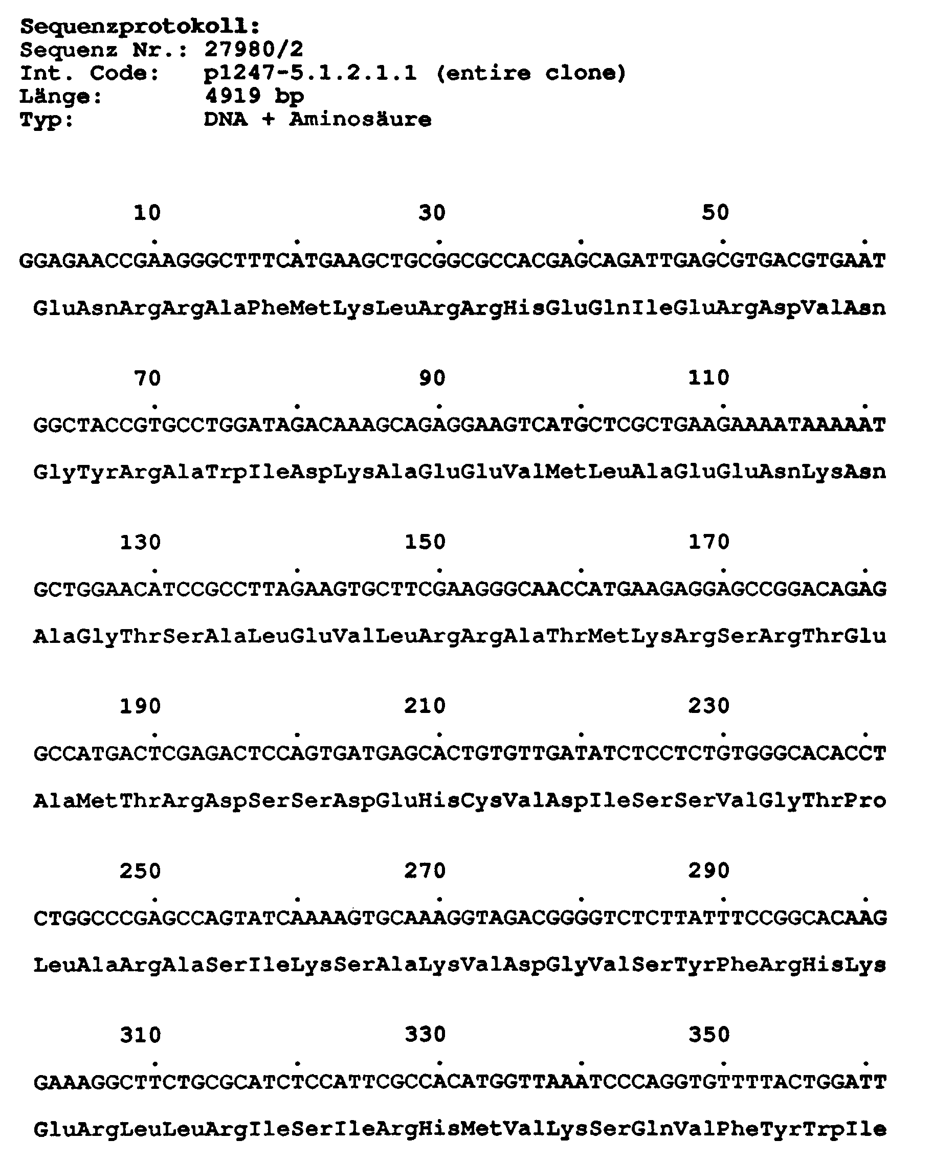
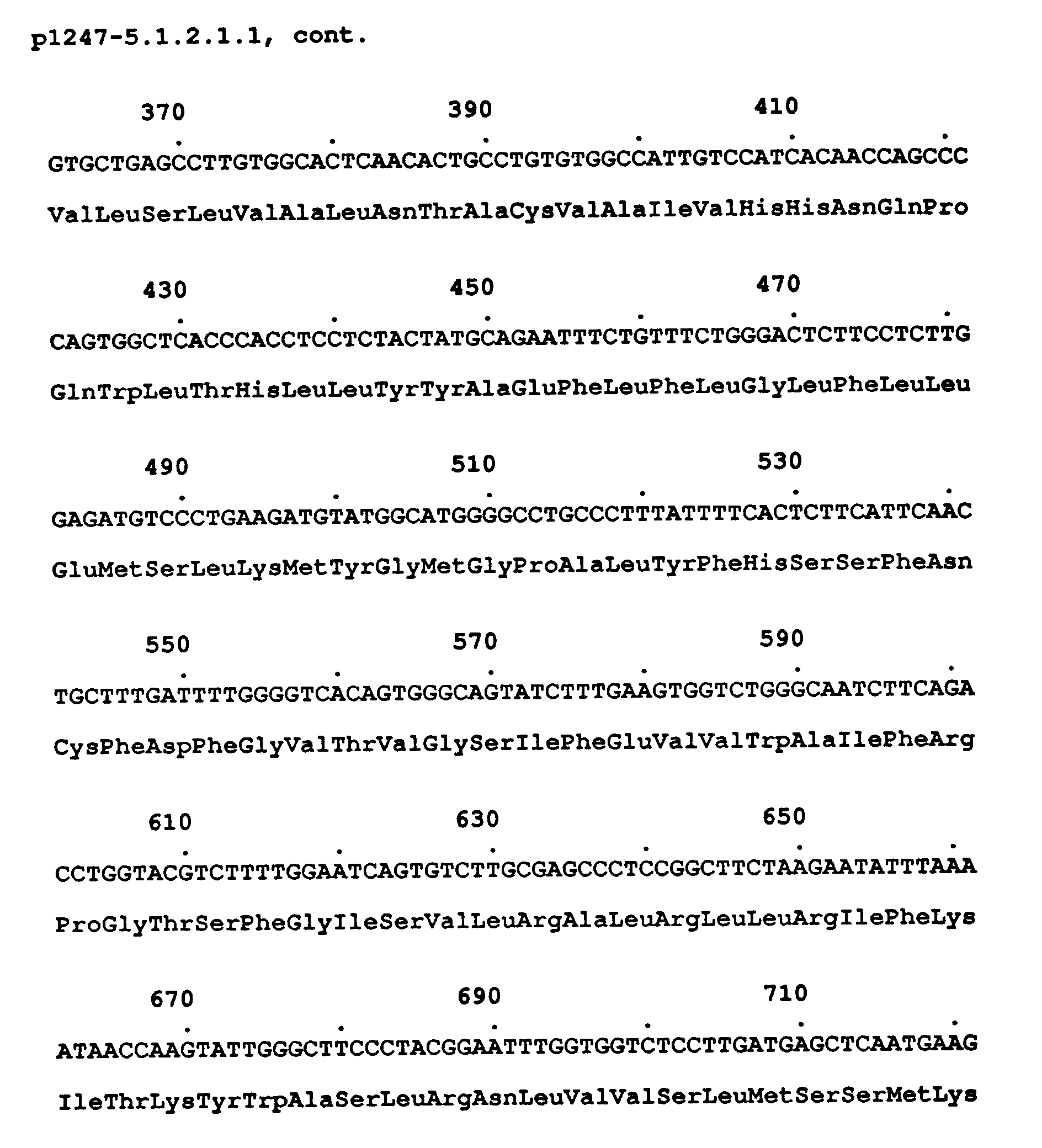
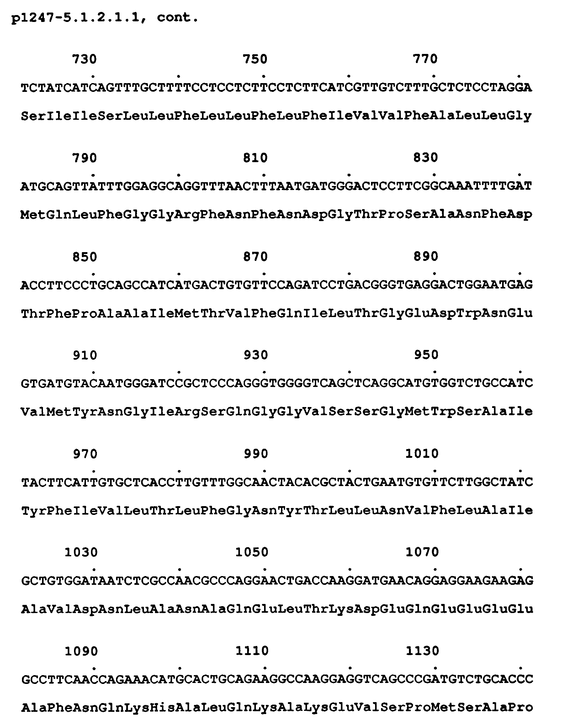
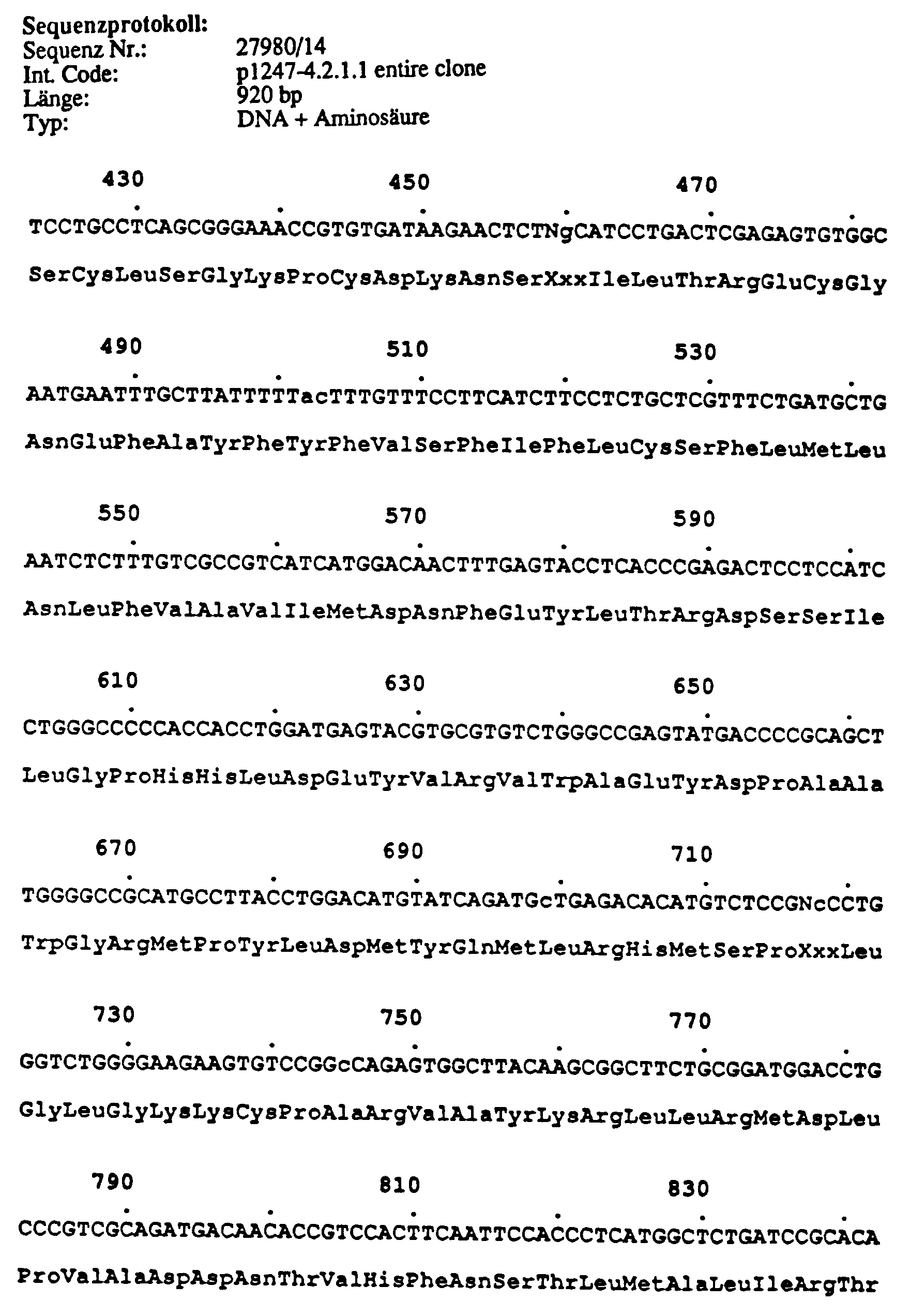
- Length 683 nucleotides, position AS 1,000-1,230 (3 'additional 3 AS); comprises sequences of the third domain from IIIs6 to fourth domain IVs3.
- Length 811 bp Position AS 1.115-1.390 and thus includes the entire domain IV (s1 to s6) and flanking sequences on both sides.
- This clone contains cDNA sequences coding for part of domain III (s6), the entire fourth domain and part of the c-terminal cytoplasmic part of the protein.
- the clone pR9112-10.1.1.1 differs from pR9112-2.1.1.1 and pR9112-4.1.1.1 by a 57 bp insert (1.4541-1.510).
- This insert has splicing consensus sequences at both ends and thus offers the possibility for alternative splicing.
- this clone from IVs3-4 contains the complete coding region up to the actual C-terminus of the protein, as well as about 500 bp of the 3 'untranslated region of the mRNA.
- Length 2,837 bp has an internal EcoRI interface (2 subfragments: 2,197 bp, 640 bp).
- the 5 'end of the cDNA clone is between domain II and III, the 3' end at AS 1.622; the clone comprises the complete domains III and IV and part of the sequence coding for the C-terminus.
- the 5 'end of the cDNA clone overlaps the 3' end of the clone pCA9.3 over a length of 830 bp.
- the 5 'end of the cDNA clone begins immediately after domain I (AS 337); the 3 'end is at AS 922; the cDNA clone comprises sequences of the second cytoplasmic section (between domains I and II) up to and including the 4th transmembrane region of domain III (IIIS4).
- this clone contains a putative splice donor site at position 60, so that the first 60 nucleotides may represent intron sequences.
- the region coding for a Ca channel (from position 61) comprises part of the N-terminal cytoplasmic region, as well as the entire domain I and the first membrane-breaking region (S1) of domain II.
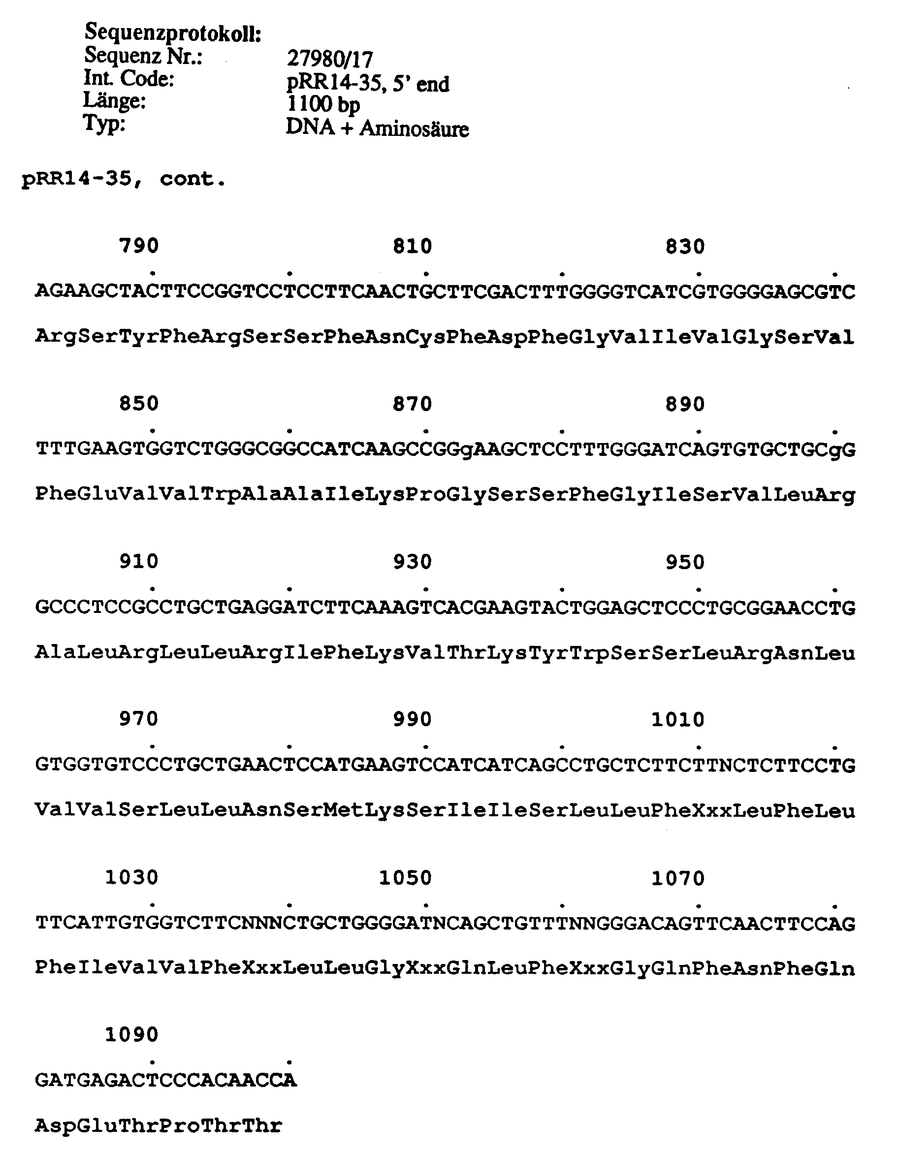
- Clone pRR14-35 supplements the Ca-channel cDNA contained in pR14-5.3.3.1 at the 5 'end by 129 AS and thereby eliminates the artificially existing Alu repeat sequence at the 5' end of this clone. Both clones can be combined using a common Bgl II restriction interface.
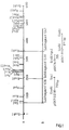
- the upper part (A) of Figure 1 shows the cDNA of the carp skeletal muscle Ca-channel with the interfaces for some restriction endonucleases and the subfragments that were used as screening samples.
- the lower part (B) are DNA fragments of the human clones p1247-5.1.2.1.1 and p1247-14.1.1, which were generated with the restriction enzymes EcoRI / SacI or EcoRI / KpnI, as well as the entire cDNA insert of the clone p1247- 9.1.1.2, which were also used as screening samples.
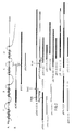
- Figure 2 shows in the upper part (A) a schematic representation of the ⁇ 1 subunit of the skeletal muscle Ca channel from rabbits.
- the 4 domains are marked as boxes and labeled with Roman numerals. Within these domains, the transmembrane regions are highlighted as black blocks.
- the sections of the human neuronal Ca channels isolated to date are arranged according to their size and their homology to the rabbit skeletal muscle Ca channel protein.
- the complete cDNA coding for a calcium channel subtype is composed of overlapping lambda phages by means of suitable restriction sites and cloned into a eukaryotic expression vector.
- This vector is used to transform suitable eukaryotic cells and to produce stable cell lines which express the protein of a calcium channel subtype. These stable cell lines are then used in the receptor binding test described.
Landscapes
- Health & Medical Sciences (AREA)
- Chemical & Material Sciences (AREA)
- Life Sciences & Earth Sciences (AREA)
- Organic Chemistry (AREA)
- Medicinal Chemistry (AREA)
- Molecular Biology (AREA)
- Gastroenterology & Hepatology (AREA)
- Toxicology (AREA)
- Biochemistry (AREA)
- Biophysics (AREA)
- General Health & Medical Sciences (AREA)
- Genetics & Genomics (AREA)
- Immunology (AREA)
- Zoology (AREA)
- Proteomics, Peptides & Aminoacids (AREA)
- Cell Biology (AREA)
- Medicines That Contain Protein Lipid Enzymes And Other Medicines (AREA)
- Measuring Or Testing Involving Enzymes Or Micro-Organisms (AREA)
- Pharmaceuticals Containing Other Organic And Inorganic Compounds (AREA)
- Glass Compositions (AREA)
- Peptides Or Proteins (AREA)
- Micro-Organisms Or Cultivation Processes Thereof (AREA)
- Preparation Of Compounds By Using Micro-Organisms (AREA)
- Medicines Containing Material From Animals Or Micro-Organisms (AREA)
Abstract
Description
Calcium-Ionen haben in jedem biologischen System vielfältige Funktionen. Die zelluläre Calciumhomöostase spielt speziell für die Physiologie von Nervenzellen eine wesentliche Rolle. Die intrazelluläre Calcium-Konzentration beträgt etwa 0,1 µM gegenüber 1 mM außerhalb der Nervenzelle. Die Regulation dieses starken Konzentrationsgefälles (x 10.000) erfolgt primär durch spannungsabhängige Calcium-Kanäle (VOCC), die von bestimmten Calcium-Antagonisten blockier werden können. Während einer cerebralen Ischämie (Hirnschlag) wird die Calciumhomöostase in Neuronen des betroffenen Infarktgebietes erheblich verändert Die spannungsabhängigen Calcium-Kanäle werden durch anhaltende Membrandepolarisationen im geöffneten Zustand gehalten, welches einen massiven Einstrom von Calcium-Ionen zur Folge hat. Die intrazelluläre Calcium-Konzentration steigt dabei um das 1000fache an. Der hohe Überschuß an Calcium aktiviert durch die Bindung an Calmodulin verschiedene Calcium/Calmodulin anhängige zelluläre Enzymsysteme, wie z.B. Kinasen, Proteasen und Phospholipasen, die zusammen, bei anhaltender Aktivierung, zu irrreversiblen Nervenzellschädigungen führen.Calcium ions have a wide range of functions in every biological system. The cellular calcium homeostasis plays an essential role especially for the physiology of nerve cells. The intracellular calcium concentration is about 0.1 µM compared to 1 mM outside the nerve cell. This strong concentration gradient (x 10,000) is primarily regulated by voltage-dependent calcium channels (VOCC), which can be blocked by certain calcium antagonists. During cerebral ischemia (stroke), the calcium homeostasis in neurons of the affected infarct area is significantly changed. The voltage-dependent calcium channels are kept open by continuous membrane depolarizations, which results in a massive influx of calcium ions. The intracellular calcium concentration increases 1000-fold. The high excess of calcium activates various calcium / calmodulin-dependent cellular enzyme systems, such as e.g. Kinases, proteases and phospholipases, which together with continued activation lead to irreversible nerve cell damage.
Ein therapeutischer Ansatz zur Neuroprotektion bei cerebraler Ischämie ist die reversible Blockierung des massiven Calcium-Einstroms in die Nervenzelle. Die spannungsabhängigen neuronalen Calcium-Kanäle sind hierbei ein geeigneter pharmakologischer Angriffspunkt. VOCCs existieren in verschiedenen Muskelzellen (Gefäß-, Herz- und Skelettmuskel), Neuronen und sekretorischen Zellen mit gewebespezifischen physiologischen Eigenschaften.A therapeutic approach to neuroprotection in cerebral ischemia is the reversible blocking of the massive influx of calcium into the nerve cell. The voltage-dependent neuronal calcium channels are a suitable pharmacological target. VOCCs exist in different muscle cells (Vascular, cardiac and skeletal muscle), neurons and secretory cells with tissue-specific physiological properties.
Elektrophysiologische Untersuchungen (Tsien et al., 1988, Trends in Neurol. Sci.11: 431-438) deuten auf mindestens drei verschiedene Typen von VOCCs hin (L-, N-und T-Kanäle). Die 1,4-Dihydropyridine (DHPs) sind potente Blocker der L-Typ Calcium-Kanäle, die sowohl in Muskelzellen als auch in Nervenzellen zu finden sind. Der Kaninchen-Skelettmuskel Dihydropyridin Rezeptor ist biochemisch charakterisiert und kloniert worden (Tanabe et al., 1987, Nature 328:313-318). Die Primärsequenz dieser α1 UE des VOCC konnte von den cDNA Daten abgeleitet werden und ist konsistent mit einem 212 kD Transmembranprotein mit fünf N-Glycosylierungsstellen und sieben möglichen Phosphorylierungsstellen. Das Protein enthält vier untereinander ähnliche Transmembrandomänen mit jeweils sechs - vermutlich α-helikalen - membrangängigen Segmenten (S1-S6). Jeweils das vierte Transmembransegment (S4) jeder Domäne enthält ein geordnetes Muster an positiven Ladungen (Lys, Arg), welche den Spannungssensor des Calcium-Kanals bilden können. Die Struktur dieser klonierten α1 UE ist konsistent mit einer Ionen leitenden, spannungsgesteuerten Einheit des DHP-sensitiven Calcium-Kanals.Electrophysiological studies (Tsien et al., 1988, Trends in Neurol. Sci.11: 431-438) indicate at least three different types of VOCCs (L, N and T channels). The 1,4-dihydropyridines (DHPs) are potent blockers of the L-type calcium channels, which can be found in both muscle cells and nerve cells. The rabbit skeletal muscle dihydropyridine receptor has been characterized and cloned biochemically (Tanabe et al., 1987, Nature 328: 313-318). The primary sequence of this α1 UE of the VOCC could be derived from the cDNA data and is consistent with a 212 kD transmembrane protein with five N-glycosylation sites and seven possible phosphorylation sites. The protein contains four mutually similar transmembrane domains, each with six - presumably α-helical - membrane-permeable segments (S1-S6). The fourth transmembrane segment (S4) of each domain contains an ordered pattern of positive charges (Lys, Arg), which can form the voltage sensor of the calcium channel. The structure of this cloned α1 UE is consistent with an ion-conducting, voltage-controlled unit of the DHP-sensitive calcium channel.
Der klonierte Skelettmuskel DHP-R cDNA Klon des Karpfens (Grabner et al., 1991, Proc. Natl. Acad. Sci. (USA), 88:727-731) wurde als Hybridisierungsprobe eingesetzt, um verwandte Gene des humanen Calcium-Kanals aus Neuronen zu isolieren und zu charakterisieren. Mit dieser Klonierungsstrategie konnte eine Anzahl von verschiedenen homologen cDNA Klonen aus neuronalen cDNA Banken des Menschen isoliert und charakterisiert werden, die deutlich anzeigen, daß verschiedene Calcium-Kanal Subtypen im ZNS des Menschen existieren. Neuronale Subtypen besitzen neue Rezeptorstellen, für die bislang keine Liganden (Agonisten, Antagonisten) bekannt sind. Die klonierten Calcium-Kanal Subtypen sollen in transformierten Tierzellen (z.B. cos Zellen, Maus L Zellen, CHO Zellen etc.) exprimiert werden (Gluzman, 1981, Cell 23:175, und Chen, et al., 1987, Mol. Cell. Biol. 7:2745-2752) und in Bindungstests und/oder funktionellen Testsystemen zur Auffindung (Screening) neuer, Subtyp-spezifischer Liganden eingesetzt werden. Hierbei werden komplette oder partielle cDNA Gene von verschiedenen Calcium-Kanal Subtypen (inkl. Herz-, Blutgefäß- und Skelettmuskel-Kanäle) in geeignete eukaryontische Expressionsvektoren (Sambrook et al., 1989, in: Molecular Cloning, A laboratory manual, ed. Chris Nolan, Cold Spring Harbor Laboratory Press, New York, N.Y.) kloniert. Die Proteinexpression wird dabei entweder durch homologe Regulatorelemente (Promotoren und Enhancer) oder heterologe Promotoren (virale, z.B. SV40, BPV, CMV, etc. oder induzierbare, z.B. Metallothionein, cAMP, Calcium, Temperatur, etc.) in Kombination mit bekannten Enhancern und RNA Prozessierungssignalen (z.B. Capping, Poly A) gesteuert.The cloned skeletal muscle DHP-R cDNA clone of the carp (Grabner et al., 1991, Proc. Natl. Acad. Sci. (USA), 88: 727-731) was used as a hybridization sample to identify related genes from the human calcium channel Isolate and characterize neurons. With this cloning strategy, a number of different homologous cDNA clones from human neural cDNA banks could be isolated and characterized, which clearly indicate that different calcium channel subtypes exist in the human CNS. Neural subtypes have new receptor sites for which no ligands (agonists, antagonists) are known to date. The cloned calcium channel subtypes are said to be expressed in transformed animal cells (eg cos cells, mouse L cells, CHO cells etc.) (Gluzman, 1981, Cell 23: 175, and Chen, et al., 1987, Mol. Cell. Biol . 7: 2745-2752) and in binding tests and / or functional test systems for the discovery (screening) of new, subtype-specific ligands. Here, complete or partial cDNA genes from different calcium channels Subtypes (including cardiac, blood vessel and skeletal muscle channels) into suitable eukaryotic expression vectors (Sambrook et al., 1989, in: Molecular Cloning, A laboratory manual, ed. Chris Nolan, Cold Spring Harbor Laboratory Press, New York, NY ) cloned. The protein expression is either by homologous regulatory elements (promoters and enhancers) or heterologous promoters (viral, e.g. SV40, BPV, CMV, etc. or inducible, e.g. metallothionein, cAMP, calcium, temperature, etc.) in combination with known enhancers and RNA Processing signals (eg capping, poly A) controlled.
Weiterhin sollen mit diesen rekombinanten Zellsystemen funktionale Calcium-Flux Assays entwickelt werden, mit deren Hilfe spezifische Liganden auf ihre agonistische bzw. antagonistische Wirkung überprüft werden können. Der Unterschied und Hauptvorteil dieser rekombinanten Assays, im Vergleich zu herkömmlichen Assays (Hirnmembranpräparationen, Zellinien), liegt in der Reinheit des Rezeptor/Kanal Präparates, da ausschließlich der rekombinant exprimierte neuronale Calcium-Kanal Subtyp in beliebiger Anzahl auf einer Tierzell-Oberfläche präsent ist. Dies ist eine essentielle Voraussetzung für die Selektion spezifischer neuronaler Calcium-Kanal Liganden, die möglichst keine Wirkung auf Calcium-Kanäle nicht-neuronaler Gewebetypen zeigen sollen.Functional calcium flux assays are also to be developed with these recombinant cell systems, with the help of which specific ligands can be checked for their agonistic or antagonistic effect. The difference and main advantage of these recombinant assays compared to conventional assays (brain membrane preparations, cell lines) lies in the purity of the receptor / channel preparation, since only the recombinantly expressed neuronal calcium channel subtype is present in any number on an animal cell surface. This is an essential prerequisite for the selection of specific neuronal calcium channel ligands which should have no effect on calcium channels of non-neuronal tissue types if possible.
Nachfolgend sind einige Beispiele für die Anwendung der oben beschriebenen rekombinanten Screeningassays aufgeführt.Below are some examples of the use of the recombinant screening assays described above.
Die mit humanen Calcium-Kanal Subtypen transformierten Tierzellen (Beispiel: siehe oben) können kultiviert und zur Präparation von Membranen eingesetzt werden. Diese Membranpräparationen können für Bindungsstudien mit verschiedenen radioaktiv markierten Substanzklassen (Beisp. 1-5) zum Screening neuer Liganden (Kompetitionstest) eingesetzt werden. Beispiele für bekannte Calcium-Kanal bindende Substanzen sind:
- 1. Phenylalkylamine,
- 2. Benzothiazepine,
- 3. Dihydropyridine,
- 4. Bisphenylbutylpiperidine,
- 5. Omega Conotoxine.
- 1. phenylalkylamines,
- 2. benzothiazepines,
- 3. dihydropyridines,
- 4. bisphenylbutylpiperidine,
- 5. Omega Conotoxins.
Die Zellmembranen von humanen Calcium-Kanal Subtypen transformierten Kulturzellen (s.o.) können mit Kalium-Ionen oder mit Alkaloiden wie z.B. Veratridine depolarisiert werden. Membrandepolarisation führt zur Öffnung von Calcium-Kanälen, was einen Einstrom (Flux) von Calcium-Ionen in die Zellen zur Folge hat. Dieser spannunngsabhängige Calcium-Einstrom kann mit radioaktiv markiertem Calcium (45Ca) gemessen werden (Beisp.: Messing et al., 1985, J. Pharmacology and Exp. Therapeutics 235:407-411) und zum funktionellen Testen/Screening von Calcium-Kanal Antagonisten oder Agonisten eingesetzt werden.The cell membranes of human calcium channel subtype transformed culture cells (see above) can be treated with potassium ions or with alkaloids such as e.g. Veratridine can be depolarized. Membrane depolarization leads to the opening of calcium channels, which results in an influx (flux) of calcium ions into the cells. This voltage-dependent calcium influx can be measured with radioactively labeled calcium (45Ca) (example: Messing et al., 1985, J. Pharmacology and Exp. Therapeutics 235: 407-411) and for the functional testing / screening of calcium channel antagonists or agonists are used.
Humane Calcium-Kanal exprimierende Tierzellen (s.o.) können in Gegenwart von Calcium sensitiven, fluoreszierenden Farbstoffen (z.B. Fura-2 oder Fluoro-3) für Messungen der intrazellulären Calcium-Konzentration nach Öffnung und Blockierung der Calcium-Kanäle eingesetzt werden (Beisp.: Rosario et al.1989 , Neurosci. 29,735-747). Die Änderung der intrazellulären Calcium-Konzentration kann dabei fluorimetrisch (spektrophotometrisch) gemessen werden. Dieses rekombinante Zellsystem kann als funktioneller Test für das Auffinden Subtyp-spezifischer Calcium-Kanal Liganden(Agonisten und Antagonisten) eingesetzt werden.Animal cells expressing human calcium channel (see above) can be used in the presence of calcium-sensitive, fluorescent dyes (eg Fura-2 or Fluoro-3) for measurements of the intracellular calcium concentration after opening and blocking of the calcium channels (example: Rosario et al. 1989, Neurosci. 29,735-747). The change in the intracellular calcium concentration can be measured fluorimetrically (spectrophotometric). This recombinant cell system can be used as a functional test for finding subtype-specific calcium channel ligands (agonists and antagonists).
Die durch Membrandepolarisation erzeugten Calcium-Ströme können elektrophysiologisch gemessen werden (Beisp.: Carbone et al. 1990, Pflügers Arch., 416: 170-179). Die Wirkung von potentiellen Calcium-Kanal Antagonisten oder Agonisten kann direkt an humanen Calcium-Kanälen mit Hilfe der rekombinanten Tierzellinien (s.o.) physikalisch gemessen und pharmakologisch charakterisiert werden.The calcium currents generated by membrane depolarization can be measured electrophysiologically (example: Carbone et al. 1990, Pflügers Arch., 416: 170-179). The effect of potential calcium channel antagonists or agonists can be physically measured and pharmacologically characterized directly on human calcium channels using the recombinant animal cell lines (see above).
Viele zelluläre Prozesse werden von der intrazellulären Calcium-Ionen Konzentration reguliert (z.B. Rezeptor mediierte Signalübertragung, verschiedene Enzymreaktionen, wie z.B. Phosphorylierungen, Dephosphorylierungen, Neurotransmitter-Freisetzung, Ca-abhängige Genregulation, etc.). Einige dieser biochemischen Reaktionen sind mit einem spezifischen Assay meßbar. In einem rekombinanten Calcium-Kanal exprimierenden Zellsystem kann somit indirekt (physiologisch) die Wirkung von Calcium-Kanal Modulatoren auf Calcium abhängige Zellvorgänge erfaßt werden(Beisp.: Zernig et al. 1986, Eur.J.Pharmacol. 128,221-229).Many cellular processes are regulated by the intracellular calcium ion concentration (e.g. receptor mediated signal transmission, various enzyme reactions such as phosphorylation, dephosphorylation, neurotransmitter release, Ca-dependent gene regulation, etc.). Some of these biochemical reactions can be measured with a specific assay. In a recombinant calcium channel-expressing cell system, the effect of calcium channel modulators on calcium-dependent cell processes can thus be indirectly (physiologically) recorded (example: Zernig et al. 1986, Eur.J. Pharmacol. 128, 221-229).
Zusätzlich können durch gezielte Mutagenesen eingeführte Veränderungen, wie z.B. Punktmutationen, Insertionen, Deletionen, Austausch von DNA-Segmenten verschiedener Calciumkanal-Subtypen, direkte Auswirkungen auf physiologische Vorgänge erfaßt werden (Bsp.: Yool and Schwarz, 1991, Nature 349:700-704).In addition, changes introduced by targeted mutagenesis, such as point mutations, insertions, deletions, exchange of DNA segments of different calcium channel subtypes, direct effects on physiological processes can be recorded (e.g. Yool and Schwarz, 1991, Nature 349: 700-704) .
Zur Isolierung humaner neuronaler Ca-Kanäle mittels Homologiescreening wurden folgende käuflich erworbenen cDNA-Banken eingesetzt:
- a) cDNA Bank aus einer humanen Neuroblastomazellinie
- Vektor:
- Lambda gt10
- Quelle:
- Fa. Clontech Laboratories, Inc.Palo Alto,CA.USA: (Kat.Nr.HL 1007a)
- b) cDNA Bank aus humanem Hippocampus;
- Vektor:
- Lambda ZAPII
- Quelle:
- Fa. Stratagene Inc., La Jolla,CA, USA (Kat. Nr. 936205)
- c) cDNA Bank aus humanem temporalem Cortex
- Vektor:
- Lambda ZAPII
- Quelle:
- Fa. Stratagene Inc., La Jolla,CA,USA (Kat. Nr. 935205)
- d) cDNA Bank aus humanem visuellem Cortex
- Vektor:
- Lambda gt10
- Quelle:
- Fa. Clontech Laboratories,Palo Alto, CA,USA (Kat.Nr.HL 1081a)
- e) cDNA Bank aus humanem frontalem Cortex
- Vektor:
- Lambda ZAPII
- Quelle:
- Fa. Stratagene Inc., La Jolla,CA, USA (Kat. Nr. 935205)
- a) cDNA bank from a human neuroblastoma cell line
- Vector:
- Lambda GT10
- Source:
- Clontech Laboratories, Inc. Palo Alto, CA.USA: (Cat. No. HL 1007a)
- b) cDNA bank from human hippocampus;
- Vector:
- Lambda ZAPII
- Source:
- Stratagene Inc., La Jolla, CA, USA (Cat. No. 936205)
- c) cDNA bank from human temporal cortex
- Vector:
- Lambda ZAPII
- Source:
- Stratagene Inc., La Jolla, CA, USA (Cat. No. 935205)
- d) cDNA bank from human visual cortex
- Vector:
- Lambda GT10
- Source:
- Clontech Laboratories, Palo Alto, CA, USA (cat.no.HL 1081a)
- e) cDNA bank from human frontal cortex
- Vector:
- Lambda ZAPII
- Source:
- Stratagene Inc., La Jolla, CA, USA (Cat. No. 935205)
Das Ausplattieren der cDNA Banken und das Prozessieren der Nitrozellulosefilter erfolgte gemäß den Angaben der Hersteller oder nach Sambrook et al., 1989, (Molecular Cloning, A laboratory manual, ed. Chris Nolan, Cold Spring Harbor Laboratory Press,New York,N.Y.).The cDNA banks were plated out and the nitrocellulose filters were processed in accordance with the manufacturer's instructions or according to Sambrook et al., 1989, (Molecular Cloning, A laboratory manual, ed. Chris Nolan, Cold Spring Harbor Laboratory Press, New York, N.Y.).
Als Hybridisierungsprobe diente ein 6.1 kb langer cDNA Klon (Abb. 1), der die gesamte kodierende Region, einschließlich 5'- und 3'- nicht translatierter Bereiche der α₁-Untereinheit (UE) des Ca-Kanals der Skelettmuskulatur des Karpfens (Cyprinus carpio) enthält (Grabner et al., 1990, Proc. Natl. Acad. Sci. (USA), 88: 727-731). Für das Homologiescreening wurden folgende Abschnitte dieses Klons eingesetzt (Abb.1):
- der komplette cDNA Klon (6.1 kb)
- Subfragment 1 336
- Subfragment 1 509
- Subfragment 1 247.
- the complete cDNA clone (6.1 kb)
- Subfragment 1 336
- Subfragment 1 509
- Subfragment 1 247.
Zum weiteren Screenen von cDNA Banken wurden folgende Fragmente humaner Ca-Kanal cDNA Klone eingesetzt:
- Insert des Klons p 1247-9.1.1.2(811 bp)
- Subfragment des Klons p 1247-14.1.1.1 (EcoRI-KpnI; 205 bp)
- Subfragment des Klons p 1247-5.1.2.1.1 (EcoRI-SacI; 710bp)
- Insert des Klons pCA 33 (684 bp)
- Subfragmente des Klons pCA 3 (EcoRI-EcoRI, 640pb; PstI-PstI, 198 bp; PstI-PstI,600bp)
- Insert of clone p 1247-9.1.1.2 (811 bp)
- Subfragment of clone p 1247-14.1.1.1 (EcoRI-KpnI; 205 bp)
- Subfragment of clone p 1247-5.1.2.1.1 (EcoRI-SacI; 710bp)
- Insert of the clone pCA 33 (684 bp)
- Subfragments of clone pCA 3 (EcoRI-EcoRI, 640pb; PstI-PstI, 198 bp; PstI-PstI, 600bp)
Zur Markierung von doppelsträngigen DNA Fragmenten wurden Standardprotokolle (Sambrook et al., 1989 in: Molecular Cloning, A laboratory manual, ed. Chris Nolan, Cold Spring Harbor Laboratory Press, New York, N.Y.) in Verbindung mit einem käuflichen "Random Primed Labeling Kit" (Boehringer Mannheim GmbH, Postfach 310120,D-6800 Mannheim; Ka. Nr. 1004 760) angewendet.Standard protocols (Sambrook et al., 1989 in: Molecular Cloning, A laboratory manual, ed. Chris Nolan, Cold Spring Harbor Laboratory Press, New York, NY) in connection with a commercially available "Random Primed Labeling Kit" were used to label double-stranded DNA fragments "(Boehringer Mannheim GmbH, Postfach 310120, D-6800 Mannheim; Ka. No. 1004 760) applied.
Die Nitrozellulosefilter wurden über Nacht mit radioaktiv markierten cDNA Fragmenten in 30 % Formamid, 5 x Denhardt's Lösung, 5 x SSC bei 42°C hybridisiert und anschließend wie folgt gewaschen:
- 2 x 20 Minuten in 2 x SSC, 0,05 % SDS bei Raumtemperatur
- 2 x 20 Minuten in 0,2 x SSC, 0,2 % SDS bei 45°C und
- 1 x 20 Minuten in 0,2 x SSC bei Raumtemperatur.
- 2 x 20 minutes in 2 x SSC, 0.05% SDS at room temperature
- 2 x 20 minutes in 0.2 x SSC, 0.2% SDS at 45 ° C and
- 1 x 20 minutes in 0.2 x SSC at room temperature.
Danach wurde ein Kodak X-OMAT AR Röntgenfilm für unterschiedliche Zeiten bei -80°C mit Verstärkerfolien durch die Filter belichtet.A Kodak X-OMAT AR X-ray film was then exposed through the filters for several times at -80 ° C with intensifying screens.
Die Lambda Phagen DNA wurde isoliert, mit EcoRI geschnitten, die cDNA Inserts in ein pUC-Derivat (pT7T3 18U; Fa. Pharmacia) subkloniert und anschließend mit Sequenase (Fa. USB, Cleveland, Ohio, USA) mittels der Dideoxy-Terminationsmethode nach Sanger (Sanger et al., 1977, Proc. Natl. Acad. Sci. USA, 74: 5463-5467.) die Nukleotidsequenz bestimmt.The lambda phage DNA was isolated, cut with EcoRI, the cDNA inserts subcloned into a pUC derivative (pT7T3 18U; Pharmacia) and then with Sequenase (USB, Cleveland, Ohio, USA) using the Sanger dideoxy termination method (Sanger et al., 1977, Proc. Natl. Acad. Sci. USA, 74 : 5463-5467.) Determined the nucleotide sequence.
Die cDNA Inserts aus positiven Lambda-ZAPII Phagen wurden nach einem Protokoll des Herstellers (Stratagene) mittels eines fl-abgeleiteten Helferphagens vorgenommen und in die Plasmidform überführt.The cDNA inserts from positive Lambda-ZAPII phages were carried out according to a manufacturer's protocol (Stratagene) using an fl-derived helper phage and converted into the plasmid form.
Aus XL1-Blue Zellen, die ein rekombinates pBluescript Plasmid trugen, wurde Plasmid DNA präpariert (Sambrook, J., et al., (1989) in: Molecular cloning, A laboratory manual, ed. Chris Nolan, Cold Spring Harbor Laboratory Press, New York, N.Y.) und jeweils 0,5 µg dieser DNA mit dem Restriktionsenzym EcoRI behandelt. Aus Anzahl und Größe der entstandenen DNA Fragmente konnte die Gesamtlänge der insertierten cDNA abgeleitet werden. Die Nukleotidsequenz der vorhandenen cDNA wurde mit SEQUENASE (USB, Cleveland, Ohio, USA) nach der Methode von Sanger an doppelsträngiger DNA ermittelt.Plasmid DNA was prepared from XL1-Blue cells which carried a recombinant pBluescript plasmid (Sambrook, J., et al., (1989) in: Molecular cloning, A laboratory manual, ed. Chris Nolan, Cold Spring Harbor Laboratory Press, New York, NY) and 0.5 µg of this DNA each were treated with the restriction enzyme EcoRI. The total length of the inserted cDNA could be derived from the number and size of the resulting DNA fragments. The nucleotide sequence of the cDNA present was determined using SEQUENASE (USB, Cleveland, Ohio, USA) using the Sanger method on double-stranded DNA.
Folgende cDNA Klone wurden bisher isoliert und konnten durch DNA- und Aminosäuresequenzvergleiche mit anderen bekannten Ca-Kanalsequenzen als Ca-Kanäle identifiziert werden (als Referenz dient die Nukleotidsequenz des Ca-Kanals der Skelettmuskulatur des Kaninchens (Tanabe et al., Nature 328, 313-318), die Numerierung der Nukleotide bzw. Aminosäuren ist analog zur Numerierung der EMBL-Datenbank):The following cDNA clones have been isolated so far and could be identified by comparison of DNA and amino acid sequences with other known Ca channel sequences as Ca channels (the nucleotide sequence of the Ca channel of the skeletal muscles of the rabbit serves as reference (Tanabe et al., Nature 328, 313- 318), the numbering of the nucleotides or amino acids is analogous to the numbering of the EMBL database):
In ihren überlappenden Sequenzen übereinstimmende cDNA-Subklone werden über geeignete Restriktionsschnittstellen zu einem kompletten oder partiellen cDNA-Klon zusammengesetzt, der für einen bestimmten Calciumkanal-Subtyp kodiert. Dieses cDNA-Gen wird mittels eines eukaryontischen Expressionsvektor in Sängerzellen exprimiert und in den unter den Beispielen Punkt 1 bis 5 beschriebenen Test eingesetzt.Matching cDNA subclones in their overlapping sequences are assembled via suitable restriction sites to form a complete or partial cDNA clone which codes for a specific calcium channel subtype. This cDNA gene is expressed by means of a eukaryotic expression vector in singer cells and used in the test described in Examples 1 to 5.
Länge: 683 Nukleotide, Position AS 1.000-1.230 (3' zusätzliche 3 AS); umfaßt Sequenzen der dritten Domäne ab IIIs6 bis vierte Domäne IVs3.Length: 683 nucleotides, position AS 1,000-1,230 (3 'additional 3 AS); comprises sequences of the third domain from IIIs6 to fourth domain IVs3.
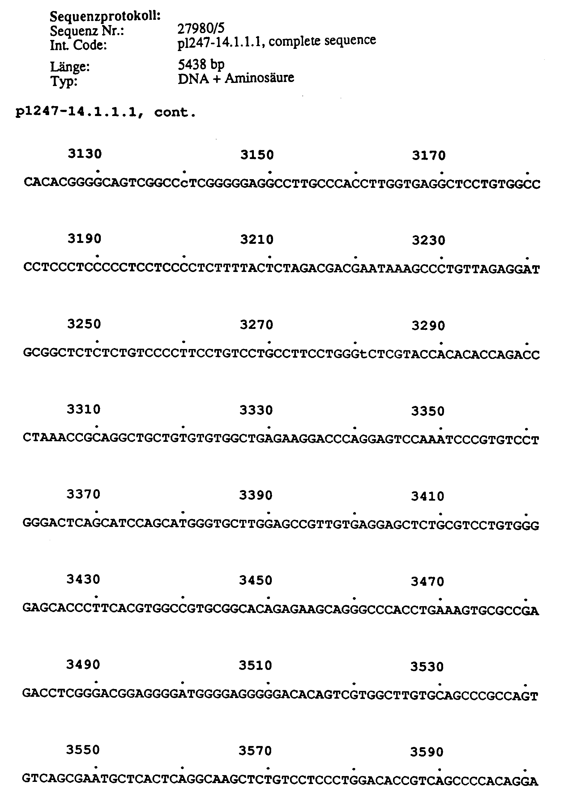
Länge von ca 4.919 bp; Position ab AS 343 bis zum Ende der kodierenden Region; enthält damit nach Domäne I das komplette Gen.Length of approximately 4,919 bp; Position from AS 343 to the end of the coding region; contains the complete gene after domain I.
Länge 811 bp; Position AS 1.115-1.390 und umfaßt damit die gesamte Domäne IV (s1 bis s6)und flankierende Sequenzen auf beiden Seiten.
Länge 1.354 bp; Position AS 1.050-1.512 und umfaßt damit das Ende der dritten Domäne (IIIs6) und die gesamte Domäne IV und etwa 130 c-terminal flankierende AS, die dem letzten zytoplasmatischen Teil des Proteins zuzuordnen sind.Length 1,354 bp; Position AS 1.050-1.512 and thus includes the end of the third domain (IIIs6) and the entire domain IV and about 130 c-terminally flanking AS, which are to be assigned to the last cytoplasmic part of the protein.
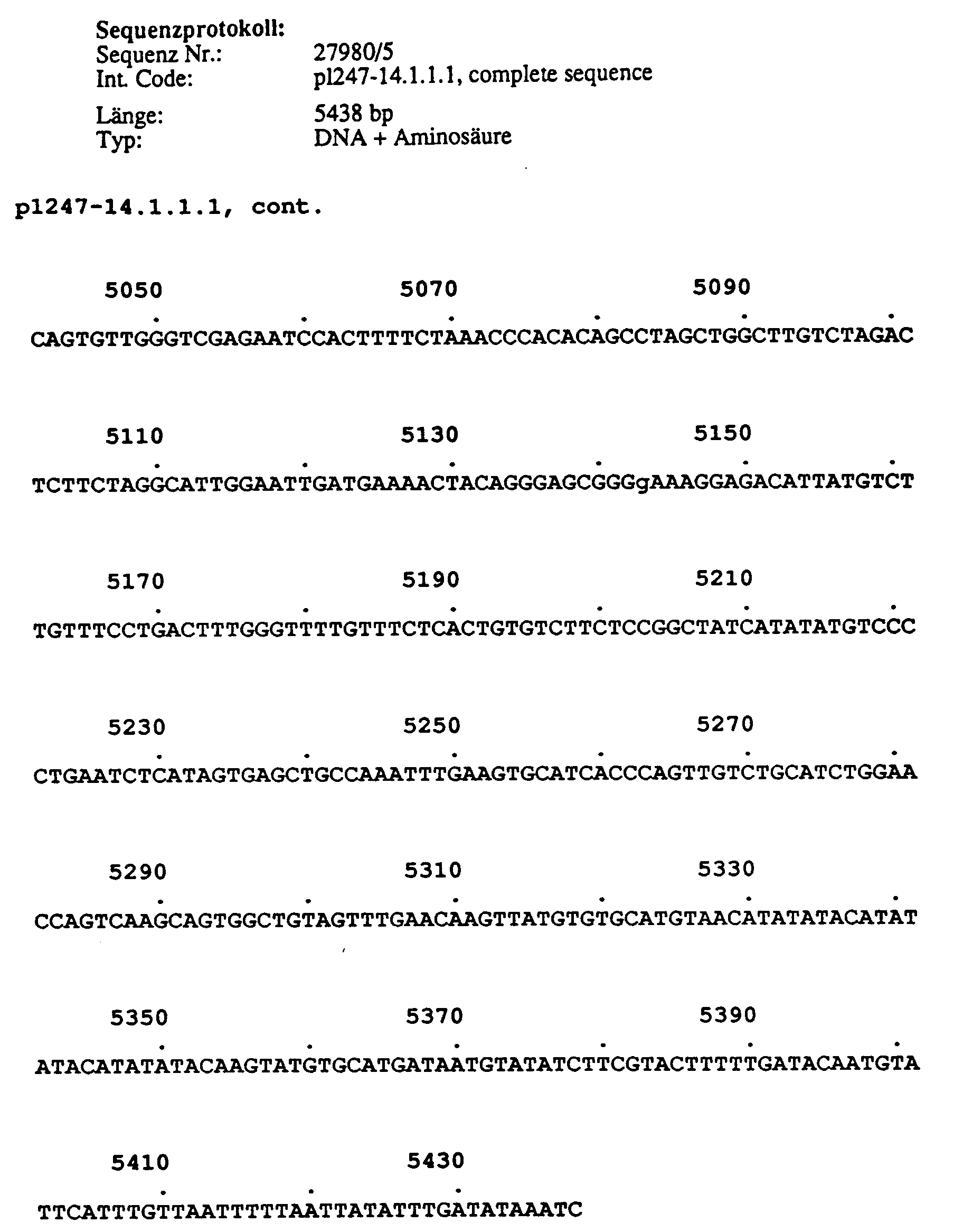
Länge: 5.438 bp; Position AS 967-1.327. Dieser Klon überlappt über einen großen Bereich (Position 1-3.238) mit Klon pR14-5.3.3.1. (Position 2.988-4.232). Im überlappenden Teil sind beide Klone nahezu identisch. Es bestehen folgende Unterschiede zu pR14-5.3.3.1 (Nukleotid und Position bei pR14-5.3.3.1 sind jeweils in Klammern angegeben):
- (1) Cytosin an Position 520 (T; 3.507); keine Änderung der abgeleiteten Proteinsequenz.
- (2) Cytosin an Position 775 (G; 3.768); keine Änderung der abgeleiteten Proteinsequenz.
- (3) Cytosin an Position 1.617 (T; 4.611).
- (4) Adenosin an Position 2.360 (G; 5.353).
- (5) Deletion von sechs Nukleotiden an Position 708 (CGGAAA;3.695-3.700).
- (6) Deletion eines Adenosinrestes an Position 1.013; dies führt im Vergleich zu pR14-5.3.3.1 zu einer Verschiebung im Leseraster, so daß an Position 1.028-1.030 ein Stop-Codon das abgeleitete Protein terminiert.
- (7) Ab Position 3.240 sind weitere 2.199 Nukleotide der 3'-nichttranslatierten Region vorhanden, die bei pR14-5.3.3.1 fehlen. Es folgt dann ein Teil eines Polyadenylat-Schwanzes.
- (1) cytosine at position 520 (T; 3,507); no change in the derived protein sequence.
- (2) cytosine at position 775 (G; 3,768); no change in the derived protein sequence.
- (3) Cytosine at position 1.617 (T; 4.611).
- (4) Adenosine at position 2,360 (G; 5,353).
- (5) Deletion of six nucleotides at position 708 (CGGAAA; 3,695-3,700).
- (6) deletion of an adenosine residue at position 1,013; this leads to a shift in the reading frame in comparison to pR14-5.3.3.1, so that a stop codon terminates the derived protein at position 1.028-1.030.
- (7) From position 3.240 there are a further 2,199 nucleotides of the 3'-untranslated region that are missing from pR14-5.3.3.1. Part of a polyadenylate tail then follows.
Aufgrund der hohen Ähnlichkeit der Klone p1247-14.1.1.1 und pR14-5.3.3.1 ist davon auszugehen, daß es sich bei der Deletion von einem Nukleotid an Position 1.013 um einen Artefakt während der cDNA Synthese handelt.Due to the high similarity of the clones p1247-14.1.1.1 and pR14-5.3.3.1 it can be assumed that the deletion of a nucleotide at position 1.013 is an artifact during the cDNA synthesis.
Länge ca. 1.722 bp; Position AS 1.223-1.870; dieser Klon enthält somit ab s4 den c-terminalen Teil der vierten Domäne, einschließlich der kompletten kodierenden Sequenz bis zum tatsächlichen C-Terminus des Proteins. Dieser Klon ist fast identisch mit überlappenden Sequenzen des Klons 1247-9.1.1.2. Die Sequenzen von pR9112-4.1.1.1 und pR9112-2.1.1.1 sind im wesentlichen identisch (1 bp Unterschied von bisher 1.464 sequenzierten Nukleotiden des Überlapps); die Klone sind wahrscheinlich überlappende cDNA Klone der selben mRNA.Length approx. 1,722 bp; Position AS 1.223-1.870; from s4 this clone thus contains the c-terminal part of the fourth domain, including the complete coding sequence up to the actual C-terminus of the protein. This clone is almost identical to overlapping sequences from clone 1247-9.1.1.2. The sequences of pR9112-4.1.1.1 and pR9112-2.1.1.1 are essentially identical (1 bp difference from 1,464 sequenced nucleotides of the overlap); the clones are likely to be overlapping cDNA clones of the same mRNA.
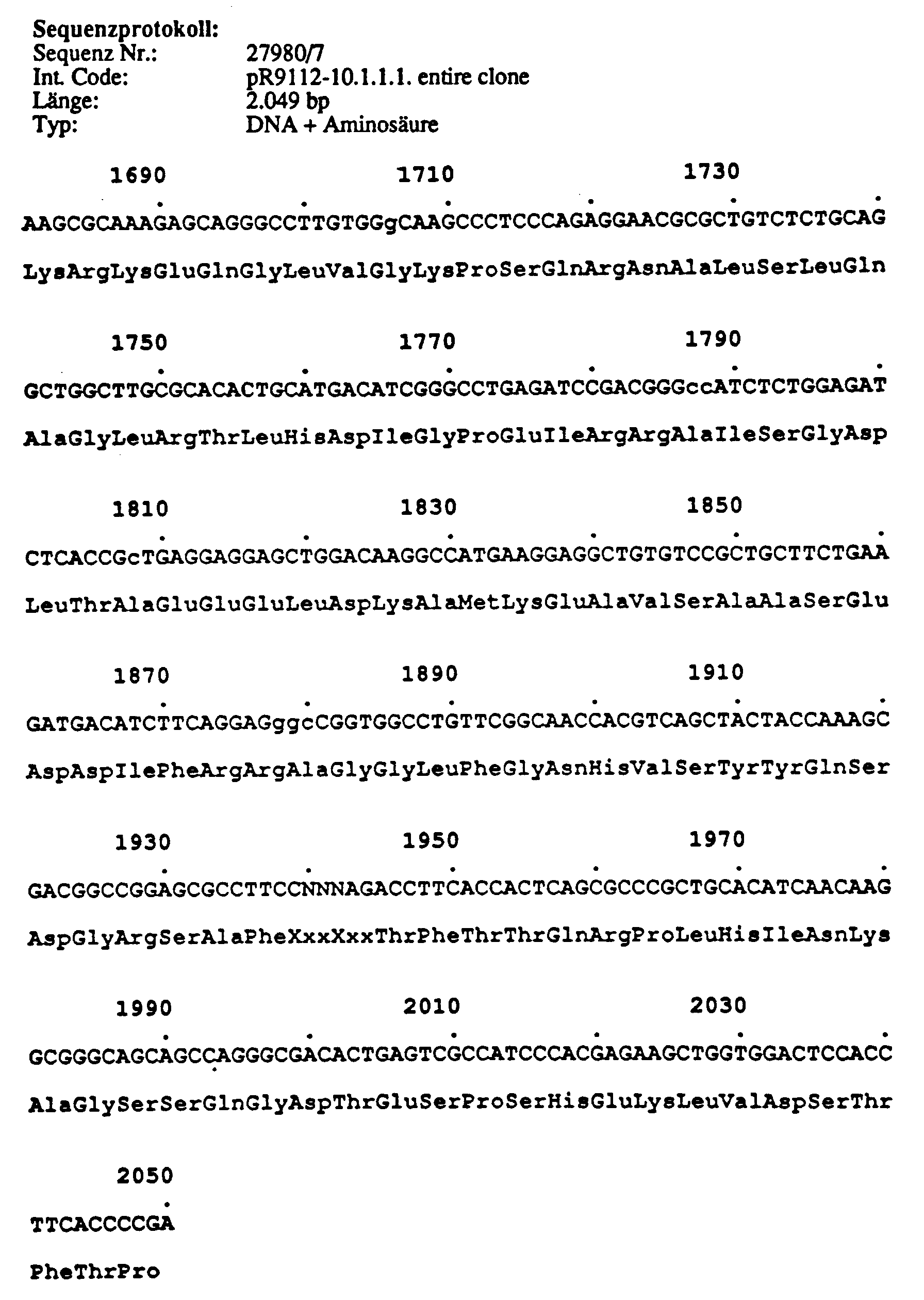
Länge 2.049 bp; Position von AS 991-1.650. Dieser Klon enthält cDNA Sequenzen, die für einen Teil der Domäne III (s6), die gesamte vierte Domäne und einen Teil des c-terminalen zytoplasmatischen Teils des Proteins kodieren. Der Klon pR9112-10.1.1.1 unterscheidet sich von pR9112-2.1.1.1 und pR9112-4.1.1.1 durch einen 57 bp Insert (1.4541-1.510). Dieses Insert besitzt an beiden Enden Splicing-Konsensussequenzen und bietet damit die Mögiichkeit für alternatives Splicing.Length 2,049 bp; Position of AS 991-1.650. This clone contains cDNA sequences coding for part of domain III (s6), the entire fourth domain and part of the c-terminal cytoplasmic part of the protein. The clone pR9112-10.1.1.1 differs from pR9112-2.1.1.1 and pR9112-4.1.1.1 by a 57 bp insert (1.4541-1.510). This insert has splicing consensus sequences at both ends and thus offers the possibility for alternative splicing.
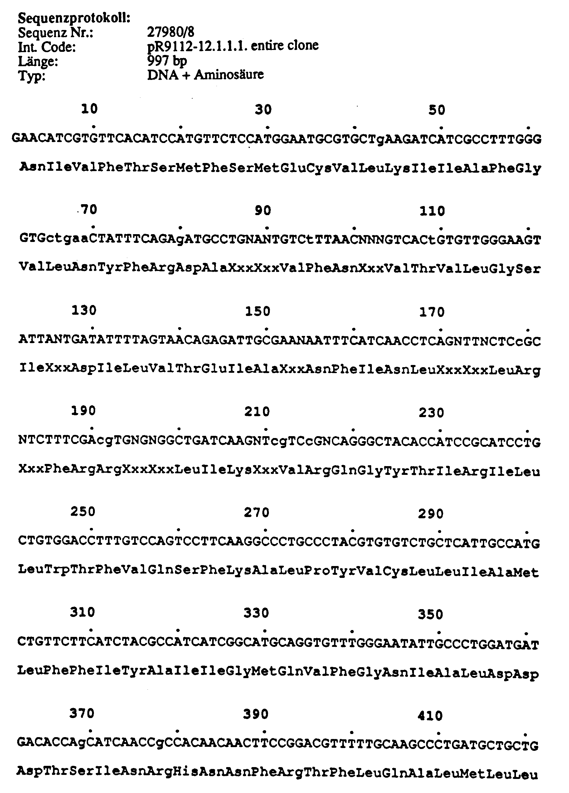
Länge 997 bp; Position bis AS 1.509. Die Seqzenz dieses cDNA-Klons ist fast identisch mit Klon p1247-14.1.1.1. Das 5' Ende des Klons enthält dazu als Kloningsartefakt etwa 250 bp mitochondrialer DNA, sowie ein poly(A)-Anteil von 39 bp.Length 997 bp; Position up to AS 1.509. The sequence of this cDNA clone is almost identical to clone p1247-14.1.1.1. The 5 'end of the clone contains about 250 bp mitochondrial DNA as a cloning artifact, as well as a poly (A) portion of 39 bp.
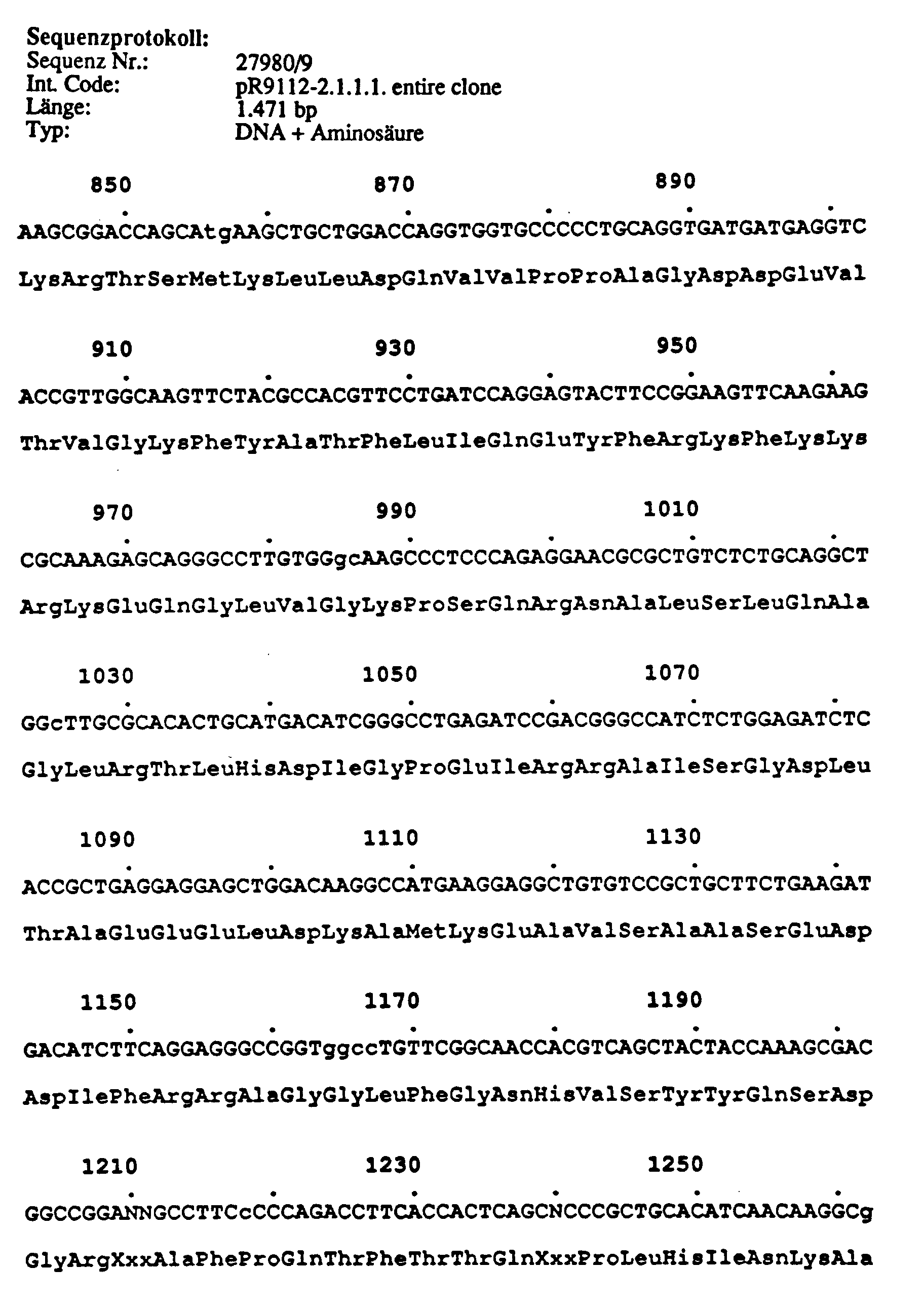
Länge 1.471 bp; wie cDNA Klon pR9112-4.1.1.1, enthält dieser Klon ab IVs3-4 die komplette kodierende Region bis zum tatsächlichen C-Terminus des Proteins, sowie etwa 500 bp der 3' nicht translatierten Region der mRNA.Length 1,471 bp; Like cDNA clone pR9112-4.1.1.1, this clone from IVs3-4 contains the complete coding region up to the actual C-terminus of the protein, as well as about 500 bp of the 3 'untranslated region of the mRNA.
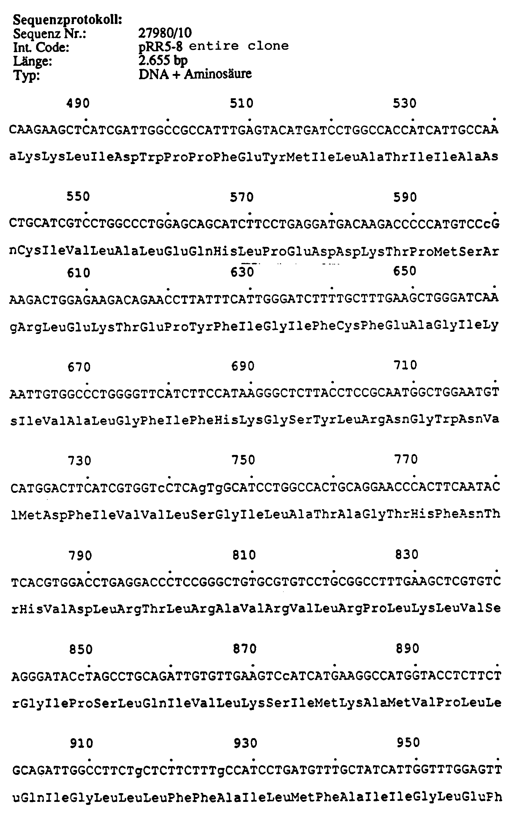
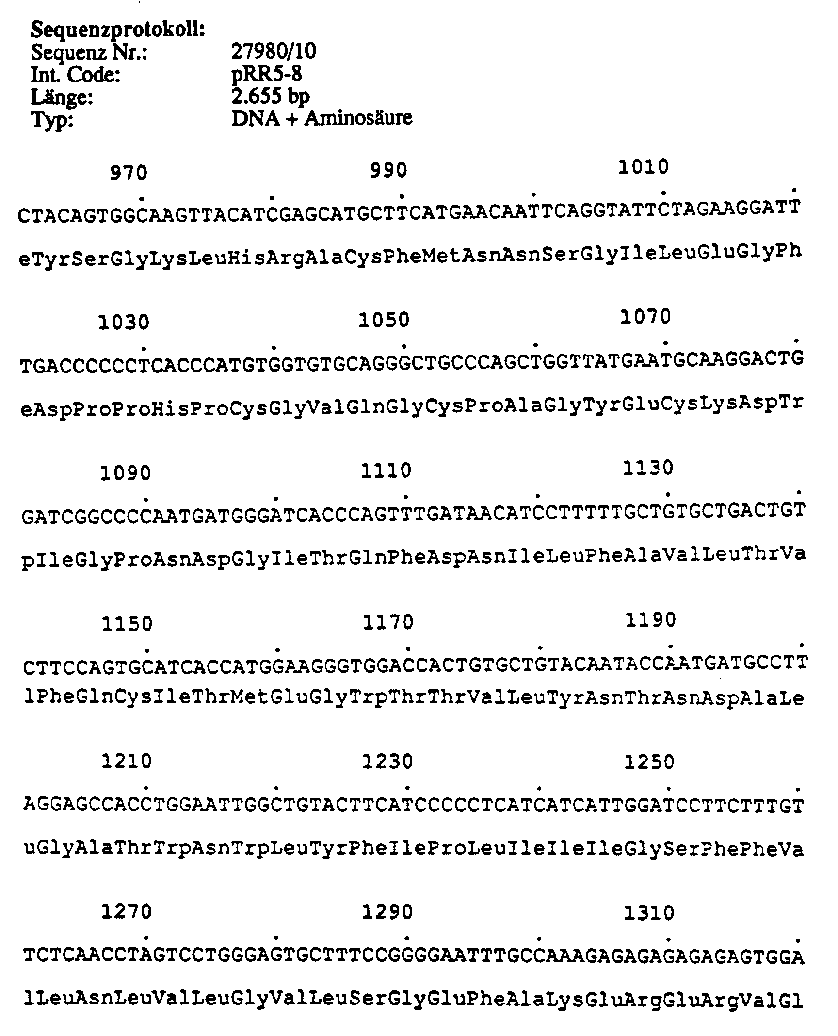
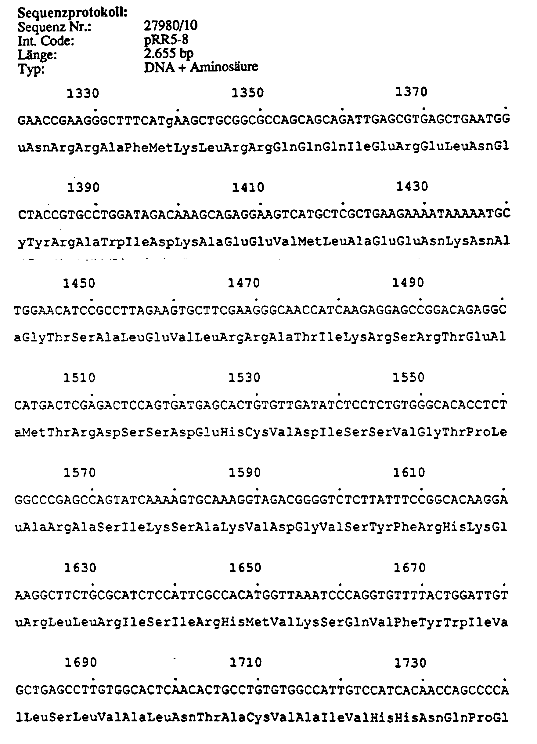
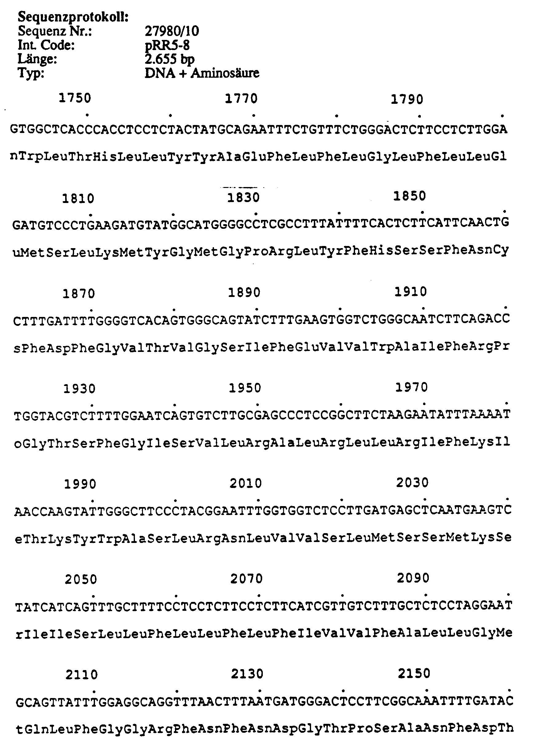
Länge 2.655 bp; am 5'-Ende enthält dieser Klon 235 bp nichttranslatierte Sequenz gefolgt von einem ATG Startcodon (Pos. 236-238). Von diesem Startcodon erstreckt sich ein offenes Leseraster bis zum 3'-Ende des Klons. Das daraus abgeleitete Protein beginnt mit dem tatsächlichen N-Terminus eines Ca-Kanal cDNA-Gens und enthält die Domänen I und II, sowie einen Teil des intrazellulären Loops zwischen Domäne II und III. Ab Position 1.318 bis zum 3'-Ende überlappt der Klon mit dem Klon p1247-5.1.2.1.1. Beide Klone können z.B. an einer gemeinsamen Xhol-Schnittstelle (Pos. 1.506-1.511 bei pRR5-8) miteinander kombiniert werden zu einer cDNA, die neben dem zytoplasmatischen N-Terminus für die Domänen I-IV und angrenzende zytoplasmatische c-terminale Bereiche des Ca-Kanals kodiert.Length 2,655 bp; at the 5 'end this clone contains 235 bp untranslated sequence followed by an ATG start codon (pos. 236-238). An open reading frame extends from this start codon to the 3 'end of the clone. The protein derived therefrom begins with the actual N-terminus of a Ca channel cDNA gene and contains domains I and II, as well as part of the intracellular loop between domains II and III. From position 1.318 to the 3 'end, the clone overlaps with clone p1247-5.1.2.1.1. Both clones can e.g. at a common Xhol interface (pos. 1.506-1.511 at pRR5-8) are combined with one another to form a cDNA which, in addition to the cytoplasmic N-terminus, codes for domains I-IV and adjacent cytoplasmic c-terminal areas of the Ca channel.
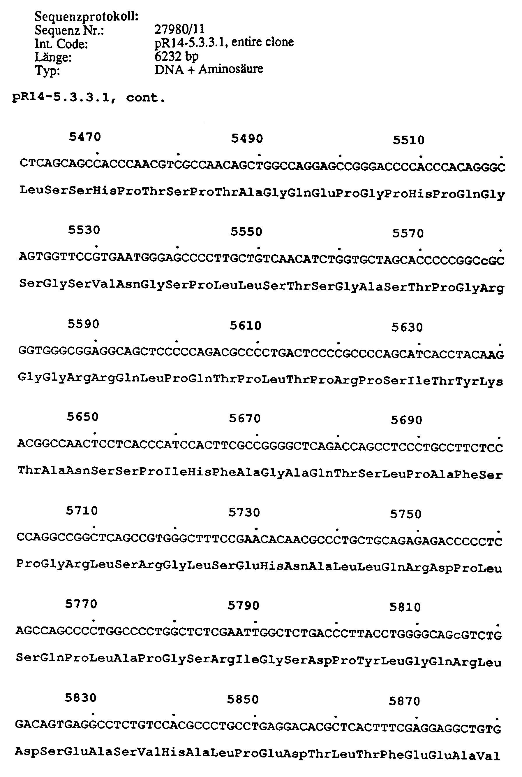
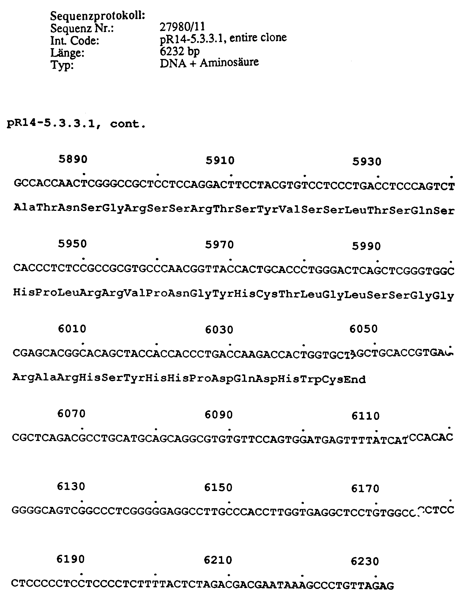
Länge: 6.232 bp; Position AS 358 bis zum C-Terminus eines Ca-Kanals. Am 5'-Ende enthält dieser Klon 252 bp, die 85 % Homologie zu humanen Alu-repeat Sequenzen aufweisen und möglicherweise artifiziell während der cDNA Klonierung an die restlichen 5.980 bp der Ca-Kanal cDNA ligiert wurden. Das offene Leseraster des für einen Ca-Kanal kodierenden cDNA Stranges kodiert für 1.931 AS und umspannt die Domänen II bis IV, sowie den C-terminalen cytoplasmatischen Anteil des Ca-Kanals. Es folgen 187 bp 3'-nichttranslatierte Region, einschließlich eines Polyadenylierungssignal an Position 6.215-6.220, und läuft in ein poly A-Ende aus (44 Adenosinreste).Length: 6,232 bp; Position AS 358 up to the C-terminus of a Ca channel. At the 5 'end, this clone contains 252 bp, which have 85% homology to human Alu repeat sequences and which may have been artificially ligated to the remaining 5,980 bp of the Ca channel cDNA during cDNA cloning. The open reading frame of the cDNA strand coding for a Ca channel codes for 1,931 AS and spans domains II to IV, as well as the C-terminal cytoplasmic portion of the Ca channel. This is followed by 187 bp 3 'untranslated region, including a polyadenylation signal at position 6.215-6.220, and terminates in a poly A end (44 adenosine residues).
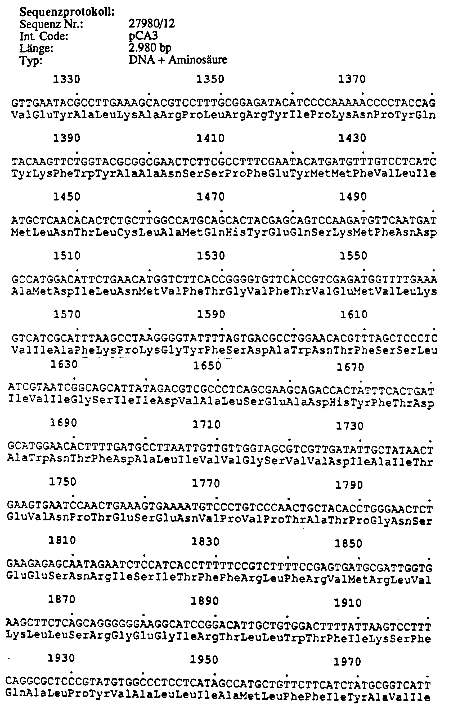
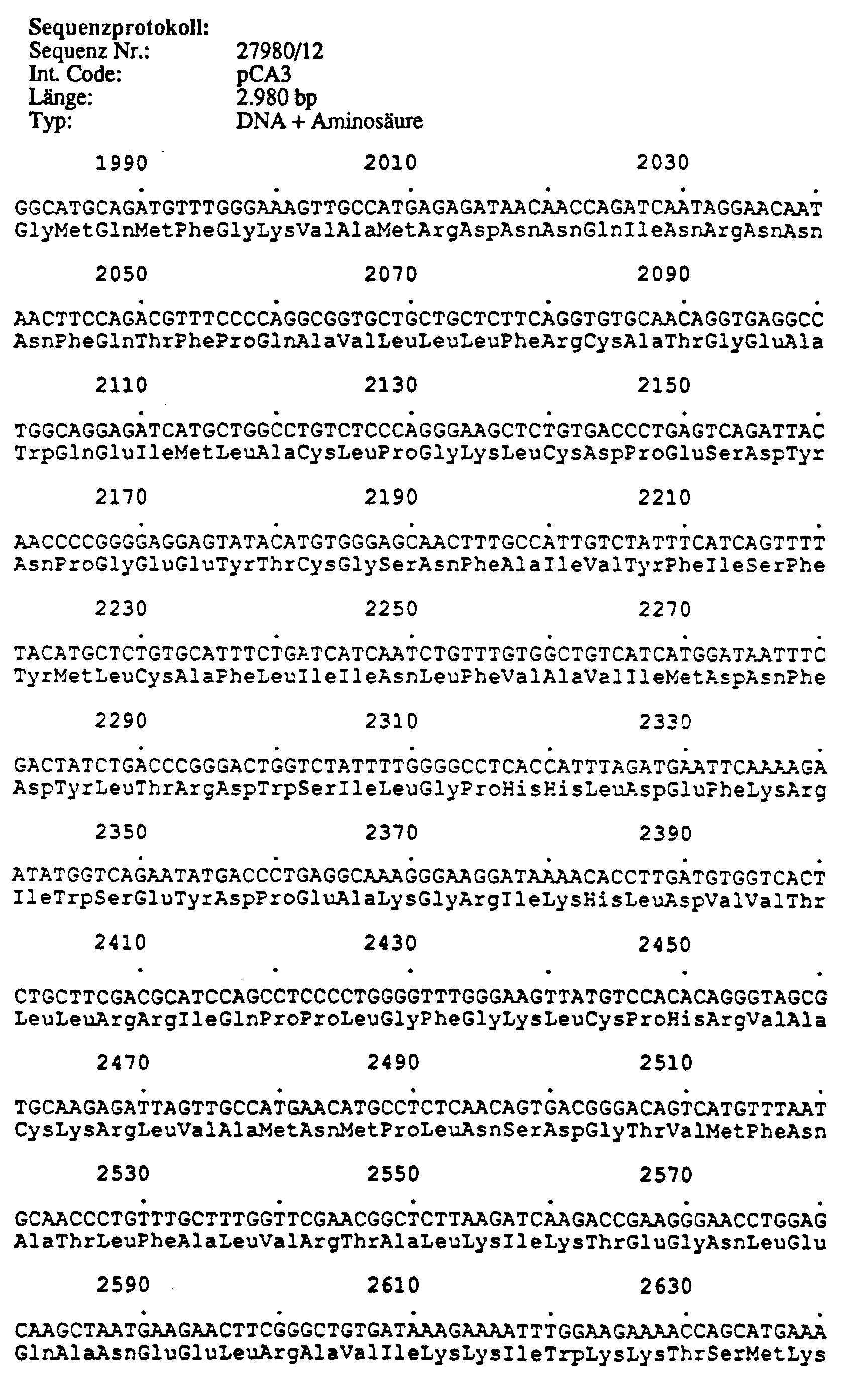
Länge 2.837 bp; besitzt eine interne EcoRI-Schnittstelle (2 Subfragmente: 2.197 bp, 640 bp). Das 5'-Ende des cDNA Klons liegt zwischen Domäne II und III, das 3'-Ende bei AS 1.622; der Klon umfaßt die vollständigen Domänen III und IV und einen Teil der für den C-Terminus kodierenden Sequenz. Das 5'-Ende des cDNA-Klons überlappt mit dem 3'-Ende des Klons pCA9.3 auf einer Länge von 830 bp. Beide cDNA-Klone sind über 671 bp identisch, lediglich die ersten 159 bp des 5'-Endes von Klon pCA3 zeigen zu dem entsprechenden Abschnitt des Klons pCA9.3 keinerlei Homologien (Abb. 2, schraffierter Bereich). Kon pCA 3 und Klon pCA9.3 über eine gemeinsamePmII Restriktionsschnittstelle im überlappenden Bereich kombiniert werden.Length 2,837 bp; has an internal EcoRI interface (2 subfragments: 2,197 bp, 640 bp). The 5 'end of the cDNA clone is between domain II and III, the 3' end at AS 1.622; the clone comprises the complete domains III and IV and part of the sequence coding for the C-terminus. The 5 'end of the cDNA clone overlaps the 3' end of the clone pCA9.3 over a length of 830 bp. Both cDNA clones are identical over 671 bp, only the first 159 bp of the 5 'end of clone pCA3 show no homologies with the corresponding section of clone pCA9.3 (Fig. 2, hatched area).
Länge 1.857 bp; das 5'-Ende des cDNA Klons beginnt unmittelbar nach Domäne I (AS 337); das 3'-Ende liegt bei AS 922; der cDNA-Klon umfaßt Sequenzen des zweiten zytoplasmatischen Abschnittes (zwischen Domäne I und II) bis einschließlich der 4. Transmembranregion der Domäne III(IIIS4).Length 1,857 bp; the 5 'end of the cDNA clone begins immediately after domain I (AS 337); the 3 'end is at AS 922; the cDNA clone comprises sequences of the second cytoplasmic section (between domains I and II) up to and including the 4th transmembrane region of domain III (IIIS4).
Länge 920 bp; Position AS 1.178-1.496 der Kaninchenskelettmuskel α₁-Untereinheit (Domäne IVs3-IVs6). Die Sequenz des Klons p1247-4.2.1.1 ist vollständig in der Sequenz des Klons p1247-10.1.1.1 enthalten. Beide Klone sind bis auf eine 6 Basenpaare umfassende Insertion in Klon 1247-4 (Position 88-93) und 2 weitere Basenaustausche identisch.Length 920 bp; Position AS 1.178-1.496 of the rabbit skeletal muscle α 1 subunit (domain IVs3-IVs6). The sequence of the clone p1247-4.2.1.1 is completely contained in the sequence of the clone p1247-10.1.1.1. Both clones are identical except for an insertion into clone 1247-4 (position 88-93) and 6 further base exchanges, which comprises 6 base pairs.
Länge: 1,424 bp; Position AS 25-458. Am 5'-Ende enthält dieser Klon an Position 60 eine putative Splice-Donor-Stelle, so daß die ersten 60 Nukleotide möglicherweise Intronsequenzen darstellen. Der für einen Ca-Kanal kodierende Bereich (ab Position 61) umfaßt einen Teil der N-terminalen cytoplasmatischen Region, sowie die gesamte Domäne I und die erste membrandurchbrechende Region (S1) der Domäne II.Length: 1.424 bp; Position AS 25-458. At the 5 'end, this clone contains a putative splice donor site at position 60, so that the first 60 nucleotides may represent intron sequences. The region coding for a Ca channel (from position 61) comprises part of the N-terminal cytoplasmic region, as well as the entire domain I and the first membrane-breaking region (S1) of domain II.
Länge: 910 bp; Position AS 409-713. Dieser Klon überlappt am 5'-Ende 151 bp mit dem 3'-Ende des Klons pR5-6cort (100 % Identität über 151 bp). Diese beiden Klone stellen daher unabhängig klonierte cDNA Abschnitte ein und derselben mRNA dar und können z.B. über die gemeinsame Stu I Restruktionsabschnittstelle miteinander kombiniert werden.Length: 910 bp; Position AS 409-713. This clone overlaps at the 5 'end 151 bp with the 3' end of the clone pR5-6cort (100% identity over 151 bp). These two Clones therefore represent independently cloned cDNA sections of one and the same mRNA and can be combined with one another, for example, via the common Stu I restriction site.
Länge: ca 3.400bp, hiervon sind bisher 1.100 bp sequenziert; Position: das 5'-Ende des Klons liegt bei AS 257 und somit zwischen den Membrandurchgängen S5 und S6 der Domäne I. Im bisher sequenzierten Bereich überlappt der Klon Sequenzidentisch mit pR14-5.3.3.1 (Position 253-964). Somit können die beiden Klone pRR14-35 und pR14-5.3.3.1 als 2 unabhängig klonierte cDNA Abschnitte einer mRNA angesehen werden. Klon pRR14-35 ergänzt die in pR14-5.3.3.1 enthaltene Ca-Kanal cDNA zum 5'-Ende um 129 AS und eliminiert hierbei die artiflziell vorhandene Alu-repeat Sequenz am 5'-Ende dieses Klons. Beide Klone können über eine gemeinsame Bgl II Restriktionsschnittstelle miteinander kombiniert werden.Length: approx. 3,400 bp, of which 1,100 bp have been sequenced so far; Position: the 5 'end of the clone is at AS 257 and thus between the membrane passages S5 and S6 of domain I. In the region sequenced to date, the clone overlaps identical to pR14-5.3.3.1 (position 253-964). Thus, the two clones pRR14-35 and pR14-5.3.3.1 can be viewed as 2 independently cloned cDNA sections of an mRNA. Clone pRR14-35 supplements the Ca-channel cDNA contained in pR14-5.3.3.1 at the 5 'end by 129 AS and thereby eliminates the artificially existing Alu repeat sequence at the 5' end of this clone. Both clones can be combined using a common Bgl II restriction interface.
Im oberen Teil (A) von Abbildung 1 ist die cDNA des Skelettmuskel Ca-Kanals des Karpfens mit den Schnittstellen für einige Restriktionsendonukleasen und den Subfragmenten dargestellt, die als Screeningproben verwendet wurden. Im unteren Teil (B) sind DNA Fragmente der humanen Klone p1247-5.1.2.1.1 und p1247-14.1.1, die mit den Restriktionsenzymen EcoRI/SacI bzw. EcoRI/KpnI erzeugt wurden, sowie das gesamte cDNA Insert des Klons p1247-9.1.1.2, die ebenfalls als Screeningproben verwendet wurden.The upper part (A) of Figure 1 shows the cDNA of the carp skeletal muscle Ca-channel with the interfaces for some restriction endonucleases and the subfragments that were used as screening samples. In the lower part (B) are DNA fragments of the human clones p1247-5.1.2.1.1 and p1247-14.1.1, which were generated with the restriction enzymes EcoRI / SacI or EcoRI / KpnI, as well as the entire cDNA insert of the clone p1247- 9.1.1.2, which were also used as screening samples.
Abbildung 2 zeigt im oberen Teil (A) eine schematische Darstellung der α₁-Untereinheit des Skelettmuskel Ca-Kanals vom Kaninchen. Die 4 Domänen sind als Kästen gekennzeichnet und mit römischen Ziffern beschriftet. Innerhalb dieser Domänen sind die Transmembranbereiche als schwarze Blöcke hervorgehoben Im unteren Teil (B) sind die Teilstücke der bisher isolierten humanen neuronalen Ca-Kanäle entsprechend ihrer Größe und ihrer Homologie zu dem Kaninchen Skelettmuskel Ca-Kanal Protein angeordnet.Figure 2 shows in the upper part (A) a schematic representation of the α₁ subunit of the skeletal muscle Ca channel from rabbits. The 4 domains are marked as boxes and labeled with Roman numerals. Within these domains, the transmembrane regions are highlighted as black blocks. In the lower part (B), the sections of the human neuronal Ca channels isolated to date are arranged according to their size and their homology to the rabbit skeletal muscle Ca channel protein.
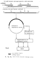
Schematische Darstellung der Klonierung und Expression gewebespezifischer humaner neuronaler Calcium-Kanal-Subtypen und deren Verwendung in einem Testsystem. Die vollständige, für einen Calcium-Kanal-Subtyp kodierende cDNA wird mittels geeigneter Restriktionsschnittstellen aus überlappenden Lambda-Phagen zusammengesetzt und in einen eukaryontischen Expressionsvektor kloniert.Schematic representation of the cloning and expression of tissue-specific human neuronal calcium channel subtypes and their use in a test system. The complete cDNA coding for a calcium channel subtype is composed of overlapping lambda phages by means of suitable restriction sites and cloned into a eukaryotic expression vector.
Mit diesem Vektor werden geeignete eukaryontische Zellen transformiert und stabile Zellinien hergestellt, die das Protein eines Calcium-Kanal-Subtyps exprimieren. Diese stabilen Zellinien werden dann in den beschriebenen Rezeptor-Bindungstest eingesetzt.This vector is used to transform suitable eukaryotic cells and to produce stable cell lines which express the protein of a calcium channel subtype. These stable cell lines are then used in the receptor binding test described.
Zusätzlich in die für den Calcium-Kanal kodierende cDNA eingeführte Mutationen sollen zur Identifizierung von Struktur-Funktionsdomänen führen.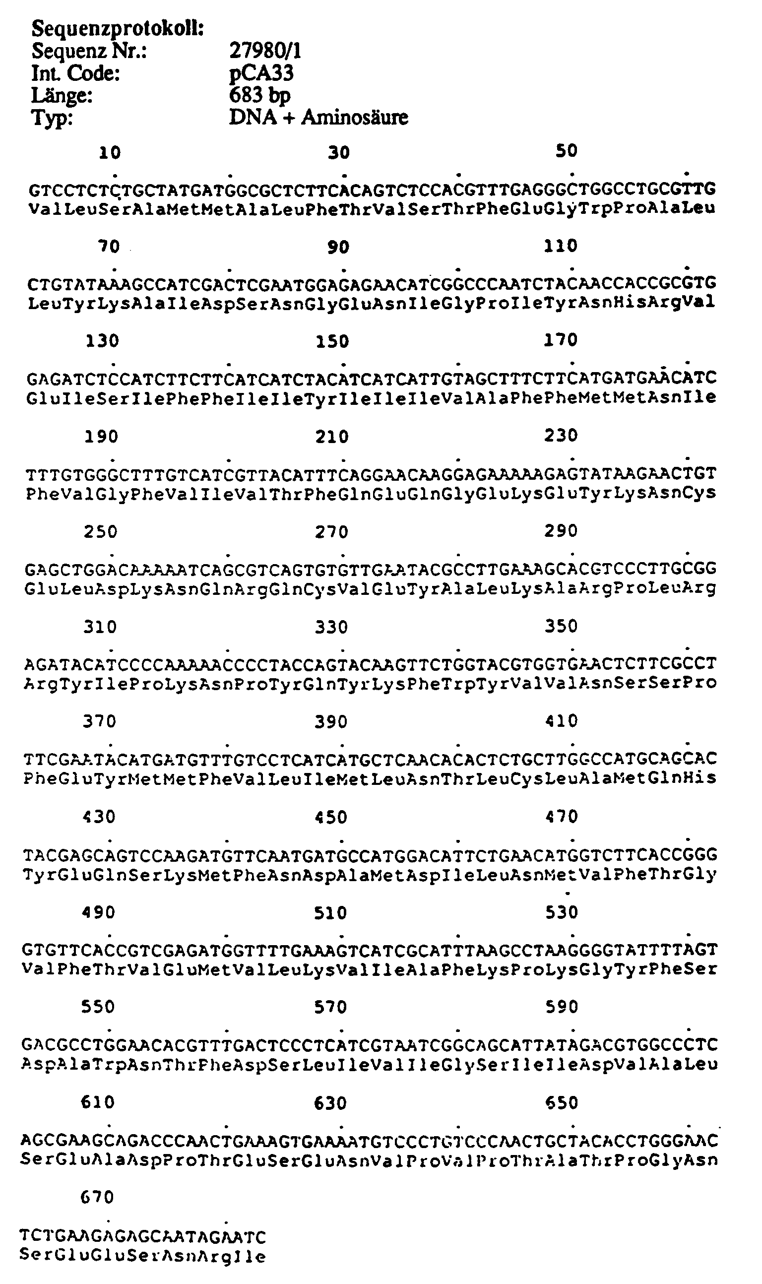


Claims (3)
Applications Claiming Priority (2)
| Application Number | Priority Date | Filing Date | Title |
|---|---|---|---|
| DE4110785A DE4110785A1 (en) | 1991-04-04 | 1991-04-04 | TISSUE-SPECIFIC HUMAN NEURONAL CALCIUM CHANNEL SUBTYPES AND THEIR USE |
| DE4110785 | 1991-04-04 |
Publications (3)
| Publication Number | Publication Date |
|---|---|
| EP0507170A2 true EP0507170A2 (en) | 1992-10-07 |
| EP0507170A3 EP0507170A3 (en) | 1992-11-19 |
| EP0507170B1 EP0507170B1 (en) | 1997-01-15 |
Family
ID=6428742
Family Applications (1)
| Application Number | Title | Priority Date | Filing Date |
|---|---|---|---|
| EP92104970A Revoked EP0507170B1 (en) | 1991-04-04 | 1992-03-23 | Calcium channel subtype specific of human neuronal tissue and its use |
Country Status (7)
| Country | Link |
|---|---|
| EP (1) | EP0507170B1 (en) |
| JP (1) | JPH05320194A (en) |
| AT (1) | ATE147781T1 (en) |
| DE (2) | DE4110785A1 (en) |
| DK (1) | DK0507170T3 (en) |
| ES (1) | ES2097231T3 (en) |
| GR (1) | GR3022585T3 (en) |
Cited By (19)
| Publication number | Priority date | Publication date | Assignee | Title |
|---|---|---|---|---|
| WO1995004144A1 (en) * | 1993-07-30 | 1995-02-09 | Neurex Corporation | Dna encoding a human calcium channel alpha-1e subunit |
| US5429921A (en) * | 1988-04-04 | 1995-07-04 | The Salk Institute Biotechnology/Industrial Assoc. Inc. | Assays for agonists and antagonists of recombinant human calcium channels |
| US5618720A (en) * | 1988-04-04 | 1997-04-08 | Sibia Neurosciences, Inc. | Cells expressing calcium channel α2 subunit-encoding DNA, optionally with a reporter gene for screening assays |
| US5686241A (en) * | 1988-04-04 | 1997-11-11 | Sibia Neurosciences, Inc. | Probes and assays for calcium channel α2 subunit-encoding nucleic acids |
| US5726035A (en) * | 1990-02-20 | 1998-03-10 | Sibia Neurosciences, Inc. | Recombinant production of mammalian calcium channel gamma subunits |
| US5792846A (en) * | 1988-04-04 | 1998-08-11 | Sibia Neurosciences, Inc. | Human calcium channel compositions and methods |
| US5846757A (en) * | 1988-04-04 | 1998-12-08 | Sibia Neurosciences, Inc. | Human calcium channel α1, α2, and β subunits and assays using them |
| US5851824A (en) * | 1988-04-04 | 1998-12-22 | Sibia Neurosciences, Inc. | Human calcium channel α-1C/α-1D, α-2, β-1, and γsubunits and cells expressing the DNA |
| US5874236A (en) * | 1988-04-04 | 1999-02-23 | Sibia Neurosciences. Inc. | DNA encoding human calcium channel α-1A, β1, β-2, and β-4 subunits, and assays using cells that express the subunits |
| US6040436A (en) * | 1996-09-16 | 2000-03-21 | American Home Products Corporation | Nucleic acid encoding human neuronal calcium channel subunits |
| US6090623A (en) * | 1993-08-11 | 2000-07-18 | Merck & Co., Inc. | Recombinant human calcium channel β4 subunits |
| US6096514A (en) * | 1988-04-04 | 2000-08-01 | Sibia Neurosciences, Inc. | Human calcium channel compositions and methods |
| US6387696B1 (en) | 1988-04-04 | 2002-05-14 | Merck & Co., Inc. | Human calcium channel compositions and methods |
| US6528630B1 (en) | 1997-12-03 | 2003-03-04 | Merck & Co., Inc. | Calcium channel compositions and methods |
| US6653097B1 (en) | 1991-08-15 | 2003-11-25 | Merck & Co., Inc. | Human calcium channel compositions and methods |
| EP1469074A1 (en) * | 1991-08-15 | 2004-10-20 | MERCK & CO. INC. | Human calcium channel compositions and methods |
| EP1392344A4 (en) * | 2001-05-17 | 2005-09-21 | Univ Australian | METHOD FOR MODULATING THE ACTIVITY OF CALCIUM CHANNELS IN CARDIAC CELLS AND REAGENTS FOR CARRYING OUT SAID METHOD |
| US7157243B1 (en) | 1997-02-28 | 2007-01-02 | Neuromed Pharmaceuticals Ltd. | DNA encoding mammalian T-type calcium channels |
| US7414110B2 (en) | 1988-04-04 | 2008-08-19 | Merck & Co., Inc. | Human calcium channel compositions and methods |
Family Cites Families (2)
| Publication number | Priority date | Publication date | Assignee | Title |
|---|---|---|---|---|
| US4950739A (en) * | 1988-02-10 | 1990-08-21 | New York University | Membrane calcium channels and factors and methods for blocking, isolating and purifying calcium channels |
| EP0424397B1 (en) * | 1988-04-04 | 1996-06-19 | The Salk Institute Biotechnology Industrial Associates, Inc. | Calcium channel compositions and methods |
-
1991
- 1991-04-04 DE DE4110785A patent/DE4110785A1/en not_active Withdrawn
-
1992
- 1992-03-23 DK DK92104970.6T patent/DK0507170T3/en active
- 1992-03-23 DE DE59207873T patent/DE59207873D1/en not_active Revoked
- 1992-03-23 ES ES92104970T patent/ES2097231T3/en not_active Expired - Lifetime
- 1992-03-23 EP EP92104970A patent/EP0507170B1/en not_active Revoked
- 1992-03-23 AT AT92104970T patent/ATE147781T1/en not_active IP Right Cessation
- 1992-03-31 JP JP4103753A patent/JPH05320194A/en active Pending
-
1997
- 1997-02-19 GR GR970400270T patent/GR3022585T3/en unknown
Cited By (25)
| Publication number | Priority date | Publication date | Assignee | Title |
|---|---|---|---|---|
| US5846757A (en) * | 1988-04-04 | 1998-12-08 | Sibia Neurosciences, Inc. | Human calcium channel α1, α2, and β subunits and assays using them |
| US6013474A (en) * | 1988-04-04 | 2000-01-11 | Sibia Neurosciences, Inc. | Calcium channel compositions and methods |
| US5618720A (en) * | 1988-04-04 | 1997-04-08 | Sibia Neurosciences, Inc. | Cells expressing calcium channel α2 subunit-encoding DNA, optionally with a reporter gene for screening assays |
| US5686241A (en) * | 1988-04-04 | 1997-11-11 | Sibia Neurosciences, Inc. | Probes and assays for calcium channel α2 subunit-encoding nucleic acids |
| US6096514A (en) * | 1988-04-04 | 2000-08-01 | Sibia Neurosciences, Inc. | Human calcium channel compositions and methods |
| US5792846A (en) * | 1988-04-04 | 1998-08-11 | Sibia Neurosciences, Inc. | Human calcium channel compositions and methods |
| US5874236A (en) * | 1988-04-04 | 1999-02-23 | Sibia Neurosciences. Inc. | DNA encoding human calcium channel α-1A, β1, β-2, and β-4 subunits, and assays using cells that express the subunits |
| US7063950B1 (en) | 1988-04-04 | 2006-06-20 | Harpold Michael M | Nucleic acids encoding human calcium channel and methods of use thereof |
| US5429921A (en) * | 1988-04-04 | 1995-07-04 | The Salk Institute Biotechnology/Industrial Assoc. Inc. | Assays for agonists and antagonists of recombinant human calcium channels |
| US5876958A (en) * | 1988-04-04 | 1999-03-02 | Sibia Neurosciences, Inc. | Assays of cells expressing human calcium channels containing α1 β subunits |
| US6387696B1 (en) | 1988-04-04 | 2002-05-14 | Merck & Co., Inc. | Human calcium channel compositions and methods |
| US7414110B2 (en) | 1988-04-04 | 2008-08-19 | Merck & Co., Inc. | Human calcium channel compositions and methods |
| US5851824A (en) * | 1988-04-04 | 1998-12-22 | Sibia Neurosciences, Inc. | Human calcium channel α-1C/α-1D, α-2, β-1, and γsubunits and cells expressing the DNA |
| US5726035A (en) * | 1990-02-20 | 1998-03-10 | Sibia Neurosciences, Inc. | Recombinant production of mammalian calcium channel gamma subunits |
| EP1469074A1 (en) * | 1991-08-15 | 2004-10-20 | MERCK & CO. INC. | Human calcium channel compositions and methods |
| US6653097B1 (en) | 1991-08-15 | 2003-11-25 | Merck & Co., Inc. | Human calcium channel compositions and methods |
| WO1995004144A1 (en) * | 1993-07-30 | 1995-02-09 | Neurex Corporation | Dna encoding a human calcium channel alpha-1e subunit |
| US6090623A (en) * | 1993-08-11 | 2000-07-18 | Merck & Co., Inc. | Recombinant human calcium channel β4 subunits |
| US6140485A (en) * | 1996-09-16 | 2000-10-31 | American Home Products Corp. | Nucleic acid encoding human neuronal calcium channel subunits |
| US6040436A (en) * | 1996-09-16 | 2000-03-21 | American Home Products Corporation | Nucleic acid encoding human neuronal calcium channel subunits |
| US7157243B1 (en) | 1997-02-28 | 2007-01-02 | Neuromed Pharmaceuticals Ltd. | DNA encoding mammalian T-type calcium channels |
| US7501263B2 (en) | 1997-02-28 | 2009-03-10 | Neuromed Technologies, Inc. | Nucleic acids encoding mammalian T-type calcium channels |
| US7517672B2 (en) | 1997-02-28 | 2009-04-14 | Neuromed Pharmaceuticals Ltd. | Nucleic acids encoding mammalian T-type calcium channels |
| US6528630B1 (en) | 1997-12-03 | 2003-03-04 | Merck & Co., Inc. | Calcium channel compositions and methods |
| EP1392344A4 (en) * | 2001-05-17 | 2005-09-21 | Univ Australian | METHOD FOR MODULATING THE ACTIVITY OF CALCIUM CHANNELS IN CARDIAC CELLS AND REAGENTS FOR CARRYING OUT SAID METHOD |
Also Published As
| Publication number | Publication date |
|---|---|
| GR3022585T3 (en) | 1997-05-31 |
| EP0507170B1 (en) | 1997-01-15 |
| ATE147781T1 (en) | 1997-02-15 |
| DE4110785A1 (en) | 1992-10-08 |
| EP0507170A3 (en) | 1992-11-19 |
| ES2097231T3 (en) | 1997-04-01 |
| DK0507170T3 (en) | 1997-07-07 |
| JPH05320194A (en) | 1993-12-03 |
| DE59207873D1 (en) | 1997-02-27 |
Similar Documents
| Publication | Publication Date | Title |
|---|---|---|
| EP0507170B1 (en) | Calcium channel subtype specific of human neuronal tissue and its use | |
| EP0556651B1 (en) | Human neuronal beta-subunits of voltage-dependent calcium channels and use of the same | |
| DE69734359T2 (en) | RECOMBINANT VASCULAR ENDOTHELIAL CELL GROWTH FACTOR D (VEGF-D) | |
| DE69434067T2 (en) | HUMAN NEURONAL NICOTIN ACETYLCHOLINE RECEPTOR COMPOUNDS AND METHODS OF ITS USE | |
| DE69404401T3 (en) | Erythropoietin analogues with additional glycosylation sites | |
| DE69426033T2 (en) | DNA ENCODING THE ALPHA-1E UNIT OF THE HUMAN CALCIUM CHANNEL | |
| DE69432794T2 (en) | HUMAN CALCIUM CHANNELS AND USES THEREOF | |
| DE68926713T2 (en) | CALCIUM CHANNEL COMPOSITIONS AND METHODS | |
| DE69232718T2 (en) | Identification of a human receptor tyrosine kinase gene | |
| DE69434115T2 (en) | INHIBITOR OF THE GROWTH FACTOR FOR VITAL TREATMENT CELLS | |
| DE69029370T3 (en) | Erythropoietin isoforms | |
| DE69033109T2 (en) | FIBROBLAST GROWTH FACTOR RECEPTORS | |
| DE69232986T2 (en) | TGF-BETA TYPE III RECEPTOR, FOR CODING CDNS AND THEIR USE | |
| DE69334187T2 (en) | Stably transfected cell lines expressing human GABA-A receptors with the subunit combination alpha-2, beta-3 and gamma-2 | |
| DE69132447T2 (en) | HUMAN DOPAMINE D4 RECEPTOR AND ITS USE | |
| DE69635517T2 (en) | HUMAN NEURONAL ACETYLCHOLIN RECEPTOR COMPOUNDS AND METHODS OF THEIR APPLICATION | |
| DE69534597T2 (en) | MELATONIN RECEPTORS WITH HIGH AFFINITY AND ITS USES | |
| DE69333019T2 (en) | DELTA OPIOID RECEPTOR GENES | |
| DE69625448T2 (en) | Protein analogs of the LDL receptor and their coding genes | |
| EP1287142B1 (en) | Nucleic acid molecule comprising a nucleic acid sequence coding for an sdf-1 gamma chemokine, a neuropeptide precursor or at least one neuropeptide | |
| DE69434090T2 (en) | EPSILON SUB-UNIT OF THE GABA A-RECEPTOR | |
| DE68922845T2 (en) | GENES AND PROTEINS CODED BY YOU, REGULATING THE GENE EXPRESSION OF THE INTERLEUKIN 2 RECEPTOR AND OF HUMAN LYMPHOTROPIC RETROVIRUS. | |
| DE19617940A1 (en) | Epididymis-specific receptor protein and its use | |
| DE69230903T2 (en) | Human KA-1 receptor, receptor from the EAA1 family (Excitatory Amino Acid) with high affinity for kainate | |
| DE69518927T2 (en) | A PROTEIN INTERACTING WITH KATP CHANNELS |
Legal Events
| Date | Code | Title | Description |
|---|---|---|---|
| PUAI | Public reference made under article 153(3) epc to a published international application that has entered the european phase |
Free format text: ORIGINAL CODE: 0009012 |
|
| PUAL | Search report despatched |
Free format text: ORIGINAL CODE: 0009013 |
|
| AK | Designated contracting states |
Kind code of ref document: A2 Designated state(s): AT BE CH DE DK ES FR GB GR IT LI LU NL SE |
|
| AK | Designated contracting states |
Kind code of ref document: A3 Designated state(s): AT BE CH DE DK ES FR GB GR IT LI LU NL SE |
|
| 17P | Request for examination filed |
Effective date: 19921216 |
|
| 17Q | First examination report despatched |
Effective date: 19950922 |
|
| GRAG | Despatch of communication of intention to grant |
Free format text: ORIGINAL CODE: EPIDOS AGRA |
|
| GRAH | Despatch of communication of intention to grant a patent |
Free format text: ORIGINAL CODE: EPIDOS IGRA |
|
| GRAH | Despatch of communication of intention to grant a patent |
Free format text: ORIGINAL CODE: EPIDOS IGRA |
|
| GRAA | (expected) grant |
Free format text: ORIGINAL CODE: 0009210 |
|
| AK | Designated contracting states |
Kind code of ref document: B1 Designated state(s): AT BE CH DE DK ES FR GB GR IT LI LU NL SE |
|
| REF | Corresponds to: |
Ref document number: 147781 Country of ref document: AT Date of ref document: 19970215 Kind code of ref document: T |
|
| REG | Reference to a national code |
Ref country code: CH Ref legal event code: NV Representative=s name: E. BLUM & CO. PATENTANWAELTE Ref country code: CH Ref legal event code: EP |
|
| GBT | Gb: translation of ep patent filed (gb section 77(6)(a)/1977) |
Effective date: 19970125 |
|
| REF | Corresponds to: |
Ref document number: 59207873 Country of ref document: DE Date of ref document: 19970227 |
|
| ITF | It: translation for a ep patent filed | ||
| ET | Fr: translation filed | ||
| REG | Reference to a national code |
Ref country code: ES Ref legal event code: FG2A Ref document number: 2097231 Country of ref document: ES Kind code of ref document: T3 |
|
| REG | Reference to a national code |
Ref country code: GR Ref legal event code: FG4A Free format text: 3022585 |
|
| REG | Reference to a national code |
Ref country code: DK Ref legal event code: T3 |
|
| PLBQ | Unpublished change to opponent data |
Free format text: ORIGINAL CODE: EPIDOS OPPO |
|
| PLBI | Opposition filed |
Free format text: ORIGINAL CODE: 0009260 |
|
| PLBF | Reply of patent proprietor to notice(s) of opposition |
Free format text: ORIGINAL CODE: EPIDOS OBSO |
|
| 26 | Opposition filed |
Opponent name: SIBIA NEUROSCIENCES, INC. Effective date: 19971014 |
|
| NLR1 | Nl: opposition has been filed with the epo |
Opponent name: SIBIA NEUROSCIENCES, INC. |
|
| PLBF | Reply of patent proprietor to notice(s) of opposition |
Free format text: ORIGINAL CODE: EPIDOS OBSO |
|
| PLAW | Interlocutory decision in opposition |
Free format text: ORIGINAL CODE: EPIDOS IDOP |
|
| APAC | Appeal dossier modified |
Free format text: ORIGINAL CODE: EPIDOS NOAPO |
|
| APAE | Appeal reference modified |
Free format text: ORIGINAL CODE: EPIDOS REFNO |
|
| APAC | Appeal dossier modified |
Free format text: ORIGINAL CODE: EPIDOS NOAPO |
|
| PLAW | Interlocutory decision in opposition |
Free format text: ORIGINAL CODE: EPIDOS IDOP |
|
| PGFP | Annual fee paid to national office [announced via postgrant information from national office to epo] |
Ref country code: DE Payment date: 20010214 Year of fee payment: 10 |
|
| PGFP | Annual fee paid to national office [announced via postgrant information from national office to epo] |
Ref country code: SE Payment date: 20010215 Year of fee payment: 10 |
|
| PGFP | Annual fee paid to national office [announced via postgrant information from national office to epo] |
Ref country code: FR Payment date: 20010220 Year of fee payment: 10 |
|
| PGFP | Annual fee paid to national office [announced via postgrant information from national office to epo] |
Ref country code: GR Payment date: 20010227 Year of fee payment: 10 Ref country code: CH Payment date: 20010227 Year of fee payment: 10 |
|
| PGFP | Annual fee paid to national office [announced via postgrant information from national office to epo] |
Ref country code: AT Payment date: 20010315 Year of fee payment: 10 |
|
| PGFP | Annual fee paid to national office [announced via postgrant information from national office to epo] |
Ref country code: ES Payment date: 20010320 Year of fee payment: 10 |
|
| PGFP | Annual fee paid to national office [announced via postgrant information from national office to epo] |
Ref country code: GB Payment date: 20010321 Year of fee payment: 10 |
|
| PGFP | Annual fee paid to national office [announced via postgrant information from national office to epo] |
Ref country code: LU Payment date: 20010326 Year of fee payment: 10 |
|
| PGFP | Annual fee paid to national office [announced via postgrant information from national office to epo] |
Ref country code: BE Payment date: 20010328 Year of fee payment: 10 |
|
| PGFP | Annual fee paid to national office [announced via postgrant information from national office to epo] |
Ref country code: NL Payment date: 20010330 Year of fee payment: 10 Ref country code: DK Payment date: 20010330 Year of fee payment: 10 |
|
| RDAH | Patent revoked |
Free format text: ORIGINAL CODE: EPIDOS REVO |
|
| RDAH | Patent revoked |
Free format text: ORIGINAL CODE: EPIDOS REVO |
|
| RDAG | Patent revoked |
Free format text: ORIGINAL CODE: 0009271 |
|
| STAA | Information on the status of an ep patent application or granted ep patent |
Free format text: STATUS: PATENT REVOKED |
|
| REG | Reference to a national code |
Ref country code: CH Ref legal event code: PL |
|
| 27W | Patent revoked |
Effective date: 20010913 |
|
| GBPR | Gb: patent revoked under art. 102 of the ep convention designating the uk as contracting state |
Free format text: 20010913 |
|
| NLR2 | Nl: decision of opposition | ||
| APAH | Appeal reference modified |
Free format text: ORIGINAL CODE: EPIDOSCREFNO |