EP0364255B2 - Multiplex genomic DNA amplification for deletion detection - Google Patents
Multiplex genomic DNA amplification for deletion detection Download PDFInfo
- Publication number
- EP0364255B2 EP0364255B2 EP89310424A EP89310424A EP0364255B2 EP 0364255 B2 EP0364255 B2 EP 0364255B2 EP 89310424 A EP89310424 A EP 89310424A EP 89310424 A EP89310424 A EP 89310424A EP 0364255 B2 EP0364255 B2 EP 0364255B2
- Authority
- EP
- European Patent Office
- Prior art keywords
- sequences
- primers
- dna
- primer
- sequence
- Prior art date
- Legal status (The legal status is an assumption and is not a legal conclusion. Google has not performed a legal analysis and makes no representation as to the accuracy of the status listed.)
- Expired - Lifetime
Links
- 230000037430 deletion Effects 0.000 title claims description 58
- 238000012217 deletion Methods 0.000 title claims description 58
- 238000001514 detection method Methods 0.000 title claims description 26
- 230000004544 DNA amplification Effects 0.000 title description 16
- 239000013615 primer Substances 0.000 claims abstract description 93
- 238000000034 method Methods 0.000 claims abstract description 74
- 108020004414 DNA Proteins 0.000 claims abstract description 57
- 108091028043 Nucleic acid sequence Proteins 0.000 claims abstract description 27
- 239000003155 DNA primer Substances 0.000 claims abstract description 15
- 208000026350 Inborn Genetic disease Diseases 0.000 claims abstract description 10
- 208000016361 genetic disease Diseases 0.000 claims abstract description 10
- 238000000137 annealing Methods 0.000 claims abstract description 9
- 108091081021 Sense strand Proteins 0.000 claims abstract description 5
- 230000000692 anti-sense effect Effects 0.000 claims abstract description 5
- 108090000623 proteins and genes Proteins 0.000 claims description 29
- 238000006243 chemical reaction Methods 0.000 claims description 28
- 230000000295 complement effect Effects 0.000 claims description 28
- 210000003754 fetus Anatomy 0.000 claims description 16
- 230000015572 biosynthetic process Effects 0.000 claims description 9
- 238000003786 synthesis reaction Methods 0.000 claims description 9
- 201000006938 muscular dystrophy Diseases 0.000 claims description 8
- 210000001766 X chromosome Anatomy 0.000 claims description 7
- 208000019291 X-linked disease Diseases 0.000 claims description 7
- 210000002593 Y chromosome Anatomy 0.000 claims description 7
- 108010069091 Dystrophin Proteins 0.000 claims description 6
- FDGQSTZJBFJUBT-UHFFFAOYSA-N hypoxanthine Chemical compound O=C1NC=NC2=C1NC=N2 FDGQSTZJBFJUBT-UHFFFAOYSA-N 0.000 claims description 6
- 102000001039 Dystrophin Human genes 0.000 claims description 4
- 101000986595 Homo sapiens Ornithine transcarbamylase, mitochondrial Proteins 0.000 claims description 4
- 208000000599 Ornithine Carbamoyltransferase Deficiency Disease Diseases 0.000 claims description 4
- 206010052450 Ornithine transcarbamoylase deficiency Diseases 0.000 claims description 4
- 208000035903 Ornithine transcarbamylase deficiency Diseases 0.000 claims description 4
- 102100028200 Ornithine transcarbamylase, mitochondrial Human genes 0.000 claims description 4
- 208000001001 X-linked ichthyosis Diseases 0.000 claims description 4
- 201000011278 ornithine carbamoyltransferase deficiency Diseases 0.000 claims description 4
- 208000026079 recessive X-linked ichthyosis Diseases 0.000 claims description 4
- UGQMRVRMYYASKQ-UHFFFAOYSA-N Hypoxanthine nucleoside Natural products OC1C(O)C(CO)OC1N1C(NC=NC2=O)=C2N=C1 UGQMRVRMYYASKQ-UHFFFAOYSA-N 0.000 claims description 3
- 208000027642 X-Linked Genetic disease Diseases 0.000 claims description 3
- 230000007812 deficiency Effects 0.000 claims description 3
- 238000000926 separation method Methods 0.000 claims description 3
- 230000003321 amplification Effects 0.000 abstract description 40
- 238000003199 nucleic acid amplification method Methods 0.000 abstract description 40
- 108091034117 Oligonucleotide Proteins 0.000 abstract description 18
- 108091029865 Exogenous DNA Proteins 0.000 abstract description 5
- 102000053602 DNA Human genes 0.000 abstract 1
- 108020004682 Single-Stranded DNA Proteins 0.000 abstract 1
- 239000000047 product Substances 0.000 description 33
- 206010013801 Duchenne Muscular Dystrophy Diseases 0.000 description 21
- 108700024394 Exon Proteins 0.000 description 19
- 238000004458 analytical method Methods 0.000 description 19
- 238000003752 polymerase chain reaction Methods 0.000 description 18
- 239000000523 sample Substances 0.000 description 18
- 239000012634 fragment Substances 0.000 description 14
- 101150015424 dmd gene Proteins 0.000 description 12
- 230000035772 mutation Effects 0.000 description 11
- 238000003793 prenatal diagnosis Methods 0.000 description 9
- 238000003556 assay Methods 0.000 description 8
- 108700028369 Alleles Proteins 0.000 description 7
- 208000037265 diseases, disorders, signs and symptoms Diseases 0.000 description 7
- 150000007523 nucleic acids Chemical group 0.000 description 7
- 238000003745 diagnosis Methods 0.000 description 6
- 201000010099 disease Diseases 0.000 description 6
- 238000009396 hybridization Methods 0.000 description 6
- 210000004027 cell Anatomy 0.000 description 5
- 238000007403 mPCR Methods 0.000 description 5
- 102000054765 polymorphisms of proteins Human genes 0.000 description 5
- 230000035945 sensitivity Effects 0.000 description 5
- 239000011543 agarose gel Substances 0.000 description 4
- 230000027455 binding Effects 0.000 description 4
- 238000001962 electrophoresis Methods 0.000 description 4
- 230000002068 genetic effect Effects 0.000 description 4
- 238000012216 screening Methods 0.000 description 4
- 238000012360 testing method Methods 0.000 description 4
- 238000001712 DNA sequencing Methods 0.000 description 3
- KCXVZYZYPLLWCC-UHFFFAOYSA-N EDTA Chemical compound OC(=O)CN(CC(O)=O)CCN(CC(O)=O)CC(O)=O KCXVZYZYPLLWCC-UHFFFAOYSA-N 0.000 description 3
- PEDCQBHIVMGVHV-UHFFFAOYSA-N Glycerine Chemical compound OCC(O)CO PEDCQBHIVMGVHV-UHFFFAOYSA-N 0.000 description 3
- 241000700605 Viruses Species 0.000 description 3
- 239000007795 chemical reaction product Substances 0.000 description 3
- 210000004252 chorionic villi Anatomy 0.000 description 3
- 230000001419 dependent effect Effects 0.000 description 3
- 229960001484 edetic acid Drugs 0.000 description 3
- 230000001605 fetal effect Effects 0.000 description 3
- 239000000203 mixture Substances 0.000 description 3
- 108020004707 nucleic acids Proteins 0.000 description 3
- 102000039446 nucleic acids Human genes 0.000 description 3
- 230000003252 repetitive effect Effects 0.000 description 3
- 238000011160 research Methods 0.000 description 3
- QKNYBSVHEMOAJP-UHFFFAOYSA-N 2-amino-2-(hydroxymethyl)propane-1,3-diol;hydron;chloride Chemical compound Cl.OCC(N)(CO)CO QKNYBSVHEMOAJP-UHFFFAOYSA-N 0.000 description 2
- QTBSBXVTEAMEQO-UHFFFAOYSA-N Acetic acid Chemical compound CC(O)=O QTBSBXVTEAMEQO-UHFFFAOYSA-N 0.000 description 2
- 241000894006 Bacteria Species 0.000 description 2
- 230000004568 DNA-binding Effects 0.000 description 2
- 102000004190 Enzymes Human genes 0.000 description 2
- 108090000790 Enzymes Proteins 0.000 description 2
- 101100443349 Homo sapiens DMD gene Proteins 0.000 description 2
- 108091092195 Intron Proteins 0.000 description 2
- TWRXJAOTZQYOKJ-UHFFFAOYSA-L Magnesium chloride Chemical compound [Mg+2].[Cl-].[Cl-] TWRXJAOTZQYOKJ-UHFFFAOYSA-L 0.000 description 2
- 239000000020 Nitrocellulose Substances 0.000 description 2
- 238000012408 PCR amplification Methods 0.000 description 2
- 238000002105 Southern blotting Methods 0.000 description 2
- JLCPHMBAVCMARE-UHFFFAOYSA-N [3-[[3-[[3-[[3-[[3-[[3-[[3-[[3-[[3-[[3-[[3-[[5-(2-amino-6-oxo-1H-purin-9-yl)-3-[[3-[[3-[[3-[[3-[[3-[[5-(2-amino-6-oxo-1H-purin-9-yl)-3-[[5-(2-amino-6-oxo-1H-purin-9-yl)-3-hydroxyoxolan-2-yl]methoxy-hydroxyphosphoryl]oxyoxolan-2-yl]methoxy-hydroxyphosphoryl]oxy-5-(5-methyl-2,4-dioxopyrimidin-1-yl)oxolan-2-yl]methoxy-hydroxyphosphoryl]oxy-5-(6-aminopurin-9-yl)oxolan-2-yl]methoxy-hydroxyphosphoryl]oxy-5-(6-aminopurin-9-yl)oxolan-2-yl]methoxy-hydroxyphosphoryl]oxy-5-(6-aminopurin-9-yl)oxolan-2-yl]methoxy-hydroxyphosphoryl]oxy-5-(6-aminopurin-9-yl)oxolan-2-yl]methoxy-hydroxyphosphoryl]oxyoxolan-2-yl]methoxy-hydroxyphosphoryl]oxy-5-(5-methyl-2,4-dioxopyrimidin-1-yl)oxolan-2-yl]methoxy-hydroxyphosphoryl]oxy-5-(4-amino-2-oxopyrimidin-1-yl)oxolan-2-yl]methoxy-hydroxyphosphoryl]oxy-5-(5-methyl-2,4-dioxopyrimidin-1-yl)oxolan-2-yl]methoxy-hydroxyphosphoryl]oxy-5-(5-methyl-2,4-dioxopyrimidin-1-yl)oxolan-2-yl]methoxy-hydroxyphosphoryl]oxy-5-(6-aminopurin-9-yl)oxolan-2-yl]methoxy-hydroxyphosphoryl]oxy-5-(6-aminopurin-9-yl)oxolan-2-yl]methoxy-hydroxyphosphoryl]oxy-5-(4-amino-2-oxopyrimidin-1-yl)oxolan-2-yl]methoxy-hydroxyphosphoryl]oxy-5-(4-amino-2-oxopyrimidin-1-yl)oxolan-2-yl]methoxy-hydroxyphosphoryl]oxy-5-(4-amino-2-oxopyrimidin-1-yl)oxolan-2-yl]methoxy-hydroxyphosphoryl]oxy-5-(6-aminopurin-9-yl)oxolan-2-yl]methoxy-hydroxyphosphoryl]oxy-5-(4-amino-2-oxopyrimidin-1-yl)oxolan-2-yl]methyl [5-(6-aminopurin-9-yl)-2-(hydroxymethyl)oxolan-3-yl] hydrogen phosphate Polymers Cc1cn(C2CC(OP(O)(=O)OCC3OC(CC3OP(O)(=O)OCC3OC(CC3O)n3cnc4c3nc(N)[nH]c4=O)n3cnc4c3nc(N)[nH]c4=O)C(COP(O)(=O)OC3CC(OC3COP(O)(=O)OC3CC(OC3COP(O)(=O)OC3CC(OC3COP(O)(=O)OC3CC(OC3COP(O)(=O)OC3CC(OC3COP(O)(=O)OC3CC(OC3COP(O)(=O)OC3CC(OC3COP(O)(=O)OC3CC(OC3COP(O)(=O)OC3CC(OC3COP(O)(=O)OC3CC(OC3COP(O)(=O)OC3CC(OC3COP(O)(=O)OC3CC(OC3COP(O)(=O)OC3CC(OC3COP(O)(=O)OC3CC(OC3COP(O)(=O)OC3CC(OC3COP(O)(=O)OC3CC(OC3COP(O)(=O)OC3CC(OC3CO)n3cnc4c(N)ncnc34)n3ccc(N)nc3=O)n3cnc4c(N)ncnc34)n3ccc(N)nc3=O)n3ccc(N)nc3=O)n3ccc(N)nc3=O)n3cnc4c(N)ncnc34)n3cnc4c(N)ncnc34)n3cc(C)c(=O)[nH]c3=O)n3cc(C)c(=O)[nH]c3=O)n3ccc(N)nc3=O)n3cc(C)c(=O)[nH]c3=O)n3cnc4c3nc(N)[nH]c4=O)n3cnc4c(N)ncnc34)n3cnc4c(N)ncnc34)n3cnc4c(N)ncnc34)n3cnc4c(N)ncnc34)O2)c(=O)[nH]c1=O JLCPHMBAVCMARE-UHFFFAOYSA-N 0.000 description 2
- 210000004381 amniotic fluid Anatomy 0.000 description 2
- 210000004369 blood Anatomy 0.000 description 2
- 239000008280 blood Substances 0.000 description 2
- 239000003795 chemical substances by application Substances 0.000 description 2
- 210000000349 chromosome Anatomy 0.000 description 2
- 230000002860 competitive effect Effects 0.000 description 2
- 239000002299 complementary DNA Substances 0.000 description 2
- 230000007547 defect Effects 0.000 description 2
- 230000006872 improvement Effects 0.000 description 2
- 230000001965 increasing effect Effects 0.000 description 2
- 230000001939 inductive effect Effects 0.000 description 2
- 238000004519 manufacturing process Methods 0.000 description 2
- 229920001220 nitrocellulos Polymers 0.000 description 2
- 239000002773 nucleotide Substances 0.000 description 2
- 125000003729 nucleotide group Chemical group 0.000 description 2
- 244000045947 parasite Species 0.000 description 2
- 230000008569 process Effects 0.000 description 2
- 239000011541 reaction mixture Substances 0.000 description 2
- 239000007787 solid Substances 0.000 description 2
- 210000001519 tissue Anatomy 0.000 description 2
- 108091032973 (ribonucleotides)n+m Proteins 0.000 description 1
- 108091003079 Bovine Serum Albumin Proteins 0.000 description 1
- 102100026735 Coagulation factor VIII Human genes 0.000 description 1
- 108010014303 DNA-directed DNA polymerase Proteins 0.000 description 1
- 102000016928 DNA-directed DNA polymerase Human genes 0.000 description 1
- 102100031780 Endonuclease Human genes 0.000 description 1
- 201000003542 Factor VIII deficiency Diseases 0.000 description 1
- 208000009292 Hemophilia A Diseases 0.000 description 1
- 101000911390 Homo sapiens Coagulation factor VIII Proteins 0.000 description 1
- 208000009625 Lesch-Nyhan syndrome Diseases 0.000 description 1
- 241001465754 Metazoa Species 0.000 description 1
- 108020004485 Nonsense Codon Proteins 0.000 description 1
- 108020005187 Oligonucleotide Probes Proteins 0.000 description 1
- 239000005662 Paraffin oil Substances 0.000 description 1
- 201000011252 Phenylketonuria Diseases 0.000 description 1
- 108010092799 RNA-directed DNA polymerase Proteins 0.000 description 1
- 108091028664 Ribonucleotide Proteins 0.000 description 1
- VMHLLURERBWHNL-UHFFFAOYSA-M Sodium acetate Chemical compound [Na+].CC([O-])=O VMHLLURERBWHNL-UHFFFAOYSA-M 0.000 description 1
- 101710137500 T7 RNA polymerase Proteins 0.000 description 1
- 108010006785 Taq Polymerase Proteins 0.000 description 1
- 229960000583 acetic acid Drugs 0.000 description 1
- 238000000246 agarose gel electrophoresis Methods 0.000 description 1
- 230000004075 alteration Effects 0.000 description 1
- BFNBIHQBYMNNAN-UHFFFAOYSA-N ammonium sulfate Chemical compound N.N.OS(O)(=O)=O BFNBIHQBYMNNAN-UHFFFAOYSA-N 0.000 description 1
- 229910052921 ammonium sulfate Inorganic materials 0.000 description 1
- 235000011130 ammonium sulphate Nutrition 0.000 description 1
- 208000005980 beta thalassemia Diseases 0.000 description 1
- 238000001574 biopsy Methods 0.000 description 1
- 229940098773 bovine serum albumin Drugs 0.000 description 1
- 102220106904 c.Exon Human genes 0.000 description 1
- 230000002950 deficient Effects 0.000 description 1
- 239000005547 deoxyribonucleotide Substances 0.000 description 1
- 125000002637 deoxyribonucleotide group Chemical group 0.000 description 1
- 238000011161 development Methods 0.000 description 1
- 230000029087 digestion Effects 0.000 description 1
- 208000035475 disorder Diseases 0.000 description 1
- 239000003814 drug Substances 0.000 description 1
- 230000000694 effects Effects 0.000 description 1
- ZMMJGEGLRURXTF-UHFFFAOYSA-N ethidium bromide Chemical compound [Br-].C12=CC(N)=CC=C2C2=CC=C(N)C=C2[N+](CC)=C1C1=CC=CC=C1 ZMMJGEGLRURXTF-UHFFFAOYSA-N 0.000 description 1
- 229960005542 ethidium bromide Drugs 0.000 description 1
- 210000002950 fibroblast Anatomy 0.000 description 1
- GNBHRKFJIUUOQI-UHFFFAOYSA-N fluorescein Chemical compound O1C(=O)C2=CC=CC=C2C21C1=CC=C(O)C=C1OC1=CC(O)=CC=C21 GNBHRKFJIUUOQI-UHFFFAOYSA-N 0.000 description 1
- 238000012224 gene deletion Methods 0.000 description 1
- 102000054766 genetic haplotypes Human genes 0.000 description 1
- 230000037442 genomic alteration Effects 0.000 description 1
- 229940012229 genone Drugs 0.000 description 1
- 239000012362 glacial acetic acid Substances 0.000 description 1
- 210000004209 hair Anatomy 0.000 description 1
- 230000000977 initiatory effect Effects 0.000 description 1
- 238000002955 isolation Methods 0.000 description 1
- 239000007788 liquid Substances 0.000 description 1
- 229910001629 magnesium chloride Inorganic materials 0.000 description 1
- 238000005259 measurement Methods 0.000 description 1
- 230000007246 mechanism Effects 0.000 description 1
- 210000004379 membrane Anatomy 0.000 description 1
- 239000012528 membrane Substances 0.000 description 1
- 239000013642 negative control Substances 0.000 description 1
- 230000009871 nonspecific binding Effects 0.000 description 1
- 229940124276 oligodeoxyribonucleotide Drugs 0.000 description 1
- 239000002751 oligonucleotide probe Substances 0.000 description 1
- 150000004713 phosphodiesters Chemical class 0.000 description 1
- 239000002987 primer (paints) Substances 0.000 description 1
- 230000037452 priming Effects 0.000 description 1
- 230000005855 radiation Effects 0.000 description 1
- 230000001105 regulatory effect Effects 0.000 description 1
- 230000004044 response Effects 0.000 description 1
- 108091008146 restriction endonucleases Proteins 0.000 description 1
- 238000007894 restriction fragment length polymorphism technique Methods 0.000 description 1
- 230000002441 reversible effect Effects 0.000 description 1
- 239000002336 ribonucleotide Substances 0.000 description 1
- 125000002652 ribonucleotide group Chemical group 0.000 description 1
- 208000007056 sickle cell anemia Diseases 0.000 description 1
- 239000001632 sodium acetate Substances 0.000 description 1
- 235000017281 sodium acetate Nutrition 0.000 description 1
- WGTODYJZXSJIAG-UHFFFAOYSA-N tetramethylrhodamine chloride Chemical compound [Cl-].C=12C=CC(N(C)C)=CC2=[O+]C2=CC(N(C)C)=CC=C2C=1C1=CC=CC=C1C(O)=O WGTODYJZXSJIAG-UHFFFAOYSA-N 0.000 description 1
- MPLHNVLQVRSVEE-UHFFFAOYSA-N texas red Chemical compound [O-]S(=O)(=O)C1=CC(S(Cl)(=O)=O)=CC=C1C(C1=CC=2CCCN3CCCC(C=23)=C1O1)=C2C1=C(CCC1)C3=[N+]1CCCC3=C2 MPLHNVLQVRSVEE-UHFFFAOYSA-N 0.000 description 1
- 230000009261 transgenic effect Effects 0.000 description 1
- DGVVWUTYPXICAM-UHFFFAOYSA-N β‐Mercaptoethanol Chemical compound OCCS DGVVWUTYPXICAM-UHFFFAOYSA-N 0.000 description 1
Images


Classifications
-
- C—CHEMISTRY; METALLURGY
- C12—BIOCHEMISTRY; BEER; SPIRITS; WINE; VINEGAR; MICROBIOLOGY; ENZYMOLOGY; MUTATION OR GENETIC ENGINEERING
- C12Q—MEASURING OR TESTING PROCESSES INVOLVING ENZYMES, NUCLEIC ACIDS OR MICROORGANISMS; COMPOSITIONS OR TEST PAPERS THEREFOR; PROCESSES OF PREPARING SUCH COMPOSITIONS; CONDITION-RESPONSIVE CONTROL IN MICROBIOLOGICAL OR ENZYMOLOGICAL PROCESSES
- C12Q1/00—Measuring or testing processes involving enzymes, nucleic acids or microorganisms; Compositions therefor; Processes of preparing such compositions
- C12Q1/68—Measuring or testing processes involving enzymes, nucleic acids or microorganisms; Compositions therefor; Processes of preparing such compositions involving nucleic acids
- C12Q1/6876—Nucleic acid products used in the analysis of nucleic acids, e.g. primers or probes
- C12Q1/6883—Nucleic acid products used in the analysis of nucleic acids, e.g. primers or probes for diseases caused by alterations of genetic material
-
- C—CHEMISTRY; METALLURGY
- C07—ORGANIC CHEMISTRY
- C07K—PEPTIDES
- C07K14/00—Peptides having more than 20 amino acids; Gastrins; Somatostatins; Melanotropins; Derivatives thereof
- C07K14/435—Peptides having more than 20 amino acids; Gastrins; Somatostatins; Melanotropins; Derivatives thereof from animals; from humans
- C07K14/46—Peptides having more than 20 amino acids; Gastrins; Somatostatins; Melanotropins; Derivatives thereof from animals; from humans from vertebrates
- C07K14/47—Peptides having more than 20 amino acids; Gastrins; Somatostatins; Melanotropins; Derivatives thereof from animals; from humans from vertebrates from mammals
- C07K14/4701—Peptides having more than 20 amino acids; Gastrins; Somatostatins; Melanotropins; Derivatives thereof from animals; from humans from vertebrates from mammals not used
- C07K14/4707—Muscular dystrophy
- C07K14/4708—Duchenne dystrophy
-
- C—CHEMISTRY; METALLURGY
- C12—BIOCHEMISTRY; BEER; SPIRITS; WINE; VINEGAR; MICROBIOLOGY; ENZYMOLOGY; MUTATION OR GENETIC ENGINEERING
- C12Q—MEASURING OR TESTING PROCESSES INVOLVING ENZYMES, NUCLEIC ACIDS OR MICROORGANISMS; COMPOSITIONS OR TEST PAPERS THEREFOR; PROCESSES OF PREPARING SUCH COMPOSITIONS; CONDITION-RESPONSIVE CONTROL IN MICROBIOLOGICAL OR ENZYMOLOGICAL PROCESSES
- C12Q1/00—Measuring or testing processes involving enzymes, nucleic acids or microorganisms; Compositions therefor; Processes of preparing such compositions
- C12Q1/68—Measuring or testing processes involving enzymes, nucleic acids or microorganisms; Compositions therefor; Processes of preparing such compositions involving nucleic acids
- C12Q1/6844—Nucleic acid amplification reactions
- C12Q1/6858—Allele-specific amplification
-
- C—CHEMISTRY; METALLURGY
- C12—BIOCHEMISTRY; BEER; SPIRITS; WINE; VINEGAR; MICROBIOLOGY; ENZYMOLOGY; MUTATION OR GENETIC ENGINEERING
- C12Q—MEASURING OR TESTING PROCESSES INVOLVING ENZYMES, NUCLEIC ACIDS OR MICROORGANISMS; COMPOSITIONS OR TEST PAPERS THEREFOR; PROCESSES OF PREPARING SUCH COMPOSITIONS; CONDITION-RESPONSIVE CONTROL IN MICROBIOLOGICAL OR ENZYMOLOGICAL PROCESSES
- C12Q1/00—Measuring or testing processes involving enzymes, nucleic acids or microorganisms; Compositions therefor; Processes of preparing such compositions
- C12Q1/68—Measuring or testing processes involving enzymes, nucleic acids or microorganisms; Compositions therefor; Processes of preparing such compositions involving nucleic acids
- C12Q1/6844—Nucleic acid amplification reactions
- C12Q1/686—Polymerase chain reaction [PCR]
-
- C—CHEMISTRY; METALLURGY
- C12—BIOCHEMISTRY; BEER; SPIRITS; WINE; VINEGAR; MICROBIOLOGY; ENZYMOLOGY; MUTATION OR GENETIC ENGINEERING
- C12Q—MEASURING OR TESTING PROCESSES INVOLVING ENZYMES, NUCLEIC ACIDS OR MICROORGANISMS; COMPOSITIONS OR TEST PAPERS THEREFOR; PROCESSES OF PREPARING SUCH COMPOSITIONS; CONDITION-RESPONSIVE CONTROL IN MICROBIOLOGICAL OR ENZYMOLOGICAL PROCESSES
- C12Q2600/00—Oligonucleotides characterized by their use
- C12Q2600/156—Polymorphic or mutational markers
-
- C—CHEMISTRY; METALLURGY
- C12—BIOCHEMISTRY; BEER; SPIRITS; WINE; VINEGAR; MICROBIOLOGY; ENZYMOLOGY; MUTATION OR GENETIC ENGINEERING
- C12Q—MEASURING OR TESTING PROCESSES INVOLVING ENZYMES, NUCLEIC ACIDS OR MICROORGANISMS; COMPOSITIONS OR TEST PAPERS THEREFOR; PROCESSES OF PREPARING SUCH COMPOSITIONS; CONDITION-RESPONSIVE CONTROL IN MICROBIOLOGICAL OR ENZYMOLOGICAL PROCESSES
- C12Q2600/00—Oligonucleotides characterized by their use
- C12Q2600/16—Primer sets for multiplex assays
Definitions
- This invention relates to the field of simultaneous detection of deletions in genomic DNA sequences by the process of amplification of multiple sequences within the hemizygous or homozygous genome.
- the nucleic acid sequences are amplified by the process of simultaneous multiple repetitive reactions.
- This method of deletion detection is useful in a variety of areas including screening for genetic disease, and animal husbandry.
- Multiplex DNA amplification is also applicable to the simultaneous analysis of multiple genomic sequences and is useful in forensic medicine, disease screening, and in the development of recombinant or transgenic organisms.
- PCR polymerase chain reaction
- Routine screening for genetic diseases and exogenous DNA sequences, such as virus, with PCR has been limited by the ability to conduct tests for only a single sequence at a time. Screening for a plurality of possible DNA sequences requires a cumbersomely large number of separate assays, thus increasing the time, expense, and tedium of performing such assays.
- PCR diagnosis has been limited since point mutations leading to DMD have not been identified. Approximately 60% of the cases of DMD are due to deletions. The other 40% are unknown at present, but probably involve mutations of the intron-exon splice sites or the creation of premature stop codons. Thus a large gene like the DMD gene must be screened with multiple assays.
- the procedures of the present application provide improved methods for the detection of deletions in hemizygous genes on the X and Y chromosomes.
- the procedures are effective in detecting genetic diseases caused by deletions on the X or Y chromosome, for example, DMD. They are also effective in detecting homozygous deletions and may be used to simultaneously screen for many possible homozygous or hemizygous deletions as long as parts of the appropriate genetic sequences are known.
- the procedure for multiplex amplification also enables simultaneous analysis of multiple genetic loci regardless of the presence or absence of deletions.
- the present invention is directed to a method for the simultaneous detection of more than two DNA sequences using more than two pairs of primers.
- EP-A-0,237,362 may suggest a multiplex PCR (2 sets of primers)
- a further examination of the specification shows that there are no working examples of any multiplex PCR.
- line 29 only uses two different sets of primers. The rest of the description of this application deals with standard PCR conditions.
- EP-A-0,256,630 only describes the use of multiple tests in which each test would have its own separate amplifying sequence. Even if the example were to be described as relating to multiple PCR applications, there is no description of how this would be achieved. There are no examples of any multiplex amplification in this disclosure.
- the present invention seeks to provide a method for simultaneously detecting target DNA sequences in a sample, preferably deletions from a plurality of genomic DNA sequences.
- the present invention preferably detects X-linked genetic diseases, such as DMD.
- a method for simultaneously detecting in a sample target DNA sequences comprising the steps of:
- Additional embodiments include detection of deletions at a plurality of genomic DNA sequences on the X and Y chromosomes or on autosomal chromosomes when the deletions are homozygous.
- a variety of X-linked diseases can be detected including ornithine transcarbamylase deficiency, hypoxanthine phosphoribosyltransferfase deficiency, steroid sulfatase deficiency and X-linked muscular dystrophy.
- X-linked muscular dystrophy is detected using more than two paired primers which are complementary to different sequences within the gene coding for the protein dystrophin.
- Other embodiments include multiple oligonucleotide primers useful in detecting X-linked genetic disease.
- Figure 1 is a schematic representation of the DMD gene illustrating the approximate size of the locus, the position of the amplified fragments and the location of the genomic regions that have been cloned and sequenced.
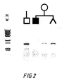
- Figure 2 is an example of a PCR reaction used to detect a deletion in fetal DNA for prenatal diagnosis.
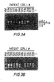
- Figure 3 represents the multiplex DNA amplification of lymphoblast DNA from unrelated male DMD patients.
- A. and B. show two sets of ten samples.
- Each DRL # refers to the R.J. Kleberg Center for Human Genetics Diagnostic Research Laboratory family number.
- MW Hae III digested ⁇ X174 DNA.
- (-) no template DNA added to the reaction.
- the relationship between the amplified region and the region on the gene is indicated to the right of A. The letters correspond to those on Figure 1 .
- Figure 4 represents Multiplex DNA amplification for prenatal diagnosis of DMD. Shown are the results of amplification using DNA from an affected male (AM; lymphoblast DNA) and a male fetus (MF; cultured amniotic fluid cell DNA) from six different families. Both the affected male and the fetal DNAs of DRL #s 521 and 531 display a deletion of region f ( Fig. 1 ); diagnosing these fetuses as affected. In DRL # 43C the affected male is deleted for all regions except f, while the fetus is unaffected. The affected male in DRL # 483 is deleted for region a, while the male fetus is unaffected. Neither of the samples from DRL #s 485 or 469 displayed a deletion with this technique.
- AM lymphoblast DNA
- MF male fetus
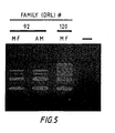
- Figure 5 represents Multiplex DNA amplification from chorionic villus specimen (CVS) DNA.
- CVS chorionic villus specimen
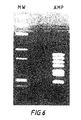
- Figure 6 shows amplification of seven exon regions of the DMD locus.
- oligonucleotide primers as used herein defines a molecule comprised of more than three deoxyribonucleotides or ribonucleotides. Its exact length will depend on many factors relating to the ultimate function and use of the oligonucleotide primer, including temperature, source of the primer and use of the method.
- the oligonucleotide primer can occur naturally, as in a purified restriction digest, or be produced synthetically.
- the oligonucleotide primer is capable of acting as an initiation point for synthesis when placed under conditions which induce synthesis of a primer extension product complementary to a nucleic acid strand.
- the conditions can include the presence of nucleotides and an inducing agent such as a DNA polymerase at a suitable temperature and pH.
- the primer is a single-stranded oligodeoxyribonucleotide of sufficient length to prime the synthesis of an extension product from a specific sequence in the presence of an inducing agent.
- the oligonucleotides are usually at least greater than 12 mers in length. In the preferred embodiment, the oligonucleotide primers are about 18 to 29 mers in length. Sensitivity and specificity of the oligonucleotide primers are determined by the primer length and uniqueness of sequence within a given sample of template DNA.
- the oligonucleotide primer is usually selected for its ability to anneal to intron sequences in the proximity of the 5' or 3' end of the exon or to anneal to a sequence at the intron-exon junction. Since the known deletion defects resulting in genetic diseases result from deletions that include the exons or intron-splice site regions, it is preferable to have primers complementary to intron sequences.
- Each primer pair herein was selected to be substantially complementary to the different strands of each specific sequence to be amplified.
- one primer of each pair is sufficiently complementary to hybridize with a part of the sequence in the sense strand and the other primer of each pair is sufficiently complementary to hybridize with a different part of the same sequence in the anti-sense strand.
- the primer sequence need not reflect the exact sequence of the template, the more closely it does reflect the exact sequence the better the binding during the annealing stage.
- each primer preferably binds at a site on the sequence of interest distant from the other primer.
- the distance between the primers should be sufficient to allow the synthesis of an extension product between the two binding sites, yet close enough so that the extension product of each primer, when separated from its template, can serve as a template for the other primer.
- the extension products from the two paired primers are complementary to each other and can serve as templates for further synthesis. The further apart the binding sites, the more genomic DNA there is which can be screened. However, if the distance is too great the extension products will not efficiently overlap with the primers and thus amplification will not occur.
- extension product refers to the nucleotide sequence which is synthesized from the 3' end of the oligonucleotide primer and which is complementary to the strand to which the oligonucleotide primer is bound.
- each extension product can be distinguished from all the others because it has a different label attached or is of a different size or binds to a specifically labelled oligonucleotide.
- labels can include radioisotopes, fluorescers, chemiluminescers, enzymes and antibodies.
- Various factors affect the choice of the label. These include the effect of the label on the rate of hybridization and binding of the primer to the DNA, the sensitivity of the label, the ease of making the labeled primer, probe or extension products, the ability to automate, available instrumentation, convenience and the like.
- a different radioisotope could be used such as 32 P, 3 H, or 14 C; a different fluorescer such as fluorescein, tetramethylrhodamine, Texas Red or 4-chloro-7- nitrobenzo-2-oxa-1-diazole (NBD); or a mixture of different labels such as radioisotopes, fluorescers and chemiluminescers.
- the primers can be selected such that the amplified extension products for each sequence are of different lengths and thus can be separated by a variety of methods known in the art. Similarily, the extension products could include a restriction fragment length polymorphism which could be used to distinguish different extension products.
- each primer or its extension product can be differentiated from all the other primers when they are in a mixture.
- probes which bind to the amplified extension products could be labeled and sets of probes which distinguish alleles of a single sequence within a multiplex DNA amplification reaction may be used whether or not labelled.
- Each specific, different DNA sequence, which is to be detected herein, can derive from genomic DNA of the organism or exogenous DNA such as virus, bacteria or parasites.
- the source of genomic DNA from the organism to be tested can be blood, hair or tissue (including chorionic villi, amniotic cells, fibroblasts and biopsies).
- the source of DNA may be freshly obtained or have been suitably stored for extended periods of time.
- the DNA must be of sufficient quality to permit amplification.
- the genomic DNA can be prepared by a variety of techniques known to one skilled in the art.
- deletion refers to those genomic DNA sequences in which one or more nucleic acid base has been deleted from the sequence and thus is no longer present in the gene.
- the size of the deletion can affect the sensitivity of the amplification procedure. Generally, the larger the deletion the larger the sensitivity.
- any specific known nucleic acid sequence can be detected by the present method.
- at least part of the sequence is deleted from the genome. It is only necessary that a sufficient number of bases at both ends of the sequence be known in sufficient detail to prepare oligonucleotide primers which will hybridize to the different strands of the desired sequence at relative positions along the sequence.
- the oligonucleotide primers may be prepared using any suitable method, for example, phosphotriester and phosphodiester methods or automated embodiments thereof, the synthesis of oligonucleotides on a modified solid support, the isolation from a biological source (restriction endonuclease digestion), and the generation by enzymatically directed copying of a DNA or RNA template.
- One embodiment of the present invention is a method for simultaneously detecting deletions at a plurality of DNA sequences, comprising the steps of: treating said DNA to form single stranded complementary strands; adding a plurality of paired oligonucleotide primers, each pair specific for a different sequence, one primer of each pair substantially complementary to a part of the sequence in the sense-strand and the other primer of each pair substantially complementary to a different part of the same sequence in the complementary anti-sense strand; annealing the plurality of primers to their complementary sequences; simultaneously extending said plurality of annealed primers from each primer's 3' terminus to synthesize an extension product complementary to the strands annealed to each primer, said extension products, after separation from the complement, serving as templates for the synthesis of an extension product from the other primer of each pair; separating said extension products from said templates to produce single-stranded molecules; amplifying said single-stranded molecules by repeating, at least once, said annealing, extending and
- One preferred embodiment of the present invention is a method for detecting deletions at a plurality of genomic DNA sequences, wherein said sequences are selected from a group of sequences on the X and Y chromosomes. It is preferrable to detect hemizygous genes on the X and Y chromosomes, since this increases the level of sensitivity. When the procedure is used to detect the heterozygous state, it requires quantitative measurement, and thus is much less efficient than detecting the presence or absence of sequences as is done for hemizygous genes.
- the multiplex amplification method of the present invention will detect this by either failing to produce an oligonucleotide sequence or by production of an oligonucleotide sequence of a different size. Furthermore multiple exons can be screened at the same time. Thus, it is easy to detect the presence of a deletion. However, in looking at heterozygous states, where the chromosomes have one normal gene and one deleted gene, the normal gene will produce a normal product, and thus there is the necessity to measure the quantitative difference in the production of extension products.
- a second embodiment of the present invention is to permit simultaneous amplification of multiple, possibly unrelated sequences for the purpose of their simultaneous analysis.
- Such analysis may simply involve the determination of whether exogenous sequences (virus, bacteria or other parasites) are present within a sample of DNA, or might involve the detection of polymorphisms or mutations within a plurality of sequences.
- the polymorphisms or mutations can be detected by a variety of methods well known to those skilled in the art. The methods include, but are not limited to, direct DNA sequencing, allele-specific oligonucleotide hybridization, and competitive oligonucleotide priming.
- the multiplex genomic DNA amplification method is preferably used to detect X-linked diseases resulting from deletions in the genomic DNA sequence.
- Genetic diseases can be caused by a variety of mechanisms including mutations and deletions. The procedure described herein was developed for detection of genetic diseases which result from deletions within the genome. Examples of some X-linked diseases which are candidates for the use of multiplex genomic DNA amplification are ornithine transcarbamylase deficiency, hypoxanthine phosphoribosyltransferase deficiency, steroid sulfatase deficiency and X-linked muscular dystrophy. Other disorders on the X chromosome or genes on the Y chromosome can also be easily detected.
- the procedure is also applicable to the detection of any set of known point mutations within a set of genomic sequences.
- the procedure is also applicable to the simultaneous detection of any set of exogenous DNA sequences in a given DNA sample.
- the procedure is also applicable to the simultaneous detection of any set of polymorphic or variable tandemly repetitive sequences within a genone.
- the advantages of the multiplex amplification system are that numerous diseases or specific DNA sequence alterations can be detected in the same assay. For example, primers to hypoxanthine phosphoribosyltransferfase deficiency, steroid sulfatase deficiency, X-linked muscular dystrophy, ornithine transcarbamylase deficiency and other X-linked diseases can all be run simultaneously on the same sample. Furthermore, the multiplex amplification procedure is useful for very large genes with multiple exons, such as the dystrophin gene. Because of the large size of the dystrophin locus, Mullis type PCR amplification is not able to scan the whole gene in one assay.
- Deletions at the DMD locus can encompass any of the approximately 60 plus exons which are distributed over more than 2 million bases of DNA. Virtually all of these exons are separated by large introns and so up to 60 separate reactions could be required for complete analysis of DMD deletions.
- the present invention of a multiplex genomic DNA amplification for deletion detection can be employed to perform simultaneous examination of multiple exons. For example, oligonucleotide primers flanking separate DMD gene exons can be synthesized and combined and used for multiplex DNA applications. At present, up to at least 7 different DMD gene sequences have been examined simultaneously.
- the entire procedure for the multiplex amplification from start-up to photography of the results takes less than 5 hours.
- the relative locations of the amplified regions do not affect the results and exons have been amplified which have been separated by at least 1000 kb.
- the PCR amplification technique of Mullis is adequate for one and possibly two pair of primers, but when greater than two pairs of primers are used the procedure will not adequately amplify all the appropriate sequences.
- multiplex DNA amplification can be used to simultaneously analyze these polymorphisms to determine the haplotype or to determine the identity or source of DNA (genetic footprinting).
- the number of analyses which can be run simultaneously is unlimited, however, the upper limit is probably about 20 and is dependent on the size differences required for resolution and/or the number of labels or methods which are available to resolve the extension products.
- the ability to simultaneously amplify only 9 exons would allow the detection of greater than 90% of all known DMD deletions in a single reaction.
- the ability to simultaneously amplify even as few as 10 exons allows the rapid and simple diagnosis of DMD deletions using only a few separate reactions. Assuming that there are about 60 exons in the DMD gene and that the exons are widely separated such that primers are needed for every exon, a maximum of 6 separate assays is needed to detect all deletions in this gene.
- the following conditions are currently in use to perform simultaneous amplification of a plurality of separate genomic regions within the human DMD gene. These conditions may need to be slightly modified depending on the particular regions to be amplified, the number and length of sequences to be amplified, and the choice of oligonucleotide primers.
- the time of reaction is highly dependent on the overall sequence length. Thus, as the number of amplified sequences increase and/or the length of amplified sequences increases, the time must be increased.
- the temperature is dependent on the length, the uniqueness of the primer sequence and the relative percentage of GC bases. The longer the primers, the higher the temperature needed. The more unique the sequence, the lower the temperature needed to amplify. GC rich primers need higher temperatures to prevent cross hybridization and to allow unique amplification. However, as the AT percentage increases, higher temperatures cause these primers to melt. Thus, these primers must be lengthened for the reaction to work.
- Template DNA was prepared from the tissue chosen for analysis using a variety of well-established methods known to those skilled in the art. Typically, 100 ⁇ l reaction volumes were utilized. Approximately 500 ng of DNA was added to a solution comprised of the following: 67 mM Tris-HCL [pH 8.8 at 25°]; 6.7 mM magnesium chloride; 16.6 mM ammonium sulfate; 10 mM ⁇ -mercaptoethanol; 6.7 ⁇ M ethylene diamine tetra-acetic acid (EDTA); and 170 ⁇ g/mL bovine serum albumin. This solution can be prepared beforehand and appears to be stable for very long periods of storage at -70°.
- Genomic DNA deletions and/or exogenous DNA sequences were determined by examining the amplification products. For example, the lack of an expected amplification product indicates a deletion. Many methods for this determination are known to those skilled in the art.
- the preferred method involves electrophoresis of about one-twentieth of the reaction on a 1.4% (weight/vol) agarose gel in the following buffer: 40 mM tris-HCl; 20 mM sodium acetate, 1 mM EDTA (adjusted to pH 7.2 with glacial acetic acid), and 0.5 ⁇ g/ ⁇ l. of ethidium bromide. Electrophoresis was performed at 3.7 volts/cM for 100 minutes per 14 cM of agarose gel length. Analysis was completed by examining the electrophoresed reaction products on an ultraviolet radiation transilluminator, and the results were photographed for permanent records.
- the agarose gel is transferred to an appropriate DNA binding medium such as nitrocellulose using well-established procedures, for example, Southern blotting.
- Individual DNA sequences within the amplified DNA fragments can be determined by a variety of techniques including allele-specific oligonucleotide hybridization.
- reaction products may be further analyzed prior to electrophoresis on agarose gel by competitive oligonucleotide primer amplification, using separate allele-specific primers for each amplified DNA sequence of the multiplex amplification reaction products.
- a third method for determining DNA sequence differences within individual amplification products does not require electrophoresis.
- aliquots of the multiplex amplification reaction are sequentially applied to an appropriate DNA binding membrane such as nitrocellulose, and then each aliquot is analyzed via hybridization with individual members of sets of allele-specific oligonucleotide (ASO) probes, each separate aliquot being hybridized with one member of a pair of ASO probes specific for one member of the multiply amplified DNA sequences.
- ASO allele-specific oligonucleotide
- Figure 1 is a schematic representation of the DMD locus. The relative location of the exons used in the DMD gene amplification examples are illustrated.
- each exon is designated a, b, c, d, e, f, or g and corresponds to the same letter in Fig. 1 .
- the exon number is listed. If the exon number is not known, the size of the genomic Hind III fragment containing that exon is listed. Also shown is the size of the exon in base pairs (bp).
- the PCR primer sequences are shown in 5'-3' orientation. The forward primer (F), hybridizes 5' of the exon, and the reverse primer (R), hybridizes 3' of the exon. The size of the amplified fragment obtained with each primer set is also shown.
- Table 2 are the exon and flanking intron sequences for Exon 17.
- the exon is from 227 to 402.
- the primer sequences used to amplify this sequence are 7 to 33 and 396 to 421.
- Table 3 is the exon and flanking intron sequences for Exon d of Table 1 [or, the exon located on a 4.1 Kb Hind III fragment].
- the exon is from 295 to 442.
- the primer sequences used to amplify this sequence are 269 to 293 and 512 to 536.
- Table 4 is the exon and flanking intron sequences for Exon e of Table 1 [0.5 Kb Hind III fragment exon].
- the exon is from 396 to 571.
- the primer sequences used to amplify this sequence are 51 to 75 and 572 to 597.
- Table 5 is the exon and flanking intron sequences for Exon f, Table 1 [overlaps the 1.2 Kb and 3.8 Kb Hind III fragments].
- the exon is from 221 to 406.
- the primer sequences used to amplify this sequence are 26 to 53 and 516 to 541.
- Table 6 is the exon and flanking intron sequences for Exon 12.
- the exon is from 180 to 329.
- the primer sequences used to amplify this sequence are 27 to 52 and 332 to 357.
- Table 7 is the exon and flanking intron sequences for the Exon located on a 10 Kb Hind III fragment.
- the exon is from 1 to 150.
- Table 8 is the exon and flanking intron sequences for the Exon located on a 1.6 Kb Hind III fragment from 512 to 622.
- Table 9 is the exon and flanking intron sequences for the Exon located on a 3.1 Kb Hind III fragment.
- the exon is from 519 to 751.
- Table 10 is the exon and flanking intron sequences for the Exon located on a 1.5 Kb Hind III fragment.
- the exon is from 190 to 337.
- the results of this analysis are shown in Figure 2 .
- the PCR products (one-twentieth of the total reaction) were obtained with template DNA isolated from a control male ⁇ , the male fetus being diagnosed A, the DMD carrier mother ( ⁇ ) and an affected male brother of the fetus ⁇ . Also shown is a DNA molecular weight standard (MW; Hae III digested ⁇ X174 DNA). The results demonstrate that the affected male carries a deletion of' Exon 17, which was not amplified, but that the fetus does not carry the deletion and is therefore unaffected. These results indicate that PCR is useful in the diagnosis of DMD cases containing a deletion involving this exon.
- FIG. 3A and 3B An example of multiplex detection is shown in Figures 3A and 3B .
- oligonucleotide primers represent the flanking regions of six separate DMD gene exons. They were combined into a reaction vial and used for multiplex genomic DNA amplifications. Template DNA was isolated from lymphoblasts (from blood sample). Analysis was by agarose gel electrophoresis.
- Figure 3A When non-deleted DNA was used as a template, the six dispersed regions of the DMD gene were simultaneously and specificially amplified ( Figure 3A , Sample #534). Discrete deletions, which were detected with this method, are shown in Figures 3A and 3B . Several DNA samples containing normal, partial or total DMD gene deletions are shown. Figures 3A and 3B also show a DNA molecular weight standard (MW: Hae III digested ⁇ X174 DNA), and a negative control (-) where no template DNA was added to the reactions. Figure 3A also indicates which amplified DNA fragment corresponds to which exon (a-f) of Figure 1 .
- MW Hae III digested ⁇ X174 DNA
- FIG. 4 shows Multiplex DNA amplification for prenatal diagnosis of DMD. Shown are the results of amplification using DNA from affected males (AM; lymphoblast DNA) and male fetuses (MF; cultured amniotic fluid cell DNA) from six different families. Analysis was as described in Example 1. Both the affected male and the fetal DNA of DRL #s 521 and 531 display a deletion of region f ( Figure 1 ). Thus these fetuses were diagnosed as affected. In DRL # 43C the affected male is deleted for all regions except f, while the fetus is unaffected.
- AM affected males
- MF male fetuses
- DRL #483 The affected male in DRL #483 is deleted for region a, while the male fetus is unaffected. Neither of the samples from DRL #s 485 or 469 displayed a deletion with this technique. Thus, if a deletion defect causes DMD in these families it occurred in an untested exon.
- Figure 5 demonstrates Multiplex DNA amplification from CVS DNA.
- Both the affected male (AM; lymphoblast DNA) and the male fetus (MF; CVS DNA) from DRL # 92 display a deletion of regions e and f ( Fig. 1 ). Thus the fetus was diagnosed as affected.
- CVS DNA from DRL # 120 did not display a deletion with this technique.
- Samples were analyzed as described in Example 1.
Landscapes
- Chemical & Material Sciences (AREA)
- Life Sciences & Earth Sciences (AREA)
- Organic Chemistry (AREA)
- Health & Medical Sciences (AREA)
- Proteomics, Peptides & Aminoacids (AREA)
- Zoology (AREA)
- Wood Science & Technology (AREA)
- Engineering & Computer Science (AREA)
- Genetics & Genomics (AREA)
- Molecular Biology (AREA)
- Analytical Chemistry (AREA)
- General Health & Medical Sciences (AREA)
- Biochemistry (AREA)
- Biophysics (AREA)
- General Engineering & Computer Science (AREA)
- Physics & Mathematics (AREA)
- Biotechnology (AREA)
- Bioinformatics & Cheminformatics (AREA)
- Immunology (AREA)
- Microbiology (AREA)
- Chemical Kinetics & Catalysis (AREA)
- Pathology (AREA)
- Toxicology (AREA)
- Gastroenterology & Hepatology (AREA)
- Medicinal Chemistry (AREA)
- Measuring Or Testing Involving Enzymes Or Micro-Organisms (AREA)
- Saccharide Compounds (AREA)
Abstract
Description
- This invention relates to the field of simultaneous detection of deletions in genomic DNA sequences by the process of amplification of multiple sequences within the hemizygous or homozygous genome. The nucleic acid sequences are amplified by the process of simultaneous multiple repetitive reactions. This method of deletion detection is useful in a variety of areas including screening for genetic disease, and animal husbandry. Multiplex DNA amplification is also applicable to the simultaneous analysis of multiple genomic sequences and is useful in forensic medicine, disease screening, and in the development of recombinant or transgenic organisms.
- This invention is an improvement on currently established procedures for the detection of genetic diseases resulting from mutations and deletions in genomic DNA sequences. Prenatal diagnosis and carrier detection of many X-linked diseases are available via Southern analysis using full length CDNA clones. Unfortunately, there are several major limitations that prevent widespread and routine use of Southern analysis for diagnosis of genetic disease. In many of the X-linked diseases, the defective sequences are unknown and probes are unavailable. In other diseases, such as X-linked muscular dystrophy, there are multiple exons, at least 60, scattered over a large area of genomic DNA, approximately 2.4 million bases. The introns average 35 Kb in length. In the case of muscular dystrophy, at least 7-9 separate cDNA subclones are necessary for Southern blot analysis to resolve each exon-containing restriction fragment for hyplotype assignment or diagnosis of genomic alterations. Furthermore, Southern analysis is an expensive, tedious, and time-consuming technique that requires the use of radioisotopes, making it unsuitable for routine use in clinical laboratories.
- An alternative to Southern analysis for mutation and deletion detection is the polymerase chain reaction (PCR) described by Mullis et al. in
U. S. Patent No. 4,683,195 which issued on July 28, 1987 and by Mullis inU. S. Patent No. 4,683,202 which issued on July 28, 1987 . With PCR, specific regions of a gene can be amplified up to a million-fold from nanogram quantities of genomic DNA. After amplification the nucleic acid sequences can be analyzed for the presence of mutant alleles either by direct DNA sequencing or by hybridization with allele-specific oligonucleotide probes. The PCR technique has proven useful in the diagnosis of several diseases including β-thalassemia, hemophilia A, sickle cell anemia and phenylketonuria. Routine screening for genetic diseases and exogenous DNA sequences, such as virus, with PCR, has been limited by the ability to conduct tests for only a single sequence at a time. Screening for a plurality of possible DNA sequences requires a cumbersomely large number of separate assays, thus increasing the time, expense, and tedium of performing such assays. For example, in some diseases, such as Duchenne muscular dystrophy (DMD), PCR diagnosis has been limited since point mutations leading to DMD have not been identified. Approximately 60% of the cases of DMD are due to deletions. The other 40% are unknown at present, but probably involve mutations of the intron-exon splice sites or the creation of premature stop codons. Thus a large gene like the DMD gene must be screened with multiple assays. - In both
U. S. Patent Nos. 4,683,195 and4,683,202 , procedures are described for amplification of specific sequences. Both patents describe procedures for detecting the presence or absence of at least one specific nucleic acid sequence in a sample containing a mixture of sequences. Although the patents claim at least one sequence and state that multiple sequences can be detected, they do not provide an effective procedure for amplifying multiple sequences at the same time. In the examples, single sequences are amplified or multiple sequences are amplified sequentially. Adding primers for a second sequence is usually possible, but when primers for more than two sequences are added the procedure falls apart. The present application is an improvement on the PCR method and solves the problems encountered when primers for multiple sequences are reacted simultaneously. The present invention describes a procedure for simultaneous amplification of multiple sequences, and for the application of this multiplex amplification procedure in order to detect a plurality of deletions within the same gene or within multiple genes. - The procedures of the present application provide improved methods for the detection of deletions in hemizygous genes on the X and Y chromosomes. The procedures are effective in detecting genetic diseases caused by deletions on the X or Y chromosome, for example, DMD. They are also effective in detecting homozygous deletions and may be used to simultaneously screen for many possible homozygous or hemizygous deletions as long as parts of the appropriate genetic sequences are known. The procedure for multiplex amplification also enables simultaneous analysis of multiple genetic loci regardless of the presence or absence of deletions.
- The present invention is directed to a method for the simultaneous detection of more than two DNA sequences using more than two pairs of primers.
- Although
EP-A-0,237,362 may suggest a multiplex PCR (2 sets of primers) , a further examination of the specification shows that there are no working examples of any multiplex PCR. Further, the one hypothetical example given on page 14, line 29 only uses two different sets of primers. The rest of the description of this application deals with standard PCR conditions. - Nature, Vol. 329, 24th September 1987, pages 293-294 (Chehab et al), at best shows the use of two sets of primers. Looking at
Figure 1 , in the normal control, one can see a nice band for the beta strand. However, there is a very weak response to the alpha strand. This indicates that, even if amplification was obtained, it was a very weak amplification when two sets of primers were used. Again, there is no evidence that more than two sets of primers were used. - American Journal of Human Genetics, Vol. 43, No. 3 Suppl., September 1988, abstract No. (0711) 3.2; (Chamberlain et al) is an abstract which suggests that multiplexing can be done but does not give any of the conditions and does not teach how it can be done.
- Science, Vol. 239, 29th January 1988, pages 491-494 (Stoflet et al) again shows at most two sets of primers being amplified. It further has the problem that it is a much more complicated procedure than the present invention. There is the necessity for the addition of T7 RNA polymerase followed by a sequence and reverse transcriptase to identify the amplification products. This is a much more extensive procedure than the present invention for detecting multiple sequences. There is no evidence in this article of being able to amplify more than two sets of sequences at the same time.
-
EP-A-0,256,630 only describes the use of multiple tests in which each test would have its own separate amplifying sequence. Even if the example were to be described as relating to multiple PCR applications, there is no description of how this would be achieved. There are no examples of any multiplex amplification in this disclosure. - Nucleic Acid Research, vol. 15, no. 22, 1987, pages 9129-9142 (Heilig et al) refers to studying sequences of the intron-exon boundaries. However, at the time of filing of this application, intron sequences had only been published for exons 8 and 19. Many of the other intron sequences were not known at the time of filing. Although the complete cDNA sequence for the dystrophin gene had been published, few exon/intron boundaries were available at the time of filing this application. Of the few intron/exon boundaries that were known, several were incorrect.
- The article in Cell, Vol. 53, 22nd April 1988, pages 219-228, Cell Press (Koenig et al) mostly discloses intron/exon boundaries which were inferred from sequence homology. It should be noted, however, that the sequence date of the present application proved that several of the intron/exon boundaries inferred in this article were wrong.
- Chamberlain et al, Am. J. Human Genetics, 43(3), A17, 1988 speculates on the possibility to use PCR to amplify multiple sequences using several sets of primers but lacks technical details necessary so as to allow carrying out multiplex PCR.
- The present invention seeks to provide a method for simultaneously detecting target DNA sequences in a sample, preferably deletions from a plurality of genomic DNA sequences. The present invention preferably detects X-linked genetic diseases, such as DMD.
- In accordance with the present invention, there is provided a method for simultaneously detecting in a sample target DNA sequences, comprising the steps of:
- providing in a common reaction vessel the sample in single-stranded form and pairs of oligonucleotide primers, each pair specific for a different sequence, one primer of each pair substantially complementary to a part of the sequence in the sense-strand and the other primer of each pair substantially complementary to a different part of the same sequence in the complementary anti-sense strand;
- annealing the pairs of primers to their complementary sequences;
- simultaneously extending said pairs of annealed primers from each primer's 3' terminus to synthesize an extension product complementary to the strands annealed to each primer, said extension products, after separation from their complement, serving as templates for the synthesis of an extension product from the other primer of each pair;
- separating said extension products from said templates to produce single-stranded molecules of the target sequences;
- amplifying said single stranded target sequences by repeating, at least once, said annealing, extending and separating steps; and
- identifying whether said amplified extension products have been synthesised from each different sequence, as a measure of the presence or absence of each target sequence, characterised in that:-
- Additional embodiments include detection of deletions at a plurality of genomic DNA sequences on the X and Y chromosomes or on autosomal chromosomes when the deletions are homozygous. A variety of X-linked diseases can be detected including ornithine transcarbamylase deficiency, hypoxanthine phosphoribosyltransferfase deficiency, steroid sulfatase deficiency and X-linked muscular dystrophy.
- In another embodiment, X-linked muscular dystrophy is detected using more than two paired primers which are complementary to different sequences within the gene coding for the protein dystrophin. Other embodiments include multiple oligonucleotide primers useful in detecting X-linked genetic disease.
- Other and further objets, features and advantages will be apparent from the following description of the presently preferred embodiments of the invention given for the purpose of disclosure when taken in conjunction with the accompanying drawings.
- The invention will be more readily understood from a reading of the following specification and by references to the accompanying drawings, forming a part thereof:
-
Figure 1 is a schematic representation of the DMD gene illustrating the approximate size of the locus, the position of the amplified fragments and the location of the genomic regions that have been cloned and sequenced. -
Figure 2 is an example of a PCR reaction used to detect a deletion in fetal DNA for prenatal diagnosis. -
Figure 3 represents the multiplex DNA amplification of lymphoblast DNA from unrelated male DMD patients. A. and B. show two sets of ten samples. Each DRL # refers to the R.J. Kleberg Center for Human Genetics Diagnostic Research Laboratory family number. MW: Hae III digested φX174 DNA. (-): no template DNA added to the reaction. The relationship between the amplified region and the region on the gene is indicated to the right of A. The letters correspond to those onFigure 1 . -
Figure 4 represents Multiplex DNA amplification for prenatal diagnosis of DMD. Shown are the results of amplification using DNA from an affected male (AM; lymphoblast DNA) and a male fetus (MF; cultured amniotic fluid cell DNA) from six different families. Both the affected male and the fetal DNAs of DRL #s 521 and 531 display a deletion of region f (Fig. 1 ); diagnosing these fetuses as affected. InDRL # 43C the affected male is deleted for all regions except f, while the fetus is unaffected. The affected male inDRL # 483 is deleted for region a, while the male fetus is unaffected. Neither of the samples from DRL #s 485 or 469 displayed a deletion with this technique. -
Figure 5 represents Multiplex DNA amplification from chorionic villus specimen (CVS) DNA. Both the affected male (AM; lymphoblast DNA) and the male fetus (MF; CVS DNA) fromDRL # 92 display a deletion of regions e and f (Fig. 1 ), diagnosing the fetus as affected. CVS DNA fromDRL # 120 did not display a deletion with this technique. -
Figure 6 shows amplification of seven exon regions of the DMD locus. - The drawings are not necessarily to scale and certain features of the invention may be exaggerated in scale or shown in schematic form in the interests of clarity and conciseness.
- The term "oligonucleotide primers" as used herein defines a molecule comprised of more than three deoxyribonucleotides or ribonucleotides. Its exact length will depend on many factors relating to the ultimate function and use of the oligonucleotide primer, including temperature, source of the primer and use of the method. The oligonucleotide primer can occur naturally, as in a purified restriction digest, or be produced synthetically. The oligonucleotide primer is capable of acting as an initiation point for synthesis when placed under conditions which induce synthesis of a primer extension product complementary to a nucleic acid strand. The conditions can include the presence of nucleotides and an inducing agent such as a DNA polymerase at a suitable temperature and pH. In the preferred embodiment, the primer is a single-stranded oligodeoxyribonucleotide of sufficient length to prime the synthesis of an extension product from a specific sequence in the presence of an inducing agent. In the deletion detection procedure, the oligonucleotides are usually at least greater than 12 mers in length. In the preferred embodiment, the oligonucleotide primers are about 18 to 29 mers in length. Sensitivity and specificity of the oligonucleotide primers are determined by the primer length and uniqueness of sequence within a given sample of template DNA. Primers which are too short, for example less than about 12 mers, may show non-specific binding to a wide variety of sequences in the genomic DNA and thus are not very helpful. In the preferred embodiment, the oligonucleotide primer is usually selected for its ability to anneal to intron sequences in the proximity of the 5' or 3' end of the exon or to anneal to a sequence at the intron-exon junction. Since the known deletion defects resulting in genetic diseases result from deletions that include the exons or intron-splice site regions, it is preferable to have primers complementary to intron sequences.
- Each primer pair herein was selected to be substantially complementary to the different strands of each specific sequence to be amplified. Thus, one primer of each pair is sufficiently complementary to hybridize with a part of the sequence in the sense strand and the other primer of each pair is sufficiently complementary to hybridize with a different part of the same sequence in the anti-sense strand. Thus, although the primer sequence need not reflect the exact sequence of the template, the more closely it does reflect the exact sequence the better the binding during the annealing stage.
- Within a primer pair, each primer preferably binds at a site on the sequence of interest distant from the other primer. In the preferred embodiment the distance between the primers should be sufficient to allow the synthesis of an extension product between the two binding sites, yet close enough so that the extension product of each primer, when separated from its template, can serve as a template for the other primer. The extension products from the two paired primers are complementary to each other and can serve as templates for further synthesis. The further apart the binding sites, the more genomic DNA there is which can be screened. However, if the distance is too great the extension products will not efficiently overlap with the primers and thus amplification will not occur.
- As used herein the term "extension product" refers to the nucleotide sequence which is synthesized from the 3' end of the oligonucleotide primer and which is complementary to the strand to which the oligonucleotide primer is bound.
- As used herein the term "differentially labeled" shall indicate that each extension product can be distinguished from all the others because it has a different label attached or is of a different size or binds to a specifically labelled oligonucleotide. One skilled in the art will recognize that a variety of labels are available. For example, these can include radioisotopes, fluorescers, chemiluminescers, enzymes and antibodies. Various factors affect the choice of the label. These include the effect of the label on the rate of hybridization and binding of the primer to the DNA, the sensitivity of the label, the ease of making the labeled primer, probe or extension products, the ability to automate, available instrumentation, convenience and the like. For example, a different radioisotope could be used such as 32P, 3H, or 14C; a different fluorescer such as fluorescein, tetramethylrhodamine, Texas Red or 4-chloro-7- nitrobenzo-2-oxa-1-diazole (NBD); or a mixture of different labels such as radioisotopes, fluorescers and chemiluminescers. Alternatively, the primers can be selected such that the amplified extension products for each sequence are of different lengths and thus can be separated by a variety of methods known in the art. Similarily, the extension products could include a restriction fragment length polymorphism which could be used to distinguish different extension products. In these examples, each primer or its extension product can be differentiated from all the other primers when they are in a mixture. Alternatively, probes which bind to the amplified extension products could be labeled and sets of probes which distinguish alleles of a single sequence within a multiplex DNA amplification reaction may be used whether or not labelled.
- Each specific, different DNA sequence, which is to be detected herein, can derive from genomic DNA of the organism or exogenous DNA such as virus, bacteria or parasites. The source of genomic DNA from the organism to be tested can be blood, hair or tissue (including chorionic villi, amniotic cells, fibroblasts and biopsies). The source of DNA may be freshly obtained or have been suitably stored for extended periods of time. The DNA must be of sufficient quality to permit amplification. The genomic DNA can be prepared by a variety of techniques known to one skilled in the art.
- As used herein, the term "deletion" refers to those genomic DNA sequences in which one or more nucleic acid base has been deleted from the sequence and thus is no longer present in the gene. The size of the deletion can affect the sensitivity of the amplification procedure. Generally, the larger the deletion the larger the sensitivity.
- Any specific known nucleic acid sequence can be detected by the present method. Preferably, at least part of the sequence is deleted from the genome. It is only necessary that a sufficient number of bases at both ends of the sequence be known in sufficient detail to prepare oligonucleotide primers which will hybridize to the different strands of the desired sequence at relative positions along the sequence.
- The oligonucleotide primers may be prepared using any suitable method, for example, phosphotriester and phosphodiester methods or automated embodiments thereof, the synthesis of oligonucleotides on a modified solid support, the isolation from a biological source (restriction endonuclease digestion), and the generation by enzymatically directed copying of a DNA or RNA template.
- One embodiment of the present invention is a method for simultaneously detecting deletions at a plurality of DNA sequences, comprising the steps of: treating said DNA to form single stranded complementary strands; adding a plurality of paired oligonucleotide primers, each pair specific for a different sequence, one primer of each pair substantially complementary to a part of the sequence in the sense-strand and the other primer of each pair substantially complementary to a different part of the same sequence in the complementary anti-sense strand; annealing the plurality of primers to their complementary sequences; simultaneously extending said plurality of annealed primers from each primer's 3' terminus to synthesize an extension product complementary to the strands annealed to each primer, said extension products, after separation from the complement, serving as templates for the synthesis of an extension product from the other primer of each pair; separating said extension products from said templates to produce single-stranded molecules; amplifying said single-stranded molecules by repeating, at least once, said annealing, extending and separating steps; and identifying said amplified extension product from each different sequence.
- One preferred embodiment of the present invention is a method for detecting deletions at a plurality of genomic DNA sequences, wherein said sequences are selected from a group of sequences on the X and Y chromosomes. It is preferrable to detect hemizygous genes on the X and Y chromosomes, since this increases the level of sensitivity. When the procedure is used to detect the heterozygous state, it requires quantitative measurement, and thus is much less efficient than detecting the presence or absence of sequences as is done for hemizygous genes. For example, if part of an exon has been deleted the multiplex amplification method of the present invention will detect this by either failing to produce an oligonucleotide sequence or by production of an oligonucleotide sequence of a different size. Furthermore multiple exons can be screened at the same time. Thus, it is easy to detect the presence of a deletion. However, in looking at heterozygous states, where the chromosomes have one normal gene and one deleted gene, the normal gene will produce a normal product, and thus there is the necessity to measure the quantitative difference in the production of extension products.
- A second embodiment of the present invention is to permit simultaneous amplification of multiple, possibly unrelated sequences for the purpose of their simultaneous analysis. Such analysis may simply involve the determination of whether exogenous sequences (virus, bacteria or other parasites) are present within a sample of DNA, or might involve the detection of polymorphisms or mutations within a plurality of sequences. The polymorphisms or mutations can be detected by a variety of methods well known to those skilled in the art. The methods include, but are not limited to, direct DNA sequencing, allele-specific oligonucleotide hybridization, and competitive oligonucleotide priming.
- The multiplex genomic DNA amplification method is preferably used to detect X-linked diseases resulting from deletions in the genomic DNA sequence. Genetic diseases can be caused by a variety of mechanisms including mutations and deletions. The procedure described herein was developed for detection of genetic diseases which result from deletions within the genome. Examples of some X-linked diseases which are candidates for the use of multiplex genomic DNA amplification are ornithine transcarbamylase deficiency, hypoxanthine phosphoribosyltransferase deficiency, steroid sulfatase deficiency and X-linked muscular dystrophy. Other disorders on the X chromosome or genes on the Y chromosome can also be easily detected. The procedure is also applicable to the detection of any set of known point mutations within a set of genomic sequences. The procedure is also applicable to the simultaneous detection of any set of exogenous DNA sequences in a given DNA sample. The procedure is also applicable to the simultaneous detection of any set of polymorphic or variable tandemly repetitive sequences within a genone.
- The advantages of the multiplex amplification system are that numerous diseases or specific DNA sequence alterations can be detected in the same assay. For example, primers to hypoxanthine phosphoribosyltransferfase deficiency, steroid sulfatase deficiency, X-linked muscular dystrophy, ornithine transcarbamylase deficiency and other X-linked diseases can all be run simultaneously on the same sample. Furthermore, the multiplex amplification procedure is useful for very large genes with multiple exons, such as the dystrophin gene. Because of the large size of the dystrophin locus, Mullis type PCR amplification is not able to scan the whole gene in one assay. Thus, it is necessary for multiple site amplification within the gene to detect all possible deletions which could result in disease. Deletions at the DMD locus can encompass any of the approximately 60 plus exons which are distributed over more than 2 million bases of DNA. Virtually all of these exons are separated by large introns and so up to 60 separate reactions could be required for complete analysis of DMD deletions. To simplify this task, the present invention of a multiplex genomic DNA amplification for deletion detection can be employed to perform simultaneous examination of multiple exons. For example, oligonucleotide primers flanking separate DMD gene exons can be synthesized and combined and used for multiplex DNA applications. At present, up to at least 7 different DMD gene sequences have been examined simultaneously. The entire procedure for the multiplex amplification from start-up to photography of the results takes less than 5 hours. The relative locations of the amplified regions do not affect the results and exons have been amplified which have been separated by at least 1000 kb. The PCR amplification technique of Mullis is adequate for one and possibly two pair of primers, but when greater than two pairs of primers are used the procedure will not adequately amplify all the appropriate sequences.
- One skilled in the art readily appreciates that as more exon gene sequences become available the applicability of this test will expand to examine for deletions in multiple genes at the same time or examine multiple sites within the same gene at the same time. The later example is important for genes such as dystrophin which are so large that primers annealed to the ends of the gene will not traverse the whole gene sequence. Thus the necessity of doing multiple analysis to detect deletions in different regions of the gene. In addition, as specific mutations within multiple unrelated genes become known, multiplex DNA amplification can be applied to simultaneously assay for the presence of any of these mutations.
- Furthermore, as specific or highly variable DNA sequence polymorphisms become known in various genetic Loci, multiplex DNA amplification can be used to simultaneously analyze these polymorphisms to determine the haplotype or to determine the identity or source of DNA (genetic footprinting).
- The number of analyses which can be run simultaneously is unlimited, however, the upper limit is probably about 20 and is dependent on the size differences required for resolution and/or the number of labels or methods which are available to resolve the extension products. The ability to simultaneously amplify only 9 exons would allow the detection of greater than 90% of all known DMD deletions in a single reaction. The ability to simultaneously amplify even as few as 10 exons allows the rapid and simple diagnosis of DMD deletions using only a few separate reactions. Assuming that there are about 60 exons in the DMD gene and that the exons are widely separated such that primers are needed for every exon, a maximum of 6 separate assays is needed to detect all deletions in this gene. Under the same assumptions the Mullis PCR method would require 60 separate reactions to detect the deletions in this gene. Thus, as the size of the gene increases and the number of exons which cannot be detected together increases the advantages of this method are greatly enhanced. Furthermore, use of an automatic PCR apparatus (such as that produced by Perkin-Elmer/Cetus) and DNA sequencing machines will facilitate resolution and detection of amplified DNA fragments, will help automate the assay and will lead to the method being applied routinely in clinical laboratories without the need for highly trained research personnel.
- The following examples are offered by way of illustration and are not intended to limit the invention in any manner. In the examples all percentages are by weight, if for solids and by volume if for liquids, and all temperatures are in degrees Celsius unless otherwise noted.
- The following conditions are currently in use to perform simultaneous amplification of a plurality of separate genomic regions within the human DMD gene. These conditions may need to be slightly modified depending on the particular regions to be amplified, the number and length of sequences to be amplified, and the choice of oligonucleotide primers. The time of reaction is highly dependent on the overall sequence length. Thus, as the number of amplified sequences increase and/or the length of amplified sequences increases, the time must be increased. The temperature is dependent on the length, the uniqueness of the primer sequence and the relative percentage of GC bases. The longer the primers, the higher the temperature needed. The more unique the sequence, the lower the temperature needed to amplify. GC rich primers need higher temperatures to prevent cross hybridization and to allow unique amplification. However, as the AT percentage increases, higher temperatures cause these primers to melt. Thus, these primers must be lengthened for the reaction to work.
- Template DNA was prepared from the tissue chosen for analysis using a variety of well-established methods known to those skilled in the art. Typically, 100 µl reaction volumes were utilized. Approximately 500 ng of DNA was added to a solution comprised of the following: 67 mM Tris-HCL [pH 8.8 at 25°]; 6.7 mM magnesium chloride; 16.6 mM ammonium sulfate; 10 mM β-mercaptoethanol; 6.7 µM ethylene diamine tetra-acetic acid (EDTA); and 170 µg/mL bovine serum albumin. This solution can be prepared beforehand and appears to be stable for very long periods of storage at -70°. The enzyme, Taq polymerase, was added to achieve a final concentration of 100 units/mL. This reaction mixture was gently mixed. The reaction mixture was overlaid with about 50 µL of paraffin oil, and the reaction vessel (preferably a 0.5 ml microcentrifuge tube) was centrifuged at 14,000 x g for 10 sec. Amplification was performed either by manually transferring the reaction vessels between glycerol filled heat blocks at the appropriate temperatures, or automatically transferring the reaction vessels with a Perkin-Elmer/Cetus thermocycler using the 'step-cycle' functions. The reaction was controlled by regulated and repetitive temperature changes of various duration. Initially the reaction was heated to 94° for 7 minutes. Subsequently 25 cycles of the following temperature durations were applied: 94° for 1 minute, then 55° for 45 seconds, then 65° for 3 1/2 minutes. Following completion of the final cycle the reaction was incubated at 65° for an additional 7 minutes. Reactions were then stored at 4° until analysis.
- Genomic DNA deletions and/or exogenous DNA sequences were determined by examining the amplification products. For example, the lack of an expected amplification product indicates a deletion. Many methods for this determination are known to those skilled in the art. The preferred method involves electrophoresis of about one-twentieth of the reaction on a 1.4% (weight/vol) agarose gel in the following buffer: 40 mM tris-HCl; 20 mM sodium acetate, 1 mM EDTA (adjusted to pH 7.2 with glacial acetic acid), and 0.5µg/µl. of ethidium bromide. Electrophoresis was performed at 3.7 volts/cM for 100 minutes per 14 cM of agarose gel length. Analysis was completed by examining the electrophoresed reaction products on an ultraviolet radiation transilluminator, and the results were photographed for permanent records.
- When the analysis requires determination of DNA sequence polymorphisms or mutations within individual amplification products the agarose gel is transferred to an appropriate DNA binding medium such as nitrocellulose using well-established procedures, for example, Southern blotting. Individual DNA sequences within the amplified DNA fragments can be determined by a variety of techniques including allele-specific oligonucleotide hybridization. Alternatively, reaction products may be further analyzed prior to electrophoresis on agarose gel by competitive oligonucleotide primer amplification, using separate allele-specific primers for each amplified DNA sequence of the multiplex amplification reaction products.
- A third method for determining DNA sequence differences within individual amplification products does not require electrophoresis. In this method, aliquots of the multiplex amplification reaction are sequentially applied to an appropriate DNA binding membrane such as nitrocellulose, and then each aliquot is analyzed via hybridization with individual members of sets of allele-specific oligonucleotide (ASO) probes, each separate aliquot being hybridized with one member of a pair of ASO probes specific for one member of the multiply amplified DNA sequences.
-
Figure 1 is a schematic representation of the DMD locus. The relative location of the exons used in the DMD gene amplification examples are illustrated. - For detection of DMD, a variety of probes can be used either in individual PCR reactions or in combinations in multiplex PCR reactions. These probes are shown in Table 1.
Table 1. Summary of DMD gene multiplex amplification primer sets. Exon and Size Primer Sequence Amplified Deleted a. Exon 8
(182bp)F-GTCCTTTACACACTTTACCTGTTGAG
R-GGCCTCATTCTCATGTTCTAATTAG360 bp 11.3% b. Exon 17
(178bp)F-GACTTTCGATGTTGAGATTACTTTCCC
R-AAGCTTGAGATGCTCTCACCTTTTCC416 bp 9.4% c. Exon 19
(88bp)F-TTCTACCACATCCCATTTTCTTCCA
R-GATGGCAAAAGTGTTGAGAAAAAGTC459 bp 10.3% d. 4.1Kb Hind III
(148bp)F-CTTGATCCATATGCTTTTACCTGCA
R-TCCATCACCCTTCAGAACCTGATCT268 bp 4.0% e. 0.5Kb Hind III
(176bp)F-AAACATGGAACATCCTTGTGGGGAC
R-CATTCCTATTAGATCTGTCGCCCTAC547 bp 8.4% f. 1.2/3.8Kb Hind III
(159bp)F-TTGAATACATTGGTTAAATCCCAACATG
R-CCTGAATAAAGTCTTCCTTACCACAC506 bp 18.2% g. Exon 12
(151bp)F-GATAGTGGGCTTTACTTACATCCTTC
R-GAAAGCACGCAACATAAGATACACCT337 bp 9.6% Total: 38% - In Table 1 each exon is designated a, b, c, d, e, f, or g and corresponds to the same letter in
Fig. 1 . When the exon number is known it is listed. If the exon number is not known, the size of the genomic Hind III fragment containing that exon is listed. Also shown is the size of the exon in base pairs (bp). The PCR primer sequences are shown in 5'-3' orientation. The forward primer (F), hybridizes 5' of the exon, and the reverse primer (R), hybridizes 3' of the exon. The size of the amplified fragment obtained with each primer set is also shown. - The percentage of analyzed DMD patients that are deleted for each indicated exon is shown in column four. This total number is less than the sum of the individual exon deletion frequencies because many deletions encompass multiple exons.
- In Table 2 are the exon and flanking intron sequences for Exon 17. The exon is from 227 to 402. The primer sequences used to amplify this sequence are 7 to 33 and 396 to 421.

- In Table 3 is the exon and flanking intron sequences for Exon d of Table 1 [or, the exon located on a 4.1 Kb Hind III fragment]. The exon is from 295 to 442. The primer sequences used to amplify this sequence are 269 to 293 and 512 to 536.

- In Table 4 is the exon and flanking intron sequences for Exon e of Table 1 [0.5 Kb Hind III fragment exon]. The exon is from 396 to 571. The primer sequences used to amplify this sequence are 51 to 75 and 572 to 597.

- In Table 5 is the exon and flanking intron sequences for Exon f, Table 1 [overlaps the 1.2 Kb and 3.8 Kb Hind III fragments]. The exon is from 221 to 406. The primer sequences used to amplify this sequence are 26 to 53 and 516 to 541.


- In Table 7 is the exon and flanking intron sequences for the Exon located on a 10 Kb Hind III fragment. The exon is from 1 to 150.

- In Table 8 is the exon and flanking intron sequences for the Exon located on a 1.6 Kb Hind III fragment from 512 to 622.


- In Table 9 is the exon and flanking intron sequences for the Exon located on a 3.1 Kb Hind III fragment. The exon is from 519 to 751.


- In Table 10 is the exon and flanking intron sequences for the Exon located on a 1.5 Kb Hind III fragment. The exon is from 190 to 337.

- An example of prenatal diagnosis with PCR deletion detection is demonstrated using synthesized oligonucleotide primers (set b, Table 1). This primer set corresponds to the intron sequences flanking Exon 17 of the human DMD gene, a region which has been isolated and sequenced (Table 2).
- The results of this analysis are shown in
Figure 2 . The PCR products (one-twentieth of the total reaction) were obtained with template DNA isolated from a control male □, the male fetus being diagnosed A, the DMD carrier mother (○) and an affected male brother of the fetus ■. Also shown is a DNA molecular weight standard (MW; Hae III digested φX174 DNA). The results demonstrate that the affected male carries a deletion of' Exon 17, which was not amplified, but that the fetus does not carry the deletion and is therefore unaffected. These results indicate that PCR is useful in the diagnosis of DMD cases containing a deletion involving this exon. - An example of multiplex detection is shown in
Figures 3A and 3B . - This analysis was done using six primer pairs (sets a-f, Table 1) and the conditions described in Example 1. Automatic rather than manual amplification was performed. These oligonucleotide primers represent the flanking regions of six separate DMD gene exons. They were combined into a reaction vial and used for multiplex genomic DNA amplifications. Template DNA was isolated from lymphoblasts (from blood sample). Analysis was by agarose gel electrophoresis.
- When non-deleted DNA was used as a template, the six dispersed regions of the DMD gene were simultaneously and specificially amplified (
Figure 3A , Sample #534). Discrete deletions, which were detected with this method, are shown inFigures 3A and 3B . Several DNA samples containing normal, partial or total DMD gene deletions are shown.Figures 3A and 3B also show a DNA molecular weight standard (MW: Hae III digested φX174 DNA), and a negative control (-) where no template DNA was added to the reactions.Figure 3A also indicates which amplified DNA fragment corresponds to which exon (a-f) ofFigure 1 . - Multiplex PCR has been used successfully in several prenatal diagnoses. The conditions are as described above in Example 1.
Figure 4 shows Multiplex DNA amplification for prenatal diagnosis of DMD. Shown are the results of amplification using DNA from affected males (AM; lymphoblast DNA) and male fetuses (MF; cultured amniotic fluid cell DNA) from six different families. Analysis was as described in Example 1. Both the affected male and the fetal DNA of DRL #s 521 and 531 display a deletion of region f (Figure 1 ). Thus these fetuses were diagnosed as affected. InDRL # 43C the affected male is deleted for all regions except f, while the fetus is unaffected. The affected male inDRL # 483 is deleted for region a, while the male fetus is unaffected. Neither of the samples from DRL #s 485 or 469 displayed a deletion with this technique. Thus, if a deletion defect causes DMD in these families it occurred in an untested exon. -
Figure 5 demonstrates Multiplex DNA amplification from CVS DNA. Both the affected male (AM; lymphoblast DNA) and the male fetus (MF; CVS DNA) fromDRL # 92 display a deletion of regions e and f (Fig. 1 ). Thus the fetus was diagnosed as affected. CVS DNA fromDRL # 120 did not display a deletion with this technique. Samples were analyzed as described in Example 1. These results demonstrate that the multiplex amplification technique works well for prenatal diagnosis when CVS DNA is used as the template for amplification. - This example demonstrates that seven separate DNA sequences can be simultaneously amplified using the multiplex amplification technique. Conditions were as described in Example 1. Primer sets a-g (Table 1) were added to the reaction. Thus seven exon regions of the DMD gene (
Figure 1 ) were amplified (Figure 6 ).
Claims (7)
- A method for simultaneously detecting in a -sample target DNA sequences, comprising the steps of:providing in a common reaction vessel the sample in single-stranded form and pairs of oilgonucleotide primers, each pair specific for a different sequence, one primer of each pair substantially complementary to a part of the sequence in the sense-strand and the other primer of each pair substantially complementary to a different part of the same sequence in the complementary anti-sense strand;annealing the pairs of primers to their complementary sequences;simultaneously extending said pairs of annealed primers from each primer's 3' terminus to synthesize an extension product complementary to the strands annealed to each primer, said extension products, after separation from their complement, serving as templates for the synthesis of an extension product from the other primer of each pair;separating said extension products from said templates to produce single-stranded molecules of the target sequences;amplifying said single stranded target sequences by repeating, at least once, said annealing, extending and separating steps; andidentifying whether said amplified extension products have been synthesised from each different sequence, as a measure of the presence or absence of each target sequence, characterised in that:-the method is adapted to detect simultaneously more than two target sequences by utilising more than two pairs of oligonucleotide primers.
- The method of Claim 1 for detecting deletions from genomic DNA sequences, wherein said sequences are selected from the group of sequences on the X and Y chromosomes.
- The method of Claim 2 for the detection of X-linked disease, wherein said genomic DNA sequences contain a deletion that causes a genetic disease.
- The method of Claim 3 for the detection of said X-linked genetic diseases selected from the group consisting of ornithine transcarbamylase deficiency, hypoxanthine phosphoribosyltransferfase deficiency, steroid sulfatase deficiency and X-linked muscular dystrophy.
- The method of Claim 4 for the detection of X-linked muscular dystrophy, wherein each pair of said primers is complementary to different sequences within the gene coding for the dystrophin protein.
- The method of Claim 5, wherein the pairs of primers are selected from the group consisting of:(1) 5'-GACTTTCGATGTTGAGATTACTTTCCC-3'(2) 5'-AAGCTTGAGATGCTCTCACCTTTTCC-3',(1) 5 '-GTCCTTTACACACTTTACCTGTTGAG-3'(2) 5'-GGCCTCATTCTCATGTTCTAATTAG-3',(1) 5'-AAACATGGAACATCCTTGTGGGGAC-3'(2) 5'-CATTCCTATTAGATCTGTCGCCCTAC-3',(1) 5'-GATAGTGGGCTTTACTTACATCCTTC-3'(2) 5'-GAAAGCACGCAACATAAGATACACCT-3',(1) 5'-CTTGATCCATATGCTTTTACCTGCA-3'(2) 5'-TCCATCACCCTTCAGAACCTGATCT-3',(1) 5'-TTGAATACATTGGTTAAATCCCAACATG-3'(2) 5'-CCTGAATAAAGTCTTCCTTACCACAC-3',and(1) 5'-TTCTACCACATCCCATTTTCTTCCA-3'(2) 5'-GATGGCAAAAGTGTTGAGAAAAAGTC-3'.
- The method of Claim 3, wherein said genomic DNA is from fetal tissue.
Applications Claiming Priority (2)
| Application Number | Priority Date | Filing Date | Title |
|---|---|---|---|
| US25668988A | 1988-10-12 | 1988-10-12 | |
| US256689 | 1988-10-12 |
Publications (4)
| Publication Number | Publication Date |
|---|---|
| EP0364255A2 EP0364255A2 (en) | 1990-04-18 |
| EP0364255A3 EP0364255A3 (en) | 1991-07-17 |
| EP0364255B1 EP0364255B1 (en) | 2001-05-23 |
| EP0364255B2 true EP0364255B2 (en) | 2008-12-31 |
Family
ID=22973199
Family Applications (1)
| Application Number | Title | Priority Date | Filing Date |
|---|---|---|---|
| EP89310424A Expired - Lifetime EP0364255B2 (en) | 1988-10-12 | 1989-10-11 | Multiplex genomic DNA amplification for deletion detection |
Country Status (9)
| Country | Link |
|---|---|
| US (1) | US5582989A (en) |
| EP (1) | EP0364255B2 (en) |
| JP (1) | JPH07136000A (en) |
| KR (1) | KR900006520A (en) |
| AT (1) | ATE201451T1 (en) |
| AU (2) | AU634175B2 (en) |
| CA (1) | CA1339731C (en) |
| DE (1) | DE68929299T3 (en) |
| ES (1) | ES2156851T5 (en) |
Families Citing this family (129)
| Publication number | Priority date | Publication date | Assignee | Title |
|---|---|---|---|---|
| IE61148B1 (en) * | 1988-03-10 | 1994-10-05 | Ici Plc | Method of detecting nucleotide sequences |
| CA1339731C (en) * | 1988-10-12 | 1998-03-17 | Charles T. Caskey | Multiplex genomic dna amplification for deletion detection |
| US5192659A (en) * | 1989-08-25 | 1993-03-09 | Genetype Ag | Intron sequence analysis method for detection of adjacent and remote locus alleles as haplotypes |
| FR2667079B1 (en) * | 1990-09-26 | 1993-08-13 | Genset Sa | METHODS, PRIMER OLIGONUCLEOTIDES AND PROBE OLIGONUCLEOTIDES FOR DETECTION OF PATHOGENIC BACTERIA OF THE ORAL CAVITY. |
| IE66125B1 (en) * | 1991-01-31 | 1995-12-13 | Zeneca Ltd | Detection method for variant nucleotides |
| US6100099A (en) * | 1994-09-06 | 2000-08-08 | Abbott Laboratories | Test strip having a diagonal array of capture spots |
| DE69329641T2 (en) * | 1992-03-31 | 2001-06-21 | Abbott Laboratories, Abbott Park | METHOD FOR MULTIPLE LIGASE CHAIN REACTION |
| US5869252A (en) * | 1992-03-31 | 1999-02-09 | Abbott Laboratories | Method of multiplex ligase chain reaction |
| US5633134A (en) * | 1992-10-06 | 1997-05-27 | Ig Laboratories, Inc. | Method for simultaneously detecting multiple mutations in a DNA sample |
| US5470723A (en) * | 1993-05-05 | 1995-11-28 | Becton, Dickinson And Company | Detection of mycobacteria by multiplex nucleic acid amplification |
| US5422252A (en) * | 1993-06-04 | 1995-06-06 | Becton, Dickinson And Company | Simultaneous amplification of multiple targets |
| SG63589A1 (en) * | 1993-05-14 | 1999-03-30 | Johnson & Johnson Clin Diag | Diagnostic compositions elements methods and test kits for amplification and detection of two or more dna's using primers having matched melting temperatures |
| US5925517A (en) * | 1993-11-12 | 1999-07-20 | The Public Health Research Institute Of The City Of New York, Inc. | Detectably labeled dual conformation oligonucleotide probes, assays and kits |
| US5928905A (en) * | 1995-04-18 | 1999-07-27 | Glaxo Group Limited | End-complementary polymerase reaction |
| NL9401082A (en) * | 1994-06-28 | 1996-02-01 | Ingeny Bv | Method for detecting mutations. |
| US5550020A (en) * | 1994-07-08 | 1996-08-27 | Visible Genetics Inc. | Method, reagents and kit for diagnosis and targeted screening for retinoblastoma |
| US20020055101A1 (en) | 1995-09-11 | 2002-05-09 | Michel G. Bergeron | Specific and universal probes and amplification primers to rapidly detect and identify common bacterial pathogens and antibiotic resistance genes from clinical specimens for routine diagnosis in microbiology laboratories |
| CN1151269C (en) * | 1995-06-06 | 2004-05-26 | 扬·维吉 | Method of and apparatus for diagnostic DNA testing |
| US20030022184A1 (en) * | 1996-02-12 | 2003-01-30 | Oncormed. Inc. | Coding sequences of the human BRCA1 gene |
| US6838256B2 (en) | 1996-02-12 | 2005-01-04 | Gene Logic Inc. | Coding sequences of the human BRCA1 gene |
| JP3898228B2 (en) * | 1996-04-12 | 2007-03-28 | ザ パブリック ヘルス リサーチ インスティチュート オブ ザ シティー オブ ニューヨーク インク | Detection probes, kits and assays |
| US5811269A (en) * | 1996-04-30 | 1998-09-22 | Becton, Dickinson And Company | Detection of mycobacteria by multiplex nucleic acid amplification |
| US6287763B1 (en) * | 1996-06-10 | 2001-09-11 | Millennium Pharmaceuticals, Inc. | Screening methods for compounds useful in the regulation of body weight |
| US6124092A (en) * | 1996-10-04 | 2000-09-26 | The Perkin-Elmer Corporation | Multiplex polynucleotide capture methods and compositions |
| US20030165971A1 (en) * | 1996-10-31 | 2003-09-04 | Billing-Medel Patricia A. | Reagents and methods useful for detecting diseases of the breast |
| US20050153373A1 (en) * | 1996-10-31 | 2005-07-14 | Billing-Medel Patricia A. | Reagents and methods useful for detecting diseases of the breast |
| US20050202499A1 (en) * | 1996-10-31 | 2005-09-15 | Billing-Medel Patricia A. | Reagents and methods useful for detecting diseases of the breast |
| US20030049636A1 (en) | 1999-05-03 | 2003-03-13 | Bergeron Michel G. | Species-specific, genus-specific and universal DNA probes and amplification primers to rapidly detect and identify common bacterial and fungal pathogens and associated antibiotic resistance genes from clinical specimens for diagnosis in microbiology laboratories |
| US20100267012A1 (en) | 1997-11-04 | 2010-10-21 | Bergeron Michel G | Highly conserved genes and their use to generate probes and primers for detection of microorganisms |
| WO1998030721A1 (en) * | 1997-01-10 | 1998-07-16 | Pioneer Hi-Bred International, Inc. | Hybridization-based genetic amplification and analysis |
| US6048689A (en) * | 1997-03-28 | 2000-04-11 | Gene Logic, Inc. | Method for identifying variations in polynucleotide sequences |
| US20020137904A1 (en) * | 1998-03-27 | 2002-09-26 | Patricia A. Billing-Medel | Reagents and methods useful for detecting diseases of the gastrointestinal tract |
| EP0975804A2 (en) * | 1997-04-18 | 2000-02-02 | Abbott Laboratories | Amplification based mutation detection |
| US20050208567A1 (en) * | 1997-04-25 | 2005-09-22 | Billing-Medel Patricia A | Reagents and methods useful for detecting diseases of the prostate |
| US20090269814A1 (en) * | 1998-05-22 | 2009-10-29 | Murphy Patricia D | Method of Analyzing a BRCA2 Gene in a Human Subject |
| US6492109B1 (en) * | 1997-09-23 | 2002-12-10 | Gene Logic, Inc. | Susceptibility mutation 6495delGC of BRCA2 |
| FR2775002B1 (en) * | 1998-02-19 | 2003-01-10 | France Etat | METHOD OF DIAGNOSING RESPIRATORY PATHOGENS BY MOLECULAR BIOLOGY |
| US7090975B2 (en) | 1998-03-13 | 2006-08-15 | Promega Corporation | Pyrophosphorolysis and incorporation of nucleotide method for nucleic acid detection |
| US6686163B2 (en) | 1998-05-06 | 2004-02-03 | Gene Logic Inc. | Coding sequence haplotype of the human BRCA1 gene |
| US6037130A (en) * | 1998-07-28 | 2000-03-14 | The Public Health Institute Of The City Of New York, Inc. | Wavelength-shifting probes and primers and their use in assays and kits |
| US6703228B1 (en) | 1998-09-25 | 2004-03-09 | Massachusetts Institute Of Technology | Methods and products related to genotyping and DNA analysis |
| EP1001037A3 (en) * | 1998-09-28 | 2003-10-01 | Whitehead Institute For Biomedical Research | Pre-selection and isolation of single nucleotide polymorphisms |
| US6197510B1 (en) | 1998-10-01 | 2001-03-06 | Bio-Id Diagnostic Inc. | Multi-loci genomic analysis |
| ATE456677T1 (en) * | 1999-07-09 | 2010-02-15 | Gen Probe Inc | HIV-1 DETECTION USING NUCLEIC ACID AMPLIFICATION |
| EP2322666A3 (en) | 1999-09-28 | 2011-08-10 | Geneohm Sciences Canada, Inc. | Highly conserved gene and its use to generate species-specific, genus-specific, family-specific, group-specific and universal nucleic acid probes for microorganisms. |
| AU1777401A (en) * | 1999-11-17 | 2001-05-30 | Xiuli Chen | Simultaneous detection of hbv, hcv and hiv in plasma samples using a multiplex capture assay |
| US7250252B2 (en) * | 1999-12-30 | 2007-07-31 | David Aaron Katz | Amplification based polymorphism detection |
| AU2001263226A1 (en) * | 2000-05-17 | 2001-11-26 | The Trustees Of Boston University | Novel compositions and methods for carrying out multiple pcr reactions on a single sample |
| US7087414B2 (en) | 2000-06-06 | 2006-08-08 | Applera Corporation | Methods and devices for multiplexing amplification reactions |
| EP2351853A1 (en) | 2000-06-06 | 2011-08-03 | Life Technologies Corporation | Method and devices for multiplexing amplification reactions |
| US6605451B1 (en) | 2000-06-06 | 2003-08-12 | Xtrana, Inc. | Methods and devices for multiplexing amplification reactions |
| US6727355B2 (en) * | 2000-08-25 | 2004-04-27 | Jcr Pharmaceuticals Co., Ltd. | Pharmaceutical composition for treatment of Duchenne muscular dystrophy |
| JP4836366B2 (en) * | 2000-08-25 | 2011-12-14 | 雅文 松尾 | Duchenne muscular dystrophy treatment |
| US6582920B2 (en) | 2000-09-01 | 2003-06-24 | Gen-Probe Incorporated | Amplification of HIV-1 RT sequences for detection of sequences associated with drug-resistance mutations |
| US20050272085A1 (en) * | 2000-09-06 | 2005-12-08 | Hodge Timothy A | Methods for forensic and congenic screening |
| US20050266494A1 (en) * | 2000-09-06 | 2005-12-01 | Hodge Timothy A | System and method for computer network ordering of biological testing |
| US20030087286A1 (en) * | 2000-09-06 | 2003-05-08 | Hodge Timothy A. | Isolation of eukaryotic genomic DNA using magnetically responsive solid functionalized particles |
| US7494817B2 (en) * | 2000-09-06 | 2009-02-24 | Transnet Yx, Inc. | Methods for genotype screening using magnetic particles |
| US7011943B2 (en) | 2000-09-06 | 2006-03-14 | Transnetyx, Inc. | Method for detecting a designated genetic sequence in murine genomic DNA |
| US20050239125A1 (en) * | 2000-09-06 | 2005-10-27 | Hodge Timothy A | Methods for genotype screening |
| US20030082605A1 (en) * | 2000-09-06 | 2003-05-01 | Hodge Timothy A. | Genomic DNA detection method and system thereof |
| ATE446382T1 (en) * | 2001-02-07 | 2009-11-15 | Krzysztof Kucharczyk | MULTITEMPERATURE ßSINGLE STRAND CONFORMATION POLYMORPHISMß (MSSCP) |
| US7101663B2 (en) * | 2001-03-02 | 2006-09-05 | University of Pittsburgh—of the Commonwealth System of Higher Education | PCR method |
| US6428964B1 (en) | 2001-03-15 | 2002-08-06 | Exact Sciences Corporation | Method for alteration detection |
| US20040126760A1 (en) * | 2001-05-17 | 2004-07-01 | Natalia Broude | Novel compositions and methods for carrying out multple pcr reactions on a single sample |
| US20060014186A1 (en) * | 2001-09-04 | 2006-01-19 | Hodge Timothy A | Methods for genotype screening of a strain disposed on an adsorbent carrier |
| CA2467577A1 (en) * | 2001-11-19 | 2003-05-30 | Parallele Bioscience, Inc. | Multiplex pcr |
| US20110151438A9 (en) | 2001-11-19 | 2011-06-23 | Affymetrix, Inc. | Methods of Analysis of Methylation |
| KR100446621B1 (en) * | 2001-12-18 | 2004-09-07 | 삼성전자주식회사 | Primer sets for MODY3 gene amplification by multiplex polymerase chain reaction |
| US6977162B2 (en) | 2002-03-01 | 2005-12-20 | Ravgen, Inc. | Rapid analysis of variations in a genome |
| NZ535044A (en) * | 2002-03-01 | 2008-12-24 | Ravgen Inc | Non-invasive method to determine the genetic sequence of foetal DNA from a sample from a pregnant female thereby detecting any alternation in gene sequence as compared with the wild type sequence |
| US7442506B2 (en) | 2002-05-08 | 2008-10-28 | Ravgen, Inc. | Methods for detection of genetic disorders |
| US7727720B2 (en) | 2002-05-08 | 2010-06-01 | Ravgen, Inc. | Methods for detection of genetic disorders |
| US9388459B2 (en) | 2002-06-17 | 2016-07-12 | Affymetrix, Inc. | Methods for genotyping |
| US7108976B2 (en) * | 2002-06-17 | 2006-09-19 | Affymetrix, Inc. | Complexity management of genomic DNA by locus specific amplification |
| KR100442832B1 (en) * | 2002-07-10 | 2004-08-02 | 삼성전자주식회사 | Primer sets for MODY2 gene amplification by multiplex polymerase chain reaction |
| US20050202436A1 (en) * | 2002-07-30 | 2005-09-15 | Baback Gharizadeh | Target-specific multiple sequencing primer pool for microbial typing and sequencing applications in DNA-sequencing technologies |
| US8304184B2 (en) * | 2002-07-30 | 2012-11-06 | Baback Gharizadeh | Genotyping using multiple variant-specific primer pools |
| US7459273B2 (en) * | 2002-10-04 | 2008-12-02 | Affymetrix, Inc. | Methods for genotyping selected polymorphism |
| CA2410795A1 (en) * | 2002-11-01 | 2004-05-01 | University Of Ottawa | A method for the amplification of multiple genetic targets |
| US8323897B2 (en) | 2002-12-04 | 2012-12-04 | Applied Biosystems, Llc | Multiplex amplification of polynucleotides |
| MXPA05012482A (en) * | 2003-05-20 | 2006-07-03 | Investigen Inc | System for detecting polynucleotides. |
| JP2007521003A (en) * | 2003-07-02 | 2007-08-02 | ケイヘーネ・エヌ・ブイ | Splice site AFLP |
| US8114978B2 (en) | 2003-08-05 | 2012-02-14 | Affymetrix, Inc. | Methods for genotyping selected polymorphism |
| JP4476050B2 (en) * | 2004-06-30 | 2010-06-09 | 株式会社ニデック | Perimeter |
| AU2005286876A1 (en) * | 2004-09-20 | 2006-03-30 | University Of Pittsburgh - Of The Commonwealth System Of Higher Education | Multiple mode multiplex reaction quenching method |
| US7405044B2 (en) * | 2004-10-08 | 2008-07-29 | Reliagene Technologies Inc. | Multiplex PCR for simultaneous quantitation of human nuclear, mitochondrial, and male Y-chromosome DNA |
| CN101193905B (en) | 2005-02-11 | 2014-06-25 | 纪念斯隆-凯特林癌症中心 | Methods and compositions for detecting a drug resistant EGFR mutant |
| US7452671B2 (en) * | 2005-04-29 | 2008-11-18 | Affymetrix, Inc. | Methods for genotyping with selective adaptor ligation |
| US8785130B2 (en) * | 2005-07-07 | 2014-07-22 | Bio-Id Diagnostic Inc. | Use of markers including nucleotide sequence based codes to monitor methods of detection and identification of genetic material |
| JP2009506788A (en) * | 2005-09-06 | 2009-02-19 | ジェン−プローブ・インコーポレーテッド | Methods, compositions and kits for isothermal amplification of nucleic acids |
| US11306351B2 (en) | 2005-12-21 | 2022-04-19 | Affymetrix, Inc. | Methods for genotyping |
| US7785786B2 (en) * | 2006-01-23 | 2010-08-31 | Quest Diagnostics Investments Incorporated | Methods for detecting nucleic acids using multiple signals |
| WO2007100711A2 (en) * | 2006-02-24 | 2007-09-07 | Investigen, Inc. | Methods and compositions for detecting polynucleotides |
| AU2007225038B2 (en) | 2006-03-15 | 2013-08-29 | Perkinelmer Health Sciences, Inc. | Integrated nucleic acid assays |
| US8900828B2 (en) | 2006-05-01 | 2014-12-02 | Cepheid | Methods and apparatus for sequential amplification reactions |
| US8119352B2 (en) * | 2006-06-20 | 2012-02-21 | Cepheld | Multi-stage amplification reactions by control of sequence replication times |
| US9150906B2 (en) | 2006-06-28 | 2015-10-06 | Bio-Id Diagnostic Inc. | Determination of variants produced upon replication or transcription of nucleic acid sequences |
| CN101484898A (en) * | 2006-07-04 | 2009-07-15 | 株式会社岛津制作所 | Apparatus for designing nucleic acid amplification primers, program for designing primers and server apparatus for designing primers |
| WO2008147382A1 (en) * | 2006-09-27 | 2008-12-04 | Micronics, Inc. | Integrated microfluidic assay devices and methods |
| US20080096193A1 (en) * | 2006-10-24 | 2008-04-24 | Charles Robert Bupp | Methods and compositions for detecting polynucleotides |
| US8153372B2 (en) * | 2006-12-19 | 2012-04-10 | The Board Of Regents For Oklahoma State University | Method for simultaneously determining in a single multiplex reaction gender of donors and quantities of genomic DNA and ratios thereof, presence and extent of DNA degradation, and PCR inhibition within a human DNA sample |
| JP5340167B2 (en) | 2006-12-21 | 2013-11-13 | ジェン−プロウブ インコーポレイテッド | Methods and compositions for nucleic acid amplification |
| US7862999B2 (en) * | 2007-01-17 | 2011-01-04 | Affymetrix, Inc. | Multiplex targeted amplification using flap nuclease |
| WO2008103702A2 (en) * | 2007-02-23 | 2008-08-28 | Investigen, Inc. | Methods and compositions for rapid light-activated isolation and detection of analytes |
| CA2894945C (en) | 2007-04-04 | 2018-01-09 | Netbio, Inc. | Methods for rapid multiplexed amplification of target nucleic acids |
| US9388457B2 (en) | 2007-09-14 | 2016-07-12 | Affymetrix, Inc. | Locus specific amplification using array probes |
| JP5523327B2 (en) | 2007-10-12 | 2014-06-18 | レオニックス,インコーポレイテッド | Integrated microfluidic device and method |
| US20090233809A1 (en) * | 2008-03-04 | 2009-09-17 | Affymetrix, Inc. | Resequencing methods for identification of sequence variants |
| US9074244B2 (en) | 2008-03-11 | 2015-07-07 | Affymetrix, Inc. | Array-based translocation and rearrangement assays |
| WO2010147654A2 (en) | 2009-06-15 | 2010-12-23 | Netbio Inc. | Improved methods for forensic dna quantitation |
| CA2766391C (en) | 2009-07-01 | 2021-04-20 | Gen-Probe Incorporated | Methods and compositions for multiplex nucleic acid amplification |
| AU2010300475A1 (en) | 2009-09-30 | 2012-05-03 | The Regents Of The University Of California | Cofactors and methods for use for individuals |
| KR101851117B1 (en) | 2010-01-29 | 2018-04-23 | 마이크로닉스 인코포레이티드. | Sample-to-answer microfluidic cartridge |
| US20130059762A1 (en) | 2011-04-28 | 2013-03-07 | Life Technologies Corporation | Methods and compositions for multiplex pcr |
| EP3072977B1 (en) | 2011-04-28 | 2018-09-19 | Life Technologies Corporation | Methods and compositions for multiplex pcr |
| US20130059738A1 (en) | 2011-04-28 | 2013-03-07 | Life Technologies Corporation | Methods and compositions for multiplex pcr |
| AU2012253271A1 (en) | 2011-05-12 | 2014-01-09 | Netbio, Inc. | Methods and compositions for rapid multiplex amplification of STR loci |
| WO2013010074A1 (en) | 2011-07-13 | 2013-01-17 | Primeradx, Inc. | Multimodal methods for simultaneous detection and quantification of multiple nucleic acids in a sample |
| EP2935908B1 (en) | 2012-12-21 | 2019-08-14 | PerkinElmer Health Sciences, Inc. | Fluidic circuits and related manufacturing methods |
| JP6935167B2 (en) | 2012-12-21 | 2021-09-15 | ペルキネルマー ヘルス サイエンシーズ, インコーポレイテッド | Low elasticity film for microfluidic use |
| CN104919035B (en) | 2012-12-21 | 2017-08-11 | 精密公司 | Portable fluorescence detecting system and micro- determination box |
| WO2014182844A1 (en) | 2013-05-07 | 2014-11-13 | Micronics, Inc. | Microfluidic devices and methods for performing serum separation and blood cross-matching |
| US10190153B2 (en) | 2013-05-07 | 2019-01-29 | Micronics, Inc. | Methods for preparation of nucleic acid-containing samples using clay minerals and alkaline solutions |
| EP2994543B1 (en) | 2013-05-07 | 2018-08-15 | Micronics, Inc. | Device for preparation and analysis of nucleic acids |
| EP3052193B1 (en) | 2013-10-03 | 2018-01-31 | Oklahoma Medical Research Foundation | Biomarkers for systemic lupus erythematosus disease activity, and intensity and flare |
| US20190390278A1 (en) | 2017-01-26 | 2019-12-26 | Oklahoma Medical Research Foundation | Biomarkers for systemic lupus erythematosus disease activity, and intensity and flare |
| CA3116539C (en) | 2018-10-18 | 2023-10-03 | Oklahoma Medical Research Foundation | Biomarkers for a systemic lupus erythematosus (sle) disease activity immune index that characterizes disease activity |
| US20240060147A1 (en) | 2020-12-31 | 2024-02-22 | Ingénierie et Analyse en Génétique Environnementale | Pathogen detection in liquid matrix |
Family Cites Families (3)
| Publication number | Priority date | Publication date | Assignee | Title |
|---|---|---|---|---|
| CA1284931C (en) * | 1986-03-13 | 1991-06-18 | Henry A. Erlich | Process for detecting specific nucleotide variations and genetic polymorphisms present in nucleic acids |
| US4965190A (en) * | 1986-07-31 | 1990-10-23 | Howard Hughes Medical Institute | Methods for the identification of mutations in the human phenylalanine hydroxylase gene using DNA probes |
| CA1339731C (en) * | 1988-10-12 | 1998-03-17 | Charles T. Caskey | Multiplex genomic dna amplification for deletion detection |
-
1989
- 1989-09-25 CA CA000612823A patent/CA1339731C/en not_active Expired - Lifetime
- 1989-10-06 AU AU42605/89A patent/AU634175B2/en not_active Ceased
- 1989-10-11 KR KR1019890014542A patent/KR900006520A/en not_active Application Discontinuation
- 1989-10-11 ES ES89310424T patent/ES2156851T5/en not_active Expired - Lifetime
- 1989-10-11 AT AT89310424T patent/ATE201451T1/en not_active IP Right Cessation
- 1989-10-11 EP EP89310424A patent/EP0364255B2/en not_active Expired - Lifetime
- 1989-10-11 DE DE68929299T patent/DE68929299T3/en not_active Expired - Lifetime
- 1989-10-12 JP JP1266102A patent/JPH07136000A/en active Pending
-
1993
- 1993-05-18 AU AU38621/93A patent/AU3862193A/en not_active Abandoned
-
1994
- 1994-09-30 US US08/315,673 patent/US5582989A/en not_active Expired - Lifetime
Also Published As
| Publication number | Publication date |
|---|---|
| KR900006520A (en) | 1990-05-08 |
| DE68929299T3 (en) | 2009-07-09 |
| AU4260589A (en) | 1990-04-26 |
| EP0364255A3 (en) | 1991-07-17 |
| DE68929299T2 (en) | 2001-12-06 |
| DE68929299D1 (en) | 2001-06-28 |
| AU3862193A (en) | 1993-08-12 |
| US5582989A (en) | 1996-12-10 |
| CA1339731C (en) | 1998-03-17 |
| ES2156851T3 (en) | 2001-08-01 |
| ATE201451T1 (en) | 2001-06-15 |
| EP0364255A2 (en) | 1990-04-18 |
| ES2156851T5 (en) | 2009-06-23 |
| EP0364255B1 (en) | 2001-05-23 |
| JPH07136000A (en) | 1995-05-30 |
| AU634175B2 (en) | 1993-02-18 |
Similar Documents
| Publication | Publication Date | Title |
|---|---|---|
| EP0364255B2 (en) | Multiplex genomic DNA amplification for deletion detection | |
| EP0873419B1 (en) | Method of and apparatus for diagnostic dna testing | |
| JP2853864B2 (en) | Methods for detecting nucleotide sequences | |
| Kogan et al. | An improved method for prenatal diagnosis of genetic diseases by analysis of amplified DNA sequences | |
| KR100290332B1 (en) | Complex Amplification Method for Short Repetition Array Loci | |
| Shuber et al. | Efficient 12-mutation testing in the CFTR gene: a general model for complex mutation analysis | |
| US5545527A (en) | Method for testing for mutations in DNA from a patient sample | |
| Jones et al. | Application and evaluation of denaturing HPLC for molecular genetic analysis in tuberous sclerosis | |
| Adinolfi et al. | Rapid detection of selected aneuploidies by quantitative fluorescent PCR | |
| EP1015636A1 (en) | Dna bracketing locus compatible standards for electrophoresis | |
| WO1993018177A1 (en) | Diagnosis of cystic fibrosis using allele specific multiplex polymerase chain reactions | |
| US6063567A (en) | Method, reagents and kit for diagnosis and targeted screening for retinoblastoma | |
| El-Hashemite et al. | Single cell detection of beta-thalassaemia mutations using silver stained SSCP analysis: an application for preimplantation diagnosis. | |
| EP1730162A2 (en) | Artificial mutation controls for diagnostic testing | |
| Chang et al. | Rapid diagnosis of β‐thalassaemia by mutagenically separated polymerase chain reaction (MS‐PCR) and its application to prenatal diagnosis | |
| CN111593115B (en) | Primer and probe combination and kit for multiplex real-time fluorescence PCR detection of beta-thalassemia gene mutation | |
| Bhardwaj et al. | Neonatal hemoglobinopathy screening: molecular genetic technologies | |
| Dozy et al. | Characterization of β‐thalassemia mutations by denaturing gradient gel electrophoresis: patterns in the Mediterranean mutations | |
| Cotter | The role of polymerase chain reaction in pathology | |
| Alford et al. | DNA diagnosis in monogenic diseases | |
| Glickman et al. | Introduction to DNA-based genetic diagnostics | |
| AU699613C (en) | Method of and apparatus for diagnostic DNA testing | |
| Summar et al. | Molecular Techniques in Genetic and Chromosomal Diagnosis | |
| Tavtigian et al. | Molecular Diagnostics: Methods and Limitations | |
| Maglott et al. | Applications of Recombinant DNA Technology to the Diagnosis of Genetic Disease: Molecular Methods for Detecting the Genetic Basis of Diseases |
Legal Events
| Date | Code | Title | Description |
|---|---|---|---|
| PUAI | Public reference made under article 153(3) epc to a published international application that has entered the european phase |
Free format text: ORIGINAL CODE: 0009012 |
|
| AK | Designated contracting states |
Kind code of ref document: A2 Designated state(s): AT BE CH DE ES FR GB IT LI NL |
|
| PUAL | Search report despatched |
Free format text: ORIGINAL CODE: 0009013 |
|
| AK | Designated contracting states |
Kind code of ref document: A3 Designated state(s): AT BE CH DE ES FR GB IT LI NL |
|
| 17P | Request for examination filed |
Effective date: 19920109 |
|
| 17Q | First examination report despatched |
Effective date: 19920605 |
|
| APAB | Appeal dossier modified |
Free format text: ORIGINAL CODE: EPIDOS NOAPE |
|
| GRAG | Despatch of communication of intention to grant |
Free format text: ORIGINAL CODE: EPIDOS AGRA |
|
| GRAG | Despatch of communication of intention to grant |
Free format text: ORIGINAL CODE: EPIDOS AGRA |
|
| GRAH | Despatch of communication of intention to grant a patent |
Free format text: ORIGINAL CODE: EPIDOS IGRA |
|
| GRAH | Despatch of communication of intention to grant a patent |
Free format text: ORIGINAL CODE: EPIDOS IGRA |
|
| GRAA | (expected) grant |
Free format text: ORIGINAL CODE: 0009210 |
|
| ITF | It: translation for a ep patent filed | ||
| AK | Designated contracting states |
Kind code of ref document: B1 Designated state(s): AT BE CH DE ES FR GB IT LI NL |
|
| REF | Corresponds to: |
Ref document number: 201451 Country of ref document: AT Date of ref document: 20010615 Kind code of ref document: T |
|
| REG | Reference to a national code |
Ref country code: CH Ref legal event code: EP Ref country code: CH Ref legal event code: NV Representative=s name: RITSCHER & SEIFERT |
|
| REF | Corresponds to: |
Ref document number: 68929299 Country of ref document: DE Date of ref document: 20010628 |
|
| REG | Reference to a national code |
Ref country code: ES Ref legal event code: FG2A Ref document number: 2156851 Country of ref document: ES Kind code of ref document: T3 |
|
| ET | Fr: translation filed | ||
| REG | Reference to a national code |
Ref country code: GB Ref legal event code: IF02 |
|
| PLBQ | Unpublished change to opponent data |
Free format text: ORIGINAL CODE: EPIDOS OPPO |
|
| PLBI | Opposition filed |
Free format text: ORIGINAL CODE: 0009260 |
|
| PLBI | Opposition filed |
Free format text: ORIGINAL CODE: 0009260 |
|
| PLBQ | Unpublished change to opponent data |
Free format text: ORIGINAL CODE: EPIDOS OPPO |
|
| PG25 | Lapsed in a contracting state [announced via postgrant information from national office to epo] |
Ref country code: NL Free format text: LAPSE BECAUSE OF NON-PAYMENT OF DUE FEES Effective date: 20020501 |
|
| 26 | Opposition filed |
Opponent name: INNOGENETICS N.V. Effective date: 20020222 |
|
| PLBF | Reply of patent proprietor to notice(s) of opposition |
Free format text: ORIGINAL CODE: EPIDOS OBSO |
|
| GBPC | Gb: european patent ceased through non-payment of renewal fee |
Effective date: 20011011 |
|
| 26 | Opposition filed |
Opponent name: INNOGENETICS N.V. Effective date: 20020222 Opponent name: BIOMERIEUX Effective date: 20020225 Opponent name: ROCHE DIAGNOSTICS GMBH Effective date: 20020225 Opponent name: PE CORPORATION (NY) Effective date: 20020222 |
|
| REG | Reference to a national code |
Ref country code: CH Ref legal event code: PL |
|
| NLR1 | Nl: opposition has been filed with the epo |
Opponent name: INNOGENETICS N.V. |
|
| NLV4 | Nl: lapsed or anulled due to non-payment of the annual fee |
Effective date: 20020501 |
|
| REG | Reference to a national code |
Ref country code: CH Ref legal event code: AEN Free format text: DAS PATENT IST AUFGRUND DES WEITERBEHANDLUNGSANTRAGS VOM 26.06.2002 REAKTIVIERT WORDEN. |
|
| PLBF | Reply of patent proprietor to notice(s) of opposition |
Free format text: ORIGINAL CODE: EPIDOS OBSO |
|
| PLBF | Reply of patent proprietor to notice(s) of opposition |
Free format text: ORIGINAL CODE: EPIDOS OBSO |
|
| PLBF | Reply of patent proprietor to notice(s) of opposition |
Free format text: ORIGINAL CODE: EPIDOS OBSO |
|
| PLBF | Reply of patent proprietor to notice(s) of opposition |
Free format text: ORIGINAL CODE: EPIDOS OBSO |
|
| NLXE | Nl: other communications concerning ep-patents (part 3 heading xe) |
Free format text: A REQUEST FOR RESTORATION TO THE PRIOR STATE (ARTICLE 23 OF THE PATENTS ACT 1995) HAS BEEN FILED ON 20030203. |
|
| REG | Reference to a national code |
Ref country code: GB Ref legal event code: 728V |
|
| PLAX | Notice of opposition and request to file observation + time limit sent |
Free format text: ORIGINAL CODE: EPIDOSNOBS2 |
|
| PLBB | Reply of patent proprietor to notice(s) of opposition received |
Free format text: ORIGINAL CODE: EPIDOSNOBS3 |
|
| NLXE | Nl: other communications concerning ep-patents (part 3 heading xe) |
Free format text: THE REQUEST FOR RESTORATION TO THE PRIOR STATE AS PROVIDED FOR IN ARTICLE 23 OF THE PATENTS ACT 1995 (SEE PUBLICATION IN HEADING XE OF THE PATENT BULLETIN OF 20030401/04) HAS BEEN REJECTED. |
|
| PLBP | Opposition withdrawn |
Free format text: ORIGINAL CODE: 0009264 |
|
| PLBP | Opposition withdrawn |
Free format text: ORIGINAL CODE: 0009264 |
|
| REG | Reference to a national code |
Ref country code: GB Ref legal event code: 728Y |
|
| APAH | Appeal reference modified |
Free format text: ORIGINAL CODE: EPIDOSCREFNO |
|
| PG25 | Lapsed in a contracting state [announced via postgrant information from national office to epo] |
Ref country code: IT Free format text: LAPSE BECAUSE OF NON-PAYMENT OF DUE FEES;WARNING: LAPSES OF ITALIAN PATENTS WITH EFFECTIVE DATE BEFORE 2007 MAY HAVE OCCURRED AT ANY TIME BEFORE 2007. THE CORRECT EFFECTIVE DATE MAY BE DIFFERENT FROM THE ONE RECORDED. Effective date: 20051011 |
|
| PLBP | Opposition withdrawn |
Free format text: ORIGINAL CODE: 0009264 |
|
| RDAF | Communication despatched that patent is revoked |
Free format text: ORIGINAL CODE: EPIDOSNREV1 |
|
| APBP | Date of receipt of notice of appeal recorded |
Free format text: ORIGINAL CODE: EPIDOSNNOA2O |
|
| APAH | Appeal reference modified |
Free format text: ORIGINAL CODE: EPIDOSCREFNO |
|
| APBQ | Date of receipt of statement of grounds of appeal recorded |
Free format text: ORIGINAL CODE: EPIDOSNNOA3O |
|
| PLBP | Opposition withdrawn |
Free format text: ORIGINAL CODE: 0009264 |
|
| APBU | Appeal procedure closed |
Free format text: ORIGINAL CODE: EPIDOSNNOA9O |
|
| PLAY | Examination report in opposition despatched + time limit |
Free format text: ORIGINAL CODE: EPIDOSNORE2 |
|
| PLBC | Reply to examination report in opposition received |
Free format text: ORIGINAL CODE: EPIDOSNORE3 |
|
| PUAH | Patent maintained in amended form |
Free format text: ORIGINAL CODE: 0009272 |
|
| STAA | Information on the status of an ep patent application or granted ep patent |
Free format text: STATUS: PATENT MAINTAINED AS AMENDED |
|
| 27A | Patent maintained in amended form |
Effective date: 20081231 |
|
| AK | Designated contracting states |
Kind code of ref document: B2 Designated state(s): AT BE CH DE ES FR GB IT LI NL |
|
| REG | Reference to a national code |
Ref country code: CH Ref legal event code: AEN Free format text: AUFRECHTERHALTUNG DES PATENTES IN GEAENDERTER FORM |
|
| PGFP | Annual fee paid to national office [announced via postgrant information from national office to epo] |
Ref country code: CH Payment date: 20081027 Year of fee payment: 20 Ref country code: DE Payment date: 20081201 Year of fee payment: 20 |
|
| PGFP | Annual fee paid to national office [announced via postgrant information from national office to epo] |
Ref country code: AT Payment date: 20080919 Year of fee payment: 20 Ref country code: ES Payment date: 20081027 Year of fee payment: 20 |
|
| PGFP | Annual fee paid to national office [announced via postgrant information from national office to epo] |
Ref country code: BE Payment date: 20081112 Year of fee payment: 20 |
|
| PGFP | Annual fee paid to national office [announced via postgrant information from national office to epo] |
Ref country code: FR Payment date: 20081018 Year of fee payment: 20 |
|
| REG | Reference to a national code |
Ref country code: CH Ref legal event code: NV Representative=s name: SCHMAUDER & PARTNER AG PATENTANWALTSBUERO |
|
| REG | Reference to a national code |
Ref country code: ES Ref legal event code: DC2A Date of ref document: 20090513 Kind code of ref document: T5 |
|
| PGFP | Annual fee paid to national office [announced via postgrant information from national office to epo] |
Ref country code: GB Payment date: 20081029 Year of fee payment: 20 |
|
| REG | Reference to a national code |
Ref country code: CH Ref legal event code: PCAR Free format text: SCHMAUDER & PARTNER AG PATENT- UND MARKENANWAELTE VSP;ZWAENGIWEG 7;8038 ZUERICH (CH) |
|
| REG | Reference to a national code |
Ref country code: CH Ref legal event code: PL |
|
| BE20 | Be: patent expired |
Owner name: *BAYLOR COLLEGE OF MEDICINE Effective date: 20091011 |
|
| REG | Reference to a national code |
Ref country code: GB Ref legal event code: PE20 Expiry date: 20091010 |
|
| REG | Reference to a national code |
Ref country code: ES Ref legal event code: FD2A Effective date: 20091013 |
|
| PG25 | Lapsed in a contracting state [announced via postgrant information from national office to epo] |
Ref country code: ES Free format text: LAPSE BECAUSE OF EXPIRATION OF PROTECTION Effective date: 20091013 |
|
| PG25 | Lapsed in a contracting state [announced via postgrant information from national office to epo] |
Ref country code: GB Free format text: LAPSE BECAUSE OF EXPIRATION OF PROTECTION Effective date: 20091010 |