EP0357611B1 - A method of detection of carcinogenic human papillomavirus - Google Patents
A method of detection of carcinogenic human papillomavirus Download PDFInfo
- Publication number
- EP0357611B1 EP0357611B1 EP88902077A EP88902077A EP0357611B1 EP 0357611 B1 EP0357611 B1 EP 0357611B1 EP 88902077 A EP88902077 A EP 88902077A EP 88902077 A EP88902077 A EP 88902077A EP 0357611 B1 EP0357611 B1 EP 0357611B1
- Authority
- EP
- European Patent Office
- Prior art keywords
- hpv16
- dna
- lane
- nucleotide sequence
- hpv18
- Prior art date
- Legal status (The legal status is an assumption and is not a legal conclusion. Google has not performed a legal analysis and makes no representation as to the accuracy of the status listed.)
- Expired - Lifetime
Links
- 230000000711 cancerogenic effect Effects 0.000 title claims abstract description 10
- 231100000315 carcinogenic Toxicity 0.000 title claims abstract description 10
- 238000000034 method Methods 0.000 title claims description 42
- 241000701806 Human papillomavirus Species 0.000 title claims description 41
- 238000001514 detection method Methods 0.000 title claims description 18
- 238000003752 polymerase chain reaction Methods 0.000 claims abstract description 55
- 239000000523 sample Substances 0.000 claims abstract description 44
- 108020005187 Oligonucleotide Probes Proteins 0.000 claims abstract description 12
- 239000002751 oligonucleotide probe Substances 0.000 claims abstract description 12
- 239000003155 DNA primer Substances 0.000 claims abstract description 5
- 241000341655 Human papillomavirus type 16 Species 0.000 claims description 80
- 239000013615 primer Substances 0.000 claims description 41
- 238000009396 hybridization Methods 0.000 claims description 35
- 108091034117 Oligonucleotide Proteins 0.000 claims description 24
- 102000016928 DNA-directed DNA polymerase Human genes 0.000 claims description 17
- 108010014303 DNA-directed DNA polymerase Proteins 0.000 claims description 17
- 239000000203 mixture Substances 0.000 claims description 17
- 239000002773 nucleotide Substances 0.000 claims description 16
- 125000003729 nucleotide group Chemical group 0.000 claims description 16
- 230000002285 radioactive effect Effects 0.000 claims description 13
- 102000002260 Alkaline Phosphatase Human genes 0.000 claims description 9
- 108020004774 Alkaline Phosphatase Proteins 0.000 claims description 9
- 230000000295 complement effect Effects 0.000 claims description 7
- 230000015572 biosynthetic process Effects 0.000 claims description 4
- 238000010494 dissociation reaction Methods 0.000 claims description 4
- 230000005593 dissociations Effects 0.000 claims description 4
- 238000010438 heat treatment Methods 0.000 claims description 4
- 241000589500 Thermus aquaticus Species 0.000 claims description 3
- 238000001816 cooling Methods 0.000 claims description 2
- 241000700605 Viruses Species 0.000 abstract description 5
- 241000722343 Human papillomavirus types Species 0.000 abstract 1
- 238000001502 gel electrophoresis Methods 0.000 abstract 1
- 108020004414 DNA Proteins 0.000 description 52
- 239000000243 solution Substances 0.000 description 32
- 210000004027 cell Anatomy 0.000 description 29
- 108020005202 Viral DNA Proteins 0.000 description 21
- 239000012528 membrane Substances 0.000 description 20
- 238000001574 biopsy Methods 0.000 description 19
- 208000000260 Warts Diseases 0.000 description 18
- 230000003321 amplification Effects 0.000 description 18
- 238000003199 nucleic acid amplification method Methods 0.000 description 18
- 201000010153 skin papilloma Diseases 0.000 description 18
- FAPWRFPIFSIZLT-UHFFFAOYSA-M Sodium chloride Chemical compound [Na+].[Cl-] FAPWRFPIFSIZLT-UHFFFAOYSA-M 0.000 description 17
- 238000012360 testing method Methods 0.000 description 17
- XLYOFNOQVPJJNP-UHFFFAOYSA-N water Chemical compound O XLYOFNOQVPJJNP-UHFFFAOYSA-N 0.000 description 17
- 208000022361 Human papillomavirus infectious disease Diseases 0.000 description 16
- 239000000499 gel Substances 0.000 description 16
- 230000008696 hypoxemic pulmonary vasoconstriction Effects 0.000 description 16
- 206010028980 Neoplasm Diseases 0.000 description 15
- 239000000047 product Substances 0.000 description 14
- TWRXJAOTZQYOKJ-UHFFFAOYSA-L Magnesium chloride Chemical compound [Mg+2].[Cl-].[Cl-] TWRXJAOTZQYOKJ-UHFFFAOYSA-L 0.000 description 13
- JLCPHMBAVCMARE-UHFFFAOYSA-N [3-[[3-[[3-[[3-[[3-[[3-[[3-[[3-[[3-[[3-[[3-[[5-(2-amino-6-oxo-1H-purin-9-yl)-3-[[3-[[3-[[3-[[3-[[3-[[5-(2-amino-6-oxo-1H-purin-9-yl)-3-[[5-(2-amino-6-oxo-1H-purin-9-yl)-3-hydroxyoxolan-2-yl]methoxy-hydroxyphosphoryl]oxyoxolan-2-yl]methoxy-hydroxyphosphoryl]oxy-5-(5-methyl-2,4-dioxopyrimidin-1-yl)oxolan-2-yl]methoxy-hydroxyphosphoryl]oxy-5-(6-aminopurin-9-yl)oxolan-2-yl]methoxy-hydroxyphosphoryl]oxy-5-(6-aminopurin-9-yl)oxolan-2-yl]methoxy-hydroxyphosphoryl]oxy-5-(6-aminopurin-9-yl)oxolan-2-yl]methoxy-hydroxyphosphoryl]oxy-5-(6-aminopurin-9-yl)oxolan-2-yl]methoxy-hydroxyphosphoryl]oxyoxolan-2-yl]methoxy-hydroxyphosphoryl]oxy-5-(5-methyl-2,4-dioxopyrimidin-1-yl)oxolan-2-yl]methoxy-hydroxyphosphoryl]oxy-5-(4-amino-2-oxopyrimidin-1-yl)oxolan-2-yl]methoxy-hydroxyphosphoryl]oxy-5-(5-methyl-2,4-dioxopyrimidin-1-yl)oxolan-2-yl]methoxy-hydroxyphosphoryl]oxy-5-(5-methyl-2,4-dioxopyrimidin-1-yl)oxolan-2-yl]methoxy-hydroxyphosphoryl]oxy-5-(6-aminopurin-9-yl)oxolan-2-yl]methoxy-hydroxyphosphoryl]oxy-5-(6-aminopurin-9-yl)oxolan-2-yl]methoxy-hydroxyphosphoryl]oxy-5-(4-amino-2-oxopyrimidin-1-yl)oxolan-2-yl]methoxy-hydroxyphosphoryl]oxy-5-(4-amino-2-oxopyrimidin-1-yl)oxolan-2-yl]methoxy-hydroxyphosphoryl]oxy-5-(4-amino-2-oxopyrimidin-1-yl)oxolan-2-yl]methoxy-hydroxyphosphoryl]oxy-5-(6-aminopurin-9-yl)oxolan-2-yl]methoxy-hydroxyphosphoryl]oxy-5-(4-amino-2-oxopyrimidin-1-yl)oxolan-2-yl]methyl [5-(6-aminopurin-9-yl)-2-(hydroxymethyl)oxolan-3-yl] hydrogen phosphate Polymers Cc1cn(C2CC(OP(O)(=O)OCC3OC(CC3OP(O)(=O)OCC3OC(CC3O)n3cnc4c3nc(N)[nH]c4=O)n3cnc4c3nc(N)[nH]c4=O)C(COP(O)(=O)OC3CC(OC3COP(O)(=O)OC3CC(OC3COP(O)(=O)OC3CC(OC3COP(O)(=O)OC3CC(OC3COP(O)(=O)OC3CC(OC3COP(O)(=O)OC3CC(OC3COP(O)(=O)OC3CC(OC3COP(O)(=O)OC3CC(OC3COP(O)(=O)OC3CC(OC3COP(O)(=O)OC3CC(OC3COP(O)(=O)OC3CC(OC3COP(O)(=O)OC3CC(OC3COP(O)(=O)OC3CC(OC3COP(O)(=O)OC3CC(OC3COP(O)(=O)OC3CC(OC3COP(O)(=O)OC3CC(OC3COP(O)(=O)OC3CC(OC3CO)n3cnc4c(N)ncnc34)n3ccc(N)nc3=O)n3cnc4c(N)ncnc34)n3ccc(N)nc3=O)n3ccc(N)nc3=O)n3ccc(N)nc3=O)n3cnc4c(N)ncnc34)n3cnc4c(N)ncnc34)n3cc(C)c(=O)[nH]c3=O)n3cc(C)c(=O)[nH]c3=O)n3ccc(N)nc3=O)n3cc(C)c(=O)[nH]c3=O)n3cnc4c3nc(N)[nH]c4=O)n3cnc4c(N)ncnc34)n3cnc4c(N)ncnc34)n3cnc4c(N)ncnc34)n3cnc4c(N)ncnc34)O2)c(=O)[nH]c1=O JLCPHMBAVCMARE-UHFFFAOYSA-N 0.000 description 13
- 201000011510 cancer Diseases 0.000 description 13
- 230000003902 lesion Effects 0.000 description 13
- 239000000872 buffer Substances 0.000 description 12
- DBMJMQXJHONAFJ-UHFFFAOYSA-M Sodium laurylsulphate Chemical compound [Na+].CCCCCCCCCCCCOS([O-])(=O)=O DBMJMQXJHONAFJ-UHFFFAOYSA-M 0.000 description 10
- 235000019333 sodium laurylsulphate Nutrition 0.000 description 10
- 239000003153 chemical reaction reagent Substances 0.000 description 9
- 239000011780 sodium chloride Substances 0.000 description 9
- 230000003612 virological effect Effects 0.000 description 9
- HEDRZPFGACZZDS-UHFFFAOYSA-N Chloroform Chemical compound ClC(Cl)Cl HEDRZPFGACZZDS-UHFFFAOYSA-N 0.000 description 8
- 102000004190 Enzymes Human genes 0.000 description 8
- 108090000790 Enzymes Proteins 0.000 description 8
- 108091028043 Nucleic acid sequence Proteins 0.000 description 8
- 238000013459 approach Methods 0.000 description 8
- 239000012153 distilled water Substances 0.000 description 8
- QKNYBSVHEMOAJP-UHFFFAOYSA-N 2-amino-2-(hydroxymethyl)propane-1,3-diol;hydron;chloride Chemical compound Cl.OCC(N)(CO)CO QKNYBSVHEMOAJP-UHFFFAOYSA-N 0.000 description 7
- 241001631646 Papillomaviridae Species 0.000 description 7
- 238000009595 pap smear Methods 0.000 description 7
- 238000006243 chemical reaction Methods 0.000 description 6
- 208000015181 infectious disease Diseases 0.000 description 6
- 230000010354 integration Effects 0.000 description 6
- 229910001629 magnesium chloride Inorganic materials 0.000 description 6
- LFQSCWFLJHTTHZ-UHFFFAOYSA-N Ethanol Chemical compound CCO LFQSCWFLJHTTHZ-UHFFFAOYSA-N 0.000 description 5
- 208000006105 Uterine Cervical Neoplasms Diseases 0.000 description 5
- 238000000376 autoradiography Methods 0.000 description 5
- 210000003679 cervix uteri Anatomy 0.000 description 5
- 230000003544 deproteinization Effects 0.000 description 5
- 239000012139 lysis buffer Substances 0.000 description 5
- 230000035945 sensitivity Effects 0.000 description 5
- 206010008342 Cervix carcinoma Diseases 0.000 description 4
- KCXVZYZYPLLWCC-UHFFFAOYSA-N EDTA Chemical compound OC(=O)CN(CC(O)=O)CCN(CC(O)=O)CC(O)=O KCXVZYZYPLLWCC-UHFFFAOYSA-N 0.000 description 4
- 241000883306 Huso huso Species 0.000 description 4
- 238000012408 PCR amplification Methods 0.000 description 4
- 208000009608 Papillomavirus Infections Diseases 0.000 description 4
- 201000010881 cervical cancer Diseases 0.000 description 4
- 238000001962 electrophoresis Methods 0.000 description 4
- ZMMJGEGLRURXTF-UHFFFAOYSA-N ethidium bromide Chemical compound [Br-].C12=CC(N)=CC=C2C2=CC=C(N)C=C2[N+](CC)=C1C1=CC=CC=C1 ZMMJGEGLRURXTF-UHFFFAOYSA-N 0.000 description 4
- 229960005542 ethidium bromide Drugs 0.000 description 4
- 238000011156 evaluation Methods 0.000 description 4
- 239000012634 fragment Substances 0.000 description 4
- PHTQWCKDNZKARW-UHFFFAOYSA-N isoamylol Chemical compound CC(C)CCO PHTQWCKDNZKARW-UHFFFAOYSA-N 0.000 description 4
- 229920003023 plastic Polymers 0.000 description 4
- 239000004033 plastic Substances 0.000 description 4
- 229920002401 polyacrylamide Polymers 0.000 description 4
- 239000000843 powder Substances 0.000 description 4
- QTBSBXVTEAMEQO-UHFFFAOYSA-N Acetic acid Chemical compound CC(O)=O QTBSBXVTEAMEQO-UHFFFAOYSA-N 0.000 description 3
- ZMXDDKWLCZADIW-UHFFFAOYSA-N N,N-Dimethylformamide Chemical compound CN(C)C=O ZMXDDKWLCZADIW-UHFFFAOYSA-N 0.000 description 3
- 108700026244 Open Reading Frames Proteins 0.000 description 3
- ISWSIDIOOBJBQZ-UHFFFAOYSA-N Phenol Chemical compound OC1=CC=CC=C1 ISWSIDIOOBJBQZ-UHFFFAOYSA-N 0.000 description 3
- HEMHJVSKTPXQMS-UHFFFAOYSA-M Sodium hydroxide Chemical compound [OH-].[Na+] HEMHJVSKTPXQMS-UHFFFAOYSA-M 0.000 description 3
- 238000002105 Southern blotting Methods 0.000 description 3
- 208000037065 Subacute sclerosing leukoencephalitis Diseases 0.000 description 3
- 206010042297 Subacute sclerosing panencephalitis Diseases 0.000 description 3
- 230000002159 abnormal effect Effects 0.000 description 3
- 239000011543 agarose gel Substances 0.000 description 3
- 238000000137 annealing Methods 0.000 description 3
- 208000019065 cervical carcinoma Diseases 0.000 description 3
- 230000002380 cytological effect Effects 0.000 description 3
- LOKCTEFSRHRXRJ-UHFFFAOYSA-I dipotassium trisodium dihydrogen phosphate hydrogen phosphate dichloride Chemical group P(=O)(O)(O)[O-].[K+].P(=O)(O)([O-])[O-].[Na+].[Na+].[Cl-].[K+].[Cl-].[Na+] LOKCTEFSRHRXRJ-UHFFFAOYSA-I 0.000 description 3
- 238000002474 experimental method Methods 0.000 description 3
- 229910052739 hydrogen Inorganic materials 0.000 description 3
- 239000001257 hydrogen Substances 0.000 description 3
- RAXXELZNTBOGNW-UHFFFAOYSA-N imidazole Natural products C1=CNC=N1 RAXXELZNTBOGNW-UHFFFAOYSA-N 0.000 description 3
- 238000011534 incubation Methods 0.000 description 3
- 238000002372 labelling Methods 0.000 description 3
- 230000036210 malignancy Effects 0.000 description 3
- 239000002953 phosphate buffered saline Substances 0.000 description 3
- 238000002360 preparation method Methods 0.000 description 3
- 238000012216 screening Methods 0.000 description 3
- 230000001568 sexual effect Effects 0.000 description 3
- 238000005406 washing Methods 0.000 description 3
- QRXMUCSWCMTJGU-UHFFFAOYSA-N 5-bromo-4-chloro-3-indolyl phosphate Chemical compound C1=C(Br)C(Cl)=C2C(OP(O)(=O)O)=CNC2=C1 QRXMUCSWCMTJGU-UHFFFAOYSA-N 0.000 description 2
- 206010073941 Anorectal human papilloma virus infection Diseases 0.000 description 2
- 206010008263 Cervical dysplasia Diseases 0.000 description 2
- 102000004594 DNA Polymerase I Human genes 0.000 description 2
- 108010017826 DNA Polymerase I Proteins 0.000 description 2
- 239000003298 DNA probe Substances 0.000 description 2
- 241000701959 Escherichia virus Lambda Species 0.000 description 2
- 241000701828 Human papillomavirus type 11 Species 0.000 description 2
- 239000004677 Nylon Substances 0.000 description 2
- 108010081689 Osteopontin Proteins 0.000 description 2
- 239000012807 PCR reagent Substances 0.000 description 2
- 206010034325 Penile wart Diseases 0.000 description 2
- 229920005439 Perspex® Polymers 0.000 description 2
- 108010021757 Polynucleotide 5'-Hydroxyl-Kinase Proteins 0.000 description 2
- 102000008422 Polynucleotide 5'-hydroxyl-kinase Human genes 0.000 description 2
- 102100030684 Sphingosine-1-phosphate phosphatase 1 Human genes 0.000 description 2
- 238000013019 agitation Methods 0.000 description 2
- BFNBIHQBYMNNAN-UHFFFAOYSA-N ammonium sulfate Chemical compound N.N.OS(O)(=O)=O BFNBIHQBYMNNAN-UHFFFAOYSA-N 0.000 description 2
- 229910052921 ammonium sulfate Inorganic materials 0.000 description 2
- 235000011130 ammonium sulphate Nutrition 0.000 description 2
- 239000000427 antigen Substances 0.000 description 2
- 108091007433 antigens Proteins 0.000 description 2
- 102000036639 antigens Human genes 0.000 description 2
- 230000001413 cellular effect Effects 0.000 description 2
- 208000007951 cervical intraepithelial neoplasia Diseases 0.000 description 2
- 201000006612 cervical squamous cell carcinoma Diseases 0.000 description 2
- 238000009833 condensation Methods 0.000 description 2
- 230000005494 condensation Effects 0.000 description 2
- SUYVUBYJARFZHO-RRKCRQDMSA-N dATP Chemical compound C1=NC=2C(N)=NC=NC=2N1[C@H]1C[C@H](O)[C@@H](COP(O)(=O)OP(O)(=O)OP(O)(O)=O)O1 SUYVUBYJARFZHO-RRKCRQDMSA-N 0.000 description 2
- SUYVUBYJARFZHO-UHFFFAOYSA-N dATP Natural products C1=NC=2C(N)=NC=NC=2N1C1CC(O)C(COP(O)(=O)OP(O)(=O)OP(O)(O)=O)O1 SUYVUBYJARFZHO-UHFFFAOYSA-N 0.000 description 2
- 238000011161 development Methods 0.000 description 2
- 238000010586 diagram Methods 0.000 description 2
- 238000000605 extraction Methods 0.000 description 2
- 229940057995 liquid paraffin Drugs 0.000 description 2
- 108020004707 nucleic acids Proteins 0.000 description 2
- 102000039446 nucleic acids Human genes 0.000 description 2
- 150000007523 nucleic acids Chemical group 0.000 description 2
- 229920001778 nylon Polymers 0.000 description 2
- 239000002245 particle Substances 0.000 description 2
- 239000008188 pellet Substances 0.000 description 2
- 239000004926 polymethyl methacrylate Substances 0.000 description 2
- 230000008569 process Effects 0.000 description 2
- 239000011535 reaction buffer Substances 0.000 description 2
- 239000011541 reaction mixture Substances 0.000 description 2
- 210000005000 reproductive tract Anatomy 0.000 description 2
- 239000001509 sodium citrate Substances 0.000 description 2
- ATHGHQPFGPMSJY-UHFFFAOYSA-N spermidine Chemical compound NCCCCNCCCN ATHGHQPFGPMSJY-UHFFFAOYSA-N 0.000 description 2
- 206010041823 squamous cell carcinoma Diseases 0.000 description 2
- 238000010186 staining Methods 0.000 description 2
- 238000003786 synthesis reaction Methods 0.000 description 2
- 238000012800 visualization Methods 0.000 description 2
- 206010001197 Adenocarcinoma of the cervix Diseases 0.000 description 1
- 208000034246 Adenocarcinoma of the cervix uteri Diseases 0.000 description 1
- 229920000936 Agarose Polymers 0.000 description 1
- 206010059313 Anogenital warts Diseases 0.000 description 1
- 241000972773 Aulopiformes Species 0.000 description 1
- 108091003079 Bovine Serum Albumin Proteins 0.000 description 1
- 208000005623 Carcinogenesis Diseases 0.000 description 1
- 208000009458 Carcinoma in Situ Diseases 0.000 description 1
- KRKNYBCHXYNGOX-UHFFFAOYSA-K Citrate Chemical compound [O-]C(=O)CC(O)(CC([O-])=O)C([O-])=O KRKNYBCHXYNGOX-UHFFFAOYSA-K 0.000 description 1
- 241000252203 Clupea harengus Species 0.000 description 1
- 108020004635 Complementary DNA Proteins 0.000 description 1
- 102000053602 DNA Human genes 0.000 description 1
- 108020003215 DNA Probes Proteins 0.000 description 1
- 238000007900 DNA-DNA hybridization Methods 0.000 description 1
- IAZDPXIOMUYVGZ-UHFFFAOYSA-N Dimethylsulphoxide Chemical compound CS(C)=O IAZDPXIOMUYVGZ-UHFFFAOYSA-N 0.000 description 1
- 206010058314 Dysplasia Diseases 0.000 description 1
- 108010067770 Endopeptidase K Proteins 0.000 description 1
- 102000009024 Epidermal Growth Factor Human genes 0.000 description 1
- 101800003838 Epidermal growth factor Proteins 0.000 description 1
- 241000588724 Escherichia coli Species 0.000 description 1
- 229920001917 Ficoll Polymers 0.000 description 1
- 208000026350 Inborn Genetic disease Diseases 0.000 description 1
- 239000007836 KH2PO4 Substances 0.000 description 1
- 206010064912 Malignant transformation Diseases 0.000 description 1
- 206010027476 Metastases Diseases 0.000 description 1
- 101710135898 Myc proto-oncogene protein Proteins 0.000 description 1
- 102100038895 Myc proto-oncogene protein Human genes 0.000 description 1
- 108700020796 Oncogene Proteins 0.000 description 1
- 102000043276 Oncogene Human genes 0.000 description 1
- 229910019142 PO4 Inorganic materials 0.000 description 1
- 208000002471 Penile Neoplasms Diseases 0.000 description 1
- 108700020978 Proto-Oncogene Proteins 0.000 description 1
- 102000052575 Proto-Oncogene Human genes 0.000 description 1
- 108020004511 Recombinant DNA Proteins 0.000 description 1
- 208000035415 Reinfection Diseases 0.000 description 1
- VMHLLURERBWHNL-UHFFFAOYSA-M Sodium acetate Chemical compound [Na+].CC([O-])=O VMHLLURERBWHNL-UHFFFAOYSA-M 0.000 description 1
- 206010041848 Squamous cell carcinoma of the cervix Diseases 0.000 description 1
- 208000034254 Squamous cell carcinoma of the cervix uteri Diseases 0.000 description 1
- 101710150448 Transcriptional regulator Myc Proteins 0.000 description 1
- 239000007983 Tris buffer Substances 0.000 description 1
- 230000005856 abnormality Effects 0.000 description 1
- 238000009825 accumulation Methods 0.000 description 1
- 230000009471 action Effects 0.000 description 1
- 239000001166 ammonium sulphate Substances 0.000 description 1
- 238000004458 analytical method Methods 0.000 description 1
- 208000025009 anogenital human papillomavirus infection Diseases 0.000 description 1
- 201000004201 anogenital venereal wart Diseases 0.000 description 1
- 229940098773 bovine serum albumin Drugs 0.000 description 1
- 238000010804 cDNA synthesis Methods 0.000 description 1
- 230000036952 cancer formation Effects 0.000 description 1
- 231100000504 carcinogenesis Toxicity 0.000 description 1
- 230000004663 cell proliferation Effects 0.000 description 1
- 239000006285 cell suspension Substances 0.000 description 1
- 201000006662 cervical adenocarcinoma Diseases 0.000 description 1
- 238000002573 colposcopy Methods 0.000 description 1
- 239000002299 complementary DNA Substances 0.000 description 1
- 230000006378 damage Effects 0.000 description 1
- 230000034994 death Effects 0.000 description 1
- 230000003247 decreasing effect Effects 0.000 description 1
- 238000004925 denaturation Methods 0.000 description 1
- 230000036425 denaturation Effects 0.000 description 1
- 230000010460 detection of virus Effects 0.000 description 1
- 238000003745 diagnosis Methods 0.000 description 1
- 238000002405 diagnostic procedure Methods 0.000 description 1
- 230000004069 differentiation Effects 0.000 description 1
- 238000010790 dilution Methods 0.000 description 1
- 239000012895 dilution Substances 0.000 description 1
- 229960001760 dimethyl sulfoxide Drugs 0.000 description 1
- VHJLVAABSRFDPM-QWWZWVQMSA-N dithiothreitol Chemical compound SC[C@@H](O)[C@H](O)CS VHJLVAABSRFDPM-QWWZWVQMSA-N 0.000 description 1
- 229940116977 epidermal growth factor Drugs 0.000 description 1
- 238000001704 evaporation Methods 0.000 description 1
- 230000008020 evaporation Effects 0.000 description 1
- 235000013861 fat-free Nutrition 0.000 description 1
- 210000002950 fibroblast Anatomy 0.000 description 1
- 210000003953 foreskin Anatomy 0.000 description 1
- 208000016361 genetic disease Diseases 0.000 description 1
- 230000002068 genetic effect Effects 0.000 description 1
- 210000004392 genitalia Anatomy 0.000 description 1
- 230000036433 growing body Effects 0.000 description 1
- 235000019514 herring Nutrition 0.000 description 1
- 201000004933 in situ carcinoma Diseases 0.000 description 1
- 230000002452 interceptive effect Effects 0.000 description 1
- 230000036212 malign transformation Effects 0.000 description 1
- 230000003211 malignant effect Effects 0.000 description 1
- 239000003550 marker Substances 0.000 description 1
- 230000007246 mechanism Effects 0.000 description 1
- 108020004999 messenger RNA Proteins 0.000 description 1
- 235000013336 milk Nutrition 0.000 description 1
- 239000008267 milk Substances 0.000 description 1
- 210000004080 milk Anatomy 0.000 description 1
- 238000002156 mixing Methods 0.000 description 1
- 229910000402 monopotassium phosphate Inorganic materials 0.000 description 1
- 235000019796 monopotassium phosphate Nutrition 0.000 description 1
- 230000008722 morphological abnormality Effects 0.000 description 1
- 230000004660 morphological change Effects 0.000 description 1
- 238000007899 nucleic acid hybridization Methods 0.000 description 1
- 208000030940 penile carcinoma Diseases 0.000 description 1
- 210000003899 penis Anatomy 0.000 description 1
- 235000021317 phosphate Nutrition 0.000 description 1
- 150000003013 phosphoric acid derivatives Chemical group 0.000 description 1
- 238000002264 polyacrylamide gel electrophoresis Methods 0.000 description 1
- 229920000036 polyvinylpyrrolidone Polymers 0.000 description 1
- 239000001267 polyvinylpyrrolidone Substances 0.000 description 1
- 235000013855 polyvinylpyrrolidone Nutrition 0.000 description 1
- 239000013641 positive control Substances 0.000 description 1
- GNSKLFRGEWLPPA-UHFFFAOYSA-M potassium dihydrogen phosphate Chemical compound [K+].OP(O)([O-])=O GNSKLFRGEWLPPA-UHFFFAOYSA-M 0.000 description 1
- 108090000623 proteins and genes Proteins 0.000 description 1
- 238000000163 radioactive labelling Methods 0.000 description 1
- 230000010076 replication Effects 0.000 description 1
- 235000019515 salmon Nutrition 0.000 description 1
- 230000000391 smoking effect Effects 0.000 description 1
- 239000001632 sodium acetate Substances 0.000 description 1
- 235000017281 sodium acetate Nutrition 0.000 description 1
- NLJMYIDDQXHKNR-UHFFFAOYSA-K sodium citrate Chemical compound O.O.[Na+].[Na+].[Na+].[O-]C(=O)CC(O)(CC([O-])=O)C([O-])=O NLJMYIDDQXHKNR-UHFFFAOYSA-K 0.000 description 1
- URLJMZWTXZTZRR-UHFFFAOYSA-N sodium myristyl sulfate Chemical compound CCCCCCCCCCCCCCOS(O)(=O)=O URLJMZWTXZTZRR-UHFFFAOYSA-N 0.000 description 1
- 238000011895 specific detection Methods 0.000 description 1
- 229940063673 spermidine Drugs 0.000 description 1
- 238000003860 storage Methods 0.000 description 1
- 239000013589 supplement Substances 0.000 description 1
- 239000000725 suspension Substances 0.000 description 1
- 238000012956 testing procedure Methods 0.000 description 1
- 238000013518 transcription Methods 0.000 description 1
- 230000035897 transcription Effects 0.000 description 1
- 238000001890 transfection Methods 0.000 description 1
- 239000001226 triphosphate Substances 0.000 description 1
- 235000011178 triphosphate Nutrition 0.000 description 1
- 125000002264 triphosphate group Chemical class [H]OP(=O)(O[H])OP(=O)(O[H])OP(=O)(O[H])O* 0.000 description 1
- LENZDBCJOHFCAS-UHFFFAOYSA-N tris Chemical compound OCC(N)(CO)CO LENZDBCJOHFCAS-UHFFFAOYSA-N 0.000 description 1
- HRXKRNGNAMMEHJ-UHFFFAOYSA-K trisodium citrate Chemical compound [Na+].[Na+].[Na+].[O-]C(=O)CC(O)(CC([O-])=O)C([O-])=O HRXKRNGNAMMEHJ-UHFFFAOYSA-K 0.000 description 1
- 229940038773 trisodium citrate Drugs 0.000 description 1
- 230000004222 uncontrolled growth Effects 0.000 description 1
- VBEQCZHXXJYVRD-GACYYNSASA-N uroanthelone Chemical compound C([C@@H](C(=O)N[C@H](C(=O)N[C@@H](CS)C(=O)N[C@@H](CC(N)=O)C(=O)N[C@@H](CS)C(=O)N[C@H](C(=O)N[C@@H]([C@@H](C)CC)C(=O)NCC(=O)N[C@@H](CC=1C=CC(O)=CC=1)C(=O)N[C@@H](CO)C(=O)NCC(=O)N[C@@H](CC(O)=O)C(=O)N[C@@H](CCCNC(N)=N)C(=O)N[C@@H](CS)C(=O)N[C@@H](CCC(N)=O)C(=O)N[C@@H]([C@@H](C)O)C(=O)N[C@@H](CCCNC(N)=N)C(=O)N[C@@H](CC(O)=O)C(=O)N[C@@H](CC(C)C)C(=O)N[C@@H](CCCNC(N)=N)C(=O)N[C@@H](CC=1C2=CC=CC=C2NC=1)C(=O)N[C@@H](CC=1C2=CC=CC=C2NC=1)C(=O)N[C@@H](CCC(O)=O)C(=O)N[C@@H](CC(C)C)C(=O)N[C@@H](CCCNC(N)=N)C(O)=O)C(C)C)[C@@H](C)O)NC(=O)[C@H](CO)NC(=O)[C@H](CC(O)=O)NC(=O)[C@H](CC(C)C)NC(=O)[C@H](CO)NC(=O)[C@H](CCC(O)=O)NC(=O)[C@@H](NC(=O)[C@H](CC=1NC=NC=1)NC(=O)[C@H](CCSC)NC(=O)[C@H](CS)NC(=O)[C@@H](NC(=O)CNC(=O)CNC(=O)[C@H](CC(N)=O)NC(=O)[C@H](CC(C)C)NC(=O)[C@H](CS)NC(=O)[C@H](CC=1C=CC(O)=CC=1)NC(=O)CNC(=O)[C@H](CC(O)=O)NC(=O)[C@H](CC=1C=CC(O)=CC=1)NC(=O)[C@H](CO)NC(=O)[C@H](CO)NC(=O)[C@H]1N(CCC1)C(=O)[C@H](CS)NC(=O)CNC(=O)[C@H]1N(CCC1)C(=O)[C@H](CC=1C=CC(O)=CC=1)NC(=O)[C@H](CO)NC(=O)[C@@H](N)CC(N)=O)C(C)C)[C@@H](C)CC)C1=CC=C(O)C=C1 VBEQCZHXXJYVRD-GACYYNSASA-N 0.000 description 1
- 230000035899 viability Effects 0.000 description 1
- 238000003260 vortexing Methods 0.000 description 1
- 210000003905 vulva Anatomy 0.000 description 1
Images


Classifications
-
- C—CHEMISTRY; METALLURGY
- C12—BIOCHEMISTRY; BEER; SPIRITS; WINE; VINEGAR; MICROBIOLOGY; ENZYMOLOGY; MUTATION OR GENETIC ENGINEERING
- C12Q—MEASURING OR TESTING PROCESSES INVOLVING ENZYMES, NUCLEIC ACIDS OR MICROORGANISMS; COMPOSITIONS OR TEST PAPERS THEREFOR; PROCESSES OF PREPARING SUCH COMPOSITIONS; CONDITION-RESPONSIVE CONTROL IN MICROBIOLOGICAL OR ENZYMOLOGICAL PROCESSES
- C12Q1/00—Measuring or testing processes involving enzymes, nucleic acids or microorganisms; Compositions therefor; Processes of preparing such compositions
- C12Q1/68—Measuring or testing processes involving enzymes, nucleic acids or microorganisms; Compositions therefor; Processes of preparing such compositions involving nucleic acids
- C12Q1/6844—Nucleic acid amplification reactions
- C12Q1/686—Polymerase chain reaction [PCR]
-
- C—CHEMISTRY; METALLURGY
- C12—BIOCHEMISTRY; BEER; SPIRITS; WINE; VINEGAR; MICROBIOLOGY; ENZYMOLOGY; MUTATION OR GENETIC ENGINEERING
- C12Q—MEASURING OR TESTING PROCESSES INVOLVING ENZYMES, NUCLEIC ACIDS OR MICROORGANISMS; COMPOSITIONS OR TEST PAPERS THEREFOR; PROCESSES OF PREPARING SUCH COMPOSITIONS; CONDITION-RESPONSIVE CONTROL IN MICROBIOLOGICAL OR ENZYMOLOGICAL PROCESSES
- C12Q1/00—Measuring or testing processes involving enzymes, nucleic acids or microorganisms; Compositions therefor; Processes of preparing such compositions
- C12Q1/70—Measuring or testing processes involving enzymes, nucleic acids or microorganisms; Compositions therefor; Processes of preparing such compositions involving virus or bacteriophage
- C12Q1/701—Specific hybridization probes
- C12Q1/708—Specific hybridization probes for papilloma
Definitions
- This invention relates to a method for the specific detection of the DNA of papillomaviruses in clinical samples.
- the test aims to differentiate, in the shortest possible time, whether cells from the anogenital region contain types of papillomavirus that are associated with cancer or whether they contain types of papillomavirus that are generally associated with benign lesions. Such differentiation has important implications in patient evaluation and follow-up.
- Cancer of the cervix is the most common cancer in women ( ⁇ 25% of all female cancer). Moreover, the incidence is increasing in younger women. Indeed, approximately 2% of routine cervical smears show abnormal cytology, indicating an epidemic. Such an epidemic is current in many western and developing countries. Sexual activity appears to be an important predisposing factor in the epidemiology of carcinogenesis and precancerous lesions. An early age of sexual intercourse and multiplicity of sexual partners are associated statistically with an increased risk of malignancy [Harris et al., Br. J. Cancer 42: 359-63, 1980].
- HPV human papillomavirus
- the new techniques in molecular biology can be utilised to bypass such problems and provide more objective information.
- nucleic acid hybridization techniques the viral DNA can be identified directly and at an earlier stage of infection. Indeed, using these approaches, HPV types have been found in both benign and premalignant lesions.
- HPV6 de Villiers et al., J. Virol. 40: 932-5, 1981
- HPV11 Gassman et al., Proc. Natl. Acad. Sci.
- HPV HPV that infect the anogenital area
- DNA is double-stranded. Each strand of DNA is a complementary 'mirror image' of the other. The DNA strands are held together by hydrogen bonding.
- Our techniques for detecting viral DNAs are based on the ability of the unique sequence of nucleotides in a DNA strand to bond with ('hybridize') to a sequence complementary to it.
- a DNA sequence for all or a unique part of a papillomavirus type it is possible to use this as a 'homing probe' in order to detect the virus in a sample of cervical cells from a patient.
- the DNA for use as probe is labelled either with a radioactive isotope or nonradioactive label so that it can be detected later.
- To increase the sensitivity of the test we have utilized a method for amplification of the HPV DNA sequences in the sample.
- PCR 'polymerase chain reaction'
- the PCR technique is described in papers that deal with prenatal diagnostic testing for specific genetic abnormalities [Sakii et al., Science 230: 1350-4, 1985; Sakii et al., Nature 324:163-6, 1986; Scharf et al., Science 233: 1076-8, 1986].
- the PCR technique is the subject of the following: Australian Patent Application AU-A-55322/86, Cetus Corp. "Process for Amplifying Nucleic Acid Sequences", U.S. Priority Date 28.3.85; Australian Patent Application AU-A-55323/86, "Amplification and Detection of Target Nucleic Acid by Hybridization Probe", U.S. Priority Date 28.3.85.
- a small, unique portion of the HPV DNA sequence is amplified by the PCR procedure.
- a region in the E6 region has been chosen.
- any other region in the viral DNA may also be chosen.
- the PCR step requires two ⁇ 20mer oligonucleotide primers that flank the region to be amplified.
- One primer is complementary to the (+)-strand of a region of the DNA and the other is complementary to the (-)-strand.
- DNA strands are themselves templates for the PCR primers, repeated cycles of denaturation, primer annealing, and extension result in the exponential accumulation of the ⁇ 100 ⁇ 200 bp region defined by the primers.
- the specific DNA is detected. Various means are possible for doing this. The amount of DNA produced may be sufficient for direct visualization after electrophoresis on a gel and staining.
- the present invention provides :- A method of detection of carcinogenic human papillomavirus HPV16, HPV33 or HPV18 which comprises:
- the invention also extends to the specific oligonucleotide primers used, and to specific labelled oligonucleotide probes, as described hereinafter.
- oligonucleotides For each HPV type or category two synthetic oligonucleotides are synthesized for use as primers in the extension and amplification step of PCR. These oligonucleotides correspond to DNA flanking a specific "target" sequence of interest, this being a sequence of DNA in the viral genome that is unique for HPV and differs from one HPV type or category ('high risk' vs 'low risk') to the next.
- target sequence of interest this being a sequence of DNA in the viral genome that is unique for HPV and differs from one HPV type or category ('high risk' vs 'low risk') to the next.
- oligonucleotides described below to be suitable; however, other appropriate sequences can also be chosen for the specific viral types indicated or for other types of papillomavirus.
- a suitable target sequence has been chosen within the E6 open reading frame of the viral genome.
- Suitable DNA sequences flanking this target sequence for use as primers are as follows (where position number refers to the nucleotide sequence of the viral genome) and are the same for each of these HPVs: These primers have complementary sequences on HPV6 and HPV11. They differ from sequences in the genomes of HPV16, HPV18 and HPV33, where separate primers flanking a chosen DNA target sequence are synthesized for these, together with oligonucleotides suitable for use as probes, using the same principal as described above and below, respectively, for HPV6 and HPV11.
- suitable sequences flanking a chosen target sequence are as follows:
- suitable sequences flanking a chosen target sequence are as follows: It is most convenient to add primers for all HPV types to each sample. The sample may then be divided and probed for each category of HPV types (high risk vs low risk).
- oligonucleotide (target) sequence for use as probe. This corresponds to a region between the two primers, and is identical for each of the viral types HPV6 and HPV11, but differs, of course, from any other viral or other DNA sequence. These sequences are as follows: For detection of HPV16 and HPV33 the following oligonucleotide (target) sequence is synthesized for use as a probe: For detection of HPV18 specifically, or within the category of high-risk HPVs, the following oligonucleotide (target) sequence is synthesized for use as a probe: The exact location of all of these sequences may be seen by examination of the published sequences of the viral genomes.
- nonradioactive labelling of oligonucleotides for use as hybridization probes are more appropriate for use in a HPV detection kit as they generally have acceptably long half-lives and avoid dangers from and the need for a licence for use of radioactivity.
- Alkaline phosphatase labelled oligonucleotides of any sequence specified are available made-to-order from BRESA.
- Lysis buffer 50 mM NaCl, 50 mM Tris, pH 7.5, 0.5% SDS, 10 mM EDTA.
- Deproteinization reagent 1 phenol/chloroform/isoamyl alcohol (25:24:1, v/v)
- Deproteinization reagent 2 chloroform/isoamyl alcohol (24:1, v/v)
- PCR reagent buffer 10 x PCR buffer 10 »l dNTPs 16 »l (4 »l of each 50 mM stock) Dimethylsulphoxide 10 »l
- Prehybridization solution 6 x SSC, 25 x Denhardt's solution, 0.5% sodium dodecyl sulphate.
- Hybridization solution 6 x SSC, 1 x Denhardt's solution and 0.5% SDS, with 100 ng/ml alkaline phosphatase linked oligonucleotide probe.
- Wash solution 1 6 x SSC, 0.1% SDS
- Wash solution 2 6 x SSC
- a DNA polymerase that is resistant to 95°C is used instead. Use of this enzyme facilitates the test to some extent and, most importantly, reduces costs to ⁇ 20%.
- lysis buffer by adding 1 ml distilled water to lysis buffer powder and then mixing it with the enzyme powder in vial.
- Results have indicated the importance of direct HPV detection in patient evaluation. For example, many patients that had normal cytology of cervical cells were found to be infected with the carcinogenic varieties of HPV. This may reflect the fact that our test can detect infection at an earlier stage, perhaps even before the virus has had a chance to cause a morphological change in the cell. Moreover, the lesions associated with the potentially carcinogenic varieties of HPV are often flat, rather than the large obvious condylamas caused by the more benign varieties, and are therefore not as easily seen in the clinic.
Abstract
Description
- This invention relates to a method for the specific detection of the DNA of papillomaviruses in clinical samples. In particular, the test aims to differentiate, in the shortest possible time, whether cells from the anogenital region contain types of papillomavirus that are associated with cancer or whether they contain types of papillomavirus that are generally associated with benign lesions. Such differentiation has important implications in patient evaluation and follow-up.
- Cancer of the cervix is the most common cancer in women (∼25% of all female cancer). Moreover, the incidence is increasing in younger women. Indeed, approximately 2% of routine cervical smears show abnormal cytology, indicating an epidemic. Such an epidemic is current in many western and developing countries. Sexual activity appears to be an important predisposing factor in the epidemiology of carcinogenesis and precancerous lesions. An early age of sexual intercourse and multiplicity of sexual partners are associated statistically with an increased risk of malignancy [Harris et al., Br. J. Cancer 42: 359-63, 1980]. The consorts are often men with penile warts ('high risk males'), and a very high proportion (>90%) of cervical carcinoma tissue contain detectable DNA sequences for known varieties of the human papillomavirus (HPV). This supports a growing body of evidence implicating certain types of HPV as the sexually transmitted factor involved in the development of squamous-cell carcinoma of the cervix [zur Hausen et al., Progr. Med.Virol. 30: 170-86, 1984; zur Hausen, Progr. Med.Virol. 32: 15-21, 1985; zur Hausen, Cancer 59: 1692-6; Campion et al., Lancet i: 943-6, 1985]. The prevalence of cervical cancer and precancerous lesions is becoming increasingly more common in younger women. Without treatment it can be fatal, the death rate being ∼100 per million women per year in Western countries. Fortunately, if detected at an early stage, effective treatment is available that can eliminate the fail consequences.
- The immediate management and subsequent follow-up of young women with abnormal cytological smears who still wish to bear children presents many problems. This has been compounded by uncertainty about the interpretation of smears with features of papillomavirus infection ("koilocytes") as well as dysplasia. Moreover, the current cytological testing tool for cervical cancer screening, the Pap smear, has a false negative rate of ∼20%. Significant numbers of dysplastic lesions regress spontaneously, others fail to progress, while a few progress rapidly. Thus, from an ill-defined cloud of morphological abnormalities occasional cancers develop. At present there is no clear way to predict whether cancer will result if a Pap smear happens to be abnormal. Clinical examination of many of these patients has failed to find warty lesions (condylomata accuminata) on the external genitalia or, indeed, on the cervix itself. The more difficult procedure of colposcopy, after the application of 3% acetic acid, is, in fact, required, revealing the presence of flat ("non-condylomatous") warts (which are invisible to the naked eye). These are the suspected premalignant lesions. Histopathological progression of the wart to carcinoma in situ and frank malignancy has been well described [e.g., Dyson et al., J. Clin. Path. 37: 126-31, 1984]. An increasingly prevalent problem is the occurrence of invasive cancer within 3 years of a negative Pap smear [Berkowitz et al., Gynecol. Oncol. 8: 311, 1979; Holman et al., Med. J. Aust. 2: 597, 1981]. Whereas the presence of papillomavirus replication may be confirmed in cervical condylomata by detection of virus particles or the group-specific antigen, neither particles nor antigen have, however, been found in squamous cell carcinoma tissue.
- In contrast to the uncertainty and controversy that surrounds the interpretation of tests based on morphology, the new techniques in molecular biology can be utilised to bypass such problems and provide more objective information. By using nucleic acid hybridization techniques the viral DNA can be identified directly and at an earlier stage of infection. Indeed, using these approaches, HPV types have been found in both benign and premalignant lesions.
- At present ∼50 types of the papillomavirus have been distinguished in human infection. Different ones infect different epithelial areas. The particular types of HPV that commonly infect the genital tract include those assigned the
numbers - Thus the types of HPV that infect the anogenital area can be assigned to two categories as follows:
- 1. "Low-risk":
HPV 6 andHPV 11, withtype 6 being the most common of all anogenital types. - 2. "High-risk": HPV16, HPV18, HPV31, HPV33, HPV35, HPV39, HPV43 and HPV44. The frequency of occurence of the higher risk types is in decreasing order. Thus, within the high risk category, HPV16 is most common (45-60%), HPV18 is next most common (20-30%) and the others are rarer, the last 4 being discovered only recently and reported in 1986 (total frequency for all of these rarer types is, collectively, ∼15%). Other rarer types are likely to be discovered in due course.
- In support of a role for HPVs in cervical cancer the following findings are noteworthy:
- (i) DNAs of known high risk HPVs have been detected in ∼90% of cervical adenocarcinomas and squamous cell carcinomas [Zachow et al., Nature 300: 771-3, 1982; Gissmann et al., 1984, ibid.].
- (ii) High risk HPV DNA has been found in metastases arising from cervical tumours [Lancaster et al., Am. J. Obstet. Gynecol. 154: 115-9, 1986].
- (iii) Instead of being present in cells in the usual episomal form, DNAs of high risk HPVs have been found integrated into human genomic DNA [Schwartz et al., Nature 314: 111-4, 1985; Lehn et al., Proc. Natl. Acad. Sci. USA 82: 5540-4, 1985; Kreider et al., Nature 317:639-41, 1985; Matsukura et al., J. Virol. 58: 979-82, 1986; Schneider-Gadicke and Schwartz, EMBO J. 5: 2285-92, 1986; Di Luca et al., J. Gen. Virol. 67: 583-9, 1986]. Such integration has been suggested to be necessary for malignant conversion of the cells, supported by findings of integration also in precarcinoma tissue [Shirasawa et al., J. Gen. Virol. 67: 2011-5, 1986].
- (iv) The integration pattern usually interupts or deletes specific regions of the HPV16 or 18 DNA, but consistently leaves intact the E6 and E7 openreading frames (ORFs) ([Pater and Pater, Virology 145: 313-8, 1985], which continue to express, at least in cell lines derived from cervical carcinomas [Smotkin and Wettstein, Proc. Natl Acad. Sci. USA 83: 4680-4, 1986; Androphy et al., EMBO J. 6: 989-92, 1987; Baker et al., J. Virol. 61:962-71, 1987; Takebeet al., Biochem. Biophys. Res. Commun. 143: 837-44, 1987].
- (v) A splice donor exists in the E6 ORF of HPV16 and 18 (but not HPV6 and 11) which can result in the generation of an ORF which when translated resembles epidermal growth factor [zur Hausen, Lancet 489-91, 1986].
- (vi) Integration in cervical cell lines (HeLa, CaSki, SiHa, SW756, etc) is often near proto-oncogenes [Dürst et al., Proc. Natl. Acad. Sci. USA 84: 1070-4, 1987; Popescu et al., Cytogenet. Cell Genet. 44: 58-62, 1987; Popescu et al., J. Virol. 51: 1682-5, 1987; Shirasawa et al., J. Gen. Virol. 68: 583-91, 1987].
- (vii) Such integration is associated with increased expression of c-myc and c-ras mRNA (Durst et al., 1987, ibid; Shirasawa et al., 1987, ibid.), consistent with the suggestion that cis-activation of cellular oncogenes by HPV might be associated with malignant transformation of cervical cells.
- (viii) Human fibroblasts and keritanocytes can be transformed by transfection with HPV16 [Pirisi et al., J. Virol. 61: 1061-6, 1987], as can NIH 3T3 cells [Tsunokawa et al., Proc. Natl. Acad. Sci. USA 83: 2200-3, 1986; Yasumoto et al., J. Virol. 57: 572-7, 1986].
- (ix) Integration might disrupt genes coding for cellular interfering factors; this may cripple the cells normal defence mechanism that suppresses uncontrolled growth and transcription of the virus [zur Hausen, Lancet 489-91, 1986].
- Whereas penile warts in males only very rarely result in cancer of the penis, when transmitted to the cervix cancer is much more likely to follow. About one-third of patients who have histologically-confirmed HPV infection of the cervix can be expected to develop cervical intraepithelial neoplasia (CIN) within a year [Nashet al.. Obstet. Gynecol. 69: 160-2, 1987]. The lag time between infection and cancer is, however, often 10-30 years. Thus the unique environment of the cervix, coupled with other factors, such as smoking [Trevathan et al., J. Am. Med. Ass. 250: 499-504, 1983], also contribute to the onset of the cancer. Damage of DNA by the latter, coupled with HPV's action to cause cellular proliferation, may explain the onset of malignancy. Treatment of women with precancerous lesions involves surgical extirpation of the affected area. Moreover, it is now believed by many that treatment should also involve her infected male consort, in order to avoid reinfection and infection of other women by the man.
- Since the cytological test used presently for routine screening of cervical cells (Pap smear) is considered subjective and does not allow the identification of the particular type of HPV in a lesion, we foresee that the more specific approach of DNA-DNA hybridization for direct viral detection will in due course he used routinely to supplement or even replace cytology in primary screening.
- In the clinical evaluation of a patient it is important to distinguish between those lesions harbouring the potentially carcinogenic (high-risk) types from those associated with the the more benign (low-risk) types.
- The evaluation of patient specimens for HPV infection has been facilitated by techniques of filter hybridization. Such techniques have been used to detect HPV DNA in cervical scrapes collected in parallel with samples for routine cytology [Wagner et al., Obstet. Gynecol. 64: 767-72, 1984; Wickenden et al., Lancet i: 65-7, 1984; Schneider et al., Science 216: 1065-70, 1982]. In Jan 1985 a project was begun by Dr Morris at The University of Sydney to develop a direct test for the anogenital types of HPV; with a particular aim of evaluating whether the infection was by one of the high risk types of HPV or by one of the low risk types. The first publication describing a preliminary version of the test, which involved recombinant viral DNAs, is: B.R. Henderson, C.H. Thompson, B.R. Rose, Y.E. Cossart and B.J. Morris, "Detection of specific types of human papillomavirus in cervical scrapes, anal scrapes, and anogenital biopsies by DNA hybridization", Journal of Medical Virology 12: 381-93, 1987. This paper was submitted early in 1986. Since then the sensitivity of the recombinant DNA test has been increased 100-fold and further improvements are being made continually. Over 5,000 clinical specimens have been tested to date. These have been mainly from Sydney S.T.D. clinics. The data so far has established the viability and usefulness of direct viral detection for determination of the presence and nature of HPV infection in cervical scrapes and other anogenital specimens. Our other recent publications using this approach include: B.R. Rose, C.H. Thompson, A.M. McDonald, B.R. Henderson, Y.E. Cossart & B.J. Morris, "Cell biology of cultures of anogenital warts", British Journal of Dermatology 116: 311-22, 1987; B.J. Parker, Y.E. Cossart, C.H. Thompson, B.R. Rose & B. R. Henderson, "The clinical management and laboratory assessment of anal warts", Medical Journal of Australia 147: 59-63, 1987; P.M. Katelaris, Y.E. Cossart, B.R. Rose, B. Nightingale, E. Sorich, C.H. Thompson, P.B. Dallas & B.J. Morris, "Human papillomavirus: The untreated male reservoir", Journal of Urology, in press, 1988. Much other work has yet to be published.
- For main strand DNA sequences of the most common anogenital HPV types see as follows:
The sequence of HPV6b is given in Schwarz et al., EMBO J. 2: 2361-8, 1983.
The sequence of HPV11 is given in Dartmann et al., Virology 151: 124-30, 1986.
The sequence of HPV16 is given in Seedorf et al., Virology 145: 181-5, 1985.
The sequence of HPV18 is given in Matlashewski et al. . J. Gen Virol. 67: 1909-16, 1986.
The sequence of HPV33 is given in Cole and Streeck, J. Virol. 58: 991-5, 1986. - DNA is double-stranded. Each strand of DNA is a complementary 'mirror image' of the other. The DNA strands are held together by hydrogen bonding. Our techniques for detecting viral DNAs are based on the ability of the unique sequence of nucleotides in a DNA strand to bond with ('hybridize') to a sequence complementary to it. Thus, armed with a DNA sequence for all or a unique part of a papillomavirus type, it is possible to use this as a 'homing probe' in order to detect the virus in a sample of cervical cells from a patient. The DNA for use as probe is labelled either with a radioactive isotope or nonradioactive label so that it can be detected later. To increase the sensitivity of the test we have utilized a method for amplification of the HPV DNA sequences in the sample.
- In order to increase the sensitivity of the detection technique we use a method described originally for diagnosis of genetic diseases. This is known as a 'polymerase chain reaction' (PCR). It is used to amplify enzymatically a specific DNA sequence before hybridization with synthetic oligonucleotide probe. A typical amplification factor is ∼250,000 copies starting from one copy of viral DNA. Such an approach not only increases sensitivity, but fulfills requirements of specificity, speed, simplicity, and amenability to nonradioactive detection methods expected of a more versatile testing procedure. It is also amenable to assembly as a kit and to automation, both of which we have accomplished. The PCR technique is described in papers that deal with prenatal diagnostic testing for specific genetic abnormalities [Sakii et al., Science 230: 1350-4, 1985; Sakii et al., Nature 324:163-6, 1986; Scharf et al., Science 233: 1076-8, 1986]. The PCR technique is the subject of the following: Australian Patent Application AU-A-55322/86, Cetus Corp. "Process for Amplifying Nucleic Acid Sequences", U.S. Priority Date 28.3.85; Australian Patent Application AU-A-55323/86, "Amplification and Detection of Target Nucleic Acid by Hybridization Probe", U.S. Priority Date 28.3.85.
- Briefly, a small, unique portion of the HPV DNA sequence, ∼100-200 bp long, is amplified by the PCR procedure. In the examples a region in the E6 region has been chosen. However, any other region in the viral DNA may also be chosen. The PCR step requires two ∼20mer oligonucleotide primers that flank the region to be amplified. One primer is complementary to the (+)-strand of a region of the DNA and the other is complementary to the (-)-strand. The annealing of primer to the (+)-strand of denatured sample viral DNA, followed by extension with, e.g., the Klenow fragment of Escherichia coli DNA polymerase, or other enzymes that carry out a similar reaction, and deoxynucleotide triphosphates results in the synthesis of a (-)-strand fragment containing a 'target' sequence residing, between the hybridization sites of the primer. At the same time a similar reaction occurs with the other primer, creating a new (+)-strand. The principle of the method is shown in the following diagram.

Since these newly-synthesized DNA strands are themselves templates for the PCR primers, repeated cycles of denaturation, primer annealing, and extension result in the exponential accumulation of the ∼100·200 bp region defined by the primers. Next, the specific DNA is detected. Various means are possible for doing this. The amount of DNA produced may be sufficient for direct visualization after electrophoresis on a gel and staining. - In particular, the present invention provides :-
A method of detection of carcinogenic human papillomavirus HPV16, HPV33 or HPV18 which comprises: - (a) applying a polymerase chain reaction technique to a sample of human cervical tissue cells so as to amplify the amount of a selected nucleotide sequence located within the E6 region and characteristic of carcinogenic HPV16 and HPV33 or HPV18 present, comprising the steps of:
- (i) heating to dissociate the DNA strands,
- (ii) adding oligonucleotide primers defining each end of said nucleotide sequence,
- (iii) cooling to allow the primers to anneal to the dissociated DNA strands,
- (iv) adding a DNA polymerase,
- (v) at the cooled temperature allowing formation of DNA complementary to each strand of said nucleotide sequence,
- (vi) heating to dissociation temperature, and repeating steps (iii) to (vi), optionally omitting step (iv) where a heat stable DNA polymerase is used; and
- (b) detecting the presence or absence in the amplified sample of said nucleotide sequence of HPV16 and HPV33 or HPV18.
- The invention also extends to the specific oligonucleotide primers used, and to specific labelled oligonucleotide probes, as described hereinafter.
- Embodiments of this invention will now be described by way of example only.
-
- 1. Scrapes or wart tissue are collected in the clinic by routine methods and stored, if necessary, for up to 3 days before transport to the laboratory for analysis.
- 2. Cells are lysed and their DNA denatured.
- 3. Viral DNA is amplified by a polymerase chain reaction.
- 4. Viral DNA is detected directly, e.g., by electrophoresis and staining [= end-point of test],
OR - 4a.Samples (multiple) are applied to a charged nylon membrane using a dot manifold, along with appropriate standards and controls.
- 5. Filter is prehybridized.
- 6. Filter is hybridized with labelled mixed viral DNA probes, one mixture containing probes for the 'high risk' (potentially carcinogenic) and the other probes for the 'low risk' HPVs.
- 7. Filters are washed under conditions of appropriate stringency where only closely related sequences will remain attached (through hydrogen bonding of the complementary DNA strands of each).
- 8. Autoradiography. (Black spots of exposure on the X-ray film where a sample has been applied shows that it contains a HPV virus of the category probed for.), OR
- 8a.Perform nonradioacive detection method. (Coloured spot or other signal, as appropriate, shows that it contains a HPV virus of the category probed for.)
- For each HPV type or category two synthetic oligonucleotides are synthesized for use as primers in the extension and amplification step of PCR. These oligonucleotides correspond to DNA flanking a specific "target" sequence of interest, this being a sequence of DNA in the viral genome that is unique for HPV and differs from one HPV type or category ('high risk' vs 'low risk') to the next. We have found the oligonucleotides described below to be suitable; however, other appropriate sequences can also be chosen for the specific viral types indicated or for other types of papillomavirus.
- For HPV6 and HPV11 a suitable target sequence has been chosen within the E6 open reading frame of the viral genome. Suitable DNA sequences flanking this target sequence for use as primers are as follows (where position number refers to the nucleotide sequence of the viral genome) and are the same for each of these HPVs:


These primers have complementary sequences on HPV6 and HPV11. They differ from sequences in the genomes of HPV16, HPV18 and HPV33, where separate primers flanking a chosen DNA target sequence are synthesized for these, together with oligonucleotides suitable for use as probes, using the same principal as described above and below, respectively, for HPV6 and HPV11. - For HPV16 and
HPV 33 suitable sequences flanking a chosen target sequence are as follows:
For HPV18 suitable sequences flanking a chosen target sequence are as follows:
It is most convenient to add primers for all HPV types to each sample. The sample may then be divided and probed for each category of HPV types (high risk vs low risk). - Synthesize oligonucleotide (target) sequence for use as probe. This corresponds to a region between the two primers, and is identical for each of the viral types HPV6 and HPV11, but differs, of course, from any other viral or other DNA sequence. These sequences are as follows:

For detection of HPV16 and HPV33 the following oligonucleotide (target) sequence is synthesized for use as a probe:
For detection of HPV18 specifically, or within the category of high-risk HPVs, the following oligonucleotide (target) sequence is synthesized for use as a probe:
The exact location of all of these sequences may be seen by examination of the published sequences of the viral genomes. It should be emphasized that this same principle, namely synthesis of oligonucleotides that will hybridize to opposite strands of the DNA flanking a specific target sequence in HPV(s), can apply equally well to other sets of sequences within the sequences of HPVs described in the publications listed above and in sequences of other HPVs when published. - 5' end label the ∼30mers with [γ-³²P]dATP using T4 polynucleotide kinase [Berkner and Folk, J. Biol. Chem. 252: 3176-80, 1977]. A kit is available from Dupont (New England Nuclear) that can be used for this step.
i. Mix:DNA with 5' terminal phosphates 1-50 pmoles 10 x exchange reaction buffer* 5 » l 5 mM ADP 3 »l [γ-³²P]dATP (sp. act. 3000 Ci/mmol) 100 pmoles (i.e., 30 »l of a 10 mCi/ml solution) Distilled water to 50 »l T4 polynucleotide kinase 1 »l (20 units) *10x Exchange reaction buffer: 0.5 M imidazole.Cl (pH 6.6)
0.1 M MgCl₂
50 mM dithiothreitol
1 mM spermidine
1 mM EDTA
ii. Incubate at 37°C for 30 minutes.
iii. Add 2 »l of 0.5 M EDTA.
iv. Extract once with phenol/chloroform. - Various methods are becoming available for nonradioactive labelling of oligonucleotides for use as hybridization probes. These are more appropriate for use in a HPV detection kit as they generally have acceptably long half-lives and avoid dangers from and the need for a licence for use of radioactivity. We have used non-radioactive oligonucleotides made by one such new technique (performed by BRESA, Adelaide) in which alkaline phosphatase is coupled directly to the target oligonucleotide. Alkaline phosphatase labelled oligonucleotides of any sequence specified are available made-to-order from BRESA.
- Collection buffer = phosphate buffered saline (PBS) = 140 mM NaCl, 3 mM KCl, 8 mM Na₂HPO₄.12H₂O, 1.5 mM KH₂PO₄.
Lysis buffer = 50 mM NaCl, 50 mM Tris, pH 7.5, 0.5% SDS, 10 mM EDTA.
Enzyme powder = Proteinase K (concentration after addition of 10 ml lysis buffer = 50 »g/ml)
Deproteinization reagent 1 = phenol/chloroform/isoamyl alcohol (25:24:1, v/v)
Deproteinization reagent 2 = chloroform/isoamyl alcohol (24:1, v/v)
Extraction solution = 3 M sodium acetate
PCR buffer (if using normal Klenow) = 50 mM NaCl, 10 mM Tris.HCl, pH7.6, 10 mM MgCl₂.
PCR buffer (if using heat stable DNA polymerase) = 50 mM Tris.HCl, pH 8.8 (at 25°C), 10 mM ammonium sulphate, 10 mM MgCl₂.
PCR reagent buffer =10 x PCR buffer 10 »l dNTPs 16 »l (4 »l of each 50 mM stock) Dimethylsulphoxide 10 » l Primers 1 »l of each 500 ng/»l stock (=4 »l in total) DNA (cells or DNA) x »l dH₂O Make volume up to 100 »l
DNA polymerase solution = 1 U/»l of DNA polymerase (Klenow fragment)
Prehybridization solution = 6 x SSC, 25 x Denhardt's solution, 0.5% sodium dodecyl sulphate.
Hybridization solution = 6 x SSC, 1 x Denhardt's solution and 0.5% SDS, with 100 ng/ml alkaline phosphatase linked oligonucleotide probe.
Wash solution 1 = 6 x SSC, 0.1% SDS
Wash solution 2 = 6 x SSC
Wash solution 3 = 1 M NaCl, 0.1 M Tris.HCl, pH 9.5, 5 mM MgCl₂
Colour reagent solution = 0.33 mg/ml nitroblue tetrazollium (NBT), 0.17 mg/ml 5-bromo-4-chloro-3-indoyl phosphate (BCIP), 0.33% v/v dimethylformamide in 0.1 M Tris.HCl, pH 9.5, 0.1M NaCl, 5mM Mg Cl₂
TE = 10 mM Tris.HCl,pH 80, 1 mM EDTA
SSC = standard saline citrate (1 x SSC = 0.15 M sodium chloride, 0.015 M trisodium citrate, pH 7.0)
Denhardt's solution = 5 g Ficoll, 5 g polyvinylpyrrolidone, 5 g bovine serum albumin, made up to 500 ml with distilled water
SDS = sodium dodecyl sulphate -
- 1. Collect scrape from the patient in the clinic using a speculum (i.e., as for a Pap smear) or, preferably, by cervicovaginal lavage in the case of cervical screening.
- 2. Place the stick with scrape in collection tube provided. (This tube contains sterile collection buffer.)
- 3. The scrape tissue may be kept at 4°C for 2-3 days or frozen at -20°C for longer term storage.
-
- 1. Take collection tube and shake off residual cells on speculum into collection buffer in tube. Pour suspension into an Eppendorf tube.
- 2. Microfuge for 15 seconds. Pour off PBS from pellet. Resuspend cells in 500 »l fresh collection buffer by vortexing briefly. Microfuge again.
- 3.
Repeat step 2 two more times. - 4. Determine number of cells per ml using a haemocytometer.
-
- 1. Add a volume of cell suspension that contains ∼10,000 cells to 35 »l distilled water in an Eppendorf tube.
- 2. Place tube in water bath heated to 95-98°C
- 3. After 10 minutes remove tube and microfuge briefly to remove condensation.
- 4. Immediately add 65 »l PCR reagent buffer.
- 5. Place tube in a second water bath heated to 37°C.
- 6. After 2 min add 1 »l DNA polymerase solution; allow reaction to procede for 2 min at 37°C.
- 7. Place tube back in 95-98°C water bath for 2 minutes, then microfuge briefly.
- 8. Repeat steps 5-7 twenty five times.
- A DNA polymerase that is resistant to 95°C is used instead. Use of this enzyme facilitates the test to some extent and, most importantly, reduces costs to ∼20%. We have used the enzyme marketed by New England Biolabs and can be purified from a strain from hot springs in Rotorua, New Zealand. Protocol is as above, except 93°C for 5 min, add 50 »l liquid paraffin to prevent evaporation, then: add 4 U polymerase and incubate in 50°C bath for 30 s, 63°C bath for 90 s, and 93°C bath for 30 s. Repeat this cycle e.g. 50 times.
- A number of approaches are possible from this point on. Some of those we use are described.
A. POLYACRYLAMIDE GEL ELECTROPHORESIS FOR DIRECT VISUALIZATION OF PCR PRODUCTS - 1. Pour a 12% non-denaturing polyacrylamide gel.
- 2. When the gel has set load half of each PCR mixture (50 »l) and electrophorese for 2 h at 15 V/cm. Include 0.5 »g of a pBR322/HpaII digest as molecular weight marker.
- 3. Alter electrophoresis immerse gel in ethidium bromide solution (50 »g/ml in distilled water) and allow to stain for 30 min.
- 4. Sample(s) positive for HPV16 or HPV33 show as a discrete band of approximately 200 bp. Samples positive for HPV6 or HPV11 show as a discrete band of approximately 120 bp. Samples positive for HPV6/11 and HPV16/33 show as discrete 200 bp and 120 bp bands.
- 1. Application of samples to membrane
- i) Place 25 »l of sample in an Eppendorf tube. Add 25 »l of 1.0 M sodium hydroxide solution, followed by 12.5 »l of distilled water.
- ii) Place tube in 95-98°C water bath for 3 minutes.
- iii) Apply all of mixture to nylon membrane provided using Hybri-dot® manifold.
- iv) Rinse with 2 x SSC.
- v) Allow membrane to dry.
- 2. Probing membrane with alkaline phosphatase-linked oligonucleotide probe
- i) Prehybridization - Place membrane in plastic bag provided and add 10 ml of prehybridization solution. Seal bag and incubate at 30°C for 40 min with agitation.
- ii) Hybridization - Empty contents of bag and replace with 10 ml of hybridization solution. Reseal plastic bag and incubate at 30°C for at 40 min with agitation.
- iii) Washing - Remove membrane from bag and place in a dish containing 500 ml of
wash solution 1 preheated to 37°C Agitate for 10 minutes at this temperature. Replace solution with 500 ml ofwash solution 2 and agitate for a further 10 min at 37°C. Replace solution yet again, this time with 200 ml ofwash solution 3, and agitate for 5 min at room temperature. Repeat thewash solution 3 step five more times. - iv) Colour development - Place membrane in shallow dish containing 25 ml of colour reagent solution. Allow colour to develop for several hours or overnight at room temperature.
- 1. The membrane is blotted with filter paper, but not allowed to dry, and while still moist, is taped to a sheet of 3MM Whatman paper, then covered with cling wrap.
- 2. This is applied to a suitable x-ray film (e.g., Kodak XR-5) in a dark-room and sealed in an autoradiography cassette with two intensifying screens (DuPont).
- 3. Autoradiography is allowed to proceed at -80°C for 2.5-12 h.
- 4. Dark spots on the autoradiograph indicate the presence of HPV type.
- Prepare lysis buffer by adding 1 ml distilled water to lysis buffer powder and then mixing it with the enzyme powder in vial.
-
- 1. Rinse
tissue 3 times in 500 »l collection buffer (as above for scrapes). - 2. Cut tissue into ∼1 mm pieces.
- 3. Add to tissue 700 »l lysis buffer enzyme solution.
- 4. Leave at 37°C until tissue appears completely digested or overnight at 37°C.
- 5. Add 350 »
l deproteinization reagent 1 and 350 »l deproteinization reagent 2. Mix vigorously, - 6. Microfuge for 15 seconds and remove top layer into a fresh tube (discard lower phase).
- 7. Repeat steps 5 and 6 three times
- 8. Add 700 »l of
deproteinization reagent 2, mix and microfuge for 15 seconds - 9. Remove top layer into a fresh tube and add 100 »l of extraction solution. Immediately add 1.4 ml of ice-cold absolute ethanol. Place at -20°C for 1 hour or -80°C for 15 minutes.
- 10. Microfuge for 10 minutes and then pour off ethanol carefully. Allow pellet (=DNA) to dry for 15 minutes.
- 11. Resuspend DNA in 50 ml of TE and, if possible, determine DNA concentration at 260 nm using a spectrophotometer. (1 O.D.₂₆₀ unit = 50 »g/ml.)
-
- 1. Add a volume of DNA solution that contains ∼1 »g of DNA to 100 »l of PCR reaction mixture in an Eppendorf tube.
- 2. Place tube in water bath heated to 95-98°C.
- 3. After 10 min microfuge tube briefly (∼5 seconds) to remove condensation.
- 4. Place tube in water bath preheated to 37°C.
- 5. After 2 min add 1 »l of DNA polymerase solution and continue 37°C incubation for 2 min.
- 6. Place tube back in 95-98°C water bath for 2 min.
- 7. Repeat steps 4-6 twenty five times.
- See as above for scrapes protocols.
- Standards: Pure viral DNA of HPV6, HPV11, HPV16, HPV18 and HPV33 or oligonucleotides corresponding to at least the hybridizing target region of each HPV are added to the membrane in amounts of 125, 12.5, 1.25 and 0.125 pg for complete HPV or correspondingly less for oligonucleotides.
-
- i) Neonatal foreskin DNA (detects the unlikely occurrence of any nonspecific hybridization to human skin DNA) in amounts of 10 ng, 50 ng, 100 ng and 2 »g is applied to each membrane.
- ii) Positive control: DNA from HPV6/11 and HPV16/18/33 infected samples.
- iii) Alu-repeated sequence DNA (to hybridize to the Alu probe) in amounts of 10 pg, 50 pg, 100 pg and 2 ng is applied to each membrane. (Optional)
- We have designed and constructed a working prototype machine for performing the PCR reaction automatically.
- Several hours-2 days.
- Results have indicated the importance of direct HPV detection in patient evaluation. For example, many patients that had normal cytology of cervical cells were found to be infected with the carcinogenic varieties of HPV. This may reflect the fact that our test can detect infection at an earlier stage, perhaps even before the virus has had a chance to cause a morphological change in the cell. Moreover, the lesions associated with the potentially carcinogenic varieties of HPV are often flat, rather than the large obvious condylamas caused by the more benign varieties, and are therefore not as easily seen in the clinic.
- Drawings are given in the figures in lieu of photographs as the photocopying process gives poor resolution.
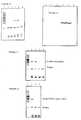
- A. PCR amplification of pure HPV16 viral DNA using normal Klenow. 1 ng HPV16/pBR322 was subjected to 20 rounds of: 95°C for 2 min (strand dissociation), then 37°C for 2 min (annealing), then 1 U Klenow enzyme added and 37°C incubation for 2 min. Half (50 »l) of the resulting mixture was electrophoresed on a 2% agarose gel and the result is shown in Figure 1. Amount = 2.5 x 100 ng = 250 ng. Region amplified = 1/40th of total HPV16 sequence. Total amplification = 250 x 40 = 10,000 x. (i.e., efficiency of reaction was 60% for each cycle).
- B. PCR amplification of wart biopsy DNA with HPV16/33 test. The DNA of anal wart Gap289 was phenol/chloroform extracted and ethanol precipitated. 1 »g of DNA was then subjected to 23 rounds of amplification (each: 95°C for 2 min, then 37°C for 2 min, 1 U Klenow added and 37°C for 2 min). 40% (50»l) was electrophoresed on a 2% agarose gel. Ethidium bromide stained DNA is shown in Figure 2. The PCR sample and standards were spotted onto a membrane and hybridization performed overnight with ³²P-labelled HPV16/33 target oligonucleotide probe. Wash conditions were 22°C for 5 min, then 38°C for 10 min in 5 x SSPE/0.1% SDS. Result of autoradiography is shown in Figure 3. Note that amounts are for recombinant viral DNAs used, only a fraction of which represents amplified sequence, within which is the target hybridization region.
- C. PCR amplification of pure HPV16 recombinant viral DNA using normal Klenow. 1 ng of HPV16/pBR322, 10 mM Tris.HCl, pH 7.6, 10 mM MgCl₂, 50 mM NaCl, 500 ng of each of the HPV16/33 primers, 4 »l of each dNTP (50 mM stock). 23 rounds of: 98°C for 2 min, then 37°C for 2 min, add Klenow, then 37°C for 2 min. Electrophoresis on 2% agarose gel. Ethidium bromide stained gel is shown in Figure 4.
Lanes lane 2. To prove this Southern blotting was then performed on this gel, using for hybridization conditions: 5 x SSPE, 2% SDS, 0.5 ml BLOTTO, 0.5 ml of 10 mg/ml salmon sperm DNA, 5.5 ml distilled water. Prehybodization was for 1 h at 30°C and hybridization was overnight at 30°C in the above solution with ∼1 ng/ml of end-labelled target HPV16/33 oligonucleotide probe. Washing was for 10 min at 38°C in 1 x SSPE, 0.1% SDS. Result in Figure 5, showing massive hybridization to a band at the expected position in lane 2 (i.e., at the same position as the band of stained DNA in Figure 4 lane 2). - D. Biopsies of anal warts. Patient specimens: Gap119, Gap278, Km, Gap289 11912C. Conditions as descibed above in C. Figure 6 is an autoradiograph after radioactive probing: Km and Gap289 were +ve, i.e., contained HPV16.
- E. Southern blot of biopsies +ve for HPV16. This experiment shows that the HPV16/33 oligo probe hybridizes to the correct restriction fragments in unamplified biopsies and pure viral DNA; also that there is no cross hybridization to other HPV types.
- Lane 1 - 8 »g Gap289 cut with BamHI/PstI
- Lane 2 - blank
- Lane 3 - 100 ng HPV33 cut with Bg/II/PstI
- Lane 4 - 100 ng HPV16 cut with BamHI/PstI
- Lane 5 - 100 ng HPV18 cut with EcoRI/XbaI
- Lane 6 - 100 ng
HPV 11 cut with BamHI/PstI - Lane 7 - 100 ng HPV6 cut with EcoRI/PstI
lane 4 and to HPV33 inlane 3. No hybridization occurred toHPVs lanes HPVs 16 and 33. Hybridization inlane 1 shows that specimen Gap289 contained HPV16. - F. Cervical scrapes - HPV16/33 PCR amplification. This shows the technique can detect specific HPV in scrape specimens. Scrapes: Pt and Mo. ∼10,000 cells were suspended in 35 »l water, heated to 98°C for 10 min, 65 »l PCR mixture added and normal protocol followed with 30 rounds of amplification using normal Klenow. Samples were dotted onto membranes and probed with radioactive oligo probe. Result (Figure 8): Pt scrape was +ve with HPV16/33 probe, Mo was -ve.
- G. Southern blot of scrape. This was to confirm that Pt scrape had given real amplification. (i.e., is proof of validity of result in H.) Gel used was 1.5% agarose. In Fig. 7:
- Lane 1 - 25 »l Pt PCR digested with Bg/I/BamHI in PCR buffer.
- Lane 2 - 40 ng of HPV33 insert.
- Lane 3-40 ng of HPV16 insert.
- Lane 4 - 0.5 »g of pBR322 digested with HpaII.
- Lane 5 - Bacteriophage λ digested with HindIII.
lane 1 for the patient specimen's PCR products and bands inlanes
The following examples (H-J) show additional positive results with the technique used on various biopsies and scrapes. - H. Various scrapes and anogenital wart biopsies - HPV6/11 PCR. In Figures 12 and 13:
- Lane 1 - 0.4 »g pBR322 cut with HpaII.
- Lane 2 - 0.9 »g F9885 wart DNA, HPV6/11 oligos only.
- Lane 3 - 1 »g Ow wart DNA, HPV6/11 and HPV16/33 oligos.
- Lane 4 - 3 »l cells from F11912 cervical scrape, HPV6/11 oligos only.
- Lane 5 - 3 »l cells from F11912 vaginal scrape, HPV6/11 oligos only.
- Lane 6 - 5 »l cells from F11912 rectal scrape, HPV6/11 oligos only.
lanes - I. Warts - HPV6/11 PCR. Results are shown in Figure 14:
- Lane 1 - pBR322 cut with HpaII
- Lane 2 - 750 ng of F8408 labial wart
- Lane 3 - 1 »g Gap252 anal wart
- Lane 4 - 860 ng Tr anal wart
- Lane 5 - 750 ng Bi anal wart
- J. Cervical warts - HPV6/11 and HPV16/33 primers. 50»l run on 12% polyacrylamide gel; results are shown in Figure 15.
- Lane 1 - pBR322 cut with HpaII
- Lane 2 - ∼500 ng Hn biopsy
- Lane 3 - ∼500 ng Hs biopsy
- Lane 4 - ∼500 ng Ls biopsy
- K. Cervical scrapes - HPV6/11 and HPV16/33. This example shows that primers for different HPVs can be used together in the same reaction mixture to generate amplification products unique for each HPV type. Results are shown in Figure 16.
- Lane 1 - pBR322 cut with HpaII.
- Lane 2 - 1 ng of HPV6/pAT153 and 1 ng of HPV16/pBR322; HPV6/11 and HPV16/33 primers.
- Lane 3 - 3 »l cells of CSC019 scrape, HPV6/11 and HPV16/33 primers.
- Lane 4 - 3 »l cells of CSC328 scrape, HPV6/11 primers.
- Lane 5 - 3 »l cells of Pt scrape, HPV6/11 primers.
lane 2 of Figure 16 one can see the 200 bp HPV16/33 amplification product (faint) and the 120 bp HPV6/11 product (darker). Lanes 3-5 were negative (only primer bands seen), i.e., did not contain HPV6/11. - L. Scrapes and biopsies - HPV16/33 PCR. This experiment is a comparison of radioactive and nonradioactive probing for the detection of HPVs by the PCR method. In Fig. 12:
- Lane 1 - pBR322 cut with HpaII
- Lane 2 - Blank
- Lane 3 - HPV6/pAT153 and HPV16/pBR322 PCR from I
- Lane 4 - 15 »l of Km PCR
- Lane 5 - 15 »l of Gap289 PCR
- Lane 6 - 15 »l Pt scrape PCR
lanes lane 3. In Figure 18 a single band of hybridization to the ∼200 bp HPV16/33 amplification products can be seen in each of lanes 6-3. Figure 19 shows the result of a 1.5 h exposure to the x-ray film: hybridization can be seen to the ∼200 bp HPV16/33 amplification products. Figure 20 is a dot blot probed with radioactive target oligo probe (Right: diagram of position of samples on dot blot; Left: result of hybridization). - M. Heat stable polymerase - HPV6 PCR. 1 ng HPV6/pAT153, 2 units of Thermus aquaticus DNA polymerase added initially and after every 10 rounds for 40 rounds. The conditions used were exactly as specified in the New England Biolabs protocol. The result (Figure 21) demonstrates that the heat stable polymerase is also able to produce the desired amplification products. In this figure:
- Lane 1 - pBR322/HpaII
- Lane 2 - 40 »l of HPV6/pAT153 PCR
- N. Heat stable polymerase - HPV16 and biopsy. After amplification HPV16/pBR322 cut with HinfI should give 2 fragments of 52 bp and 142 bp. 5 ng HPV33/plink and 1 »g Gap289 were subjected to 40 rounds with heat stable polymerase (
lanes - Lane 1 - pBR322/HpaII
- Lane 2 - HPV16/pBR322 cut with HinfI
(20»l PCR + 2 »l 10 x HinfI buffer + 1 »l enzyme + 6 »l dH₂O) - Lane 3 - 20 »l of HPV16/pBR322 PCR
- Lane 4 - 20 »l of HPV33/plink PCR
- Lane 5 - 20 »l of Gap289
- O. Diluted biopsy, 100 PCR rounds using heat stable polymerase. Wart biopsy Gap289 (1 »g/»l) was diluted 1:10³, 1:10⁶ and 1:10⁹ and 100 rounds of amplification were performed using 1 »l of each dilution and 4 U of heat stable DNA polymerase initially, then an additional 2 U added after 50 rounds. Other biopsies and scrapes were subjected to 50 rounds only. Conditions were as in example P. The result shows the extreme sensitivity of the test: the best results with this excessive number of rounds of amplification were obtained with the most dilute sample. Figure 23 shows the ethidium bromide stained DNA gel. The gel was electroblotted and probed with radioactively labelled HPV6/11 (Figure 24) and HPV16/33 (Figure 25) target oligo probe. The result shows the extreme specificity of the test. In Fig. 15 (b) it can be seen that only in
lane - Lane 1 - pBR322 cut with HpaII
- Lane 2 - Gap289 (1:10³) PCR (HPV16/33 primers only)
- Lane 3 - Gap289 (1:10⁶) PCR ( HPV16/33 primers only)
- Lane 4 - Gap289 (1:10⁹) PCR ( HPV16/33 primers only)
- Lane 5 - Anal scrape Gap402 PCR (HPV6/11 and HPV16/33 primers)
- Lane 6 - Bs wart PCR (HPV6/11 and HPV16/33 primers)
- Lane 7 - Cervical scrape 11912 (HPV16/33 primers only)
- Lane 8 - Vaginal scrape 11912 (HPV16/33 primers only)
- P. Hybridization of target oligo probe to unamplified HPV16, HPV33 and biopsies. Ordinary restricted HPVs and biopsies were run on a gel and stained (Figure 26), then probed with HPV16/33 target oligo probe. Result is shown in Figure 27, where hybridization can be seen in each case (lanes 2-5) and to correct restriction fragment in the case of recombinant viral DNA. Six days of exposure to x-ray film were required for this unamplified DNA. In Figures 26 and 27:
- Lane 1 - Bacteriophage λ cut with HindIII/EcoRI
- Lane 2 - 5 »g Bs wart DNA, Bg/II/BamHI
- Lane 3 - 5 »g Gap289, Bg/II/BamHI
- Lane 4 - HPV16/pBR322, BamHI
- Lane 5 - HPV33/plink, Bg/II
- Lane 6 - SPP-1
- Q. Dot blots showing hybridization of HPV16/33 alkaline phosphatase oligo probe to HPV16 are shown in Figure 28.
- R. HPV18 PCR. 30 rounds with normal Klenow DNA polymerase Result is shown in Figure 29, where the lanes are:
- Lane 1 - HPV16 insert PCR products; HPV16/33 primers.
- Lane 2 - HPV18 insert PCR products; HPV18 primers used.
lane 2. Inlane 1 are the HPV16 PCR products of ∼200 bp.
B. ALKALINE PHOSPHATASE OLIGONUCLEOTIDE PROBE ONTO DOT BLOT
C. RADIOACTIVE OLIGONUCLEOTIDE PROBE ONTO DOT BLOT
i) Prehybridization The membrane is prehybridized for at least 1 h at 30°C. Several approaches can be used for this.
(a) One uses a specially designed perspex block constructed in the Laboratory's mechanical workshop. The block is stood upright in a 30°C water bath with the membrane, bathed in the following solution, inside.
(b) Another approach is to cut out two sheets of Whatman type 542 paper to dimensions slightly larger than the membrane. Next, place membrane on top of one sheet and wet membrane with 2 ml prehybridization solution. Then place other sheet of Whatman on top and place the resulting 'sandwich' in a plastic bag with the rest of the prehybridization solution.
| Prehybridization mixture: (10 ml total volume) | |
| 10% (wt/vol) non fat milk powder (e.g., Diploma®) | 0.5 ml |
| 20x SSC (3M NaCl, 0.3M sodium citrate) | 2.5 ml |
| 20% (wt/vol) SDS (sodium dodecyl sulphate) | 0.5 |
| 10 mg/ml carrier DNA (e.g., from herring sperm) | 2.0 ml |
| Sterile distilled water | 4.5 ml |
ii) Hybridization Pour off prehybridization mixture and replace with hybridization mixture, which is the same except for the addition of radioactively labelled viral DNA (1 ng/ml hybridization mixture).
Hybridization is allowed to proceed overnight at 30°C in the perspex block or plastic bag.
iii) Washing membranes Membrane filters are washed under conditions of high stringency. This is to remove all radioactive DNA probe except that which is attached specifically, by hydrogen bonding, to identical viral DNA sequences that may be present within a sample.
iv) Autoradiography
Claims (10)
- A method of detection of carcinogenic human papillomavirus HPV16, HPV33 or HPV18 which comprises:(a) applying a polymerase chain reaction technique to a sample of human cervical tissue cells so as to amplify the amount of a selected nucleotide sequence located within the E6 region and characteristic of carcinogenic HPV16 and HPV33 or HPV18 present, comprising the steps of:(i) heating to dissociate the DNA strands,(ii) adding oligonucleotide primers defining each end of said nucleotide sequence,(iii) cooling to allow the primers to anneal to the dissociated DNA strands,(iv) adding a DNA polymerase,(v) at the cooled temperature allowing formation of DNA complementary to each strand of said nucleotide sequence,(vi) heating to dissociation temperature, and repeating steps (iii) to (vi), optionally omitting step (iv) where a heat stable DNA polymerase is used; and(b) detecting the presence or absence in the amplified sample of said nucleotide sequence of HPV16 and HPV33 or HPV18.
- A method according to claim 1 wherein the pair of primers defining the selected nucleotide sequence are:(i) For HPV16 and HPV33
⁵'TGAGGTATATGACTTTGCTTTT³ʼ
and
³'AATTAATCCACATAAT⁵or (ii) for HPV18
⁵'ACAGTATTGGAACTTACAGA³'
and
³'TTTACATATCTAAAAATAAG⁵' - A method according to Claim 1, wherein the DNA polymerase is a polymerase stable at the dissociation temperature.
- A method according to claim 3, wherein the DNA polymerase is a thermophilic polymerase from Thermus aquaticus.
- A method according to Claim 1 wherein the presence or absence of said nucleotide sequence is detected by separating the components of the amplified sample on a gel and detecting the presence or absence of a DNA having the same number of nucleotides as said nucleotide sequence.
- A method according to Claim 1 wherein in order to detect the presence or absence of said nucleotide sequence, there is added a labelled oligonucleotide hybridization probe corresponding to a region of said nucleotide sequence.
- A method according to Claim 6 wherein the oligonucleotide probe is:(i) for HPV16 and HPV33
⁵'GTGAGTATAGACATTAT³'or (ii) for HPV18
⁵'GATTTATTTGTGGTGTATAGA³' - A method according to Claim 6 or 7 wherein the oligonucleotide probe is labelled with a ³²P radioactive label, or with alkaline phosphatase enzyme label.
- A primer composition for use in a method according to Claim 1, which consists of:(i) for HPV16 and HPV33 a mixture of
⁵'TGAGGTATATGACTTTGCTTTT³'
and
³'AATTAATCCACATAAT⁵'
or(ii) for HPV18 a mixture of ⁵'ACAGTATTGGAACTTACAGA³'
and
³'TTTACATATCTAAAAATAAG⁵' - A labelled oligonucleotide probe for use in a method according to Claim 6 wherein the oligonucleotide is(i) for HPV16
⁵'GTGAGTATAGACATTAT³'or (ii) for HPV18
⁵'GATTTATTTGTGGTGTATAGA³'
Applications Claiming Priority (3)
| Application Number | Priority Date | Filing Date | Title |
|---|---|---|---|
| AU559/87 | 1987-02-26 | ||
| AUPI055987 | 1987-02-26 | ||
| PCT/AU1988/000047 WO1988006634A1 (en) | 1987-02-26 | 1988-02-24 | A method of detection of carcinogenic human papillomavirus |
Publications (3)
| Publication Number | Publication Date |
|---|---|
| EP0357611A1 EP0357611A1 (en) | 1990-03-14 |
| EP0357611A4 EP0357611A4 (en) | 1991-10-02 |
| EP0357611B1 true EP0357611B1 (en) | 1995-04-26 |
Family
ID=3772035
Family Applications (1)
| Application Number | Title | Priority Date | Filing Date |
|---|---|---|---|
| EP88902077A Expired - Lifetime EP0357611B1 (en) | 1987-02-26 | 1988-02-24 | A method of detection of carcinogenic human papillomavirus |
Country Status (6)
| Country | Link |
|---|---|
| US (2) | US5783412A (en) |
| EP (1) | EP0357611B1 (en) |
| JP (3) | JP3096704B2 (en) |
| AT (1) | ATE121794T1 (en) |
| DE (1) | DE3853678T2 (en) |
| WO (1) | WO1988006634A1 (en) |
Families Citing this family (44)
| Publication number | Priority date | Publication date | Assignee | Title |
|---|---|---|---|---|
| US5447839A (en) * | 1988-09-09 | 1995-09-05 | Hoffmann-La Roche Inc. | Detection of human papillomavirus by the polymerase chain reaction |
| US5182377A (en) * | 1988-09-09 | 1993-01-26 | Hoffmann-La Roche Inc. | Probes for detection of human papillomavirus |
| DE3838269A1 (en) * | 1988-11-11 | 1990-05-17 | Behringwerke Ag | IDENTIFYING HUMAN PAPILLOMAVIRUS DNA AND ITS EXPRESSION IN ZERVIX SLICES |
| GB2225112A (en) * | 1988-11-22 | 1990-05-23 | Ici Plc | Hybridisation probes |
| US5043272A (en) * | 1989-04-27 | 1991-08-27 | Life Technologies, Incorporated | Amplification of nucleic acid sequences using oligonucleotides of random sequence as primers |
| JP2791685B2 (en) * | 1989-06-08 | 1998-08-27 | 寳酒造株式会社 | Papillomavirus detection method |
| US5863717A (en) * | 1989-11-03 | 1999-01-26 | Abbott Laboratories | Use of conserved oligonucleotide primers to amplify human papillomavirus DNA sequences |
| US5580970A (en) * | 1989-12-01 | 1996-12-03 | Amoco Corporation | Detection of HPV transcripts |
| DE69033850T2 (en) * | 1989-12-01 | 2002-06-06 | Vysis Inc | Nucleic acid probes for the detection of HPV transcripts |
| KR960012069B1 (en) * | 1989-12-04 | 1996-09-12 | 아이시스 파마슈티칼스, 인코포레이티드 | Antisense oligonucleotide inhibiting papillomavirus |
| NL9000134A (en) * | 1990-01-19 | 1991-08-16 | Stichting Res Fonds Pathologie | PRIMERS AND METHOD FOR DETECTING HUMAN PAPILLOMA VIRUS GENOTYPS BY M.B.V. PCR. |
| DK0515506T3 (en) * | 1990-02-16 | 2000-05-08 | Hoffmann La Roche | Method for detecting carcinogenic human papillomaviruses |
| GB9015845D0 (en) * | 1990-07-19 | 1990-09-05 | Emery Vincent C | Diagnostic method |
| IL97226A0 (en) * | 1991-02-13 | 1992-05-25 | Orgenics Ltd | Detection of high and low risk human papillomavirus by enzymatic amplification of dna |
| US5346811A (en) * | 1991-07-22 | 1994-09-13 | Cerveceria Polar | Method and products for human papillomavirus detection |
| US5565339A (en) * | 1992-10-08 | 1996-10-15 | Hoffmann-La Roche Inc. | Compositions and methods for inhibiting dimerization of primers during storage of polymerase chain reaction reagents |
| DE4409436A1 (en) * | 1994-03-19 | 1995-09-21 | Boehringer Mannheim Gmbh | Process for processing nucleic acids |
| DE19506561C1 (en) * | 1995-02-24 | 1996-10-10 | Deutsches Krebsforsch | A method for the early detection of HPV-associated carcinomas or high-grade HPV-induced dysplasias |
| DE19526717A1 (en) * | 1995-07-21 | 1997-01-23 | Florian Dr Med Heirler | Diagnosis of cervical cancer |
| JP2000500342A (en) | 1995-11-15 | 2000-01-18 | ジェン−プローブ・インコーポレーテッド | Nucleic acid probes complementary to human papillomavirus nucleic acids and related methods and kits |
| AU1324000A (en) * | 1998-10-26 | 2000-05-15 | Ventana Medical Systems, Inc. | Detection of human papilloma virus in papanicolaou (pap) smears |
| US7569344B2 (en) * | 1998-10-26 | 2009-08-04 | Ventana Medical Systems, Inc. | Detection of human papilloma virus in papanicolaou (Pap) smears |
| US20060275784A1 (en) * | 1999-10-26 | 2006-12-07 | Ventana Medical Systems, Inc. | Detection of Human Papilloma Virus in Papanicolaou (Pap) Smears |
| AU2001256976A1 (en) * | 2000-04-03 | 2001-10-15 | Cytyc Corporation | Detection and typing of human papillomavirus using pna probes |
| US6936443B2 (en) * | 2000-04-03 | 2005-08-30 | Cytyc Corporation | Detection and typing of human papillomavirus using PNA probes |
| US6811549B2 (en) * | 2001-02-16 | 2004-11-02 | William H. Fleming | Administration of therapeutic or diagnostic agents using interlabial pad |
| EP2272988A3 (en) * | 2002-01-07 | 2011-06-15 | Norchip A/S | Method for detecting human papillomavirus mRNA |
| US20050100554A1 (en) * | 2002-02-14 | 2005-05-12 | Amanda Jackson | Complexes and methods of using same |
| WO2004003199A1 (en) * | 2002-07-01 | 2004-01-08 | Kankyo Engineering Co., Ltd. | Novel method of acquiring nucleic acid or gene |
| US20040137551A1 (en) * | 2003-01-13 | 2004-07-15 | Markovic Nenad S. | Cervical acid phosphatase - papanicolaou (CAP-PAP) test kit, method and accesories, processes for producing and using the same |
| US7527948B2 (en) | 2003-09-25 | 2009-05-05 | Third Wave Technologies, Inc. | Detection of HPV |
| WO2005033333A2 (en) * | 2003-10-07 | 2005-04-14 | Dako Denmark A/S | Methods and compositions for the diagnosis of cancer |
| EP1819835B1 (en) | 2004-12-08 | 2010-08-04 | Gen-Probe Incorporated | Detection of nucleic acids from multiple types of human papillomaviruses |
| GB0500999D0 (en) * | 2005-01-18 | 2005-02-23 | Glaxosmithkline Biolog Sa | Detection method and materials therefor |
| US20070031826A1 (en) * | 2005-08-05 | 2007-02-08 | My Gene | Diagnostic kit for determining the genotype of a human papilloma virus and method of using thereof |
| JPWO2007148762A1 (en) * | 2006-06-22 | 2009-11-19 | 雅彦 黒田 | Human papillomavirus (HPV) detection method, primer set and detection kit used therefor |
| US8080643B2 (en) * | 2006-09-05 | 2011-12-20 | Third Wave Technologies, Inc. | HPV primers |
| WO2008106610A1 (en) * | 2007-02-28 | 2008-09-04 | Quantrx Biomedical Corporation | Folded perineal pad |
| KR100914911B1 (en) * | 2007-07-10 | 2009-08-31 | 의료법인제일의료재단 | Method and kit for detecting human papillomavirus quantitatively and qualitatively using real-time pcr and hpv dna chip |
| KR100906124B1 (en) | 2007-07-19 | 2009-07-07 | 캐치바이진 주식회사 | A qualitative assay method of high risk group geno-type Human Papillomavirus DNA and a qualitative assay kit of the same |
| DE102008035903A1 (en) * | 2008-07-31 | 2010-02-18 | Epcos Ag | Electrical gas filled overvoltage arrester, has short circuit device arranged at distance from electrodes using coatings applied on short circuit device such that electrodes are electrically insulated from each other |
| NO330943B1 (en) * | 2009-04-30 | 2011-08-22 | Unilabs Telelabs As | A method for detecting and / or typing and / or quantifying human papillomavirus (HPV) type, primers and probes thereof, and diagnostic and use thereof. |
| AU2011258501B2 (en) | 2010-05-25 | 2016-07-07 | Qiagen Gaithersburg, Inc. | Fast results hybrid capture assay and associated strategically-truncated probes |
| KR101761701B1 (en) | 2014-07-29 | 2017-07-26 | 의료법인 제일의료재단 | HPV Specific Probe and DNA Chip for Detecting Genetic Type of HPV Containing Thereof |
Family Cites Families (5)
| Publication number | Priority date | Publication date | Assignee | Title |
|---|---|---|---|---|
| US4683195A (en) * | 1986-01-30 | 1987-07-28 | Cetus Corporation | Process for amplifying, detecting, and/or-cloning nucleic acid sequences |
| DK171161B1 (en) * | 1985-03-28 | 1996-07-08 | Hoffmann La Roche | A method for detecting the presence or absence of at least one specific nucleic acid sequence in a sample or for distinguishing two different nucleic acid sequences in this sample |
| US4683202A (en) * | 1985-03-28 | 1987-07-28 | Cetus Corporation | Process for amplifying nucleic acid sequences |
| US5176995A (en) * | 1985-03-28 | 1993-01-05 | Hoffmann-La Roche Inc. | Detection of viruses by amplification and hybridization |
| NL9000134A (en) * | 1990-01-19 | 1991-08-16 | Stichting Res Fonds Pathologie | PRIMERS AND METHOD FOR DETECTING HUMAN PAPILLOMA VIRUS GENOTYPS BY M.B.V. PCR. |
-
1988
- 1988-02-24 DE DE3853678T patent/DE3853678T2/en not_active Expired - Lifetime
- 1988-02-24 US US07/423,401 patent/US5783412A/en not_active Expired - Fee Related
- 1988-02-24 JP JP63501788A patent/JP3096704B2/en not_active Expired - Fee Related
- 1988-02-24 WO PCT/AU1988/000047 patent/WO1988006634A1/en active IP Right Grant
- 1988-02-24 AT AT88902077T patent/ATE121794T1/en active
- 1988-02-24 EP EP88902077A patent/EP0357611B1/en not_active Expired - Lifetime
-
1997
- 1997-12-30 US US09/000,634 patent/US6218104B1/en not_active Expired - Fee Related
-
2000
- 2000-02-21 JP JP2000043673A patent/JP2000189200A/en active Pending
- 2000-12-13 JP JP2000379281A patent/JP2001197894A/en active Pending
Also Published As
| Publication number | Publication date |
|---|---|
| JP2001197894A (en) | 2001-07-24 |
| ATE121794T1 (en) | 1995-05-15 |
| JP3096704B2 (en) | 2000-10-10 |
| EP0357611A1 (en) | 1990-03-14 |
| US5783412A (en) | 1998-07-21 |
| WO1988006634A1 (en) | 1988-09-07 |
| EP0357611A4 (en) | 1991-10-02 |
| DE3853678D1 (en) | 1995-06-01 |
| JPH02502334A (en) | 1990-08-02 |
| DE3853678T2 (en) | 1995-08-31 |
| AU1393888A (en) | 1988-09-26 |
| JP2000189200A (en) | 2000-07-11 |
| AU611135B2 (en) | 1991-06-06 |
| US6218104B1 (en) | 2001-04-17 |
Similar Documents
| Publication | Publication Date | Title |
|---|---|---|
| EP0357611B1 (en) | A method of detection of carcinogenic human papillomavirus | |
| Lungu et al. | Typing of human papillomaviruses by polymerase chain reaction amplification with L1 consensus primers and RFLP analysis | |
| Gregoire et al. | Amplification of human papillomavirus DNA sequences by using conserved primers | |
| Pao et al. | Identification of human papillomavirus DNA sequences in peripheral blood mononuclear cells | |
| Melchers et al. | Human papillomavirus detection in urine samples from male patients by the polymerase chain reaction | |
| EP0539461B1 (en) | Method for the diagnosis of human pappiloma virus type 16 using the polymerase chain reaction | |
| Soler et al. | Detection of human papillomavirus types 6, 11, 16 and 18 in mucosal and cutaneous lesions by the multiplex polymerase chain reaction | |
| Tham et al. | Diagnostic Sensitivity of Polymerase Chain Reaction and Southern Blot Hybridization for the Detection df Human Papillomavirus DNA in Biopsy Specimens from Cervical Lesions | |
| Chan et al. | Prevalence of six types of human papillomavirus in inverted papilloma and papillary transitional cell carcinoma of the bladder: an evaluation by polymerase chain reaction. | |
| JP2007524421A (en) | Detection of human papillomavirus | |
| Wilczynski et al. | Human papillomavirus type 6 in squamous cell carcinoma of the bladder and cervix | |
| Yoshida et al. | Quantitative real-time polymerase chain reaction analysis of the type distribution, viral load, and physical status of human papillomavirus in liquid-based cytology samples from cervical lesions | |
| EP2373819B1 (en) | Multiparameter assay | |
| Zheng et al. | Simultaneous detection by consensus multiplex PCR of high-and low-risk and other types of human papilloma virus in clinical samples | |
| Miller et al. | Human papillomavirus type 16 DNA in esophageal carcinomas from Alaska natives | |
| Milde-Langosch et al. | Human papillomavirus and c-myc/c-erb B2 in uterine and vulvar lesions | |
| Konno et al. | Progression of squamous cell carcinoma of the uterine cervix from cervical intraepithelial neoplasia infected with human papillomavirus: a retrospective follow-up study by in situ hybridization and polymerase chain reaction | |
| Park et al. | Detection of human papillomavirus genotypes in cervical neoplasia from Korean women using polymerase chain reaction | |
| Herckenrode et al. | Prevalence of human papillomavirus (HPV) infection in basque country women using slot‐blot hybridization: A survey of women at low risk of developing cervical cancer | |
| AU611135C (en) | A method of detection of carcinogenic human papillomavirus | |
| Sigurdsson et al. | Human papillomavirus (HPV) in an Icelandic population: The role of HPV DNA testing based on hybrid capture and PCR assays among women with screen‐detected abnormal pap smears | |
| Salam et al. | General primer‐mediated polymerase chain reaction for simultaneous detection and typing of human papillomavirus DNA in laryngeal squamous cell carcinomas | |
| Bavin et al. | Comparison of cervical cytology and the polymerase chain reaction for HPV 16 to identify women with cervical disease in a general practice population | |
| Melchers et al. | Prevalence of genital HPV infections in a regularly screened population in The Netherlands in relation to cervical cytology | |
| Soler et al. | Detection of multiple types of human papillomavirus in a giant condyloma from a grafted patient. Analysis by immunohistochemistry, in situ hybridisation, Southern blot and polymerase chain reaction |
Legal Events
| Date | Code | Title | Description |
|---|---|---|---|
| PUAI | Public reference made under article 153(3) epc to a published international application that has entered the european phase |
Free format text: ORIGINAL CODE: 0009012 |
|
| 17P | Request for examination filed |
Effective date: 19890824 |
|
| AK | Designated contracting states |
Kind code of ref document: A1 Designated state(s): AT BE CH DE FR GB IT LI LU NL SE |
|
| A4 | Supplementary search report drawn up and despatched |
Effective date: 19910813 |
|
| AK | Designated contracting states |
Kind code of ref document: A4 Designated state(s): AT BE CH DE FR GB IT LI LU NL SE |
|
| 17Q | First examination report despatched |
Effective date: 19921216 |
|
| GRAA | (expected) grant |
Free format text: ORIGINAL CODE: 0009210 |
|
| AK | Designated contracting states |
Kind code of ref document: B1 Designated state(s): AT BE CH DE FR GB IT LI LU NL SE |
|
| PG25 | Lapsed in a contracting state [announced via postgrant information from national office to epo] |
Ref country code: IT Free format text: LAPSE BECAUSE OF FAILURE TO SUBMIT A TRANSLATION OF THE DESCRIPTION OR TO PAY THE FEE WITHIN THE PRE;WARNING: LAPSES OF ITALIAN PATENTS WITH EFFECTIVE DATE BEFORE 2007 MAY HAVE OCCURRED AT ANY TIME BEFORE 2007. THE CORRECT EFFECTIVE DATE MAY BE DIFFERENT FROM THE ONE RECORDED.SCRIBED TIME-LIMIT Effective date: 19950426 Ref country code: FR Effective date: 19950426 Ref country code: BE Effective date: 19950426 Ref country code: NL Free format text: LAPSE BECAUSE OF FAILURE TO SUBMIT A TRANSLATION OF THE DESCRIPTION OR TO PAY THE FEE WITHIN THE PRESCRIBED TIME-LIMIT Effective date: 19950426 Ref country code: AT Effective date: 19950426 |
|
| REF | Corresponds to: |
Ref document number: 121794 Country of ref document: AT Date of ref document: 19950515 Kind code of ref document: T |
|
| REF | Corresponds to: |
Ref document number: 3853678 Country of ref document: DE Date of ref document: 19950601 |
|
| EN | Fr: translation not filed | ||
| NLV1 | Nl: lapsed or annulled due to failure to fulfill the requirements of art. 29p and 29m of the patents act | ||
| PG25 | Lapsed in a contracting state [announced via postgrant information from national office to epo] |
Ref country code: LU Free format text: LAPSE BECAUSE OF NON-PAYMENT OF DUE FEES Effective date: 19960229 |
|
| PLBE | No opposition filed within time limit |
Free format text: ORIGINAL CODE: 0009261 |
|
| STAA | Information on the status of an ep patent application or granted ep patent |
Free format text: STATUS: NO OPPOSITION FILED WITHIN TIME LIMIT |
|
| 26N | No opposition filed | ||
| REG | Reference to a national code |
Ref country code: CH Ref legal event code: PUE Owner name: THE UNIVERSITY OF SYDNEY TRANSFER- BIOSEARCH INTER |
|
| REG | Reference to a national code |
Ref country code: GB Ref legal event code: 732E |
|
| REG | Reference to a national code |
Ref country code: CH Ref legal event code: PFA Free format text: BIOSEARCH INTERNATIONAL PTY LTD.,LEVEL 10 60 PARK STREET,SYDNEY/NSW 2000 (AU) TRANSFER- BIOSEARCH INTERNATIONAL PTY LTD.,5TH FLOOR 99 ELIZABETH STREET,SYDNEY/NSW 2000 (AU) |
|
| REG | Reference to a national code |
Ref country code: GB Ref legal event code: IF02 |
|
| REG | Reference to a national code |
Ref country code: CH Ref legal event code: NV Representative=s name: BUGNION S.A. Ref country code: CH Ref legal event code: PUE Owner name: POLARTECHNICS LTD Free format text: BIOSEARCH INTERNATIONAL PTY LTD.#5TH FLOOR 99 ELIZABETH STREET#SYDNEY/NSW 2000 (AU) -TRANSFER TO- POLARTECHNICS LTD#LEVEL 1, 140 WILLIAM STREET SYDNEY,#NEW SOUTH WALES 2011 (AU) |
|
| REG | Reference to a national code |
Ref country code: GB Ref legal event code: 732E |
|
| PGFP | Annual fee paid to national office [announced via postgrant information from national office to epo] |
Ref country code: CH Payment date: 20070516 Year of fee payment: 20 |
|
| PGFP | Annual fee paid to national office [announced via postgrant information from national office to epo] |
Ref country code: DE Payment date: 20070517 Year of fee payment: 20 |
|
| PGFP | Annual fee paid to national office [announced via postgrant information from national office to epo] |
Ref country code: SE Payment date: 20070529 Year of fee payment: 20 |
|
| PGFP | Annual fee paid to national office [announced via postgrant information from national office to epo] |
Ref country code: GB Payment date: 20070516 Year of fee payment: 20 |
|
| REG | Reference to a national code |
Ref country code: GB Ref legal event code: PE20 |
|
| REG | Reference to a national code |
Ref country code: CH Ref legal event code: PL |
|
| EUG | Se: european patent has lapsed | ||
| PG25 | Lapsed in a contracting state [announced via postgrant information from national office to epo] |
Ref country code: GB Free format text: LAPSE BECAUSE OF EXPIRATION OF PROTECTION Effective date: 20080223 |