EP0347771B1 - Device for enhancing fluorescence and kinetics and methods of using the device - Google Patents
Device for enhancing fluorescence and kinetics and methods of using the device Download PDFInfo
- Publication number
- EP0347771B1 EP0347771B1 EP89110958A EP89110958A EP0347771B1 EP 0347771 B1 EP0347771 B1 EP 0347771B1 EP 89110958 A EP89110958 A EP 89110958A EP 89110958 A EP89110958 A EP 89110958A EP 0347771 B1 EP0347771 B1 EP 0347771B1
- Authority
- EP
- European Patent Office
- Prior art keywords
- fluorescence
- kinetics
- support
- substrate
- supports
- Prior art date
- Legal status (The legal status is an assumption and is not a legal conclusion. Google has not performed a legal analysis and makes no representation as to the accuracy of the status listed.)
- Expired - Lifetime
Links
- 230000002708 enhancing effect Effects 0.000 title claims abstract description 64
- 238000000034 method Methods 0.000 title claims description 26
- 102000004190 Enzymes Human genes 0.000 claims abstract description 60
- 108090000790 Enzymes Proteins 0.000 claims abstract description 60
- 239000007850 fluorescent dye Substances 0.000 claims abstract description 45
- 244000005700 microbiome Species 0.000 claims abstract description 34
- 230000007062 hydrolysis Effects 0.000 claims abstract description 20
- 238000006460 hydrolysis reaction Methods 0.000 claims abstract description 20
- GLNDAGDHSLMOKX-UHFFFAOYSA-N coumarin 120 Chemical compound C1=C(N)C=CC2=C1OC(=O)C=C2C GLNDAGDHSLMOKX-UHFFFAOYSA-N 0.000 claims abstract description 15
- 239000000126 substance Substances 0.000 claims abstract description 12
- HSSLDCABUXLXKM-UHFFFAOYSA-N resorufin Chemical compound C1=CC(=O)C=C2OC3=CC(O)=CC=C3N=C21 HSSLDCABUXLXKM-UHFFFAOYSA-N 0.000 claims abstract description 7
- 239000000758 substrate Substances 0.000 claims description 88
- HSHNITRMYYLLCV-UHFFFAOYSA-N 4-methylumbelliferone Chemical class C1=C(O)C=CC2=C1OC(=O)C=C2C HSHNITRMYYLLCV-UHFFFAOYSA-N 0.000 claims description 50
- 238000012360 testing method Methods 0.000 claims description 48
- 239000012530 fluid Substances 0.000 claims description 23
- UPSFMJHZUCSEHU-JYGUBCOQSA-N n-[(2s,3r,4r,5s,6r)-2-[(2r,3s,4r,5r,6s)-5-acetamido-4-hydroxy-2-(hydroxymethyl)-6-(4-methyl-2-oxochromen-7-yl)oxyoxan-3-yl]oxy-4,5-dihydroxy-6-(hydroxymethyl)oxan-3-yl]acetamide Chemical compound CC(=O)N[C@@H]1[C@@H](O)[C@H](O)[C@@H](CO)O[C@H]1O[C@H]1[C@H](O)[C@@H](NC(C)=O)[C@H](OC=2C=C3OC(=O)C=C(C)C3=CC=2)O[C@@H]1CO UPSFMJHZUCSEHU-JYGUBCOQSA-N 0.000 claims description 15
- UFHFLCQGNIYNRP-UHFFFAOYSA-N Hydrogen Chemical compound [H][H] UFHFLCQGNIYNRP-UHFFFAOYSA-N 0.000 claims description 9
- GNBHRKFJIUUOQI-UHFFFAOYSA-N fluorescein Chemical compound O1C(=O)C2=CC=CC=C2C21C1=CC=C(O)C=C1OC1=CC(O)=CC=C21 GNBHRKFJIUUOQI-UHFFFAOYSA-N 0.000 claims description 6
- 239000011800 void material Substances 0.000 claims description 6
- JBIJLHTVPXGSAM-UHFFFAOYSA-N 2-naphthylamine Chemical compound C1=CC=CC2=CC(N)=CC=C21 JBIJLHTVPXGSAM-UHFFFAOYSA-N 0.000 claims description 3
- 150000004775 coumarins Chemical class 0.000 claims description 3
- 238000006243 chemical reaction Methods 0.000 abstract description 29
- 239000000725 suspension Substances 0.000 abstract description 26
- 239000007788 liquid Substances 0.000 abstract description 5
- 239000000872 buffer Substances 0.000 description 28
- 239000000243 solution Substances 0.000 description 26
- 239000000523 sample Substances 0.000 description 23
- 241000894007 species Species 0.000 description 23
- 239000001913 cellulose Substances 0.000 description 18
- 229920002678 cellulose Polymers 0.000 description 18
- IAZDPXIOMUYVGZ-UHFFFAOYSA-N Dimethylsulphoxide Chemical compound CS(C)=O IAZDPXIOMUYVGZ-UHFFFAOYSA-N 0.000 description 16
- 241000894006 Bacteria Species 0.000 description 15
- 230000001580 bacterial effect Effects 0.000 description 13
- 230000000694 effects Effects 0.000 description 12
- 206010056740 Genital discharge Diseases 0.000 description 11
- 210000004027 cell Anatomy 0.000 description 11
- 238000004458 analytical method Methods 0.000 description 10
- 238000002474 experimental method Methods 0.000 description 9
- 239000000463 material Substances 0.000 description 9
- 238000012512 characterization method Methods 0.000 description 7
- 239000002054 inoculum Substances 0.000 description 7
- -1 β-napthylamine Chemical compound 0.000 description 7
- 239000002028 Biomass Substances 0.000 description 6
- 108090000204 Dipeptidase 1 Proteins 0.000 description 6
- 238000003556 assay Methods 0.000 description 6
- 102000006635 beta-lactamase Human genes 0.000 description 6
- 238000001514 detection method Methods 0.000 description 6
- 238000011534 incubation Methods 0.000 description 6
- 238000005259 measurement Methods 0.000 description 6
- 239000011780 sodium chloride Substances 0.000 description 6
- GTAAIHRZANUVJS-ZDUSSCGKSA-N (2s)-2-amino-4-methyl-n-(4-methyl-2-oxochromen-7-yl)pentanamide Chemical compound CC1=CC(=O)OC2=CC(NC(=O)[C@@H](N)CC(C)C)=CC=C21 GTAAIHRZANUVJS-ZDUSSCGKSA-N 0.000 description 5
- IELZRWMIHYDLTD-UHFFFAOYSA-N 2-piperidin-3-ylethanamine;dihydrochloride Chemical compound Cl.Cl.NCCC1CCCNC1 IELZRWMIHYDLTD-UHFFFAOYSA-N 0.000 description 5
- 241000588724 Escherichia coli Species 0.000 description 5
- 239000004793 Polystyrene Substances 0.000 description 5
- FAPWRFPIFSIZLT-UHFFFAOYSA-M Sodium chloride Chemical compound [Na+].[Cl-] FAPWRFPIFSIZLT-UHFFFAOYSA-M 0.000 description 5
- 108010046334 Urease Proteins 0.000 description 5
- 230000008859 change Effects 0.000 description 5
- 230000005284 excitation Effects 0.000 description 5
- 230000003287 optical effect Effects 0.000 description 5
- 229920002223 polystyrene Polymers 0.000 description 5
- 239000000047 product Substances 0.000 description 5
- 239000002904 solvent Substances 0.000 description 5
- XLYOFNOQVPJJNP-UHFFFAOYSA-N water Substances O XLYOFNOQVPJJNP-UHFFFAOYSA-N 0.000 description 5
- 229910001868 water Inorganic materials 0.000 description 5
- JGSARLDLIJGVTE-MBNYWOFBSA-N Penicillin G Chemical compound N([C@H]1[C@H]2SC([C@@H](N2C1=O)C(O)=O)(C)C)C(=O)CC1=CC=CC=C1 JGSARLDLIJGVTE-MBNYWOFBSA-N 0.000 description 4
- 239000004743 Polypropylene Substances 0.000 description 4
- 241000589517 Pseudomonas aeruginosa Species 0.000 description 4
- 239000006227 byproduct Substances 0.000 description 4
- 210000000170 cell membrane Anatomy 0.000 description 4
- 230000007423 decrease Effects 0.000 description 4
- 230000004069 differentiation Effects 0.000 description 4
- 238000011081 inoculation Methods 0.000 description 4
- 230000003993 interaction Effects 0.000 description 4
- 239000011159 matrix material Substances 0.000 description 4
- 239000004033 plastic Substances 0.000 description 4
- 229920003023 plastic Polymers 0.000 description 4
- 229920001155 polypropylene Polymers 0.000 description 4
- 230000035945 sensitivity Effects 0.000 description 4
- 239000007787 solid Substances 0.000 description 4
- IWSOXHMIRLSLKT-QMMMGPOBSA-N (2s)-2-amino-n-(4-methyl-2-oxochromen-7-yl)propanamide Chemical compound CC1=CC(=O)OC2=CC(NC(=O)[C@@H](N)C)=CC=C21 IWSOXHMIRLSLKT-QMMMGPOBSA-N 0.000 description 3
- BCHIXGBGRHLSBE-UHFFFAOYSA-N (4-methyl-2-oxochromen-7-yl) dihydrogen phosphate Chemical compound C1=C(OP(O)(O)=O)C=CC2=C1OC(=O)C=C2C BCHIXGBGRHLSBE-UHFFFAOYSA-N 0.000 description 3
- 102000004400 Aminopeptidases Human genes 0.000 description 3
- 108090000915 Aminopeptidases Proteins 0.000 description 3
- LFQSCWFLJHTTHZ-UHFFFAOYSA-N Ethanol Chemical compound CCO LFQSCWFLJHTTHZ-UHFFFAOYSA-N 0.000 description 3
- 241000588915 Klebsiella aerogenes Species 0.000 description 3
- 241000588772 Morganella morganii Species 0.000 description 3
- 241000079899 Pedipes mirabilis Species 0.000 description 3
- 241000607715 Serratia marcescens Species 0.000 description 3
- XSQUKJJJFZCRTK-UHFFFAOYSA-N Urea Chemical compound NC(N)=O XSQUKJJJFZCRTK-UHFFFAOYSA-N 0.000 description 3
- 239000002253 acid Substances 0.000 description 3
- 230000002378 acidificating effect Effects 0.000 description 3
- 239000003242 anti bacterial agent Substances 0.000 description 3
- 229940088710 antibiotic agent Drugs 0.000 description 3
- 230000003115 biocidal effect Effects 0.000 description 3
- 239000004202 carbamide Substances 0.000 description 3
- 238000000151 deposition Methods 0.000 description 3
- 238000001506 fluorescence spectroscopy Methods 0.000 description 3
- 239000000203 mixture Substances 0.000 description 3
- 230000002829 reductive effect Effects 0.000 description 3
- 238000011282 treatment Methods 0.000 description 3
- QGZKDVFQNNGYKY-UHFFFAOYSA-N Ammonia Chemical compound N QGZKDVFQNNGYKY-UHFFFAOYSA-N 0.000 description 2
- FMMWHPNWAFZXNH-UHFFFAOYSA-N Benz[a]pyrene Chemical compound C1=C2C3=CC=CC=C3C=C(C=C3)C2=C2C3=CC=CC2=C1 FMMWHPNWAFZXNH-UHFFFAOYSA-N 0.000 description 2
- CURLTUGMZLYLDI-UHFFFAOYSA-N Carbon dioxide Chemical compound O=C=O CURLTUGMZLYLDI-UHFFFAOYSA-N 0.000 description 2
- HEDRZPFGACZZDS-UHFFFAOYSA-N Chloroform Chemical compound ClC(Cl)Cl HEDRZPFGACZZDS-UHFFFAOYSA-N 0.000 description 2
- 241000588921 Enterobacteriaceae Species 0.000 description 2
- 241000644323 Escherichia coli C Species 0.000 description 2
- 102000004882 Lipase Human genes 0.000 description 2
- 108090001060 Lipase Proteins 0.000 description 2
- 239000004367 Lipase Substances 0.000 description 2
- 241000588655 Moraxella catarrhalis Species 0.000 description 2
- 241000191967 Staphylococcus aureus Species 0.000 description 2
- 238000010521 absorption reaction Methods 0.000 description 2
- 239000000853 adhesive Substances 0.000 description 2
- 230000001070 adhesive effect Effects 0.000 description 2
- 230000008901 benefit Effects 0.000 description 2
- 125000003460 beta-lactamyl group Chemical group 0.000 description 2
- 239000012472 biological sample Substances 0.000 description 2
- 239000008280 blood Substances 0.000 description 2
- 210000004369 blood Anatomy 0.000 description 2
- 238000009640 blood culture Methods 0.000 description 2
- 210000001124 body fluid Anatomy 0.000 description 2
- 239000010839 body fluid Substances 0.000 description 2
- WDECIBYCCFPHNR-UHFFFAOYSA-N chrysene Chemical compound C1=CC=CC2=CC=C3C4=CC=CC=C4C=CC3=C21 WDECIBYCCFPHNR-UHFFFAOYSA-N 0.000 description 2
- 238000010276 construction Methods 0.000 description 2
- ZYGHJZDHTFUPRJ-UHFFFAOYSA-N coumarin Chemical compound C1=CC=C2OC(=O)C=CC2=C1 ZYGHJZDHTFUPRJ-UHFFFAOYSA-N 0.000 description 2
- 239000003599 detergent Substances 0.000 description 2
- 239000003085 diluting agent Substances 0.000 description 2
- 229940092559 enterobacter aerogenes Drugs 0.000 description 2
- 230000002255 enzymatic effect Effects 0.000 description 2
- 230000007071 enzymatic hydrolysis Effects 0.000 description 2
- 238000006047 enzymatic hydrolysis reaction Methods 0.000 description 2
- 238000011067 equilibration Methods 0.000 description 2
- 239000011521 glass Substances 0.000 description 2
- 230000012010 growth Effects 0.000 description 2
- IPCSVZSSVZVIGE-UHFFFAOYSA-M hexadecanoate Chemical compound CCCCCCCCCCCCCCCC([O-])=O IPCSVZSSVZVIGE-UHFFFAOYSA-M 0.000 description 2
- 230000002209 hydrophobic effect Effects 0.000 description 2
- 238000013095 identification testing Methods 0.000 description 2
- 230000002401 inhibitory effect Effects 0.000 description 2
- 238000012417 linear regression Methods 0.000 description 2
- 235000019421 lipase Nutrition 0.000 description 2
- QSHDDOUJBYECFT-UHFFFAOYSA-N mercury Chemical compound [Hg] QSHDDOUJBYECFT-UHFFFAOYSA-N 0.000 description 2
- 229910052753 mercury Inorganic materials 0.000 description 2
- 230000000813 microbial effect Effects 0.000 description 2
- 238000012544 monitoring process Methods 0.000 description 2
- 229940076266 morganella morganii Drugs 0.000 description 2
- 238000003909 pattern recognition Methods 0.000 description 2
- 229940056360 penicillin g Drugs 0.000 description 2
- 239000007793 ph indicator Substances 0.000 description 2
- 239000008363 phosphate buffer Substances 0.000 description 2
- BBEAQIROQSPTKN-UHFFFAOYSA-N pyrene Chemical compound C1=CC=C2C=CC3=CC=CC4=CC=C1C2=C43 BBEAQIROQSPTKN-UHFFFAOYSA-N 0.000 description 2
- 230000005855 radiation Effects 0.000 description 2
- 230000004044 response Effects 0.000 description 2
- 239000012086 standard solution Substances 0.000 description 2
- 210000001519 tissue Anatomy 0.000 description 2
- 230000009466 transformation Effects 0.000 description 2
- ORHBXUUXSCNDEV-UHFFFAOYSA-N umbelliferone Chemical class C1=CC(=O)OC2=CC(O)=CC=C21 ORHBXUUXSCNDEV-UHFFFAOYSA-N 0.000 description 2
- LHNIIDJCEODSHA-OQRUQETBSA-N (6r,7r)-3-[(e)-2-(2,4-dinitrophenyl)ethenyl]-8-oxo-7-[(2-thiophen-2-ylacetyl)amino]-5-thia-1-azabicyclo[4.2.0]oct-2-ene-2-carboxylic acid Chemical compound N([C@H]1[C@H]2SCC(=C(N2C1=O)C(=O)O)\C=C\C=1C(=CC(=CC=1)[N+]([O-])=O)[N+]([O-])=O)C(=O)CC1=CC=CS1 LHNIIDJCEODSHA-OQRUQETBSA-N 0.000 description 1
- VOXZDWNPVJITMN-ZBRFXRBCSA-N 17β-estradiol Chemical compound OC1=CC=C2[C@H]3CC[C@](C)([C@H](CC4)O)[C@@H]4[C@@H]3CCC2=C1 VOXZDWNPVJITMN-ZBRFXRBCSA-N 0.000 description 1
- ZPLCXHWYPWVJDL-UHFFFAOYSA-N 4-[(4-hydroxyphenyl)methyl]-1,3-oxazolidin-2-one Chemical compound C1=CC(O)=CC=C1CC1NC(=O)OC1 ZPLCXHWYPWVJDL-UHFFFAOYSA-N 0.000 description 1
- YUDPTGPSBJVHCN-DZQJYWQESA-N 4-methylumbelliferyl beta-D-galactoside Chemical compound C1=CC=2C(C)=CC(=O)OC=2C=C1O[C@@H]1O[C@H](CO)[C@H](O)[C@H](O)[C@H]1O YUDPTGPSBJVHCN-DZQJYWQESA-N 0.000 description 1
- 102000002260 Alkaline Phosphatase Human genes 0.000 description 1
- 108020004774 Alkaline Phosphatase Proteins 0.000 description 1
- ATRRKUHOCOJYRX-UHFFFAOYSA-N Ammonium bicarbonate Chemical group [NH4+].OC([O-])=O ATRRKUHOCOJYRX-UHFFFAOYSA-N 0.000 description 1
- 239000007989 BIS-Tris Propane buffer Substances 0.000 description 1
- 241000222122 Candida albicans Species 0.000 description 1
- 229930186147 Cephalosporin Natural products 0.000 description 1
- 229920000742 Cotton Polymers 0.000 description 1
- 241000233866 Fungi Species 0.000 description 1
- 230000005526 G1 to G0 transition Effects 0.000 description 1
- 102000005744 Glycoside Hydrolases Human genes 0.000 description 1
- 108010031186 Glycoside Hydrolases Proteins 0.000 description 1
- 239000007996 HEPPS buffer Substances 0.000 description 1
- 241000606768 Haemophilus influenzae Species 0.000 description 1
- 241000606766 Haemophilus parainfluenzae Species 0.000 description 1
- 241000588747 Klebsiella pneumoniae Species 0.000 description 1
- 150000008575 L-amino acids Chemical class 0.000 description 1
- 241000588650 Neisseria meningitidis Species 0.000 description 1
- 239000004677 Nylon Substances 0.000 description 1
- 229910019142 PO4 Inorganic materials 0.000 description 1
- 235000021314 Palmitic acid Nutrition 0.000 description 1
- 241001494479 Pecora Species 0.000 description 1
- 229930182555 Penicillin Natural products 0.000 description 1
- BELBBZDIHDAJOR-UHFFFAOYSA-N Phenolsulfonephthalein Chemical compound C1=CC(O)=CC=C1C1(C=2C=CC(O)=CC=2)C2=CC=CC=C2S(=O)(=O)O1 BELBBZDIHDAJOR-UHFFFAOYSA-N 0.000 description 1
- 239000004698 Polyethylene Substances 0.000 description 1
- 241000018778 Potamocarcinus magnus Species 0.000 description 1
- 206010060862 Prostate cancer Diseases 0.000 description 1
- 208000000236 Prostatic Neoplasms Diseases 0.000 description 1
- 239000007983 Tris buffer Substances 0.000 description 1
- 229920004890 Triton X-100 Polymers 0.000 description 1
- 238000002835 absorbance Methods 0.000 description 1
- 239000002250 absorbent Substances 0.000 description 1
- 230000002745 absorbent Effects 0.000 description 1
- 229920006397 acrylic thermoplastic Polymers 0.000 description 1
- XECAHXYUAAWDEL-UHFFFAOYSA-N acrylonitrile butadiene styrene Chemical compound C=CC=C.C=CC#N.C=CC1=CC=CC=C1 XECAHXYUAAWDEL-UHFFFAOYSA-N 0.000 description 1
- 239000004676 acrylonitrile butadiene styrene Substances 0.000 description 1
- 229920000122 acrylonitrile butadiene styrene Polymers 0.000 description 1
- 239000002390 adhesive tape Substances 0.000 description 1
- 150000001370 alpha-amino acid derivatives Chemical class 0.000 description 1
- 235000008206 alpha-amino acids Nutrition 0.000 description 1
- 230000037354 amino acid metabolism Effects 0.000 description 1
- 229910021529 ammonia Inorganic materials 0.000 description 1
- 239000001099 ammonium carbonate Substances 0.000 description 1
- 235000012501 ammonium carbonate Nutrition 0.000 description 1
- 230000003321 amplification Effects 0.000 description 1
- 239000012491 analyte Substances 0.000 description 1
- 230000003466 anti-cipated effect Effects 0.000 description 1
- 238000003491 array Methods 0.000 description 1
- QVGXLLKOCUKJST-UHFFFAOYSA-N atomic oxygen Chemical compound [O] QVGXLLKOCUKJST-UHFFFAOYSA-N 0.000 description 1
- WQZGKKKJIJFFOK-FPRJBGLDSA-N beta-D-galactose Chemical compound OC[C@H]1O[C@@H](O)[C@H](O)[C@@H](O)[C@H]1O WQZGKKKJIJFFOK-FPRJBGLDSA-N 0.000 description 1
- 239000013060 biological fluid Substances 0.000 description 1
- 238000004422 calculation algorithm Methods 0.000 description 1
- 238000004364 calculation method Methods 0.000 description 1
- 239000001569 carbon dioxide Substances 0.000 description 1
- 229910002092 carbon dioxide Inorganic materials 0.000 description 1
- 230000015556 catabolic process Effects 0.000 description 1
- 238000006555 catalytic reaction Methods 0.000 description 1
- 229940124587 cephalosporin Drugs 0.000 description 1
- 150000001780 cephalosporins Chemical class 0.000 description 1
- 210000001175 cerebrospinal fluid Anatomy 0.000 description 1
- 239000007795 chemical reaction product Substances 0.000 description 1
- 239000003153 chemical reaction reagent Substances 0.000 description 1
- 239000007330 chocolate agar Substances 0.000 description 1
- 238000003776 cleavage reaction Methods 0.000 description 1
- 239000003245 coal Substances 0.000 description 1
- 239000005515 coenzyme Substances 0.000 description 1
- 238000004737 colorimetric analysis Methods 0.000 description 1
- 239000013068 control sample Substances 0.000 description 1
- 229960000956 coumarin Drugs 0.000 description 1
- 235000001671 coumarin Nutrition 0.000 description 1
- 230000008021 deposition Effects 0.000 description 1
- 239000002274 desiccant Substances 0.000 description 1
- 238000013461 design Methods 0.000 description 1
- 238000007865 diluting Methods 0.000 description 1
- 201000010099 disease Diseases 0.000 description 1
- 208000037265 diseases, disorders, signs and symptoms Diseases 0.000 description 1
- 229940079593 drug Drugs 0.000 description 1
- 239000003814 drug Substances 0.000 description 1
- 238000001704 evaporation Methods 0.000 description 1
- 230000008020 evaporation Effects 0.000 description 1
- 238000005206 flow analysis Methods 0.000 description 1
- GVEPBJHOBDJJJI-UHFFFAOYSA-N fluoranthrene Natural products C1=CC(C2=CC=CC=C22)=C3C2=CC=CC3=C1 GVEPBJHOBDJJJI-UHFFFAOYSA-N 0.000 description 1
- 238000012921 fluorescence analysis Methods 0.000 description 1
- 229920002313 fluoropolymer Polymers 0.000 description 1
- 239000004811 fluoropolymer Substances 0.000 description 1
- 239000011888 foil Substances 0.000 description 1
- 235000013305 food Nutrition 0.000 description 1
- 150000008195 galaktosides Chemical class 0.000 description 1
- 210000004392 genitalia Anatomy 0.000 description 1
- 239000003365 glass fiber Substances 0.000 description 1
- 229930182470 glycoside Natural products 0.000 description 1
- 150000002338 glycosides Chemical class 0.000 description 1
- LIIALPBMIOVAHH-UHFFFAOYSA-N herniarin Chemical compound C1=CC(=O)OC2=CC(OC)=CC=C21 LIIALPBMIOVAHH-UHFFFAOYSA-N 0.000 description 1
- JHGVLAHJJNKSAW-UHFFFAOYSA-N herniarin Natural products C1CC(=O)OC2=CC(OC)=CC=C21 JHGVLAHJJNKSAW-UHFFFAOYSA-N 0.000 description 1
- 238000005286 illumination Methods 0.000 description 1
- 238000001727 in vivo Methods 0.000 description 1
- 238000002955 isolation Methods 0.000 description 1
- 238000004519 manufacturing process Methods 0.000 description 1
- 230000005499 meniscus Effects 0.000 description 1
- 230000004060 metabolic process Effects 0.000 description 1
- VUZPPFZMUPKLLV-UHFFFAOYSA-N methane;hydrate Chemical compound C.O VUZPPFZMUPKLLV-UHFFFAOYSA-N 0.000 description 1
- HOVAGTYPODGVJG-UHFFFAOYSA-N methyl beta-galactoside Natural products COC1OC(CO)C(O)C(O)C1O HOVAGTYPODGVJG-UHFFFAOYSA-N 0.000 description 1
- ZLQJVGSVJRBUNL-UHFFFAOYSA-N methylumbelliferone Natural products C1=C(O)C=C2OC(=O)C(C)=CC2=C1 ZLQJVGSVJRBUNL-UHFFFAOYSA-N 0.000 description 1
- 150000007522 mineralic acids Chemical class 0.000 description 1
- WQEPLUUGTLDZJY-UHFFFAOYSA-N n-Pentadecanoic acid Natural products CCCCCCCCCCCCCCC(O)=O WQEPLUUGTLDZJY-UHFFFAOYSA-N 0.000 description 1
- IPCSVZSSVZVIGE-UHFFFAOYSA-N n-hexadecanoic acid Natural products CCCCCCCCCCCCCCCC(O)=O IPCSVZSSVZVIGE-UHFFFAOYSA-N 0.000 description 1
- 239000006225 natural substrate Substances 0.000 description 1
- 239000013642 negative control Substances 0.000 description 1
- 238000010606 normalization Methods 0.000 description 1
- 238000003199 nucleic acid amplification method Methods 0.000 description 1
- 229920001778 nylon Polymers 0.000 description 1
- 238000005457 optimization Methods 0.000 description 1
- 150000007524 organic acids Chemical class 0.000 description 1
- 230000005789 organism growth Effects 0.000 description 1
- 239000001301 oxygen Substances 0.000 description 1
- 229910052760 oxygen Inorganic materials 0.000 description 1
- 230000036961 partial effect Effects 0.000 description 1
- 230000001575 pathological effect Effects 0.000 description 1
- 150000002960 penicillins Chemical class 0.000 description 1
- 230000035699 permeability Effects 0.000 description 1
- 229960003531 phenolsulfonphthalein Drugs 0.000 description 1
- NBIIXXVUZAFLBC-UHFFFAOYSA-K phosphate Chemical compound [O-]P([O-])([O-])=O NBIIXXVUZAFLBC-UHFFFAOYSA-K 0.000 description 1
- 239000010452 phosphate Substances 0.000 description 1
- 229920003229 poly(methyl methacrylate) Polymers 0.000 description 1
- 229920000515 polycarbonate Polymers 0.000 description 1
- 239000004417 polycarbonate Substances 0.000 description 1
- 125000003367 polycyclic group Chemical group 0.000 description 1
- 229920000573 polyethylene Polymers 0.000 description 1
- 239000004800 polyvinyl chloride Substances 0.000 description 1
- 229920000915 polyvinyl chloride Polymers 0.000 description 1
- 238000002360 preparation method Methods 0.000 description 1
- 230000008569 process Effects 0.000 description 1
- 102000004196 processed proteins & peptides Human genes 0.000 description 1
- 108090000765 processed proteins & peptides Proteins 0.000 description 1
- 238000012545 processing Methods 0.000 description 1
- 230000007425 progressive decline Effects 0.000 description 1
- 230000035484 reaction time Effects 0.000 description 1
- 230000000717 retained effect Effects 0.000 description 1
- 238000012552 review Methods 0.000 description 1
- 239000012488 sample solution Substances 0.000 description 1
- 229920006395 saturated elastomer Polymers 0.000 description 1
- 230000007017 scission Effects 0.000 description 1
- 210000000582 semen Anatomy 0.000 description 1
- 238000004611 spectroscopical analysis Methods 0.000 description 1
- 230000002269 spontaneous effect Effects 0.000 description 1
- 239000004094 surface-active agent Substances 0.000 description 1
- ISXSCDLOGDJUNJ-UHFFFAOYSA-N tert-butyl prop-2-enoate Chemical compound CC(C)(C)OC(=O)C=C ISXSCDLOGDJUNJ-UHFFFAOYSA-N 0.000 description 1
- 238000013519 translation Methods 0.000 description 1
- LENZDBCJOHFCAS-UHFFFAOYSA-N tris Chemical compound OCC(N)(CO)CO LENZDBCJOHFCAS-UHFFFAOYSA-N 0.000 description 1
- 239000006150 trypticase soy agar Substances 0.000 description 1
- 210000002700 urine Anatomy 0.000 description 1
- 238000007738 vacuum evaporation Methods 0.000 description 1
- 238000009736 wetting Methods 0.000 description 1
- 150000008498 β-D-glucosides Chemical class 0.000 description 1
Images


Classifications
-
- C—CHEMISTRY; METALLURGY
- C12—BIOCHEMISTRY; BEER; SPIRITS; WINE; VINEGAR; MICROBIOLOGY; ENZYMOLOGY; MUTATION OR GENETIC ENGINEERING
- C12Q—MEASURING OR TESTING PROCESSES INVOLVING ENZYMES, NUCLEIC ACIDS OR MICROORGANISMS; COMPOSITIONS OR TEST PAPERS THEREFOR; PROCESSES OF PREPARING SUCH COMPOSITIONS; CONDITION-RESPONSIVE CONTROL IN MICROBIOLOGICAL OR ENZYMOLOGICAL PROCESSES
- C12Q1/00—Measuring or testing processes involving enzymes, nucleic acids or microorganisms; Compositions therefor; Processes of preparing such compositions
- C12Q1/34—Measuring or testing processes involving enzymes, nucleic acids or microorganisms; Compositions therefor; Processes of preparing such compositions involving hydrolase
-
- C—CHEMISTRY; METALLURGY
- C12—BIOCHEMISTRY; BEER; SPIRITS; WINE; VINEGAR; MICROBIOLOGY; ENZYMOLOGY; MUTATION OR GENETIC ENGINEERING
- C12M—APPARATUS FOR ENZYMOLOGY OR MICROBIOLOGY; APPARATUS FOR CULTURING MICROORGANISMS FOR PRODUCING BIOMASS, FOR GROWING CELLS OR FOR OBTAINING FERMENTATION OR METABOLIC PRODUCTS, i.e. BIOREACTORS OR FERMENTERS
- C12M41/00—Means for regulation, monitoring, measurement or control, e.g. flow regulation
- C12M41/46—Means for regulation, monitoring, measurement or control, e.g. flow regulation of cellular or enzymatic activity or functionality, e.g. cell viability
-
- C—CHEMISTRY; METALLURGY
- C12—BIOCHEMISTRY; BEER; SPIRITS; WINE; VINEGAR; MICROBIOLOGY; ENZYMOLOGY; MUTATION OR GENETIC ENGINEERING
- C12Q—MEASURING OR TESTING PROCESSES INVOLVING ENZYMES, NUCLEIC ACIDS OR MICROORGANISMS; COMPOSITIONS OR TEST PAPERS THEREFOR; PROCESSES OF PREPARING SUCH COMPOSITIONS; CONDITION-RESPONSIVE CONTROL IN MICROBIOLOGICAL OR ENZYMOLOGICAL PROCESSES
- C12Q1/00—Measuring or testing processes involving enzymes, nucleic acids or microorganisms; Compositions therefor; Processes of preparing such compositions
- C12Q1/02—Measuring or testing processes involving enzymes, nucleic acids or microorganisms; Compositions therefor; Processes of preparing such compositions involving viable microorganisms
- C12Q1/04—Determining presence or kind of microorganism; Use of selective media for testing antibiotics or bacteriocides; Compositions containing a chemical indicator therefor
-
- C—CHEMISTRY; METALLURGY
- C12—BIOCHEMISTRY; BEER; SPIRITS; WINE; VINEGAR; MICROBIOLOGY; ENZYMOLOGY; MUTATION OR GENETIC ENGINEERING
- C12Q—MEASURING OR TESTING PROCESSES INVOLVING ENZYMES, NUCLEIC ACIDS OR MICROORGANISMS; COMPOSITIONS OR TEST PAPERS THEREFOR; PROCESSES OF PREPARING SUCH COMPOSITIONS; CONDITION-RESPONSIVE CONTROL IN MICROBIOLOGICAL OR ENZYMOLOGICAL PROCESSES
- C12Q2334/00—O-linked chromogens for determinations of hydrolase enzymes, e.g. glycosidases, phosphatases, esterases
- C12Q2334/20—Coumarin derivatives
- C12Q2334/22—4-Methylumbelliferyl, i.e. beta-methylumbelliferone, 4MU
-
- C—CHEMISTRY; METALLURGY
- C12—BIOCHEMISTRY; BEER; SPIRITS; WINE; VINEGAR; MICROBIOLOGY; ENZYMOLOGY; MUTATION OR GENETIC ENGINEERING
- C12Q—MEASURING OR TESTING PROCESSES INVOLVING ENZYMES, NUCLEIC ACIDS OR MICROORGANISMS; COMPOSITIONS OR TEST PAPERS THEREFOR; PROCESSES OF PREPARING SUCH COMPOSITIONS; CONDITION-RESPONSIVE CONTROL IN MICROBIOLOGICAL OR ENZYMOLOGICAL PROCESSES
- C12Q2337/00—N-linked chromogens for determinations of peptidases and proteinases
- C12Q2337/20—Coumarin derivatives
- C12Q2337/22—7-Amino-4-methylcoumarin, i.e. AMC, MCA
Definitions
- the present invention relates generally to a device for detecting fluorescence, a method to characterize enzymes and a method to identify a microorganism.
- the device may be used to rapidly identify bacteria and other organisms and to determine enzyme profiles of other biological specimens.
- Enzymatic hydrolysis of fluorogenic substrates to yield a detectable fluorescent species is known to be useful in a variety of diagnostic applications. For example, a number of researchers have investigated the possibility of using a panel of fluorogenic substrates to determine which enzymes are present in a sample containing an unidentified microorganism and correlating the profile to known profiles to identify the unknown microorganism. Notwithstanding the desirability of using this fluorogenic detection system, widespread use of it has not been adopted for a variety of reasons which make it impracticable.
- Bascomb describes a kit containing tests for 26 constitutive enzymes. In each test the enzyme is determined by its ability to interact with a specific substrate.
- a test card or other apparatus has a plurality of wells or compartments which separately contain specific substrate solutions for each of the enzyme tests together with other reagents for the tests.
- a bacterial suspension is added to each compartment and a detectable product is developed after a relatively short incubation period. The amount of the corresponding enzyme in each sample is then determined by spectrometric analysis using either colorimetry or fluorimetry.
- Bascomb teaches that either the 26 test assay or the 7 test assay gives a unique fingerprint for the species or group of species. A quantitative determination of enzyme activity for each group or species can be used to identify the group or species by comparison to activity profiles of previously identified bacteria. The specific tests described determine activity by detecting absorbance in a flow cell. Discrete sample analysis and continuous flow analysis can be used.
- the method of the Bascomb patent requires a large biomass and a high fluid volume as well as a relatively long incubation time.
- fluorescence analysis using umbelliferone derivatives is described as a method to detect and determine the number of small numbers of microorganisms.
- an umbelliferone derivative is added to a sample solution and the mixture solution is incubated. Thereafter, insoluble residues (e.g. cells) are removed and fluorescence is read in a conventional detector. The amount of fluorescence is then related to the number of microorganisms.
- the examples describe experiments where a solution containing the substrate is mixed with a solution containing bacteria. After incubation the pH is adjusted and the mixture is centrifuged to remove insoluble cells. Thereafter the fluorescence of any liberated 4-methylumbelliferone is determined. In some cases coenzymes are used. In other cases the cells are disrupted to increase the amount of liberated enzymes.
- Fluorogenic substrates are also known to be useful to assay extracellular enzymes present in living organisms (Snyder, A. et al., "Pattern Recognition Analysis of In-Vivo Enzyme-Substrate Fluorescence Velocities in Microorganism Detection and Identification”; App. & Enviro Micro , 51:969-977 (1986)). Reaction times were fifteen minutes or less. Assays were carried out in 2 ml buffer. This work also forms the basis of an International Patent Application entitled “Viable Microorganism Detection by Induced Fluorescence” with the University of Cincinnati as applicant (Int'l Pub. No. WO 86/05206 dated September 12, 1986).
- free fluors are known to be quenched or enhanced by variations in enviromental conditions such as pH, redox potential or oxygen partial pressure. These fluors are used to detect or monitor the enviromental condition that affects their fluorescence.
- fluorogenic substrates When fluorogenic substrates are stored dry, they need to be available to react with the enzyme so that they rapidly reach a steady-state reaction following addition of an aqueous test suspension or solution. This challenge is not easily met because fluorogenic substrates show various solubilities in water. The least water soluble are generally the lipase substrates. This challenge is particularly difficult for those substrates having low aqueous solubility.
- cellulose filter paper One material that has been used as a support for fluorescent materials is cellulose filter paper. Whatman No. 4 paper was used for analysis at 4° K of pyrene, benzo[a]pyrene, chrysene and solvent-refined-coal by Tuan Vo-Dinh, "Fluorescence Line-Narrowing Spectrometry of Polycyclic Compounds on Filter Paper Substrates"; Anal. Chem . 58:3135-3139 (1986).
- One susceptibility testing product line uses filter paper to store fluorogenic substrates.
- Sensititre TM Susceptibility panels (a product of Radiometer of Copenhagen, Inc., Copenhagen, Denmark) fluorogenic substrates are provided dry on filter paper.
- the filter paper strips are placed in broth and the fluorogenic substrate is eluted into solution. Thereafter the broth is dispensed into microwells where the solution susceptibility test is performed.
- microorganism to be identified is obtained from an isolated colony from an overnight streak plate prepared from a clinical sample, or directly from a positive blood culture vial.
- the number of microorganisms available is limited.
- the amount of biological fluid or tissue available for analysis may be limited.
- a high biomass concentration is necessary.
- a test system should be miniaturized to the extent reasonably possible. Miniaturization to allow use of a small biomass causes another problem. With a given amount of fluorogenic substrate and available enzyme concentration, miniturization of the system reduces the amount of substrate hydrolized per unit time. The total fluorescence change per unit time is also reduced.
- the device of the present invention has a selected carrier having at least one kinetics and fluorescence enhancing support mounted on or in it.
- the kinetics and fluorescence enhancing support preferably has deposited on it a dry substance selected from the group consisting of fluorogenic substrates, ⁇ -methyl-umbelliferone, 7-amino- 4 methyl coumarin, ⁇ -naphthylamine, fluorescein, and resorufin.
- the kinetics and fluorescence enhancing support has a high surface area to void volume ratio as defined in claim 1 to retain an effective amount of the dry substance.
- the device has a plurality of kinetics and fluorescence enhancing supports and the dry substance is a fluorogenic substrate.
- the surface area and void volume should be sufficient within the range of claim 1 to carry an effective amount of the dry substance which is available for wetting with an aqueous sample received in the void volume of the support.
- the dry substance is conveniently deposited on its kinetics and fluorescence enhancing support by dissolving it in a suitable anhydrous solvent and depositing the solution on the kinetics and fluorescence enhancing support.
- the solvent is removed by suitable means, such as vacuum dessication.
- a solution of the substance can be absorbed into its kinetics and fluorescence enhancing support and the kinetics and fluorescence enhancing support has a surface area-to-volume ratio sufficient to leave a void volume to receive the specimen to be analyzed.
- the kinetics and fluorescence enhancing support having its dry substance dried on it may then be stored, preferably in a low humidity and low temperature environment, for extended periods of time.
- the enzyme characterization method of the present invention utilizes a dry substance selected from the group of fluorogenic substrates, 4-methy-umbelliferone, 7-amino-4-methyl coumarin, ⁇ -napthylamine, fluorescein, and resorufin to determine rapidly the level of one or more enzymes or to determine a profile of enzymes extant in a biological specimen.
- the method is useful to screen for disease states (e.g., excessive alkaline phosphatase in seminal fluid is indicative of prostate cancer) and to identify an organism present in the specimen. In most cases, the organisms being determined will be bacteria. However, other microorganisms, such as fungi, can also be identified.
- the method of the invention may also be useful to characterize the enzyme profiles of a variety of biological specimens including suspensions of microorganisms and body fluids or dispersed tissue samples. It may also be used to detect antibiotic susceptibility and minimum inhibitory drug concentration for a selected organism by using fluorogenic substrates, free fluors, or both in combination with varying concentrations of selected antibiotics to detect the presence or absence of organism metabolism or growth.
- a fluid sample is added to one or more of a plurality of kinetics and fluorescence enhancing supports.
- Each kinetics and fluorescence enhancing support has dried on it a fluorogenic substrate or a fluor selected form the group consisting of 4-methyl-umbelliferone, derivatives of coumarin 7-amino-4-methyl coumarin, ⁇ -napthylamine, fluorescein, and resorufin.
- the enzymes present in the sample hydrolize their substrates. If the substrates are fluorogenic, the hydrolysis rates are determined by measuring the rate of fluorescent product production.
- the kinetics and fluorescence enhancing support has deposited on it an enzyme substrate and a dry free fluor that is enhanced or quenched in the presence of a hydrolysis product (e.g. acid, base, O 2 ).
- a hydrolysis product e.g. acid, base, O 2
- the presence of the enzyme that hydrolizes the particular substrate is detected and if desired a rate-of- reaction profile of one or more enzymes in the sample is established.
- the enzyme rate-of-reaction profile of the sample is then analyzed.
- the rate-of-reaction profile is compared with reference enzyme rate-of-reaction profiles of known microorganisms in order to identify the unknown microorganism.
- an absence of enzyme activity in the presence of antibiotics as compared to the presence of enzyme activity of a control sample is indicative of antibiotic effectiveness.
- Another advantage of the kinetics and fluorescence enhancing supports of the invention is that they greatly enhance fluorescence when compared to the fluorescence of the same fluor when measured in a purely liquid environment. Surprisingly, an interaction between the fluor and the support causes considerable amplification of fluorescence. This observation is contrary to anticipated results because scattering and absorption should reduce the intensity of the exciting radiation that reaches the fluor at any depth within the solid support. Similarly, the intensity of the emitted fluorescent light produced at any depth in the solid support should also be reduced because of scattering and absorption by the support.
- an external fluorescent signal obtained from a clear solution of fluor that has been absorbed into a solid support and excited by a given intensity of exciting radiation should be less intense than a signal obtained from an illumination of the clear solution alone with an identical excitation energy.
- the fluorescent signal becomes many times larger when a fluor solution is absorbed into the solid support.
- the preferred test device 10 of the present invention is prepared by securing an appropriate number and array of kinetics and fluorescence enhancing supports 11 onto a carrier 12 such as a card or other surface support.
- the carrier is preferably (but not necessarily) rigid and planar. Suitable materials include polyethylene, polypropylene, polyvinyl chloride, acrylonitrile-butadiene-styrene, fluoropolymers, polycarbonates, and acrylics. Polypropylene is preferred.
- the carrier may be a tray 13 or strip having a plurality of test wells 14, i.e. a conventional microwell tray or strip.
- the device is assembled by placing the kinetics and fluorescence enhancing supports 15 in the individual test wells 14 in a suitable array and number for the intended application.
- the kinetics and fluorescence enhancing support is not reactive with the dry fluorogenic substrate deposited on it; the kinetics and fluorescence enhancing support preferably has properties which enhance fluorescence and should itself not exhibit substantial fluorescence; the kinetics and fluorescence enhancing support has a surface area-to-volume ratio sufficient to retain an effective amount of dry substrate and preferably to receive a sample suspension or solution; the kinetics and fluorescence enhancing support is sufficiently hydrophilic to be wettable by a fluid sample; and the kinetics and fluorescence enhancing support has a large number of sites capable of retaining hydrophobic materials.
- the materials for use as the kinetics and fluorescence enhancing support of the present invention is alpha cellulose.
- alpha cellulose in the form of cotton lint paper (e.g. paper nos. 740-E and 903 available from Schleicher & Schuell, Keene, NH) and a support matrix available in R&D quantities under the designation Hydrophilic HDC TM (Pall BioSupport Corp., Glen Cove, NY).
- cotton lint paper e.g. paper nos. 740-E and 903 available from Schleicher & Schuell, Keene, NH
- Hydrophilic HDC TM Pall BioSupport Corp., Glen Cove, NY
- the thickness and surface area of the kinetics and fluorescence enhancing support are influential criteria in the optimization of the methods of the invention.
- the thickness of the kinetics and fluorescence enhancing support should be sufficient to carry an effective amount of substrate for reaction with the available enzyme or enzymes present in a sample.
- the support is supplied with only dry free fluor deposited on it, preferably the thickness is sufficient to receive the remaining components of the intended reaction system during the process.
- a thickness of from 0.1 mm to 2.0 mm is suitable.
- a thickness of 0.2 mm to 1.0 mm can be used conveniently, and a thickness of from 0.5 mm to about 0.9 mm is preferred.
- the thickness in cooperation with the surface area, determines the volume of fluid required to completely wet the kinetics and fluorescence enhancing support.
- the shape of the kinetics and fluorescence enhancing support is not critical.
- the kinetics and fluorescence enhancing support is formed as a right circular cylinder (disk) having a diameter of from about 1.0 mm to about 10.0 mm which corresponds to a superficial area available for retaining dry substrate, free fluor, or both of about 0.8 mm 2 to about 80 mm 2 .
- the void volume is from about 1 microliter (0.001 cm 3 ) to about 25 microliters (0.025 cm 3 ).
- the substrates useful in the present invention are those fluorogenic and non-fluorogenic substrates which have been found to be reactive with enzymes present in the sample.
- the fluorogenic substrates are usually selected from fluorogenic analogs of organic and inorganic acids, glycosides and peptides.
- the enzyme characterization method of the invention is used to identify microorganisms, use of the same fluor to prepare each of the fluorogenic substrates is preferred, although different fluors may be used for different substrates.
- the fluor is coupled to the natural substrate moiety by any suitable means, usually by covalent binding. Suitable fluors may include, but are not necessarily limited to 4-methylumbelliferone, 7-amino-4-methyl coumarin and other coumarin derivatives, ⁇ -napthylamine derivatives, and adducts of resorufin and fluorescein.
- the kinetics and fluorescence enhancing supports of the invention are preferably prepared by dissolving the fluorogenic substrate or free fluor in a suitable solvent, such as dimethylsulfoxide or chloroform.
- a suitable solvent such as dimethylsulfoxide or chloroform.
- the dissolved substrate or free fluor is deposited upon its kinetics and fluorescence enhancing support.
- the solvent may be removed by suitable means such as vacuum dessication. In this manner the substrate is retained on the kinetics and fluorescence enhancing support.
- the kinetics and fluorescence enhancing supports may then be stored, preferably under conditions of low temperature and low humidity for extended periods of time.
- the preferred device has the kinetics and fluorescence enhancing supports mounted onto a card. This embodiment eliminates the need to place the fluid in a well or container.
- cellulose sheets are coated with an adhesive on one side and are stamped through a die onto a planar plastic polypropylene support, leaving separated cellulose disks affixed to the plastic surface,. After the disks have been coated with fluorogenic substrate or free fluor and dried, a fluid solution or suspension may then be placed directly on each disk, and the fluorescence read instrumentally.
- the fluid volume employed is less than or approximately equal to the volume of fluid necessary to saturate the disk, no well or container is necessary to contain the fluid.
- the enzyme characterization method of the present invention allows for the rapid identification of microorganisms isolated from clinical samples.
- clinical samples may include urine, stool, wound, throat, genital samples, or normally sterile body fluids such as blood or cerebral spinal fluid.
- the microorganisms are usually isolated from the specimen prior to identification.
- Colonies of bacterial cultures when prepared from the biological specimen, are harvested after a sufficient period of growth (usually about 18 hours).
- the harvested colony is suspended in a suitable aqueous liquid for identification by the method of the invention.
- the prepared suspension of microorganisms or the biological specimen is deposited directly on the kinetics and fluorescence enhancing supports.
- the assay may be performed with intact cells, some treatment of the cells to increase enzyme availability may be desirable. For example, low levels of a detergent may be used to increase cell membrane permeability. As an extreme example, complete disruption of the cell membrane may be desirable. Any suitable known treatment may be used to affect the cell membrane in the desired manner.
- the number of kinetics and fluorescence enhancing supports required to identify a particular microorganism will depend on the microorganism. In some cases, a single support may be enough. In other cases, forty or more different supports may be required to differentiate one microorganism from another having a very similar profile.
- each kinetics and fluorescence enhancing support differs from others either in the identity of the fluorogenic substrate (or free fluor) or its concentration.
- the kinetics and fluorescence enhancing support is saturated with the sample while free fluid on the surface of the kinetics and fluorescence enhancing support is minimized.
- Typical saturation volumes for kinetics and fluorescence enhancing supports within the preferred size range are from about 3 microliters to about 25 microliters.
- the degree of reaction of an enzyme with each of the substrates is determined by examination of each kinetics and fluorescence enhancing support with a fluorometer using appropriate excitation and emission wavelengths for the particular fluor.
- a profile of reaction rates of enzymes with the plurality of fluorogenic substrates is then determined and the resulting profile is compared with reference rate profiles of known microorganisms to identify the particular microorganism for which the profile was generated.
- An initial fluorescence reading is taken as soon after inoculation as convenient for each kinetics and fluorescence enhancing support using epifluorescence techniques, with excitation and emission wavelengths selected appropriately for the particular fluor. Subsequent readings are then taken at selected intervals of time. One minute intervals, or some other appropriate time period, are used to accumulate fluorescence readings over a period of incubation which can conveniently range from 2 minutes to 30 minutes.
- Rates-of-reaction can then be determined for each of the kinetics and fluorescence enhancing supports. These rates may be normalized to the turbidity of the sample suspension. Due to the wide range of the resultant fluorescence intensities observed from such reactions, use of a variable photometer detection sensitivity for some of the substrates utilized is preferred. One means of providing this variable sensitivity is to change the high voltage applied to the photomultiplier tube used to monitor the fluorescent emission. This adjustment may be either manually or automatically controlled. Normalizing the reaction rates relative to the sensitivity employed to monitor the reaction is also preferred.
- a rate profile for all of the kinetics and fluorescence enhancing supports is generated and then compared to the previously established rate profiles from a database of known organisms.
- An appropriate algorithm may then be employed to determine the best-fit identification of the unknown microorganism. For example, in one identification method, the probability that a particular reference strain will yield a substrate hydrolysis rate within a given range of rates is determined for each substrate of the plurality of substrates. Then the actual hydrolysis rates of an unknown organism are compared to a database containing the probabilities for each of the reference strains to determine a likelihood that the unknown is a member of each of the particular reference strains.
- the likelihoods thus determined are then normalized for the unknown by dividing the likelihood that the unknown is the same species as the reference species by the sum of the likelihoods for all the reference species. These normalized likelihoods are then multiplied by 100 to express them as percentages.
- the reference strain having the highest percentage likelihood is the strain that the unknown is most likely to be. Desirably, all data acquisition and analysis is performed with a computer.
- An additional advantage of using the kinetics and fluoroescence enhancing supports of the present invention is more reproducible instrumented measurement of fluid fluorescence.
- the hydrophobicity of the well surface will cause variation in the shape of the meniscus affecting accuracy of the measurement. Placement of the fluid drop in the well also becomes critical, because very small volumes of fluid do not uniformly cover the bottom of the well.
- a fluorometer designed to read methylumbelliferone disposed in microwell trays was used to study the fluorescence characteristics of the free fluors 4-methylumbelliferone (Sigma Chemical Company, St. Louis, MO 63178) and 7-amino-4-methyl coumarin (Polysciences, Inc., Warrington, PA 18976).
- fluorescence in wells containing cellulose disks is directly proportional to the amount of fluor added for any given volume of reconstituting buffer.
- fluorescence in the presence of cellulose disks decreases with increasing volumes of reconstituting buffer.
- the fluorescence observed is nearly proportional to the final concentration ( ⁇ g/ml) of fluor. This effect is opposite to that seen when no disks are present in the wells of an identical tray.
- 7-amino-4-methyl coumarin also yields relative fluorescence values directly proportional to the total amount of fluor present for any given volume of reconstituting buffer. In the presence of cellulose disks the relative fluorescence also decreases with the addition of reconstituting buffer for a given total amount of fluor.
- This example demonstrates that wells or containers are not necessary for reactions to occur between substrate and inoculum when the substrate is dried onto a substrate kinetics and fluorescence enhancing support.
- Two of the disks on the tape and two of the disks in the wells of the tray were each inoculated with 25 microliters of a suspension of Escherichia coli (ATCC 25922) in 0.1 M TRIS-saline buffer, of pH 7.8.
- the optical density of the inoculum suspension in a glass tube 16 mm in diameter was 1.8 at 600 nm wavelength as measured with a Spectronic 88 spectrophotometer (Bausch and Lomb, Rochester, NY 14692).
- the remaining four disks were inoculated with buffer alone to serve as controls.
- the rate of fluorescence increase for all disks was monitored at one-minute intervals using a Dynatech MicroFLUOR TM reader. As shown in Fig.
- the observed rates obtained with disks taped on the tray were comparable to those obtained with disks in the wells.
- no wells or containers are required to yield satisfactory determination of the rate of fluorescence increase due to substrate/inoculum reaction.
- the size of the physical system can be greatly reduced, as can the inoculum volume required to perform the test.
- Each disk was inoculated with a suspension of Pseudomonas aeruginosa (ATCC 27853) in 0.1 M TRIS-saline buffer, pH 7.8.
- the cell density of the organism suspension as measured in a 16 mm diameter tube was adjusted to an optical density of 1.8 at 600 nm.
- Control disks were inoculated with buffer alone.
- the rates of fluorescence increase were monitored at one-minute intervals with a MicroFLUOR TM reader.
- the results obtained with all the supports are summarized in Table 4.
- the synthetic supports produced poor reactions with the hydrophobic palmitate substrate.
- the nylon supports tended to destabilize the palmitate.
- Glass fiber supports either inhibited reactions or caused breakdown of the substrate.
- the cellulose substrates produced the best differentiation of organism rates from control rates of reaction.
- the 4-methylumbelliferyl palmitate hydrolysis rates obtained with organisms also tended to be higher and more consistent with the cellulose supports than with the other supports, and the cellulose support did not destabilize this substrate.
- This example shows that the observed rates of fluorogenic substrate hydrolysis are greater with substrates dried onto the kinetics and fluorescence enhancing supports of the present invention than with substrates dried onto the bottoms of microwells.
- Eight of the wells having substrate-containing disks and eight of the wells containing dessicated substrate alone were each inoculated with 100 microliters of a suspension of Pseudomonas aeruginosa (ATCC 35032) in 0.1 M TRIS-saline buffer, pH 7.8.
- the cell density of the inoculum was adjusted to a value of 1.76 at 600 nm as measured in a 16 mm diameter tube.
- the remaining wells with disks and wells containing substrate received buffer without organism so as to serve as controls.
- the rates of substrate hydrolysis were determined by monitoring the fluorescence increase at one-minute intervals with a MicroFLUOR TM reader (Dynatech). As shown in Fig. 4, the rates obtained with the substrate dried on cellulose disks were considerably higher than those obtained with the substrate dried into the wells directly. Without intending to be bound by any theory, one reason for this enhanced fluorescence may be the larger surface area presented by the disk for substrate deposition and subsequent substrate/inoculum interaction, since the 4-methylumbelliferyl palmitate is buffer insoluble, and thus would be expected to undergo enzymatic hydrolysis primarily at the interface between the substrate at the surface of the support and the liquid inoculum.
- ⁇ -Cellulose disks (Cat. No. 740-E, Schleicher & Schuell) containing 0.1 mg of fluorogenic substrate per disk were placed in wells of black polystyrene microwell trays and were inoculated with 50 microliters of organism suspension in 0.1 M tris-saline buffer, pH 7.8. The optical density of the suspension was adjusted to 1.76 at 600 nm.
- the substrates tested included 4-methylumbelliferone derivatives of palmitic acid, ⁇ -D-glucoside, phosphate and galactoside, plus L-alanine-7-amido-4-methyl coumarin.
- SA Staphylococcus aureus
- PA Pseudomonas aeruginosa
- EC Escherichia coli
- MM Morganella morganii
- EA Enterobacter aerogenes
- Table 5 show the mean rates of hydrolysis plus-or-minus one standard deviation for each species/substrate combination.
- Each mean represents the average of 50 rate experiments; five strains per species, 10 samples per strain.
- Table 5 also includes a differentiation matrix indicating which species pairs are differentiated by each substrate. If the mean rates (+/- one std. deviation) for two species did not overlap, the rates were significantly different, and the two species were considered differentiated by that substrate. The differentiated pairs are indicated by a (+). Species pairs which were not differentiated by the substrate are indicated by a blank space. All ten species were differentiated by the five substrates using this method.
- the test measured rates of fluorescence release, as catalyzed by bacterial enzymes generally of the following classes: aminopeptidases, lipases, or glycosidases.
- Panels were especially manufactured for these tests by the Strouse and Apogee companies (both of Baltimore, MD). Panels were made from flat black polypropylene sheets, each having a length and width of a standard microwell tray. Schleicher & Schuell 740-E absorbent paper (Keene, NH 03431) was punched to a diameter of 6 mm, and attached via adhesive backing (Strouse V-23) to the plastic support. Disk spacing was the same as for wells of a standard microwell tray.
- Substrates were prepared by dissolving them in dimethyl sulfoxide (DMSO) and depositing 20 ⁇ l of the solutions on the appropriate disks on the panel. The panels were then dried by vacuum evaporation for three hours, packaged in sealed foil pouches with a desiccant, and stored at -20° C. Shortly before testing with inocula the panels were allowed to warm to room temperature. Table 7 lists the substrate solutions used and Table 8 lists the standard solutions. TABLE 8 STANDARD SOLUTIONS SOLUTION ABBREVIATION CONCENTRATION ( ⁇ g/disk) 4-methylumbelliferone 4-MU 0.012 0.8 7-amino-4-methyl coumarin AMC 0.012 0.8
- Test inocula were prepared from colonies which were incubated overnight on tryptic soy agar with 5% sheep blood, or on chocolate agar for fastidious organisms.
- Organism suspensions were prepared to an optical density of 1.76 at 610 nm, in screw cap glass tubes 16 mm X 125 mm.
- the diluent was 0.85% NaCl, 0.02% Triton® X100, and 0.1 M Tris which was titrated to pH 8.0.
- Five strains of each of the species were tested with the exceptions of P. magnus and C. albicans , where only 4 strains were tested, and E. coli, where the five strains were tested in duplicate.
- the beam from the instrument's mercury arc lamp was optically split, and output was monitored by a photodetector.
- Data were normalized internally within the instrument for source variation.
- Data were further normalized externally by dividing the fluorescence data for the substrates by the fluorescence of the appropriate free fluor standards either 4-methylumbelliferone or 7-amino-4-methyl coumarin. Enzyme rates are thus expressed as nanograms of free fluor released per minute.
- Thresholds for each substrate were determined by examining the spontaneous hydrolysis rates from control panels which were inoculated with buffer only. These were compared to rates from the data base for all organism tested. A threshold rate which would indicate a significant difference from a control rate was determined.
- the substrate values were thereby normalized to the unit vector. This method was found to permit direct comparison of inocula of various cell densities without knowledge of the actual cell densities.
- the nearest neighbor for each of the 128 strains tested was computed by taking the absolute difference for each substrate for each species pair in a confusion matrix. The sum of these absolute differences for all substrates within a species pair was determined. The minimum difference between an "unknown” and a "reference” strain determined the nearest neighbor, or best identification of the unknown.
- each strain tested identified to another strain of the same species as its nearest neighbor for 95% of the tests.
- Fluorogenic substrates and free fluors may be used to determine enzyme levels of biological samples.
- the activity of some enzymes has been determined traditionally by testing for acidic and basic by-products of enzyme catalyzed reactions.
- Fluorescent pH indicators such 4-methylumbelliferone, may be useful for this purpose in properly buffered systems.
- the fluorescence of 4-methylumbelliferone is affected by pH in the range of approximately pH 6.2-8.6. When this change in fluorescence is measured on the fluorescence enhancing supports of the present invention as opposed to in microwells the sensitivity is greatly enhanced.
- a fluor such as 4-methylumbelliferone which is affected by pH when deposited on the kinetics and fluorescence enhancing supports of the present invention can be used to provide bacterial identifications in much less time than conventional testing.
- the device of the present invention was used to test for the presence of urease.
- a solution containing urea (10 % by weight), 4-umbelliferone (500 ng) and phosphate buffer (1.25 mM, pH 7.4) was deposited on an ⁇ -cellulose disk (740-E Schleicher & Schuell, Keene, N.H.)and allowed to dry. Thereafter the disks were reconstituted with 25 ul of organism suspensions in normal saline (0.85% NaCl). The organism suspensions were prepared to a density of 2.0 McFarland units. Fluorescence was read immediately following inoculation and again after 15 minutes incubation at room temperature. The increase in fluorescence units observed are reported in Table 10.
- TABLE 10 TEST ORGANISM INCREASE IN FLUORESCENCE UNITS P. mirabilis (ATCC 2011) 86 M. morganii (ATCC 434) 957 Diluent only 35
- the M. moganii is a very strong urease producer
- the P. mirabilis tested is a weak urease producer.
- Organisms which express the enzyme beta-lactamase are resistant to penicillins and cephalosporin antibiotics which contain beta-lactam rings that are hydrolized by the enzyme. As shown in below, hydrolysis of the beta lactam ring of the monobasic acid penicillin G yields a dibasic acid. in the presence of Beta-lactamase plus water hydrolyzes to: This phenomenon was used to assay for the presence of beta-lactamase using the kinetics and fluorescence enhancing device of the present invention. For this experiment cellulose discs were prepared to contain 600 ⁇ g of Penicillin G and 5 ⁇ g ⁇ -methylumbelliferone which was adjusted to pH 8.0 when in aqueous form. The discs were dried overnight at 35° C.
- Organism suspensions were prepared and adjusted to a density of 3.0 McFarland units in 25 mM phosphate buffer which had been titrated to pH 8.0. Aliquots of the suspensions (25 ⁇ l) were inoculated onto each disk and an initial fluorescence reading was taken. A final fluorescence reading was taken 15 minutes later. The change in fluorescence signal was determined. The organisms were also tested using a standard beta-lactamase test (Nitrocefin disk, Becton Dickinson Microbiology Systems, Cockeysville, MD). As can be seen from the data reported in Table 11, the organisms which tested positive for beta-lactamase in the commercial test showed substantially greater decreases in fluorescence than the organisms which tested negative. Additionally the strain of B.
- catarrhalis tested is known to produce more beta-lactamase than the strain of S. aureus tested.
- TABLE 11 STRAIN TESTED STANDARD TEST FLUORESCENCE DECREASE PERCENT CHANGE S. edidermidis neg 255 92 ATCC 154 N. meningitidis neg 337 122 ATCC 425 S. aureus pos 530 192 ATCC 29213 B. catarrhalis pos 779 282 ATCC 2907 Negative control N/A 276 N/A
Landscapes
- Chemical & Material Sciences (AREA)
- Life Sciences & Earth Sciences (AREA)
- Organic Chemistry (AREA)
- Health & Medical Sciences (AREA)
- Zoology (AREA)
- Engineering & Computer Science (AREA)
- Wood Science & Technology (AREA)
- Bioinformatics & Cheminformatics (AREA)
- Proteomics, Peptides & Aminoacids (AREA)
- Biotechnology (AREA)
- Microbiology (AREA)
- Biochemistry (AREA)
- Analytical Chemistry (AREA)
- General Engineering & Computer Science (AREA)
- General Health & Medical Sciences (AREA)
- Genetics & Genomics (AREA)
- Molecular Biology (AREA)
- Immunology (AREA)
- Physics & Mathematics (AREA)
- Biophysics (AREA)
- Toxicology (AREA)
- Cell Biology (AREA)
- Biomedical Technology (AREA)
- Sustainable Development (AREA)
- Investigating, Analyzing Materials By Fluorescence Or Luminescence (AREA)
- Measuring Or Testing Involving Enzymes Or Micro-Organisms (AREA)
- Investigating Or Analysing Materials By The Use Of Chemical Reactions (AREA)
- Apparatus Associated With Microorganisms And Enzymes (AREA)
- Compositions Of Oxide Ceramics (AREA)
- Electrotherapy Devices (AREA)
- Measurement Of The Respiration, Hearing Ability, Form, And Blood Characteristics Of Living Organisms (AREA)
- Auxiliary Devices For And Details Of Packaging Control (AREA)
Abstract
Description
- The present invention relates generally to a device for detecting fluorescence, a method to characterize enzymes and a method to identify a microorganism. The device may be used to rapidly identify bacteria and other organisms and to determine enzyme profiles of other biological specimens.
- Enzymatic hydrolysis of fluorogenic substrates to yield a detectable fluorescent species is known to be useful in a variety of diagnostic applications. For example, a number of researchers have investigated the possibility of using a panel of fluorogenic substrates to determine which enzymes are present in a sample containing an unidentified microorganism and correlating the profile to known profiles to identify the unknown microorganism. Notwithstanding the desirability of using this fluorogenic detection system, widespread use of it has not been adopted for a variety of reasons which make it impracticable.
- When laboratories identify organisms from clinical isolates an important goal is rapid identification. Most commercial bacterial identification systems require 18 to 24 hours or longer following isolation of an organism to achieve identification. Some of the current "rapid" systems take 3 to 13 hours. These systems generally rely upon the detection of acidic or basic by-products of sugar or amino acid metabolism produced following a period of organism growth.
- One method for identifying specific bacterial species using enzymatic cleavage of substrates is described in US 4,603,108A to Bascomb. Bascomb describes a kit containing tests for 26 constitutive enzymes. In each test the enzyme is determined by its ability to interact with a specific substrate. A test card or other apparatus has a plurality of wells or compartments which separately contain specific substrate solutions for each of the enzyme tests together with other reagents for the tests. In use a bacterial suspension is added to each compartment and a detectable product is developed after a relatively short incubation period. The amount of the corresponding enzyme in each sample is then determined by spectrometric analysis using either colorimetry or fluorimetry.
- Another procedure described in the patent uses 7 tests for rapid differentiation of commonly encountered bacterial groups. Bascomb teaches that either the 26 test assay or the 7 test assay gives a unique fingerprint for the species or group of species. A quantitative determination of enzyme activity for each group or species can be used to identify the group or species by comparison to activity profiles of previously identified bacteria. The specific tests described determine activity by detecting absorbance in a flow cell. Discrete sample analysis and continuous flow analysis can be used. The method of the Bascomb patent requires a large biomass and a high fluid volume as well as a relatively long incubation time.
- Other scientists have used fluorogenic substrates to identify microorganisms. Westley, J.W. et al, discuss the use of alpha-amino acid β-napthylamide substrates for identification of 24 strains of bacteria ("Aminopeptidase Profiles of Various Bacteria"; Appl. Micro., 15:822-825, 1967). Bacteria were suspended in solution and incubated with substrate solutions. The fluorescence of the released β-naphthylamine was measured after 4 hours of incubation.
- Another paper describing the use of fluorometric analysis to measure enzyme hydrolysis of 19 L-amino acid β-napthylamides is Peterson, E.W. et al., "Rapid Detection of Selected Gram-Negative Bacteria by Aminopeptidase Profiles"; J. Food Sci., 43:1853- 1856 (1978). A profile for each culture was obtained in 4 to 6 hours.
- A review of the literature pertaining to use of fluorogenic substrates to profile microbial enzyme activity is contained in Godsey, J.H. et al., "Rapid Identification of Enterobacteriaceae with Microbial Enzyme Activity Profiles"; J. Clin. Micro., 13:483-490 (1981). The Godsey group reports use of eighteen fluorogenic substrates in a study of 539 strains of the family Enterobacteriaceae. Hydrolysis rates were monitored for the first 30 minutes in 2 ml of buffer containing substrate at 37° C. All substrates except urea were derivatives of 4-methylumbelliferone, β-napthylamine or 7-amino-4-methyl coumarin.
- In US 4,591,554 A to Koumara et al, fluorescence analysis using umbelliferone derivatives is described as a method to detect and determine the number of small numbers of microorganisms. In the method an umbelliferone derivative is added to a sample solution and the mixture solution is incubated. Thereafter, insoluble residues (e.g. cells) are removed and fluorescence is read in a conventional detector. The amount of fluorescence is then related to the number of microorganisms. The examples describe experiments where a solution containing the substrate is mixed with a solution containing bacteria. After incubation the pH is adjusted and the mixture is centrifuged to remove insoluble cells. Thereafter the fluorescence of any liberated 4-methylumbelliferone is determined. In some cases coenzymes are used. In other cases the cells are disrupted to increase the amount of liberated enzymes.
- Fluorogenic substrates are also known to be useful to assay extracellular enzymes present in living organisms (Snyder, A. et al., "Pattern Recognition Analysis of In-Vivo Enzyme-Substrate Fluorescence Velocities in Microorganism Detection and Identification"; App. & Enviro Micro, 51:969-977 (1986)). Reaction times were fifteen minutes or less. Assays were carried out in 2 ml buffer. This work also forms the basis of an International Patent Application entitled "Viable Microorganism Detection by Induced Fluorescence" with the University of Cincinnati as applicant (Int'l Pub. No.
WO 86/05206 dated September 12, 1986). - Yet another technique to fingerprint bacteria based on the differences in enzyme content and activity is described in Chou-Pong Pau et al, "A Rapid Enzymatic Procedure for 'Fingerprinting' Bacteria by Using Pattern Recognition of Two-Dimensional Fluorescence Data"; Clin. Chem. 32:987-991 (1986). In that system a mixture of 6 fluorogenic substrates is used, each with a different fluorescent moiety. Fluorescence increases are monitored over a 30-minute period. A Fourier transformation of the fluorescence data is used to produce a two-dimensional array which is characteristic of each test organism. This method requires the use of complicated and expensive equipment to perform the measurements and to execute the mathematical transformation.
- Use of free fluors in diagnostics is well known. Many free fluors are known to be quenched or enhanced by variations in enviromental conditions such as pH, redox potential or oxygen partial pressure. These fluors are used to detect or monitor the enviromental condition that affects their fluorescence.
- Each of the methods described above to identify or quantify the amount of analyte present in a sample by detecting or monitoring enzyme hydrolysis of a fluorogenic substrate requires an aqueous environment. Similarly, when free fluors are used to monitor enviromental changes in a biological test system, an aqueous enviroment is required. Problems arise in designing aqueous test systems using free fluors or fluorogenic substrates and having acceptable shelf lives because free fluors and fluorogenic substrates need dry conditions to best maintain their stability. Thus one design challenge is the problem of providing the free fluor or fluorogenic substrate in a dry state.
- When fluorogenic substrates are stored dry, they need to be available to react with the enzyme so that they rapidly reach a steady-state reaction following addition of an aqueous test suspension or solution. This challenge is not easily met because fluorogenic substrates show various solubilities in water. The least water soluble are generally the lipase substrates. This challenge is particularly difficult for those substrates having low aqueous solubility.
- One material that has been used as a support for fluorescent materials is cellulose filter paper. Whatman No. 4 paper was used for analysis at 4° K of pyrene, benzo[a]pyrene, chrysene and solvent-refined-coal by Tuan Vo-Dinh, "Fluorescence Line-Narrowing Spectrometry of Polycyclic Compounds on Filter Paper Substrates"; Anal. Chem. 58:3135-3139 (1986).
- One susceptibility testing product line uses filter paper to store fluorogenic substrates. In SensititreTM Susceptibility panels (a product of Radiometer of Copenhagen, Inc., Copenhagen, Denmark) fluorogenic substrates are provided dry on filter paper. In use, the filter paper strips are placed in broth and the fluorogenic substrate is eluted into solution. Thereafter the broth is dispensed into microwells where the solution susceptibility test is performed.
- Another problem encountered when characterizing an enzyme profile to identify a microorganism or a pathological state is the problem of inadequate biomass. Desirably in microorganism identification tests, the microorganism to be identified is obtained from an isolated colony from an overnight streak plate prepared from a clinical sample, or directly from a positive blood culture vial. In both of these circumstances the number of microorganisms available is limited. Similarly when a biological sample is tested for endogenous enzyme content, the amount of biological fluid or tissue available for analysis may be limited. Thus the amount of biomass needed to perform the characterization should be minimized. To achieve rapid results, a high biomass concentration is necessary. To satisfy these two criteria, a test system should be miniaturized to the extent reasonably possible. Miniaturization to allow use of a small biomass causes another problem. With a given amount of fluorogenic substrate and available enzyme concentration, miniturization of the system reduces the amount of substrate hydrolized per unit time. The total fluorescence change per unit time is also reduced.
- Thus a need exists for a miniaturized systen which has an acceptable shelf life, requires a small biomass of sample, provides fast equilibration to steady-state kinetics following addition of sample, and yields an enhanced fluorescence signal.
- The device of the present invention has a selected carrier having at least one kinetics and fluorescence enhancing support mounted on or in it. The kinetics and fluorescence enhancing support preferably has deposited on it a dry substance selected from the group consisting of fluorogenic substrates, β-methyl-umbelliferone, 7-amino- 4 methyl coumarin, β-naphthylamine, fluorescein, and resorufin. The kinetics and fluorescence enhancing support has a high surface area to void volume ratio as defined in
claim 1 to retain an effective amount of the dry substance. - Preferably the device has a plurality of kinetics and fluorescence enhancing supports and the dry substance is a fluorogenic substrate. The surface area and void volume should be sufficient within the range of
claim 1 to carry an effective amount of the dry substance which is available for wetting with an aqueous sample received in the void volume of the support. - The dry substance is conveniently deposited on its kinetics and fluorescence enhancing support by dissolving it in a suitable anhydrous solvent and depositing the solution on the kinetics and fluorescence enhancing support. The solvent is removed by suitable means, such as vacuum dessication. Alternatively, a solution of the substance can be absorbed into its kinetics and fluorescence enhancing support and the kinetics and fluorescence enhancing support has a surface area-to-volume ratio sufficient to leave a void volume to receive the specimen to be analyzed. The kinetics and fluorescence enhancing support having its dry substance dried on it may then be stored, preferably in a low humidity and low temperature environment, for extended periods of time.
- The enzyme characterization method of the present invention utilizes a dry substance selected from the group of fluorogenic substrates, 4-methy-umbelliferone, 7-amino-4-methyl coumarin, β-napthylamine, fluorescein, and resorufin to determine rapidly the level of one or more enzymes or to determine a profile of enzymes extant in a biological specimen. The method is useful to screen for disease states (e.g., excessive alkaline phosphatase in seminal fluid is indicative of prostate cancer) and to identify an organism present in the specimen. In most cases, the organisms being determined will be bacteria. However, other microorganisms, such as fungi, can also be identified. The method of the invention may also be useful to characterize the enzyme profiles of a variety of biological specimens including suspensions of microorganisms and body fluids or dispersed tissue samples. It may also be used to detect antibiotic susceptibility and minimum inhibitory drug concentration for a selected organism by using fluorogenic substrates, free fluors, or both in combination with varying concentrations of selected antibiotics to detect the presence or absence of organism metabolism or growth.
- In the enzyme characterization method of the present invention, a fluid sample is added to one or more of a plurality of kinetics and fluorescence enhancing supports. Each kinetics and fluorescence enhancing support has dried on it a fluorogenic substrate or a fluor selected form the group consisting of 4-methyl-umbelliferone, derivatives of coumarin 7-amino-4-methyl coumarin, β-napthylamine, fluorescein, and resorufin. The enzymes present in the sample hydrolize their substrates. If the substrates are fluorogenic, the hydrolysis rates are determined by measuring the rate of fluorescent product production. If the substrate is not fluorogenic, the kinetics and fluorescence enhancing support has deposited on it an enzyme substrate and a dry free fluor that is enhanced or quenched in the presence of a hydrolysis product (e.g. acid, base, O2). In this manner the presence of the enzyme that hydrolizes the particular substrate is detected and if desired a rate-of- reaction profile of one or more enzymes in the sample is established. The enzyme rate-of-reaction profile of the sample is then analyzed.
- In the case of microorganism identification, the rate-of-reaction profile is compared with reference enzyme rate-of-reaction profiles of known microorganisms in order to identify the unknown microorganism. In antibiotic susceptibility and minimum inhibitory concentration testing, an absence of enzyme activity in the presence of antibiotics as compared to the presence of enzyme activity of a control sample is indicative of antibiotic effectiveness.
- When the kinetics and fluorescence enhancing supports of the present invention are used with dried fluorogenic substrates which are contacted with their enzymes in a fluid sample, substantial enhancement of the enzyme-substrate interaction is observed. The kinetics of the enzyme-substrate interaction are enhanced in that rapid equilibration to steady-state kinetics is observed. Substrates which are readily water-soluble rapidly dissolve. These substrates thereby rapidly reach steady-state kinetics. Surprisingly, even those fluorogenic substrates considered to have marginal water solubility rapidly reach steady-state reaction conditions. Substrates having low solubility display a high enzyme-specific reaction rate upon contact with a fluid biological specimen if they have been deposited upon a kinetics and fluorescence enhancing support of appropriate character.
- Another advantage of the kinetics and fluorescence enhancing supports of the invention is that they greatly enhance fluorescence when compared to the fluorescence of the same fluor when measured in a purely liquid environment. Surprisingly, an interaction between the fluor and the support causes considerable amplification of fluorescence. This observation is contrary to anticipated results because scattering and absorption should reduce the intensity of the exciting radiation that reaches the fluor at any depth within the solid support. Similarly, the intensity of the emitted fluorescent light produced at any depth in the solid support should also be reduced because of scattering and absorption by the support. Thus an external fluorescent signal obtained from a clear solution of fluor that has been absorbed into a solid support and excited by a given intensity of exciting radiation should be less intense than a signal obtained from an illumination of the clear solution alone with an identical excitation energy. In the present invention the fluorescent signal becomes many times larger when a fluor solution is absorbed into the solid support.
-
- Fig. 1 shows a preferred construction of the device of the present invention with the kinetics and fluorescence enhancing supports secured to a planar support;
- Fig. 2 shows an alternative construction of the device of the present invention with the kinetics and fluorescence enhancing supports in test wells;
- Fig. 3 shows rates of hydrolysis by E. coli of 4-methylumbelliferyl β-D-galactoside deposited on kinetics and fluorescence enhancing supports in accordance with the present invention (Example 2);
- Fig. 4 shows rates of hydrolysis by P. aeruginosa of 4-methylumbelliferyl palmitate deposited in microwells and deposited on kinetics and fluorescence enhancing supports in accordance with the present invention (Example 4);
- Fig. 5 shows rates of hydrolysis by K. pneumoniae of 4-methylumbelliferyl phosphate deposited in microwells and deposited on kinetics and fluorescence enhancing supports in accordance with the present invention (Example 4);
- Fig. 6 shows the rate at which S. Marcescens hydrolizes L-leucine-7-amido-4-methyl coumarin (Example 5);
- Fig. 7 shows the rate at which S. Marcescens hydrolizes L-phenylalanine-7-amido-4-methyl coumarin (Example 5);
- Fig. 8 shows the rate at which S. Marcescens hydrolizes L-alanine-7-amido-4-methyl coumarin (Example 5); and
- Fig. 9 shows the effect of pH on the fluorescence signal of 4-methylumbelliferone (Example 8).
- As shown in Fig. 1 the
preferred test device 10 of the present invention is prepared by securing an appropriate number and array of kinetics andfluorescence enhancing supports 11 onto acarrier 12 such as a card or other surface support. The carrier is preferably (but not necessarily) rigid and planar. Suitable materials include polyethylene, polypropylene, polyvinyl chloride, acrylonitrile-butadiene-styrene, fluoropolymers, polycarbonates, and acrylics. Polypropylene is preferred. - Alternatively, the carrier may be a
tray 13 or strip having a plurality oftest wells 14, i.e. a conventional microwell tray or strip. In this case the device is assembled by placing the kinetics and fluorescence enhancing supports 15 in theindividual test wells 14 in a suitable array and number for the intended application. - The kinetics and fluorescence enhancing support is not reactive with the dry fluorogenic substrate deposited on it; the kinetics and fluorescence enhancing support preferably has properties which enhance fluorescence and should itself not exhibit substantial fluorescence; the kinetics and fluorescence enhancing support has a surface area-to-volume ratio sufficient to retain an effective amount of dry substrate and preferably to receive a sample suspension or solution; the kinetics and fluorescence enhancing support is sufficiently hydrophilic to be wettable by a fluid sample; and the kinetics and fluorescence enhancing support has a large number of sites capable of retaining hydrophobic materials. The materials for use as the kinetics and fluorescence enhancing support of the present invention is alpha cellulose. Particularly preferred are alpha cellulose in the form of cotton lint paper (e.g. paper nos. 740-E and 903 available from Schleicher & Schuell, Keene, NH) and a support matrix available in R&D quantities under the designation Hydrophilic HDCTM (Pall BioSupport Corp., Glen Cove, NY).
- In general, the thickness and surface area of the kinetics and fluorescence enhancing support are influential criteria in the optimization of the methods of the invention. Where the kinetics and fluorescence enhancing support has a substrate deposited on it, the thickness of the kinetics and fluorescence enhancing support should be sufficient to carry an effective amount of substrate for reaction with the available enzyme or enzymes present in a sample. Similarly where the support is supplied with only dry free fluor deposited on it, preferably the thickness is sufficient to receive the remaining components of the intended reaction system during the process. In general, a thickness of from 0.1 mm to 2.0 mm is suitable. A thickness of 0.2 mm to 1.0 mm can be used conveniently, and a thickness of from 0.5 mm to about 0.9 mm is preferred. The thickness, in cooperation with the surface area, determines the volume of fluid required to completely wet the kinetics and fluorescence enhancing support.
- The shape of the kinetics and fluorescence enhancing support is not critical. Conveniently the kinetics and fluorescence enhancing support is formed as a right circular cylinder (disk) having a diameter of from about 1.0 mm to about 10.0 mm which corresponds to a superficial area available for retaining dry substrate, free fluor, or both of about 0.8 mm2 to about 80 mm2. The void volume is from about 1 microliter (0.001 cm3) to about 25 microliters (0.025 cm3).
- The substrates useful in the present invention are those fluorogenic and non-fluorogenic substrates which have been found to be reactive with enzymes present in the sample. The fluorogenic substrates are usually selected from fluorogenic analogs of organic and inorganic acids, glycosides and peptides.
- When the enzyme characterization method of the invention is used to identify microorganisms, use of the same fluor to prepare each of the fluorogenic substrates is preferred, although different fluors may be used for different substrates. The fluor is coupled to the natural substrate moiety by any suitable means, usually by covalent binding. Suitable fluors may include, but are not necessarily limited to 4-methylumbelliferone, 7-amino-4-methyl coumarin and other coumarin derivatives, β-napthylamine derivatives, and adducts of resorufin and fluorescein.
- The kinetics and fluorescence enhancing supports of the invention are preferably prepared by dissolving the fluorogenic substrate or free fluor in a suitable solvent, such as dimethylsulfoxide or chloroform. The dissolved substrate or free fluor is deposited upon its kinetics and fluorescence enhancing support. After the solution is deposited upon the kinetics and fluorescence enhancing support, the solvent may be removed by suitable means such as vacuum dessication. In this manner the substrate is retained on the kinetics and fluorescence enhancing support. The kinetics and fluorescence enhancing supports may then be stored, preferably under conditions of low temperature and low humidity for extended periods of time.
- The preferred device has the kinetics and fluorescence enhancing supports mounted onto a card. This embodiment eliminates the need to place the fluid in a well or container. In one preferred form of the invention, cellulose sheets are coated with an adhesive on one side and are stamped through a die onto a planar plastic polypropylene support, leaving separated cellulose disks affixed to the plastic surface,. After the disks have been coated with fluorogenic substrate or free fluor and dried, a fluid solution or suspension may then be placed directly on each disk, and the fluorescence read instrumentally. When the fluid volume employed is less than or approximately equal to the volume of fluid necessary to saturate the disk, no well or container is necessary to contain the fluid.
- The enzyme characterization method of the present invention allows for the rapid identification of microorganisms isolated from clinical samples. Such clinical samples may include urine, stool, wound, throat, genital samples, or normally sterile body fluids such as blood or cerebral spinal fluid. The microorganisms are usually isolated from the specimen prior to identification.
- Colonies of bacterial cultures, when prepared from the biological specimen, are harvested after a sufficient period of growth (usually about 18 hours). The harvested colony is suspended in a suitable aqueous liquid for identification by the method of the invention. The prepared suspension of microorganisms or the biological specimen is deposited directly on the kinetics and fluorescence enhancing supports.
- While the assay may be performed with intact cells, some treatment of the cells to increase enzyme availability may be desirable. For example, low levels of a detergent may be used to increase cell membrane permeability. As an extreme example, complete disruption of the cell membrane may be desirable. Any suitable known treatment may be used to affect the cell membrane in the desired manner.
- The number of kinetics and fluorescence enhancing supports required to identify a particular microorganism will depend on the microorganism. In some cases, a single support may be enough. In other cases, forty or more different supports may be required to differentiate one microorganism from another having a very similar profile.
- In the enzyme characterization method a fluid sample, regardless of the method of preparation, is deposited onto each of the kinetics and fluorescence enhancing supports. As indicated, each kinetics and fluorescence enhancing support differs from others either in the identity of the fluorogenic substrate (or free fluor) or its concentration. Preferably the kinetics and fluorescence enhancing support is saturated with the sample while free fluid on the surface of the kinetics and fluorescence enhancing support is minimized. Typical saturation volumes for kinetics and fluorescence enhancing supports within the preferred size range are from about 3 microliters to about 25 microliters.
- After a suitable period of time, generally from about 2 minutes to about 30 minutes, the degree of reaction of an enzyme with each of the substrates is determined by examination of each kinetics and fluorescence enhancing support with a fluorometer using appropriate excitation and emission wavelengths for the particular fluor.
- A profile of reaction rates of enzymes with the plurality of fluorogenic substrates is then determined and the resulting profile is compared with reference rate profiles of known microorganisms to identify the particular microorganism for which the profile was generated.
- An initial fluorescence reading is taken as soon after inoculation as convenient for each kinetics and fluorescence enhancing support using epifluorescence techniques, with excitation and emission wavelengths selected appropriately for the particular fluor. Subsequent readings are then taken at selected intervals of time. One minute intervals, or some other appropriate time period, are used to accumulate fluorescence readings over a period of incubation which can conveniently range from 2 minutes to 30 minutes.
- Rates-of-reaction can then be determined for each of the kinetics and fluorescence enhancing supports. These rates may be normalized to the turbidity of the sample suspension. Due to the wide range of the resultant fluorescence intensities observed from such reactions, use of a variable photometer detection sensitivity for some of the substrates utilized is preferred. One means of providing this variable sensitivity is to change the high voltage applied to the photomultiplier tube used to monitor the fluorescent emission. This adjustment may be either manually or automatically controlled. Normalizing the reaction rates relative to the sensitivity employed to monitor the reaction is also preferred.
- When using the method to identify an unknown microorganism, a rate profile for all of the kinetics and fluorescence enhancing supports is generated and then compared to the previously established rate profiles from a database of known organisms. An appropriate algorithm may then be employed to determine the best-fit identification of the unknown microorganism. For example, in one identification method, the probability that a particular reference strain will yield a substrate hydrolysis rate within a given range of rates is determined for each substrate of the plurality of substrates. Then the actual hydrolysis rates of an unknown organism are compared to a database containing the probabilities for each of the reference strains to determine a likelihood that the unknown is a member of each of the particular reference strains. The likelihoods thus determined are then normalized for the unknown by dividing the likelihood that the unknown is the same species as the reference species by the sum of the likelihoods for all the reference species. These normalized likelihoods are then multiplied by 100 to express them as percentages. The reference strain having the highest percentage likelihood is the strain that the unknown is most likely to be. Desirably, all data acquisition and analysis is performed with a computer.
- An additional advantage of using the kinetics and fluoroescence enhancing supports of the present invention is more reproducible instrumented measurement of fluid fluorescence. When determining the fluorescence of small volumes of fluid in microwells, the hydrophobicity of the well surface will cause variation in the shape of the meniscus affecting accuracy of the measurement. Placement of the fluid drop in the well also becomes critical, because very small volumes of fluid do not uniformly cover the bottom of the well. These problems can be partially overcome by the addition of detergents to the diluting fluid or by surface pretreatment of the plastic wells, but these treatments create additional complications for the system (e.g. surfactants affect organism cell membranes).
- The use of very turbid bacterial suspensions in conventional systems also affects measurement of fluorescence. In a purely liquid system, these turbid suspensions (approximately MacFarland No. 4 concentration) interfere with the reading of fluorescence by directly scattering and absorbing much of the excitation energy. When the kinetics and fluorescence enhancing supports of the present invention are used, the bacteria become contained within the kinetics and fluorescence enhancing supports. In the system of the present invention highly turbid suspensions exhibit little effect on the desired fluorescence measurement.
- The following examples further illustrate various features of the invention, but are intended in no way to limit the scope of the invention which is defined in the appended claims.
- A fluorometer designed to read methylumbelliferone disposed in microwell trays (MicroFLUORTM Reader, Dynatech Laboratories, Inc., Chantilly, VA 22021) was used to study the fluorescence characteristics of the free fluors 4-methylumbelliferone (Sigma Chemical Company, St. Louis, MO 63178) and 7-amino-4-methyl coumarin (Polysciences, Inc., Warrington, PA 18976).
- The relationship of concentration and volume to fluorescence was studied with 4-methylumbelliferone. The fluor was dispensed into black polystyrene microwell trays (MicroFLUOR™ "B" Plates, Catalog No. 011-010-780, Dynatech Laboratories, Inc., Chantilly, VA). The following protocol was observed: 25 µl of fluor, dissolved in reagent alcohol, was dispensed into each test well; the alcohol was allowed to evaporate in a 35° C incubator until dry; the fluor was reconstituted with 0.1 M HEPPS buffer adjusted to pH 8.0 (United States Biochemical Corporation); the plate was read, and results are given below in Table 1 in terms of relative fluorescence units.
TABLE 1 4-METHYLUMBELLIFERONE RELATIVE FLUORESCENCE AS A FUNCTION OF TOTAL FLUOR PRESENT AND VOLUME OF RECONSTITUTING BUFFER Micrograms 4-Methylumbelliferone Volume of Reconstituting Buffer, (µl) 50 100 150 200 250 0.0 6 10 15 28 29 0.0156 54 69 75 84 98 0.0313 111 135 118 174 174 0.0625 229 274 254 317 327 0.125 440 536 541 620 640 - This data shows that the fluorescence signal is directly proportional to the amount of fluor present in the well. Given any single concentration of fluor, a trend of increasing signal with increasing volume is also observed.
- An experiment very similar to that described above and using the same protocol was performed, except that the 4-methylumbelliferone was deposited on a disk punched from α-cellulose filter paper (Catalog No. 740-E, Schleicher & Schuell, Inc., Keene, NH 03431) which was placed on the bottom of the well of a black microwell tray (Dynatech's MicroFLUORTM "B" Plates). Data from this experiment is shown in Table 2.
TABLE 2 4-METHYLUMBELLIFERONE RELATIVE FLUORESCENCE AS A FUNCTION OF TOTAL FLUOR PRESENT AND VOLUME OF RECONSTITUTING BUFFER WITH PAPER DISKS DISPOSED IN EACH WELL Micrograms 4-Methylumbelliferone Volume of Reconstituting Buffer, (µl) 25 50 75 100 0.0 207 130 78 90 0.0156 245 146 169 131 0.0313 529 304 243 193 0.0625 1045 579 478 413 0.125 2109 1231 965 857 NOTE: The values included above for wells containing disks and fluor have had subtracted the fluorescence values measured using wells having blank cellulose disks and a corresponding amount of buffer prior to reading. - As in the case with 4-methylumbelliferone in wells with no disks, fluorescence in wells containing cellulose disks is directly proportional to the amount of fluor added for any given volume of reconstituting buffer. However, fluorescence in the presence of cellulose disks decreases with increasing volumes of reconstituting buffer. In fact, the fluorescence observed is nearly proportional to the final concentration (µg/ml) of fluor. This effect is opposite to that seen when no disks are present in the wells of an identical tray. These same trends in fluorescence behavior in the presence of disks were noted in other experiments even when the reconstituting buffer was added in volumes as small as 4, 8, 16, and 24 µl per disk. Approximately 25 µl of fluid is required to saturate a cellulose disk of 6,4 mm (0.25 inches) diameter.
- A similar experiment was repeated using the fluor 7-amino-4-methyl coumarin. The protocol employed was identical to that used in the previously described experiments. Data are presented in Table 3.
TABLE 3 RELATIVE FLUORESCENCE OF 7-AMINO-4-METHYL COUMARIN IN MICROWELLS WITH AND WITHOUT CELLULOSE DISKS AS A FUNCTION OF TOTAL FLUOR AND VOLUME OF RECONSTITUTING BUFFER Matrix Total Coumarin (µg) Reconstituting Buffer (µl) 25 50 100 DISKS 0.0 145 99 85 0.125 362 226 170 0.250 548 299 241 0.500 881 528 357 NO DISKS 0.0 1 2 3 0.125 5 8 10 0.250 8 13 18 0.500 17 23 29 - As had been previously shown for 4-methyl-umbelliferone in the presence of disks, 7-amino-4-methyl coumarin also yields relative fluorescence values directly proportional to the total amount of fluor present for any given volume of reconstituting buffer. In the presence of cellulose disks the relative fluorescence also decreases with the addition of reconstituting buffer for a given total amount of fluor.
- The marked fluorescence enhancement resulting from free fluor addition to cellulose supports is clearly evident when the data with and without disks is compared.
- This example demonstrates that wells or containers are not necessary for reactions to occur between substrate and inoculum when the substrate is dried onto a substrate kinetics and fluorescence enhancing support.
- Aliquots of 20 microliters of a 0.5% solution of 4-methylumbelliferone coupled to β-D-galactoside dissolved in dimethylsulfoxide (DMSO) were added to each of eight α-cellulose (Catalog No. 740-E, Schleicher & Schuell, Inc. Keene, NH) disks. The DMSO was removed by vacuum dessication. Four of the dried disks were attached to the top of a 96-well, black polystyrene microwell tray using double-sided adhesive tape. The remaining four disks were each placed into the bottom of four wells of the tray. Two of the disks on the tape and two of the disks in the wells of the tray were each inoculated with 25 microliters of a suspension of Escherichia coli (ATCC 25922) in 0.1 M TRIS-saline buffer, of pH 7.8. The optical density of the inoculum suspension in a
glass tube 16 mm in diameter was 1.8 at 600 nm wavelength as measured with a Spectronic 88 spectrophotometer (Bausch and Lomb, Rochester, NY 14692). The remaining four disks were inoculated with buffer alone to serve as controls. The rate of fluorescence increase for all disks was monitored at one-minute intervals using a Dynatech MicroFLUORTM reader. As shown in Fig. 3, the observed rates obtained with disks taped on the tray were comparable to those obtained with disks in the wells. Thus, no wells or containers are required to yield satisfactory determination of the rate of fluorescence increase due to substrate/inoculum reaction. By eliminating the need for wells or containers, the size of the physical system can be greatly reduced, as can the inoculum volume required to perform the test. - Various support materials were tested for the ability to enhance fluorescence and kinetics. To test each support, disks of (0.250 inch) 6.4 mm diameter were punched from sheet stock of each of the materials listed in Table 4. A 20 microliter aliquot of a 0.5% solution of fluorogenic substrate was added to each disk. The substrates tested were 4-methylumbelliferyl phosphate (a buffer soluble substrate) and 4-methylumbelliferyl palmitate (a buffer insoluble substrate). The disks containing substrate were dried by vacuum dessication and were then placed into wells of a 96 well, black polystyrene microwell tray (Dynatech MicroFLUORTM "B"). Each disk was inoculated with a suspension of Pseudomonas aeruginosa (ATCC 27853) in 0.1 M TRIS-saline buffer, pH 7.8. The cell density of the organism suspension as measured in a 16 mm diameter tube was adjusted to an optical density of 1.8 at 600 nm. Control disks were inoculated with buffer alone. The rates of fluorescence increase were monitored at one-minute intervals with a MicroFLUORTM reader.
- The results obtained with all the supports are summarized in Table 4. The synthetic supports produced poor reactions with the hydrophobic palmitate substrate. The nylon supports tended to destabilize the palmitate. Glass fiber supports either inhibited reactions or caused breakdown of the substrate. The cellulose substrates produced the best differentiation of organism rates from control rates of reaction. The 4-methylumbelliferyl palmitate hydrolysis rates obtained with organisms also tended to be higher and more consistent with the cellulose supports than with the other supports, and the cellulose support did not destabilize this substrate.


- This example shows that the observed rates of fluorogenic substrate hydrolysis are greater with substrates dried onto the kinetics and fluorescence enhancing supports of the present invention than with substrates dried onto the bottoms of microwells.
- Aliquots of 20 microliters of a 0.5% solution of 4-methylumbelliferyl palmitate in pure DMSO were added to each of twelve α-cellulose disks (Cat. No. 740-E, Schleicher & Schuell) and to each of twelve empty wells of a black polystyrene, 96-well microwell tray (Dynatech MicroFLUORTM "B"). The DMSO was removed from the disks and tray by vacuum dessication. The disks containing substrate were placed into each of 12 empty wells of the tray. Eight of the wells having substrate-containing disks and eight of the wells containing dessicated substrate alone were each inoculated with 100 microliters of a suspension of Pseudomonas aeruginosa (ATCC 35032) in 0.1 M TRIS-saline buffer, pH 7.8. The cell density of the inoculum was adjusted to a value of 1.76 at 600 nm as measured in a 16 mm diameter tube. The remaining wells with disks and wells containing substrate received buffer without organism so as to serve as controls.
- The rates of substrate hydrolysis were determined by monitoring the fluorescence increase at one-minute intervals with a MicroFLUORTM reader (Dynatech). As shown in Fig. 4, the rates obtained with the substrate dried on cellulose disks were considerably higher than those obtained with the substrate dried into the wells directly. Without intending to be bound by any theory, one reason for this enhanced fluorescence may be the larger surface area presented by the disk for substrate deposition and subsequent substrate/inoculum interaction, since the 4-methylumbelliferyl palmitate is buffer insoluble, and thus would be expected to undergo enzymatic hydrolysis primarily at the interface between the substrate at the surface of the support and the liquid inoculum.
- In a similar experiment to that listed above and employing the same protocol, evidence for fluorescence enhancement by the cellulose disks in reactions involving the buffer-soluble substrate, 4-methylumbelliferyl phosphate, is also provided. The disks and wells containing substrate were inoculated with the organism Klebsiella pneumoniae (ATCC 33495). As shown in Fig. 5, the rates observed with substrate dried onto cellulose disks were observed to be much higher than with substrate dried directly into the tray wells.
- This example provides direct evidence that the kinetics and fluorescence enhancing supports substantially improve reaction rates with both buffer soluble and buffer insoluble fluorogenic substrates. Thus the use of cellulose disks provides the ability to rapidly attain and then maintain steady-state reactions with this invention.
- The following example illustrates a variety of rate kinetics which have been observed using the enzyme characterization method of the present invention. Paper disks (No. 740-E, Schleicher & Schuell) containing 0.1 milligrams of fluorogenic substrate were inoculated with 25 microliters of a suspension of Seratia marcescens (ATCC 1343). Cell density of the suspension was adjusted to an optical density of 1.76 at 600 nm as read in a 16 mm diameter tube. As shown in Fig. 6, the plot of fluorescence increase as a function of time observed with L-leucine-7-amido-4-methyl -4-methyl coumarin was nearly linear over the entire ten-minute test period. The similar kinetic plot, Fig. 7, obtained with the substrate L-phenylalanine- 7-amido-4-methyl coumarin (Sigma) was nearly hyperbolic in shape, with an initial lag phase followed by a more rapid increase in fluorescence. The kinetic curve obtained, Fig. 8, with L-alanine-7-amido-4-methyl coumarin was parabolic in shape, with an initial phase of rapid reaction followed by a progressive decrease in rate with increasing time. Thus, different substrate/organism combinations produce rates with different kinetic characteristics. In cases where the kinetics are linear, or close to linear, the rate of reaction can be estimated by a linear regression analysis or from a two-point calculation employing the initial and final measurements of fluorescence. Non-linear kinetic data can be treated with higher order regression formulae; data showing an initial lag phase, followed by a period of linear response, followed by a terminal stationary phase sigmoidal response can be analyzed by treating the linear, middle portion of the curve.
- This example demonstrates that various species of microorganisms may be differentiated by differences in the rates at which they hydrolyze various fluorogenic substrates.
- α-Cellulose disks (Cat. No. 740-E, Schleicher & Schuell) containing 0.1 mg of fluorogenic substrate per disk were placed in wells of black polystyrene microwell trays and were inoculated with 50 microliters of organism suspension in 0.1 M tris-saline buffer, pH 7.8. The optical density of the suspension was adjusted to 1.76 at 600 nm. The substrates tested included 4-methylumbelliferone derivatives of palmitic acid, β-D-glucoside, phosphate and galactoside, plus L-alanine-7-amido-4-methyl coumarin. The organisms tested included Staphylococcus aureus (SA), Pseudomonas aeruginosa (PA), Escherichia coli (EC), Morganella morganii (MM) and Enterobacter aerogenes (EA). The control (C) reactions were performed using inoculation with buffer only. The rates (nanograms liberated fluor/min.) of substrate hydrolysis were determined by reading the fluorescence increase every minute for ten minutes using a MicroFLUORTM reader (Dynatech) to yield data values proportional to sample fluorescence and then computing the slope of the linear portion of each kinetic curve by linear regression. The results in Table 5 show the mean rates of hydrolysis plus-or-minus one standard deviation for each species/substrate combination. Each mean represents the average of 50 rate experiments; five strains per species, 10 samples per strain. Table 5 also includes a differentiation matrix indicating which species pairs are differentiated by each substrate. If the mean rates (+/- one std. deviation) for two species did not overlap, the rates were significantly different, and the two species were considered differentiated by that substrate. The differentiated pairs are indicated by a (+). Species pairs which were not differentiated by the substrate are indicated by a blank space. All ten species were differentiated by the five substrates using this method.


- This test was designed to test the interspecies differentiation and the intra-species reproducibility provided by bacterial enzyme rate analysis for 25 of the bacteria most commonly isolated from blood culture bottles. The bacteria tested are listed in Table 6.

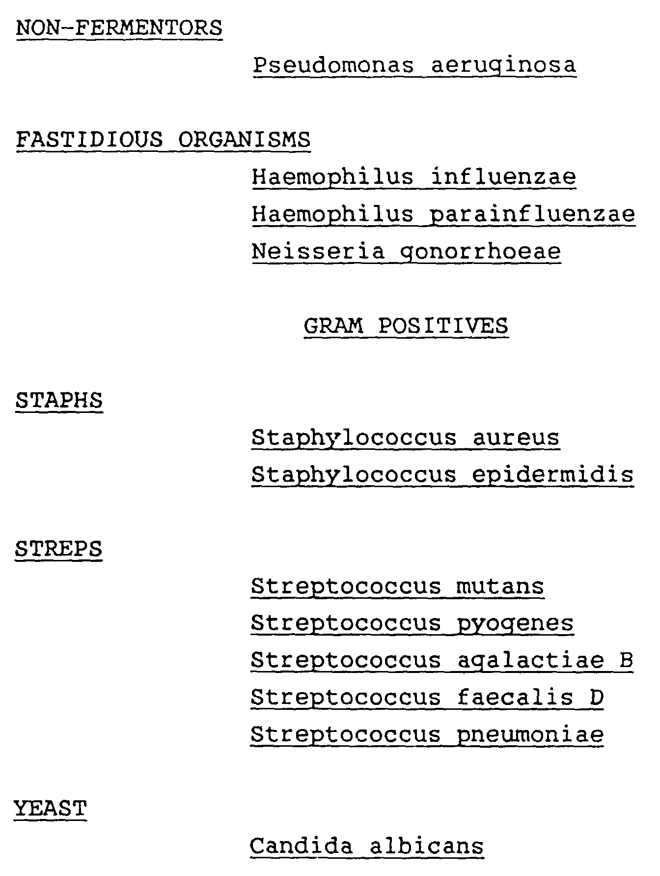
- Forty-six different fluorogenic substrates were tested. These substrates were fluorescently labeled with either 4-methylumbelliferone or 7-amino-4-methyl coumarin. The test measured rates of fluorescence release, as catalyzed by bacterial enzymes generally of the following classes: aminopeptidases, lipases, or glycosidases.
- Data were taken every minute over the course of ten minutes by an instrument built expressly for this function. It had a mercury arc lamp as a source, which was internally monitored for normalization. The excitation wavelength used in this test was 365 nanometers. The emission at 440 nanometers was monitored by a gain-programmable photomultiplier tube. The instrument was controlled by a Ziatech (San Luis Obispo, CA) microprocessor which also controlled X-Y translation of the sample and performed data processing. Sample inoculation was automatic with 25 microliters of bacterial suspension delivered to each substrate site.
- Panels were especially manufactured for these tests by the Strouse and Apogee companies (both of Baltimore, MD). Panels were made from flat black polypropylene sheets, each having a length and width of a standard microwell tray. Schleicher & Schuell 740-E absorbent paper (Keene, NH 03431) was punched to a diameter of 6 mm, and attached via adhesive backing (Strouse V-23) to the plastic support. Disk spacing was the same as for wells of a standard microwell tray.
- Substrates were prepared by dissolving them in dimethyl sulfoxide (DMSO) and depositing 20 µl of the solutions on the appropriate disks on the panel. The panels were then dried by vacuum evaporation for three hours, packaged in sealed foil pouches with a desiccant, and stored at -20° C. Shortly before testing with inocula the panels were allowed to warm to room temperature. Table 7 lists the substrate solutions used and Table 8 lists the standard solutions.


TABLE 8 STANDARD SOLUTIONS SOLUTION ABBREVIATION CONCENTRATION (µg/disk) 4-methylumbelliferone 4-MU 0.012 0.8 7-amino-4-methyl coumarin AMC 0.012 0.8 - Test inocula were prepared from colonies which were incubated overnight on tryptic soy agar with 5% sheep blood, or on chocolate agar for fastidious organisms. Organism suspensions were prepared to an optical density of 1.76 at 610 nm, in screw
cap glass tubes 16 mm X 125 mm. The diluent was 0.85% NaCl, 0.02% Triton® X100, and 0.1 M Tris which was titrated to pH 8.0. Five strains of each of the species were tested with the exceptions of P. magnus and C. albicans, where only 4 strains were tested, and E. coli, where the five strains were tested in duplicate. - The beam from the instrument's mercury arc lamp was optically split, and output was monitored by a photodetector. Data were normalized internally within the instrument for source variation. Data were further normalized externally by dividing the fluorescence data for the substrates by the fluorescence of the appropriate free fluor standards either 4-methylumbelliferone or 7-amino-4-methyl coumarin. Enzyme rates are thus expressed as nanograms of free fluor released per minute.
- Thresholds for each substrate were determined by examining the spontaneous hydrolysis rates from control panels which were inoculated with buffer only. These were compared to rates from the data base for all organism tested. A threshold rate which would indicate a significant difference from a control rate was determined.
- The data generated were analyzed by nearest neighbor analysis utilizing unit sphere projection. This entailed the following steps:
- 1) A rate was determined based on 1 and 5 minute data points or by using a default rate estimator for readings which exceeded the dynamic range of the instrument.
- 2) The rate was compared to a threshold selected for each substrate. If the rate was smaller than the threshold then the ratio was set to 1, otherwise the ratio of the rate to the threshold value for that substrate was computed.
- 3) The log of that ratio was taken.
- 4) The log ratio value of each of the substrates was squared and then those values were summed.
- 5) The square root of the sum was then computed (this is the magnitude of the substrate rate vector).
- 6) The log ratio for each substrate was divided by the magnitude derived in 5 above.
- The substrate values were thereby normalized to the unit vector. This method was found to permit direct comparison of inocula of various cell densities without knowledge of the actual cell densities.
- The nearest neighbor for each of the 128 strains tested was computed by taking the absolute difference for each substrate for each species pair in a confusion matrix. The sum of these absolute differences for all substrates within a species pair was determined. The minimum difference between an "unknown" and a "reference" strain determined the nearest neighbor, or best identification of the unknown.
- Using this technique, each strain tested identified to another strain of the same species as its nearest neighbor for 95% of the tests. The following species mis-identifications occurred:
ACTUAL ID MIS-IDENTIFCATION K. pneumoniae P. mirabilis H. influenzae H. parainfluenzae E. aerogenes K. pneumoniae E. coli C. freundii E. coli C. freundii - However, 95% accuracy is well within the expected performance for commercially available identification test kits.
- Fluorogenic substrates and free fluors may be used to determine enzyme levels of biological samples. The activity of some enzymes has been determined traditionally by testing for acidic and basic by-products of enzyme catalyzed reactions. Fluorescent pH indicators, such 4-methylumbelliferone, may be useful for this purpose in properly buffered systems. The fluorescence of 4-methylumbelliferone is affected by pH in the range of approximately pH 6.2-8.6. When this change in fluorescence is measured on the fluorescence enhancing supports of the present invention as opposed to in microwells the sensitivity is greatly enhanced.
- In this example, 500 ng of 4-methylumbelliferone dissolved in reagent alcohol was deposotied on a 6 mm disk of Schleicher & Schuell 740-E filter paper (Keene, N.H.) and in dry microwells (MicroFluor "B"TM, Dynatech, Chantilly, Va.). The solvent was allowed to dry by evaporation. The fluor was reconstituted with 25 microliters of BIS-TRIS Propane buffer, the pH of which was adjusted in increments of 0.2 to be between pH 6.2 and 8.6. Fluorescence was read with a fluorometer (Dynatech). As can be seen from the data shown in Fig. 9, the fluorescence on the α-cellulose support is greatly enhanced over that in the microwells.
- Traditional bacterial identification schema employ arrays of biochemical materials which are incubated with an unknown bacterial species. If the bacteria react with a material, the by-product is often basic or acidic. These by-products can be detected with appropriate pH indicators such as phenol red or bromothymol blue. Thus, such reactions are interpreted as being either positive or negative, and bacteria are grouped and speciated by these patterns of positive or negative reactions.
- A fluor such as 4-methylumbelliferone which is affected by pH when deposited on the kinetics and fluorescence enhancing supports of the present invention can be used to provide bacterial identifications in much less time than conventional testing.
- Testing for the presence of urease is common and useful in bacterial identification. If urease is present urea hydrolizes to form ammonia, carbon dioxide and water. In solution the end product is ammonium carbonate which increases the pH of the solution.
- The device of the present invention was used to test for the presence of urease. A solution containing urea (10 % by weight), 4-umbelliferone (500 ng) and phosphate buffer (1.25 mM, pH 7.4) was deposited on an α-cellulose disk (740-E Schleicher & Schuell, Keene, N.H.)and allowed to dry. Thereafter the disks were reconstituted with 25 ul of organism suspensions in normal saline (0.85% NaCl). The organism suspensions were prepared to a density of 2.0 McFarland units. Fluorescence was read immediately following inoculation and again after 15 minutes incubation at room temperature. The increase in fluorescence units observed are reported in Table 10.
TABLE 10 TEST ORGANISM INCREASE IN FLUORESCENCE UNITS P. mirabilis (ATCC 2011) 86 M. morganii (ATCC 434) 957 Diluent only 35 - The M. moganii is a very strong urease producer, and the P. mirabilis tested is a weak urease producer.
- Organisms which express the enzyme beta-lactamase are resistant to penicillins and cephalosporin antibiotics which contain beta-lactam rings that are hydrolized by the enzyme. As shown in below, hydrolysis of the beta lactam ring of the monobasic acid penicillin G yields a dibasic acid.
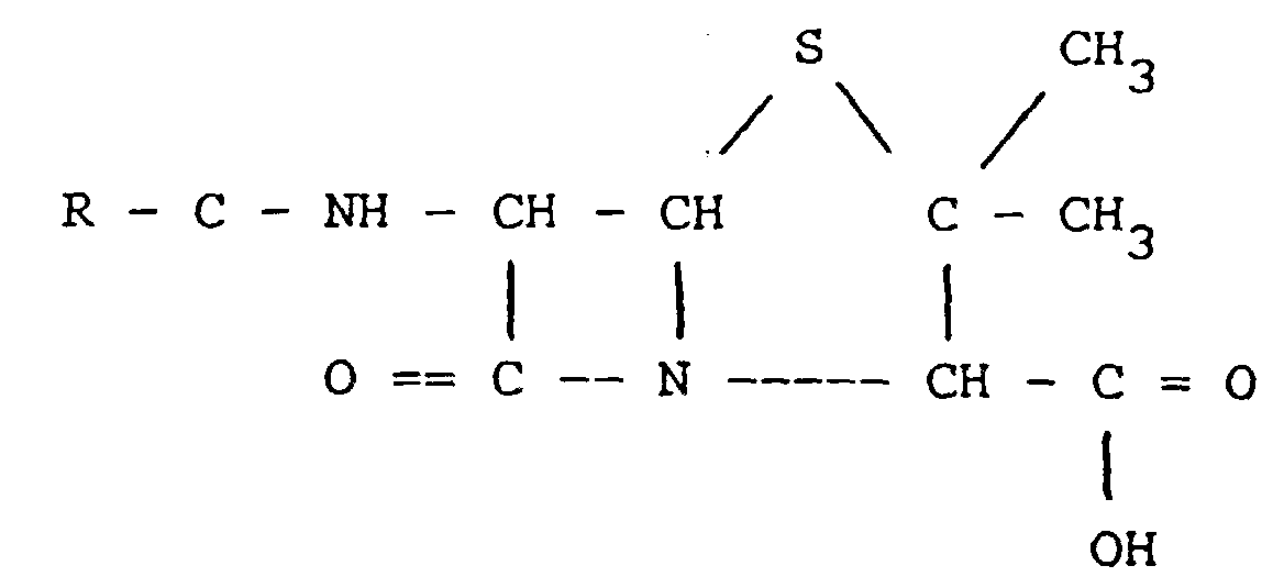
plus water hydrolyzes to: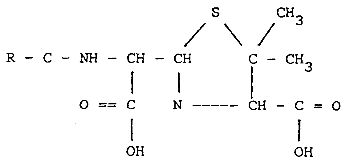
- Organism suspensions were prepared and adjusted to a density of 3.0 McFarland units in 25 mM phosphate buffer which had been titrated to pH 8.0. Aliquots of the suspensions (25 µl) were inoculated onto each disk and an initial fluorescence reading was taken. A final fluorescence reading was taken 15 minutes later. The change in fluorescence signal was determined. The organisms were also tested using a standard beta-lactamase test (Nitrocefin disk, Becton Dickinson Microbiology Systems, Cockeysville, MD). As can be seen from the data reported in Table 11, the organisms which tested positive for beta-lactamase in the commercial test showed substantially greater decreases in fluorescence than the organisms which tested negative. Additionally the strain of B. catarrhalis tested is known to produce more beta-lactamase than the strain of S. aureus tested.
TABLE 11 STRAIN TESTED STANDARD TEST FLUORESCENCE DECREASE PERCENT CHANGE S. edidermidis neg 255 92 ATCC 154 N. meningitidis neg 337 122 ATCC 425 S. aureus pos 530 192 ATCC 29213 B. catarrhalis pos 779 282 ATCC 2907 Negative control N/A 276 N/A
Claims (10)
- A kinetics and fluorescence enhancing test device comprising:a carrier,at least one kinetics and fluorescence enhancing support of α-cellulose and a dry substance selected from fluorogenic substrates and free fluor deposited on the support, wherein the support has a surface area of 0.8-80 mm2 and a void volume of 0.001cm3-0.025cm3.
- The device of claim 1 wherein a plurality of supports are provided.
- The device of claim 2 wherein at least some supports within the plurality differ from other supports within the plurality in the identify or concentration of the fluorogenic substrate.
- The device of claim 2 wherein the fluorogenic substrates are selected from the group consisting of derivatives of 4-methylumbelliferone, coumarin derivatives, 7-amino-4-methyl coumarin, β-naphthylamine, fluorescein, resorufin.
- The device of claim 1 wherein the dry substance is selected from the group of 4-methylumbelliferone, resorufin and fluorescein.
- The device of claim 5 further comprising a non fluorogenic enzyme substrate deposited on the support.
- The device of claims 2 wherein the carrier is microwell plate with a plurality of test wells and the supports are in the test wells.
- The device of claim 2 wherein the carrier is a planar card and the supports are secured to the card.
- A method to characterize enzymes present in a fluid sample comprising:providing the test device of claim 2;adding a fluid sample to each support;testing each support to detect fluorescence hydrolysis products to develop a profile of enzyme content in the sample.
- A method to identify a microorganism in a fluid sample comprising characterizing the enzymes expressed by the microorganism by:providing the test device of claim 3;adding a portion of the fluid sample to each support;testing each support to detect fluorescent hydrolysis products to develop a profile of enzyme content; andidentifying the microorganism by comparing the profile of enzyme content detected to profiles of enzyme content of reference microorganisms.
Applications Claiming Priority (2)
| Application Number | Priority Date | Filing Date | Title |
|---|---|---|---|
| US20967788A | 1988-06-20 | 1988-06-20 | |
| US209677 | 1988-06-20 |
Publications (3)
| Publication Number | Publication Date |
|---|---|
| EP0347771A2 EP0347771A2 (en) | 1989-12-27 |
| EP0347771A3 EP0347771A3 (en) | 1991-03-27 |
| EP0347771B1 true EP0347771B1 (en) | 1996-08-14 |
Family
ID=22779788
Family Applications (1)
| Application Number | Title | Priority Date | Filing Date |
|---|---|---|---|
| EP89110958A Expired - Lifetime EP0347771B1 (en) | 1988-06-20 | 1989-06-16 | Device for enhancing fluorescence and kinetics and methods of using the device |
Country Status (9)
| Country | Link |
|---|---|
| EP (1) | EP0347771B1 (en) |
| JP (1) | JP2617576B2 (en) |
| AT (1) | ATE141413T1 (en) |
| AU (1) | AU616957B2 (en) |
| DE (1) | DE68926935T2 (en) |
| DK (1) | DK304689A (en) |
| ES (1) | ES2090026T3 (en) |
| FI (1) | FI96434C (en) |
| GR (1) | GR3021384T3 (en) |
Cited By (1)
| Publication number | Priority date | Publication date | Assignee | Title |
|---|---|---|---|---|
| US6077669A (en) * | 1997-11-04 | 2000-06-20 | Becton Dickinson And Company | Kit and method for fluorescence based detection assay |
Families Citing this family (19)
| Publication number | Priority date | Publication date | Assignee | Title |
|---|---|---|---|---|
| IE65282B1 (en) * | 1990-04-12 | 1995-10-18 | Becton Dickinson Co | Identifying microorganisms by measuring enzymatic activity in the presence of enzyme activity affecting agents |
| EP0516814B1 (en) * | 1990-12-28 | 1996-09-04 | Dade MicroScan Inc. | Method and composition for determining antimicrobial susceptibility of the majority of clinically significant gram positive organisms |
| US5716798A (en) * | 1992-09-22 | 1998-02-10 | Becton Dickinson And Company | Enhanced detection of microorganisms in samples |
| JPH06142688A (en) * | 1992-11-02 | 1994-05-24 | Daiki Kk | Control method for intermittent aeration in purifying method for sewage of intermittent aeration type |
| EP0790861A1 (en) * | 1995-09-12 | 1997-08-27 | Becton, Dickinson and Company | Device and method for dna amplification and assay |
| ATE324463T1 (en) | 1997-02-04 | 2006-05-15 | Mycometer Aps | METHOD FOR SELECTIVE DETERMINATION OF FUNGI BIOMASS |
| AU768886B2 (en) * | 1998-10-27 | 2004-01-08 | Ibbex, Inc. | Chromogenic substrates of sialidase and methods of making and using the same |
| WO2003063693A2 (en) * | 2002-01-31 | 2003-08-07 | Expressive Constructs, Inc. | Method for detecting microorganisms |
| EP2853593B1 (en) * | 2003-03-07 | 2017-10-04 | DSM IP Assets B.V. | Hydrolases, nucleic acids encoding them and mehods for making and using them |
| FR2855529A1 (en) * | 2003-05-28 | 2004-12-03 | Jean Philippe Goddard | Generating a profile of reactivity for a sample, useful e.g. for diagnosis, comprises simultaneous determination of enzymatic activity against many different substrates |
| WO2005111229A1 (en) * | 2004-05-13 | 2005-11-24 | Kitakyushu Foundation For The Advancement Of Industry, Science And Technology | Substrate for enzymatic activity detection and method of detecting enzymatic activity therewith |
| JP2006061023A (en) * | 2004-08-24 | 2006-03-09 | Suntory Ltd | Screening method and screening apparatus using microchamber array |
| JP2006258744A (en) * | 2005-03-18 | 2006-09-28 | Nokodai Tlo Kk | Fluorescence measurement method |
| WO2007035763A2 (en) * | 2005-09-20 | 2007-03-29 | The Arizona Board Of Regents, A Body Corporate Acting On Behalf Of Arizona State University | Methods for detecting and characterizing microorganisms |
| AT509355B1 (en) | 2010-02-10 | 2012-04-15 | Univ Graz Tech | TEST ARRANGEMENT |
| CN103189524B (en) | 2010-11-01 | 2015-01-28 | 3M创新有限公司 | Biological sterilization indicator and method of use |
| CN103189523B (en) | 2010-11-01 | 2016-03-02 | 3M创新有限公司 | Biological sterilization indicator |
| EP3269822B1 (en) | 2010-11-24 | 2019-11-27 | 3M Innovative Properties Company | Methods to detect a biological activity |
| JP2020129984A (en) * | 2019-02-13 | 2020-08-31 | インテグラムヘルスデザイン株式会社 | Method for measuring enzyme activity |
Family Cites Families (5)
| Publication number | Priority date | Publication date | Assignee | Title |
|---|---|---|---|---|
| WO1983003624A1 (en) * | 1982-04-14 | 1983-10-27 | Plc Unilever | Microbiological test processes and apparatus |
| EP0215066A1 (en) * | 1985-02-27 | 1987-03-25 | University Of Cincinnati | Viable microorganism detection by induced fluorescence |
| US4717658A (en) * | 1985-12-03 | 1988-01-05 | Miles Inc. | Gram negative bacteria screening method with horseshoe crab amebocyte lysate (LAL) |
| DE3855762T2 (en) * | 1987-11-05 | 1997-05-07 | James D Berg | Direct method to detect very low levels of coliform contamination |
| JPH0216965U (en) * | 1988-07-14 | 1990-02-02 |
-
1989
- 1989-04-20 AU AU33244/89A patent/AU616957B2/en not_active Ceased
- 1989-06-16 ES ES89110958T patent/ES2090026T3/en not_active Expired - Lifetime
- 1989-06-16 EP EP89110958A patent/EP0347771B1/en not_active Expired - Lifetime
- 1989-06-16 AT AT89110958T patent/ATE141413T1/en not_active IP Right Cessation
- 1989-06-16 DE DE68926935T patent/DE68926935T2/en not_active Expired - Fee Related
- 1989-06-19 FI FI892994A patent/FI96434C/en not_active IP Right Cessation
- 1989-06-20 DK DK304689A patent/DK304689A/en not_active Application Discontinuation
- 1989-06-20 JP JP1158138A patent/JP2617576B2/en not_active Expired - Lifetime
-
1996
- 1996-10-16 GR GR960402757T patent/GR3021384T3/en unknown
Cited By (1)
| Publication number | Priority date | Publication date | Assignee | Title |
|---|---|---|---|---|
| US6077669A (en) * | 1997-11-04 | 2000-06-20 | Becton Dickinson And Company | Kit and method for fluorescence based detection assay |
Also Published As
| Publication number | Publication date |
|---|---|
| DK304689A (en) | 1989-12-21 |
| ES2090026T3 (en) | 1996-10-16 |
| AU3324489A (en) | 1989-12-21 |
| ATE141413T1 (en) | 1996-08-15 |
| FI892994A0 (en) | 1989-06-19 |
| AU616957B2 (en) | 1991-11-14 |
| FI892994L (en) | 1989-12-21 |
| JPH0246280A (en) | 1990-02-15 |
| FI96434C (en) | 1996-06-25 |
| GR3021384T3 (en) | 1997-01-31 |
| EP0347771A3 (en) | 1991-03-27 |
| DK304689D0 (en) | 1989-06-20 |
| DE68926935D1 (en) | 1996-09-19 |
| FI96434B (en) | 1996-03-15 |
| DE68926935T2 (en) | 1997-03-06 |
| JP2617576B2 (en) | 1997-06-04 |
| EP0347771A2 (en) | 1989-12-27 |
Similar Documents
| Publication | Publication Date | Title |
|---|---|---|
| US5236827A (en) | Device for enhancing fluorescence and kinetics and methods of using the device | |
| AU616957B2 (en) | Device for enhancing fluorescence and kinetics and methods of using the device | |
| EP0558827B1 (en) | Process and kit for detecting microbial metabolism | |
| US5064756A (en) | Microbial susceptibility assay using 7-n-(aminoacyl)-7-amido-4-methylcoumarins | |
| US5079144A (en) | Microorganism testing with a hydrolyzable fluorogenic substrate | |
| US9279141B2 (en) | Method of detecting a biological activity | |
| EP0509791B1 (en) | Microbial monitoring device | |
| US3933592A (en) | Method of detecting living microorganisms | |
| US8828680B2 (en) | Combined rapid susceptibility assay and microorganism identification system | |
| US5336600A (en) | Method and reagents for the detection of microorganisms | |
| Delisle et al. | Rapid detection of Escherichia coli in urine samples by a new chromogenic beta-glucuronidase assay | |
| JPH11510051A (en) | Multi-zone sterilization indicator | |
| AU630549B2 (en) | Identifying microorganisms by measuring enzymatic activity in the presence of enzyme activity affecting agents | |
| JPH07114708B2 (en) | Methods and compositions for determining antimicrobial susceptibility of the majority of clinically significant Gram-positive microorganisms | |
| EP0094554B1 (en) | Test for esterase activity in a liquid sample | |
| JPS6156098A (en) | Detection reagent and method of anti-bacterial active substance | |
| US6750038B1 (en) | Rapid antibiotic susceptibility test | |
| EP1324736A2 (en) | Rapid antibiotic susceptibility test | |
| JP2015504684A (en) | In vitro detection of microorganisms by azoreductase activity | |
| CA2012287A1 (en) | Rapid detection of e.coli |
Legal Events
| Date | Code | Title | Description |
|---|---|---|---|
| PUAI | Public reference made under article 153(3) epc to a published international application that has entered the european phase |
Free format text: ORIGINAL CODE: 0009012 |
|
| AK | Designated contracting states |
Kind code of ref document: A2 Designated state(s): AT BE CH DE ES FR GB GR IT LI LU NL SE |
|
| 17P | Request for examination filed |
Effective date: 19901204 |
|
| PUAL | Search report despatched |
Free format text: ORIGINAL CODE: 0009013 |
|
| AK | Designated contracting states |
Kind code of ref document: A3 Designated state(s): AT BE CH DE ES FR GB GR IT LI LU NL SE |
|
| 17Q | First examination report despatched |
Effective date: 19930429 |
|
| GRAH | Despatch of communication of intention to grant a patent |
Free format text: ORIGINAL CODE: EPIDOS IGRA |
|
| GRAH | Despatch of communication of intention to grant a patent |
Free format text: ORIGINAL CODE: EPIDOS IGRA |
|
| GRAA | (expected) grant |
Free format text: ORIGINAL CODE: 0009210 |
|
| AK | Designated contracting states |
Kind code of ref document: B1 Designated state(s): AT BE CH DE ES FR GB GR IT LI LU NL SE |
|
| PG25 | Lapsed in a contracting state [announced via postgrant information from national office to epo] |
Ref country code: GR Free format text: LAPSE BECAUSE OF FAILURE TO SUBMIT A TRANSLATION OF THE DESCRIPTION OR TO PAY THE FEE WITHIN THE PRESCRIBED TIME-LIMIT Effective date: 19960814 |
|
| REF | Corresponds to: |
Ref document number: 141413 Country of ref document: AT Date of ref document: 19960815 Kind code of ref document: T |
|
| REF | Corresponds to: |
Ref document number: 68926935 Country of ref document: DE Date of ref document: 19960919 |
|
| ET | Fr: translation filed | ||
| REG | Reference to a national code |
Ref country code: ES Ref legal event code: FG2A Ref document number: 2090026 Country of ref document: ES Kind code of ref document: T3 |
|
| ITF | It: translation for a ep patent filed | ||
| REG | Reference to a national code |
Ref country code: CH Ref legal event code: NV Representative=s name: PATENTANWALTSBUREAU R. A. MASPOLI |
|
| REG | Reference to a national code |
Ref country code: ES Ref legal event code: FG2A Ref document number: 2090026 Country of ref document: ES Kind code of ref document: T3 |
|
| REG | Reference to a national code |
Ref country code: GR Ref legal event code: FG4A Free format text: 3021384 |
|
| PG25 | Lapsed in a contracting state [announced via postgrant information from national office to epo] |
Ref country code: GB Free format text: LAPSE BECAUSE OF NON-PAYMENT OF DUE FEES Effective date: 19970616 Ref country code: AT Free format text: LAPSE BECAUSE OF NON-PAYMENT OF DUE FEES Effective date: 19970616 |
|
| PG25 | Lapsed in a contracting state [announced via postgrant information from national office to epo] |
Ref country code: SE Effective date: 19970617 Ref country code: ES Free format text: LAPSE BECAUSE OF THE APPLICANT RENOUNCES Effective date: 19970617 |
|
| PLBE | No opposition filed within time limit |
Free format text: ORIGINAL CODE: 0009261 |
|
| STAA | Information on the status of an ep patent application or granted ep patent |
Free format text: STATUS: NO OPPOSITION FILED WITHIN TIME LIMIT |
|
| PG25 | Lapsed in a contracting state [announced via postgrant information from national office to epo] |
Ref country code: LU Free format text: LAPSE BECAUSE OF NON-PAYMENT OF DUE FEES Effective date: 19970630 Ref country code: LI Free format text: LAPSE BECAUSE OF NON-PAYMENT OF DUE FEES Effective date: 19970630 Ref country code: CH Free format text: LAPSE BECAUSE OF NON-PAYMENT OF DUE FEES Effective date: 19970630 Ref country code: BE Effective date: 19970630 |
|
| 26N | No opposition filed | ||
| BERE | Be: lapsed |
Owner name: BECTON DICKINSON AND CY Effective date: 19970630 |
|
| PG25 | Lapsed in a contracting state [announced via postgrant information from national office to epo] |
Ref country code: NL Effective date: 19980101 |
|
| GBPC | Gb: european patent ceased through non-payment of renewal fee |
Effective date: 19970616 |
|
| REG | Reference to a national code |
Ref country code: CH Ref legal event code: PL |
|
| PG25 | Lapsed in a contracting state [announced via postgrant information from national office to epo] |
Ref country code: FR Free format text: LAPSE BECAUSE OF NON-PAYMENT OF DUE FEES Effective date: 19980227 |
|
| EUG | Se: european patent has lapsed |
Ref document number: 89110958.9 |
|
| NLV4 | Nl: lapsed or anulled due to non-payment of the annual fee |
Effective date: 19980101 |
|
| PG25 | Lapsed in a contracting state [announced via postgrant information from national office to epo] |
Ref country code: DE Free format text: LAPSE BECAUSE OF NON-PAYMENT OF DUE FEES Effective date: 19980303 |
|
| REG | Reference to a national code |
Ref country code: FR Ref legal event code: ST |
|
| REG | Reference to a national code |
Ref country code: FR Ref legal event code: ST |
|
| REG | Reference to a national code |
Ref country code: ES Ref legal event code: FD2A Effective date: 20001009 |
|
| PG25 | Lapsed in a contracting state [announced via postgrant information from national office to epo] |
Ref country code: IT Free format text: LAPSE BECAUSE OF NON-PAYMENT OF DUE FEES;WARNING: LAPSES OF ITALIAN PATENTS WITH EFFECTIVE DATE BEFORE 2007 MAY HAVE OCCURRED AT ANY TIME BEFORE 2007. THE CORRECT EFFECTIVE DATE MAY BE DIFFERENT FROM THE ONE RECORDED. Effective date: 20050616 |