EP0328939A2 - Méthode, réactif, trousse et anticorps monoclonal pour le diagnostic du cancer dans les organes humains de la digestion par détermination de la glutathione-S-transférase acide - Google Patents
Méthode, réactif, trousse et anticorps monoclonal pour le diagnostic du cancer dans les organes humains de la digestion par détermination de la glutathione-S-transférase acide Download PDFInfo
- Publication number
- EP0328939A2 EP0328939A2 EP89101717A EP89101717A EP0328939A2 EP 0328939 A2 EP0328939 A2 EP 0328939A2 EP 89101717 A EP89101717 A EP 89101717A EP 89101717 A EP89101717 A EP 89101717A EP 0328939 A2 EP0328939 A2 EP 0328939A2
- Authority
- EP
- European Patent Office
- Prior art keywords
- antibody
- human
- transferase
- monoclonal antibody
- glutathione
- Prior art date
- Legal status (The legal status is an assumption and is not a legal conclusion. Google has not performed a legal analysis and makes no representation as to the accuracy of the status listed.)
- Granted
Links
Images


Classifications
-
- G—PHYSICS
- G01—MEASURING; TESTING
- G01N—INVESTIGATING OR ANALYSING MATERIALS BY DETERMINING THEIR CHEMICAL OR PHYSICAL PROPERTIES
- G01N33/00—Investigating or analysing materials by specific methods not covered by groups G01N1/00 - G01N31/00
- G01N33/48—Biological material, e.g. blood, urine; Haemocytometers
- G01N33/50—Chemical analysis of biological material, e.g. blood, urine; Testing involving biospecific ligand binding methods; Immunological testing
- G01N33/53—Immunoassay; Biospecific binding assay; Materials therefor
- G01N33/5306—Improving reaction conditions, e.g. reduction of non-specific binding, promotion of specific binding
-
- C—CHEMISTRY; METALLURGY
- C07—ORGANIC CHEMISTRY
- C07K—PEPTIDES
- C07K16/00—Immunoglobulins [IGs], e.g. monoclonal or polyclonal antibodies
- C07K16/18—Immunoglobulins [IGs], e.g. monoclonal or polyclonal antibodies against material from animals or humans
- C07K16/28—Immunoglobulins [IGs], e.g. monoclonal or polyclonal antibodies against material from animals or humans against receptors, cell surface antigens or cell surface determinants
- C07K16/30—Immunoglobulins [IGs], e.g. monoclonal or polyclonal antibodies against material from animals or humans against receptors, cell surface antigens or cell surface determinants from tumour cells
- C07K16/3046—Stomach, Intestines
-
- C—CHEMISTRY; METALLURGY
- C07—ORGANIC CHEMISTRY
- C07K—PEPTIDES
- C07K16/00—Immunoglobulins [IGs], e.g. monoclonal or polyclonal antibodies
- C07K16/40—Immunoglobulins [IGs], e.g. monoclonal or polyclonal antibodies against enzymes
Definitions
- This invention relates to a method of determining human acid glutathione S-transferase in a human assay sample, a reagent therefor, a kit therefor, a method of diagnosing cancer in digestive organs, and to a monoclonal antibody. Particularly, it relates to a method of determining human acid glutathione S-transferase in an assay sample immunologically using an antibody and thus diagnosing cancer in digestive organs, a reagent therefor, a kit therefor, and a monoclonal antibody for use therein.
- Glutathione S-transferase (to be sometimes abbreviated "GST") is known as a detoxifying enzyme which catalyzes conjugation reactions between various substances invading the living body or metabolites in the body and reduced glutathione. Many of these substances and metabolites react with proteins or nucleic acid in the body to become a cause of various lesions, for example carcinogenesis. The reactivity of these substances is neutralized by conjugation with glutathione, and they are converted into more water-soluble products which are metabolized, for example, in the liver and finally excreted out of the body.
- GST occurs widely in many biological species including mammals, particularly in abundance in the cytoplasm of liver, and in varying amounts in the spleen, kidneys, lungs, brain, skeletal muscle, testicles and small intestines. Special molecular species have been isolated from the placenta or red blood cells.
- GST is composed of many enzymes and has species specificity and specificity for organs or tissues.
- Human basic GST has an isoelectric point (pI) of 7 to 9, and is composed of two subunits both having a molecular weight of about 23,000.
- pI isoelectric point
- Five molecular species, ⁇ , ⁇ , ⁇ , ⁇ , ⁇ , ⁇ , ⁇ , are known to exist in human basic GST. These molecular species exist in the liver and also in the kidneys, testicles, small intestines, brain and lungs of normal healthy adults.
- Human acid GST has an isoelectric point (pI) of 4 to 5 and is composed of two subunits both having a molecular weight of about 22,000.
- pI isoelectric point
- a sepcies ⁇ of human acid GST is known to exist in placenta, and its species ⁇ is known to exist in the red blood cells. It hardly exists in the liver of normal healthy adults.
- the present inventors attempted to use a well-practiced immunological method involving the use of a polyclonal antibody for the determination of human acid GST.
- this method it is difficult to determine minute amounts of human acid GST in a human assay sample stably with specificity and a certain level because human acid GST is an enzyme including many kinds of isozymes and the quality of the polyclonal antibody itself varies depending upon its source of procurement.
- the present inventors found that usually, human acid GST is not present, or present only in a tiny amount in a body fluid, particularly plasma, of a normal subject, but when a digestive organ is attacked by cancer, glutathione S-transferase comes into being in the body fluid and becomes detectable, or its amount in the body fluid increases.
- the present inventors also found that acid GST is present in the body fluids of patients with digestive organ cancer in detectable amounts or in larger amounts than acid GST in normal subjects.
- an immunological assay system which can recognize acid GST produced by commonly proliferating cells of digestive organs in liver cancer cells, stomach cancer cells, esophagus cancer cells, etc. irrespective of the type of the cancer cells, it would of course be able to be utilized for determining human acid GST in a human assay sample such as a human serum or plasma sample, and permit simple diagnosis of cancer of digestive organs.
- the amount of acid GST in a body fluid of a patient with digestive organ cancer is larger than that in a body fluid of a normal subject, its absolute amount is very minute (for example, several ng/ml to several hundred ng/ml). It is essential therefore to the above diagnosis of cancer to develop a method of determining a minute amount of acid GST with simplicity and high sensitivity.
- Another object of this invention is to provide a simple method of determining human acid GST in a human assay sample commonly with high sensitivity independently of the type of proliferating cells of digestive organs which produce acid GST.
- Still another object of this invention is to provide a method of diagnosing cancer in a human digestive organ which comprises determining the amount of human GST in a human assay sample, particularly human serum sample, and diagnosing the onset of cancer in a human digestive organ, the state of its progress or its cure on the basis of the measured amount of human GST.
- Yet another object of this invention is to provide an assay reagent and its kit for determining human acid GST stably with high sensitivity which can be used in the above determination method and the diagnosing method.
- a further object of this invention is to provide a method which comprises taking out a tissue from a human digestive organ, determining the amount of human acid GST in the tissue, and diagnosing cancer in the digestive organ.
- a still further object of this invention is to provide a practical and simple immunological method of determining human acid GST and a reagent and a kit therefor which help to discover the onset of cancer in a human digestive organ or cancer in the organ in the early stage.
- human acid GST present in very small amounts in a human assay sample can be determined stably with high sensitivity, and the method and the kit used for it are simple.
- the amount of human acid GST in an assay sample taken from a patient with cancer in digestive organs is minute but larger than that of a sample taken from a normal subject.
- the method of this invention can determine this minute amount of human acid GST accurately with high sensitivity and can help to diagnose cancer in digestive organs, particularly the discovery of cancer.
- Example 6 the amount of human acid GST in serum samples of normal subjects and patients with various digestive organ cancers was determined by the method of this invention. The result shows that it is about 11.2 ng/ml on an average for the normal subjects, whereas it is about 27.5 ng/ml for the patient with esophagus cancer, about 34.7 ng/ml for the patient with stomach cancer, about 29.5 ng/ml for the patient with colon cancer, and about 26.9 ng/ml for the patient with liver cancer.
- significant differences were clearly seen in the amount of human acid GST between the normal subjects and the cancer patients. This shows a clear correlation between the amount of human acid GST and cancer in digetive organs.
- One important feature of the assay system in accordance with this invention is that a tiny amount of human acid GST ascribed mainly to cancer in various digestive organs can be commonly measured accurately.
- the term "digestive organs" used in the present specification and the appended claims denotes organs relating to the digestive system, and more specifically stomach, esophagus, colon, liver and cholecyst.
- the determination of human acid GST in accordance with this invention is effective for the diagnosis of cancer in these organs, specifically checking of carcinogenesis, monitoring of the progress of cancer, and determination of cure of cancer as a result of treatment.
- Diagnosis of cancers by the determination of human acid GST in accordance with this invention applies not only to cancers in the above digestive organs, but also to other diseases, particularly other cancers, which show an increased level of acid GST over normal subjects.
- the human assay sample used in the determination of human acid GST may be various human tissues or body fluids, and they include, for example, serum, plasma, ascites, tissue cells of the digestive organs, and culture supernatants of these cells.
- a human serum or plasma sample is most preferred.
- human acid GST is composed of two subunits having a molecular weight of about 22,000.
- human acid GST In human acid GST-producing cells or culture supernatants thereof, human acid GST is believed to be composed of two subunits. Many experiments, however, show that in a fraction of blood obtained by ordinary centrifugation or the like, for example serum or plasma, human acid GST, in many cases, exists as a single subunit, although no clear cause of this has been known. Accordingly, the present inventors presume from the results of many experiments that human acid GST in the fractionated serum or plasma exits as a single subunit.
- the assay system of this invention permits determination of human acid GST whether it is composed of two subunits or only a single subunit.
- the human assay sample to be determined actually is, in many cases, human serum or plasma, and human acid GST in such a sample exists mostly as a single subunit. Accordingly, the amount of human acid GST in the assay sample can be calculated on the basis of a calibration curve prepared under the assumption that human acid GST is composed of a single subunit.
- the polyclonal antibody used in this invention is obtained as an antibody component of anti-human acid GST anti-serum which is obtained by immunizing an animal with human acid GST- ⁇ as an antigen by a known method.
- Goat anti-human acid GST-polyclonal antibody and rabbit anti-human acid GST-polyclonal antibody are preferably used in this invention.
- Polyclonal antibodies purified by affinity chromatography of the antigen are more preferred.
- the equivalent fragment of the polyclonal antibody as used in this invention means a fragment of the above polyclonal antibody which reacts with human acid GST in substantially the same way as in the reaction of the polyclonal antibody with human acid GST (antigen).
- Fab, Fab′ and F(ab′)2, above all Fab′ or F(ab′)2 are preferred.
- the monoclonal antibody to human placenta-derived acid GST (GST- ⁇ ) used in this invention is a monoclonal antibody which specifically recognizes human placenta-derived human acid GST (GST- ⁇ ) produced by proliferating cells of cancer cells of digestive organs such as human liver, stomach and esophagus, and preferivelyably has a common epitope in human acid GST produced by these cancer cells.
- the monoclonal antibodies used in this invention are of the IgG class, and are advantageously mouse antibodies.
- the monoclonal antibodies used in this invention specifically recognize human acid GST, but do not at all recognize, and therefore do not cross-react with, human basic GST.
- the present invention provides the following three monoclonal antibodies.
- the three monoclonal antibodies (a), (b) and (c) have different epitopes in GST- ⁇ and excellent affinity for the objects of this invention.
- the monoclonal antibodies (a) and (b) exhibit reactivity with GST- ⁇ directly fixed to the surface of a solid carrier and GST- ⁇ floating in a liquid phase.
- the monoclonal antibody (c) shows reactivity with GST- ⁇ fixed directly on the surface of a solid carrier, but does not show substantial reactivity with GST floating in a liquid phase.
- Direct fixation of GST- ⁇ to the surface of a solid carrier is effected by the following procedure.
- GST- ⁇ prepared in a concentration of 1 microgram/ml is added in an amount of 100 microliters to each well of a polystyrene plate (made by Sumitomo Bakelite Company), and left to stand overnight at 4°C.
- the plate is then washed three times with phosphate buffer (PBS) and after-coated with 1% BSA-PBS to prepare a sample in which GST- ⁇ is fixed.
- PBS phosphate buffer
- BSA-PBS 1% BSA-PBS
- the monoclonal antibodies to human acid GST used in this invention may be obtained by methods described below.
- antibody-producing cells of an animal immunized with human placenta-derived GST are fused with myeloma cells to give a hybridoma capable of producing a monoclonal antibody to GST- ⁇ .
- the hybridoma and/or a cell line derived from it is cultured, and the resulting monoclonal antibody to GST- ⁇ is recovered from the culture.
- Human GST- ⁇ used for immunization is composed of two subunits both having a molecular weight of about 22,000, shows a single band in sodium dodecyl sulfate polyacrylamide gel electrophoresis (SDS-Page) and has an isoelectric point (pI) of 4 to 5.
- the hybridoma capable of producing a monoclonal antibody which recognizes human GST- ⁇ can be produced by a cell fusion technique known per se .
- An animal such as monkey, horse, bovine, goat, sheep, rabbit, rat and mouse is immunized with human GST- ⁇ , and antibody-producing cells (lymphocytes) are taken from the spleen, lymph nodes, etc. of the immunized animal.
- the antibody-producing cells are then fused with myeloma cells of man or another animal.
- mouse myeloma cells such as P3-X63-Ag8, P3X63-Ag8-U1, P3-NSl/1-Ag4-1, P3-X63-Ag8-6.5.3, SP2/0-Ag14, Fo and MPC11-45.6TG1.7 of BALB/C mice.
- the cell fusion conditions are, for example, as follows:- The antibody-producing cells and the myeloma cells are mixed in a mixing ratio of from 10:1 to 1:10, preferably from 1:1 to 1:3, and a suitable cell fusion solution such as RPMI 1640 containing about 35 % of polyethylene glycol having a molecular weight of about 1,000 to 6,000 and 7.5 % of dimethyl sulfoxide is added. The mixture is stirred at room temperature to 37 °C for 1 to several minutes. The mixture is then gradually diluted with RPMI 1640 containing 10 % of bovine fetal serum.
- RPMI 1640 containing about 35 % of polyethylene glycol having a molecular weight of about 1,000 to 6,000 and 7.5 % of dimethyl sulfoxide
- the diluted mixture is adjusted to a cell density of 1 to 5 x 10⁵/ml with a HAT (hypoxanthine-aminopterine-thymidine) selective medium.
- HAT hyperxanthine-aminopterine-thymidine
- the resulting medium was added to a 96-well plate at a rate of 0.2 ml per well, and incubated at 35 to 38 °C for 2 to 3 weeks in air containing 5 % CO2.
- HAT hypoxia-xanthine-aminopterine-thymidine
- This screening step can be carried out by examining the hybridoma colonies in accordance with an enzyme-linked immunosorbent assay (ELISA) to determine whether the monoclonal antibodies produced by the hybridoma react with human GST- ⁇ .
- ELISA enzyme-linked immunosorbent assay
- the hybridoma colonies which secrete the desired monoclonal antibody are then cloned.
- This step can be carried out by using a limiting dilution method. About 2 to 3 weeks later, the colonies grown in the 96-well plate are picked up and again examined for antibody activity on human GST- ⁇ by ELISA. The elected hybridoma colonies are cultured to produce a monoclonal antibody specific for human GST- ⁇ .
- Another method of obtaining the monoclonal antibody comprises transfecting the antibody-producing cells with Epstein-Barr virus (E-B virus for short) to prepare transformed cells, culturing the transformed cells and/or a cell line derived therefrom, and recovering a monoclonal antibody having the property of binding to human GST- ⁇ from the culture.
- E-B virus Epstein-Barr virus
- E-B virus belongs to a virus of a group of hepesviruses, which is considered to be the cause of Burkitt's lymphoma and rhinal and throat cancer.
- the antibody-producing cells are infected with E-B virus, and about 2 to 3 weeks later, incubated in a 5 % CO2 incubator to form many heterogeneous colonies of transformed cells. From these colonies, only those colonies which secrete a monoclonal antibody specific for human GST- ⁇ are selected by the same method as described above. The selected colonies are cloned to form the desired transformed cells.
- the selected hybridoma or transformed cells are cultured to produce the desired specific monoclonal antibody.
- the hybridoma or transformed cells which produce an antibody recognizing human GST- ⁇ and are selected by cloning may be frozen and stored, or may be cultured in quantities by a suitable method. From the culture supernatant, the monoclonal antibody specifically binding to human GST- ⁇ can be obtained. It is also possible to transplant the cells into an animal to form a tumor, and obtain the desired antibody from the ascites or serum of the animal.
- the monoclonal antibody may be purified by, for example, affinity chromatography using protein A.
- the monoclonal antibodies used in this invention are prepared and purified by the above methods, and are useful for assaying human acid GST or diagnosing cancer of digestive organs. Equivalent fragments of these monoclonal antibodies may also be used in the above assay and diagnosis.
- the "equivalent fragments” mean fragments which show reactivity with human acid GST in substantially the same way as the corresponding monoclonal antibodies. Specifically, they are Fab, Fab′ and F(ab′)2, especially preferably Fab′ and F(ab′)2.
- a first antibody to human acid GST is fixed to a suitable insoluble solid carrier such as a plastic receptacle (the antibody will be referred to as the "fixed antibody"). Then, the surface of the insoluble solid carrier is coated with a suitable substance (such as bovine serum albumin) to void a non-specific combination of the insoluble solid carrier with the reagent or the assay sample.
- a suitable insoluble solid carrier such as a plastic receptacle
- the insoluble solid carrier to which the first antibody is fixed is then contacted with the human assay sample at a fixed temperature for a fixed period of time to perform reaction. During this time, human acid GST in the assay sample binds to the fixed antibody (first antibody).
- a solution such as an aqueous solution
- a suitable labelling substance such as an enzyme
- the insoluble solid carrier is washed with a suitable washing solution, and the amount of the labelling substance labelled on the second antibody on the insoluble solid carrier is measured.
- Either one of the first and second antibodies is a polyclonal antibody, and the other, a monoclonal antibody.
- the above reaction may also be carried out by mixing the fixed antibody (first antibody), the labelled antibody (second antibody) and the assay sample containing human acid GST simultaneously and contacting the three simultaneously at a fixed temperature for a fixed period of time.
- the amount of human acid GST in the human assay sample can be calculated from the amount of the labelling substance on the second antibody.
- the assay reagent for immunological determination of human acid GST is composed of the antibody bound to the insoluble solid carrier and the labelled antibody.
- the kit for immunological determination of human acid GST comprises the assay reagent and as adjuvants for utilizing the assay reagent efficiently and conveniently, a dissolving agent for dissolving a solid reagent or a liquid assay sample, for example, a washing agent for washing away the antigen and antibody non-specifically bound to the insoluble solid carrier, and when an antibody labelled with an enzyme is used, a substrate for measuring enzyme activity and a reaction stopper therefor, and other adjuvants usually employed in other immunological assay kits.
- a dissolving agent for dissolving a solid reagent or a liquid assay sample
- a washing agent for washing away the antigen and antibody non-specifically bound to the insoluble solid carrier
- a substrate for measuring enzyme activity and a reaction stopper therefor for measuring enzyme activity and a reaction stopper therefor
- the insoluble solid carrier may be in various shapes, such as a tray, a sphere, a fiber, a rod, a disc, a receptacle, a cell and a test tube.
- the labelling substance in the labelled antibody may be, for example, an enzyme such as peroxidase, alkaline phosphatase or beta-D-galactosidase; a fluorescent substance such as fluorescent isothiocyanate and phycobiliprotein, a luminescent substance such as isolucinol, lucigenin, or a radioactive substance such as 125I, 131I, 14C and 3H.
- an enzyme such as peroxidase, alkaline phosphatase or beta-D-galactosidase
- a fluorescent substance such as fluorescent isothiocyanate and phycobiliprotein
- a luminescent substance such as isolucinol, lucigenin
- a radioactive substance such as 125I, 131I, 14C and 3H.
- Other labelling substances which are generally used in immunological assay may also be used in this invention.
- the labelling agent is an enzyme
- a substrate for measuring its activity and as required a coloring agent may be used.
- H2O2 is used as the substrate and ammonium 2,2′-azinodi-(3-ethylbenzothiazolinesulfonate) (ABTS), 5-aminosalicyclic acid, o-phenylenediamine, 4-aminoantipyrine and 3,3′,5,5′-tetramethylbenzidine may be used as the coloring agent.
- ABTS ammonium 2,2′-azinodi-(3-ethylbenzothiazolinesulfonate)
- 5-aminosalicyclic acid o-phenylenediamine
- 4-aminoantipyrine 4-aminoantipyrine
- 3,3′,5,5′-tetramethylbenzidine 3,5,5′-tetramethylbenzidine
- beta-D-galactosidase When beta-D-galactosidase is used as the enzyme, fluorescein-di-(beta-D-galactopyranoside) and 4-methylumbelliferyl-beta-D-galactopyranoside may be used as the substrate.
- one of the first antibody and the labelled second antibody is the above polyclonal antibody and the other is the monoclonal antibody 2H or monoclonal antibody 6A because this combination gives higher sensitivity.
- An especially preferred combination is a combination of the polyclonal antibody as the first antibody and the monoclonal antibody 2H or monoclonal antibody 6A as the labelled second antibody.
- an antigen-antibody reaction adjusting agent to be described below may be used under the following conditions. This permits immunological determination of human acid GST with higher sensitivity, and is preferred for the objects of this invention.
- a protein having an average molecular weight of 16,000 to 50,000 and an isoelectric point of 1.0 to 5.0 or a mixture containing it is added to the immunological reaction solution as an antigen-antibody reaction regulating agent, and the final concentration of the antigen-antibody reaction regulating agent in the immunological reaction solution is adjusted to 0.02 to 0.9 % by weight.
- the "average molecular weight" of the protein used in this invention is measured by an osmotic pressure method by which the average molecular weight of the protein is measured by utilizing the fact that when a solution of a polymer and a solvent are contacted with each other via a semipermeable membrane which is permeable to the polymer solution and the solvent but impermeable to the dissolved polymer, the difference in osmotic pressure between the two liquids becomes a parameter of the molecular weight of the polymer. It is measured at 4 °C by using a 6.66M urea solution as the solvent.
- the "isoelectric point" of the protein denotes a value measured by a chromatofocusing method which separates a protein according to its isoelectric point. Specifically, it is measured by using a column (0.5 cm in diameter and 45 cm in length) filled with a gel of PBE 94 (produced by Pharmacia Co.) and 0.025M imidazole HCl (pH 7.4) as an eluent.
- the protein used as the antigen-antibody reaction regulating agent examples include casein, pepsin, ovoglycoprotein and orosomucoid.
- casein casein
- pepsin casein
- ovoglycoprotein ovoglycoprotein
- orosomucoid a protein having a molecular weight of less than 16,000
- non-specific adsorption tends to increase.
- molecular weight of the protein exceeds 50,000, the decrease of the immunologically non-specific reaction tends to be insufficient, and the specific immunological reaction tends to decrease.
- the molecular weight of the protein used is 16,000 to 50,000, preferably 20,000 to 46,000.
- the protein used as the antigen-antibody reaction regulating agent has an isoelectric point of 1.0 to 5.0, preferably 1.2 to 4.8.
- a mixture containing the above protein may be used as the antigen-antibody reaction regulating agent.
- Such a mixture may comprise 10 to 60 % by weight, preferably 20 to 50 % by weight, of the protein and 30 to 80 % by weight, preferably 40 to 60 % by weight, of a sugar (such as lactose) as main components, and fats (e.g., 0.5 to 2 % by weight), ashes (e.g., 5 to 12 % by weight) and water (e.g., 2 to 8 % by weight).
- a sugar such as lactose
- fats e.g., 0.5 to 2 % by weight
- ashes e.g., 5 to 12 % by weight
- water e.g., 2 to 8 % by weight
- a typical example of such a mixture is skimmed milk.
- Skimmed milk contains casein as the protein, but is characterized by having better dispersibility in the immunological reaction solution, a higher non-specific binding effect per unit weight of the protein, and better preservability (less prone to form a precipitate) at 4 °C than casein singly used.
- the skimmed milk may be defatted milk of any type and origin. Most typically, it is commercial skimmed milk produced by Difco Co.
- the protein solution (such as a skimmed milk solution) is prepared by the following procedure.
- a protein or its mixture e.g., skimmed milk
- phosphate-buffered physiological saline e.g., phosphate-buffered physiological saline
- the mixture is stirred for about 1 hour.
- the mixture is then ultrasonicated to give a solution which, for example, passes through a 0.45 micron millipore membrane.
- Immunological assay was carried out using protein solutions (or skimmed milk solutions) of various concentrations.
- a protein solution or a skimmed milk solution having a concentration of less than 0.02 % by weight
- a non-specific reaction markedly increased in spite of the absence of an antigen.
- the lower limit to the concentration of the protein should be 0.02 % by weight. If the concentration exceeds 0.9 % by weight, the specific immunological reaction decreases and the storage stability of the protein solution in a refrigerator tends to be reduced. Accordingly, the upper limit of the concentration of the protein should be 0.9 % by weight.
- the concentration of the protein which ensures the stability of the reagent and effectively reduces the non-specific reaction is suitably within the range of 0.02 to 0.9 % by weight, prefrably 0.05 to 0.7 % by weight.
- the present invention not only provides an immunological assay method in accordance with the so-called sandwich method and a kit therefor but also a method of determining human acid GST by tissue staining and a monoclonal antibody used therefor.
- a tumor tissue sample is taken from a digestive organ or other organ, and stained by a known method using the monoclonal antibody in accordance with this invention to determine the amount of human acid GST in the tissue cells. The result of this determination permit diagnosis of cancer.
- a method of diagnosing cancer on a human digestive organ which comprises
- a monoclonal antibody to human placenta-derived glutathione S-transferase which can specifically recognize a region of the amino acid sequence of human placenta-derived glutathione S-transferase ranging from the 141st amino acid residue Thr from the N-terminus to the 175th amino acid residue Leu (monoclonal antibody 5F) is most suitable as the monoclonal antibody used in the above tissue staining.
- BSA bovine serum albumin
- PBS phosphate-buffered saline 3 %
- BSA/PBS PBS containing 3 % (w/v) of BSA PVDF membrane: polyvinylidene difluoride membrane (produced by Pharmacia Co.)
- HRP horseradish peroxidase
- Human placenta was minced in 10 mM phosphate buffer (pH 7.4) containing 0.2M sucrose, and homogenized by a homogenizer. The homogenate was centrifuged at 10,000 G and 4 °C for 30 minutes, and the supernatant was collected. The supernatant was further centrifuged at 100,000 G and 4 °C for 1 hour, and the resulting supernatant was collected. This supernatant was dialyzed against 10 mM phosphate buffer (pH 6.8). The dialyzate was passed through a carboxymthyl-filled column equilibrated with 10 mM phosphate buffer and a non-adsorbed fraction was collected.
- This fraction was a column filled with reduction-type glutathione-fixed Sepharose to permit adsorption of GST- ⁇ . Then, the column was washed with 10 mM phosphate buffer (pH 7.4). When the absorption intensity at 280 nm in the ultraviolet adsorption spectrum of the effluent reached less than 0.02, Tris buffer (pH 8.0) containing reduction-type glutathione was passed through the column to elute the adsorbed matter. The eluate was ultrafiltered and then gel-filtered on a Sephadex G-100 (produced by Pharmacia Co.) column using 10 mM PBS (pH 7.4) to give a reaction containing human GST- ⁇ .
- This fraction was again concentrated by ultra-filtration, and electrofocused (pH 3.5-10) on electrofocusing column (made by LKB Company) using sucrose for preparation of a concentration gradient and Anpholine (produced by Pharmacia Co.; pH 3.5-10) for preparation of a pH gradient to give a fraction containing purified human GST- .
- the human GST- ⁇ extracted and collected from human placenta as above was emulsified in Freund's complete adjuvant and administered intraperitoneally to 7-week old BALB/C mice at a rate of 100 mg/mouse. Fifteen days later, the mice were additionally immunized by the same method as the first immunization. Ten days later, it was determined that the amount of an antibody increased in the blood. After a further period of 7 days, the antigen was intravenously administered to the mice as a final immunization at a rate of 100 mg/mouse.
- myeloma cells, P3-X63-Ag8-U1 had been cultured in RPMI 1640 (produced by Gibco Ltd.) containing 15 % of bovine fetal serum.
- RPMI 1640 produced by Gibco Ltd.
- spleen cells taken out from the mice and the myeloma cells, P3-X63-Ag8-U1 were fused in the presence of polyethylene glycol 4000 by the method of Oi et al. (see Selective Methods in Cellular Immunology, 1980, pp. 351-372), and added to a 96-well microplate.
- the medium was replaced by RPMI medium supplemented with 100 ⁇ M hypoxanthine, 0.4 ⁇ M aminopterine and 15 ⁇ M thymidine (HAT medium).
- HAT medium RPMI medium supplemented with 100 ⁇ M hypoxanthine, 0.4 ⁇ M aminopterine and 15 ⁇ M thymidine
- Human GST- ⁇ was adhered to an ELISA plate, and blocked with a mixture of 10 mM PBS (pH 7.4) and 3 % (w/v) of BSA. After the blocking, 50 ml of the hybridoma culture was added to the ELISA plate and left to stand at room temperature for 2 hours. Then, the hybridoma culture was removed, and the plate was washed. Then, 100 ml of a peroxidase-labelled goat anti-mouse IgG-Fc-specific antibody (2 mg/ml) was added, and reacted at 3 °C for 1.5 hours. This enzyme-labelled antibody solution was removed, and the plate was washed.
- the hybridoma culture producing antibodies to human GST- ⁇ was screened, and cloned by a limiting dilution method. Finally, three hybridomas each as a single clone were obtained.
- the hybridomas were administered to the abdominal cavities of BALB/C mice to which pristan had been administered. The hybridomas were proliferated to give ascites containing monoclonal antibodies. A 50 % saturated aqueous solution of ammonium sulfate was added to the ascites to precipitate antibodies. The precipitate was dissolved in 0.1M PBS (pH 8.0), and dialyzed. The dialyzate was charged on a protein A-fixed Sepharose CL4B column (produced by Pharmacia Co.), and the antibodies were eluted with 0.2M glycine-HCl buffer (pH 3.0), neutralized and purified.
- the monoclonal antibodies obtained from the three hybridomas were named 5F, 2H and 6A, respectively.
- human GST- ⁇ was subjected to SDS-PAGE.
- SDS-PAGE the protein was transferred from the slab gel to a nitrocellulose sheet using a 20 % (V/V) aqueous methanol solution containing 25 mM glycine as a buffer for an electrolytic solution with a potential gradient of 7 V/cm over 2 hours.
- the lanes of the nitrocellulose sheet were separated from each other.
- One peeled sheet was subjected to protein staining with Amide Black, and the other remaining sheet was subjected to enzyme-linked immunosorbent assay by the following procedure.
- the sheet was blocked with 3 % (w/v) BSA/PBS, and a monoclonal anibody (2H or 5F) was added as a first antibody.
- a peroxidase-labelled goat anti-mouse IgG-Fc-specific antibody was added as a second antibody.
- the sheet was then washed and a substrate solution composed of 0.04 % of 3,3′-diaminobenzidine, 0.0034 % of H2O2 and 0.01M PBS was added to induce coloration.
- the antigen was fixed.
- Human GST- ⁇ was digested by mixing 2.5 micrograms of human GST- ⁇ , 5 microliters of Tris-HCl (25 mM, pH 7.7), 40 micrograms of CNBr and 10 microliters of formic acid, and maintaining the mixture overnight at 25 °C.
- Human GST- ⁇ was digested by mixing 25 micrograms of GST- ⁇ , 50 microliters of 25 mM TRis-HCl (pH 7.7) and 10 microliters of a fixed trypsin suspension (20 U/microliter, Sigma Co., U. S. A.), and maintaining the mixture at 37 °C for 10 minutes.
- SDS-PAGE was carried out using a 17.5 % acrylamide gel containing 0.1 % of SDS and a 5 % acrylamide slab gel containing 0.1 % of SDS.
- markers for 97.4 K, 66.1 K, 45.0 K, 31.0 K, 21.5 K and 14.4 K were obtained from Biorad Lab., and markers for 21.5 K, 12.5 K and 6.5 K, from Berhlinger-Mannheim.
- the human GST- ⁇ (2.5 micrograms) digested with CNBr and 5 micrograms of human GST- ⁇ digested with fixed trypsin were placed on one lane of the slab gel, and electrophoresed at a fixed current of 40 mA.
- the protein was electrophoretically transferred from the acrylamide slab gel to the nitrocellulose membrane (250 aA, 2.5 hours) using an electric transfer device made by Biorad Lab.
- the nitrocellulose membrane was after-coated with PBS containing 3 % BSA and reacted at 37 °C for 1 hour, and then with 3 % BSA containing 40 micrograms of MCA at 4 °C for 16 hours.
- the nitrocellulose membrane was then washed three times with PBS (T-PBS) containing 0.05 % Tween 20, and reacted with a 3 % BSA solution containing a peroxidase-labelled anti-mouse IgA (Capel) for 2 hours.
- the nitrocellulose membrane was washed four times with T-PBS, and a protein derived from human GST- ⁇ on the nitrocellulose membrane was colored using 4-chloro-1-naphthol.
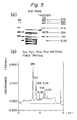
- Figure 6 shows that the monoclonal antibody 2H recognizes a 5.6 K fragment of the trypsin-digestion product, and the monoclonal antibodies 6A and 5F recognize a 7.4 K fragment of the trypsin-digestion product.
- a peptide composed of 69 residues, 141Thr-209Gln, on the C-terminus side was synthesized as two fragments, i.e. fragment A (176Leu-209Gln) and fragment B (141Thr-175Leu).
- the synthesized peptides were purified, and spoted on a nitrocellulose membrane in an amount of 100 ng and 10 ng, respectively. Dot blotting was carried out using the monoclonal antibodies. As shown in Figure 10, it was found that the monoclonal antibody 6A reacted specifically with fragment A, and the monoclonal antibody 5F, with fragment B.
- a PBS solution of human GST- ⁇ in a concentration of 1 microgram/ml was added at a rate of 50 microliters/well to fix the antigen (human GST- ⁇ ).
- the plate was then after-coated with 1 % PBS.
- 50 microliters of a solution of each of various monoclonal antibodies and 50 microliters of 1 % BSA/PBS containing an antigen in an amount 1, 2.5 or 5.0 times the amount of the solid-phase antigen were added in each well and reacted at room temperature for 1 hour.
- a dilute solution of antimouse IgG/HRP complex was added.
- the plate was washed and tetramethylbenzimine was added to induce coloration. The results are shown in Figure 12.
- the results show that the monoclonal antibodies are classified into two groups having different reactivities, namely the monoclonal antibodies 6A and 2H showing high reactivity with the antigen liberated in the liquid phase, and the monoclonal antibody 5F showing no reactivity with the antigen liberated in the liquid phase.
- the solution in the test tube was removed by sucking, and the test tube was washed with PBS. Then, 0.4 ml of a 0.1M phosphate/citrate buffer (pH 4.0) containing 0.02 % of 3,3′,5,5′-tetramethylbenzidinehydrochloride and 0.005 % of H2O2 was added to the test tube and incubated at 37 °C for 30 minutes. Then, 1 ml of a 1N aqueous solution of sulfuric acid was added as a reaction stopper to stop the enzyme reaction.
- a 0.1M phosphate/citrate buffer pH 4.0
- the absorbance of the solution at 450 nm was measured by using a spectrophotometer, and plotted against the concentration of a standard substance to give a calibration curve for determination of human placenta-derived GST (see Figure 13).
- Human GST- ⁇ (200 micrograms) was mixed with Freund's complete adjuvant, and the mixture was subcutaneously injected at the back of a rabbit. Two weeks later, 100 micrograms of human GST- ⁇ was mixed with 1 ml of Freund's incomplete adjuvant, and the mixture was likewise administered subcutaneously. The booster was applied three times in total, and ten days after the final immunization, the whole blood was drawn from the animal.
- the antibody was purified by using protein A-fixed Sepharose CL4B.
- One gram of the protein A-fixed Sepharose CL4B (produced by Pharmacia Co.) was swollen with PBS, and packed into a column.
- Antiserum (3 cc) was mixed with 3 cc of 0.1M phosphate buffer (pH 8.5), and the mixture was applied to the resulting column.
- the column was then well washed with 0.1M phosphate buffer.
- the antibody bound to the protein A column was eluted with 0.1M citrate buffer. The eluted antibody solution was dialyzed against PBS to give a purified antibody.
- Polystyrene beads (diameter 6 mm) were well washed, and left to stand for one day at 4 °C in a PBS (pH 7.4) solution containing each of the various anti-human GST- ⁇ antibodies in a concentration of 20 micrograms/ml. The beads were then washed with PBS, and after-coated by leaving it to stand for one day at 4 °C in a 0.5 % aqueous solution of BSA to give anti-human GST- ⁇ antibody-fixed beads.
- PBS pH 7.4
- the maleimidized antibody and the thiol-introduced HRP were mixed and concentrated to a protein concentration of 4 mg/ml under ice cooling.
- the concentrate was left to stand for one day at 4 °C, and gel-filtered on a column filled with an ultrogel AcA44 (LKB Company) to give a HRP-labelled anti-human GST- ⁇ antibody.
- the fragment A (20 mg) was dissolved in 1 ml of 0.1M carbonate buffer (pH 9.0), and reacted overnight at 4 °C with 1 ml of CNBr-activated Sepharose 4B (Pharmacia Co.).
- the fragment A-fixed Sepharose 4B was washed with 3M KSCN and filled in a column, and reacted with 2 ml of rabbit GST- ⁇ polyclonal antibody.
- the column was washed with 100 ml of PBS, and eluted with 3M KSCN. The absorbance at 280 nm of the eluate was 0.016.
- an assay system composed of the polyclonal antibody and the monoclonal antibody recognizing the sequence from the N-terminus to the 44th residue from the N-terminus and having a different epitope from the polyclonal antibody and an assay system composed of the polyclonal antibody and the monoclonal antibody recognizing the sequence from the 176th residue to the 209th residue from the N-terminus have high sensitivity with a low N/S ratio.
- the HRP-labelled monoclonal antibody 6A-F(ab′)2 showed three to four times as high selectivity as monoclonal antibody 6A-IgG.
- the HRP-labelled rabbit anti-human GST- ⁇ polyclonal antibody Fab′ showed 8 to 10 times as high sensitivity as IgG.
- a liver cancer tissue was taken and fixed to paraffin, and then an ultrathin slice was prepared.
- the slice was treated with 0.1 % trypsin at 37 °C for 30 minutes.
- the slice was then reacted with a methanol solution containing hydrogen peroxide (98 ml methanol/2 ml 30 % hydrogen peroxide) at room temperature for 30 minutes.
- the reacted slice was washed with PBS and reacted with monoclonal antibody-5F (diluted to 1 microgram/ml) at room temperature for 30 minutes.
- the slice was then reacted with peroxidase-labelled anti-mouse IgG (diluted to 1000-fold) at room temperature for 30 minutes.
- Serum samples were taken from a normal subject and patients with various gastrointestinal and hematological diseases. Each of the samples (50 microliters) was put in a test tube, and diluted with 150 microliters of a PBS solution (pH 7.4) containing 0.5 % BAS. One bead to which rabbit anti-human placenta-derived acid GST polyclonal antibody was fixed and 200 microliters of a PBS solution (PH 7.4) containing HRP-labelled monoclonal antibody 6A 0.5 % BSA were added to the test tube, and the entire mixture was incubated at 37 °C for 4 hours.
- a PBS solution pH 7.4
- HRP-labelled monoclonal antibody 6A 0.5 % BSA
- the product was then subjected to washing, enzyme reaction and stopping of the reaction by the same operation as in the preparation of the calibration curve described hereinabove. Then, the absorption intensity at 450 nm of the product was measured by a spectral photometer, and the concentration of the product was determined from the calibration curve.
Landscapes
- Health & Medical Sciences (AREA)
- Chemical & Material Sciences (AREA)
- Life Sciences & Earth Sciences (AREA)
- Immunology (AREA)
- Molecular Biology (AREA)
- Organic Chemistry (AREA)
- Engineering & Computer Science (AREA)
- General Health & Medical Sciences (AREA)
- Biochemistry (AREA)
- Medicinal Chemistry (AREA)
- Biomedical Technology (AREA)
- Cell Biology (AREA)
- Urology & Nephrology (AREA)
- Proteomics, Peptides & Aminoacids (AREA)
- Hematology (AREA)
- Genetics & Genomics (AREA)
- Biophysics (AREA)
- Physics & Mathematics (AREA)
- Biotechnology (AREA)
- General Physics & Mathematics (AREA)
- Pathology (AREA)
- Analytical Chemistry (AREA)
- Microbiology (AREA)
- Food Science & Technology (AREA)
- Chemical Kinetics & Catalysis (AREA)
- Preparation Of Compounds By Using Micro-Organisms (AREA)
- Peptides Or Proteins (AREA)
Priority Applications (1)
| Application Number | Priority Date | Filing Date | Title |
|---|---|---|---|
| AT89101717T ATE99059T1 (de) | 1988-02-01 | 1989-02-01 | Verfahren, reagens, satz und monoklonale antikoerper zur diagnose von krebs in menschlichen verdauungsorganen durch bestimmung von saurer glutathion-s-transferase. |
Applications Claiming Priority (6)
| Application Number | Priority Date | Filing Date | Title |
|---|---|---|---|
| JP19602/88 | 1988-02-01 | ||
| JP1960288 | 1988-02-01 | ||
| JP268292/88 | 1988-10-26 | ||
| JP63268292A JPH0750115B2 (ja) | 1988-02-01 | 1988-10-26 | ヒト酸性グルタチオンs―トランスフェラーゼの測定方法 |
| JP63282546A JPH02131595A (ja) | 1988-11-10 | 1988-11-10 | ヒトの胎盤由来グルタチオンs−トランスフェラーゼに対するモノクローナル抗体 |
| JP282546/88 | 1988-11-10 |
Publications (3)
| Publication Number | Publication Date |
|---|---|
| EP0328939A2 true EP0328939A2 (fr) | 1989-08-23 |
| EP0328939A3 EP0328939A3 (en) | 1990-05-30 |
| EP0328939B1 EP0328939B1 (fr) | 1993-12-22 |
Family
ID=27282694
Family Applications (1)
| Application Number | Title | Priority Date | Filing Date |
|---|---|---|---|
| EP19890101717 Expired - Lifetime EP0328939B1 (fr) | 1988-02-01 | 1989-02-01 | Méthode, réactif, trousse et anticorps monoclonal pour le diagnostic du cancer dans les organes humains de la digestion par détermination de la glutathione-S-transférase acide |
Country Status (4)
| Country | Link |
|---|---|
| EP (1) | EP0328939B1 (fr) |
| AU (1) | AU608117B2 (fr) |
| DE (1) | DE68911550T2 (fr) |
| ES (1) | ES2060676T3 (fr) |
Cited By (4)
| Publication number | Priority date | Publication date | Assignee | Title |
|---|---|---|---|---|
| EP0347138A2 (fr) * | 1988-06-13 | 1989-12-20 | Clinical Diagnostic Systems, Inc. | Composition de liaison spécifique avec une protéine ou un hydrate de carbone de pI bas, une trousse diagnostique, et méthode d'utilisation |
| WO1993022452A1 (fr) * | 1992-05-01 | 1993-11-11 | Cormac Gerard Kilty | Mesure d'un marqueur enzymatique afin de faciliter le diagnostic |
| EP0692716A1 (fr) * | 1994-07-12 | 1996-01-17 | Bayer Ag | Procédé pour la détection de glutathione-S-transferase et son application diagnostique |
| WO2002036781A2 (fr) * | 2000-10-31 | 2002-05-10 | Bayer Aktiengesellschaft | Regulation de la glutathione-s-transferase humaine |
Citations (1)
| Publication number | Priority date | Publication date | Assignee | Title |
|---|---|---|---|---|
| EP0245520A1 (fr) * | 1985-11-21 | 1987-11-19 | Teijin Limited | Anticorps monoclonal contre la glutathione s-transferase et son utilisation dans le diagnostic du cancer |
-
1989
- 1989-02-01 AU AU29524/89A patent/AU608117B2/en not_active Ceased
- 1989-02-01 DE DE1989611550 patent/DE68911550T2/de not_active Expired - Fee Related
- 1989-02-01 ES ES89101717T patent/ES2060676T3/es not_active Expired - Lifetime
- 1989-02-01 EP EP19890101717 patent/EP0328939B1/fr not_active Expired - Lifetime
Patent Citations (1)
| Publication number | Priority date | Publication date | Assignee | Title |
|---|---|---|---|---|
| EP0245520A1 (fr) * | 1985-11-21 | 1987-11-19 | Teijin Limited | Anticorps monoclonal contre la glutathione s-transferase et son utilisation dans le diagnostic du cancer |
Non-Patent Citations (1)
| Title |
|---|
| ARCHIVES OF BIOCHEMISTRY AND BIOPHYSICS, vol. 245, no. 2, March 1986, pages 543-547; I.Y. WANG et al.: "Muliple Ya Subunits of Glutathione S-Transferase Detected by Monoclonal Antibodies" * |
Cited By (6)
| Publication number | Priority date | Publication date | Assignee | Title |
|---|---|---|---|---|
| EP0347138A2 (fr) * | 1988-06-13 | 1989-12-20 | Clinical Diagnostic Systems, Inc. | Composition de liaison spécifique avec une protéine ou un hydrate de carbone de pI bas, une trousse diagnostique, et méthode d'utilisation |
| EP0347138A3 (en) * | 1988-06-13 | 1990-09-05 | Eastman Kodak Company (A New Jersey Corporation) | Specific binding composition comprising a low pi protein or carbohydrate and a diagnostic test kit and method of use |
| WO1993022452A1 (fr) * | 1992-05-01 | 1993-11-11 | Cormac Gerard Kilty | Mesure d'un marqueur enzymatique afin de faciliter le diagnostic |
| EP0692716A1 (fr) * | 1994-07-12 | 1996-01-17 | Bayer Ag | Procédé pour la détection de glutathione-S-transferase et son application diagnostique |
| WO2002036781A2 (fr) * | 2000-10-31 | 2002-05-10 | Bayer Aktiengesellschaft | Regulation de la glutathione-s-transferase humaine |
| WO2002036781A3 (fr) * | 2000-10-31 | 2003-02-06 | Bayer Ag | Regulation de la glutathione-s-transferase humaine |
Also Published As
| Publication number | Publication date |
|---|---|
| DE68911550T2 (de) | 1994-05-11 |
| AU2952489A (en) | 1989-08-03 |
| ES2060676T3 (es) | 1994-12-01 |
| DE68911550D1 (de) | 1994-02-03 |
| EP0328939B1 (fr) | 1993-12-22 |
| AU608117B2 (en) | 1991-03-21 |
| EP0328939A3 (en) | 1990-05-30 |
Similar Documents
| Publication | Publication Date | Title |
|---|---|---|
| US5712100A (en) | Antibodies to peptides having NGF-like activity, and use thereof | |
| US5230999A (en) | Monoclonal antibody to endothelin-3 or precursor thereof and use thereof | |
| US5681707A (en) | Method of immunological assaying human osteocalcin, reagent and kit therefor, antibody to human osteocalcin, hybridoma producing said antibody, and method of producing it | |
| EP0454782B1 (fr) | Haptoglobine apparentee au cancer (hpr) | |
| EP0605410B1 (fr) | Diagnostic immunologique de la polyarthrite rhumatoide | |
| US5427917A (en) | Method of determining human acid glutathione S-transferase, reagent, therefor, kit therefor, method of diagnosing cancer in digestive organs, and monoclonal antibody for use therein | |
| EP0245520B1 (fr) | Anticorps monoclonal contre la glutathione s-transferase et son utilisation dans le diagnostic du cancer | |
| EP0328939B1 (fr) | Méthode, réactif, trousse et anticorps monoclonal pour le diagnostic du cancer dans les organes humains de la digestion par détermination de la glutathione-S-transférase acide | |
| US4863850A (en) | Method for diagnosis of rheumatoid arthritis | |
| EP0418590B1 (fr) | Anticorps, leur production et utilisation | |
| EP0331100A1 (fr) | Anticorps anti-endothéline et leur utilisation | |
| US5425942A (en) | Polyfunctinal protease | |
| EP0410004B1 (fr) | Analyse immunologique de l'osteocalcine humaine, reactif et kit utilises pour ladite analyse | |
| EP0401370B1 (fr) | Analyse immunologique enzymatique selon un procedure en sandwich du collagene humain de type iv | |
| EP0345811B1 (fr) | Anticorps monoclonaux spécifiques pour fibrinopeptide A humain | |
| EP0350218A2 (fr) | Anticorps monoclonaux qui reconnaissent le polypeptide gamma atrial natriurétique, hybridomes qui les produisent et leur préparation et application | |
| EP0504423A1 (fr) | Procede et kit pour l'nalyse immunologique du propeptide de l'osteocalcine et de la pro-osteocalcine | |
| EP0465652A1 (fr) | Anticorps contre une chaine lourde de myosine des muscles lisses | |
| EP0417298A1 (fr) | Detection d'activateur de facteur tissulaire humain | |
| BHATTACHARYA et al. | Production and Characterization of Monoclonal Antibody to a 60-kD Glycoprotein in Ovarian Carcinoma1 | |
| JPH0750115B2 (ja) | ヒト酸性グルタチオンs―トランスフェラーゼの測定方法 | |
| EP0332421B1 (fr) | Anticorps monoclonaux reconnaissant l'inhibiteur humain de trypsine secrété par le pancréas, hybridomes les produisant, et leur préparation et utilisation | |
| EP0259773A2 (fr) | Un anticorps spécifique d'un antigène de cancer | |
| EP0602248A1 (fr) | Anticorps monoclonal agissant contre l'apoproteine d du surfactant des poumons humains et utilisation de cet anticorps | |
| Lee et al. | An enzyme immunoassay for pregnancy-specific β-1 glycoprotein (SP1), employing monoclonal antibodies |
Legal Events
| Date | Code | Title | Description |
|---|---|---|---|
| PUAI | Public reference made under article 153(3) epc to a published international application that has entered the european phase |
Free format text: ORIGINAL CODE: 0009012 |
|
| AK | Designated contracting states |
Kind code of ref document: A2 Designated state(s): AT BE CH DE ES FR GB IT LI NL SE |
|
| PUAL | Search report despatched |
Free format text: ORIGINAL CODE: 0009013 |
|
| AK | Designated contracting states |
Kind code of ref document: A3 Designated state(s): AT BE CH DE ES FR GB IT LI NL SE |
|
| 17P | Request for examination filed |
Effective date: 19900918 |
|
| 17Q | First examination report despatched |
Effective date: 19920430 |
|
| GRAA | (expected) grant |
Free format text: ORIGINAL CODE: 0009210 |
|
| ITF | It: translation for a ep patent filed |
Owner name: BARZANO' E ZANARDO MILA |
|
| AK | Designated contracting states |
Kind code of ref document: B1 Designated state(s): AT BE CH DE ES FR GB IT LI NL SE |
|
| REF | Corresponds to: |
Ref document number: 99059 Country of ref document: AT Date of ref document: 19940115 Kind code of ref document: T |
|
| PGFP | Annual fee paid to national office [announced via postgrant information from national office to epo] |
Ref country code: ES Payment date: 19940203 Year of fee payment: 6 |
|
| REF | Corresponds to: |
Ref document number: 68911550 Country of ref document: DE Date of ref document: 19940203 |
|
| ET | Fr: translation filed | ||
| PLBE | No opposition filed within time limit |
Free format text: ORIGINAL CODE: 0009261 |
|
| STAA | Information on the status of an ep patent application or granted ep patent |
Free format text: STATUS: NO OPPOSITION FILED WITHIN TIME LIMIT |
|
| REG | Reference to a national code |
Ref country code: ES Ref legal event code: FG2A Ref document number: 2060676 Country of ref document: ES Kind code of ref document: T3 |
|
| 26N | No opposition filed | ||
| EAL | Se: european patent in force in sweden |
Ref document number: 89101717.0 |
|
| PG25 | Lapsed in a contracting state [announced via postgrant information from national office to epo] |
Ref country code: ES Free format text: LAPSE BECAUSE OF NON-PAYMENT OF DUE FEES Effective date: 19950202 |
|
| PGFP | Annual fee paid to national office [announced via postgrant information from national office to epo] |
Ref country code: NL Payment date: 19951220 Year of fee payment: 8 |
|
| PGFP | Annual fee paid to national office [announced via postgrant information from national office to epo] |
Ref country code: FR Payment date: 19951221 Year of fee payment: 8 |
|
| PGFP | Annual fee paid to national office [announced via postgrant information from national office to epo] |
Ref country code: SE Payment date: 19951227 Year of fee payment: 8 Ref country code: GB Payment date: 19951227 Year of fee payment: 8 |
|
| PGFP | Annual fee paid to national office [announced via postgrant information from national office to epo] |
Ref country code: BE Payment date: 19960102 Year of fee payment: 8 |
|
| PGFP | Annual fee paid to national office [announced via postgrant information from national office to epo] |
Ref country code: AT Payment date: 19960112 Year of fee payment: 8 |
|
| PGFP | Annual fee paid to national office [announced via postgrant information from national office to epo] |
Ref country code: CH Payment date: 19960214 Year of fee payment: 8 |
|
| PGFP | Annual fee paid to national office [announced via postgrant information from national office to epo] |
Ref country code: DE Payment date: 19960329 Year of fee payment: 8 |
|
| PG25 | Lapsed in a contracting state [announced via postgrant information from national office to epo] |
Ref country code: GB Effective date: 19970201 Ref country code: AT Effective date: 19970201 |
|
| PG25 | Lapsed in a contracting state [announced via postgrant information from national office to epo] |
Ref country code: SE Effective date: 19970202 |
|
| PG25 | Lapsed in a contracting state [announced via postgrant information from national office to epo] |
Ref country code: LI Effective date: 19970228 Ref country code: CH Effective date: 19970228 Ref country code: BE Effective date: 19970228 |
|
| BERE | Be: lapsed |
Owner name: TEIJIN LTD Effective date: 19970228 |
|
| PG25 | Lapsed in a contracting state [announced via postgrant information from national office to epo] |
Ref country code: NL Effective date: 19970901 |
|
| GBPC | Gb: european patent ceased through non-payment of renewal fee |
Effective date: 19970201 |
|
| REG | Reference to a national code |
Ref country code: CH Ref legal event code: PL |
|
| PG25 | Lapsed in a contracting state [announced via postgrant information from national office to epo] |
Ref country code: FR Effective date: 19971030 |
|
| PG25 | Lapsed in a contracting state [announced via postgrant information from national office to epo] |
Ref country code: DE Effective date: 19971101 |
|
| EUG | Se: european patent has lapsed |
Ref document number: 89101717.0 |
|
| NLV4 | Nl: lapsed or anulled due to non-payment of the annual fee |
Effective date: 19970901 |
|
| REG | Reference to a national code |
Ref country code: FR Ref legal event code: ST |
|
| REG | Reference to a national code |
Ref country code: ES Ref legal event code: FD2A Effective date: 19990301 |
|
| PG25 | Lapsed in a contracting state [announced via postgrant information from national office to epo] |
Ref country code: IT Free format text: LAPSE BECAUSE OF NON-PAYMENT OF DUE FEES;WARNING: LAPSES OF ITALIAN PATENTS WITH EFFECTIVE DATE BEFORE 2007 MAY HAVE OCCURRED AT ANY TIME BEFORE 2007. THE CORRECT EFFECTIVE DATE MAY BE DIFFERENT FROM THE ONE RECORDED. Effective date: 20050201 |