EP0322084A2 - Tumor cell inhibition factor - Google Patents
Tumor cell inhibition factor Download PDFInfo
- Publication number
- EP0322084A2 EP0322084A2 EP88305198A EP88305198A EP0322084A2 EP 0322084 A2 EP0322084 A2 EP 0322084A2 EP 88305198 A EP88305198 A EP 88305198A EP 88305198 A EP88305198 A EP 88305198A EP 0322084 A2 EP0322084 A2 EP 0322084A2
- Authority
- EP
- European Patent Office
- Prior art keywords
- fraction
- molecular weight
- amino acid
- polypeptide
- tumor cell
- Prior art date
- Legal status (The legal status is an assumption and is not a legal conclusion. Google has not performed a legal analysis and makes no representation as to the accuracy of the status listed.)
- Withdrawn
Links
Images


Classifications
-
- A—HUMAN NECESSITIES
- A61—MEDICAL OR VETERINARY SCIENCE; HYGIENE
- A61K—PREPARATIONS FOR MEDICAL, DENTAL OR TOILETRY PURPOSES
- A61K38/00—Medicinal preparations containing peptides
-
- C—CHEMISTRY; METALLURGY
- C07—ORGANIC CHEMISTRY
- C07K—PEPTIDES
- C07K14/00—Peptides having more than 20 amino acids; Gastrins; Somatostatins; Melanotropins; Derivatives thereof
- C07K14/435—Peptides having more than 20 amino acids; Gastrins; Somatostatins; Melanotropins; Derivatives thereof from animals; from humans
- C07K14/52—Cytokines; Lymphokines; Interferons
Definitions
- This invention relates to a novel factor which exerts an inhibitory effect on tumor cells.
- TDF Tumor-degenerating factor
- FNF fibroblast-originating necrotic factor
- the object of this invention is to provide a novel tumor cell inhibition factor which can be obtained from human-origin fibroblasts, and which may occasionally be referred to FTX hereinafter.
- the novel tumor cell inhibition factor of the present invention is prepared by the following method.
- the process comprises the following steps: cultivating an established cell line obtained from a human kidney, removing contaminants from the supernatant of the culture fluid and obtaining a fraction containing a substance having a molecular weight of 10,000 or more, dialysing the fraction obtained and passing through of anion exchanger, subjecting the unabsorbed fraction to a chromatofocussing treatment on an anion exchanger, collecting a fraction having a molecular weight of 10,000 or more from the unabsorbed fraction, for example, by subjecting the unabsorbed fraction to ultrafiltration on a membrane of molecular weight cut-off level of 10,000, and subjecting it to gel filtration by high performance liquid chromatography (for example, on a TSK GEL 3000 and 2000 SW column at a pH of 7.0, attained for example by equilibration with a phosphate buffer solution containing sodium chloride) to obtain an active protein peak between molecular weight of 67 K and 32 K.
- high performance liquid chromatography for example, on a TSK GEL 3000 and 2000 SW column at
- the active protein thus obtained may be purified further by subjecting its fraction to a reverse phase high performance liquid chromatography using for example a PR-304 column.
- Cells of a human-origin fibroblast strain established by cultivation are used as starting cells.
- An example of preferable starting cells is an established cell line of human kidney origin.
- a typical example is one which has been established by subculturing a primary culture or diploid cell obtained from a fetal human kidney.
- the media for cell cultivation which may be used in this invention are serum-free media such as Waymouth's medium and Dulbecco's modified MEM medium, preferably a serum-free medium supplemented with a proper amount (0,05 to 0.2% (w/v)) of human serum albumin or a medium supplemented with a low concentration (0.05 to 0.2% (w/v)) of serum.
- the medium may further be supplemented with lactalbumin hydrolysate, transferin, various kind of amino acids, various aliphatic acids, and hormones such as insulin.
- % (w/v) means herein a percentage of a solute by weight per solution volume.
- air a mixture of 95% of air and 5% of CO2
- flow rate 10 to 500 ml/minute
- a cultivation temperature of 20 to 37°C is preferable.
- the culture fluid is exchanged approximately every 2 to 3 days.
- the substance according to this invention is present in the culture supernatant, it is purified, after removal of cells, based on its physico-chemical and bio-chemical properties.
- the purification may be conducted by proper combination of concentration, treatment by ion exchangers, a fractionation based on the molecular weight, etc.
- the cultured medium is centrifuged (for example, at 1500 rpm for 5 minutes) to recover supernatant.
- the supernatant is treated with a cation exchanger to obtain an unadsorbed fraction.
- a cation exchanger for the treatment is the CM-exchanger (CM-Sephadex).
- CM-Sephadex CM-Sephadex
- the carrier is equilibrated with a buffer solution of a pH of 5-6 and an ionic strength of 0.1-0.2M and is then brought in contact with a solution containing the above-mentioned culture supernatant, whereby contaminants are adsorbed onto the exchanger, to recover an unadsorbed fraction.
- the unadsorbed fraction is then treated with an anion exchanger to obtain an unadsorbed fraction.
- anion exchangers suitable for the treatment is the DEAE exchanger (DEAE-Sepharose), and the fraction unadsorbed by the treatment is collected as follows.
- the fraction unadsorbed by CM-Sephadex is treated with an ultrafiltration membrane of molecular weight cut-off level of 10,000 (e.g., PelliconTM, mfd, by Millipore Co.) to obtain a high molecular weight fraction.
- the fraction thus obtained is concentrated and then dialyzed against, for example, a 50 mM tris-HCl buffer solution (pH 7.0).
- the resulting dialysate is introduced into a DEAE-Sepharaose column equilibrated with the same buffer solution and then the same buffer solution is passed through the column to collect the eluent.
- the unadsorbed fraction thus collected is then concentrated. Similar procedure is repeated three times for the concentration to obtain a concentrated solution of the unadsorbed fraction.
- the concentrated solution is then subjected to chromato-focussing on an anion exchanger, for example Poly buffer exchanger, to obtain an unadsorbed fraction.
- the chromatofocussing treatment is carried out as follows.
- the concentrated solution obtained by the DEAE exchanger treatment is dialyzed against a 25 M triethylamine-HCl buffer solution (pH 11), the dialysate is introduced into a PolybufferTM Exchanger PBE 118 column equilibrated with the same buffer solution, then the same buffer solution is passed through the column, and the eluent is collected to obtain an unadsorbed fraction.
- the unadsorbed fraction is then subjected to gel filtration in the following manner to obtain an active protein fraction.
- the fraction unadsorbed by the Polybuffer Exchanger PBE-118 column is treated with an ultrafiltration membrane of molecular weight cut-off level of 10,000 (e.g., PM-10TM, mfd, by Amicon Co.) to collect a high molecular weight fraction.
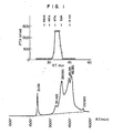
- the fraction is concentrated and subjected to gel filtration by HPLC (high performance liquid chromatography) using a TSK GEL 3000 and 2000 SW column equilibrated with a 0.1 M phosphate buffer solution (pH 7.0) containing 0.15 M NaCl, whereby a protein peak with attendant activity is obtained between molecular weight markers, Enolase (MW 67 K) and Adenylate Kinase (MW 32 K) (see Fig. 1).
- the active fraction is subjected to a reverse phase HPLC (high performance liquid chromato graphy) in the following manner to obtain a purer product.
- a reverse phase HPLC high performance liquid chromato graphy
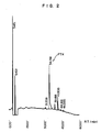
- the active fractions were collected and subjected to a reverse phase HPLC using a PR-304 column (chromatography conditions: A-solution ... 0.1% trifluoroacetic acid; B-solution... A-solution containing 80% acetonitrile; A-solution ⁇ B-solution 60 minutes linear gradient) to obtain the purer product (see Fig. 2).
- the product has a specific activity (U/mg) of 290,000. It was obtained in an amount of about 0.126 mg in terms of protein from 447,000 mg of protein of the fraction unadsorbed by CM-Sephadex.
- the tumor cell inhibition substance thus obtained is a novel substance as judged from its characteristic properties described below. It may also be used as a reagent for biochemistry and pharmacology. For use as a medicinal agent it may be subjected, as described, to sterilization, asepsis, and medical preparation-forming processes according to conventional methods for medicine preparation. Thus, medicinal agents containing a novel tumor cell inhibition substance are provided.
- FTX The sample of the substance isolated by the above method of this invention (hereinafter referred to as FTX) was subjected to two-times step dilution by the use of a 96-well microplate (50 ⁇ l/well).
- the amount of dye incorporated into living cells was determined from the absorbance at 590 nm by using Multiscan (mfd. by Titertec. Co.).
- Fig. 3 shows the growth inhibition effect of FTX on KB cells (rhinopharyngeal cancer cells, ATCC-CCL-17).
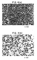
- Fig. 4 shows the cancer cell degeneration effect obtained by FTX addition.
- the FTX isolated by the above method exerted no effect on the NBT-reducing ability of ML-1 cells. Thus, it shows no differentiation-inducing ability.
- the gel slices were each extracted at 4°C overnight in 1 ml of a medium for activity determination and the biological activity in respective slices was determined.
- the electrophoresis was conducted at 30 mA for about 4 hours.
- a standard molecular weight curve was prepared by using a standard molecular weight kit (mfd. by Pharmacia Co.).
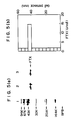
- the molecular weight of the FTX of this invention was determined from its biological activity and its mobility in electrophoresis ascertained by means of protein argentation image. The results thus obtained are shown in Fig. 5.
- the symbols in Fig. 5 mean the following.
- the FTX isolated by the abovementioned method of this invention had a molecular weight of about 36,000 ⁇ 1,000 as judged from the mobility of the molecular weight markers.
- the isoelectric point of the FTX isolated by the abovementioned process of this invention was determined by preparative flat-bed electrofucussing (KLB) according to the method described in Biochem. Biophys. Acta., 194 , 335 (1969) and Acta. Chem. Scand., 20 , 820 (1966). It was found that the FTX of this invention had a pI of 10.5 or more.
- a 5 ⁇ g portion of the FTX of this invention was hydrolyzed by treating it with 6N-HCl for 20 hours.
- the amino acid composition was then determined by the PICO-TAGTM method.
- the protein isolated by the abovementioned method is a glycoprotein.
- a 5 ⁇ g portion of the FTX isolated by the above method was hydrolyzed by treating it with 4 N-HCl for 4 hours and then the analysis of amino sugar was carried out in a similar manner to that mentioned above, to detect glucosamine. From this result it was confirmed that the FTX isolated by the above method of this invention is a glycoprotein.
- the growth-inhibitory action of the FTX isolated by the above method of this invention was not neutralized by anti-TNF monoclonal antibody,anti-IFN- ⁇ - ⁇ , ⁇ monoclonal antibody, and anti-IL-1 polyclonal antibody.
- the mRNA which coded the tumor cell inhibition factor of this invention is isolated from fetal human kidney-origin fibroblasts. Then, the gene is cloned, and the base sequence of the gene is determined.
- the base sequence and the amino acid sequence of intended polypeptide which is estimated from the base sequence are shown in Fig. 7.
- the base sequence consists of 1254 bases from the translation initiation signal ATG to the termination signal TAG. When translated into amino acids, it leads to a 417 amino acid sequence.
- the sequence of 20 amino acids from the 29th, Ala, to the 49th, Ala entirely coincides with the amino acid sequence of the N-terminal of the polypeptide. It is considered that the amino acid acid sequence from the first, Met, to the 28th, Ala, is a signal sequence necessary for passage through cell membrane.
- the structural polypeptide estimated from the base sequence consists of 389 amino acids and contains 18 potential Asn linked glycosylation sites (Asn-X-Set/Thr).
- the substance of the present invention was fragmented by lysylendopeptides, and the fragments were purified by means of reverse high performance liquid chromatography.
- the purified fragments were analyzed with respect to amino acids contained, C-terminal peptide was identified in terms of existence or non-existence of a lysine residue, and the existence of a C-terminal amino acid of the C-terminal peptide is confirmed by effecting the digestion using carboxypeptidase Y.
- the natural-origin biological substance present was estimated as being constructed from two kinds of polypeptide, i.e., one having amino acids from N-terminal to 169th Ser and the other having the amino acids from N-terminal to 170th Pro, among the amino acid sequence of 389 amino acid residues estimated from the base sequence of the cDNA.
- tumor cell inhibition factor of this invention comprises the following amino acid sequence.
- the DNA sequence containing a DNA sequence coding the substance of the present invention is obtained from either cDNA obtained from mRNA which has been extracted from the starting cells, or a library of chromosome DNA of the starting cells.
- the amino acid sequence of the substance is estimated from the DNA sequence obtained. Further, this DNA sequence can be utilized in the preparation of the substance of the invention according to gene recombination method.
- RNA is prepared from 1.2 x 10¹⁰ cultivated cells.
- a serum-free medium obtained by adding 0.05 - 0.2% (w/v) of human serum albumin to the Waymouth medium
- total RNA is prepared from 1.2 x 10¹⁰ cultivated cells.
- guanidine thiocyanate-cesium chloride method Biochemistry, 18 , 5294 (1979)].
- To cells are added 400 ml of a solution containing 6 M guanidine thiocyanate, 5 mM sodium citrate, 0.5% Sarkosyl, 0.1 M 2-mercaptoethanol and 0.1% Antifoam-A, and the whole is homogenized.
- Four grams of cesium chloride is dissolved for 10 ml of the homogenized solution.
- RNA solution Poly(A)+ RNA is purified from the total RNA by affinity chromatography utilizing the affinity for oligo ( ⁇ T).
- Oligo ( ⁇ T) cellulose is equilibrated with 10 mM tris-HCl buffer solution (pH 7.5), containing 1 mM EDTA, 0.5 M NaCl and 0.1% SDS.
- the total RNA is charged in the equilibrated column, and the adsorbed fraction is eluted with 10 mM tris-HCl buffer solution (pH 7.5) containing 1 mM EDTA and 0.05% SDS to obtain poly(A)+ RNA.
- the poly(A))+ RNA is fractionated according to the dimension of RNA by means of sucrose density gradient centrifugation.
- the poly(A)+ RNA solution is placed on 5% - 25% sucrose density gradient buffer solution (10 mM tris-HCl buffer solution (pH 7.5), containing 1 mM EDTA, 0.1 M NaCl, 0.1% SDS) and subjected to ultracentrifuge in a Beckman Ultracentrifuge (50.1 Ti rotor) at 20,000 rpm at 20°C for 16 hours, thus to be separated into 24 fractions.
- sucrose density gradient buffer solution 10 mM tris-HCl buffer solution (pH 7.5), containing 1 mM EDTA, 0.1 M NaCl, 0.1% SDS
- a hybridization method is used for cloning the intended gene.
- a preferably used probe for hybridization is a synthetic oligodeoxynucleotide. Two kinds of oligodioxynucleotide (17 bases) are synthesized which show complementarity to the base sequence estimated from the N-terminal amino acid sequence (see Fig. 6).
- a probe mixture comprising 16 kinds of complemental strand (probe A) to the base sequence corresponding to the sequence from Met, the second amino acid from the N-terminal, to Asn, the seventh from the N-terminal, on one hand, and a probe mixture comprising 48 kinds of complemental strand (probe B) to the base sequence corresponding to the sequence from the 13th, Cys, to the 18th, Phe, on the other hand are synthesized in a DNA synthesizer (Model 381 A, mfd. by Applied Biosystem Co.).
- the synthetic probes are used after labelling the 5′-terminal with T4 polynucleotide kinase (available from New England Biolabs) by using [ ⁇ -32P] ATP (available from Amersham).
- the poly(A)+ RNA is denatured.
- a formaldehyde method is used.
- the RNA is dissolved in a 10 mM sodium phosphate buffer (pH 6.8) containing 50% by volume of formamide, 6% by volume of formaldehyde, 0.2 mM EDTA, 10% of glycerine, and 0.0% (w/v) of BPB, and is denatured at 65°C for 5 minutes.
- the denatured RNA is subjected to electrophoresis using formaldehyde-agarose gel.
- RNA is transferred from the gel to a Zeta Probe blotting membrane (available from Bio Rad) by means of a capillary method.
- the RNA adsorbed onto the membrane and the probe labelled in 2 above are subjected to hybridization.
- the membrane is treated in a hybridization solution containing 6 x SSC (0.9 M NaCl, 90 mM sodium citrate) 20 mM sodium phosphate (pH 6.8), 7% SDS, 10 x Denhardt's (0.2% (w/v) Ficoll 400, 0.2% (w/v) polyvinylpyrrolidone, 0.2% (w/v) bovine serum albumin), 10% (w/v) dextran sulfate, 100 ⁇ g/ml of tRNA of yeast origin, and 32P-terminal labelled probed at 37°C overnight to effect hybridization, and then washed two times with washing solution containing 3 x SSC, 10 x Denhardt's, 5% SDS, 25 mM sodium phosphat
- the washed membrane is made to expose X-ray film for several days.
- the exposed X-ray film is developed and the size of the poly (A)+ RNA hybridized with the labelled probe was determined. As a result, it was found that a band of a size of 2.7 kilobase (kb) appeared using both of the synthetic probes A and B.
- cDNA is synthesized from the fraction containing poly(A) ⁇ RNA having a size of 2.7 kilobase which had been hybridized with both of probes A and B.
- the synthesis of cDNA was performed by using a cDNA Synthesis System (Amersham) and according to the protocol. From 5 ⁇ g of poly(A)+ RNA was obtained 3.3 ⁇ g of double stranded cDNA. Then, The cDNA library is prepared from the double stranded cDNA.
- ⁇ gt 10 is used as the vector of DNA. ⁇ gt 10 forms a plaque of relatively large size and hence gives a good yield of recombinants.
- an EcoRI linker Before an EcoRI linker is connected to the cDNA, it is methylated by the use of EcoRI methylase with the aim of protecting the EcoRI site present inside the cDNA.
- a 1.5 ⁇ g portion of the double stranded cDNA is dissolved in a solution containing of 0.1 M NaCl, 0.1 M tris-HCl (pH 8.0), 1 mM EDTA, 80 ⁇ M S-adenosylmethionine, 30 units of EcoRI methylase (available from New England Biolabs) is added thereto, and the whole is allowed to react at 37°C for 60 minutes.
- the reaction mixture is extracted with phenol/chloroform and the cDNA is recovered from the extract by precipitation with ethanol.
- the recovered cDNA is allowed to react at 15°C overnight in a solution containing 66 mM tris-HCl (pH 7.5), 6.6 mM MgCl2, 10 mM dithiothreitol, 0.1 mM ATP, and 1.5 ⁇ g of EcoRI linker (dGGAATTCC, available from New England Biolabs) by addition of 20 units of T4DNA ligase (available from New England Biolabs).
- the cDNA having the EcoRI linker connected thereto is digested with 150 units of EcoRI (available from Takara Shuzo Co.).
- the cDNA digested with EcoRI is separated from excess of the linker by means of Biogel A 50 m column chromatography.
- a recombinant phage is prepared from the recombinant DNA by an in vitro packaging method.
- an in vitro packaging kit of Gigapack gold (available from Strategene Co.) is used.
- the titer of the cDNA library was examined by using Escherichia coli c600 as an indicator strain. It was found that 3.9 x 10⁶ plaques had been obtained.
- the intended cDNA is screened from the cDNA library obtained in 4 above by means of plaque hybridization using a 32P terminal-labelled probe.
- the method of phage amplification on a filter (method in Enzymology, 68 , 389, (1979)) is used.
- Escherichia coli c600 is infested with 2.4 x 10⁵ recombinant phages, and allowed to form plaques on TB agar, which is then stored at 4°C.
- a colony plaque screen filter (NEN) is immersed in a suspension of Escherichia coli c600 to adsorb the bacteria on the filter, and then dried at room temperature.
- the filter is placed on Tb agar on which plaques have been formed, and is brought into contact therewith for several minutes.
- the filter is peeled off and placed on a new TB agar, which is then incubated overnight at 37°C.
- the filter is peeled off, immersed in a solution containing of 0.5 M NaOH, 1.5 M NaCl for 5 minutes, further twice in 0.5 M tris-HCl buffer solution (pH 8.0) containing of 1.5 M NaCl for 5 minutes, and then in 2 x SSPE (0.36 NaCl, 20 mM Na2HPO4, 2 mM EDTA). Thereafter, it is dried at room temperature and then baked under vacuum at 80°C for 1 hour.
- the 32p terminal-labelled oligodeoxynucleotide fragments of B and A used in 3 above are employed as hybridization probes.
- the filter is immersed in a solution containing 6 x SSC (0.9 M NaCl, 90 mM sodium citrate), 20 mM sodium phosphate (pH 6.8), 10 x Denhardt's, 7% SDS, 10% dextran sulfate, 100 ⁇ g/ml of tRNA of Yeast origin, and 9 pmol 32p-terminal-labelled probe (specific activity: 5 x 10⁶ cpm/pmol), and incubated overnight at 37°C to effect hybridization.
- 6 x SSC 0.9 M NaCl, 90 mM sodium citrate
- 20 mM sodium phosphate pH 6.8
- 10 x Denhardt's 7% SDS
- 10% dextran sulfate 100 ⁇ g/ml of tRNA of Yeast origin
- the filter is washed twice with 6 x SSC, then three times at 35°C for 30 minutes with washing solution containing a 3 x SSC, 10 x Denhardt's, 5% SDS, 25 mM sodium phosphate (pH 7.4) and then dried. Then, it is subjected to autoradiography.
- the plaques on the agar plate which correspond to the signal that has appeared at a position common to probes A and B are scratched off and suspended in a solution containing 0.1 M NaCl, 8.1 mM MgSO4, 0.01% gelatin, 50 mM tris-HCl (pH 7.5).
- the plaque-forming reaction is again effected for the suspension to obtain 15 kinds of positive, single recombinant phages.
- phages whose cDNA have a size of 2.7 Kbp are selected to extract DNA therefrom, and the cDNA is introduced into the proliferation vector of Escherichia coli , preferably into pUC 18.
- the vector pSVG2 possesses the enhancer, promoter, splicing junction, and poly A signal of SV 40 virus and contains necessary elements for expressing a useful substance in animal cells [cf. Japanese Patent Application Kokai (Laid-Open) No. 236,493/87].
- the pSVG2 was digested with EcoRI and further dephosphorylated at the 5′-terminal with alkaline phosphates of bovine small intestine origin. Then, the resulting product was subjected to 1% low-m.p. gel electrophoresis to recover DNA.
- the pF-1 which contained the whole cDNA of the substance of this invention was digested with SSpI and then subjected to 1% low-m.p.
- the 1.7 kb fragment and the EcoRI-digested pSVG2 were reacted with the aid of T4 DNA ligase (available from Takara Shuzo Co., Ltd.) and transformed into Escherichia coli JM 109, thus to obtain an ampicillin-resistant transformant. From among the transformants, there was obtained pSV-F1 in which the 1.7 kb fragment had been inserted in positive direction.
- the substance of this invention of natural origin comprises peptide of from Ala of N-terminal to 169Ser or 170Pro of C-terminal
- expression plasmids pSV-F2 and pSV-F3 respectively capable of coding up to 169Ser and 170Pro (see Fig. 9).
- the pF-1 was digested with StuI which cuts the former at AGGCCT (167Arg 168Pro), further subjected to dephosphorylation at the 5′-terminal with the aid of alkaline phosphates of bovine small intestine origin, and then subjected to 1% low m.p. gel electrophoresis, to recover DNA.
- a synthetic oligonucleotide of 26 bases having termination codon TGA and EcoRI site at CCTTCC (168Pro 169Ser) (5′CCTTCCTGAGGAATTCCTCAGGAAGG3′) and a synthetic oligonucleotide of 32 bases having termination codon TGA and EcoRI site at CCTTCCCCA (168Pro 169Ser 170Pro) (5′CCTTCCCCATGAGGAATTCCTCATGGGGAAGG3′) were synthesized by the use of an automatic DNA synthesizer (mfd. by Applied Biosynthesis Co.).
- the 5′-terminal of the synthetic oligonucleotide of 26 mers and of 32 mers were phosphorylated with the aid of polynucleotide kinase (available from Takara Shuzo Co., Ltd.) and were connected with the StuI-digested pF-1 fragment with the aid of T4 DNA ligase (available from Takara Shuzo Co., Ltd.).
- the resulting product was then digested with EcoRI and SacI and subjected to 4% polyacrylamide gel electrophoresis to recover 0.2 kb fragment.
- the recovered SacI-EcoRI fragment was connected to the SacI-EcoRI site of cloning vector pUC 18 with the help of T4 DNA ligase (available from Takara Shuzo Co., Ltd.) and transformed into Escherichia coli JM 109.
- the base sequence of the plasmid insertion fragment of the transformant was determined by the dideoxy method and was confirmed to be the intended one.
- the above-mentioned plasmid was digested with SacI and EcoRI and then subjected to 4% polyacrylamide gel electrophoresis to recover 0.2 kb fragment.
- pF-I was digested with EcoRI and SacI to recover 0.61 kb fragment which contains 5′-side of 5′-side coding region, 5′-non coding region and signal sequence of the cDNA.
- the recovered EcoRI, SacI 0.61 kb fragment and SacI, EcoRI 0.2 kb fragment to which synthetic oligonucleotide had been connected were inserted to expression vector pSVG2 digested with EcoRI, with the aid of T4 DNA ligase and the resulting product was transformed into Escherichia coli JM 109. From the transformant there were obtained expression plasmids pSV-F2 and pSV-F3 which had been connected in intended direction.
- Host cells to be used preferably are Chinese hamster overy cells (CHO-K1).
- Preferably used culture fluid is Ham's F-12 medium containing 10% of fetal bovine serum.
- Transfection of recombinant DNA into cells is performed by the calcium phosphate precipitation method (Strain et al. Biochem. J., 218 , 475-482, 1984).
- a method is employed which introduces a drug-resistance gene simultaneously to the cell and selecting the resulting drug-resistant strain (Sbraman, Anal. Biochem., 135 , 1-15, 1983).
- pSV-NEO formed by connecting to pVSG2 and Neo r gene (aminoglycoside 3′-phospho-transferase II) which shows resistance to G-418 in animal cells (cf. Japanese Patent Application Kokai (Laid-Open) No. 236,493/87) (see Fig. 10).
- HEPES buffer solution of double concentration (2 x HBS) 10 g/l HEPES (N-(2-hydroxyethyl)piperazine-N′-2-ethanesulfonic acid), 6 g/l NaCl, adjusted to pH 7.1]
- 50 ⁇ l of an aqueous solution of 70 mM NaH2PO4 and 70 mM Na2HPO4 100 ⁇ l of 1 ⁇ g/ml salmon sperm DNA solution
- 100 ⁇ l of recombinant DNA 10 ⁇ g of pSV-F1 (pSV-F2, pSV-F3) and 0.1 ⁇ g of pSV-NEO (cf. Fig.
- the host cell there may alternatively be used a human-origin cultivation-established fibroblast.
- Such modifications may for example comprise amino acid deletions, insertions, additions, substitutions, replacements and inversions; and also alteration to or elimination of any glycosylation.
- DNA encoding the polypeptide products thereof can now be obtained by routine methods such as direct DNA synthesis, or isolation and cloning of cDNA using specific oligonucleotide probes for selected regions of the sequence disclosed herein, or combinations of these. If desired, suitable encoding DNA can be isolated from a genomic library using such oligonucleotide probes.
- allelic variants may arise, depending for example upon the source of the DNA.
Landscapes
- Health & Medical Sciences (AREA)
- Life Sciences & Earth Sciences (AREA)
- Chemical & Material Sciences (AREA)
- General Health & Medical Sciences (AREA)
- Organic Chemistry (AREA)
- Proteomics, Peptides & Aminoacids (AREA)
- Medicinal Chemistry (AREA)
- Biochemistry (AREA)
- Immunology (AREA)
- Genetics & Genomics (AREA)
- Gastroenterology & Hepatology (AREA)
- Molecular Biology (AREA)
- Zoology (AREA)
- Toxicology (AREA)
- Engineering & Computer Science (AREA)
- Bioinformatics & Cheminformatics (AREA)
- Biophysics (AREA)
- Pharmacology & Pharmacy (AREA)
- Epidemiology (AREA)
- Animal Behavior & Ethology (AREA)
- Public Health (AREA)
- Veterinary Medicine (AREA)
- Peptides Or Proteins (AREA)
- Preparation Of Compounds By Using Micro-Organisms (AREA)
- Micro-Organisms Or Cultivation Processes Thereof (AREA)
Abstract
A tumor cell inhibition factor, which is a glycoprotein having single band of molecular weight of about 36,000 daltons is produced from culture liquid of human-origin fibroblasts and purified by collecting substances of molecular weight of about 10,000 and more followed by an ion-exchange method, chromatography and gel filtration. The DNA sequence coding the factor is determined, and an expression vector capable of expressing the DNA sequence and recombinant host cells transformed with the vector are prepared.
Description
- This invention relates to a novel factor which exerts an inhibitory effect on tumor cells.
- The existence of a tumor cell inhibition factor in the human fibroblast culture fluid is disclosed in JP-A-88423/84 and JP-A-18721/86. The product in the former reference is called Tumor-degenerating factor (TDF) and is a glycoprotein of molecular weight of 145,000, and that in the latter is called fibroblast-originating necrotic factor (FNF) having a molecular weight of about 10,000 obtained from human fibroblasts transformed into tumor cells (
SV 40 transformed cell). - The object of this invention is to provide a novel tumor cell inhibition factor which can be obtained from human-origin fibroblasts, and which may occasionally be referred to FTX hereinafter.
- The novel tumor cell inhibition factor of the present invention is prepared by the following method.
- Preparation of the present FTX from an established cell line from a human kidney.
- The process comprises the following steps:
cultivating an established cell line obtained from a human kidney,
removing contaminants from the supernatant of the culture fluid and obtaining a fraction containing a substance having a molecular weight of 10,000 or more,
dialysing the fraction obtained and passing through of anion exchanger,
subjecting the unabsorbed fraction to a chromatofocussing treatment on an anion exchanger, collecting a fraction having a molecular weight of 10,000 or more from the unabsorbed fraction, for example, by subjecting the unabsorbed fraction to ultrafiltration on a membrane of molecular weight cut-off level of 10,000, and subjecting it to gel filtration by high performance liquid chromatography (for example, on aTSK GEL 3000 and 2000 SW column at a pH of 7.0, attained for example by equilibration with a phosphate buffer solution containing sodium chloride) to obtain an active protein peak between molecular weight of 67 K and 32 K. - The active protein thus obtained may be purified further by subjecting its fraction to a reverse phase high performance liquid chromatography using for example a PR-304 column.
- The above method of isolation of the present invention is explained in more detail in sections (1)-(3) below, further sections describing the characteristics of a protein isolated by the above method and how they were confirmed (4), sequencing of DNA (5), formation of an expression vector (6) and transformation of host cells (7).
- Cells of a human-origin fibroblast strain established by cultivation are used as starting cells. An example of preferable starting cells is an established cell line of human kidney origin. A typical example is one which has been established by subculturing a primary culture or diploid cell obtained from a fetal human kidney.
- The media for cell cultivation which may be used in this invention are serum-free media such as Waymouth's medium and Dulbecco's modified MEM medium, preferably a serum-free medium supplemented with a proper amount (0,05 to 0.2% (w/v)) of human serum albumin or a medium supplemented with a low concentration (0.05 to 0.2% (w/v)) of serum. The medium may further be supplemented with lactalbumin hydrolysate, transferin, various kind of amino acids, various aliphatic acids, and hormones such as insulin.
- The symbol "% (w/v)" means herein a percentage of a solute by weight per solution volume.
- Preferably, air (a mixture of 95% of air and 5% of CO₂) is introduced into the medium in a suitable amount (flow rate: 10 to 500 ml/minute). A cultivation temperature of 20 to 37°C is preferable. The culture fluid is exchanged approximately every 2 to 3 days.
- Since the substance according to this invention is present in the culture supernatant, it is purified, after removal of cells, based on its physico-chemical and bio-chemical properties. For example, the purification may be conducted by proper combination of concentration, treatment by ion exchangers, a fractionation based on the molecular weight, etc.
- More specifically, it can be recovered in the following manner.
- First, the cultured medium is centrifuged (for example, at 1500 rpm for 5 minutes) to recover supernatant.
- The supernatant is treated with a cation exchanger to obtain an unadsorbed fraction. An example of suitable cation exchangers for the treatment is the CM-exchanger (CM-Sephadex). The carrier is equilibrated with a buffer solution of a pH of 5-6 and an ionic strength of 0.1-0.2M and is then brought in contact with a solution containing the above-mentioned culture supernatant, whereby contaminants are adsorbed onto the exchanger, to recover an unadsorbed fraction.
- The unadsorbed fraction is then treated with an anion exchanger to obtain an unadsorbed fraction.
- An example of anion exchangers suitable for the treatment is the DEAE exchanger (DEAE-Sepharose), and the fraction unadsorbed by the treatment is collected as follows. The fraction unadsorbed by CM-Sephadex is treated with an ultrafiltration membrane of molecular weight cut-off level of 10,000 (e.g., Pellicon™, mfd, by Millipore Co.) to obtain a high molecular weight fraction. The fraction thus obtained is concentrated and then dialyzed against, for example, a 50 mM tris-HCl buffer solution (pH 7.0). The resulting dialysate is introduced into a DEAE-Sepharaose column equilibrated with the same buffer solution and then the same buffer solution is passed through the column to collect the eluent. The unadsorbed fraction thus collected is then concentrated. Similar procedure is repeated three times for the concentration to obtain a concentrated solution of the unadsorbed fraction. The concentrated solution is then subjected to chromato-focussing on an anion exchanger, for example Poly buffer exchanger, to obtain an unadsorbed fraction.
- The chromatofocussing treatment is carried out as follows. The concentrated solution obtained by the DEAE exchanger treatment is dialyzed against a 25 M triethylamine-HCl buffer solution (pH 11), the dialysate is introduced into a Polybuffer™ Exchanger PBE 118 column equilibrated with the same buffer solution, then the same buffer solution is passed through the column, and the eluent is collected to obtain an unadsorbed fraction.
- The unadsorbed fraction is then subjected to gel filtration in the following manner to obtain an active protein fraction.
- The fraction unadsorbed by the Polybuffer Exchanger PBE-118 column is treated with an ultrafiltration membrane of molecular weight cut-off level of 10,000 (e.g., PM-10™, mfd, by Amicon Co.) to collect a high molecular weight fraction. The fraction is concentrated and subjected to gel filtration by HPLC (high performance liquid chromatography) using a
TSK GEL 3000 and 2000 SW column equilibrated with a 0.1 M phosphate buffer solution (pH 7.0) containing 0.15 M NaCl, whereby a protein peak with attendant activity is obtained between molecular weight markers, Enolase (MW 67 K) and Adenylate Kinase (MW 32 K) (see Fig. 1). - Then, the active fraction is subjected to a reverse phase HPLC (high performance liquid chromato graphy) in the following manner to obtain a purer product. Thus, the active fractions were collected and subjected to a reverse phase HPLC using a PR-304 column (chromatography conditions: A-solution ... 0.1% trifluoroacetic acid; B-solution... A-solution containing 80% acetonitrile; A-solution → B-
solution 60 minutes linear gradient) to obtain the purer product (see Fig. 2). The product has a specific activity (U/mg) of 290,000. It was obtained in an amount of about 0.126 mg in terms of protein from 447,000 mg of protein of the fraction unadsorbed by CM-Sephadex. - The tumor cell inhibition substance thus obtained is a novel substance as judged from its characteristic properties described below. It may also be used as a reagent for biochemistry and pharmacology. For use as a medicinal agent it may be subjected, as described, to sterilization, asepsis, and medical preparation-forming processes according to conventional methods for medicine preparation. Thus, medicinal agents containing a novel tumor cell inhibition substance are provided.
- It exerts growth inhibition and degeneration effects at least on KB cells, of rhinopharyngeal cancer origin, of strain ATCC CCL-17.
- The sample of the substance isolated by the above method of this invention (hereinafter referred to as FTX) was subjected to two-times step dilution by the use of a 96-well microplate (50 µl/well). A KB cell fluid containing 10 x 10⁴ cells/ml (human rhinopharyngeal cancer origin, 50 µl/well) was added thereto and cultivated at 37°C in an atmosphere containing 5% of CO₂ for 7 days. After cultivation, the supernatant was discarded and the remaining cells were stained with a 0.05% crystal violet staining solution. The amount of dye incorporated into living cells was determined from the absorbance at 590 nm by using Multiscan (mfd. by Titertec. Co.). The proportion of the absorbance of the treated cell of the FTX sample to the absorbance of the untreated cell was determined and was defined as growth rate. The dilution at which the FTX sample showed 50% growth inhibition was used to represent the FTX activity (U/ml). Fig. 3 shows the growth inhibition effect of FTX on KB cells (rhinopharyngeal cancer cells, ATCC-CCL-17). Fig. 4 shows the cancer cell degeneration effect obtained by FTX addition.
- The FTX isolated by the above method exerted no effect on the NBT-reducing ability of ML-1 cells. Thus, it shows no differentiation-inducing ability.
- It did not suppress the proliferation of virus in the FL-Sindbis virus system and exerted no inhibitory effect of L cells.
- single band of 36,000±1,000 daltons as determined by electrophoresis using SDS/polyacrylamide gel under reduced and nonreduced conditions.
- According to the method of Laemmli [Nature, 227, 680 (1970)], 0.3 µg of the FTX dissolved in Tris-buffer solution containing 2% SDS to which 2% of 2-mercaptoethyl alcohol had been added or not added were each subjected to electrophoresis using acrylamide gel containing 0.1% SDS. At the same time, for activity determination, 10 µl of concentrated solution (corresponding to 96 U) of a FTX sample which had been purified by the step of gel filtration by HPLC using TSK gel G3000 - 2000 SW column was subjected to electrophoresis. After electrophoresis, the gel was sliced to a thickness of 5 mm. The gel slices were each extracted at 4°C overnight in 1 ml of a medium for activity determination and the biological activity in respective slices was determined. The electrophoresis was conducted at 30 mA for about 4 hours. A standard molecular weight curve was prepared by using a standard molecular weight kit (mfd. by Pharmacia Co.). The molecular weight of the FTX of this invention was determined from its biological activity and its mobility in electrophoresis ascertained by means of protein argentation image. The results thus obtained are shown in Fig. 5. The symbols in Fig. 5 mean the following.
- 94 K (phosphorylase b), 67 K (bovine serum albumin), 43 K (ovalbumin), 30 K (carbonic anhydrase), 20.1 K (soybean trypsin inhibitor), 14.4 K (α-lactalbumin).
- The FTX isolated by the abovementioned method of this invention had a molecular weight of about 36,000 ± 1,000 as judged from the mobility of the molecular weight markers.
- The isoelectric point of the FTX isolated by the abovementioned process of this invention was determined by preparative flat-bed electrofucussing (KLB) according to the method described in Biochem. Biophys. Acta., 194, 335 (1969) and Acta. Chem. Scand., 20, 820 (1966). It was found that the FTX of this invention had a pI of 10.5 or more.
-
AMINO ACID P mole mole % Asp 773.56 9.25 Glu 395.42 4.72 Ser 947.61 11.32 Gly 447.52 5.35 His 284.40 3.39 Arg 354.72 4.24 Thr 806.66 9.64 Ala 981.16 11.72 Pro 308.71 3.69 Tyr 305.52 3.66 Val 603.42 7.20 Met 337.81 4.04 Cys ND - Ile 360.89 4.31 Leu 558.08 6.67 Phe 494.43 5.90 Trp ND - Lis 412.36 4.92 TOTAL 8372.27 100.00 Applied FTX: 5 µg ND: not determined - A 5 µg portion of the FTX of this invention was hydrolyzed by treating it with 6N-HCl for 20 hours. The amino acid composition was then determined by the PICO-TAG™ method.
- The protein isolated by the abovementioned method is a glycoprotein.
- A 5 µg portion of the FTX isolated by the above method was hydrolyzed by treating it with 4 N-HCl for 4 hours and then the analysis of amino sugar was carried out in a similar manner to that mentioned above, to detect glucosamine. From this result it was confirmed that the FTX isolated by the above method of this invention is a glycoprotein.
-

- A 14 µg portion of the FTX isolated by the above method of this invention was reduced and alkylated, and then its N- terminal amino acid sequence was determined by an automatic Edman degradation method using a Gas-Phase Protein Sequencer Model 470 A (mfd. by Applied Biosystems Co.). The result obtained confirms that a novel substance having the above sequence is provided by this invention which substance is different in amino acid sequence from previously known anti-tumor substances.
- The growth-inhibitory action of the FTX isolated by the above method of this invention was not neutralized by anti-TNF monoclonal antibody,anti-IFN-α-β,γ monoclonal antibody, and anti-IL-1 polyclonal antibody.
- The mRNA which coded the tumor cell inhibition factor of this invention is isolated from fetal human kidney-origin fibroblasts. Then, the gene is cloned, and the base sequence of the gene is determined.
- The detailed method will be explained hereinafter.
- The base sequence and the amino acid sequence of intended polypeptide which is estimated from the base sequence are shown in Fig. 7. The base sequence consists of 1254 bases from the translation initiation signal ATG to the termination signal TAG. When translated into amino acids, it leads to a 417 amino acid sequence. The sequence of 20 amino acids from the 29th, Ala, to the 49th, Ala, entirely coincides with the amino acid sequence of the N-terminal of the polypeptide. It is considered that the amino acid acid sequence from the first, Met, to the 28th, Ala, is a signal sequence necessary for passage through cell membrane. The structural polypeptide estimated from the base sequence consists of 389 amino acids and contains 18 potential Asn linked glycosylation sites (Asn-X-Set/Thr).
- Next, the substance of the present invention was fragmented by lysylendopeptides, and the fragments were purified by means of reverse high performance liquid chromatography. The purified fragments were analyzed with respect to amino acids contained, C-terminal peptide was identified in terms of existence or non-existence of a lysine residue, and the existence of a C-terminal amino acid of the C-terminal peptide is confirmed by effecting the digestion using carboxypeptidase Y. As the result, the natural-origin biological substance present was estimated as being constructed from two kinds of polypeptide, i.e., one having amino acids from N-terminal to 169th Ser and the other having the amino acids from N-terminal to 170th Pro, among the amino acid sequence of 389 amino acid residues estimated from the base sequence of the cDNA.
- These findings strongly support the conclusion that the tumor cell inhibition factor of this invention comprises the following amino acid sequence.
- As starting cells, those of a human-origin established cell line, especially a human kidney-origin established cell line, are used. The DNA sequence containing a DNA sequence coding the substance of the present invention is obtained from either cDNA obtained from mRNA which has been extracted from the starting cells, or a library of chromosome DNA of the starting cells.
- The amino acid sequence of the substance is estimated from the DNA sequence obtained. Further, this DNA sequence can be utilized in the preparation of the substance of the invention according to gene recombination method.
- Cells are cultivated by using a serum-free medium obtained by adding 0.05 - 0.2% (w/v) of human serum albumin to the Waymouth medium, and then total RNA is prepared from 1.2 x 10¹⁰ cultivated cells. Preferably it is performed by guanidine thiocyanate-cesium chloride method [Biochemistry, 18, 5294 (1979)]. To cells are added 400 ml of a solution containing 6 M guanidine thiocyanate, 5 mM sodium citrate, 0.5% Sarkosyl, 0.1 M 2-mercaptoethanol and 0.1% Antifoam-A, and the whole is homogenized. Four grams of cesium chloride is dissolved for 10 ml of the homogenized solution. Ten milliliters of 5.7 M cesium chloride solution is placed in a polyallomer centrifuge tube, then 24 ml of the homogenized solution is placed thereon, and the resulting system is ultracentrifuged in a Beckman Ultracentrifuge (60 Ti rotor) at 35,000 rpm at 20°C for 16 hours. After centrifugation, the pellet is washed twice with 70% ethanol, and then heated in 10 ml of 10 mM tris-HCl buffer solution (pH 7.4) containing 5 mM EDTA and 1% SDS at 65°C for 5 minutes to form a solution. The solution is mixed with an equal amount of chloroform-n-butanol mixture (4 : 1, v/v). The resulting mixture is stirred and then centrifuged to obtain an aqueous phase. The aqueous phase is mixed with one tenth the amount of 3 M sodium acetate solution (pH 5.2) and 2.5 times the amount of ethanol. The mixture is allowed to stand at -70°C for 30 minutes and then centrifuged to obtain pellet. The pellet is dissolved in distilled water to obtain a total RNA solution. Poly(A)⁺ RNA is purified from the total RNA by affinity chromatography utilizing the affinity for oligo (αT). Oligo (αT) cellulose is equilibrated with 10 mM tris-HCl buffer solution (pH 7.5), containing 1 mM EDTA, 0.5 M NaCl and 0.1% SDS. The total RNA is charged in the equilibrated column, and the adsorbed fraction is eluted with 10 mM tris-HCl buffer solution (pH 7.5) containing 1 mM EDTA and 0.05% SDS to obtain poly(A)⁺ RNA. The poly(A))⁺ RNA is fractionated according to the dimension of RNA by means of sucrose density gradient centrifugation. The poly(A)⁺ RNA solution is placed on 5% - 25% sucrose density gradient buffer solution (10 mM tris-HCl buffer solution (pH 7.5), containing 1 mM EDTA, 0.1 M NaCl, 0.1% SDS) and subjected to ultracentrifuge in a Beckman Ultracentrifuge (50.1 Ti rotor) at 20,000 rpm at 20°C for 16 hours, thus to be separated into 24 fractions.
- A hybridization method is used for cloning the intended gene. A preferably used probe for hybridization is a synthetic oligodeoxynucleotide. Two kinds of oligodioxynucleotide (17 bases) are synthesized which show complementarity to the base sequence estimated from the N-terminal amino acid sequence (see Fig. 6). A probe mixture comprising 16 kinds of complemental strand (probe A) to the base sequence corresponding to the sequence from Met, the second amino acid from the N-terminal, to Asn, the seventh from the N-terminal, on one hand, and a probe mixture comprising 48 kinds of complemental strand (probe B) to the base sequence corresponding to the sequence from the 13th, Cys, to the 18th, Phe, on the other hand are synthesized in a DNA synthesizer (Model 381 A, mfd. by Applied Biosystem Co.). The synthetic probes are used after labelling the 5′-terminal with T4 polynucleotide kinase (available from New England Biolabs) by using [γ-³²P] ATP (available from Amersham).
- By the northern blotting hybridization method, the size fractions of the poly(A)⁺ RNA purified in ① above were examined to determine that in which the nucleic acid encoding the intended protein was present. The poly(A)⁺ RNA is denatured. Preferably a formaldehyde method is used. The RNA is dissolved in a 10 mM sodium phosphate buffer (pH 6.8) containing 50% by volume of formamide, 6% by volume of formaldehyde, 0.2 mM EDTA, 10% of glycerine, and 0.0% (w/v) of BPB, and is denatured at 65°C for 5 minutes. The denatured RNA is subjected to electrophoresis using formaldehyde-agarose gel. The electrophored RNA is transferred from the gel to a Zeta Probe blotting membrane (available from Bio Rad) by means of a capillary method. The RNA adsorbed onto the membrane and the probe labelled in ② above are subjected to hybridization. The membrane is treated in a hybridization solution containing 6 x SSC (0.9 M NaCl, 90 mM sodium citrate) 20 mM sodium phosphate (pH 6.8), 7% SDS, 10 x Denhardt's (0.2% (w/v)
Ficoll 400, 0.2% (w/v) polyvinylpyrrolidone, 0.2% (w/v) bovine serum albumin), 10% (w/v) dextran sulfate, 100 µg/ml of tRNA of yeast origin, and ³²P-terminal labelled probed at 37°C overnight to effect hybridization, and then washed two times with washing solution containing 3 x SSC, 10 x Denhardt's, 5% SDS, 25 mM sodium phosphate buffer solution (pH 7.5) at 35°C for 30 minutes. The washed membrane is made to expose X-ray film for several days. The exposed X-ray film is developed and the size of the poly (A)⁺ RNA hybridized with the labelled probe was determined. As a result, it was found that a band of a size of 2.7 kilobase (kb) appeared using both of the synthetic probes A and B. - cDNA is synthesized from the fraction containing poly(A)⁻ RNA having a size of 2.7 kilobase which had been hybridized with both of probes A and B. The synthesis of cDNA was performed by using a cDNA Synthesis System (Amersham) and according to the protocol. From 5 µg of poly(A)⁺ RNA was obtained 3.3 µg of double stranded cDNA. Then, The cDNA library is prepared from the double stranded cDNA. Preferably,
λ gt 10 is used as the vector of DNA.λ gt 10 forms a plaque of relatively large size and hence gives a good yield of recombinants. Before an EcoRI linker is connected to the cDNA, it is methylated by the use of EcoRI methylase with the aim of protecting the EcoRI site present inside the cDNA. A 1.5 µg portion of the double stranded cDNA is dissolved in a solution containing of 0.1 M NaCl, 0.1 M tris-HCl (pH 8.0), 1 mM EDTA, 80 µM S-adenosylmethionine, 30 units of EcoRI methylase (available from New England Biolabs) is added thereto, and the whole is allowed to react at 37°C for 60 minutes. The reaction mixture is extracted with phenol/chloroform and the cDNA is recovered from the extract by precipitation with ethanol. The recovered cDNA is allowed to react at 15°C overnight in a solution containing 66 mM tris-HCl (pH 7.5), 6.6 mM MgCl₂, 10 mM dithiothreitol, 0.1 mM ATP, and 1.5 µg of EcoRI linker (dGGAATTCC, available from New England Biolabs) by addition of 20 units of T4DNA ligase (available from New England Biolabs). The cDNA having the EcoRI linker connected thereto is digested with 150 units of EcoRI (available from Takara Shuzo Co.). The cDNA digested with EcoRI is separated from excess of the linker by means of Biogel A 50 m column chromatography. From 1.5 µg of the double stranded cDNA, was obtained 465 ng of double stranded cDNA having the EcoRI linker connected thereto. A 100 ng portion of the double stranded cDNA and 1 µg ofλ gt 10 EcoRI arm are allowed to react at 15°C overnight in 5 µl of a reaction solution containing 66 mM tris-HCl (pH 7.5), 6.6 mM MgCl₂, 5 mM dithiothreitol, 1 m MATP, and 2.8 units of T4DNA ligase (available from Takara Shuzo Co.) to prepare recombinant DNA ofλ gt 10 and cDNA. Then, a recombinant phage is prepared from the recombinant DNA by an in vitro packaging method. Preferably, an in vitro packaging kit of Gigapack gold (available from Strategene Co.) is used. The titer of the cDNA library was examined by using Escherichia coli c600 as an indicator strain. It was found that 3.9 x 10⁶ plaques had been obtained. - The intended cDNA is screened from the cDNA library obtained in ④ above by means of plaque hybridization using a ³²P terminal-labelled probe. Preferably, the method of phage amplification on a filter (method in Enzymology, 68, 389, (1979)) is used. Escherichia coli c600 is infested with 2.4 x 10⁵ recombinant phages, and allowed to form plaques on TB agar, which is then stored at 4°C. A colony plaque screen filter (NEN) is immersed in a suspension of Escherichia coli c600 to adsorb the bacteria on the filter, and then dried at room temperature. The filter is placed on Tb agar on which plaques have been formed, and is brought into contact therewith for several minutes. The filter is peeled off and placed on a new TB agar, which is then incubated overnight at 37°C. After incubation, the filter is peeled off, immersed in a solution containing of 0.5 M NaOH, 1.5 M NaCl for 5 minutes, further twice in 0.5 M tris-HCl buffer solution (pH 8.0) containing of 1.5 M NaCl for 5 minutes, and then in 2 x SSPE (0.36 NaCl, 20 mM Na₂HPO₄, 2 mM EDTA). Thereafter, it is dried at room temperature and then baked under vacuum at 80°C for 1 hour. The ³²p terminal-labelled oligodeoxynucleotide fragments of B and A used in ③ above are employed as hybridization probes. The filter is immersed in a solution containing 6 x SSC (0.9 M NaCl, 90 mM sodium citrate), 20 mM sodium phosphate (pH 6.8), 10 x Denhardt's, 7% SDS, 10% dextran sulfate, 100 µg/ml of tRNA of Yeast origin, and 9 pmol ³²p-terminal-labelled probe (specific activity: 5 x 10⁶ cpm/pmol), and incubated overnight at 37°C to effect hybridization. Thereafter, the filter is washed twice with 6 x SSC, then three times at 35°C for 30 minutes with washing solution containing a 3 x SSC, 10 x Denhardt's, 5% SDS, 25 mM sodium phosphate (pH 7.4) and then dried. Then, it is subjected to autoradiography. The plaques on the agar plate which correspond to the signal that has appeared at a position common to probes A and B are scratched off and suspended in a solution containing 0.1 M NaCl, 8.1 mM MgSO₄, 0.01% gelatin, 50 mM tris-HCl (pH 7.5). The plaque-forming reaction is again effected for the suspension to obtain 15 kinds of positive, single recombinant phages. Among these, phages whose cDNA have a size of 2.7 Kbp are selected to extract DNA therefrom, and the cDNA is introduced into the proliferation vector of Escherichia coli, preferably into pUC 18.
- From a plasmid pF-1 in which the cDNA of the substance of this invention had been inserted at the EcoRI site of an Escherichia coli-cloning vector pUC 18, there was prepared a plasmid pSV-F1 in which a 1.7 kb fragment containing part of the 3′ non coding region, 5′-side non coding region, signal sequence and the coding region of the cDNA had been inserted at the EcoRI site of an animal cell expression vector pSVG₂ in positive direction (see Fig. 8).
- The vector pSVG₂ possesses the enhancer, promoter, splicing junction, and poly A signal of
SV 40 virus and contains necessary elements for expressing a useful substance in animal cells [cf. Japanese Patent Application Kokai (Laid-Open) No. 236,493/87]. The pSVG₂ was digested with EcoRI and further dephosphorylated at the 5′-terminal with alkaline phosphates of bovine small intestine origin. Then, the resulting product was subjected to 1% low-m.p. gel electrophoresis to recover DNA. The pF-1 which contained the whole cDNA of the substance of this invention was digested with SSpI and then subjected to 1% low-m.p. gel electrophoresis to recover 2.3 kb fragment. To the recovered DNA was connected an EcoRI linker (dpGGAATTCC, available from New England Biolabs). The recovered 2.3 kb fragment and the EcoRI linker were reacted with the aid of T4 DNA ligase (available from Takara Shuzo Co., Ltd.). The reaction product was further subjected to EcoRI digestion and then to 1% low-m.p. gel electrophoresis to recover 1.7 kb fragment. The 1.7 kb fragment and the EcoRI-digested pSVG₂ were reacted with the aid of T4 DNA ligase (available from Takara Shuzo Co., Ltd.) and transformed into Escherichia coli JM 109, thus to obtain an ampicillin-resistant transformant. From among the transformants, there was obtained pSV-F1 in which the 1.7 kb fragment had been inserted in positive direction. - Since the substance of this invention of natural origin comprises peptide of from Ala of N-terminal to ¹⁶⁹Ser or ¹⁷⁰Pro of C-terminal, there were prepared expression plasmids pSV-F2 and pSV-F3 respectively capable of coding up to ¹⁶⁹Ser and ¹⁷⁰Pro (see Fig. 9). The pF-1 was digested with StuI which cuts the former at AGGCCT (¹⁶⁷Arg ¹⁶⁸Pro), further subjected to dephosphorylation at the 5′-terminal with the aid of alkaline phosphates of bovine small intestine origin, and then subjected to 1% low m.p. gel electrophoresis, to recover DNA. To the StuI site of the recovered DNA, was connected a synthetic oligonucleotide. A synthetic oligonucleotide of 26 bases having termination codon TGA and EcoRI site at CCTTCC (¹⁶⁸Pro ¹⁶⁹Ser) (5′CCTTCCTGAGGAATTCCTCAGGAAGG3′) and a synthetic oligonucleotide of 32 bases having termination codon TGA and EcoRI site at CCTTCCCCA (¹⁶⁸Pro ¹⁶⁹Ser ¹⁷⁰Pro) (5′CCTTCCCCATGAGGAATTCCTCATGGGGAAGG3′) were synthesized by the use of an automatic DNA synthesizer (mfd. by Applied Biosynthesis Co.). The 5′-terminal of the synthetic oligonucleotide of 26 mers and of 32 mers were phosphorylated with the aid of polynucleotide kinase (available from Takara Shuzo Co., Ltd.) and were connected with the StuI-digested pF-1 fragment with the aid of T4 DNA ligase (available from Takara Shuzo Co., Ltd.). The resulting product was then digested with EcoRI and SacI and subjected to 4% polyacrylamide gel electrophoresis to recover 0.2 kb fragment. The recovered SacI-EcoRI fragment was connected to the SacI-EcoRI site of cloning vector pUC 18 with the help of T4 DNA ligase (available from Takara Shuzo Co., Ltd.) and transformed into Escherichia coli JM 109. The base sequence of the plasmid insertion fragment of the transformant was determined by the dideoxy method and was confirmed to be the intended one. The above-mentioned plasmid was digested with SacI and EcoRI and then subjected to 4% polyacrylamide gel electrophoresis to recover 0.2 kb fragment.
- On the other hand, pF-I was digested with EcoRI and SacI to recover 0.61 kb fragment which contains 5′-side of 5′-side coding region, 5′-non coding region and signal sequence of the cDNA. The recovered EcoRI, SacI 0.61 kb fragment and SacI, EcoRI 0.2 kb fragment to which synthetic oligonucleotide had been connected were inserted to expression vector pSVG₂ digested with EcoRI, with the aid of T₄ DNA ligase and the resulting product was transformed into Escherichia coli JM 109. From the transformant there were obtained expression plasmids pSV-F2 and pSV-F3 which had been connected in intended direction.
- Host cells to be used preferably are Chinese hamster overy cells (CHO-K₁). Preferably used culture fluid is Ham's F-12 medium containing 10% of fetal bovine serum. Transfection of recombinant DNA into cells is performed by the calcium phosphate precipitation method (Strain et al. Biochem. J., 218, 475-482, 1984). To select a cell in which the present substance has been expressed, a method is employed which introduces a drug-resistance gene simultaneously to the cell and selecting the resulting drug-resistant strain (Sbraman, Anal. Biochem., 135, 1-15, 1983). In the present invention, there was used pSV-NEO formed by connecting to pVSG₂ and Neor gene (
aminoglycoside 3′-phospho-transferase II) which shows resistance to G-418 in animal cells (cf. Japanese Patent Application Kokai (Laid-Open) No. 236,493/87) (see Fig. 10). - A 2.5 ml volume of HEPES buffer solution of double concentration (2 x HBS) [10 g/ℓ HEPES (N-(2-hydroxyethyl)piperazine-N′-2-ethanesulfonic acid), 6 g/ℓ NaCl, adjusted to pH 7.1], 50 µl of an aqueous solution of 70 mM NaH₂PO₄ and 70 mM Na₂HPO₄, 100 µl of 1 µg/ml salmon sperm DNA solution, and 100 µl of recombinant DNA [10 µg of pSV-F1 (pSV-F2, pSV-F3) and 0.1 µg of pSV-NEO (cf. Fig. 10)] were placed in a tube and made up with sterilized water to a total volume of 4.7 ml, thus to obtain B solution. To the B solution was added 300 µl of A solution (2 M CaCl₂). The A solution was slowly added dropwise to the B solution with stirring while air was being introduced into the B solution. After completion of the addition of A solution, introduction of air was continued for further 1 to 2 minutes to form a uniform precipitate. The resulting suspension was then allowed to stand for 10 minutes. 5 x 10⁵ CHO cells were inoculated on a 100 mm Petri dish on the day preceeding transfection and cultivated in a CO₂ incubator for 24 hours. The culture fluid used was Ham's F-12 medium containing 10% fetal bovine serum. Onto the culture dish was added dropwise 1 ml of the microfine precipitate suspension and spread uniformly. The microfine precipitates of DNA-calcium phosphate were made to be adsorbed to the cells by allowing the mixture to stand still for 10 minutes. Then, the whole was again cultivated in a CO₂ incubator. Medium exchange was made 18 hours after transfection. After further 48 hours, the medium was replaced with a G-418-containing medium. The concentration of G-418 was 400 µg/ml. Colonies which had grown in the G-418- containing medium were separated by the cylinder method to obtain g-418 resistant transformant cells.
- As the host cell, there may alternatively be used a human-origin cultivation-established fibroblast.
-
- Fig. 1 shows graphs illustrating the gel filtration pattern of FTX obtained by using TSK Gel 3000 - 2000 SW. The upper graph shows a FTX activity pattern. The lower graph shows the light absorption of protein.
- Fig. 2 is a graph showing the reversed phase HPLC pattern obtained by using RP-304 column. The symbol "FTX" indicated the substance of this invention.
- Fig. 3 is a graph showing the inhibitory effect of FTX on the growth of KB cells. The KB cell growth inhibition rate (%) is calculated by the following equation.

- Figs. 4 shows microphotographs (x 100) illustrating the growth inhibitory effect of FTX on KB cells. Fig. 4(b) is the microphotograph of the morphology of KB cells to which 4 U/ml of FTX has been added, and Fig. 4(a) is that of KB cells to which no FTX has been added.
- Fig. 5 shows graphs illustrating the molecular weight determination of FTX by SDS polyacrylamide gel electrophoresis.
- Fig. 5(a): Protein argentation band ... lane-1: marker proteins: lane-2: FTX (RP-304 step) 0.3 µg, reduced; lane-3: the same, but nonreduced.
- Fig. 5(b): Activity of FTX extracted from a gel slice (5 mm/slice) ... FTX (TSK gel G3000 - 2000 SW step); 96 U/lane.
- Fig. 6 is a chart showing the sequence of synthetic oligodeoxynucleotide used in scrutinizing the gene of the FTX of this invention.
- Fig. 7 shows charts showing the base sequence of cDNA of FTX of this invention and the amino acid sequence of polypeptide estimated from the base sequence. The symbol * indicates an N-glucoside linkage sugar chain structure.
- Fig. 8 is a diagram illustrating the preparation of pSV-F1.
- Fig. 9 is a diagram illustrating the preparation of pSV-F2 and pSV-F3.
- Fig. 10 is a diagram illustrating the selection plasmid pSV-NEO used in transfection, wherein Apr is an ampicillin-resistant gene, CIP is a bovine small intestine-origin alkaline phosphatase, S-J is a splicing junction, NeOr is a neomycin-resistant gene, and
SV 40 poly-A is Poly-A addition signal ofSV 40. - Having elucidated the DNA sequence and expressed it in a recombinant form to produce a protein, it will be apparent to those skilled in the art that various modifications to the nature of the protein can be made while retaining the essential functional characteristics of tumor cell inhibition factor.
- Such modifications may for example comprise amino acid deletions, insertions, additions, substitutions, replacements and inversions; and also alteration to or elimination of any glycosylation.
- It will also be apparent to those skilled in the art using the information disclosed herein that DNA encoding the polypeptide products thereof can now be obtained by routine methods such as direct DNA synthesis, or isolation and cloning of cDNA using specific oligonucleotide probes for selected regions of the sequence disclosed herein, or combinations of these. If desired, suitable encoding DNA can be isolated from a genomic library using such oligonucleotide probes.
- In addition to the possibility of specifically engineered modifications, natural allelic variants may arise, depending for example upon the source of the DNA.
Claims (10)
1. A tumor cell inhibition factor which is a glycoprotein obtainable from a culture supernatant of human-origin fibroblasts and which has the following characteristic properties:
(a) molecular weight: single band of 36,000 ± 1,000 daltons as determined by SDS-polyacrylamide gel electrophoresis under reduced and nonreduced conditions,
(b) Isoelectric point: pI of at least 10.5,
(c) Affinity for concanavalin A: adsorption,
(d) N-terminal amino acid sequence:
(e) Amino acid composition:
AMINO ACID P mole mole %
Asp 773.56 9.25
Glu 395.42 4.72
Ser 947.61 11.32
Gly 447.52 5.35
His 284.40 3.39
Arg 354.72 4.24
Thr 806.66 9.64
Ala 981.16 11.72
Pro 308.71 3.69
Tyr 305.52 3.66
Val 603.42 7.20
Met 337.81 4.04
Cys ND -
Ile 360.89 4.31
Leu 558.08 6.67
Phe 494.43 5.90
Trp ND -
Lis 412.36 4.92
TOTAL 8372.27 100.00
Amount of the substance applied: 5 µg
ND: not determined
(f) Biological activity: It exerts growth inhibition and degeneration effects at least on KB cells of rhinopharyngeal cancer origin (ATCC CCL-17),
(g) Cross reactivity: It is not neutralized with an anti-TNF monoclonal anitbody, anti-interferon α-, β- and γ-monoclonal antibody, and an anti-IL-1 polyclonal antibody.
2. A DNA sequence which encodes a polypeptide comprising the following amino acid sequence or an allele or modification of the polypeptide having characteristics of tumor cell inhibition factor.
3. A DNA sequence according to Claim 2 wherein the DNA sequence which codes the tumor cell inhibition factor is represented by the base sequence of from base number 298 up to and including base number 804 or 807 shown in Fig. 7.
4. An expression vector capable of expressing in a suitable host cell the DNA sequence of claim 2 or claim 3.
5. Recombinant host cells or host cell cultures transformed with the vector of Claim 4.
6. A process for producing the tumor cell inhibition factor of Claim 1, which comprisies cultivating an established cell line obtained from a fetal human kidney or other suitable cell source, removing contaminants from the supernatant of the culture fluid and obtaining a fraction containing a substance having a molecular weight of 10,000 or more, dialysing the fraction obtained and passing the treated fraction through an anion exchanger, subjecting the unabsorbed fraction to a chromatofocussing treatment on an anion exchanger, subjecting the unabsorbed fraction to ultrafiltration on a membrane of molecular weight cut-off level of 10,000 to collect a high molecular weight fraction, and subjecting the high molecular weight fraction to gel filtration by high performance liquid chromatography to obtain an active protein fraction peak between MW 67 K and 32 K to recover an active ingredient.
7. A process according to Claim 6, wherein the active protein fraction is subjected to a reverse phase high performance liquid chromatography to obtain an active ingredient in pure form.
8. A polypeptide comprising the amino acid sequence as defined in Claim 2, or an allele or modification thereof having characteristics of tumor cell inhibition factor.
9. A process for producing a polypeptide of Claim 8, which comprises expressing in a recombinant host cell a DNA insert encoding said polypeptide.
10. A polypeptide to Claim 8 for use as a medicine.
Applications Claiming Priority (2)
| Application Number | Priority Date | Filing Date | Title |
|---|---|---|---|
| JP32284387A JPS6410998A (en) | 1986-12-22 | 1987-12-22 | Tumor cell inhibitory factor |
| JP322843/87 | 1987-12-22 |
Publications (2)
| Publication Number | Publication Date |
|---|---|
| EP0322084A2 true EP0322084A2 (en) | 1989-06-28 |
| EP0322084A3 EP0322084A3 (en) | 1990-05-16 |
Family
ID=18148226
Family Applications (1)
| Application Number | Title | Priority Date | Filing Date |
|---|---|---|---|
| EP88305198A Withdrawn EP0322084A3 (en) | 1987-12-22 | 1988-06-08 | Tumor cell inhibition factor |
Country Status (2)
| Country | Link |
|---|---|
| EP (1) | EP0322084A3 (en) |
| KR (1) | KR890009412A (en) |
Cited By (4)
| Publication number | Priority date | Publication date | Assignee | Title |
|---|---|---|---|---|
| EP0638126A4 (en) * | 1992-04-27 | 1995-10-25 | Univ Kansas State | Inhibitory factor. |
| US5916871A (en) * | 1992-04-27 | 1999-06-29 | Kansas State University Research Foundation | Inhibitory factor |
| US7115568B2 (en) | 1997-03-14 | 2006-10-03 | Daiichi Pharmaceutical Co., Ltd. | Methods using TCF II |
| US7306791B2 (en) | 1997-03-11 | 2007-12-11 | Daiichi Sankyo Co., Ltd. | Agent for preventing and/or treating multiple organ failure |
-
1988
- 1988-06-04 KR KR1019880006753A patent/KR890009412A/en not_active Withdrawn
- 1988-06-08 EP EP88305198A patent/EP0322084A3/en not_active Withdrawn
Non-Patent Citations (1)
| Title |
|---|
| J. NATL. CANCER INST., vol. 74, no. 3, March 1985, pages 575-581; ATSUO TANAKA et al.: "Production and characterization of tumor-degenerating factor" * |
Cited By (5)
| Publication number | Priority date | Publication date | Assignee | Title |
|---|---|---|---|---|
| EP0638126A4 (en) * | 1992-04-27 | 1995-10-25 | Univ Kansas State | Inhibitory factor. |
| US5916871A (en) * | 1992-04-27 | 1999-06-29 | Kansas State University Research Foundation | Inhibitory factor |
| US7306791B2 (en) | 1997-03-11 | 2007-12-11 | Daiichi Sankyo Co., Ltd. | Agent for preventing and/or treating multiple organ failure |
| US7115568B2 (en) | 1997-03-14 | 2006-10-03 | Daiichi Pharmaceutical Co., Ltd. | Methods using TCF II |
| US7138372B2 (en) | 1997-03-14 | 2006-11-21 | Daiichi Pharmaceutical Co., Ltd. | Agent for preventing and/or treating cachexia |
Also Published As
| Publication number | Publication date |
|---|---|
| KR890009412A (en) | 1989-08-01 |
| EP0322084A3 (en) | 1990-05-16 |
Similar Documents
| Publication | Publication Date | Title |
|---|---|---|
| US5750654A (en) | Leukaemia inhibitory factor | |
| AU646822B2 (en) | Erythropoietin isoforms | |
| Malik et al. | Molecular cloning, sequence analysis, and functional expression of a novel growth regulator, oncostatin M | |
| JP2740417B2 (en) | Preparation method of human nerve growth factor by genetic recombination | |
| Cameron et al. | Purification to homogeneity and amino acid sequence analysis of two anionic species of human interleukin 1. | |
| IE881597L (en) | Expression of Human Proapolipoprotein A-I | |
| US5120535A (en) | Oncostatin M and novel compositions having anti-neoplastic activity | |
| CA2032675A1 (en) | Platelet-derived growth factor b chain analogs and method for homogeneous production thereof | |
| EP0200748B1 (en) | Human tumor necrosis factor | |
| EP0708112B9 (en) | RPDL protein and DNA encoding the same | |
| DE69022901T2 (en) | Streptokinase proteins, corresponding genes, corresponding plasmids and processes for their production. | |
| EP0251730A2 (en) | Production of human lysozyme | |
| EP0322084A2 (en) | Tumor cell inhibition factor | |
| US5463029A (en) | Purification of fusion proteins comprising GM-CSF and IL-3 | |
| MILLER et al. | Synthesis of growth hormone, prolactin, and proopiomelanocortin by intact adult ovine pituitary tissue in vitro | |
| JP2675294B2 (en) | Human tumor necrosis factor | |
| EP0559428B1 (en) | Novel polypeptides and DNAs encoding them | |
| JP2623807B2 (en) | Serine protease and serine protease gene | |
| JPH05504134A (en) | Novel proteins with Oncostatin M activity and methods for their preparation | |
| US20030004098A1 (en) | Leukaemia inhibitory factor | |
| KR0121322B1 (en) | Recombinant DNA Molecules and Host Cells for the Preparation of Leukemia Inhibitors | |
| CN1290747A (en) | New human metal thioalbumen and its coding sequence | |
| HU204086B (en) | Process for producing non-human mammal animal interferons, dna sequemces coding form them and pharmaceutical compositions comprising sand interferons | |
| NZ224105A (en) | Leukaemia inhibitory factor | |
| NO179210B (en) | Method for producing a LIF polypeptide, recombinant DNA molecule encoding the LIF polypeptide and host cell to express this |
Legal Events
| Date | Code | Title | Description |
|---|---|---|---|
| PUAI | Public reference made under article 153(3) epc to a published international application that has entered the european phase |
Free format text: ORIGINAL CODE: 0009012 |
|
| AK | Designated contracting states |
Kind code of ref document: A2 Designated state(s): BE CH DE ES FR GB IT LI NL SE |
|
| PUAL | Search report despatched |
Free format text: ORIGINAL CODE: 0009013 |
|
| AK | Designated contracting states |
Kind code of ref document: A3 Designated state(s): BE CH DE ES FR GB IT LI NL SE |
|
| STAA | Information on the status of an ep patent application or granted ep patent |
Free format text: STATUS: THE APPLICATION IS DEEMED TO BE WITHDRAWN |
|
| 18D | Application deemed to be withdrawn |
Effective date: 19901117 |