EP0321973B1 - Expression of the virally encoded protease P2A of HRV2 - Google Patents
Expression of the virally encoded protease P2A of HRV2 Download PDFInfo
- Publication number
- EP0321973B1 EP0321973B1 EP88121480A EP88121480A EP0321973B1 EP 0321973 B1 EP0321973 B1 EP 0321973B1 EP 88121480 A EP88121480 A EP 88121480A EP 88121480 A EP88121480 A EP 88121480A EP 0321973 B1 EP0321973 B1 EP 0321973B1
- Authority
- EP
- European Patent Office
- Prior art keywords
- hrv2
- expression
- protease
- expression system
- insert
- Prior art date
- Legal status (The legal status is an assumption and is not a legal conclusion. Google has not performed a legal analysis and makes no representation as to the accuracy of the status listed.)
- Expired - Lifetime
Links
- 239000004365 Protease Substances 0.000 title claims description 41
- 108091005804 Peptidases Proteins 0.000 title claims description 38
- 102100037486 Reverse transcriptase/ribonuclease H Human genes 0.000 title claims 4
- 101800001494 Protease 2A Proteins 0.000 claims description 98
- 101800001066 Protein 2A Proteins 0.000 claims description 72
- 108090000765 processed proteins & peptides Proteins 0.000 claims description 46
- 239000013612 plasmid Substances 0.000 claims description 44
- 108020004414 DNA Proteins 0.000 claims description 36
- 102000035195 Peptidases Human genes 0.000 claims description 34
- 210000004027 cell Anatomy 0.000 claims description 31
- 108090000623 proteins and genes Proteins 0.000 claims description 30
- 108020001507 fusion proteins Proteins 0.000 claims description 27
- 102000037865 fusion proteins Human genes 0.000 claims description 27
- 239000003112 inhibitor Substances 0.000 claims description 24
- 102000004169 proteins and genes Human genes 0.000 claims description 24
- 230000003612 virological effect Effects 0.000 claims description 22
- 102000004196 processed proteins & peptides Human genes 0.000 claims description 16
- 238000012360 testing method Methods 0.000 claims description 16
- 238000000034 method Methods 0.000 claims description 15
- 239000002773 nucleotide Substances 0.000 claims description 13
- 125000003729 nucleotide group Chemical group 0.000 claims description 13
- 239000002299 complementary DNA Substances 0.000 claims description 12
- 229920001184 polypeptide Polymers 0.000 claims description 10
- 238000003556 assay Methods 0.000 claims description 8
- 239000013604 expression vector Substances 0.000 claims description 8
- 108091026890 Coding region Proteins 0.000 claims description 7
- 230000004927 fusion Effects 0.000 claims description 7
- 108010067390 Viral Proteins Proteins 0.000 claims description 6
- 102000053602 DNA Human genes 0.000 claims description 5
- 230000008569 process Effects 0.000 claims description 5
- 210000003705 ribosome Anatomy 0.000 claims description 5
- 101710118046 RNA-directed RNA polymerase Proteins 0.000 claims 2
- 235000001014 amino acid Nutrition 0.000 description 54
- 150000001413 amino acids Chemical class 0.000 description 53
- 239000012634 fragment Substances 0.000 description 36
- 239000000047 product Substances 0.000 description 36
- 238000003776 cleavage reaction Methods 0.000 description 34
- 230000007017 scission Effects 0.000 description 34
- 230000037430 deletion Effects 0.000 description 32
- 238000012217 deletion Methods 0.000 description 32
- 230000002797 proteolythic effect Effects 0.000 description 28
- 235000018417 cysteine Nutrition 0.000 description 25
- 238000000338 in vitro Methods 0.000 description 25
- LFQSCWFLJHTTHZ-UHFFFAOYSA-N Ethanol Chemical compound CCO LFQSCWFLJHTTHZ-UHFFFAOYSA-N 0.000 description 24
- 235000019419 proteases Nutrition 0.000 description 22
- 235000018102 proteins Nutrition 0.000 description 22
- 239000013598 vector Substances 0.000 description 22
- OKKJLVBELUTLKV-UHFFFAOYSA-N Methanol Chemical compound OC OKKJLVBELUTLKV-UHFFFAOYSA-N 0.000 description 21
- 108091034117 Oligonucleotide Proteins 0.000 description 21
- FAPWRFPIFSIZLT-UHFFFAOYSA-M Sodium chloride Chemical compound [Na+].[Cl-] FAPWRFPIFSIZLT-UHFFFAOYSA-M 0.000 description 20
- 241000588724 Escherichia coli Species 0.000 description 19
- 101800001491 Protease 3C Proteins 0.000 description 19
- 238000002703 mutagenesis Methods 0.000 description 19
- 231100000350 mutagenesis Toxicity 0.000 description 19
- 230000035772 mutation Effects 0.000 description 19
- XUJNEKJLAYXESH-UHFFFAOYSA-N cysteine Natural products SCC(N)C(O)=O XUJNEKJLAYXESH-UHFFFAOYSA-N 0.000 description 18
- 125000000151 cysteine group Chemical class N[C@@H](CS)C(=O)* 0.000 description 18
- 239000000499 gel Substances 0.000 description 18
- DHMQDGOQFOQNFH-UHFFFAOYSA-N Glycine Chemical compound NCC(O)=O DHMQDGOQFOQNFH-UHFFFAOYSA-N 0.000 description 17
- 239000000243 solution Substances 0.000 description 17
- 239000000758 substrate Substances 0.000 description 17
- 241000991587 Enterovirus C Species 0.000 description 16
- TWRXJAOTZQYOKJ-UHFFFAOYSA-L Magnesium chloride Chemical compound [Mg+2].[Cl-].[Cl-] TWRXJAOTZQYOKJ-UHFFFAOYSA-L 0.000 description 16
- 210000004899 c-terminal region Anatomy 0.000 description 15
- 230000003197 catalytic effect Effects 0.000 description 15
- 238000001262 western blot Methods 0.000 description 15
- HEDRZPFGACZZDS-UHFFFAOYSA-N Chloroform Chemical compound ClC(Cl)Cl HEDRZPFGACZZDS-UHFFFAOYSA-N 0.000 description 14
- 102000004190 Enzymes Human genes 0.000 description 14
- 108090000790 Enzymes Proteins 0.000 description 14
- XUJNEKJLAYXESH-REOHCLBHSA-N L-Cysteine Chemical compound SC[C@H](N)C(O)=O XUJNEKJLAYXESH-REOHCLBHSA-N 0.000 description 14
- 125000003275 alpha amino acid group Chemical group 0.000 description 14
- 229940088598 enzyme Drugs 0.000 description 14
- 241000709661 Enterovirus Species 0.000 description 13
- 239000000872 buffer Substances 0.000 description 13
- 230000000694 effects Effects 0.000 description 13
- 230000014616 translation Effects 0.000 description 13
- 238000013519 translation Methods 0.000 description 13
- QKNYBSVHEMOAJP-UHFFFAOYSA-N 2-amino-2-(hydroxymethyl)propane-1,3-diol;hydron;chloride Chemical compound Cl.OCC(N)(CO)CO QKNYBSVHEMOAJP-UHFFFAOYSA-N 0.000 description 12
- QTBSBXVTEAMEQO-UHFFFAOYSA-N Acetic acid Chemical compound CC(O)=O QTBSBXVTEAMEQO-UHFFFAOYSA-N 0.000 description 12
- 208000000474 Poliomyelitis Diseases 0.000 description 12
- HNDVDQJCIGZPNO-UHFFFAOYSA-N histidine Natural products OC(=O)C(N)CC1=CN=CN1 HNDVDQJCIGZPNO-UHFFFAOYSA-N 0.000 description 12
- 238000012545 processing Methods 0.000 description 12
- 108010076039 Polyproteins Proteins 0.000 description 11
- 239000002609 medium Substances 0.000 description 11
- 239000012071 phase Substances 0.000 description 11
- 241000709664 Picornaviridae Species 0.000 description 10
- 210000002966 serum Anatomy 0.000 description 10
- 239000011780 sodium chloride Substances 0.000 description 10
- 239000006228 supernatant Substances 0.000 description 10
- KCXVZYZYPLLWCC-UHFFFAOYSA-N EDTA Chemical compound OC(=O)CN(CC(O)=O)CCN(CC(O)=O)CC(O)=O KCXVZYZYPLLWCC-UHFFFAOYSA-N 0.000 description 9
- JLCPHMBAVCMARE-UHFFFAOYSA-N [3-[[3-[[3-[[3-[[3-[[3-[[3-[[3-[[3-[[3-[[3-[[5-(2-amino-6-oxo-1H-purin-9-yl)-3-[[3-[[3-[[3-[[3-[[3-[[5-(2-amino-6-oxo-1H-purin-9-yl)-3-[[5-(2-amino-6-oxo-1H-purin-9-yl)-3-hydroxyoxolan-2-yl]methoxy-hydroxyphosphoryl]oxyoxolan-2-yl]methoxy-hydroxyphosphoryl]oxy-5-(5-methyl-2,4-dioxopyrimidin-1-yl)oxolan-2-yl]methoxy-hydroxyphosphoryl]oxy-5-(6-aminopurin-9-yl)oxolan-2-yl]methoxy-hydroxyphosphoryl]oxy-5-(6-aminopurin-9-yl)oxolan-2-yl]methoxy-hydroxyphosphoryl]oxy-5-(6-aminopurin-9-yl)oxolan-2-yl]methoxy-hydroxyphosphoryl]oxy-5-(6-aminopurin-9-yl)oxolan-2-yl]methoxy-hydroxyphosphoryl]oxyoxolan-2-yl]methoxy-hydroxyphosphoryl]oxy-5-(5-methyl-2,4-dioxopyrimidin-1-yl)oxolan-2-yl]methoxy-hydroxyphosphoryl]oxy-5-(4-amino-2-oxopyrimidin-1-yl)oxolan-2-yl]methoxy-hydroxyphosphoryl]oxy-5-(5-methyl-2,4-dioxopyrimidin-1-yl)oxolan-2-yl]methoxy-hydroxyphosphoryl]oxy-5-(5-methyl-2,4-dioxopyrimidin-1-yl)oxolan-2-yl]methoxy-hydroxyphosphoryl]oxy-5-(6-aminopurin-9-yl)oxolan-2-yl]methoxy-hydroxyphosphoryl]oxy-5-(6-aminopurin-9-yl)oxolan-2-yl]methoxy-hydroxyphosphoryl]oxy-5-(4-amino-2-oxopyrimidin-1-yl)oxolan-2-yl]methoxy-hydroxyphosphoryl]oxy-5-(4-amino-2-oxopyrimidin-1-yl)oxolan-2-yl]methoxy-hydroxyphosphoryl]oxy-5-(4-amino-2-oxopyrimidin-1-yl)oxolan-2-yl]methoxy-hydroxyphosphoryl]oxy-5-(6-aminopurin-9-yl)oxolan-2-yl]methoxy-hydroxyphosphoryl]oxy-5-(4-amino-2-oxopyrimidin-1-yl)oxolan-2-yl]methyl [5-(6-aminopurin-9-yl)-2-(hydroxymethyl)oxolan-3-yl] hydrogen phosphate Polymers Cc1cn(C2CC(OP(O)(=O)OCC3OC(CC3OP(O)(=O)OCC3OC(CC3O)n3cnc4c3nc(N)[nH]c4=O)n3cnc4c3nc(N)[nH]c4=O)C(COP(O)(=O)OC3CC(OC3COP(O)(=O)OC3CC(OC3COP(O)(=O)OC3CC(OC3COP(O)(=O)OC3CC(OC3COP(O)(=O)OC3CC(OC3COP(O)(=O)OC3CC(OC3COP(O)(=O)OC3CC(OC3COP(O)(=O)OC3CC(OC3COP(O)(=O)OC3CC(OC3COP(O)(=O)OC3CC(OC3COP(O)(=O)OC3CC(OC3COP(O)(=O)OC3CC(OC3COP(O)(=O)OC3CC(OC3COP(O)(=O)OC3CC(OC3COP(O)(=O)OC3CC(OC3COP(O)(=O)OC3CC(OC3COP(O)(=O)OC3CC(OC3CO)n3cnc4c(N)ncnc34)n3ccc(N)nc3=O)n3cnc4c(N)ncnc34)n3ccc(N)nc3=O)n3ccc(N)nc3=O)n3ccc(N)nc3=O)n3cnc4c(N)ncnc34)n3cnc4c(N)ncnc34)n3cc(C)c(=O)[nH]c3=O)n3cc(C)c(=O)[nH]c3=O)n3ccc(N)nc3=O)n3cc(C)c(=O)[nH]c3=O)n3cnc4c3nc(N)[nH]c4=O)n3cnc4c(N)ncnc34)n3cnc4c(N)ncnc34)n3cnc4c(N)ncnc34)n3cnc4c(N)ncnc34)O2)c(=O)[nH]c1=O JLCPHMBAVCMARE-UHFFFAOYSA-N 0.000 description 9
- 239000000203 mixture Substances 0.000 description 9
- 150000007523 nucleic acids Chemical class 0.000 description 9
- QFVHZQCOUORWEI-UHFFFAOYSA-N 4-[(4-anilino-5-sulfonaphthalen-1-yl)diazenyl]-5-hydroxynaphthalene-2,7-disulfonic acid Chemical compound C=12C(O)=CC(S(O)(=O)=O)=CC2=CC(S(O)(=O)=O)=CC=1N=NC(C1=CC=CC(=C11)S(O)(=O)=O)=CC=C1NC1=CC=CC=C1 QFVHZQCOUORWEI-UHFFFAOYSA-N 0.000 description 8
- 108010005843 Cysteine Proteases Proteins 0.000 description 8
- 102000005927 Cysteine Proteases Human genes 0.000 description 8
- PEDCQBHIVMGVHV-UHFFFAOYSA-N Glycerine Chemical compound OCC(O)CO PEDCQBHIVMGVHV-UHFFFAOYSA-N 0.000 description 8
- 108010048188 MS2 polymerase Proteins 0.000 description 8
- ISWSIDIOOBJBQZ-UHFFFAOYSA-N Phenol Chemical compound OC1=CC=CC=C1 ISWSIDIOOBJBQZ-UHFFFAOYSA-N 0.000 description 8
- 238000010276 construction Methods 0.000 description 8
- 238000004128 high performance liquid chromatography Methods 0.000 description 8
- 229910001629 magnesium chloride Inorganic materials 0.000 description 8
- 102000039446 nucleic acids Human genes 0.000 description 8
- 108020004707 nucleic acids Proteins 0.000 description 8
- 238000012163 sequencing technique Methods 0.000 description 8
- 238000012546 transfer Methods 0.000 description 8
- NKLPQNGYXWVELD-UHFFFAOYSA-M coomassie brilliant blue Chemical compound [Na+].C1=CC(OCC)=CC=C1NC1=CC=C(C(=C2C=CC(C=C2)=[N+](CC)CC=2C=C(C=CC=2)S([O-])(=O)=O)C=2C=CC(=CC=2)N(CC)CC=2C=C(C=CC=2)S([O-])(=O)=O)C=C1 NKLPQNGYXWVELD-UHFFFAOYSA-M 0.000 description 7
- ZDXPYRJPNDTMRX-UHFFFAOYSA-N glutamine Natural products OC(=O)C(N)CCC(N)=O ZDXPYRJPNDTMRX-UHFFFAOYSA-N 0.000 description 7
- 208000015181 infectious disease Diseases 0.000 description 7
- WEVYAHXRMPXWCK-UHFFFAOYSA-N Acetonitrile Chemical compound CC#N WEVYAHXRMPXWCK-UHFFFAOYSA-N 0.000 description 6
- 239000004475 Arginine Substances 0.000 description 6
- CSNNHWWHGAXBCP-UHFFFAOYSA-L Magnesium sulfate Chemical compound [Mg+2].[O-][S+2]([O-])([O-])[O-] CSNNHWWHGAXBCP-UHFFFAOYSA-L 0.000 description 6
- 108010022999 Serine Proteases Proteins 0.000 description 6
- 102000012479 Serine Proteases Human genes 0.000 description 6
- 241000700605 Viruses Species 0.000 description 6
- 239000011543 agarose gel Substances 0.000 description 6
- 229960000723 ampicillin Drugs 0.000 description 6
- AVKUERGKIZMTKX-NJBDSQKTSA-N ampicillin Chemical compound C1([C@@H](N)C(=O)N[C@H]2[C@H]3SC([C@@H](N3C2=O)C(O)=O)(C)C)=CC=CC=C1 AVKUERGKIZMTKX-NJBDSQKTSA-N 0.000 description 6
- ODKSFYDXXFIFQN-UHFFFAOYSA-N arginine Natural products OC(=O)C(N)CCCNC(N)=N ODKSFYDXXFIFQN-UHFFFAOYSA-N 0.000 description 6
- 230000029087 digestion Effects 0.000 description 6
- 238000001727 in vivo Methods 0.000 description 6
- 238000011534 incubation Methods 0.000 description 6
- 108091032973 (ribonucleotides)n+m Proteins 0.000 description 5
- 102000012410 DNA Ligases Human genes 0.000 description 5
- 108010061982 DNA Ligases Proteins 0.000 description 5
- WQZGKKKJIJFFOK-GASJEMHNSA-N Glucose Natural products OC[C@H]1OC(O)[C@H](O)[C@@H](O)[C@@H]1O WQZGKKKJIJFFOK-GASJEMHNSA-N 0.000 description 5
- COLNVLDHVKWLRT-QMMMGPOBSA-N L-phenylalanine Chemical compound OC(=O)[C@@H](N)CC1=CC=CC=C1 COLNVLDHVKWLRT-QMMMGPOBSA-N 0.000 description 5
- 102000003960 Ligases Human genes 0.000 description 5
- 108090000364 Ligases Proteins 0.000 description 5
- 241000283973 Oryctolagus cuniculus Species 0.000 description 5
- 239000007984 Tris EDTA buffer Substances 0.000 description 5
- 108700022715 Viral Proteases Proteins 0.000 description 5
- 238000000211 autoradiogram Methods 0.000 description 5
- 230000001580 bacterial effect Effects 0.000 description 5
- 230000000903 blocking effect Effects 0.000 description 5
- 239000006285 cell suspension Substances 0.000 description 5
- 238000005119 centrifugation Methods 0.000 description 5
- 239000008103 glucose Substances 0.000 description 5
- 239000008188 pellet Substances 0.000 description 5
- COLNVLDHVKWLRT-UHFFFAOYSA-N phenylalanine Natural products OC(=O)C(N)CC1=CC=CC=C1 COLNVLDHVKWLRT-UHFFFAOYSA-N 0.000 description 5
- 239000002243 precursor Substances 0.000 description 5
- 238000010186 staining Methods 0.000 description 5
- JZRWCGZRTZMZEH-UHFFFAOYSA-N thiamine Chemical compound CC1=C(CCO)SC=[N+]1CC1=CN=C(C)N=C1N JZRWCGZRTZMZEH-UHFFFAOYSA-N 0.000 description 5
- 238000013518 transcription Methods 0.000 description 5
- 230000035897 transcription Effects 0.000 description 5
- 241000709687 Coxsackievirus Species 0.000 description 4
- 239000004471 Glycine Substances 0.000 description 4
- FFEARJCKVFRZRR-BYPYZUCNSA-N L-methionine Chemical compound CSCC[C@H](N)C(O)=O FFEARJCKVFRZRR-BYPYZUCNSA-N 0.000 description 4
- 239000000020 Nitrocellulose Substances 0.000 description 4
- 229920001213 Polysorbate 20 Polymers 0.000 description 4
- DTQVDTLACAAQTR-UHFFFAOYSA-N Trifluoroacetic acid Chemical compound OC(=O)C(F)(F)F DTQVDTLACAAQTR-UHFFFAOYSA-N 0.000 description 4
- 238000004458 analytical method Methods 0.000 description 4
- 230000000890 antigenic effect Effects 0.000 description 4
- 238000006243 chemical reaction Methods 0.000 description 4
- 239000003795 chemical substances by application Substances 0.000 description 4
- 238000010367 cloning Methods 0.000 description 4
- 239000013613 expression plasmid Substances 0.000 description 4
- 230000005764 inhibitory process Effects 0.000 description 4
- 238000002372 labelling Methods 0.000 description 4
- 238000004519 manufacturing process Methods 0.000 description 4
- 229930182817 methionine Natural products 0.000 description 4
- 239000013642 negative control Substances 0.000 description 4
- 229920001220 nitrocellulos Polymers 0.000 description 4
- 235000010486 polyoxyethylene sorbitan monolaurate Nutrition 0.000 description 4
- 102000002260 Alkaline Phosphatase Human genes 0.000 description 3
- 108020004774 Alkaline Phosphatase Proteins 0.000 description 3
- UXVMQQNJUSDDNG-UHFFFAOYSA-L Calcium chloride Chemical compound [Cl-].[Cl-].[Ca+2] UXVMQQNJUSDDNG-UHFFFAOYSA-L 0.000 description 3
- 108020004705 Codon Proteins 0.000 description 3
- 108090000204 Dipeptidase 1 Proteins 0.000 description 3
- 102100038132 Endogenous retrovirus group K member 6 Pro protein Human genes 0.000 description 3
- 101710091045 Envelope protein Proteins 0.000 description 3
- 239000007836 KH2PO4 Substances 0.000 description 3
- 101710163270 Nuclease Proteins 0.000 description 3
- 241001144416 Picornavirales Species 0.000 description 3
- 101710188315 Protein X Proteins 0.000 description 3
- 239000007983 Tris buffer Substances 0.000 description 3
- 238000013459 approach Methods 0.000 description 3
- 102000006635 beta-lactamase Human genes 0.000 description 3
- 230000015572 biosynthetic process Effects 0.000 description 3
- 210000004369 blood Anatomy 0.000 description 3
- 239000008280 blood Substances 0.000 description 3
- 239000001110 calcium chloride Substances 0.000 description 3
- 235000011148 calcium chloride Nutrition 0.000 description 3
- 229910001628 calcium chloride Inorganic materials 0.000 description 3
- 238000006555 catalytic reaction Methods 0.000 description 3
- 238000001514 detection method Methods 0.000 description 3
- ZPWVASYFFYYZEW-UHFFFAOYSA-L dipotassium hydrogen phosphate Chemical compound [K+].[K+].OP([O-])([O-])=O ZPWVASYFFYYZEW-UHFFFAOYSA-L 0.000 description 3
- 235000019797 dipotassium phosphate Nutrition 0.000 description 3
- 229910000396 dipotassium phosphate Inorganic materials 0.000 description 3
- 230000008034 disappearance Effects 0.000 description 3
- 239000000284 extract Substances 0.000 description 3
- 230000006870 function Effects 0.000 description 3
- 235000011187 glycerol Nutrition 0.000 description 3
- 230000002209 hydrophobic effect Effects 0.000 description 3
- 239000002198 insoluble material Substances 0.000 description 3
- 229910052943 magnesium sulfate Inorganic materials 0.000 description 3
- 235000019341 magnesium sulphate Nutrition 0.000 description 3
- 239000003550 marker Substances 0.000 description 3
- 230000035800 maturation Effects 0.000 description 3
- 229910000402 monopotassium phosphate Inorganic materials 0.000 description 3
- 235000019796 monopotassium phosphate Nutrition 0.000 description 3
- 239000012038 nucleophile Substances 0.000 description 3
- 239000000256 polyoxyethylene sorbitan monolaurate Substances 0.000 description 3
- GNSKLFRGEWLPPA-UHFFFAOYSA-M potassium dihydrogen phosphate Chemical compound [K+].OP(O)([O-])=O GNSKLFRGEWLPPA-UHFFFAOYSA-M 0.000 description 3
- 238000002360 preparation method Methods 0.000 description 3
- 230000006337 proteolytic cleavage Effects 0.000 description 3
- 230000009467 reduction Effects 0.000 description 3
- 108091008146 restriction endonucleases Proteins 0.000 description 3
- 239000012723 sample buffer Substances 0.000 description 3
- 238000000926 separation method Methods 0.000 description 3
- 238000000527 sonication Methods 0.000 description 3
- 239000000126 substance Substances 0.000 description 3
- 239000008399 tap water Substances 0.000 description 3
- 235000020679 tap water Nutrition 0.000 description 3
- 230000001225 therapeutic effect Effects 0.000 description 3
- 229960003495 thiamine Drugs 0.000 description 3
- 235000019157 thiamine Nutrition 0.000 description 3
- 239000011721 thiamine Substances 0.000 description 3
- LENZDBCJOHFCAS-UHFFFAOYSA-N tris Chemical compound OCC(N)(CO)CO LENZDBCJOHFCAS-UHFFFAOYSA-N 0.000 description 3
- 239000007160 ty medium Substances 0.000 description 3
- 230000029302 virus maturation Effects 0.000 description 3
- YBJHBAHKTGYVGT-ZKWXMUAHSA-N (+)-Biotin Chemical compound N1C(=O)N[C@@H]2[C@H](CCCCC(=O)O)SC[C@@H]21 YBJHBAHKTGYVGT-ZKWXMUAHSA-N 0.000 description 2
- JKMHFZQWWAIEOD-UHFFFAOYSA-N 2-[4-(2-hydroxyethyl)piperazin-1-yl]ethanesulfonic acid Chemical compound OCC[NH+]1CCN(CCS([O-])(=O)=O)CC1 JKMHFZQWWAIEOD-UHFFFAOYSA-N 0.000 description 2
- 108010091324 3C proteases Proteins 0.000 description 2
- QRXMUCSWCMTJGU-UHFFFAOYSA-N 5-bromo-4-chloro-3-indolyl phosphate Chemical compound C1=C(Br)C(Cl)=C2C(OP(O)(=O)O)=CNC2=C1 QRXMUCSWCMTJGU-UHFFFAOYSA-N 0.000 description 2
- 229920001817 Agar Polymers 0.000 description 2
- IJGRMHOSHXDMSA-UHFFFAOYSA-N Atomic nitrogen Chemical compound N#N IJGRMHOSHXDMSA-UHFFFAOYSA-N 0.000 description 2
- 241000894006 Bacteria Species 0.000 description 2
- 108090000317 Chymotrypsin Proteins 0.000 description 2
- 241000723655 Cowpea mosaic virus Species 0.000 description 2
- 241000710188 Encephalomyocarditis virus Species 0.000 description 2
- 108060002716 Exonuclease Proteins 0.000 description 2
- 241000710198 Foot-and-mouth disease virus Species 0.000 description 2
- 239000007995 HEPES buffer Substances 0.000 description 2
- 241000709701 Human poliovirus 1 Species 0.000 description 2
- HNDVDQJCIGZPNO-YFKPBYRVSA-N L-histidine Chemical compound OC(=O)[C@@H](N)CC1=CN=CN1 HNDVDQJCIGZPNO-YFKPBYRVSA-N 0.000 description 2
- 125000001429 N-terminal alpha-amino-acid group Chemical group 0.000 description 2
- 108090000526 Papain Proteins 0.000 description 2
- 108010021757 Polynucleotide 5'-Hydroxyl-Kinase Proteins 0.000 description 2
- 102000008422 Polynucleotide 5'-hydroxyl-kinase Human genes 0.000 description 2
- KZSNJWFQEVHDMF-UHFFFAOYSA-N Valine Natural products CC(C)C(N)C(O)=O KZSNJWFQEVHDMF-UHFFFAOYSA-N 0.000 description 2
- 208000036142 Viral infection Diseases 0.000 description 2
- 239000008272 agar Substances 0.000 description 2
- 239000003443 antiviral agent Substances 0.000 description 2
- 125000000637 arginyl group Chemical group N[C@@H](CCCNC(N)=N)C(=O)* 0.000 description 2
- 239000013592 cell lysate Substances 0.000 description 2
- 210000004671 cell-free system Anatomy 0.000 description 2
- 238000012512 characterization method Methods 0.000 description 2
- 238000002512 chemotherapy Methods 0.000 description 2
- 229960002376 chymotrypsin Drugs 0.000 description 2
- 230000001276 controlling effect Effects 0.000 description 2
- 230000006378 damage Effects 0.000 description 2
- 201000010099 disease Diseases 0.000 description 2
- 208000037265 diseases, disorders, signs and symptoms Diseases 0.000 description 2
- 239000000975 dye Substances 0.000 description 2
- 239000012149 elution buffer Substances 0.000 description 2
- 102000013165 exonuclease Human genes 0.000 description 2
- 238000002474 experimental method Methods 0.000 description 2
- 238000000605 extraction Methods 0.000 description 2
- 238000004992 fast atom bombardment mass spectroscopy Methods 0.000 description 2
- 230000004992 fission Effects 0.000 description 2
- KWIUHFFTVRNATP-UHFFFAOYSA-N glycine betaine Chemical compound C[N+](C)(C)CC([O-])=O KWIUHFFTVRNATP-UHFFFAOYSA-N 0.000 description 2
- XLYOFNOQVPJJNP-ZSJDYOACSA-N heavy water Substances [2H]O[2H] XLYOFNOQVPJJNP-ZSJDYOACSA-N 0.000 description 2
- 125000000487 histidyl group Chemical group [H]N([H])C(C(=O)O*)C([H])([H])C1=C([H])N([H])C([H])=N1 0.000 description 2
- 229920001519 homopolymer Polymers 0.000 description 2
- 230000003053 immunization Effects 0.000 description 2
- 238000002649 immunization Methods 0.000 description 2
- 230000002401 inhibitory effect Effects 0.000 description 2
- 238000002347 injection Methods 0.000 description 2
- 239000007924 injection Substances 0.000 description 2
- 238000003780 insertion Methods 0.000 description 2
- 230000037431 insertion Effects 0.000 description 2
- 230000003993 interaction Effects 0.000 description 2
- 238000002955 isolation Methods 0.000 description 2
- 229930027917 kanamycin Natural products 0.000 description 2
- 229960000318 kanamycin Drugs 0.000 description 2
- SBUJHOSQTJFQJX-NOAMYHISSA-N kanamycin Chemical compound O[C@@H]1[C@@H](O)[C@H](O)[C@@H](CN)O[C@@H]1O[C@H]1[C@H](O)[C@@H](O[C@@H]2[C@@H]([C@@H](N)[C@H](O)[C@@H](CO)O2)O)[C@H](N)C[C@@H]1N SBUJHOSQTJFQJX-NOAMYHISSA-N 0.000 description 2
- 229930182823 kanamycin A Natural products 0.000 description 2
- 230000014759 maintenance of location Effects 0.000 description 2
- 230000004048 modification Effects 0.000 description 2
- 238000012986 modification Methods 0.000 description 2
- 238000013021 overheating Methods 0.000 description 2
- 235000019834 papain Nutrition 0.000 description 2
- 229940055729 papain Drugs 0.000 description 2
- 108010083127 phage repressor proteins Proteins 0.000 description 2
- 229920002401 polyacrylamide Polymers 0.000 description 2
- 239000013641 positive control Substances 0.000 description 2
- 230000017854 proteolysis Effects 0.000 description 2
- 230000002441 reversible effect Effects 0.000 description 2
- 238000010254 subcutaneous injection Methods 0.000 description 2
- 239000007929 subcutaneous injection Substances 0.000 description 2
- DPJRMOMPQZCRJU-UHFFFAOYSA-M thiamine hydrochloride Chemical compound Cl.[Cl-].CC1=C(CCO)SC=[N+]1CC1=CN=C(C)N=C1N DPJRMOMPQZCRJU-UHFFFAOYSA-M 0.000 description 2
- 230000009385 viral infection Effects 0.000 description 2
- QRXMUCSWCMTJGU-UHFFFAOYSA-L (5-bromo-4-chloro-1h-indol-3-yl) phosphate Chemical compound C1=C(Br)C(Cl)=C2C(OP([O-])(=O)[O-])=CNC2=C1 QRXMUCSWCMTJGU-UHFFFAOYSA-L 0.000 description 1
- OPIFSICVWOWJMJ-AEOCFKNESA-N 5-bromo-4-chloro-3-indolyl beta-D-galactoside Chemical compound O[C@@H]1[C@@H](O)[C@@H](O)[C@@H](CO)O[C@H]1OC1=CNC2=CC=C(Br)C(Cl)=C12 OPIFSICVWOWJMJ-AEOCFKNESA-N 0.000 description 1
- 244000144725 Amygdalus communis Species 0.000 description 1
- 108091003079 Bovine Serum Albumin Proteins 0.000 description 1
- 101710132601 Capsid protein Proteins 0.000 description 1
- 108090000712 Cathepsin B Proteins 0.000 description 1
- 102000004225 Cathepsin B Human genes 0.000 description 1
- 108090000619 Cathepsin H Proteins 0.000 description 1
- 102000004175 Cathepsin H Human genes 0.000 description 1
- 108090000613 Cathepsin S Proteins 0.000 description 1
- 102100035654 Cathepsin S Human genes 0.000 description 1
- 101710094648 Coat protein Proteins 0.000 description 1
- 208000003322 Coinfection Diseases 0.000 description 1
- 241000723607 Comovirus Species 0.000 description 1
- 206010011224 Cough Diseases 0.000 description 1
- 102000004594 DNA Polymerase I Human genes 0.000 description 1
- 108010017826 DNA Polymerase I Proteins 0.000 description 1
- 102000016928 DNA-directed DNA polymerase Human genes 0.000 description 1
- 108010014303 DNA-directed DNA polymerase Proteins 0.000 description 1
- 206010013786 Dry skin Diseases 0.000 description 1
- 206010013952 Dysphonia Diseases 0.000 description 1
- 241000196324 Embryophyta Species 0.000 description 1
- 241001131785 Escherichia coli HB101 Species 0.000 description 1
- 102000003782 Eukaryotic Initiation Factor-4F Human genes 0.000 description 1
- 108010057194 Eukaryotic Initiation Factor-4F Proteins 0.000 description 1
- 102000009123 Fibrin Human genes 0.000 description 1
- 108010073385 Fibrin Proteins 0.000 description 1
- BWGVNKXGVNDBDI-UHFFFAOYSA-N Fibrin monomer Chemical compound CNC(=O)CNC(=O)CN BWGVNKXGVNDBDI-UHFFFAOYSA-N 0.000 description 1
- 241001295925 Gegenes Species 0.000 description 1
- 102100021181 Golgi phosphoprotein 3 Human genes 0.000 description 1
- 208000010473 Hoarseness Diseases 0.000 description 1
- 241000282412 Homo Species 0.000 description 1
- 241000430519 Human rhinovirus sp. Species 0.000 description 1
- CKLJMWTZIZZHCS-REOHCLBHSA-N L-aspartic acid Chemical compound OC(=O)[C@@H](N)CC(O)=O CKLJMWTZIZZHCS-REOHCLBHSA-N 0.000 description 1
- KZSNJWFQEVHDMF-BYPYZUCNSA-N L-valine Chemical compound CC(C)[C@H](N)C(O)=O KZSNJWFQEVHDMF-BYPYZUCNSA-N 0.000 description 1
- 239000006142 Luria-Bertani Agar Substances 0.000 description 1
- 101710125418 Major capsid protein Proteins 0.000 description 1
- 241000710185 Mengo virus Species 0.000 description 1
- VVQNEPGJFQJSBK-UHFFFAOYSA-N Methyl methacrylate Chemical compound COC(=O)C(C)=C VVQNEPGJFQJSBK-UHFFFAOYSA-N 0.000 description 1
- 108091028043 Nucleic acid sequence Proteins 0.000 description 1
- 101710141454 Nucleoprotein Proteins 0.000 description 1
- 108010038807 Oligopeptides Proteins 0.000 description 1
- 102000015636 Oligopeptides Human genes 0.000 description 1
- 102000004160 Phosphoric Monoester Hydrolases Human genes 0.000 description 1
- 108090000608 Phosphoric Monoester Hydrolases Proteins 0.000 description 1
- 108091000080 Phosphotransferase Proteins 0.000 description 1
- 229920005372 Plexiglas® Polymers 0.000 description 1
- 101710083689 Probable capsid protein Proteins 0.000 description 1
- 101800003376 Protease-polymerase Proteins 0.000 description 1
- 108010076504 Protein Sorting Signals Proteins 0.000 description 1
- 208000036071 Rhinorrhea Diseases 0.000 description 1
- 206010039101 Rhinorrhoea Diseases 0.000 description 1
- 206010061494 Rhinovirus infection Diseases 0.000 description 1
- 241000961587 Secoviridae Species 0.000 description 1
- 108020004682 Single-Stranded DNA Proteins 0.000 description 1
- 101710172711 Structural protein Proteins 0.000 description 1
- 108010003533 Viral Envelope Proteins Proteins 0.000 description 1
- 108020000999 Viral RNA Proteins 0.000 description 1
- 206010000210 abortion Diseases 0.000 description 1
- 231100000176 abortion Toxicity 0.000 description 1
- 239000000370 acceptor Substances 0.000 description 1
- XYAUIVRRMJYYHR-UHFFFAOYSA-N acetic acid;propane-1,2,3-triol Chemical compound CC(O)=O.OCC(O)CO XYAUIVRRMJYYHR-UHFFFAOYSA-N 0.000 description 1
- 230000009471 action Effects 0.000 description 1
- 230000001154 acute effect Effects 0.000 description 1
- 239000002671 adjuvant Substances 0.000 description 1
- 235000004279 alanine Nutrition 0.000 description 1
- 125000003295 alanine group Chemical group N[C@@H](C)C(=O)* 0.000 description 1
- 125000000539 amino acid group Chemical group 0.000 description 1
- BFNBIHQBYMNNAN-UHFFFAOYSA-N ammonium sulfate Chemical compound N.N.OS(O)(=O)=O BFNBIHQBYMNNAN-UHFFFAOYSA-N 0.000 description 1
- 229910052921 ammonium sulfate Inorganic materials 0.000 description 1
- 239000003242 anti bacterial agent Substances 0.000 description 1
- 229940088710 antibiotic agent Drugs 0.000 description 1
- 229940019748 antifibrinolytic proteinase inhibitors Drugs 0.000 description 1
- 235000003704 aspartic acid Nutrition 0.000 description 1
- WQZGKKKJIJFFOK-VFUOTHLCSA-N beta-D-glucose Chemical compound OC[C@H]1O[C@@H](O)[C@H](O)[C@@H](O)[C@@H]1O WQZGKKKJIJFFOK-VFUOTHLCSA-N 0.000 description 1
- OQFSQFPPLPISGP-UHFFFAOYSA-N beta-carboxyaspartic acid Natural products OC(=O)C(N)C(C(O)=O)C(O)=O OQFSQFPPLPISGP-UHFFFAOYSA-N 0.000 description 1
- 229960003237 betaine Drugs 0.000 description 1
- 230000033228 biological regulation Effects 0.000 description 1
- 229960002685 biotin Drugs 0.000 description 1
- 235000020958 biotin Nutrition 0.000 description 1
- 239000011616 biotin Substances 0.000 description 1
- UDSAIICHUKSCKT-UHFFFAOYSA-N bromophenol blue Chemical compound C1=C(Br)C(O)=C(Br)C=C1C1(C=2C=C(Br)C(O)=C(Br)C=2)C2=CC=CC=C2S(=O)(=O)O1 UDSAIICHUKSCKT-UHFFFAOYSA-N 0.000 description 1
- LLSDKQJKOVVTOJ-UHFFFAOYSA-L calcium chloride dihydrate Chemical compound O.O.[Cl-].[Cl-].[Ca+2] LLSDKQJKOVVTOJ-UHFFFAOYSA-L 0.000 description 1
- 229940041514 candida albicans extract Drugs 0.000 description 1
- 210000000234 capsid Anatomy 0.000 description 1
- 238000012754 cardiac puncture Methods 0.000 description 1
- 230000008859 change Effects 0.000 description 1
- 230000000973 chemotherapeutic effect Effects 0.000 description 1
- 238000004587 chromatography analysis Methods 0.000 description 1
- 238000004040 coloring Methods 0.000 description 1
- 150000001875 compounds Chemical class 0.000 description 1
- 238000011109 contamination Methods 0.000 description 1
- SUYVUBYJARFZHO-RRKCRQDMSA-N dATP Chemical compound C1=NC=2C(N)=NC=NC=2N1[C@H]1C[C@H](O)[C@@H](COP(O)(=O)OP(O)(=O)OP(O)(O)=O)O1 SUYVUBYJARFZHO-RRKCRQDMSA-N 0.000 description 1
- SUYVUBYJARFZHO-UHFFFAOYSA-N dATP Natural products C1=NC=2C(N)=NC=NC=2N1C1CC(O)C(COP(O)(=O)OP(O)(=O)OP(O)(O)=O)O1 SUYVUBYJARFZHO-UHFFFAOYSA-N 0.000 description 1
- RGWHQCVHVJXOKC-SHYZEUOFSA-J dCTP(4-) Chemical compound O=C1N=C(N)C=CN1[C@@H]1O[C@H](COP([O-])(=O)OP([O-])(=O)OP([O-])([O-])=O)[C@@H](O)C1 RGWHQCVHVJXOKC-SHYZEUOFSA-J 0.000 description 1
- HAAZLUGHYHWQIW-KVQBGUIXSA-N dGTP Chemical compound C1=NC=2C(=O)NC(N)=NC=2N1[C@H]1C[C@H](O)[C@@H](COP(O)(=O)OP(O)(=O)OP(O)(O)=O)O1 HAAZLUGHYHWQIW-KVQBGUIXSA-N 0.000 description 1
- NHVNXKFIZYSCEB-XLPZGREQSA-N dTTP Chemical compound O=C1NC(=O)C(C)=CN1[C@@H]1O[C@H](COP(O)(=O)OP(O)(=O)OP(O)(O)=O)[C@@H](O)C1 NHVNXKFIZYSCEB-XLPZGREQSA-N 0.000 description 1
- 230000002950 deficient Effects 0.000 description 1
- 230000001419 dependent effect Effects 0.000 description 1
- 238000002845 discoloration Methods 0.000 description 1
- BNIILDVGGAEEIG-UHFFFAOYSA-L disodium hydrogen phosphate Chemical compound [Na+].[Na+].OP([O-])([O-])=O BNIILDVGGAEEIG-UHFFFAOYSA-L 0.000 description 1
- 229910000397 disodium phosphate Inorganic materials 0.000 description 1
- 235000019800 disodium phosphate Nutrition 0.000 description 1
- 241001493065 dsRNA viruses Species 0.000 description 1
- 238000004043 dyeing Methods 0.000 description 1
- 238000001962 electrophoresis Methods 0.000 description 1
- 239000000839 emulsion Substances 0.000 description 1
- 230000002255 enzymatic effect Effects 0.000 description 1
- 230000009088 enzymatic function Effects 0.000 description 1
- 238000001976 enzyme digestion Methods 0.000 description 1
- 239000003797 essential amino acid Substances 0.000 description 1
- 235000020776 essential amino acid Nutrition 0.000 description 1
- 238000012869 ethanol precipitation Methods 0.000 description 1
- BDWFYHUDXIDTIU-UHFFFAOYSA-N ethanol;propane-1,2,3-triol Chemical compound CCO.OCC(O)CO BDWFYHUDXIDTIU-UHFFFAOYSA-N 0.000 description 1
- 239000012894 fetal calf serum Substances 0.000 description 1
- 229950003499 fibrin Drugs 0.000 description 1
- 230000002068 genetic effect Effects 0.000 description 1
- 238000010438 heat treatment Methods 0.000 description 1
- 230000006658 host protein synthesis Effects 0.000 description 1
- 239000005457 ice water Substances 0.000 description 1
- 230000001900 immune effect Effects 0.000 description 1
- 239000012133 immunoprecipitate Substances 0.000 description 1
- 238000010348 incorporation Methods 0.000 description 1
- 230000006698 induction Effects 0.000 description 1
- 239000004615 ingredient Substances 0.000 description 1
- 230000000977 initiatory effect Effects 0.000 description 1
- 150000002484 inorganic compounds Chemical class 0.000 description 1
- 238000011835 investigation Methods 0.000 description 1
- BPHPUYQFMNQIOC-NXRLNHOXSA-N isopropyl beta-D-thiogalactopyranoside Chemical compound CC(C)S[C@@H]1O[C@H](CO)[C@H](O)[C@H](O)[C@H]1O BPHPUYQFMNQIOC-NXRLNHOXSA-N 0.000 description 1
- 239000007788 liquid Substances 0.000 description 1
- 239000006166 lysate Substances 0.000 description 1
- DHRRIBDTHFBPNG-UHFFFAOYSA-L magnesium dichloride hexahydrate Chemical compound O.O.O.O.O.O.[Mg+2].[Cl-].[Cl-] DHRRIBDTHFBPNG-UHFFFAOYSA-L 0.000 description 1
- 230000007246 mechanism Effects 0.000 description 1
- 108020004999 messenger RNA Proteins 0.000 description 1
- MYWUZJCMWCOHBA-VIFPVBQESA-N methamphetamine Chemical compound CN[C@@H](C)CC1=CC=CC=C1 MYWUZJCMWCOHBA-VIFPVBQESA-N 0.000 description 1
- 238000010369 molecular cloning Methods 0.000 description 1
- DVEKCXOJTLDBFE-UHFFFAOYSA-N n-dodecyl-n,n-dimethylglycinate Chemical compound CCCCCCCCCCCC[N+](C)(C)CC([O-])=O DVEKCXOJTLDBFE-UHFFFAOYSA-N 0.000 description 1
- JPXMTWWFLBLUCD-UHFFFAOYSA-N nitro blue tetrazolium(2+) Chemical compound COC1=CC(C=2C=C(OC)C(=CC=2)[N+]=2N(N=C(N=2)C=2C=CC=CC=2)C=2C=CC(=CC=2)[N+]([O-])=O)=CC=C1[N+]1=NC(C=2C=CC=CC=2)=NN1C1=CC=C([N+]([O-])=O)C=C1 JPXMTWWFLBLUCD-UHFFFAOYSA-N 0.000 description 1
- 229910052757 nitrogen Inorganic materials 0.000 description 1
- 150000002894 organic compounds Chemical class 0.000 description 1
- 239000000137 peptide hydrolase inhibitor Substances 0.000 description 1
- VLTRZXGMWDSKGL-UHFFFAOYSA-N perchloric acid Chemical compound OCl(=O)(=O)=O VLTRZXGMWDSKGL-UHFFFAOYSA-N 0.000 description 1
- 102000020233 phosphotransferase Human genes 0.000 description 1
- 208000025223 poliovirus infection Diseases 0.000 description 1
- 239000002244 precipitate Substances 0.000 description 1
- 238000000734 protein sequencing Methods 0.000 description 1
- 238000000746 purification Methods 0.000 description 1
- 230000002829 reductive effect Effects 0.000 description 1
- 239000013074 reference sample Substances 0.000 description 1
- 102000037983 regulatory factors Human genes 0.000 description 1
- 108091008025 regulatory factors Proteins 0.000 description 1
- 230000010076 replication Effects 0.000 description 1
- 210000002345 respiratory system Anatomy 0.000 description 1
- 230000000717 retained effect Effects 0.000 description 1
- 238000012552 review Methods 0.000 description 1
- 239000012266 salt solution Substances 0.000 description 1
- 150000003839 salts Chemical class 0.000 description 1
- 238000012216 screening Methods 0.000 description 1
- 235000004400 serine Nutrition 0.000 description 1
- 150000003355 serines Chemical class 0.000 description 1
- 239000001509 sodium citrate Substances 0.000 description 1
- NLJMYIDDQXHKNR-UHFFFAOYSA-K sodium citrate Chemical compound O.O.[Na+].[Na+].[Na+].[O-]C(=O)CC(O)(CC([O-])=O)C([O-])=O NLJMYIDDQXHKNR-UHFFFAOYSA-K 0.000 description 1
- 239000007790 solid phase Substances 0.000 description 1
- 239000002904 solvent Substances 0.000 description 1
- 239000011550 stock solution Substances 0.000 description 1
- 239000012536 storage buffer Substances 0.000 description 1
- 238000000547 structure data Methods 0.000 description 1
- 238000003786 synthesis reaction Methods 0.000 description 1
- KYMBYSLLVAOCFI-UHFFFAOYSA-N thiamine Chemical compound CC1=C(CCO)SCN1CC1=CN=C(C)N=C1N KYMBYSLLVAOCFI-UHFFFAOYSA-N 0.000 description 1
- 231100000419 toxicity Toxicity 0.000 description 1
- 230000001988 toxicity Effects 0.000 description 1
- 238000001890 transfection Methods 0.000 description 1
- 230000009466 transformation Effects 0.000 description 1
- 239000012137 tryptone Substances 0.000 description 1
- 125000000430 tryptophan group Chemical group [H]N([H])C(C(=O)O*)C([H])([H])C1=C([H])N([H])C2=C([H])C([H])=C([H])C([H])=C12 0.000 description 1
- 239000004474 valine Substances 0.000 description 1
- 230000029812 viral genome replication Effects 0.000 description 1
- 239000011534 wash buffer Substances 0.000 description 1
- 238000005406 washing Methods 0.000 description 1
- 230000003313 weakening effect Effects 0.000 description 1
- 210000002268 wool Anatomy 0.000 description 1
- 239000012138 yeast extract Substances 0.000 description 1
Images


Classifications
-
- C—CHEMISTRY; METALLURGY
- C07—ORGANIC CHEMISTRY
- C07K—PEPTIDES
- C07K14/00—Peptides having more than 20 amino acids; Gastrins; Somatostatins; Melanotropins; Derivatives thereof
- C07K14/005—Peptides having more than 20 amino acids; Gastrins; Somatostatins; Melanotropins; Derivatives thereof from viruses
-
- C—CHEMISTRY; METALLURGY
- C12—BIOCHEMISTRY; BEER; SPIRITS; WINE; VINEGAR; MICROBIOLOGY; ENZYMOLOGY; MUTATION OR GENETIC ENGINEERING
- C12N—MICROORGANISMS OR ENZYMES; COMPOSITIONS THEREOF; PROPAGATING, PRESERVING, OR MAINTAINING MICROORGANISMS; MUTATION OR GENETIC ENGINEERING; CULTURE MEDIA
- C12N15/00—Mutation or genetic engineering; DNA or RNA concerning genetic engineering, vectors, e.g. plasmids, or their isolation, preparation or purification; Use of hosts therefor
- C12N15/09—Recombinant DNA-technology
- C12N15/11—DNA or RNA fragments; Modified forms thereof; Non-coding nucleic acids having a biological activity
- C12N15/62—DNA sequences coding for fusion proteins
-
- C—CHEMISTRY; METALLURGY
- C12—BIOCHEMISTRY; BEER; SPIRITS; WINE; VINEGAR; MICROBIOLOGY; ENZYMOLOGY; MUTATION OR GENETIC ENGINEERING
- C12N—MICROORGANISMS OR ENZYMES; COMPOSITIONS THEREOF; PROPAGATING, PRESERVING, OR MAINTAINING MICROORGANISMS; MUTATION OR GENETIC ENGINEERING; CULTURE MEDIA
- C12N9/00—Enzymes; Proenzymes; Compositions thereof; Processes for preparing, activating, inhibiting, separating or purifying enzymes
- C12N9/14—Hydrolases (3)
- C12N9/48—Hydrolases (3) acting on peptide bonds (3.4)
- C12N9/50—Proteinases, e.g. Endopeptidases (3.4.21-3.4.25)
- C12N9/503—Proteinases, e.g. Endopeptidases (3.4.21-3.4.25) derived from viruses
- C12N9/506—Proteinases, e.g. Endopeptidases (3.4.21-3.4.25) derived from viruses derived from RNA viruses
-
- C—CHEMISTRY; METALLURGY
- C12—BIOCHEMISTRY; BEER; SPIRITS; WINE; VINEGAR; MICROBIOLOGY; ENZYMOLOGY; MUTATION OR GENETIC ENGINEERING
- C12Q—MEASURING OR TESTING PROCESSES INVOLVING ENZYMES, NUCLEIC ACIDS OR MICROORGANISMS; COMPOSITIONS OR TEST PAPERS THEREFOR; PROCESSES OF PREPARING SUCH COMPOSITIONS; CONDITION-RESPONSIVE CONTROL IN MICROBIOLOGICAL OR ENZYMOLOGICAL PROCESSES
- C12Q1/00—Measuring or testing processes involving enzymes, nucleic acids or microorganisms; Compositions therefor; Processes of preparing such compositions
- C12Q1/34—Measuring or testing processes involving enzymes, nucleic acids or microorganisms; Compositions therefor; Processes of preparing such compositions involving hydrolase
- C12Q1/37—Measuring or testing processes involving enzymes, nucleic acids or microorganisms; Compositions therefor; Processes of preparing such compositions involving hydrolase involving peptidase or proteinase
-
- G—PHYSICS
- G01—MEASURING; TESTING
- G01N—INVESTIGATING OR ANALYSING MATERIALS BY DETERMINING THEIR CHEMICAL OR PHYSICAL PROPERTIES
- G01N33/00—Investigating or analysing materials by specific methods not covered by groups G01N1/00 - G01N31/00
- G01N33/48—Biological material, e.g. blood, urine; Haemocytometers
- G01N33/50—Chemical analysis of biological material, e.g. blood, urine; Testing involving biospecific ligand binding methods; Immunological testing
- G01N33/53—Immunoassay; Biospecific binding assay; Materials therefor
- G01N33/569—Immunoassay; Biospecific binding assay; Materials therefor for microorganisms, e.g. protozoa, bacteria, viruses
- G01N33/56983—Viruses
-
- G—PHYSICS
- G01—MEASURING; TESTING
- G01N—INVESTIGATING OR ANALYSING MATERIALS BY DETERMINING THEIR CHEMICAL OR PHYSICAL PROPERTIES
- G01N33/00—Investigating or analysing materials by specific methods not covered by groups G01N1/00 - G01N31/00
- G01N33/48—Biological material, e.g. blood, urine; Haemocytometers
- G01N33/50—Chemical analysis of biological material, e.g. blood, urine; Testing involving biospecific ligand binding methods; Immunological testing
- G01N33/53—Immunoassay; Biospecific binding assay; Materials therefor
- G01N33/573—Immunoassay; Biospecific binding assay; Materials therefor for enzymes or isoenzymes
-
- C—CHEMISTRY; METALLURGY
- C07—ORGANIC CHEMISTRY
- C07K—PEPTIDES
- C07K2319/00—Fusion polypeptide
-
- C—CHEMISTRY; METALLURGY
- C07—ORGANIC CHEMISTRY
- C07K—PEPTIDES
- C07K2319/00—Fusion polypeptide
- C07K2319/50—Fusion polypeptide containing protease site
-
- C—CHEMISTRY; METALLURGY
- C07—ORGANIC CHEMISTRY
- C07K—PEPTIDES
- C07K2319/00—Fusion polypeptide
- C07K2319/61—Fusion polypeptide containing an enzyme fusion for detection (lacZ, luciferase)
-
- C—CHEMISTRY; METALLURGY
- C07—ORGANIC CHEMISTRY
- C07K—PEPTIDES
- C07K2319/00—Fusion polypeptide
- C07K2319/70—Fusion polypeptide containing domain for protein-protein interaction
- C07K2319/74—Fusion polypeptide containing domain for protein-protein interaction containing a fusion for binding to a cell surface receptor
- C07K2319/75—Fusion polypeptide containing domain for protein-protein interaction containing a fusion for binding to a cell surface receptor containing a fusion for activation of a cell surface receptor, e.g. thrombopoeitin, NPY and other peptide hormones
-
- C—CHEMISTRY; METALLURGY
- C12—BIOCHEMISTRY; BEER; SPIRITS; WINE; VINEGAR; MICROBIOLOGY; ENZYMOLOGY; MUTATION OR GENETIC ENGINEERING
- C12N—MICROORGANISMS OR ENZYMES; COMPOSITIONS THEREOF; PROPAGATING, PRESERVING, OR MAINTAINING MICROORGANISMS; MUTATION OR GENETIC ENGINEERING; CULTURE MEDIA
- C12N2770/00—MICROORGANISMS OR ENZYMES; COMPOSITIONS THEREOF; PROPAGATING, PRESERVING, OR MAINTAINING MICROORGANISMS; MUTATION OR GENETIC ENGINEERING; CULTURE MEDIA ssRNA viruses positive-sense
- C12N2770/00011—Details
- C12N2770/32011—Picornaviridae
- C12N2770/32711—Rhinovirus
- C12N2770/32722—New viral proteins or individual genes, new structural or functional aspects of known viral proteins or genes
-
- Y—GENERAL TAGGING OF NEW TECHNOLOGICAL DEVELOPMENTS; GENERAL TAGGING OF CROSS-SECTIONAL TECHNOLOGIES SPANNING OVER SEVERAL SECTIONS OF THE IPC; TECHNICAL SUBJECTS COVERED BY FORMER USPC CROSS-REFERENCE ART COLLECTIONS [XRACs] AND DIGESTS
- Y10—TECHNICAL SUBJECTS COVERED BY FORMER USPC
- Y10S—TECHNICAL SUBJECTS COVERED BY FORMER USPC CROSS-REFERENCE ART COLLECTIONS [XRACs] AND DIGESTS
- Y10S530/00—Chemistry: natural resins or derivatives; peptides or proteins; lignins or reaction products thereof
- Y10S530/82—Proteins from microorganisms
- Y10S530/826—Viruses
Definitions
- the present invention relates to DNA molecules which code for fusion proteins from enzymatically active components and polypeptide components which can be split off from them, expression systems which contain these DNA molecules and the use of these as test systems for inhibitors of viral proteases.
- Rhinoviruses are ss (+) RNA viruses and represent a genus within the Picornaviridae (Cooper, P. D et al., 1978, Intervirology 10 , 165-180; MacNaughton, MR, 1982, Current Top. Microbiol. Immunol. 97 , 1 - 26). They are common, affecting the upper respiratory tract of humans and causing acute infections that lead to runny nose, cough, hoarseness, etc. and are commonly referred to as colds (Stott, EJ and Killington, RA, 1972, Ann. Rev. Microbiol. 26 , 503-524). Rhinovirus infections are among the most common human diseases.
- the genomic single-stranded (+) RNA of the rhinoviruses is modified shortly after infection by cleavage of the oligopeptide VPg bound to the 5 ′ end and serves as mRNA for the synthesis of a polyprotein which encompasses the entire continuous reading frame of the nucleic acid sequence (Butterworth, BE, 1973 , Virology 56 , 439-453; Mc Lean, C. and Rueckert, RR, 1973, J. Virol. 11 , 341-344; Mc Lean, C. et al., 1976, J. Virol. 19 , 903-914 ).
- the mature viral proteins are produced exclusively from this polyprotein by proteolytic cleavage, the active proteases themselves being part of this polyprotein.
- the first step in this processing is the cleavage of the precursor of the envelope proteins, which is carried out by the protease P2A.
- the sequence of the protease P2A lies immediately behind the section coding for the envelope proteins. Due to its location in the polyprotein, P2A is the first detectable enzymatic function of the virus. To a certain extent, it cleaves itself from the precursor of the envelope proteins and is responsible for separating the capsid precursor P1 from the rest of the polyprotein. The separation of the coat protein region from the section responsible for replication takes place already during the translation of the polyprotein.
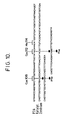
- This step is essential for the further course of the viral infection. It is known from the poliovirus system that all enzymes involved in this maturation cleavage are probably virally coded (Toyoda, H. et al., 1986, Cell, 45 , 761-770). There are three types of split signals in poliovirus (Fig. 1); the most commonly used QG sites recognized by the viral protease P3C and the YG site used by P2A as the recognition signal. Initially, the protease P3C became the focus of interest in elucidating the proteolytic processing of picornaviruses. Proteolytic activity equivalent to the P3C could be described very early in EMC (Pelham, HRB, 1978, Eur. J.
- P2A is indirectly responsible for this modification of p220 in infected cells (Krausslich, HG et al., 1987, J. Virol. 61 , 2711-2718).
- the question of the transactivity of the two proteases P3C and P2A could be answered positively in the poliovirus system insofar as in vitro expressed poliovirus polypeptide precursors, which contained the proteolytic recognition sequences, could be processed by exogenous P3C and P2A proteases (Nicklin, MJH et al. , 1987, Proc. Natl. Acad. Sci. USA 84 , 4002-4006).
- the cleavage site specificity of the viral proteases was determined in the poliovirus system by N-terminal sequencing of most poliovirus proteins (Pallansch, MA et al., 1984, J. Virol. 49 , 873-880).
- HRV2 Cloning and sequencing of HRV2 (Skern, T. et al., 1985 Nucleic Acids Res. 13 , 2111-2126) was able to deduce most cleavage sites on the basis of sequence comparisons with poliovirus and HRV14.
- the position of the interfaces between VP4 / VP2, VP2 / VP3 and VP3 / VP1 could be determined by N-terminal sequencing of VP2, VP3 and VP1.
- the cleavage signal between VP1 and P2A was determined in part by C-terminal sequencing of VP1 (Kowalski, H. et al., 1987, J. Gen. Virol. 86 , 3197-3200). Five different split signals were found in HRV2: QS, QG, QN, AG and ES (Fig. 2).
- Cysteine proteases are widespread in nature (e.g. papain, cathepsin B, H and S) and their characterization and inhibition is of great scientific and therapeutic value (for an overview see Turk, V., 1986, Cysteine Proteinases and their Inhibitors, Walter de Gruyter; Barrett, AJ and Salvesen, G., 1986, Proteinase Inhibitors, Elsevier).
- a wide variety of inorganic and organic compounds, as well as peptide derivatives and proteins are known in the picornaviral system, which have an inhibitory effect on the proteolytic processing of these viruses.
- the viral enzyme P2A is particularly preferred.
- the chemotherapeutic approach is preferably the inhibition of the enzymatic activity by, for example, specific inhibitors.
- the object of the present invention was therefore to provide a system with which it is possible to test potential inhibitors for the viral maturation process.
- This expression system consisting of a plasmid portion and an insert, had to be able to produce a viral polypeptide which serves as substrate and which also has P2A protease activity.
- a system contains as an insert a DNA molecule which codes for a fusion protein from an enzymatically active part and a polypeptide part which can be split off from it.
- the enzymatically active part is a viral protease, preferably the viral protease HRV2-P2A.
- the cleavable polypeptide portion is a viral protein, preferably a viral protein VP1, particularly preferably the viral protein HRV2-VP1, in particular (VP3) -VP1 from HRV2 or parts thereof.
- An example of such a system preferably contains the HRV2 sequence from 2145-3698 in the correct reading frame as an insert.
- This insert can be inserted into any expression vector which is capable of effectively producing the substrate in a suitable host organism transformed with this vector.
- Such an expression vector preferably contains a ribosomal binding site, a coding region for a fusion portion which increases the stability of the expressed fusion protein encoded by the insert in the cell, a promoter controlling the fusion protein, a polylinker region in three different reading frames, preferably with interfaces for restriction enzymes.
- ori region and a selection marker.
- pEx34c expression vector used in the present invention is such a derivative.
- the above-mentioned insert was inserted into this vector as an EcoRI / HindIII fragment.
- PEx34c x 18521 was obtained (Fig. 4).
- an inactive enzyme substrate for P2A was also required, which was obtained, for example, by deletion mutation.
- Such a deletion mutant of the pEx34c x 18521 was called pEx34c x 18731. This mutant ends at nucleotide number 3321 and therefore no longer has the presumed active center of P2A (FIG. 9).
- the two plasmids were expressed in vitro in a bacterial, cell-free system, for example the "Procaryotic DNA directed Translation Kit” from Amersham. 7 shows the result. It shows that such an in vitro system is ideally suited for testing inhibitors against P2A.
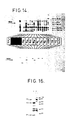
- Essential residues must therefore be present between the 6th and 10th amino acids from the C-terminus which are indispensable for the catalytic activity. If one considers the amino acid sequence of 2A and its amino acids highly conserved within the rhino, polio and coxsackie viruses (see FIG. 15), it can be seen that between the 6th and 10th amino acid only the arginine at position 134 is conserved from the C-terminus of 2A. In order to find out whether this residue is of fundamental importance in catalysis, arginine 134 was converted into a glutamine by in vitro mutagenesis.
- the mutated expression vector pEx18521 [Arg134 ⁇ Gln] was expressed as described in Example 2 and the pattern of the expression products was analyzed on a protein gel and with the aid of a Western blot (see FIG. 16). The importance of Arg 134 was clearly demonstrated: the mutation of arginine 134 to glutamine leads to the disappearance of proteolytic activity; only the unprocessed 75K protein is formed.
- proteases 2A and 3C from rhino, polio and coxsackieviruses By comparing the amino acid sequence of proteases 2A and 3C from rhino, polio and coxsackieviruses and by in vitro mutagenesis studies on protease 3C from polio (Ivanoff, LA, et al. (1986) Proc. Natl. Acad. Sci. USA, 83, 5392 - 5396) it can be assumed that these enzymes use a cysteine and a histidine in the active center for proteolysis. In the case of protease 2A from HRV2, the probably active center can be assigned to amino acids 100 to 140 (see FIG. 15).
- cysteine functions as a nucleophile
- the homology of the region around the probably active center to the serine proteases is larger than to the cysteine proteinases (see FIG. 20).
- the comparisons were made with the protein sequence databases PIR (Sidman, KS, George, DC, Barker, WC and Hunt, LT (1988) Nucleic Acids Res., 16, 1869-1871) and SWISSPROT (Cameron, GN (1988) Nucleic Acids Res., 16, 1865-1867) using the FASTP program (Lipman, DJ and Pearson, WR (1985) Science, 227, 1435-1451).
- Protease 2A appears to have a cysteine function as a nucleophile in the conserved environment (GDSGG) of the serine proteases (Fig. 20). Mechanically, the 2A proteases surprisingly belong to the class of cysteine proteases, although there is an environment typical for serine proteases. Certain other residues are also highly conserved, e.g. B. the glycine and the cysteine before the active center and the glycine before a hydrophobic amino acid after the likely catalytic center (see Fig. 15).
- cysteines were exchanged for serines (approximately the same space requirement), while in Example 4 cysteines were replaced by tryptophan residues (significantly larger space requirement), which can lead to structural changes.
- a 16 amino acid long peptide was used as the peptide substrate (Ac-TRPIITT AG PSDMYVH). It contains 8 amino acids before and 8 amino acids after the expected cleavage site of 2A (cleavage takes place between the underlined amino acids).
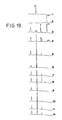
- the original 16 amino acid peptide substrate and a reference peptide representing the C-terminal cleavage product (GPSDMYVH) were separated on an HPLC column. Expression of the 2A expression systems was induced in E. coli strain 537 as described in Example 2. After removal of insoluble material, the supernatant was mixed with an aqueous peptide solution from Ac-TRPIITT AG PSDMYVH and incubated.
- FIG. 19 shows the HPLC profiles of the peptides after incubation of the peptide substrate in various bacterial extracts.
- the peak t R 20.8 min (uncleaved peptide substrate; Ac-TRPIITTAGPSDMYVH; more open, thicker Arrow; absorbs at 214 nm and at 280 nm).
- the present invention provides an expression system with which it is possible to find and optimize potential inhibitors against P2A; a basic requirement for the implementation of the convincing therapeutic concept by suppressing the proteolytic activity of the viral protease P2A to prevent the maturation process of the viral system required for a viral infection. Due to the pronounced homology of the P2A region also to other groups of Picornaviridiae, it is also conceivable to use the inhibitors found by this system according to the invention therapeutically also against infections of other Picornaviruses.
- the present invention provides an effective "trans assay” for protease 2A. It has been shown that peptides can be used as cleavage site analogues, which enables detailed and rapid biochemical characterization of protease 2A. With the help of this "trans assay" according to the invention, it is thus possible to check substances for their inhibitory activity with regard to protease 2A activity; it therefore ensures broad-based inhibitor screening. With its help, it is possible to find a specific viral protease 2A inhibitor.
- the present invention also relates to the use of the expression system as a test system for inhibitors of viral proteases, preferably for P2A inhibitors, particularly preferably for HRV2-P2A inhibitors.
- E. coli W6 constitutively expresses the gene for the wild-type lambda repressor and is suitable for growing up the pEx plasmids.
- E. coli 537 on the other hand, carries the cI 853 lambda repressor mutation (inactive at 42 ° C.) on another plasmid which also carries a kanamycin resistance gene (K. Strebel, E. Beck, K. Strohmaier and H. Schaller; 1986, J. Virol. 57 , 983-991).
- an expression system By inserting the EcoRI / HindIII fragment from p18521 into pEx34c, an expression system could be obtained which comprises the region: (VP3) -VP1-P2A- (P2B) of HRV2 (2145-3698; see Fig. 4).
- This expression system produces a viral polypeptide which serves as a substrate and which also has P2A protease activity.
- the expression vector pEx34c x 18521 was grown up in E. coli W6 (lambda). As described above, the vector was isolated from 500 ml of an overnight culture using the large scale preparation technique.
- 2 ⁇ g of pEx34c x 18521 were digested with HindIII and purified using DE81 as described above.
- the T4 DNA ligase approach was used directly to transform E. coli W6 (lambda) used. Some of the clones were picked and the respective plasmid DNA was sequenced according to Maxam and Gilbert (Maxam, A. and Gilbert, W., 1980, Nucleic Acids Res. 65 , 499-560).
- a clone whose cDNA ends with HRV2 nucleotide number 3321 (see Skern, T. et al., 1985, Nucleic Acids Res. 13 , 2111-2126) was named 18731. This deletion mutant of 18521 was used as an inactive P2A enzyme substrate for expression studies.
- the "transfer sandwich” was compiled according to the scheme below (avoid air bubbles!): - Pol -> scotch brite -> 2 layers 3 MM -> Gel -> Nitrocellulose -> 2 layers 3 MM -> scotch brite -> + Pol
- the protein gel was also equilibrated in transfer buffer for 2 min prior to assembly.
- the transfer buffer can also be prepared as a 10 x solution (24.2 g Tris and 112.6 g glycine per liter without methanol).
- the transfer was carried out in the transfer buffer at about 1 ampere, 2 hours in a protein blot apparatus in the presence of 0.1% Empigen BB (alkyldimethylammonium betaine; No. 62 852, Marchon France SA) (RE Mandrell et al .; J Immunol. Meth. 67 , p. 1 (1984)).
- the efficiency of the transfer was checked using the pre-stained marker proteins.
- the filters with the proteins bound to them were placed in 50 ml "Blocking Solution” overnight at room temperature, that is PBS: 137.0 mM NaCl 2.7 mM KCl 8.0 mM Na2HPO4 1.5 mM KH2PO4 0.5 mM MgCl2.6H2O 1.0 mM CaCl2.2H2O bathed with 1% BSA, 1% Tween 20 (polyoxyethylene (20) sorbitan monolaurate) and 10% heat inactivated fetal calf serum (HIFKS).
- the polyclonal antiserum against VP1 was preincubated with an E. coli lysate before use to remove E. coli-specific antibodies.
- the filter was incubated in approx. 50 ml "Blocking Solution” with the alkaline phosphatase-conjugated rabbit anti-antibody (diluted 1/5000 to 1/7500 in "Blocking Solution”) for 3 hours at room temperature. Finally, the filter well rinsed again under running tap water (15 min) and washed three times as above with 50 ml PBS (+ 1% Tween 20).
- the alkaline phosphatase conjugated anti-rabbit IgG (Fc), as well as the dyes NBT and BCIP come from Promega Biotec (Protoblot TM system).
- the dyeing reaction was also stopped by rinsing under running tap water after about 1 min. 6 shows a typical image of a Western blot of pEx34c x 18521 or 18731.
- pEx34c x 18521 and 18731 were grown and prepared on a large scale in W6 (lambda) as described above. 3 ⁇ g of the circular plasmids were then used per batch in the "Procaryotic DNA directed Translation kit" from Amersham (N 380). This bacterial cell-free system allows in vitro expression of genes located on a plasmid, provided that the relevant control signals such as the Pribnow box for the initiation of transcription and the Shine-Dalgarno sequence for the translation exists. The expressed products are demonstrated by incorporation of S-35 methionine. In order to express the two products 18521 and 18731 in vitro , the incubation conditions of the manufacturer were used.
- a pATI53 vector the expression product of which is the ⁇ -lactamase, can be used as a reference sample on the autoradiogram (FIG. 7, lane 1).
- the high molecular band on the autoradiogram corresponds to the unprocessed 18521 product; the double or triple bands may be due to abortions during transcription or translation.
- This in vitro system is very well suited to test possible inhibitors against P2A.
- the HRV2-P2A comprises 142 amino acids, the amino acid residues involved in the catalysis being located in positions 114 (histidine) and 106 (cysteine) and 112 (cysteine) (FIG. 15).
- the two mutation events for the two cysteines at positions 112 and 106 of HRV2-P2A each produced several hundred white plaques.
- the mutation efficiency was over 95%.
- 5 white plaques were cut out and transferred into 1.5 ml of 2x TY medium with 20 ⁇ l of JM 101 (overnight culture in minimal medium) and incubated for 6 hours at 37 ° C. with shaking. Then 3 min centrifuged (Eppendorf centrifuge) and the double-stranded mutant M13mp8 x 18521 vector was isolated from the cells using the mini-preparation method.
- the isolated M13mp8 plasmids were digested in 1x Eco / Hind buffer as described above, separated on a 1.2% agarose gel and the mutated EcoRI / HindIII fragments from 18521 were isolated using DE 81 paper. These mutated EcoRI / HindIII fragments were back-ligated into the pEx34c vector, 250 ng each of pEx34c (EcoRI / HindIII) and approx. 200 to 400 ng mutated EcoRI / HindIII fragment in 20 ⁇ l of 1x ligase buffer and 1 U of T4 DNA Ligase (Boehringer Mannheim) were incubated for half an hour at room temperature and overnight at 16 ° C.
- the mixture was extracted twice with phenol / chloroform and the DNA was precipitated by ethanol.
- the plasmid 18521/112 which had the cysteine in the immediate vicinity of the histidine residue replaced by phenylalanine, showed an increased activity of P2A, while the mutation of the cysteine located further away from the histidine led to an inhibition of the proteolytic activity (see FIG. 11 ).
- Plasmid DNA from pEx34c x 18521 was prepared by the large-scale method (see above). 300 ⁇ g were digested with the restriction enzyme HindIII (Boehringer Mannheim) in a total volume of 200 ⁇ l. 20 ⁇ l of this mixture were incubated at 30 ° C for 6 - 15 minutes with the exonuclease Bal3l (Biolabs) according to the manufacturer in a volume of 30 ⁇ l.
- the nuclease reaction was started by adding 20 ⁇ l of a 0.25 M EDTA solution was stopped and placed on ice. After extraction with phenol / chloroform and chloroform, the DNA was precipitated with ethanol and taken up in 10 ul H2O. Protruding ends of the plasmid DNA were filled in with the Klenow fragment of the DNA polymerase (Biolabs) and 2.5 ⁇ M each of the nucleotides dATP, dCTP, dGTP and dTTP. The plasmids were extracted and precipitated as described above. After further digestion with PstI Boehringer Mannheim), the fragments were separated on an agarose gel and eluted using DE 81 paper.
- BLUESCRIPT vector (Stratagene Cloning Systems) were digested with EcoRV and PstI, purified by extraction and precipitated. The isolated fragments, which have a blunt end, from the Bal31 digest and polymerase treatment, and an overhanging end, from the PstI digest, were ligated into the cut BLUESCRIPT vector (FIG. 13). Competent E. coli strain JM109 cells were transformed with the ligase solution. Transformants were selected on agar plates with ampicillin and X-Gal / IPTG. 109 deletion mutants were obtained from the time values 7, 9, 11, 13 and 15.
- plasmid DNA was prepared from these clones by the mini-preparation method, cut with PstI and HindIII and separated on an agarose gel.
- the PstI site was retained by the ligation, while the HindIII site comes from the polylinker of the BLUESCRIPT vector and is only 3 nucleotides after the insert.
- a marker lambda DNA digested with HindIII
- those clones could be identified whose deletions only affect the C-terminus of P2A.
- 54 clones were identified after the Dideoxy method according to Sanger et al. (Zimmer, et al. Proc. Natl. Acad.
- protease 2A represent a potential antigenic determinant.
- a peptide (PC20) which had precisely this amino acid sequence, was synthesized as described in Example 9 and used to induce antibodies in rabbits: 570 ⁇ g of this peptide were 4 ml of PBS solution was added and drawn up in a 5 ml syringe. In a second 5 ml syringe, 0.5 ml of Freund's adjuvant (CAF; GIBCO) was drawn up and the two components were then mixed using a three-way stopcock valve until an emulsion was formed. The rabbit was punctured arterially in the ear to obtain a pre-serum for the negative control.
- CAF Freund's adjuvant
- the immunization was carried out by subcutaneous injection of the peptide / CFA mixture at 4 different sites (0.2 ml / injection site) of the back area. After 5 weeks, 1.2 ml of peptide solution was "boosted” intramuscularly into the back part by injection of 0.2 ml each. Eight days later, the blood was taken by cardiac puncture. The blood was allowed to clot at room temperature, fibrin and shaped Ingredients were removed with a sterile stick and the blood centrifuged at 2000 rpm. The serum was aliquoted and stored at -18 ° C.
- a rabbit was arterially punctured in the ear to obtain a pre-serum for the negative control.
- the immunization was carried out by subcutaneous injection of a peptide / CFA mixture at 4 different points in the back area.
- the serum obtained was aliquoted and stored at -18 ° C.
- a 264 bp DNA fragment was obtained by digestion with Apa I (nucleotide number 3458 of the HRV2 cDNA) and Hind III (restriction site comes from the polylinker region of the vector; see Example 1) according to the manufacturer's instructions (Biolabs) two double-stranded oligonucleotides WT 12 and WT 34 were replaced with Apa I / Hind III "sticky ends" (see FIG. 17).
- the kinase mixtures of WT1 and WT2, or of WT3 and WT4 were then combined, incubated for 10 min at 68 ° C., 30 min at 45 ° C., 10 min at room temperature and then briefly on ice.
- the kinased and hybridized oligonucleotides were combined, the concentration was adjusted to 1 mM with a 10 mM ATP solution and 28 U T4-DNA ligase (Boehringer Mannheim) were added for the ligation.
- the ligation itself was first carried out at 20 ° C. for 2 hours, then another 7 U of T4 DNA ligase were added and the ligation mixture was incubated at 14 ° C. for 40 hours.
- the ligation mixture was then incubated at 70 ° C. for 10 min, brought to 100 mM with a 1M NaCl solution and the resulting multimeric forms of the oligonucleotide were "re-cut" with 50 U Apa I and 20 U Hind III.
- the peptides used in this example were synthesized by the "solid phase” method (Merrifield, RB (1963) J. Am. Chem. Soc., 85, 2149-2154). The peptides were then purified by "reverse phase” HPLC (0.1% trifluoroacetic acid and acetonitrile as mobile phase) and with the aid of HPLC analysis, “fast atom bombardment mass spectrometry (FAB-MS) and amino acid sequencing (Hunkapiller, MW and Hood, LE, loc.cit.). As described in Example 2, expression of the 2A expression systems (see Example 8) was induced in E. coli strain 537.
- the cells were broken open using an MSE Ultrasonic Power device 3 times 30 seconds; in an ice water bath), whereby a pause of 30 seconds was switched on between the individual sonizations to avoid overheating of the samples.
- FIG. 19 shows the HPLC profiles of the peptides after incubation of the peptide substrate in various bacterial extracts.
Landscapes
- Health & Medical Sciences (AREA)
- Life Sciences & Earth Sciences (AREA)
- Chemical & Material Sciences (AREA)
- Engineering & Computer Science (AREA)
- Molecular Biology (AREA)
- Genetics & Genomics (AREA)
- Organic Chemistry (AREA)
- Biomedical Technology (AREA)
- Immunology (AREA)
- Biochemistry (AREA)
- General Health & Medical Sciences (AREA)
- Biotechnology (AREA)
- Wood Science & Technology (AREA)
- Zoology (AREA)
- Virology (AREA)
- Microbiology (AREA)
- Bioinformatics & Cheminformatics (AREA)
- General Engineering & Computer Science (AREA)
- Hematology (AREA)
- Physics & Mathematics (AREA)
- Medicinal Chemistry (AREA)
- Urology & Nephrology (AREA)
- Biophysics (AREA)
- Proteomics, Peptides & Aminoacids (AREA)
- Analytical Chemistry (AREA)
- Food Science & Technology (AREA)
- Cell Biology (AREA)
- General Physics & Mathematics (AREA)
- Pathology (AREA)
- Gastroenterology & Hepatology (AREA)
- Tropical Medicine & Parasitology (AREA)
- Plant Pathology (AREA)
- Medicines That Contain Protein Lipid Enzymes And Other Medicines (AREA)
- Enzymes And Modification Thereof (AREA)
- Peptides Or Proteins (AREA)
- Measuring Or Testing Involving Enzymes Or Micro-Organisms (AREA)
- Preparation Of Compounds By Using Micro-Organisms (AREA)
Description
Gegenstand der vorliegenden Erfindung sind DNA-Moleküle, die für Fusionsproteine aus enzymatisch aktiven Anteilen und von diesen abspaltbaren Polypeptidanteilen kodieren, Expressionssysteme, die diese DNA-Moleküle enthalten sowie die Verwendung dieser als Testsysteme für Inhibitoren viraler Proteasen.The present invention relates to DNA molecules which code for fusion proteins from enzymatically active components and polypeptide components which can be split off from them, expression systems which contain these DNA molecules and the use of these as test systems for inhibitors of viral proteases.
Rhinoviren sind ss(+)RNA - Viren und repräsentieren eine Gattung innerhalb der Picornaviridae (Cooper, P. D et al., 1978, Intervirology 10, 165 - 180; MacNaughton, M. R., 1982, Current Top. Microbiol. Immunol. 97, 1 - 26). Sie sind weit verbreitet, befallen den oberen respiratorischen Trakt des Menschen und verursachen akute Infektionen, die zu Schnupfen, Husten, Heiserkeit etc. führen und allgemein als Erkältungen bezeichnet werden (Stott, E. J. und Killington, R. A., 1972, Ann. Rev. Microbiol. 26, 503 - 524). Infektionen durch Rhinoviren zählen zu den häufigsten Erkrankungen des Menschen. Die Krankheit verläuft zwar meist harmlos, dennoch kommt es -bedingt durch eine vorübergehende Schwächung des Organismus- zu Sekundärinfektionen durch andere Viren oder Bakterien, die dann unter Umständen schwere Erkrankungen zur Folge haben. Von den insgesamt ca. 115 verschiedenen, bekannten Serotypen von humanen Rhinoviren sind bis jetzt 3 Serotypen kloniert und komplett sequenziert worden: Deutsche Patentanmeldung P 35 05 148.5; Skern; T. et al., 1985; Nucleic Acids Res. 13, 2111-2126; Düchler, M. et al., 1987, Proc. Natl. Acad. Sci. USA 84, 2605 - 2609; Stanway, G. et al., 1984, Nucleic Acids Res. 12, 7859 - 7877; Callahan, P. L. et al., 1985, Proc. Natl. Acad. Sci. USA 82, 732 - 736).Rhinoviruses are ss (+) RNA viruses and represent a genus within the Picornaviridae (Cooper, P. D et al., 1978, Intervirology 10 , 165-180; MacNaughton, MR, 1982, Current Top. Microbiol. Immunol. 97 , 1 - 26). They are common, affecting the upper respiratory tract of humans and causing acute infections that lead to runny nose, cough, hoarseness, etc. and are commonly referred to as colds (Stott, EJ and Killington, RA, 1972, Ann. Rev. Microbiol. 26 , 503-524). Rhinovirus infections are among the most common human diseases. Although the disease is usually harmless, secondary infections by other viruses or bacteria can occur due to a temporary weakening of the organism, which may then result in serious illnesses. Up to now, 3 of the 115 different known serotypes of human rhinoviruses have been cloned and completely sequenced: German patent application P 35 05 148.5; Skern; T. et al., 1985; Nucleic Acids Res. 13 , 2111-2126; Düchler, M. et al., 1987, Proc. Natl. Acad. Sci. USA 84 , 2605-2609; Stanway, G. et al., 1984, Nucleic Acids Res. 12 , 7859-7877; Callahan, PL et al., 1985, Proc. Natl. Acad. Sci. USA 82 , 732-736).
Ein Vergleich der Aminosäuresequenzen der einzelnen Proteine zeigt, daß die viralen Enzyme in besonderem Maße konserviert sind. So beträgt etwa die Homologie zwischen der Protease P2A von HRV89 und HRV2 85%; bei der Protease P3C sind 75% der Aminosäuren identisch (Düchler, M. et al., 1987). Diese Werte liegen wesentlich über den durchschnittlich im Gesamtprotein beobachteten Prozentsätzen. Man kann daher davon ausgehen, daß gerade die viralen Enzyme in der Evolution besonders gut konserviert sind und in ihren Eigenschaften bei verschiedenen Rhinoviren sehr ähnlich sind.
Kaum ein anderes virales System ist bei seiner Regulation des Infektionsablaufes dermaßen von einer kontrolliert limitierten Proteolyse abhängig, wie das der Picornaviridae. Die genomische einzelsträngige (+)RNA der Rhinoviren wird kurz nach der Infektion durch Abspaltung des an das 5′ Ende gebundenen Oligopeptids VPg modifiziert und dient als mRNA für die Synthese eines Polyproteins, das den gesamten durchgehenden Leserahmen der Nukleinsäuresequenz umfaßt (Butterworth, B. E., 1973, Virology 56, 439 - 453; Mc Lean, C. und Rueckert, R. R., 1973, J. Virol. 11, 341 - 344; Mc Lean, C. et al., 1976, J. Virol. 19, 903 - 914). Die reifen viralen Proteine entstehen ausschließlich durch proteolytische Spaltung aus diesem Polyprotein, wobei die dabei wirksamen Proteasen selbst Bestandteil dieses Polyproteins sind. Der erste Schritt bei diesem Prozessieren ist die Abspaltung der Vorstufe der Hüllenproteine, die durch die Protease P2A vorgenommen wird. In der Reihenfolge der Gene liegt die Sequenz der Protease P2A unmittelbar hinter dem für die Hüllenproteine kodierenden Abschnitt. P2A ist somit aufgrund ihrer Lokalisation im Polyprotein die erste nachweisbare enzymatische Funktion des Virus.
Sie spaltet sich gewissermaßen selbst vom Precursor der Hüllenproteine ab und ist für die Trennung des Kapsidprecursors P1 vom Rest des Polyproteins verantwortlich. Die Trennung der Hüllenproteinregion von dem für die Replikation verantwortlichen Abschnitt findet bereits während der Translation des Polyproteins statt.A comparison of the amino acid sequences of the individual proteins shows that the viral enzymes are particularly conserved. For example, the homology between protease P2A of HRV89 and HRV2 is 85%; in the protease P3C, 75% of the amino acids are identical (Düchler, M. et al., 1987). These values are significantly higher than the percentages observed on average in the total protein. It can therefore be assumed that the viral enzymes are particularly well preserved in evolution and are very similar in their properties in various rhinoviruses.
Hardly any other viral system is so dependent on controlled limited proteolysis to regulate the course of infection as Picornaviridae. The genomic single-stranded (+) RNA of the rhinoviruses is modified shortly after infection by cleavage of the oligopeptide VPg bound to the 5 ′ end and serves as mRNA for the synthesis of a polyprotein which encompasses the entire continuous reading frame of the nucleic acid sequence (Butterworth, BE, 1973 , Virology 56 , 439-453; Mc Lean, C. and Rueckert, RR, 1973, J. Virol. 11 , 341-344; Mc Lean, C. et al., 1976, J. Virol. 19 , 903-914 ). The mature viral proteins are produced exclusively from this polyprotein by proteolytic cleavage, the active proteases themselves being part of this polyprotein. The first step in this processing is the cleavage of the precursor of the envelope proteins, which is carried out by the protease P2A. In the order of the genes, the sequence of the protease P2A lies immediately behind the section coding for the envelope proteins. Due to its location in the polyprotein, P2A is the first detectable enzymatic function of the virus.
To a certain extent, it cleaves itself from the precursor of the envelope proteins and is responsible for separating the capsid precursor P1 from the rest of the polyprotein. The separation of the coat protein region from the section responsible for replication takes place already during the translation of the polyprotein.
Dieser Schritt ist für den weiteren Ablauf der viralen Infektion essentiell.
Vom Poliovirussystem weiß man, daß wahrscheinlich alle an dieser Reifungsspaltung beteiligten Enzyme viral kodiert sind (Toyoda, H. et al., 1986, Cell, 45, 761-770). Im Poliovirus findet man drei Typen von Spaltsignalen (Fig. 1); die am meisten verwendeten Q-G Stellen, welche von der viralen Protease P3C erkannt werden, und die Y-G Stelle, welche von P2A als Erkennungssignal verwendet wird. Zunächst rückte die Protease P3C in den Mittelpunkt des Interesses bei der Aufklärung des proteolytischen Processings von Picornaviren. Schon sehr früh konnte eine der P3C äquivalente proteolytische Aktivität in EMC beschrieben werden (Pelham, H. R. B. , 1978, Eur. J. Biochem. 85, 457 - 461; Palmenberg A. C. et al., 1979, J. Virol. 32, 770 - 778). Im Laufe weiterer Untersuchungen stellte sich heraus, daß das Leaderpeptid (L) von Cardio- (z. B. EMCV) und Aphtoviren (z. B. FMDV), welches bei Rhino- und Enteroviren nicht vorhanden ist, am proteolytischen Processing von EMCV beteiligt ist (Palmenberg, A. C., 1987, J. Cell. Biochem. 33, 1191-1198). In weiterer Folge konnte durch die Isolierung von Polio P3C und mit Hilfe von immunologischen Methoden gezeigt werden, daß P3C sich selbst autokatalytisch aus dem Polyprotein herausschneidet, um dann in "trans" alle potentiellen Q-G Spaltstellen anzugreifen.This step is essential for the further course of the viral infection.
It is known from the poliovirus system that all enzymes involved in this maturation cleavage are probably virally coded (Toyoda, H. et al., 1986, Cell, 45 , 761-770). There are three types of split signals in poliovirus (Fig. 1); the most commonly used QG sites recognized by the viral protease P3C and the YG site used by P2A as the recognition signal. Initially, the protease P3C became the focus of interest in elucidating the proteolytic processing of picornaviruses. Proteolytic activity equivalent to the P3C could be described very early in EMC (Pelham, HRB, 1978, Eur. J. Biochem. 85 , 457-461; Palmenberg AC et al., 1979, J. Virol. 32 , 770-778 ). In the course of further investigations, it was found that the leader peptide (L) of cardio (e.g. EMCV) and aphtoviruses (e.g. FMDV), which is not present in rhino and enteroviruses, is involved in the proteolytic processing of EMCV (Palmenberg, AC, 1987, J. Cell. Biochem. 33, 1191-1198). Subsequently, the isolation of Polio P3C and with the help of immunological methods showed that P3C cuts itself autocatalytically out of the polyprotein and then attacks all potential QG cleavage sites in " trans ".
Der Einsatz von rekombinanten Systemen, die unter anderem die P3C-Region repräsentierten, ermöglichte die Expression von P3C einiger Entero- und Rhinoviren (Werner, G. et al., 1986, J. Virol. 57, 1084 - 1093) und die genaue Charakterisierung der P3C von Polio (Hanecak, R. et al., 1984, Cell 37, 1037 - 1073; Korant, B. D. und Towatari, T., 1986, Biomed. Biochim. Acta 45, 1529 - 1535) sowie der äquivalenten proteolytischen Funktion in FMDV (Klump, W. et al., 1984, Proc. Natl. Acad. Sci. USA 81, 3351 - 3355; Burroughs, J. N. et al., 1984, J. Virol. 50, 878 - 883). Durch in vitro Mutagenesestudien konnte nachgewiesen werden, daß der Austausch der hoch konservierten Aminosäuren Cystein (Positionsnummer 147) und Histidin (Positionsnummer 161) in P3C von Poliovirus zu einem inaktiven Enzym führt, während die Mutation des nicht konservierten Cysteins (Positionsnummer 153) keinen nennenswerten Einfluß auf die proteolytische Aktivität von Polio-P3C hat. Diese durch Oligonukleotide bewirkte Mutagenese von rekombinanten P3C und die zusätzlich durchgeführten Inhibitorstudien lassen den Schluß zu, daß Polio-P3C zu der Klasse der Cysteinproteasen gehört (Ivanoff, L. A. et al., 1986, Proc. Natl. Acad. Sci. USA 83, 5392 - 5396). Ebenfalls durch in vitro Mutagenese von Polio-P3C (und zwar durch Austausch des konservierten Valin gegen Alanin in Position 54 der Protease) konnte gezeigt werden, daß diese Mutation in einer "full size" cDNA von Polio nach Transfektion in COS 1 Zellen zu einem Polymerase-defizienten Virus führt (Dewalt, P. G. und Semler, B. L., 1987,J. Virol. 61, 2162 - 2170).The use of recombinant systems, which represented the P3C region, among other things, made it possible to express P3C of some entero- and rhinoviruses (Werner, G. et al., 1986, J. Virol. 57 , 1084-1093) and to characterize them precisely the P3C from Polio (Hanecak, R. et al., 1984, Cell 37 , 1037-1073; Korant, BD and Towatari, T., 1986, Biomed. Biochim. Acta 45 , 1529 - 1535) and the equivalent proteolytic function in FMDV (Klump, W. et al., 1984, Proc. Natl. Acad. Sci. USA 81 , 3351-3355; Burroughs, JN et al., 1984, J. Virol. 50 , 878-883). In vitro mutagenesis studies have shown that the exchange of the highly conserved amino acids cysteine (position number 147) and histidine (position number 161) in P3C of poliovirus leads to an inactive enzyme, while the mutation of the non-conserved cysteine (position number 153) has no significant effect on the proteolytic activity of Polio-P3C. This mutagenesis of recombinant P3C caused by oligonucleotides and the additional inhibitor studies carried out allow the conclusion that polio-P3C belongs to the class of cysteine proteases (Ivanoff, LA et al., 1986, Proc. Natl. Acad. Sci. USA 83 , 5392 - 5396). It was also shown by in vitro mutagenesis of polio-P3C (by replacing the conserved valine with alanine in position 54 of the protease) that this mutation in a "full size" cDNA from polio after transfection in
Antikörper, die gegen Polio-P3C entwickelt wurden, unterbanden zwar eindeutig sämtliche an Q-G durchgeführten Spaltungen, nicht aber die Spaltung zwischen Y-G (Hanecak, R. et al., 1982, Proc. Natl. Acad. Sci. USA 79, 3973 - 3977). Diese Beobachtung führte zum Schluß, daß das proteolytische Processing an Y - G Stellen einer eigenen Protease bedarf. Der Sitz dieser zweiten proteolytischen Aktivität konnte in Poliovirus eindeutig P2A zugeordnet werden. Interessant war der Befund, daß P2A eine alternative Spaltung in der Protease-Polymeraseregion (3CD) durchführt, welche ebenfalls an einer Y - G Stelle stattfindet. Dieser Spaltung dürfte aber keine biologische Bedeutung während der Virusreplikation zukommen (Toyoda, H. et al., loc. cit.).
Da es während der Infektion mit Poliovirus in HeLa-Zellen sehr rasch zu einem Abschalten der Wirtsproteinsynthese kommt, die Translation der Poliovirus-RNA jedoch unbehindert ablaufen kann, nahm man an, daß ein oder mehrere Regulationsfaktoren der Translation während der Infektion verändert werden. Tatsächlich zeigen ältere Befunde, daß der eukaryontische Initiationsfaktor 4F durch proteolytische Spaltung der p220 Komponente während der Poliovirusinfektion in HeLa-Zellen verändert wird (Etchison, D. et al., 1984, J. Virol. 51, 832 - 837; Etchison, D. et al., 1982, J. Biol. Chem. 257, 14806 - 14810). In der Folge konnte gezeigt werden, daß P2A indirekt für diese Modifikation von p220 in infizierten Zellen verantwortlich ist (Kräusslich, H. G. et al., 1987, J. Virol. 61, 2711 - 2718).
Die Frage nach der Transaktivität der beiden Proteasen P3C und P2A konnte im Poliovirussystem insofern positiv beantwortet werden, als in vitro exprimierte Poliovirus-Polypeptidvorstufen, die die proteolytischen Erkennungsequenzen enthielten, durch exogene P3C bzw. P2A Proteasen prozessiert werden konnten (Nicklin, M. J. H. et al., 1987, Proc. Natl. Acad. Sci. USA 84, 4002 - 4006).Antibodies developed against Polio-P3C clearly prevented all cleavages performed on QG, but not cleavage between YG (Hanecak, R. et al., 1982, Proc. Natl. Acad. Sci. USA 79 , 3973-3977). This observation led to the conclusion that proteolytic processing at Y - G sites requires its own protease. The seat of this second proteolytic activity was clearly assigned to P2A in poliovirus. It was interesting to find that P2A performs an alternative cleavage in the protease polymerase region (3CD), which also takes place at a Y - G site. However, this cleavage is not of biological importance during virus replication (Toyoda, H. et al., Loc. Cit.).
Since the host protein synthesis is switched off very quickly during the infection with poliovirus in HeLa cells, but the translation of the poliovirus RNA can proceed unimpeded, it was assumed that one or more translation regulatory factors were changed during the infection. Indeed, older findings show that the eukaryotic initiation factor 4F is changed by proteolytic cleavage of the p220 component during poliovirus infection in HeLa cells (Etchison, D. et al., 1984, J. Virol. 51 , 832-837; Etchison, D. et al., 1982, J. Biol. Chem. 257 , 14806-14810). It could subsequently be shown that P2A is indirectly responsible for this modification of p220 in infected cells (Krausslich, HG et al., 1987, J. Virol. 61 , 2711-2718).
The question of the transactivity of the two proteases P3C and P2A could be answered positively in the poliovirus system insofar as in vitro expressed poliovirus polypeptide precursors, which contained the proteolytic recognition sequences, could be processed by exogenous P3C and P2A proteases (Nicklin, MJH et al. , 1987, Proc. Natl. Acad. Sci. USA 84 , 4002-4006).
Sehr interessant sind auch die Befunde, daß zwei den picornaviralen Proteasen P3C und P2A ähnliche Proteine in dem pflanzenviralen System der Comoviridae (Cowpea Mosaic Virus) gefunden wurden (Garcia, J. A. et al., 1987, Virology 159, 67 - 75; Verver, J. et al., 1987, EMBO 6, 549 - 554). Diese beiden viralen Proteine sind am proteolytischen Processing der beiden durch zwei getrennt verpackte ss (+)RNA-Moleküle (B und M RNA) kodierten Polyproteine beteiligt, wobei eine große Ähnlichkeit der beiden Cowpea mosaic-Virus-Proteasen in Sequenz und Spaltspezifität zu den Picornaviren festzustellen ist. Diese bemerkenswerte Homologie von nicht strukturellen Proteinen zwischen Picorna- und Comoviren weist nicht nur auf eine genetische Verwandtschaft zwischen diesen beiden Virusfamilien hin, sondern zeigt auch auf, wie essentiell das virale proteolytische Processing für diese beiden Virusfamilien ist.
Die dritte Art der viralen Reifungspaltung, nämlich die von VP0 (Vorläuferprotein von VP2 und VP4) konnte bei Mengo und Rhinovirus mit Hilfe von Röntgenstrukturdaten beschrieben werden. Dieses letzte proteolytische Ereignis bei der viralen Maturation scheint auf einem ungewöhnlichen autokatalytischen Serinprotease Typ zu beruhen, bei welchem basische Gruppen der viralen RNA an der Ausbildung des katalytischen Zentrums beteiligt sind, wobei diese basischen Gruppen als Protonenakzeptor fungieren (Arnold, E. et al., 1987, Proc. Natl. Acad. Sci. USA 84, 21 - 25).Also of great interest are the findings that two proteins similar to the picornaviral proteases P3C and P2A were found in the plant viral system of the Comoviridae (Cowpea Mosaic Virus) (Garcia, JA et al., 1987, Virology 159 , 67-75; Verver, J . et al., 1987, EMBO 6 , 549-554). These two viral proteins are involved in the proteolytic processing of the two polyproteins encoded by two separately packaged ss (+) RNA molecules (B and M RNA), whereby the two Cowpea mosaic virus proteases are very similar in sequence and cleavage specificity to the Picornaviruses can be determined. This remarkable homology of non-structural proteins between Picorna and Comoviruses not only indicates a genetic relationship between these two virus families, but also shows how essential viral proteolytic processing is for these two virus families.
The third type of viral maturation cleavage, namely that of VP0 (precursor protein of VP2 and VP4) could be described in Mengo and rhinovirus using X-ray structure data. This last proteolytic event in viral maturation appears to be based on an unusual autocatalytic serine protease type in which basic groups of viral RNA are involved in the formation of the catalytic center, these basic groups acting as proton acceptors (Arnold, E. et al., 1987, Proc. Natl. Acad. Sci. USA 84 , 21-25).
Die Spaltstellenspezifität der viralen Proteasen wurde im Poliovirussystem durch N-terminales Sequenzieren der meisten Poliovirusproteine ermittelt (Pallansch, M. A. et al., 1984, J. Virol. 49, 873 - 880). Durch das Klonieren und Sequenzieren von HRV2 (Skern, T. et al., 1985 Nucleic Acids Res. 13, 2111-2126) konnten an Hand von Sequenzvergleichen mit Poliovirus und HRV14 die meisten Spaltstellen abgeleitet werden. Außerdem konnte die Lage der Schnittstellen zwischen VP4/VP2, VP2/VP3 sowie VP3/VP1 durch N-terminales Sequenzieren von VP2, VP3 und VP1 bestimmt werden. Das Spaltsignal zwischen VP1 und P2A wurde teilweise durch C-terminales Sequenzieren von VP1 bestimmt (Kowalski, H. et al., 1987, J. Gen. Virol. 86, 3197 - 3200). So konnten in HRV2 fünf verschiedene Spaltsignale gefunden werden: Q-S, Q-G, Q-N, A-G und E-S (Fig. 2).The cleavage site specificity of the viral proteases was determined in the poliovirus system by N-terminal sequencing of most poliovirus proteins (Pallansch, MA et al., 1984, J. Virol. 49 , 873-880). By the Cloning and sequencing of HRV2 (Skern, T. et al., 1985 Nucleic Acids Res. 13 , 2111-2126) was able to deduce most cleavage sites on the basis of sequence comparisons with poliovirus and HRV14. In addition, the position of the interfaces between VP4 / VP2, VP2 / VP3 and VP3 / VP1 could be determined by N-terminal sequencing of VP2, VP3 and VP1. The cleavage signal between VP1 and P2A was determined in part by C-terminal sequencing of VP1 (Kowalski, H. et al., 1987, J. Gen. Virol. 86 , 3197-3200). Five different split signals were found in HRV2: QS, QG, QN, AG and ES (Fig. 2).
Cysteinproteasen sind in der Natur weit verbreitet (z. B. Papain, Cathepsin B, H und S) und ihre Charakterisierung und Inhibierung ist von großem wissenschaftlichen und therapeutischen Wert (zur Übersicht siehe Turk, V., 1986, Cysteine Proteinases and their Inhibitors, Walter de Gruyter; Barrett, A. J. und Salvesen, G., 1986, Proteinase Inhibitors, Elsevier). Auch im picornaviralen System sind derzeit verschiedenste anorganische und organische Verbindungen, sowie Peptidderivate und Proteine bekannt, die eine inhibitorische Wirkung auf das proteolytische Processing dieser Viren besitzen. Der Effekt dieser Substanzen beruht auf der direkten Wechselwirkung mit den Proteasen (Kettner, C. A. et al., 1987, US Patent Nr.: 4,652,552; Korant, B. D. et al., 1986, J. Cell. Biochem. 32, 91 -95) und/oder auf dem indirekten Weg der Wechselwirkung mit Substraten dieser Proteasen (Geist, F. C., et al., 1987, Antimicrob. Agents Chemother. 31, 622 - 624; Perrin, D. D. und Stünzi, H., 1984, Viral Chemotherapy 1, 288 - 189). Das Problem bei den meisten dieser Substanzen ist die relativ hohe Konzentration, die zur Inhibierung nötig ist und die zum Teil große Toxizität dieser Verbindungen.Cysteine proteases are widespread in nature (e.g. papain, cathepsin B, H and S) and their characterization and inhibition is of great scientific and therapeutic value (for an overview see Turk, V., 1986, Cysteine Proteinases and their Inhibitors, Walter de Gruyter; Barrett, AJ and Salvesen, G., 1986, Proteinase Inhibitors, Elsevier). A wide variety of inorganic and organic compounds, as well as peptide derivatives and proteins are known in the picornaviral system, which have an inhibitory effect on the proteolytic processing of these viruses. The effect of these substances is based on the direct interaction with the proteases (Kettner, CA et al., 1987, US Patent No.: 4,652,552; Korant, BD et al., 1986, J. Cell. Biochem. 32 , 91-95) and / or by the indirect route of interaction with substrates of these proteases (Geist, FC, et al., 1987, Antimicrob. Agents Chemother. 31 , 622-624; Perrin, DD and Stünzi, H., 1984,
Wie bereits dargelegt wurde, ist der Infektionsablauf durch Picornaviren entscheidend von den viralen Enzymen abhängig. Da gerade diese Enzyme besonders gut konserviert und in ihren Eigenschaften bei verschiedenen Rhinoviren sehr ähnlich sind, bieten sie sich geradezu als Ziel eines chemotherapeutischen Eingriffs an. Besonders bevorzugt ist hierbei das virale Enzym P2A. Der chemotherapeutische Ansatz ist vorzugsweise die Inhibierung der enzymatischen Aktivität durch beispielsweise spezifische Inhibitoren.As already explained, the course of infection by Picornaviruses depends crucially on the viral enzymes. Since these enzymes are particularly well preserved and their properties are very similar in the case of various rhinoviruses, they offer themselves as the target of a chemotherapy intervention. The viral enzyme P2A is particularly preferred. The chemotherapeutic approach is preferably the inhibition of the enzymatic activity by, for example, specific inhibitors.
Inhibiert man die erste proteolytische Aktivität, die P2A-Aktivität, so unterbindet man jeden weiteren Reifungsprozeß des viralen Systems. Auf Grund der ausgeprägten Homologie der P2A-Region von HRV2 nicht nur zu anderen Rhinoviren, sondern auch zu Vertretern anderer Gruppen der Picornaviridae, ist es durchaus denkbar, daß ein Inhibitor gegen HRV2-P2A auch auf andere Picornaviren anwendbar ist.If the first proteolytic activity, the P2A activity, is inhibited, then any further maturation process of the viral system is prevented. Due to the pronounced homology of the P2A region of HRV2 not only to other rhinoviruses, but also to representatives of other groups of the Picornaviridae, it is quite conceivable that an inhibitor against HRV2-P2A can also be applied to other Picornaviruses.
Aufgabe der vorliegenden Erfindung war es daher, ein System bereitzustellen, mit dem es möglich ist, potentielle Inhibitoren für den viralen Reifungsprozeß auszutesten.The object of the present invention was therefore to provide a system with which it is possible to test potential inhibitors for the viral maturation process.
Da P2A "in statu nascendi" aktiv ist und selbst Bestandteil des Substrates ist, mußte ein geeignetes "Substratenzym" konstruiert werden, das es ermöglicht die P2A-Aktivität anhand ihrer Spaltprodukte zu verfolgen. Ferner erkennt P2A nur eine Spaltstelle im Polyprotein. Gerade diese Eigenschaft macht die virale Funktion zum geeigneten Ziel eines therapeutischen Angriffs. Unter diesem Gesichtspunkt wurde in der vorliegenden Erfindung ein Expressionssystem von HRV2-P2A entwickelt, um P2A näher zu charakterisieren und eine geeignete Methode zur Auffindung von Inhibitoren gegen P2A zu etablieren.Since P2A is active "in statu nascendi" and is itself part of the substrate, a suitable "substrate enzyme" had to be constructed which enables P2A activity to be monitored using its cleavage products. Furthermore, P2A recognizes only one cleavage point in the polyprotein. It is precisely this property that makes viral function a suitable target for a therapeutic attack. From this point of view, an expression system of HRV2-P2A was developed in the present invention in order to characterize P2A in more detail and to establish a suitable method for the detection of inhibitors against P2A.
Dieses Expressionssystem, bestehend aus einem Plasmidanteil und einem Insert, mußte in der Lage sein, ein als Substrat dienendes virales Polypeptid, das gleichzeitig auch P2A-Protease-Aktivität aufweist, zu produzieren. Erfindungsgemäß enthält ein solches System als Insert ein DNA-Molekül, das für ein Fusionsprotein aus einem enzymatisch aktiven Anteil und einem von diesem abspaltbaren Polypeptidanteil kodiert. Der enzymatisch aktive Anteil ist dabei eine virale Protease, bevorzugt die virale Protease HRV2-P2A. Der abspaltbare Polypeptidanteil ist ein virales Protein, bevorzugt ein virales Protein VP1, besonders bevorzugt das virale Protein HRV2-VP1, insbesondere (VP3)-VP1 von HRV2 oder Teilen von diesen.This expression system, consisting of a plasmid portion and an insert, had to be able to produce a viral polypeptide which serves as substrate and which also has P2A protease activity. According to the invention, such a system contains as an insert a DNA molecule which codes for a fusion protein from an enzymatically active part and a polypeptide part which can be split off from it. The enzymatically active part is a viral protease, preferably the viral protease HRV2-P2A. The cleavable polypeptide portion is a viral protein, preferably a viral protein VP1, particularly preferably the viral protein HRV2-VP1, in particular (VP3) -VP1 from HRV2 or parts thereof.
Ein Beispiel für ein solches System enthält als Insert vorzugsweise die HRV2 Sequenz von 2145-3698 im richtigen Leserahmen. Dieses Insert kann in jeden beliebigen Expressionsvektor eingefügt werden, der in der Lage ist, in einem, mit diesem Vektor transformierten geeigneten Wirtsorganismus, das Substrat effektiv zu produzieren. Ein solcher Expressionsvektor enthält vorzugsweise eine ribosomale Bindungsstelle, eine kodierende Region für einen die Stabilität des exprimierten, von dem Insert kodierten Fusionsproteins in der Zelle erhöhenden Fusionsanteils, einen das Fusionsprotein kontrollierenden Promotor, eine Polylinkerregion in drei verschiedenen Leserahmen, vorzugsweise mit Schnittstellen für Restriktionsenzyme eine "ori"-Region und einen Selektionsmarker.An example of such a system preferably contains the HRV2 sequence from 2145-3698 in the correct reading frame as an insert. This insert can be inserted into any expression vector which is capable of effectively producing the substrate in a suitable host organism transformed with this vector. Such an expression vector preferably contains a ribosomal binding site, a coding region for a fusion portion which increases the stability of the expressed fusion protein encoded by the insert in the cell, a promoter controlling the fusion protein, a polylinker region in three different reading frames, preferably with interfaces for restriction enzymes. ori "region and a selection marker.
Besonders bevorzugt ist:
- die prokaryontische ribosomale Bindungsstelle
- einen Teil der kodierenden Region für die ersten 98 N-terminalen Aminosäuren der MS2-Polymerase; dieser Fusionsanteil weist hydrophobe und basische Aminosäuren auf und bewirkt die Herabsetzung der Löslichkeit des Fusionsproteins und erhöht die Stabilität des exprimierten Produktes in der Zelle.
- das Fusionsprotein steht unter der Kontrolle des linken lambda-Promotors
- eine kleine Polylinkerregion in 3 verschiedenen Leserahmen (pExa,b und c) mit Schnittstellen für EcoRI, BamHI, HindIII, PstI, BglII und XbaI ermöglicht das Einsetzen von geeigneten DNA-Fragmenten in Phase hinter dem Fusionsproteinanteil
- die "ori"- und die Ampicillin-resistenz-region von pBR322.
- the prokaryotic ribosomal binding site
- part of the coding region for the first 98 N-terminal amino acids of the MS2 polymerase; this fusion component has hydrophobic and basic amino acids and brings about a reduction in the solubility of the fusion protein and increases the stability of the expressed product in the cell.
- the fusion protein is under the control of the left lambda promoter
- A small polylinker region in 3 different reading frames (pExa, b and c) with interfaces for EcoRI, BamHI, HindIII, PstI, BglII and XbaI enables the insertion of suitable DNA fragments in phase behind the fusion protein portion
- the "ori" and ampicillin resistance region of pBR322.
Beispiele solcher Vektoren sind Derivate des Plasmides pPLc-24 (E. Remault, P. Stanssens und W. Fiers; 1981, Gene 15, 81-93), die bereits durch K. Strebel et al. 1986, J. Virol. 57, 983-991 als Derivate in Form von pEx-Vektoren bekannt wurden. Der in der vorliegenden Erfindung verwendete pEx34c Expressionsvektor ist ein solches Derivat. In diesen Vektor wurde das oben erwähnte Insert als EcoRI/HindIII Fragment eingefügt. Man erhielt pEx34c x 18521 (Fig. 4). Zur Durchführung von Expressionsstudien wurde darüberhinaus noch ein inaktives Enzymsubstrat für P2A benötigt, das beispielsweise durch Deletionsmutation erhalten wurde. Eine solche Deletionsmutante des pEx34c x 18521 wurde pEx34c x 18731 genannt. Diese Mutante endet bei Nukleotidnummer 3321 und besitzt daher das vermutete aktive Zentrum der P2A nicht mehr (Fig. 9).Examples of such vectors are derivatives of the plasmid pPLc-24 (E. Remault, P. Stanssens and W. Fiers; 1981, Gene 15 , 81-93), which have already been described by K. Strebel et al. 1986, J. Virol. 57 , 983-991 were known as derivatives in the form of pEx vectors. The pEx34c expression vector used in the present invention is such a derivative. The above-mentioned insert was inserted into this vector as an EcoRI / HindIII fragment.
Die antigenische Spezifität der exprimierten Substrate von pEx34c x 18521 bzw. pEx34c x 18731 wurde mit Hilfe eines polyklonalen Serums gegen VP1, das integraler Bestandteil beider Plasmide ist, durch einen Westernblot nachgewiesen (Fig. 6).The antigenic specificity of the expressed substrates of pEx34c x 18521 or
Um die Eignung dieses Systems zur Austestung möglicher Inhibitoren zu demonstrieren, wurden die beiden Plasmide in einem bakteriellen, zellfreien System, beispielsweise dem "Procaryotic DNA directed Translation kit" von Amersham in vitro zur Expression gebracht. Das Ergebnis gibt Fig. 7 wieder. Sie zeigt, daß sich ein solches in vitro System hervorragend eignet, um Inhibitoren gegen P2A auszutesten.In order to demonstrate the suitability of this system for testing possible inhibitors, the two plasmids were expressed in vitro in a bacterial, cell-free system, for example the "Procaryotic DNA directed Translation Kit" from Amersham. 7 shows the result. It shows that such an in vitro system is ideally suited for testing inhibitors against P2A.
Um weitere Informationen über das katalytische Zentrum der viralen Protease P2A zu erhalten, wurden Mutagenese Studien am vermuteten aktiven Zentrum vorgenommen. Da es sich um eine SH-Protease handeln, dürfte (Cys...His im aktiven Zentrum), wurden beide in Frage kommenden Cysteine in Position 106 bzw. 112 gegen Phenylalanin durch Oligonukleotid-Mutagenese ausgetauscht. Die so mutierten Plasmide wurden im prokaryotischen in vitro Translationssystem zur Expression gebracht. Dabei zeigte sich, daß das Plasmid, in dem das Cystein an Position 112, in unmittelbarer Nähe zum Histidinrest ausgetauscht wurde, eine erhöhte P2A-Aktivität aufwies. Die Mutation an Position 106 führte zu einer Inhibierung der proteolytischen Aktivität.
Durch in vivo Markierung von HRV2-P2A und Vergleich der Expressionsprodukte von pEx34c x 18521 mit denen einer weiteren Deletionsmutante konnten essentielle Teile des C-Terminus von P2A weiter abgesichert werden. Diese Mutante, die mit der für HRV2 kodierenden Region 18 Aminosäuren vor dem Carboxyterminus von P2A endet, wurde 13L genannt (Fig. 14). Sie wurde ebenfalls durch Einwirkung der Exonuklease Bal3l hergestellt. Sowohl pEx34c x 18521 als auch 13L wurden in E.coli hochgezüchtet und in vivo mit S-35 Methionin markiert. Die Expressionsprodukte wurden autoradiographiert (Fig. 12). Die vom Expressionssystem 18521 herrührende spezifische Bande, die P2A plus einen Teil von P2B repräsentiert, ist im Expressionssystem der Deletionsmutante 13L nicht zu erkennen.In order to obtain further information about the catalytic center of the viral protease P2A, mutagenesis studies were carried out on the suspected active center. Since it is an SH protease (Cys ... His in the active center), both of the cysteines in question in
By in vivo labeling of HRV2-P2A and comparison of the expression products of pEx34c x 18521 with those of another deletion mutant, essential parts of the C-terminus of P2A could be further secured will. This mutant, which ends with the region coding for HRV2 18 amino acids before the carboxy terminus of P2A, was named 13L (FIG. 14). It was also made by the action of exonuclease Bal3l. Both pEx34c x 18521 and 13L were grown up in E. coli and labeled with S-35 methionine in vivo . The expression products were autoradiographed (Fig. 12). The specific band originating from the
Die Deletionsstudien im C-terminalen Bereich der Protease 2A (siehe Beispiel 5) zeigten, daß dieser Bereich von 2A um mindestens 6 Aminosäuren verkürzt werden kann, ohne das proteolytische Processing zu beeinflussen (siehe Fig. 14). Die Deletion zwischen der zehnten und sechsten Aminosäure vom C-Terminus von 2A (entspricht der 137. und 133. Aminosäure in Fig. 15) führt offensichtlich zur Zerstörung eines essentiellen Abschnitts von 2A. Allen weiteren Mutanten, welche eine Deletion von mehr als 10 Aminosäuren am C-Terminus von 2A aufweisen, fehlt ebenfalls die Fähigkeit die VP1/2A Spaltstelle proteolytisch zu prozessieren (siehe Beispiel 5, Fig. 14). Es müssen daher zwischen der 6. und 10. Aminosäure vom C-Terminus essentielle Reste vorhanden sein, die für die katalytische Aktivität unentbehrlich sind. Betrachtet man die Aminosäuresequenz von 2A und ihre innerhalb der Rhino-, Polio- und Coxsackieviren hochkonservierten Aminosäuren (siehe Fig. 15), so erkennt man, daß zwischen der 6. und 10. Aminosäure vom C-Terminus von 2A nur das Arginin in Position 134 konserviert ist. Um nun herauszufinden, ob dieser Rest eine fundamentale Bedeutung in der Katalyse besitzt, wurde das Arginin 134 durch in vitro Mutagenese in ein Glutamin umgewandelt. Der mutierte Expressionsvektor pEx18521[Arg134-->Gln] wurde, wie in Beispiel 2 beschrieben, zur Expression gebracht und das Muster der Expressionsprodukte auf einem Proteingel und mit Hilfe eines Western blot analysiert (siehe Fig. 16). Eindeutig konnte die Bedeutung des Arg 134 nachgewiesen werden: Die Mutation des Arginin 134 zu Glutamin führt zum Verschwinden der proteolytischen Aktivität; es wird ausschließlich das unprozessierte 75K Protein gebildet.The deletion studies in the C-terminal region of
Durch Vergleich der Aminosäuresequenz der Proteasen 2A und 3C von Rhino-, Polio- und Coxsackieviren und durch in vitro Mutagenesestudien an Protease 3C von Polio (Ivanoff, L.A., et al. (1986) Proc. Natl. Acad. Sci. USA, 83, 5392 - 5396) kann angenommen werden, daß diese Enzyme ein Cystein und ein Histidin im aktiven Zentrum zur Proteolyse verwenden. Im Fall der Protease 2A von HRV2 kann man das wahrscheinlich aktive Zentrum den Aminosäuren 100 bis 140 zuordnen (siehe Fig. 15). Um die Rolle einiger hochkonservierter Aminosäuren in dieser Region zu untersuchen, wurde unter Verwendung einer Oligonukleotidkassette die in vitro Mutagenese und die Deletion einzelner Aminosäuren in dieser Region durchgeführt. Parallel dazu wurde diese Methode verwendet, um native Protease 2A zu exprimieren. Ausgehend von pEx18521 wurde durch Verdau mit Apa I (Nukleotidnummer 3458 der HRV2 cDNA) und Hind III (Restriktionsschnittstelle stammt aus der Polylinkerregion des Vektors; siehe Beispiel 1) nach Vorschrift des Herstellers (Biolabs) ein 264 bp langes DNA-Fragment gewonnen, welches durch zwei doppelsträngige Oligonukleotide mit Apa I/Hind III "sticky ends" ersetzt wurde. Dieses aus den zwei doppelsträngigen Oligonukleotiden WT 12 und WT 34 aufgebaute DNA-Fragment enthält, zum Unterschied zu dem 264 bp Fragment von pEx18521, nur mehr die 2A spezifische Sequenz. Zwei in Phase befindliche Stopkodons wurden zusätzlich hinter dem Kodon für die letzte Aminosäure (Glutamin) der 2A eingeführt. Dadurch bricht die Translation exakt nach der letzten Aminosäure von 2A (Glutamin 142) ab und es ergibt sich nach erfolgter Selbstspaltung zwischen VP1 und 2A die Möglichkeit eines Produktionssystems für eine native Protease 2A (siehe Fig. 17).
Das gereinigte, doppelsträngige ApaI/HindIII Fragment wurde sequenziert, ein positiver Klon ausgewählt und zu Expressionsstudien sowie zu Transaktivitätstests herangezogen. Die Induktion der Expression erfolgte wie in Beispiel 2 beschrieben und erbrachte das in Fig. 18 gezeigte Ergebnis. Bei Anfärbung mit Coomassie Blue (Fig. 18 Coomassie Blue) erkennt man bei Verwendung des Expressionssystems pEx2A das 50K Spaltprodukt, bestehend aus 98 Aminosäuren der MS2 Polymerase, dem C-terminalen Teil von VP3 und dem gesamten VP1. Bande D in Fig. 18 Coomassie Blue, Spur 5 stellt die native Protease 2A dar; die Bande bei ca. 65K (Bande A) repräsentiert die unprozessierte Form von pEx2A. Der Western blot mit HRV2 Antiserum wurde, wie in Beispiel 2 beschrieben, durchgeführt und zeigt ein ähnliches Muster wie bei Verwendung von pEx18521 (Fig. 18 Anti HRV2). Setzt man jedoch ein Antiserum (PC20; siehe Beispiel 7) ein, das gegen ein Peptid, welches die letzten 20 Aminosäuren von der Protease 2A von HRV2 enthält, gerichtet ist, so wird im Falle von pEx2A eine spezifische Bande erkannt, die der reifen 2A entspricht (Fig. 18 Anti PC20, Spur 5, Bande D). Um nun zu verifizieren, daß es sich dabei tatsächlich um die Protease 2A von HRV2 handelt, und um zu zeigen, daß die in vivo Spaltstelle (Kowalski et al., loc. cit.) erkannt worden war, wurde die Bande D aus dem Gel eluiert (wie in Beispiel 1 und 2 beschrieben) und N-terminal sequenziert (Hunkapiller, M.W. and Hood, L.E. (1983) Science, 219, 650 - 659). Die Sequenz war identisch mit den in Fig. 15 gezeigten Aminosäuren am N-Terminus von 2A. Überraschenderweise wurde bei Verwendung von Anti-PC20 Antiserum das Spaltprodukt von pEx18521 (2A+N-terminale Teile von 2B) von diesem Antiserum nicht erkannt. Möglicherweise bewirkt der Fusionsanteil eine andere Konformation des Proteins, die nicht vom Peptidantikörper erkannt werden kann oder der PC20 Antikörper reagiert nur mit dem freien C-Terminus von 2A.By comparing the amino acid sequence of
The purified double-stranded ApaI / HindIII fragment was sequenced, a positive clone was selected and used for expression studies and transactivity tests. Expression was induced as described in Example 2 and gave the result shown in FIG. When staining with Coomassie Blue (FIG. 18 Coomassie Blue), when using the expression system pEx2A, one recognizes the 50K cleavage product, consisting of 98 amino acids of MS2 polymerase, the C-terminal part of VP3 and the entire VP1. Band D in Fig. 18 Coomassie Blue,
Unter Ausnützung der oben beschriebenen Oligonukleotidkassette wurden durch Austausch mit anderen Oligonukleotiden Mutationen bzw. Deletionen in das wahrscheinlich aktive Zentrum von 2A eingeführt siehe Figur 15 und 17). Diese Mutanten wurden, wie für pEx2A beschrieben, kloniert, sequenziert und exprimiert. Ebenfalls in Fig. 18 sind Coomassie Blue und Western blots mit HRV2- und PC20-Antiserum dieser Expressionsprodukte zu sehen. Alle Mutanten, bis auf pEx2A[Pro103-->Gly], zeigten keine proteolytische Aktivität mehr. Die Mutation Pro103-->Gly führt zu einer Reduzierung der proteolytischen Aktivität auf etwa 70% (siehe Fig. 18 Coomassie Blue und 18 Anti HRV2).
Obwohl offensichtlich ein Cystein als Nukleophil fungiert, ist überraschenderweise die Homologie der Region um das wahrscheinlich aktive Zentrum zu den Serinproteasen (wie z.B. Chymotrypsin) größer, als zu den Cysteinproteinasen (siehe Fig. 20). Die Vergleiche wurden mit den Proteinsequenz-Datenbanken PIR (Sidman, K.S., George, D.C., Barker, W.C. and Hunt, L.T. (1988) Nucleic Acids Res., 16, 1869 - 1871) und SWISSPROT (Cameron, G.N. (1988) Nucleic Acids Res., 16, 1865 - 1867) unter Anwendung des FASTP-Programms (Lipman, D.J. and Pearson, W.R. (1985) Science, 227, 1435 - 1451) erhalten. Bei Vergleich des wahrscheinlich aktiven Zentrums der Protease 2A (siehe Fig. 15) wurden überraschenderweise nur Homologien mit Serinproteasen, nicht aber mit Cysteinproteasen festgestellt. Ein ähnliches Ergebnis wurde 1986 von Gorbalenya et al. (Gorbalenya, A.E., et al. (1986) FEBS, 194, 253 - 257) für die Protease 3C von Poliovirus festgestellt, wobei allerdings eine wesentlich geringere Homologie gefunden wurde. Bemerkenswert ist die Tatsache, daß das Cystein 6 Aminosäuren nach dem wahrscheinlich aktiven Nukleophil, in den Serinproteasen und allen 2A Proteasen, nicht aber in den 3C Proteasen konserviert ist.
Bei der Protease 2A scheint es so, als fungiere ein Cystein als Nukleophil in der konservierten Umgebung (GDSGG) der Serinproteasen (Fig. 20). Mechanistisch gesehen gehören die 2A Proteasen demnach überraschenderweise der Klasse der Cysteinproteasen an, wobei allerdings eine für Serinproteasen typische Umgebung existiert. Bestimmte andere Reste sind ebenfalls stark konserviert, z. B. das Glycin und das Cystein vor dem aktiven Zentrum und das Glycin vor einer hydrophoben Aminosäure nach dem wahrscheinlich katalytischen Zentrum (siehe Fig. 15). Da die Homologie mit Cysteinproteasen, wie oben beschrieben, sehr gering ist, ist es ebenfalls zweifelhaft, ob das konservierte Histidin (His114 in HRV2 2A) als funktionelles Analogon zum Histidin der Cysteinproteasen angesehen werden kann (z.B. Papain). Die Mutation des Histidin 114 in HRV2 zeigte zwar, daß es für die Aktivität essentiell ist, nicht aber ob es direkt am katalytischen Mechanismus beteiligt ist. Außerdem findet man eine konservierte Asparaginsäure und ein zweites konserviertes Histidin in allen bis jetzt untersuchten 2A und 3C Proteasen. Dies könnte ein Hinweis auf die mögliche Existenz eines "charge relay system", ähnlich dem im Chymotrypsin, sein. Die Tatsache, daß es bei Austausch der beiden Cysteine von pEx2A(106 und 112) in beiden Fällen zu einem Verschwinden der proteolytischen Aktivität kommt, steht im scheinbaren Widerspruch zu den Ergebnissen in Beispiel 4. Man muß jedoch bedenken, daß es sich bei der Mutation in pEx18521 um ein Expressionsprodukt handelt, welches zum Unterschied zu pEx2A einen zusätzlichen C-terminalen Fusionsproteinanteil von 39 Aminosäuren von 2B plus 22 Aminosäuren (aus dem Vektor) aufweist, der durchaus einen großen Einfluß auf die Stabilität und damit auf die Aktivität von 2A ausüben kann (z. B. Knüpfung einer anderen Disulfidbrückenbindung).Using the oligonucleotide cassette described above, mutations or deletions were introduced into the probably active center of 2A by exchange with other oligonucleotides (see FIGS. 15 and 17). These mutants were cloned, sequenced and expressed as described for pEx2A. 18 also shows Coomassie Blue and Western blots with HRV2 and PC20 antiserum of these expression products. All mutants, except for pEx2A [Pro103 -> Gly], no longer showed any proteolytic activity. The mutation Pro103 -> Gly leads to a reduction in proteolytic activity to about 70% (see FIG. 18 Coomassie Blue and 18 Anti HRV2).
Surprisingly, although a cysteine functions as a nucleophile, the homology of the region around the probably active center to the serine proteases (such as chymotrypsin) is larger than to the cysteine proteinases (see FIG. 20). The comparisons were made with the protein sequence databases PIR (Sidman, KS, George, DC, Barker, WC and Hunt, LT (1988) Nucleic Acids Res., 16, 1869-1871) and SWISSPROT (Cameron, GN (1988) Nucleic Acids Res., 16, 1865-1867) using the FASTP program (Lipman, DJ and Pearson, WR (1985) Science, 227, 1435-1451). When comparing the likely active center of
Außerdem wurden wie in Beispiel 8 beschrieben, Cysteine gegen Serine (etwa gleich großer Raumbedarf) ausgetauscht, während in Beispiel 4 Cysteine durch Tryptophan-Reste ersetzt wurden (wesentlich größerer Raumbedarf), was durchaus zu strukturellen Veränderungen führen kann.In addition, as described in Example 8, cysteines were exchanged for serines (approximately the same space requirement), while in Example 4 cysteines were replaced by tryptophan residues (significantly larger space requirement), which can lead to structural changes.
Zur Überprüfung der in vitro Transaktivität ("trans-assay") wurde als Peptid-Substrat ein 16 Aminosäuren langes Peptid eingesetzt (Ac-TRPIITTAGPSDMYVH). Es enthält 8 Aminosäuren vor und 8 Aminosäuren nach der erwarteten Spaltstelle von 2A (Spaltung findet zwischen den unterstrichenen Aminosäuren statt). Das ursprüngliche 16 Aminosäuren lange Peptid-Substrat und ein Referenzpeptid, welches das C-terminale Spaltprodukt (GPSDMYVH) repräsentiert, wurden auf einer HPLC Säule aufgetrennt. Die Expression der 2A-Expressionsysteme wurde wie in Beispiel 2 beschrieben im E.coli-Stamm 537 induziert. Nach Entfernung unlöslichen Materials wurde der Überstand mit einer wäßrigen Peptidlösung von Ac-TRPIITTAGPSDMYVH versetzt und inkubiert. Nach Aufarbeitung wurde der Peptid-enthaltende Überstand chromatographisch aufgetrennt. Figur 19 zeigt die HPLC Profile der Peptide nach Inkubation des Peptid-Substrates in verschiedenen bakteriellen Extrakten. Bei Verwendung des Extraktes von E. coli Zellen, die das pEx2A Expressionssystem besitzen, verschwindet der Peak tR=20,8 min (ungespaltenes Peptid-Substrat; Ac-TRPIITTAGPSDMYVH; offener, dicker Pfeil; absorbiert bei 214 nm und bei 280 nm). Dafür sind zwei neue Peaks erkennbar; der eine repräsentiert das N-terminale Spaltprodukt (Ac-TRPIITTA; voller, vertikaler Pfeil; tR=16,8 min; absorbiert nur bei 214 nm); der zweite Peak repräsentiert das C-terminale Spaltprodukt (GPSDMYVH; voller horizontaler Pfeil; tR=12,2 min; absorbiert bei 214 nm und bei 280 nm). Die Tatsache, daß der Peak mit der Retentionszeit tR=12,2 min tatsächlich das C-terminale Spaltprodukt darstellt, wurde einerseits durch Komigration dieses Spaltpeptides mit dem Referenzpeptid (siehe Fig. 19, Nummer 2), andererseits wie oben beschrieben durch N-terminale Sequenzierung verifiziert. Das Spaltprodukt mit der Retentionszeit tR=16,8 min kann aufgrund seines blockierten N-Terminus nicht sequenziert werden. Neben pEx2A zeigt nur noch das Expressionsystem pEx2A[Pro103-->Gly] eine, wenn auch drastisch reduzierte Transaktivität (etwa 10%; siehe Fig. 19, Nummer 4). Alle anderen Mutanten von pEx2A zeigen keine Transaktivität (Fig. 19). Besonders überraschend und bemerkenswert ist die Tatsache, daß auch das proteolytisch aktive Expressionssystem pEx18521 keine Transaktivität aufweist. Offensichtlich beeinflußt der C-terminale Fusionsanteil (39 Aminosäuren von 2B und 22 Aminosäuren von der Vektor DNA) aufgrund der Löslichkeit des Fusionsproteins von 2A die Aktivität in trans, nicht aber die Aktivität in cis (Fig. 19, Nummer 5).To check the in vitro transactivity ("trans-assay") a 16 amino acid long peptide was used as the peptide substrate (Ac-TRPIITT AG PSDMYVH). It contains 8 amino acids before and 8 amino acids after the expected cleavage site of 2A (cleavage takes place between the underlined amino acids). The original 16 amino acid peptide substrate and a reference peptide representing the C-terminal cleavage product (GPSDMYVH) were separated on an HPLC column. Expression of the 2A expression systems was induced in E. coli strain 537 as described in Example 2. After removal of insoluble material, the supernatant was mixed with an aqueous peptide solution from Ac-TRPIITT AG PSDMYVH and incubated. After working up, the peptide-containing supernatant was separated by chromatography. FIG. 19 shows the HPLC profiles of the peptides after incubation of the peptide substrate in various bacterial extracts. When using the extract of E. coli cells that have the pEx2A expression system, the peak t R = 20.8 min (uncleaved peptide substrate; Ac-TRPIITTAGPSDMYVH; more open, thicker Arrow; absorbs at 214 nm and at 280 nm). Two new peaks can be seen for this; one represents the N-terminal cleavage product (Ac-TRPIITTA; full, vertical arrow; t R = 16.8 min; absorbs only at 214 nm); the second peak represents the C-terminal cleavage product (GPSDMYVH; full horizontal arrow; t R = 12.2 min; absorbed at 214 nm and at 280 nm). The fact that the peak with the retention time t R = 12.2 min actually represents the C-terminal cleavage product was confirmed on the one hand by comigrating this cleavage peptide with the reference peptide (see FIG. 19, number 2) and on the other hand by N- Terminal sequencing verified. The cleavage product with the retention time t R = 16.8 min cannot be sequenced due to its blocked N-terminus. In addition to pEx2A, only the expression system pEx2A [Pro103 -> Gly] shows a transactivity, albeit a drastically reduced one (about 10%; see FIG. 19, number 4). All other mutants of pEx2A show no transactivity (Fig. 19). Particularly surprising and remarkable is the fact that the proteolytically active expression system pEx18521 also has no transactivity. Obviously, the C-terminal fusion portion (39 amino acids from 2B and 22 amino acids from the vector DNA) influences the activity in trans but not the activity in cis due to the solubility of the fusion protein of 2A (FIG. 19, number 5).
Durch die vorliegende Erfindung wird ein Expressionssystem bereitgestellt, mit dem es möglich ist, potentielle Inhibitoren gegen P2A aufzufinden und zu optimieren; eine Grundvoraussetzung für die Umsetzung des überzeugend erscheinenden therapeutischen Konzepts, durch Unterdrückung der proteolytischen Aktivität der viralen Protease P2A den für eine virale Infektion erforderlichen Reifungsprozeß des viralen Systems zu unterbinden. Durch die ausgeprägte Homologie der P2A-Region auch zu anderen Gruppen der Picornaviridiae ist es auch denkbar, die durch dieses erfindungsgemäße System aufgefundenen Inhibitoren therapeutisch auch gegen Infektionen anderer Picornaviren einzusetzen.The present invention provides an expression system with which it is possible to find and optimize potential inhibitors against P2A; a basic requirement for the implementation of the convincing therapeutic concept by suppressing the proteolytic activity of the viral protease P2A to prevent the maturation process of the viral system required for a viral infection. Due to the pronounced homology of the P2A region also to other groups of Picornaviridiae, it is also conceivable to use the inhibitors found by this system according to the invention therapeutically also against infections of other Picornaviruses.
Die vorliegende Erfindung stellt erstmals einen wirkungsvollen "trans assay" für die Protease 2A bereit. Es konnte gezeigt werden, daß Peptide als Spaltstellen-Analoga verwendet werden können, wodurch eine detaillierte und rasche biochemische Charakterisierung der Protease 2A ermöglicht wird. Mit Hilfe dieses erfindungsgemäßen "trans assay" ist es damit möglich, Substanzen auf ihre Inhibitorwirkung bezüglich der Protease 2A-Aktivität zu überprüfen; er gewährleistet also ein Inhibitorscreening auf breiter Basis. Mit seiner Hilfe ist es möglich, einen spezifischen viralen Protease-2A-Inhibitor zu finden. Gegenstand der vorliegenden Erfindung ist auch die Verwendung des Expressionsystems als Testsystem für Inhibitoren viraler Proteasen, bevorzugt für P2A-Inhibitoren, besonders bevorzugt für HRV2-P2A-Inhibitoren.For the first time, the present invention provides an effective "trans assay" for
Die vorliegende Erfindung umfaßt daher im einzelnen ein:
- Expressionssystem aus einem Plasmidanteil und einem Insert, bei dem der Plasmidanteil in der Lage ist, ein von dem Insert kodiertes Fusionsprotein effektiv zu exprimieren und bei dem das Insert für ein Fusionsprotein aus einem enzymatisch aktiven Anteil einer rhinoviralen 2A Protease und einem von diesem autokatalytisch abspaltbaren Polypeptidanteil kodiert.
- Expressionssystem, bei dem der Plasmidanteil eine prokaryontische ribosomale Bindungsstelle, eine kodierende Region für einen die Stabilität des exprimierten, von dem Insert kodierten Fusionsproteins in der Zelle erhöhenden Fusionsanteils, einen das Fusionsprotein kontrollierenden Promotor, eine Polylinkerregion in drei verschiedenen Leserahmen, vorzugsweise mit Schnittstellen für EcoRI, BamHI, HindIII, PstI, BglII und XbaI und die "ori"- und Ampicillin-resistenz-region von pBR322 enthält.
- Expressionssystem, bei dem das Insert ein wie oben angegebenes DNA-Molekül im richtigen Leserahmen ist.
- Verwendung der Expressionsysteme als Testsysteme für Inhibitoren viraler Proteasen.
- Verwendung der Expressionsysteme als Testsysteme für P2A-Inhibitoren, bevorzugt für HRV2-P2A-Inhibitoren.
- "Trans-assay" als Testsystem für virale Inhibitoren, das eines der oben angegebenen Expressionssysteme enthält.
- "Trans-assay" als Testsystem für virale Inhibitoren, bei dem das Expressionssystem ein wie oben angegebenes DNA-Molekül im richtigen Leserahmen enthält.
- Expression system comprising a plasmid component and an insert, in which the plasmid component is capable of effectively expressing a fusion protein encoded by the insert and in which the insert for a fusion protein consists of an enzymatically active component of a rhinoviral 2A protease and a polypeptide component which can be cleaved off autocatalytically encoded.
- Expression system in which the plasmid portion encodes a prokaryotic ribosomal binding site, a coding region for the stability of the expressed, from the insert Contains fusion protein in the cell-increasing fusion portion, a promoter controlling the fusion protein, a polylinker region in three different reading frames, preferably with interfaces for EcoRI, BamHI, HindIII, PstI, BglII and XbaI and the "ori" and ampicillin resistance region of pBR322 .
- Expression system in which the insert is a DNA molecule as specified above in the correct reading frame.
- Use of the expression systems as test systems for inhibitors of viral proteases.
- Use of the expression systems as test systems for P2A inhibitors, preferably for HRV2-P2A inhibitors.
- "Trans-assay" as a test system for viral inhibitors, which contains one of the expression systems specified above.
- "Trans-assay" as a test system for viral inhibitors, in which the expression system contains a DNA molecule as specified above in the correct reading frame.
-
Fig. 1
Das picornavirale Polyprotein und seine proteolytischen Spaltstellen.Fig. 1
The picornaviral polyprotein and its proteolytic cleavage sites. -
Fig. 2
Spaltstellen im Polyprotein von HRV2, HRV89, HRV14 und Poliovirus Typ 1; diejenigen Aminosäuren von HRV2 und 89, die durch Proteinsequenzierung ermittelt wurden, sind unterstrichen.Fig. 2
Cleavage sites in the polyprotein of HRV2, HRV89, HRV14 andpoliovirus type 1; those amino acids of HRV2 and 89, which were determined by protein sequencing, are underlined. -
Fig. 3
Konstruktion von pEx34c x 18521Fig. 3
Construction of pEx34c x 18521 -
Fig. 4
Aufbau des Expressionsplasmides pEx34c x 18521Fig. 4
Construction of the expressionplasmid pEx34c x 18521 -
Fig. 5
Elektrophoretische Trennung der Expressionsprodukte von pEx34c x 18521 (Spur 1) und PEx34c x 18731 (Spur 2), sowie der viralen Hüllenproteine von HRV2 (Spur HRV2) auf einem 10%igen SDS-Polyacrylamidgel und Anfärbung mit Coomassie-Brillant-Blau.Fig. 5
Electrophoretic separation of the expression products of pEx34c x 18521 (lane 1) and PEx34c x 18731 (lane 2), as well as the viral envelope proteins of HRV2 (lane HRV2) on a 10% SDS polyacrylamide gel and staining with Coomassie brilliant blue. -
Fig. 6
Western blot der Expressionsprodukte von pEx34c x 18521 bzw. 18731 mit einem polyklonalen anti-VP1 Serum; in Spur 1 bis 3 ist das Expressionsprodukt von 3 verschiedenen Klonen von 18521, in Spur 4 von 18731 aufgetrennt. Die nur schwach erkennbaren höhermolekularen Banden in Spur 1 bis 3 repräsentieren das noch unprozessierte Expressionsprodukt von 18521.Fig. 6
Western blot of the expression products of pEx34c x 18521 or 18731 with a polyclonal anti-VP1 serum; inlane 1 to 3 the expression product of 3 different clones from 18521 is separated, inlane 4 from 18731. The weakly recognizable higher molecular bands inlane 1 to 3 represent the still unprocessed expression product from 18521. -
Fig. 7
Autoradiogramm der "in vitro" Transkription und Translation von pEx34c x 18521 und pEx34c x 18731- Spur 1
- pAT 153 (Kontrollplasmid; β-Lactamase)
- Spur 2 und 3
- p18731 (Deletionsmutante von 18521)
- Spur 4 und 5
- p18521 (Expressionsprodukt mit aktiver Protease P2A von HRV2)
- a
- unprozessiertes Fusionsprotein von 18521 bestehend aus: MS2-Pol. -VP1- P2A und einem N-terminalen Teil von P2B
- b
- durch Deletion verkürztes, nicht mehr prozessierbares Fusionsprotein von p18731
- c
- prozessiertes Fusionsprotein von p18521 bestehend aus: Ms2-Pol. und VP1 (Spaltprodukt ist nicht nachweisbar in p18731)
- d
- β-Lactamase
- e
- wahrscheinlich virale Protease P2A und ein N-terminaler Teil von P2B; entstanden durch autokatalytische Spaltung von p18521 (ebenfalls nicht vorhanden in p18731)
Autoradiogram of the "in vitro" transcription and translation of pEx34c x 18521 and pEx34c x 18731-
Lane 1 - pAT 153 (control plasmid; β-lactamase)
-
Lane - p18731 (deletion mutant of 18521)
-
Lane - p18521 (expression product with active protease P2A from HRV2)
- a
- unprocessed fusion protein from 18521 consisting of: MS2-Pol. -VP1- P2A and an N-terminal part of P2B
- b
- shortened, no longer processable fusion protein of p18731 by deletion
- c
- processed fusion protein of p18521 consisting of: Ms2-Pol. and VP1 (fission product cannot be detected in p18731)
- d
- β-lactamase
- e
- probably viral protease P2A and an N-terminal part of P2B; created by autocatalytic cleavage of p18521 (also not available in p18731)
-
Fig. 8
Aminosäuresequenzvergleich der wahrscheinlichen katalytischen Region der viralen Proteasen P2A und P3C von HRV2, 14 und 89, sowie von Poliovirus Typ 1. Die unterstrichenen Aminosäuren (Cystein und Histidin; mit dicken Pfeilen gekennzeichnet) sind wahrscheinlich am Aufbau des katalytischen Zentrums beteiligt.Fig. 8
Amino acid sequence comparison of the probable catalytic region of the viral proteases P2A and P3C of HRV2, 14 and 89, as well as ofpoliovirus type 1. The underlined amino acids (cysteine and histidine; marked with thick arrows) are probably involved in the construction of the catalytic center. -
Fig. 9
Nukleotid- und Aminosäuresequenz der viralen Protease P2A von HRV2. Die dicken Pfeile geben die Aminosäureposition der beiden Cysteine (106 und 112) und des Histidins (in Position 114), welche möglicherweise am Aufbau des katalytischen Zentrums beteiligt sind, an.Fig. 9
Nucleotide and amino acid sequence of the viral protease P2A from HRV2. The bold arrows indicate the amino acid position of the two cysteines (106 and 112) and the histidine (in position 114), which may be involved in the construction of the catalytic center. -
Fig. 10
Abschnitt der cDNA des katalytischen Zentrums von HRV2-P2A sowie die Sequenz der Oligonukleotide zur Mutagenese der beiden Cysteinreste in Position 106 und 112Fig. 10
Section of the cDNA of the catalytic center of HRV2-P2A and the sequence of the oligonucleotides for mutagenesis of the two cysteine residues inposition -
Fig. 11
Autoradiogramm der in vitro Transkription/Translation des mutierten pEx34c x 18521:- Spur 1:
- Austausch des Cysteins in Position 112 gegen Phenylalanin in pEx34c x 18521
- Spur 2:
- nicht mutiertes pEx34c x 18521
- Spur 3:
- Austausch des Cysteins in Position 106 gegen Phenylalanin in pEx34c x 18521
- Spur 4:
- pAT153
Autoradiogram of the in vitro transcription / translation of the mutated pEx34c x 18521:- Lane 1:
- Exchange of the cysteine at
position 112 for phenylalanine inpEx34c x 18521 - Lane 2:
-
non-mutated pEx34c x 18521 - Lane 3:
- Exchange of the cysteine at
position 106 for phenylalanine inpEx34c x 18521 - Lane 4:
- pAT153
-
Fig. 12
In vivo Markierung von P2A- 1
- 18521; Plasmid mit aktiver Form von P2A, bestehend aus: Teil der MS2-Polymerase/VP1/P2A/N-Terminus von P2B (siehe Beispiel 1)
- 2
- 13L; Deletionsmutante von 18521;
HRV2-kodierende Region endet ca. 18 Aminosäuren vor dem C-Terminus von P2A - P
- Pellet
- S
- Überstand
- a
- unprozessiertes Expressionsprodukt von 18521
- b
- nicht prozessierbares Expressionsprodukt von 13L
- c
- prozessiertes Expressionsprodukt von 18521 bestehend aus: Teil der MS2-Polymerase/VP1
- d
- wahrscheinlich P2A repräsentierendes Peptid bestehend aus: P2A/N-Terminus von P2B; nicht anwesend in der nicht prozessierenden Deletionsmutante 13L
In vivo labeling of P2A- 1
- 18521; Plasmid with active form of P2A, consisting of: part of the MS2 polymerase / VP1 / P2A / N terminus of P2B (see example 1)
- 2nd
- 13L; Deletion mutant of 18521;
HRV2 coding region ends approximately 18 amino acids before the C-terminus of P2A - P
- Pellet
- S
- Got over
- a
- unprocessed expression product from 18521
- b
- 13L non-processable expression product
- c
- processed expression product of 18521 consisting of: part of the MS2 polymerase / VP1
- d
- Peptide probably representing P2A consisting of: P2A / N terminus of P2B; not present in the non-processing deletion mutant 13L
-
Fig. 13
Konstruktionsschema zur Herstellung von Deletionsmutanten der C-terminalen HRV2-P2A-RegionFig. 13
Construction scheme for the production of deletion mutants of the C-terminal HRV2-P2A region -
Fig. 14
Auftrennung der am C-Terminus deletierten Expressionsprodukte von pEx34c x 18521 auf einem 10%igen SDS-Polyacrylamidgel und Anfärbung mit Coomassie-Brillant Blau (obere Bildhälfte), sowie ein Western blot mit einem polyklonalen Serum gegen VP1 von HRV2 (untere Bildhälfte).
Die Spuren HRV2 (Positivkontrolle für VP1). pEx34c (Negativkontrolle) und 18521 (Positivkontrolle für proteolytisches Processing durch P2A) dienen als Referenzmarker. Außer 13W zeigen alle anderen Deletionsmutanten kein proteolytisches Processing mehr.Fig. 14
Separation of the expression products of pEx34c x 18521 deleted at the C-terminus on a 10% SDS polyacrylamide gel and staining with Coomassie brilliant blue (upper half of the image), and a Western blot with a polyclonal serum against VP1 of HRV2 (lower half of the image).
The traces HRV2 (positive control for VP1). pEx34c (negative control) and 18521 (positive control for proteolytic processing by P2A) serve as reference markers. With the exception of 13W, all other deletion mutants no longer show any proteolytic processing. -
Fig. 15
Aminosäuresequenz der HRV2-Protease-2A-Region (fette Buchstaben). Die Großbuchstaben weisen auf die zwischen Rhino-, Polio- und Coxsackieviren identischen Aminosäuren hin. Doppelpfeile zeigen die Position und die Art des Austausches bzw. der Deletion an, die zur Charakterisierung des wahrscheinlich aktiven Zentrums und des C-Terminus der Protease 2A von HRV2 herangezogen wurden.Fig. 15
Amino acid sequence of theHRV2 protease 2A region (bold letters). The capital letters indicate the amino acids that are identical between rhino, polio and coxsackieviruses. Double arrows indicate the position and type of exchange or deletion used to characterize the likely active center and C-terminus ofprotease 2A from HRV2. -
Fig. 16
Proteinmuster (Coomassie Blue) von pEx18521 (Spur 1) und der Mutante pEx18521[Arg134-->Gln].Fig. 16
Protein pattern (Coomassie Blue) of pEx18521 (lane 1) and the mutant pEx18521 [Arg134 -> Gln]. -
Fig. 17
Oligonukleotidkassette zur Mutagenese des wahrscheinlich aktiven Zentrums der Protease 2A von HRV2. Durch Kombination der beiden doppelsträngigen Oligonukleotide WT12+WT34 erhält man die kodierende Region für die Wildtyp-Protease 2A. Nach der letzten Aminosäure von 2A (Glutamin 142) wurden zwei Stoppkodons eingefügt. Kombiniert man das doppelsträngige Oligonukleotid WT34 anstelle von WT12 mit einem Oligonukleotid, das aufgrund seiner geänderten Basensequenz eine geeignete Mutation oder Deletion besitzt, kann man jede gewünschte Veränderung in der Aminosäuresequenz in dieser Region herbeiführen (hier gezeigt am Beispiel für die Konstruktion der Wildtyp-2A und den Mutationen für Cys106-->Ser, Cys112-->Ser und His114-->Gly).Fig. 17
Oligonucleotide cassette for mutagenesis of the probably active center ofprotease 2A from HRV2. The coding region for the wild-type protease 2A is obtained by combining the two double-stranded oligonucleotides WT12 + WT34. Two stop codons were inserted after the last amino acid of 2A (glutamine 142). If you combine the double-stranded oligonucleotide WT34 instead of WT12 with an oligonucleotide that has a suitable mutation or deletion due to its changed base sequence, you can bring about any desired change in the amino acid sequence in this region (shown here using the example for the construction ofwild type 2A and the mutations for Cys106 -> Ser, Cys112 -> Ser and His114 -> Gly). -
Fig. 18 a-c
Zeigt das Proteinmuster von pEx34c (Negativkontrolle), pEx18521, pEx18731, pEx2A und sämtlichen Mutanten von pEx2A im wahrscheinlich aktiven Zentrum von 2A.- Bande A
- entspricht dem unprozessierten Expressionsprodukt (65 K) von pEx2A bzw. von den Mutanten von pEx2A
- Bande B
- entspricht dem Expressionsprodukt (58 K) von pEx18731
- Bande C
- entspricht dem prozessierten Expressionsprodukt (50 K; MS2-Polymeraseanteil+C-terminaler Teil von VP3+gesamtes VP1) von pEx18521, pEx2A und pEx2A[Pro103-->Gly]
- Bande D
- entspricht der reifen Protease 2A (15 K)
Shows the protein pattern of pEx34c (negative control), pEx18521, pEx18731, pEx2A and all mutants of pEx2A in the probably active center of 2A.- Gang A
- corresponds to the unprocessed expression product (65 K) of pEx2A or of the mutants of pEx2A
- Gang B
- corresponds to the expression product (58 K) of pEx18731
- Gang C
- corresponds to the processed expression product (50 K; MS2 polymerase fraction + C-terminal part of VP3 + total VP1) of pEx18521, pEx2A and pEx2A [Pro103 -> Gly]
- Gang D
- corresponds to the
mature protease 2A (15 K)
-
Fig. 18a Coomassie Blue;
mit Coomassie Blue gefärbtes ProteingelFigure 18a Coomassie Blue ;
protein gel colored with Coomassie Blue -
Fig. 18b Anti HRV2;
Western blot des gleichen Gels mit einem polyklonalen Antiserum gegen HRV2Figure 18b Anti HRV2 ;
Western blot the same gel with a polyclonal antiserum against HRV2 -
Fig. 18c Anti PC20;
Western blot des gleichen Gels mit einem polyklonalen Antiserum gegen PC20 (Peptid das aus den letzten 20 Aminosäuren von 2A aufgebaut ist; siehe Beispiel 7)Figure 18c Anti PC20 ;
Western blot of the same gel with a polyclonal antiserum against PC20 (peptide composed of the last 20 amino acids of 2A; see Example 7) -
Fig. 19
HPLC Profile von Peptiden, welche nach Inkubation (siehe Beispiel 9) aus Überständen von verschiedenen 2A Expressionssystemen isoliert wurden.- 1
- reines Spaltpeptid (Ac-TRPIITTAGPSDMYVH), tR=20,8 min (offener großer Pfeil)
- 2
- reines C-terminales Spaltprodukt (GPSDMYVH), tR=12,2 min (fetter horizontaler Pfeil)
- 3
- Peptidmuster bei Verwendung des pEx2A Überstandes, vertikaler fetter Pfeil zeigt das zweite Spaltprodukt an (Ac-TRPIITTA),
tR=16,8 min - 4
- pEx2A[Pro103-->Gly]
- 5
- pEx18521
- 6
- pEx18731
- 7
- pEx2A[Cys106-->Ser]
- 8
- pEx2A[His114-->Gly]
- 9
- pEx2A[Cys112-->Ser]
- 10
- pEx2A[ΔGly104]
- 11
- pEx2A[ΔGly107/108]
- 12
- pEx34c
HPLC profiles of peptides which were isolated from supernatants from different 2A expression systems after incubation (see Example 9).- 1
- pure cleavage peptide (Ac-TRPIITTAGPSDMYVH), t R = 20.8 min (open large arrow)
- 2nd
- pure C-terminal fission product (GPSDMYVH), t R = 12.2 min (bold horizontal arrow)
- 3rd
- Peptide pattern when using the pEx2A supernatant, vertical bold arrow indicates the second cleavage product (Ac-TRPIITTA),
t R = 16.8 min - 4th
- pEx2A [Pro103 -> Gly]
- 5
- pEx18521
- 6
- pEx18731
- 7
- pEx2A [Cys106 -> Ser]
- 8th
- pEx2A [His114 -> Gly]
- 9
- pEx2A [Cys112 -> Ser]
- 10th
- pEx2A [ΔGly104]
- 11
- pEx2A [ΔGly107 / 108]
- 12th
- pEx34c
-
Fig. 20
Vergleich der Aminosäuresequenz des wahrscheinlich aktiven Zentrums der Protease 2A von HRV2 (von Aminosäure 94 bis 142) mit den Proteinsequenzdatenbanken PIR und SWISSPROT.Fig. 20
Comparison of the amino acid sequence of the probably active center ofprotease 2A from HRV2 (fromamino acid 94 to 142) with the protein sequence databases PIR and SWISSPROT.
Die nachfolgenden Beispiele sollen die Erfindung nur erläutern, sie jedoch in keiner Weise irgenwie einschränken.The following examples are only intended to illustrate the invention, but in no way restrict it in any way.
Ausgehend von pUC9 und den cDNA-Klonen von HRV2 (Fig. 3) wurde ein Expressionssystem für P2A konstruiert. Zunächst wurden ca. 10 µg pUC9 durch Doppelverdau mit BamHI und PstI in der Polylinkerregion geöffnet. Die linearisierte Form von pUC9 wurde von Spuren des ungeschnittenen Plasmids mit Hilfe von Whatman DE81-Papier getrennt (Dretzen, G.M., Bellard, P., Sassone-Corsi und Chambon, P.; 1981, Anal. Biochem. 112, 295 - 298). Dazu wurde nach Auftrennung der DNA-Fragmente am Agarosegel vor und hinter die zu isolierende DNA-Bande ein Schlitz geschnitten, in den jeweils ein Streifen DE81-Papier gesteckt wurde. Die Elektrophorese wurde weitergeführt (Gel soll nicht mit Puffer bedeckt sein), bis das gewünschte DNA-Fragment vollständig an den vorderen DE81-Streifen gebunden worden war. Der hintere DE81-Streifen verhinderte eine Verunreinigung durch größere DNA-Fragmente. Das DE81-Papier mit dem gebundenen DNA-Fragment wurde in ein 1,5 ml Eppendorfröhrchen (mit einem Ausflußloch im Gefäßboden und darübergelagerter Polyallomerwolle) transferiert und zweimal 5 min mit je 400 µl Waschpuffer (0,2M NaCl, 25mM TrisHcl pH=8,0, 1mM EDTA) gewaschen, wobei durch kurze Zentrifugation (ca. 1 sek) die Waschlösung im zweiten darunter befindlichen Eppendorfgefäß aufgefangen wurde. Anschließend wurde die gebundene DNA zweimal durch 15 Minuten dauernde Inkubationen in je 200 µl Elutionspuffer (1M NaCl, 25mM TrisHcl pH=7,5, 1mM EDTA) vom DE81-Papier gewaschen. Die 400 µl Eluat wurden 10 min in der Eppendorfzentrifuge (15000g) zentrifugiert, um Papierstückchen zu entfernen. Der Überstand wurde vorsichtig in ein neues Eppendorfgefäß transferiert, mit 800 µl 96% Ethanol versetzt, bei -20°C ausgefällt (ca. 2 Stunden), zweimal mit 70% Ethanol gewaschen und getrocknet. Parallel dazu wurde das Plasmid von Klon 719 mit BglII und PstI und das Plasmid von Klon 107 mit PstI verdaut (Fig. 3). Diese beiden Plasmide sind pBR322-Vektoren und enthalten HRV2-cDNA-Fragmente, welche über homopolymere G-C-Regionen in pBR322 insertiert worden waren. Das BglII/PstI-Fragment von Klon 719 repräsentiert die HRV2-cDNA Region von 2145 - 2421 (siehe Fig. 3); das PstI/PstI-Fragment von Klon 107 deckt die anschließende Region 2421-3100 ab. Diese beiden Fragmente wurden in die BamHI und PstI Stelle von pUC9 insertiert, wobei beide Schnittstellen (BglII und BamHI) zerstört wurden. Diese Konstruktion wurde p18 genannt (siehe Fig. 3).
Um für die Transformation kompetente Zellen zu erhalten, wurde eine modifizierte Vorschrift von Mandel und Higa (Mandel, M. und Higa, A., 1970, J. Mol. Biol. 53, 159-162) angewandt. Dazu wurden 0,5 ml einer "über Nachtkultur" von E. coli Stamm HB 101 in 50 ml LB-Medium (10 g/l Trypton, 5 g/l Hefeextrakt, 10 g/l NaCl) überimpft, bis zu einer OD600 von ca. 0,4 hochgezüchtet und anschließend 5 min bei 5 k und 4°C pelletiert. Die Bakterien wurden dann in 25 ml 0,1M MgCl₂ (eiskalt) vorsichtig resuspendiert, 5 min auf Eis gestellt und abermals 5 min bei 5 k und 4°C zentrifugiert. Das Pellet wurde in 25 ml 0,1M CaCl₂ (eiskalt) resuspendiert, 4 Stunden auf Eis gestellt und 5 min bei 5 k und 4°C zentrifugiert. Die Zellen wurden in 2,5 ml 1x Lagerpuffer (0,1M CaCl₂/Glycerin = 4/l% v/v) aufgenommen, 20 min auf Eis gestellt, in 100 µl Portionen aliquotisiert und in flüssigem Stickstoff schockgefroren, sowie bei -80°C gelagert. Zu 100 µl auf Eis aufgetauter kompetenter Zellsuspension wurden 5 µl des oben beschriebenen Ligationsansatzes zugegeben, die Zellen 1 Stunde auf Eis und 2 min bei 42°C inkubiert und abschließend 5 min auf Eis gestellt. Vor dem Ausplattieren der Zellen wurden 900 µl LB-Medium zugesetzt, 10 min bei 37°C inkubiert und je 200 µl Zellsuspension auf LB-Agar-Platten (1,5%iger Agar in LB-Medium mit 100 mg/l Ampicillin) aufgebracht und über Nacht inkubiert. Mit dem Plasmid p18 wurden wie oben angeführt kompetente JM 101 Zellen transformiert. Einige der erhaltenen Ampr Klone wurden einer Restriktionsanalyse unterworfen und von einem der positiven Klone (18/1) wurde Plasmid-DNA im Großmaßstab gewonnen (T. Maniatis et al. 1982, Molecular Cloning: A Laboratory Manual; Cold Spring Harbor, 86ff). Das Plasmid wurde anschließend über eine Sephacryl-S-1000 Säule gereinigt (Durchmesser 0,9cm, Länge 20cm). Als Elutionspuffer wurde ein TE-Puffer verwendet. Das Eluat wurde in ca. 0,5 ml Fraktionen aufgetrennt und die einzelnen Fraktionen bei 260 nm ausgemessen (üblicherweise erscheint der Plasmidpeak zwischen der 9. und 14. Fraktion). Der Beginn des RNA-Peaks ist ab der 17. Fraktion zu erwarten (OD über 3,0). Die in Frage kommenden Fraktionen wurden vereinigt, lyophilisiert, das Plasmid in 0,5 ml TE-Puffer aufgenommen, 5 min bei 65°C inkubiert, mit Phenol/Chloroform und Chloroform extrahiert, ausgefällt, zentrifugiert, getrocknet und in 100 µl TE-Puffer gelöst. Etwa 10 µg von Plasmid 18/1 wurden mit AccI und HindIII (die AccI Stelle stammt von der HRV2-cDNA, die HindIII Stelle von der Polylinkeregion von pUC9) geschnitten. Parallel dazu wurde der Klon 521 mit AccI und HindIII verdaut. Das AccI/HindIII-Fragment von Klon 521 umfaßt die HRV2-cDNA von Nukleotidnummer 3075-3698 (Fig. 3). Das AccI/HindIII-Fragment von 521 wurde in 18/1 (AccI/HindIII) ligiert und damit kompetente JM 101, wie oben beschrieben transformiert. Die erhaltenen Kolonien wurden durch Restriktionsanalyse überprüft (EcoRI, PstI, AccI und HindIII) und die Plasmid DNA einiger Klone wurde sequenziert. Ein Klon, der die HRV2-Sequenz von 2145 - 3698 und den richtigen Leserahmen aufwies, wurde 18521 benannt und zur Expression ausgewählt. In der Konstruktion 18521 sind die homopolymeren G-C-Regionen, die noch von der Klonierung in pBR322 herstammen 5′ seitig von der HindIII Stelle vorhanden. p18521 wurde mit EcoRI und HindIII geschnitten, das Fragment mit DE81 Papier isoliert und in pEx34c (EcoRI/HindIII) durch Ligation insertiert. pEx34 ist ein 3,0 kb Expressionsvektor (ein Derivat von pPLc24; E. Remault, P. Stanssens und W. Fiers; 1981, Gene 15, 81-93) welcher folgende Abschnitte enthält:
- -
- die prokaryontische ribosomale Bindungsstelle
- -
- einen Teil der kodierenden Region für die ersten 98 N-terminalen Aminosäuren der MS2-Polymerase; dieser Fusionsanteil weist hydrophobe und basische Aminosäuren auf und bewirkt die Herabsetzung der Löslichkeit des Fusionsproteins und erhöht die Stabilität des exprimierten Produktes in der Zelle.
- -
- das Fusionsprotein steht unter der Kontrolle des linken lambda-Promotors
- -
- eine kleine Polylinkerregion in 3 verschiedenen Leserahmen (pExa,b und c) mit Schnittstellen für EcoRI, BamHI und HindIII ermöglicht das Einsetzen von geeigneten DNA-Fragmenten in Phase hinter dem Fusionsproteinanteil
- -
- die "ori"- und die Ampicillin-resistenz-region von pBR322.
In order to obtain cells competent for the transformation, a modified regulation by Mandel and Higa (Mandel, M. and Higa, A., 1970, J. Mol. Biol. 53 , 159-162) was used. For this purpose, 0.5 ml of an "overnight culture" of E. coli strain HB 101 in 50 ml LB medium (10 g / l tryptone, 5 g / l yeast extract, 10 g / l NaCl) was inoculated up to an OD600 of grown approximately 0.4 and then pelleted for 5 min at 5 k and 4 ° C. The bacteria were then carefully resuspended in 25 ml 0.1M MgCl₂ (ice cold), placed on ice for 5 min and centrifuged again at 5 k and 4 ° C. for 5 min. The pellet was resuspended in 25 ml 0.1M CaCl₂ (ice cold), placed on ice for 4 hours and centrifuged for 5 min at 5 k and 4 ° C. The cells were taken up in 2.5 ml of 1x storage buffer (0.1M CaCl₂ / glycerol = 4 / l% v / v), placed on ice for 20 min, aliquoted in 100 μl portions and snap frozen in liquid nitrogen, and at -80 ° C stored. To 100 µl Competent cell suspension thawed on ice, 5 μl of the ligation mixture described above were added, the cells were incubated on ice for 1 hour and at 42 ° C. for 2 minutes and finally placed on ice for 5 minutes. Before the cells were plated out, 900 μl of LB medium were added, incubated for 10 min at 37 ° C. and 200 μl of cell suspension each was applied to LB agar plates (1.5% agar in LB medium with 100 mg / l ampicillin) and incubated overnight. Competent JM 101 cells were transformed with plasmid p18 as mentioned above. Some of the Amp r clones obtained were subjected to restriction analysis and plasmid DNA was obtained on a large scale from one of the positive clones (18/1) (T. Maniatis et al. 1982, Molecular Cloning: A Laboratory Manual; Cold Spring Harbor, 86ff) . The plasmid was then purified on a Sephacryl-S-1000 column (diameter 0.9 cm,
- -
- the prokaryotic ribosomal binding site
- -
- part of the coding region for the first 98 N-terminal amino acids of the MS2 polymerase; this fusion component has hydrophobic and basic amino acids and brings about a reduction in the solubility of the fusion protein and increases the stability of the expressed product in the cell.
- -
- the fusion protein is under the control of the left lambda promoter
- -
- A small polylinker region in 3 different reading frames (pExa, b and c) with interfaces for EcoRI, BamHI and HindIII enables the insertion of suitable DNA fragments in phase behind the fusion protein portion
- -
- the "ori" and ampicillin resistance region of pBR322.
E. coli W6(lambda) exprimiert konstitutiv das Gen für den Wildtyp lambda-Repressor und eignet sich zum Hochzüchten der pEx-Plasmide. E. coli 537 dagegen trägt die cI 853-lambda- Repressor-Mutation (inaktiv bei 42°C) auf einem weiteren Plasmid, das auch ein Kanamycin Resistenzgen trägt (K. Strebel, E. Beck, K. Strohmaier und H. Schaller; 1986, J. Virol. 57, 983-991).
Durch Insertion des EcoRI/HindIII Fragmentes von p18521 in pEx34c konnte ein Expressionssystem erhalten werden, welches die Region: (VP3)-VP1-P2A-(P2B) von HRV2 (2145-3698; siehe Fig. 4) umfaßt.
Dieses Expressionssystem Produziert ein als Substrat dienendes virales Polypeptid, das gleichzeitig auch P2A-Protease-Aktivität aufweist.
Um nun ein inaktives Enzymsubstrat für P2A zu erhalten wurde der Expressionsvektor pEx34c x 18521 in E. coli W6(lambda) hochgezüchtet. Wie oben beschrieben wurde der Vektor nach der Großmaßstabpräparationstechnik aus 500 ml einer Übernachtkultur isoliert. 2 µg von pEx34c x 18521 wurden mit HindIII verdaut und mit Hilfe von DE81 wie oben beschrieben gereinigt. Anschließend wurde wie folgt der linearisierte Vektor mit Bal3l - Nuklease verdaut: ca. 1 µg des mit HindIII linearisierten Vektors wurden mit 1U Bal3l- Nuklease (Biolabs) in 20 mM TrisHCl pH=8, 600 mM NaCl, 12 mM MgCl2 und 1 mM EDTA inkubiert. Aliquote Proben wurden nach 1-2-3-4-5-6 und 8 Minuten entnommen und durch Zugabe von EDTA (Endkonzentration 30 mM) wurde der Verdau gestoppt. Die DNA wurde durch Ethanolpräzipitation wiedergewonnen und 100 ng des Plasmids wurden mit 100 U T4-Ligase in 10 mM TrisHCl pH=7,5, 6 mM MgCl2, 6 mM BME und 1 mM ATP über Nacht bei 15°C inkubiert. Der T4-DNA-Ligaseansatz wurde direkt zur Transformation von E. coli W6(lambda) verwendet. Einige der Klone wurden gepickt und die jeweilige Plasmid DNA wurde nach Maxam und Gilbert sequenziert (Maxam, A. und Gilbert, W., 1980, Nucleic Acids Res. 65, 499 - 560). Ein Klon dessen cDNA mit der HRV2-Nukleotidnummer 3321 (siehe Skern, T. et al., 1985, Nucleic Acids Res. 13, 2111 - 2126) endet, wurde 18731 benannt. Diese Deletionsmutante von 18521 wurde als inaktives P2A-Enzymsubstrat zu Expressionsstudien herangezogen.E. coli W6 (lambda) constitutively expresses the gene for the wild-type lambda repressor and is suitable for growing up the pEx plasmids. E. coli 537, on the other hand, carries the cI 853 lambda repressor mutation (inactive at 42 ° C.) on another plasmid which also carries a kanamycin resistance gene (K. Strebel, E. Beck, K. Strohmaier and H. Schaller; 1986, J. Virol. 57 , 983-991).
By inserting the EcoRI / HindIII fragment from p18521 into pEx34c, an expression system could be obtained which comprises the region: (VP3) -VP1-P2A- (P2B) of HRV2 (2145-3698; see Fig. 4).
This expression system produces a viral polypeptide which serves as a substrate and which also has P2A protease activity.
In order to obtain an inactive enzyme substrate for P2A, the expression
Das Plasmid pEx34c x 18521 und Plasmide von Klonen des Bal3l-Verdaus von pEx34c x 18521 wurden in E. coli Zellen transferiert. Die Zellen wurden über Nacht bei 28°C in LB-Medium (+100 mg Ampicillin/l und 25 mg Kanamycin/l) hochgezüchtet. Die Kulturen wurden anschließend 1:5 mit vorgewärmten (42°C) LB-Medium ohne Antibiotika verdünnt (Induktion des lambda-PL-Promotors) und für 2 Stunden unter heftigem Schütteln bei 42°C inkubiert. Nach 2 Stunden wurden die Zellen von 1 ml der Kultur geerntet (2 min in der Eppendorfzentrifuge, 4°C) und in 500 µl kalten Sonikierungspuffer (150 mM NaCl, 50 mM TrisHCl pH=8 und 1 mM EDTA) resuspendiert. Das Aufbrechen der Zellen wurde mit Hilfe eines M. S. E. Ultrasonic Power Gerätes vorgenommen (3 mal je 5 sek; 1,7 Amp.), wobei zwischen den einzelnen Sonikierungen eine Pause von 45 sek eingeschaltet wurde, um ein Überhitzen der Proben zu vermeiden. Unlösliches Material wurde anschließend durch 2 min Zentrifugation in der Eppendorfzentrifuge gewonnen und die Pellets in 200 µl Sample buffer (4% SDS, 125 mM TrisHCl pH=6,8, 10% BME, 10% Glycerin und 0,02% Bromphenolblau) gelöst. Nach Erhitzen auf 95°C für 4 min, wurden je 10 µl der Proben auf einem 10% SDS-PAA-Gel (Lämmli, U. K., 1970, Nature 227, 680-685) aufgetrennt. Kontrollexperimente zeigten, daß alle exprimierten Proteine im Sonikierungspuffer unlöslich sind. Die Gele wurden anschließend mit Coomassie Brilliant Blue wie folgt gefärbt:
- -Färbung:
- 30- 60 min in 50% Methanol, 10
% Essigsäure und 0,1% Coomassie Brilliant Blue - - Entfärbung:
- über Nacht in 5% Methanol und 10% Essigsäure
- - Glycerinbad:
- 30 min in 7
% Essigsäure und 2% Glycerin - - Ethanolbad:
- 1
bis 2 min in 96% Ethanol - - Trocknen:
- auf 3MM Papier; 2-3 Stunden bei 80°C auf Geltrockner (Hoefer, SE 1160)
In Fig. 5 ist ein typisches Bild einer Coomassie Brilliant Blue Anfärbung einer Expression
Um die antigenische Spezifität dieser exprimierten
Für den Elektrotransfer der aufgetrennten Proteine vom Gel auf die
20 mM Tris-Base
150 mM Glycin
20% Methanol p.a.
(pH=8,8; braucht nicht mehr titriert zu werden).The plasmid pEx34c x 18521 and plasmids from clones of the Bal3l digestion of pEx34c x 18521 were transferred into E. coli cells. The cells were grown overnight at 28 ° C in LB medium (+100 mg ampicillin / l and 25 mg kanamycin / l). The cultures were then diluted 1: 5 with preheated (42 ° C) LB medium without antibiotics (induction of the lambda PL promoter) and incubated for 2 hours at 42 ° C with vigorous shaking. After 2 hours the cells were harvested from 1 ml of the culture (2 min in the Eppendorf centrifuge, 4 ° C.) and resuspended in 500 μl cold sonication buffer (150 mM NaCl, 50 mM TrisHCl pH = 8 and 1 mM EDTA). The cells were broken open with the aid of an MSE Ultrasonic Power device (3 times every 5 seconds; 1.7 amps), with a pause of 45 seconds being switched on between the individual sonications in order to avoid overheating of the samples. Insoluble material was then obtained by centrifugation in the Eppendorf centrifuge for 2 min and the pellets in 200 μl sample buffer (4% SDS, 125 mM TrisHCl pH = 6.8, 10% BME, 10% glycerol and 0.02% bromophenol blue) dissolved. After heating to 95 ° C for 4 min, 10 ul each of the samples were separated on a 10% SDS-PAA gel (Lämmli, UK, 1970, Nature 227 , 680-685). Control experiments showed that all expressed proteins are insoluble in the sonication buffer. The gels were then stained with Coomassie Brilliant Blue as follows:
- -Coloring:
- 30-60 min in 50% methanol, 10% acetic acid and 0.1% Coomassie Brilliant Blue
- - discoloration:
- overnight in 5% methanol and 10% acetic acid
- - Glycerin bath:
- 30 min in 7% acetic acid and 2% glycerin
- - ethanol bath:
- 1 to 2 min in 96% ethanol
- - Dry:
- on 3MM paper; 2-3 hours at 80 ° C on a gel dryer (Hoefer, SE 1160)
FIG. 5 shows a typical image of a Coomassie Brilliant Blue staining of an expression of pEx34c x 18521 in 537. The
To demonstrate the antigenic specificity of these expressed forms of 18521 and 18731, a Western blot was carried out using a polyclonal serum against VP1 (VP1 is an integral part of both expression plasmids pEx34c x 18521 and 18731, respectively). The Western blot was carried out as follows:
4 layers of 3MM paper were used for the electrical transfer of the separated proteins from the gel to the nitrocellulose (Whatman) and 1 layer of nitrocellulose (Schleicher and Schuell, BA85, 0.45 µm) cut exactly to the dimensions of the separating gel and preincubated in transfer buffer:
20 mM Tris base
150 mM glycine
20% methanol pa
(pH = 8.8; no longer needs to be titrated).
Die Zusammenstellung des "Transfer-Sandwich" erfolgte nach dem unten angeführten Schema (Luftblasen vermeiden!):
- Pol --> scotch brite --> 2 Lagen 3 MM --> Gel --> Nitrozellulose --> 2 Lagen 3 MM --> scotch brite --> + Pol
Das Proteingel wurde ebenfalls vor dem Zusammenbau 2 min in Transferpuffer äquilibriert. Der Transferpuffer kann auch als 10 x Lösung (24,2g Tris und 112,6 g Glycin pro Liter ohne Methanol) hergestellt werden. Der Transfer wurde im Transferpuffer bei ca. 1 Ampere, 2 Stunden in einer Protein-Blot-Apparatur in Anwesenheit von 0,1% Empigen BB (Alkyldimethylammoniumbetain; Nr. 62 852, Marchon France S.A.) durchgeführt (R.E. Mandrell et al.; J. Immunol. Meth. 67, S. 1 (1984)). Die Effizienz des Transfers wurde an Hand der vorgefärbten Markerproteine überprüft.The "transfer sandwich" was compiled according to the scheme below (avoid air bubbles!):
- Pol -> scotch brite -> 2
The protein gel was also equilibrated in transfer buffer for 2 min prior to assembly. The transfer buffer can also be prepared as a 10 x solution (24.2 g Tris and 112.6 g glycine per liter without methanol). The transfer was carried out in the transfer buffer at about 1 ampere, 2 hours in a protein blot apparatus in the presence of 0.1% Empigen BB (alkyldimethylammonium betaine; No. 62 852, Marchon France SA) (RE Mandrell et al .; J Immunol. Meth. 67 , p. 1 (1984)). The efficiency of the transfer was checked using the pre-stained marker proteins.
Die Filter mit den daraufgebundenen Proteinen wurden über Nacht bei Raumtemperatur in 50 ml "Blocking Solution", das ist PBS:
137,0 mM NaCl
2,7 mM KCl
8,0 mM Na₂HPO₄
1,5 mM KH₂PO₄
0,5 mM MgCl₂.6H₂O
1,0 mM CaCl₂.2H₂O
mit 1% BSA, 1% Tween 20 (Polyoxyethylen(20)-sorbitanmonolaurat) und 10% Hitze-inaktiviertem fötalem Kälberserum (HIFKS), gebadet.
Das polyklonale Antiserum gegen VP1 (ATCC VR-1112 AS/GP) wurde vor Verwendung mit einem E. coli - Lysat präinkubiert, um E. coli - spezifische Antikörper zu entfernen. Dazu mischte man gleiche Volumina Antiserum und E. coli-Zellysat, inkubierte 2 Stunden bei Raumtemperatur und über Nacht bei 4°C. Die kreuzreagierenden E. coli Proteine wurden als Immunopräzipitat durch Zentrifugation (5 min, Eppendorfzentrifuge) vom Überstand getrennt. Das Nitrozellulosefilter wurde anschließend 3 Stunden in "Blocking Solution" mit dem polyklonalen Antikörper (präinkubiertes polyklonales Antiserum 1/500 bis 1/1000 in "Blocking Solution" verdünnt) in einer Plexiglasbox auf einer Wippe inkubiert. Danach wurde das Filter unter fließendem Leitungswasser gut gespült (ca. 15 min) und dreimal je 15 min mit 50 ml PBS (+1% Tween 20) gewaschen. Im letzten Schritt wurde das Filter in ca. 50 ml "Blocking Solution" mit den alkalische Phosphatase konjugierten Kaninchen-Anti-Antikörper (1/5000 bis 1/7500 in "Blocking Solution" verdünnt) 3 Stunden bei Raumtemperatur inkubiert. Abschließend wurde das Filter wieder unter fließendem Leitungswasser gut gespült (15 min) und dreimal wie oben mit je 5o ml PBS (+1% Tween 20) gewaschen. Die Anfärbung erfolgte in 10 ml Phosphatasepuffer:
100 mM TrisHCl pH=9,5
100 mM NaCl
5 mM MgCl₂
in Anwesenheit der Farbstoffe Nitro-blue-Tetrazolium (NBT; 165 µg/ml) und 5-Bromo-4-chloro-3-indolyl-Phosphat (BCIP; 82,5 µg/ml). Das alkalische Phosphatase konjugierte Anti-Kaninchen-IgG (Fc), sowie die Farbstoffe NBT und BCIP stammen von Promega Biotec (Protoblot TM-System). Die Färbereaktion wurde ebenfalls durch Spülen unter fließendem Leitungswasser nach ca. 1 min abgestoppt. In Fig. 6 ist ein typisches Bild eines Western blots von pEx34c x 18521 bzw. 18731 wiedergegeben.The filters with the proteins bound to them were placed in 50 ml "Blocking Solution" overnight at room temperature, that is PBS:
137.0 mM NaCl
2.7 mM KCl
8.0 mM Na₂HPO₄
1.5 mM KH₂PO₄
0.5 mM MgCl₂.6H₂O
1.0 mM CaCl₂.2H₂O
bathed with 1% BSA, 1% Tween 20 (polyoxyethylene (20) sorbitan monolaurate) and 10% heat inactivated fetal calf serum (HIFKS).
The polyclonal antiserum against VP1 (ATCC VR-1112 AS / GP) was preincubated with an E. coli lysate before use to remove E. coli-specific antibodies. To this end, equal volumes of antiserum and E. coli cell lysate were mixed, incubated for 2 hours at room temperature and overnight at 4 ° C. The cross-reacting E. coli proteins were separated from the supernatant as an immunoprecipitate by centrifugation (5 min, Eppendorf centrifuge). The nitrocellulose filter was then incubated for 3 hours in "blocking solution" with the polyclonal antibody (preincubated polyclonal antiserum diluted 1/500 to 1/1000 in "blocking solution") in a plexiglass box on a rocker. The filter was then rinsed well under running tap water (approx. 15 min) and washed three times for 15 min each with 50 ml PBS (+ 1% Tween 20). In the last step, the filter was incubated in approx. 50 ml "Blocking Solution" with the alkaline phosphatase-conjugated rabbit anti-antibody (diluted 1/5000 to 1/7500 in "Blocking Solution") for 3 hours at room temperature. Finally, the filter well rinsed again under running tap water (15 min) and washed three times as above with 50 ml PBS (+ 1% Tween 20). The staining was carried out in 10 ml of phosphatase buffer:
100 mM TrisHCl pH = 9.5
100 mM NaCl
5 mM MgCl₂
in the presence of the dyes nitro blue tetrazolium (NBT; 165 µg / ml) and 5-bromo-4-chloro-3-indolyl phosphate (BCIP; 82.5 µg / ml). The alkaline phosphatase conjugated anti-rabbit IgG (Fc), as well as the dyes NBT and BCIP come from Promega Biotec (Protoblot TM system). The dyeing reaction was also stopped by rinsing under running tap water after about 1 min. 6 shows a typical image of a Western blot of pEx34c x 18521 or 18731.
pEx34c x 18521 bzw. 18731 wurden wie oben beschrieben im Großmaßstab in W6(lambda) hochgezüchtet und präpariert. Jeweils 3 µg der zirkulären Plasmide wurden dann pro Ansatz im "Procaryotic DNA directed Translation kit" von Amersham (N 380) eingesetzt. Dieses bakterielle zellfreie System erlaubt die in vitro Expression von Genen die auf einem Plasmid lokalisiert sind, vorausgesetzt, daß die relevanten Kontrollsignale wie die Pribnow-box für die Initiation der Transkription und die Shine-Dalgarno Sequenz für die Translation vorhanden sind. Nachgewiesen werden die exprimierten Produkte durch Einbau von S-35 Methionin. Um die beiden Produkte 18521 und 18731 in vitro zu exprimieren, wurde nach den Inkubationsbedingungen des Herstellers vorgegangen. Als Referenzprobe wurde ein pATI53 Vektor eingesetzt, dessen Expressionsprodukt die β-Lactamase, als Marker auf dem Autoradiogramm verwendet werden kann (Fig. 7, Spur 1).
In Spur 2 und 3 bzw. 4 und 5 ist ein typisches Bild von 18521 bzw. 18731 zu sehen; die hochmolekulare Bande auf dem Autoradiogramm entspricht dem nicht prozessierten 18521 Produkt; die Doppel- bzw. Dreifachbanden dürften auf Abbrüche während der Transkription bzw. Translation zurückzuführen sein. Dieses in vitro System eignet sich sehr gut, um mögliche Inhibitoren gegen P2A auszutesten.pEx34c x 18521 and 18731 were grown and prepared on a large scale in W6 (lambda) as described above. 3 µg of the circular plasmids were then used per batch in the "Procaryotic DNA directed Translation kit" from Amersham (N 380). This bacterial cell-free system allows in vitro expression of genes located on a plasmid, provided that the relevant control signals such as the Pribnow box for the initiation of transcription and the Shine-Dalgarno sequence for the translation exists. The expressed products are demonstrated by incorporation of S-35 methionine. In order to express the two
In tracks 2 and 3 or 4 and 5 you can see a typical picture from 18521 and 18731; the high molecular band on the autoradiogram corresponds to the unprocessed 18521 product; the double or triple bands may be due to abortions during transcription or translation. This in vitro system is very well suited to test possible inhibitors against P2A.
Betrachtet man die Sequenz der P2A-Region einiger Picornaviren und vergleicht diese mit dem katalytischen Zentrum von P3C (zweite viral kodierte Protease), so erkennt man die Konservierung der Aminosäuresequenz dieser Region, insbesondere des wahrscheinlich katalytischen Zentrums (repräsentiert durch Cystein und Histidin). Sowohl bei P2A als auch bei P3C dürfte es sich um eine SH-Protease (Cys....His im aktiven Zentrum) handeln, was der Aminosäuresequenzvergleich in Fig. 8 belegt. Die HRV2-P2A umfaßt 142 Aminosäuren, wobei die an der Katalyse beteiligten Aminosäurereste in Position 114 (Histidin) und 106 (Cystein) bzw. 112 (Cystein) lokalisiert sind (Fig. 15).If one looks at the sequence of the P2A region of some Picornaviruses and compares this with the catalytic center of P3C (second virally coded protease), one can see the conservation of the amino acid sequence of this region, in particular the probably catalytic center (represented by cysteine and histidine). Both P2A and P3C are likely to be SH proteases (Cys .... His in the active center), as evidenced by the amino acid sequence comparison in FIG. 8. The HRV2-P2A comprises 142 amino acids, the amino acid residues involved in the catalysis being located in positions 114 (histidine) and 106 (cysteine) and 112 (cysteine) (FIG. 15).
Um nun mehr Information über das katalytische Zentrum zu erhalten, wurden beide in Frage kommenden Cysteine in Position 106 bzw. 112 von HRV2-P2A gegen Phenylalanin durch Oligonukleotid-Mutagenese ausgetauscht. Dazu wurde der Amersham kit "Oligonucleotide-directed in vitro mutagenesis system" verwendet, der auf der Methode von Eckstein et al. (J. W. Taylor, J. Ott und F. Eckstein, 1985, Nucleic Acids Res. 43, 8764-8785) aufgebaut ist. Zunächst mußten dafür 2 Mutationsoligos synthetisiert werden (Applied Biosystems Model 381A DNA Synthesizer; nach Vorschrift des Herstellers), die sowohl das dem Histidin Position 114) näher gelegene Cystein in Position 112 als auch das entferntere Cystein (Position 106) mutagenisieren können (Fig. 10).
Um eine einzelsträngige DNA der P2A-Region zu gewinnen, wurden ca. 10 µg pEx34c x 18521 in 100 µl 1x Eco/Hind-Puffer (50 mM NaCl, 75 mM TrisHCl pH=7,5, 7,5 mM MgCl₂) mit je 50 U an EcoRI und HindIII 4 Stunden bei 37°C verdaut. Das entstandene EcoRI/HindIII-Fragment von 18521 wurde wie oben beschrieben mit DE 81 Papier von einem 1,2% Agarosegel isoliert und in 50 µl TE-Puffer gelöst. Parallel dazu wurden 15 µg M13mp8 in 30 µl 1x Eco/Hind-Puffer mit je 10 U EcoRI und HindIII 2 Stunden bei 37°C verdaut. Mit 0,5 M EDTA pH=8 wurde der Restriktionsenzymverdau gestoppt (Endkonzentration 20 mM), einmal mit Phenol/Chloroform und einmal mit Chloroform extrahiert, mit Ethanol präzipitiert, getrocknet und in 5 µl TE-Puffer aufgenommen.
800 ng EcoRI/HindIII-Fragment von 18521 und 200 ng M13mp8 (EcoRI/HindIII) wurden in 10 µl 1x Ligasepuffer (50 mM TrisHCl pH=7,5, 10 mM MgCl₂, 10 mM DTT, 1 mM ATP und 50 ng/ul BSA) mit 5 U T4-DNA-Ligase bei 14°C über Nacht ligiert. 5 µl des Ligaseansatzes wurden verwendet um 200 µl kompetente JM 101 zu transformieren; dabei und bei allen weiteren Schritten der in vitro Mutagenese wurde nach dem Protokoll des Herstellers (Amersham) vorgegangen, wobei folgende Modifikationen eingeführt wurden:
- 1.) Anstelle des E. coli Stammes TGl -wie von Amersham empfohlen- wurde JM 101 verwendet, da in den TGl Zellen das Insert (18521) im M13mp8 Vektor nicht stabil war.
- 2.) Die Übernachtkultur von JM 101 wurde in Minimalmedium hochgezüchtet; das Minimalmedium enthält pro Liter:
10,5g K₂HPO₄
4,5g KH₂PO₄
1,0g (NH4)2SO₄
0,5g Natriumcitrat x 2H₂O
0,2g MgSO₄ x 7H₂O
0,01g Thiamin HCl
5,0g Glucose; wobei Thiamin HCl und Glucose erst nach dem Autoklavieren der Salzlösung in Form von steril filtrierten Stammlösungen zugesetzt werden. - 3.) Um JM 101 Zellen in die log-Phase zu bringen, wurde die
Übernachtkultur 1/1000 in 2x TY-Medium verdünnt.
In order to obtain a single-stranded DNA of the P2A region, about 10 ug pEx34c x 18521 in 100 ul 1x Eco / Hind buffer (50 mM NaCl, 75 mM TrisHCl pH = 7.5, 7.5 mM MgCl₂) with each 50 U digested with EcoRI and HindIII for 4 hours at 37 ° C. The resulting EcoRI / HindIII fragment from 18521 was isolated from a 1.2% agarose gel with DE 81 paper as described above and dissolved in 50 μl TE buffer. In parallel, 15 µg M13mp8 were digested in 30 µl 1x Eco / Hind buffer with 10 U EcoRI and HindIII for 2 hours at 37 ° C. The restriction enzyme digestion was stopped with 0.5 M EDTA pH = 8 (
800 ng EcoRI / HindIII fragment of 18521 and 200 ng M13mp8 (EcoRI / HindIII) were in 10 ul 1x ligase buffer (50 mM TrisHCl pH = 7.5, 10 mM MgCl₂, 10 mM DTT, 1 mM ATP and 50 ng / ul BSA) ligated with 5 U T4 DNA ligase at 14 ° C overnight. 5 µl of the Ligase approaches were used to transform 200 µl competent JM 101; in this and in all further steps of the in vitro mutagenesis, the protocol of the manufacturer (Amersham) was used, the following modifications being introduced:
- 1.) Instead of the E. coli strain TGl - as recommended by Amersham - JM 101 was used because the insert (18521) in the M13mp8 vector was not stable in the TGl cells.
- 2.) The overnight culture of JM 101 was grown up in minimal medium; the minimum medium contains per liter:
10.5g K₂HPO₄
4.5g KH₂PO₄
1.0g (NH 4) 2 SO₄
0.5g sodium citrate x 2H₂O
0.2g MgSO₄ x 7H₂O
0.01g thiamine HCl
5.0g glucose; whereby thiamine HCl and glucose are only added after the autoclaving of the salt solution in the form of sterile filtered stock solutions. - 3.) In order to bring JM 101 cells into the log phase, the overnight culture was diluted 1/1000 in 2x TY medium.
Die beiden Mutationsereignisse für die zwei Cysteine in Position 112 und 106 von HRV2-P2A erbrachten jeweils einige hundert weiße Plaques. Die Mutationseffizienz lag dabei bei über 95%. Je 5 weiße Plaques wurden ausgestochen und in je 1,5 ml 2x TY-Medium mit 20 µl JM 101 (Übernachtkultur im Minimalmedium) transferiert und 6 Stunden bei 37°C unter Schütteln inkubiert. Anschließend wurde 3 min zentrifugiert (Eppendorfzentrifuge) und aus den Zellen wurde der doppelsträngige mutierte M13mp8 x 18521 - Vektor nach der Minipräparationsmethode isoliert. Die isolierten M13mp8 Plasmide wurden in 1x Eco/Hind-Puffer wie oben beschrieben verdaut, auf einem 1,2% Agarosegel aufgetrennt und mit Hilfe von DE 81 Papier wurden die mutierten EcoRI/HindIII-Fragmente von 18521 isoliert. Diese mutierten EcoRI/HindIII-Fragmente wurden in den pEx34c - Vektor zurückligiert, wobei je 250 ng pEx34c (EcoRI/HindIII) und ca. 200 bis 400 ng mutiertes EcoRI/HindIII - Fragment in 20 µl 1x Ligasepuffer und 1 U T4-DNA-Ligase (Boehringer Mannheim) eine halbe Stunde bei Raumtemperatur und über Nacht bei 16°C inkubiert wurden. 10 µl des Ligationsansatzes wurden zur Transformation von kompetenten JM 101 Zellen eingesetzt. Dabei wurden für pEx34c x 18521/106 (entspricht dem Plasmid mit Mutation des Cysteins in Position 106) 15 Klone und für pEx34c x 18521/112 (Mutation des Cysteins in Position 112) 24 Klone erhalten. Jeweils 5 Klone wurden ausgewählt und deren Plasmide im Großmaßstab isoliert. Die gereinigten Plasmide wurden mit HindIII linearisiert und mit 100 U bakterieller alkalischer Phosphatase in 100 mM TrisHCl pH=8 3 Stunden bei 65°C inkubiert. Nach Zugabe von EDTA auf 20 mM wurde zweimal mit Phenol/Chloroform extrahiert und die DNA durch Ethanol gefällt. Die DNA wurde anschließend in 50 µl 50 mM TrisHCl pH=8, 10 mM MgCl₂, 5 mM DTE, mit 25 µCi gamma P-32 ATP (5000 Ci/mmol, Amersham) und 4 U T4-Polynukleotidkinase (BRL) 30 min bei 37°C inkubiert und die markierte DNA mit Ethanol präzipitiert. Unter Zuhilfenahme von EcoRI, das am 5′-Ende des Inserts in pEx34c x 18521/106 bzw. 112 schneidet, wurde die Insert-DNA, die in einem Strang mit P-32 markiert war, erhalten. Die Sequenzierung von 18521/106 und 18521/112 erfolgte nach Maxam und Gilbert (A. Maxam und W. Gilbert, 1980, Methods Enzymol. 65, 499 - 560). Je ein Plasmid von 18521/106 bzw. 112 das die entsprechende mutierte Sequenz im katalytischen Zentrum von P2A aufwies, wurde wie im 2. Beispiel beschrieben im prokaryontischen in vitro Translationssystem von Amersham zur Expression gebracht. Das Plasmid 18521/112, welches das Cystein in unmittelbarer Nähe des Histidinrestes gegen Phenylalanin ausgetauscht vorliegen hat, zeigte dabei eine erhöhte Aktivität von P2A, während die Mutation des vom Histidin weiter entfernt lokalisierten Cysteins zu einer Inhibierung der proteolytischen Aktivität führte (siehe Fig. 11).The two mutation events for the two cysteines at
Zur in vivo Markierung von HRV2-P2A wurden 2 verschiedene Expressionsplasmide eingesetzt. Dabei handelt es sich um das oben beschriebene pEx34c x 18521 Expressionssystem und um eine Deletionsmutante von 18521. Diese Deletionsmutante wurde wie folgt hergestellt:
Plasmid-DNA von pEx34c x 18521 wurde nach der Großmaßstabmethode (siehe oben) hergestellt. 300 µg wurden mit dem Restriktionsenzym HindIII (Boehringer Mannheim) in einem Gesamtvolumen von 200 µl verdaut. Je 20 µl dieses Ansatzes wurden bei 30°C 6 - 15 Minuten mit der Exonuklease Bal3l (Biolabs) nach Angaben des Herstellers in einem Volumen von 30 µl inkubiert. Die Nukleasereaktion wurde durch Zugabe von 20 µl einer 0.25 M EDTA Lösung gestoppt und auf Eis gegeben. Nach Extraktion mit Phenol/Chloroform und Chloroform wurde die DNA mit Ethanol ausgefällt und in 10 µl H₂O aufgenommen. Mit dem Klenow Fragment der DNA-Polymerase (Biolabs) und je 2.5 µM der Nukleotide dATP, dCTP, dGTP und dTTP wurden überstehende Enden der Plasmid-DNA aufgefüllt. Die Plasmide wurden wie oben beschrieben extrahiert und ausgefällt. Nach einem weiteren Verdau mit PstI Boehringer Mannheim) wurden die Fragmente auf einem Agarose Gel aufgetrennt und mit Hilfe von DE 81 Papier eluiert. 10 µg des BLUESCRIPT Vektors (Stratagene Cloning Systems) wurden mit EcoRV und PstI verdaut, durch Extraktion gereinigt und ausgefällt. Die isolierten Fragmente, die ein stumpfes Ende, vom Bal3l Verdau und Polymerase Behandlung, und ein überhängendes Ende, vom PstI Verdau, besitzen, wurden in den aufgeschnittenen BLUESCRIPT Vektor hineinligiert (Fig. 13). Kompetente E. coli Stamm JM109 Zellen wurden mit der Ligaselösung transformiert. Transformanten wurden auf Agarplatten mit Ampicillin und X-Gal/IPTG selektiert. Von den Zeitwerten 7, 9, 11, 13 und 15 wurden 109 Deletionsmutanten erhalten. Um die Größe und Lage der Deletionen zu lokalisieren wurde von diesen Klonen Plasmid-DNA nach der Minipräparationsmethode hergestellt, mit PstI und HindIII geschnitten und auf einem Agarose Gel aufgetrennt. Die PstI Stelle blieb durch die Ligation erhalten, während die HindIII Stelle aus dem Polylinker des BLUESCRIPT Vektors stammt und nur 3 Nukleotide nach dem Insert liegt. Durch Größenvergleich mit einem Marker (Lambda DNA mit HindIII verdaut) und untereinander konnten jene Klone identifiziert werden, deren Deletionen nur den C-Terminus von P2A betreffen. 54 Klone wurden nach der Dideoxymethode nach Sanger et al. (Zimmern, et al. Proc. Natl. Acad. Sci. USA, 1978, 75, 4257-4261 ) mit einem Oligonukleotidprimer (Applied Biosystems) sequenziert. 29 Klone, die Deletionen im Bereich des katalytischen Zentrums der Protease P2A und in Richtung C-Terminus von P2A aufwiesen, wurden in den pEx34c Vektor zurückkloniert. Dazu wurden je 1 µg Plasmid-DNA mit PstI und HindIII geschnitten, auf einem Agarose Gel aufgetrennt und die kleineren Fragmente mit DE 81 Papier (siehe Beispiel 1) isoliert. Der Vektor pEx34C x 18521 wurde ebenfalls mit diesen Enzymen verdaut und das größere Fragment aus dem Gel isoliert. Nach Ligation der Deletionsfragmente mit dem Vektorfragment wurden kompetente E. coli, Stamm 537, transformiert. Die Expression und der Nachweis der Fusionsproteine erfolgte durch Western blot Analyse mit einem polyklonalen Serum gegen VP1. Dabei zeigte der Klon 13A, dessen C-Terminus von P2A um 10 Aminosäuren verkürzt worden war (Deletionsmutante endet mit Nukleotidnummer 3555), keine proteolytische Aktivität mehr, während eine zweite Deletionsmutante 13W, der 6 Aminosäuren vom P2A-C-Terminus fehlten (endet mit Nukleotidnummer 3569) proteolytische Aktivität aufwies. Das bedeutet, daß der C-terminale Bereich von P2A um mindestens 6 Aminosäuren verkürzt werden kann, ohne das proteolytische Processing zu beeinflussen. Die Deletion zwischen der zehnten und sechsten Aminosäure des P2A-C-Terminus führt offensichtlich zur Zerstörung eines essentiellen Abschnitts von P2A (s. Fig. 14).Two different expression plasmids were used for in vivo labeling of HRV2-P2A. This is the pEx34c x 18521 expression system described above and a deletion mutant of 18521. This deletion mutant was produced as follows:
Plasmid DNA from
Eine weitere Deletionsmutante von 18521, die keine proteolytische Funktion von P2A aufwies, endet mit der für HRV2 kodierenden Region 18 Aminosäuren vor dem Carboxyterminus von P2A. Diese Mutante wurde 13L genannt (Fig. 14).
Die beiden Expressionsplasmide 18521 und 13L in E. coli, Stamm 537 wurden wie in Beispiel 1 angeführt über Nacht bei 28°C hochgezüchtet. Anschließend wurden 100 µl der Zellsuspension in 5 ml M9-Medium:
10 ml 10x M9-Salze (siehe Maniatis, T. loc. cit.)
0,5 ml 1M MgSO₄
1 ml 20% Glucose
0,2 ml Thiaminlösung (20 mg/ml)
1 ml Biotinlösing (0.2 mg/ml)
0,1 ml Aminosäurelösung von Ile, His, Val, Thr und Leu; je 20 mg/ml 0,01 ml 1M CaCl2 auf 100 ml aufgefüllt.
auf OD600 von 0,15 verdünnt (das entspricht etwa 100 ul Zell-suspension in 5 ml M9-Medium. Die Kultur wurde dann bei 28°C bis zu einer OD600 von 0,3 hochgezüchtet (ca. 2 Stunden bei 28° C). Die Expression der unter der Kontrolle des linken lambda-Promoter stehenden HRV2-Sequenzen, erfolgte wie in Beispiel 1 beschrieben (2 Stunden bei 42°C). Nach der induzierten Expression wurden 1 ml der Zellsuspension entnommen, kurz zentrifugiert (30 sek in der Eppendorfzentrifuge) und in 100 µl M9-Medium (+ 100 µCi S-35 Methionin) aufgenommen. Die Zellen wurden kurz (3 min bei 42°C) inkubiert und die Reaktion mit kaltem PBS gestoppt. Die Zellen wurden kurz zentrifugiert und in 1 ml PBS resuspendiert, um die überschüssige Radioaktivität zu entfernen. Die wie in Beispiel 1 sonikierten Proben wurden 3 min zentrifugiert und das Pellet in 50 µl Lämmli-Sample-puffer gelöst. Der Überstand der Zellysate wurde mit 200 µl 50% TCA versetzt, 30 min bei 4°C inkubiert, 10 min zentrifugiert und das Pellet in 50 µl Lämmli-Sample-puffer gelöst. Je 10 µl der Proben wurden auf einem 12,5% SDS-PAA (Dicke 0,75mm) aufgetrennt. In Fig. 12 ist das Autoradiogramm dieses Experimentes zu sehen. Sowohl im Überstand als auch im Pellet des Expressionsystems von 18521 ist eine spezifische Bande zu erkennen, die P2A (plus einem Teil von P2B) repräsentiert. Diese Bande ist im Expressionssystem der Deletionsmutante 13L nicht anzutreffen (siehe Fig. 12).Another deletion mutant of 18521, which had no proteolytic function of P2A, ends with the region coding for HRV2 18 amino acids before Carboxy terminus of P2A. This mutant was named 13L (Fig. 14).
The two
10 ml 10x M9 salts (see Maniatis, T. loc. Cit.)
0.5 ml 1M MgSO₄
1
0.2 ml thiamine solution (20 mg / ml)
1 ml biotin solvent (0.2 mg / ml)
0.1 ml amino acid solution from Ile, His, Val, Thr and Leu; Make up to 20 ml / ml of 0.01 ml of 1M CaCl2 to 100 ml.
diluted to OD600 of 0.15 (this corresponds to approximately 100 μl of cell suspension in 5 ml of M9 medium. The culture was then grown at 28 ° C. to an OD600 of 0.3 (approx. 2 hours at 28 ° C.) The expression of the HRV2 sequences under the control of the left lambda promoter was carried out as described in Example 1 (2 hours at 42 ° C.) After the induced expression, 1 ml of the cell suspension was removed, briefly centrifuged (30 seconds in the Eppendorf centrifuge) and taken up in 100 μl of M9 medium (+ 100 μCi S-35 methionine), the cells were briefly incubated (3 min at 42 ° C.) and the reaction was stopped with cold PBS, the cells were briefly centrifuged and placed in 1 ml PBS was resuspended in order to remove the excess radioactivity The samples sonicated as in Example 1 were centrifuged for 3 min and the pellet was dissolved in 50 μl Lämmli sample buffer The supernatant of the cell lysates was mixed with 200
Identifikation einer essentiellen Aminosäure (Arg 134) im C-Terminus der Protease 2A.Identification of an essential amino acid (Arg 134) in the C-terminus of
Betrachtet man die Aminosäuresequenz von 2A und ihre innerhalb der Rhino-, Polio- und Coxsackieviren hochkonservierten Aminosäuren (siehe Fig. 15), so erkennt man, daß innerhalb der letzten 6 und 10 Aminosäuren vom C-Terminus von 2A nur das Arginin in Position 134 konserviert ist. Um nun herauszufinden, ob dieser Rest eine fundamentale Bedeutung in der Katalyse besitzt, wurde das Arginin 134 durch in vitro Mutagenese in ein Glutamin umgewandelt. Die Mutagenese wurde an einem 1,3 kb Pst I/Hind III Fragment von pEx18521 (entspricht pEx34cx18521), welches in die Pst I/Hind III Stelle von BLUESCRIPT subkloniert worden war, durchgeführt. Diese, von einem Oligonukleotid gesteuerte in vitro Mutagenese, wurde mit Hilfe eines Amersham kits (Oligonucleotide-directed in vitro mutagenesis) nach der Vorschrift des Herstellers durchgeführt. Drei positive Klone wurden, wie oben beschrieben, sequenziert. Das mutierte Pst I/Hind III Fragment wurde dazu verwendet, um das Wildtyp Pst I/Hind III Fragment in pEx18521 zu ersetzen. Der mutierte Expressionsvektor pEx18521[Arg134-->Gln] wurde, wie in Beispiel 2 beschrieben, zur Expression gebracht und das Muster der Expressionsprodukte auf einem Proteingel und mit Hilfe eines Western blot analysiert (siehe Fig. 16). Die Mutation des Arginin 134 zu Glutamin führt zum Verschwinden der proteolytischen Aktivität; es wird ausschließlich das unprozessierte 75K Protein gebildet.Looking at the amino acid sequence of 2A and its amino acids highly conserved within the rhino, polio and coxsackie viruses (see FIG. 15), it can be seen that within the last 6 and 10 amino acids from the C-terminus of 2A only the arginine in position 134 is preserved. In order to find out whether this residue is of fundamental importance in catalysis, arginine 134 was converted into a glutamine by in vitro mutagenesis. The mutagenesis was carried out on a 1.3 kb Pst I / Hind III fragment of pEx18521 (corresponds to pEx34cx18521) which had been subcloned into the Pst I / Hind III site of BLUESCRIPT. This, of an oligonucleotide-controlled mutagenesis in vitro was an Amersham kit (Oligonucleotide-directed in vitro mutagenesis) is carried out using according to the instructions of the manufacturer. Three positive clones were sequenced as described above. The mutated Pst I / Hind III fragment was used to replace the wild-type Pst I / Hind III fragment in pEx18521. The mutated expression vector pEx18521 [Arg134 → Gln] was expressed as described in Example 2 and the pattern of the expression products was analyzed on a protein gel and with the aid of a Western blot (see FIG. 16). The mutation of arginine 134 to glutamine leads to the disappearance of proteolytic activity; only the unprocessed 75K protein is formed.
Die letzten 20 Aminosäuren der Protease 2A stellen eine potentielle antigene Determinante dar. Ein Peptid (PC20), welches genau diese Aminosäuresequenz aufwies, wurde wie in Beispiel 9 beschrieben synthetisiert und zur Induktion von Antikörpern in Kaninchen verwendet: 570 µg dieses Peptids wurden in 0,4 ml PBS Lösung aufgenommen und in einer 5 ml Spritze aufgezogen. In einer zweiten 5 ml Spritze wurden 0,5 ml Freund'sches Adjuvans (CAF; GIBCO) aufgezogen und die beiden Komponenten wurden anschließend mit Hilfe eines Dreiweg-Sperrhahnventils vermischt, bis eine Emulsion gebildet wurde. Das Kaninchen wurde im Ohr arteriell punktiert, um ein Pre-Serum für die Negativkontrolle zu erhalten. Die Immunisierung erfolgte durch subkutane Injektion der Peptid/CFA Mischung an 4 verschiedenen Stellen (0,2 ml/Injektionsstelle) des Rückenbereiches. Nach 5 Wochen wurde mit 1,2 ml Peptidlösung durch Injektion von jeweils 0,2 ml intramuskulär in den Rückenteil "geboostet". Acht Tage später wurde das Blut durch Herzpunktion entnommen. Das Blut ließ man bei Zimmertemperatur gerinnen, Fibrin und geformte Bestandteile wurden mit einem sterilen Stäbchen entfernt und das Blut bei 2000 rpm zentrifugiert. Das Serum wurde aliquotisiert und bei -18°C gelagert.The last 20 amino acids of
Die Überprüfung des Hydrophobizitätsprofils der Protease 2A nach Kyte und Doolittle (Kyte,J. and Doolittle, R.F. (1983) J. Mol. Biol. 157, 105-132) und die Sekundärstrukturanalyse dieser Region (Pallai, P.V., Mabila, M., Goodman, M., Vale, W. and Rivier, J. (1983) J. Am. Chem. Soc. 85, 2149 - 2154 ) wiesen darauf hin, daß die letzten 20 Aminosäuren der Protease 2A eine potentielle antigene Determinante darstellen. Ein Peptid (PC20), das genau diese Aminosäuresequenz aufwies, wurde daher synthetisiert, beispielsweise der Vorschrift aus Beispiel 9 folgend und zur Induktion von Antikörpern in Kaninchen verwendet. Ein Kaninchen wurde im Ohr arteriell punktiert, um ein Pre-Serum für die Negativkontrolle zu erhalten. Die Immunisierung erfolgte durch subkutane Injektion einer Peptid/CFA Mischung an 4 verschiedenen Stellen des Rückenbereiches. Das gewonnene Serum wurde aliquotisiert und bei -18°C gelagert.The review of the hydrophobicity profile of
Etablierung eines Expressionssystems zur Darstellung nativer Protease 2A, sowie Analyse von Punktmutanten in dem wahrscheinlich aktiven Zentrum von 2A.Establishment of an expression system for the display of
Um die Rolle einiger hochkonservierter Aminosäuren in der Region des wahrscheinlich aktiven Zentrums zu untersuchen (siehe Fig. 15), wurde unter Verwendung einer Oligonukleotidkassette die in vitro Mutagenese und die Deletion einzelner Aminosäuren in dieser Region durchgeführt. Parallel dazu wurde diese Methode verwendet, um native Protease 2A zu exprimieren. Ausgehend von pEx18521 wurde durch Verdau mit Apa I (Nukleotidnummer 3458 der HRV2 cDNA) und Hind III (Restriktionsschnittstelle stammt aus der Polylinkerregion des Vektors; siehe Beispiel 1) nach Vorschrift des Herstellers (Biolabs) ein 264 bp langes DNA-Fragment gewonnen, welches durch zwei doppelsträngige Oligonukleotide WT 12 und WT 34 mit Apa I/Hind III "sticky ends" ersetzt wurde (siehe Fig. 17). Zunächst wurde je 1 µg der einzelsträngigen Oligonukleotide WT1, WT2, WT3 und WT4 in jeweils 10 µl (20 mM TrisHCl PH=7,5, 10 mM MgCl2, 20 mM DTT und 1 mM ATP) mit je 2 U T4-Polynukleotidkinase (Biolabs) 30 min bei 37°C kinasiert. Anschließend wurden die Kinaseansätze von WT1 und WT2, bzw. von WT3 und WT4 vereinigt, 10 min bei 68°C, 30 min bei 45°C, 10 min bei Raumtemperatur und anschließend kurz auf Eis inkubiert. Die dermaßen kinasierten und hybridisierten Oligonukleotide wurden vereinigt, mit einer 10 mM ATP Lösung wurde die Konzentration auf 1 mM eingestellt und für die Ligation mit 28 U T4-DNA-Ligase (Boehringer Mannheim) versetzt. Die Ligation selbst wurde zunächst 2 Stunden bei 20°C durchgeführt, dann wurden nochmals 7 U T4-DNA-Ligase zugesetzt und der Ligationsansatz 40 Stunden bei 14°C inkubiert. Anschließend wurde der Ligationsansatz bei 70°C 10 min inkubiert, mit einer 1M NaCl-Lösung auf 100 mM gebracht und mit 50 U Apa I und 20 U Hind III wurden die entstandenen multimeren Formen des Oligonukleotids "nachgeschnitten". Die Reinigung und Isolierung des korrekten, doppelsträngigen Apa I/Hind III Fragmentes, sowie die Rückklonierung dieses Fragmentes in pEx18521 (mit Apa I und Hind III geschnitten) und die Identifikation mittels Sequenzierung wurde, wie in Beispiel 1 beschrieben, durchgeführt. Ein positiver Klon wurde ausgewählt und zu Expressionsstudien sowie zu Transaktivitätstests herangezogen. Die Induktion der Expression erfolgte genau wie in Beispiel 2 beschrieben und erbrachte das in Fig. 18 gezeigte Ergebnis.In order to investigate the role of some highly conserved amino acids in the region of the probably active center (see FIG. 15), the in vitro mutagenesis and the deletion of individual amino acids in this region were carried out using an oligonucleotide cassette carried out. In parallel, this method was used to express
Die in diesem Beispiel verwendeten Peptide wurden nach der "solid phase" Methode (Merrifield, R.B. (1963) J. Am. Chem. Soc., 85, 2149 - 2154) synthetisiert. Anschließend wurden die Peptide über "reverse phase" HPLC gereinigt (0,1% Trifluoressigsäure und Acetonitril als mobile Phase) und mit Hilfe von HPLC Analyse, "fast atom bombardment mass spectrometry (FAB-MS) und Aminosäuresequenzierung (Hunkapiller, M.W. and Hood, L.E., loc. cit.) identifiziert.
Wie in Beispiel 2 beschrieben wurde die Expression der 2A Expressionssysteme (siehe Beispiel 8) im E. coli Stamm 537 induziert. Nach 2 Stunden Inkubation bei 42°C wurden die Zellen von 1 ml der Kultur geerntet (2 min Eppendorfzentrifuge, 4°C) und in 500 µl HEPES Puffer (100 mM NaCl, 10 mM HEPES pH=7,4, 1 mM EDTA und 1 mM DTT) resuspendiert. Das Aufbrechen der Zellen wurde mit Hilfe eines M.S.E. Ultrasonic Power Gerätes vorgenommen 3 mal 30 sek; im Eiswasserbad), wobei zwischen den einzelnen Sonizierungen eine Pause von 30 sek eingeschaltet wurde, um ein Überhitzen der Proben zu vermeiden. Unlösliches Material wurde durch Zentrifugation (2 min Eppendorfzentrifuge) entfernt und jeweils 100 µl vom Überstand wurden mit 5 µl einer wässerigen Peptidlösung von Ac-TRPIITTAGPSDMYVH (4 mg/ml) versetzt und bei 37°C 20 min lang inkubiert. Die Reaktion wurde durch Zugabe des gleichen Volumens an 1 M HClO₄ abgestoppt. Die Proben wurden anschließend 20 min auf Eis inkubiert, 10 min bei 4°C zentrifugiert (Eppendorfzentrifuge), mit gleichem Volumen 1,4 M K₂HPO₄ versetzt, 5 min bei Raumtemperatur inkubiert und das entstandene Präzipitat durch Zentrifugation entfernt (5 min, Eppendorfzentrifuge). Der Peptid enthaltende Überstand wurde anschließend über "reverse phase" HPLC aufgetrennt, wobei eine 0,1% Trifluoressigsäure und Acetonitril als mobile Phase eingesetzt wurden.The peptides used in this example were synthesized by the "solid phase" method (Merrifield, RB (1963) J. Am. Chem. Soc., 85, 2149-2154). The peptides were then purified by "reverse phase" HPLC (0.1% trifluoroacetic acid and acetonitrile as mobile phase) and with the aid of HPLC analysis, "fast atom bombardment mass spectrometry (FAB-MS) and amino acid sequencing (Hunkapiller, MW and Hood, LE, loc.cit.).
As described in Example 2, expression of the 2A expression systems (see Example 8) was induced in E. coli strain 537. After 2 hours incubation at 42 ° C, the cells were harvested from 1 ml of the culture (2 min Eppendorf centrifuge, 4 ° C) and in 500 µl HEPES buffer (100 mM NaCl, 10 mM HEPES pH = 7.4, 1 mM EDTA and 1 mM DTT) resuspended. The cells were broken open using an MSE
Das ursprüngliche 16 Aminosäuren lange Peptid-Substrat und ein Referenzpeptid, welches das C-terminale Spaltprodukt (GPSDMYVH) repräsentiert, wurden wie oben beschrieben ebenfalls auf der HPLC Säule aufgetrennt und N-terminal sequenziert wie oben beschrieben. Figur 19 zeigt die HPLC Profile der Peptide nach Inkubation des Peptid-Substrates in verschiedenen bakteriellen Extrakten.The original 16 amino acid peptide substrate and a reference peptide representing the C-terminal cleavage product (GPSDMYVH) were also separated on the HPLC column as described above and N-terminally sequenced as described above. FIG. 19 shows the HPLC profiles of the peptides after incubation of the peptide substrate in various bacterial extracts.
Claims (11)
- Expression system consisting of a plasmid component and an insert, characterised in that the plasmid component is capable of effectively expressing a fusion protein coded by the insert and the insert codes for a fusion protein consisting of an enzymatically active component of a rhinoviral 2A protease and a polypeptide component capable of being autocatalytically cleaved therefrom.
- Expression system according to claim 1, characterized in that the plasmid component contains a prokaryotic ribosomal binding site, a coding region for a fusion component which increases the stability of the expressed fusion protein in the cell coded by the insert, a promoter controlling the fusion protein, a polylinker region in three different reading frames, preferably with cutting sites for EcoRI, BamHI, HindIII, PstI, BglII and XbaI and the "ori" and ampicillin-resistance region of pBR322.
- Expression system according to claim 1, characterised in that the enzymatically active component is P2A-(P2B) from HRV2.
- Expression system according to claim 1, characterised in that the enzymatically active component is the HRV2-P2A-protease.
- Expression system according to claim 1, characterised in that the cleavable polypeptide component is a rhinoviral protein, preferably a rhino-viral protein VP1, more preferably (VP3) - VP1 of HRV2, more particularly the viral protein HRV2-VP1 or parts thereof.
- Expression system according to claim 1, characterised in that the insert codes for (VP3) - VP1-P2A-(P2B) of HRV2 or for (VP3) - VP1-P2A of HRV2 or contains the nucleotides 2145 to 3698 of the cDNA of HRV2 or the nucleotides 2145 to 3585 of the cDNA of HRV2.
- Expression system according to one of claims 1 to 6, characterized in that the insert is a DNA molecule according to one of claims 1 to 6 in the correct reading frame.
- Use of the expression system according to one of claims 1 to 7 as a test system for inhibitors of rhino-viral proteases.
- Use of the expression system according to one of claims 1 to 8, as a test system for P2A inhibitors, preferably for HRV2 P2A inhibitors.
- "Trans-assay" as a test system for rhinoviral inhibitors, characterized in that it contains the expression system according to one of claims 1 to 7.
- Process for effective expression of a rhinoviral 2A protease, characterized in thata. an insert which codes for a fusion protein from an enzymatically active component constituting a rhino viral 2A protease, and a polypeptide component which can be cleaved autocatalytically from this component, is cloned into an expression vector,b. transformed into a suitable host organism andc. the fusion protein is expressed in the host organism.
Priority Applications (1)
| Application Number | Priority Date | Filing Date | Title |
|---|---|---|---|
| AT88121480T ATE96839T1 (en) | 1987-12-23 | 1988-12-22 | EXPRESSION OF THE VIRAL ENCODED PROTEASE P2A OF HRV2. |
Applications Claiming Priority (4)
| Application Number | Priority Date | Filing Date | Title |
|---|---|---|---|
| DE3743848 | 1987-12-23 | ||
| DE19873743848 DE3743848A1 (en) | 1987-12-23 | 1987-12-23 | Expression of the virally encoded protease P2A of HRV2 |
| DE19883825118 DE3825118A1 (en) | 1988-07-23 | 1988-07-23 | Expression of the virally encoded protease P2A of HRV2 |
| DE3825118 | 1988-07-23 |
Publications (3)
| Publication Number | Publication Date |
|---|---|
| EP0321973A2 EP0321973A2 (en) | 1989-06-28 |
| EP0321973A3 EP0321973A3 (en) | 1990-06-13 |
| EP0321973B1 true EP0321973B1 (en) | 1993-11-03 |
Family
ID=25863170
Family Applications (1)
| Application Number | Title | Priority Date | Filing Date |
|---|---|---|---|
| EP88121480A Expired - Lifetime EP0321973B1 (en) | 1987-12-23 | 1988-12-22 | Expression of the virally encoded protease P2A of HRV2 |
Country Status (5)
| Country | Link |
|---|---|
| US (1) | US5149783A (en) |
| EP (1) | EP0321973B1 (en) |
| JP (1) | JPH0231678A (en) |
| DE (1) | DE3885431D1 (en) |
| ES (1) | ES2059482T3 (en) |
Families Citing this family (23)
| Publication number | Priority date | Publication date | Assignee | Title |
|---|---|---|---|---|
| FI903144A7 (en) * | 1989-06-25 | 1990-12-26 | Boehringer Ingelheim Int | Modified VP1/P2A regions of rhinovirus systems |
| EP0421109A3 (en) * | 1989-09-11 | 1992-01-08 | American Cyanamid Company | Screening methods for protease inhibitors |
| EP0431784A3 (en) * | 1989-11-20 | 1992-09-02 | Eli Lilly And Company | Methods for treating retroviral infection |
| US5599906A (en) * | 1990-04-13 | 1997-02-04 | Schering Corporation | Protease assays |
| AU7742691A (en) * | 1990-04-13 | 1991-11-11 | Schering Corporation | Novel protease assays |
| JPH05506999A (en) * | 1990-08-28 | 1993-10-14 | ベーリンガー インゲルハイム インターナショナル ゲゼルシャフト ミット ベシュレンクテル ハフツング | Test system for monitoring viral proteinase activity |
| JP3142638B2 (en) * | 1991-06-21 | 2001-03-07 | 三井化学株式会社 | Thermal recording materials and phenolic compounds |
| AU2377392A (en) * | 1991-07-09 | 1993-02-11 | Robert Balint | A method for the identification of protease inhibitors |
| DE4136443A1 (en) * | 1991-11-06 | 1993-05-13 | Boehringer Ingelheim Int | EXPRESSION OF THE TIRE PROTEINASE 2A, ITS PARTIAL CLEANING AND PROVISION OF COMPETITIVE SUBSTRATES |
| MX9206981A (en) * | 1991-12-06 | 1994-08-31 | Whitehead Biomedical Inst | RECOMBINANT VACCINES AND METHOD FOR PRODUCING THEM. |
| RU2215035C2 (en) * | 1991-12-06 | 2003-10-27 | Уайтхэд институт фор биомедикл рисерч | Recombinant polioviral vector (variants), method for its preparing (variants), method for induction of immune response in individual (variants), method for preparing protein (variants), vaccine composition for therapy and prophylaxis of infectious diseases (variants) |
| US5955072A (en) | 1994-07-13 | 1999-09-21 | Sankyo Company, Limited | Expression systems utilizing autolyzing fusion proteins and a reducing polypeptide |
| US6063562A (en) * | 1994-09-16 | 2000-05-16 | Sepracor, Inc. | In vitro method for predicting the evolutionary response of HIV protease to a drug targeted thereagainst |
| US5912167A (en) * | 1995-06-06 | 1999-06-15 | Wisconsin Alumni Research Foundation | Autocatalytic cleavage site and use thereof in a protein expression vector |
| US5763196A (en) * | 1996-01-26 | 1998-06-09 | Boehringer Mannheim Corporation | Assays using cross-linked polypeptide fragments of β-galactosidase |
| ATE211480T1 (en) * | 1996-01-26 | 2002-01-15 | Roche Diagnostics Corp | BISMALEIC ACID IMIDE CROSSLINKER |
| US5976857A (en) * | 1996-01-26 | 1999-11-02 | Boehringer Mannheim Corporation | Cross-linked polypeptide fragments of β-galactosidase |
| JP5480458B2 (en) | 1999-08-09 | 2014-04-23 | サンド・アクチエンゲゼルシヤフト | Protein production by self-proteolytic cleavage |
| EP1200604B1 (en) | 1999-08-09 | 2006-08-09 | Sandoz Ag | Production of proteins |
| US20030215425A1 (en) * | 2001-12-07 | 2003-11-20 | Simard John J. L. | Epitope synchronization in antigen presenting cells |
| US6861234B1 (en) | 2000-04-28 | 2005-03-01 | Mannkind Corporation | Method of epitope discovery |
| BRPI0613784A2 (en) * | 2005-07-21 | 2011-02-01 | Abbott Lab | multiple gene expression including sorf constructs and methods with polyproteins, proproteins and proteolysis |
| EP2493923A1 (en) * | 2009-10-30 | 2012-09-05 | Abbott Laboratories | Sorf constructs and multiple gene expression |
Family Cites Families (2)
| Publication number | Priority date | Publication date | Assignee | Title |
|---|---|---|---|---|
| US4652552A (en) * | 1984-09-10 | 1987-03-24 | E. I. Du Pont De Nemours And Company | Tetrapeptide methyl ketone inhibitors of viral proteases |
| DE3505148A1 (en) * | 1985-02-15 | 1986-10-30 | Boehringer Ingelheim International GmbH, 6507 Ingelheim | POLYPEPTIDES OF THE RHINOVIRUS STEM HRV2 AND THE DNA MOLECUES CODING THEREFORE |
-
1988
- 1988-12-22 EP EP88121480A patent/EP0321973B1/en not_active Expired - Lifetime
- 1988-12-22 ES ES88121480T patent/ES2059482T3/en not_active Expired - Lifetime
- 1988-12-22 DE DE88121480T patent/DE3885431D1/en not_active Expired - Fee Related
- 1988-12-23 US US07/288,894 patent/US5149783A/en not_active Expired - Fee Related
- 1988-12-23 JP JP63325729A patent/JPH0231678A/en active Pending
Non-Patent Citations (1)
| Title |
|---|
| From Genes to clones, VCH, 1987, p. 137-138 * |
Also Published As
| Publication number | Publication date |
|---|---|
| ES2059482T3 (en) | 1994-11-16 |
| DE3885431D1 (en) | 1993-12-09 |
| EP0321973A2 (en) | 1989-06-28 |
| US5149783A (en) | 1992-09-22 |
| EP0321973A3 (en) | 1990-06-13 |
| JPH0231678A (en) | 1990-02-01 |
Similar Documents
| Publication | Publication Date | Title |
|---|---|---|
| EP0321973B1 (en) | Expression of the virally encoded protease P2A of HRV2 | |
| Liebig et al. | Purification of two picornaviral 2A proteinases: Interaction with eIF-4. gamma. and influence on the in vitro translation | |
| DE60318954T2 (en) | INHIBITOR RESISTANT HCV NS3 PROTEASE | |
| DE69620432T2 (en) | NEW 3 'TERMINAL SEQUENCE OF HEPATITIS C VIRAL GENOM AND ITS DIAGNOSTIC AND THERAPEUTIC USE | |
| DE69429631T2 (en) | conotoxin peptides | |
| DE69024104T2 (en) | Polypeptides and polypeptide analogs with inhibitory activity towards human elastase | |
| DE69025247T2 (en) | Proteins with anticoagulant properties | |
| DE3600571A1 (en) | DNA SEQUENCES CODING FOR PROTEINS WITH THE BIOLOGICAL ACTIVITY OF HUSI-TYPI INHIBITORS, GENE-TECHNOLOGICAL METHODS FOR THE PRODUCTION OF THESE PROTEINS AND MEDICINAL PRODUCTS CONTAINING THESE PROTEINS | |
| DE69226580T2 (en) | Inhibitor of collagen-induced platelet aggregation | |
| DE3628658A1 (en) | POLYPEPTIDES OF THE RHINOVIRUS STEM HRV89 AND THE DNA MOLECUES CODING THEREFORE | |
| WO1992003559A2 (en) | Test system for checking the activity of viral proteinases | |
| DE3730331A1 (en) | NEW POLYPEPTIDES, METHOD FOR THE PRODUCTION THEREOF, THE VECTORS THEREFOR AND MEDICINAL PRODUCTS THEREOF | |
| EP0410147B1 (en) | Modified VP1/P2A regions of rhinovirus systems | |
| DE60127299T2 (en) | CLEANED ACTIVE HCV NS2 / 3 PROTEASE | |
| DE3713272A1 (en) | Plasminogen activator inhibitor type 2 (PAI-2) | |
| DD294500A5 (en) | METHOD FOR PRODUCING A LARGE NUMBER OF PEPTIDE ANALOGS | |
| DE3825118A1 (en) | Expression of the virally encoded protease P2A of HRV2 | |
| EP0496327A1 (en) | Polypeptides with prourokinase activity, plasmids coding therefor and process for their preparation and use | |
| Chisholm et al. | Expression and partial purification of recombinant tomato ringspot nepovirus 3C-like proteinase: comparison of the activity of the mature proteinase and the VPg-proteinase precursor | |
| EP0332576B1 (en) | Modified proteins | |
| DE3743848A1 (en) | Expression of the virally encoded protease P2A of HRV2 | |
| DE4136443A1 (en) | EXPRESSION OF THE TIRE PROTEINASE 2A, ITS PARTIAL CLEANING AND PROVISION OF COMPETITIVE SUBSTRATES | |
| EP0942995A1 (en) | Novel calpaines, production and use thereof | |
| EP0564801A1 (en) | Analysis of host cell shut-off | |
| DE69938242T2 (en) | REGIONS OF THE PAPILLOMAIRUS E1 HELICASE INVOLVES E1 OLIGOMERIZATION |
Legal Events
| Date | Code | Title | Description |
|---|---|---|---|
| PUAI | Public reference made under article 153(3) epc to a published international application that has entered the european phase |
Free format text: ORIGINAL CODE: 0009012 |
|
| AK | Designated contracting states |
Kind code of ref document: A2 Designated state(s): AT BE CH DE ES FR GB GR IT LI LU NL SE |
|
| RIN1 | Information on inventor provided before grant (corrected) |
Inventor name: KOWALSKI, HEINRICH Inventor name: DUECHLER, MARKUS Inventor name: ZORN, MANFRED Inventor name: SKERN, TIMOTHY, DR. Inventor name: BLAAS, DIETER, DIPL.-ING. DR. Inventor name: KUECHLER, ERNST, PROF. DR. Inventor name: FESSL, FRIEDERIKE Inventor name: SOMMERGRUBER, WOLFGANG, DR. |
|
| PUAL | Search report despatched |
Free format text: ORIGINAL CODE: 0009013 |
|
| AK | Designated contracting states |
Kind code of ref document: A3 Designated state(s): AT BE CH DE ES FR GB GR IT LI LU NL SE |
|
| 17P | Request for examination filed |
Effective date: 19901108 |
|
| 17Q | First examination report despatched |
Effective date: 19920415 |
|
| GRAA | (expected) grant |
Free format text: ORIGINAL CODE: 0009210 |
|
| AK | Designated contracting states |
Kind code of ref document: B1 Designated state(s): AT BE CH DE ES FR GB GR IT LI LU NL SE |
|
| REF | Corresponds to: |
Ref document number: 96839 Country of ref document: AT Date of ref document: 19931115 Kind code of ref document: T |
|
| REF | Corresponds to: |
Ref document number: 3885431 Country of ref document: DE Date of ref document: 19931209 |
|
| EPTA | Lu: last paid annual fee | ||
| ITF | It: translation for a ep patent filed | ||
| GBT | Gb: translation of ep patent filed (gb section 77(6)(a)/1977) |
Effective date: 19940111 |
|
| ET | Fr: translation filed | ||
| REG | Reference to a national code |
Ref country code: GR Ref legal event code: FG4A Free format text: 3009967 |
|
| PLBE | No opposition filed within time limit |
Free format text: ORIGINAL CODE: 0009261 |
|
| STAA | Information on the status of an ep patent application or granted ep patent |
Free format text: STATUS: NO OPPOSITION FILED WITHIN TIME LIMIT |
|
| 26N | No opposition filed | ||
| REG | Reference to a national code |
Ref country code: ES Ref legal event code: FG2A Ref document number: 2059482 Country of ref document: ES Kind code of ref document: T3 |
|
| PGFP | Annual fee paid to national office [announced via postgrant information from national office to epo] |
Ref country code: GR Payment date: 19941209 Year of fee payment: 8 |
|
| EAL | Se: european patent in force in sweden |
Ref document number: 88121480.3 |
|
| PGFP | Annual fee paid to national office [announced via postgrant information from national office to epo] |
Ref country code: LU Payment date: 19951101 Year of fee payment: 8 |
|
| PGFP | Annual fee paid to national office [announced via postgrant information from national office to epo] |
Ref country code: SE Payment date: 19951109 Year of fee payment: 8 |
|
| PGFP | Annual fee paid to national office [announced via postgrant information from national office to epo] |
Ref country code: FR Payment date: 19951117 Year of fee payment: 8 |
|
| PGFP | Annual fee paid to national office [announced via postgrant information from national office to epo] |
Ref country code: GB Payment date: 19951120 Year of fee payment: 8 |
|
| PGFP | Annual fee paid to national office [announced via postgrant information from national office to epo] |
Ref country code: ES Payment date: 19951121 Year of fee payment: 8 |
|
| PGFP | Annual fee paid to national office [announced via postgrant information from national office to epo] |
Ref country code: AT Payment date: 19951204 Year of fee payment: 8 |
|
| PGFP | Annual fee paid to national office [announced via postgrant information from national office to epo] |
Ref country code: CH Payment date: 19951205 Year of fee payment: 8 |
|
| PGFP | Annual fee paid to national office [announced via postgrant information from national office to epo] |
Ref country code: DE Payment date: 19951229 Year of fee payment: 8 |
|
| PGFP | Annual fee paid to national office [announced via postgrant information from national office to epo] |
Ref country code: NL Payment date: 19951230 Year of fee payment: 8 |
|
| PGFP | Annual fee paid to national office [announced via postgrant information from national office to epo] |
Ref country code: BE Payment date: 19960109 Year of fee payment: 8 |
|
| PG25 | Lapsed in a contracting state [announced via postgrant information from national office to epo] |
Ref country code: LU Free format text: LAPSE BECAUSE OF NON-PAYMENT OF DUE FEES Effective date: 19961222 Ref country code: GB Effective date: 19961222 Ref country code: AT Effective date: 19961222 |
|
| PG25 | Lapsed in a contracting state [announced via postgrant information from national office to epo] |
Ref country code: SE Effective date: 19961223 Ref country code: ES Free format text: LAPSE BECAUSE OF EXPIRATION OF PROTECTION Effective date: 19961223 |
|
| PG25 | Lapsed in a contracting state [announced via postgrant information from national office to epo] |
Ref country code: LI Effective date: 19961231 Ref country code: CH Effective date: 19961231 Ref country code: BE Effective date: 19961231 |
|
| BERE | Be: lapsed |
Owner name: BOEHRINGER INGELHEIM INTERNATIONAL G.M.B.H. Effective date: 19961231 |
|
| PG25 | Lapsed in a contracting state [announced via postgrant information from national office to epo] |
Ref country code: GR Free format text: THE PATENT HAS BEEN ANNULLED BY A DECISION OF A NATIONAL AUTHORITY Effective date: 19970630 |
|
| PG25 | Lapsed in a contracting state [announced via postgrant information from national office to epo] |
Ref country code: NL Effective date: 19970701 |
|
| REG | Reference to a national code |
Ref country code: GR Ref legal event code: MM2A Free format text: 3009967 |
|
| GBPC | Gb: european patent ceased through non-payment of renewal fee |
Effective date: 19961222 |
|
| REG | Reference to a national code |
Ref country code: CH Ref legal event code: PL |
|
| PG25 | Lapsed in a contracting state [announced via postgrant information from national office to epo] |
Ref country code: FR Effective date: 19970829 |
|
| NLV4 | Nl: lapsed or anulled due to non-payment of the annual fee |
Effective date: 19970701 |
|
| PG25 | Lapsed in a contracting state [announced via postgrant information from national office to epo] |
Ref country code: DE Effective date: 19970902 |
|
| EUG | Se: european patent has lapsed |
Ref document number: 88121480.3 |
|
| REG | Reference to a national code |
Ref country code: FR Ref legal event code: ST |
|
| REG | Reference to a national code |
Ref country code: ES Ref legal event code: FD2A Effective date: 20010301 |
|
| PG25 | Lapsed in a contracting state [announced via postgrant information from national office to epo] |
Ref country code: IT Free format text: LAPSE BECAUSE OF NON-PAYMENT OF DUE FEES;WARNING: LAPSES OF ITALIAN PATENTS WITH EFFECTIVE DATE BEFORE 2007 MAY HAVE OCCURRED AT ANY TIME BEFORE 2007. THE CORRECT EFFECTIVE DATE MAY BE DIFFERENT FROM THE ONE RECORDED. Effective date: 20051222 |