EP0260880A2 - Verfahren zur Auslösung der Zytotoxizität - Google Patents
Verfahren zur Auslösung der Zytotoxizität Download PDFInfo
- Publication number
- EP0260880A2 EP0260880A2 EP87308016A EP87308016A EP0260880A2 EP 0260880 A2 EP0260880 A2 EP 0260880A2 EP 87308016 A EP87308016 A EP 87308016A EP 87308016 A EP87308016 A EP 87308016A EP 0260880 A2 EP0260880 A2 EP 0260880A2
- Authority
- EP
- European Patent Office
- Prior art keywords
- cells
- human
- cell
- fragment
- cdna
- Prior art date
- Legal status (The legal status is an assumption and is not a legal conclusion. Google has not performed a legal analysis and makes no representation as to the accuracy of the status listed.)
- Withdrawn
Links
Images


Classifications
-
- C—CHEMISTRY; METALLURGY
- C07—ORGANIC CHEMISTRY
- C07K—PEPTIDES
- C07K16/00—Immunoglobulins [IG], e.g. monoclonal or polyclonal antibodies
- C07K16/18—Immunoglobulins [IG], e.g. monoclonal or polyclonal antibodies against material from animals or humans
- C07K16/28—Immunoglobulins [IG], e.g. monoclonal or polyclonal antibodies against material from animals or humans against receptors, cell surface antigens or cell surface determinants
- C07K16/2803—Immunoglobulins [IG], e.g. monoclonal or polyclonal antibodies against material from animals or humans against receptors, cell surface antigens or cell surface determinants against the immunoglobulin superfamily
- C07K16/2806—Immunoglobulins [IG], e.g. monoclonal or polyclonal antibodies against material from animals or humans against receptors, cell surface antigens or cell surface determinants against the immunoglobulin superfamily against CD2
-
- C—CHEMISTRY; METALLURGY
- C07—ORGANIC CHEMISTRY
- C07K—PEPTIDES
- C07K14/00—Peptides having more than 20 amino acids; Gastrins; Somatostatins; Melanotropins; Derivatives thereof
- C07K14/435—Peptides having more than 20 amino acids; Gastrins; Somatostatins; Melanotropins; Derivatives thereof from animals; from humans
- C07K14/705—Receptors; Cell surface antigens; Cell surface determinants
- C07K14/70503—Immunoglobulin superfamily
- C07K14/70507—CD2
-
- A—HUMAN NECESSITIES
- A61—MEDICAL OR VETERINARY SCIENCE; HYGIENE
- A61K—PREPARATIONS FOR MEDICAL, DENTAL OR TOILETRY PURPOSES
- A61K38/00—Medicinal preparations containing peptides
-
- C—CHEMISTRY; METALLURGY
- C07—ORGANIC CHEMISTRY
- C07K—PEPTIDES
- C07K2317/00—Immunoglobulins specific features
- C07K2317/30—Immunoglobulins specific features characterized by aspects of specificity or valency
- C07K2317/34—Identification of a linear epitope shorter than 20 amino acid residues or of a conformational epitope defined by amino acid residues
Definitions
- This invention relates to cytolytic human T-lymphocytes ("T-cells”) and human natural killer cells (“NK cells”).
- T11 sheep erythrocyte binding glycoprotein [relative molecular mass (M r ) 50,000] is expressed throughout human T-lymphocyte ontogeny and appears to play an important physiological role in T-cell activation.
- MHC major histocompatibility complex
- IL-2 interleukin-2
- T111 and T112 epitopes are expressed on resting as well as activated T cells
- anti-T113 antibodies recognize a spatially distinct epitope that is preferentially expressed on mitogen- or antigen-activated T lymphocytes and thymocytes.
- the T113 epitope appears to represent a conformational change since it can be induced on resting T cells within 30 min at 0°C by treatment with anti-T112.
- T c -cells human cytolytic T-cells
- human NK cells can be turned on by contacting the T c or NK cells with a substance or mixture of substances which binds specifically to the T11 sheep erythrocyte binding glycoprotein of those cells and is capable of turning on the cytolytic effector of those cells.
- binds specifically is meant binding to the T11 molecule to the substantial exclusion of other surface molecules.
- the mixture of substances specifically binding to T11 includes an antibody (preferably monoclonal) specific for the T113 epitope, together with either an antibody (preferably monoclonal) specific for the T111 epitope or an antibody (preferably monoclonal) specific for the T112 epitope (anti-T112 is preferred over anti-T111 ).
- the invention permits the extracorporeal turning on of the cytotoxicity of cells of a patient (e.g., leukemia and other cancer patients whose cancer cells are to be killed, or an allograft recipient, whose rejection-inducing T-cells, or whose graft-versus-host disease T-cells are to be killed) so that, when the turned-on cells are reintroduced into the patient, they will attack and kill the patient's deleterious cells.
- the method of the invention is rapid and effective, and can be used to turn on cytotoxicity of any cells bearing the T11 receptor.
- T11 cDNA sequence encoding the human T11 molecule.
- T11 cDNA was completely sequenced the T11 cDNA, and thus know the deduced amino acid sequence of the T11 protein.
- T11 cDNA was inserted into an expression vector, which was used to transfect mammalian cells, which have been shown to express immunologically functional T11.
- T11 has allowed us to identify the functional domains of the T11 molecule, which include the signal peptide, the external domain, the transmembrane anchor, and the internal cytoplasmic domain. Because it is the external domain which is recognized by and binds to the natural ligand of T11 found on human lymphocytes, truncated T11 can be produced by recombinant cells according to the invention, which truncated T11 will be secreted by transfected cells for isolation and use in therapeutic applications which depend on the ability of the external domain of T11 to bind to its ligand, as will be explained in more detail below.
- the preferred genetic constructions thus are those encoding a human lymphocyte activation inhibiting fragment of T11, from which there is deleted the transmembrane anchor and the cytoplasmic domain. Such fragments generally will bind to human lymphocytes and human red blood cells which express a homologous set of surface structures.
- a human lymphocyte inhibiting fragment is, as defined herein, a fragment which is capable of competing with the naturally-present T11 on the surface of a human lymphocyte, thus interfering with the ability of the lymphocyte to make contact with its target cells (if the lymphocyte is a cytolytic cell), or with macrophages having T11 binding structures which permit the cell-to-cell contact necessary for lymphocyte proliferation.
- the fragment is contacted with the lymphocytes prior to stimulation with mitogen, and degree of proliferation is measured, using standard techniques, and the result compared to a control in which the fragment was not used.
- the crude lysate was centrifuged at 3000 ⁇ g for 20 min. The supernatant was made 0.5% in sodium deoxycholate and ultra-centrifuged for 60 min at 150,000 ⁇ g. 25 ⁇ 106 Jurkat cells from the same culture were surface radiolabelled by lactoperoxidase-catalyzed iodination. 2 ⁇ 107 radiolabelled cells were treated with 0.5 ml of lysis buffer and added to the large scale lysate.
- the combined lysates were applied at 0.75 ml/min to a 15 ml pre-clear column containing irrelevant mouse monoclonal antibodies anti-T3 (8C8), anti-Ti3 (9H5) and anti- ⁇ 2 microglobulin coupled to protein A Sepharose beads CL-4B (Pharmacia) at 5 mg antibody per ml of beads, followed by a 5 ml specific antibody column containing anti-T111 (8B5) coupled to protein A Sepharose at 5 mg/ml.
- the anti-T11 column was washed with 10mM Tris, pH 8.0 with or without detergents and eluted in 1 ml fractions with 0.1M glycine, PH 3.0, 0.5% Triton-X 100. The fractions were collected in tubes containing 60 ⁇ l 1M Tris pH 8.0.
- Fractions containing radioactivity were pooled, made 10% in glycerol and 2% in SDS, heated at 60° C for 20 min, loaded onto a 10% preparative polyacrylamide gel in a single lane 10 cm wide, and electrophoresed for 16 h at 40 volts. A 0.5 cm wide strip of the gel was dried and autoradiographed and the rest stained with Coomassie blue. Stained bands containing T11 were localized by comparison with the migration of surface labelled T11 as shown by the autoradiographed strip. Three regions of the gel at approximately 55, 53 and 50KD were excised.
- the ethanol precipitates were resuspended in 0.1% SDS and proteins were sequenced by Edman degradation on a gas phase protein sequenator (Applied Biosystems, model 120A) using the 03RPTH program and aqueous trifluoroacetic acid conversion chemistry.
- the PTH amino acids were identified with an on-line PTH analyzer (Applied Biosystems, model 120A) using a narrow-bore C18 reverse phase column run in acetate buffered 5% aqueous tetrahydrofuran and developed with acetonitrile. 5-10 ⁇ g from the 55, 53 and 50 KD bands were analyzed and an identical N-terminal sequence was obtained for each. 18 of the 19 N-terminal positions were assigned as follows:
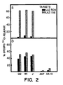
- T11 triggering could activate T c -cell effector function
- QQ is a representative T8+ T c -cell clone specific for the HLA-B7 molecule expressed by Laz 156, an allogeneic Epstein-Barr virus (EBV)-transformed B-lymphoblastoid cell line with the HLA phenotype A2, A3, B7, B40, Dr2, 4.
- EBV Epstein-Barr virus
- Laz 509 is an EBV-transformed B-cell line from an HLA-disparate donor (HLA genotype: A2, A25, B13, Bw38, Cw6, Dr7).
- the monoclonal antibodies anti-T11 1c (IgG2b), anti-T112(IgG2a) and anti-T113(IgG3) were produced as described in Meuer et al. (1984) Cell 36 , 897, and used in ascites form at a 1:100 final dilution. Lysis was measured using a standard Cr-release assay in which T c cells and labelled targets were mixed at a 3:1 ratio in the presence of the indicated concentrations of antibody in a final volume of 0.2 ml. After centrifugation (1,200 g, 10 min), assay plates were incubated for 4h at 37°C before re-centrifugation and removal of aliquots of supernatant for assay of ⁇ radioactivity.
- Anti-T1 Monoclonal antibodies to other cell-surface markers expressed by QQ (anti-T1) or not expressed by QQ (anti-T4 and anti-T6) separately or together have no such effect.
- anti-T11-induced cytotoxicty results from the synergistic effects of particular combinations of anti-T11 monoclonal antibodies (anti-T111 plus anti-T113 or anti-T112 plus anti-T113).
- T11 may be important in T-cell activation, although other factors appear to be involved because the combination of anti-T111 and anti-T112 does not induce activation and because previous studies have shown that anti-T112 or anti-T113 alone cannot induce T-cell proliferation when coupled to Sepharose.
- anti-T11 antibodies clearly results from the action of the antibodies on the T-cell clone because (1) the target cells do not express T11 and (2) the anti-T11 antibodies do not induce lysis of target cells in the absence of T-cells. Furthermore, the effect can also be observed if the T-cells are pretreated with the anti-T11 antibodies.
- T lymphocytes that have acquired cytolytic function during differentiation can be induced to lyse a variety of target cells in an apparently nonspecific fashion by treatment with appropriate combination of anti-T11 antibodies.
- cytotoxicity is not induced in clones whose active genetic programme lacks the cytotoxic machinery.
- T11 triggering induces lymphotoxin (LT) secretion by T-cell clones
- LT lymphotoxin
- the detection of LT-mediated cytotoxicity generally requires sensitive target cell lines, longer assays and prior target cell conditioning.
- NK cells Natural killer cells
- T3-Ti complex the vast majority of human Natural killer (NK) cells, like early thymocytes, lack Ti ⁇ -chain messenger RNA, and consequently express no surface T3-Ti complex.
- the 50 kD T11 glycoprotein is found on the surface of most NK cells.
- anti-T111 antibodies to inhibit antigen-specific cytolysis suggests that the T11 molecule may also be essential for the activation of cytolytic effector function through the T3-Ti complex. This notion is supported by the observation that the combination of anti-T112 plus anti-T113 induces nonspecific cytotoxicity and yet reduces the level of specific killing to that of nonspecific killing (Fig. 1).
- the 5B library was plated at 5-7 ⁇ 103 colonies per 150 mm plate. Replicas were made onto GeneScreen Plus (NEN), amplified with chloramphenicol at 150 mug/ml, denatured, neutralized, washed, dried, and hybridized with redundant oligonucleotide probes. The above amino acid sequence information was used to design these anti-sense oligonucleotide probes:
- Probes were synthesized using cyanoethyl phosphoramidites on an Applied Biosystems model 381A DNA synthesizer. After cleavage from the controlled pore glass column with NH4OH, they were purified by passage over Sep-Pak C18 column (Millipore) and electrophoresis in 20% polyacrylamide gels.
- oligonucleotide probes were end-labelled with T4 polynucleotide kinase (Bethesda Research Labs) using 200 m ⁇ Ci ⁇ -32P-ATP (Amersham) for 0.2 ⁇ g of oligonucleotide.
- Amplified filters were hybridized with 192 redundant 20mers at 40 C in plaque screen buffer (50mM Tris, pH 7.5, 1 M NaCl, 1% SDS, 0.1 sodium pyrophosphate, 0.2% BSA, 0.2% Ficoll 400, 0.2% PYP) for 16 h and washed at 43 C in plaque screen salt (50mM Tris PH 7.5, 1 M NaCl, 1% SDS, 0.2% sodium pyrophosphate) for 30 min. Colonies positive in the first round of screening were picked, replica plated, and rescreened with the 20mer. Identical replicas were also hybridized with the 48-redundant 14mer at 28C and washed at 31C. The 20mers were used to screen the cDNA library.
- plaque screen buffer 50mM Tris, pH 7.5, 1 M NaCl, 1% SDS, 0.1 sodium pyrophosphate, 0.2% BSA, 0.2% Ficoll 400, 0.2% PYP
- plaque screen salt 50mM Tris PH 7.5
- RNA was transferred to nitrocellulose and hybridized to 32P-labeled PB1 DNA in 50% formamide, 5X Denhardt's, 5X SSC, 0.1% SDS and 250 m ⁇ g/ml salmon sperm DNA for 12 h at 42 C.
- the probe was added at 2 ⁇ 106 cpm/ml.
- the filter was washed in 2 ⁇ SSC, 0.1% SDS at 25 C for 20 min and in 0.1% SSC, 0.1% SDS at 50 C for 30 min and exposed to film for 48 h with an intensifying screen.
- PB1 DNA was labelled by random priming as described (Feinberg et al., 1983).
- DNA from REX was restriction digested and electrophoresed in a 0.8% agarose gel. The gel was denatured, neutralized and transferred to nitrocellulose using 10 X SSC. Hybridization and washing procedures were as for northern analysis.
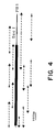
- the 1.6Kb insert of PB1 (solid line) was separated from the plasmid by BamHI digestion and subcloned into the M13 sequencing vector mp18.
- the M13 universal primer was used to derive initial sequence at the 5 ⁇ end. Subsequently, primers of 17 nucleotides were used for dideoxy sequencing. Sequencing reactions were also performed on M13 clones which contained BamH I-Sac I and Sac I-BamH I fragments as shown.
- the open reading frame of the insert is identified by the thick solid bar.
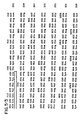
- Fig. 6 there is shown the cDNA and predicted protein sequence of PB1.
- the probable signal peptide ( ⁇ ), the NH2-terminus of the mature protein ( ) and the position of a CNBr cleavage derived fragment ( ) are shown.
- Polyadenylation signals at nucleotides 1103 and 1505 are underlined.
- the last nucleotide before the poly(A) tail in clone PB2 is indicated by an arrowhead at position 1125.
- the complete nucleotide sequence of clone PB1 is 1,522 bases in length and flanked by a poly(A) sequence at its 3 ⁇ end.
- An open reading frame of 1,080 bases begins with an ATG methionine codon and is flanked by 23 nucleotides of 5 ⁇ untranslated sequence and by 419 bases of 3 ⁇ untranslated sequence.
- a polyadenylation signal (AATAAA) is located 18 bases upstream from the beginning of the poly(A) tail (Fig. 6 ).
- PB2 is identical to PB1 except that it lacks four nucleotides present at the 5 ⁇ end of PB1 and has a shorter 3 ⁇ untranslated region.
- a poly(A) tail was noted in PB2 after the nucleotide corresponding to residue 1125 in PB1 (arrow head).
- the bases from positions 1102 to 1103 form part of a Gln codon at amino acid 336 and a stop codon as well as the first five bases of the polyadenylation signal AATAAA (nucleotides 1102-1106) for mRNA corresponding to clone PB2.
- the N-terminal lysine of the mature polypeptide is preceded by a sequence coding for a stretch of 24 hydrophobic amino acids which likely represents the signal sequence required for the T11 precursor to be transported across the endoplasmic reticulum.
- the cDNA sequence predicts three potential N-linked glycosylation sites (Asn-X-Ser/Thr) on the mature protein at amino acid positions 65, 117 and 126.
- An extremely hydrophobic stretch of 25 amino acids characteristic in size and composition of a transmembrane domain is found at positions 186-210. This region is followed by seven basic amino acids within the next ten residues consistent with the notion that this is the start of the intracytoplasmic domain. 21% of residues in the region of amino acids 211-336 are prolines.
- This cDNA sequence predicts a molecular weight for the mature polypeptide backbone of 37,994 daltons.
- Northern analysis using polyA+ RNA or cytoplasmic RNA from a variety of sources indicates that expression of the PB1 and PB2 sequences is T lineage specific and yields two common bands of 1.7 and 1.3Kb.
- each of six human T lineage cells tested including activated T helper clones, thymus derived tumors, normal thymocytes, activated T cells, and resting peripheral blood T lymphocytes expresses the 1.7 and 1.3Kb transcripts.
- non-T lineage cells such as normal peripheral blood B cells and macrophages, an EBV-transformed B lymphoblastoid line, Laz 509, and the non-lymphoid hematopoietic cell lines HL-60 and U937 lack both transcripts.
- PB1 and PB2 inserts ⁇ 400 bases
- PB1 and PB2 cDNAs correspond to the 1.7Kb and 1.3Kb transcripts, respectively.
- an oligonucleotide based on a sequence from the 3 ⁇ untranslated region unique to PB1 selectively hybridizes in northern analysis to sequences in the 1.7Kb site region.
- PB1 homologous sequences are also expressed in murine thymocytes.
- PB1 homologous sequences are also expressed in murine thymocytes.
- only the 1.3Kb molecular weight species is detected in polyA+ mRNA from Balb/cJ thymocytes. It is thus likely that the murine species lacks the second polyadenylation signal in the 3 ⁇ untranslated region of a homologous gene. This result also suggests that a highly homologous T11-related structure exists on murine T lineage cells.
- the T11 molecule is encoded by a single copy gene
- T11 genes present in the human genome were southern analysis was performed with the 32P-labeled PB1 insert.
- the probe hybridizes to a set of bonds of approximately 8 and 7Kb in a BamH I digest of genomic REX T cell DNA, to a single band >14Kb in EcoR I restricted REX DNA, and to a set of 7.4, 6.0 and 0.7Kb fragments in Hind III digested REX DNA.
- Analysis of DNA from granuloctyes gave an identical pattern, indicating that T11 is in the human genome as a single copy gene and, unlike the T cell receptor alpha, beta and gamma genes or the B cell IgH and light chain genes, does not rearrange in lymphoid cells.
- PB1 and PB2 cDNAs encode a functional T11 protein
- COS-1 cells were transfected with PB1 or PB2 plasmid cDNAs or the unrelated IL-3 cDNA.
- COS-1 cells were plated at 4 ⁇ 104 cells/cm2 in 6 well plates in RPMI 1640 (Gibco), 10% fetal calf serum, 0.03% glutamine, 1% penicillin-streptomycin.
- RPMI 1640 Gibco
- 10% fetal calf serum 0.03% glutamine
- penicillin-streptomycin 1% penicillin-streptomycin.
- transfected cells were tested for SRBC binding capacity. Incubation of SRBC with COS-1 cells resulted in no rosette formation. In contrast, after transfection of COS-1 cells with PB2 SRBC rosetting was observed. The SRBC rosette forming capacity was detected in COS-1 cells transfected with either of the two PB cDNAs separately or together.
- the T11 molecule has a hydrophobic signal peptide (L), which is cleaved upon biosynthesis.
- the hydrophilic, 185 amino acid long external domain of the molecule is located outside the cell membrane.
- the hydrophobic 25 amino acid long transmembrane (TM) anchor region is embedded in the T-cell membrane, and the 126 amino acid long cytoplasmic domain of the molecule remains in the cytosol.
- the proline-rich region of this domain is indicated by P-P-P.
- the amino acid numbering corresponds to the mature peptide.
- T11 Cells transfected with PB1, like T-cells which naturally produce T11, do not secrete T11, but rather retain the T11 molecule by means of the transmembrane anchor, with only the external domain exposed.
- Such a truncated molecule can be prepared in a manner analogous to the method by which an anchor-minus IL-2 receptor molecule was prepared by Treiger et al. (1986) J. Immunol. 136 , 4099. The truncated IL-2 receptor was found to be capable of binding to its ligand, IL-2.
- PB1 or PB2 cDNA representing the gene for the entire T11 molecule will be restricted with Pvu II.
- This enzyme uniquely cuts within the 1,522 base pair T11 molecule cDNA insert at base 629, resulting in removal of all transmembrane and intracytoplasmic sequences and seven amino acids of the external domain.
- CTAAGAATTCTTAG 14 base phosphorylated synthetic oligonucleotide
- GAATTC six base recognition sequence for EcoR I
- TTAG four nucleotides complementary to CTAA
- this fragment could be blunted by T4 DNA polymerase to remove the Pst site and then be ligated to the EcoR I linker by T4 ligase. Finally, it could be digested with EcoR I before ligation into the unique EcoR I site of the publicly available PcEXV-1 expression vector.
- Human NK and T c cells can be activated via the T11 molecule in vitro or in vivo and used in the treatment of any medical condition characterized by the presence of unwanted cells.
- the method can be used to turn on the cytotoxicity of T c and NK cells so that they will attack and kill pathogen-infected cells, e.g., cells infected with bacterial, fungal, viral, or protozoan pathogens; or tumor cells, e.g., lung, colorectal, or esophageal cancers.
- pathogen-infected cells e.g., cells infected with bacterial, fungal, viral, or protozoan pathogens
- tumor cells e.g., lung, colorectal, or esophageal cancers.
- the first step in in vitro activation is to obtain resting NK and/or T c cells, either from the patient or a suitable donor. This is typically done by separating out lymphocytes from blood and then, optionally, culturing the lymphocytes in the presence of Interleukin-2 to expand their numbers. (The methods by which lymphocytes are separated out, cultured, treated with Interleukin-2, and used to treat cancer patients are described in detail in Rosenberg et al. (1985) New Eng. J. Med. 313 , 1485.)
- lymphocytes and/or NK cells are incubated with the cytotoxicity-inducing substance for a relatively short time period (e.g., four hours at 30-40°C, in the presence of calcium ions), and subsequently infused into the patient.
- Infusion can be via an arterial or venous catheter or into a large peripheral vein.
- the activating substance can be administered in vivo , most preferably by direct perfusion of the tumor with the substance, e.g., via the hepatic artery to induce cytotoxicity of existing NK and T c Cells.
- the methods of the invention induces cytotoxicity in a way which bypasses the normal T11 recognition mechanism, and enables the resultant activated NK and T c cells to attack and kill their target cells without further treatment.
- NK cells have a broad range of target specificity, while T c cells recognize tumor cells by virtue of tumor-specific antigens.
- treatment according to the invention will activate the totality of NK and T c cells, and be complete in a few hours.
- the T11 protein or its truncated, secreted form, can be used in a variety of diagnostic and therapeutic applications, all of which are based on the binding of T11 to its natural ligand on human lymphocytes and homologous surface structures present on target cells which facilitate T lymphocyte - target cell interactions, which result in target cell lysis or lymphocyte proliferation.
- diagnostic and therapeutic applications all of which are based on the binding of T11 to its natural ligand on human lymphocytes and homologous surface structures present on target cells which facilitate T lymphocyte - target cell interactions, which result in target cell lysis or lymphocyte proliferation.
- the T11 molecule is expressed on the surface of many human T-cell malignancies, e.g., T-cell leukemias and lymphomas.
- autoimmune diseases e.g., rheumatoid arthritis and Systemic Lupus Erythmatosis (SLE)
- SLE Systemic Lupus Erythmatosis
- T11 can be used as an immunogen to produce polyclonal or monoclonal anti-T11 antibodies, using conventional techniques. These antibodies can be labeled with any conventional label, e.g., radioisotopes, and used in conventional immunoassay methods to measure serum T11 levels and thus monitor patients having T-cell-associated diseases. Particularly sensitive ELISA-type assays will employ two anti-T11 antibodies, each to a different antigenic determinant on the surface of T11, in a sandwich format.
- any conventional label e.g., radioisotopes
- anti-T11 antibodies Another use for the anti-T11 antibodies is in the purification of recombinant T11, produced as described above.
- the anti-T11 is coupled to a column and T11-containing media is passed through the column so that the T11 reversibly binds to the T11, after which T11 is eluted in purified form, according to conventional methods.
- the disease states which can be treated using T11 include medical conditions characterized by unwanted activity of the immune system which results in excess T-cell activation, which plays a key role in the amplification of the immune response. These conditions include rheumatoid arthritis; Systemic Lupus Erythmatosis; juvenile onset diabetes; multiple sclerosis; allergic conditions; inflammatory conditions such as eczema, ulcerative colitis, inflammatory bowel disease, and Crohn's disease; and allograft rejection (e.g., rejection of a transplanted heart or kidney). Soluble T11 competes with the surface-bound T11 for its ligand on target cells thus dampening immune response amplification.
- T11 admixed with a Pharmaceutically acceptable carrier substance such as saline, is administered intravenously to a human patient in an effective amount, e.g., 20 ⁇ g to 500 ⁇ g per kg body weight.
- a Pharmaceutically acceptable carrier substance such as saline
- T11 can be administered directly to the site where needed most; for example, T11 can be injected directly into the inflamed joint of a human patient suffering from rhematoid arthritis.
- Plasmid PB1 was deposited in the American Type Culture Collection, Rockville, Maryland on October 1, 1986 and assigned ATCC Accession Number 40268.
- Dana-Farber Cancer Institute, Inc. agrees that the designated culture having ATCC Accession Number 40268 will be maintained throughout the effective life of a patent granted, for 30 years from the date of deposit, or for 5 years after the last request for the deposit after issuance of the patent, whichever is the longer, and it guarantees replenishment of the culture having ATCC Accession Number 40268 at the depository in the event that a deposit becomes nonviable during the effective life of a patent granted, for 30 years from the date of deposit, or for 5 years after the last request for the deposit after issuance of the patent, whichever is longer.
Landscapes
- Health & Medical Sciences (AREA)
- Chemical & Material Sciences (AREA)
- Immunology (AREA)
- Life Sciences & Earth Sciences (AREA)
- Organic Chemistry (AREA)
- General Health & Medical Sciences (AREA)
- Medicinal Chemistry (AREA)
- Proteomics, Peptides & Aminoacids (AREA)
- Biochemistry (AREA)
- Biophysics (AREA)
- Molecular Biology (AREA)
- Genetics & Genomics (AREA)
- Gastroenterology & Hepatology (AREA)
- Toxicology (AREA)
- Zoology (AREA)
- Cell Biology (AREA)
- Peptides Or Proteins (AREA)
- Preparation Of Compounds By Using Micro-Organisms (AREA)
- Medicines Containing Material From Animals Or Micro-Organisms (AREA)
- Micro-Organisms Or Cultivation Processes Thereof (AREA)
Applications Claiming Priority (4)
| Application Number | Priority Date | Filing Date | Title |
|---|---|---|---|
| US90641386A | 1986-09-11 | 1986-09-11 | |
| US906413 | 1986-09-11 | ||
| US93287186A | 1986-11-18 | 1986-11-18 | |
| US932871 | 1986-11-18 |
Publications (2)
| Publication Number | Publication Date |
|---|---|
| EP0260880A2 true EP0260880A2 (de) | 1988-03-23 |
| EP0260880A3 EP0260880A3 (de) | 1990-03-07 |
Family
ID=27129460
Family Applications (1)
| Application Number | Title | Priority Date | Filing Date |
|---|---|---|---|
| EP87308016A Withdrawn EP0260880A3 (de) | 1986-09-11 | 1987-09-10 | Verfahren zur Auslösung der Zytotoxizität |
Country Status (2)
| Country | Link |
|---|---|
| EP (1) | EP0260880A3 (de) |
| DE (1) | DE260880T1 (de) |
Cited By (11)
| Publication number | Priority date | Publication date | Assignee | Title |
|---|---|---|---|---|
| EP0329363A1 (de) * | 1988-02-18 | 1989-08-23 | Schering Biotech Corporation | Markierer der T-Zelle Aktivierung |
| FR2636953A1 (fr) * | 1988-09-26 | 1990-03-30 | Roussy Inst Gustave | Nouveaux polypeptides produits par des lymphocytes humains, genes codant pour ces polypeptides et applications pharmaceutiques et biologiques |
| WO1990013574A1 (en) * | 1989-05-05 | 1990-11-15 | The Australian National University | A cytolytic t-cell activating factor derived from b-lymphocytes |
| US5136022A (en) * | 1988-02-18 | 1992-08-04 | Schering Corporation | T cell activation markers |
| WO1993006852A3 (en) * | 1991-10-07 | 1993-07-22 | Biogen Inc | Methods of improving allograft or xenograft tolerance by administration of an lfa-3 or cd2 binding protein |
| EP0637593A1 (de) * | 1993-08-02 | 1995-02-08 | MERCK PATENT GmbH | Bispezifische Auslösemoleküle, die das Lymphozytantigen CD2 und Tumorantigene erkennen |
| WO1996013584A1 (en) * | 1994-11-01 | 1996-05-09 | Targeted Genetics Corporation | Chimeric receptors for the generation of selectively-activatable th-independent cytotoxic t cells |
| US6984382B1 (en) | 1994-05-02 | 2006-01-10 | Bernd Groner | Bifunctional protein, preparation and use |
| US7323171B2 (en) | 1991-10-07 | 2008-01-29 | Astellas Us Llc | Methods of treating skin conditions using inhibitors of the CD2/LFA-3 interaction |
| US7662921B2 (en) | 2004-05-07 | 2010-02-16 | Astellas Us Llc | Methods of treating viral disorders |
| US7858095B2 (en) | 2001-07-24 | 2010-12-28 | Astellas Us Llc | Method for treating or preventing sclerotic disorders using CD-2 binding agents |
Family Cites Families (1)
| Publication number | Priority date | Publication date | Assignee | Title |
|---|---|---|---|---|
| US4381292A (en) * | 1980-11-14 | 1983-04-26 | The Board Of Trustees Of The Leland Stanford Jr. University | Anti-human T-lymphocyte monoclonal antibody |
-
1987
- 1987-09-10 EP EP87308016A patent/EP0260880A3/de not_active Withdrawn
- 1987-09-10 DE DE1987308016 patent/DE260880T1/de active Pending
Cited By (17)
| Publication number | Priority date | Publication date | Assignee | Title |
|---|---|---|---|---|
| EP0329363A1 (de) * | 1988-02-18 | 1989-08-23 | Schering Biotech Corporation | Markierer der T-Zelle Aktivierung |
| WO1989007646A1 (en) * | 1988-02-18 | 1989-08-24 | Schering Biotech Corporation | T cell activation markers |
| US5001230A (en) * | 1988-02-18 | 1991-03-19 | Schering Corporation | T cell activation markers |
| US5136022A (en) * | 1988-02-18 | 1992-08-04 | Schering Corporation | T cell activation markers |
| FR2636953A1 (fr) * | 1988-09-26 | 1990-03-30 | Roussy Inst Gustave | Nouveaux polypeptides produits par des lymphocytes humains, genes codant pour ces polypeptides et applications pharmaceutiques et biologiques |
| WO1990003394A3 (fr) * | 1988-09-26 | 1990-07-12 | Roussel Uclaf | Nouvelles lymphokines, sequences d'adn codant pour ces lymphokines et compositions pharmaceutiques contenant ces lymphokines |
| EP0367641A3 (en) * | 1988-09-26 | 1990-08-29 | Roussel-Uclaf | Lymphokines, dna sequences encoding these lymphokines and pharmaceutical compositions containing these lymphokines |
| WO1990013574A1 (en) * | 1989-05-05 | 1990-11-15 | The Australian National University | A cytolytic t-cell activating factor derived from b-lymphocytes |
| WO1993006852A3 (en) * | 1991-10-07 | 1993-07-22 | Biogen Inc | Methods of improving allograft or xenograft tolerance by administration of an lfa-3 or cd2 binding protein |
| US7323171B2 (en) | 1991-10-07 | 2008-01-29 | Astellas Us Llc | Methods of treating skin conditions using inhibitors of the CD2/LFA-3 interaction |
| EP0637593A1 (de) * | 1993-08-02 | 1995-02-08 | MERCK PATENT GmbH | Bispezifische Auslösemoleküle, die das Lymphozytantigen CD2 und Tumorantigene erkennen |
| US5798229A (en) * | 1993-08-02 | 1998-08-25 | Merck Patent Gesellschaft Mit Beschrankter Haftung | Bispecific molecules recognizing lymphocyte antigen CD2 and tumor antigens |
| US6984382B1 (en) | 1994-05-02 | 2006-01-10 | Bernd Groner | Bifunctional protein, preparation and use |
| WO1996013584A1 (en) * | 1994-11-01 | 1996-05-09 | Targeted Genetics Corporation | Chimeric receptors for the generation of selectively-activatable th-independent cytotoxic t cells |
| US6083751A (en) * | 1994-11-01 | 2000-07-04 | Targeted Genetics Corporation | Chimeric receptors for the generation of selectively-activatable TH-independent cytotoxic T cells |
| US7858095B2 (en) | 2001-07-24 | 2010-12-28 | Astellas Us Llc | Method for treating or preventing sclerotic disorders using CD-2 binding agents |
| US7662921B2 (en) | 2004-05-07 | 2010-02-16 | Astellas Us Llc | Methods of treating viral disorders |
Also Published As
| Publication number | Publication date |
|---|---|
| DE260880T1 (de) | 1988-11-03 |
| EP0260880A3 (de) | 1990-03-07 |
Similar Documents
| Publication | Publication Date | Title |
|---|---|---|
| Gilks et al. | Progression of interleukin-2 (IL-2)-dependent rat T cell lymphoma lines to IL-2-independent growth following activation of a gene (Gfi-1) encoding a novel zinc finger protein | |
| AU634464B2 (en) | Dna sequences, recombinant dna molecules and processes for producing lymphocyte function associated antigen-3 | |
| CA2308765C (en) | The use of an ox-2 protein or nucleic acid in immunomodulation | |
| EP0734442A1 (de) | Diagnose von Krebs - Metastasen durch das MTS - 1 Gen | |
| KR100620334B1 (ko) | 헤르페스 심플렉스 바이러스 유입 매개체에 대한 리간드및 사용 방법 | |
| CA2293735A1 (en) | Lag-3 splice variants | |
| AU676680B2 (en) | Isolation, characterization, and use of the human beta subunit of the high affinity receptor for immunoglobulin | |
| Giblin et al. | A secreted form of the human lymphocyte cell surface molecule CD8 arises from alternative splicing. | |
| AU642130B2 (en) | DNA sequences, recombinant DNA molecules and processes for producing PI-linked lymphocyte function associated antigen-3 | |
| EP0260880A2 (de) | Verfahren zur Auslösung der Zytotoxizität | |
| US6040426A (en) | Human Th2 specific protein | |
| US7138243B2 (en) | NTB-A, a surface molecule involved in natural killer cells activity | |
| JP3104187B2 (ja) | インターフエロン―ガンマ結合蛋白 | |
| AU2002325333A1 (en) | NTB-A, a surface molecule involved in natural killer cells activity | |
| US5550055A (en) | Recombinant DNA-produced T11 and fragments thereof | |
| US20070148686A1 (en) | Protein present at the surface of hematopoietic stem cells of the lymphoid line and of nk cells, and uses thereof | |
| EP0436400A1 (de) | Intrazelluläres Antigen in einer Subpopulation von CD8+T-Lymphozyten und ein monoklonaler Antikörper spezifisch dafür | |
| JPH04500603A (ja) | クローン化腎炎抗原 | |
| US5830754A (en) | Recombinant DNA-produced T11 and fragments thereof | |
| US6277971B1 (en) | Nef-attachable protein, DNA encoding the protein and a monoclonal antibody against said protein | |
| JPS63146823A (ja) | 細胞毒性の作用 | |
| YODOI et al. | IL-2 receptor gene regulation by cytokines; Role of ATL-derived factor (s)(ADFs) in ATL | |
| JPWO1996033217A1 (ja) | 細胞接着タンパク質、それを含む免疫抑制剤、ならびにそれにより誘導された細胞を含む免疫抑制剤 | |
| HK1009291B (en) | Dna sequences, recombinant dna molecules and processes for producing lymphocyte function associated antigen-3 |
Legal Events
| Date | Code | Title | Description |
|---|---|---|---|
| PUAI | Public reference made under article 153(3) epc to a published international application that has entered the european phase |
Free format text: ORIGINAL CODE: 0009012 |
|
| AK | Designated contracting states |
Kind code of ref document: A2 Designated state(s): BE DE FR GB IT NL SE |
|
| ITCL | It: translation for ep claims filed |
Representative=s name: BARZANO' E ZANARDO MILANO S.P.A. |
|
| EL | Fr: translation of claims filed | ||
| TCNL | Nl: translation of patent claims filed | ||
| DET | De: translation of patent claims | ||
| PUAL | Search report despatched |
Free format text: ORIGINAL CODE: 0009013 |
|
| AK | Designated contracting states |
Kind code of ref document: A3 Designated state(s): BE DE FR GB IT NL SE |
|
| 17P | Request for examination filed |
Effective date: 19900813 |
|
| 17Q | First examination report despatched |
Effective date: 19901128 |
|
| STAA | Information on the status of an ep patent application or granted ep patent |
Free format text: STATUS: THE APPLICATION IS DEEMED TO BE WITHDRAWN |
|
| 18D | Application deemed to be withdrawn |
Effective date: 19911231 |
|
| RIN1 | Information on inventor provided before grant (corrected) |
Inventor name: RICHARDSON, NEIL Inventor name: CHANG, HSIU-CHING Inventor name: SAYRE, PETER Inventor name: SILICIANO, ROBERT F. Inventor name: REINHERZ, ELLIS L. |