EP0249983B2 - Reagent kit for use in sandwich immunoassay - Google Patents
Reagent kit for use in sandwich immunoassay Download PDFInfo
- Publication number
- EP0249983B2 EP0249983B2 EP87108799A EP87108799A EP0249983B2 EP 0249983 B2 EP0249983 B2 EP 0249983B2 EP 87108799 A EP87108799 A EP 87108799A EP 87108799 A EP87108799 A EP 87108799A EP 0249983 B2 EP0249983 B2 EP 0249983B2
- Authority
- EP
- European Patent Office
- Prior art keywords
- igg
- afp
- labeled antibody
- solid
- reaction solution
- Prior art date
- Legal status (The legal status is an assumption and is not a legal conclusion. Google has not performed a legal analysis and makes no representation as to the accuracy of the status listed.)
- Expired - Lifetime
Links
- 238000003018 immunoassay Methods 0.000 title claims description 41
- 239000003153 chemical reaction reagent Substances 0.000 title description 2
- 239000007790 solid phase Substances 0.000 claims description 75
- 229940027941 immunoglobulin g Drugs 0.000 claims description 74
- 238000006243 chemical reaction Methods 0.000 claims description 71
- 241000283707 Capra Species 0.000 claims description 65
- 241000283973 Oryctolagus cuniculus Species 0.000 claims description 63
- 210000002966 serum Anatomy 0.000 claims description 41
- 108090000790 Enzymes Proteins 0.000 claims description 39
- 102000004190 Enzymes Human genes 0.000 claims description 39
- 229940088598 enzyme Drugs 0.000 claims description 39
- 239000000427 antigen Substances 0.000 claims description 30
- 102000036639 antigens Human genes 0.000 claims description 30
- 108091007433 antigens Proteins 0.000 claims description 30
- 241000700199 Cavia porcellus Species 0.000 claims description 28
- 108010015776 Glucose oxidase Proteins 0.000 claims description 14
- 239000004366 Glucose oxidase Substances 0.000 claims description 14
- 229940116332 glucose oxidase Drugs 0.000 claims description 14
- 235000019420 glucose oxidase Nutrition 0.000 claims description 14
- 239000012634 fragment Substances 0.000 claims description 9
- 239000000243 solution Substances 0.000 description 99
- 238000003556 assay Methods 0.000 description 51
- 230000000052 comparative effect Effects 0.000 description 42
- 239000004793 Polystyrene Substances 0.000 description 35
- 229920002223 polystyrene Polymers 0.000 description 35
- 230000009871 nonspecific binding Effects 0.000 description 32
- 102100023635 Alpha-fetoprotein Human genes 0.000 description 27
- -1 potassium ferricyanide Chemical compound 0.000 description 19
- 239000012064 sodium phosphate buffer Substances 0.000 description 19
- 230000002494 anti-cea effect Effects 0.000 description 17
- KCXVZYZYPLLWCC-UHFFFAOYSA-N EDTA Chemical compound OC(=O)CN(CC(O)=O)CCN(CC(O)=O)CC(O)=O KCXVZYZYPLLWCC-UHFFFAOYSA-N 0.000 description 15
- HWYHZTIRURJOHG-UHFFFAOYSA-N luminol Chemical compound O=C1NNC(=O)C2=C1C(N)=CC=C2 HWYHZTIRURJOHG-UHFFFAOYSA-N 0.000 description 15
- 108091003079 Bovine Serum Albumin Proteins 0.000 description 12
- MHAJPDPJQMAIIY-UHFFFAOYSA-N Hydrogen peroxide Chemical compound OO MHAJPDPJQMAIIY-UHFFFAOYSA-N 0.000 description 12
- PXIPVTKHYLBLMZ-UHFFFAOYSA-N Sodium azide Chemical compound [Na+].[N-]=[N+]=[N-] PXIPVTKHYLBLMZ-UHFFFAOYSA-N 0.000 description 12
- 229940098773 bovine serum albumin Drugs 0.000 description 12
- 230000035945 sensitivity Effects 0.000 description 11
- 238000001514 detection method Methods 0.000 description 8
- 230000027455 binding Effects 0.000 description 7
- 239000000872 buffer Substances 0.000 description 7
- 239000012847 fine chemical Substances 0.000 description 7
- 241001465754 Metazoa Species 0.000 description 6
- 239000008351 acetate buffer Substances 0.000 description 6
- 238000005119 centrifugation Methods 0.000 description 6
- 238000002967 competitive immunoassay Methods 0.000 description 6
- 238000000034 method Methods 0.000 description 6
- BVKZGUZCCUSVTD-UHFFFAOYSA-L Carbonate Chemical compound [O-]C([O-])=O BVKZGUZCCUSVTD-UHFFFAOYSA-L 0.000 description 5
- WQZGKKKJIJFFOK-GASJEMHNSA-N Glucose Natural products OC[C@H]1OC(O)[C@H](O)[C@@H](O)[C@@H]1O WQZGKKKJIJFFOK-GASJEMHNSA-N 0.000 description 5
- 230000000694 effects Effects 0.000 description 5
- 239000008103 glucose Substances 0.000 description 5
- 239000000203 mixture Substances 0.000 description 5
- 102000018071 Immunoglobulin Fc Fragments Human genes 0.000 description 4
- 108010091135 Immunoglobulin Fc Fragments Proteins 0.000 description 4
- ZMXDDKWLCZADIW-UHFFFAOYSA-N N,N-Dimethylformamide Chemical compound CN(C)C=O ZMXDDKWLCZADIW-UHFFFAOYSA-N 0.000 description 4
- 239000012506 Sephacryl® Substances 0.000 description 4
- PEEHTFAAVSWFBL-UHFFFAOYSA-N Maleimide Chemical compound O=C1NC(=O)C=C1 PEEHTFAAVSWFBL-UHFFFAOYSA-N 0.000 description 3
- 229920005654 Sephadex Polymers 0.000 description 3
- 239000012507 Sephadex™ Substances 0.000 description 3
- 210000004369 blood Anatomy 0.000 description 3
- 239000008280 blood Substances 0.000 description 3
- 239000004615 ingredient Substances 0.000 description 3
- 238000005259 measurement Methods 0.000 description 3
- PVGATNRYUYNBHO-UHFFFAOYSA-N (2,5-dioxopyrrolidin-1-yl) 4-(2,5-dioxopyrrol-1-yl)butanoate Chemical compound O=C1CCC(=O)N1OC(=O)CCCN1C(=O)C=CC1=O PVGATNRYUYNBHO-UHFFFAOYSA-N 0.000 description 2
- WTDHULULXKLSOZ-UHFFFAOYSA-N Hydroxylamine hydrochloride Chemical compound Cl.ON WTDHULULXKLSOZ-UHFFFAOYSA-N 0.000 description 2
- 102000057297 Pepsin A Human genes 0.000 description 2
- 108090000284 Pepsin A Proteins 0.000 description 2
- 108010026331 alpha-Fetoproteins Proteins 0.000 description 2
- BFNBIHQBYMNNAN-UHFFFAOYSA-N ammonium sulfate Chemical class N.N.OS(O)(=O)=O BFNBIHQBYMNNAN-UHFFFAOYSA-N 0.000 description 2
- UFULAYFCSOUIOV-UHFFFAOYSA-N cysteamine Chemical compound NCCS UFULAYFCSOUIOV-UHFFFAOYSA-N 0.000 description 2
- 239000012153 distilled water Substances 0.000 description 2
- 230000036046 immunoreaction Effects 0.000 description 2
- 239000000463 material Substances 0.000 description 2
- 229960003151 mercaptamine Drugs 0.000 description 2
- 230000036963 noncompetitive effect Effects 0.000 description 2
- 229940111202 pepsin Drugs 0.000 description 2
- 239000002244 precipitate Substances 0.000 description 2
- 239000012086 standard solution Substances 0.000 description 2
- XLYOFNOQVPJJNP-UHFFFAOYSA-N water Chemical compound O XLYOFNOQVPJJNP-UHFFFAOYSA-N 0.000 description 2
- HCILGDWKPMTTSN-UHFFFAOYSA-N 2-(2,5-dioxopyrrolidin-1-yl)-4-(2,5-dioxopyrrol-1-yl)butanoic acid Chemical compound O=C1CCC(=O)N1C(C(=O)O)CCN1C(=O)C=CC1=O HCILGDWKPMTTSN-UHFFFAOYSA-N 0.000 description 1
- OBYNJKLOYWCXEP-UHFFFAOYSA-N 2-[3-(dimethylamino)-6-dimethylazaniumylidenexanthen-9-yl]-4-isothiocyanatobenzoate Chemical compound C=12C=CC(=[N+](C)C)C=C2OC2=CC(N(C)C)=CC=C2C=1C1=CC(N=C=S)=CC=C1C([O-])=O OBYNJKLOYWCXEP-UHFFFAOYSA-N 0.000 description 1
- BTBUEUYNUDRHOZ-UHFFFAOYSA-N Borate Chemical compound [O-]B([O-])[O-] BTBUEUYNUDRHOZ-UHFFFAOYSA-N 0.000 description 1
- 238000010521 absorption reaction Methods 0.000 description 1
- 150000008064 anhydrides Chemical class 0.000 description 1
- 230000002860 competitive effect Effects 0.000 description 1
- 230000000593 degrading effect Effects 0.000 description 1
- 230000001419 dependent effect Effects 0.000 description 1
- 238000010586 diagram Methods 0.000 description 1
- MHMNJMPURVTYEJ-UHFFFAOYSA-N fluorescein-5-isothiocyanate Chemical compound O1C(=O)C2=CC(N=C=S)=CC=C2C21C1=CC=C(O)C=C1OC1=CC(O)=CC=C21 MHMNJMPURVTYEJ-UHFFFAOYSA-N 0.000 description 1
- 238000012986 modification Methods 0.000 description 1
- 230000004048 modification Effects 0.000 description 1
- 230000002285 radioactive effect Effects 0.000 description 1
- 238000011946 reduction process Methods 0.000 description 1
- 238000000926 separation method Methods 0.000 description 1
- 238000002791 soaking Methods 0.000 description 1
- 238000001179 sorption measurement Methods 0.000 description 1
Images


Classifications
-
- G—PHYSICS
- G01—MEASURING; TESTING
- G01N—INVESTIGATING OR ANALYSING MATERIALS BY DETERMINING THEIR CHEMICAL OR PHYSICAL PROPERTIES
- G01N33/00—Investigating or analysing materials by specific methods not covered by groups G01N1/00 - G01N31/00
- G01N33/48—Biological material, e.g. blood, urine; Haemocytometers
- G01N33/50—Chemical analysis of biological material, e.g. blood, urine; Testing involving biospecific ligand binding methods; Immunological testing
- G01N33/53—Immunoassay; Biospecific binding assay; Materials therefor
- G01N33/543—Immunoassay; Biospecific binding assay; Materials therefor with an insoluble carrier for immobilising immunochemicals
- G01N33/54393—Improving reaction conditions or stability, e.g. by coating or irradiation of surface, by reduction of non-specific binding, by promotion of specific binding
-
- G—PHYSICS
- G01—MEASURING; TESTING
- G01N—INVESTIGATING OR ANALYSING MATERIALS BY DETERMINING THEIR CHEMICAL OR PHYSICAL PROPERTIES
- G01N33/00—Investigating or analysing materials by specific methods not covered by groups G01N1/00 - G01N31/00
- G01N33/48—Biological material, e.g. blood, urine; Haemocytometers
- G01N33/50—Chemical analysis of biological material, e.g. blood, urine; Testing involving biospecific ligand binding methods; Immunological testing
- G01N33/53—Immunoassay; Biospecific binding assay; Materials therefor
- G01N33/563—Immunoassay; Biospecific binding assay; Materials therefor involving antibody fragments
Definitions
- This invention relates to sandwich immunoassays utilizing immunoreaction for measuring an extremely small quantity of an ingredient included in blood or other organic samples and, more particularly to an improved combination of reagents for use in such sandwich immunoassays to increase the accuracy, of measurement of the ingredient.
- Immunoassays have been used widely for measuring an extremely small quantity of an ingredient included in blood or other organic samples by utilizing immunoreaction or antigen-antibody reaction. Such immunoassays are generally of the two types; the “competitive immunoassay” and the “non-competitive immunoassay”.
- the competitive immunoassay employs a predetermined quantity of labeled antigen when binds to a predetermined quantity of antibody in a competitive manner with target antigen. The quantity of the target antigen is measured from the quantity of the labeled antigen binding to the antibody or the quantity of the labeled antigen which does not bind to the antibody.
- the labeled antigen is required to have a high concentration for its reaction to the antibody.
- the competitive immunoassay exhibits a poor measurement sensitivity as low as about a twentieth of the quantity of the labeled antigen.
- the non-competitive immunoassay is represented by a sandwich immunoassay which employs a solid phase coated on its surface with an excessive quantity of insolubilized antibody to trap a target antigen to be measured.
- the solid phase is washed and then incubated with an excessive amount of labeled antibody so that the labeled antibody can react with the trapped target antigen.
- the excessive labeled antibody is washed out away from the solid phase.
- the quantity of the target antigen is measured from the quantity of the label provided on the labeled antibody binding to the antigen.
- the sandwich immunoassay can provide a high measurement sensitivity, there is a tendency of non-specific binding of the labeled antibody to the solid phase, causing a considerable reduction in the sensitivity and measurable range of the sandwich immunoassay.
- a main object of the invention is to reduce the degree of non-specific binding of the labeled antibody to the solid phase so as to increase the accuracy of the sandwich immunoassay.
- kits for use in a sandwich immunoassay employing labeled and solid-phase antibodies reactive with an antigen in the presence of a reaction solution to bind the antigen between the labeled and solid-phase antibodies, said kit containing a solid-phase antibody bound to a solid phase, a labeled antibody having a label enzyme bound to the antibody and a reaction solution, and being characterized in that the labeled antibody is originated from a rabbit, the solid-phase antibody is originated from a goat and the label enzyme is glucose oxidase.
- a target antigen is assayed by a sandwich enzyme immunoassay (EIA) technique employing first and second antibodies reactive in a specific manner with the target antigen in the presence of a reaction solution to trap the target antigen between the first and second antibodies.
- the antibodies are obtained from serum drawn from a selected animal into which the target antigen is injected.
- the first antibody is referred to as a solid-phase antibody which is coated on a solid phase such as polystyrene balls.
- the second antibody is referred to as a labeled antibody which has a label enzyme bound thereon.
- the label enzyme is glucose oxidase.
- a chemiluminescence technique is. used to measure the quantity of the label enzyme which corresponds to the quantity of the target antigen.
- reaction solution A 0.056 mol/l sodium phosphate buffer (pH 6.3) containing 0.1% sodium azide, 0.2% bovine serum albumin (BSA) and 0.337% NaCI. This reaction solution is referred to as reaction solution A.
- AFP is injected six times under the back skin of selected one of animals including rabbit, goat and the like at intervals of one week. On the seventh week, a booster is injected into the selected animal. After five days, a proper amount of blood is drawn from the selected animal.
- Anti-AFP immunoglobulin G (IgG) is purified from the drawn serum in the following manner. First of all, 2 ml saturated ammonium sulfate is dripped into 2 ml of serum. The resulting solution is agitated slowly at a temperature of 4°C for two or three hours. The resulting cloudy solution is then placed in a centrifugation tube. Centrifugation is performed at a temperature of 4°C with a contrifugal separator rotating at a speed of 3000 rpm for 15 minutes.
- the supernatuant is removed from the centrifugation tube, whereas the precipitate is dissolved with 2 ml of 0.1 mol/l sodium phosphate buffer (pH 6.0) containing 5 mmol/l ethylenediaminetetraacetic acid (EDTA).
- the resulting solution is eluted at a flow rate of 6 ml/hour on s Sephacryl S-300 (a trademark of Pharmacia Fine Chemicals, Sweden) column (1 x 90 cm) equilibrated with 0.1 mol/l sodium phosphate buffer (pH 6.0) containing 5 mmol/l ethylenediaminetetraacetic acid (EDTA).
- the eluted solution is divided into 1 ml of samples.
- the lgG fractions, which absorb light at 280 nm, are gathered from the respective samples.
- the resulting anti-AFP IgG is coated through physical absorption on a solid phase by soaking a polystyrene ball (6.5 mm in diameter) available from Ichiko Co., Japan in 0.1 mol/l sodium phosphate buffer (pH 7.5) containing 0.1 mg/ml anti-AFP IgG all the night through at a temperature of 4 °C.
- the IgG-coated polystyrene ball is washed three times in 0.1 mol I sodium phosphate buffer (pH 7.0) and then washed three times with 0.01 mol/l sodium phosphate buffer (pH 7.0) containing 0.1 mol/l NaCI 0.1% bovine serum albumin (BSA) and 0.1% NaN 3 .
- the washed polystyrene ball is stored at a temperature of 4 °C in 0.01 molil sodium phosphate buffer (pH 7.0) containing 0.1 mol/l NaCI, 0.1% bovine serum albumin (BSA) and 0.1% NaN 3 .
- pH 7.0 0.01 molil sodium phosphate buffer
- BSA bovine serum albumin
- the labeled antibody, anti-AFP IgG-GOD is prepared through the following three steps:
- the labeled antibody (anti-AFP IgG-GOD) is bound through the antigen (AFP) to the solid-phase antibody (anti-AFP IgG) in the presence of the reaction solution A (0.056 mol/l sodium phosphate buffer (pH 6.3) containing 0.1% sodium azide, 0.2% bovine serum albumin (BSA) and 0.337% NaCI) in the following manner.
- the anti-AFP IgG-coated polystyrene ball is incubated at room temperature with AFP (antigen) diluted with the reaction solution A in an assay tube for six hours. The polystylene ball is then washed three times with distilled water.
- the washed polystyrene ball is incubated at room temperature with anti-AFP IgG-GOD (labeled antibody) in the reaction solution A all the night through.
- the polystyrene ball is then washed again with distilled water and transferred into another clean assay tube.
- a chemiluminescent technique is used to determine the quantity of the label enzyme; that is, the quantity of the antigen.
- 0.3 ml of 0.01 mol/l acetate buffer (pH 5.1) containing mol/l glucose is added to the assay tube.
- the assay tube is held stationary at a temperature of 37°C for two hours.
- the resulting solution is taken to prepare a 0.1 ml sample.
- the sample is added with 0.5 ml of 0.2 mol/l carbonate buffer (pH 9.8) containing 2 x 10 -7 mol/l luminol and then 0.5 ml of 6 x 10 -3 mol/l potassium ferricyanide.
- the concentration of the resulting hydrogen peroxide is measured from the light emitted upon chemiluminescent reaction of the luminol and potassium ferricyanide and the hydrogen peroxide by a Luminometer UPD-8000 (a trademark of Meidensha Electric Mfg., Co., Japan) which starts its operation 15 second after the luminol and potassium ferricyanide are added to the assay tube and counts the light emitted for 30 seconds.
- a Luminometer UPD-8000 (a trademark of Meidensha Electric Mfg., Co., Japan) which starts its operation 15 second after the luminol and potassium ferricyanide are added to the assay tube and counts the light emitted for 30 seconds.
- antigen has been described in connection with ⁇ -fetoprotein (AFP), it is to be noted that it may be anti-carcinoembryonic antigen (CEA) or the like.
- Anti-CEA IgG is purified from serum drawn from selected one of animals including, rabbit, goat and the like into which CEA is injected six times at intervals of one week and a booster is injected on the seventh week.
- EIA sandwich enzyme immunoassay
- the inventors found that the degree of non-specific binding of the labeled antibody to the solid phase was reduced when the labeled antibody was prepared from serum originated from rabbit, the solid-phase antibody was prepared from serum originated from goat and the label enzyme was glucose oxidase. A large number of tests were conducted to prove the effective combinations of the solid-phase and labeled antibodies.
- the degree of non-specific binding of the labeled antibody to the solid phase was determined.
- an IgG-coated polystyrene ball was incubated at room temperature with IgG-GOD diluted hundredfold with 0.3 ml of reaction solution A in an assay tube all the night through. The polystyrene ball was washed three times with the reaction solution A and then transferred into another clean assay tube. Following this, 0.3 ml of 0.01 mol/l acetate buffer (pH 5.1) containing 0.5 mol/l glucose was added to the assay tube. The assay tube was held stationary at a temperature of 37°C for two hours. The resulting solution was taken to prepare a 0.1 ml sample.
- the sample was added with 0.5 ml of 0.2 mol/l carbonate buffer (pH 9.8) containing 2x10 -7 mol/l luminol and then with 0.5 ml of 6x10 -3 mol/l potassium ferricyanide.
- the light emitted upon chemiluminescent reaction of the luminol and potassium ferricyanide and the resulting hydrogen peroxide was measured by a Luminometer UPD-8000 which started its operation 15 seconds after the luminol and potassium ferricyanide were added to the assay tube and counted the light emitted for 30 seconds.
- the degree of non-specific binding of the labeled antibody to the solid phase was represented by the non-specific bonding ratio given as C1/C2 x 100 (%) where C1 is the number of counts measured for the labeled antibody binding to the polystyrene ball and C2 is the number of counts corresponding to the whole amount of the labeled antibody used in the assay tube.
- Example 1 The labeled antibody used was rabbit anti-AFP lgG-GOD and the solid-phase antibody used was goat anti-AFP IgG.
- the rabbit anti-AFP IgG was Nordic Co., Lot No. 16-184.
- a goat anti-AFP IgG-coated polystyrene ball was incubated with the rabbit anti-AFP IgG-GOD in the presence of the reaction solution A mixed with guinea pig serum.
- Example 2 The labeled antibody used was normal rabbit IgG-GOD and the solid-phase antibody used was normal goat IgG.
- the normal rabbit IgG was Miles Co., Lot No. 0019.
- the normal goat IgG was Cappel Co., Lot No. 24511.
- a normal goat IgG-coated polystyrene ball was incubated with the normal rabbit IgG-GOD in the presence of the reaction solution A mixed with guinea pig serum.
- the non-specific binding percentage ranges from 0.078% to 0.190% for combinations where the labeled antibody is originated from rabbit and the solid-phase antibody is originated from rabbit or where the labeled antibody is originated from goat and the solid-phase antibody is originated from goat.
- the non-specific binding percentage ranges from 0.015% to 0.017% for combinations where the labeled antibody is originated from rabbit and the solid-phase antibody is originated from goat. It can be seen from the test results that the degree of non-specific binding of the labeled antibody to the solid phase can be reduced to a remarkable extent when the labeled antibody is originated from rabbit and the solid-phase antibody is originated from goat.
- the sensitivity and measurable range of such sandwich enzyme immunoassay was determined.
- an anti-AFP IgG-coated polystyrene ball was incubated at room temperature with 0.1 ml of AFP standard solution diluted with 0.2 ml of reaction solution A in an assay tube for six hours.
- the polystyrene ball was washed three times with the reaction solution A.
- the washed polystyrene ball was incubated with 0.1 ml of anti-AFP IgG-GOD labeled antibody diluted at a proper ratio with the reaction solution A.
- the assay tube was held stationary at room teperature all the night through.
- the polystyrene ball was then washed three times with the reaction solution A and transferred into another clean assay tube. Following this, 0.3 ml of 0.01 mol/l acetate buffer (pH 5.1) containing 0.5 mol/l glucose was added to the assay tube. The assay tube was held stationary at a temperature of 37°C for two hours. The resulting solution was taken to prepare a 0.1 ml sample. The sample was added with 0.5 ml of 0.2 mol/l carbonate buffer (pH 9.8) containing 2x10 -7 mol/l luminol and then with 0.5 ml of 6x10 -3 mol/l potassium ferricyanide.
- the light emitted upon chemiluminescent reaction of the luminol and potassium ferricyanide and the resulting hydrogen peroxide was measured by a Luminometer UPD-8000 which started its operation 15 seconds after the luminol and potassium ferricyanide were added to the assay tube and counted the light emitted for 30 seconds. Similar procedures are repeated for normal rabbit IgG antibodies and normal goat IgG antibodies.
- Example 1 relates to Example 1 and Comparative Examples 1 and 2. It can be seen from these test results that the sensitivity and measurable range of the sandwich enzyme immunoassay are superior when the labeled antibody is originated from rabbit and the solid-phase antibody is originated from goat to those obtained when the labeled antibody is originated from rabbit and the solid-phase antibody is originated from rabbit or when the labeled antibody is originated from goat and the solid-phase antibody is originated from goat.
- Sandwich enzyme immunoassay was repeated ten times within a day (within assay) using the antibody in Example 1 and Comparative Examples 1 and 2. The results are shown in Tables 2 and 3. Similarly, sandwich enzyme immunoassay was repeated six times on different six days (between assay) using the antibody in Example 1 and Comparative Examples 1 and 2. The results are shown in Tables 4 and 5. TABLE 4 Example No. Number of Assay Repetitions AFP (ng/ml) Variation Coefficient (%) Average Value ⁇ Standard Deviation 1 6 30 ⁇ 2.1 7.1 1 6 142 ⁇ 6.1 4.3 TABLE 5 Comparative Example No.
- the IgG structure can be divided into an antigen-binding F(ab') 2 fragment and a crystallizable (Fc) fragment when it is held at a temperature of 37°C in the presence of pepsin for 18 hours.
- the Fc fragment has a portion P4 for adsorption to a cell.
- the Fc fragment has no ability of binding to antigen and is crystallized at low temperature.
- the F(ab') 2 fragment can be divided under a reduction process, for example, in the presence of mercaptoethylamine, into two Fab' fragments having a portion P1 capable of binding to antigen.
- the Fab' fragment is prepared as the result of separation of the Fc fragment from IgG in the following manner:
- 2 ml saturated ammonium sulfate is dripped into 2 ml of the serum drawn from a selected animal as described previously.
- the resulting solution is agitated slowly at a temperature of 4°C for two or three hours.
- the resulting cloudy solution is then placed in a centrifugation tube. Centrifugation is performed at a temperature of 4°C with a contrifugal separator rotating at a speed of 3000 rpm for 15 minutes.
- the supernatuant is removed from the centrifugation tube, whereas the precipitate is dissolved with 2 ml of 0.1 mol/l sodium phosphate buffer (pH 6.0) containing 5 mmol/l ethylenediaminetetraacetic acid (EDTA).
- EDTA ethylenediaminetetraacetic acid
- the resulting solution is eluted at a flow rate of 6 ml/hour on a Sephacryl S-300 (a trademark of Pharmacia Fine Chemicals, Sweden) column (1 x 90 cm) equilibrated with 0.1 mol/l acetate buffer (pH 4.5) containing 0.1 mmol/l NaCI.
- the eluted solution is divided into 1 ml of samples.
- the lgG fractions, which absorb light at 280 nm are gathered from the respective samples.
- 0.1 ml of 3 mg/ml pepsin solution is added to the gathered IgG.
- the resulting solution is incubated at a temperature of 37°Cfor 18 hours.
- the resulting solution is eluted at a flow rate of 6 ml/hour on a Sephacryl S-200 (a trademark of Pharmacia Fine Chemicals, Sweden) column (1 x 90 cm) equilibrated with 0.1 mol/l sodium phosphate buffer (pH 7.5).
- the eluted solution is divided into 1 ml of samples.
- the F(ab')2 fractions which absorb light at 280 nm, are gathered from the respective samples.
- the gathered F(ab')2 is added with 0.05 ml of 0.1 mol/l mercaptoethylamine and incubated at a temperature of 37°Cfor 90 minutes.
- the resulting solution is desalted at a flow rate of 6 ml/hour on a Sephadex G-25 (a trademark of Pharmacia Fine Chemicals, Sweden) column (1 x 30 cm) equilibrated with 0.1 mol/l sodium phosphate buffer (pH 6.0) containing 5 mmol/l EDTA.
- the desalted solution is divided into 1 ml of Fab' samples.
- Example 3 The labeled antibody used was rabbit anti-AFP Fab'-GOD and the solid-phase antibody used was goat anti-AFP IgG. A goat anti-AFP IgG-coated polystyrene ball was incubated with the rabbit anti-AFP Fab'-GOD in the presence of the reaction solution A mixed with guinea pig serum.
- Example 4 The labeled antibody used was rabbit anti-CEA Fab'-GOD and the solid-phase antibody used was goat anti-CEA IgG. A goat anti-CEA IgG-coated polystyrene ball was incubated with the rabbit anti-CEA Fab'-GOD in the presence of the reaction solution A mixed with guinea pig serum.
- the degree of non-specific binding of the labeled antibody to the solid phase can be reduced to a remarkable extent when the labeled antibody is rabbit Fab' and the solid-phase antibody is goat IgG than when the labeled antibody is rabbit Fab' and the solid-phase antibody is rabbit IgG or when the labeled antibody is goat Fab' and the solid-phase antibody is goat IgG.
- Detection Limit AFP Comparative Example No. Detection Limit AFP (ng/tube) 3 0.01 5 0.1 6 0.25 7 0.1 TABLE 10
- Example No. Detection Limit CEA Comparative Example No. Detection Limit CEA (ng/tube) 4 0.01 8 0.25 9 0.1 10 0.5
- the measurable range and detection limit of sandwich enzyme immunoassay are superior when the labeled antibody used is Fab'-GOD to those obtained when the labeled antibody used is IgG-GOD.
- the measurable range and detection limit of sandwich enzyme immunoassay are superior when the labeled antibody is rabbit Fab' and the solid-phase antibody is goat IgG than when the labeled antibody is rabbit Fab' and the solid-phase antibody is rabbit IgG or when the labeled antibody is goat Fab' and the solid-phase antibody is goat IgG.
- Sandwich enzyme immunoassay was repeated ten times within a day (within assay) using the antibodies in Examples 3-4 and Comparative Examples 5-10. The results are shown in Tables 11-14. Similarly, sandwich enzyme immunoassay was repeated six times on different six days (between assay) using the antibodies in Examples 3-4 and Comparative Examples 5-10. The results are shown in Tables 15-18. TABLE 11 Example No. Number of Assay Repetitions AFP (ng/ml) Variation Coefficient (%) Average Value ⁇ Standard Deviation 3 10 2.7 ⁇ 0.092 3.4 TABLE 12 Comparative Example No.
- the inventors also found that the degree of non-specific binding of the labeled antibody to the solid phase can be reduced by adding normal guinea pig serum to the reaction solution A in the present of which the labeled antibody binds to the solid-phase antibody through the antigen. A number of tests were conducted to prove the advantageous effect on the degree of non-specific binding of the labeled antibody to the solid phase.
- Example 5 The labeled antibody used was rabbit anti-AFP IgG-GOD and the solid-phase antibody used was goat anti-AFP IgG.
- the reaction solution used was a mixture of the reaction solution A and normal guinea pig serum. The volume ratio of the normal guinea pig serum to the reaction solution A was 1:5.
- the degree of non-specific binding of the labeled antibody to the solid phase was determined.
- a goat anti-AFP IgG-coated polystyrene ball was incubated at room temperature with goat anti-AFP IgG dilluted with 0.3 ml of a reaction solution mixture X containing the reaction solution A (0.056 mol/l sodium phosphate buffer (pH 6.3) containing 0.1% sodium azide, 0.2% bovine serum albumin (BSA) and 0.337% NaCI) and normal guinea pig serum in an assay tube all the night through.
- the volume ratio of the normal guinea pig serum to the reaction solution A was 1:5.
- the polystyrene ball was washed three times with the reaction solution mixture X and then transferred into another clean assay tube. Following this, 0.3 ml of 0.01 mol/l acetate buffer (pH 5.1) containing 0.5 mol/l glucose was added to the assay tube. The assay tube was held stationary at a temperature of 37°C for two hours. The resulting solution was taken to prepare a 0.1 ml sample. The sample was added with 0.5 ml of 0.2 mol/l carbonate buffer (pH 9.8) containing 2 x 10 -7 mol/l luminol and then with 0.5 ml of 6 x 10 -3 mol/l potassium ferricyanide.
- the light emitted upon chemiluminescent reaction of the luminol and potassium ferricyanide and the resulting hydrogen peroxide was measured by a Luminometer UPD-8000 (a trademark of Meidensha Electric Mfg. Co. Japan) which starts its counting operation 15 second after the luminol and potassium ferricyanide were added to the assay tube and counted the light emitted for 30 seconds.
- the degree of non-specific binding of the labeled antibody to the solid phase was represented by the non-specific binding ratio given as C1/C2 x 100 (%) where C1 is the number of counts measured for the labeled antibody binding to the polystyrene ball and C2 is the number of counts corresponding to the whole amount of the labeled antibody used in the assay tube.
- the results of the non-specific binding tests are illustrated in Fig. 3.
- the non-specific binding percentage was as small as 0.006% for Example 5.
- the non-specific binding percentage ranged from 0.016% to 0.25%. It is, therefore, apparent that the degree of non-specific binding of the labeled antibody to the solid phase can be reduced to a remarkable extent by adding normal guinea pig serum to the reaction solution.
- sandwich enzyme immunoassay was determined for the following example:
- Example 6 The labeled antibody used was rabbit anti-AFP IgG-GOD and the solid-phase antibody used was goat anti-AFP IgG.
- the reaction solution used was a mixture of the raction solution A and normal guinea pig serum.
- the volume ratio of the normal guinea pig serum to the reaction solution A was 1:5.
- a goat anti-AFP IgG-coated polystyrene ball was incubated at room temperature with AFP standard solution diluted with 0.3 ml of reaction solution A in an assay tube for six hours.
- the polystyrene ball was washed three times with the reaction solution A.
- the washed polystyrene ball was incubated with 0.1 ml of anti-AFP IgG-GOD labeled antibody diluted at a proper ratio with a mixture of the reaction solution A and normal guinea pig serum.
- the volume ratio of the normal guinea pig serum to the reaction solution A was 1:5.
- the assay tube was held stationary at room temperature all the night through.
- the polystyrene ball was then washed three times with the reaction solution A and transferred into another clean assay tube. Following this, 0.3 ml of 0.01 mol/l acetate buffer (pH 5.1) containing 0.5 mol/l glucose was added to the assay tube. The assay tube was held stationary at a temperature of 37°C for two hours. The resulting solution was taken to prepare a 0.1 ml sample. The sample was added with 0.5 ml of 0.2 mol/l carbonate buffer (pH 9.8) containing 2 x 10 -7 mol/l luminol and then with 0.5 ml of 6 x 10 -3 mol/l potassium ferricyanide.
- the light emitted upon chemiluminescent reaction of the luminol and potassium ferricyanide and the resulting hydrogen peroxide was measured by a Luminometer UPD-800 which started its operation 15 seconds after the luminol and potassium ferricyanide were added to the assay tube and counted the light emitted for 30 seconds.
- Comparative Example 15 The labeled antibody used was rabbit anti-AFP IgG-GOD and the solid-phase antibody used was goat anti-AFP IgG.
- the reaction solution used was the reaction solution A mixed with no normal guinea pig serum.
- Fig. 4 illustrates the results.
- the sandwich enzyme immunoassay measurable range and sensitivity obtained for Example 6 were 1.0-1000 ng/ml and 1.0 ng/ml, respectively.
- the sandwich enzyme immunoassay measurable range and sensitivity were 50-1000 ng/ml and 50 ng/ml, respectively, for Comparative Example 15. It is, therefore, apparent that the sandwich enzyme immunoassay sensitivity and measurable range are improved when normal guinea pig serum is added to the reaction solution A.
- Sandwich enzyme immunoassay was repeated ten times within a day (within assay) for Example 6 and Comparative Example 15. The results are shown in Tables 19 and 20. Similarly, sandwich enzyme immunoassay was repeated on different days (between assay) for Example 6 and Comparative Example 15. The results are shown in Tabels 21 and 22. TABLE 19 Example No. Number of Assay Repetitions AFP (ng/ml) Variation Coefficient (%) Average Value ⁇ Standard Deviation 6 10 15 ⁇ 1.2 7.8 6 10 123 ⁇ 3.8 3.1 6 10 506 ⁇ 14.7 2.9 TABLE 20 Comparative Example No.
- reaction solution has been described as containing sodium phosphate buffer (pH 6.3), it is to be noted that it may be replaced with sodium phosphate buffer (pH 6.0-7.5), borate buffer (pH 8.0-8.5) or the like.
- the label may be a fluorescent material (fluorescein isothiocyanate, tetramethylrhodamine isothiocyanate.
- a chemiluminescent material aminoethylethyl isoluminol, aminobutylethyl isoluminol, aminopentylethyl isoluminol, aminohexylethyl isoluminol, or the like
- a radioactive isotope 3 H, 14 C, 32 P, 125 I, 131 I, or the like.
Landscapes
- Health & Medical Sciences (AREA)
- Immunology (AREA)
- Life Sciences & Earth Sciences (AREA)
- Engineering & Computer Science (AREA)
- Chemical & Material Sciences (AREA)
- Molecular Biology (AREA)
- Biomedical Technology (AREA)
- Hematology (AREA)
- Urology & Nephrology (AREA)
- Biotechnology (AREA)
- Microbiology (AREA)
- Cell Biology (AREA)
- Food Science & Technology (AREA)
- Medicinal Chemistry (AREA)
- Physics & Mathematics (AREA)
- Analytical Chemistry (AREA)
- Biochemistry (AREA)
- General Health & Medical Sciences (AREA)
- General Physics & Mathematics (AREA)
- Pathology (AREA)
- Chemical Kinetics & Catalysis (AREA)
- Medicines Containing Antibodies Or Antigens For Use As Internal Diagnostic Agents (AREA)
Description
- This invention relates to sandwich immunoassays utilizing immunoreaction for measuring an extremely small quantity of an ingredient included in blood or other organic samples and, more particularly to an improved combination of reagents for use in such sandwich immunoassays to increase the accuracy, of measurement of the ingredient.
- Immunoassays have been used widely for measuring an extremely small quantity of an ingredient included in blood or other organic samples by utilizing immunoreaction or antigen-antibody reaction. Such immunoassays are generally of the two types; the "competitive immunoassay" and the "non-competitive immunoassay". The competitive immunoassay employs a predetermined quantity of labeled antigen when binds to a predetermined quantity of antibody in a competitive manner with target antigen. The quantity of the target antigen is measured from the quantity of the labeled antigen binding to the antibody or the quantity of the labeled antigen which does not bind to the antibody. In the competitive immunoassay, however, the labeled antigen is required to have a high concentration for its reaction to the antibody. In addition, the competitive immunoassay exhibits a poor measurement sensitivity as low as about a twentieth of the quantity of the labeled antigen.
- The non-competitive immunoassay is represented by a sandwich immunoassay which employs a solid phase coated on its surface with an excessive quantity of insolubilized antibody to trap a target antigen to be measured. The solid phase is washed and then incubated with an excessive amount of labeled antibody so that the labeled antibody can react with the trapped target antigen. The excessive labeled antibody is washed out away from the solid phase. The quantity of the target antigen is measured from the quantity of the label provided on the labeled antibody binding to the antigen. Although the sandwich immunoassay can provide a high measurement sensitivity, there is a tendency of non-specific binding of the labeled antibody to the solid phase, causing a considerable reduction in the sensitivity and measurable range of the sandwich immunoassay.
- Therefore, a main object of the invention is to reduce the degree of non-specific binding of the labeled antibody to the solid phase so as to increase the accuracy of the sandwich immunoassay.
- It is another object of the invention to provide a kit for use in a sandwich immunoassay to reduce the degree of non-specific binding of the labeled antibody to the solid phase to a considerable extent.
- It is still another object of the invention to provide a kit for use in a sandwich immunoassay to increase the sensitivity and measurable range of the sandwich immunoassay.
- It is still another object of the invention to provide a kit for use in a sandwich immunoassay to reduce the degree of deviation of sandwich immunoassay.
- There is provided, in accordance with the invention, a kit for use in a sandwich immunoassay employing labeled and solid-phase antibodies reactive with an antigen in the presence of a reaction solution to bind the antigen between the labeled and solid-phase antibodies, said kit containing a solid-phase antibody bound to a solid phase, a labeled antibody having a label enzyme bound to the antibody and a reaction solution, and being characterized in that the labeled antibody is originated from a rabbit, the solid-phase antibody is originated from a goat and the label enzyme is glucose oxidase.
- This invention will be described in greater detail by reference to the following description taken in connection with the accompanying drawings, in which:
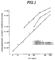
- Fig. 1 is a graph showing the relationship between chemiluminescent intensity and AFP quantity;
- Fig. 2 is a schematic diagram showing the lgG structure;
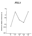
- Fig.3 is a graph plotting non-specific binding percentage with respect to given examples; and
- Fig. 4 is a graph showing the relationship between chemiluminescent intensity and AFP quantity.
- One embodiment of the invention will be described. A target antigen is assayed by a sandwich enzyme immunoassay (EIA) technique employing first and second antibodies reactive in a specific manner with the target antigen in the presence of a reaction solution to trap the target antigen between the first and second antibodies. The antibodies are obtained from serum drawn from a selected animal into which the target antigen is injected. The first antibody is referred to as a solid-phase antibody which is coated on a solid phase such as polystyrene balls. The second antibody is referred to as a labeled antibody which has a label enzyme bound thereon. The label enzyme is glucose oxidase. A chemiluminescence technique is. used to measure the quantity of the label enzyme which corresponds to the quantity of the target antigen. These procedures will be decribed in greater detail on an assumption that the target antigen is α-fetoprotein (AFP) and the label enzyme is glucose oxidase (GOD). The reaction solution used is 0.056 mol/l sodium phosphate buffer (pH 6.3) containing 0.1% sodium azide, 0.2% bovine serum albumin (BSA) and 0.337% NaCI. This reaction solution is referred to as reaction solution A.
- First of all, AFP is injected six times under the back skin of selected one of animals including rabbit, goat and the like at intervals of one week. On the seventh week, a booster is injected into the selected animal. After five days, a proper amount of blood is drawn from the selected animal.
- Anti-AFP immunoglobulin G (IgG) is purified from the drawn serum in the following manner. First of all, 2 ml saturated ammonium sulfate is dripped into 2 ml of serum. The resulting solution is agitated slowly at a temperature of 4°C for two or three hours. The resulting cloudy solution is then placed in a centrifugation tube. Centrifugation is performed at a temperature of 4°C with a contrifugal separator rotating at a speed of 3000 rpm for 15 minutes. The supernatuant is removed from the centrifugation tube, whereas the precipitate is dissolved with 2 ml of 0.1 mol/l sodium phosphate buffer (pH 6.0) containing 5 mmol/l ethylenediaminetetraacetic acid (EDTA). The resulting solution is eluted at a flow rate of 6 ml/hour on s Sephacryl S-300 (a trademark of Pharmacia Fine Chemicals, Sweden) column (1 x 90 cm) equilibrated with 0.1 mol/l sodium phosphate buffer (pH 6.0) containing 5 mmol/l ethylenediaminetetraacetic acid (EDTA). The eluted solution is divided into 1 ml of samples. The lgG fractions, which absorb light at 280 nm, are gathered from the respective samples.
- The resulting anti-AFP IgG is coated through physical absorption on a solid phase by soaking a polystyrene ball (6.5 mm in diameter) available from Ichiko Co., Japan in 0.1 mol/l sodium phosphate buffer (pH 7.5) containing 0.1 mg/ml anti-AFP IgG all the night through at a temperature of 4 °C. The IgG-coated polystyrene ball is washed three times in 0.1 mol I sodium phosphate buffer (pH 7.0) and then washed three times with 0.01 mol/l sodium phosphate buffer (pH 7.0) containing 0.1 mol/l NaCI 0.1% bovine serum albumin (BSA) and 0.1% NaN3. The washed polystyrene ball is stored at a temperature of 4 °C in 0.01 molil sodium phosphate buffer (pH 7.0) containing 0.1 mol/l NaCI, 0.1% bovine serum albumin (BSA) and 0.1% NaN3.
- The labeled antibody, anti-AFP IgG-GOD, is prepared through the following three steps:
- (1) Maleimide glucose oxidase (Maleimide GOD) is prepared as follows. Succinimidyl-4-maleimidobutyrate (GMBS) is added to about 3 mg of glucose oxidase (GOD) placed in each of four test tubes containing 0.3 ml of 0.1 mol I sodium phosphate buffer (pH 7.0) to produce a solution having a GOD/GMBS ratio of 1/50. This solution is incubated at a temperature of 30 °C for one hour. The resulting solution is desalted at a flow rate of 12 ml/hour on a Sephadex G-25 (a trademark of Pharmacia Fine Chemicals, Sweden) column (1 x 30 cm) equilibrated with 0.1 mol/l sodium phosphate buffer (pH 6.0). The desalted solution is divided into 1 ml of maleimido GOD samples.
- (2) The sulfhydrated anti-AFP immunoglobulin G (SH-IgG) is prepared as follows. First of all, 2 ml of IgG solution containing 10 mg of anti-AFP IgG, 0.1 mol/l sodium phosphate buffer (pH 6.0) and 5 mmol/l ethylenediaminetetraacetic acid (EDTA) is prepared. The IgG solution is added to a solution containing S-acetylmercaptosuccimic anhydride (ASMSa) dissolved in N, N-dimethylformamide (DMF) to produce a solution having an IgG/ASMSa ratio, by mol concentration, of 1/300. The resulting solution is agitated at a temperature of 30°C for 30 minutes. In succession, this solution is added with 0.1 ml of 0.1 mol/l Tris-HCλ buffer (pH 7.0), 0.02 ml of 0.1 mol I ethylenediaminetetraacetic acid (EDTA) (pH 7.0) and 0.1 ml of 1 mol/l hydroxylamine hydrochloride (pH 7.0). The resulting solution is incubated at a temperature of 30°C for 4 minutes and then the resulting solution is desalted at a flow rate of 12 ml/hour on a Sephadex G-25 (a trademark of Pharmacia Fine Chemicals, Sweden) column (1x30 cm) equilibrated with 0.1 mol/l sodium phosphate buffer (pH 6.0) containing 5 mmol/l ethylenediaminetetraacetic acid (EDTA). The desalted solution is divided into 1 ml of SH-IgG samples. These SH-IgG samples are concentrated in an ice-cooled collodion bag.
- (3) The labeled antibody was prepared as follows. The maleimido GOD is added to the SH-IgG solution with the same mol concentration as the maleimide GOD. After the resulting solution is held one hour at a temperature of 30°C and then held at a temperature of 4°C stationary all the night through, it is eluted at a flow rate of 6 ml/hour on a Sephacryl S-300 (a trademark of Pharmacia Fine Chemicals, Sweden) column (1 x 90 cm) equilibrated with 0.1 mol/l sodium phosphate buffer solution (pH 6.5) containing 5 mmol/l ethylenediaminetetraacetic acid (EDTA). The eluted solution is divided into 1 ml of samples. The anti-AFP IgG-GOD (labeled antibody) is gathered from the respective samples and stored at a temperature of 4°C in a solution containing 0.1% NaN3 and 0.1% bovine serum albumin (BSA).
- The labeled antibody (anti-AFP IgG-GOD) is bound through the antigen (AFP) to the solid-phase antibody (anti-AFP IgG) in the presence of the reaction solution A (0.056 mol/l sodium phosphate buffer (pH 6.3) containing 0.1% sodium azide, 0.2% bovine serum albumin (BSA) and 0.337% NaCI) in the following manner. The anti-AFP IgG-coated polystyrene ball is incubated at room temperature with AFP (antigen) diluted with the reaction solution A in an assay tube for six hours. The polystylene ball is then washed three times with distilled water. The washed polystyrene ball is incubated at room temperature with anti-AFP IgG-GOD (labeled antibody) in the reaction solution A all the night through. The polystyrene ball is then washed again with distilled water and transferred into another clean assay tube.
- A chemiluminescent technique is used to determine the quantity of the label enzyme; that is, the quantity of the antigen. First of all, 0.3 ml of 0.01 mol/l acetate buffer (pH 5.1) containing mol/l glucose is added to the assay tube. The assay tube is held stationary at a temperature of 37°C for two hours. The resulting solution is taken to prepare a 0.1 ml sample. The sample is added with 0.5 ml of 0.2 mol/l carbonate buffer (pH 9.8) containing 2 x 10⁻⁷ mol/l luminol and then 0.5 ml of 6 x 10⁻³ mol/l potassium ferricyanide. The concentration of the resulting hydrogen peroxide is measured from the light emitted upon chemiluminescent reaction of the luminol and potassium ferricyanide and the hydrogen peroxide by a Luminometer UPD-8000 (a trademark of Meidensha Electric Mfg., Co., Japan) which starts its operation 15 second after the luminol and potassium ferricyanide are added to the assay tube and counts the light emitted for 30 seconds.
- Although the antigen has been described in connection with α-fetoprotein (AFP), it is to be noted that it may be anti-carcinoembryonic antigen (CEA) or the like. Anti-CEA IgG is purified from serum drawn from selected one of animals including, rabbit, goat and the like into which CEA is injected six times at intervals of one week and a booster is injected on the seventh week.
- A serious problem involved with such sandwich enzyme immunoassay (EIA) is the tendency of non-specific binding of a relatively great quantity of labeled antibody to the solid phase, degrading the accuracy of sandwich enzyme immunoassay. The inventors found that the degree of non-specific binding of the labeled antibody to the solid phase was reduced when the labeled antibody was prepared from serum originated from rabbit, the solid-phase antibody was prepared from serum originated from goat and the label enzyme was glucose oxidase. A large number of tests were conducted to prove the effective combinations of the solid-phase and labeled antibodies.
- The degree of non-specific binding of the labeled antibody to the solid phase was determined. For this purpose, an IgG-coated polystyrene ball was incubated at room temperature with IgG-GOD diluted hundredfold with 0.3 ml of reaction solution A in an assay tube all the night through. The polystyrene ball was washed three times with the reaction solution A and then transferred into another clean assay tube. Following this, 0.3 ml of 0.01 mol/l acetate buffer (pH 5.1) containing 0.5 mol/l glucose was added to the assay tube. The assay tube was held stationary at a temperature of 37°C for two hours. The resulting solution was taken to prepare a 0.1 ml sample. The sample was added with 0.5 ml of 0.2 mol/l carbonate buffer (pH 9.8) containing 2x10⁻⁷ mol/l luminol and then with 0.5 ml of 6x10⁻³ mol/l potassium ferricyanide. The light emitted upon chemiluminescent reaction of the luminol and potassium ferricyanide and the resulting hydrogen peroxide was measured by a Luminometer UPD-8000 which started its operation 15 seconds after the luminol and potassium ferricyanide were added to the assay tube and counted the light emitted for 30 seconds. The degree of non-specific binding of the labeled antibody to the solid phase was represented by the non-specific bonding ratio given as C1/C2 x 100 (%) where C1 is the number of counts measured for the labeled antibody binding to the polystyrene ball and C2 is the number of counts corresponding to the whole amount of the labeled antibody used in the assay tube.
- Example 1 - The labeled antibody used was rabbit anti-AFP lgG-GOD and the solid-phase antibody used was goat anti-AFP IgG. The rabbit anti-AFP IgG was Nordic Co., Lot No. 16-184. A goat anti-AFP IgG-coated polystyrene ball was incubated with the rabbit anti-AFP IgG-GOD in the presence of the reaction solution A mixed with guinea pig serum.
- Example 2 - The labeled antibody used was normal rabbit IgG-GOD and the solid-phase antibody used was normal goat IgG. The normal rabbit IgG was Miles Co., Lot No. 0019. The normal goat IgG was Cappel Co., Lot No. 24511. A normal goat IgG-coated polystyrene ball was incubated with the normal rabbit IgG-GOD in the presence of the reaction solution A mixed with guinea pig serum.
- For comparison of the non-specific binding reduction effect obtainable by the invention, tests were conducted for the following comparative examples:
- Comparative Example 1 - The labeled antibody used was goat anti-AFP IgG-GOD and the solid-phase antibody used was goat anti-AFP IgG. A goat anti-AFP IgG-coated polystyrene ball was incubated with the goat anti-AFP IgG-GOD in the presence of the reaction solution A mixed with guinea pig serum.
- Comparative Example 2 - The labeled antibody used was rabbit anti-AFP IgG-GOD and the solid-phase antibody used was rabbit anti-AFP IgG. A rabbit anti-AFP IgG-coated polystyrene ball was incubated with the rabbit anti-AFP IgG-GOD in the presence of the reaction solution A mixed with guinea pig serum.
- Comparative Example 3 - The labeled antibody used was normal goat IgG-GOD and the solid-phase antibody used was normal goat IgG. A normal goat IgG-coated polystyrene ball was incubated with the normal goat IgG-GOD in the presence of the reaction solution A mixed with guinea pig serum.
- Comparative Example 4 - The labeled antibody used was normal rabbit IgG-GOD and the solid-phase antibody used was normal rabbit IgG. A normal rabbit IgG-coated polystyrene ball was incubated with the normal rabbit IgG-GOD in the presence of the reaction solution A mixed with guinea pig serum.
- The results of the non-specific binding tests are illustrated in Table 1.
TABLE 1 Example No. Average Non-Specific Binding Percentage (%) Comparative Example No. Average Non-Specific Binding Percentage (%) 1 0.015 1 0.078 2 0.017 2 0.190 3 0.115 4 0.148 - The non-specific binding percentage ranges from 0.078% to 0.190% for combinations where the labeled antibody is originated from rabbit and the solid-phase antibody is originated from rabbit or where the labeled antibody is originated from goat and the solid-phase antibody is originated from goat. The non-specific binding percentage ranges from 0.015% to 0.017% for combinations where the labeled antibody is originated from rabbit and the solid-phase antibody is originated from goat. It can be seen from the test results that the degree of non-specific binding of the labeled antibody to the solid phase can be reduced to a remarkable extent when the labeled antibody is originated from rabbit and the solid-phase antibody is originated from goat.
- The sensitivity and measurable range of such sandwich enzyme immunoassay was determined. For this purpose, an anti-AFP IgG-coated polystyrene ball was incubated at room temperature with 0.1 ml of AFP standard solution diluted with 0.2 ml of reaction solution A in an assay tube for six hours. The polystyrene ball was washed three times with the reaction solution A. The washed polystyrene ball was incubated with 0.1 ml of anti-AFP IgG-GOD labeled antibody diluted at a proper ratio with the reaction solution A. The assay tube was held stationary at room teperature all the night through. The polystyrene ball was then washed three times with the reaction solution A and transferred into another clean assay tube. Following this, 0.3 ml of 0.01 mol/l acetate buffer (pH 5.1) containing 0.5 mol/l glucose was added to the assay tube. The assay tube was held stationary at a temperature of 37°C for two hours. The resulting solution was taken to prepare a 0.1 ml sample. The sample was added with 0.5 ml of 0.2 mol/l carbonate buffer (pH 9.8) containing 2x10⁻⁷ mol/l luminol and then with 0.5 ml of 6x10⁻³ mol/l potassium ferricyanide. The light emitted upon chemiluminescent reaction of the luminol and potassium ferricyanide and the resulting hydrogen peroxide was measured by a Luminometer UPD-8000 which started its operation 15 seconds after the luminol and potassium ferricyanide were added to the assay tube and counted the light emitted for 30 seconds. Similar procedures are repeated for normal rabbit IgG antibodies and normal goat IgG antibodies.
- The results of tests for the sandwich enzyme
TABLE 2 Example No. Number of Assay Repetitions AFP (ng/ml) Variation Coefficient (%) Average Value ± Standard Deviation 1 10 11 ± 1.2 10.9 1 10 52 ± 4.3 8.3 1 10 295 ± 15.0 5.1 TABLE 3 Comparative Example No. Number of Assay Repetitions AFP (ng/ml) Variation Coefficient (%) Average Value ± Standard Deviation 1 10 9 ± 1.9 21.8 1 10 49 ± 7.4 15.0 1 10 151 ± 21.9 14.5 2 10 13 ± 3.0 23.4 2 10 53 ± 9.3 17.5 2 10 155 ± 21.5 13.9 - Sandwich enzyme immunoassay was repeated ten times within a day (within assay) using the antibody in Example 1 and Comparative Examples 1 and 2. The results are shown in Tables 2 and 3. Similarly, sandwich enzyme immunoassay was repeated six times on different six days (between assay) using the antibody in Example 1 and Comparative Examples 1 and 2. The results are shown in Tables 4 and 5.
TABLE 4 Example No. Number of Assay Repetitions AFP (ng/ml) Variation Coefficient (%) Average Value ± Standard Deviation 1 6 30 ± 2.1 7.1 1 6 142 ± 6.1 4.3 TABLE 5 Comparative Example No. Number of Assay Repetitions AFP (ng/ml) Variation Coefficient (%) Average Value ± Standard Deviation 1 6 34 ± 8.5 25.0 1 6 136 ± 25.7 18.9 2 6 13 ± 2.5 18.9 2 6 125 ± 20.6 16.5 - It can be seen from these results that smaller variations in the results of the sandwich enzyme immunoassay can be obtained when the labeled antibody is originated from rabbit and the solid-phase antibody is originated from goat than when the labeled antibody is originated from rabbit and the solid-phase antibody is originated from rabbit or when the labeled antibody is originated from goat and the solid-phase antibody is originated from goat.
- Referring to Fig. 2, there is illustrated in a schematic form the structure of Immunoglobulin G (IgG). The IgG structure can be divided into an antigen-binding F(ab')2 fragment and a crystallizable (Fc) fragment when it is held at a temperature of 37°C in the presence of pepsin for 18 hours. The Fc fragment has a portion P4 for adsorption to a cell. The Fc fragment has no ability of binding to antigen and is crystallized at low temperature. The F(ab')2 fragment can be divided under a reduction process, for example, in the presence of mercaptoethylamine, into two Fab' fragments having a portion P1 capable of binding to antigen. The inventors found that the degree of non-specific binding of the labeled antibody to the solid phase is dependent on the presence of the Fc fragment and can be reduced by replacing the IgG-GOD with Fab'-GOD when the solid-phase is originated from goat the labeled antibody is originated from rabbit. The Fab' fragment is prepared as the result of separation of the Fc fragment from IgG in the following manner:
- First of all, 2 ml saturated ammonium sulfate is dripped into 2 ml of the serum drawn from a selected animal as described previously. The resulting solution is agitated slowly at a temperature of 4°C for two or three hours. The resulting cloudy solution is then placed in a centrifugation tube. Centrifugation is performed at a temperature of 4°C with a contrifugal separator rotating at a speed of 3000 rpm for 15 minutes. The supernatuant is removed from the centrifugation tube, whereas the precipitate is dissolved with 2 ml of 0.1 mol/l sodium phosphate buffer (pH 6.0) containing 5 mmol/l ethylenediaminetetraacetic acid (EDTA). The resulting solution is eluted at a flow rate of 6 ml/hour on a Sephacryl S-300 (a trademark of Pharmacia Fine Chemicals, Sweden) column (1 x 90 cm) equilibrated with 0.1 mol/l acetate buffer (pH 4.5) containing 0.1 mmol/l NaCI. The eluted solution is divided into 1 ml of samples. The lgG fractions, which absorb light at 280 nm are gathered from the respective samples. Following this, 0.1 ml of 3 mg/ml pepsin solution is added to the gathered IgG. The resulting solution is incubated at a temperature of 37°Cfor 18 hours. The resulting solution is eluted at a flow rate of 6 ml/hour on a Sephacryl S-200 (a trademark of Pharmacia Fine Chemicals, Sweden) column (1 x 90 cm) equilibrated with 0.1 mol/l sodium phosphate buffer (pH 7.5). The eluted solution is divided into 1 ml of samples. The F(ab')2 fractions, which absorb light at 280 nm, are gathered from the respective samples. The gathered F(ab')2 is added with 0.05 ml of 0.1 mol/l mercaptoethylamine and incubated at a temperature of 37°Cfor 90 minutes. The resulting solution is desalted at a flow rate of 6 ml/hour on a Sephadex G-25 (a trademark of Pharmacia Fine Chemicals, Sweden) column (1 x 30 cm) equilibrated with 0.1 mol/l sodium phosphate buffer (pH 6.0) containing 5 mmol/l EDTA. The desalted solution is divided into 1 ml of Fab' samples.
- A number of tests were conducted substantially in the same manner as described previously to prove the advantageous effects on the non-specific binding of the Fab'-GOD labeled antibody to the solid phase.
- Example 3 - The labeled antibody used was rabbit anti-AFP Fab'-GOD and the solid-phase antibody used was goat anti-AFP IgG. A goat anti-AFP IgG-coated polystyrene ball was incubated with the rabbit anti-AFP Fab'-GOD in the presence of the reaction solution A mixed with guinea pig serum.
- Example 4 - The labeled antibody used was rabbit anti-CEA Fab'-GOD and the solid-phase antibody used was goat anti-CEA IgG. A goat anti-CEA IgG-coated polystyrene ball was incubated with the rabbit anti-CEA Fab'-GOD in the presence of the reaction solution A mixed with guinea pig serum.
- For comparison of the non-specific binding reduction effect obtainable by the invention, tests were conducted for the following comparative examples:
- Comparative Example 5 - The labeled antibody used was rabbit anti-AFP IgG-GOD and the solid-phase antibody used was goat anti-AFP IgG. A goat anti-AFP IgG-coated polystyrene ball was incubated with the rabbit anti-AFP IgG-GOD in the presence of the reaction solution A mixed with guinea pig serum.
- Comparative Example 6 - The labeled antibody used was rabbit anti-AFP Fab'-GOD and the solid-phase antibody used was rabbit anti-AFP IgG. A rabbit anti-AFP IgG-coated polystyrene ball was incubated with the rabbit anti-AFP Fab'-GOD in the presence of the reaction solution A mixed with guinea pig serum.
- Comparative Example 7 - The labeled antibody used was goat anti-AFP Fab'-GOD and the solid-phase antibody used was goat anti-AFP IgG. A goat anti-AFP IgG-coated polystyrene ball was incubated with the goat anti-AFP Fab'-GOD in the presence of the reaction solution A mixed with guinea pig serum.
- Comparative Example 8 - The labeled antibody used was rabbit anti-CEA IgG-GOD and the solid-phase antibody used was goat anti-CEA IgG. A goat anti-CEA IgG-coated polystyrene ball was incubated with the rabbit anti-CEA IgG-GOD in the presence of the reaction solution A mixed with guinea pig serum.
- Comparative Example 9 - The labeled antibody used was rabbit anti-CEA Fab'-GOD and the solid-phase antibody used was rabbit anti-CEA IgG. A rabbit anti-CEA IgG-coated polystyrene ball was incubated with the rabbit anti-CEA Fab'-GOD in the presence of the reaction solution A mixed with guinea pig serum.
- Comparative Example 10 - The labeled antibody used was goat anti-CEA Fab'-GOD and the solid-phase antibody used was goat anti-CEA IgG. A goat anti-CEA IgG-coated polystyrene ball was incubated with the goat anti-CEA Fab'-GOD in the presence of the reaction solution A mixed with guinea pig serum.
- The results of the non-specific binding tests are illustrated in Table 6.
TABLE 6 Example No. Average Non-Specific Binding Percentage (%) Comparative Example No. Average Non-Specific Binding Percentage (%) 3 4.7 x 10⁻³ 5 1.5 x 10⁻² 4 3.8 x 10⁻³ 6 2.3 x 10⁻² 7 1.2 x 10⁻² 8 1.3 x 10⁻² 9 1.4 x 10⁻² 10 1.8 x 10⁻² - It can be seen from the test results that the degree of non-specific binding of the labeled antibody to the solid phase can be reduced to a remarkable extent when the labeled antibody is rabbit Fab' and the solid-phase antibody is goat IgG than when the labeled antibody is rabbit Fab' and the solid-phase antibody is rabbit IgG or when the labeled antibody is goat Fab' and the solid-phase antibody is goat IgG.
- The measurable range and detection limit of sandwich enzyme immunoassay were determined substantially in the same manner as described previously. The results of tests for the sandwich enzyme immunoassay measurable range and detection limit are illustrated in Tables 7-10.
TABLE 7 Example No. Measurable Range AFP (ng/ml) Comparative Example No. Measurable Range AFP (ng/ml) 3 0.1 - 50 5 1.0 - 100 6 2.5 - 100 7 1.0 - 100 TABLE 8 Example No. Measurable Range CEA (ng/ml) Comparative Example No. Measurable Range CEA (ng/ml) 4 0.1 - 100 8 2.5 - 250 9 1.0 - 250 10 5.0 - 250 TABLE 9 Example No. Detection Limit AFP (ng/tube) Comparative Example No. Detection Limit AFP (ng/tube) 3 0.01 5 0.1 6 0.25 7 0.1 TABLE 10 Example No. Detection Limit CEA (ng/tube) Comparative Example No. Detection Limit CEA (ng/tube) 4 0.01 8 0.25 9 0.1 10 0.5 - It can be seen from these test results that the measurable range and detection limit of sandwich enzyme immunoassay are superior when the labeled antibody used is Fab'-GOD to those obtained when the labeled antibody used is IgG-GOD. In addition, the measurable range and detection limit of sandwich enzyme immunoassay are superior when the labeled antibody is rabbit Fab' and the solid-phase antibody is goat IgG than when the labeled antibody is rabbit Fab' and the solid-phase antibody is rabbit IgG or when the labeled antibody is goat Fab' and the solid-phase antibody is goat IgG.
- Sandwich enzyme immunoassay was repeated ten times within a day (within assay) using the antibodies in Examples 3-4 and Comparative Examples 5-10. The results are shown in Tables 11-14. Similarly, sandwich enzyme immunoassay was repeated six times on different six days (between assay) using the antibodies in Examples 3-4 and Comparative Examples 5-10. The results are shown in Tables 15-18.
TABLE 11 Example No. Number of Assay Repetitions AFP (ng/ml) Variation Coefficient (%) Average Value ± Standard Deviation 3 10 2.7 ± 0.092 3.4 TABLE 12 Comparative Example No. Number of Assay Repetitions AFP (ng/ml) Variation Coefficient (%) Average Value ± Standard Deviation 5 10 2.8 ± 0.15 5.3 6 10 2.9 ± 0.36 12.5 7 10 2.8 ± 0.39 13.9 TABLE 13 Example No. Number of Assay Repetitions CEA (ng/ml) Variation Coefficient (%) Average Value ± Standard Deviation 4 10 5.2 ± 0.19 3.6 TABLE 14 Comparative Example No. Number of Assay Repetitions CEA (ng/ml) Variation Coefficient (%) Average Value ± Standard Deviation 8 10 5.0 ± 0.16 3.2 9 10 5.2 ± 0.95 18.2 10 10 5.5 ± 0.91 16.5 TABLE 15 Example No. Number of Assay Repetitions AFP (ng/ml) Variation Coefficient (%) Average Value ± Standard Deviation 3 6 2.6 ± 0.065 2.5 TABLE 16 Comparative Example No. Number of Assay Repetitions AFP (ng/ml) Variation Coefficient (%) Average Value ± Standard Deviation 5 6 2.5 ± 0.12 4.8 6 6 2.4 ± 0.38 15.8 7 6 2.6 ± 0.48 18.6 TABLE 17 Example No. Number of Assay Repetitions CEA (ng/ml) Variation Coefficient (%) Average Value ± Standard Deviation 4 6 5.5 ± 0.21 3.8 TABLE 18 Comparative Example No. Number of Assay Repetitions CEA (ng/ml) Variation Coefficient (%) Average Value ± Standard Deviation 8 6 4.8 ± 0.24 5.0 9 6 5.3 ± 0.83 15.6 10 6 5.3 ± 0.78 14.8 - It can be seen from these results that smaller variations in the results of the sandwich enzyme immunoassay can be obtained when the labeled antibody is rabbit Fab' and the solid-phase antibody is goat IgG.
- The inventors also found that the degree of non-specific binding of the labeled antibody to the solid phase can be reduced by adding normal guinea pig serum to the reaction solution A in the present of which the labeled antibody binds to the solid-phase antibody through the antigen. A number of tests were conducted to prove the advantageous effect on the degree of non-specific binding of the labeled antibody to the solid phase.
- Example 5 - The labeled antibody used was rabbit anti-AFP IgG-GOD and the solid-phase antibody used was goat anti-AFP IgG. The reaction solution used was a mixture of the reaction solution A and normal guinea pig serum. The volume ratio of the normal guinea pig serum to the reaction solution A was 1:5.
- The degree of non-specific binding of the labeled antibody to the solid phase was determined. For this purpose, a goat anti-AFP IgG-coated polystyrene ball was incubated at room temperature with goat anti-AFP IgG dilluted with 0.3 ml of a reaction solution mixture X containing the reaction solution A (0.056 mol/l sodium phosphate buffer (pH 6.3) containing 0.1% sodium azide, 0.2% bovine serum albumin (BSA) and 0.337% NaCI) and normal guinea pig serum in an assay tube all the night through. The volume ratio of the normal guinea pig serum to the reaction solution A was 1:5. The polystyrene ball was washed three times with the reaction solution mixture X and then transferred into another clean assay tube. Following this, 0.3 ml of 0.01 mol/l acetate buffer (pH 5.1) containing 0.5 mol/l glucose was added to the assay tube. The assay tube was held stationary at a temperature of 37°C for two hours. The resulting solution was taken to prepare a 0.1 ml sample. The sample was added with 0.5 ml of 0.2 mol/l carbonate buffer (pH 9.8) containing 2 x 10⁻⁷ mol/l luminol and then with 0.5 ml of 6 x 10⁻³ mol/l potassium ferricyanide. The light emitted upon chemiluminescent reaction of the luminol and potassium ferricyanide and the resulting hydrogen peroxide was measured by a Luminometer UPD-8000 (a trademark of Meidensha Electric Mfg. Co. Japan) which starts its counting operation 15 second after the luminol and potassium ferricyanide were added to the assay tube and counted the light emitted for 30 seconds. The degree of non-specific binding of the labeled antibody to the solid phase was represented by the non-specific binding ratio given as C1/C2 x 100 (%) where C1 is the number of counts measured for the labeled antibody binding to the polystyrene ball and C2 is the number of counts corresponding to the whole amount of the labeled antibody used in the assay tube.
- For comparison of the non-specific binding reduction obtainable by the invention, a test was conducted substantially in the same manner for the following comparative examples:
- Comparative Example 11 - The labeled antibody used was rabbit anti-AFP IgG-GOD and the solid-phase antibody used was goat anti-AFP IgG. The reaction solution used was the reaction solution A mixed with normal rabbit serum. The volume ratio of the normal rabbit serum to the reaction solution A was 1:5.
- Comparative Example 12 - The labeled antibody used was rabbit anti-AFP IgG-GOD and the solid-phase antibody used was goat anti-AFP IgG. The reaction solution used was the reaction solution A mixed with normal goat serum. The volume ratio of the normal goat serum to the reaction solution A was 1:5.
- Comparative Example 13 - The labeled antibody used was rabbit anti-AFP IgG-GOD and the solid-phase antibody used was goat anti-AFP IgG. The reaction solution used was the reaction solution A mixed with normal horse serum. The volume ratio of the normal horse serum to the reaction solution A was 1:5.
- Comparative Example 14 - The labeled antibody used was rabbit anti-AFP IgG-GOD and the solid-phase antibody used was goat anti-AFP IgG. The reaction solution used was the reaction solution A.
- The results of the non-specific binding tests are illustrated in Fig. 3. The non-specific binding percentage was as small as 0.006% for Example 5. On the other hand for Comparative Examples 11-14, the non-specific binding percentage ranged from 0.016% to 0.25%. It is, therefore, apparent that the degree of non-specific binding of the labeled antibody to the solid phase can be reduced to a remarkable extent by adding normal guinea pig serum to the reaction solution.
- The sensitivity and measurable range of sandwich enzyme immunoassay was determined for the following example:
- Example 6 - The labeled antibody used was rabbit anti-AFP IgG-GOD and the solid-phase antibody used was goat anti-AFP IgG. The reaction solution used was a mixture of the raction solution A and normal guinea pig serum. The volume ratio of the normal guinea pig serum to the reaction solution A was 1:5.
- A goat anti-AFP IgG-coated polystyrene ball was incubated at room temperature with AFP standard solution diluted with 0.3 ml of reaction solution A in an assay tube for six hours. The polystyrene ball was washed three times with the reaction solution A. The washed polystyrene ball was incubated with 0.1 ml of anti-AFP IgG-GOD labeled antibody diluted at a proper ratio with a mixture of the reaction solution A and normal guinea pig serum. The volume ratio of the normal guinea pig serum to the reaction solution A was 1:5. The assay tube was held stationary at room temperature all the night through. The polystyrene ball was then washed three times with the reaction solution A and transferred into another clean assay tube. Following this, 0.3 ml of 0.01 mol/l acetate buffer (pH 5.1) containing 0.5 mol/l glucose was added to the assay tube. The assay tube was held stationary at a temperature of 37°C for two hours. The resulting solution was taken to prepare a 0.1 ml sample. The sample was added with 0.5 ml of 0.2 mol/l carbonate buffer (pH 9.8) containing 2 x 10⁻⁷ mol/l luminol and then with 0.5 ml of 6 x 10⁻³ mol/l potassium ferricyanide. The light emitted upon chemiluminescent reaction of the luminol and potassium ferricyanide and the resulting hydrogen peroxide was measured by a Luminometer UPD-800 which started its operation 15 seconds after the luminol and potassium ferricyanide were added to the assay tube and counted the light emitted for 30 seconds.
- Similar procedures were repeated for the following comparative example in order to prove the advantageous effects on the sandwich enzyme immunoassay:
- Comparative Example 15 - The labeled antibody used was rabbit anti-AFP IgG-GOD and the solid-phase antibody used was goat anti-AFP IgG. The reaction solution used was the reaction solution A mixed with no normal guinea pig serum.
- Fig. 4 illustrates the results. The sandwich enzyme immunoassay measurable range and sensitivity obtained for Example 6 were 1.0-1000 ng/ml and 1.0 ng/ml, respectively. On the other hand, the sandwich enzyme immunoassay measurable range and sensitivity were 50-1000 ng/ml and 50 ng/ml, respectively, for Comparative Example 15. It is, therefore, apparent that the sandwich enzyme immunoassay sensitivity and measurable range are improved when normal guinea pig serum is added to the reaction solution A.
- Sandwich enzyme immunoassay was repeated ten times within a day (within assay) for Example 6 and Comparative Example 15. The results are shown in Tables 19 and 20. Similarly, sandwich enzyme immunoassay was repeated on different days (between assay) for Example 6 and Comparative Example 15. The results are shown in Tabels 21 and 22.
TABLE 19 Example No. Number of Assay Repetitions AFP (ng/ml) Variation Coefficient (%) Average Value ± Standard Deviation 6 10 15 ± 1.2 7.8 6 10 123 ± 3.8 3.1 6 10 506 ± 14.7 2.9 TABLE 20 Comparative Example No. Number of Assay Repetitions AFP (ng/ml) Variation Coefficient (%) Average Value ± Standard Deviation 15 10 60 ± 9.4 15.6 15 10 151 ± 16.9 11.2 15 10 615 ± 60.3 9.8 TABLE 21 Example No. Number of Assay Repetitions AFP (ng/ml) Variation Coefficient (%) Average Value ± Standard Deviation 6 6 9.8 ± 6.5 6.6 6 6 62.7 ± 18.8 3.0 TABLE 22 Comparative Example No. Number of Assay Repetitions AFP (ng/ml) Variation Coefficient (%) Average Value ± Standard Deviation 15 6 141 ± 18.8 13.3 15 6 708 ± 61.6 8.7 - It can be seen from these results that smaller variations in the results of the sandwich enzyme immunoassay can be obtained when normal guinea pig serum is added to the reaction solution.
- Although the reaction solution has been described as containing sodium phosphate buffer (pH 6.3), it is to be noted that it may be replaced with sodium phosphate buffer (pH 6.0-7.5), borate buffer (pH 8.0-8.5) or the like. In addition, although the invention has been described in connection with sandwich enzyme immunoassay, it is to be noted that the label may be a fluorescent material (fluorescein isothiocyanate, tetramethylrhodamine isothiocyanate. or the like), a chemiluminescent material (aminoethylethyl isoluminol, aminobutylethyl isoluminol, aminopentylethyl isoluminol, aminohexylethyl isoluminol, or the like), or a radioactive isotope (3H, 14C, 32P, 125I, 131I, or the like).
- While the invention has been described in conjunction with specific embodiments thereof. it is intended to embrace all alternatives, modifications and variations that fall within the scope of the appended claims.
Claims (6)
- A kit for use in a sandwich immunoassay employing labeled and solid-phase antibodies reactive with an antigen in the presence of a reaction solution to bind the antigen between the labeled and solid-phase antibodies, said kit containing a solid-phase antibody bound to a solid phase, a labeled antibody having a label enzyme bound to the antibody and a reaction solution, characterized in that the labeled antibody is originated from a rabbit, and the solid-phase antibody is originated from a goat and the label enzyme is glucose oxidase.
- A kit as claimed in claim 1, wherein the labeled antibody is immunoglobulin G having a label enzyme bound thereto.
- A kit as claimed in claim 1, wherein the labeled antibody is a Fab' fragment having a label enzyme bound thereto, the Fab' fragment being produced from immunoglobulin G.
- A kit as claimed in claim 1, wherein the reaction solution contains normal guinea pig serum.
- A kit as claimed in claim 4, wherein the labeled antibody is immunoglobulin G having a label enzyme bound thereto.
- A kit as claimed in claim 5, wherein the labeled antibody is a Fab' fragment having a label enzyme bound thereto, the Fab' fragment being produced from immunoglobulin G.
Applications Claiming Priority (6)
| Application Number | Priority Date | Filing Date | Title |
|---|---|---|---|
| JP61142885A JPS62299763A (en) | 1986-06-20 | 1986-06-20 | Immunological measuring reagent |
| JP142885/86 | 1986-06-20 | ||
| JP62082512A JPS63145960A (en) | 1986-06-23 | 1987-04-03 | Non-singular adsorption inhibitor for labeled antibody |
| JP82512/87 | 1987-04-03 | ||
| JP62149324A JPS63313067A (en) | 1987-06-16 | 1987-06-16 | Reagent for measuring antigen |
| JP149324/87 | 1987-06-16 |
Publications (4)
| Publication Number | Publication Date |
|---|---|
| EP0249983A2 EP0249983A2 (en) | 1987-12-23 |
| EP0249983A3 EP0249983A3 (en) | 1989-10-11 |
| EP0249983B1 EP0249983B1 (en) | 1993-10-20 |
| EP0249983B2 true EP0249983B2 (en) | 1997-03-19 |
Family
ID=27303939
Family Applications (1)
| Application Number | Title | Priority Date | Filing Date |
|---|---|---|---|
| EP87108799A Expired - Lifetime EP0249983B2 (en) | 1986-06-20 | 1987-06-19 | Reagent kit for use in sandwich immunoassay |
Country Status (2)
| Country | Link |
|---|---|
| EP (1) | EP0249983B2 (en) |
| DE (1) | DE3787834T3 (en) |
Families Citing this family (1)
| Publication number | Priority date | Publication date | Assignee | Title |
|---|---|---|---|---|
| WO1989010974A1 (en) * | 1988-05-11 | 1989-11-16 | Trustees Of The Sisters Of Charity Of Australia | Enzyme immunoassay system |
Family Cites Families (4)
| Publication number | Priority date | Publication date | Assignee | Title |
|---|---|---|---|---|
| US4002532A (en) * | 1974-10-21 | 1977-01-11 | Weltman Joel K | Enzyme conjugates |
| US4414324A (en) * | 1979-05-21 | 1983-11-08 | Bma Laboratory Services, Inc. | Immunoassay method and apparatus |
| EP0125893A3 (en) * | 1983-05-12 | 1986-10-15 | Sumitomo Chemical Company, Limited | The quantitative analysis of antigen by the enzyme-antibody bridge method |
| EP0149602A1 (en) * | 1983-07-25 | 1985-07-31 | Beckman Instruments, Inc. | Immunometric assay using polyclonal and monoclonal antibodies and a kit for use therein |
-
1987
- 1987-06-19 DE DE3787834T patent/DE3787834T3/en not_active Expired - Fee Related
- 1987-06-19 EP EP87108799A patent/EP0249983B2/en not_active Expired - Lifetime
Also Published As
| Publication number | Publication date |
|---|---|
| EP0249983A2 (en) | 1987-12-23 |
| EP0249983A3 (en) | 1989-10-11 |
| EP0249983B1 (en) | 1993-10-20 |
| DE3787834D1 (en) | 1993-11-25 |
| DE3787834T2 (en) | 1994-03-10 |
| DE3787834T3 (en) | 1997-07-31 |
Similar Documents
| Publication | Publication Date | Title |
|---|---|---|
| EP0365685B1 (en) | Freeze-dried composition comprising a horseradish peroxidase-labelled Fab' fragment of an anti-human interferon-beta monclonal antibody and trehalose; EIA kit containing the composition | |
| Nishizono et al. | Rapid and sensitive chemiluminescent enzyme immunoassay for measuring tumor markers | |
| US5876935A (en) | Luminescent specific binding assay | |
| Ikariyama et al. | Luminescence immunoassay of human serum albumin with hemin as labeling catalyst | |
| US4375972A (en) | Heterogeneous chemiluminescent immunoassays utilizing metallo porphyrin tag | |
| US4835101A (en) | Luminescent analyses with enhanced storage stability | |
| Watanabe et al. | Enzyme immunoassay for human ferritin. | |
| EP0124366B1 (en) | Method of measuring biological ligands | |
| Reichstein et al. | Laser-excited time-resolved solid-phase fluoroimmunoassays with the new europium chelate 4, 7-bis (chlorosulfophenyl)-1, 10-phenanthroline-2, 9-dicarboxylic acid as label | |
| Place et al. | The fixation of anti-HBsAg on plastic surfaces | |
| NO874140L (en) | IMMUNOMETRIC DETERMINATION PROCEDURES. | |
| EP0249983B2 (en) | Reagent kit for use in sandwich immunoassay | |
| WO1999060401A1 (en) | Immunoassay reagents and immunoassay method | |
| GB2158578A (en) | Immunometric method for the determination of a hapten | |
| EP0249955B1 (en) | Kit for use in sandwich immunoassay | |
| EP0353895A1 (en) | Immunoassay method | |
| Kohno et al. | Novel enzyme immunoassay of anti‐insulin IgG in human serum | |
| KR960016337B1 (en) | Reagent kit for use in sandwich immunoassay | |
| EP0061071B1 (en) | Activated apoglucose oxidase, method of preparing it, its use in specific binding assay methods and reagent means and test kits containing it | |
| Ishikawa | Ultrasensitive and rapid enzyme immunoassay | |
| Aoyagi et al. | The reduction of nonspecific binding in chemiluminescent sandwich enzyme immunoassays | |
| KR960016338B1 (en) | Sandwich Immunoassay Kit | |
| JP3706214B2 (en) | Immunological analysis method | |
| JPS58149700A (en) | Composite containing peroxidase, its preparation and reagent | |
| Ishikawa et al. | Potential of the immune complex transfer enzyme immunoassay for antigens and antibodies to improve the sensitivity and its limitations |
Legal Events
| Date | Code | Title | Description |
|---|---|---|---|
| PUAI | Public reference made under article 153(3) epc to a published international application that has entered the european phase |
Free format text: ORIGINAL CODE: 0009012 |
|
| AK | Designated contracting states |
Kind code of ref document: A2 Designated state(s): DE FR GB |
|
| PUAL | Search report despatched |
Free format text: ORIGINAL CODE: 0009013 |
|
| AK | Designated contracting states |
Kind code of ref document: A3 Designated state(s): DE FR GB |
|
| 17P | Request for examination filed |
Effective date: 19900123 |
|
| 17Q | First examination report despatched |
Effective date: 19920131 |
|
| GRAA | (expected) grant |
Free format text: ORIGINAL CODE: 0009210 |
|
| AK | Designated contracting states |
Kind code of ref document: B1 Designated state(s): DE FR GB |
|
| REF | Corresponds to: |
Ref document number: 3787834 Country of ref document: DE Date of ref document: 19931125 |
|
| ET | Fr: translation filed | ||
| PLBI | Opposition filed |
Free format text: ORIGINAL CODE: 0009260 |
|
| 26 | Opposition filed |
Opponent name: BEHRINGWERKE AKTIENGESELLSCHAFT Effective date: 19940720 |
|
| PLAW | Interlocutory decision in opposition |
Free format text: ORIGINAL CODE: EPIDOS IDOP |
|
| PLAW | Interlocutory decision in opposition |
Free format text: ORIGINAL CODE: EPIDOS IDOP |
|
| PUAH | Patent maintained in amended form |
Free format text: ORIGINAL CODE: 0009272 |
|
| STAA | Information on the status of an ep patent application or granted ep patent |
Free format text: STATUS: PATENT MAINTAINED AS AMENDED |
|
| 27A | Patent maintained in amended form |
Effective date: 19970319 |
|
| AK | Designated contracting states |
Kind code of ref document: B2 Designated state(s): DE FR GB |
|
| ET3 | Fr: translation filed ** decision concerning opposition | ||
| PGFP | Annual fee paid to national office [announced via postgrant information from national office to epo] |
Ref country code: GB Payment date: 19990525 Year of fee payment: 13 |
|
| PGFP | Annual fee paid to national office [announced via postgrant information from national office to epo] |
Ref country code: FR Payment date: 19990616 Year of fee payment: 13 |
|
| PGFP | Annual fee paid to national office [announced via postgrant information from national office to epo] |
Ref country code: DE Payment date: 19990811 Year of fee payment: 13 |
|
| PG25 | Lapsed in a contracting state [announced via postgrant information from national office to epo] |
Ref country code: GB Free format text: LAPSE BECAUSE OF NON-PAYMENT OF DUE FEES Effective date: 20000619 |
|
| GBPC | Gb: european patent ceased through non-payment of renewal fee |
Effective date: 20000619 |
|
| PG25 | Lapsed in a contracting state [announced via postgrant information from national office to epo] |
Ref country code: FR Free format text: LAPSE BECAUSE OF NON-PAYMENT OF DUE FEES Effective date: 20010228 |
|
| REG | Reference to a national code |
Ref country code: FR Ref legal event code: ST |
|
| PG25 | Lapsed in a contracting state [announced via postgrant information from national office to epo] |
Ref country code: DE Free format text: LAPSE BECAUSE OF NON-PAYMENT OF DUE FEES Effective date: 20010403 |