EP0245098B1 - Méthode et appareil pour juger la déformation d'une colonne vertébrale - Google Patents
Méthode et appareil pour juger la déformation d'une colonne vertébrale Download PDFInfo
- Publication number
- EP0245098B1 EP0245098B1 EP87304069A EP87304069A EP0245098B1 EP 0245098 B1 EP0245098 B1 EP 0245098B1 EP 87304069 A EP87304069 A EP 87304069A EP 87304069 A EP87304069 A EP 87304069A EP 0245098 B1 EP0245098 B1 EP 0245098B1
- Authority
- EP
- European Patent Office
- Prior art keywords
- vertebrae
- deformation
- vertebral body
- wedge
- shaped
- Prior art date
- Legal status (The legal status is an assumption and is not a legal conclusion. Google has not performed a legal analysis and makes no representation as to the accuracy of the status listed.)
- Expired - Lifetime
Links
- 238000000034 method Methods 0.000 title claims description 33
- 241000251468 Actinopterygii Species 0.000 claims description 40
- 238000005259 measurement Methods 0.000 claims description 17
- 230000006870 function Effects 0.000 claims description 13
- 238000004364 calculation method Methods 0.000 claims description 7
- 230000000694 effects Effects 0.000 claims description 7
- 238000002560 therapeutic procedure Methods 0.000 claims description 6
- 208000010392 Bone Fractures Diseases 0.000 claims 4
- 206010017076 Fracture Diseases 0.000 claims 4
- 206010010214 Compression fracture Diseases 0.000 claims 1
- 210000000115 thoracic cavity Anatomy 0.000 description 38
- 230000000994 depressogenic effect Effects 0.000 description 11
- 208000001132 Osteoporosis Diseases 0.000 description 10
- 238000004458 analytical method Methods 0.000 description 4
- 210000000988 bone and bone Anatomy 0.000 description 4
- 238000012790 confirmation Methods 0.000 description 4
- 238000010586 diagram Methods 0.000 description 3
- 238000012545 processing Methods 0.000 description 3
- 230000000007 visual effect Effects 0.000 description 3
- 206010068975 Bone atrophy Diseases 0.000 description 2
- 208000020084 Bone disease Diseases 0.000 description 2
- 238000011156 evaluation Methods 0.000 description 2
- 206010058907 Spinal deformity Diseases 0.000 description 1
- 238000004590 computer program Methods 0.000 description 1
- 238000003745 diagnosis Methods 0.000 description 1
- 229940079593 drug Drugs 0.000 description 1
- 239000003814 drug Substances 0.000 description 1
- 210000004705 lumbosacral region Anatomy 0.000 description 1
- 238000000491 multivariate analysis Methods 0.000 description 1
- 230000005855 radiation Effects 0.000 description 1
- 210000002320 radius Anatomy 0.000 description 1
- 210000002303 tibia Anatomy 0.000 description 1
- 210000000623 ulna Anatomy 0.000 description 1
- 210000000689 upper leg Anatomy 0.000 description 1
Images


Classifications
-
- A—HUMAN NECESSITIES
- A61—MEDICAL OR VETERINARY SCIENCE; HYGIENE
- A61B—DIAGNOSIS; SURGERY; IDENTIFICATION
- A61B5/00—Measuring for diagnostic purposes; Identification of persons
- A61B5/103—Measuring devices for testing the shape, pattern, colour, size or movement of the body or parts thereof, for diagnostic purposes
- A61B5/107—Measuring physical dimensions, e.g. size of the entire body or parts thereof
- A61B5/1079—Measuring physical dimensions, e.g. size of the entire body or parts thereof using optical or photographic means
-
- A—HUMAN NECESSITIES
- A61—MEDICAL OR VETERINARY SCIENCE; HYGIENE
- A61B—DIAGNOSIS; SURGERY; IDENTIFICATION
- A61B5/00—Measuring for diagnostic purposes; Identification of persons
- A61B5/72—Signal processing specially adapted for physiological signals or for diagnostic purposes
- A61B5/7235—Details of waveform analysis
- A61B5/7264—Classification of physiological signals or data, e.g. using neural networks, statistical classifiers, expert systems or fuzzy systems
-
- G—PHYSICS
- G16—INFORMATION AND COMMUNICATION TECHNOLOGY [ICT] SPECIALLY ADAPTED FOR SPECIFIC APPLICATION FIELDS
- G16H—HEALTHCARE INFORMATICS, i.e. INFORMATION AND COMMUNICATION TECHNOLOGY [ICT] SPECIALLY ADAPTED FOR THE HANDLING OR PROCESSING OF MEDICAL OR HEALTHCARE DATA
- G16H15/00—ICT specially adapted for medical reports, e.g. generation or transmission thereof
-
- G—PHYSICS
- G16—INFORMATION AND COMMUNICATION TECHNOLOGY [ICT] SPECIALLY ADAPTED FOR SPECIFIC APPLICATION FIELDS
- G16H—HEALTHCARE INFORMATICS, i.e. INFORMATION AND COMMUNICATION TECHNOLOGY [ICT] SPECIALLY ADAPTED FOR THE HANDLING OR PROCESSING OF MEDICAL OR HEALTHCARE DATA
- G16H50/00—ICT specially adapted for medical diagnosis, medical simulation or medical data mining; ICT specially adapted for detecting, monitoring or modelling epidemics or pandemics
- G16H50/50—ICT specially adapted for medical diagnosis, medical simulation or medical data mining; ICT specially adapted for detecting, monitoring or modelling epidemics or pandemics for simulation or modelling of medical disorders
-
- G—PHYSICS
- G16—INFORMATION AND COMMUNICATION TECHNOLOGY [ICT] SPECIALLY ADAPTED FOR SPECIFIC APPLICATION FIELDS
- G16H—HEALTHCARE INFORMATICS, i.e. INFORMATION AND COMMUNICATION TECHNOLOGY [ICT] SPECIALLY ADAPTED FOR THE HANDLING OR PROCESSING OF MEDICAL OR HEALTHCARE DATA
- G16H50/00—ICT specially adapted for medical diagnosis, medical simulation or medical data mining; ICT specially adapted for detecting, monitoring or modelling epidemics or pandemics
- G16H50/20—ICT specially adapted for medical diagnosis, medical simulation or medical data mining; ICT specially adapted for detecting, monitoring or modelling epidemics or pandemics for computer-aided diagnosis, e.g. based on medical expert systems
Definitions
- the present invention relates to a method and apparatus for judging deformation of a vertebral body. More particularly, the present invention relates to a method and apparatus for judging deformation of a vertebral body which is one of the bones forming the spinal column. A judgment of the presence of vertebral body deformation accompanied by osteoporosis, as well as a classification of the deformation, is very important for grasping the progress of osteoporosis and for confirmation of the effect of therapy.
- EP-A 0069229 discloses a method for reducing the effect of patient movement in X-ray diagnosis, in which different images are derived from a video signal containing a sequence of images of a patient under examination, first and subsequent images contained in the signal being stored and used to form a secondary mask which is then subtracted from images contained in the video signal.
- a system for the analysis of spinal deformities is known from Medical & Biomedical Engineering & Computing 1982, 20 , 715-726 in which a deformity in the curvature of the spine as a whole is measured by the digitization of X-ray data.
- the ratio (a/d) of the central length (a) to the front brim length (d) of the third lumbar vertebrae is determined as the index (Lumbar Spine Score) for the degree of bone atrophy, or a change in the longitudinal and lateral bone beams of the third lumbar vertebrae is observed as the index for the degree of bone atrophy (severity classified by Jikei University), or a judgment of the fracture of a vertebral body is made by measurement of the central length (a), front brim length (d) and rear brim length (c) of a vertebral body [The New England Journal of Medicine, vol. 306, 446 (February 25, 1982)], a method for classifying the type of deformation of a vertebral body has not heretofore been known.
- the objects of the present invention are to eliminate the above-mentioned disadvantages of the prior art and to provide a method for judging deformation of a vertebral body in a subject, characterised in that the method comprises the steps of:
- apparatus for judging deformation of a vertebral body according to the method of claim 1, the apparatus being defined in claim 7.
- the present inventors have made an intensive study of a method of objective evaluation of vertebral body deformation, and found that the presence of vertebral body deformation can be objectively judged according to the judgment standard in which lengths of the vertebral body and their ratios are combined by accurately measuring the lengths of a front brim, rear brim, central portion from a profile X-ray image (i.e., an image on a film taken from the side by X-ray radiation) of a vertebral body and determining the ratios of the respective portions of the vertebral body, and further found that the type of vertebral body deformation can be judged according to the judgment standard in which lengths of the vertebral body and their ratios are combined, and the progress over a period of time of the vertebral body deformation can be judged from the change in the deformation type as well as a change in the lengths of the vertebral body, and thus accomplished the present invention.
- the profile X-ray image of vertebral body was visually observed by a physician when judging the vertebral body deformation, but according to the present invention, the lengths of the respective portions of the vertebral body are measured, judgment standards are prepared with reference to a judgment by a physician, and the presence of vertebral body deformation as well as a classification of the deformation type are objectively performed following the judgment standards.
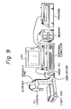
- the ratio c/d of the rear brim length to the front brim length must be between 0.7 and 1.4 as the first condition (a). For, as described below, if c/d becomes 1.4 or more, the front brim portion becomes a deformed wedge-shaped vertebrae, and if c/d becomes 0.7 or less, the rear brim portion becomes a deformed inverse wedge-shaped vertebrae, which in practice is very rare.
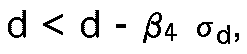
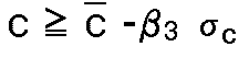
- At least one of rear brim length c and front brim length d must be greater than c - 2 ⁇ and d - 1.5 ⁇ when the average values of the rear brim length c and front brim length d of vertebral body without deformation determined in (i) are made c and d , respectively.
- a , c , d all have a high value, namely a ⁇ ⁇ a ⁇ - 2 ⁇ a , c ⁇ ⁇ c ⁇ - 2 ⁇ c , d ⁇ ⁇ d ⁇ - 1.5 ⁇ d , but even when the vertebral body is slightly compressed to become a ⁇ ⁇ a ⁇ - 2 ⁇ a , provided that at least one of the conditions of c ⁇ ⁇ c ⁇ - 2 ⁇ c or d ⁇ ⁇ d ⁇ - 1.5 ⁇ d , can be satisfied, no clear vertebral body deformation is recognized and judgment of "no deformation" may be made.
- At least one of a/c and a/d must be more than 0.8. For, if both of a/c and a/d are 0.8 or less, namely the center length a is much smaller, than the rear brim length c and front brim length d , a fish vertebrae condition as described below is determined.
- “Fish vertebrae” is a vertebrae where a depressed fracture or a pressure fracture has occurred at the central portion, whereby the central length (a) is particularly smaller than the front brim length (d) and rear brim length (c) (see Fig. 3). Accordingly, among the so called “no deformation” classifications, except for wedge-shaped vertebrae, flat vertebrae, and inverse wedge-shaped vertebrae, the central length (a) is particularly smaller.
- the fish vertebrae (type II) is also inclusive of deformations with a shortened central length like fish vertebrae, as a result of an upper brim pressure fracture, upper brim depressed fracture, lower brim pressure fracture, lower brim depressed fracture, etc.
- the upper limit as 1.4 and the lower limit as 0.7 as the judgment standard for c/d as a preferable example, but these values can be selected freely, for example, in the case of the upper limit, from the range of 1.25 to 1.55, preferably from 1.33 to 1.5, more preferably from 1.4 to 1.45.
- the lower limit can be selected freely from the range of 0.8 to 0.6, preferably from 0.75 to 0.65.
- 0.8 was selected in the case of "no deformation” and "fish vertebrae", but this value can be freely selected from the range of 0.65 to 0.9, preferably from 0.7 to 0.85, more preferably from 0.75 to 0.8.
- c - 2 ⁇ c and d - 1.5 ⁇ d were selected in the case of "no deformation", “flat vertebrae”, and “fish vertebrae” but the judgment standard for c can be freely selected from the range of c - 1.0 ⁇ c to c - 2.5 ⁇ c , preferably c - 1.25 ⁇ c to c - 2.25 ⁇ c , more preferably c - 1.5 ⁇ c to c - 2.0 ⁇ c .
- the judgment standard for d also can be similarly selected from the range of d - 1.0 ⁇ d to d - 2.5 ⁇ d , preferably d - 1.25 ⁇ d to d - 2.25 ⁇ d , more preferably d - 1.5 ⁇ d to d - 2.0 ⁇ d .
- ⁇ 1 to ⁇ 6 may optionally be determined from a discriminating function capable of discriminating two groups with two variants or two groups with one variant by using values obtained by measuring two of a , b , c , d , and e with respect to at least five profile X-ray image in each of two types among four types (i.e., wedge-shaped vertabrae, fish vertabrae flat vertebral, and no deformation).
- ⁇ 1 is z5 obtained by the following discriminant function using the ratio c/d derived from c and d, which are obtained by measuring at least 5 profile X-ray images in each of two types selected from "wedge-shaped vertebrae" and "no deformation”:
- the present inventors have made an intensive study of a method of objective evaluation of vertebral body deformation, and found that the presence of a vertebral body deformation as well as the deformation type can be judged objectively by use of a discriminant function by accurately measuring the lengths of, for example, the front brim (d), rear brim (c), central portion (a), and the vertebral body width (b) from a profile X-ray image of a vertebral body, and further found that the progress over a period of time of the vertebral body deformation can be judged from the change in the type of said deformation as well as the change in the lengths of the vertebral body, and thus accomplished the present invention.
- a discriminant function by accurately measuring the lengths of, for example, the front brim (d), rear brim (c), central portion (a), and the vertebral body width (b) from a profile X-ray image of a vertebral body, and further found that the progress over a period of time of the verte
- vertebral body deformation has been judged by a physician by visual observation of the profile X-ray image of a vertebral body, but according to the method of the present invention, the lengths of the respective portions of a vertebral body are measured and for the vertebral bodies judged by a physician to be "no deformation", "wedge-shaped vertebrae", “fish vertebrae”, and "flat vertebrae", discrimination is made by discriminant functions to determine discriminant formulae for the respective types of vertebral body deformations, and thereafter, the presence of vertebral body deformation and the type of deformation are objectively judged by this discriminant formulae for a patient whose vertebral body deformation is to be judged.
- deformed vertebral bodies are classified in the present invention generally into wedge-shaped vertebrae, fish vertebrae, and flat vertebrae.
- the wedge-shaped vertebrae (type I) deformation has occurred at the front brim portion as shown in Fig. 4, and the front brim length has become particularly smaller than the rear brim length, including in addition to the so called wedge-shaped vertebrae deformation with a shortened front brim length like wedge-shaped vertebrae as the result of upper brim pressure fracture, an upper brim depressed fracture, lower brim pressure fracture, and lower brim depressed fracture, etc.
- the depressed fracture or pressure fracture has occurred at the central portion, whereby the central length has become particularly smaller than the front brim length and rear brim length (see Fig.
- the front brim portion, central portion, and rear brim portion are deformed relatively uniformly under pressure (see Fig. 4); namely, all of the front rear length, central length, and rear brim length are smaller.
- wedge-shaped vertebrae (type IV). This type is defined as a vertebral body wherein deformation has occurred at the rear brim portion, and thus the rear brim length is smaller than the front brim length, contrary to the wedge-shaped vertebrae where deformation has occurred at the front brim portion, and the front brim portion is smaller than the rear brim length (see Fig. 4). In practice, however, such a vertebrae does not substantially exist. Accordingly, it is sufficient if the four types, wedge-shaped vertebrae, fish vertebrae, flat vertebrae and "no deformation", can be classified.
- the discriminant function is represented by the following formula in which the ratio (c/d) of c and d measured from at least 5 profile X-ray images in each of two types of :wedge-shaped vertebrae" and "no deformation” and the ratio (c/d) of less than z5 obtained from the vertebral body to be judged is judged as "no deformation” and the ratio (c/d) equal to or larger than z5 is judged as "wedge-shaped vertebrae".
- the apparatus for judging the deformation of vertebral body according to the present invention is used for the practice of the above-mentioned method of the present invention.
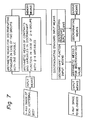
- This apparatus is basically composed of an imputting means for at least two indices of a , b , c , and d , an arithmetic means for effecting necessary operations for the descrimination, a discriminating means by using the calculation results, a means for inputting the discriminant function and/or standard necessary for the calculation and discrimination, and an output means for outputting the judgment result.
- a rule means As the inputting means, a rule means, a digitizer means, an image processing means in which an image inputting means such as a TV camera is used.
- c/d, a/c, and a/d are determined by the arithmetic means, and c , d , ⁇ c , ⁇ d , and ⁇ 1 - ⁇ 6 are inputted as discriminating standards by the discriminating standard inputting means, and the discriminating means can discriminate the results by these standards.
- the present apparatus can be further provided with a means for obtained the discriminating standards from a number of the profile X-ray images having no substantial deformation.
- the present apparatus can be optionally provided with an arithmetic means capable of discriminating two groups with two variants, which are used for determining the indices ⁇ 1 to ⁇ 6 , or of discriminating two groups with one variant.
- the data of at least two types of the four types of the vertebral body i.e., "wedge-shaped vertebrae", “fish vertebrae”, “flat vertebrae”, and "no deformation" should be input.
- the arithmetic means is connected to a discriminating standard input means for inputting the discriminant function, whereby the arithmetic means calculates the discriminating values with said discriminant function and the discriminating means determine the type of the vertebral body based on the maximum discriminating value.
- the present apparatus is provided with a function capable of discriminating the four types of the vertebral body, "wedge-shaped vertebrae", “fish vertebrae”, “flat vertebrae”, and "no deformation" from a one-dimensional formula containing four variables a , b , c , and d .
- the apparatus which is capable of judging the two types of the vertebral body with one variant.
- the type of vertebral body deformation can be objectively evaluated, and also the change in the said deformation type and the progress over a period of time of the vertebral body deformation can be judged.
- the present method and apparatus is very useful for determining the progress of bone disease such as osteoporosis, etc., and for confirmation of the effect of therapy.
- the type of vertebral body deformation can be objectively evaluated, and further the change in the type of deformation type and the progress over a period of time of the vertebral body deformation can be judged.
- the present method and apparatus is very useful for determining the progress of bone disease such as osteoporosis, etc., and for confirmation of the effect of therapy.
- the second lumbar vertebrae changed from a "no deformation" classification to a type III (flat vertebrae) within six months.
- the conditions of the front brim and rear brim of the seventh thoracic vertebrae, and the center of the tenth thoracic vertebrae tend to have worsened. Also, the conditions of the centers of the tenth thoracic vertebrae and the third lumbar vertebrae had further worsened.
- vertebral bodies including 6 vertebral bodies judged as wedge-shaped vertebrae, 25 vertebral bodies judged as fish vertebrae, eight vertebral bodies judged as flat vertebrae and 147 vertebral bodies judged as "no deformation" according to visual observation by a physician of a profile X-ray image, with the third lumbar vertebrae as the center, of osteoporosis patients (women), eight points of 1 to 8 shown in Fig.
- the discriminant value for each vertebral body was calculated by substituting the measured values of a , b , c , and d , into the above four discriminant formulae, and the type having the maximum numerical value was judged to be the vertebral body deformation type.
- Vertebrae body measured values (mm) Deformation type a b c d c/d 1 20.0 22.5 24.7 16.5 1.50 Wedge-shaped vertebrae 2 21.6 30.2 24.4 21.3 1.14 No deformation 3 17.5 32.9 21.7 13.6 1.60 Wedge-shaped vertebrae 4 17.7 38.3 29.6 15.6 1.90 " 5 22.0 28.8 23.8 19.2 1.24 No deformation 6 12.9 38.2 21.8 13.2 1.64 Wedge-shaped vertebrae 7 21.0 28.3 24.5 21.2 1.15 No deformation 8 18.9 32.0 22.4 18.5 1.21 " 9 19.8 30.8 24.3 18.7 1.30 " 10 19.3 31.3 22.2 15.6 1.43 Wedge-shaped vertebrae
Landscapes
- Health & Medical Sciences (AREA)
- Engineering & Computer Science (AREA)
- Life Sciences & Earth Sciences (AREA)
- Public Health (AREA)
- Medical Informatics (AREA)
- General Health & Medical Sciences (AREA)
- Pathology (AREA)
- Biomedical Technology (AREA)
- Physics & Mathematics (AREA)
- Biophysics (AREA)
- Heart & Thoracic Surgery (AREA)
- Molecular Biology (AREA)
- Surgery (AREA)
- Animal Behavior & Ethology (AREA)
- Primary Health Care (AREA)
- Epidemiology (AREA)
- Veterinary Medicine (AREA)
- Artificial Intelligence (AREA)
- Evolutionary Computation (AREA)
- Fuzzy Systems (AREA)
- Mathematical Physics (AREA)
- Computer Vision & Pattern Recognition (AREA)
- Physiology (AREA)
- Psychiatry (AREA)
- Signal Processing (AREA)
- Dentistry (AREA)
- Oral & Maxillofacial Surgery (AREA)
- Data Mining & Analysis (AREA)
- Databases & Information Systems (AREA)
- Apparatus For Radiation Diagnosis (AREA)
- Measurement Of The Respiration, Hearing Ability, Form, And Blood Characteristics Of Living Organisms (AREA)
Claims (8)
- Procédé pour évaluer la déformation d'un corps vertébral dans un sujet, comprenant les étapes consistant :(i) à mesurer d'une longueur centrale a, une longueur du bord arrière c et une longueur du bord avant d à partir de plusieurs radiographies de profil obtenues à partir de corps vertébraux ayant substantiellement aucune déformation à une partie correspondant à une partie dudit corps vertébral à évaluer, à stocker les mesures dans une mémoire, et à déterminer les valeurs de c/d, a/c et a/d, les valeurs moyennes c et d respectivement des longueurs de bord arrière c et des longueurs de bord avant d, et les déviations de référence σc et σd respectivement des valeurs c et d;(ii) à mesurer a, c et d à partir de plusieurs radiographies de profil obtenues à partir de corps vertébraux ayant au moins une déformation, à stocker les mesures dans ladite mémoire, et à déterminer les valeurs de c/d, a/c, a/d et
c ,d , σc et σd;(iii) à préparer une référence d'évaluation à partir des données obtenues dans les étapes (i) et (ii) afin de déterminer la nature de la déformation selon une classification consistant en vertèbres "sans déformation", vertèbres "cunéiformes", vertèbres "cunéiformes inversées", vertèbres "en poisson", et vertèbres "plates";(iv) à mesurer a, c et d à partir d'une radiographie de profil dudit corps vertébral à évaluer, à stocker les mesures dans ladite mémoire, et à déterminer c/d, a/c et a/d; et(v)à classifier la déformation du corps vertébral à évaluer à partir de c/d dans le cas de vertèbres "cunéiformes" et de vertèbres "cunéiformes inversées", à partir dec ,d , σc et σd, et c/d dans le cas de vertèbres "plates", et à partir dec ,d , σc, σd, a/c, a/d et c/d dans le cas de vertèbres "sans déformation" et de vertèbres "en poisson", afin d'évaluer la déformation et de contrôler l'effet thérapeutique sur ledit corps vertébral à évaluer. - Procédé selon la revendication 1, dans lequel on effectue ladite étape (v) selon les sous-étapes suivantes :(i)
 (ii)
(ii) (iii)
(iii)
 (iv)
(iv)


 (v)
(v)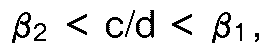


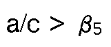
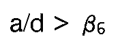
- Procédé selon la revendication 2, dans lequel β₁ à β₆ sont tels que 1,33 ≦ β₁ ≦ 1,5, 0,65 ≦ β₂ ≦ 0,75, 1,25 ≦ β₃ ≦ 2,25, 1,25 ≦ β₄ ≦ 2,25, 0,7 ≦ β₅ ≦ 0,85, et 0,75 ≦ β₆ ≦ 0,85.
- Procédé selon la revendication 3, dans lequel β₁ est 1,4, β₂ 0,7, β₃ 2, β₄ 1,5, β₅ 0,8, et β₆ 0,8.
- Procédé selon l'une quelconque des revendications 2 à 4, dans lequel au moins un des β₁ à β₆ sont déterminés par une fonction discriminante capable de disoriminer deux groupes avec une ou deux variables en utilisant deux de a, b, c et d ou de leur rapport, qui sont obtenus en mesurant au moins 5 radiographies de profil dans chacun des deux types choisis parmi quatre types, "vertèbres cunéiformes", vertèbres en poisson", "vertèbres plates" et "sans déformation".
- Procédé selon l'une quelconque des revendications précédentes, dans lequel on effectue la mesure par un moyen de graduation, un moyen de conversion numérique, ou un moyen pour la mesure automatique des longueurs à partir du traitement d'images par mémorisation de la radiographie à l'aide d'un moyen d'entrée automatique de radiographies.
- Appareil pour évaluer la déformation d'un corps vertébral selon le procédé de la revendication 1, l'appareil comprenant (i) un moyen d'entrée pour introduire des valeurs d'du moins la longueur centrale a, la longueur du bord arrière c et la longueur du bord avant d à partir d'une radiographie de profil d'un corps vertébral à évaluer, (ii) un moyen arithmétique pour exécuter le calcul de c/d, a/c, a/d, les valeurs moyennes
c ,d de c et d et les déviations de référence σc et σd des valeurs c et d, respectivement, nécessaires pour déterminer la nature de la déformation selon une classification consistant en vertèbres "cunéiformes" vertèbres "cunéiformes inversées", vertèbres "en poisson", vertèbres "pistes" et "sans déformation" en utilisant lesdites valeurs d'entrée, (iii) un moyen pour discriminer les vertèbres "cunéiformes" et des vertèbres "cunéiformes inversées" à partir de c/d, des vertèbres "plates" à partir dec ,d , σc, σd et c/d, et des vertèbres "sans déformation" et "en poisson" à partir dec ,d , σc, σd, a/c, a/d et c/d, (iv) un moyen pour entrer la référence d'évaluation nécessaire audit calcul et à ladite discrimination, et (v) un moyen de sortie pour sortir le résultat de l'évaluation. - Utilisation de l'appareil selon la revendication 7, pour la mise en oeuvre du procédé selon l'une quelconque des revendications 2 à 6.
Applications Claiming Priority (4)
| Application Number | Priority Date | Filing Date | Title |
|---|---|---|---|
| JP10319886A JPS62261344A (ja) | 1986-05-07 | 1986-05-07 | X線写真像の評価方法 |
| JP103198/86 | 1986-05-07 | ||
| JP61178882A JPS6335233A (ja) | 1986-07-31 | 1986-07-31 | X線写真像の評価方法 |
| JP178882/86 | 1986-07-31 |
Publications (3)
| Publication Number | Publication Date |
|---|---|
| EP0245098A2 EP0245098A2 (fr) | 1987-11-11 |
| EP0245098A3 EP0245098A3 (en) | 1989-03-01 |
| EP0245098B1 true EP0245098B1 (fr) | 1993-12-01 |
Family
ID=26443849
Family Applications (1)
| Application Number | Title | Priority Date | Filing Date |
|---|---|---|---|
| EP87304069A Expired - Lifetime EP0245098B1 (fr) | 1986-05-07 | 1987-05-07 | Méthode et appareil pour juger la déformation d'une colonne vertébrale |
Country Status (2)
| Country | Link |
|---|---|
| EP (1) | EP0245098B1 (fr) |
| DE (1) | DE3788299T2 (fr) |
Families Citing this family (6)
| Publication number | Priority date | Publication date | Assignee | Title |
|---|---|---|---|---|
| CA1297952C (fr) * | 1987-10-05 | 1992-03-24 | Diagnospine Research Inc. | Methode et equipement pour l'evaluation de la flexibilite de la colonne vertebrale |
| US5099859A (en) * | 1988-12-06 | 1992-03-31 | Bell Gene D | Method and apparatus for comparative analysis of videofluoroscopic joint motion |
| GB9003172D0 (en) * | 1990-02-13 | 1990-04-11 | Benn Computer Consultants | Analysing screen-displayed images |
| US5088504A (en) * | 1990-10-23 | 1992-02-18 | National Upper Cervical Chiropractic Research Assn. | Machine and method for measuring skeletal misalignments |
| US5080109A (en) * | 1991-02-15 | 1992-01-14 | Arme Jr Joseph F | Method and apparatus for analysis of postural abnormalities |
| EP0544974A1 (fr) * | 1991-11-29 | 1993-06-09 | Benny Johansson | Interface entre appareil médical et périphériques |
Family Cites Families (1)
| Publication number | Priority date | Publication date | Assignee | Title |
|---|---|---|---|---|
| US4430749A (en) * | 1981-06-30 | 1984-02-07 | Siemens Gammasonics, Inc. | Medical imaging apparatus and method for furnishing difference images |
-
1987
- 1987-05-07 EP EP87304069A patent/EP0245098B1/fr not_active Expired - Lifetime
- 1987-05-07 DE DE87304069T patent/DE3788299T2/de not_active Expired - Fee Related
Non-Patent Citations (1)
| Title |
|---|
| MEDICAL & BIOLOGICAL ENGINEERING & COMPUTING, vol. 20, no. 6, November 1982, pages 715-726, Stevenage, Herts, GB; J. KORESKA et al.: "Portable desktop computer-aided digitiser system for the analysis of spinal deformities" * |
Also Published As
| Publication number | Publication date |
|---|---|
| DE3788299D1 (de) | 1994-01-13 |
| EP0245098A3 (en) | 1989-03-01 |
| DE3788299T2 (de) | 1994-03-31 |
| EP0245098A2 (fr) | 1987-11-11 |
Similar Documents
| Publication | Publication Date | Title |
|---|---|---|
| US5224035A (en) | Method and apparatus for judging deformation of vertebral body | |
| US6430427B1 (en) | Method for obtaining trabecular index using trabecular pattern and method for estimating bone mineral density using trabecular indices | |
| Cody et al. | Correlations between vertebral regional bone mineral density (rBMD) and whole bone fracture load | |
| JP2719444B2 (ja) | 骨の形態を自動的に判断及び分析する方法及び装置 | |
| US5172695A (en) | Method for improved prediction of bone fracture risk using bone mineral density in structural analysis | |
| Zhou et al. | Geometrical dimensions of the lower lumbar vertebrae–analysis of data from digitised CT images | |
| KR100343777B1 (ko) | 톱니 모양의 래크를 이용한 골소주 지표 보정방법 | |
| US4721112A (en) | Bone evaluation method | |
| EP0648467A1 (fr) | Osteometrie et appareil osteometrique | |
| Savage et al. | Utilizing artificial intelligence to determine bone mineral density via chest computed tomography | |
| EP0245098B1 (fr) | Méthode et appareil pour juger la déformation d'une colonne vertébrale | |
| Sone et al. | Age-related changes in vertebral height ratios and vertebral fracture | |
| US20100135549A1 (en) | Vertebral fracture prediction | |
| Glukhov et al. | Sagittal balance of the cervical spine in children older than 4 years: what is the norm? | |
| US20020181755A1 (en) | Method for measuring bone mineral density by using X-ray image | |
| US20040114726A1 (en) | Method for Calibrating bone mineral density index variation and recording medium for storing program for executing the same | |
| Al-Bashir et al. | Computer-based Cobb angle measurement using deflection points in adolescence idiopathic scoliosis from radiographic images | |
| Bouet et al. | Radiographic volumetric risk factors for late enophthalmos prediction in orbital blow-out fractures: a retrospective study | |
| AlNouri et al. | Comparison of edge detection algorithms for automated radiographic measurement of the carrying angle | |
| RU2727449C1 (ru) | Способ количественной оценки степени изменения ягодичных мышц у больных идиопатическим сколиозом | |
| Zdravkovic et al. | A new radiographic method of measuring carpal collapse | |
| US7609867B2 (en) | Method for determining a three-dimensional structure from a two-dimensional image, in particular a bone structure | |
| Naidoo et al. | A novel reconstructive approach of the lumbar vertebral column from 2D MRI to 3D models | |
| Wolansky et al. | The lateral atlanto-dens interval: normal range of asymmetry | |
| Wastl et al. | Computerized classification of maturity stages of hand bones of children and juveniles |
Legal Events
| Date | Code | Title | Description |
|---|---|---|---|
| PUAI | Public reference made under article 153(3) epc to a published international application that has entered the european phase |
Free format text: ORIGINAL CODE: 0009012 |
|
| AK | Designated contracting states |
Kind code of ref document: A2 Designated state(s): CH DE FR GB IT LI |
|
| PUAL | Search report despatched |
Free format text: ORIGINAL CODE: 0009013 |
|
| AK | Designated contracting states |
Kind code of ref document: A3 Designated state(s): CH DE FR GB IT LI |
|
| 17P | Request for examination filed |
Effective date: 19890427 |
|
| 17Q | First examination report despatched |
Effective date: 19920304 |
|
| GRAA | (expected) grant |
Free format text: ORIGINAL CODE: 0009210 |
|
| AK | Designated contracting states |
Kind code of ref document: B1 Designated state(s): CH DE FR GB IT LI |
|
| REF | Corresponds to: |
Ref document number: 3788299 Country of ref document: DE Date of ref document: 19940113 |
|
| ET | Fr: translation filed | ||
| ITF | It: translation for a ep patent filed | ||
| PLBE | No opposition filed within time limit |
Free format text: ORIGINAL CODE: 0009261 |
|
| STAA | Information on the status of an ep patent application or granted ep patent |
Free format text: STATUS: NO OPPOSITION FILED WITHIN TIME LIMIT |
|
| 26N | No opposition filed | ||
| PGFP | Annual fee paid to national office [announced via postgrant information from national office to epo] |
Ref country code: CH Payment date: 20000327 Year of fee payment: 14 |
|
| PGFP | Annual fee paid to national office [announced via postgrant information from national office to epo] |
Ref country code: FR Payment date: 20000425 Year of fee payment: 14 |
|
| PGFP | Annual fee paid to national office [announced via postgrant information from national office to epo] |
Ref country code: GB Payment date: 20000502 Year of fee payment: 14 |
|
| PGFP | Annual fee paid to national office [announced via postgrant information from national office to epo] |
Ref country code: DE Payment date: 20000630 Year of fee payment: 14 |
|
| PG25 | Lapsed in a contracting state [announced via postgrant information from national office to epo] |
Ref country code: GB Free format text: LAPSE BECAUSE OF NON-PAYMENT OF DUE FEES Effective date: 20010507 |
|
| PG25 | Lapsed in a contracting state [announced via postgrant information from national office to epo] |
Ref country code: LI Free format text: LAPSE BECAUSE OF NON-PAYMENT OF DUE FEES Effective date: 20010606 Ref country code: CH Free format text: LAPSE BECAUSE OF NON-PAYMENT OF DUE FEES Effective date: 20010606 |
|
| GBPC | Gb: european patent ceased through non-payment of renewal fee |
Effective date: 20010507 |
|
| REG | Reference to a national code |
Ref country code: CH Ref legal event code: PL |
|
| PG25 | Lapsed in a contracting state [announced via postgrant information from national office to epo] |
Ref country code: FR Free format text: LAPSE BECAUSE OF NON-PAYMENT OF DUE FEES Effective date: 20020131 |
|
| PG25 | Lapsed in a contracting state [announced via postgrant information from national office to epo] |
Ref country code: DE Free format text: LAPSE BECAUSE OF NON-PAYMENT OF DUE FEES Effective date: 20020301 |
|
| PG25 | Lapsed in a contracting state [announced via postgrant information from national office to epo] |
Ref country code: IT Free format text: LAPSE BECAUSE OF NON-PAYMENT OF DUE FEES;WARNING: LAPSES OF ITALIAN PATENTS WITH EFFECTIVE DATE BEFORE 2007 MAY HAVE OCCURRED AT ANY TIME BEFORE 2007. THE CORRECT EFFECTIVE DATE MAY BE DIFFERENT FROM THE ONE RECORDED. Effective date: 20050507 |