EP0241106B1 - Utilisation d'une immunoglobuline marquée et identifiable ou de l'un de ses fragments dans la préparation d'un agent pour la détection d'un site d'inflammation in vivo - Google Patents
Utilisation d'une immunoglobuline marquée et identifiable ou de l'un de ses fragments dans la préparation d'un agent pour la détection d'un site d'inflammation in vivo Download PDFInfo
- Publication number
- EP0241106B1 EP0241106B1 EP87300395A EP87300395A EP0241106B1 EP 0241106 B1 EP0241106 B1 EP 0241106B1 EP 87300395 A EP87300395 A EP 87300395A EP 87300395 A EP87300395 A EP 87300395A EP 0241106 B1 EP0241106 B1 EP 0241106B1
- Authority
- EP
- European Patent Office
- Prior art keywords
- immunoglobulin
- inflammation
- fragment
- site
- agent
- Prior art date
- Legal status (The legal status is an assumption and is not a legal conclusion. Google has not performed a legal analysis and makes no representation as to the accuracy of the status listed.)
- Expired - Lifetime
Links
Images


Classifications
-
- A—HUMAN NECESSITIES
- A61—MEDICAL OR VETERINARY SCIENCE; HYGIENE
- A61K—PREPARATIONS FOR MEDICAL, DENTAL OR TOILETRY PURPOSES
- A61K51/00—Preparations containing radioactive substances for use in therapy or testing in vivo
- A61K51/02—Preparations containing radioactive substances for use in therapy or testing in vivo characterised by the carrier, i.e. characterised by the agent or material covalently linked or complexing the radioactive nucleus
- A61K51/04—Organic compounds
- A61K51/08—Peptides, e.g. proteins, carriers being peptides, polyamino acids, proteins
- A61K51/10—Antibodies or immunoglobulins; Fragments thereof, the carrier being an antibody, an immunoglobulin or a fragment thereof, e.g. a camelised human single domain antibody or the Fc fragment of an antibody
- A61K51/1093—Antibodies or immunoglobulins; Fragments thereof, the carrier being an antibody, an immunoglobulin or a fragment thereof, e.g. a camelised human single domain antibody or the Fc fragment of an antibody conjugates with carriers being antibodies
-
- A—HUMAN NECESSITIES
- A61—MEDICAL OR VETERINARY SCIENCE; HYGIENE
- A61K—PREPARATIONS FOR MEDICAL, DENTAL OR TOILETRY PURPOSES
- A61K47/00—Medicinal preparations characterised by the non-active ingredients used, e.g. carriers or inert additives; Targeting or modifying agents chemically bound to the active ingredient
- A61K47/50—Medicinal preparations characterised by the non-active ingredients used, e.g. carriers or inert additives; Targeting or modifying agents chemically bound to the active ingredient the non-active ingredient being chemically bound to the active ingredient, e.g. polymer-drug conjugates
- A61K47/51—Medicinal preparations characterised by the non-active ingredients used, e.g. carriers or inert additives; Targeting or modifying agents chemically bound to the active ingredient the non-active ingredient being chemically bound to the active ingredient, e.g. polymer-drug conjugates the non-active ingredient being a modifying agent
- A61K47/68—Medicinal preparations characterised by the non-active ingredients used, e.g. carriers or inert additives; Targeting or modifying agents chemically bound to the active ingredient the non-active ingredient being chemically bound to the active ingredient, e.g. polymer-drug conjugates the non-active ingredient being a modifying agent the modifying agent being an antibody, an immunoglobulin or a fragment thereof, e.g. an Fc-fragment
- A61K47/6801—Drug-antibody or immunoglobulin conjugates defined by the pharmacologically or therapeutically active agent
- A61K47/6803—Drugs conjugated to an antibody or immunoglobulin, e.g. cisplatin-antibody conjugates
- A61K47/6811—Drugs conjugated to an antibody or immunoglobulin, e.g. cisplatin-antibody conjugates the drug being a protein or peptide, e.g. transferrin or bleomycin
- A61K47/6817—Toxins
-
- A—HUMAN NECESSITIES
- A61—MEDICAL OR VETERINARY SCIENCE; HYGIENE
- A61K—PREPARATIONS FOR MEDICAL, DENTAL OR TOILETRY PURPOSES
- A61K47/00—Medicinal preparations characterised by the non-active ingredients used, e.g. carriers or inert additives; Targeting or modifying agents chemically bound to the active ingredient
- A61K47/50—Medicinal preparations characterised by the non-active ingredients used, e.g. carriers or inert additives; Targeting or modifying agents chemically bound to the active ingredient the non-active ingredient being chemically bound to the active ingredient, e.g. polymer-drug conjugates
- A61K47/51—Medicinal preparations characterised by the non-active ingredients used, e.g. carriers or inert additives; Targeting or modifying agents chemically bound to the active ingredient the non-active ingredient being chemically bound to the active ingredient, e.g. polymer-drug conjugates the non-active ingredient being a modifying agent
- A61K47/68—Medicinal preparations characterised by the non-active ingredients used, e.g. carriers or inert additives; Targeting or modifying agents chemically bound to the active ingredient the non-active ingredient being chemically bound to the active ingredient, e.g. polymer-drug conjugates the non-active ingredient being a modifying agent the modifying agent being an antibody, an immunoglobulin or a fragment thereof, e.g. an Fc-fragment
- A61K47/6835—Medicinal preparations characterised by the non-active ingredients used, e.g. carriers or inert additives; Targeting or modifying agents chemically bound to the active ingredient the non-active ingredient being chemically bound to the active ingredient, e.g. polymer-drug conjugates the non-active ingredient being a modifying agent the modifying agent being an antibody, an immunoglobulin or a fragment thereof, e.g. an Fc-fragment the modifying agent being an antibody or an immunoglobulin bearing at least one antigen-binding site
-
- A—HUMAN NECESSITIES
- A61—MEDICAL OR VETERINARY SCIENCE; HYGIENE
- A61K—PREPARATIONS FOR MEDICAL, DENTAL OR TOILETRY PURPOSES
- A61K49/00—Preparations for testing in vivo
- A61K49/06—Nuclear magnetic resonance [NMR] contrast preparations; Magnetic resonance imaging [MRI] contrast preparations
- A61K49/08—Nuclear magnetic resonance [NMR] contrast preparations; Magnetic resonance imaging [MRI] contrast preparations characterised by the carrier
- A61K49/10—Organic compounds
- A61K49/14—Peptides, e.g. proteins
- A61K49/16—Antibodies; Immunoglobulins; Fragments thereof
-
- A—HUMAN NECESSITIES
- A61—MEDICAL OR VETERINARY SCIENCE; HYGIENE
- A61K—PREPARATIONS FOR MEDICAL, DENTAL OR TOILETRY PURPOSES
- A61K51/00—Preparations containing radioactive substances for use in therapy or testing in vivo
- A61K51/02—Preparations containing radioactive substances for use in therapy or testing in vivo characterised by the carrier, i.e. characterised by the agent or material covalently linked or complexing the radioactive nucleus
- A61K51/04—Organic compounds
- A61K51/08—Peptides, e.g. proteins, carriers being peptides, polyamino acids, proteins
- A61K51/10—Antibodies or immunoglobulins; Fragments thereof, the carrier being an antibody, an immunoglobulin or a fragment thereof, e.g. a camelised human single domain antibody or the Fc fragment of an antibody
- A61K51/1018—Antibodies or immunoglobulins; Fragments thereof, the carrier being an antibody, an immunoglobulin or a fragment thereof, e.g. a camelised human single domain antibody or the Fc fragment of an antibody against material from animals or humans
-
- A—HUMAN NECESSITIES
- A61—MEDICAL OR VETERINARY SCIENCE; HYGIENE
- A61P—SPECIFIC THERAPEUTIC ACTIVITY OF CHEMICAL COMPOUNDS OR MEDICINAL PREPARATIONS
- A61P29/00—Non-central analgesic, antipyretic or antiinflammatory agents, e.g. antirheumatic agents; Non-steroidal antiinflammatory drugs [NSAID]
-
- C—CHEMISTRY; METALLURGY
- C07—ORGANIC CHEMISTRY
- C07K—PEPTIDES
- C07K16/00—Immunoglobulins [IG], e.g. monoclonal or polyclonal antibodies
-
- C—CHEMISTRY; METALLURGY
- C07—ORGANIC CHEMISTRY
- C07K—PEPTIDES
- C07K16/00—Immunoglobulins [IG], e.g. monoclonal or polyclonal antibodies
- C07K16/12—Immunoglobulins [IG], e.g. monoclonal or polyclonal antibodies against material from bacteria
-
- A—HUMAN NECESSITIES
- A61—MEDICAL OR VETERINARY SCIENCE; HYGIENE
- A61K—PREPARATIONS FOR MEDICAL, DENTAL OR TOILETRY PURPOSES
- A61K2121/00—Preparations for use in therapy
-
- A—HUMAN NECESSITIES
- A61—MEDICAL OR VETERINARY SCIENCE; HYGIENE
- A61K—PREPARATIONS FOR MEDICAL, DENTAL OR TOILETRY PURPOSES
- A61K2123/00—Preparations for testing in vivo
-
- A—HUMAN NECESSITIES
- A61—MEDICAL OR VETERINARY SCIENCE; HYGIENE
- A61K—PREPARATIONS FOR MEDICAL, DENTAL OR TOILETRY PURPOSES
- A61K38/00—Medicinal preparations containing peptides
Definitions
- This invention is directed to substances for use in diagnosing inflammation sites in an individual.
- Inflammation occurs as a consequence of tissue damage.
- This tissue damage can result from microbial invasion, auto-immune processes, tissue infection, allograft rejection, or such hurtful or destructive external influences as heat, cold, radiant energy, electrical or chemical stimuli, or mechanical trauma.
- the inflammatory response is quite similar, consisting of a complicated set of functional and cellular adjustments, involving the microcirculation, fluid shifts, and inflammatory cells (leukocytes).
- soluble chemical substances are elaborated which initiate the inflammatory response.
- the inflammatory response consists of a complex series of events which may be summarized as:
- radionuclide scans which have been utilized are:
- GB-A-2004464 discloses the use of radioactively labelled serum albumin in the quantitative measurement of the anti-inflammatory action of drugs.
- the present invention relates to substances for use in a substantially non-invasive method of diagnosing sites of inflammation.
- the present inventors have discovered that when immunoglobulins are allowed to contact both inflamed and non-inflamed sites in an individual, they tend to accumulate at the inflamed sites. Further, it was surprisingly discovered that the accumulation at the site of inflammation is not dependent upon the epitopic specificity of the immunoglobulins used. This effect, the concentration of immunoglobulin at the site of inflammation and its use in diagnostic imaging, has not been previously recognized. In other words, non-specific immunoglobulins or mixtures thereof can be used.
- the present invention thus relates to substances for use in an in vivo method of detecting an inflammation site in an individual.
- This method comprises administering to the individual a detectably labeled immunoglobulin or fragment thereof, wherein the immunoglobulin substantially accumulates at the site when the site is inflamed.
- a detectably labelled immunoglobulin or a fragment thereof wherein said immunoglobulin or fragment: (a) substantially accumulates at an inflammation site; and (b) comprises one or more of (i) pooled, human, polyclonal immunoglobulin, (ii) Fc fragment of pooled, human, immunoglobulin, (iii) monoclonal antibody which substantially accumulates at an inflammation site without substantial epitopic specificity; (iv) Fc fragment of monoclonal antibody; in the preparation of an agent for use in the detection of an inflammation site in vivo.
- the immunoglobulin does not substantially accumulate at said site when said site is not inflamed.
- the immunoglobulin may be monoclonally derived or polyclonally derived.
- the detectable label may be a radioactive isotope (such as 99m Tc, 123 l, 131 1, 111 ln 97 Ru, 67 Cu, 67 Ga, 68 Ga , 72 As, 89 Zr, and 201 Tl) or a paramagnetic label (such as 157 Gd, 55 Mn, 162 Dy, 52 Cr, and 56 Fe).
- the label is paramagnetic, it may be detected by magnetic resonance imaging or positron emission tomography.
- Administration of the labelled immunoglobulin may be parenteral, for example by intradermal, subcutaneous, intramuscular, intraperitoneal, or intravenous injection. Alternatively or in addition it may be by gradual perfusion, for example by intravenous or peristaltic means.
- a first detectably labelled immunoglobulin or a fragment thereof as described above, and the use of a diagnostically effective amount of a second detectably labelled specific antibody or a fragment thereof, wherein said detectably labelled specific antibody can bind to an antigen or epitope associated with the underlying cause of the inflammatory response in the preparation of an agent for a method of detecting the underlying cause of an inflammatory response in vivo in an individual, which method comprises:
- the underlying cause of the inflammatory response may be treated therapeutically with an appropriate agent.
- the treatment may utilize a therapeutically labelled antibody specific for said underlying cause of the inflammatory response, bound to said agent.
- the specific antibody may be monoclonally derived or polyclonally derived.
- the agent may be a drug, a lectin (such as the alpha-chain of ricin), a toxin (such as diphtheria toxin) or a radioactive isotope (such as 125 131 l 90 y, 67 Cu, 217 Bi, 211 At, 212 Pb, 47 Sc, or 109 Pd).
- the individual may be a human.
- the inflammation may be a result of a microbial infection, a viral infection, a trauma, an autoimmune process, or a tumour.
- the agent may be an antimicrobial such as an anti-bacterial, an anti-fungal, an anti-viral, or an antiparasitic.
- a diagnostic imaging agent comprising pooled human immunoglobulin, substantially free of epitopic binding capacity at sites of inflammation, labelled with a detectable label, provided that the immunoglobulin is not labelled with 99m Tc via electrolysis or with 111 In via 1-(p-bromoacetimidobenzyl)-EDTA.
- a diagnostic imaging agent comprising the Fc fragment of pooled human immunoglobulin with a detectable label.
- the detectable label may be a radioactive isotope (such as 99m Tc, 123 1, 131 I, 111 In, 97 Ru, 67 C U , 67 Ga, 68 Ga, 72 As, 89 Zr, and 201 T1) or a paramagnetic label (such as 157 Gd, 55 Mn, 162 Dy, 52 Cr, and 5 6 Fe).
- a radioactive isotope such as 99m Tc, 123 1, 131 I, 111 In, 97 Ru, 67 C U , 67 Ga, 68 Ga, 72 As, 89 Zr, and 201 T1
- a paramagnetic label such as 157 Gd, 55 Mn, 162 Dy, 52 Cr, and 5 6 Fe
- Figure 3 Imaging of inflammation with non-specific murine immunoglobulin.
- This figure shows the kinetics of radiolabeled immunoglobulin in two animals with inflammatory sites in the left thigh due to injection of viable bacteria. Although the inflammation site is clearly detectable at 24 and 48 hours post-injection, the site is no longer detected by 160 hours post-injection.
- Figure 4 Imaging of inflammation in human patient.
- Immunoglobulin scan of human patient showing localization of mycotic aneurysm in femoral graft.
- Figure 5 Imaging of inflammation in human patient.
- Immunoglobulin scan of human patient indicating a diverticular abscess.
- Figure 6 Imaging of inflammation in human patient.
- Immunoglobulin scans pre- and post-treatment of human patient with enteritis demonstrating the absence of inflammation after treatment.
- Figure 7 Imaging of inflammation in human patient.
- inflammation or "inflamed site” are used to denote conditions occurring in an individual due to tissue damage, regardless of the underlying cause or etiology. The concept is more fully described above in the Background Art.
- the immunoglobulins of the invention can be either polyclonally or monoclonally derived, the important factor being that the epitopic specificity of the immunoglobulin used is irrelevant to enable it to localize at the inflammation site.
- Polyclonal immunoglobulin preparations can be derived directly from the blood of the desired animal species.
- polyclonal immunoglobulin preparations can be prepared from outdated units of blood utilizing protocols known or readily ascertainable to those of ordinary skill of the art. Such products are commercially available (Sandoz Limited; Cutter Laboratories) and are conventionally used in the treatment of immunodeficiency states, but not in diagnosis.
- polyclonal immunoglobulin preparations may be prepared from the blood of immunized individuals of the desired species following immunization with any of a variety of antigens followed by harvesting of the blood and processing it according to defined techniques.
- a distinctive advantage of non-specific, polyclonal immunoglobulin preparations is that by preparing immunoglobulin from the same species into which it will be injected, immune reactions across species barriers are prevented and repeated injections of the same product are less likely to cause side-effects. It should be emphasized that cross-species injections can be done. However, their use might increase the incidence of untoward reactions such as anaphylactic reactions, febrile reactions, and/or the generation of an immune response to the foreign immunoglobulin protein that will block its effective use, as well as endanger the health of the patient. The avoidance of such reactions adds greatly to the appeal of using an immunoglobulin preparation which is from the same species as that being diagnosed.
- immunoglobulins used in the invention. They need not have epitopic specificity for the inflamed site, but they nevertheless accumulate substantially at the inflamed site. Thus, immunoglobulin, having essentially any epitopic specificity, will work in the invention.
- Monoclonal immunoglobulins which can be used according to the method of the invention can be prepared using hybridoma fusion techniques (Kohler et al., European Journal of Immunology 6:292, 1976) or can be derived from known secreting myeloma cell lines such as those available from depositories such as the American Type Culture Collection. As with the polyclonal immunoglobulin preparation, no antigenic or epitopic specificity is needed for the monoclonal immunoglobulin preparation to function effectively in this method. As a consequence, monoclonals of any specificity can be used.
- the detectably labeled immunoglobulin is advantageously given in a dose which is diagnostically effective.
- diagnostically effective means that the amount of detectably labeled immunoglobulin administered is sufficient to enable detection of the site of inflammation when compared to the background signal.
- the dosage of detectably labeled immunoglobulin for diagnosis will vary depending on considerations such as age, condition, sex, and extent of disease in the patient, counterindications, if any, and other variables, to be adjusted by the individual physician. Dosage can vary from 0.01 mg/kg to 2,000 mg/kg, preferably 0.1 mg/kg to 1,000 mg/kg.
- immunoglobulin or a fragment thereof as used in this invention is meant to include intact molecules as well as fragments thereof, such as, for example, the Fc fragment, which is capable of accumulating at the site of inflammation.
- Fc portion or part thereof as used in this invention is meant to include intact Fc fragments as well as portions of the Fc fragment capable of accumulating at the site of inflammation.
- diagnostically labeled means that the immunoglobulin has attached to it a diagnostically detectable label.
- labels and methods of labeling known to those of ordinary skill in the art.
- Examples of the types of labels which can be used in the present invention include radioactive isotopes and paramagnetic isotopes.
- Those of ordinary skill in the art will know of other suitable labels for binding to the immunoglobulins used in the invention, or will be able to ascertain such, using routine experimentation.
- the binding of these labels to the immunoglobulin can be done using standard techniques common to those of ordinary skill in the art.
- the type of detection instrument available is a major factor in selecting a given radionuclide.
- the radionuclide chosen must have a type of decay which is detectable for a given type of instrument.
- any conventional method for visualizing diagnostic imaging can be utilized in accordance with this invention.
- radionuclide for in vivo diagnosis is that the half-life of a radionuclide be long enough so that it is still detectable at the time of maximum uptake by the target, but short enough so that deleterious radiation upon the host is minimized.
- a radionuclide used for in vivo imaging will lack a particulate emission, but produce a large number of photons in a 140-200 keV range, which may be readily detected by conventional gamma cameras.
- radionuclides may be bound to immunoglobulin either directly or indirectly by using an intermediary functional group.
- Intermediary functional groups which are often used to bind radioisotopes which exist as metallic ions to immunoglobulins are diethylenetriaminepentaacetic acid (DTPA) and ethylenediaminetetracetic acid (EDTA).
- DTPA diethylenetriaminepentaacetic acid
- EDTA ethylenediaminetetracetic acid
- Typical examples of metallic ions which can be bound to immunoglobulins are 99m Tc, 123 1, 111 In, 131 1, 97 R u , 67 C U , 67 G a , 125 1, 68 Ga, 72 As, 89 Zr, and 201 TI.
- the immunoglobulins used in the method of the invention can also be labeled with paramagnetic isotypes for purposes of in vivo diagnosis.
- Elements which are particularly useful (as in Magnetic Resonance Imaging (MRI) techniques) in this manner include 157 Gd, 55Mn, 162 Dy, 52 C r , and 56 F e .
- the method of the invention can be used to monitor the course of inflammation in an individual.
- the method of the invention can be used to monitor the course of inflammation in an individual.
- Another embodiment of the invention includes a method for diagnosing the specific underlying cause of the inflammation at the site.
- an individual suspected of having an inflammatory site is first administered a diagnostically effective amount of immunoglobulin, as previously described.
- This detectably labeled immunoglobulin may be of the same or different species as the individual to which it is being administered.
- the individual suspected of having an inflammatory site is then imaged to determine the presence of a site of inflammation. If the individual is found to have an inflammatory site, the individual is then given an antibody preparation(s) specific for the underlying cause of the inflammation which is suspected.
- This specific antibody can be from an individual of the same, or a different, species to that of the individual having the inflammatory site.
- a therapeutic agent such as therapeutically labeled antibody specific for the underlying cause of the inflammatory process at the inflammation site.
- therapeutically conjugated means that a specific antibody used in the just-described preferred method of the invention is conjugated to a therapeutic agent.
- the therapeutic agents used in this matter act directly upon the underlying cause of the inflammation.
- therapeutic agents which can be coupled to the specific antibodies used according to the method of the invention are drugs, radioisotopes, lectins, toxins, and antimicrobials.
- Lectins are proteins, usually isolated from plant material, which bind to specific sugar moieties. Many lectins are also able to agglutinate cells and stimulate lymphocytes. Ricin is a toxic lectin which has been used immunotherapeutically. This is accomplished by binding the alpha-peptide chain of ricin, which is responsible for toxicity, to the antibody molecule to enable site-specific delivery of the toxic effect.
- Diphtheria toxin is a protein produced by Corynebacterium diphtheriae. This toxin consists of an alpha and beta subunit which, under proper conditions, can be separated. The toxic component can be bound to antibody and used for site-specific delivery to the primary underlying cause of the inflammatory response.
- radioisotopes which can be bound to specific antibody for therapeutic purposes, used according to the method of the invention, are 125 I, 131 1, 90 y, 67 C u , 217 Bi, 211At, 212Pb, 47 Sc, and 109 Pd.
- Anti-microbials are substances which inhibit such infectious microorganisms as bacteria, viruses, fungi, and parasites. These anti-microbials can be any of those known to those of ordinary skill in the art.
- Preparations of the imaging immunoglobulins for parenteral administration include sterile aqueous or non-aqueous solutions, suspensions, and emulsions.
- non-aqueous solvents are propyleneglycol, polyethyleneglycol, vegetable oil such as olive oil, and injectable organic esters such as ethyloleate.
- Aqueous carriers include water, alcoholic/aqueous solutions, emulsions or suspensions, including saline and buffered media, parenteral vehicles including sodium chloride solution, Ringer's dextrose, dextrose and sodium chloride, lactated Ringer's, or fixed oils.
- Intravenous vehicles include fluid and nutrient replenishers, electrolyte replenishers, such as those based on Ringer's dextrose, and the like. Preservatives and other additives may also be present, such as, for example, antimicrobials, anti-oxidants, chelating agents, and inert gases and the like. See, generally, Remington's Pharmaceutical Science, 16th ed., Mac Eds,1980.
- This invention can be utilized to delineate inflammation at a wide variety of body sites and to diagnose inflammation resulting from a variety of causes such as infections with parasites, microbes, viruses or fungi; trauma; autoimmune processes; or tumors.
- the invention is also useful as a means to evaluate the efficacy of, and responses to, therapeutic treatment of inflammation.
- the diversity of body sites at which inflammation may be identified are exemplified by muscle, vascular walls, abdomen, groin, and other organ sites.
- Deep thigh inflammation was created in 250 gm Sprague-Dawley rats by the injection of 10 8 viable E. coli, Staphylococcus aureus, or Pseudomonas aeruginosa suspended in 0.1 ml of phosphate-buffered saline.
- each animal was injected via the tail vein with 65 ug of a commercially available, intravenous, polyclonal human immunoglobulin preparation (Sandoglobulin@) labeled with 100 ⁇ Ci of 125 1 (Iodogen®, Pierce Chemical Co., Rockford, II.) ; 100 ⁇ Ci of 67 Ga; and 65 ⁇ g of human albumen labeled with 100 ⁇ Ci of 99m Te.
- Sandoglobulin@ labeled with 100 ⁇ Ci of 125 1 (Iodogen®, Pierce Chemical Co., Rockford, II.)
- 100 ⁇ Ci of 67 Ga 100 ⁇ Ci of 67 Ga
- human albumen labeled with 100 ⁇ Ci of 99m Te.
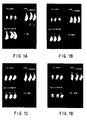
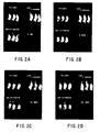
- FIGS 1 and 2 illustrate two representative experiments involving three animals in each experiment. Note that the same three animals were imaged at each time point. These images show that there was essentially no imaging of the inflammatory lesion with 67 Ga (considered the standard inflammatory scanning technique); minimal transient imaging with the 99m Tc. labeled albumen; but obvious, increasing visualization of the site of inflammation with the 125 1-labeled polyclonal human immunoglobulin preparation.
- a "dose-response” experiment was carried out utilizing 125 1-labeled polyclonal human immunoglobulin, in which doses of 65 ⁇ g, 650 ⁇ g, 5.8 mg, and 122 mg were utilized per animal. The procedures of Example 1 were used. In these experiments, the amount of radioactivity was kept constant (100 ⁇ Ci), and increasing amounts of "cold" immunoglobulin were added to increase the amount of protein injected. Thus, quantities approximately equivalent to a standard dose, 10x standard, 100x standard, and 1000x standard, were injected. All of these concentrations yielded similar images and similar counts over time, suggesting that the receptors for the immunoglobulin were not saturated even at a 1000x dose schedule. Because of the absence of epitopic specificity of the immunoglobulin preparation used, the accumulation of labeled immunoglobulin at the site of inflammation is not due to conventional recognition of epitopic determinants.
- Deep thigh inflammation was created in Sprague-Dawley rats as previously described above in Example 1. Twenty-four hours later, at a time when each animal clearly evidenced inflammation in the injected thigh, 65 ug of murine monoclonal antibody labeled with 100 uCi of either 125 I or 111 In were injected intravenously via the tail vein.
- the murine monoclonal antibodies employed in these experiments were of the IgG 1 or IgG 2 subclass and were of three epitopic specificities. The two epitopic specificities which were used were:
- the anti-arsenate monoclonal antibodies were injected into animals having either E. coli, S. aureus, or P. aeruginosa infection.
- the anti-E. coli monoclonal antibody was injected into animals bearing staphylococcal or Pseudomonas infection.
- Pseudomonas aeruginosa Type I deep thigh infection in Sprague-Dawley rats was created as described above. Twenty-four hours later, 0.65 ug of 125 1-labeled monoclonal antibodies of one of two possible antigenic specificities were injected intravenously via the tail vein. Half of the animals were injected with a murine monoclonal IgG 1 antibody specific for Type I P. aeruginosa. The remaining animals were injected with murine monoclonal IgG 1 antibody specific for arsenate hapten which is an antigen not found in mammalian tissues or bacteria. The anti-arsenate antibody is thus a control preparation with no epitopic specificity for this animal model.
- Deep thigh inflammation was created in Sprague-Dawley rats as described in Example 1 above. After 24 hours when each animal had clear evidence of inflammation, they were injected with 65 ⁇ g of either 125 labeled Fc fragment (100 ⁇ Ci), 125 1-labeled Fab fragment (100 ⁇ Ci), or 125 1-labeled polyclonal human immunoglobulin (100 ⁇ Ci, Sandoglobulin@).
- the immunoglobulin fragments were derived from a polyclonal human immunoglobulin preparation (Sandoglobulin@) using standard techniques. The animals were then scanned using a gamma camera as described in Example 1.
- the non-specific immunoglobulin scan can be utilized in a variety of inflammatory states such as those due to a variety of infectious agents or the non-infectious inflammatory agent turpentine.
- approximately 10 8 organisms, or 0.5 cc of turpentine was injected into the thigh of 100 gram Sprague-Dawley rats.
- the non-specific immunoglobulin scan can detect inflammation at different anatomical sites.
- a Unilateral nephrectomies were carried out in two groups of rats. In one group, the operation was done sterilely. In the other group, the nephrectomy sites were inoculated with 10 8 E. coli. Both groups of animals were injected with 111 In-labeled Sandoglobulin@ 48 hours later, with serial scans then carried out with the gamma camera. In every instance, scans taken 24 hours post-injection could clearly distinguish sterile post-operative surgically induced inflammation from deep surgical wound infection.
- Intra-abdominal abscesses were created with Bacteriodes fragilis or mixed bowel bacteria according to standard methods. Approximately 7-10 days later, 111 In-labeled Sandoglobulin@ was administered intravenously and serial images obtained with the gamma camera. In each instance, the scans demonstrated the site of the intra-abdominal abscess.
- Pneumocyctis carinii pneumonia is an opportunistic pneumonia observed in immunocompromised patients, particularly those with AIDS. Early diagnosis, and assessment of response to therapy, particularly in those patients with equivocal chest x-ray, is an important clinical need. Model Pneumocyctis pneumonia was created in rats by placing them on a protein-deficient diet and treating them with daily cortisone and tetracycline for two months. This resulted in the development of Pneumocyctis pneumonia.
- Anti-inflammatory therapies do not influence the utility of this scanning technique.
- Groups of animals were pre-treated for 5-7 days with one of the following anti-inflammatory regimens: methylprednisilone, 5 mg IP daily; indomethacin 10 mg/kg/day IV; and acetylsalicylic acid administered in the drinking water to achieve a blood level of 15-25 mg/liter (therapeutic levels used in treatment of humans with rheumatoid arthritis).
- Model thigh infection was then created with E. coli as described in Example 1, and 24 hours later (while the anti-inflammatory therapy was continued)
- 111 In-labeled Sandoglobulin@ was injected intravenously, with serial scans then carried out.
- Example 6 The Fab and Fc studies described in Example 6 above were repeated utilizing model infection created with either E. coli or Klebsiella pneumoniae. In every instance, the Fab fragment gave no images, whereas the Fc fragment gave images similar to those obtained with immunoglobulins. These results were identical to those seen in Example 6.
- a 62-year-old woman with extensive vascular disease undervent a left femoral artery-inferior tibial artery bypass graft on 31/10/86; on 18/11/86 she underwent a right femoral-popliteal arterial bypass graft; and on 2/12/86 a thrombectomy of the right femoral artery. Over the next two weeks, she had a low-grade fever, with minimal findings at her operative site. On 16/12/86, she underwent an immunoglobulin scan which revealed a clear-cut accumulation in the lower portion of her right femoral graft ( Figure 4) of radiolabeled immunoglobulin 6 hours post-injection of the reagent.
- a middle-aged woman presented with fever, lower abdominal pain, and a palpable mass on rectal examination.
- a computerized tomographic (CT) study of her abdomen revealed no abnormality.
- An immunoglobulin scan revealed a clear-cut accumulation of radiolabeled immunoglobulin in the lower abdomen as early as 3 hours post-injection ( Figure 5).
- Surgical exploration revealed a large diverticular abscess at the anatomical site outlined by the immunoglobulin scan.
Landscapes
- Health & Medical Sciences (AREA)
- Life Sciences & Earth Sciences (AREA)
- Chemical & Material Sciences (AREA)
- Medicinal Chemistry (AREA)
- General Health & Medical Sciences (AREA)
- Immunology (AREA)
- Proteomics, Peptides & Aminoacids (AREA)
- Animal Behavior & Ethology (AREA)
- Public Health (AREA)
- Veterinary Medicine (AREA)
- Epidemiology (AREA)
- Engineering & Computer Science (AREA)
- Bioinformatics & Cheminformatics (AREA)
- Organic Chemistry (AREA)
- Pharmacology & Pharmacy (AREA)
- Molecular Biology (AREA)
- Physics & Mathematics (AREA)
- Nuclear Medicine, Radiotherapy & Molecular Imaging (AREA)
- Genetics & Genomics (AREA)
- Biophysics (AREA)
- Biochemistry (AREA)
- Optics & Photonics (AREA)
- Radiology & Medical Imaging (AREA)
- Toxicology (AREA)
- Rheumatology (AREA)
- Chemical Kinetics & Catalysis (AREA)
- General Chemical & Material Sciences (AREA)
- Pain & Pain Management (AREA)
- Medicines Containing Antibodies Or Antigens For Use As Internal Diagnostic Agents (AREA)
- Medicines That Contain Protein Lipid Enzymes And Other Medicines (AREA)
- Pharmaceuticals Containing Other Organic And Inorganic Compounds (AREA)
- Preparation Of Compounds By Using Micro-Organisms (AREA)
- Peptides Or Proteins (AREA)
- Medicines Containing Material From Animals Or Micro-Organisms (AREA)
- Investigating Or Analysing Biological Materials (AREA)
- External Artificial Organs (AREA)
- Chemical Treatment Of Metals (AREA)
Claims (18)
Priority Applications (1)
| Application Number | Priority Date | Filing Date | Title |
|---|---|---|---|
| EP93202324A EP0577225B9 (fr) | 1986-01-16 | 1987-01-16 | Diagnostic et traitement d'inflammation |
Applications Claiming Priority (4)
| Application Number | Priority Date | Filing Date | Title |
|---|---|---|---|
| US81958586A | 1986-01-16 | 1986-01-16 | |
| US819585 | 1986-01-16 | ||
| WOPCT/US87/00079 | 1987-01-12 | ||
| PCT/US1987/000079 WO1987004351A1 (fr) | 1986-01-16 | 1987-01-12 | Procede pour diagnostiquer et traiter des inflammations |
Related Child Applications (1)
| Application Number | Title | Priority Date | Filing Date |
|---|---|---|---|
| EP93202324.5 Division-Into | 1993-08-06 |
Publications (4)
| Publication Number | Publication Date |
|---|---|
| EP0241106A2 EP0241106A2 (fr) | 1987-10-14 |
| EP0241106A3 EP0241106A3 (en) | 1989-02-08 |
| EP0241106B1 true EP0241106B1 (fr) | 1994-08-31 |
| EP0241106B2 EP0241106B2 (fr) | 1998-07-08 |
Family
ID=25228549
Family Applications (2)
| Application Number | Title | Priority Date | Filing Date |
|---|---|---|---|
| EP93202324A Expired - Lifetime EP0577225B9 (fr) | 1986-01-16 | 1987-01-16 | Diagnostic et traitement d'inflammation |
| EP87300395A Expired - Lifetime EP0241106B2 (fr) | 1986-01-16 | 1987-01-16 | Utilisation d'une immunoglobuline marquée et identifiable ou de l'un de ses fragments dans la préparation d'un agent pour la détection d'un site d'inflammation in vivo |
Family Applications Before (1)
| Application Number | Title | Priority Date | Filing Date |
|---|---|---|---|
| EP93202324A Expired - Lifetime EP0577225B9 (fr) | 1986-01-16 | 1987-01-16 | Diagnostic et traitement d'inflammation |
Country Status (9)
| Country | Link |
|---|---|
| EP (2) | EP0577225B9 (fr) |
| JP (1) | JP2515357B2 (fr) |
| AT (2) | ATE110572T1 (fr) |
| AU (1) | AU597032B2 (fr) |
| CA (1) | CA1288688C (fr) |
| DE (3) | DE3750434T3 (fr) |
| ES (2) | ES2056812T5 (fr) |
| GR (1) | GR940300027T1 (fr) |
| WO (1) | WO1987004351A1 (fr) |
Families Citing this family (7)
| Publication number | Priority date | Publication date | Assignee | Title |
|---|---|---|---|---|
| JP2515357B2 (ja) * | 1986-01-16 | 1996-07-10 | ザ・ジェネラル・ホスピタル・コ−ポレ−ション | 炎症の診断方法および治療方法 |
| FR2639231A1 (fr) * | 1988-11-23 | 1990-05-25 | Merieux Inst | Utilisation de fragments fc d'immunoglobulines g humaines, de peptides analogues du fragment fc, ou d'immunoglobulines g humaines dans l'obtention d'un medicament preparatoire a l'administration chez l'homme, dans un but therapeutique ou de diagnostic, d'anticorps monoclonaux |
| US5792444A (en) * | 1989-05-09 | 1998-08-11 | The General Hospital Corporation | Labeled chemotactic peptides to image focal sites of infection or inflammation |
| CA2016235C (fr) * | 1989-05-09 | 2002-03-12 | Alan J. Fischman | Peptides chimiotactiques marques pour l'imagerie des foyers d'infection ou d'inflammation |
| EP0485664A1 (fr) * | 1990-11-13 | 1992-05-20 | Mallinckrodt Diagnostica (Holland) B.V. | Procédé de préparation d'agent diagnostique pour la détection des inflammations |
| ATE175976T1 (de) * | 1993-10-22 | 1999-02-15 | Nihon Mediphysics Co Ltd | Peptide mit affinität für entzündungen und deren radiomarkierte, diagnostische zusammensetzungen |
| DE19514088A1 (de) * | 1995-04-13 | 1996-10-17 | Deutsches Krebsforsch | Konjugat zur Behandlung von Entzünduns-, Infektions- und/oder Hauterkrankungen |
Family Cites Families (18)
| Publication number | Priority date | Publication date | Assignee | Title |
|---|---|---|---|---|
| GB2004464B (en) * | 1977-06-30 | 1982-01-13 | Cehovic G | Method of testing inflammation |
| US4315906A (en) * | 1979-05-21 | 1982-02-16 | New England Nuclear Corporation | Cold insoluble globulin, its purification and use |
| US4472371A (en) * | 1979-10-29 | 1984-09-18 | Summa Medical Corporation | Radiolabeled antibody to anti-tumor associated antigen and process |
| US4444744A (en) * | 1980-03-03 | 1984-04-24 | Goldenberg Milton David | Tumor localization and therapy with labeled antibodies to cell surface antigens |
| GB2101407A (en) * | 1981-05-14 | 1983-01-12 | Bulpitt And Sons Limited | Automatic electric kettle |
| US4522918A (en) * | 1981-12-15 | 1985-06-11 | Jeffery Schlom | Process for producing monoclonal antibodies reactive with human breast cancer |
| US4472509A (en) * | 1982-06-07 | 1984-09-18 | Gansow Otto A | Metal chelate conjugated monoclonal antibodies |
| US4732864A (en) * | 1983-10-06 | 1988-03-22 | E. I. Du Pont De Nemours And Company | Trace-labeled conjugates of metallothionein and target-seeking biologically active molecules |
| GR850552B (fr) * | 1984-03-07 | 1985-07-05 | Du Pont | |
| US4636380A (en) * | 1984-04-23 | 1987-01-13 | Wong Dennis W | Novel physiologic chemical method of labeling protein substances with the radionuclides of indium |
| US4634586A (en) * | 1984-05-21 | 1987-01-06 | The Board Of Trustees Of The Leland Stanford Junior University | Reagent and method for radioimaging leukocytes |
| US4659839A (en) * | 1984-10-10 | 1987-04-21 | Mallinckrodt, Inc. | Coupling agents for radiolabeled antibody fragments |
| US4639365A (en) * | 1984-10-18 | 1987-01-27 | The Board Of Regents, The University Of Texas System | Gadolinium chelates as NMR contrast agents |
| GB8426470D0 (en) * | 1984-10-19 | 1984-11-28 | Technology Licence Co Ltd | Monoclonal antibodies |
| AU597877B2 (en) * | 1984-12-26 | 1990-06-14 | Teijin Limited | Anti-pseudomonas aeruginosa human monoclonal antibody |
| JP2515357B2 (ja) * | 1986-01-16 | 1996-07-10 | ザ・ジェネラル・ホスピタル・コ−ポレ−ション | 炎症の診断方法および治療方法 |
| GB8610202D0 (en) * | 1986-04-25 | 1986-05-29 | Technology Licence Co Ltd | Monoclonal antibodies |
| GB8621910D0 (en) * | 1986-09-11 | 1986-10-15 | Technology Licence Co Ltd | Monoclonal antibodies |
-
1987
- 1987-01-12 JP JP62501013A patent/JP2515357B2/ja not_active Expired - Fee Related
- 1987-01-12 WO PCT/US1987/000079 patent/WO1987004351A1/fr not_active Ceased
- 1987-01-12 AU AU69377/87A patent/AU597032B2/en not_active Ceased
- 1987-01-15 CA CA000527403A patent/CA1288688C/fr not_active Expired - Lifetime
- 1987-01-16 EP EP93202324A patent/EP0577225B9/fr not_active Expired - Lifetime
- 1987-01-16 DE DE3750434T patent/DE3750434T3/de not_active Expired - Fee Related
- 1987-01-16 DE DE3752380T patent/DE3752380T2/de not_active Expired - Fee Related
- 1987-01-16 DE DE0577225T patent/DE577225T1/de active Pending
- 1987-01-16 ES ES87300395T patent/ES2056812T5/es not_active Expired - Lifetime
- 1987-01-16 ES ES93202324T patent/ES2053403T3/es not_active Expired - Lifetime
- 1987-01-16 EP EP87300395A patent/EP0241106B2/fr not_active Expired - Lifetime
- 1987-01-16 AT AT87300395T patent/ATE110572T1/de not_active IP Right Cessation
- 1987-01-16 AT AT93202324T patent/ATE283710T1/de not_active IP Right Cessation
-
1994
- 1994-05-31 GR GR940300027T patent/GR940300027T1/el unknown
Also Published As
| Publication number | Publication date |
|---|---|
| DE3752380D1 (de) | 2005-01-05 |
| DE577225T1 (de) | 1994-09-22 |
| ES2056812T3 (es) | 1994-10-16 |
| EP0241106A3 (en) | 1989-02-08 |
| WO1987004351A1 (fr) | 1987-07-30 |
| DE3752380T2 (de) | 2005-11-03 |
| ES2053403T1 (es) | 1994-08-01 |
| EP0577225B9 (fr) | 2005-03-02 |
| CA1288688C (fr) | 1991-09-10 |
| GR940300027T1 (en) | 1994-05-31 |
| JPS63502280A (ja) | 1988-09-01 |
| DE3750434T3 (de) | 1999-04-08 |
| EP0241106A2 (fr) | 1987-10-14 |
| AU6937787A (en) | 1987-08-14 |
| EP0577225A1 (fr) | 1994-01-05 |
| JP2515357B2 (ja) | 1996-07-10 |
| ES2056812T5 (es) | 1998-11-16 |
| ATE283710T1 (de) | 2004-12-15 |
| EP0577225B1 (fr) | 2004-12-01 |
| AU597032B2 (en) | 1990-05-24 |
| EP0241106B2 (fr) | 1998-07-08 |
| DE3750434T2 (de) | 1995-03-23 |
| ES2053403T3 (es) | 2005-06-01 |
| DE3750434D1 (de) | 1994-10-06 |
| ATE110572T1 (de) | 1994-09-15 |
Similar Documents
| Publication | Publication Date | Title |
|---|---|---|
| US4926869A (en) | Method for the diagnosis and treatment of inflammation | |
| Rubin et al. | 111In-labeled nonspecific immunoglobulin scanning in the detection of focal infection | |
| KR0185995B1 (ko) | 감염 또는 염증의 병소부위를 이미지화하기 위한 표지된 주화성 펩티드 | |
| Fischman et al. | Detection of acute inflammation with 111In-labeled nonspecific polyclonal IgG | |
| Becker et al. | The use of monoclonal antibodies and antibody fragments in the imaging of infectious lesions | |
| US4925648A (en) | Detection and treatment of infectious and inflammatory lesions | |
| US4634586A (en) | Reagent and method for radioimaging leukocytes | |
| JP3288996B2 (ja) | 白血球付着レセプターβ鎖に対するモノクローナル抗体と、この抗体の製造方法と、その応用 | |
| Corstens et al. | Radioimmunoconjugates in the detection of infection and inflammation | |
| EP0241106B2 (fr) | Utilisation d'une immunoglobuline marquée et identifiable ou de l'un de ses fragments dans la préparation d'un agent pour la détection d'un site d'inflammation in vivo | |
| Lashford et al. | Therapeutic Application of Radiolabeled Monoclonal Antibody | |
| USRE35152E (en) | Method for the diagnosis and treatment of inflammation | |
| Kemshead et al. | 131-I coupled to monoclonal antibodies as therapeutic agents for neuroectodermally derived tumors: fact or fiction? | |
| US5200178A (en) | Method for tumor detection | |
| US5277894A (en) | Method for tumor detection and treatment | |
| Da Costa et al. | Immunoscintigraphy in Hodgkin's disease and anaphastic large cell lymphomas: Results in 18 patients using the iodine radiolabeled monoclonal antibody HRS-3 | |
| Babich et al. | Targeted imaging of infection | |
| Greager et al. | Localization of human sarcoma with radiolabeled monoclonal antibody: A preliminary study | |
| Thakur | Immunoscintigraphic imaging of inflammatory lesions: preliminary findings and future possibilities | |
| CA1339340C (fr) | Methode pour la detection et le traitement de tumeurs | |
| Begent et al. | Second antibody for improvement of antibody imaging: liposome-entrapped and free preparations in animal and human studies | |
| Britton et al. | Bacterial Specific Infection Imaging | |
| Carrasquillo et al. | Considerations in the diagnosis and therapy of human T-cell lymphoma with monoclonal antibodies | |
| Greager et al. | Localization of human sarcoma with radiolabeled monoclonal antibody—a follow-up report | |
| Ledermann | Investigation of two methods to improve the therapeutic potential of radiolabelled antibodies: Use of an anti-antibody and suppression of the host anti-antibody response |
Legal Events
| Date | Code | Title | Description |
|---|---|---|---|
| PUAI | Public reference made under article 153(3) epc to a published international application that has entered the european phase |
Free format text: ORIGINAL CODE: 0009012 |
|
| AK | Designated contracting states |
Kind code of ref document: A2 Designated state(s): AT BE CH DE ES FR GB GR IT LI LU NL SE |
|
| PUAL | Search report despatched |
Free format text: ORIGINAL CODE: 0009013 |
|
| AK | Designated contracting states |
Kind code of ref document: A3 Designated state(s): AT BE CH DE ES FR GB GR IT LI LU NL SE |
|
| 17P | Request for examination filed |
Effective date: 19890629 |
|
| 17Q | First examination report despatched |
Effective date: 19900906 |
|
| GRAA | (expected) grant |
Free format text: ORIGINAL CODE: 0009210 |
|
| ITF | It: translation for a ep patent filed | ||
| AK | Designated contracting states |
Kind code of ref document: B1 Designated state(s): AT BE CH DE ES FR GB GR IT LI LU NL SE |
|
| REF | Corresponds to: |
Ref document number: 110572 Country of ref document: AT Date of ref document: 19940915 Kind code of ref document: T |
|
| REF | Corresponds to: |
Ref document number: 3750434 Country of ref document: DE Date of ref document: 19941006 |
|
| REG | Reference to a national code |
Ref country code: ES Ref legal event code: FG2A Ref document number: 2056812 Country of ref document: ES Kind code of ref document: T3 |
|
| ET | Fr: translation filed | ||
| RIN2 | Information on inventor provided after grant (corrected) |
Free format text: RUBIN, ROBERT H. * STRAUSS, WILLIAM H. |
|
| EAL | Se: european patent in force in sweden |
Ref document number: 87300395.8 |
|
| REG | Reference to a national code |
Ref country code: GR Ref legal event code: FG4A Free format text: 3014077 |
|
| K2C1 | Correction of patent specification (title page) published |
Effective date: 19940831 |
|
| NLXE | Nl: other communications concerning ep-patents (part 3 heading xe) |
Free format text: PAT.BUL.01/95 CORR.: STRAUSS, SKILLMAN, NEW JERSEY VER. ST. V. AM. (SEE EUROPEAN PATENT BULLETIN 110195/02) |
|
| PLBI | Opposition filed |
Free format text: ORIGINAL CODE: 0009260 |
|
| 26 | Opposition filed |
Opponent name: MALLINCKRODT MEDICAL B.V. Effective date: 19950526 |
|
| NLR1 | Nl: opposition has been filed with the epo |
Opponent name: MALLINCKRODT MEDICAL B.V. |
|
| PLBF | Reply of patent proprietor to notice(s) of opposition |
Free format text: ORIGINAL CODE: EPIDOS OBSO |
|
| PLAW | Interlocutory decision in opposition |
Free format text: ORIGINAL CODE: EPIDOS IDOP |
|
| PLAW | Interlocutory decision in opposition |
Free format text: ORIGINAL CODE: EPIDOS IDOP |
|
| PUAH | Patent maintained in amended form |
Free format text: ORIGINAL CODE: 0009272 |
|
| STAA | Information on the status of an ep patent application or granted ep patent |
Free format text: STATUS: PATENT MAINTAINED AS AMENDED |
|
| 27A | Patent maintained in amended form |
Effective date: 19980708 |
|
| AK | Designated contracting states |
Kind code of ref document: B2 Designated state(s): AT BE CH DE ES FR GB GR IT LI LU NL SE |
|
| REG | Reference to a national code |
Ref country code: CH Ref legal event code: AEN Free format text: MAINTIEN DU BREVET DONT L'ETENDUE A ETE MODIFIEE |
|
| NLR2 | Nl: decision of opposition | ||
| PG25 | Lapsed in a contracting state [announced via postgrant information from national office to epo] |
Ref country code: GR Free format text: THE PATENT HAS BEEN ANNULLED BY A DECISION OF A NATIONAL AUTHORITY Effective date: 19980902 |
|
| NLR3 | Nl: receipt of modified translations in the netherlands language after an opposition procedure | ||
| REG | Reference to a national code |
Ref country code: ES Ref legal event code: DC2A Kind code of ref document: T5 Effective date: 19981008 |
|
| ET3 | Fr: translation filed ** decision concerning opposition | ||
| REG | Reference to a national code |
Ref country code: GB Ref legal event code: IF02 |
|
| PGFP | Annual fee paid to national office [announced via postgrant information from national office to epo] |
Ref country code: GB Payment date: 20041210 Year of fee payment: 19 Ref country code: AT Payment date: 20041210 Year of fee payment: 19 |
|
| PGFP | Annual fee paid to national office [announced via postgrant information from national office to epo] |
Ref country code: NL Payment date: 20041215 Year of fee payment: 19 |
|
| PGFP | Annual fee paid to national office [announced via postgrant information from national office to epo] |
Ref country code: LU Payment date: 20041220 Year of fee payment: 19 |
|
| PGFP | Annual fee paid to national office [announced via postgrant information from national office to epo] |
Ref country code: FR Payment date: 20050105 Year of fee payment: 19 |
|
| PGFP | Annual fee paid to national office [announced via postgrant information from national office to epo] |
Ref country code: SE Payment date: 20050107 Year of fee payment: 19 |
|
| PGFP | Annual fee paid to national office [announced via postgrant information from national office to epo] |
Ref country code: GR Payment date: 20050118 Year of fee payment: 19 |
|
| PGFP | Annual fee paid to national office [announced via postgrant information from national office to epo] |
Ref country code: ES Payment date: 20050119 Year of fee payment: 19 |
|
| PGFP | Annual fee paid to national office [announced via postgrant information from national office to epo] |
Ref country code: BE Payment date: 20050127 Year of fee payment: 19 |
|
| PGFP | Annual fee paid to national office [announced via postgrant information from national office to epo] |
Ref country code: DE Payment date: 20050131 Year of fee payment: 19 |
|
| PGFP | Annual fee paid to national office [announced via postgrant information from national office to epo] |
Ref country code: CH Payment date: 20050317 Year of fee payment: 19 |
|
| PG25 | Lapsed in a contracting state [announced via postgrant information from national office to epo] |
Ref country code: GB Free format text: LAPSE BECAUSE OF NON-PAYMENT OF DUE FEES Effective date: 20060116 Ref country code: AT Free format text: LAPSE BECAUSE OF NON-PAYMENT OF DUE FEES Effective date: 20060116 |
|
| PG25 | Lapsed in a contracting state [announced via postgrant information from national office to epo] |
Ref country code: SE Free format text: LAPSE BECAUSE OF NON-PAYMENT OF DUE FEES Effective date: 20060117 Ref country code: ES Free format text: LAPSE BECAUSE OF NON-PAYMENT OF DUE FEES Effective date: 20060117 |
|
| PG25 | Lapsed in a contracting state [announced via postgrant information from national office to epo] |
Ref country code: LU Free format text: LAPSE BECAUSE OF NON-PAYMENT OF DUE FEES Effective date: 20060131 Ref country code: LI Free format text: LAPSE BECAUSE OF NON-PAYMENT OF DUE FEES Effective date: 20060131 Ref country code: FR Free format text: LAPSE BECAUSE OF NON-PAYMENT OF DUE FEES Effective date: 20060131 Ref country code: CH Free format text: LAPSE BECAUSE OF NON-PAYMENT OF DUE FEES Effective date: 20060131 Ref country code: BE Free format text: LAPSE BECAUSE OF NON-PAYMENT OF DUE FEES Effective date: 20060131 |
|
| PGFP | Annual fee paid to national office [announced via postgrant information from national office to epo] |
Ref country code: IT Payment date: 20060131 Year of fee payment: 20 |
|
| PG25 | Lapsed in a contracting state [announced via postgrant information from national office to epo] |
Ref country code: NL Free format text: LAPSE BECAUSE OF NON-PAYMENT OF DUE FEES Effective date: 20060801 Ref country code: DE Free format text: LAPSE BECAUSE OF NON-PAYMENT OF DUE FEES Effective date: 20060801 |
|
| REG | Reference to a national code |
Ref country code: CH Ref legal event code: PL |
|
| EUG | Se: european patent has lapsed | ||
| GBPC | Gb: european patent ceased through non-payment of renewal fee |
Effective date: 20060116 |
|
| NLV4 | Nl: lapsed or anulled due to non-payment of the annual fee |
Effective date: 20060801 |
|
| REG | Reference to a national code |
Ref country code: FR Ref legal event code: ST Effective date: 20060929 |
|
| REG | Reference to a national code |
Ref country code: ES Ref legal event code: FD2A Effective date: 20060117 |
|
| BERE | Be: lapsed |
Owner name: THE *GENERAL HOSPITAL CORP. Effective date: 20060131 |