EP0214520A2 - Immunotestverfahren zur Bestimmung von RAS-codierten Proteinen - Google Patents
Immunotestverfahren zur Bestimmung von RAS-codierten Proteinen Download PDFInfo
- Publication number
- EP0214520A2 EP0214520A2 EP86111527A EP86111527A EP0214520A2 EP 0214520 A2 EP0214520 A2 EP 0214520A2 EP 86111527 A EP86111527 A EP 86111527A EP 86111527 A EP86111527 A EP 86111527A EP 0214520 A2 EP0214520 A2 EP 0214520A2
- Authority
- EP
- European Patent Office
- Prior art keywords
- antibody
- polypeptide
- ras
- oncogene
- encoded
- Prior art date
- Legal status (The legal status is an assumption and is not a legal conclusion. Google has not performed a legal analysis and makes no representation as to the accuracy of the status listed.)
- Withdrawn
Links
Images


Classifications
-
- C—CHEMISTRY; METALLURGY
- C07—ORGANIC CHEMISTRY
- C07K—PEPTIDES
- C07K16/00—Immunoglobulins [IG], e.g. monoclonal or polyclonal antibodies
- C07K16/18—Immunoglobulins [IG], e.g. monoclonal or polyclonal antibodies against material from animals or humans
- C07K16/32—Immunoglobulins [IG], e.g. monoclonal or polyclonal antibodies against material from animals or humans against translation products of oncogenes
Definitions
- RNA tumor viruses and human tumor cells contain genomic sequences, termed oncogenes, that encode specific proteins which have been shown to be responsible for the induction of various human and animal cancers (6,18).
- This invention is based upon the discovery that normally intracellular, oncogene-encoded proteins may be detected in biological fluids such as serum and that such detection may be be utilized to diagnose or monitor neoplastic conditions or states.
- DNA isolated from viral or chemically transformed cells has the ability to cause transformation by transfection of normal cell lines.
- Transforming DNAS have been derived from a variety of human tumors including carcinoma of the bladder, lung, colon, pancreas, sarcomas, hemopoietic tumors, and neuroblastomas (6,20).
- D is- crete transforming DNA sequences have been identified in the DNA of both transformed cells and retroviruses; these sequences are termed oncogenes (16).
- the oncogenic DNA sequences derived from RNA tumor viruses show great homology with DNA sequences from normal uninfected cells (4). These homologous sequences found in viruses and cells are termed viral oncogenes (v-onc) and cellular oncogenes (c-onc), respectively.
- c-onc genes are universally present in the DNA of all individuals and appear to be the evolutionary progenitors of v-onc genes. It is believed that the RNA tumor viruses have acquired oncogenes by recombinational events between the retrovirus and the host genome (transduction) (26).
- Cellular oncogenes are normal genes which are conserved throughout evolution and are believed to have normal functional roles in the cell (2). In their "non-cancer inducing state" they are termed proto-oncogenes.
- the proto-oncogenes are not oncogenic or tumorigenic until they are activated in some way.
- a number of different genetic mechanisms cause the somatic mutation of normal cellular genes that results in the activated oncogenes found in tumor cells.
- Mechanisms of oncogene activation include overexpression point mutations, translocations, gene rearrangement, and gene amplification, which may be induced by chemical or physical carcinogenic means, or by the integration of a viral genome adjacent to the proto-oncogene sequences in the host DNA (15).
- Oncogenes There are approximately 20 known oncogenes (3,32) and several have been shown to be associated with specific forms of cancer. Oncogenes show sequence homology with normal cellular genes that encode proteins such as growth factors, growth factor membrane receptors, GTP - binding proteins, and specific protein kinases.
- oncogenes Studies have been undertaken to correlate various oncogenes with specific cancer types. The expression of 15 different oncogenes was associated with twenty different tumor types by DNA/RNA hybridization with viral probes (25). The oncogenes (c-onc) c-myc, c-fos, c-H-ras, and c-K-ras were expressed in all tumor tissue examined, while other oncogenes appeared to correlate with specific tumor types.
- a family of oncogenes found to be most important in the study of human oncogenesis is the ras oncogene.
- the ras gene family consists of three structurally similar members: H-ras, K-ras, and N-ras.
- H- and K-ras transforming genes were first isolated from the Harvey and Kirsten strains of murine sarcoma virus (MuSV). Subsequently, H- and K- ras oncogenes were directly isolated from a variety of human tumor cell lines (by hybridization) and the cloned H- and K-ras genes were found to transform mouse NIH 3T3 cells in culture by transfection (8).
- the H- and N-ras genes consists of four exons and the K-ras has in addition, an alternative fourth exon (24).
- the first three exons are more highly conserved than the fourth exon and together they encode a protein of 21,000 daltons, designated p21, comprising 189 amino acids.
- the p2l has been shown to be structurally and immunologically related to guanine nucleotide binding proteins (10,17).
- the p21 protein has been shown by both genetic and biological experimentation to be directly responsible for the ras gene transformation of cells in culture.
- the H-ras oncogene isolated from a human bladder carcinoma cell line (T24) was the first oncogene that was shown to differ from the normal H-ras gene by a single base substitution (G to T) which substituted valine for glycine at position twelve in the amino acid sequence (27).
- the resultant mutant protein was extremely potent in inducing transformation (28).
- the activated or structurally altered ras oncogene protein has been found in human lung and colon carcinomas (9).
- ras p2l protein is found on the inner aspect of the cell membrane, and therefore is an intracellular protein, it was reasoned that since considerable tissue destruction is involved in the malignant process, ras p2l protein may be released into the sera of cancer patients.
- the studies described herein are concerned with a sensitive serum-based immunoassay that utilizes monoclonal antibodies to detect ras encoded p21 protein in the sera of a variety of cancer patients but at much higher frequency than in normal human sera. This assay has potential for expansion to other body fluids such as urine and to include all oncogenes associated with the presence of a neoplastic condition.
- the invention concerns a method for diagnosing in a subject a neoplastic condition associated with the presence of an activated oncogene which comprises detecting in a sample of a biological fluid from the subject the presence of at least a portion of a polypeptide encoded by the activated oncogene, the polypeptide normally occurring intracellularly.
- the detecting may comprise contacting the sample under suitable conditions with a first matrix-bound antibody specific for the polypeptide to form a matrix-first antibody-polypeptide complex, contacting the complex so formed with a second antibody labelled with a detectable marker to form a second complex comprising matrix-first antibody-polypeptide-second antibody, and, finally, detecting the second complex so formed, thereby detecting the presence of the oncogene-encoded polypeptide.
- the detecting may comprise contacting the sample under suitable conditions with an antibody specific for the portion of the polypeptide to form an antibody-polypeptide complex and detecting the complex so formed, thereby detecting in the sample the presence of the oncogene-encoded polypeptide.
- the detecting may also comprise contacting a detectable control polypeptide with a matrix-bound antibody molecule specific for the portion of the polypeptide in the presence of the oncogene-encoded polypeptide, thereby forming a matrix-antibody-control polypeptide complex, quantitatively determining the number of complexes so formed and comparing the number so determined with the number of complexes so formed in the absence of the oncogene-encoded polypeptide, a decrease in the number of complexes formed indicating the presence of the oncogene-encoded polypeptide in the sample.
- This invention also concerns a method for diagnosing in a subject a neoplastic condition associated with the presence of an activated oncogene which comprises contacting the sample under suitable conditions with a first antibody specific for an epitope on the portion of the polypeptide to be detected, and with a second antibody specific for a different epitope on the portion of the polypeptide to be detected so as to form a first antibody-polypeptide-second antibody complex, the complex being detectable.
- This invention also concerns a method for diagnosing in a subject a neoplastic condition associated with the presence of an activated oncogene which comprises quantitatively determining in a sample of a biological fluid from the subject the amount of an oncogene-encoded polypeptide and comparing the amount of the polypeptide so determined to the amount in a sample from a normal subject, the presence of a significantly different amount indicating the presence of the neoplastic condition.
- the invention also concerns a method for monitoring the course of a neoplastic condition in a subject which comprises quantitatively determining in a first sample of a biological fluid from the subject the presence of an oncogene-encoded polypeptide and comparing the amount so determined with the amount present in a second sample from the subject, such samples being taken at different points in time, a difference in the amounts determined being indicative of the course of the neoplastic condition.
- the invention also concerns a method for typing tumors which comprises quantitatively determining in a sample of a biological fluid from a subject with a neoplastic condition the amount of one or more oncogene-encoded polypeptides in the sample, the presence of specific amounts or relative amounts thereof being indicative of a specific tumor type.
- the invention also concerns a method for typing tumors which comprises detecting in a sample of a biological fluid from a subject with neoplastic condition the presence of one or more oncogene-encoded polypeptides in the sample, the presence or absence of a specific combination thereof being indicative of a specific tumor type.
- the invention also concerns a method for detecting an activated oncogene which comprises detecting in a sample of a biological fluid at least a portion of a polypeptide encoded by the activated oncogene, the polypeptide occurring intracellularly.
- the invention concerns a method for screening putative therapeutic agents for the treatment of a neoplastic condition which comprises quantitatively determining in a first sample from a subject with the neoplastic condition the amount of an oncogene-encoded polypeptide associated with the condition, administering to the subject a therapeutic amount of the agent such that the agent is contacted with a neoplastic cell associated with the condition to produce a treated subject, determining after a suitable period the amount of the polypeptide in a sample from the treated subject, and comparing the amount of polypeptide determined in the first sample with the amount determined in the sample from the treated subject, a significant difference indicating the effectiveness of the agent.
- the invention concerns a method for diagnosing in a subject a neoplastic condition associated with the presence of an activated oncogene.
- the method comprises detecting in a sample of a biological fluid from the subject, e.g., an animal or a human, the presence of at least a portion of a polypeptide associated with an activated oncogene.
- the activated oncogene may be c-H-ras, C-K-ras, C-N-ras, c-myc, c-N-myc, c-L-myc, c-R-myc, c-abl, c-fos, or the oncogene which encodes polypeptide p53, although other activated oncogenes may be involved.
- the polypeptide encoded by the oncogene may be p2l of c-H-ras, c-K-ras or c-N-ras, or may be the polypeptide encoded by c-myc (p62, p65), c-N-myc (p64, p66), phl/c-abl (p210), c-L-myc, c-R-myc, c-fos (p55), or p53, or any other polypeptide which is encoded by an activated oncogene.
- the polypeptide may be a fusion polypeptide in part encoded by the oncogene and in part by the chromosomal DNA of the subject.
- the polypeptide may normally be found associated with the inner aspect of the cell membrane or with the nucleus, but may also be an integral membrane protein.
- the biological fluid is sera; other fluids, e.g., urine, cerebro-spinal fluid, amniotic fluid, sputum, a lung lavage, ascites fluid, saliva, any mucous-type bodily secretion, blood, or plasma may be used.
- this comprises contacting the sample under suitable conditions with a matrix-bound antibody specific for the polypeptide, thereby forming a matrix-antibody-polypeptide complex.
- This complex is further contacted with a second antibody molecule labelled with a detectable marker to form a second complex consisting of matrix-first antibody-polypeptide - second antibody and detecting the second complex so formed thereby detecting the presence of the oncogene-encoded -polypeptide.
- the monoclonal antibodies used were produced by hybridoma cell lines which are fully available from the American Type Culture Collection in Rockville, Maryland, U.S.A. 20852.
- the matrix-bound antibody may be v-H-ras (Ab-1) produced by hybridoma Y13-259 (ATCC No. CRL1742) and the second antibody may be v-H-ras (Ab-2) produced by hybridoma Y13-238 (ATCC No. CRL1741).
- the first antibody is a monoclonal antibody attached to an agarose matrix, e.g., Affi-Gel * 10 (Bio-Rad, Richmond, CA), although the first antibody may be a polyclonal antibody or one of a selected combination (a "cocktail") of antibodies, and may be attached to a Sepharose * (Pharmacia, Piscataway, N.J.) matrix, a tube, a bead or any of a number of support matrices.
- an agarose matrix e.g., Affi-Gel * 10 (Bio-Rad, Richmond, CA)
- a Sepharose * Pharmacia, Piscataway, N.J.
- the second antibody is a monoclonal antibody although it may be a polyclonal antibody, and the attached detectable marker is 125I, or a variety of other radioactive labels, e.g., cobalt, or any of a variety of colorometric, fluorometric or luminescent markers or may be the product of an enzymatic reaction.
- the detection of the oncogene-encoded polypeptide comprises contacting the sample under suitable conditions with an antibody specific for the portion of the polypeptide, thus forming an antibody-polypeptide complex.
- the complex so formed is then detected.
- the antibody is a monoclonal antibody, such antibody forming a detectable immunoprecipitate when contacted with the oncogene-encoded polypeptide.
- the antibody may be monoclonal, polyclonal or one of a selected combination of antibodies.
- the antibody may also be labelled with a detectable marker, such as 1251, although the marker may be any one of a variety of radioactive markers, may be a colorometric, fluorometric or luminescent marker, or may be the product of an enzymatic reaction.
- the antibody may be v-H-ras (Ab-1) produced by hybridoma cell line Y13-259 (ATCC No. CRL1742), which is fully available from the American Type Culture Collection in Rockville, Maryland, U.S.A. 20852.
- the detection of the polypeptide comprises contacting a detectable control polypeptide with an antibody specific for the portion of the polypeptide in the presence of the oncogene-encoded polypeptide, thus forming a complex comprising antibody-control polypeptide.
- the number of complexes so formed is quantitatively determined oy methods known to those with average skill in the art. The number so determined is compared to the number of complexes formed in the absence of the oncogene-encoded polypeptide, a decrease in the number of complexes formed indicating the presence of the oncogene-encoded polypeptide in the sample.
- the antibody molecule may be monoclonal, polyclonal or one of a selected combination of antibodies, and may be bound to a matrix such as agarose, Sepharose * , part of a tube or a bead.
- the control polypeptide may be labelled with a radioactive label, e.g., 125 I, or any of a variety of suitable radioactive labels, or be a colorometric, fluorometric or luminescent marker or be the product of an enzymatic reaction.
- the antibody may be v-H-ras (Ab-1) produced by hybridoma cell line Y13-259 (ATCC No. CRL17 4 2), which is fully available from the American Type Culture Collection in Rockville, Maryland, U.S.A. 20852.
- This invention also concerns a method for diagnosing in a subject a neoplastic condition associated with the presence of an activated oncogene which comprises contacting the sample under suitable conditions with a first antibody specific for an epitope on the portion of the polypeptide to be detected, and with a second antibody specific for a different epitope on the portion of the polypeptide to be detected so as to form a first antibody-polypeptide-second antibody complex, the complex being detectable.
- Each antibody may be a monoclonal or polyclonal antibody.
- Detection may be effected by means of an enzymatic reaction, a chemical reaction, a wavelength shift of absorbed light, or an emission of light.
- the antibodies used were produced by hybridoma cell lines which are fully available from the American Type Culture Collection in Rockville, Maryland, U.S.A. 20852.
- the first antibody may be v-H-ras (Ab-1) produced by hybridoma Y13-259 (ATCC No. CRL1742) and the second antibody may be v-H-ras (Ab-2) produced by Y13-238 (ATCC No. CRL1741).
- a method for diagnosing in a subject a neoplastic condition associated with the presence of an activated oncogene comprises quantitatively determining, using techniques known to those skilled in the art, in a sample of a biological fluid from the subject, the amount of an oncogene-encoded polypeptide. The amount so determined is compared to the amount in a sample from a normal subject, e.g., a subject without a tumor, the presence of a significantly different amount indicating the presence of the neoplastic condition. Further disclosed is a method for monitoring the course of a neoplastic condition in a subject.
- the method comprises quantitatively determining, by methods known to those skilled in the art, in a first sample of a biological fluid from the subject, the presence of an oncogene-encoded polypeptide.
- the amount so determined is compared to the amount present in a second sample from the subject, such samples being taken at different points in time, i.e., one sample taken at a sufficient period after the other sample to allow for tumor growth or regression.
- a difference in the amounts determined is indicative of the course of the neoplastic condition, e.g., growth or regression.
- Neoplastic conditions include, but are not limited to carcinomas of the lung, bladder, breast, uterus, prostate, colon, adenocarcinoma of the lung, neuroblastomas, melanomas, rhabdomyosarcomas, lymphomas and leukemias.
- a method for typing tumors comprises detecting in a sample of a biological fluid from a subject with a neoplastic condition the presence of one or more oncogene-encoded polypeptides, e.g., c-N-ras p2l or p53.
- the presence or absence of a specific combination may be indicative of a specific tumor type.
- a method for typing tumors comprises quantitatively determining in a sample of a biological fluid from a subject with a neoplastic condition the amount of one or more oncogene-encoded polypeptides, e.g., the amount of c-N-ras p2l or of p53.
- the presence of specific amounts or relative amounts of the polypeptides e.g., significant increase in the amount of p53, being indicative of a specific tumor type.
- a method for detecting an activated oncogene comprises detecting in a sample of a biological fluid at least a portion of a polypeptide encoded by the activated oncogene, the polypeptide occurring intracellularly.
- a method for screening putative therapeutic agents for the treatment of a neoplatic condition comprises quantitatively determining in a first sample from a subject with the neoplastic condition the amount of an oncogene-encoded polypeptide e.g., c-K-ras p21, associated with the condition.
- a therapeutic amount of the agent is then administered to the subject such that the agent is contacted with a neoplastic cell associated with the condition to produce a treated subject.
- a suitable period e.g., a period long enough in time to allow significant tumor growth or regression
- the amount of the polypeptide in a sample from the treated subject is determined.
- the amount of polypeptide determined in the first sample is compared with the amount determined in the sample from the treated subject, a significant difference in the amount of polypeptide from the two samples indicating the effectiveness of the agent, i.e., whether it is associated with tumor regression.
- Hybridoma lines were maintained in RPMI 1640 supplemented with 10% fetal calf serum (MA Bioproducts, Wal- kersville, MD). In order to obtain large amounts of immunoglobulin, the cells were injected into pristane (Aldrich) primed rodents and ascites fluid collected after 5-10 days. Human tumor cell lines were routinely maintained in high glucose Dulbecco's modification of Eagle's medium (MEM) supplemented with 5% fetal calf serum and the transfected, N-, K- or H-ras NIH (3T3) Cl 2.2 cell lines were supplemented with 5% Colorado calf serum (Colorado Serum Co.).
- MEM Eagle's medium
- Cellular extracts were prepared from Al-1, HD-8, and K562 cells by disruption in a dounce homogenizer with a loose-fitting pestle by 30 strokes.
- the adherent cells, H D-8 and NI H 3T3 Cl 2.2, were scraped from 150mm tissue culture dishes (Falcon) with a rubber policeman in the presence of phosphate buffered saline (PBS; lOmM sodium phosphate, pH 7.2, 0.9% sodium chloride) containing 1% Triton * X-100 (PBS-Triton,Rohm & Haas, Philadelphia, PA).
- PBS phosphate buffered saline
- Triton * X-100 PBS-Triton,Rohm & Haas, Philadelphia, PA
- the cell extracts were centrifuged at 15,000 RPM (Sorvall® RC-5B, SS-34 rotor Ivan Sorvall, Inc., Norwalk, CT) for 10 minutes to remove cellular debris.
- the cellular extracts were diluted in PBS, and protein concentrations determined by the method of Lowry et al (21).
- Adherent cell monolayers were incubated for 1 hour at 37°C in Dulbecco's medium (without methionine) supplemented with 5% dialyzed calf serum (Gibco). The media was removed and the cells replenished with fresh media containing 0.2 mCi/ml of 35 S methionine (met) (1 0 7 5 Ci/mmol; Amersham Corp.) and incubated for two more hours at 37°C as previously described (30). The cells were washed twice with phosphate buffered saline (PBS; 10 mM sodium phosphate, pH 7.20, 0.9% sodium chloride) at 37°C.
- PBS phosphate buffered saline
- the cell monolayers were disrupted in PBS containing 1% Triton® X-100, 0.5% sodium deoxycholate and 0.1% sodium dodecyl sulfate (PBSTDS).
- PBSTDS sodium dodecyl sulfate
- Cellular extracts were clarified by centrifugation at 30,000 RPM in a Beckman® centrifuge (L8-80M, Beckman Instruments, Inc., Fullerton, CA) equipped with a 70 Ti rotor.
- Immunoglobulin was purified from tissue culture medium of hybridoma cell lines, ascites fluid, or serum of hyperimmune animals. Serum or ascites fluid was centrifuged at 15,000 RPM for 30 minutes at 4°C (Sorvall® RC-5B centrifuge equipped with a SS-34 rotor) and culture medium from the hybridoma cell lines was centrifuged at 3,000 RPM for 30 minutes at 4°C (Sorvall® RC-3B centrifuged equipped with a H-6,OOOA rotor). The volumes were recorded and the solution adjusted to final concentration of 20mM Tris-HCl pH 7.8 and 50% saturated ammonium sulfate (385 grams/liter, Schwarz/Mann - enzyme grade). The solution was stirred for 3 hours at 4°C and centrifuged for 30 minutes at 15,000 RPM for sera or ascites fluid (RC-5B), and 3,000 RPM for one hour for the cell culture medium (RC-3B) .
- the immunoglobulin-containing precipitates were dissolved in PBS containing 0.02% sodium azide, dialyzed against 3 changes of PBS for 15 hours and applied to a column (1.6 x 20 cm, Pharmacia) containing a resin of CM-Affi-Gel e Blue (Bio-Rad, Richmond, CA) equilibrated with PBS.
- a column 1.6 x 20 cm, Pharmacia
- CM-Affi-Gel e Blue Bio-Rad, Richmond, CA
- One ml fractions were collected using a SuperRac ( LKB 2211, LKB, Rockville, MD) equipped with a type C collection rack, at a flow rate of 1 ml/min.
- Precipitates were dissolved in a minimum volume of PBS and dialyzed in Spectrapor' 2 dialysis tubing (Spectrum Medical Industries, Inc., Los Angeles, CA) for 15 hours at 4°C against 3 changes of DEAE-buffer A (0.02 M Tris-HC1, pH 8.5 containing 0.02% sodium azide).
- DEAE-buffer A 0.45 mm Millex® filter (Millipore Corp., Bedford, MA) prior to HPLC chromatography.
- the sample was injected through a 14 ml sample loop (UM 6K sample injector, Waters) onto a TSK DEAE column (150 x 0.5 mm Bio Rad cat 155-0104).
- a Waters automated gradient controller (Model 680) and pump system (model 510) was utilized for column elution monitored by a variable wavelength detector (Waters Lambda-Max, Model 481) set at 280 nm and a conductivity monitor (Bio-Rad Model #1670440).
- the sample was eluted in a gradient mode using 0.02 M Tris-HCl pH 8.5 as buffer A and 0.02 M Tris-HCI containing 0.3 M NaCl pH 7.0 as buffer B. Peak Fractions were analyzed for purity by SDS-PAGE.
- Purified monoclonal antibodies were dialyzed against PBS at 4°C and the Affi-Gel® 10 (Bio-Rad), which had previously been washed with cold distilled H 2 0, was added to the purified monoclonal antibody solution at a concentration of 7.0mg of protein per ml of Affi-Gel® 10. Generally 70 mg of antibody ligand was added to 10 ml of Affi-Gel® 10. The solution was gently agitated on a Labquake® rotator (Lab Industries #400-10, Lab Industries, Berkeley, CA) for four hours at 4°C followed by the addition of ethanolamine (Eastman Kodak) at a final concentration of 0.1 M to block unreacted ester groups.
- Affi-Gel® 10 Bio-Rad
- the gel matrix was washed extensively with PBS by centrifugation at 2,500 RP M for 10 minutes until the gel was free of reactants as judged by obtaining zero absorbance at 280 nm (OD280).
- the antibody ligand for coupling consisted of either v-H-ras (Ab-1) or v-H-ras ( A b-2) and for others both antibodies were coupled to the gel matrix.
- Monoclonal antibody cytokeratin (Ab-1), prepared against cytokeratin 18 and a second monoclonal antibody v-fms (Ab-2) prepared against v-fms protein were the control ligands coupled to Affi-Gel® 10 at the same protein/gel ratio.
- Control uncoupled Affi-Gel® 10 was used in designated experiments. To ensure that antibody function was maintained following the coupling procedure, the capture gel matrix was incubated with cell extract containing ras protein and bound protein eluted with sample buffer for analysis by SDS-PAGE.
- Monoclonal antibodies were biotinylated according to a protocol distributed by LKB Laboratories. Two hundred microliters of Act BIOTIN solution prepared by adding 2.0 mg of Act BIOTIN to 0.5 ml of anhydrous dimethylformamide (Pierce), was added to 10 mg of immunoglobulin dissolved in 10 ml of 0.2 M sodium bicarbonate pH 8.8 containing 0.15 M NaCl. The reaction was allowed to proceed for 15 minutes at room temperature followed by termination of the reaction with 0.1 ml of 1.0 M ammonium chloride pH 6.0. The biotinylated antibody was dialyzed against PBS to remove other salts and 1.0 ml aliquots stored at -20°C.
- v-H-ras (Ab-1) was used to immunoprecipitate ras p21 protein following biotinylation.
- the v-H-ras (Ab-1) (0.5 l g) was reacted with decreasing volumes of cellular extract containing the ras p2l protein and immunoprecipitated with 0.05 ml of a strepavidin-agarose suspension at 0.24 mg/ml.
- IODO-GEN® (Fierce Chemical, Rock- ford, Illinois), which had previously been dissolved in chloroform at a concentration of 10 mg/ml, lyophilized, and kept frozen, were warmed to room temperature. The following were added, in order, to the tube containing IODO GEN®; 0.025 ml of Buffer II (0.4 Tris-HCl, 0.4 mM EDTA, pH 7.4), monoclonal antibody at a concentration of 1.0 mg/.l ml, and 1.0 mC i of Na 125 I (Amersham® # IMS.40 Amersham International, Buckinghamshire, England).
- Buffer II 0.4 Tris-HCl, 0.4 mM EDTA, pH 7.4
- the iodination reaction was allowed to proceed for one minute with gentle shaking and the mixture was subjected to gel filtration chromatography using a G-25 Sephadex® PD10 column (Pharmacia, P iscataway, N.J.) equilibrated with 10 mM Tris-HCl, 10 mM NaCl, pH 7.8, to remove unreacted free 125 I.
- a G-25 Sephadex® PD10 column Pharmacia, P iscataway, N.J.
- TCA trichloroacetic acid
- V II I Immunoprecipitation of Oncogene-Encoded Proteins with Immunoglobulin.
- the 35 S methionine-labeled cellular extract was mixed with 20 ul fetal calf serum per ml final volume, and centrifuged at 3000 RPM for 15 minutes in a table top refrigerated centrifuge (Beckman® TJ-6, Beckman Instruments, Inc., Fullerton, CA).
- a table top refrigerated centrifuge (Beckman® TJ-6, Beckman Instruments, Inc., Fullerton, CA).
- One ml of cell extract was reacted for 15 hours at 4°C with 1 microgram of monoclonal antibody and 50 ul of a 10% (v/v) suspension of Protein-A agarose (BRL) containing 0.175 mg Protein-A.
- BBL Protein-A agarose
- 5 micrograms of goat anti-rat immunoglobulin was also added to bridge the rat immunoglobulin to the Protein-A agarose.
- the immunocomplexes were washed three times in PBSTDS and collected by centrifugation at 1000 RPM for 10 minutes.
- the 35 S Met labeled immunoprecipitates were analyzed by electrophoresis using a 5-20% acrylamide SDS-PAGE and subjected to autoradiography.
- Samples were diluted in 20 microliters of sample buffer containing 6.0 M urea (Ultrapure, BRL), 0.1 M Tris-HCl (Sigma; T-1503), pH 6.8, 15% glycerol (Kodak; 114-9939) 2% sodium dodecyl sulfate (Bio-Rad; #l16-0302) and 5% B-mercaptoethanol (Bio-Rad; #161-07-10), and electrophoresed on a 5 to 20% acrylamide gradient essentially as described by Laemmli (19).
- sample buffer containing 6.0 M urea (Ultrapure, BRL), 0.1 M Tris-HCl (Sigma; T-1503), pH 6.8, 15% glycerol (Kodak; 114-9939) 2% sodium dodecyl sulfate (Bio-Rad; #l16-0302) and 5% B-mercaptoethanol (Bio-Rad; #161-07-10), and electrophoresed on
- affinity capture matrix v-H-ras (Ab-l/Ab-2 Affi-Gel * 10) suspension was added to a tube containing 4 mls of PBS with 4% BSA, blocked for one half to three hours, pelleted by centrifugation at 2,800 RPM, then washed once in PBS.
- the blocked, washed capture matrix was used to probe for p2l in extracts from HD8 cell membranes and serum from cancer patients.
- Either HD8 cell membrane extract or patient serum was added to each tube containing a pellet of affinity matrix and allowed to react for one to two hours.
- the matrix was pelleted and washed once in 4.0 mls of PBS containing .05% Tween® 20.
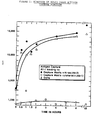
- PBS-BSA was added to the gel matrix pellet along with various concentrations of cell extract supernatants as designated in individual experiments (Results). The mixture was either incubated overnight at 4°C or for various periods of time at 37 0 c (time course experiment) to optimize the binding of the ras p2l protein to the antigen capture matrix. Following two washes with PBS , 0.1 ml of 125 I labelled v-H-ras (Ab-1) or v-H-ras (Ab-2) monoclonal antibody (60,000 cpm, 1.0 uCi 125 Iodine per ug protein) was added, and the mixture incubated for 1.5 hours at 37°C. The gel matrix was washed one time with PBSTDS and two times with PBS and counted in a gamma counter (LKB #1274 Riagamma).
- the gel matrix pellet was suspended in two times its volume of P B S containing 0.1% sodium azide, and 0.01 ml was transferred to a round bottom tube (12 x 75 mm, Enkay) containing a bacterial growth and protease inhibitor solution (BGIPI, 0.05 ml) of final concentrations of 0.005% TPCK (Sigma), 0.01% soybean trypsin inhibitor (Sigma), 0.01% sodiun azide, and 0.01% sodium fluoride and 2.75 ul (0.054 TIU) of an aprotinin (Sigma) (19.8 TIU/ml).
- BGIPI bacterial growth and protease inhibitor solution
- the gel matrix was washed 2 times with 3.0 ml PBS, and approximately 0.1 ml volume of liquid was retained with the gel matrix pellet.
- the pellet was rocked for 2 hours at 37°C with 0. 1 m l of 125 I-labeled monoclonal antibody [v-H-ras (Ab-1) or v-H-ras (Ab-2) - approximately 60,000 CPM, 1.0 uC i 125 Iodine per 1 . 0 microgram protein], washed one time with 3.0 ml of PBSTDS, two times with 3.0 ml PBS, and finally counted in a gamma counter.
- Monoclonal antibodies with specificity for ras p2l were covalently bound to Tresyl-activated Sepharose® 4B (Pharmacia, Lot. MC01773) as described by the manufacturer.
- Biological specimens in ras buffer (Tris-HC1 10 mM, pH 7.5, 1mM magnesium chloride, 1 mM dithiothieo- tol, 0.1 mM, GTP, 0.1% octylglucoside and 0.02% sodium azide) were applied at 4°C, the column was washed with 10 mM Tris-HCl, pH 8.0 buffer containing, 0.5 M sodium chloride until the absorbance at 280 nm reached base line.
- the ras protein was eluted with 0.1 M sodium citrate at pH 3.5 and individual fractions (3 ml) were immediately neutralized with solid Tris base and 0.3 ml of lOx ras buffer.
- Samples to be analyzed were first subjected to SDS-PAGE as described on 5-20% acrylamide gradient gels. After electrophoresis, transfer of proteins to nitrocellulose (Schleicher & Schuell, BA 85, 0.45 um) was performed. Proteins were transferred at 70 volts for 2-3 hours at 4°C. Greater than 90% of the ras protein was transferred from the gel as determined by staining the gel with Coomassie Blue after transfer.
- the nitrocellulose was incubated in 0.5% non-fat powdered milk (Carna- tion®, Societe des Produits Nestle, S.A., Vevey, Swit- zerland) in Phosphate buffered saline pH 7.4 (PBS) containing 0.02% sodium azide for 1 hour at room temperature.
- PBS Phosphate buffered saline pH 7.4
- the filter was then washed with PBS contain- i n g 0.1% Tween® 20 (Bio-Rad Lab) and incubated with 125 [I] labeled monoclonal antibody (2.0 x 10 6 CPM/ml) diluted in the same buffer for 1.5 hours at 37 0 C .
- the filters were washed three times with PBS-Tween® 20, dried and exposed to Kodak XR-2 film at -70°C using intensifying screens.
- Monoclonal antibodies to various oncogene encoded proteins were purified from ascites fluid of six different cloned hybridoma cell lines as described in Materials and Methods. Following final purification by HPLC (TSK-DEAE), the immunoglobulin fractions were pooled and their purity assessed by SDS-PAGE. Immunoglobulin was purified from ascites fluid as described in "Materials and Methods" and purity assessed by SDS-PAGE using a 5-20% linear acrylamide gradient. Samples of 30 micrograms, from each pooled immunoglobulin fraction, were applied to the individual lanes. Immunoglobulins were reduced with a final concentration of 5% B-Mercaptoethanol in SDS-PAGE sample buffer.
- v-H-ras pantropic anti-ras p2l, v-H-ras (Ab-2) H and K specific anti-ras p2l, v-fes (Ab-1), v-fms (Ab-2), p53 (Ab-1), and cytokeratin (Ab-1) were purified to homogeneity and upon reduction with B-mercaptoethanol separated into their respective heavy and light immunoglobulin chains. All the immunoglobulins were of the IgG class except the v-fes (Ab-1) immunoglobulin which was of the IgM class since its heavy chain electrophoresed at 70,000 daltons.
- the immunoprecipitates were analyzed by SDS-PAGE and the gels subjected to autoradiography.
- 35 S methionine labeled cellular extracts from tumor (A431, A375, A549, A204, A673, T-24, HeLa), transfected (Al-1) transformed (GA FeSV mink), and control (mink CC164 NIH 3T3 Cl 2.2) cell lines (described in Tables 2 and 3) were prepared as described in "Materials and Methods”.
- Oncogene encoded proteins were immunoprecipitated, as described in "Materials and Methods”.
- Immunoprecipitates were analyzed by SDS-PAGE using a 5-20% linear acrylamide gradient.
- the p53 protein was detected in A431, A375, A549, A2 04, GA FeSV Mink and control mink cells (CCL64), but its expression was not elevated in the other tumor cell lines tested.
- the transformed cell line GA FeSV mink expressed the p110 fes oncogene protein, while its control untransformed parental cell line, CCL64, did not.
- the Al-1, HL60-2, and OS-8-20-6-2 (OS-8) cell lines are derived from the NIH3T3 Cl 2.2 (3T3) mouse fibroblast cell lines transfected with the H-, N-, and K-ras genes, respectively. Further characterization of the antibody specificity of the v-H-ras (Ab-2) and v-H-ras ( A b-1) monoclonal antibodies was determined using these particular cell lines expressing the different ras gene encoded proteins. The specificity of the monoclonal antibodies v-H-ras (Ab-1) and v-H-ras (Ab-2) was determined by their ability to react with the human H, N, or K ras gene encoded protein.
- the v-fes (Ab-1) monoclonal antibody was included as a control.
- the immunoprecipitates were analyzed by SDS-PAGE, using a 5-20% linear acrylamide gradient, and autoradiography.
- v-H-ras (Ab-1) monoclonal antibody immunoprecipitated the H-ras (Al-1), K-ras (OS-8), and N-ras (HL-60-2) ras p21 protein; however, and the v-H-ras (Ab-2) specifically recognized both the H - ras (Al-1) and K-ras (OS-8) p21 protein but not the N - ras ( H L-60-2).
- the monoclonal antibody v-H-ras ( A b-1) is pantropic in that it recognizes a common determinant on the different ras encoded proteins.
- the v-H-ras (Ab-2) monoclonal antibody does not precipitate the N-ras gene product and is specific for K- and H - ras.
- the control v-fes (Ab-1) monoclonal antibody did not immunoprecipitate any p2l from extracts of ras gene transfected cell lines.
- the monoclonal antibody v-H-ras (Ab-1) was biotinylated and tested for its ability to immunoprecipitate ras p2l protein. Analysis of the modified antibody preparations was performed. The function of monoclonal antibody v-H-ras (Ab-1) following biotinylation was assessed by its ability to immunoprecipitate ras p21 in 35 S-labeled methionine cellular extracts (Al-1, T24, HD-8, canine thymus).
- Increasing amounts of cellular extracts were immunoprecipitated using 0.5 micrograms biotinylated antibody and 50 microliters of strepavidin-agarose suspension at 0.24 mg/ml.
- the immunoprecipitates were analyzed by SDS-PAGE, using a 5-20% linear acrylamide gradient, and autoradiography. Additionally, the functional capacity of monoclonals v-H-ras (Ab-2) and v-H-ras (Ab-1) coupled to the Affi-Gel® 10 matrix was assessed by its ability to bind ras p21 protein from 35 S - methionine labeled HD-8 cellular extract.
- the ability to immunoprecipitate ras p2l from a variety of cell types in a concentration dependent fashion rules out the possibility that the monoclonal antibodies were damaged by the biotinylation procedure.
- a volume of capture matrix containing 0.5 to 10 micrograms of covalently linked antibody, was added to a constant volume of 1.0 ml of cellular extract. It was found that increasing the amounts of capture matrix added over 1.0 micrograms did not increase the amount of ras p21 protein bound.
- a preliminary antigen capture procedure employed biotinylated v-H-ras (Ab-1) as the detector labeling system.
- Ab-1 biotinylated v-H-ras
- HD-8 cell membranes were used as a source of ras p2l protein.
- the cells (1 x 10 7 ) were disrupted in hypotonic PBS (diluted 1:4 with water) with a dounce homogenizer and centrifuged at 15,000 RPM for ten minutes at 4°C, all as described in Materials and Methods.
- the cell membranes were suspended in 1.0 ml PBSTDS and incubated with the capture matrix (v-H-ras (Ab-1) and v-H-ras (Ab-2), 0.01 ml) followed by biotinylated v-H-ras (Ab-1) as described in Materials and Methods.
- the antigen capture matrix developed a blue color, indicating the presence of ras p2l protein, in the tube containing membrane extract of HD-8 cells but not any controls.
- Treatment of the gel matrix with 4.0% bovine serum albumin (BSA) before addition of the cell extract caused less of a blue color to develop. Therefore, this treatment decreases some non-specific binding of biotinylated second reporter antibody thus reducing the background.
- a control of membrane extract incubated with unlinked gel matrix was negative.
- the second reporter antibody did not bind non-specifically to the v-H-ras (Ab-2)/v-H-ras (Ab-1) antibody linked antigen capture matrix incubated with PBS or PBS-BSA alone.
- the reporter antibody was iodinated.
- Supernatants, from cellular extracts of HL-60, OS-8, Al-1, 3T3, and HD-8 cell lines were prepared as described in "Materials and Methods". Increasing quantities of cell extract from each cell line were incubated with the antigen capture matrix (v-H-ras (Ab-2) linked Affi-Gel® 10) to optimize the binding of the cell extract to the matrix.
- the reporter antibody, v-H-ras (Ab-1) was incubated with the cell extract-matrix as described in "Materials and Methods”.
- the anti-ras p21 capture matrix bound 10-20 fold more sample (as reflected by bound reporter v-H-ras (Ab-1) antibody-CPM) than the control matrix of anti-cytokeratin (anti-CK-4) antibody.
- the control 3T3 cells did not bind to the v-H-ras (Ab-2) complexed affinity matrix and demonstrated approximately the same amount of binding as the ras p2l containing extracts did to the control matrix, thus establishing the relative amount of non-specific binding, or background.
- addition of increasing concentrations of cell extract to the v-H-ras (Ab-2)-matrix showed increased binding, a linear response was not obtained.
- the antigen capture procedure was performed using v-H-ras (Ab-2) capture affinity matrix and 125 I-v-H-ras (Ab-1) reporter antibody as described in "Materials and Methods".
- Table 5 is a summary of up to four separate antigen capture experiments on the sera of a total of 70 cancer patients. Sera obtained from patients with non-neoplastic conditions are listed at the end of the table. Counts (CPM) higher than 700 were obtained in 22 out of 70 sera from cancer patients and the sera samples from non-cancer patients and normal donors were all below 700. The ras p21 protein was ostensibly detected in approximately 33% of patients sera examined.
- Antigen capture reactivity was detected in four sera samples which were previously negative using the "original" configuration of antibody presentation.
- the antigen capture procedure can be designed to be either specific for a particular ras protein or more general depending on the specificities of the antibodies used and the order of their presentation.
- v-H-ras (Ab-1) , v-H-ras (Ab-2), v-fms (Ab-2) and cytokeratin (Ab-1) were used as antibodies linked to capture matrices and v-H-ras (Ab-1), v-H-ras (Ab-2), and v-fms (Ab-2) were used as reporter antibodies.
- the test should be successful in obtaining negative results with a large number of sera from normal human donors. Forty-six normal human sera samples were incubated with both or either configuration of antibody presentation, i.e., v-H-ras (Ab-2) capture matrix with v-H-ras (Ab-1) reporter antibody and v-H-ras (Ab-1) capture matrix with v-H-ras (Ab-2) reporter antibody.
- a solution phase antigen capture test was evaluated. This assay was performed as described in "Materials and Methods.” As shown in Figure 2, the standard curve of the solution-phase antigen capture test was linear with respect to ras p2l for values between 1 and 50 units of p21 in which 1 unit is approximately equal to 1 ng of purified ras p21. Internal standards A, B, C containing 23.75, 6.25 and 1.25 units ras p2l reactivity respectively were assayed with each standard curve and were found to fall on the curve at all times.
- the Al-1 cell line contains 1000 units of ras p21 reactivity per mg of total cellular extract protein, and this reactivity corresponds to the human c-H-ras gene encoded protein.
- the OS-8 cell line known to be transfected with the human c-K-ras gene was found to contain 725 units of ras p21 reactivity per mg total cellular extract protein; and the HL-60-2 cell line, expressing human c-N-ras p21, assay result was 102 units per mg cellular extract protein.
- the c-H-ras p21 levels in the Al-1 cell line represent an arbitrary estimate based on the results of the purification experiments, and one unit is approximately equal to one nanogram of c-H-ras p21.
- the relative levels of 35 S-Methionine labeled p21 immunoprecipitable from these cell lines was estimated to be equal for A1-1 and OS-8 and approximately 50 percent lower for HL-60-2 (data not shown). These data indicate that the solution phase antigen capture test measures c-H-ras and K-ras encoded p21 with equal sensitivity but is slightly less sensitive for c-N-ras p21.
- Al-1 cellular extract was subjected to gel filtration over a column of AcA 54 (LKB Rockville, MD), that had been previously calibrated with molecular weight markers, and the fractions were analyzed for ras p2l content by antigen capture and Western blotting, Figure 3.
- Figure 4 shows similar data for a representative lung carcinoma. In both cases the p2l antigen capture reactivity and Western blotting reactivity eluted from the column at a position corresponding to a molecular weight of 21,000.
- the solution-phase antigen capture test was used in a series of control experiments and for the testing of both cancer patient and control human sera.
- the ras p2l content of cancer and normal human sera were compared and biotinylated capture antibody (v-H-ras Ab-1) was replaced by anti-p53 and v-fms monoclonal antibodies (Table 10).
- Testing of a cancer patient's sera (0.1 ml) resulted in a value of 1815 cpm, over a background value of 174 cpm for normal sera, and this corresponds to 65 units ras p21 per ml patient sera.
- the colon cancer patient sera tested contained 14 values above the mean of normal sera controls and 7 below the mean. A similar distribution was obtained for ovarian cancer patient sera with 13 positives out of 22 samples. The melanoma cancer patient sera all tested positive, but all of these patients were known to have advanced cancer. The levels of p21 were found to correlate with the relative success or failure of therapy.
- the antigen capture reactivity from serum was purified by monoclonal affinity chromatography and was found to Western blot at 21,000 daltons (Fig. 7).
- the purified ras reactivity was subjected to molecular sizing over a column of AcA 54 and chromatographed between the void volume of the column and the BSA marker.
- the ras p21 reactivity of cancer patient sera was found to Western blot at 21,000 daltons.
- ras p2l specific monoclonal antibodies to measure elevated ras oncogene expression by aminohistopathology. These procedures have the common problem of requiring tumor biopsy material and thus are not readily amenable to either early cancer detection or monitoring of tumor progression.
- ras p21 is a cellular membrane protein and thus not secreted physiologically into the blood stream, its presence in serum at appreciable levels is possible if there is sufficient tumor cell destruction.
- a determination as to whether ras p2l is present in human serum is important with respect to the wider implications of such a determination to other oncogene systems.
- the initial approach to the development of an immunologic test for ras p2l was to select a system that was highly sensitive, but also adaptable to a wide range of formats and applications.
- the antigen capture procedure was chosen for several reasons.
- the approach involves the use of a broadly reactive antibody coupled to a solid support matrix to purify and concentrate the oncogene-encoded protein, in this case ras p21, from cellular extracts, serum, or other biological material.
- the second antibody coupled to an indicator, either isotopic or non-isotopic, is then used to demonstrate the bound antigen. This procedure is adaptable to a wide range of manual and automated formats for antigen concentration by the first antibody.
- the use of the coupled second antibody as the detector systems makes possible the adaptation of the test to any of a wide range of available colormetric, fluorescent, luminescent or isotopic systems.
- the use of the coupled second antibody as a detector allows forrnat- ting of the test to make specific determinations as to the nature of the bound antigen. For instance, in the ras p21 oncogene system, it is possible to select detector reagents that discriminate H-, N- and K-ras, and even that recognize as to whether any one of the ras oncogene products has a point mutation at one of the positions known to be critical to ras oncogene activation. Similarly, the same format could be applied to other oncogene systems by selecting detector antibodies that discriminate the activated from the non-activated form of the oncogene-encoded protein.
Landscapes
- Chemical & Material Sciences (AREA)
- Health & Medical Sciences (AREA)
- Organic Chemistry (AREA)
- Medicinal Chemistry (AREA)
- Immunology (AREA)
- Biochemistry (AREA)
- Biophysics (AREA)
- General Health & Medical Sciences (AREA)
- Genetics & Genomics (AREA)
- Life Sciences & Earth Sciences (AREA)
- Molecular Biology (AREA)
- Proteomics, Peptides & Aminoacids (AREA)
- Oncology (AREA)
- Peptides Or Proteins (AREA)
- Preparation Of Compounds By Using Micro-Organisms (AREA)
- Investigating Or Analysing Biological Materials (AREA)
Applications Claiming Priority (4)
| Application Number | Priority Date | Filing Date | Title |
|---|---|---|---|
| US76786285A | 1985-08-21 | 1985-08-21 | |
| US767862 | 1985-08-21 | ||
| US88561786A | 1986-07-23 | 1986-07-23 | |
| US885617 | 1986-07-23 |
Publications (2)
| Publication Number | Publication Date |
|---|---|
| EP0214520A2 true EP0214520A2 (de) | 1987-03-18 |
| EP0214520A3 EP0214520A3 (de) | 1988-03-16 |
Family
ID=27117975
Family Applications (1)
| Application Number | Title | Priority Date | Filing Date |
|---|---|---|---|
| EP86111527A Withdrawn EP0214520A3 (de) | 1985-08-21 | 1986-08-20 | Immunotestverfahren zur Bestimmung von RAS-codierten Proteinen |
Country Status (4)
| Country | Link |
|---|---|
| EP (1) | EP0214520A3 (de) |
| AU (1) | AU615876B2 (de) |
| CA (1) | CA1281641C (de) |
| IL (1) | IL79755A0 (de) |
Cited By (7)
| Publication number | Priority date | Publication date | Assignee | Title |
|---|---|---|---|---|
| EP0206065A3 (de) * | 1985-06-27 | 1989-04-19 | James R. Feramisco | Verfahren zum Nachweis von oncogenischen und proto-oncogenischen Proteinen in extrazelligen biologischen Flüssigkeiten |
| EP0662612A1 (de) * | 1994-01-05 | 1995-07-12 | Channa Shalitin | Verfahren zum Nachweis benigner und maligner Tumore unter Verwendung neuer Antikörper |
| US5591587A (en) * | 1983-08-17 | 1997-01-07 | The Scripps Research Institute | Polypeptide-induced monoclonal receptors to protein ligands |
| US5674753A (en) * | 1991-06-03 | 1997-10-07 | Chiron Diagnostics Corporation | Epidermal growth factor receptor ectodomain |
| US6551789B1 (en) | 1983-08-17 | 2003-04-22 | The Scripps Research Institute | Process for characterizing a biological sample using patterns of oncogene expression |
| US6861511B1 (en) | 1986-06-04 | 2005-03-01 | Bayer Corporation | Detection and quantification of neu related proteins in the biological fluids of humans |
| JP2008547027A (ja) * | 2005-06-23 | 2008-12-25 | バイエル ヘルスケア エルエルシー | 体液中のrasp21の定量的検定法 |
Families Citing this family (1)
| Publication number | Priority date | Publication date | Assignee | Title |
|---|---|---|---|---|
| US5015568A (en) * | 1986-07-09 | 1991-05-14 | The Wistar Institute | Diagnostic methods for detecting lymphomas in humans |
Family Cites Families (4)
| Publication number | Priority date | Publication date | Assignee | Title |
|---|---|---|---|---|
| CA1252046A (en) * | 1982-11-04 | 1989-04-04 | Martin J. Cline | Methods for oncogenic detection |
| EP0149602A1 (de) * | 1983-07-25 | 1985-07-31 | Beckman Instruments, Inc. | Immunometrische bestimmung in der polyklonale und monoklonale antikörper verwendet werden und ein testsatz dafür |
| US4798787A (en) * | 1984-09-19 | 1989-01-17 | Cetus Corporation | Peptide antibodies and their use in detecting oncogene products |
| EP0203587A3 (de) * | 1985-05-30 | 1989-08-09 | F. Hoffmann-La Roche Ag | Ras-Oncogene-Peptide und Antikörper |
-
1986
- 1986-08-18 AU AU61540/86A patent/AU615876B2/en not_active Ceased
- 1986-08-18 IL IL79755A patent/IL79755A0/xx unknown
- 1986-08-19 CA CA000516260A patent/CA1281641C/en not_active Expired
- 1986-08-20 EP EP86111527A patent/EP0214520A3/de not_active Withdrawn
Cited By (8)
| Publication number | Priority date | Publication date | Assignee | Title |
|---|---|---|---|---|
| US5591587A (en) * | 1983-08-17 | 1997-01-07 | The Scripps Research Institute | Polypeptide-induced monoclonal receptors to protein ligands |
| US6551789B1 (en) | 1983-08-17 | 2003-04-22 | The Scripps Research Institute | Process for characterizing a biological sample using patterns of oncogene expression |
| EP0206065A3 (de) * | 1985-06-27 | 1989-04-19 | James R. Feramisco | Verfahren zum Nachweis von oncogenischen und proto-oncogenischen Proteinen in extrazelligen biologischen Flüssigkeiten |
| US6861511B1 (en) | 1986-06-04 | 2005-03-01 | Bayer Corporation | Detection and quantification of neu related proteins in the biological fluids of humans |
| US5674753A (en) * | 1991-06-03 | 1997-10-07 | Chiron Diagnostics Corporation | Epidermal growth factor receptor ectodomain |
| EP0662612A1 (de) * | 1994-01-05 | 1995-07-12 | Channa Shalitin | Verfahren zum Nachweis benigner und maligner Tumore unter Verwendung neuer Antikörper |
| JP2008547027A (ja) * | 2005-06-23 | 2008-12-25 | バイエル ヘルスケア エルエルシー | 体液中のrasp21の定量的検定法 |
| EP1893997A4 (de) * | 2005-06-23 | 2009-02-18 | Siemens Healthcare Diagnostics | Quantitative assays für ras p21 in körperflüssigkeiten |
Also Published As
| Publication number | Publication date |
|---|---|
| IL79755A0 (en) | 1986-11-30 |
| AU6154086A (en) | 1987-02-26 |
| EP0214520A3 (de) | 1988-03-16 |
| CA1281641C (en) | 1991-03-19 |
| AU615876B2 (en) | 1991-10-17 |
Similar Documents
| Publication | Publication Date | Title |
|---|---|---|
| US5519118A (en) | Human MDM2 protein involved in human tumors | |
| Shea et al. | Identification of an anionic form of glutathione transferase present in many human tumors and human tumor cell lines | |
| Hirai | A collaborative clinical study of carcinoembryonic antigen in Japan | |
| Simmons et al. | Analysis of complex relationships between age, p53, epidermal growth factor receptor, and survival in glioblastoma patients | |
| Takahashi et al. | Association of serum endoglin with metastasis in patients with colorectal, breast, and other solid tumors, and suppressiveeffect of chemotherapy on the serum endoglin | |
| Rusch et al. | Overexpression of the epidermal growth factor receptor and its ligand transforming growth factor alpha is frequent in resectable non-small cell lung cancer but does not predict tumor progression. | |
| EP0453560B1 (de) | Lokalisation und charakterisierung des wilms-tumor-gens | |
| Sikora et al. | Detection of the c-myc oncogene product in testicular cancer | |
| US4584268A (en) | Method and compositions for carcinoma diagnosis | |
| Bradley et al. | Increased expression of the epidermal growth factor receptor on human colon carcinoma cells | |
| CA2118015A1 (en) | Method of detecting tumors containing complexes of p53 and hsp70 | |
| Albanopoulos et al. | Prognostic significance of circulating antibodies against carcinoembryonic antigen (anti-CEA) in patients with colon cancer | |
| Takahashi et al. | Antibody to ras proteins in patients with colon cancer. | |
| EP0576476B1 (de) | Immunoassay zum nachweis des mutanten p53 polypeptids in biologischen flüssigkeiten | |
| ES2351938T3 (es) | Reactivos y métodos para su uso en el diagnóstico, clasificación y terapia del cáncer . | |
| EP0214520A2 (de) | Immunotestverfahren zur Bestimmung von RAS-codierten Proteinen | |
| EP0354808B1 (de) | Polypeptid-induzierte monoklonale Rezeptoren gegen Proteinliganden | |
| US6083707A (en) | Thymidine kinase TK1, peptide, corresponding antibodies and use of these in determination of tumor proliferation | |
| Stock et al. | Ras related oncogene protein as a tumor marker in transitional cell carcinoma of the bladder | |
| Hamer et al. | Production and characterization of anti-RAS p21 monoclonal antibodies | |
| CA1340368C (en) | Detection, quantitation and classification of ras proteins in body fluids and tissues | |
| Stass et al. | Antigen-antibody complexes related to the baboon endogenous virus in humans with acute lymphoblastic leukemia-hand mirror variant (ALL-HMC) | |
| Masson et al. | Evaluation of CEA, CA 19-9, CA-50, CA-195, and TATI with special reference to pancreatic disorders | |
| Mirowski et al. | Serological and immunohistochemical detection of a 65-kDa oncofetal protein in breast cancer | |
| Coombs et al. | Immunocytochemical localization of c‐erbB‐2 protein in transitional cell carcinoma of the urinary bladder |
Legal Events
| Date | Code | Title | Description |
|---|---|---|---|
| PUAI | Public reference made under article 153(3) epc to a published international application that has entered the european phase |
Free format text: ORIGINAL CODE: 0009012 |
|
| AK | Designated contracting states |
Kind code of ref document: A2 Designated state(s): AT BE CH DE FR GB IT LI LU NL SE |
|
| PUAL | Search report despatched |
Free format text: ORIGINAL CODE: 0009013 |
|
| AK | Designated contracting states |
Kind code of ref document: A3 Designated state(s): AT BE CH DE FR GB IT LI LU NL SE |
|
| 17P | Request for examination filed |
Effective date: 19880906 |
|
| 17Q | First examination report despatched |
Effective date: 19900907 |
|
| REG | Reference to a national code |
Ref country code: FR Ref legal event code: ST |
|
| STAA | Information on the status of an ep patent application or granted ep patent |
Free format text: STATUS: THE APPLICATION IS DEEMED TO BE WITHDRAWN |
|
| 18D | Application deemed to be withdrawn |
Effective date: 19930313 |